Embed Size (px)
Citation preview

| INVESTIGATION
Host Mitochondrial Association Evolved in theHuman Parasite Toxoplasma gondii via
Neofunctionalization of a Gene DuplicateYaw Adomako-Ankomah,*,1 Elizabeth D. English,*,1 Jeffrey J. Danielson,* Lena F. Pernas,†
Michelle L. Parker,‡ Martin J. Boulanger,‡ Jitender P. Dubey,§ and Jon P. Boyle*,2
*Department of Biological Sciences, Kenneth P. Dietrich School of Arts and Sciences, University of Pittsburgh, Pittsburgh,Pennsylvania 15260, †Department of Microbiology and Immunology, Stanford University School of Medicine, Stanford, California94305, ‡Department of Biochemistry and Microbiology, University of Victoria, Victoria, British Columbia, VP8 5C2, Canada, and§Animal Parasitic Diseases Laboratory, Beltsville Agricultural Research Center, Agricultural Research Service, US Department of
Agriculture, Beltsville, Maryland 20705
ABSTRACT In Toxoplasma gondii, an intracellular parasite of humans and other animals, host mitochondrial association (HMA) isdriven by a gene family that encodes multiple mitochondrial association factor 1 (MAF1) proteins. However, the importance of MAF1gene duplication in the evolution of HMA is not understood, nor is the impact of HMA on parasite biology. Here we used within- andbetween-species comparative analysis to determine that the MAF1 locus is duplicated in T. gondii and its nearest extant relativeHammondia hammondi, but not another close relative, Neospora caninum. Using cross-species complementation, we determined thatthe MAF1 locus harbors multiple distinct paralogs that differ in their ability to mediate HMA, and that only T. gondii and H. hammondiharbor HMA+ paralogs. Additionally, we found that exogenous expression of an HMA+ paralog in T. gondii strains that do not normallyexhibit HMA provides a competitive advantage over their wild-type counterparts during a mouse infection. These data indicate thatHMA likely evolved by neofunctionalization of a duplicateMAF1 copy in the common ancestor of T. gondii and H. hammondi, and thatthe neofunctionalized gene duplicate is selectively advantageous.
KEYWORDS gene duplication; Toxoplasma gondii; Hammondia hammondi; Neospora caninum; neofunctionalization
GENE duplication is known to underlie the evolution ofnew gene functions and ultimately organismal pheno-
types (Ohno 1970; Espinosa-Cantu et al. 2015). The expectedoutcome of most gene duplication events is that they willbe lost by nonsense mutation and/or resolution of the locus(Ohno 1970; Lynch and Conery 2000; Lynch and Force 2000).However, those that confer a selective advantage through genedosage, subfunctionalization, or neofunctionalization, can be-come fixed in the population (Ohno 1970; Lynch and Conery2000; Lynch and Force 2000; Espinosa-Cantu et al. 2015). The
phenotypic impact of locus expansions can be high in bothnatural and laboratory settings. When grown in noncompati-ble human cells, vaccinia virus was found to expand, diversify,and then contract the K3L locus, resulting in a highly adaptedvirus with a single K3L gene that could now disrupt the anti-viral host protein Protein Kinase R (Elde et al. 2012). Labora-tory studies with bacteria show that adaptation to selectiveconditions (stress or antibiotic exposure) via gene expansionand diversification occursmuchmore frequently than via pointmutation (Kugelberg et al. 2006, 2010). Field studies withDrosophila spp. have identified Cyp6g1 duplication and diver-sification events as one source of resistance to insecticides suchas dichlorodiphenyltrichloroethane (DDT) (Emerson et al.2008; Cridland and Thornton 2010; Schmidt et al. 2010).
The examples above detail the importance of gene dupli-cation in the evolution within species both in the laboratoryand in the field. However, less is known about the impactof gene duplication and diversification events in definingspecies-specific traits (or even defining the species themselves,
Copyright © 2016 by the Genetics Society of Americadoi: 10.1534/genetics.115.186270Manuscript received December 18, 2015; accepted for publication February 14, 2016;published Early Online February 22, 2016.Available freely online through the author-supported open access option.Supplemental material is available online at www.genetics.org/lookup/suppl/doi:10.1534/genetics.115.186270/-/DC1.1These authors contributed equally to this work.2Corresponding author: Department of Biological Sciences, University of Pittsburgh,LSA 101, 4249 Fifth Ave., Pittsburgh, PA 15260. E-mail: [email protected]
Genetics, Vol. 203, 283–298 May 2016 283

whichwas postulatedbyOhno(1970)). It is certainly clear thatthere are specific gene duplication events that distinguishclosely related species (such as humans and chimpanzees)(Bailey and Eichler 2006), but exampleswhere species-specificgene expansions have been linked to species-specific traits arefew.
Pathogens provide a unique setting in which to study theevolution and emergence of novel traits, given their largepopulation size and the intense selective pressures placedupon them by the host. We use comparative approaches tounderstand the evolution of unique traits in members ofApicomplexa, a phylum of parasites of great importance inhuman and veterinary health. Our main focus is on Toxo-plasma gondii and its near relatives. T. gondii is an importantpathogen of humans, particularly in HIV/AIDS patients andthe developing fetus. In addition, T. gondii is capable of infect-ing, causing disease in, and being transmitted by all warm-blooded animals studied to date (Dubey and Sreekumar2003). In contrast,HammondiahammondiandNeospora caninumhave comparatively restricted host ranges and are not patho-genic in rodents or humans (Goodswen et al. 2013; Walzeret al. 2013). This is despite a high level of genetic similarityand genome-wide synteny across these three species (Reidet al. 2012; Walzer et al. 2013), and in the case of T. gondiiand H. hammondi, extensive conservation of virulence effec-tors at both the sequence and functional levels (Walzer et al.2013, 2014).
The unique phenotypic and life cycle features of T. gondiihave most certainly contributed to its near global distributionand an incidence rate that ranges from 10 to 80% in humans.However, the genetic bases for these phenotypes are un-known, and to begin to address this question we have takena comparative approach to identify genetic loci that are uniqueto T. gondii compared to H. hammondi and N. caninum. Indoing so, we found that a small subset of T. gondii loci haveundergone tandem duplication, expansion, and diversificationonly in the T. gondii lineage. Specifically, expanded loci arepoorly conserved between T. gondii and its near relatives, hav-ing a higher propensity to be either missing, or not similarlyexpanded, in either N. caninum or H. hammondi (or both)(Adomako-Ankomah et al. 2014) than single-copy genes. Ona gene-by-gene basis, expanded and diversified gene familiesare known to play important roles in parasite biology andwithin-species adaptation in T. gondii and Plasmodium spp.(reviewed in Reid 2015). For example, members of the vargene family are distributed throughout the P. falciparum ge-nome and encode erythrocyte membrane antigens (PfEMPs)that are secreted into the host red blood cell during infection.PfEMPs are key determinants of parasite virulence and areunder strong diversifying selection (Freitas-Junior et al.2000; Deitsch et al. 2001; Pasternak and Dzikowski 2009).In both the field (Nair et al. 2008) and laboratory (Heinberget al. 2013), copy number increases at the gch1 locus inP. falciparum confer resistance to pyrimethamine. In T. gondii,there are .50 members of the rhoptry protein 2 (ROP2)superfamily, and they are dispersed throughout the genome
(Boothroyd and Dubremetz 2008). We recently showed thatmany members of the ROP2 superfamily are encodedby tandemly expanded gene clusters that have diversifiedsignificantly via positive selection (Reese et al. 2011;Adomako-Ankomah et al. 2014). One such example is theROP5 locus, which is crucial for mouse virulence across theT. gondii phylogeny (Reese et al. 2011; Behnke et al. 2015).The ROP5 locus harbors multiple paralogs that are understrong diversifying selection both between and withinstrains (Reese et al. 2011). Importantly, individual ROP5paralogs have synergistic, rather than additive, effects onmouse virulence, stressing the importance of paralog diver-sification in conferring the entire locus-driven phenotype(Reese et al. 2011). Importantly, duplicated and expandedloci represent a highly significant fraction of the geneticdifference between T. gondii and its nearest relatives (Wasmuthet al. 2009; Adomako-Ankomah et al. 2014). Based on thesedata, our overall hypothesis is that selective locus expansion,and subsequent selection-driven diversification of individualparalogs, have played an important role in the evolution of traitsthat are unique to T. gondii.
One such locus is mitochondrial association factor 1(MAF1) (Adomako-Ankomah et al. 2014; Pernas et al.2014). The MAF1 locus is uniquely amplified in T. gondiirelative to N. caninum and H. hammondi (annotated asExpanded Locus 4) (Adomako-Ankomah et al. 2014) andis required for host mitochondrial association (HMA) inT. gondii (Pernas et al. 2014). The MAF1 locus encodes afamily of dense granule proteins that associate with theparasitophorous vacuolar membrane (PVM) that are neces-sary and sufficient for the HMA phenotype (Pernas et al.2014), andMAF1 protein expression itself leads to significantchanges in the host immune response to infection. The discov-ery of the MAF1 gene family as being responsible for HMAopens the door to solving a long-standing question in T. gondiibiology regarding the importance of HMA in parasite infectiv-ity and virulence.
In the present study, we use intra- and interspecies com-parative analyses and molecular genetics to thoroughly tracethe functional evolutionary history of the MAF1 gene inT. gondii. While the impact of MAF1 on HMA is clear, theevolutionary history of theMAF1 locus in terms of copy num-ber and gene content is not, nor is the role of gene duplicationitself in the evolution of the HMA phenotype. TheMAF1 locusprovides a unique opportunity to assess the relative impactsof gene duplication and subsequent diversification on a ro-bust cellular phenotype. Moreover, the impact of MAF1 onparasite fitness in vivo has not been thoroughly investigated.To answer these questions, we have sequenced multipleMAF1 paralogs from three T. gondii clonotypes as well asthe nearest extant relatives of T. gondii, H. hammondi, andN. caninum. We have determined that the MAF1 locus hasundergone extensive sequence diversification and has beensubjected to positive selection in T. gondii. We show thatMAF1 copy number and gene content vary both betweenand within major T. gondii lineages and that not all copies
284 Y. Adomako-Ankomah et al.

of MAF1 mediate HMA. Through cross-species complemen-tation experiments, we show that expression of an “HMA-competent” T. gondii MAF1 paralog inN. caninum is sufficientto confer the HMA phenotype in this species, indicating thatthe HMA competence of MAF1 emerged only recently in theparasites in question. Using additional genomic and cross-species complementation experiments, we also providestrong support for a model in which the MAF1 locus dupli-cated one time in a common ancestor of all three species,diversified one time in an ancestor to H. hammondi andT. gondii, and then amplified and diversified multiple timesin the T. gondii lineage. Finally, we show that not only dodifferent MAF1 paralogs differ in their ability to mediateHMA, but also in their ability to confer a selective advantageduring infection in a mouse model. Taken together our datalink a specific gene duplication and neofunctionalizationevent in the evolution of a novel trait (host mitochondrialassociation), and in doing so we have uncovered the selectiveadvantage that likely fixed the MAF1 locus in most of theT. gondii population.
Materials and Methods
Sequence coverage and copy number analysis
Copy number analysis was performed as described previously(Adomako-Ankomah et al. 2014; Pernas et al. 2014). Briefly,raw sequence reads from multiple T. gondii strains andN. caninum (Liverpool strain) (Reid et al. 2012) were down-loaded from the NCBI trace archive in fasta format (strainsGT1, ME49, and VEG were derived from Sanger-basedshotgun sequencing; MAS, P89, FOU, VAND, and RUBwere generated using Roche 454 technology). T. gondii andN. caninum reads were aligned to the T. gondiiME49 genome(ToxoDB version 7.3; www.toxodb.org) using BLAT (param-eters: -fastMap –minIdentity = 95 –minScore = 90) (Kent2002), and coverage was calculated in each 500-bp windowusing coverageBed (from the Bedtools suite) (Quinlan andHall 2010). H. hammondi reads (strain HH34) (Lorenzi et al.2016) were aligned using Bowtie2 (using default parametersplus –end-to-end) (Langmead and Salzberg 2012), and se-quence coverage calculationsweremade using the integratedgenome browser (IGB) (Nicol et al. 2009). All coverage andannotation data were then plotted using custom scripts in Rstatistical software. To do this, start and end coordinates ofregions of theMAF1 locus were noted and data were normal-ized to the average coverage of�20 Kb upstream of the locus(Lorenzi et al. 2016).
Parasite strains and host cell maintenance
All T. gondii and N. caninum strains used in this study weremaintained by regular passage of tachyzoites from freshlylysed human foreskin fibroblast (HFF) onto new HFF mono-layers and grown at 37� in 5% CO2. HFF and NRK-mitoRFPcells (a kind gift from Jennifer Lippincott-Schwartz, NIH,Bethesda, MD) (Mitra and Lippincott-Schwartz 2010) were
grown in Dulbecco’s modified Eagle’s medium (DMEM) sup-plemented with 10% FBS, 2 mM glutamine, and 50 mg/mleach of penicillin and streptomycin.
To produce H. hammondi oocysts, interferon-g KO micewere fed 104 H. hammondi oocysts and killed�60 days post-infection (pi). Muscles from infected mice were then fed to10- to 20-week-old cats, and feces were collected during days5–11 postinfection. Unsporulated oocysts were isolated bysucrose flotation, and the resulting oocysts were allowed tosporulate at ambient temperature in 2% H2SO4 (Dubey andSreekumar 2003). Oocyst preparations were stored at 4� forno longer than 6months. Sporulated oocysts (40–80 million)were washed four times in Hank’s Buffered Saline Solution(HBSS) and treated with 10% bleach in PBS for 30 min.Pellets were resuspended in 4 ml HBSS and vortexed at max-imum speed along with 1 g of sterile glass beads (710–1180 mM, Sigma) for 30 sec, allowed to cool for 30 sec,and then vortexed for 30 sec again. DNAwas isolated directlyfrom the pellet of cracked oocysts (containing sporocysts re-leased from the oocysts and debris) using the DNAzol reagent(Invitrogen; Carlsbad, CA).
In other cases, we used the sporocyst preparation to gen-erate in vitro cultures ofH. hammondi. To do this, we exposedthe cracked oocyst preparation to PBS containing trypsin(Sigma T4799; 12.5 mg/ml) and taurocholic acid (SigmaT4009; 50 mg/ml) at 37� for 30 min. The reaction wasquenched by the addition of cDMEM (containing 10% FBS)and we removed debris from the preparation by filtrationthrough 5-mm syringe filters (Millipore). The resulting spo-rozoites were used to infect confluent monolayers of HFFsseeded on 12-mm circle glass coverslips. Samples were fixedand processed for immunofluorescence (IF) as describedbelow.
High molecular weight Southern blotting
Southern blotting was performed as previously described(Adomako-Ankomah et al. 2014). The six strains of T. gondiiused were GT1 and RH (type I), ME49 and PRU (type II), andVEG and CTG (type III). Genomic DNA from each strain wasdigested with ScaI restriction enzyme in a 100-ml reactionvolume for �12 hr and resolved by pulsed field gel electro-phoresis (Bio-Rad CHEF-DR III system). Resolved fragmentswere probed with DIG-labeled (Roche)MAF1-specific probesfollowed by chromogenic detection as per manufacturer’sprotocol.
Amplification and cloning of MAF1 paralogs fromT. gondii, H. hammondi, and N. caninum, and constructionof mutant constructs
Due to the fact that the MAF1 locus exhibits significant copynumber variation across species and strains, we used long-extension PCR and cloning to identify MAF1 paralogs inT. gondii (strains RH, ME49, and CTG), H. hammondi (strainHhCatGer041), and N. caninum (strain NC-1). Primersequences are listed in Supplemental Material, Table S1.Long extension PCR was used to minimize the potential for
Derived Trait Evolution in T. gondii 285
chimera formation between different MAF1 paralogs (as de-scribed in Pernas et al. 2014). For cloned sequences, all poly-morphisms were validated by querying a local copy of thesequence read database for the presence of that polymor-phism along with at least 40 bp of flanking sequence (RHwas compared to GT1; ME49 was compared to ME49; CTGwas compared to VEG) (Lorenzi et al. 2016). This servedthree purposes: validation of SNPs specific to a given clonallineage, elimination of PCR-derived SNPs, and controlled forthe possibility of generating interparalog chimeric sequencesduring PCRamplificationwhen the polymophismswere#40bpapart. Since our SNP curation method relied on comparingbetween members of the same clonal lineage (e.g., RH vs.GT1), it is possible that some isolate-specific SNPs were arti-ficially eliminated during curation. We did not identify anyevidence for chimerism in our sequences although this out-come cannot be completely ruled out.
Sequence analysis
Coding sequences of MAF1 paralogs from multiple T. gondiistrains and other species were analyzed using algorithmsimplemented in MEGA6 (Tamura et al. 2013) as follows:Specifically, coding sequences were translated into proteinand aligned using Muscle (default settings). Phylogenetictrees were constructed using maximum parsimony and thesubtree-pruning-regrafting algorithm (Nei and Kumar 2000).Search level was 1 and the initial trees were obtained bythe random addition of sequences (10 replicates were per-formed). Branch lengths were calculated using the averagepathway method. All positions containing gaps and missingdata were eliminated, and there were a total of 359 usefulpositions in the final dataset.
We calculated pairwise dN/dS ratios for all “b” paralogs(including the HMA-incompetent b0 paralogs) to determineif they had been under either positive or purifying selection.To do this, we used the modified Nei-Gojobori method withthe assumed transition/transversion bias of 2 (Zhang et al.1998), and as above, all positions containing gaps were elim-inated. All analyses were conducted inMEGA6 (Tamura et al.2013) and pairwise P-values for dN/dS ratios were deemedsignificant at P , 0.05.
Generation of expression constructs andtransgenic parasites
Generation of pMAF1RHb1 (N-terminally hemagglutinin(HA)-tagged MAF1b) expression construct has been de-scribed previously (Pernas et al. 2014). For pMAF1RHa, thecoding sequence forMAF1RHa was amplified from RH cDNA,cloned, and then used in a splicing by overlap extension (SOE)PCR reaction to fuse the N-terminal portion of MAF1RHb1gene and the C-terminal portion of the MAF1RHa1 gene. Thespecific construct contained the MAF1RHb1 promoter, startcodon, signal sequence, an HA tag (as in Pernas et al. 2014),and this was followed by the remainder of the C terminusencoding portion of the MAF1RHa1 gene. The TgMAF1RHb0,TgMAF1RHb1, HhMAF1a1, HhMAF1b1, and NcMAF1 con-
structs were made using SOE PCR to introduce an HA-tagfollowing the predicted signal peptide for each isoform. Plas-mid templates for the first round of PCR were generated fromgenomic DNA, which included 1116 bp upstream of the startsite to include the putative promoter. Transgenic parasite lineswere generated by transfecting TGME49Dhpt (MDLuc) andNC-1Dhpt parental strains with 50 mg of HindIII-linearizedplasmid. Stable expression lines were isolated by selection inmycophenolic acid (MPA)/xanthine followed by limiting di-lution in 96-well plates.
TgMAF1RHa1 and TgMAF1RHb1 cloning, proteinproduction, and purification
A construct encoding the predicted C-terminal domain ofTgMAF1RHb1 (Thr159 to Asp435) was codon optimizedfor Escherichia coli and synthesized by GenScript. A constructof TgMAF1RHa1 containing the analogous C-terminal do-main (TgMAF1RHa1; Ser173 to Ser443) was amplified fromT. gondii cDNA. Each construct was subcloned into amodifiedpET28a vector encoding an N-terminal hexa-histidine tagseparated from the sequence of interest by a tobacco etchvirus (TEV) protease cleavage site. Constructs were producedrecombinantly in E. coli BL21 cells. Following 4 hr of growthat 310 K and 12 hr at 303 K, the cells were harvested bycentrifugation, resuspended, and lysed using a French press.TgMAF1 proteins were purified from the soluble fraction byNi-affinity chromatography, the His tag was removed by TEVprotease, and TgMAF1 proteins were further purified by sizeexclusion chromatography on a Superdex 75 16/60 HiLoadcolumn in HBS (20 mM Hepes pH 7.5, 150–300 mM NaCl)with 1% glycerol and 1 mM dithiothreitol.
Generation of polyclonal antibodies
Female Balb/c mice were injected intraperitoneally (ip) with100 mg of either TgMAF1RHa1 antigen or TgMAF1RHb1antigen (purification described above) suspended in 100 mlPBS and mixed 1:1 with Sigma adjuvant (Sigma S6322) to afinal volume of 200 ml. Additional injections of 50 mg of theappropriate antigen mixed 1:1 with Sigma adjuvant to a finalvolume of 200ml were administered 14, 35, and 56 days afterthe initial injection. Sera were collected prior to initial injec-tion, as well as on days 31, 81, and 88. All sera were tested forreactivity against both TgMAF1RHa1 and TgMAF1RHb1 byWestern blot prior to use in Western blots or immunofluores-cence assays.
Immunofluorescence assays and confocal microscopy
HFFs or NRK-mitoRFP cells were seeded on 12-mm coverslipsin 24-well plates and grown to �80% confluency. NRK-mitoRFP cells were infected with N. caninum or T. gondiistrains expressing GFP and incubated for 8 hr. For HFFs,MitoTracker staining was performed as follows: Growthmedium on the HFF monolayer was replaced with DMEMcontaining MitoTracker (Red CMXRos, Invitrogen) at a30-nM concentration and incubated for 30 min at 37�. Cellswere then washed with PBS, infected with parasites in
286 Y. Adomako-Ankomah et al.
prewarmed DMEM, and incubated for 4 hr at 37�. After in-cubation, the infected cells were washed with PBS, fixedwith 3% paraformaldehyde in PBS for 15 min, and blocked/permeabilized in PBS containing 5% BSA and 0.2% TritonX-100. Alternatively, NRK-mitoRFP infected cells were fixedwith 3% PFA and either mounted directly or Hoechststained prior to mounting followed by visualization. Fixedcells were then immunostainedwith rat monoclonal anti-HA(3F10 clone, Roche) at 1:1000, mouse anti-MAF1a/b poly-clonal antibodies at 1:1000, or mouse monoclonal anti-MTCO2 (ab110258, Abcam) at 1:500.
Quantification of vacuole coverage
Percent vacuole coverage was determined using confocalmicroscopy and ImageJ. Populations transfected with HA-tagged TgMAF1RHb1 or HhMAF1b1 were fixed and stainedwith anti-HAandanti-MTCO2primary antibodies. Confocalimages were taken in three channels; 594 (anti-MTCO2),488 (anti-HA), and DIC. All three images were converted to8-bit images and merged using ImageJ. Vacuoles weretraced while only the DIC and green channels were visible,and then pixel intensity along the vacuole was measured inthe red channel. Pixel intensities .20 were considered tobe mitochondria. Percent vacuole coverage was calculatedby measuring the length of the vacuole trace with pixelintensity .20 and dividing it by total vacuole trace length.Twenty HA-positive vacuoles were measured for both theTgMAF1RHb1 and HhMAF1b1 populations. Ten HA-negativevacuoles were measured from each population (20 total) asa WT control.
Western blot analysis
Parasites were filtered away from host cell debris and lysed in13 SDS lysis buffer. Proteins were resolved by SDS-PAGE,transferred onto nitrocellulose membrane, and blocked for1 hr in 5% (w/v) milk in TBS-Tween20 (TBS-T). Primaryantibody incubation was performed in blocking buffer for45–120 min followed by three washes in TBS-T. Anti-HAand anti-MAF1 (Pernas et al. 2014) antibodies were usedat 1:1000 while anti-SAG1 was used at 1:2000 and rabbitanti-ROP5 (Behnke et al. 2011) was used at 1:40,000. Anti-TgMAF1RHa1 and anti-TgMAFRHb1 antibodies generatedfor this study were used at a 1:10,000 dilution. Secondaryantibody incubationwas performedwithhorseradishperoxidase-conjugated secondary antibodies to the respective primaryantibodies in blocking buffer for 45 min. Bands were visu-alizedwith SuperSignalWest Pico chemiluminescent substrate(Thermo Scientific). Densitometric analysis was performedusing ImageJ.
Animal experiments
All mouse experiments were performed with 4- to 8-wk-oldBALB/C mice. All animal procedures in this study meet thestandards of the American Veterinary Association and wereapproved locally under Institutional Animal Care and UseCommittee protocol no 12010130.
In vitro and in vivo competition assays
In vitro competition assays were performed as follows: AnME49 strain engineered to express an N-terminal HA-taggedtype I (RH) MAF1 (ME49:TgMAF1RHb1) was mixed withME49:WT in ratios 4:1 and 1:4. These twomixed populationswere used to infect HFFs at an MOI of 3. Flasks were passedvia syringe lysis every 3 days. At the 0-, 4-, and 8-wk timemarks, HFFs grown on 12-mm glass coverslips were infectedat anMOI of 3, and the proportion of HA+andHA2 parasiteswas calculated by immunofluorescence using rat a-HA (asabove) and serum from a mouse chronically infected withT. gondii at 1:1000 dilution. The ratio of HA+ to HA2 wasdetermined by counting at least 200 vacuoles. The entireexperiment was repeated two times, each time with a genet-ically distinct clone set (WT and complemented).
In vivo competition assays were performed as follows: Us-ing the same genetically engineered ME49 clone sets, weagain created mixed populations at ratios of 1:4, 1:1, 4:1,100% ME49:TgMAF1RHb1, and 100% ME49:WT. We in-jected 105 tachyzoites intraperitoneally of these five popula-tions into Balb/c mice in 200 ml of PBS (three to five mice perpopulation). In a separate experiment, we transfected ME49with the same pTgMAF1RHa1, grew the population underMPA/xanthine selection for 2 weeks, and then injected 105
tachyzoites of this mixed population into Balb/c mice asabove. On the day of injection, we used the same parasitepreparation to infect HFFs seeded on glass coverslips to quan-tify the exact input proportions using IF imaging as describedabove. Parasite burden and location were assessed daily forthe next 5 days using in vivo bioluminescence imaging(Walzer et al. 2013) since the parental ME49 strain expressedclick beetle luciferase off of a dihydrofolate reductase pro-moter (Walzer et al. 2013). On day 5 pi, all mice were killedand an intraperitoneal lavage was performed to harvest peri-toneal cells and associated parasites. Samples were spundown and resuspended in cDMEM and used to infect HFFs.After one passage, parasites were used to infect HFFs seededonto glass coverslips at an MOI of 3 to quantify proportionsusing IF imaging as above.
Data availability
All strains and plasmids available upon request. All MAF1paralog and ortholog sequences obtained for this study havebeen deposited in GenBank (accession numbers KU761333-KU761342).
Results
MAF1 is uniquely expanded in T. gondii and exhibitsinter- and intralineage copy number variation
We previously reported that MAF1 is a multicopy locus inT. gondii, and based on sequence read coverage, exhibitsstrain-specific copy number variation between representativesof the canonical T. gondii lineages (types I, II, and III: GT1,ME49, and VEG) (Adomako-Ankomah et al. 2014; Pernas et al.
Derived Trait Evolution in T. gondii 287

2014). We have extended these copy number analyses to fiveadditional T. gondii clonotypes outside of the three major line-ages and also find that MAF1 is similarly expanded in thesestrains (Figure 1A). Similar to types I, II, and III, there is sig-nificant copy number variation between strains at this locus,ranging from an estimated 8–10 copies for MAS to 4–6 copiesfor P89, FOU, VAND, and RUB (Figure 1A). While these dataprovide only an estimate of copy number differences betweenstrains, they do confirm that the multicopy state of the MAF1locus is conserved across highly diverse T. gondii isolates.
To further confirmdifferential expansionof theMAF1 locusin T. gondii, and to identify differences inMAF1 copy numberbetween them, we performed high molecular weight South-ern blot analysis of theMAF1 locus in six Toxoplasma strains.
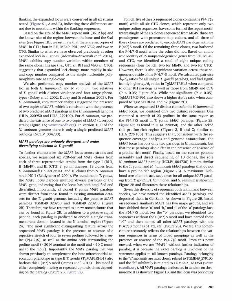
These strains comprised two each from the type I (GT1, RH),type II (ME49, PRU), and type III (VEG, CTG) lineages. Ge-nomic DNA from each strain was digested with ScaI, whichcuts on either side of the entire locus but not within, allowingfor locus size (and therefore copy number) to be estimated(Figure 1B) (Reese et al. 2011; Adomako-Ankomah et al.2014). Sequence coverage analysis shows higher copy num-ber for GT1 compared to ME49 and VEG (Figure 1A), and theSouthern blot was consistent with this observation: GT1 hasthe largest MAF1 locus (�44.9 Kb), while the MAF1 loci inME49 and VEG were smaller (28.4 Kb; Figure 1C). No otherbands were visible on the blot (which resolved fragmentsranging in size from 4.9 Kb to 53.9 Kb), indicating that theentire locus was intact for all strains. Moreover, the ScaI sites
Figure 1 The MAF1 locus exhibits copy number variationacross strains of T. gondii and has comparatively low copynumber in H. hammondi and N. caninum. (A) Coveragedepth analysis for the MAF1 locus in eight T. gondii straintypes and for the syntenic locus in H. hammondi andN. caninum. T. gondii sequences are from ToxoDB v7.3.Portions of the upper left panel of this figure were similarlyrepresented in Pernas et al. (2014). Raw reads were plot-ted as described inMaterials and Methods and normalizedto the coverage 20 Kb upstream of the repetitive locus.Arrowheads indicate the location of predicted gene se-quences based on ToxoDB (v7.3 for T. gondii; v26 for allother species). Asterisks indicate smaller repetitive se-quence unrelated to MAF1 (see Materials and Methodsfor further explanation). (B) Schematic representation ofthe MAF1 locus showing ScaI restrictions sites outside ofthe locus, the size of the regions flanking the MAF1 locus,and the size of the repeat unit used to estimate copynumber based on Southern blotting. The most relevantT. gondii ME49 gene name is indicated (from ToxoDBv7.3), although it does not fully match the sequencedparalogs. (C) ScaI-digested gDNA from each of six T. gondiistrains was resolved by PFGE and probed with a MAF1-specific probe. The blot shows copy number variationconsistent with predictions from sequence coverageanalysis for strain types GT1, ME49, and VEG. Copy num-ber for each strain was determined based on the sche-matic presented in B.
288 Y. Adomako-Ankomah et al.
flanking the expanded locus were conserved in all six strainstested (Figure S1, A and B), indicating these differences arenot due to mutations within the flanking sequences.
Based on the size of the MAF1 repeat unit (3612 bp) andthe known size of the regions between the locus and the ScaIsites (see Figure 1B), we estimate that there are six copies ofMAF1 in GT1; four in RH, ME49, PRU, and VEG; and two inCTG. Similar to what we have observed previously at otherexpanded loci in T. gondii (Adomako-Ankomah et al. 2014),MAF1 exhibits copy number variation within members ofthe same clonal lineage (i.e., GT1 vs. RH and VEG vs. CTG),suggesting that expanded loci change more rapidly in sizeand copy number compared to the single nucleotide poly-morphism rate at single-copy.
We also performed copy number analysis of the MAF1loci in both H. hammondi and N. caninum, two relativesof T. gondii with distinct virulence and host range pheno-types (Dubey et al. 2002; Dubey and Sreekumar 2003). ForH. hammondi, copy number analysis suggested the presenceof two copies ofMAF1, which is consistent with the presenceof two predictedMAF1 paralogs in the H. hammondi genome(HHA_220950 and HHA_279100). For N. caninum, we pre-dicted the existence of one to two copies of MAF1 (Liverpoolstrain; Figure 1A; www.toxodb.org). In version 10.0 of theN. caninum genome there is only a single predicted MAF1ortholog (NCLIV_004730).
MAF1 paralogs are uniquely divergent and underdiversifying selection in T. gondii
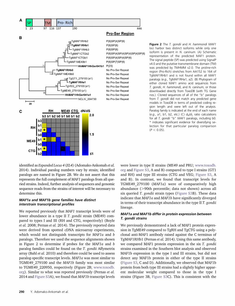
To further characterize the MAF1 locus across strains andspecies, we sequenced six PCR-derived MAF1 clones fromeach of three representative strains from the type I (RH),II (ME49), and III (CTG) T. gondii lineages, 13 clones fromH. hammondi HhCatGer041, and 10 clones from N. caninumstrain NC-1 (Rettigner et al. 2004). We found that in T. gondii,the MAF1 locus harbors multiple diverse paralogs of theMAF1 gene, indicating that the locus has both amplified anddiversified. Importantly, all cloned T. gondii MAF1 paralogswere distinct from those found in existing annotation data-sets for the T. gondii genome, including the putative MAF1paralogs TGME49_020950 and TGME49_220950 (Figure2B). Therefore, we have resorted to a new nomenclature thatcan be found in Figure 2B. In addition to a putative signalpeptide, each paralog is predicted to encode a single trans-membrane domain located in the N-terminal region (Figure2A). The most significant distinguishing feature across thesequenced MAF1 paralogs is the presence or absence of arepetitive stretch of four to seven prolines followed by a ser-ine (P{4:7}S), as well as the amino acids surrounding theproline motif (�20 N terminal to the motif and �10 C termi-nal to the motif). Importantly, the MAF1 paralog that wasshown previously to complement the host mitochondrial as-sociation phenotype in type II T. gondii (TgMAF1RHb1) alsoharbors this P{4:7}S motif (Pernas et al. 2014). This motif iseither completely missing or repeated up to six times depend-ing on the paralog (Figure 2B, Figure S2).
ForRH,fiveof the six sequencedclones contain theP{4:7}Smotif, while all six CTG clones, which represent only twounique coding sequences, have some form of the repeatmotif.Interestingly, of the six clones sequenced fromME49, threearepseudogenes with premature stop codons, and all three ofthese clones are predicted to encodeMAF1 paralogs with theP{4:7}S motif. Of the remaining three clones, two harboredthe P{4:7}S motif while the other did not. Based on aminoacid identity of 15 nonpseudogenized genes from RH, ME49,and CTG, we identified a total of eight unique codingsequences (four for RH, two for ME49, and two for CTG).However, there is also significant variation across these se-quences outside of the P{4:7}Smotif. We calculated pairwisedN/dS ratios for all unique T. gondii paralogs, and find signif-icantly higher dN/dS ratios in TgMAF1RHb3 when comparedto other RH paralogs as well as those from ME49 and CTG(P , 0.05; Figure 2C). While not significant (P . 0.05),TgMAF1ME49b1 also shows a higher dN/dS ratio when com-pared to TgMAF1RHb1 and b2 (Figure 2C).
Whenwe sequenced 13 distinct clones for theH. hammondiMAF1 locus, we identified only two distinct sequences. Onecontained a stretch of 23 prolines in the same region asthe P{4:7}S motif in T. gondii MAF1 paralogs (Figure 2B,Figure S2; as found in HHA_220950), and the other lackedthis proline-rich region (Figure 2, B and C; similar toHHA_279100). This suggests that, consistent with the se-quence coverage analysis and genome annotations, theMAF1 locus harbors only two paralogs in H. hammondi, butthat these paralogs also differ in the presence or absence ofa proline-rich motif. Finally, based on the current genomeassembly and direct sequencing of 10 clones, the soleN. caninum MAF1 paralog (NCLIV_004730) is more similarto the T. gondii and H. hammondi MAF1 paralogs that do nothave a proline-rich region (Figure 2B). A maximum likeli-hood tree of amino acid sequences for all uniqueMAF1 paral-ogs from T. gondii, H. hammondi, and N. caninum is shown inFigure 2B and illustrates these relationships.
Given this diversity of sequences both within and betweenspecies, we have named the identified MAF1 paralogs anddeposited them in GenBank. As shown in Figure 2B, basedon sequence similarity MAF1 has two major groups, and wehave dubbed these “a” and “b,” and all of the “a” paralogs lackthe P{4:7}S motif. For the “b” paralogs, we identified twosequences without the P{4:7}S motif and have named these“b0” and then named all other MAF1 paralogs with theP{4:7}S motif as b1, b2, etc. (Figure 2B). We feel this nomen-clature accurately reflects the relationships between the var-ious sequences in terms of broad groupings as well as thepresence or absence of the P{4:7}S motif. From this pointonward, when we use “MAF1” without further indication ofparalog, it is because the exact paralog is unknown or thestatement applies to all known paralogs. Paralogs belongingto the “a” subfamily are most closely related to TGME49_279100,and the “b” subfamily is related to TGME49_020950 (www.toxodb.org). AllMAF1 paralogs are located in tandemon chro-mosome II as shown in Figure 1B, and the locuswas previously
Derived Trait Evolution in T. gondii 289
identified as Expanded Locus 4 (EL4) (Adomako-Ankomah et al.2014). Individual paralog numbers vary by strain; identifiedparalogs are named in Figure 2B. We do not assert that thisrepresents the full complement ofMAF1 paralogs from all que-ried strains. Indeed, further analysis of sequences and genomicsequence reads from the strains of interest will be necessary todetermine this.
MAF1a and MAF1b gene families have distinctinterstrain transcriptional profiles
We reported previously that MAF1 transcript levels were oflower abundance in a type II T. gondii strain (ME49) com-pared to types I and III (RH and CTG, respectively) (Boyleet al. 2008; Pernas et al. 2014). The previously reported datawere derived from spotted cDNA microarray experiments,which would not distinguish transcripts for MAF1a and bparalogs. Therefore we used the sequence alignments shownin Figure 2 to determine if probes for the MAF1a and bparalog families could be found on the T. gondii Affymetrixarray (Bahl et al. 2010) and therefore could be used to assessparalog-specific transcript levels. MAF1a was most similar toTGME49_279100 and the MAF1b family was most similarto TGME49_220950, respectively (Figure 2B; www.toxodb.org). Similar to what was reported previously (Pernas et al.2014 and Figure S3A), we found thatMAF1b transcript levels
were lower in type II strains (ME49 and PRU; www.toxodb.org and Figure S3, A and B) compared to type I strains (GT1and RH) and type III strains (CTG and VEG; Figure S3, Aand B). In contrast, we found that transcript levels forTGME49_279100 (MAF1a) were of comparatively highabundance (.90th percentile; data not shown) across allsix queried T. gondii strain types (Figure S3B). These dataindicate thatMAF1a andMAF1b have significantly divergedin terms of their transcript abundance in the type II T. gondiilineage.
MAF1a and MAF1b differ in protein expression betweenT. gondii strains
We previously demonstrated a lack of MAF1 protein expres-sion in TgME49 compared to TgRH and TgCTG using a poly-clonal anti-MAF1 antibody raised against the C terminus ofTgMAF1RHb1 (Pernas et al. 2014). Using this same antibody,we compared MAF1 protein expression in the six T. gondiistrains examined in the Southern blot analysis and observedMAF1b expression in the type I and III strains, but did notdetect any MAF1b protein in either of the type II strains(Figure S3, C and D). Additionally, we observed that MAF1bprotein from both type III strains had a slightly higher appar-ent molecular weight compared to those in the type Istrains (Figure 3B, Figure S3C). This is consistent with the
Figure 2 The T. gondii and H. hammondi MAF1loci harbor two distinct isoforms while only oneisoform is present in N. caninum. (A) Schematicrepresentation of the predicted MAF1 protein.The signal peptide (SP) was predicted using SignalPv4.0 and the putative transmembrane domain (TM)was predicted by TMHMM v2.0. The proline-richregion (Pro-Rich) stretches from AA152 to 164 ofTgMAF1RHb1 and is not found within all MAF1paralogs (e.g., TgMAF1RHa1, a2). (B) Phylogram ofeither cloned MAF1 amino acid sequences fromT. gondii, H. hammondi, and N. caninum, or thosedownloaded directly from ToxoDB (with TG Genenos.). Cloned sequences of all of the “b” paralogsfrom T. gondii did not match any predicted genemodels in ToxoDB in terms of predicted coding re-gion length and were left out of the analysis.Paralog family is indicated at the end of each name(e.g., a1, b1, b2, etc.) (C) dN/dS ratio calculationsfor all T. gondii “b” MAF1 paralogs, including b0.* indicates significant evidence for diversifying se-lection for that particular paralog comparison(P , 0.05).
290 Y. Adomako-Ankomah et al.

observation that the two clones ofMAF1b sequenced from theCTG strain encode either four or six P{4:7}S repeat motifs,while the highest number of P{4:7}S repeat motifs in RHwasthree (Figure 2B, Figure S2). To determine if types I, II, andIII all express aMAF1a isoform, we generated new polyclonalantibodies against the C terminus of TgMAF1RHa1 (Ser173to Ser443) or TgMAF1RHb1 (Thr159 to Asp435) (indicatedin Figure S2).We exposed twomice to the TgMAF1RHa1 andthree mice to TgMAF1RHb1. Polyclonal serum from four ofthe five mice was specific for the input antigen, while onemouse exposed to TgMAF1RHb1 harbored antibodies thatbound to both MAF1 paralogs (Figure S3E and data notshown). Given the amount of similarity between the twoantigens, it is likely that the epitopes recognized by sera frommost of the mice were derived from the dissimilar regions.Additionally, it is likely that each polyclonal serum is capableof recognizing multiple “a” or “b” paralogs. In Western blotsagainst the input antigen, a higher molecular weight band isdetected by all of these antisera in addition to the majorspecies at the expected molecular weight (Figure S3E). Theantisera recognize the higher MW band with similar specific-ity as the purified protein. This higher MW band may be adimer of the purified protein, as it is approximately twice thesize of the major species, can be seen upon Coomassie stain-ing, and its quantity is reduced after longer boiling timesof the purified protein (Figure S4). Using antibodies frommouse 5 for immunofluorescence, we detected MAF1a pro-tein in all three strains, while once again we did not detectMAF1b in type II when using antibodies from mouse 1 (Fig-ure 3A). We also saw a similar pattern of expression byWest-ern blot (Figure 3B). The specificity of the MAF1b antiserumto MAF1b and not MAF1a was further confirmed by the factthat the MAF1b antiserum bound to type II T. gondii whenexpressing an ectopic copy of TgMAF1RHb1 (Figure 3C).These data indicate significant strain-specific variation be-tween major clonotypes in both MAF1 protein level and inthe qualitative nature of the paralogs that are expressed.
T. gondii MAF1 paralogs differ in their ability to mediatehost mitochondrial association in T. gondii andN. caninum
HMA is a strain-specific trait in T. gondii (lacking in type IIstains; Figure 4A), and this trait is consistent with reducedMAF1b transcript and protein levels in members of the type IIlineage (Figure 3, Figure S3, A–D). In contrast the MAF1agene is highly expressed at the transcript and protein levelequally well across multiple T. gondii strains. To determine iftheMAF1a and b genes differed in their ability to confer HMAin HMA- parasites, we generated N-terminally HA-taggedclones of the two paralogs that differed in the absence orpresence of theP{4:7}Smotif (TgMAF1RHa1 andTgMAF1RHb1,respectively). To do this, we cloned TgMAF1RHa1 in place ofTgMAF1RHb1, while retaining the TgMAF1RHb1 promoter inthe construct to ensure equal expression between paralogs.We expressed these genes in both a type II strain (TgME49)and in N. caninum (NC-1) (Rettigner et al. 2004) and used
confocal microscopy and mitochondrial staining to determinethe impact on HMA. TgMAF1RHb1 expression was sufficientto mediate HMA in T. gondii strain ME49 (Figure 5A) and alsoin N. caninum (Figure 5B) 18 hr postinfection. In contrast,TgMAF1RHa1 was unable to mediate HMA in either TgME49or N. caninum although its protein localization profile was sim-ilar to that of MAF1RHb1 (Figure 5, A and B, bottom). We alsogenerated clones of TgME49:MAF1RHb1 andNC-1:MAF1RHb1for electron microscopy. Both wild-type TgME49 and NC-1 have
Figure 3 T. gondii MAF1 paralog expression differs between lineages. (Aand B) Polyclonal antibodies were generated specifically against the Ctermini of TgMAF1RHa1 (Ser173 to Ser443) or TgMAF1RHb1 (Thr159to Asp435). Protein expression was compared by immunofluorescenceacross three strains representing clonotypes I, II, and III. Based on immu-nofluorescence and Western blotting, antibodies against TgMAF1RHa1detected protein in all three strains, while antibodies against TgMAF1RHb1detected protein only in RH and CTG (and not ME49). (C) Anti-bodies against TgMAF1RHb1 are able to detect TgMAF1RHb1 ex-pression in transgenic type II parasites expressing the TgMAF1RHb1protein.
Derived Trait Evolution in T. gondii 291

little, if any, HMA (Figure 5C, left). However, when these strainsexpressMAF1RHb1 they becomeHMA+and there is an increasein host mitochondria directly adjacent to the PVM (Figure 5C,right).
TgMAF1b1 from T. gondii and H. hammondi can conferthe HMA phenotype in T. gondii type II, whileTgMAF1b0 and HhMAF1a1 cannot
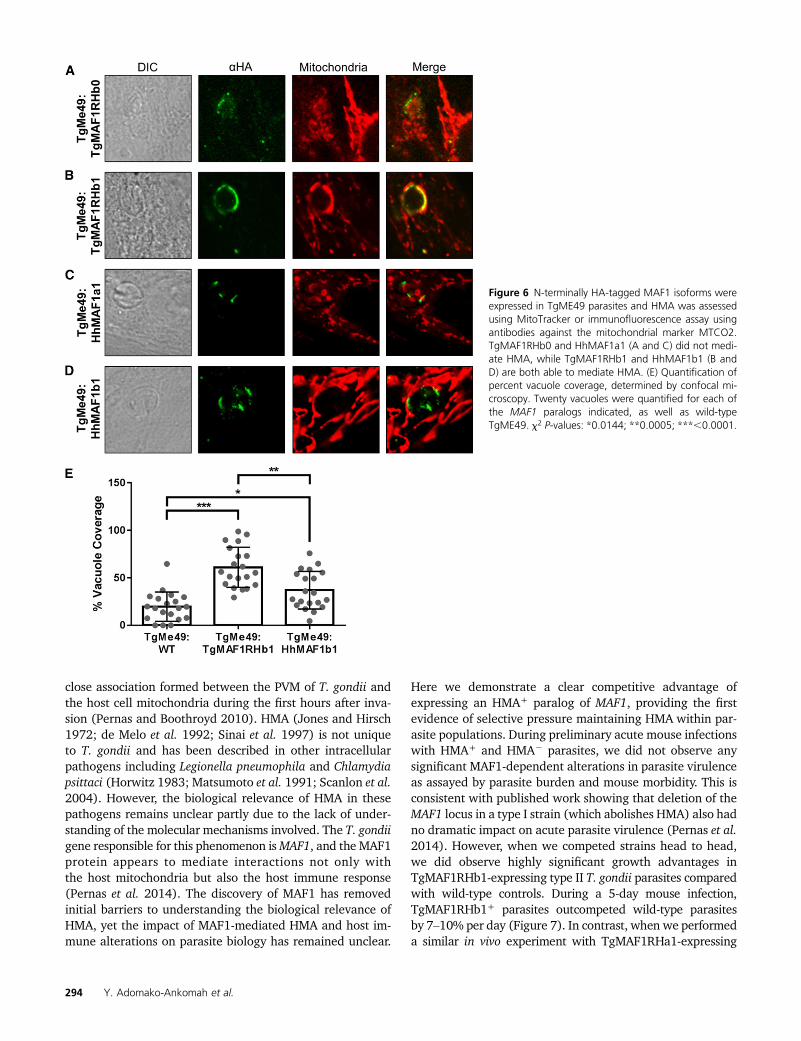
HMA is greatly reduced in the closely related N. caninum(Figure 4B) (Pernas and Boothroyd 2010), but the HMA phe-notype of the nearest extant relative ofT. gondii,H. hammondi,is unknown. To test this we assessed HMA in sporozoite-derived tachyzoites of H. hammondi (strain HhCatEth1)(Dubey et al. 2013), and found clear evidence for HMA inthis species (Figure 4C). Therefore we hypothesized thatT. gondii and H. hammondi would harbor MAF1 paralogsthat could complement the HMA defect in type II T. gondii,while N. caninum would not. To test this hypothesis, wecloned N-terminally tagged MAF1 paralogs from T. gondii,H. hammondi, and N. caninum. For T. gondii, the codingsequences for TgMAF1RHb0 and TgMAF1RHb1 with theendogenous promoters were cloned directly from RH straingenomic DNA. Similar constructs were made forH. hammondiand N. caninum. Each construct was transfected into theHMA2 TgME49 strain and the ability of each isoform tomediate HMA was assessed by immunofluorescence. Simi-lar to our results with TgMAF1RHa1, TgMAF1RHb0 wasunable to mediate HMA (Figure 6A), indicating that notall “b” paralogs are capable of mediating this phenotype.Importantly, the same was true for HhMAF1a1: When trans-fected into type II T. gondii, this protein did not confer theHMA phenotype, although it did have an expression profilethat was distinct from other MAF1 paralogs (Figure 6C). Incontrast, both TgMAF1RHb1 (as shown previously) andHhMAF1b1 could confer the HMA phenotype when ectopi-cally expressed in type II T. gondii (Figure 6, B and D). Wequantified percent vacuole coverage for 20 vacuoles for eachMAF1 paralog using confocal microscopy and found that para-sites expressing TgMAF1RHb1 or HhMAF1b1 had significantlymore vacuole membrane associated with host mitochondriathan wild-type type II parasites (Figure 6E). The localizationof TgMAF1RHb0, TgMAF1RHb1, and HhMAF1b1 are all sim-ilar. We also performed the same experiment with NcMAF1(based on NCLIV_ 004730), but we were unable to detectany protein followingmultiple (more than three) transfections.Whether this is due to upstream regulatory sequences or someother species-specific factor is unknown.
Expression of MAF1RHb1, but not MAF1RHa1, in type IIT. gondii increases competitive advantage duringinfection in vivo
To directly examine the impact ofMAF1 in an in vivo infectionsystem, we infected Balb/c mice with TgME49 wild-type ora TgMAF1RHb1-complemented line and measured theirrates of proliferation and dissemination in vivo using biolu-minescence imaging (Walzer et al. 2013). We used a suble-
thal dose for a type II strain (100 tachyzoites) (Saeij et al.2005) to allow the mice to survive the full course of theinfection and enable us to detect any subtle differences inparasite dissemination. We observed marginally higher, butstatistically insignificant, parasite burdens in infection withTgME49:TgMAF1RHb1 (Figure S5). Given the marginallyhigher parasite burden observed in mice infected withTgME49:TgMAF1RHb1 compared to wild-type TgME49, wehypothesized that this increase in parasite growth, althoughmarginal, could provide a competitive advantage during aninfection with a mixed population. To test this hypothesis,we created mixed populations of TgME49 and TgME49:TgMAF1RHb1 and infected female Balb/c mice with thesepopulations of known proportions. Mice were infected with105 tachyzoites of 1:4 or 4:1 proportions of TgME49 to
Figure 4 Host mitochondrial association is a feature of T. gondii andH. hammondi infections, but not N. caninum. (A) NRK-mitoRFP cells wereinfected with GFP-expressing type I, II, and III (RH, PRU, and CTG) para-sites. Type II parasites are HMA2, while types I and III are HMA+. (B) NRK-mitoRFP cells were infected with N. caninum strain NC-1. Cells were fixedand counterstained with Hoechst stain. Wild-type N. caninum are HMA2.(C) HFFs were infected with H. hammondi sporozoites for 8 days beforefixation. Host mitochondria were visualized using an antibody to humanMTCO2. H. hammondi is HMA+.
292 Y. Adomako-Ankomah et al.

TgME49:TgMAF1RHb1 parasites. Initial population propor-tions were quantified by immunofluorescence assay (IFA).Parasite burden was monitored by bioluminescence imagingfor 5 days, after which mice were euthanized and parasiteswere collected by peritoneal lavage and the population pro-portions were determined by IFA. After 5 days the proportionof TgMAF1RHb1-expressing parasites increased significantly,regardless of initial proportion (Figure 7A). This competitiveadvantage was not observed when mice were infected with amixed population of TgME49 and TgME49:TgMAF1RHa1parasites (Figure 7A). While we did not notice any differ-ences in growth rate in vitro between these strains, we alsoconstructed mixed populations of TgME49 and TgME49:TgMAF1RHb1 and maintained them in HFFs by serial syringe
lysis and passage in vitro for 8 weeks. Population proportionswere determined by IFA at days 0, 28, and 54–59. In three offour populations, TgMAF1RHb1-expressing parasites signifi-cantly increased in proportion compared to their wild-typecounterparts (Figure S6). However the competitive advantageof TgMAF1RHb1 expression appearsmuch greater in vivo thanin vitro, as evidenced by the 15-fold greater percent change perday of TgMAF1RHb1 expressing parasites within the popula-tion during in vivo compared to in vitro (Figure 7B).
Discussion
One of the many ways in which T. gondii interacts differentlywith host cells, compared to its relative N. caninum, is the
Figure 5 MAF1RHa1 and MAF1RHb1 differ in their abilityto complement HMA in T. gondii and N. caninum. (A) HFFswere labeled with MitoTracker and infected with para-sites transiently transfected with either HA-MAF1RHa1 orHA-MAF1RHb1. MAF1RHb1 but not MAF1RHa1 is ableto confer the HMA phenotype in TgME49. (B) Identicalresults were obtained for N. caninum. Bar, 5.0 mm. (C)HA-MAF1RHb1 was transfected into either TgME49 (top)or N. caninum (bottom), and HA-positive clones wereisolated by limiting dilution. Wild type (WT, left) andTgMAF1RHb1 complemented (right) were grown for 18 hrin HFFs and processed for electron microscopy. Asterisksindicate host mitochondria. Bar, 500 nm.
Derived Trait Evolution in T. gondii 293
close association formed between the PVM of T. gondii andthe host cell mitochondria during the first hours after inva-sion (Pernas and Boothroyd 2010). HMA (Jones and Hirsch1972; de Melo et al. 1992; Sinai et al. 1997) is not uniqueto T. gondii and has been described in other intracellularpathogens including Legionella pneumophila and Chlamydiapsittaci (Horwitz 1983; Matsumoto et al. 1991; Scanlon et al.2004). However, the biological relevance of HMA in thesepathogens remains unclear partly due to the lack of under-standing of the molecular mechanisms involved. The T. gondiigene responsible for this phenomenon isMAF1, and the MAF1protein appears to mediate interactions not only withthe host mitochondria but also the host immune response(Pernas et al. 2014). The discovery of MAF1 has removedinitial barriers to understanding the biological relevance ofHMA, yet the impact of MAF1-mediated HMA and host im-mune alterations on parasite biology has remained unclear.
Here we demonstrate a clear competitive advantage ofexpressing an HMA+ paralog of MAF1, providing the firstevidence of selective pressure maintaining HMA within par-asite populations. During preliminary acute mouse infectionswith HMA+ and HMA2 parasites, we did not observe anysignificant MAF1-dependent alterations in parasite virulenceas assayed by parasite burden and mouse morbidity. This isconsistent with published work showing that deletion of theMAF1 locus in a type I strain (which abolishes HMA) also hadno dramatic impact on acute parasite virulence (Pernas et al.2014). However, when we competed strains head to head,we did observe highly significant growth advantages inTgMAF1RHb1-expressing type II T. gondii parasites comparedwith wild-type controls. During a 5-day mouse infection,TgMAF1RHb1+ parasites outcompeted wild-type parasitesby 7–10% per day (Figure 7). In contrast, when we performeda similar in vivo experiment with TgMAF1RHa1-expressing
Figure 6 N-terminally HA-tagged MAF1 isoforms wereexpressed in TgME49 parasites and HMA was assessedusing MitoTracker or immunofluorescence assay usingantibodies against the mitochondrial marker MTCO2.TgMAF1RHb0 and HhMAF1a1 (A and C) did not medi-ate HMA, while TgMAF1RHb1 and HhMAF1b1 (B andD) are both able to mediate HMA. (E) Quantification ofpercent vacuole coverage, determined by confocal mi-croscopy. Twenty vacuoles were quantified for each ofthe MAF1 paralogs indicated, as well as wild-typeTgME49. x2 P-values: *0.0144; **0.0005; ***,0.0001.
294 Y. Adomako-Ankomah et al.

parasites, we observed no competitive advantage in the com-plemented strain vs. wild type. These data provide strong ev-idence for a selective advantage of HMA itself, since onlyTgMAF1RHb1-complemented parasites, and not TgMAF1RHa1-complemented parasites, had a growth advantage in vivo.
In addition to the selective pressure maintaining HMAwithin T. gondii populations, we have traced the evolutionaryhistory of theMAF1 locus with respect to HMA.We propose amodel in which the MAF1 gene (the “a” paralog) duplicatedinitially in a common ancestor to H. hammondi and T. gondii,and that this duplication event was followed by diversifica-tion and eventual neofunctionalization of one of the copies(into the “b” paralog) that had the capacity to mediate HMA.These data are consistent with the observation that T. gondiiandH. hammondi (Figure 4C) are HMA+, whileN. caninum isHMA2 (Pernas and Boothroyd 2010), and thatMAF1b paral-ogs from T. gondii and H. hammondi can confer the HMAphenotype to type II T. gondii, while their MAF1a counter-parts cannot. It is also possible that N. caninum is HMA2 dueto secondary loss of a functional MAF1b paralog. However,electron micrographs of intracellular N. hughesi (a close rel-ative ofN. caninum) demonstrate a clear lack of HMA (Dubeyet al. 2001), particularly in comparison to the very tight as-sociation between host mitochondria and the PVM that wesee in TgMAF1RHb1-expressing T. gondii and N. caninum(Figure 5C, right). This suggests that N. hughesi is in fact
HMA2, providing further evidence for the neofunctionaliza-tion of MAF1b in T. gondii and H. hammondi after they splitfrom the Neospora lineage. In addition, we currently do notknow the role that MAF1a protein plays in parasite biology.However, we do know that the MAF1a gene is maintainedwithin parasite populations and is expressed at high levels(Figure 3, Figure S3, A–D), despite evidence that it is notessential for parasite replication in vitro (Pernas et al.2014), and that it does not mediate HMA (Figure 5).
It was reported (Nolan et al. 2015) that an N. caninumstrain different from that used in this study (N. caninumLiverpool) associated with host mitochondria after 24 hr ofinfection. However compared to T. gondii strain RH (anHMA+ strain) (Pernas et al. 2014), mitochondria were fewerin number, significantly further away from the PV, and didnot accumulate until at least 24 hr postinfection. It may bethat MAF1b paralogs (which are missing from N. caninumLiverpool) (Reid et al. 2012) mediate a very rapid and earlyassociation with host mitochondria (during the first 24 hrpostinfection) but thatmitochondria accumulate onN. caninumvacuoles due to other as-yet-unidentified factors. Strain differ-ences between Liverpool and NC-1 also cannot yet be ruledout. Regardless, our confocal (Figure 5, A and B) and elec-tron microscopy (Figure 5C) data show that in the first18 hr postinfection there is a dramatic difference in HMA inT. gondii and N. caninum, and that we can convert N. caninum
Figure 7 Expression of TgMAF1RHb1, butnot TgMAF1RHa1, in type II T. gondii in-creases competitive advantage. (A) Micewere infected with mixed populations ofTgME49:EV and TgME49:MAF1 with theindicated isoforms and ratios. Infectionwas allowed to progress for 5 days andpopulation proportions before and af-ter infection were quantified by IFA. BothHMA+ and HMA2 MAF1 isoforms wereassessed. TgME49:TgMAF1RHb1 signifi-cantly increases in proportion to TgME49:EV. *x2 P-value ,0.05. The proportion ofTgMAF1RHa1-expressing parasites did notincrease during the infection. (B) Percentchange per day was calculated for the pop-ulations that started with 4:1 TgME49:EVto TgME49:TgMAF1RHb1 both in vitro andin vivo by dividing the total percent increaseof TgMAF1RHb expressing parasites withinthe population by the number of days ofinfection. The first bar of both the in vitroand in vivo infections represent one cloneset, while the second bar for each representsa second clone set. (C) Representative im-ages of a mixed population from A beforeand after a 5-day in vivo infection. HA stain-ing indicates TgMAF1RHb1-positive vacuoles.
Derived Trait Evolution in T. gondii 295
from being HMA2 to HMA+ by complementation with a sin-gle gene product (TgRHMAF1b1).
While we have performed an extensive survey of theMAF1paralogs found across multiple T. gondii strains and in twoother species, our PCR-basedmethods could still be subject tocreating chimeric artifacts during amplification. We did testfor this by comparing sequences with existing Sanger-basedcomplete genome sequence reads and found no evidence ofchimeric sequences. To determine this with 100% certainty,however, we would need to clone MAF1 paralogs directlyfrom T. gondii genomic DNA (as we did for T. gondii ROP5)(Reese et al. 2011).
In our isolated sequences there are significant sequencedifferences between the MAF1a and MAF1b paralogs, themost striking of which is the existence of a proline-rich repeatthat is interspersedwith serines inT. gondii, whileH. hammondiMAF1 encodes a similar proline-rich region but without theinterspersed serines. Some MAF1b paralogs from both speciesare functional with respect to HMA, while all MAF1 paralogstested (whether derived from the “b” or “a” lineage) that lackthis proline-rich region are not HMA competent. Additionally,there is a great diversity of MAF1 paralogs in T. gondii thatcan eventually be exploited to identify key residues that func-tionally distinguish HMA-competent and incompetent MAF1paralogs. Structural biology comparisons between paralogsmay also be particularly useful to identify polymorphic resi-dues that are exposed and to determine the impact of specificmutations on the overall structure.
What remains to be determined is the relative importanceof MAF1 locus amplification, which occurred only in mem-bers of the T. gondii lineage and not H. hammondi orN. caninum. Since H. hammondi is HMA+ and harbors aMAF1 paralog that is capable of mediating HMA in HMA-
parasites (type II; Figure 6) locus expansion (to more thantwo copies) is not a prerequisite to drive HMA during infec-tion, so the question remains as to the utility of having up tosix copies ofMAF1 (as shown for GT1; Figure 1C). MoreoverT. gondii strain CTG is also HMA+ (Figure 4A) and is pre-dicted to have only two MAF1 copies. The duplication ofgenes encoding secreted proteins in T. gondii, and impor-tantly their subsequent diversification and optimization,may be a means of rapid and flexible adaptation to differenthosts or even niches within a given host. In poxviruses, thisprocess has been observed in real time under selective pres-sure at the K3L locus. Under strong host-induced pressure,K3L copy number increases and individual mutations accu-mulate in the duplicate copies. Once a mutation in a singlecopy of K3L emerges that is highly selective for virus survival,the locus resolves back to a single copy due to the negativeimpact of increased genome size (Elde et al. 2012). Based onour comparisons of closely related members of the sameclonal lineage using both Southern blotting and sequencecoverage analysis, expanded loci like MAF1 appear to be incomparatively rapid flux, undergoing multiple rounds of ex-pansion and contraction on relatively short evolutionary timescales. It remains to be seen, however, if the changes inMAF1
locus size (or other T. gondii-specific expanded loci encodingsecreted effectors) (Adomako-Ankomah et al. 2014) are offunctional consequence as for the K3L locus in poxviruses.The observed differences in locus size (and therefore genecontent) could be driven by selection-driven expansion/di-versification/contraction as for K3L or simply be a result ofstochastic changes during DNA replication and/or crossingover that are more common in repetitive DNA regions. Amore detailed characterization of individual MAF1 copies,as well as their impact on HMA and parasite biology, willbe necessary to determine the full impact of the observedstrain- and species-specific features of the MAF1 locus.
Wehavenot yet explored the impact ofMAF1expression intype II strains (or MAF1 locus deletion in HMA-competentstrains like RH and CTG) on the virulence or persistence ofother life cycle stages, including bradyzoites and sporozoites.These experiments will be important to fully assess the im-pact and importance of HMA in T. gondii biology in addi-tion to the clear selective advantage of MAF1b expression(and presumably HMA) demonstrated here. It is conceiv-able that T. gondii contains built-in redundancies, whichallow the parasite to assemble manipulative strategies thatare targeted to specific hosts. For example, the IRG pathwaytargeted for neutralization by ROP5 is missing in humansand other systems where T. gondii is equally capable of surviv-ing (Niedelman et al. 2012). This seems less likely for MAF1since HMA occurs in multiple cell types from multiple species(Pernas and Boothroyd 2010; Pernas et al. 2014).
Finally it is important to note that in the type II strainsexamined MAF1b protein is undetectable and this correlateswith the HMA2 phenotype of strains from this lineage. Thisdemonstrates that while MAF1b expression (and presumablyHMA) is selectively advantageous, it is certainly not essential,and in the case of type II strains, is dispensable. This uniquephenotype with respect to MAF1b expression and HMA isconsistent with other strain-specific phenotypes in type IIparasites, including a unique lack of ROP16-driven manip-ulation of the STAT3/6 pathway and a unique ability tomodulate NFkB activation via the secreted effector GRA15(Saeij et al. 2007; Rosowski et al. 2011). However, ineukaryotic parasites like T. gondii, it is the collection ofvirulence alleles at multiple locations in the genome thatdetermine overall pathogenicity, and it may be that the si-lencing of MAF1b expression in the type II lineage may havebeen selected for as this strain evolved in a distinct niche. Itwill be interesting to determine the mechanism of MAF1bsilencing in type II strains of T. gondii as a first step towardanswering these questions.
Acknowledgments
The authors thank Abby Primack for critical reading of themanuscript. This work was funded by a Pew Scholarship inthe Biomedical Sciences and National Institutes of Health(NIH) grant AI114655 to J.P.B. NIH grant AI73756 (awardedto John C. Boothroyd, Stanford University) supported L.F.P.
296 Y. Adomako-Ankomah et al.
Literature Cited
Adomako-Ankomah, Y., G. M. Wier, A. L. Borges, H. E. Wand, andJ. P. Boyle, 2014 Differential locus expansion distinguishesToxoplasmatinae species and closely related strains of Toxo-plasma gondii. MBio 5: e01003–e01013.
Bahl, A., P. H. Davis, M. Behnke, F. Dzierszinski, M. Jagalur et al.,2010 A novel multifunctional oligonucleotide microarray forToxoplasma gondii. BMC Genomics 11: 603.
Bailey, J. A., and E. E. Eichler, 2006 Primate segmental duplica-tions: crucibles of evolution, diversity and disease. Nat. Rev.Genet. 7: 552–564.
Behnke, M. S., A. Khan, J. C. Wootton, J. P. Dubey, K. Tang et al.,2011 Virulence differences in Toxoplasma mediated by ampli-fication of a family of polymorphic pseudokinases. Proc. Natl.Acad. Sci. USA 108: 9631–9636.
Behnke, M. S., A. Khan, E. J. Lauron, J. R. Jimah, Q. Wang et al.,2015 Rhoptry proteins ROP5 and ROP18 are major murinevirulence factors in genetically divergent South American strainsof Toxoplasma gondii. PLoS Genet. 11: e1005434.
Boothroyd, J. C., and J. F. Dubremetz, 2008 Kiss and spit: the dualroles of Toxoplasma rhoptries. Nat. Rev. Microbiol. 6: 79–88.
Boyle, J. P., J. P. Saeij, S. Y. Harada, J. W. Ajioka, and J. C. Boothroyd,2008 Expression quantitative trait locus mapping of Toxoplasmagenes reveals multiple mechanisms for strain-specific differencesin gene expression. Eukaryot. Cell 7: 1403–1414.
Cridland, J. M., and K. R. Thornton, 2010 Validation of rearrange-ment break points identified by paired-end sequencing in natu-ral populations of Drosophila melanogaster. Genome Biol. Evol.2: 83–101.
de Melo, E. J., T. U. de Carvalho, and W. de Souza, 1992 Penetra-tion of Toxoplasma gondii into host cells induces changes in thedistribution of the mitochondria and the endoplasmic reticulum.Cell Struct. Funct. 17: 311–317.
Deitsch, K. W., M. S. Calderwood, and T. E. Wellems, 2001 Malaria.Cooperative silencing elements in var genes. Nature 412:875–876.
Dubey, J. P., and C. Sreekumar, 2003 Redescription of Hammondiahammondi and its differentiation from Toxoplasma gondii. Int. J.Parasitol. 33: 1437–1453.
Dubey, J. P., S. Liddell, D. Mattson, C. A. Speert, D. K. Howe et al.,2001 Characterization of the Oregon isolate of Neospora hughesifrom a horse. J. Parasitol. 87: 345–353.
Dubey, J. P., B. C. Barr, J. R. Barta, I. Bjerkas, C. Bjorkman et al.,2002 Redescription of Neospora caninum and its differentia-tion from related coccidia. Int. J. Parasitol. 32: 929–946.
Dubey, J. P., G. Tilahun, J. P. Boyle, G. Schares, S. K. Verma et al.,2013 Molecular and biological characterization of first isolatesof Hammondia hammondi from cats from Ethiopia. J. Parasitol.99: 614–618.
Elde, N. C., S. J. Child, M. T. Eickbush, J. O. Kitzman, K. S. Rogerset al., 2012 Poxviruses deploy genomic accordions to adaptrapidly against host antiviral defenses. Cell 150: 831–841.
Emerson, J. J., M. Cardoso-Moreira, J. O. Borevitz, and M. Long,2008 Natural selection shapes genome-wide patterns of copy-number polymorphism in Drosophila melanogaster. Science 320:1629–1631.
Espinosa-Cantu, A., D. Ascencio, F. Barona-Gomez, and A. DeLuna,2015 Gene duplication and the evolution of moonlighting pro-teins. Front. Genet. 6: 227.
Freitas-Junior, L. H., E. Bottius, L. A. Pirrit, K. W. Deitsch, C. Scheidiget al., 2000 Frequent ectopic recombination of virulence factorgenes in telomeric chromosome clusters of P. falciparum. Nature407: 1018–1022.
Goodswen, S. J., P. J. Kennedy, and J. T. Ellis, 2013 A review ofthe infection, genetics, and evolution of Neospora caninum: fromthe past to the present. Infect. Genet. Evol. 13: 133–150.
Heinberg, A., E. Siu, C. Stern, E. A. Lawrence, M. T. Ferdig et al.,2013 Direct evidence for the adaptive role of copy numbervariation on antifolate susceptibility in Plasmodium falciparum.Mol. Microbiol. 88: 702–712.
Horwitz, M. A., 1983 Formation of a novel phagosome by theLegionnaires’ disease bacterium (Legionella pneumophila) in hu-man monocytes. J. Exp. Med. 158: 1319–1331.
Jones, T. C., and J. G. Hirsch, 1972 The interaction betweenToxoplasma gondii and mammalian cells. II. The absence oflysosomal fusion with phagocytic vacuoles containing livingparasites. J. Exp. Med. 136: 1173–1194.
Kent, W. J., 2002 BLAT—the BLAST-like alignment tool. GenomeRes. 12: 656–664.
Kugelberg, E., E. Kofoid, A. B. Reams, D. I. Andersson, and J. R.Roth, 2006 Multiple pathways of selected gene amplificationduring adaptive mutation. Proc. Natl. Acad. Sci. USA 103:17319–17324.
Kugelberg, E., E. Kofoid, D. I. Andersson, Y. Lu, J. Mellor et al.,2010 The tandem inversion duplication in Salmonella enterica:selection drives unstable precursors to final mutation types.Genetics 185: 65–80.
Langmead, B., and S. L. Salzberg, 2012 Fast gapped-read align-ment with Bowtie 2. Nat. Methods 9: 357–359.
Lorenzi, H., A. Khan, M. S. Behnke, S. Namasivayam, L. S. Swapnaet al., 2016 Local admixture of amplified and diversifiedsecreted pathogenesis determinants shapes mosaic Toxoplasmagondii genomes. Nat. Commun. 7: 10147.
Lynch, M., and J. S. Conery, 2000 The evolutionary fate and con-sequences of duplicate genes. Science 290: 1151–1155.
Lynch, M., and A. Force, 2000 The probability of duplicate genepreservation by subfunctionalization. Genetics 154: 459–473.
Matsumoto, A., H. Bessho, K. Uehira, and T. Suda, 1991 Morphologicalstudies of the association of mitochondria with chlamydialinclusions and the fusion of chlamydial inclusions. J. ElectronMicrosc. (Tokyo) 40: 356–363.
Mitra, K., and J. Lippincott-Schwartz, 2010 Analysis of mitochon-drial dynamics and functions using imaging approaches. Curr.Protoc. Cell Biol. Chapter 4: Unit 4.25.1–21.
Nair, S., B. Miller, M. Barends, A. Jaidee, J. Patel et al.,2008 Adaptive copy number evolution in malaria parasites.PLoS Genet. 4: e1000243.
Nei, M., and S. Kumar, 2000 Molecular Evolution and Phylogenetics,Oxford University Press, Oxford.
Nicol, J. W., G. A. Helt, S. G. Blanchard, Jr., A. Raja, and A. E.Loraine, 2009 The Integrated Genome Browser: free softwarefor distribution and exploration of genome-scale datasets. Bio-informatics 25: 2730–2731.
Niedelman, W., D. A. Gold, E. E. Rosowski, J. K. Sprokholt, D. Limet al., 2012 The rhoptry proteins ROP18 and ROP5 mediateToxoplasma gondii evasion of the murine, but not the human,interferon-gamma response. PLoS Pathog. 8: e1002784.
Nolan, S. J., J. D. Romano, T. Luechtefeld, and I. Coppens,2015 Neospora caninum recruits host cell structures to its para-sitophorous vacuole and salvages lipids from organelles. Eukaryot.Cell 14: 454–473.
Ohno, S., 1970 Evolution by Gene Duplication, Springer-Verlag,Berlin.
Pasternak, N. D., and R. Dzikowski, 2009 PfEMP1: an antigenthat plays a key role in the pathogenicity and immune evasionof the malaria parasite Plasmodium falciparum. Int. J. Biochem.Cell Biol. 41: 1463–1466.
Pernas, L., and J. C. Boothroyd, 2010 Association of host mito-chondria with the parasitophorous vacuole during Toxoplasmainfection is not dependent on rhoptry proteins ROP2/8. Int. J.Parasitol. 40: 1367–1371.
Pernas, L., Y. Adomako-Ankomah, A. J. Shastri, S. E. Ewald, M.Treeck et al., 2014 Toxoplasma effector MAF1 mediates
Derived Trait Evolution in T. gondii 297
recruitment of host mitochondria and impacts the host re-sponse. PLoS Biol. 12: e1001845.
Quinlan, A. R., and I. M. Hall, 2010 BEDTools: a flexible suite ofutilities for comparing genomic features. Bioinformatics 26:841–842.
Reese, M. L., G. M. Zeiner, J. P. Saeij, J. C. Boothroyd, and J. P.Boyle, 2011 Polymorphic family of injected pseudokinases isparamount in Toxoplasma virulence. Proc. Natl. Acad. Sci. USA108: 9625–9630.
Reid, A. J., 2015 Large, rapidly evolving gene families are at theforefront of host-parasite interactions in Apicomplexa. Parasitol-ogy 142(Suppl 1): S57–S70.
Reid, A. J., S. J. Vermont, J. A. Cotton, D. Harris, G. A. Hill-Cawthorneet al., 2012 Comparative genomics of the apicomplexan para-sites Toxoplasma gondii and Neospora caninum: Coccidia differ-ing in host range and transmission strategy. PLoS Pathog.8: e1002567.
Rettigner, C., T. Leclipteux, F. De Meerschman, C. Focant, and B.Losson, 2004 Survival, immune responses and tissue cystproduction in outbred (Swiss white) and inbred (CBA/Ca)strains of mice experimentally infected with Neospora caninumtachyzoites. Vet. Res. 35: 225–232.
Rosowski, E. E., D. Lu, L. Julien, L. Rodda, R. A. Gaiser et al.,2011 Strain-specific activation of the NF-kappaB pathway byGRA15, a novel Toxoplasma gondii dense granule protein. J.Exp. Med. 208: 195–212.
Saeij, J. P., J. P. Boyle, and J. C. Boothroyd, 2005 Differencesamong the three major strains of Toxoplasma gondii and theirspecific interactions with the infected host. Trends Parasitol. 21:476–481.
Saeij, J. P., S. Coller, J. P. Boyle, M. E. Jerome, M. W. White et al.,2007 Toxoplasma co-opts host gene expression by injection ofa polymorphic kinase homologue. Nature 445: 324–327.
Scanlon, M., G. J. Leitch, G. S. Visvesvara, and A. P. Shaw,2004 Relationship between the host cell mitochondria and theparasitophorous vacuole in cells infected with Encephalitozoonmicrosporidia. J. Eukaryot. Microbiol. 51: 81–87.
Schmidt, J. M., R. T. Good, B. Appleton, J. Sherrard, G. C. Raymantet al., 2010 Copy number variation and transposable elementsfeature in recent, ongoing adaptation at the Cyp6g1 locus. PLoSGenet. 6: e1000998.
Sinai, A., P. Webster, and K. Joiner, 1997 Association of host cellendoplasmic reticulum and mitochondria with the Toxoplasmagondii parasitophorous vacuole membrane: a high affinity inter-action. J. Cell Sci. 110: 2117–2128.
Tamura, K., G. Stecher, D. Peterson, A. Filipski, and S. Kumar,2013 MEGA6: Molecular Evolutionary Genetics Analysis ver-sion 6.0. Mol. Biol. Evol. 30: 2725–2729.
Walzer, K. A., Y. Adomako-Ankomah, R. A. Dam, D. C. Herrmann,G. Schares et al., 2013 Hammondia hammondi, an avirulentrelative of Toxoplasma gondii, has functional orthologs of knownT. gondii virulence genes. Proc. Natl. Acad. Sci. USA 110: 7446–7451.
Walzer, K. A., G. M. Wier, R. A. Dam, A. R. Srinivasan, A. L. Borgeset al., 2014 Hammondia hammondi harbors functional ortho-logs of the host-modulating effectors GRA15 and ROP16 but isdistinguished from Toxoplasma gondii by a unique transcrip-tional profile. Eukaryot. Cell 13: 1507–1518.
Wasmuth, J., J. Daub, J. M. Peregrin-Alvarez, C. A. Finney, and J.Parkinson, 2009 The origins of apicomplexan sequence inno-vation. Genome Res. 19: 1202–1213.
Zhang, J., H. F. Rosenberg, and M. Nei, 1998 Positive Darwinianselection after gene duplication in primate ribonuclease genes.Proc. Natl. Acad. Sci. USA 95: 3708–3713.
Communicating editor: J. Heitman
298 Y. Adomako-Ankomah et al.

GENETICSSupporting Information
www.genetics.org/lookup/suppl/doi:10.1534/genetics.115.186270/-/DC1
Host Mitochondrial Association Evolved in theHuman Parasite Toxoplasma gondii via
Neofunctionalization of a Gene DuplicateYaw Adomako-Ankomah, Elizabeth D. English, Jeffrey J. Danielson, Lena F. Pernas,
Michelle L. Parker, Martin J. Boulanger, Jitender P. Dubey, and Jon P. Boyle
Copyright © 2016 by the Genetics Society of AmericaDOI: 10.1534/genetics.115.186270

Figure S1. A) Schematic representation of the MAF1 locus showing ScaI restrictions sites used to
determine locus size. a, b, c and d represent primers used to verify location of the ScaI restriction sites.
The most relevant T. gondii ME49 gene name is indicated (from ToxoDB v7.3), although it does not fully
match the sequenced paralogs. B) A PCR-based diagnostic digest was performed to confirm that the ScaI
sites were present at the same predicted locations in all strains queried.

Figure S2. Alignment of select MAF1 sequences from Toxoplasma gondii, Hammondia hammondi and
Neospora caninum. Alignments were performed using CLUSTAL-Omega, and visualized using JalView.
Residues are colored by percent identity. Sequences in bold were used in complementation experiments
in the present study. Portions of proteins used as antigens for antibody production indicated by red
(TgMAF1RHb1) and black (TgMAF1RHa1) boxes.
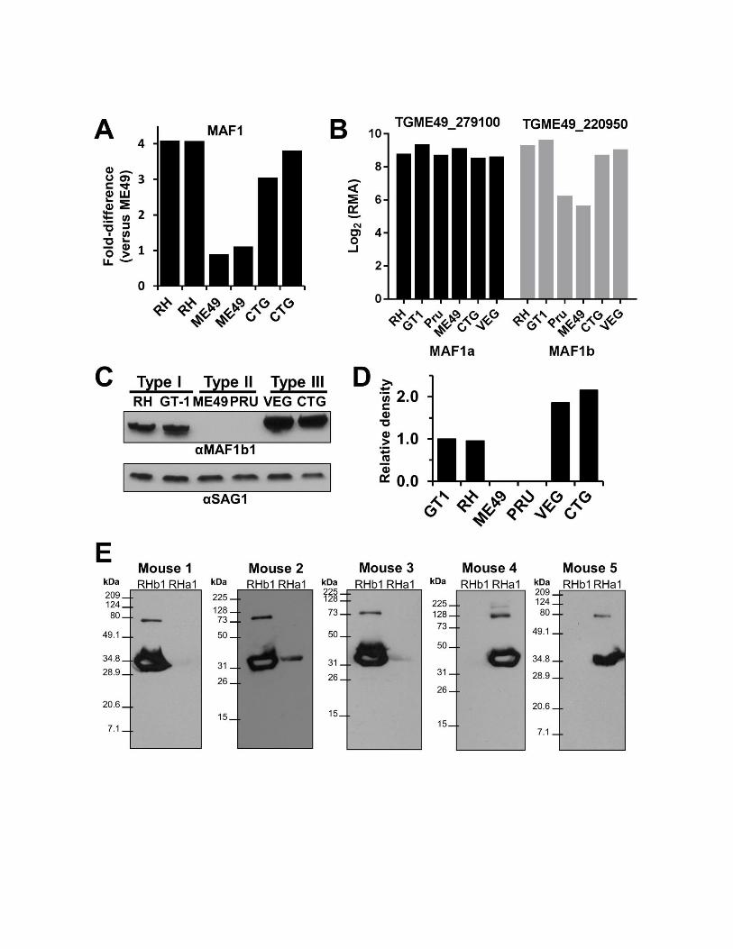

Figure S3. Transcript and protein expression analyses of MAF1a and b paralogs. A) Spotted cDNA
microarray (which would not distinguish MAF1a from MAF1b transcript) data illustrating reduced MAF1a/b
transcript levels in ME49 compared to RH and CTG. Two replicates per strain type are shown. B)
Affymetrix microarray data (derived from probes that were chosen to be unique to each gene, which
therefore would distinguish between MAF1a and b transcript levels) illustrating similar expression of MAF1a
across multiple T. gondii strains, and reduced MAF1b transcript levels in Type II T. gondii (Pru, ME49).
Data downloaded from. C) Levels of MAF1b protein were compared among 2 strains each from the 3
predominant lineages of T. gondii using polyclonal antibodies against the C-terminus of TgMAF1RHb1.
Expression polymorphism of MAF1 correlates with strain-specificity of the host mitochondrial association
phenotype. SAG1 is used as a loading control. D) Densitometric analysis of relative levels of MAF1 in the
six strains examined based on the MAF1/SAG1 intensity ratio. GT1 was set to 1. E) Isolation of paralog-
specific polyclonal mouse antisera against MAF1a1 and b1. Three mice were exposed to purified
MAF1RHb1 and 2 to purified MAF1RHa1 and used to probe blots containing the immunizing antigen. All
but mouse 2 had antibodies that were fully specific to the input antigen.
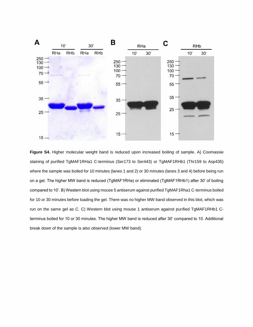
Figure S4. Higher molecular weight band is reduced upon increased boiling of sample. A) Coomassie
staining of purified TgMAF1RHa1 C-terminus (Ser173 to Ser443) or TgMAF1RHb1 (Thr159 to Asp435)
where the sample was boiled for 10 minutes (lanes 1 and 2) or 30 minutes (lanes 3 and 4) before being run
on a gel. The higher MW band is reduced (TgMAF1RHa) or eliminated (TgMAF1RHb1) after 30’ of boiling
compared to 10’. B) Western blot using mouse 5 antiserum against purified TgMAF1Rha1 C-terminus boiled
for 10 or 30 minutes before loading the gel. There was no higher MW band observed in this blot, which was
run on the same gel as C. C) Western blot using mouse 1 antiserum against purified TgMAF1RHb1 C-
terminus boiled for 10 or 30 minutes. The higher MW band is reduced after 30’ compared to 10. Additional
break down of the sample is also observed (lower MW band).
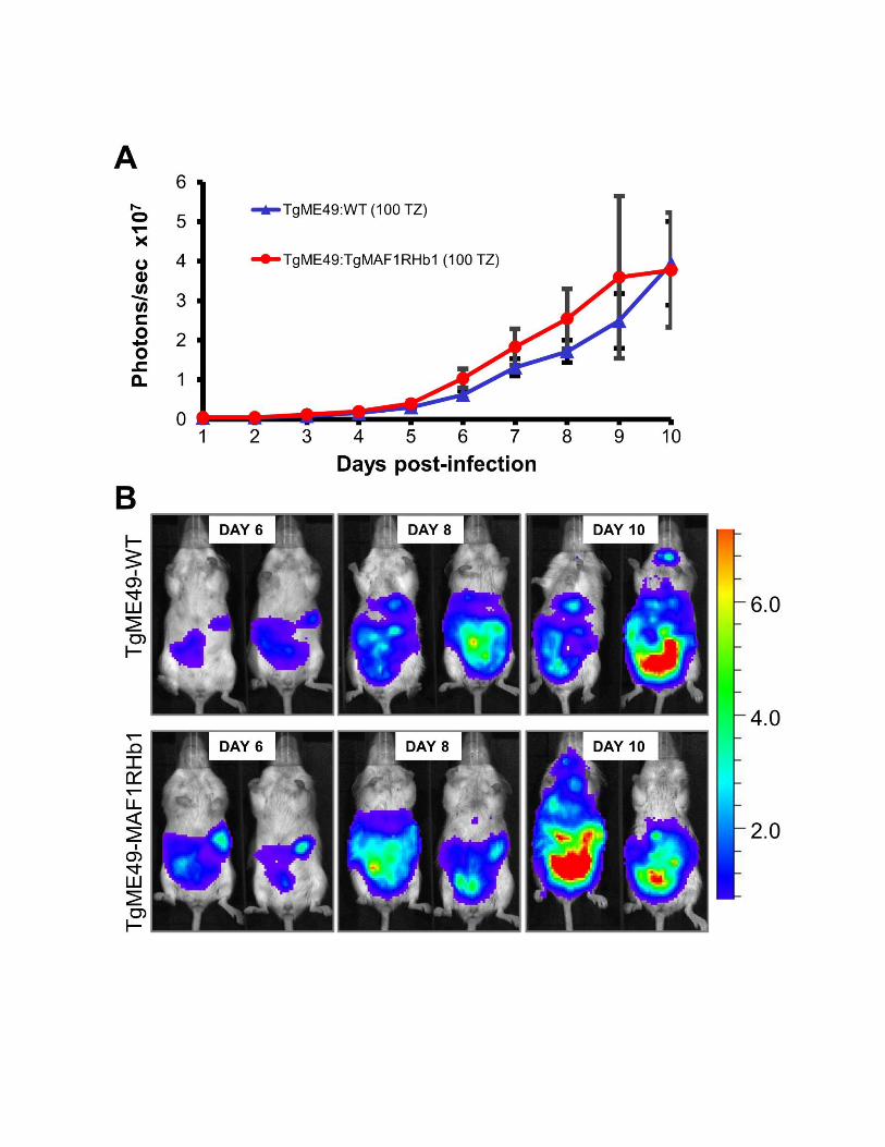
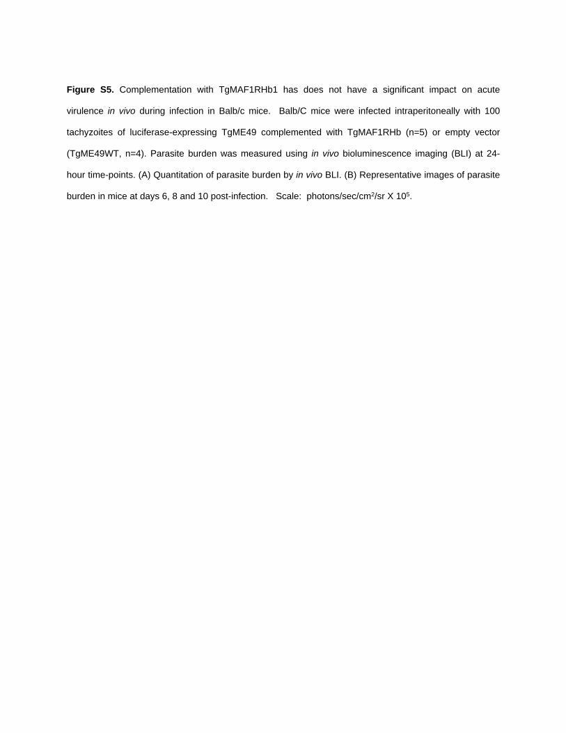
Figure S5. Complementation with TgMAF1RHb1 has does not have a significant impact on acute
virulence in vivo during infection in Balb/c mice. Balb/C mice were infected intraperitoneally with 100
tachyzoites of luciferase-expressing TgME49 complemented with TgMAF1RHb (n=5) or empty vector
(TgME49WT, n=4). Parasite burden was measured using in vivo bioluminescence imaging (BLI) at 24-
hour time-points. (A) Quantitation of parasite burden by in vivo BLI. (B) Representative images of parasite
burden in mice at days 6, 8 and 10 post-infection. Scale: photons/sec/cm2/sr X 105.
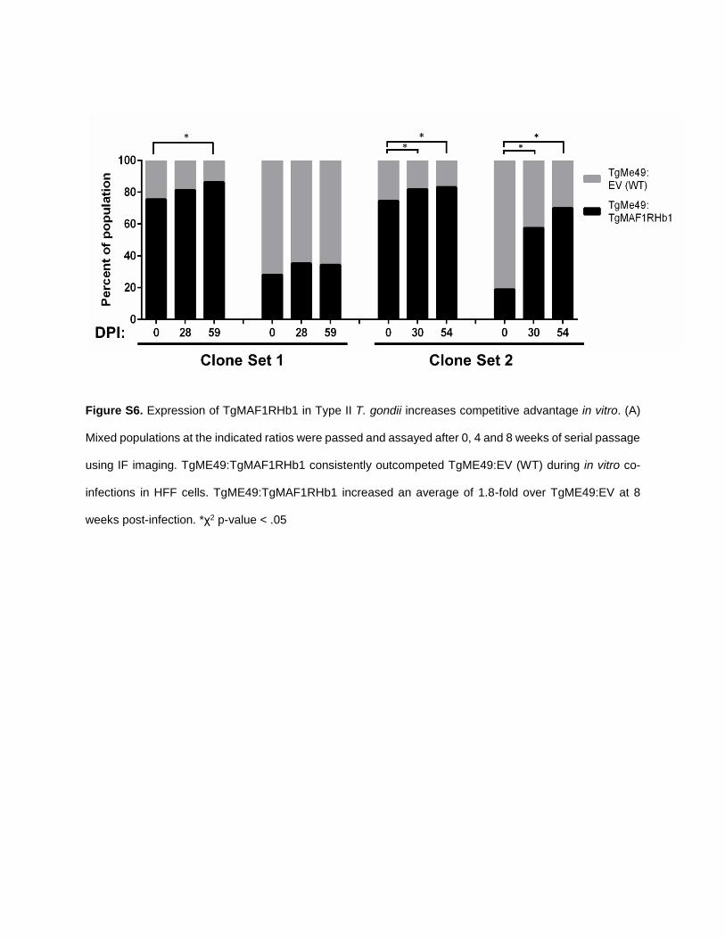
Figure S6. Expression of TgMAF1RHb1 in Type II T. gondii increases competitive advantage in vitro. (A)
Mixed populations at the indicated ratios were passed and assayed after 0, 4 and 8 weeks of serial passage
using IF imaging. TgME49:TgMAF1RHb1 consistently outcompeted TgME49:EV (WT) during in vitro co-
infections in HFF cells. TgME49:TgMAF1RHb1 increased an average of 1.8-fold over TgME49:EV at 8
weeks post-infection. *χ2 p-value < .05

Table S1. Primers used to generate southern probes, clone isoforms, and tag isoforms.
Forward (5’-3’) Primer Reverse (5’-3’) Primer Purpose
CCGAAGTTGATGGCACATTATACCGG CCGGCGAAGTAGCAGCCTCCTCCTAG Southern blot probe for AMA1
CACCaagcttAGGGAT ACGAACAACT CGCTTATTAA G
GTCttaattaaTCAGTCCAGCATGCTAGCCAGATACG
Southern blot probe for MAF1
CAGCTTCCTGCACTCTCTCTGAGG CGAAACAGTACAGACATATGACC Verify ScaI site at left flank of MAF1
CGAACAGGTCGAACGCGTTGTGCGAG TGGCAGCAAGATGGACATGCAC Verify ScaI site at right flank of MAF1
caccAGGGATACGAACAACTCGCTTAT CTACCCATGGGTCC AGC ATG CTA GCCAGATACGT
To clone MAF1 genes from T. gondii
TGATCaagcttCTGCGACGTGATCGTG CGCTGCCGCAGCCGCTGCCGACGCAGCTGCCGCTGCAGCAAGCGAAGACGAAGCCTCGTC
To create left half of PCR fragment to mutate P151-156 and P158-163 to alanine in MAF1RHb1.
gctgcagcggcagctgcgtcggcagcggctgcggcagcgGTCGAAGATCCGTTGTCGCCG
AGTCttaattaaTCAGTCCAGCATGCTAG To create right half of PCR fragment to mutate P151-156 and P158-163 to alanine in MAF1RHb1.
TGATCaagcttCTGCGACGTGATCGTG ACGGGCGGCGGCGGAGGCGGCGCTGGTGGAGGCGGCGGTGGAAGCGAAGA
To create left half of PCR fragment to mutate S157 to alanine in MAF1RHb1.
TCTTCGCTTCCACCGCCGCCTCCACCAGCGCCGCCTCCGCCGCCGCCCGT
AGTCttaattaaTCAGTCCAGCATGCTAG To create right half of PCR fragment to mutate S157 to alanine in MAF1RHb1.
atgcatATGTGG CGCATCTGGA GATGCC
GTCttaattaaTCA GTCGCCTTGCGGAAACTTGTACTTCC
Clone MAF1RHa1 from cDNA
CACCaagcttAGGGAT ACGAACAACT CGCTTATTAA G
TCGCGTAGTCCGGGACGTCGTACGGGTAACCGGCGGTCAGAGCGCCCAGC
Amplify left half of MAF1RHb1 for the MAF1RHb1/a1 chimera
CGACGTCCCGGACTACGCGATGCATGGTGGTCTAGGCAGTCAGATGTCGG
GTCttaattaaTCAGTCGCCTTGCGGAAACTTGTACTTCC
Amplify right half of MAF1RHa1 for the

MAF1RHb1/a1 chimera
CACCAGGGATACGGACAACTCGCTTAT AGGTCTAGTGAGCTGGGAAGTAAA Clone MAF1b genes from H. hammondi
CACCAGGGATACGGACAACTCGCTTAT TAAGCACATGAAACCTCATTTCAC Clone MAF1a genes from H. hammondi
GGGGGACAAGTTTGTACAAAAAAGCAGGCTAGGGATACGGACAACTCGCTTA
CGCGTAGTCCGGGACGTCGTACGGGTAACCGGCGGTCAGAGCGCCCAGCAG
Add HA tag to H. hammondi MAF1a/b by SOE (left half)
TACCCGTACGACGTCCCGGACTACGCGCTAGGCAGTCAGATCTCGGGCTCG
GGGGACCACTTTGTACAAGAAAGCTGGGTGACCTGTTTTGATTCACAGTAG
Add HA tag to H. hammondi MAF1a by SOE (right half)
TACCCGTACGACGTCCCGGACTACGCGCTAGGCAGTCAGATCTCGGGCTCG
GGGGACCACTTTGTACAAGAAAGCTGGGTTCAGTCCACCATGCTAGCCAGAT
Add HA tag to H. hammondi MAF1b by SOE (right half)
GGGGACAAGTTTGTACAAAAAAGCAGGCTGTTCGAGAGAGCGGCATGGACG
GGGGACCACTTTGTACAAGAAAGCTGGGTTCAGCCTTTTCCACCAGACC
Clone MAF1 genes from N. caninum
CGCGTAGTCCGGGACGTCGTACGGGTAACCCGGGGCCAGTACGCCCAG
CGCGTAGTCCGGGACGTCGTACGGGTAACCCGGGGCCAGTACGCCCAG
Add HA tag to N. caninum MAF1 by SOE (along with B1 and B2-containing primers from above as outer primers)