Embed Size (px)
Citation preview
Hormonally Active Vitamin D3 (1�,25-Dihydroxycholecalciferol)Triggers Autophagy in Human Macrophages That Inhibits HIV-1Infection*
Received for publication, November 23, 2010, and in revised form, March 29, 2011 Published, JBC Papers in Press, March 30, 2011, DOI 10.1074/jbc.M110.206110
Grant R. Campbell‡ and Stephen A. Spector‡§1
From the ‡Department of Pediatrics, Division of Infectious Diseases, University of California San Diego, La Jolla, California 92093and §Rady Children’s Hospital, San Diego, California 92123
Autophagy is a self-digestion pathway essential for maintain-ing cellular homeostasis and cell survival and for degradingintracellular pathogens. Human immunodeficiency virus-1(HIV-1) may utilize autophagy for replication as the autophagy-related protein-7 (ATG-7), microtubule-associated protein 1light chain 3, ATG-12, and ATG-16L2 are required for produc-tive HIV-1 infection; however, the effects of autophagy induc-tion on HIV-1 infection are unknown. HIV-1-infected individ-uals have lower levels of 1�,25-dihydroxycholecalciferol, thehormonally active form of vitamin D, than uninfected indi-viduals. with the lowest concentrations found in persons withAIDS. Using human macrophages and RNA interference forATG-5 and Beclin-1 and chemical inhibition of phosphatidyl-inositol 3-kinase, we have found that physiologically relevantconcentrations of 1�,25-dihydroxycholecalciferol induceautophagy in human macrophages through a phosphatidyl-inositol 3-kinase-, ATG-5-, and Beclin-1-dependent mecha-nism that significantly inhibits HIV-1 replication in a dose-dependent manner. We also show that the inhibition of basalautophagy inhibits HIV-1 replication. Furthermore, although1�,25-dihydroxycholecalciferol induces the secretion ofhuman cathelicidin, at the concentrations produced in vitro,cathelicidin does not trigger autophagy. Our findings supportan important role for autophagy during HIV-1 infection andprovide new insights into novel approaches to prevent andtreat HIV-1 infection and related opportunistic infections.
Human immunodeficiency virus type-1 (HIV-1)2 is a globalhealth problem that at current estimates has infected 60millionpeople and caused 25 million deaths worldwide. Currently,there are an estimated 33 million people living with HIV-1, 2million of whom are children (1). Despite the immune defensemechanisms that the host deploys against HIV-1 and improved
antiretroviral therapies, the virus persists in long-lived cellsincluding macrophages and dendritic cells.One-third ofHIV-1-infected individuals are co-infectedwith
Mycobacterium tuberculosis, a leading cause of death amongpeople living with HIV-1. Studies have suggested that 1�,25-dihydroxycholecalciferol (1,25D3), the hormonally active formof vitamin D3, exerts anti-mycobacterial effects in humanmacrophages in vitro (2) through phosphatidylinositol 3-kinase(PI3K) (3) and macroautophagy (4). Few publications haveexamined the relationship between vitamin D3 and HIV-1 dis-ease progression and survival. However, the few studies pub-lished have shown that HIV-1-infected individuals have lower1,25D3 levels than uninfected individuals (5–9) with the lowestconcentrations found in persons with AIDS (7). Moreover,depressed levels of 1,25D3 are correlated with significantlylower CD4� T cell counts and higher tumor necrosis factorlevels (6). Interestingly, consumption of foods rich in 25-hy-droxycholecalciferol (25D3), the inactive precursor for 1,25D3,correlates with an increase in CD4� T cell counts in HIV-1-infected individuals (10). Furthermore, women with low levelsof 25D3 have an increased risk of HIV-1 disease progression(11), with infants born to HIV-1-infected mothers with low25D3 levels having an increased risk of acquiring HIV-1 inutero, intrapartum, and through breastfeeding while also hav-ing a higher mortality rate (12).Macroautophagy (herein referred to as autophagy) is a traf-
ficking pathway whereby cytoplasmic constituents such as sub-cellular organelles and microbial pathogens are engulfed byautophagosomes, which fuse with lysosomes, forming autoly-sosomes, degrading the engulfed component. As an obligatoryintracellular parasite,HIV-1 survival is dependent upon its abil-ity to exploit host cell machinery for replication and dissemina-tion and to circumvent cellular processes that prevent itsgrowth. During infection, HIV-1 down-regulates Beclin-1 andmicrotubule-associated protein 1 light chain 3B-II (LC3B-II),reducing both basal autophagy and the numbers of autophago-somes per cell (13–15). However, silencing of autophagy pro-teins inhibits HIV-1 infection of HeLa cells (16). Interestingly,the HIV-1 envelope glycoprotein expressed on the surface ofinfected cells induces cell death with autophagy in unin-fected bystander CD4� T cells (17–19). Therefore, we inves-tigated the effect of 1,25D3-mediated autophagy on produc-tive HIV-1 infection of macrophages. We demonstrate that1,25D3 induces autophagy in macrophages that inhibitsHIV-1 replication.
* This work was supported, in whole or in part, by National Institutes of HealthGrants AI084573 and AI068632 (NIAID; to the International Maternal Pedi-atric Adolescent AIDS Clinical Trials Group).
1 To whom correspondence should be addressed: University of California SanDiego, 9500 Gilman Dr., La Jolla, CA 92093-0672. Tel.: 858-534-7055; Fax:858-534-7411; E-mail: [email protected].
2 The abbreviations used are: HIV, human immunodeficiency virus; 1,25D3,1�,25-dihydroxycholecalciferol; 25D3, 25-hydroxycholecalciferol; LC3B,light chain 3B; WST-1, 4-[3-(4-iodophenyl)-2-(4-nitrophenyl)-2H-5-tetrazo-lio]-1,3-benzene disulfonate; ssDNA, single-stranded DNA; TCID50, 50%tissue culture infective dose; EdU, 5-ethynyl-2�-deoxyuridine; hCAP18,human cationic antimicrobial protein 18.
THE JOURNAL OF BIOLOGICAL CHEMISTRY VOL. 286, NO. 21, pp. 18890 –18902, May 27, 2011© 2011 by The American Society for Biochemistry and Molecular Biology, Inc. Printed in the U.S.A.
18890 JOURNAL OF BIOLOGICAL CHEMISTRY VOLUME 286 • NUMBER 21 • MAY 27, 2011
by guest on October 12, 2019
http://ww
w.jbc.org/
Dow
nloaded from
EXPERIMENTAL PROCEDURES
Cells and Reagents—Monocyte-derived macrophages wereobtained by culturing monocytes isolated from the peripheralblood mononuclear cells of HIV-1 seronegative donors (withapproval from the University of California San Diego Institu-tional Review Board) in RPMI 1640 (Invitrogen) supplementedwith 10% (v/v) heat-inactivated fetal bovine serum (FBS; Gem-ini Bio-Products) and 10 ng/mlmacrophage colony stimulatingfactor (R&D Systems) for 10 days at 37 °C, 5%CO2.MonoMac1cells were purchased from Deutsche Sammlung von Mikroor-ganismen und Zellkulturen and cultured in RPMI 1640 supple-mented with 10% (v/v) FBS, 2 mM L-glutamine, 100 �M nones-sential amino acids and 1 mM sodium pyruvate at 37 °C, 5%CO2. TZM-bl cells were obtained from the AIDS Researchand Reference Reagent Program, Division of AIDS, NIAID,National Institutes of Health from Dr. John C. Kappes andTranzyme Inc. (20) and cultured inDulbecco’smodified Eagle’smedium supplemented with 10% (v/v) FBS at 37 °C, 5% CO2.1,25D3, wortmannin, pepstatin A, bafilomycin A1, SID
26681509, interferon � (IFN�), staurosporine, and rapamycinwere all purchased from Sigma.Maraviroc was purchased fromToronto Research Chemicals. Wortmannin and bafilomycinA1 were used at 100 nM, SID 26681509 was at 50 nM, maravirocwas at 10 nM, and pepstatin A was at 10 �g/ml with pretreat-ment for 1 h before the addition of 1,25D3 or rapamycin. LL-37and the scrambled control peptide were purchased fromAnaSpec. Secreted LL-37was quantified using a LL-37 enzyme-linked immunosorbent assay (ELISA;Hycult Biotech). (4-[3-(4-iodophenyl)-2-(4-nitrophenyl)-2H-5-tetrazolio]-1,3-benzenedisulfonate (WST-1) and the lactate dehydrogenase releaseassay were purchased from Roche Applied Science and usedaccording to the manufacturer’s directions. The single-stranded DNA (ssDNA) ELISA was purchased from Milliporeand used as previously described (21). LysoTracker RedDND-99 was purchased from Invitrogen and used according tothe manufacturer’s directions.HIV-1—HIV-1Ba-L was obtained through the AIDS Research
and Reference Reagent Program fromDr. SuzanneGartner andDr. Robert Gallo (22, 23). Virus stocks were prepared as previ-ously described (24). The 50% tissue culture infective dose(TCID50) was determined using the Spearman-Karber method(25) using the Alliance HIV-1 p24 antigen ELISA (PerkinElmerLife Sciences). Cells were infected with 105 TCID50 HIV-1Ba-Lper 5 � 105 cells for 3 h after pretreatment for 4 h with 1,25D3or rapamycin unless otherwise stated. Virus binding wasassessed by incubating 106 macrophages with 300 ng p24 at37 °C for 3 h. Cells were then washed extensively with Dulbec-co’s phosphate-buffered saline at 4 °C to remove unbound viri-ons and lysed in 120 �l of CelLytic M (Sigma) supplementedwith protease inhibitors (Thermo Scientific). Viral content wasassessed by monitoring p24 by ELISA. Similar experimentalconditions were used to estimate the process of viral entry, withthe exception that an additional step of trypsinization for 5minat 37 °C was performed after 5 h to remove non-internalizedvirions. The cells were then washed once with RPMI supple-mented with 10% FBS followed by 3 times with Dulbecco’sphosphate-buffered saline before lysis. HIV-1 productive infec-
tion of TZM-bl cells 48 h post-HIV-1 exposure was detectedusing the �-Gal Staining Set (Roche Applied Science).Immunoblotting—Glyceraldehyde-3-phosphate dehydroge-
nase (GAPDH) (14C10), LC3B (D11), PI3KC3 (D9A5),Beclin-1, andATG5 antibodies were obtained fromCell Signal-ing, HIV-1 Nef (3D12) antibody was from Abcam, and �-actinantibody (AC-74) was from Sigma. Cell lysates were preparedusing CelLytic M supplemented with protease inhibitors (allfrom Sigma). For co-immunoprecipitation, 50 �g anti-PI3KC3was immobilized in a coupling gel, then 50 �g of cell lysateswere incubated with the antibody-immobilized coupling gelusing the ProFound-Co-Immunoprecipitation kit (ThermoScientific). For immunoblot analyses, cell lysates were resolvedusing 2-[bis(2-hydroxyethyl)amino]-2-(hydroxymethyl)pro-pane-1,3-diol-buffered 4–12% polyacrylamide gel (Invitrogen)and transferred to nitrocellulose membranes (Thermo Scien-tific) followed by detection with the WesternBreeze chemilu-minescence kit (Invitrogen) as described previously (24, 25).Relative densities of the target bands comparedwith the�-actinor GAPDH bands were analyzed using ImageJ (NIH).Fluorescence Microscopy—Cells were fixed and permeabi-
lized in Dulbecco’s phosphate-buffered saline supplementedwith 4.5% (w/v) paraformaldehyde and 0.1% (v/v) saponin for30 min, washed, then probed with mouse anti-Gag-p17 (2D11)(Abcam) and rabbit anti-LC3B (D11) for 30 min. Cells werethen probed with a goat anti-mouse Alexa Fluor 488 and goatanti-rabbit Alexa Fluor 555-conjugated antibodies for 30 min(Invitrogen), washed, and counterstained with Hoechst 33342.Labeled cells were visualized using an Olympus IX71 invertedfluorescence microscope equipped with a 100�/1.4 NA oildifferential interference Plan Apochromat objective (NikonInstruments) and a X-Cite 120Q light source (EXFO). The fil-ters used were: for Hoechst 33342, band pass 377/50, dichroicmirror beam splitter 409, band pass 447/60; forAlexa Fluor 488,band pass 473/31, dichroic mirror beam splitter 495, band pass520/35; for Alexa Fluor 555, band pass 525/40, dichroic mirrorbeam splitter 555, band pass 585/40. Images were acquired inRGB and combined at 72 dots per inch, 24-bit depth with anOlympus DP72 digital camera head equipped with a 1.45-mil-lion pixel charge-coupled device and a Bayer color filter usingthe DP72 Image Acquisition Interface (Olympus). As the back-ground number of puncta was relatively low, population analy-sis was performed as previously described (26, 27). Briefly, cellswere classified as (a) cells with diffuse LC3B fluorescence, (b)cells with�1 dot/cell but�10 dots/cell, or (c) cells with numer-ous LC3puncta (�10dots/cell). The cutoff of�10dots/cell wasbased upon the 95% confidence level calculated using the uppertail of Student’s t distribution of puncta counts from 100macrophages each from three independent donors incubatedin full medium for 4 h using the equation (28)
Cut off � X� � S.D.t�1 � �1/n� (Eq. 1)
where X� is the mean of independent donor LC3B punctacounts, S.D. is the standard deviation, n is the number of inde-pendent controls, t is the (1 � �)th percentile of the one-tailedt-distribution with v � n � 1 degrees of freedom (29).
Vitamin D3 Inhibits HIV-1 through Autophagy
MAY 27, 2011 • VOLUME 286 • NUMBER 21 JOURNAL OF BIOLOGICAL CHEMISTRY 18891
by guest on October 12, 2019
http://ww
w.jbc.org/
Dow
nloaded from
shRNA Transduction—MISSION small hairpin (shRNA)lentiviral particles were obtained from Sigma. Lentiviraltransduction of MonoMac1 cells with particles for shRNAstargeting Beclin-1 (SHCLNV-NM_003766), ATG5 (SHCLNV-NM_004849), or scrambled non-target negative control (Scr,SHC002V) was performed according to the manufacturer’sprotocol.Flow Cytometry—FACS data were collected using a
FACSCalibur flow cytometer and analyzed usingCellQuest Pro(both from BD Biosciences). Cell surface protein expression ofCCR5 and CD4 was performed using fluorescently labeledmonoclonal antibodies for CCR5, CD4, and CD14 (allophyco-cyanin-CCR5, peridin-chlorophyll protein-CD4, and fluores-cein isothiocyanate-CD14) followed by treatment with Cytofix(all BD Biosciences). Intracellular LL-37 expression wasassessed using 2 �M monensin (EMD Chemicals)-treatedmacrophages incubated with 1,25D3. Cells were fixed and per-meabilized using Cytofix/Cytoperm (BD Biosciences), thenprobed with goat anti-LL-37 antibodies (Santa Cruz Biotech-nology) followed by Alexa Fluor 647 conjugated donkey anti-goat antibodies (Invitrogen). Intracellular staining of endoge-nous saponin-resistant LC3B was performed as previouslydescribed (30) using rabbit anti-LC3B (D11) and allophycocya-nin-conjugatedmouse anti-rabbit antibodies (BD Biosciences).TheClick-IT EdU (5-ethynyl-2�-deoxyuridine) Alexa Fluor 488kit was purchased from Invitrogen and used according to themanufacturer’s instructions.Real-time PCR—Strong-stop HIV-1 DNA quantification
was measured by real-time PCR using the LightCycler Systemand the FastStartDNAMaster SYBRGreen I (all RocheAppliedScience). Primers and run conditions were as previouslydescribed (31). All results are expressed as the ratio between thetarget gene and the RNA polymerase II and normalized so thatHIV-1 LTR in untreated cells equals 1.00.Statistics—Unpaired, two-tailed, Student’s t tests, � � 0.05
were used to assess whether themeans of two normally distrib-uted groups differed significantly.
RESULTS
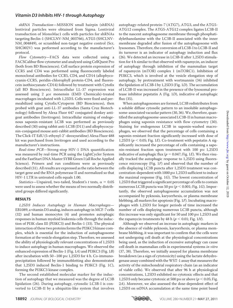
1,25D3 Induces Autophagy in Human Macrophages—EB1089, a vitaminD3 analog, induces autophagy inMCF-7 cells(32) and human monocytes (4) and promotes autophagicresponses in humanmyeloid leukemia cells through the induc-tion of PI3K class III (PI3KC3) and Beclin-1 (33). The physicalinteraction of these twoproteins forms the PI3KC3kinase com-plex, which is essential for the induction of autophagosomeformation at the vesicle elongation step. Therefore, we assessedthe ability of physiologically relevant concentrations of 1,25D3to induce autophagy in human macrophages. We observed theenhanced expression of Beclin-1 (Fig. 1A) and PI3KC3 (Fig. 1B)after incubation with 50–100 pM 1,25D3 for 4 h. Co-immuno-precipitation followed by immunoblotting also demonstratedthat 1,25D3 induced Beclin-1 binding to PI3KC3 (Fig. 1C),forming the PI3KC3 kinase complex.The second established molecular marker for the induc-
tion of autophagy that we measured was the degree of LC3Blipidation (26). During autophagy, cytosolic LC3B-I is con-verted to LC3B-II by a ubiquitin-like system that involves
autophagy-related protein-7 (ATG7), ATG3, and the ATG5-ATG12 complex. The ATG5-ATG12 complex ligates LC3B-IIto the nascent autophagosome membrane through phosphati-dylethanolamine with the LC3B-II associated with the innermembrane degraded after fusion of the autophagosome withlysosomes. Therefore, the conversion of LC3B-I to LC3B-II andits turnover is an indicator of autophagy induction and flux(26). We detected an increase in LC3B-II after 1,25D3 stimula-tion for 4 h similar to that observed with rapamycin, an inducerof autophagy through inhibition of the mammalian targetof rapamycin (mTOR) complex 1 (mTORC1). Inhibition ofPI3KC3, which is involved at the vesicle elongation step ofautophagy, by pretreatment with wortmannin (34) inhibitedthe lipidation of LC3B-I by 1,25D3 (Fig. 1D). The accumulationof LC3B-II was increased in the presence of the lysosomal pro-tease inhibitor pepstatin A (Fig. 1D), indicative of autophagicflux (35).When autophagosomes are formed, LC3B redistributes from
a soluble diffuse cytosolic pattern to an insoluble autophago-some-associated vacuolar pattern (30, 36).We, therefore, quan-tified the autophagosome-associatedLC3B-II in humanmacro-phages using saponin resistance with flow cytometry (30).Staining for endogenous LC3B in saponin-washed macro-phages, we observed that the percentage of cells containing asaponin-resistant fraction significantly increased with dose of1,25D3 (p � 0.05; Fig. 1E). Co-treatment with pepstatin A sig-nificantly increased the percentage of cells containing a sapo-nin-resistant fraction upon treatment with 100 pM 1,25D3indicative of autophagic flux (p� 0.047; Fig. 1E).We then visu-ally tracked the autophagic response to 1,25D3 using fluores-cence microscopy (Fig. 1F) and observed that the number ofcells displaying LC3B puncta after 1,25D3 treatment was con-centration-dependentwith 1000 pM 1,25D3 sufficient to inducethe maximal response (Fig. 1G). The lowest concentration of1,25D3 that triggered a significant percentage of cells to containnumerous LC3B puncta was 50 pM (p � 0.001; Fig. 1G). Impor-tantly, the observed autophagosome accumulation was notaccompanied by pyknosis, karyorrhexis, or plasma membraneblebbing, all markers for apoptosis (Fig. 1F). Incubatingmacro-phages with 1,25D3 for longer periods of time increased thenumber of cells displaying numerous LC3B puncta, althoughthis increase was only significant for 50 and 100 pM 1,25D3 andthe rapamycin treatments by 48 h (p � 0.01; Fig. 1H).Although we observed an increase in autophagic markers in
the absence of visible pyknosis, karyorrhexis, or plasma mem-brane blebbing, it was important to confirm that the cells werenot undergoing cell death at the physiological concentrationsbeing used, as the induction of excessive autophagy can causecell death in mammalian cells in experimental systems in vitro(37, 38). Therefore, we initially assayed for plasma membranebreakdown (as a sign of cytotoxicity) using the lactate dehydro-genase assay combinedwith theWST-1 assay thatmeasures theactivity of the mitochondrial respiratory chain (as an indicatorof viable cells). We observed that after 96 h at physiologicalconcentrations, 1,25D3 exhibited no cytotoxic effects and that1,25D3 only became cytotoxic at 500 pM or above (p� 0.05; Fig.2A). Moreover, we also assessed the dose-dependent effect of1,25D3 on ssDNA accumulation at the same time point based
Vitamin D3 Inhibits HIV-1 through Autophagy
18892 JOURNAL OF BIOLOGICAL CHEMISTRY VOLUME 286 • NUMBER 21 • MAY 27, 2011
by guest on October 12, 2019
http://ww
w.jbc.org/
Dow
nloaded from

on the selective thermal denaturation of apoptotic ssDNAusing low heat and formamide and subsequent detection usinga monoclonal antibody, a more specific indicator of apoptosisthan terminal deoxynucleotidyltransferase dUTP nick endlabeling (TUNEL) (39). We observed the dose-dependent
increase in the number of cells exhibiting the accumulation ofssDNA that became significant at 500 pM 1,25D3 (p � 0.0005;Fig. 2B). As wewere using rapamycin as a control for the induc-tion of autophagy, we also assessed its cytotoxic effects andobserved that at 100–200 nM, rapamycin was neither signifi-
FIGURE 1. 1,25D3 induces autophagy in human macrophages. A, macrophages were left untreated or were incubated with 1,25D3, rapamycin (Rapa) orstarved by incubation in Earle’s balanced salt solution for 4 h, lysed, then subjected to immunoblotting with antibody to Beclin-1 and �-actin. B, shown is animmunoblot using antibody to PI3KC3 and �-actin. C, shown is an immunoblot of co-immunoprecipitation with PI3KC3-specific antibody using antibody toBeclin-1 and antibody to PI3KC3 as loading control. D, shown are immunoblots of LC3B isoforms using antibody to LC3B or �-actin using macrophagespretreated with 100 nM wortmannin, 10 �g/ml pepstatin A, or vehicle control before 1,25D3 or rapamycin treatment. E, shown is a flow cytometry analysis ofsaponin-resistant LC3B-II in macrophages after 1,25D3 and/or pepstatin A treatment for 4 h. Representative dot blots with the percentage of cells displayingsaponin-resistant LC3B-II from three donors are shown. Ab, antibody. F, representative fluorescence microscopy images of macrophages from three donorsafter 1,25D3 or rapamycin treatment for 4 h are shown. Cells were fixed and permeabilized, then stained with antibody to LC3B (red) and Hoechst 33342 (blue).LC3B puncta are indicated by the white arrows. When puncta were not present, LC3B staining gives rise to a diffuse cytoplasmic pattern. The number of punctaper cell is indicated as the mean S.D. Scale bars indicate 10 �m. G, percentage of cells with LC3B puncta after designated treatments for 4 h is shown.H, quantification of cells with LC3B puncta after designated treatments at the time points indicated. Results are the means S.E. of three independentexperiments performed in triplicate. †, p � 0.05; *, p � 0.001 compared with untreated control.
Vitamin D3 Inhibits HIV-1 through Autophagy
MAY 27, 2011 • VOLUME 286 • NUMBER 21 JOURNAL OF BIOLOGICAL CHEMISTRY 18893
by guest on October 12, 2019
http://ww
w.jbc.org/
Dow
nloaded from

cantly cytotoxic as measured by lactate dehydrogenase release(p 0.05) or WST-1 conversion (p 0.2; Fig. 2C) or inducedsignificant numbers of cells to accumulate ssDNA (p 0.2; Fig.2D). We then assessed the effect of 1,25D3 and rapamycin onEdU incorporation and observed no significant effect at thephysiologically relevant concentrations tested (p 0.2; Fig. 2E).We next investigated to what extent conventional autophagy
mediators are required for 1,25D3-induced autophagosomeformation. Pretreatment with wortmannin, which suppressesautophagy regardless of nutrient status (40), inhibited autopha-gosome formation mediated by 1,25D3 with no cytotoxiceffects (Fig. 3A). Although 3-methyladenine is widely used as an
autophagy inhibitor (41), we specifically did not use it in theseexperiments because recent studies have shown that it actuallypromotes autophagy under nutrient-rich conditions (40). RNAinterference of ATG5 and Beclin-1 (Fig. 3B) resulted in thereduction in autophagosome formation after 1,25D3 treatment(Fig. 3C and 3D). Therefore, several known components ofautophagosome formation, PI3K, ATG5, and Beclin-1, areessential for 1,25D3-mediated autophagy.1,25D3 Inhibits HIV-1 Replication in Human Macrophages—
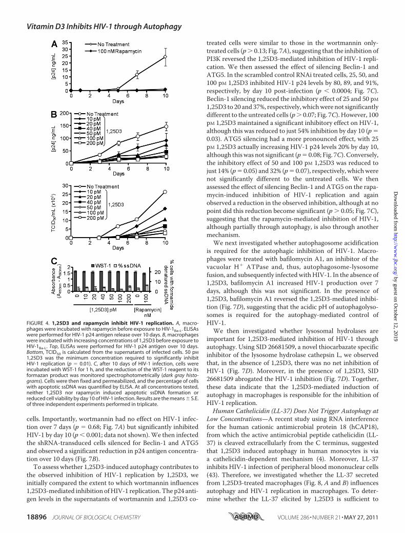
We then determined whether 1,25D3 and rapamycin influenceHIV-1 infection and replication in macrophages by comparingthe extent to which 1,25D3 and rapamycin pre-treatment influ-enced p24 antigen accumulation in the supernatants of macro-phages that were subsequently infected with HIV-1. Rapamy-cin, at achievable in vivo plasma concentrations, inhibitedHIV-1 replication by 95% over 10 days (p � 0.001; Fig. 4A).1,25D3 induced a dose-dependent inhibition of HIV-1 replica-tion with 50 pM being the minimum concentration required tosignificantly inhibit HIV-1 at 10 days post-infection (79%reduction; p � 0.01; Fig. 4B). This effect was not enhanced byfurther increasing the 1,25D3 concentration (Fig. 4B). Impor-tantly, the 1,25D3 and rapamycin treatments had no significanteffects on either cell viability (WST-1 assay) or ssDNAaccumu-lation, a specific marker for apoptotic cells (39) at day 10 (p 0.05; Fig. 4C).To understand how 1,25D3 affects HIV-1 replication, we
examined sequential steps of HIV-1 replication. Treatment ofcells with 1,25D3 did not significantly affect the expression ofeither CD4 or CCR5, which are required for HIV-1 binding andentry into cells (Fig. 5A). Consistent with this finding, bindingof HIV-1 to 1,25D3-treated cells, as measured by ELISA of cell-associated p24 Gag protein, was similar to that of untreatedcells (Fig. 5B). To assay for virus entry, we examined the quan-tity of intracellular p24 Gag (trypsin-resistant) associated withcells exposed to virus for 5 h. Both untreated cells and 1,25D3-treated cells displayed the same intracellular p24 concentrationover 5 h (p 0.35; Fig. 5C). Treatment of cells with the CCR5antagonist maraviroc provided a measurement of nonspecificbackground in these assays.The previous results suggested that 1,25D3 treatment had no
effect on HIV-1 binding or entry. Therefore, we measured viralentry by real-time PCR. Macrophages were treated with1,25D3, rapamycin, or vehicle control for 4 h before infectionwith HIV-1Ba-L for 3 h. After an additional 5-h culture in freshmedia, samples were analyzed by real-time PCR for the pres-ence and quantity of strong-stop HIV-1 DNA (with LTR R/U5primers), an early product of reverse transcription. Physiologi-cal concentrations of 1,25D3 had no effect on HIV-1Ba-L infec-tion (p � 0.8), whereas 100 nM rapamycin treatment signifi-cantly inhibited HIV-1Ba-L infection by 67% (p � 0.0001; Fig.5D). AsNef is the first viral product synthesized during theHIVlife cycle, we also analyzed the translation ofNef at 5 h by immu-noblotting and observed no difference between cells treatedwith the vehicle control or with 1,25D3 (Fig. 5E). We thenassessed for productive HIV-1 infection by assaying for Tatactivity using TZM-bl cells. We pretreated TZM-bl cells withincreasing concentrations of 1,25D3 or rapamycin for 4 h thenexposed them to HIV-1. We found that at the concentrations
FIGURE 2. Physiological concentrations of 1,25D3 are not cytotoxic.Macrophages were incubated with increasing concentrations of 1,25D3 for96 h. A, aliquots of supernatant taken before the addition of WST-1 weretested for lactate dehydrogenase (LDH) spectrophotometrically using theLDHPLUS assay (black squares). For the last hour cells were incubated withWST-1, and the reduction of the WST-1 reagent to its formazan product wasmonitored spectrophotometrically (black circles). B, quantification of thenumber of cells with apoptotic ssDNA using the ssDNA ELISA 96 h after1,25D3 treatment is shown. C, lactate dehydrogenase (black squares) andWST-1 (black circles) assay after 96 h of incubation of macrophages withincreasing concentrations of rapamycin is shown. D, quantification of thenumber of cells with apoptotic ssDNA using the ssDNA ELISA 96 h after rapa-mycin treatment is shown. E, effect of 1,25D3 and rapamycin on the uptake ofEdU by macrophages is shown. Cells were incubated with 1,25D3 or rapamy-cin for 78 h, after which cells were incubated a further 18 h in the presence of10 �M EdU. Cells were harvested, fixed, permeabilized, then probed for EdUincorporation and analyzed for the percentage of EdU-positive cells by flowcytometry. Physiological concentrations of 1,25D3 and rapamycin had nocytotoxic effects. All results are the means S.E. of three donors performed intriplicate. †, p � 0.05; *, p � 0.001 compared with untreated control cells.
Vitamin D3 Inhibits HIV-1 through Autophagy
18894 JOURNAL OF BIOLOGICAL CHEMISTRY VOLUME 286 • NUMBER 21 • MAY 27, 2011
by guest on October 12, 2019
http://ww
w.jbc.org/
Dow
nloaded from

tested 1,25D3 had no significant inhibitory effect on HIV-1productive infection as measured by Tat activity (p 0.47;Fig. 5F). Conversely, rapamycin displayed a dose-dependentinhibitory effect that only became significant at 400 nM (p �0.025; Fig. 5F).We then assessed whether the inhibition of HIV-1 infection
was due to 1,25D3-inducing the production of replication-in-competent viral particles; uninfected macrophages were incu-bated with 1 ng of p24 antigen from the day-10 aliquots of thecell-free supernatants post-1,25D3 pretreatment for 3 h thencultured for 10 days. No difference in HIV-1-replicative fitnesswas observed post-1,25D3 treatment (Fig. 5G). Next, we exam-ined the effect of 100 pM 1,25D3 on HIV-1 replication at earlytime points post-infection by treating macrophage culturesthat had been exposed toHIV-1BaL with 100 pM 1,25D3 at�4 h,at the time of infection and at 3, 5, and 7 days post-infection. Ateach time point, 7 days post-infection excepted, 1,25D3 signif-icantly suppressed HIV-1 replication (p� 0.034; Fig. 5H) in theabsence of toxic effects.We also tested the effects of rapamycin and 1,25D3 on HIV-
1-infected macrophages by comparing the extent to which1,25D3 and rapamycin treatment influenced the release of p24antigen from 20 day-old HIV-1-infected macrophage cultures.At achievable in vivo plasma concentrations, neither rapamycinnor 1,25D3 induced the significant release of p24 antigen intothe supernatant over 4 h (Figs. 5I and 5J). However, both rapa-mycin and high concentrations of 1,25D3 significantly inhib-ited HIV-1 replication over 72 h (Fig. 5I). Conversely, although
50 pM 1,25D3 inhibited the release of p24 over 72 h comparedwith the vehicle control, the inhibition was not significant, and10 pM 1,25D3 had no effect on HIV-1 p24 release (Fig. 5I).Interestingly, both 50 and 25�M rapamycin induced the releaseof significant levels of p24 antigen after just 4 h that was accom-panied by significant levels of apoptosis (Fig. 5J).1,25D3-mediated Autophagy Inhibits HIV-1 Replication in
Human Macrophages—Previous studies have shown that, inprimary macrophages, HIV-1 assembles in late endosomes ofthe recycling pathway (42) and co-localizes with LC3B. There-fore, to assess the intersection of autophagy with HIV-1 repli-cation and the role of autophagy in 1,25D3-mediated inhibitionof HIV-1, we examined the relative distribution of HIV-1 andLC3B. In the absence of 1,25D3 stimulation, the HIV-1 Gagp17-specific antibody showed some colocalization with theautophagosome marker LC3B and almost no colocalizationwith acidified lysosomes (Fig. 6A). However, upon 1,25D3treatment, the number of p17 puncta that colocalized withLC3B or LysoTracker increased significantly (Fig. 6B).As autophagy proteins are required for efficient HIV-1 rep-
lication in HeLa cells (16), to determine whether 1,25D3 affectsHIV-1 replication in macrophages through autophagy, weexamined whether the inhibition of sequential steps of theautophagy pathway inhibits HIV-1 replication. Therefore, weinitially assessed the effect of the inhibition of basal autophagyon HIV-1 replication in macrophages by comparing the extentto which wortmannin and Beclin-1 and ATG5 silencing influ-enced p24 accumulation in the supernatants of HIV-1-infected
FIGURE 3. 1,25D3-induced autophagy requires PI3K, ATG5, and Beclin-1. A, macrophages were incubated with wortmannin or vehicle control then treatedwith 100 pM 1,25D3 for 4 h. Left, quantification of the percentage of LC3B-positive autophagosome-forming cells is shown. Middle, cell supernatants weretested spectrophotometrically for lactate dehydrogenase (LDH) release as a measure of plasma membrane rupture. Right, for the last hour cells were incubatedwith WST-1, and the reduction of the WST-1 reagent to its formazan product was monitored spectrophotometrically. B, MonoMac1 cells were transfected withscrambled shRNA (S), Beclin-1-specific (B2–B5), or ATG5-specific (A1–A4) shRNA. Top, shown is an immunoblot performed with antibodies to Beclin-1, ATG5,and �-actin. Bottom, quantification of silencing effect on protein content is shown. C, shown are representative microscopy images of LC3B staining in cellstransfected with scrambled (S), ATG5 (A2), or Beclin-1 (B3) shRNA treated with 1,25D3 or rapamycin for 4 h. Cells were stained with antibody to LC3B (red) andHoechst 33342 (blue). Scale bars indicate 10 �m. D, shown is quantification of the percentage of LC3B-positive autophagosome-forming cells transfected withscrambled (S), ATG5 (A2), or Beclin-1 (B3) shRNA and treated with 1,25D3 or rapamycin. Results are the means S.E. of three independent experimentsperformed in triplicate. †, p � 0.05; *, p � 0.001 compared with untreated control cells.
Vitamin D3 Inhibits HIV-1 through Autophagy
MAY 27, 2011 • VOLUME 286 • NUMBER 21 JOURNAL OF BIOLOGICAL CHEMISTRY 18895
by guest on October 12, 2019
http://ww
w.jbc.org/
Dow
nloaded from
cells. Importantly, wortmannin had no effect on HIV-1 infec-tion over 7 days (p � 0.68; Fig. 7A) but significantly inhibitedHIV-1 by day 10 (p � 0.001; data not shown).We then infectedthe shRNA-transduced cells silenced for Beclin-1 and ATG5and observed a significant reduction in p24 antigen concentra-tion over 10 days (Fig. 7B).To assess whether 1,25D3-induced autophagy contributes to
the observed inhibition of HIV-1 replication by 1,25D3, weinitially compared the extent to which wortmannin influences1,25D3-mediated inhibition ofHIV-1 replication. The p24 anti-gen levels in the supernatants of wortmannin and 1,25D3 co-
treated cells were similar to those in the wortmannin only-treated cells (p 0.13; Fig. 7A), suggesting that the inhibition ofPI3K reversed the 1,25D3-mediated inhibition of HIV-1 repli-cation. We then assessed the effect of silencing Beclin-1 andATG5. In the scrambled control RNAi treated cells, 25, 50, and100 pM 1,25D3 inhibited HIV-1 p24 levels by 80, 89, and 91%,respectively, by day 10 post-infection (p � 0.0004; Fig. 7C).Beclin-1 silencing reduced the inhibitory effect of 25 and 50 pM1,25D3 to 20 and 37%, respectively, whichwere not significantlydifferent to the untreated cells (p 0.07; Fig. 7C). However, 100pM 1,25D3 maintained a significant inhibitory effect on HIV-1,although this was reduced to just 54% inhibition by day 10 (p �0.03). ATG5 silencing had a more pronounced effect, with 25pM 1,25D3 actually increasing HIV-1 p24 levels 20% by day 10,although thiswas not significant (p� 0.08; Fig. 7C). Conversely,the inhibitory effect of 50 and 100 pM 1,25D3 was reduced tojust 14% (p� 0.05) and 32% (p� 0.07), respectively, whichwerenot significantly different to the untreated cells. We thenassessed the effect of silencing Beclin-1 and ATG5 on the rapa-mycin-induced inhibition of HIV-1 replication and againobserved a reduction in the observed inhibition, although at nopoint did this reduction become significant (p 0.05; Fig. 7C),suggesting that the rapamycin-mediated inhibition of HIV-1,although partially through autophagy, is also through anothermechanism.We next investigated whether autophagosome acidification
is required for the autophagic inhibition of HIV-1. Macro-phages were treated with bafilomycin A1, an inhibitor of thevacuolar H� ATPase and, thus, autophagosome-lysosomefusion, and subsequently infectedwithHIV-1. In the absence of1,25D3, bafilomycin A1 increased HIV-1 production over 7days, although this was not significant. In the presence of1,25D3, bafilomycin A1 reversed the 1,25D3-mediated inhibi-tion (Fig. 7D), suggesting that the acidic pH of autophagolyso-somes is required for the autophagy-mediated control ofHIV-1.We then investigated whether lysosomal hydrolases are
important for 1,25D3-mediated inhibition of HIV-1 throughautophagy. Using SID 26681509, a novel thiocarbazate specificinhibitor of the lysosome hydrolase cathepsin L, we observedthat, in the absence of 1,25D3, there was no net inhibition ofHIV-1 (Fig. 7D). Moreover, in the presence of 1,25D3, SID26681509 abrogated the HIV-1 inhibition (Fig. 7D). Together,these data indicate that the 1,25D3-mediated induction ofautophagy in macrophages is responsible for the inhibition ofHIV-1 replication.Human Cathelicidin (LL-37) Does Not Trigger Autophagy at
Low Concentrations—A recent study using RNA interferencefor the human cationic antimicrobial protein 18 (hCAP18),from which the active antimicrobial peptide cathelicidin (LL-37) is cleaved extracellularly from the C terminus, suggestedthat 1,25D3 induced autophagy in human monocytes is viaa cathelicidin-dependent mechanism (4). Moreover, LL-37inhibits HIV-1 infection of peripheral blood mononuclear cells(43). Therefore, we investigated whether the LL-37 secretedfrom 1,25D3-treated macrophages (Fig. 8, A and B) influencesautophagy and HIV-1 replication in macrophages. To deter-mine whether the LL-37 elicited by 1,25D3 is sufficient to
FIGURE 4. 1,25D3 and rapamycin inhibit HIV-1 replication. A, macro-phages were incubated with rapamycin before exposure to HIV-1Ba-L. ELISAswere performed for HIV-1 p24 antigen release over 10 days. B, macrophageswere incubated with increasing concentrations of 1,25D3 before exposure toHIV-1Ba-L. Top, ELISAs were performed for HIV-1 p24 antigen over 10 days.Bottom, TCID50 is calculated from the supernatants of infected cells. 50 pM
1,25D3 was the minimum concentration required to significantly inhibitHIV-1 replication (p � 0.01). C, after 10 days of HIV-1 infection, cells wereincubated with WST-1 for 1 h, and the reduction of the WST-1 reagent to itsformazan product was monitored spectrophotometrically (dark gray histo-grams). Cells were then fixed and permeabilized, and the percentage of cellswith apoptotic ssDNA was quantified by ELISA. At all concentrations tested,neither 1,25D3 nor rapamycin induced apoptotic ssDNA formation orreduced cell viability by day 10 of HIV-1 infection. Results are the means S.E.of three independent experiments performed in triplicate.
Vitamin D3 Inhibits HIV-1 through Autophagy
18896 JOURNAL OF BIOLOGICAL CHEMISTRY VOLUME 286 • NUMBER 21 • MAY 27, 2011
by guest on October 12, 2019
http://ww
w.jbc.org/
Dow
nloaded from

induce autophagy, macrophages were treated with the greatestconcentration of LL-37 measured. No significant increase inthe redistribution of LC3B from diffuse to punctate stainingwas observed (p � 0.31; Fig. 8C).To assess whether LL-37 inhibits productive HIV-1 infec-
tion, we pretreated TZM-bl cells with LL-37 for 4 h thenexposed them to HIV-1. LL-37 inhibited HIV-1 productiveinfection in a dose-dependent manner (Fig. 8D), with noinhibitory effect observed at the concentrations elicited by1,25D3-treated macrophages (p 0.7; Fig. 8D). We thenexamined the effect of LL-37 on HIV-1 replication in macro-phages and observed that the concentrations elicited bymacrophages in response to 1,25D3 in vitro induced thedose-dependent augmentation of HIV-1 replication (Fig.8E). Conversely, at levels of LL-37 higher than those pro-duced by macrophages in vitro in response to physiologicalconcentrations of 1,25D3, we observed the inhibition ofHIV-1 (p � 0.05; Fig. 8E).
DISCUSSION
Our results demonstrate that physiological concentrations of1,25D3 induce autophagy in human macrophages and thatautophagy is required for the 1,25D3-mediated inhibition ofHIV-1 replication. To our knowledge these results are the firstto link the induction of autophagy with the inhibition of HIV-1replication in macrophages.Studies of 1,25D3 as an autophagy inducer have focused on
immortalized cell lines (32, 33, 44) and its potential role as ananti-oncogenic drug as it induces cell death via a caspase-inde-pendent mechanism (33) at supraphysiological concentrations(32, 45). These concentrations are associated in patients withhypercalcemia and hypercalciuria that result from the in-creased intestinal absorption of calcium combinedwith the cal-cium-immobilizing effects of vitamin D3. Therefore, 1,25D3 orvitamin D3 analogs are being pursued as anti-cancer therapiesonly in combinationwith glucocorticoids or anti-mitotic agents(46). Importantly, we observed no decrease in cell viability or
FIGURE 5. 1,25D3 does not affect HIV-1 entry or release. A, macrophages were left untreated (solid gray histogram) or treated with 100 pM 1,25D3 (dashedline) or 100 nM rapamycin (solid line). After 4 h, cells were harvested and stained for surface CD4 and CCR5. B, macrophages treated with 1,25D3, rapamycin, ormaraviroc (M) for 4 h were infected with replication competent HIV-1Ba-L. Binding was measured at 3 h postinfection by washing cells extensively, then lysingand analyzing p24 by ELISA. C, entry was measured at 5 h postinfection by washing cells extensively, then trypsinization, lysing, and subsequently analyzingintracellular p24 by ELISA. D, DNA was extracted from cells at 8 h postinfection for PCR analysis of pre-integration strong-stop HIV-1 DNA. RNA polymerase II wasamplified as a control. Results are expressed as the ratio between the target gene and the RNA polymerase II and normalized so that HIV-1 LTR in untreated cellsequals 1.00. E, macrophages lysed at 5 h postinfection were subjected to immunoblotting for both Nef and GAPDH. F, percentage of TZM-bl cells wereproductively infected with HIV-1Ba-L after 4 h treatment with 1,25D3 or rapamycin. G, macrophages were incubated with 1 ng p24 antigen from the 10-dayaliquots of cell-free supernatants post-1,25D3 or rapamycin treatment for 3 h then cultured for 10 days with ELISA performed for HIV-1 p24 antigen. H, macro-phages were incubated with 100 pM 1,25D3 at different time points with respect to infection with HIV-1Ba-L. ELISAs were performed for HIV-1 p24 antigen over10 days. I, 20-day-old HIV-1-infected macrophages were incubated with rapamycin or 1,25D3 with ELISA performed for HIV-1 p24 antigen over 3 days. J, shownis the percentage of cells with apoptotic ssDNA combined with p24 release after 4 h of treatment with rapamycin or 1,25D3. All data are the means S.E. ofthree independent experiments performed in triplicate. *, p � 0.001 compared with untreated control cells.
Vitamin D3 Inhibits HIV-1 through Autophagy
MAY 27, 2011 • VOLUME 286 • NUMBER 21 JOURNAL OF BIOLOGICAL CHEMISTRY 18897
by guest on October 12, 2019
http://ww
w.jbc.org/
Dow
nloaded from

increase in apoptotic cells resulting from physiologically rele-vant concentrations of 1,25D3 that significantly inhibitedHIV-1 through autophagy.The envelope glycoproteins, gp120 and gp41, activate pro-
tein kinase C and Akt, upstream effectors of themTORC1, thattrigger autophagy in uninfected cells (17, 19, 47).Moreover, theG-U-rich long terminal repeat region of the HIV-1 genome is aligand for Toll-like receptor 8 found in monocytes (48) thattriggers downstream signaling via Toll-interleukin-1 receptor-domains activating the interferon regulatory factor, nuclearfactor �-light-chain enhancer of activated B cells, andmitogen-activated protein kinase pathways leading to the induction ofautophagy (49). As an obligatory intracellular parasite, HIV-1survival is dependent upon its ability to exploit the host cellmachinery for replication and dissemination and to circumvent
cellular processes that prevent its growth. One such mecha-nism is that, upon infection, HIV-1 down-regulates IL-1receptor-associated kinase 4, which is essential for virtuallyall Toll-like receptor signaling (50). Recent studies haveshown that HIV-1 down-regulates autophagy in produc-tively infected CD4� T cells, U937 cells (15) and myeloiddendritic cells through an Env-dependent manner (14) andthat HIV-1 is undetectable in highly autophagic macro-phages (13). Moreover, in myeloid cells, HIV-1 Gag co-local-izes with LC3B puncta (Fig. 6) and CD9 (51). These datacombined with our observations that pharmacological in-hibitors of autophagy and RNA interference of autophagyproteins inhibits HIV-1 replication in macrophages are con-sistent with the hypothesis that HIV-1 is assembled on endo-cytic membranes that intersect with the autophagy pathway
FIGURE 6. LC3B and acidic vacuoles colocalize with HIV-1 Gag. Macrophages were treated with 100 pM 1,25D3 or vehicle control (EtOH) then infected withHIV-1Ba-L for 10 d. A, cells were fixed, permeabilized, and probed for LC3B, Gag-p17, and Hoechst 33342 (blue). Green dots indicate Gag-p17, and red dots indicateLC3B positive structures. B, after 10 days, cells were incubated with Lysotracker Red for 4 h, then fixed, permeabilized, and probed for Gag-p17. Green dotsindicate Gag-p17, and red dots indicate acidic vacuoles. Scale bars indicate 10 �m.
Vitamin D3 Inhibits HIV-1 through Autophagy
18898 JOURNAL OF BIOLOGICAL CHEMISTRY VOLUME 286 • NUMBER 21 • MAY 27, 2011
by guest on October 12, 2019
http://ww
w.jbc.org/
Dow
nloaded from

(42, 52). However, our data obtained using wortmanninappears to be in conflict with a previous study that showedthat PI3K inhibition blocked HIV infection of macrophageswithin 24 h (53). That study used LY 294002, which hasrecently been shown to inhibit a large number of otherkinases as well as ATP-binding proteins (34). Therefore, therapidity of HIV-1 inhibition observed in that study may bedue to the nonspecific inhibition of other kinases involvedduring HIV-1 replication. By triggering autophagy, physio-logically relevant concentrations of 1,25D3 overcomes theHIV-1-mediated down-regulation of autophagy and inhibits
viral replication through an autophagy-dependent mecha-nism without inducing apoptosis or necrosis even thoughthe autophagic response is similar to that observed in cancercells (33). We also show that blocking 1,25D3-triggeredautophagy with pharmacological inhibitors or RNA interfer-ence decreases the inhibition of HIV-1 replication. There-fore, this study demonstrates, to our knowledge for the firsttime, that 1,25D3 inhibits HIV-1 replication through theinduction of autophagy.In this studywe also show that blocking rapamycin-triggered
autophagy with pharmacological inhibitors or RNA interfer-
FIGURE 7. 1,25D3-mediated induction of autophagy inhibits HIV-1 replication. A, macrophages were incubated with wortmannin or vehicle control beforetreatment with 50 –100 pM 1,25D3 and infection with HIV-1Ba-L. ELISA performed for HIV-1 p24 antigen release over 7 days. B, shown is quantification of p24antigen secreted by HIV-1-infected ATG5 shRNA (A2) and Beclin-1 shRNA (B3) transduced cells compared with nonspecific scrambled shRNA-transduced cells.C, ATG5 shRNA (A2), Beclin-1 shRNA (B3), and nonspecific scrambled shRNA (S)-transduced cells were incubated with 0 –100 pM 1,25D3 or 0 –200 nM rapamycinthen infected with HIV-1Ba-L. ELISAs were performed for HIV-1 p24 antigen over 10 days. D, macrophages were incubated with bafilomycin A1, SID 26681509,or vehicle control before treatment with 50 –100 pM 1,25D3 and infection with HIV-1Ba-L. ELISAs were performed for HIV-1 p24 antigen release over 7 days.Results are the means S.E. of three independent experiments performed in triplicate.
Vitamin D3 Inhibits HIV-1 through Autophagy
MAY 27, 2011 • VOLUME 286 • NUMBER 21 JOURNAL OF BIOLOGICAL CHEMISTRY 18899
by guest on October 12, 2019
http://ww
w.jbc.org/
Dow
nloaded from

ence also decreases the inhibition of HIV-1 replication. There-fore, although it is known that rapamycin inhibits HIV-1 repli-cation through the down-regulation ofCCR5 and the inhibitionof HIV-1 mRNA synthesis (54, 55), we demonstrate the novelfinding that rapamycin also inhibits HIV-1 replication at leastin part through the induction of autophagy. A recent publica-tion observed that 50 �g/ml (54.7 �M) rapamycin treatment ofHIV-1-infected cells induced higher yields of p24 antigen intothe supernatant than from rapamycin-untreated cells andattributed this to the induction of autophagy (51). However,rapamycin is cytotoxic at concentrations above 2 �M andinduces apoptosis (56) (Fig. 2), which may lead to the secretionof HIV-1 p24 antigen even in the absence of lactate dehydro-genase release, a marker for secondary necrosis. Moreover, invivo concentrations of therapeutically administered rapamycinare 500–5000-fold lower, at which concentrations we and oth-ers observed the inhibition of HIV-1 replication (Fig. 3) and theinduction of autophagy (15, 57) (Fig. 1). Furthermore, rapamy-cin is now being used in HIV-1-infected transplantation recip-ients for its potential antiretroviral activity as well as for itsimmunosuppressive effects (58).We also investigated the effect of extracellular LL-37 on
HIV-1 replication. We observed that although HIV-1 is inhib-ited at LL-37 concentrations found in the plasma of healthyindividuals, at the concentrations found to be elicited bymacrophages in response to physiological concentrations of1,25D3 in vitro and in the absence of 1,25D3, LL-37 is unable toinduce autophagy and actually enhances HIV-1 replication.Interestingly, similar concentrations of LL-37 are found in cer-vicovaginal secretions where higher base-line genital levels areassociated with increased acquisition of HIV-1, independent ofthe behavioral and immune correlates of HIV-1 acquisition(59). It is possible that endogenous hCAP18maybemore relevantthan the secreted LL-37, as Yuk et al. (4) clearly showed that
hCAP18 is upstreamof Beclin-1 andATG5 induction post-supra-physiological levels of 1,25D3 treatment (20 nM). However, weobserved no role for exogenous LL-37 in 1,25D3-mediatedautophagy at physiological concentrations (12.5–100 pM).Vitamin D3 deficiency is conservatively defined as �50 nM
25D3,which is the estimatedmean concentration of 25D3pres-ent in people worldwide (60). A number of studies have foundthat the concentrations of vitamin D3 metabolites in HIV-1-infected persons are lower than that of uninfected controls (5, 6,8, 9) with individuals with the lowest concentrations having anincreased risk for HIV-1 disease progression (11). Moreover, ahistory ofAIDS-defining events is associatedwith lower 1,25D3concentrations (7). The major source of vitamin D3 is throughthe endogenous photochemical conversion of 7-dehydrocho-lesterol in the skin to previtamin D3 by ultraviolet B light expo-sure, which then undergoes a 1,7-sigmatropic hydrogen trans-fer forming cholecalciferol. This is then transferred from theskin by the vitamin D-binding protein and is subsequentlyhydroxylated by 25-hydroxylase CYP2R1 in hepatocytes toform 25D3 in a poorly regulated manner. Lesser amounts ofvitamin D3 metabolites are also consumed through fortifieddairy products and oily fish. Vitamin D3 status, therefore, islargely dependent upon the availability of cholecalciferol. WhyHIV-1-infected individuals tend to have lower levels of 1,25D3and/or 25D3 is largely unknown but is thought to be related toinadequate renal 1�-hydroxylationmediated by pro-inflamma-tory cytokines and/or antiretroviral drugs (6, 7, 61). The effectsof HIV-1 viral products on 1,25D3 and/or 25D3 synthesis havenot yet been evaluated. Four genes contribute to the variabilityof serum25D3 concentrations: 7-dehydrocholesterol reductase(involved in cholesterol synthesis and the availability of 7-de-hydrocholesterol in the skin), 25-hydroxylase CYP2R1 andCYP24A1 (degrades and recycles vitamin D3), and GC, whichencodes for the vitamin D-binding protein. Genetic variations
FIGURE 8. 1,25D3-induced cathelicidin does not induce autophagy or inhibit HIV-1. A, macrophages were treated with 1,25D3 and secreted LL-37measured by ELISA. B, shown is a flow cytometry analysis of LL-37 in macrophages left unstimulated (gray histograms) or stimulated with 2000 units/ml IFN�(blue histograms), 100 pM 1,25D3 (green histograms), or 200 nM rapamycin (red histograms). Left, representative flow cytometry histograms are shown. Right,mean fluorescence change is shown. C, macrophages were left untreated or treated with LL-37, fixed, permeabilized, then probed for LC3B and analyzed byfluorescence microscopy. Scale bars indicate 10 �m. Left, representative microscopy images are shown. Right, quantification of cells with LC3B puncta is shown.D, the percentage of TZM-bl cells productively infected with HIV-1Ba-L after pretreatment with increasing concentrations of LL-37 or scrambled control peptideis shown. E, macrophages were incubated with varying concentrations of LL-37 or a scrambled control peptide (CP) for 4 h before exposure to HIV-1Ba-L. ELISAswere performed for HIV-1 p24 antigen release over time. Results are the means S.E. of three independent experiments performed in triplicate.
Vitamin D3 Inhibits HIV-1 through Autophagy
18900 JOURNAL OF BIOLOGICAL CHEMISTRY VOLUME 286 • NUMBER 21 • MAY 27, 2011
by guest on October 12, 2019
http://ww
w.jbc.org/
Dow
nloaded from

at these loci were recently identified in individuals who have asubstantially increased risk of vitamin D insufficiency (62).Studies to assess the genetic variations at these loci and theirimpact on HIV-1 progression and disease status are now underway.In summary, this study demonstrates a role for autophagy
during the early phases of HIV-1 infection and a potential linkbetween vitamin D3 and rapamycin-induced autophagy andthe inhibition of HIV-1 replication in macrophages. Whethervitamin D3 supplementation will be a useful adjunct to antiret-roviral treatment of HIV-1-infected persons is unknown but istestable through clinical trials. Dissecting the molecular mech-anisms by which HIV-1 utilizes autophagic machinery willgreatly enhance our understanding of this critical nexus in thehost-virus relationship that in turn may lead to the develop-ment of novel strategies to prevent and treat HIV-1 infectionand related opportunistic infections.
Acknowledgments—We thank Carol Mundy for technical assistanceand Dennis Young (Flow Cytometry Core Facility, University of Cal-ifornia San Diego) for assistance with flow cytometry.
REFERENCES1. Joint United Nations Programme on HIV/AIDS (2008) Report on the
Global HIV/AIDS Epidemic 2008, Executive Summary, Joint United Na-tions Programme on HIV/AIDS, Geneva, Switzerland
2. Martineau, A. R., Honecker, F. U., Wilkinson, R. J., and Griffiths, C. J.(2007) J. Steroid Biochem. Mol. Biol. 103, 793–798
3. Sly, L. M., Lopez, M., Nauseef, W. M., and Reiner, N. E. (2001) J. Biol.Chem. 276, 35482–35493
4. Yuk, J. M., Shin, D. M., Lee, H. M., Yang, C. S., Jin, H. S., Kim, K. K., Lee,Z. W., Lee, S. H., Kim, J. M., and Jo, E. K. (2009) Cell Host. Microbe 6,231–243
5. Haug, C.,Muller, F., Aukrust, P., and Frøland, S. S. (1994) J. Infect. Dis. 169,889–893
6. Haug, C. J., Aukrust, P., Haug, E., Mørkrid, L., Muller, F., and Frøland, S. S.(1998) J. Clin. Endocrinol. Metab. 83, 3832–3838
7. Mueller, N. J., Fux, C. A., Ledergerber, B., Elzi, L., Schmid, P., Dang, T.,Magenta, L., Calmy, A., Vergopoulos, A., and Bischoff-Ferrari, H. A.(2010) AIDS 24, 1127–1134
8. Teichmann, J., Stephan, E., Discher, T., Lange, U., Federlin, K., Stracke, H.,Friese, G., Lohmeyer, J., and Bretzel, R. G. (2000) Metabolism 49,1134–1139
9. Teichmann, J., Stephan, E., Lange, U., Discher, T., Friese, G., Lohmeyer, J.,Stracke, H., and Bretzel, R. G. (2003) J. Infect. 46, 221–227
10. de Luis, D. A., Bachiller, P., Aller, R., de Luis, J., Izaola, O., Terroba, M. C.,Cuellar, L., and Gonzalez Sagrado, M. (2002) Nutr. Hosp. 17, 285–289
11. Mehta, S., Giovannucci, E., Mugusi, F. M., Spiegelman, D., Aboud, S.,Hertzmark, E.,Msamanga,G. I., Hunter, D., and Fawzi,W.W. (2010)PLoSONE 5, e8770
12. Mehta, S., Hunter, D. J., Mugusi, F. M., Spiegelman, D., Manji, K. P., Gio-vannucci, E. L., Hertzmark, E., Msamanga, G. I., and Fawzi, W. W. (2009)J. Infect. Dis. 200, 1022–1030
13. Espert, L., Varbanov, M., Robert-Hebmann, V., Sagnier, S., Robbins, I.,Sanchez, F., Lafont, V., and Biard-Piechaczyk, M. (2009) PLoS ONE 4,e5787
14. Blanchet, F. P., Moris, A., Nikolic, D. S., Lehmann, M., Cardinaud, S.,Stalder, R., Garcia, E., Dinkins, C., Leuba, F.,Wu, L., Schwartz, O., Deretic,V., and Piguet, V. (2010) Immunity 32, 654–669
15. Zhou, D., and Spector, S. A. (2008) AIDS 22, 695–69916. Brass, A. L., Dykxhoorn, D. M., Benita, Y., Yan, N., Engelman, A., Xavier,
R. J., Lieberman, J., and Elledge, S. J. (2008) Science 319, 921–92617. Denizot, M., Varbanov, M., Espert, L., Robert-Hebmann, V., Sagnier, S.,
Garcia, E., Curriu, M., Mamoun, R., Blanco, J., and Biard-Piechaczyk, M.(2008) Autophagy 4, 998–1008
18. Espert, L., Denizot, M., Grimaldi, M., Robert-Hebmann, V., Gay, B.,Varbanov, M., Codogno, P., and Biard-Piechaczyk, M. (2006) Med. Sci.(Paris) 22, 677–678
19. Espert, L., Denizot, M., Grimaldi, M., Robert-Hebmann, V., Gay, B.,Varbanov, M., Codogno, P., and Biard-Piechaczyk, M. (2006) J. Clin. In-vest. 116, 2161–2172
20. Platt, E. J., Wehrly, K., Kuhmann, S. E., Chesebro, B., and Kabat, D. (1998)J. Virol. 72, 2855–2864
21. Campbell, G. R.,Watkins, J. D., Loret, E. P., and Spector, S. A. (2011)AIDSRes. Hum. Retroviruses, in press
22. Gartner, S., Markovits, P., Markovitz, D. M., Kaplan, M. H., Gallo, R. C.,and Popovic, M. (1986) Science 233, 215–219
23. Popovic, M., Gartner, S., Read-Connole, E., Beaver, B., and Reitz, M. (Oc-tober 27–29, 1988) in Retroviruses of Human AIDS and Related AnimalDiseases, Colloque Des Cent Gardes (Girard, M., and Valette, L., eds) pp.21–27, Fondation Marcel Merieux, Pasteur Vaccins, Marnes-La-Co-quette, Paris
24. Campbell, G. R., Loret, E. P., and Spector, S. A. (2010) J. Biol. Chem. 285,1681–1691
25. Japour, A. J., Mayers, D. L., Johnson, V. A., Kuritzkes, D. R., Beckett, L. A.,Arduino, J. M., Lane, J., Black, R. J., Reichelderfer, P. S., D’Aquila, R. T.,Crumpacker, C. S. The RV-43 Study Group, and The AIDS Clinical TrialGroup Virology Committee Resistance Working Group (1993) Antimi-crob. Agents Chemother. 37, 1095–1101
26. Klionsky, D. J., Abeliovich, H., Agostinis, P., Agrawal, D. K., Aliev, G.,Askew, D. S., Baba, M., Baehrecke, E. H., Bahr, B. A., Ballabio, A., Bamber,B. A., Bassham, D. C., Bergamini, E., Bi, X., Biard-Piechaczyk, M., Blum,J. S., Bredesen, D. E., Brodsky, J. L., Brumell, J. H., Brunk, U. T., Bursch,W.,Camougrand, N., Cebollero, E., Cecconi, F., Chen, Y., Chin, L. S., Choi, A.,Chu, C. T., Chung, J., Clarke, P. G., Clark, R. S., Clarke, S. G., Clave, C.,Cleveland, J. L., Codogno, P., Colombo, M. I., Coto-Montes, A., Cregg,J.M., Cuervo, A.M., Debnath, J., Demarchi, F., Dennis, P. B., Dennis, P. A.,Deretic, V., Devenish, R. J., Di Sano, F., Dice, J. F., Difiglia, M., Dinesh-Kumar, S., Distelhorst, C.W., Djavaheri-Mergny,M., Dorsey, F. C., Droge,W., Dron, M., Dunn, W. A., Jr., Duszenko, M., Eissa, N. T., Elazar, Z.,Esclatine, A., Eskelinen, E. L., Fesus, L., Finley, K. D., Fuentes, J. M., Fueyo,J., Fujisaki, K., Galliot, B., Gao, F. B., Gewirtz, D.A., Gibson, S. B., Gohla, A.,Goldberg, A. L., Gonzalez, R., Gonzalez-Estevez, C., Gorski, S., Gottlieb,R. A., Haussinger, D., He, Y.W.,Heidenreich, K., Hill, J. A., Høyer-Hansen,M., Hu, X., Huang,W. P., Iwasaki, A., Jaattela,M., Jackson,W. T., Jiang, X.,Jin, S., Johansen, T., Jung, J. U., Kadowaki,M., Kang, C., Kelekar, A., Kessel,D. H., Kiel, J. A., Kim, H. P., Kimchi, A., Kinsella, T. J., Kiselyov, K., Kita-moto, K., Knecht, E., Komatsu,M., Kominami, E., Kondo, S., Kovacs, A. L.,Kroemer, G., Kuan, C. Y., Kumar, R., Kundu, M., Landry, J., Laporte, M.,Le,W., Lei, H. Y., Lenardo,M. J., Levine, B., Lieberman, A., Lim, K. L., Lin,F. C., Liou, W., Liu, L. F., Lopez-Berestein, G., Lopez-Otín, C., Lu, B.,Macleod, K. F., Malorni, W., Martinet, W., Matsuoka, K., Mautner, J.,Meijer, A. J., Melendez, A., Michels, P., Miotto, G., Mistiaen, W. P., Miz-ushima, N., Mograbi, B., Monastyrska, I., Moore, M. N., Moreira, P. I.,Moriyasu, Y., Motyl, T., Munz, C., Murphy, L. O., Naqvi, N. I., Neufeld,T. P., Nishino, I., Nixon, R. A., Noda, T., Nurnberg, B., Ogawa, M., Olein-ick, N. L., Olsen, L. J., Ozpolat, B., Paglin, S., Palmer, G. E., Papassideri, I.,Parkes, M., Perlmutter, D. H., Perry, G., Piacentini, M., Pinkas-Kramarski,R., Prescott, M., Proikas-Cezanne, T., Raben, N., Rami, A., Reggiori, F.,Rohrer, B., Rubinsztein, D. C., Ryan, K. M., Sadoshima, J., Sakagami, H.,Sakai, Y., Sandri, M., Sasakawa, C., Sass, M., Schneider, C., Seglen, P. O.,Seleverstov, O., Settleman, J., Shacka, J. J., Shapiro, I. M., Sibirny, A., Silva-Zacarin, E. C., Simon, H. U., Simone, C., Simonsen, A., Smith, M. A.,Spanel-Borowski, K., Srinivas, V., Steeves, M., Stenmark, H., Stromhaug,P. E., Subauste, C. S., Sugimoto, S., Sulzer, D., Suzuki, T., Swanson, M. S.,Tabas, I., Takeshita, F., Talbot, N. J., Talloczy, Z., Tanaka, K., Tanaka, K.,Tanida, I., Taylor, G. S., Taylor, J. P., Terman, A., Tettamanti, G., Thomp-son, C. B., Thumm, M., Tolkovsky, A. M., Tooze, S. A., Truant, R., Tu-manovska, L. V., Uchiyama, Y., Ueno, T., Uzcategui, N. L., van der Klei, I.,Vaquero, E. C., Vellai, T., Vogel, M. W., Wang, H. G., Webster, P., Wiley,J. W., Xi, Z., Xiao, G., Yahalom, J., Yang, J. M., Yap, G., Yin, X. M., Yoshi-
Vitamin D3 Inhibits HIV-1 through Autophagy
MAY 27, 2011 • VOLUME 286 • NUMBER 21 JOURNAL OF BIOLOGICAL CHEMISTRY 18901
by guest on October 12, 2019
http://ww
w.jbc.org/
Dow
nloaded from

mori, T., Yu, L., Yue, Z., Yuzaki, M., Zabirnyk, O., Zheng, X., Zhu, X., andDeter, R. L. (2008) Autophagy 4, 151–175
27. Brady, N. R., Hamacher-Brady, A., Yuan, H., and Gottlieb, R. A. (2007)FEBS J. 274, 3184–3197
28. Dixon, W. J., andMassey, F. J., Jr. (1983) Introduction To Statistical Anal-ysis, 3rd Ed., p. 92, McGraw-Hill, New York
29. Federighi, E. T. (1959) J. Am. Stat. Assoc. 54, 683–68830. Eng, K. E., Panas, M. D., Karlsson Hedestam, G. B., and McInerney, G. M.
(2010) Autophagy 6, 634–64131. Campbell, G. R., and Spector, S. A. (2008) J. Biol. Chem. 283, 30745–3075332. Høyer-Hansen, M., Bastholm, L., Mathiasen, I. S., Elling, F., and Jaattela,
M. (2005) Cell Death. Differ. 12, 1297–130933. Wang, J., Lian, H., Zhao, Y., Kauss, M. A., and Spindel, S. (2008) J. Biol.
Chem. 283, 25596–2560534. Bain, J., Plater, L., Elliott, M., Shpiro, N., Hastie, C. J., McLauchlan, H.,
Klevernic, I., Arthur, J. S., Alessi, D. R., and Cohen, P. (2007) Biochem. J.408, 297–315
35. Mizushima, N., and Yoshimori, T. (2007) Autophagy 3, 542–54536. Mizushima, N., Yamamoto, A., Hatano, M., Kobayashi, Y., Kabeya, Y.,
Suzuki, K., Tokuhisa, T., Ohsumi, Y., and Yoshimori, T. (2001) J. Cell Biol.152, 657–668
37. Galluzzi, L., Aaronson, S. A., Abrams, J., Alnemri, E. S., Andrews, D. W.,Baehrecke, E. H., Bazan, N. G., Blagosklonny,M. V., Blomgren, K., Borner,C., Bredesen, D. E., Brenner, C., Castedo, M., Cidlowski, J. A., Ciecha-nover, A., Cohen, G. M., De Laurenzi, V., De Maria, R., Deshmukh, M.,Dynlacht, B. D., El-Deiry, W. S., Flavell, R. A., Fulda, S., Garrido, C., Gol-stein, P., Gougeon, M. L., Green, D. R., Gronemeyer, H., Hajnoczky, G.,Hardwick, J. M., Hengartner, M. O., Ichijo, H., Jaattela, M., Kepp, O.,Kimchi, A., Klionsky, D. J., Knight, R. A., Kornbluth, S., Kumar, S., Levine,B., Lipton, S. A., Lugli, E., Madeo, F., Malomi, W., Marine, J. C., Martin,S. J., Medema, J. P., Mehlen, P., Melino, G., Moll, U. M., Morselli, E.,Nagata, S., Nicholson, D. W., Nicotera, P., Nunez, G., Oren, M., Pen-ninger, J., Pervaiz, S., Peter,M. E., Piacentini, M., Prehn, J. H., Puthalakath,H., Rabinovich, G. A., Rizzuto, R., Rodrigues, C. M., Rubinsztein, D. C.,Rudel, T., Scorrano, L., Simon, H. U., Steller, H., Tschopp, J., Tsujimoto,Y., Vandenabeele, P., Vitale, I., Vousden, K. H., Youle, R. J., Yuan, J., Zhi-votovsky, B., and Kroemer, G. (2009) Cell Death Differ. 16, 1093–1107
38. Kroemer, G., and Levine, B. (2008)Nat. Rev. Mol. Cell Biol. 9, 1004–101039. Frankfurt, O. S., and Krishan, A. (2001) J. Immunol. Methods 253,
133–14440. Wu, Y. T., Tan, H. L., Shui, G., Bauvy, C., Huang, Q., Wenk, M. R., Ong,
C. N., Codogno, P., and Shen, H. M. (2010) J. Biol. Chem. 285,10850–10861
41. Blommaart, E. F., Krause, U., Schellens, J. P., Vreeling-Sindelarova, H., andMeijer, A. J. (1997) Eur. J. Biochem. 243, 240–246
42. Pelchen-Matthews, A., Kramer, B., andMarsh, M. (2003) J. Cell Biol. 162,443–455
43. Bergman, P.,Walter-Jallow, L., Broliden, K., Agerberth, B., and Soderlund,J. (2007) Curr. HIV Res. 5, 410–415
44. Demasters, G., Di, X., Newsham, I., Shiu, R., and Gewirtz, D. A. (2006)Mol. Cancer Ther. 5, 2786–2797
45. Pepper, C., Thomas, A., Hoy, T., Milligan, D., Bentley, P., and Fegan, C.
(2003) Blood 101, 2454–246046. Trump, D. L., Potter, D. M., Muindi, J., Brufsky, A., and Johnson, C. S.
(2006) Cancer 106, 2136–214247. Misse, D., Gajardo, J., Oblet, C., Religa, A., Riquet, N., Mathieu, D., Yssel,
H., and Veas, F. (2005) AIDS 19, 897–90548. Heil, F., Hemmi, H., Hochrein, H., Ampenberger, F., Kirschning, C., Akira,
S., Lipford, G., Wagner, H., and Bauer, S. (2004) Science 303, 1526–152949. Delgado, M. A., Elmaoued, R. A., Davis, A. S., Kyei, G., and Deretic, V.
(2008) EMBO J. 27, 1110–112150. Pathak, S., De Souza, G. A., Salte, T., Wiker, H. G., and Asjo, B. (2009)
Scand. J. Immunol. 70, 264–27651. Kyei, G. B., Dinkins, C., Davis, A. S., Roberts, E., Singh, S. B., Dong, C.,Wu,
L., Kominami, E., Ueno, T., Yamamoto, A., Federico, M., Panganiban, A.,Vergne, I., and Deretic, V. (2009) J. Cell Biol. 186, 255–268
52. Deneka, M., Pelchen-Matthews, A., Byland, R., Ruiz-Mateos, E., andMarsh, M. (2007) J. Cell Biol. 177, 329–341
53. Francois, F., and Klotman, M. E. (2003) J. Virol. 77, 2539–254954. Roy, J., Paquette, J. S., Fortin, J. F., and Tremblay, M. J. (2002) Antimicrob.
Agents. Chemother. 46, 3447–345555. Heredia, A., Amoroso, A., Davis, C., Le, N., Reardon, E., Dominique, J. K.,
Klingebiel, E., Gallo, R. C., and Redfield, R. R. (2003) Proc. Natl. Acad. Sci.U.S.A. 100, 10411–10416
56. Harris, J., Hanrahan, O., and DeHaro, S. A. (2009)Curr. Protoc. Immunol.Chapter 14, Unit 14, 14
57. Mercalli, A., Sordi, V., Ponzoni, M., Maffi, P., De Taddeo, F., Gatti, G.,Servida, P., Bernardi, M., Bellio, L., Bertuzzi, F., Secchi, A., Bonifacio, E.,and Piemonti, L. (2006) Am. J. Transplant. 6, 1331–1341
58. Di Benedetto, F., Di Sandro, S., De Ruvo, N., Montalti, R., Ballarin, R.,Guerrini, G. P., Spaggiari,M., Guaraldi, G., andGerunda, G. (2010)Trans-plantation 89, 733–738
59. Levinson, P., Kaul, R., Kimani, J., Ngugi, E., Moses, S., MacDonald, K. S.,Broliden, K., and Hirbod, T. (2009) AIDS 23, 309–317
60. Hagenau, T., Vest, R., Gissel, T. N., Poulsen, C. S., Erlandsen, M.,Mosekilde, L., and Vestergaard, P. (2009) Osteoporos. Int. 20, 133–140
61. Welz, T., Childs, K., Ibrahim, F., Poulton, M., Taylor, C. B., Moniz, C. F.,and Post, F. A. (2010) AIDS 24, 1923–1928
62. Wang, T. J., Zhang, F., Richards, J. B., Kestenbaum, B., van Meurs, J. B.,Berry, D., Kiel, D. P., Streeten, E. A., Ohlsson, C., Koller, D. L., Peltonen, L.,Cooper, J. D., O’Reilly, P. F., Houston, D. K., Glazer, N. L., Vandenput, L.,Peacock, M., Shi, J., Rivadeneira, F., McCarthy, M. I., Anneli, P., de Boer,I. H.,Mangino,M., Kato, B., Smyth, D. J., Booth, S. L., Jacques, P. F., Burke,G. L., Goodarzi, M., Cheung, C. L., Wolf, M., Rice, K., Goltzman, D.,Hidiroglou, N., Ladouceur, M., Wareham, N. J., Hocking, L. J., Hart, D.,Arden, N. K., Cooper, C., Malik, S., Fraser,W. D., Hartikainen, A. L., Zhai,G., Macdonald, H. M., Forouhi, N. G., Loos, R. J., Reid, D. M., Hakim, A.,Dennison, E., Liu, Y., Power, C., Stevens, H. E., Jaana, L., Vasan, R. S.,Soranzo, N., Bojunga, J., Psaty, B. M., Lorentzon, M., Foroud, T., Harris,T. B., Hofman, A., Jansson, J. O., Cauley, J. A., Uitterlinden, A. G., Gibson,Q., Jarvelin, M. R., Karasik, D., Siscovick, D. S., Econs, M. J., Kritchevsky,S. B., Florez, J. C., Todd, J. A., Dupuis, J., Hypponen, E., and Spector, T. D.(2010) Lancet 376, 180–188
Vitamin D3 Inhibits HIV-1 through Autophagy
18902 JOURNAL OF BIOLOGICAL CHEMISTRY VOLUME 286 • NUMBER 21 • MAY 27, 2011
by guest on October 12, 2019
http://ww
w.jbc.org/
Dow
nloaded from

Grant R. Campbell and Stephen A. SpectorAutophagy in Human Macrophages That Inhibits HIV-1 Infection
,25-Dihydroxycholecalciferol) TriggersαHormonally Active Vitamin D3 (1
doi: 10.1074/jbc.M110.206110 originally published online March 30, 20112011, 286:18890-18902.J. Biol. Chem.
10.1074/jbc.M110.206110Access the most updated version of this article at doi:
Alerts:
When a correction for this article is posted•
When this article is cited•
to choose from all of JBC's e-mail alertsClick here
http://www.jbc.org/content/286/21/18890.full.html#ref-list-1
This article cites 57 references, 20 of which can be accessed free at
by guest on October 12, 2019
http://ww
w.jbc.org/
Dow
nloaded from