Embed Size (px)
Citation preview

Homology 3D modeling and effect of mutations

X-ray crystallography (70,714 in PDB)• need crystals
Nuclear Magnetic Resonance (NMR) (9,312)• proteins in solution• lower size limit (600 aa)
Electron microscopy (422)• Low resolution (>5A)
Determination of protein structure

Determination of protein structure
resolution 2.4 A

Determination of protein structure
resolution 2.4 A

Structural genomics
Currently: 106K 3D structures from around 35K seqs90M sequences in UniProt
only 0.04%!

Structural genomics
50% sequences covered (25% in 1995)
Currently: 106K 3D structures from around 35K seqs90M sequences in UniProt
only 0.04%!

3D structure predictionApplications: target design
Query sequence
catalytic center
known 3D
LeuGly
model 3D by homology
Gly Lys+
similar toL G
G K

3D structure predictionApplications: fit to low res 3D
Query sequence 1
low resolution 3D (electron microscopy)
Query sequence 2

Protein domains are structural units (average 160 aa) that share:
FunctionFoldingEvolution
Proteins normally are multidomain (average 300 aa)
Domains

Protein domains are structural units (average 160 aa) that share:
FunctionFoldingEvolution
Proteins normally are multidomain (average 300 aa)
Domains
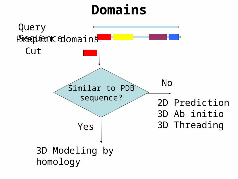
DomainsQuery Sequence
Yes
3D Modeling by homology
No
2D Prediction3D Ab initio3D Threading
Similar to PDBsequence?
Predict domainsCut

3D structure predictionAb initio
Explore conformational space
Limit the number of atoms
Break the problem into fragments of sequence
Optimize hydrophobic residue burial and pairing of beta-strands
Limited success
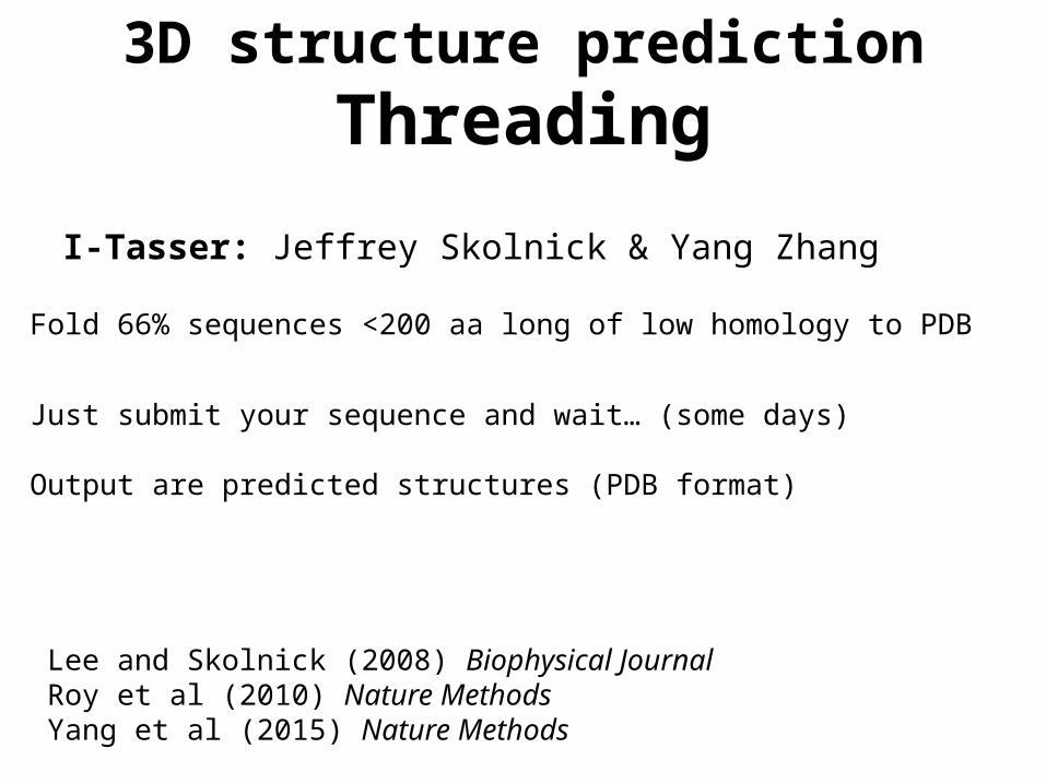
3D structure predictionThreading
I-Tasser: Jeffrey Skolnick & Yang Zhang
Lee and Skolnick (2008) Biophysical JournalRoy et al (2010) Nature MethodsYang et al (2015) Nature Methods
Fold 66% sequences <200 aa long of low homology to PDB
Just submit your sequence and wait… (some days)
Output are predicted structures (PDB format)

3D structure predictionI-Tasser
Roy et al (2010) Nature Methods

3D structure predictionI-Tasser
http://zhanglab.ccmb.med.umich.edu/I-TASSER/
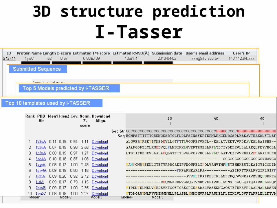
3D structure predictionI-Tasser

3D structure predictionQUARK
http://zhanglab.ccmb.med.umich.edu/QUARK/
3D structure prediction
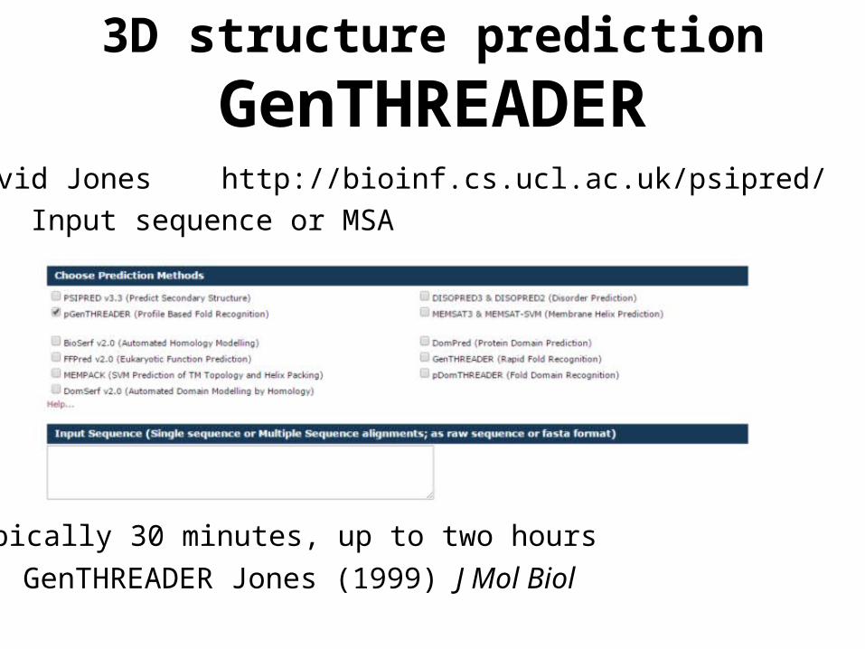
GenTHREADERDavid Jones http://bioinf.cs.ucl.ac.uk/psipred/
Input sequence or MSA
Typically 30 minutes, up to two hours
GenTHREADER Jones (1999) J Mol Biol

Output GenTHREADER
3D structure prediction
GenTHREADER

3D structure predictionPhyre
http://www.sbg.bio.ic.ac.uk/phyre2/
Kelley et al (2000) J Mol BiolKelley and Sternberg (2009) Nature Protocols
Processing time can be hours

3D structure predictionStatic solutions
Datasets of precomputed models / computations
Not flexible
Variable coverage
But you don’t have to wait
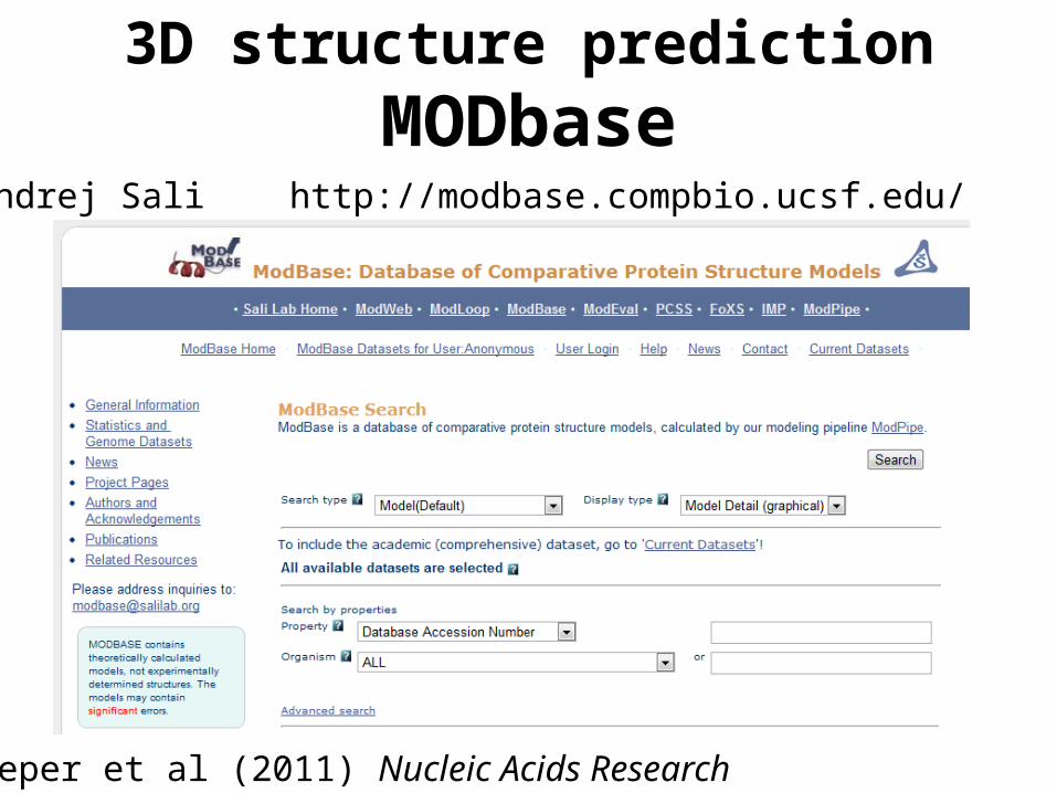
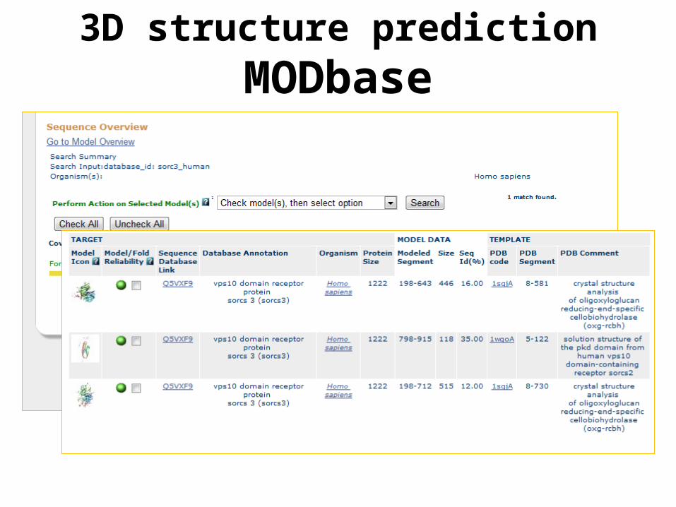
3D structure predictionMODbase
Andrej Sali http://modbase.compbio.ucsf.edu/
Pieper et al (2011) Nucleic Acids Research
3D structure predictionMODbase

Protein Model Portal
Haas et al. (2013) Database
Torsten Schwede
3D structure prediction
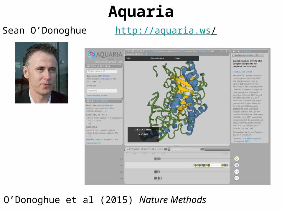
AquariaSean O’Donoghue http://aquaria.ws/
O’Donoghue et al (2015) Nature Methods

Aquaria
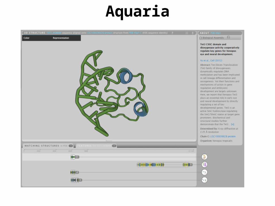
Aquaria

Aquaria

Starting aquaria(May require a Java update)
Works best in Firefox (in Chrome with reduced functionality)
Open Firefox mit JRE (from ZDV)
Go to http://aquaria.ws
Run an example. If JAVA blocked unblock it at the plugin icon
Exercise 1/5

Starting aquariaNote that aquaria.ws requires that two java plug-ins that need to be allowed to run
Exercise 1/5
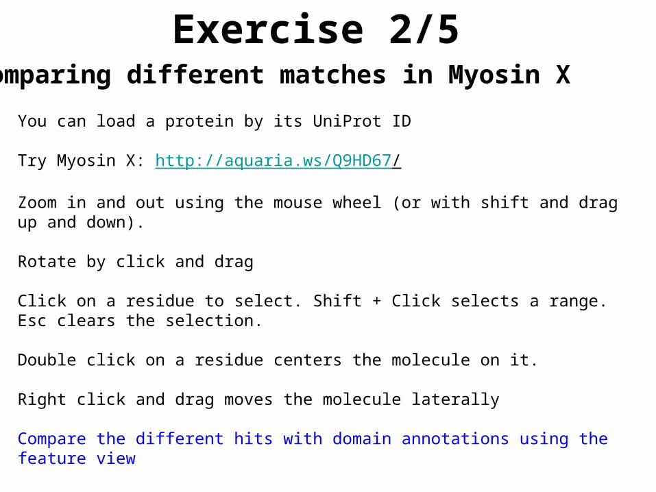
Exercise 2/5
You can load a protein by its UniProt ID
Try Myosin X: http://aquaria.ws/Q9HD67/
Zoom in and out using the mouse wheel (or with shift and drag up and down).
Rotate by click and drag
Click on a residue to select. Shift + Click selects a range. Esc clears the selection.
Double click on a residue centers the molecule on it.
Right click and drag moves the molecule laterally
Compare the different hits with domain annotations using the feature view
Comparing different matches in Myosin X
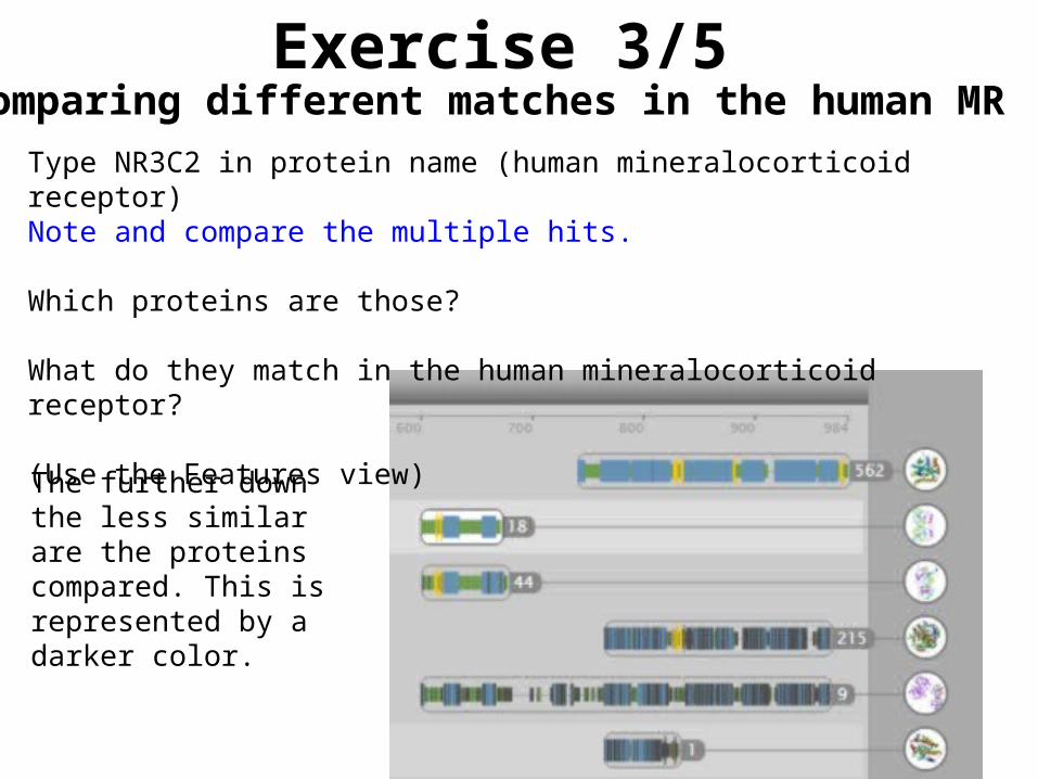
Exercise 3/5
Type NR3C2 in protein name (human mineralocorticoid receptor)Note and compare the multiple hits.
Which proteins are those?
What do they match in the human mineralocorticoid receptor?
(Use the Features view)
Comparing different matches in the human MR
The further down the less similar are the proteins compared. This is represented by a darker color.

Exercise 4/5
Load the human protein CTNNB1 (Catenin beta-1)
Click on the 'Features' tab (bottom of the window)
Double click on the feature lane titled 'Amino acid modification’ (post-translational modification). This will highlight the residues in the structure. Then you can click on the residues to see their position and amino acid.
Which two amino acid modifications are close in structure, but not in sequence? Which type of modifications are those?
Change representation to ball and stick to see the side chains. Do the side chains of the modified residues look like they could interact?
Post-translational modifications in CTNNB1

Effect of mutations
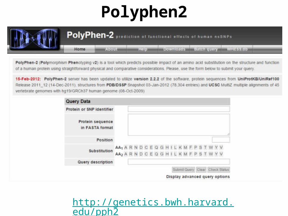
Polyphen2
http://genetics.bwh.harvard.edu/pph2/
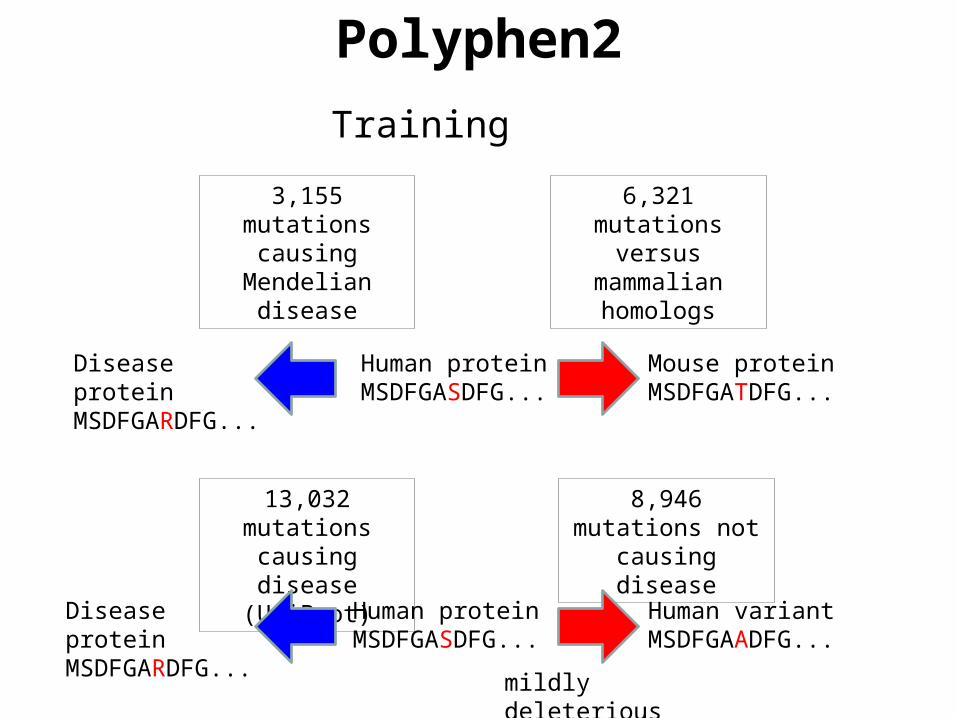
Polyphen2
Training
Human proteinMSDFGASDFG...
Human proteinMSDFGASDFG...
6,321 mutations versus
mammalian homologs
Mouse proteinMSDFGATDFG...
8,946 mutations not causing
disease
Human variantMSDFGAADFG...
mildly deleterious
3,155 mutations causing
Mendelian disease
Disease proteinMSDFGARDFG...
13,032 mutations causing disease
(UniProt)
Disease proteinMSDFGARDFG...

Polyphen2
PSIC Score
Likelihood of an amino acid to occupy a specific position in the protein sequence given the pattern of amino acid substitutions observed in the multiple sequence alignment
EGKLQVQQGTGRFISRDGNLHVNQGMGRFIPRDGNLHVNKGMGRFIPRDGNISVSKGMGRFIPRDGNISVSKGMGRFIPREGTLHTTEGSGRFISREGTLHATEGSGRYIPRDGNLHVTEGSGRYIPRDGTLHVTEGSGRYIPRDGTLHVTEGSGRYIPRDGTLHVTEGSGRYIPRDGNLHVSQGSGRFVPRDGNLFVTEGSGRFVPRDGKMFVTPGAGRFVPRDGNLLVTPGAGRFIPRDGNLLVTPGAGRFIPRDGTLSVMEGSGRFIPRDGNLHATSGTGRFIPC
Reference
Hom
olog
s
Low score High score

Polyphen2
Usage

Polyphen2

Polyphen2

Polyphen2
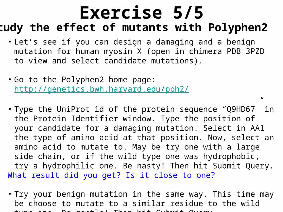
Exercise 5/5
• Let’s see if you can design a damaging and a benign mutation for human myosin X (open in chimera PDB 3PZD to view and select candidate mutations).
• Go to the Polyphen2 home page: http://genetics.bwh.harvard.edu/pph2/
• Type the UniProt id of the protein sequence “Q9HD67” in the Protein Identifier window. Type the position of your candidate for a damaging mutation. Select in AA1 the type of amino acid at that position. Now, select an amino acid to mutate to. May be try one with a large side chain, or if the wild type one was hydrophobic, try a hydrophilic one. Be nasty! Then hit Submit Query.
What result did you get? Is it close to one?
• Try your benign mutation in the same way. This time may be choose to mutate to a similar residue to the wild type one. Be gentle! Then hit Submit Query.
What result did you get? Is it close to zero?
Study the effect of mutants with Polyphen2