Embed Size (px)
Citation preview
Homeotic Evolution in the Mammalia: Diversification ofTherian Axial Seriation and the Morphogenetic Basis ofHuman OriginsAaron G. Filler1,2*
1 Department of Anthropology, Museum of Comparative Zoology, Harvard University, Cambridge, Massachusetts, United States of America,2 Department of Neurosurgery, Institute for Spinal Disorders, Cedars Sinai Medical Center, Los Angeles, California, United States of America
Background. Despite the rising interest in homeotic genes, little has been known about the course and pattern of evolution ofhomeotic traits across the mammalian radiation. An array of emerging and diversifying homeotic gradients revealed by thisstudy appear to generate new body plans and drive evolution at a large scale. Methodology/Principal Findings. This studyidentifies and evaluates a set of homeotic gradients across 250 extant and fossil mammalian species and their antecedentsover a period of 220 million years. These traits are generally expressed as co-linear gradients along the body axis rather than asdistinct segmental identities. Relative position or occurrence sequence vary independently and are subject to polarity reversaland mirroring. Five major gradient modification sets are identified: (1)–quantitative changes of primary segmental identitypattern that appeared at the origin of the tetrapods ; (2)–frame shift relation of costal and vertebral identity which diversifiesfrom the time of amniote origins; (3)–duplication, mirroring, splitting and diversification of the neomorphic laminar processfirst commencing at the dawn of mammals; (4)–emergence of homologically variable lumbar lateral processes uponcommencement of the radiation of therian mammals and ; (5)–inflexions and transpositions of the relative position of thehorizontal septum of the body and the neuraxis at the emergence of various orders of therian mammals. Convergentfunctional changes under homeotic control include laminar articular engagement with septo-neural transposition andventrally arrayed lumbar transverse process support systems. Conclusion/Significance. Clusters of homeotic transformationsmark the emergence point of mammals in the Triassic and the radiation of therians in the Cretaceous. A cluster of homeoticchanges in the Miocene hominoid Morotopithecus that are still seen in humans supports establishment of a new ‘‘hominiform’’clade and suggests a homeotic origin for the human upright body plan.
Citation: Filler AG (2007) Homeotic Evolution in the Mammalia: Diversification of Therian Axial Seriation and the Morphogenetic Basis of HumanOrigins. PLoS ONE 2(10): e1019. doi:10.1371/journal.pone.0001019
INTRODUCTIONAt the dawn of modern genetics, William Bateson’s [1] vision of
the new field he had named led him to follow his exploration of
Mendel with an exploration of traits underlying serially repeating
elements in biology. For ninety years however, his definition of
‘‘homeotic’’ variation along the body axis led to little or no
academic interest while the broader field he coined as ‘‘genetics’’
grew to dominate biology.
Among the questions that Bateson sought to address by studying
homeotics was the way in which genetic change could lead to the
emergence of new body plans. Neither classical morphology nor
standard Darwinian analysis has provided truly satisfying explana-
tions of such major body plan innovations as the origin of the
Bilaterians by symmetric right/left replication of the organism or
the origin of the vertebrates by body axis inversion of the original
Bilaterian design [2]. These appear to be abrupt massively
pleiotropic [3,4] consequences of single or small number gene
changes that have little to do with gradual shifts in population gene
frequencies under drive from natural selection.
The discovery of the homeobox in the 1970s [5,6,7] and the
subsequent growth of interest in developmental genetics
[8,9,10,11,12,13,14,15,16] has led to a revolution in evolutionary
biology. There is a new understanding of terminal addition and
the emergence of a wide variety of genetic mechanisms of
segmentation in the Bilateria [17,18,19,20]. The recent identifi-
cation of extensive similarities in the genes mediating the
mechanisms of segment formation in the embryos of spiders and
vertebrates [21] has further revealed the ancient nature of body
axis segmental morphogenesis.
It is now reasonable to return to Bateson’s project. Evolutionary
change in the system of homeotic genes seems to be involved in
body plan transformation. Modularity theory [22,23] and
a reexamination of mutationism in the light of modern
morphogenetics [24], have opened the door to a major revision
of evolutionary theory to accommodate this new understanding of
body plan innovation.
Can the study of homeotic change help show how morphoge-
netic evolution relates to the emergence of new body plans
[25,26,27,28]? Do similar considerations apply to the more modest
alterations in ‘‘body configuration’’ as it may apply to changes at
the level of infraclass, order and family within the Mammalia? The
advance of comparative genomics has accelerated our under-
standing of the way in which duplications of genes play a critical
Academic Editor: Michael Hofreiter, Max Planck Institute for EvolutionaryAnthropology, Germany
Received March 10, 2007; Accepted September 17, 2007; Published October 10,2007
Copyright: � 2007 Aaron Filler. This is an open-access article distributed underthe terms of the Creative Commons Attribution License, which permitsunrestricted use, distribution, and reproduction in any medium, provided theoriginal author and source are credited.
Funding: Portions of this work were supported by NIH PHS MusculoskeletalBiology Training Grant #5 T32 GM07117-09 0011.
Competing Interests: The author has declared that no competing interestsexist.
* To whom correspondence should be addressed. E-mail: [email protected]
PLoS ONE | www.plosone.org 1 October 2007 | Issue 10 | e1019
role in evolution [29,30]. When a gene is present in a second copy,
evolutionary constraints are relaxed–one copy may be altered
without depriving the organism of the existing effects of the
original gene. It has not been clear whether morphologies display
similar patterns of change. If morphologies do evolve in this
fashion, are the effects of these changes of minor or major
theoretical, systematic and biological importance?
This report examines the question of whether duplications and
homeotic changes have played a role in new body configuration
change in three events of special biological interest-the emergence
of mammals among the synapsid amniotes, the diversification of
mammal groups in the Late Cretaceous, and the emergence of
‘‘hominiforms’’ among the catarrhine primates in the Early
Miocene.
The study of axially arrayed serial homeotic characters in
a group such as the mammals necessitates the study of vertebrae.
This is a topic that has been relegated to limited sub-specialist and
medical interest for more than 150 years. However, before
Darwin, many of the major attempts to assemble a biological
explanation for similarity among animals involved vertebrae
explicitly. Most prominently, the widely attended zoological works
of Goethe [31,32], Geoffroy [33,34,35], and Owen [36]
represented spinal repetition series as central to understanding
biology. Recently, our new understanding of morphogenetics has
triggered a new interest in this complex anatomical arena
[37,38,39,40,41]. Still, the published literature on the evolutionary
biology of mammalian axial structures is remarkably sparse.
In addition to the progress of axial skeletal fossil discoveries, the
remarkable advances in our understanding of the embryologic
development of axial structures and their relationships to Hox, Pax
and other Bilaterian homeotic and morphogenetic gene families
have further increased the relevance of attention to evolution of
axial structures [39,40,42]. As we explore the hominoid genome
[43,44], we need careful analysis on where to look among the
thousands of genetic differences among these species [30] to best
identify critical events in the genetic genesis of human form. There
is tantalizing evidence that the major changes that distinguish
human vertebrae from those of Old World monkeys follow
a pattern that may leave a distinct and identifiable trace in the
genome.
The hominiform example is particularly compelling. Proconsu-
lid hominoids differed from old world monkeys in having a Y-5
pattern of molar cusps but were otherwise similar to them in body
form and ecological niche–most appear to have been generalized
quadrupeds [45,46,47,48]. A significant subsequent homeotic
transformation is correlated with the emergence of novel upright
(orthograde) locomotor patterns in a new hominiform clade. That
makes this clade particularly interesting as a biological trans-
formation [37,38,39] in addition to its importance in understand-
ing the relationship of homeotic change to human origins.
For most of the past two hundred years, models of the origin of
human upright posture and bipedalism have been based primarily
on evidence from the appendicular and cranial skeleton, but
evidence from the spine has played little or no role in our
understanding. A series of discoveries of axial skeletal fossils from
species including Morotopithecus bishopi [47,49], Proconsul nyanzae [45],
Oreopithecus bambolii [50,51] and Pierolapithecus catalaunicus [52] have
now provided evidence that is remarkably inconsistent with models
that have not considered axial structures in understanding posture.
Given the many unique aspects of load bearing and movement
requirements, it is not at all surprising that the lumbar vertebrae of
modern humans are strikingly different in structure and function
from typical mammalian vertebrae. However, the appearance of
most of the unique features of the Homo sapiens lumbar vertebra in
UMP 67-28, a hominoid fossil from 21.6 million years ago
[37,47,49] is very surprising. This is particularly true since the apes
of the Early and Middle Miocene have been generally considered
to have few or none of the modifications of body plan that
characterize modern apes and humans.
For a variety of reasons, the term ‘‘human’’ has been applied to
a clade of hominoids commencing at the split from the
chimpanzee lineage about six million years ago [53]. The basis
for this distinction has been upright bipedalism exclusively in the
human lineage. However, when the evidence from serial axial
structures and homeotic events are considered, the anatomical
basis for upright posture and bipedalism appears to have arisen far
earlier–it is the axial anatomy first seen in Morotopithecus. Upright
bipedalism plays a significant role in all the species of a clade that
share the morphogenetic transformation with Morotopithecus.
The significance of the anatomical adaptations to upright
posture and varying degrees of bipedalism seem among the
hominoids has been a matter of ongoing interest [54,55] [56].
Nonetheless, it has been widely accepted that specialization for full
time primary bipedal locomotion did not occur in the direct
human lineage until the split from chimpanzees had taken place
about six million years ago.
However, when the various components of axial anatomical
specialization in hominoids are fully identified, and their context in
the broader setting of mammalian homeotic evolution is made
clear, an alternate sequence of events becomes increasingly
compelling. This is the possibility that a distinct and ancient clade
within the hominoids can be identified that share a major
modification of axial architecture that underlies the upright
posture and primary bipedalism of modern humans. This morph
appears to persist across the succeeding 21 million yeas to be
preserved in primitive form in modern humans. The various other
types of specialized locomotion seen among existing hominoids are
made possible by comparatively minor secondary and tertiary
modifications of the original primitive upright, bipedal architec-
ture. This is the basis for asserting a homeotic transformation is the
basis of the origin of humanity.
RESULTS AND DISCUSSION
General Patterns of Homeotic Change in the
MammaliaThis study revealed that body configuration modification in the
Mammalia often involves emergence and change of homeotic
gradients. In a number of instances clusters of multiple different
homeotic gradient changes occurred at the stem of a major
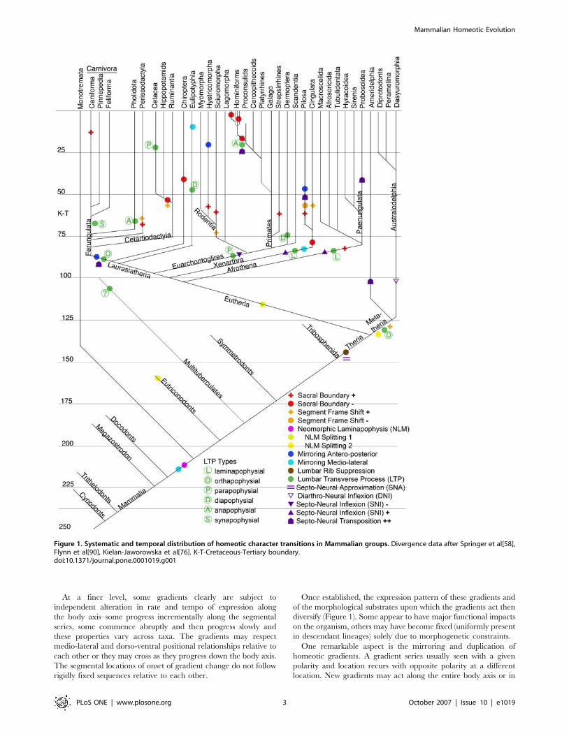
systematic radiation (Figure 1).
These clusters of homeotic change generally qualify as body
plan changes and often relate to significant alterations in the
adaptive zone of the descendant groups. These clusters of changes
are often preserved as a fixed homeotic set in the descendant
group across tens of millions or hundreds of millions of years.
Within individual lineages many of the gradients demonstrate
alterations on a sporadic basis (at the level of species or higher level
clades). Some lineages (e.g. hominiform hominoids, pilosan
xenarthrans) show a very high frequency of homeotic change for
some gradients. Other lineages show little or no homeotic change
over hundreds of millions of years (Monotremata).
Some homeotic alterations appear to be relatively highly
conserved–they fluctuate in their expression among more ancient
lineages but eventually become fixed (e.g. lumbar rib suppression).
A few homeotic features never change after their initial
appearance (e.g. emergence of the laminapophyis, septo-neural
approximation).
Mammalian Homeotic Evolution
PLoS ONE | www.plosone.org 2 October 2007 | Issue 10 | e1019
At a finer level, some gradients clearly are subject to
independent alteration in rate and tempo of expression along
the body axis–some progress incrementally along the segmental
series, some commence abruptly and then progress slowly and
these properties vary across taxa. The gradients may respect
medio-lateral and dorso-ventral positional relationships relative to
each other or they may cross as they progress down the body axis.
The segmental locations of onset of gradient change do not follow
rigidly fixed sequences relative to each other.
Once established, the expression pattern of these gradients and
of the morphological substrates upon which the gradients act then
diversify (Figure 1). Some appear to have major functional impacts
on the organism, others may have become fixed (uniformly present
in descendant lineages) solely due to morphogenetic constraints.
One remarkable aspect is the mirroring and duplication of
homeotic gradients. A gradient series usually seen with a given
polarity and location recurs with opposite polarity at a different
location. New gradients may act along the entire body axis or in
Figure 1. Systematic and temporal distribution of homeotic character transitions in Mammalian groups. Divergence data after Springer et al[58],Flynn et al[90], Kielan-Jaworowska et al[76]. K-T-Cretaceous-Tertiary boundary.doi:10.1371/journal.pone.0001019.g001
Mammalian Homeotic Evolution
PLoS ONE | www.plosone.org 3 October 2007 | Issue 10 | e1019
replicated form within each segment. The emergence of new types
of structures by duplication with subsequent diversification of the
new version mimics the pattern of change often seen with gene
duplication at the level of the genome.
Segment Identity–the Primary GradientThe basic homeotic distinction of five major spinal regions
(Table 1) is apparent in the earliest land vertebrates [57] and can
be assessed by boundary transitions. Seven cervical segments are
standard and readily identifiable in mammals and seven to nine in
most amniotes with the prominent exception of the extensive
duplication and alteration of the cervico-thoracic region at the
emergence of the avian winged archosaurs (birds). A very small
number of mammalian species have alteration in cervical vertebral
numbers on a sporadic basis.
The thoraco-lumbar transition within the vertebral series of
mammals, however, depends on a variety of gradients that defy
simple counting and categorization (Table 1)–this issue is explored
in detail below. The components of this transition are stably
arrayed in some higher taxa but subject to frequent generation of
new versions in others (Figure 2).
The lumbo-sacral boundary collectively affects multiple gradi-
ents in concert and is therefore a discreet phenomenon like the
cervico-thoracic boundary. The recent advent of a molecular
resolution to the deep relationship of mammalian groups [58,59]
provides an opportunity for observing phylogenetic patterns in the
segmental position of the lumbar/sacral boundary. Some groups
are very stable for this boundary position, some demonstrate
occasional small shifts, others are quite unstable with either
significant increases or decreases in number of segments (Figure 2).
There are a few species with highly unusual thoraco-lumbar or
lumbo-sacral boundary effects.
Scutisorex provides the most dramatic example of morphogenetic
disruption of the homeotic system among the mammals [37]
having scores or hundreds of facet pairs and a seeming duplication
of the entire lumbar region. Although most mammals–including
the numerous other species of the Soricidae-have six or seven
lumbar vertebrae, Scutisorex has twelve lumbar vertebrae.
Another informative homeotic character state is the replication of
the ‘‘diaphragmatic’’ thoraco-lumbar transition vertebra in a speci-
men of the macroscelid Petrodromus tetradactylus (USNM 241593)–
a species with a remarkably accelerated rate of morphological
evolution [60]. There is an elongated lamina with a double neural
spine. The more posterior ‘‘third’’ half of the lamina replicates the
anatomy of the last pre-diaphragmatic vertebra. This represents
discontinuous homeotic change and shows that the joint surface
reorientation seen in the diaphragmatic vertebra is indeed a home-
otically determined aspect of serial morphology.
Reduction in the number of dorsal (thoracic+lumbar) segments
is relatively uncommon. It is typical of the Order Chiroptera and
the Order Cingulata. Among hominoids this occurs in all of the
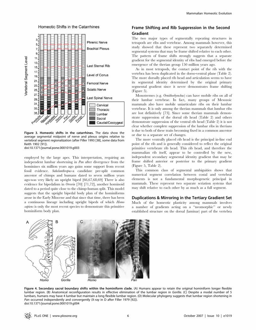
species of the hominiform clade (Figure 3, 4) but not among the
proconsulid hominoids. Some proconsulids may have tail loss
without reduction of dorsal segment numbers [61,62,63] but full
details of the sequence of these events remains unclear.
The initial reduction in number of lumbar vertebrae in the
hominiforms appears to be a shift from the catarrhine modal number
of seven down to a modal number of five or six (Figure 3). Modern
humans typically have five lumbar vertebrae, the only known
complete australopithecine lumbar spine has six [38,64,65].
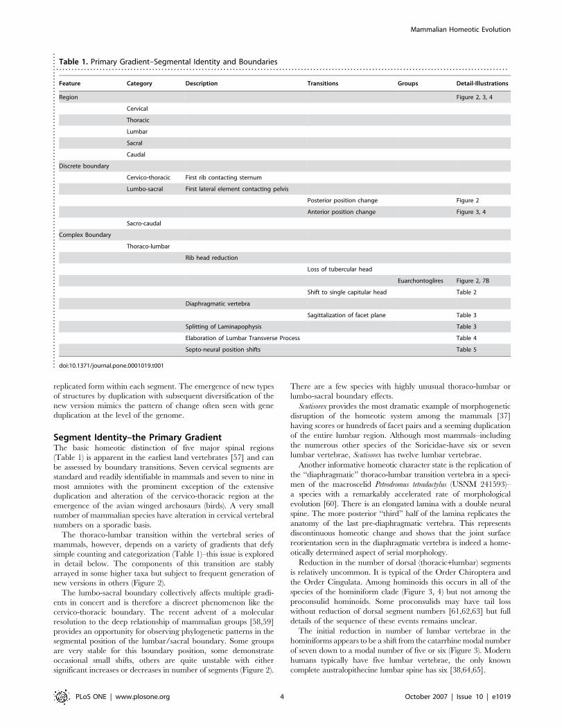
Table 1. Primary Gradient–Segmental Identity and Boundaries. . . . . . . . . . . . . . . . . . . . . . . . . . . . . . . . . . . . . . . . . . . . . . . . . . . . . . . . . . . . . . . . . . . . . . . . . . . . . . . . . . . . . . . . . . . . . . . . . . . . . . . . . . . . . . . . . . . . . . . . . . . . . . . . . . . . . . . . . . . . . . . . . .
Feature Category Description Transitions Groups Detail-Illustrations
Region Figure 2, 3, 4
Cervical
Thoracic
Lumbar
Sacral
Caudal
Discrete boundary
Cervico-thoracic First rib contacting sternum
Lumbo-sacral First lateral element contacting pelvis
Posterior position change Figure 2
Anterior position change Figure 3, 4
Sacro-caudal
Complex Boundary
Thoraco-lumbar
Rib head reduction
Loss of tubercular head
Euarchontoglires Figure 2, 7B
Shift to single capitular head Table 2
Diaphragmatic vertebra
Sagittalization of facet plane Table 3
Splitting of Laminapophysis Table 3
Elaboration of Lumbar Transverse Process Table 4
Septo-neural position shifts Table 5
doi:10.1371/journal.pone.0001019.t001....
....
....
....
....
....
....
....
....
....
....
....
....
....
....
....
....
....
....
....
....
....
....
....
....
..
Mammalian Homeotic Evolution
PLoS ONE | www.plosone.org 4 October 2007 | Issue 10 | e1019

Reduction to a modal number of four lumbar segments may
have occurred separately in Pongo, then Gorilla, and then Pan, with
the longer more flexible lumbar spine retained in primitive form in
hominines such as Australopithecus and Homo (Figure 1,4). Alter-
nately, the entire ‘‘great hominiform’’ group shared a single
common secondary event causing reduction to four lumbars, but
hominines subsequently reversed the trend to regain the modal
fifth lumbar segment [39]. This may be consistent with the
presence of upright bipedalism in the stem hominiforms, that is
transformed to diagonograde postures in the common ancestor of
great apes and humans, followed by rapid re-establishment of
bipedalism early in the course of an independent hominine
lineage.
However, as explored below, the secondary reductions of the
lumbar region may be independent, parallel convergent adapta-
tions to the various non-upright, ‘‘diagonograde’’ postures
Figure 2. Thoracic and lumbar segmental homeotic trait patterns in mammalian species.doi:10.1371/journal.pone.0001019.g002
Mammalian Homeotic Evolution
PLoS ONE | www.plosone.org 5 October 2007 | Issue 10 | e1019
employed by the large apes. This interpretation, requiring an
independent lumbar shortening in Pan after divergence from the
hominines six million years ago gains some support from recent
fossil evidence. Sahelanthropus-a candidate pre-split common
ancestor of chimps and humans dated to seven million years
ago-was very likely an upright biped [66,67,68,69] There is also
evidence for bipedalism in Orrorin [70] [71,72], another hominoid
dated to a period quite close to the chimp-human split. This model
suggests that the upright bipedal body plan of the hominiforms
arose in the Early Miocene and that since that time, there has been
a continuous lineage including upright bipeds of which Homo
sapiens is only the most recent species to demonstrate this primitive
hominiform body plan.
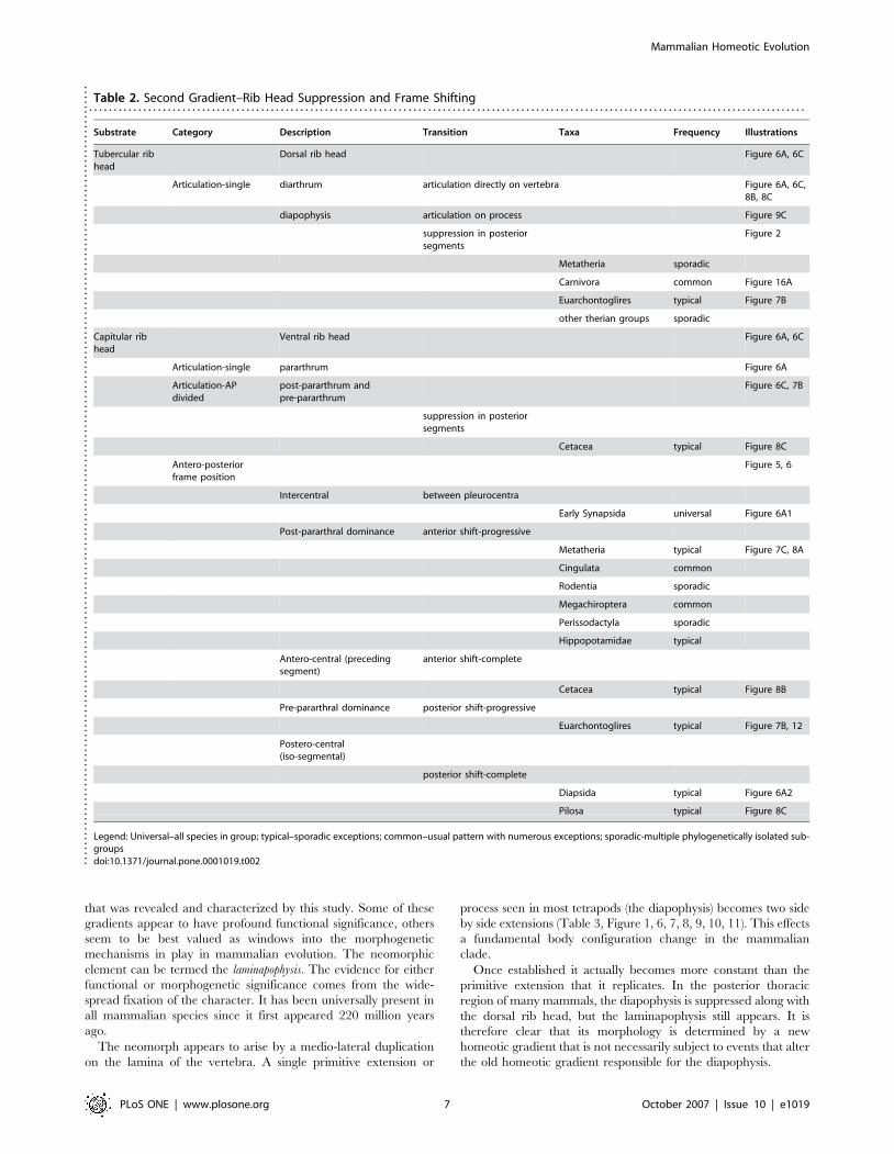
Frame Shifting and Rib Suppression in the Second
GradientThe two major types of segmentally repeating structures in
tetrapods are ribs and vertebrae. Among mammals however, this
study showed that these represent two separately determined
segmental systems that may be frame shifted relative to each other.
The pattern of frame shifts strongly suggests that a separate
gradient for the segmental identity of ribs had emerged before the
emergence of the therian group 150 million years ago.
As in most tetrapods, the contact point of the rib with the
vertebra has been duplicated in the dorso-ventral plane (Table 2).
The more dorsally placed rib head and articulation seems to have
its segmental identity determined by the original primary
segmental gradient since it never demonstrates frame shifting
(Figure 5).
Monotremes (e.g. Ornithorhynchus) can have mobile ribs on all of
their lumbar vertebrae. In fact, many groups of Mesozoic
mammals also have mobile uniarticulate ribs on their lumbar
vertebrae. It is only among the therian mammals that lumbar ribs
are lost definitively [73]. Since some therian mammals demon-
strate suppression of the dorsal rib head (Table 2) and others
demonstrate suppression of the ventral rib head (Table 2) it is not
clear whether complete suppression of the lumbar ribs in therians
is due to both of these traits becoming fixed in a common ancestor
or due to a separate set of changes.
The more ventrally placed rib head is the principal in-line end
point of the rib and is generally considered to reflect the original
primitive vertebrate rib head. This rib head, and therefore the
mammalian rib itself, appear to be controlled by the new,
independent secondary segmental identity gradient that may be
frame shifted anterior or posterior to the primary gradient
(Figure 5, Table 2).
This common class of segmental ambiguities shows that
numerical segment correlation between costal and vertebral
elements is not a fundamental morphogenetic principal in
mammals. These represent two separate seriation systems that
may shift relative to each other by as much as a full segment.
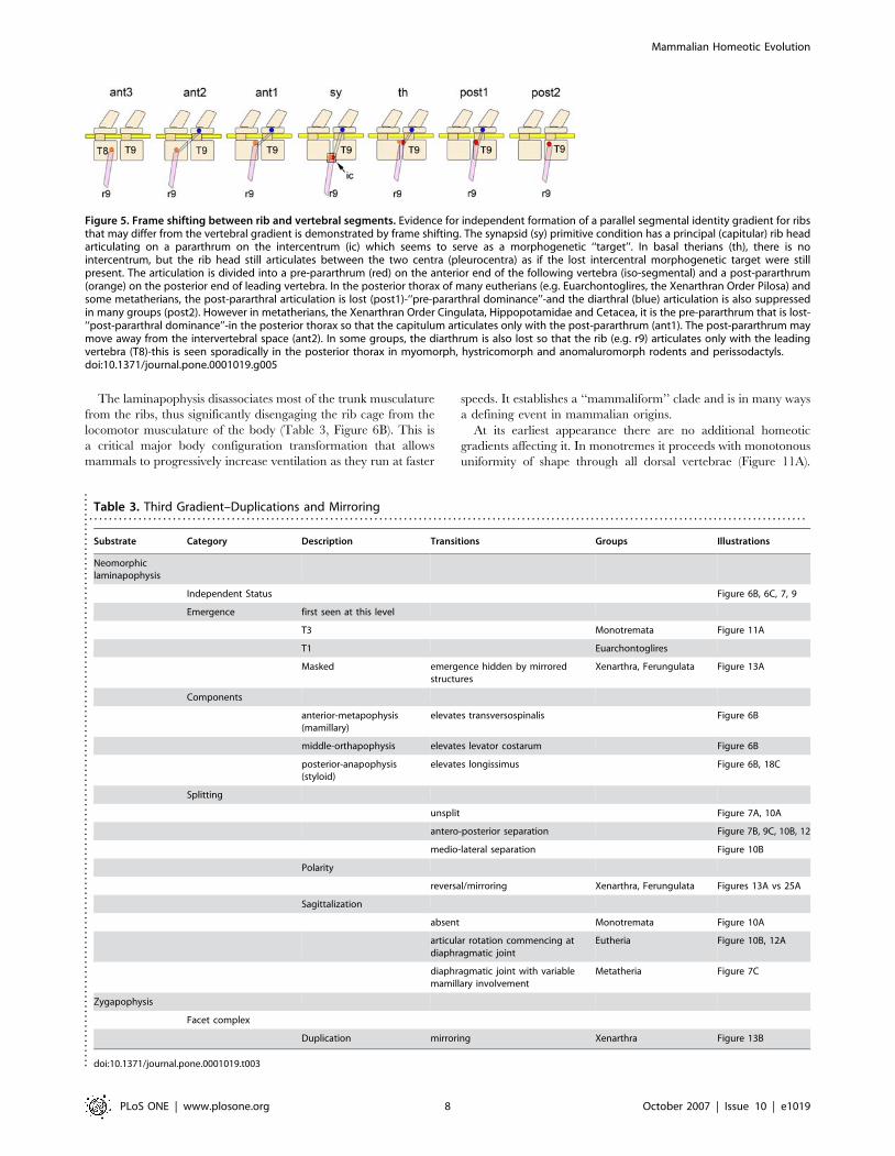
Duplications & Mirroring in the Tertiary Gradient SetMuch of the homeotic plasticity among mammals involves
a number of gradients acting on a ‘‘neomorphic’’ or newly
established structure on the dorsal (laminar) part of the vertebra
Figure 3. Homeotic shifts in the catarrhines. The data show theaverage segmental midpoint of nerve and plexus origins relative tovertebral segment regionalization (after Filler 1993 [38], some data fromKeith 1902 [91]).doi:10.1371/journal.pone.0001019.g003
Figure 4. Secondary sacral boundary shifts within the hominiform clade. (A) Humans appear to retain the original hominiform longer flexiblelumbar region. (B) Anatomical reconfiguration results in effective elimination of the lumbar region in Gorilla. (C) Despite a modal number of 5lumbars, humans may have 4 lumbar but maintain a long flexible lumbar region. (D) Molecular phylogeny suggests that lumbar region shortening inPan occurred independently and convergently (X-ray in D after Filler 1979 [92]).doi:10.1371/journal.pone.0001019.g004
Mammalian Homeotic Evolution
PLoS ONE | www.plosone.org 6 October 2007 | Issue 10 | e1019
that was revealed and characterized by this study. Some of these
gradients appear to have profound functional significance, others
seem to be best valued as windows into the morphogenetic
mechanisms in play in mammalian evolution. The neomorphic
element can be termed the laminapophysis. The evidence for either
functional or morphogenetic significance comes from the wide-
spread fixation of the character. It has been universally present in
all mammalian species since it first appeared 220 million years
ago.
The neomorph appears to arise by a medio-lateral duplication
on the lamina of the vertebra. A single primitive extension or
process seen in most tetrapods (the diapophysis) becomes two side
by side extensions (Table 3, Figure 1, 6, 7, 8, 9, 10, 11). This effects
a fundamental body configuration change in the mammalian
clade.
Once established it actually becomes more constant than the
primitive extension that it replicates. In the posterior thoracic
region of many mammals, the diapophysis is suppressed along with
the dorsal rib head, but the laminapophysis still appears. It is
therefore clear that its morphology is determined by a new
homeotic gradient that is not necessarily subject to events that alter
the old homeotic gradient responsible for the diapophysis.
Table 2. Second Gradient–Rib Head Suppression and Frame Shifting. . . . . . . . . . . . . . . . . . . . . . . . . . . . . . . . . . . . . . . . . . . . . . . . . . . . . . . . . . . . . . . . . . . . . . . . . . . . . . . . . . . . . . . . . . . . . . . . . . . . . . . . . . . . . . . . . . . . . . . . . . . . . . . . . . . . . . . . . . . . . . . . . .
Substrate Category Description Transition Taxa Frequency Illustrations
Tubercular ribhead
Dorsal rib head Figure 6A, 6C
Articulation-single diarthrum articulation directly on vertebra Figure 6A, 6C,8B, 8C
diapophysis articulation on process Figure 9C
suppression in posteriorsegments
Figure 2
Metatheria sporadic
Carnivora common Figure 16A
Euarchontoglires typical Figure 7B
other therian groups sporadic
Capitular ribhead
Ventral rib head Figure 6A, 6C
Articulation-single pararthrum Figure 6A
Articulation-APdivided
post-pararthrum andpre-pararthrum
Figure 6C, 7B
suppression in posteriorsegments
Cetacea typical Figure 8C
Antero-posteriorframe position
Figure 5, 6
Intercentral between pleurocentra
Early Synapsida universal Figure 6A1
Post-pararthral dominance anterior shift-progressive
Metatheria typical Figure 7C, 8A
Cingulata common
Rodentia sporadic
Megachiroptera common
Perissodactyla sporadic
Hippopotamidae typical
Antero-central (precedingsegment)
anterior shift-complete
Cetacea typical Figure 8B
Pre-pararthral dominance posterior shift-progressive
Euarchontoglires typical Figure 7B, 12
Postero-central(iso-segmental)
posterior shift-complete
Diapsida typical Figure 6A2
Pilosa typical Figure 8C
Legend: Universal–all species in group; typical–sporadic exceptions; common–usual pattern with numerous exceptions; sporadic-multiple phylogenetically isolated sub-groupsdoi:10.1371/journal.pone.0001019.t002..
....
....
....
....
....
....
....
....
....
....
....
....
....
....
....
....
....
....
....
....
....
....
....
....
....
....
....
....
....
....
....
....
....
....
....
....
....
....
....
.
Mammalian Homeotic Evolution
PLoS ONE | www.plosone.org 7 October 2007 | Issue 10 | e1019
The laminapophysis disassociates most of the trunk musculature
from the ribs, thus significantly disengaging the rib cage from the
locomotor musculature of the body (Table 3, Figure 6B). This is
a critical major body configuration transformation that allows
mammals to progressively increase ventilation as they run at faster
speeds. It establishes a ‘‘mammaliform’’ clade and is in many ways
a defining event in mammalian origins.
At its earliest appearance there are no additional homeotic
gradients affecting it. In monotremes it proceeds with monotonous
uniformity of shape through all dorsal vertebrae (Figure 11A).
Figure 5. Frame shifting between rib and vertebral segments. Evidence for independent formation of a parallel segmental identity gradient for ribsthat may differ from the vertebral gradient is demonstrated by frame shifting. The synapsid (sy) primitive condition has a principal (capitular) rib headarticulating on a pararthrum on the intercentrum (ic) which seems to serve as a morphogenetic ‘‘target’’. In basal therians (th), there is nointercentrum, but the rib head still articulates between the two centra (pleurocentra) as if the lost intercentral morphogenetic target were stillpresent. The articulation is divided into a pre-pararthrum (red) on the anterior end of the following vertebra (iso-segmental) and a post-pararthrum(orange) on the posterior end of leading vertebra. In the posterior thorax of many eutherians (e.g. Euarchontoglires, the Xenarthran Order Pilosa) andsome metatherians, the post-pararthral articulation is lost (post1)-‘‘pre-pararthral dominance’’-and the diarthral (blue) articulation is also suppressedin many groups (post2). However in metatherians, the Xenarthran Order Cingulata, Hippopotamidae and Cetacea, it is the pre-pararthrum that is lost-‘‘post-pararthral dominance’’-in the posterior thorax so that the capitulum articulates only with the post-pararthrum (ant1). The post-pararthrum maymove away from the intervertebral space (ant2). In some groups, the diarthrum is also lost so that the rib (e.g. r9) articulates only with the leadingvertebra (T8)-this is seen sporadically in the posterior thorax in myomorph, hystricomorph and anomaluromorph rodents and perissodactyls.doi:10.1371/journal.pone.0001019.g005
Table 3. Third Gradient–Duplications and Mirroring. . . . . . . . . . . . . . . . . . . . . . . . . . . . . . . . . . . . . . . . . . . . . . . . . . . . . . . . . . . . . . . . . . . . . . . . . . . . . . . . . . . . . . . . . . . . . . . . . . . . . . . . . . . . . . . . . . . . . . . . . . . . . . . . . . . . . . . . . . . . . . . . . .
Substrate Category Description Transitions Groups Illustrations
Neomorphiclaminapophysis
Independent Status Figure 6B, 6C, 7, 9
Emergence first seen at this level
T3 Monotremata Figure 11A
T1 Euarchontoglires
Masked emergence hidden by mirroredstructures
Xenarthra, Ferungulata Figure 13A
Components
anterior-metapophysis(mamillary)
elevates transversospinalis Figure 6B
middle-orthapophysis elevates levator costarum Figure 6B
posterior-anapophysis(styloid)
elevates longissimus Figure 6B, 18C
Splitting
unsplit Figure 7A, 10A
antero-posterior separation Figure 7B, 9C, 10B, 12
medio-lateral separation Figure 10B
Polarity
reversal/mirroring Xenarthra, Ferungulata Figures 13A vs 25A
Sagittalization
absent Monotremata Figure 10A
articular rotation commencing atdiaphragmatic joint
Eutheria Figure 10B, 12A
diaphragmatic joint with variablemamillary involvement
Metatheria Figure 7C
Zygapophysis
Facet complex
Duplication mirroring Xenarthra Figure 13B
doi:10.1371/journal.pone.0001019.t003....
....
....
....
....
....
....
....
....
....
....
....
....
....
....
....
....
....
....
....
....
....
....
....
....
....
....
....
....
.
Mammalian Homeotic Evolution
PLoS ONE | www.plosone.org 8 October 2007 | Issue 10 | e1019
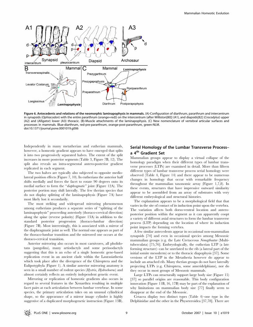
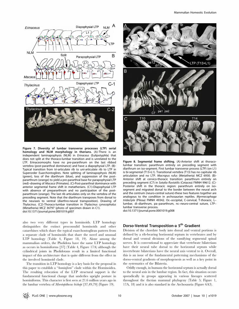
Independently in many metatherian and eutherian mammals,
however, a homeotic gradient appears to have emerged that splits
it into two progressively separated halves. The extent of the split
increases in more posterior segments (Table 3, Figure 7B, 12). The
split also reveals an intra-segmental antero-posterior gradient
replicated in each segment.
The two halves are typically also subjected to opposite medio-
lateral position effects (Figure 7, 10). In eutherians the anterior half
shifts medially and forces the facet to rotate 90 degrees onto its
medial surface to form the ‘‘diaphragmatic’’ joint (Figure 12A). The
posterior portion may shift laterally. The few therian species that
do not display splitting of the laminapophysis (Figure 7A) have
most likely lost it secondarily.
The most striking and widespread mirroring phenomenon
among eutherians produces a separate series of ‘‘splitting of the
laminapophysis’’ proceeding anteriorly (thoraco-cervical direction)
along the spine (reverse polarity) (Figure 13A) in addition to the
standard posterior progression (thoraco-lumbar direction)
(Figure 7B). Most interestingly, this is associated with a mirror of
the diaphragmatic joint as well. The normal one appears as part of
the thoraco-lumbar transition and the mirrored one occurs at the
thoraco-cervical transition.
Anterior mirroring also occurs in most carnivores, all pholido-
tans (pangolins), many artiodactyls and some perissodactyls
suggesting that this is an echo of a single homeotic gene-based
replication event in an ancient clade within the Laurasiatheria
which took place after the divergence of the Chiroptera and the
Eulipotyphyla (Figure 1). A similar anterior mirroring anatomy is
seen in a small number of rodent species (Hystrix, Hydrochoerus) and
almost certainly reflects an entirely independent genetic event.
Mirroring or replication of homeotic gradients also occurs in
regard to several features in the Xenarthra resulting in multiple
facet pairs at each articulation between lumbar vertebrae. In some
species, the primary articulation takes on an unusual cylindrical
shape, so the appearance of a mirror image cylinder is highly
suggestive of a duplicated morphogenetic instruction (Figure 13B).
Serial Homology of the Lumbar Transverse Process–
a 4th Gradient SetMammalian groups appear to display a virtual collapse of the
homology paradigm when their different types of lumbar trans-
verse processes (LTPs) are examined in detail. More than fifteen
different types of lumbar transverse process serial homology were
observed (Table 4, Figure 14) and there appear to be numerous
changes in homology that occur with remarkable frequency
throughout the mammalian taxonomic array (Figure 1,7,8). In
these events, structures that have impressive outward similarity
appear to be assembled from an array of substrates with very
different embryological and structural histories.
The explanation appears to be a morphological field that that
varies in the site of contact of its induction point upon the vertebra.
The variation affects both dorso-ventral location and antero-
posterior position within the segment as it can apparently coopt
a variety of different axial structures to form the lumbar transverse
process (LTP) depending on the location of where its induction
point impacts the forming vertebra.
A few similar antecedents appear in occasional non-mammalian
synapsids [74] and even in occasional species among Mesozoic
mammalian groups (e.g. the Late Cretaceous Nemegtbaatar (Multi-
tuberculata) [75,76]. Embryologically, the eutherian LTP (a late
forming structure)–can be unrelated to the rib (a lateral part of the
initial somite mesoderm) or to the thoracic diapophysis [37]. Some
versions of the LTP in the Metatheria however do appear to
include an attached rib. Many therian groups do not have laterally
projecting LTPs (e.g. Chiroptera, some ameridelphians), nor do
they occur in most groups of Mesozoic mammals.
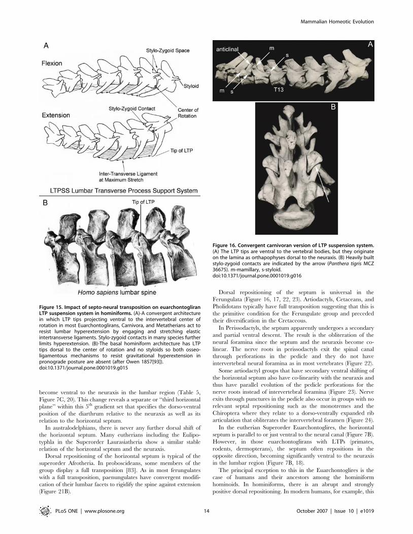
Large LTPs can structurally support large body size (Figure 15)
[37] so parallel origins are reasonable. This body configuration
innovation (Figure 11B, 16, 17B) may be part of the explanation of
why limitations on mammalian body size [77] finally seem to
disappear at the end of the Mesozoic.
Cetacea display two distinct types (Table 4)–one type in the
Delphinidae and the other in the Physeteroidea [37,78]. There are
Figure 6. Antecedents and relations of the neomorphic laminapophysis in mammals. (A)-Configuration of diarthrum, pararthrum and intercentrumin synapsids (Ophiacodon) with the entire pararthrum (orange+red)) on the intercentrum (after Williston[80]) (A1), and diapsids[82] (Crocodylus) upper(A2) and (Alligator) lower (A3) thoracic. (B)-Muscle attachments of the laminapophysis. (C) New nomenclature of vertebral articular surfaces andprocesses in mammals. Blue-diarthrum, red-pre-pararthrum, orange-post-pararthrum, green-NLM.doi:10.1371/journal.pone.0001019.g006
Mammalian Homeotic Evolution
PLoS ONE | www.plosone.org 9 October 2007 | Issue 10 | e1019
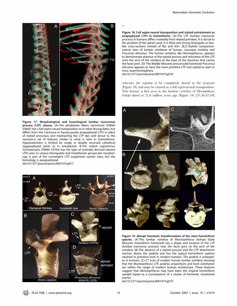
also two very different types in hominoids. LTP homology
distinguishes the extinct proconsulid hominoids and other
catarrhines which share the typical euarchontogliran pattern from
a separate clade of hominoids that share the novel and unusual
LTP homology (Table 4, Figure 18, 19). Alone among the
mammalian orders, the Pholidota have the same LTP homology
as occurs in hominiforms [37] (Table 4, Figure 17A) although the
cylindrical joints in Pholidotans result in a limited functional
impact of this architecture that is quite different from the effect in
the involved hominoid clade.
The transition in LTP homology is a key basis for the proposal in
this paper to establish a ‘‘hominiform’’ clade within the Hominoidea.
The resulting relocation of the LTP structural support is the
fundamental functional change that underlies upright posture in
hominiforms. This character is first seen at 21.6 million years ago in
the lumbar vertebra of Morotopithecus bishopi [37,49,79] (Figure 19).
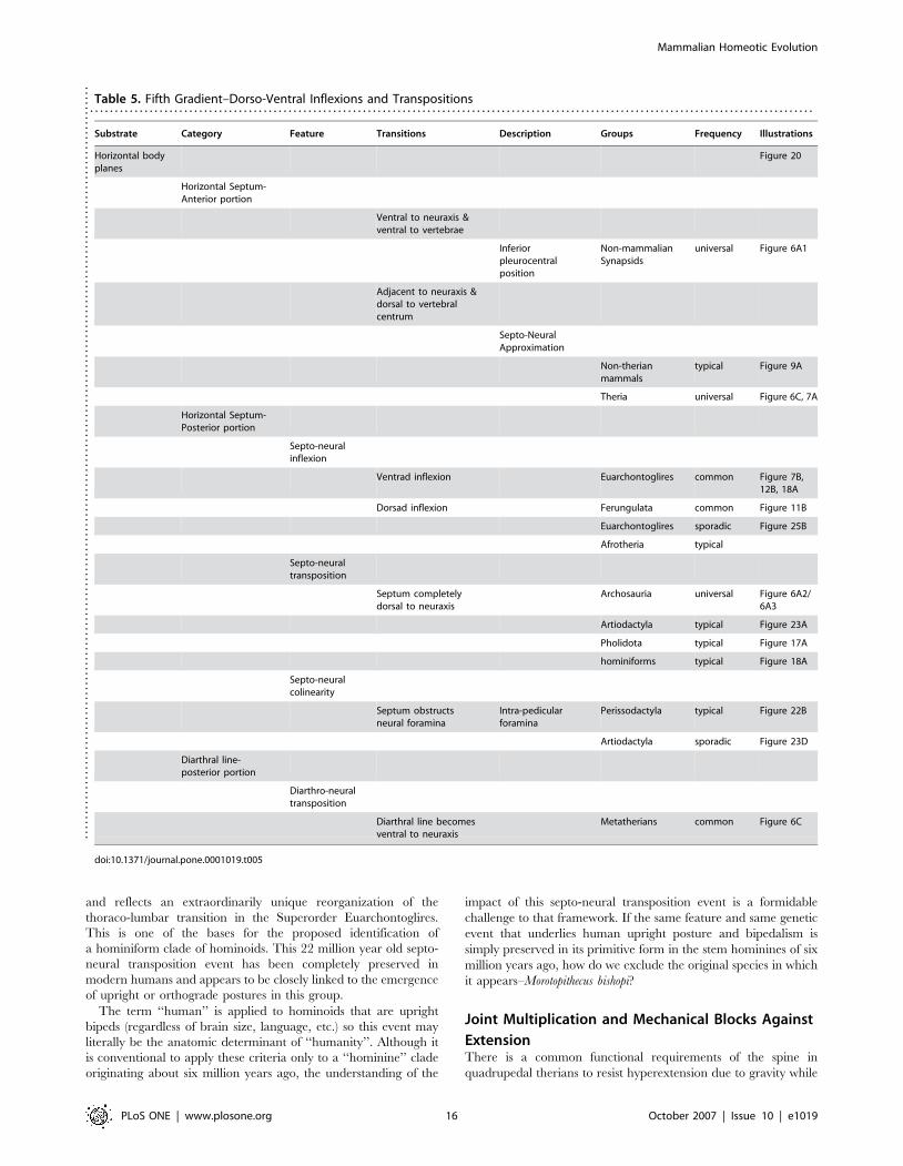
Dorso-Ventral Transposition-a 5th GradientDivision of the chordate body into dorsal and ventral portions is
defined by a rib-bearing horizontal septum in vertebrates and by
dorsal and ventral divisions of the ramifying segmental spinal
nerves. It is conventional to appreciate that vertebrate bilaterians
have their neural tube dorsal to the horizontal septum while
invertebrate bilaterians have the neural axis ventral to it. Overall,
this is an issue of the fundamental patterning mechanisms of the
dorso-ventral gradients of morphogenesis as well as a key point in
the systematics of the Bilateria.
Oddly enough, in humans the horizontal septum is actually dorsal
to the neural axis in the lumbar region. In fact, this situation occurs
sporadically in groups appearing in various lineages scattered
throughout the therian mammal phylogeny (Table 5; Figure 1,
17A, 18) and it is also standard in the Archosauria (Figure 6A3).
Figure 8. Segmental frame shifting. (A)-Anterior shift at thoraco-lumbar transition: pararthrum entirely on preceding segment withdiarthrum on iso-segment. First lumbar transverse process (LTP) (on L1)is bi-segmental (T13+L1). Transitional vertebra (T13) has no capitular ribarticulation and no LTP. Macropus rufus (Metatheria) MCZ 6930. (B)-Anterior shift at cervico-thoracic transition: pararthrum entirely onpreceding segment (C7) in Sotalia fluviatilis (Cetacea) FMNH 99612. (C)-Posterior shift in the thoracic region: pararthrum entirely on iso-segment and migrated dorsal to the border between the neural archand the centrum (neuro-central suture)-these two features together areanalogous to the condition in archosaurian reptiles. Myrmecophagatridactyla (Pilosa) FMNH 49342. Oc–occipital, C-cervical, T-thoracic, L–lumbar, di–diarthrum, pa–pararthrum, nc–neuro-central suture, LTP–lumbar transverse process.doi:10.1371/journal.pone.0001019.g008
Figure 7. Diversity of lumbar transverse processes (LTP) serialhomology and NLM morphology in therians. (A)-There is anindependent laminapophysis (NLM) in Erinaceus (Eulipotyphla) thatdoes not split at the thoraco-lumbar transition and is unrelated to theLTP. Erinaceomorphs have no pre-pararthrum on the last ribbedvertebra (post-pararthral dominance) and have a diapophysial LTP. (B)-Typical transition from tri-articulate rib to uni-articulate rib to LTP inSuperorder Euarchontoglires. Note splitting of laminapophysis (NLM)(green), loss of the diarthrum (blue), and suppression of the post-pararthrum (orange) to yield a pre-pararthral base for parapophysial LTP(red)–drawing of Macaca (Primates). (C)-Post-pararthral dominance withanterior segmental frame shift in metatherians. (C1)-Diapophysial LTPwith absence of prepararthrum and no participation of the post-pararthrum (orange). The last rib articulates only on the vertebra of thepreceding segment. Note that the diarthrum transposes from dorsal tothe neuraxis to ventral (diarthro-neural transposition). Drawing ofThylacinus. (C2)-Thoraco-lumbar transition in Thylacinus cynocephalus(Metatheria) MCZ 36797 (photo of specimen drawn in C1).doi:10.1371/journal.pone.0001019.g007
Mammalian Homeotic Evolution
PLoS ONE | www.plosone.org 10 October 2007 | Issue 10 | e1019
Dorso-ventral transposition of the horizontal septum and of the
neuraxis occurs at a crossing point that may be termed the ‘‘septo-
neural inflexion point’’ and reflects the crossing of two somewhat
independent morphogenetic gradients (see Figure 20).
The ancestral synapsid condition [80], is to have the horizontal
septum ventral to the neural canal and ventral to the entire
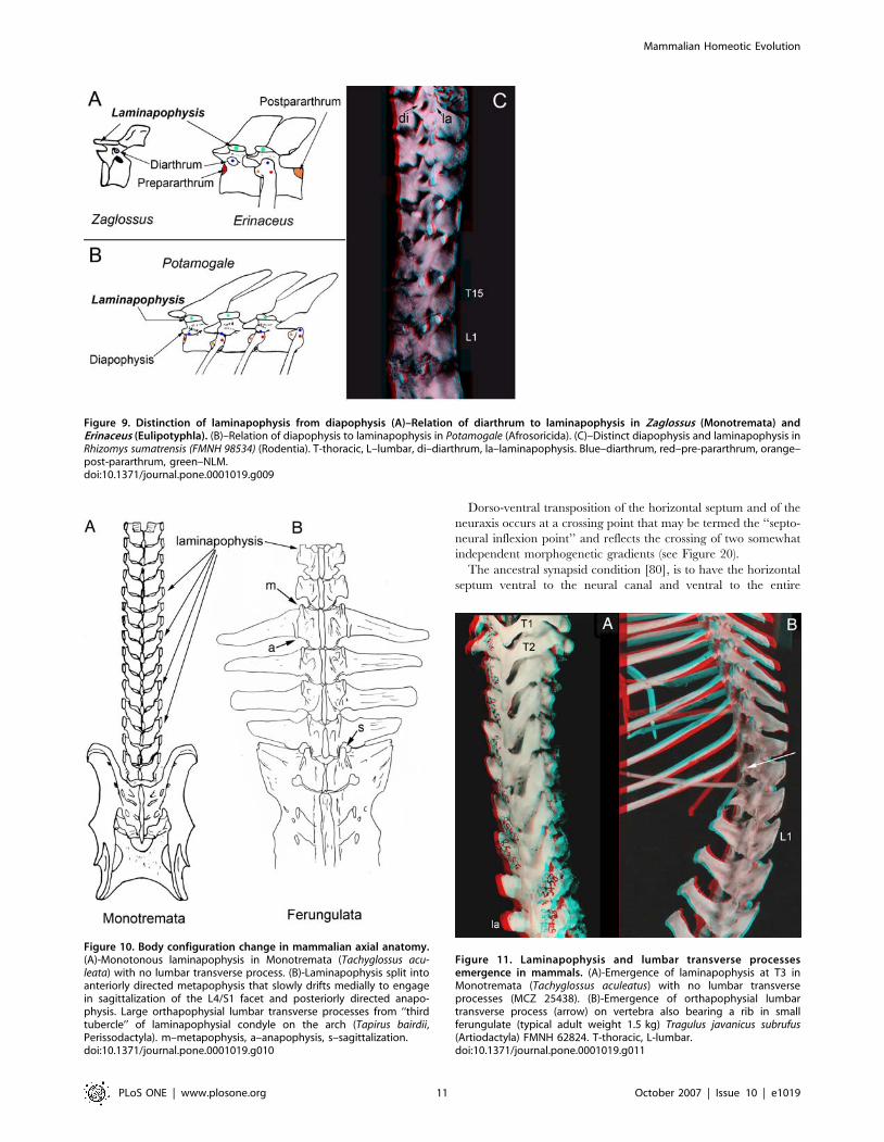
Figure 9. Distinction of laminapophysis from diapophysis (A)–Relation of diarthrum to laminapophysis in Zaglossus (Monotremata) andErinaceus (Eulipotyphla). (B)–Relation of diapophysis to laminapophysis in Potamogale (Afrosoricida). (C)–Distinct diapophysis and laminapophysis inRhizomys sumatrensis (FMNH 98534) (Rodentia). T-thoracic, L–lumbar, di–diarthrum, la–laminapophysis. Blue–diarthrum, red–pre-pararthrum, orange–post-pararthrum, green–NLM.doi:10.1371/journal.pone.0001019.g009
Figure 10. Body configuration change in mammalian axial anatomy.(A)-Monotonous laminapophysis in Monotremata (Tachyglossus acu-leata) with no lumbar transverse process. (B)-Laminapophysis split intoanteriorly directed metapophysis that slowly drifts medially to engagein sagittalization of the L4/S1 facet and posteriorly directed anapo-physis. Large orthapophysial lumbar transverse processes from ‘‘thirdtubercle’’ of laminapophysial condyle on the arch (Tapirus bairdii,Perissodactyla). m–metapophysis, a–anapophysis, s–sagittalization.doi:10.1371/journal.pone.0001019.g010
Figure 11. Laminapophysis and lumbar transverse processesemergence in mammals. (A)-Emergence of laminapophysis at T3 inMonotremata (Tachyglossus aculeatus) with no lumbar transverseprocesses (MCZ 25438). (B)-Emergence of orthapophysial lumbartransverse process (arrow) on vertebra also bearing a rib in smallferungulate (typical adult weight 1.5 kg) Tragulus javanicus subrufus(Artiodactyla) FMNH 62824. T-thoracic, L-lumbar.doi:10.1371/journal.pone.0001019.g011
Mammalian Homeotic Evolution
PLoS ONE | www.plosone.org 11 October 2007 | Issue 10 | e1019
vertebral body (Figure 6, 20). This is the condition still seen in
cynodont synapsid reptiles that are closely related to the stem
mammals [74]. The mammalian condition in which the horizontal
septum is moved to a position dorsal to the vertebral body is first
seen in monotremes [81].
The details are still unclear for some Mesozoic mammal groups,
but for all therian mammals there is a major shift of the
pararthrum (and horizontal septum) to a position near the dorsal
margin of the vertebral body (Figure 1, 7A). This reveals a major
body configuration change that brings the horizontal septum
nearly adjacent to the neuraxis. This clearly occurred in the stem
therian clade around 150 million years ago and almost never
varies in the thoracic region.
Embryologically and evolutionarily, the ribs arise at intersection
lines between the horizontal septum and segmental myosepta.
Because of this, the relatively dorsal or relatively ventral position of
the attachment point of a costal derived process or lumbar
transverse process on the vertebra reveals the relative position of
the septal and neural horizontal body planes in the animal.
In Archosaurs, there is a very abrupt and complete inflexion
and transposition (Figure 6A2/3). In the posterior neck and most
anterior thorax the primary rib head is on the mid part of the
vertebral body–ventral to the neuraxis. In most of the thorax,
everything moves completely dorsal to the neuraxis [82].
The particular type of transition seen in archosaurs almost
never occurs in mammals because the synapsid/mammalian
primary rib articulation tends not to cross the ‘‘neuro-central
suture’’ of the vertebra (where the pedicle meets the vertebral body
embryologically). In mammals, when the horizontal septum
becomes transposed to a position dorsal to the neuraxis, there
may be non-costal lumbar transverse processes (as in humans) but
there are almost never ribs dorsal to neuraxis. Exceptions to this
occur in the form of rib articulations on the pedicles in Superorder
Xenarthra (Order Pilosa) (Figure 8C) and in the Paenungulata in
Superorder Afrotheria (Figure 21B).
Septo-neural inflexion patterns have not been previously
appreciated as an important aspect of tetrapod morphologic and
functional evolution. Nonetheless, they may play an important role
in the emergence of large cursorial mammals at the close of the
Mesozoic, the emergence of the Carnivora from the ungulates at
the Cretaceous-Tertiary boundary and in the origin of the
anatomical basis of upright posture in humans in the stem
hominiform hominoids of the Early Miocene.
A different type of change in horizontal body planes occurs in
most australodelphian metatherians. This is the transposition of
the ancient more dorsal rib articulation plane (diarthral plane) to
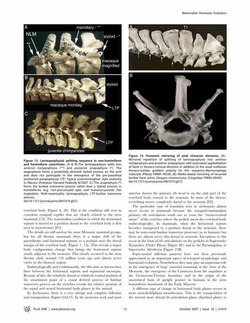
Figure 12. Laminapophysial splitting sequence in non-hominiformand hominiform catarrhines. (A & B)-The laminapophysis splits intoanterior metapophysis (**) and posterior anapophysis (*). Theanapophysis forms a posteriorly directed styloid process on the archand does not participate in the emergence of the pre-pararthralpositioned parapophysial LTP. Typical euarchontogliran style anatomyin Macaca (Primates) Harvard Peabody N/3587. (C)-The anapophysis (*)forms the lumbar transverse process rather than a styloid process inhominiforms (e.g. non-proconsulid apes and humans)-juvenile Pantroglodytes. NLM-neomorphic laminapophysis, LTP-lumbar transverseprocess.doi:10.1371/journal.pone.0001019.g012
Figure 13. Homeotic mirroring of axial character elements. (A)–Mirrored repetition of splitting of laminapophysis into anteriormetapophysis and posterior anapophysis with associated sagittalizationof facet in thoraco-cervical direction in addition to the usual eutherianthoraco-lumbar gradient polarity for this sequence-Myrmecophagatridactyla (Pilosa) FMNH 49338. (B)–Medio-lateral mirroring of recurvedlumbar facet joints–Dasypus novemcinctus (Cingulata) FMNH 60493.doi:10.1371/journal.pone.0001019.g013
Mammalian Homeotic Evolution
PLoS ONE | www.plosone.org 12 October 2007 | Issue 10 | e1019
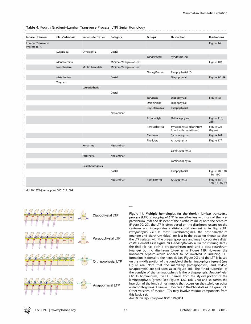
Table 4. Fourth Gradient–Lumbar Transverse Process (LTP) Serial Homology. . . . . . . . . . . . . . . . . . . . . . . . . . . . . . . . . . . . . . . . . . . . . . . . . . . . . . . . . . . . . . . . . . . . . . . . . . . . . . . . . . . . . . . . . . . . . . . . . . . . . . . . . . . . . . . . . . . . . . . . . . . . . . . . . . . . . . . . . . . . . . . . . .
Induced Element Class/Infraclass Superorder/Order Category Groups Description Illustrations
Lumbar TransverseProcess (LTP)
Figure 14
Synapsida Cynodontia Costal
Thrinaxodon Syndesmosed
Monotremata Minimal/Vestigial/absent Figure 10A
Non-therian- Multituberculata Minimal/Vestigial/absent
Nemegtbaatar Parapophysial (?)
Metatherian Costal Diapophysial Figure 7C, 8A
Therian
Laurasiatheria
Costal
Erinaceus Diapophysial Figure 7A
Delphinidae Diapophysial
Physeteroidea Parapophysial
Neolaminar
Artiodactyla Orthapophysial Figure 11B,23B
Perissodactyla Synapophysial (diarthrumfused with pararthrum)
Figure 22B(Equus)
Carnivora Synapophysial Figure 16A
Pholidota Anapophysial Figure 17A
Xenarthra Neolaminar
Laminapophysial
Afrotheria Neolaminar
Laminapophysial
Euarchontoglires
Costal Parapophysial Figure 7B, 12B,18A, 18C
Neolaminar hominiforms Anapophysial Figure 18A,18B, 19, 26, 27
doi:10.1371/journal.pone.0001019.t004....
....
....
....
....
....
....
....
....
....
....
....
....
....
....
....
....
....
....
....
....
....
....
....
....
....
....
....
....
....
..
r
Figure 14. Multiple homologies for the therian lumbar transverseprocess (LTP). Diapophysial LTP: In metatherians with loss of the pre-pararthrum (red) and descent of the diarthrum (blue) onto the centrum(Figure 7C, 20), the LTP is often based on the diarthrum, occurs on thecentrum, and incorporates a distal costal element as in Figure 8A.Parapophysial LTP: In most Euarchontoglires, the post-pararthrum(orange) and diarthrum (blue) are lost in the posterior thorax so thatthe LTP seriates with the pre-parapophysis and may incorporate a distalcostal element as in Figure 7B. Orthapophysial LTP: In most ferungulates,the final rib has both a pre-pararthrum (red) and a post-pararthrum(orange) but no diarthrum (blue) as in Figure 11B. However thehorizontal septum–which appears to be involved in inducing LTPformation–is dorsal to the neuraxis (see Figure 20) and the LTP is basedon the middle portion of the condyle of the laminapophysis (green) (seeFigure 6B). Note that the mamillary (metapophysis) and styloid(anapophysis) are still seen as in Figure 10B. The ‘‘third tubercle’’ ofthe condyle of the laminapophysis is the orthapophysis. AnapophysialLTP: In hominiforms, the LTP derives from the styloid portion of thelaminapophysis (green) (see Figures 12C, 18B, 27A) and so carries theinsertion of the longissimus muscle that occurs on the styloid on othereuarchontoglirans. A similar LTP occurs in the Pholidota as in Figure 17A.Other versions of therian LTPs may involve various components fromthis basic set.doi:10.1371/journal.pone.0001019.g014
Mammalian Homeotic Evolution
PLoS ONE | www.plosone.org 13 October 2007 | Issue 10 | e1019
become ventral to the neuraxis in the lumbar region (Table 5,
Figure 7C, 20). This change reveals a separate or ‘‘third horizontal
plane’’ within this 5th gradient set that specifies the dorso-ventral
position of the diarthrum relative to the neuraxis as well as its
relation to the horizontal septum.
In australodelphians, there is never any further dorsal shift of
the horizontal septum. Many eutherians including the Eulipo-
typhla in the Superorder Laurasiatheria show a similar stable
relation of the horizontal septum and the neuraxis.
Dorsal repositioning of the horizontal septum is typical of the
superorder Afrotheria. In proboscideans, some members of the
group display a full transposition [83]. As in most ferungulates
with a full transposition, paenungulates have convergent modifi-
cation of their lumbar facets to rigidify the spine against extension
(Figure 21B).
Dorsal repositioning of the septum is universal in the
Ferungulata (Figure 16, 17, 22, 23). Artiodactyls, Cetaceans, and
Pholidotans typically have full transposition suggesting that this is
the primitive condition for the Ferungulate group and preceded
their diversification in the Cretaceous.
In Perissodactyls, the septum apparently undergoes a secondary
and partial ventral descent. The result is the obliteration of the
neural foramina since the septum and the neuraxis become co-
linear. The nerve roots in perissodactyls exit the spinal canal
through perforations in the pedicle and they do not have
intervertebral neural foramina as in most vertebrates (Figure 22).
Some artiodactyl groups that have secondary ventral shifting of
the horizontal septum also have co-linearity with the neuraxis and
thus have parallel evolution of the pedicle perforations for the
nerve roots instead of intervertebral foramina (Figure 23). Nerve
exits through punctures in the pedicle also occur in groups with no
relevant septal repositioning such as the monotremes and the
Chiroptera where they relate to a dorso-ventrally expanded rib
articulation that obliterates the intervertebral foramen (Figure 24).
In the eutherian Superorder Euarchontoglires, the horizontal
septum is parallel to or just ventral to the neural canal (Figure 7B).
However, in those euarchontoglirans with LTPs (primates,
rodents, dermopterans), the septum often repositions in the
opposite direction, becoming significantly ventral to the neuraxis
in the lumbar region (Figure 7B, 18).
The principal exception to this in the Euarchontoglires is the
case of humans and their ancestors among the hominiform
hominoids. In hominiforms, there is an abrupt and strongly
positive dorsal repositioning. In modern humans, for example, this
Figure 15. Impact of septo-neural transposition on euarchontogliranLTP suspension system in hominiforms. (A)-A convergent architecturein which LTP tips projecting ventral to the intervertebral center ofrotation in most Euarchontoglirans, Carnivora, and Metatherians act toresist lumbar hyperextension by engaging and stretching elasticintertransverse ligaments. Stylo-zygoid contacts in many species furtherlimits hyperextension. (B)-The basal hominiform architecture has LTPtips dorsal to the center of rotation and no styloids so both osseo-ligamentous mechanisms to resist gravitational hyperextension inpronograde posture are absent (after Owen 1857[93]).doi:10.1371/journal.pone.0001019.g015
Figure 16. Convergent carnivoran version of LTP suspension system.(A) The LTP tips are ventral to the vertebral bodies, but they originateon the lamina as orthapophyses dorsal to the neuraxis. (B) Heavily builtstylo-zygoid contacts are indicated by the arrow (Panthera tigris MCZ36675). m-mamillary, s-styloid.doi:10.1371/journal.pone.0001019.g016
Mammalian Homeotic Evolution
PLoS ONE | www.plosone.org 14 October 2007 | Issue 10 | e1019
relocates the septum to be completely dorsal to the neuraxis
(Figure 18) and may be classed as a full septo-neural transposition.
This feature is first seen in the lumbar vertebra of Morotopithecus
bishopi dated at 21.6 million years ago (Figure 19) [37,46,47,49]
Figure 17. Morphological and homological lumbar transverseprocess (LTP) classes. (A)-The pholidotan Manis temminckii (FMNH35682) has a full septo-neural transposition as in other ferungulates, butdiffers from the Carnivora in having purely anapophysial LTPs in placeof styloid processes and maintaining the LTP tips well dorsal to theneuraxis-a set of features similar to what is seen in hominiforms.Hyperextension is limited by singly or doubly recurved cylindricalzygapophysial joints as in artiodactyls. B-The rodent Lagostomustrichodactylus (FMNH 53704) has the type of ventrally directed slantedLTPs seen in various ferungulate and metatherian groups-the morphol-ogy is part of the convergent LTP suspension system class, but thehomology is parapophysial.doi:10.1371/journal.pone.0001019.g017
r
Figure 18. Full septo-neural transposition and styloid entrainment asanapophysial LTPs in hominiforms. (A)-The LTP (lumbar transverseprocess) in humans differs markedly from related primates. It is dorsal tothe position of the spinal canal. It is thick and strong (triangular or box-like cross-section) instead of flat and thin. (B,C)-Styloid comparison.Lateral view of lumbar vertebrae of human, macaque monkey andProconsul africanus. The human vertebra, like Morotopithecus, appearsto demonstrate absence of the styloid process and relocation of the LTPonto the arch of the vertebra at the base of the structure that carriesthe facet joint. (D) The Middle Miocene proconsulid hominoid Proconsulafricanus appears to have the more primitive LTP and styloid as seen inmost euarchontoglirans.doi:10.1371/journal.pone.0001019.g018
Figure 19. Abrupt homeotic transformation of the stem hominiformspecies. (A)-The lumbar vertebra of Morotopithecus bishopi (EarlyMiocene hominiform hominoid) has a shape and location of the LTP(lumbar transverse process) near the facet joint on the arch of thevertebra. (B)-The absence of a styloid process and the LTP attachmentreaches above the pedicle and has the typical hominiform patternretained in primitive form in modern humans. The pedicle is enlarged-as in humans. (C)-CT scan of modern human lumbar vertebra showingthat the Morotopithecus LTP, pedicle, proportions and facet orientationare within the range of modern human architecture. These featuressuggest that Morotopithecus may have been the original hominiformupright biped as a consequence of a cluster of homeotic mutationalevents.doi:10.1371/journal.pone.0001019.g019
Mammalian Homeotic Evolution
PLoS ONE | www.plosone.org 15 October 2007 | Issue 10 | e1019
and reflects an extraordinarily unique reorganization of the
thoraco-lumbar transition in the Superorder Euarchontoglires.
This is one of the bases for the proposed identification of
a hominiform clade of hominoids. This 22 million year old septo-
neural transposition event has been completely preserved in
modern humans and appears to be closely linked to the emergence
of upright or orthograde postures in this group.
The term ‘‘human’’ is applied to hominoids that are upright
bipeds (regardless of brain size, language, etc.) so this event may
literally be the anatomic determinant of ‘‘humanity’’. Although it
is conventional to apply these criteria only to a ‘‘hominine’’ clade
originating about six million years ago, the understanding of the
impact of this septo-neural transposition event is a formidable
challenge to that framework. If the same feature and same genetic
event that underlies human upright posture and bipedalism is
simply preserved in its primitive form in the stem hominines of six
million years ago, how do we exclude the original species in which
it appears–Morotopithecus bishopi?
Joint Multiplication and Mechanical Blocks Against
ExtensionThere is a common functional requirements of the spine in
quadrupedal therians to resist hyperextension due to gravity while
Table 5. Fifth Gradient–Dorso-Ventral Inflexions and Transpositions. . . . . . . . . . . . . . . . . . . . . . . . . . . . . . . . . . . . . . . . . . . . . . . . . . . . . . . . . . . . . . . . . . . . . . . . . . . . . . . . . . . . . . . . . . . . . . . . . . . . . . . . . . . . . . . . . . . . . . . . . . . . . . . . . . . . . . . . . . . . . . . . . .
Substrate Category Feature Transitions Description Groups Frequency Illustrations
Horizontal bodyplanes
Figure 20
Horizontal Septum-Anterior portion
Ventral to neuraxis &ventral to vertebrae
Inferiorpleurocentralposition
Non-mammalianSynapsids
universal Figure 6A1
Adjacent to neuraxis &dorsal to vertebralcentrum
Septo-NeuralApproximation
Non-therianmammals
typical Figure 9A
Theria universal Figure 6C, 7A
Horizontal Septum-Posterior portion
Septo-neuralinflexion
Ventrad inflexion Euarchontoglires common Figure 7B,12B, 18A
Dorsad inflexion Ferungulata common Figure 11B
Euarchontoglires sporadic Figure 25B
Afrotheria typical
Septo-neuraltransposition
Septum completelydorsal to neuraxis
Archosauria universal Figure 6A2/6A3
Artiodactyla typical Figure 23A
Pholidota typical Figure 17A
hominiforms typical Figure 18A
Septo-neuralcolinearity
Septum obstructsneural foramina
Intra-pedicularforamina
Perissodactyla typical Figure 22B
Artiodactyla sporadic Figure 23D
Diarthral line-posterior portion
Diarthro-neuraltransposition
Diarthral line becomesventral to neuraxis
Metatherians common Figure 6C
doi:10.1371/journal.pone.0001019.t005
....
....
....
....
....
....
....
....
....
....
....
....
....
....
....
....
....
....
....
....
....
....
....
....
....
....
....
....
....
....
....
....
....
....
....
....
....
....
Mammalian Homeotic Evolution
PLoS ONE | www.plosone.org 16 October 2007 | Issue 10 | e1019
allowing dorso-ventral flexibility in locomotion. It is therefore not
surprising that there are multiple convergent anatomical structural
solutions. Most of these have not been appreciated in earlier
attempts to model the mammalian spine on a global engineering
basis without adequate attention to the context and detail of the
specific anatomical structures actually involved [84]. This study
reveals that these all tend to involve the neomorphic laminapo-
physis and LTP gradients. Both of these structures demonstrate
a high degree of morphogenetic plasticity in therians. The
participation of these structures in serial/homeotic control systems
may also play a roll in their tendency to be deployed as the bases
for convergent novel structures.
Universally in the Ferrungulata, Paenungulata, Xenarthra and
Ameridelphia where septo-neural transposition takes place, there
are supplementary modifications of the lumbar spine that relate
to resistance against extension of the spine (Table 6). Typically,
these involve modifications to provide rigid bony resistance to
lumbar hyperextension either through elaboration of multiple
additional joint surfaces (Figure 21, 25A) and/or mechanical
locking systems (Figure 22, 23). These changes commence in the
fossil record after the appearance of splitting of the laminapo-
physis 130 million years ago and appear to be modifications on
a theme based on morphologic modification of the laminapo-
physis.
Multiplication of joints in the Superorder Euarchontoglires
always involves new surfaces on the styloid process but is limited to
a small number of groups including the large rodent Hystrix cristata
(porcupine with weight up to 30 kg–and note much larger extinct
related species such as Neosteiromys pattoni) (Figure 25B). Among
primates, this occurs in some prosimians. This feature also occurs
in the Afrotheria where it is seen in both the Proboscidea and in
the afrosoricid insectivore Tenrec.
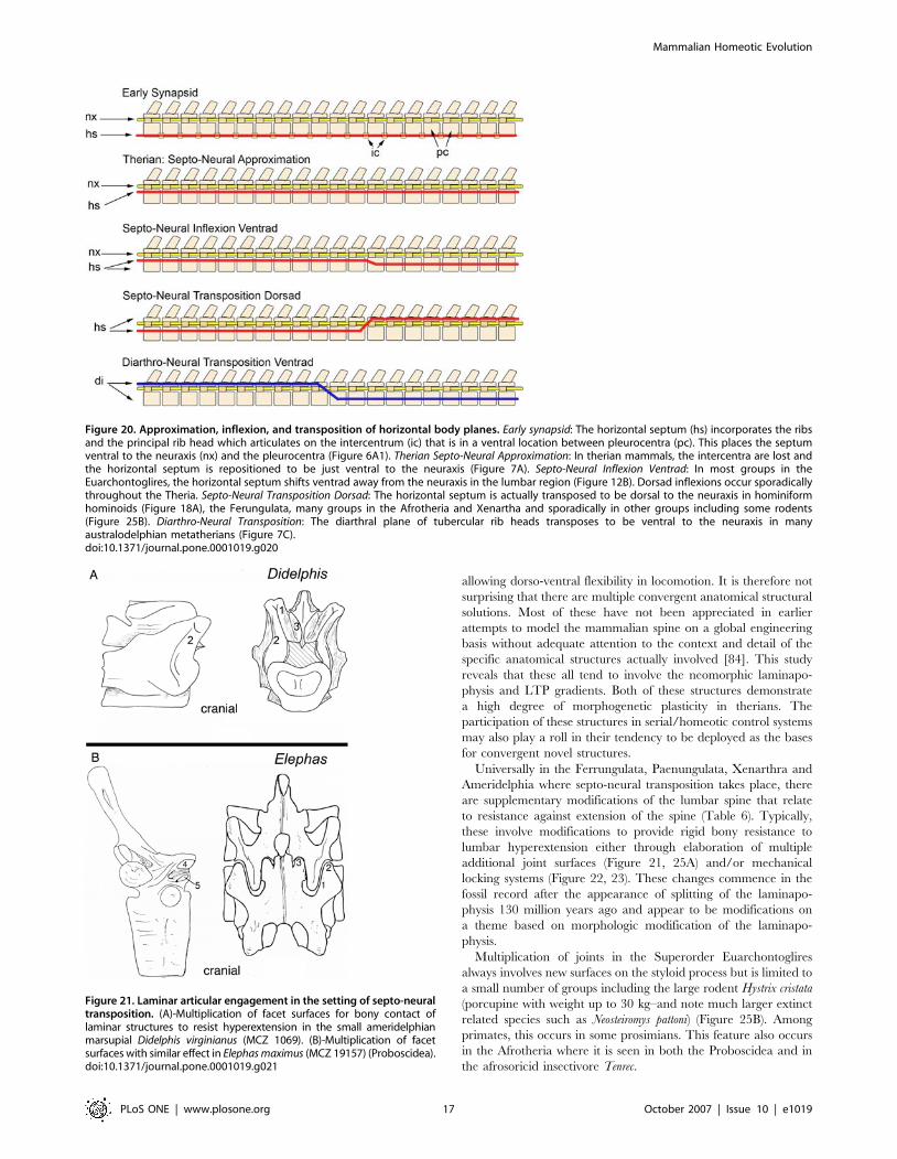
Figure 20. Approximation, inflexion, and transposition of horizontal body planes. Early synapsid: The horizontal septum (hs) incorporates the ribsand the principal rib head which articulates on the intercentrum (ic) that is in a ventral location between pleurocentra (pc). This places the septumventral to the neuraxis (nx) and the pleurocentra (Figure 6A1). Therian Septo-Neural Approximation: In therian mammals, the intercentra are lost andthe horizontal septum is repositioned to be just ventral to the neuraxis (Figure 7A). Septo-Neural Inflexion Ventrad: In most groups in theEuarchontoglires, the horizontal septum shifts ventrad away from the neuraxis in the lumbar region (Figure 12B). Dorsad inflexions occur sporadicallythroughout the Theria. Septo-Neural Transposition Dorsad: The horizontal septum is actually transposed to be dorsal to the neuraxis in hominiformhominoids (Figure 18A), the Ferungulata, many groups in the Afrotheria and Xenartha and sporadically in other groups including some rodents(Figure 25B). Diarthro-Neural Transposition: The diarthral plane of tubercular rib heads transposes to be ventral to the neuraxis in manyaustralodelphian metatherians (Figure 7C).doi:10.1371/journal.pone.0001019.g020
Figure 21. Laminar articular engagement in the setting of septo-neuraltransposition. (A)-Multiplication of facet surfaces for bony contact oflaminar structures to resist hyperextension in the small ameridelphianmarsupial Didelphis virginianus (MCZ 1069). (B)-Multiplication of facetsurfaces with similar effect in Elephas maximus (MCZ 19157) (Proboscidea).doi:10.1371/journal.pone.0001019.g021
Mammalian Homeotic Evolution
PLoS ONE | www.plosone.org 17 October 2007 | Issue 10 | e1019
Convergent Ventrally Tensioned LTP ArraysIn a number of mammalian groups including both therians and
metatherians [85], the lumbar transverse processes display
a striking slanted array that is angled ventrally so that the tips
are well below the ventral margin of the vertebral bodies (Table 7;
Figure 11B, 12B, 16A, 17B). The underlying serial homology is
unique in each group but the functional anatomy is obviously
highly convergent and independently evolved in parallel.
These and other types of arrays with the tip of the LTP ventral
to the effective axis of rotation for lumbar extension participate in
a dynamic, elastic, ligamentous system that supports the lumbar
region and resists extension (Figure 15). This appears to be an
almost opposite architectural solution by comparison with the rigid
bony locking systems that also act against extension (described in
the previous section).
In this elastic system, as the vertebral column passes into
extension, LTPs whose tips are below the effective intervertebral
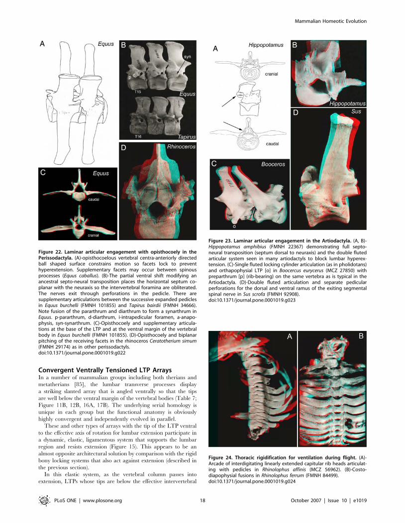
Figure 22. Laminar articular engagement with opisthocoely in thePerissodactyla. (A)-opisthocoelous vertebral centra-anteriorly directedball shaped surface constrains motion so facets lock to preventhyperextension. Supplementary facets may occur between spinousprocesses (Equus caballus). (B)-The partial ventral shift modifying anancestral septo-neural transposition places the horizontal septum co-planar with the neuraxis so the intervertebral foramina are obliterated.The nerves exit through perforations in the pedicle. There aresupplementary articulations between the successive expanded pediclesin Equus burchelli (FMNH 101855) and Tapirus bairdii (FMNH 34666).Note fusion of the pararthrum and diarthrum to form a synarthrum inEquus. p-pararthrum, d-diarthrum, i-intrapedicular foramen, a-anapo-physis, syn-synarthrum. (C)-Opisthocoely and supplementary articula-tions at the base of the LTP and at the ventral margin of the vertebralbody in Equus burchelli (FMNH 101855). (D)-Opisthocoely and biplanarpitching of the receiving facets in the rhinoceros Ceratotherium simum(FMNH 29174) as in other perissodactyls.doi:10.1371/journal.pone.0001019.g022
Figure 23. Laminar articular engagement in the Artiodactyla. (A, B)-Hippopotamus amphibius (FMNH 22367) demonstrating full septo-neural transposition (septum dorsal to neuraxis) and the double flutedarticular system seen in many artiodactyls to block lumbar hyperex-tension. (C)-Single fluted locking cylinder articulation (as in pholidotans)and orthapophysial LTP [o] in Boocercus eurycerus (MCZ 27850) withpreparthrum [p] (rib-bearing) on the same vertebra as is typical in theArtiodactyla. (D)-Double fluted articulation and separate pedicularperforations for the dorsal and ventral ramus of the exiting segmentalspinal nerve in Sus scrofa (FMNH 92908).doi:10.1371/journal.pone.0001019.g023
Figure 24. Thoracic rigidification for ventilation during flight. (A)-Arcade of interdigitating linearly extended capitular rib heads articulat-ing with pedicles in Rhinolophus affinis (MCZ 56962). (B)-Costo-diapophysial fusions in Rhinolophus ferrum (FMNH 84499).doi:10.1371/journal.pone.0001019.g024
Mammalian Homeotic Evolution
PLoS ONE | www.plosone.org 18 October 2007 | Issue 10 | e1019

axis of rotation begin to separate from each other (Figure 15A).
This applies tension to the heavy, elastic intertransverse ligaments.
These systems are more common in groups that do not have
transposition of the horizontal septum.
One group in the Euarchontoglires with septo-neural trans-
position is the hominiform hominoids. However only Pongo and
Gorilla have bony blocks to lumbar hyperextension that mimic the
situation in ungulates (Table 8; Figure 26). These features are seen
in young juveniles and are not degenerative [37] (Figure 26). This
type of block to extension is engaged when these species locomote
on all fours in a diagonograde posture (body carried at about 45
degrees rather than upright orthograde or horizontal pronograde).
In hylobatids, which engage primarily in suspensory orthograde
locomotion and posture, there apparently is a secondary ventral
shift of the septum so that the transposition is lost. Molecular
evidence suggests that hylobatid divergence took place up two to
three million years after the transposition event seen in
Morotopithecus. Developmentally, juvenile specimens of Symphalangus
and Hylobates demonstrate the unusual LTP that is typical in
hominiforms (Figure 27)-this shifts into a more ventral position as
the individual matures.
Unlike the situation in Pongo and Gorilla, diagonograde pro-
gression (partially horizontal body posture) in Pan is not supported
by bony rigidification of the lumbar region. However, Pan differs
from other hominiforms such as Morotopithecus and Homo in having
thin flat lumbar transverse processes held under tension by heavy
ilio-lumbar ligaments suspended between high iliac crests
(Figure 28). Homeotic reduction of the lumbar region in Pan
plays some role in preventing extension as well (Figure 3, 4).
Alone among the therian mammals demonstrating septo-neural
transposition, humans have no bony or ligamentous limitation of
lumbar extension (Figure 15, 18, 28). Absence of the styloid also
removes the potential for the sort of stylo-zygoid restriction seen in
some other therians (Figure 15A, 16B, 25B) as well. A triangular or
Table 6. Functional Pattern 1–Dorsal Compressive. . . . . . . . . . . . . . . . . . . . . . . . . . . . . . . . . . . . . . . . . . . . . . . . . . . . . . . . . . . . . . . . . . . . . . . . . . . . . . . . . . . . . . . . . . . . . . . . . . . . . . . . . . . . . . . . . . . . . . . . . . . . . . . . . . . . . . . . . . . . . . . . . .
Function Category Description Transitions Groups Illustrations
Resistance to Extension
Dorsal Compressive
Facet multiplication
Zygarthral duplication
Metatheria Figure 21A
Xenarthra Figure 13B, 25A
Afrotheria Figure 21B
Mamillary-Styloid Joints (MSLMmetanarthra)
Carnivora Figure 16B
Euarchontoglires Figure 25B
Afrotheria Figure 21B
Laminar articularengagement
Opisthocoely with blocking facets Perissodactyls Figure 22
Cylindrical facets Artiodactyla Figure 23B
Pholidota Figure 17A
Xenarthra Figure 13B
Double Fluted facets
Artiodactyla Figure 23A, 23C, 23D
doi:10.1371/journal.pone.0001019.t006....
....
....
....
....
....
....
....
....
....
....
....
....
....
....
....
....
....
....
....
....
....
Figure 25. Supplementary facets. (A)-Myrmecophaga tridactyla (FMNH49338) (Pilosa, Xenarthra) demonstrating extra lumbar articulations thatseem to appear as a consequence of a morphogenetic replication. (B)-Supplementary facets forming at contact points between the medialstyloid and the lateral mamillary processes in Hystrix cristata (FMNH57170) one of the few rodent groups to demonstrate septo-neuraltransposition.doi:10.1371/journal.pone.0001019.g025
Mammalian Homeotic Evolution
PLoS ONE | www.plosone.org 19 October 2007 | Issue 10 | e1019
boxlike cross section of the LTP in Morotopithecus and Homo reflects
powerful dynamic application of longissimus lumborum muscular
force in bipedal orthogrady as opposed to action as a passive strut
in a ligamentous system more typical of suspensory orthogrady
[86,87]. Transposition, absence of limitation to extension and
preservation of a long flexible lumbar region are a unique human
configuration that relates to the uniquely habitual upright
bipedalism seen in our species and lineage.
Since the full anatomical array of these changes in the lumbar
region are seen in Morotopithecus bishopi in the Early Miocene
(Figure 19), that stem hominiform species demonstrates what
appears to be the spinal configuration of an upright biped as well.
Similar configurations are now known from Oreopithecus, another
Miocene hominoid that appear to have been bipedal and to have
five lumbar vertebrae [50,51].
Many of the features attributed here to the hominiform pattern
of lumbar vertebral architecture do occur more or less sporadically
in other mammalian superorders although they are rare in the
Superorder Euarchontoglires and are not seen in any non-
hominiform primate group. It is worth considering that each of the
hominiform lineages could have undergone the septo-neural
transposition and consequent loss of the styloid and the ventrally
tensioned LTP array on a homoplastic basis. However, this is no
more convincing than the more parsimonious suggestion that all
the hominiforms known to display these features (Morotopithecus,
hylobatids, Oreopithecus, Pieralopithecus, Pongo, Gorilla, Pan, Australo-
pithecus, and Homo) share them because they emerged in a common
hominiform ancestor and are preserved as a synapomorphic
character set of the group.
ConclusionHomeotic and dorso-ventral pattern change play a significant role
in the generation of new body plans among the mammals. Clusters
of morphogenetic changes in stem groups at the origin of the
Ferungulata, the Metatheria, the hominiform hominoids, and
other superordinal and ordinal groupings have been accompanied
by sets of homeotic changes that result in new body plans.
Evaluating a full array of homeotic changes rather than attending
to simple counts of vertebral numbers makes this pattern evident.
Homeotic change can have major adaptive effects. When
a diverse radiation of taxa shares the homeotic innovations of the
stem group, there is a prima facie case to be made that the radiation
became possible because of the homeotic innovation. The
duplication that generated the laminapophysis altered the synapsid
body configuration to allow for increasing ventilation with rapid
running at the Triassic dawn of the mammalian clade. Emergence
of the lumbar transverse process (LTP) provided the basis for large
body size in therians of the Late Cretaceous. Changes in three
homeotic gradient systems (sacral boundary change, septo-neural
transposition, and emergence of a novel LTP structure) mark the
Early Miocene establishment of a body plan committed to upright
postures in the hominiform hominoids.
The hominiform homeotic transformation is bracketed between
the cercopithecoid/hominoid divergence around 24 million years
Table 7. Functional Pattern 2–Ventral Tensioning. . . . . . . . . . . . . . . . . . . . . . . . . . . . . . . . . . . . . . . . . . . . . . . . . . . . . . . . . . . . . . . . . . . . . . . . . . . . . . . . . . . . . . . . . . . . . . . . . . . . . . . . . . . . . . . . . . . . . . . . . . . . . . . . . . . . . . . . . . . . . . . . . .
Function Category Description Versions Groups Illustrations
Resistance to Extension
Ventral Tensioning Lumbar transverse process supportsystem with diagonal array
Parapophysial
Euarchontoglires Figure 15A, 17B
Orthapophysial
Artiodactyla Figure 11B
Synapophsial
Carnivora Figure 16A
Diapophysial
Metatheria Figure 7C, 8A
doi:10.1371/journal.pone.0001019.t007....
....
....
....
....
....
....
....
....
....
....
....
....
....
Table 8. Functional Pattern 3–Dorsal Tensioning. . . . . . . . . . . . . . . . . . . . . . . . . . . . . . . . . . . . . . . . . . . . . . . . . . . . . . . . . . . . . . . . . . . . . . . . . . . . . . . . . . . . . . . . . . . . . . . . . . . . . . . . . . . . . . . . . . . . . . . . . . . . . . . . . . . . . . . . . . . . . . . . . .
Function Category Description Versions Groups Illustrations
Resistance to flexion only
Reversed mechanics No ligamentous or osseousresistance to extension
Bipedal orthogradehominiforms
Figure 15B, 18A,28A/B/C
Specialized resistance toextension
Secondary extensionrestriction
Secondary modification in contextof hominiform architecture
Diagonogradehominiforms
Pedicle facet locks Pongo Figure 26A, 26C
Laminar facet block Gorilla Figure 26B, 26C
Ilio-lumbar suspension Pan Figure 28D
doi:10.1371/journal.pone.0001019.t008....
....
....
....
....
....
....
....
....
....
....
....
..
Mammalian Homeotic Evolution
PLoS ONE | www.plosone.org 20 October 2007 | Issue 10 | e1019
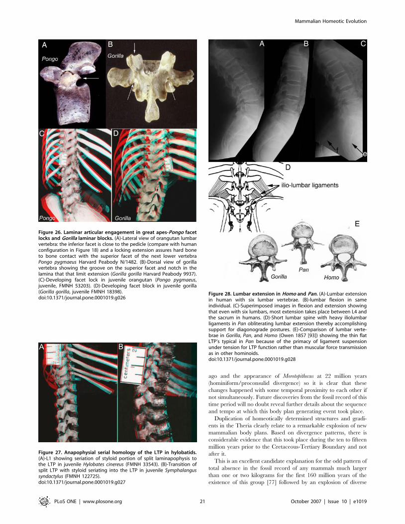
ago and the appearance of Morotopithecus at 22 million years
(hominiform/proconsulid divergence) so it is clear that these
changes happened with some temporal proximity to each other if
not simultaneously. Future discoveries from the fossil record of this
time period will no doubt reveal further details about the sequence
and tempo at which this body plan generating event took place.
Duplication of homeotically determined structures and gradi-
ents in the Theria clearly relate to a remarkable explosion of new
mammalian body plans. Based on divergence patterns, there is
considerable evidence that this took place during the ten to fifteen
million years prior to the Cretaceous-Tertiary Boundary and not
after it.
This is an excellent candidate explanation for the odd pattern of
total absence in the fossil record of any mammals much larger
than one or two kilograms for the first 160 million years of the
existence of this group [77] followed by an explosion of diverse
Figure 26. Laminar articular engagement in great apes-Pongo facetlocks and Gorilla laminar blocks. (A)-Lateral view of orangutan lumbarvertebra: the inferior facet is close to the pedicle (compare with humanconfiguration in Figure 18) and a locking extension assures hard boneto bone contact with the superior facet of the next lower vertebraPongo pygmaeus Harvard Peabody N/1482. (B)-Dorsal view of gorillavertebra showing the groove on the superior facet and notch in thelamina that that limit extension (Gorilla gorilla Harvard Peabody 9937).(C)-Developing facet lock in juvenile orangutan (Pongo pygmaeus,juvenile, FMNH 53203). (D)-Developing facet block in juvenile gorilla(Gorilla gorilla, juvenile FMNH 18398).doi:10.1371/journal.pone.0001019.g026
Figure 27. Anapophysial serial homology of the LTP in hylobatids.(A)-L1 showing seriation of styloid portion of split laminapophysis tothe LTP in juvenile Hylobates cinereus (FMNH 33543). (B)-Transition ofsplit LTP with styloid seriating into the LTP in juvenile Symphalangussyndactylus (FMNH 122725).doi:10.1371/journal.pone.0001019.g027
Figure 28. Lumbar extension in Homo and Pan. (A)-Lumbar extensionin human with six lumbar vertebrae. (B)-lumbar flexion in sameindividual. (C)-Superimposed images in flexion and extension showingthat even with six lumbars, most extension takes place between L4 andthe sacrum in humans. (D)-Short lumbar spine with heavy iliolumbarligaments in Pan obliterating lumbar extension thereby accomplishingsupport for diagonograde postures. (E)-Comparison of lumbar verte-brae in Gorilla, Pan, and Homo (Owen 1857 [93]) showing the thin flatLTP’s typical in Pan because of the primacy of ligament suspensionunder tension for LTP function rather than muscular force transmissionas in other hominoids.doi:10.1371/journal.pone.0001019.g028
Mammalian Homeotic Evolution
PLoS ONE | www.plosone.org 21 October 2007 | Issue 10 | e1019
large body mammalian forms in the last 60 million years.
Homeotic change that led to diminutive lumbar ribs in the early
mammals increased lumbar flexibility for sagittal excursion during
symmetric gaits, but seems to have left these groups without the
mechanical support in the lumbar region required for the
architecture of a large animal.
Therian mammals deploy symmetrical gaits for rapid locomo-
tion [88] [89] and they employ spinal flexion and extension to do
so. The principal locomotor drive is a simultaneous thrust with
both hind limbs while the forelimbs are free of ground contact.
This appears to be a fundamental locomotor innovation of therian
mammals, but may well have appeared in correlation with the
transition from the large robust lumbar costal processes in the
cynodonts to the small vestigial lumbar ribs seen in many
Mesozoic mammals groups.
The appearance of large mammals at the close of the
Cretaceous is at least coincident with the appearance of two
major types of architectural transformation of the lumbar spine to
provide non-muscular support against extension in the lumbar
region. These are convergent class of rigid locking systems in
groups with septo-neural transposition (Figure 21, 22, 23, 25, and
26) and the convergent class of ventrally arrayed and elastically
tensioned LTPs in a several other groups (Figure 15A, 16, 17B,
18C)–both classes of anatomical change based on homeotic
seriation of neomorphic or duplicated structures not present in the
stem mammals. In any case, there appears to have been a dramatic
increase in the rate of generation of new axial body plans once
diversification and duplication of homeotic gradients affecting the
laminapophysis and its derivatives that commenced in the
therians.
These data also support the concept of a threshold effect in
diversification of the mammals–progress awaited morphogenetic
innovation. This supports an enlarged role for a mutational view
[24] of evolutionary drive to update classic Darwinian and New
Synthesis models of the past two centuries.
METHODSA comparative evaluation of serially repeating structures and
homeotic patterning in 250 extant mammalian species and fossil
forms was carried out. For extant forms, specimens in the
collections of the Harvard University Museum of Comparative
Zoology (MCZ), Harvard Peabody Museum, Smithsonian Muse-
um (USNM) and Chicago Field Museum (FMNH) were selected to
provide coverage of all mammalian families except for the order
Chiroptera and Rodentia where coverage was at the level of the
superfamily. Specimens were selected based on preparations in
which a complete naturally articulated spine in which all details
could be observed. The objective was to obtain a representative
overview across the Class Mammalia but variation within species
was not addressed extensively. In essence there simply is not
sufficient material available to provide any real comprehensive
assessment of variation if the full systematic array of mammals is to
be covered. In addition the vertebral nomenclature of Owen [36]
was updated to distinguish between processes (‘‘apophysis’’ and
joints ‘‘arthrum’’) to clarify situation where one type of joint
appears on a different type of process [37]. Two different structures
on the same side of a vertebra could not have the same name.
ACKNOWLEDGMENTSDiscussions at various times since the inception of this project with Stephen
Jay Gould, Ernst Mayr, David Pilbeam, Russell H. Tuttle, Farish A.
Jenkins, A. W. Crompton, Irven DeVore, Erik Trinkaus, MaryEllen
Ruvolo and Terrence Deacon played a valuable role. Correspondence
regarding portions of the results from Rupert Riedl, A.J.E. Cave,
Sherwood Washburn are also appreciated. Ron Testa (FMNH) and Al
Coleman (MCZ) are thanked for photographic work.
Author Contributions
Conceived and designed the experiments: AF. Performed the experiments:
AF. Analyzed the data: AF. Contributed reagents/materials/analysis tools:
AF. Wrote the paper: AF.
REFERENCES1. Bateson W (1894) Materials for the Study of Variation Treated with Especial
Regard to Discontinuity in the Origin of Species. London; New York:
Macmillan.
2. De Robertis EM, Sasai Y (1996) A common plan for dorsoventral patterning in
Bilateria. Nature 380: 37–40.
3. Theissen G (2004) Developmental genetics: bittersweet evolution. Nature 428:
813.
4. Theissen G (2006) The proper place of hopeful monsters in evolutionary biology.
Theory Biosci 124: 349–369.
5. Lewis EB (1978) A gene complex controlling segmentation in Drosophila. Nature
276: 565–570.
6. Nusslein-Volhard C (1979) Maternal effect mutations that alter the spatial
coordinates of the embryo of Drosophila melanogaster. Symp Soc Dev Biol 37:
185–211.
7. Nusslein-Volhard C, Wieschaus E (1980) Mutations affecting segment number
and polarity in Drosophila. Nature 287: 795–801.
8. Kuratani S (2005) Craniofacial development and the evolution of the
vertebrates: the old problems on a new background. Zoolog Sci 22: 1–19.
9. Burke AC (2000) Hox genes and the global patterning of the somitic mesoderm.
Curr Top Dev Biol 47: 155–181.
10. Burke AC, Nelson CE, Morgan BA, Tabin C (1995) Hox genes and the evolution
of vertebrate axial morphology. Development 121: 333–346.
11. Nusslein-Volhard C (1994) Of flies and fishes. Science 266: 572–574.
12. van Eeden FJ, Granato M, Schach U, Brand M, Furutani-Seiki M, et al. (1996)
Mutations affecting somite formation and patterning in the zebrafish, Danio rerio.
Development 123: 153–164.
13. Nusslein-Volhard C (1996) Gradients that organize embryo development. Sci
Am 275: 54–5558–61.
14. Carroll SB (1995) Homeotic genes and the evolution of arthropods and
chordates. Nature 376: 479–485.
15. Rokas A, Kruger D, Carroll SB (2005) Animal evolution and the molecular
signature of radiations compressed in time. Science 310: 1933–1938.
16. Lovejoy CO, McCollum MA, Reno PL, Rosenman BA (2003) Developmental
biology and human evolution. Annual Review of Anthropology 32:
85–109.
17. Jacobs DK, Hughes NC, Fitz-Gibbon ST, Winchell CJ (2005) Terminal
addition, the Cambrian radiation and the Phanerozoic evolution of bilaterian
form. Evol Dev 7: 498–514.
18. Minelli A, Fusco G (2004) Evo-devo perspectives on segmentation: model
organisms, and beyond. Trends Ecol Evol 19: 423–429.
19. Minelli A, Fusco G (2005) Conserved versus innovative features in animal body
organization. J Exp Zoolog B Mol Dev Evol 304: 520–525.
20. Yu JK, Satou Y, Holland ND, Shin IT, Kohara Y, et al. (2007) Axial patterning
in cephalochordates and the evolution of the organizer. Nature.
21. Stollewerk A, Schoppmeier M, Damen WG (2003) Involvement of Notch and
Delta genes in spider segmentation. Nature 423: 863–865.
22. Gould SJ (2002) The Structure of Evolutionary Theory. Cambridge, Mass.:
Belknap Press of Harvard University Press. pp xxii, 1433.
23. Schlosser G, Wagner GP (2004) Modularity in Development and Evolution.
Chicago: University of Chicago Press. pp x, 600.
24. Stoltzfus A (2006) Mutationism and the dual causation of evolutionary change.
Evol Dev 8: 304–317.
25. Budd GE (2006) On the origin and evolution of major morphological characters.
Biol Rev Camb Philos Soc 81: 609–628.
26. Daeschler EB, Shubin NH, Jenkins FA Jr (2006) A Devonian tetrapod-like fish
and the evolution of the tetrapod body plan. Nature 440: 757–763.
27. Dahn RD, Davis MC, Pappano WN, Shubin NH (2007) Sonic hedgehog function
in chondrichthyan fins and the evolution of appendage patterning. Nature 445:
311–314.
28. Davidson EH, Erwin DH (2006) Gene regulatory networks and the evolution of
animal body plans. Science 311: 796–800.
29. Geisler R, Rauch GJ, Geiger-Rudolph S, Albrecht A, van Bebber F, et al. (2007)
Large-scale mapping of mutations affecting zebrafish development. BMC
Genomics 8: 11.
Mammalian Homeotic Evolution
PLoS ONE | www.plosone.org 22 October 2007 | Issue 10 | e1019
30. Carroll SB (2003) Genetics and the making of Homo sapiens. Nature 422:
849–857.
31. Richards RJ (2002) The Romantic Conception of Life : Science and Philosophy
in the Age of Goethe. Chicago: University of Chicago Press. pp xix, 587, [584] ofplates.
32. Goethe JWv (1817) Zur Naturwissenschaft uberhaupt, besonders zur Morpho-
logie. Stuttgard; Tubingen.
33. Appel TA (1987) Cuvier-Geoffroy Debate: French Biology in the Decades before
Darwin. New York: Oxford University Press. pp 305, [316] of plates.
34. Geoffroy Saint-Hilaire E (1830) Principes de philosophie zoologique, discutes enmars 1830, au sein de l’Academie royale des sciences. Paris: Pichon et Didier.
226 p.
35. Geoffroy Saint-Hilaire E (1822) Considerations generales sur la vertebre. Mem
Mus Hist Nat 9: 89–119.
36. Owen R (1848) On the Archetype and Homologies of the Vertebrate Skeleton.London: J. Van Voorst.
37. Filler AG (1986) Axial Character Seriation in Mammals: An Historical and
Morphological Exploration of the Origin, Development, Use and CurrentCollapse of the Homology Paradigm-PhD Thesis. Cambridge, Massachusetts-
(2nd Printing: BrownWalker Press, Boca Raton, Florida 2007, 349 p): Harvard
University. 368 p.
38. Filler AG (1993) Evolution of the sacrum in hominoids. In: Doty JR,Rengachary SS, eds. Surgical Disorders of the Sacrum. New York, N.Y.:
Thieme Medical Publishers. pp 13–20.
39. Pilbeam D (2004) The anthropoid postcranial axial skeleton: comments on
development, variation, and evolution. J Exp Zoolog B Mol Dev Evol 302:241–267.
40. Narita Y, Kuratani S (2005) Evolution of the vertebral formulae in mammals:
a perspective on developmental constraints. J Exp Zoolog B Mol Dev Evol 304:
91–106.
41. Buchholtz EA (2007) Modular evolution of the Cetacean vertebral column.Evolution & Development 9: 278–289.
42. Boszczyk BM, Boszczyk AA, Putz R (2001) Comparative and functional
anatomy of the mammalian lumbar spine. Anat Rec 264: 157–168.
43. Cheng Z, Ventura M, She X, Khaitovich P, Graves T, et al. (2005) A genome-
wide comparison of recent chimpanzee and human segmental duplications.Nature 437: 88–93.
44. Mikkelson TS, et al. (2005) Initial sequence of the chimpanzee genome and
comparison with the human genome. Nature 437: 69–87.
45. Ward CV, Walker A, Teaford MF, Odhiambo I (1993) Partial skeleton of
Proconsul nyanzae from Mfangano Island, Kenya. Am J Phys Anthropol 90:77–111.
46. Young NM, MacLatchy L (2004) The phylogenetic position of Morotopithecus.
J Hum Evol 46: 163–184.
47. MacLatchy L (2004) The oldest ape. Evol Anthropol. pp 90–103.
48. MacLatchy L, Gebo D, Kityo R, Pilbeam D (2000) Postcranial functional
morphology of Morotopithecus bishopi, with implications for the evolution ofmodern ape locomotion. J Hum Evol 39: 159–183.
49. Walker A, Rose MD (1968) Fossil hominoid vertebra from the Miocene of
Uganda. Nature 217: 980–981.
50. Kohler M, Moya-Sola S (1997) Ape-like or hominid-like? The positional
behavior of Oreopithecus bambolii reconsidered. Proc Natl Acad Sci U S A 94:11747–11750.
51. Rook L, Bondioli L, Kohler M, Moya-Sola S, Macchiarelli R (1999) Oreopithecus
was a bipedal ape after all: evidence from the iliac cancellous architecture. Proc
Natl Acad Sci U S A 96: 8795–8799.
52. Moya-Sola S, Kohler M, Alba DM, Casanovas-Vilar I, Galindo J (2004)Pierolapithecus catalaunicus, a new Middle Miocene great ape from Spain. Science
306: 1339–1344.
53. Tuttle RH (2006) Are human beings apes, or are apes people too? In: Ishida H,
Tuttle RH, Pickford M, Ogihara N, Nakatsukasa M, eds. Human Origins andEnvironmental Backgrounds. New York: Springer. pp 248–258.
54. Keith A (1923) Man’s posture: its evolution and disorders. Hunterian Lectures
I-VI. Brit Med J (1923): 451–454, 499–502, 545–548, 587–590, 624–626, 669–672.
55. Tuttle RH (1974) Darwin’s apes, dental apes, and the descent of Man: Normal
science in evolutionary anthropology. Curr Anthropol 15: 389–398.
56. Thorpe SKS, Holder RL, Crompton RH (2007) Origin of human bipedalism asan adaptation for locomotion on flexible branches. Science 316: 1328–1331.
57. Ahlberg PE, Clack JA, Blom H (2005) The axial skeleton of the Devonian
tetrapod Ichthyostega. Nature 437: 137–140.
58. Springer MS, Murphy WJ, Eizirik E, O’Brien SJ (2003) Placental mammal
diversification and the Cretaceous-Tertiary boundary. Proc Natl Acad Sci U S A100: 1056–1061.
59. Nilsson MA, Arnason U, Spencer PB, Janke A (2004) Marsupial relationships
and a timeline for marsupial radiation in South Gondwana. Gene 340: 189–196.
60. Douady CJ, Catzeflis F, Raman J, Springer MS, Stanhope MJ (2003) The
Sahara as a vicariant agent, and the role of Miocene climatic events, in thediversification of the mammalian order Macroscelidea (elephant shrews). Proc
Natl Acad Sci U S A 100: 8325–8330.
61. Ishida H, Kunimatsu Y, Takano T, Nakano Y, Nakatsukasa M (2004)Nacholapithecus skeleton from the Middle Miocene of Kenya. J Hum Evol 46:
69–103.
62. Ward CV (1993) Torso morphology and locomotion in Proconsul nyanzae.
Am J Phys Anthropol 92: 291–328.63. Nakatsukasa M, Ward CV, Walker A, Teaford MF, Kunimatsu Y, et al. (2004)
Tail loss in Proconsul heseloni. J Hum Evol 46: 777–784.
64. Robertson JT (1972) Early Hominid Posture and Locomotion. Chicago:University of Chicago Press. 361 p.
65. Haeusler M, Martelli SA, Boeni T (2002) Vertebrae numbers of the earlyhominid lumbar spine. J Hum Evol 43: 621–643.
66. Brunet M, Guy F, Pilbeam D, Mackaye HT, Likius A, et al. (2002) A new
hominid from the Upper Miocene of Chad, Central Africa. Nature 418:145–151.
67. Guy F, Lieberman DE, Pilbeam D, de Leon MP, Likius A, et al. (2005)Morphological affinities of the Sahelanthropus tchadensis (Late Miocene hominid
from Chad) cranium. Proc Natl Acad Sci U S A 102: 18836–18841.68. Zollikofer CP, Ponce de Leon MS, Lieberman DE, Guy F, Pilbeam D, et al.
(2005) Virtual cranial reconstruction of Sahelanthropus tchadensis. Nature 434:
755–759.69. Wolpoff MH, Senut B, Pickford M, Hawks J (2002) Palaeoanthropology.
Sahelanthropus or ‘Sahelpithecus’? Nature 419: 581–582; discussion 582.70. Harcourt-Smith WE, Aiello LC (2004) Fossils, feet and the evolution of human
bipedal locomotion. J Anat 204: 403–416.
71. Ohman JC, Lovejoy CO, White TD, Eckhardt RB, Galik K, et al. (2005)Questions about Orrorin femur. Science 307: 845b.
72. Galik K, Senut B, Pickford M, Gommery D, Treil J, et al. (2004) External andinternal morphology of the BAR 1002’00 Orrorin tugenensis femur. Science 305:
1450–1453.73. Li G, Luo ZX (2006) A Cretaceous symmetrodont therian with some
monotreme-like postcranial features. Nature 439: 195–200.
74. Jenkins FA (1971) The Postcranial Skeleton of African Cynodonts; Problems inthe Early Evolution of the Mammalian Postcranial Skeleton. New Haven:
Peabody Museum of Natural History Yale University. pp x, 216.75. Kielan-Jaworowska Z, Gambaryan PP (1994) Postcranial anatomy and habits of
Asian multituberculate mammals Fossils and Strata 36: 1–92.
76. Kielan-Jaworowska Z, Cifelli R, Luo Z-X (2004) Mammals from the Age ofDinosaurs: Origins, Evolution, and Structure. New York: Columbia University
Press. pp xv, 630.77. Kemp TS (2005) The Origin and Evolution of Mammals. Oxford; New York:
Oxford University Press. pp x, 331.78. Flower WH (1885) An Introduction to the Osteology of the Mammalia. London:
Macmillan. pp xi, 382.
79. Raaum RL, Sterner KN, Noviello CM, Stewart CB, Disotell TR (2005)Catarrhine primate divergence dates estimated from complete mitochondrial
genomes: concordance with fossil and nuclear DNA evidence. J Hum Evol 48:237–257.
80. Williston SW, Gregory WK (1925) The Osteology of the Reptiles. Cambridge:
Harvard University Press. pp xiii, 300.81. Gregory WK (1947) The monotremes and the palimpsest theory. Bull Am Mus
Nat Hist 88: 1–52.82. Hoffstetter R, Gasc J-P (1969) Vertebrae and ribs of modern reptiles. In: Gans C,
Bellairs AdA, Parsons TS, eds. Biology of the Reptilia-Morphology A. London:Academic Press. pp 201–310.
83. Bezuidenhout AJ, Seegers CD (1996) The osteology of the African elephant
(Loxodonta africana): vertebral column, ribs and sternum. Onderstepoort J Vet Res63: 131–147.
84. Slijper EJ (1946) Comparative biological-anatomical investigation on thevertebral column and spinal musculature of mammals. Kon Ned Akad Wet
Verh (Tweede Sec) 42: 1–128.
85. Argot C (2003) Functional-adaptive anatomy of the axial skeleton of some extantmarsupials and the paleobiology of the paleocene marsupials Mayulestes ferox and
Pucadelphys andinus. J Morphol 255: 279–300.86. Filler AG (2007) The emergence and optimization of upright posture among
hominiform hominoids and the evolutionary pathophysiology of back pain.
Neurosurgical Focus 23: E4: 1–6.87. Filler AG (2007) The Upright Ape: A New Origin of the Species. Franklin Lakes,
NJ: New Page Books. 285 p.88. Fischer MS, Schilling N, Schmidt M, Haarhaus D, Witte H (2002) Basic limb
kinematics of small therian mammals. J Exp Biol 205: 1315–1338.89. Schilling N, Hackert R (2006) Sagittal spine movements of small therian
mammals during asymmetrical gaits. J Exp Biol 209: 3925–3939.
90. Flynn JJ, Finarelli JA, Zehr S, Hsu J, Nedbal MA (2005) Molecular phylogeny ofthe carnivora (mammalia): assessing the impact of increased sampling on
resolving enigmatic relationships. Syst Biol 54: 317–337.91. Keith A (1902) The extent to which the posterior segments of the body have
been transmuted and suppressed in the evolution of man and allied primates.
J Anat Phys 37: 18–40.92. Filler AG (1979) Functional and Evolutionary Perspectives on Chimpanzee
Thoracolumbar Musculature. Masters Thesis, unpublished. [MA]. ChicagoIlli-nois: University of Chicago.
93. Owen R (1857) Osteological contributions to the natural history of thechimpanzees (Troglodytes) and orangs (Pithecus). No. V. Comparison of the lower
jaw and vertebral column of the Troglodytes gorilla, Troglodytes niger, Pithecus satyrus,
and different varieties of the human race. Trans Zool Soc London, (printed1862) 4: 89–115.
Mammalian Homeotic Evolution
PLoS ONE | www.plosone.org 23 October 2007 | Issue 10 | e1019