Embed Size (px)
Citation preview

Homeobox gene methylation in lung cancer studiedby genome-wide analysis with a microarray-basedmethylated CpG island recovery assayTibor Rauch*, Zunde Wang*, Xinmin Zhang†, Xueyan Zhong*, Xiwei Wu‡, Sean K. Lau§, Kemp H. Kernstine¶,Arthur D. Riggs*�, and Gerd P. Pfeifer*�
*Division of Biology, ‡Division of Information Sciences, §Division of Pathology, and ¶Division of Surgery, Beckman Research Institute of the Cityof Hope, Duarte, CA 91010; and †NimbleGen Systems, Inc., Madison, WI 53711
Contributed by Arthur D. Riggs, February 5, 2007 (sent for review December 11, 2006)
De novo methylation of CpG islands is a common phenomenon inhuman cancer, but the mechanisms of cancer-associated DNAmethylation are not known. We have used tiling arrays in combi-nation with the methylated CpG island recovery assay to investi-gate methylation of CpG islands genome-wide and at high reso-lution. We find that all four HOX gene clusters on chromosomes 2,7, 12, and 17 are preferential targets for DNA methylation in cancercell lines and in early-stage lung cancer. CpG islands associatedwith many other homeobox genes, such as SIX, LHX, PAX, DLX, andEngrailed, were highly methylated as well. Altogether, more thanhalf (104 of 192) of all CpG island-associated homeobox genes inthe lung cancer cell line A549 were methylated. Analysis of paralo-gous HOX genes showed that not all paralogues undergo cancer-associated methylation simultaneously. The HOXA cluster wasanalyzed in greater detail. Comparison with ENCODE-derived datashows that lack of methylation at CpG-rich sequences correlateswith presence of the active chromatin mark, histone H3 lysine-4methylation in the HOXA region. Methylation analysis of HOXAgenes in primary squamous cell carcinomas of the lung led to theidentification of the HOXA7- and HOXA9-associated CpG islands asfrequent methylation targets in stage 1 tumors. Homeobox genesare potentially useful as DNA methylation markers for early diag-nosis of the disease. The finding of widespread methylation ofhomeobox genes lends support to the hypothesis that a substan-tial fraction of genes methylated in human cancer are targets of thePolycomb complex.
DNA methylation � HOX genes � chromatin � Polycomb
DNA methylation at CpG dinucleotides is an important epige-netic modification carried out by DNA methyltransferases (1,
2). De novo methylation of CpG islands that overlap with promoterregions is commonly associated with gene silencing and is afrequent event that accompanies tumorigenesis (3–9). Cancer-specific hypermethylation of genes that suppress uncontrolled cellproliferation or promote genome stability is a key step in tumordevelopment.
Homeobox genes encode a transcription factor family that playsdecisive roles in embryogenesis and differentiation of adult cells(10). Homeobox proteins are classified into one family on the basisof their evolutionary conserved helix–loop–helix DNA-bindingmotif, called the homeodomain. Apart from the homeodomain,family members share only limited conservation outside of theDNA-binding motif. Most of the family members are scatteredthroughout the genome, but a subgroup of the homeobox genes,HOX genes, are organized into clusters. HOX genes were originallyidentified in Drosophila as factors involved in homeotic transfor-mations (11). During mammalian evolution, the ancient HOXcluster underwent duplications that were followed by gene lossesthat finally led to the emergence of the 39 present HOX genesorganized into four clusters (10). HOX proteins are essentialswitches of developmental stage- and cell-specific gene regulation,
and in this way they are key determinants of cell identity andpotential targets during tumorigenesis.
We recently developed a DNA methylation detection technique,the methylated CpG island recovery assay (MIRA) (12). Thistechnique is based on the high affinity of a complex of the MBD2band MBD3L1 proteins for CpG-methylated DNA (13) and iscompatible with microarray-based methodology (14). We previ-ously found that several homeobox genes were methylated in a lungcancer cell line (14). In the present study, we combined the MIRAtechnique with tiling arrays to obtain genome-wide CpG islandcoverage as well as high-resolution data for DNA methylationwithin HOX gene clusters in cell lines and primary lung tumors. Thedata indicate that multiple CpG islands within HOX clusters andnear other homeobox genes are frequent methylation targets inlung cancer. An in-depth analysis of the HOXA cluster revealedDNA methylation markers for stage 1 lung cancer.
ResultsMethylation of the HOXA Cluster. To explore the use of MIRA-assisted tiling platforms for genome-wide DNA methylation anal-ysis, we first used NimbleGen’s ENCODE tiling arrays. The EN-CODE array is designed to identify functional elements in �1% ofthe human genome. On these arrays, the neighboring tiling oligo-nucleotides (50 bp long) overlap with each other at 12-bp-longsequences, and in this way 30 Mb, including the HOXA chromo-somal region, is fully covered. First, we analyzed methylationpatterns by using DNA obtained from the lymphoblastoid cell lineGM06990, which is one of the cell lines used in the ENCODEproject. We compared the MIRA-enriched fraction with the inputfraction. Samples were prepared as described previously (14) byusing MseI digestion, linker ligation, amplification, and labeling ofinput and MIRA-enriched DNA. Upon examination of the EN-CODE regions, we observed that the highest level of methylationwas found on chromosome 7 in a region including the HOXA genecluster. Three independent MIRA reactions with GM06990 DNAwere conducted. Fig. 1 Upper shows the high reproducibility of thisapproach, which is aided by the overlap features of the tiling array.We found that several CpG islands within the HOXA cluster werestrongly methylated in GM06990 cells. Confirmation of the meth-ylation status was obtained by combined bisulfite restriction analysis
Author contributions: K.H.K., A.D.R., and G.P.P. designed research; T.R., Z.W., X. Zhang, X.Zhong, and S.K.L. performed research; X. Zhang contributed new reagents/analytic tools;T.R., Z.W., X.W., A.D.R., and G.P.P. analyzed data; and T.R., A.D.R., and G.P.P. wrote thepaper.
The authors declare no conflict of interest.
Abbreviations: COBRA, combined bisulfite restriction analysis; MIRA, methylated CpGisland recovery assay; NHBE cells, normal human bronchial epithelial cells.
�To whom correspondence may be addressed. E-mail: [email protected] or [email protected].
This article contains supporting information online at www.pnas.org/cgi/content/full/0701059104/DC1.
© 2007 by The National Academy of Sciences of the USA
www.pnas.org�cgi�doi�10.1073�pnas.0701059104 PNAS � March 27, 2007 � vol. 104 � no. 13 � 5527–5532
GEN
ETIC
S
Dow
nloa
ded
by g
uest
on
Dec
embe
r 20
, 202
1

(COBRA) methylation analysis (Fig. 1 Lower) and by bisulfitesequencing [Fig. 2 and supporting information (SI) Fig. 5].
The HOXA cluster contains 12 genes (11 HOX genes and EVX1)and is contained in a 155-kb-long genomic region. Most of theHOXA promoters are embedded in one of the 39 CpG islands (SITable 1) residing in the locus. The high CpG island density makesthe cluster an ideal target for testing the interdependence ofneighboring DNA methylation patterns, including, for example,theories of long-range epigenetic silencing (15). According to thishypothesis, genes located in the same neighborhood are coordi-nately silenced by epigenetic modifications. Our methylation profileanalysis does show that most CpG islands were methylated withinthe HOXA cluster (Fig. 1). However, not all CpG islands weremethylated. Highly methylated and poorly or nonmethylated CpGislands could be next to each other within the cluster (Figs. 1 and2; SI Tables 1 and 2). To scrutinize this theory in more detail, wefocused on two neighboring CpG islands that reside just 4 kb apartin the middle of the HOXA cluster. COBRAs (Fig. 1) and bisulfitesequencing data (Fig. 2) confirmed that the methylation status ofthese two neighboring CpG islands is opposite. Hypermethylationof the HOXA5 promoter suggests the formation of inactive chro-matin, whereas the unmethylated status of the neighboring HOXA6promoter should correlate with an active chromatin configuration.These expectations were confirmed by comparison with chromatinimmunoprecipitation (ChIP)-on-chip experiments previously per-formed with the GM06990 cell line with anti-histone H3K4me3antibody (from the ENCODE database) and by tiling expressionarray data for the two exons of HOXA5 and HOXA6 (from theENCODE database) (Fig. 2). The active gene-associated histonemethylation mark was not detected at the methylated HOXA5promoter. The region upstream of the HOXA6 gene, in particularCpG island 16, showed a strong signal for trimethylated K4 onhistone H3 that defines an active chromatin state. Using 5� RACE,we detected an alternatively spliced transcript emanating from CpG
island 16 and containing parts of the two HOXA6 exons (data notshown). The 3� end of CpG island 14 was methylated, indicating thata methylation boundary is between the 3� end of CpG island 14 andCpG island 15 (Fig. 2).
The Four HOX Gene Clusters Are Preferred Methylation Targets in LungCancer Cells. Cancer-associated DNA methylation changes com-monly affect CpG islands that are often associated with genepromoters and undergo hypermethylation in tumors. We next usedAgilent CpG island arrays that contain 27,800 CpG islands coveringin a tiling fashion 21 Mb of the human genome. Combining MIRAwith Agilent arrays, we compared the methylation status of CpGislands genome-wide between the lung cancer cell line A549 andnormal human bronchial epithelial (NHBE) cells. Although thefour HOX gene clusters reside on different chromosomes, we foundthat each of them is the target of extensive de novo methylation inlung cancer cells (Fig. 3 and SI Table 2). Fig. 3 shows that besidesthe HOX clusters there are several other cancer-related hypermeth-ylation hot spots showing �10-fold enrichment by MIRA in thelung cancer cell line A549. Strikingly, many of them define otherhomeobox genes, for instance the engrailed homologues EN1 andEN2, on chromosomes 2 and 7, respectively, and the SIX3/2 genepair, as well as LBX2 and LHX1. Several T box genes (TBX2, TBX3,and TBX20) also were preferred chromosomal methylation sites(Fig. 3). In addition to those, several paired-box homologues (PAXgenes 1–9, except PAX1 and PAX4) and many other homeoboxgenes were methylated in this tumor cell line (SI Table 3). Theoccurrence of apparent methylation hot spots as displayed alongthe entire length of a chromosome is most often related to thesimultaneous methylation of several CpG islands within a chromo-somal segment. This observation is illustrated for the HOX geneclusters (Figs. 1 and 2 and SI Tables 1 and 2) but also can be seenfor other chromosomal methylation hot spots. As an example, weillustrate the methylation of several CpG islands within or near the
Fig. 1. Analysis of the HOXA cluster region on chromosome 7 with ENCODE NimbleGen tiling arrays. The analysis covers a 155-kb region. DNA from the GM06990cell line was used for three independent MIRA reactions by using the amplification and labeling procedure as described in Materials and Methods. (Upper) Thesignal ratio of MIRA-enriched versus input DNA is plotted along the chromosome. The green boxes indicate the 39 CpG islands (see SI Table 1). The numberingrefers to HOXA gene number, and the arrows indicate the direction of transcription. A COBRA using BstUI digestion (see Materials and Methods) was conductedfor the regions indicated to confirm the methylation data obtained by MIRAs. Some of the BstUI cleavage fragments are too small to be visible on the gels. �,to control digestion with no BstUI; �, BstUI-digested samples.
5528 � www.pnas.org�cgi�doi�10.1073�pnas.0701059104 Rauch et al.
Dow
nloa
ded
by g
uest
on
Dec
embe
r 20
, 202
1

EN1 and TBX20 genes in SI Fig. 6. In all, 104 of 194 (54%) of thehomeobox genes associated with CpG islands in the lung cancer cellline A549 were methylated (SI Tables 2 and 3). We used ratherstringent criteria for making this assignment, and the percentage ofmethylated homeobox genes is likely an underestimation because ofthe elimination of low signal intensity spots, for instance caused bylarge spacing of adjacent restriction sites and low amplificationefficiency. Also, homeobox genes without CpG islands are notrepresented on the array, and some may be methylated already innormal cells. Taken together, these findings suggest that the inac-tivation of clusters of CpG islands near homeobox genes is acommon event in this lung cancer cell line.
Methylation Analysis of HOX Gene Paralogues. HOX proteins be-longing to a given paralogous group can be functionally similar andpartially interchangeable (16–18). To gain more information on themethylation profile of the individual genes that constitute a paralo-gous group, we analyzed the Agilent CpG island arrays in moredetail. These high-resolution tiling arrays harbor most of the 39HOX gene promoters. We determined the hypermethylation pro-file of each HOX gene promoter for all of the HOX gene clustersin the lung cancer cell line A549 versus NHBE cells (SI Table 2).The data show that many CpG islands in the HOX clusters werehypermethylated in A549 lung cancer cells relative to NHBE cells.We focused our attention on the HOX6 and HOX7 paralogues. We
Fig. 2. DNA and histone methylation profile at HOXA cluster genes in GM06990 cells. (A) DNA and histone methylation profile analysis of the entire HOXAcluster in GM06990 cells. The MIRA-enriched methylated DNA fraction and input fraction were mixed and hybridized onto NimbleGen ENCODE arrays. Note thatin this experiment, the MIRA-enriched DNA and the input DNA were processed without amplification and were directly labeled and hybridized to the array tomake these data methodologically comparable with the ChIP data. ChIP with anti-histone H3K4m3 antibody was performed, and the DNA was hybridized ontoSanger ENCODE3.1.1 DNA microarrays (data were obtained from the ENCODE database). The pink shading indicates the promoter-associated CpG islands. (B)(Upper) DNA and histone methylation and mRNA expression profile analysis of the HOXA5 and HOXA6 genes. Data for H3K4 methylation and mRNA expressionare from the ENCODE database. (Lower) Bisulfite sequencing verification of the DNA methylation status of the indicated CpG island regions.
Rauch et al. PNAS � March 27, 2007 � vol. 104 � no. 13 � 5529
GEN
ETIC
S
Dow
nloa
ded
by g
uest
on
Dec
embe
r 20
, 202
1
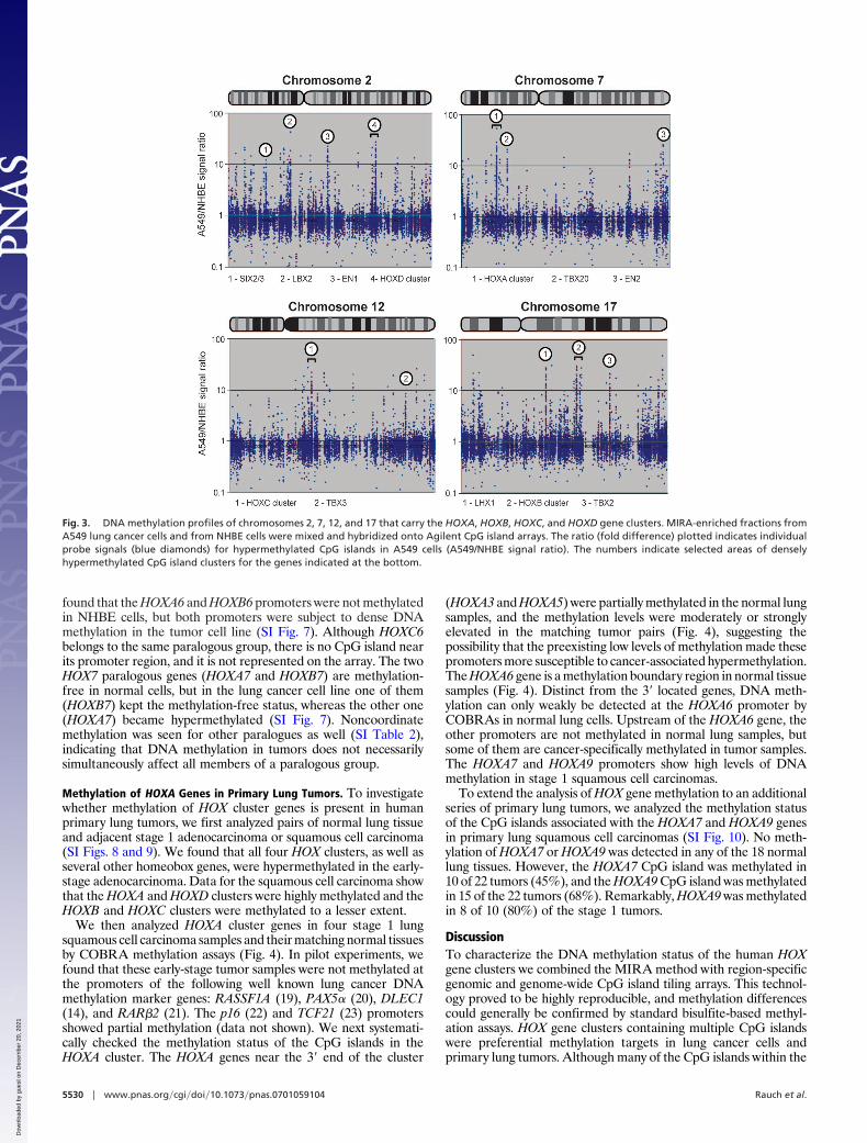
found that the HOXA6 and HOXB6 promoters were not methylatedin NHBE cells, but both promoters were subject to dense DNAmethylation in the tumor cell line (SI Fig. 7). Although HOXC6belongs to the same paralogous group, there is no CpG island nearits promoter region, and it is not represented on the array. The twoHOX7 paralogous genes (HOXA7 and HOXB7) are methylation-free in normal cells, but in the lung cancer cell line one of them(HOXB7) kept the methylation-free status, whereas the other one(HOXA7) became hypermethylated (SI Fig. 7). Noncoordinatemethylation was seen for other paralogues as well (SI Table 2),indicating that DNA methylation in tumors does not necessarilysimultaneously affect all members of a paralogous group.
Methylation of HOXA Genes in Primary Lung Tumors. To investigatewhether methylation of HOX cluster genes is present in humanprimary lung tumors, we first analyzed pairs of normal lung tissueand adjacent stage 1 adenocarcinoma or squamous cell carcinoma(SI Figs. 8 and 9). We found that all four HOX clusters, as well asseveral other homeobox genes, were hypermethylated in the early-stage adenocarcinoma. Data for the squamous cell carcinoma showthat the HOXA and HOXD clusters were highly methylated and theHOXB and HOXC clusters were methylated to a lesser extent.
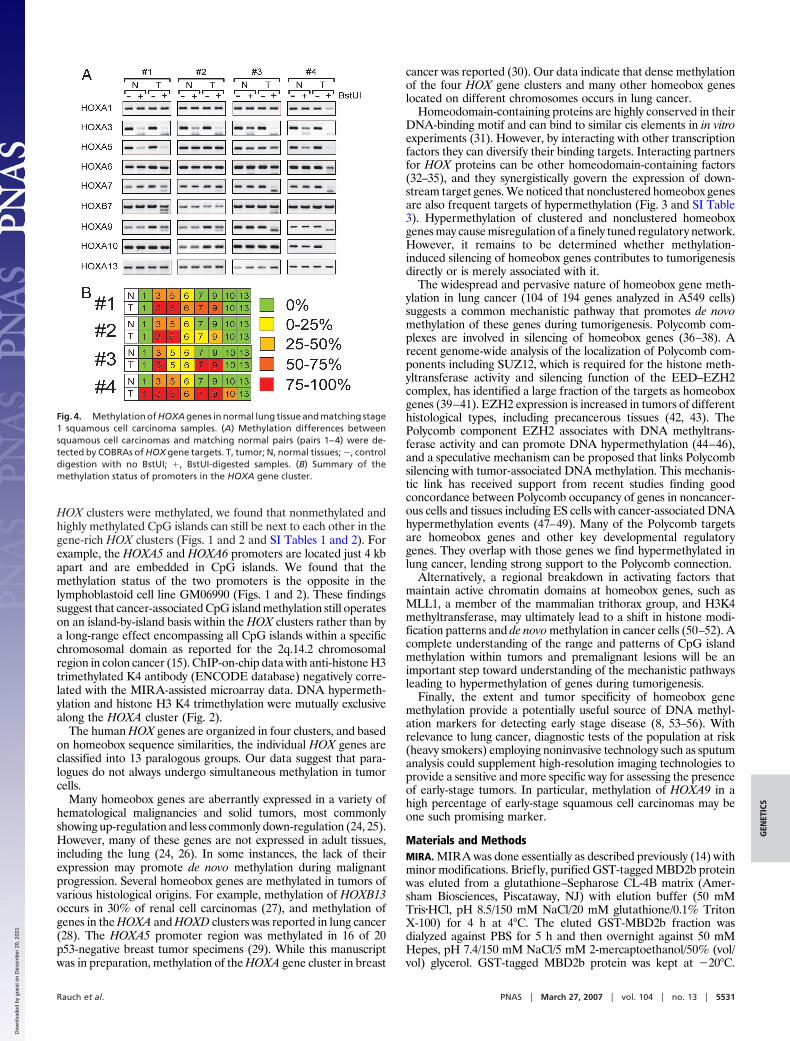
We then analyzed HOXA cluster genes in four stage 1 lungsquamous cell carcinoma samples and their matching normal tissuesby COBRA methylation assays (Fig. 4). In pilot experiments, wefound that these early-stage tumor samples were not methylated atthe promoters of the following well known lung cancer DNAmethylation marker genes: RASSF1A (19), PAX5� (20), DLEC1(14), and RAR�2 (21). The p16 (22) and TCF21 (23) promotersshowed partial methylation (data not shown). We next systemati-cally checked the methylation status of the CpG islands in theHOXA cluster. The HOXA genes near the 3� end of the cluster
(HOXA3 and HOXA5) were partially methylated in the normal lungsamples, and the methylation levels were moderately or stronglyelevated in the matching tumor pairs (Fig. 4), suggesting thepossibility that the preexisting low levels of methylation made thesepromoters more susceptible to cancer-associated hypermethylation.The HOXA6 gene is a methylation boundary region in normal tissuesamples (Fig. 4). Distinct from the 3� located genes, DNA meth-ylation can only weakly be detected at the HOXA6 promoter byCOBRAs in normal lung cells. Upstream of the HOXA6 gene, theother promoters are not methylated in normal lung samples, butsome of them are cancer-specifically methylated in tumor samples.The HOXA7 and HOXA9 promoters show high levels of DNAmethylation in stage 1 squamous cell carcinomas.
To extend the analysis of HOX gene methylation to an additionalseries of primary lung tumors, we analyzed the methylation statusof the CpG islands associated with the HOXA7 and HOXA9 genesin primary lung squamous cell carcinomas (SI Fig. 10). No meth-ylation of HOXA7 or HOXA9 was detected in any of the 18 normallung tissues. However, the HOXA7 CpG island was methylated in10 of 22 tumors (45%), and the HOXA9 CpG island was methylatedin 15 of the 22 tumors (68%). Remarkably, HOXA9 was methylatedin 8 of 10 (80%) of the stage 1 tumors.
DiscussionTo characterize the DNA methylation status of the human HOXgene clusters we combined the MIRA method with region-specificgenomic and genome-wide CpG island tiling arrays. This technol-ogy proved to be highly reproducible, and methylation differencescould generally be confirmed by standard bisulfite-based methyl-ation assays. HOX gene clusters containing multiple CpG islandswere preferential methylation targets in lung cancer cells andprimary lung tumors. Although many of the CpG islands within the
Fig. 3. DNA methylation profiles of chromosomes 2, 7, 12, and 17 that carry the HOXA, HOXB, HOXC, and HOXD gene clusters. MIRA-enriched fractions fromA549 lung cancer cells and from NHBE cells were mixed and hybridized onto Agilent CpG island arrays. The ratio (fold difference) plotted indicates individualprobe signals (blue diamonds) for hypermethylated CpG islands in A549 cells (A549/NHBE signal ratio). The numbers indicate selected areas of denselyhypermethylated CpG island clusters for the genes indicated at the bottom.
5530 � www.pnas.org�cgi�doi�10.1073�pnas.0701059104 Rauch et al.
Dow
nloa
ded
by g
uest
on
Dec
embe
r 20
, 202
1
HOX clusters were methylated, we found that nonmethylated andhighly methylated CpG islands can still be next to each other in thegene-rich HOX clusters (Figs. 1 and 2 and SI Tables 1 and 2). Forexample, the HOXA5 and HOXA6 promoters are located just 4 kbapart and are embedded in CpG islands. We found that themethylation status of the two promoters is the opposite in thelymphoblastoid cell line GM06990 (Figs. 1 and 2). These findingssuggest that cancer-associated CpG island methylation still operateson an island-by-island basis within the HOX clusters rather than bya long-range effect encompassing all CpG islands within a specificchromosomal domain as reported for the 2q.14.2 chromosomalregion in colon cancer (15). ChIP-on-chip data with anti-histone H3trimethylated K4 antibody (ENCODE database) negatively corre-lated with the MIRA-assisted microarray data. DNA hypermeth-ylation and histone H3 K4 trimethylation were mutually exclusivealong the HOXA cluster (Fig. 2).
The human HOX genes are organized in four clusters, and basedon homeobox sequence similarities, the individual HOX genes areclassified into 13 paralogous groups. Our data suggest that para-logues do not always undergo simultaneous methylation in tumorcells.
Many homeobox genes are aberrantly expressed in a variety ofhematological malignancies and solid tumors, most commonlyshowing up-regulation and less commonly down-regulation (24, 25).However, many of these genes are not expressed in adult tissues,including the lung (24, 26). In some instances, the lack of theirexpression may promote de novo methylation during malignantprogression. Several homeobox genes are methylated in tumors ofvarious histological origins. For example, methylation of HOXB13occurs in 30% of renal cell carcinomas (27), and methylation ofgenes in the HOXA and HOXD clusters was reported in lung cancer(28). The HOXA5 promoter region was methylated in 16 of 20p53-negative breast tumor specimens (29). While this manuscriptwas in preparation, methylation of the HOXA gene cluster in breast
cancer was reported (30). Our data indicate that dense methylationof the four HOX gene clusters and many other homeobox geneslocated on different chromosomes occurs in lung cancer.
Homeodomain-containing proteins are highly conserved in theirDNA-binding motif and can bind to similar cis elements in in vitroexperiments (31). However, by interacting with other transcriptionfactors they can diversify their binding targets. Interacting partnersfor HOX proteins can be other homeodomain-containing factors(32–35), and they synergistically govern the expression of down-stream target genes. We noticed that nonclustered homeobox genesare also frequent targets of hypermethylation (Fig. 3 and SI Table3). Hypermethylation of clustered and nonclustered homeoboxgenes may cause misregulation of a finely tuned regulatory network.However, it remains to be determined whether methylation-induced silencing of homeobox genes contributes to tumorigenesisdirectly or is merely associated with it.
The widespread and pervasive nature of homeobox gene meth-ylation in lung cancer (104 of 194 genes analyzed in A549 cells)suggests a common mechanistic pathway that promotes de novomethylation of these genes during tumorigenesis. Polycomb com-plexes are involved in silencing of homeobox genes (36–38). Arecent genome-wide analysis of the localization of Polycomb com-ponents including SUZ12, which is required for the histone meth-yltransferase activity and silencing function of the EED–EZH2complex, has identified a large fraction of the targets as homeoboxgenes (39–41). EZH2 expression is increased in tumors of differenthistological types, including precancerous tissues (42, 43). ThePolycomb component EZH2 associates with DNA methyltrans-ferase activity and can promote DNA hypermethylation (44–46),and a speculative mechanism can be proposed that links Polycombsilencing with tumor-associated DNA methylation. This mechanis-tic link has received support from recent studies finding goodconcordance between Polycomb occupancy of genes in noncancer-ous cells and tissues including ES cells with cancer-associated DNAhypermethylation events (47–49). Many of the Polycomb targetsare homeobox genes and other key developmental regulatorygenes. They overlap with those genes we find hypermethylated inlung cancer, lending strong support to the Polycomb connection.
Alternatively, a regional breakdown in activating factors thatmaintain active chromatin domains at homeobox genes, such asMLL1, a member of the mammalian trithorax group, and H3K4methyltransferase, may ultimately lead to a shift in histone modi-fication patterns and de novo methylation in cancer cells (50–52). Acomplete understanding of the range and patterns of CpG islandmethylation within tumors and premalignant lesions will be animportant step toward understanding of the mechanistic pathwaysleading to hypermethylation of genes during tumorigenesis.
Finally, the extent and tumor specificity of homeobox genemethylation provide a potentially useful source of DNA methyl-ation markers for detecting early stage disease (8, 53–56). Withrelevance to lung cancer, diagnostic tests of the population at risk(heavy smokers) employing noninvasive technology such as sputumanalysis could supplement high-resolution imaging technologies toprovide a sensitive and more specific way for assessing the presenceof early-stage tumors. In particular, methylation of HOXA9 in ahigh percentage of early-stage squamous cell carcinomas may beone such promising marker.
Materials and MethodsMIRA. MIRA was done essentially as described previously (14) withminor modifications. Briefly, purified GST-tagged MBD2b proteinwas eluted from a glutathione–Sepharose CL-4B matrix (Amer-sham Biosciences, Piscataway, NJ) with elution buffer (50 mMTris�HCl, pH 8.5/150 mM NaCl/20 mM glutathione/0.1% TritonX-100) for 4 h at 4°C. The eluted GST-MBD2b fraction wasdialyzed against PBS for 5 h and then overnight against 50 mMHepes, pH 7.4/150 mM NaCl/5 mM 2-mercaptoethanol/50% (vol/vol) glycerol. GST-tagged MBD2b protein was kept at �20°C.
Fig. 4. Methylation of HOXA genes in normal lung tissue and matching stage1 squamous cell carcinoma samples. (A) Methylation differences betweensquamous cell carcinomas and matching normal pairs (pairs 1–4) were de-tected by COBRAs of HOX gene targets. T, tumor; N, normal tissues; �, controldigestion with no BstUI; �, BstUI-digested samples. (B) Summary of themethylation status of promoters in the HOXA gene cluster.
Rauch et al. PNAS � March 27, 2007 � vol. 104 � no. 13 � 5531
GEN
ETIC
S
Dow
nloa
ded
by g
uest
on
Dec
embe
r 20
, 202
1

His-tagged MBD3L1 was prepared as described previously (14).Genomic DNA was fragmented by MseI digestion, and linkerligation was done as described earlier (14). The linker-ligatedfraction was incubated with GST-MBD2b and His-MBD3L1 pro-teins overnight as described previously (14). MagneGST beads (2.5�l) (Promega, Madison, WI) preblocked with JM110 bacterialDNA, were added to the binding reaction and incubated at 4°C for45 min. Beads were washed three times with washing buffer (10 mMTris�HCl, pH 7.5/700 mM NaCl/1 mM EDTA/3 mM MgCl2/0.1%Triton X-100), and the methylated CpG-enriched fraction waseluted by using Qiaquick PCR purification kits (Qiagen, Valencia,CA). Eluted fractions or MseI-digested and linker-ligated inputfractions were PCR-amplified as described previously (14).
Sample Labeling and Hybridization to NimbleGen and Agilent Arrays.The labeling of dsDNA, microarray hybridization, and scanningwere performed by the NimbleGen Service Laboratory (Madison,WI) as described previously (57). The NimbleGen ENCODE array,which contains �385,000 50-mer oligonucleotides and covers theENCODE regions at 38-bp spacing, was used. Data were extractedfrom scanned images by using NimbleScan 2.3 extraction software(NimbleGen Systems, Inc.).
Human CpG island microarrays, which contain 237,000 oligo-nucleotide probes covering 27,800 CpG islands, were purchasedfrom Agilent Technologies (Santa Clara, CA). Genomic DNA wasfragmented by MseI (5�TTAA) and Csp6I (5�GTAC) digestion andthen subjected to linker ligation and MIRA as described above.
Two micrograms each of the amplicons from MIRA-enrichedtumor DNA and control samples were labeled with BioPrime ArrayCGH Genomic Labeling kit (Invitrogen, Carlsbad, CA) with eitherCy5-dCTP (tumor) or Cy3-dCTP (control) in 87.5-�l reactions(both Cy3- and cy5-dCTP were obtained from GE Healthcare,Piscataway, NJ). The purified labeled samples were then mixed, andmicroarray hybridization was performed according to the AgilentChIP-on-chip protocol (version 9.0). The hybridized arrays werescanned on an Axon 4000B microarray scanner (Molecular De-vices, Sunnyvale CA), and the images were analyzed with AxonGenePix software version 5.1. Image and data analysis were doneas described previously (14).
DNA Methylation Analysis Using COBRA and Bisulfite Sequencing.Stage 1 lung adenocarcinoma and squamous cell carcinoma samplesand matching normal tissues removed with surgery were obtainedfrom the frozen tumor bank of the City of Hope National MedicalCenter (Duarte, CA). The COBRAs were done according to themethod of Xiong and Laird (58) using digestion with BstUI(5�-CGCG). DNA was treated and purified with an EpiTectBisulfite kit (Qiagen). PCR primers for amplification of specifictargets in bisulfite-treated DNA are listed in SI Table 4. Forsequence analysis, the PCR products obtained after bisulfite con-version were cloned into the pDrive PCR cloning vector (Qiagen),and five individual clones were sequenced.
We thank Steven Bates for assistance with cell culture. This work wassupported by National Institutes of Health Grant CA104967 (to G.P.P.).
1. Riggs AD (1975) Cytogenet Cell Genet 14:9–25.2. Holliday R, Pugh JE (1975) Science 187:226–232.3. Baylin SB, Ohm JE (2006) Nat Rev Cancer 6:107–116.4. Jones PA, Baylin SB (2002) Nat Rev Genet 3:415–428.5. Costello JF, Fruhwald MC, Smiraglia DJ, Rush LJ, Robertson GP, Gao X, Wright
FA, Feramisco JD, Peltomaki P, Lang JC, et al. (2000) Nat Genet 24:132–138.6. Esteller M, Corn PG, Baylin SB, Herman JG (2001) Cancer Res 61:3225–3229.7. Issa JP (2004) Nat Rev Cancer 4:988–993.8. Ushijima T (2005) Nat Rev Cancer 5:223–231.9. Egger G, Liang G, Aparicio A, Jones PA (2004) Nature 429:457–463.
10. Garcia-Fernandez J (2005) Nat Rev Genet 6:881–892.11. Lewis EB (1978) Nature 276:565–570.12. Rauch T, Pfeifer GP (2005) Lab Invest 85:1172–1180.13. Jiang CL, Jin SG, Pfeifer GP (2004) J Biol Chem 279:52456–52464.14. Rauch T, Li H, Wu X, Pfeifer GP (2006) Cancer Res 66:7939–7947.15. Frigola J, Song J, Stirzaker C, Hinshelwood RA, Peinado MA, Clark SJ (2006) Nat
Genet 38:540–549.16. Tvrdik P, Capecchi MR (2006) Dev Cell 11:239–250.17. Greer JM, Puetz J, Thomas KR, Capecchi MR (2000) Nature 403:661–665.18. Wellik DM, Hawkes PJ, Capecchi MR (2002) Genes Dev 16:1423–1432.19. Dammann R, Li C, Yoon JH, Chin PL, Bates S, Pfeifer GP (2000) Nat Genet 25:315–319.20. Palmisano WA, Crume KP, Grimes MJ, Winters SA, Toyota M, Esteller M, Joste N,
Baylin SB, Belinsky SA (2003) Cancer Res 63:4620–4625.21. Grote HJ, Schmiemann V, Geddert H, Rohr UP, Kappes R, Gabbert HE, Bocking
A (2005) Int J Cancer 116:720–725.22. Bearzatto A, Conte D, Frattini M, Zaffaroni N, Andriani F, Balestra D, Tavecchio
L, Daidone MG, Sozzi G (2002) Clin Cancer Res 8:3782–3787.23. Smith LT, Lin M, Brena RM, Lang JC, Schuller DE, Otterson GA, Morrison CD,
Smiraglia DJ, Plass C (2006) Proc Natl Acad Sci USA 103:982–987.24. Grier DG, Thompson A, Kwasniewska A, McGonigle GJ, Halliday HL, Lappin TR
(2005) J Pathol 205:154–171.25. Samuel S, Naora H (2005) Eur J Cancer 41:2428–2437.26. Golpon HA, Geraci MW, Moore MD, Miller HL, Miller GJ, Tuder RM, Voelkel NF
(2001) Am J Pathol 158:955–966.27. Okuda H, Toyota M, Ishida W, Furihata M, Tsuchiya M, Kamada M, Tokino T,
Shuin T (2006) Oncogene 25:1733–1742.28. Shiraishi M, Sekiguchi A, Oates AJ, Terry MJ, Miyamoto Y (2002) Oncogene
21:3659–3662.29. Raman V, Martensen SA, Reisman D, Evron E, Odenwald WF, Jaffee E, Marks J,
Sukumar S (2000) Nature 405:974–978.30. Novak P, Jensen T, Oshiro MM, Wozniak RJ, Nouzova M, Watts GS, Klimecki WT,
Kim C, Futscher BW (2006) Cancer Res 66:10664–10670.31. Catron KM, Iler N, Abate C (1993) Mol Cell Biol 13:2354–2365.32. Williams TM, Williams ME, Innis JW (2005) Dev Biol 277:457–471.33. Liu Z, Shi W, Ji X, Sun C, Jee WS, Wu Y, Mao Z, Nagy TR, Li Q, Cao X (2004)
J Biol Chem 279:11313–11319.
34. Saleh M, Rambaldi I, Yang XJ, Featherstone MS (2000) Mol Cell Biol 20:8623–8633.35. Shanmugam K, Green NC, Rambaldi I, Saragovi HU, Featherstone MS (1999) Mol
Cell Biol 19:7577–7588.36. Cao R, Zhang Y (2004) Mol Cell 15:57–67.37. Kim SY, Paylor SW, Magnuson T, Schumacher A (2006) Development (Cambridge,
UK) 133:4957–4968.38. Erhardt S, Su IH, Schneider R, Barton S, Bannister AJ, Perez-Burgos L, Jenuwein T,
Kouzarides T, Tarakhovsky A, Surani MA (2003) Development (Cambridge, UK)130:4235–4248.
39. Boyer LA, Plath K, Zeitlinger J, Brambrink T, Medeiros LA, Lee TI, Levine SS,Wernig M, Tajonar A, Ray MK, et al. (2006) Nature 441:349–353.
40. Bracken AP, Dietrich N, Pasini D, Hansen KH, Helin K (2006) Genes Dev20:1123–1136.
41. Lee TI, Jenner RG, Boyer LA, Guenther MG, Levine SS, Kumar RM, Chevalier B,Johnstone SE, Cole MF, Isono K, et al. (2006) Cell 125:301–313.
42. Ding L, Erdmann C, Chinnaiyan AM, Merajver SD, Kleer CG (2006) Cancer Res66:4095–4099.
43. Varambally S, Dhanasekaran SM, Zhou M, Barrette TR, Kumar-Sinha C, SandaMG, Ghosh D, Pienta KJ, Sewalt RG, Otte AP, et al. (2002) Nature 419:624–629.
44. Vire E, Brenner C, Deplus R, Blanchon L, Fraga M, Didelot C, Morey L, Van EyndeA, Bernard D, Vanderwinden JM, et al. (2006) Nature 439:871–874.
45. Hernandez-Munoz I, Taghavi P, Kuijl C, Neefjes J, van Lohuizen M (2005) Mol CellBiol 25:11047–11058.
46. Reynolds PA, Sigaroudinia M, Zardo G, Wilson MB, Benton GM, Miller CJ, HongC, Fridlyand J, Costello JF, Tlsty TD (2006) J Biol Chem 281:24790–24802.
47. Ohm JE, McGarvey KM, Yu X, Cheng L, Schuebel KE, Cope L, Mohammad HP,Chen W, Daniel VC, et al. (2007) Nat Genet 39:237–242.
48. Schlesinger Y, Straussman R, Keshet I, Farkash S, Hecht M, Zimmerman J, EdenE, Yakhini Z, Ben-Shushan E, Reubinoff BE, et al. (2007) Nat Genet 39:232–236.
49. Widschwendter M, Fiegl H, Egle D, Mueller-Holzner E, Spizzo G, Marth C, Weisen-berger DJ, Campan M, Young J, Jacobs I, Laird PW (2007) Nat Genet 39:157–158.
50. Guenther MG, Jenner RG, Chevalier B, Nakamura T, Croce CM, Canaani E, YoungRA (2005) Proc Natl Acad Sci USA 102:8603–8608.
51. Popovic R, Zeleznik-Le NJ (2005) J Cell Biochem 95:234–242.52. Terranova R, Agherbi H, Boned A, Meresse S, Djabali M (2006) Proc Natl Acad Sci
USA 103:6629–6634.53. Belinsky SA (2004) Nat Rev Cancer 4:707–717.54. Laird PW (2003) Nat Rev Cancer 3:253–266.55. Ahrendt SA, Chow JT, Xu LH, Yang SC, Eisenberger CF, Esteller M, Herman JG,
Wu L, Decker PA, Jen J, Sidransky D (1999) J Natl Cancer Inst 91:332–339.56. Belinsky SA, Nikula KJ, Palmisano WA, Michels R, Saccomanno G, Gabrielson E,
Baylin SB, Herman JG (1998) Proc Natl Acad Sci USA 95:11891–11896.57. Selzer RR, Richmond TA, Pofahl NJ, Green RD, Eis PS, Nair P, Brothman AR,
Stallings RL (2005) Genes Chromosomes Cancer 44:305–319.58. Xiong Z, Laird PW (1997) Nucleic Acids Res 25:2532–2534.
5532 � www.pnas.org�cgi�doi�10.1073�pnas.0701059104 Rauch et al.
Dow
nloa
ded
by g
uest
on
Dec
embe
r 20
, 202
1