Embed Size (px)
Citation preview

Hog1 activation delays mitotic exit via phosphorylationof Net1Silvia Tognettia,b,1, Javier Jiméneza,1,2, Matteo Viganòa, Alba Ducha,b
, Ethel Queraltc, Eulàlia de Nadala,b,3,and Francesc Posasa,b,3
aDepartament de Ciències Experimentals i de la Salut, Universitat Pompeu Fabra, 08003 Barcelona, Spain; bInstitute for Research in Biomedicine (IRBBarcelona), The Barcelona Institute of Science and Technology, 08028 Barcelona, Spain; and cCell Cycle Group, Cancer Epigenetics and Biology Program(PEBC), L’Hospitalet de Llobregat, Institut d’Investigacions Biomèdica de Bellvitge (IDIBELL), 08908 Barcelona, Spain
Edited by Douglas Koshland, University of California, Berkeley, CA, and approved March 11, 2020 (received for review October 19, 2019)
Adaptation to environmental changes is crucial for cell fitness. InSaccharomyces cerevisiae, variations in external osmolarity triggerthe activation of the stress-activated protein kinase Hog1(high-osmolarity glycerol 1), which regulates gene expression, me-tabolism, and cell-cycle progression. The activation of this kinaseleads to the regulation of G1, S, and G2 phases of the cell cycle toprevent genome instability and promote cell survival. Here weshow that Hog1 delays mitotic exit when cells are stressed duringmetaphase. Hog1 phosphorylates the nucleolar protein Net1, al-tering its affinity for the phosphatase Cdc14, whose activity isessential for mitotic exit and completion of the cell cycle. The un-timely release of Cdc14 from the nucleolus upon activation ofHog1 is linked to a defect in ribosomal DNA (rDNA) and telomeresegregation, and it ultimately delays cell division. A mutant ofNet1 that cannot be phosphorylated by Hog1 displays reducedviability upon osmostress. Thus, Hog1 contributes to maximizingcell survival upon stress by regulating mitotic exit.
cell cycle | mitosis | osmostress | Net1 | MAPK
Upon sudden environmental changes, cells must induce arapid and transient adaptive response to ensure survival.
The response to variations in extracellular osmolarity has beenevolutionarily conserved, and it involves the activation ofmitogen-activated protein kinase (MAPK) signaling cascades. InSaccharomyces cerevisiae, the effector of the high-osmolarityglycerol (HOG) pathway is the Hog1 MAPK, a functional ho-molog of p38 in higher eukaryotes. Upon phosphorylation, Hog1induces a cytoplasmatic response that acts on glycerol and iontransporters, metabolism, and translation. Additionally, Hog1rapidly translocates into the nucleus, where it modulates tran-scription to control gene expression and alters cell-cycle pro-gression (reviewed in refs. 1–3).The effect of osmostress on cell-cycle progression has been
addressed extensively in budding yeast (4–15). These studieshave unraveled a series of Hog1-dependent events that are finelytuned to prevent genetic instability and ensure maximal survival.Activation of the HOG pathway during each phase of the cellcycle leads to an alteration in the speed of progression, and themediators of this transient effect are phase-specific. Cells in G1transiently arrest the cell cycle upon exposure to osmostress. Thisevent is dependent on Hog1 in a dual manner: 1) stabilization ofthe cyclin-dependent kinase inhibitor Sic1 (10); and 2) down-regulation of G1 cyclins via phosphorylation of the transcrip-tion regulators Whi5 and Msa1 (10, 11, 16). The G1-to-S tran-sition is also delayed via Hog1-induced transcriptional inhibitionof Clb5 (4), whereas S phase is regulated not only by delaying theaccumulation of Clb5 and Clb6 (15) but also by directly acting oncomponents of the replicative machinery such as Mrc1 (8). Hog1also impinges on the G2-to-M transition by down-regulatingClb2 expression and stabilizing Swe1, a negative regulator ofCdc28 whose degradation is required for entry into mitosis (5, 7,17). Moreover, Hog1 controls the levels of a long noncodingRNA on CDC28 to facilitate cell-cycle reentry upon stress (12).
Finally, exit from mitosis appears to be promoted upon osmostressin late anaphase-arrested mutants via modulation of the release ofthe phosphatase Cdc14 (14). However, the effect of osmostresson early mitotic cells is still unclear.In an unperturbed cell cycle, progression through mitosis de-
pends on Cdc14 (reviewed in refs. 18 and 19). The activity ofCdc14 is blocked from G1 to metaphase as a result of its bindingto the nucleolar protein Net1 (20, 21). Cdc14 release and acti-vation occur in two steps. The first drives Cdc14 relocalizationfrom the nucleolus to the nucleus and depends on the activationof Cdc5 (22–25) and FEAR (Cdc fourteen early anaphase re-lease) pathway (reviewed in refs. 18, 19, and 26). This initialrelease is mediated by the activation of the Clb2–Cdc28 complex,which phosphorylates Net1 on at least six sites, thereby desta-bilizing the Net1–Cdc14 complex (22, 27, 28). This release wasrecently reported to additionally depend on nucleolar ribosomalDNA (rDNA) condensation (29). The functions of nuclearCdc14 are essential for the establishment of a successful ana-phase, and they include regulation of the anaphase spindle (30,31), chromosome movements, and positioning of the anaphasenucleus (32) and segregation of rDNA (33–36) and telomeres(37, 38). The second step, activated during late mitosis and
Significance
Proper chromosome segregation is critical for the maintenanceof genomic information in every cell division, which is requiredfor cell survival. Cells have orchestrated a myriad of controlmechanisms to guarantee proper chromosome segregation.Upon stress, cells induce a number of adaptive responses tomaximize survival that range from regulation of gene expres-sion to control of cell-cycle progression. We have found herethat in response to osmostress, cells also regulate mitosis toensure proper telomeric and rDNA segregation during adap-tation. Osmostress induces a Hog1-dependent delay of cell-cycle progression in early mitosis by phosphorylating Net1,thereby impairing timely nucleolar release and activation ofCdc14, core elements of mitosis regulation. Thus, Hog1 acti-vation prevents segregation defects to maximize survival.
Author contributions: S.T., J.J., M.V., A.D., E.Q., E.d.N., and F.P. designed research; S.T., J.J.,M.V., and A.D. performed research; S.T., J.J., M.V., E.Q., E.d.N., and F.P. analyzed data; andS.T., E.d.N., and F.P. wrote the paper.
The authors declare no competing interest.
This article is a PNAS Direct Submission.
This open access article is distributed under Creative Commons Attribution-NonCommercial-NoDerivatives License 4.0 (CC BY-NC-ND).1S.T. and J.J. contributed equally to this work.2Present address: Department of Basic Sciences, Faculty of Medicine and Health Sciences,Universitat Internacional de Catalunya, 08195 Barcelona, Spain.
3To whom correspondence may be addressed. Email: [email protected] [email protected].
This article contains supporting information online at https://www.pnas.org/lookup/suppl/doi:10.1073/pnas.1918308117/-/DCSupplemental.
First published April 7, 2020.
8924–8933 | PNAS | April 21, 2020 | vol. 117 | no. 16 www.pnas.org/cgi/doi/10.1073/pnas.1918308117
Dow
nloa
ded
by g
uest
on
Dec
embe
r 6,
202
1

dependent on Cdc14 nuclear localization, promotes the full re-lease of Cdc14 into the cytoplasm and relies on the MEN (mi-totic exit network) (reviewed in refs. 39 and 40). As a result ofMEN activation, Cdc14 is phosphorylated at sites adjacent to itsnuclear localization signal and is consequently retained in thecytoplasm (41). Cytoplasmatic Cdc14 directly promotes mitoticexit via dephosphorylation of the APC activator Cdh1, thetranscription factor Swi5, and the Cdc28 inhibitor Sic1. Addi-tionally, cytoplasmatic Cdc14 is required for completion of mi-tosis as it dephosphorylates a number of Cdc28 substrates,erasing the phosphorylation marks accumulated during the cellcycle (42–45). Among its cytoplasmatic targets, Cdc14 is alsoresponsible for activating the RAM (regulation of Ace2 and
morphogenesis) pathway, which leads to the transcriptional ac-tivation of genes responsible for cell separation (46, 47), therebyensuring timely septum disruption after cytokinesis (reviewed inrefs. 48–50).Here we show that the activation of Hog1 in metaphase leads
to delayed mitosis. This defect was not found to be linked tomitotic spindle formation or elongation, or to nuclear division. Incontrast, the timely release of Cdc14 was affected upon geneticactivation of Hog1. Hog1 phosphorylated the nucleolar proteinNet1 and thus negatively regulated Cdc14 release. Correspond-ingly, a Net1 unphosphorylatable mutant partially rescued theCdc14 localization defect. Additionally, Hog1 activation resultedin defective segregation of the late segregating regions (rDNA
B
C
0
20
40
60
80
100
120
0 10 15 20 25 30
0 15 30
WTsln1ts
sln1ts hog1
Time after release at 37°C (min)%
cel
ls w
ith a
naph
ase
spin
dle
0102030405060708090
100
0 5 10 15 20
% c
ells
with
rele
ased
Cdc
14
Time after release at 37°(min)
*
*** NS*
WT
sln1ts
sln1ts hog1
0 10
Con
trol
sln1
tsho
g1sl
n1ts
0 10 15 20 25 30 35 40 50 60
WT
Time after release at 37°C (min)
140%
0 10 15 20 25 30 35 40 50 60
sln1ts
Time after release at 37°C (min)
140%
0 10 15 20 25 30 35 40 50 60
sln1ts hog1
Time after release at 37°C (min)
Metaphase
EarlyAnaphase
LateAnaphase/Telophase
G1
Metaphase Early Anaphase
Late Anaphase/ Telophase
G1
120
100
80
60
40
20
0
120
100
80
60
40
20
0
120
100
80
60
40
20
0
% c
ells
% c
ells
% c
ells
A
Fig. 1. Hog1 activation induces a defect in cell division and Cdc14 release in metaphase-arrested cells. (A) GAL1p-CDC20 cells were synchronized in meta-phase in YPRaff at 25 °C for 3 h and switched to 37 °C for 1 h before release upon galactose addition. Nuclear dynamics were monitored by DAPI staining.Data represent mean and SD. Representative images of the WT strain show the temporal progression of nuclear division by DAPI staining. (B) Cells weretreated as in A. Mitotic spindle length was measured by immunofluorescence (α-tubulin). Data represent mean of the percentage of cells with anaphasespindles (>2 μm) and SD. Representative microscopy images of the WT strain show how the mitotic spindles (α-tubulin, green) elongate over time in relationto nuclear division (DAPI, blue). (C) Cells were treated as in A. Cdc14 release was analyzed by fluorescence microscopy on fixed cells using strains bearing GFP-tagged Cdc14. Data represent mean and SD. Asterisks indicate the Student’s t test assuming unequal variance analysis comparing the WT with sln1ts (NS, nosignificance, P > 0.05; *P ≤ 0.05, **P ≤ 0.01, ***P ≤ 0.005). Representative images correspond to Cdc14 signal at time 0 and 10 min after release at therestrictive temperature.
Tognetti et al. PNAS | April 21, 2020 | vol. 117 | no. 16 | 8925
CELL
BIOLO
GY
Dow
nloa
ded
by g
uest
on
Dec
embe
r 6,
202
1

and telomeres), which was rescued by the Net1 unphosphorylatablemutant. Remarkably, this mutant is partially osmosensitive. Thus,Net1 is a target of Hog1 required to facilitate osmoadaptationduring early stages of mitosis.
ResultsHog1 Activation Induces a Defect in Cell Division and Cdc14 Release inMetaphase-Arrested Cells. To study whether osmostress resulted ina delay after G2, we synchronized cells at early mitosis by meansof expressing CDC20 under the control of the inducible galactosepromoter (GAL1p-CDC20), which arrested cells in metaphase.The GAL1p-CDC20 expression system consists of the re-placement of the CDC20 promoter by the GAL1 promoter, andthus cells arrest in metaphase in the absence of galactose andreenter into the cell cycle again in the presence of galactose. Thisis a well-established and accepted tool for the synchronization ofcells when analyzing specific mitotic events (e.g., refs. 51 and 52).The ability of cells to progress into a new cell cycle was analyzedby flow cytometry after release in control conditions or uponosmostress (SI Appendix, Fig. S1A). Control cells progressednormally through mitosis and entered a new cell cycle 50 to60 min after release. Remarkably, when released in the presenceof 0.4 M NaCl, cells showed progression to a new G1 phase onlyafter 90 min. Additionally, when mitosis progression was moni-tored by means of degradation of the mitotic cyclin Clb2, a delayin Clb2 degradation was observed in osmostressed comparedwith untreated cells (SI Appendix, Fig. S1B). These data indicatethat osmostress induces a striking delay after release from earlymitosis. However, after an initial delay, cells were able to restorecell-cycle progression (SI Appendix, Fig. S1A), suggesting thattheir viability is not compromised. GAL1p-CDC20 cells pro-moted osmoadaptation upon stress similar to wild-type (W303)cells (SI Appendix, Figs. S1C and S2C). Similar results wereobtained when cells were synchronized using α-factor and re-leased in control conditions or stressed with 0.4 M NaCl whenentering mitosis (SI Appendix, Fig. S2 A and B). This is consistentwith the idea that cells are able to adapt to the environmentalchange and survive despite being exposed to stressful conditions.Of note, cells expressing CDC20 from the MET3 promoter[where Cdc20 expression was regulated by the presence of me-thionine in the culture media (53)] also showed delayed pro-gression into a new cell cycle upon stress when compared withcontrol conditions (SI Appendix, Fig. S2D), thereby indicatingthat the phenotype observed upon osmostress does not dependon the synchronization method.To verify the involvement of the MAPK pathway in the ob-
served phenomena upon osmostress after release from earlymitosis synchronized cells, we compared a GAL1p-CDC20 strain(from here on, WT) with a strain additionally bearing atemperature-sensitive SLN1 allele (sln1ts), which leads to con-stitutive genetic activation of Hog1 independent of externalstimuli when exposed to the restrictive temperature (54, 55). It isworth mentioning that activation of the HOG pathway by geneticmanipulation has served to unravel the direct role of Hog1 in thecell cycle (4, 7, 8, 10, 15). Cell-cycle progression and Clb2 proteindynamics were monitored under these conditions. Consistentwith what was observed upon salt addition, genetic activation ofHog1 promoted a delay in the progression into a new G1 phasefor cells released from metaphase arrest at 37 °C (SI Appendix,Fig. S1D). This delay was also reflected in a defect in full timelydegradation of Clb2 in the sln1ts strain (SI Appendix, Fig. S1E).Remarkably, for both cell-cycle progression and Clb2 degrada-tion and reaccumulation, HOG1 deletion in the sln1ts back-ground fully suppressed the delay caused by Hog1 activation (SIAppendix, Fig. S1 D and E). Taken together, these data indicatethat Hog1 mediates a delay when cells are released fromearly mitosis.
Then, we attempted to determine which mitotic event(s) wasimpaired upon stress. Initially, we monitored the dynamics ofnuclear division by DAPI staining of GAL1p-CDC20 cells re-leased from metaphase arrest at the restrictive temperature(Fig. 1A). In the WT strain, most of the cells at time 0 showed aclear metaphase phenotype, with one nuclear signal localized onthe mother cell. Over time, the percentage of binucleated ana-phase cells progressively increased, then augmenting the per-centage of late anaphase/telophase cells (separated nuclei withmother and daughter cells still attached to one another), to fi-nally almost fully reach the typical G1 phenotype of separatedcells with a concentrated nuclear signal at 40 min (Fig. 1 A, Top).sln1ts nuclear distribution over time differed greatly from theWT. Cells progressed normally through anaphase but, althoughshowing seemingly separated nuclei, failed to reach physicalseparation of mother and daughter (Fig. 1 A, Middle). Onceagain, sln1ts hog1 cells mimicked the behavior of the controlstrain (Fig. 1 A, Bottom). These data indicate that the activationof Hog1 results in a delay in the late stages of mitosis.A crucial step of successful mitosis is the establishment of a
functional spindle for chromosome segregation. To test whethermitotic spindle stability or elongation was affected, cells carryingGAL1p-CDC20 were synchronized and released at the restrictivetemperature, and tubulin was immunostained to measure spindlelength in WT, sln1ts, and sln1ts hog1 cells (Fig. 1B). All strainswere observed to show a timely enrichment in the number ofcells with anaphase spindles (>2 μm), thereby indicating thatHog1 activation does not impair mitotic spindle elongation. Onthe other hand, the persistence of longer spindles observed insln1ts cells suggests a delay in spindle disassembly.The phosphatase Cdc14 is a master regulator of progression
through mitosis (reviewed in refs. 56 and 57), and its localizationis modulated by interaction with the nucleolar protein Net1 (20,21). We thus assessed whether Cdc14 localization is impairedupon Hog1 activation. GAL1p-CDC20 strains additionallybearing a enhanced GFP (yeGFP)-tagged version of endogenousCdc14 were analyzed by fluorescence microscopy upon releasefrom metaphase arrest at the restrictive temperature in WT,sln1ts, and sln1ts hog1 strains on fixed cells (Fig. 1C). The controlstrain displayed a rapid release of Cdc14, which reached itsmaximum 15 min after release from metaphase arrest. In con-trast, the sln1ts strain failed to initiate Cdc14 release as promptly.Additionally, successful relocalization of Cdc14 in this strainoccurred in only ∼71% of the cells compared with ∼92% ofcontrol cells at the same time point (15 min after release). sln1ts
hog1 cells mimicked the pattern of the control strain. Theseobservations indicate that Hog1 activation impairs the timelyrelease of Cdc14. Given the crucial function of this phosphatasein mitotic exit, this finding could explain the delay observed incell-cycle progression.
Hog1 Phosphorylates Net1 In Vitro and In Vivo.Upon stress-inducedactivation, the MAPK cascade, through its effector Hog1, is re-sponsible for a rapid alteration in the phosphoproteome of thecell to mediate an effective response to environmental changes(58). We found that Cdc14 release is impaired upon geneticactivation of Hog1 and thus tested whether this phosphatase is adirect target of Hog1. Cdc14 phosphorylation was assessed in anin vitro kinase assay using glutathione S-transferase (GST)-tag-ged versions of Cdc14, Hog1, and its activator Pbs2 (in its con-stitutively active form, Pbs2EE) purified from Escherichia coli(Fig. 2A). Under the experimental conditions tested, Hog1 didnot phosphorylate Cdc14. However, Cdc14 release has beenreported to depend on its binding to the nucleolar protein Net1and the initial release is in fact promoted by Clb2–Cdc28–dependent phosphorylation on Net1 (22, 28). We then hypothesizedthat Hog1 acts directly on Net1 to negatively regulate the release ofCdc14. To test for Net1 phosphorylation, we performed an in vitro
8926 | www.pnas.org/cgi/doi/10.1073/pnas.1918308117 Tognetti et al.
Dow
nloa
ded
by g
uest
on
Dec
embe
r 6,
202
1

kinase assay with GST-tagged Net1 (Fig. 2B). Net1 was phosphory-lated by Hog1, thereby indicating that Net1 is a direct substrateof the kinase. To further explore Net1 as a substrate of Hog1, wescreened the functional domains of Net1 (SI Appendix, Fig. S3A)and generated a series of GST-tagged truncations of the protein,which were tested for in vitro phosphorylation (SI Appendix, Fig.S3B). These analyses showed that the Hog1-dependent phos-phorylation sites were present in the first 511 amino acids of the
N terminus of the protein (a fragment we called F2). Strikingly,this region contains the Cdc14-binding domain (amino acids 1 to341) (59) and Clb2–Cdc28 phosphorylation sites responsible forpromoting Cdc14 release (22, 28). The Net1 F2 sequence containsseveral S/T-P sites that can be phosphorylated by Hog1. All theseserine and threonine residues were mutated to alanine, alone or incombination, expressed as recombinant GST–Net1 F2, and testedfor Hog1 phosphorylation in vitro as done previously (SI Appen-dix, Fig. S4). The combination of alanine mutations on T62 andS385 almost fully abolished the phosphorylation signal in com-parison with the WT (Fig. 2C).To validate Net1 as a target of the osmostress-induced response,
we tested its phosphorylation status in vivo. GAL1p-CDC20 strainsbearing tandem affinity purification (TAP)-tagged endogenousNet1 were synchronized in metaphase, cultures were divided intotwo groups, and one was stressed with 0.4 M NaCl for 5 min (SIAppendix, Fig. S3C). Net1 mobility shift was analyzed on Phos-taggels in WT cells and hog1 and Net1T62A,S385A mutants (Fig. 2D). Inresponse to osmotic shock, the control strain showed a reducedmobility shift of Net1 signal when compared with the untreatedsample. Importantly, no clear differences between the treated anduntreated hog1 and Net1T62A,S385A mutant strains were observed.This result suggests that, upon osmotic stress, Net1 is phosphory-lated in vivo by Hog1 on residues T62 and S385.
Hog1-Dependent Phosphorylation of Net1 Impairs Cdc14 Release.Since Hog1 phosphorylates Net1 and activation of this kinasealters Cdc14 release, we envisioned that these phosphorylationscould affect the stability of the Net1–Cdc14 complex. To addressthis question, we tested whether Hog1-dependent phosphoryla-tion of Net1 interferes with the capacity of Clb2–Cdc28 tomodify Net1 itself, thus affecting Cdc14 release. To this end, weperformed a two-step in vitro kinase assay in which GST–Net1F2 was incubated sequentially with activated Hog1 andClb2–Cdc28 (SI Appendix, Fig. S5A). The Clb2–Cdc28 complexphosphorylated Net1 in vitro, regardless of Hog1, indicating thatHog1 does not prevent Clb2–Cdc28 phosphorylation on Net1.To rule out that the residues required for phosphorylation ofHog1 and Clb2–Cdc28 could be the same, Net1 F2 wild type andT62A, S385A were tested in an in vitro kinase assay withClb2–Cdc28 (SI Appendix, Fig. S5B). Clb2–Cdc28 could phos-phorylate the alanine mutant, suggesting that Clb2–Cdc28 andHog1 phosphorylations on Net1 occur on different residues.Next, the interaction between Net1 and Cdc14 was recon-
stituted in vitro in a multistep binding assay in order to re-capitulate the contribution of Clb2–Cdc28 and Hog1 to thestability of the complex (Fig. 3 C and D and schematic repre-sentation in Fig. 3 A and B). Briefly, purified Cdc14 and Net1 F2proteins from E. coli were allowed to interact, and the complexwas retained on an affinity resin. To promote the disassembly ofthe complex, samples were then incubated with Clb2–Cdc28immunoprecipitated from metaphase-arrested yeast cells. To testwhether Hog1 induced stabilization of the Net1–Cdc14 complex,purified Hog1 and Pbs2EE from E. coli were added to the re-action prior to Clb2–Cdc28. Samples were extensively washed torelease all unspecifically bound proteins and analyzed by West-ern blot (Fig. 3C). Finally, the relative % of Net1 binding wasquantified (Fig. 3D). The unspecific binding of Net1 to the resinwas tested in the absence of Cdc14 (Fig. 3C, sample 1). The assayperformed with unmodified Net1 F2 showed the interactionbetween Net1 and Cdc14 (Fig. 3 A, C, and D, sample 2). Addi-tionally, as expected, the addition of Clb2–Cdc28 caused amarked destabilization of the complex (decreased GST–Net1binding; Fig. 3 A, C, and D, sample 3), an observation consistentwith the findings of a previous study (22). However, of note,when Hog1 was added to the reaction, the interaction betweenNet1 and Cdc14 appeared to be stabilized, even in the presenceof Clb2–Cdc28 (Fig. 3 A, C, and D, sample 4). These results thus
A
B
C
D
GST-Hog1/Pbs2EE
GST-Cdc14 +
Cdc14 -
+-- ++++
Coomassie Autorad.
Pbs2EE -
Hog1 -
*Cdc14
Coomassie Autorad.
GST-Hog1/Pbs2EE
GST-Net1 ++-- ++++
Pbs2EE -*
Net1 - Net1
Coomassie Autorad.
GST-Net1 F2- - +GST-Net1 F2 T62A,S385A
+ + +GST-Pbs2EE
+ + -- - ++ + +
+ + -
Net1 F2 -Pbs2EE -
Hog1 -
- + + -1goH-TSG + +
Net1 F2
0.4 M NaCl
WT
Phostag gel
Net1-TAP
- +
Net
1T6
2A,
S38
5A
- +
hog1
- +
Fig. 2. Hog1 phosphorylates Net1 on T62 and S385 in vitro and in vivo. (A)Bacterially expressed GST-tagged Cdc14 was used as substrate for the in vitrokinase assay with GST–Hog1. Degradation/contaminating bands are in-dicated with an asterisk. (B) Bacterially expressed GST–Net1 was tested assubstrate of GST–Hog1 in vitro. Degradation/contaminating bands are in-dicated with an asterisk. (C) Comparison of the phosphorylation signal of thewild-type fragment F2 of Net1 (amino acids 1 to 511) with the fragmentmutated to alanine on T62 and S385 in an in vitro kinase assay withGST–Hog1. (D) GAL1p-CDC20 cells bearing TAP-tagged endogenous Net1were synchronized in metaphase in YPD and harvested in the control con-dition or after a 5-min treatment with 0.4 M NaCl. Protein extracts wereanalyzed in a 6% polyacrylamide gel containing 10 μM Phos-tag, and Net1mobility was followed by Western blot (α-TAP).
Tognetti et al. PNAS | April 21, 2020 | vol. 117 | no. 16 | 8927
CELL
BIOLO
GY
Dow
nloa
ded
by g
uest
on
Dec
embe
r 6,
202
1
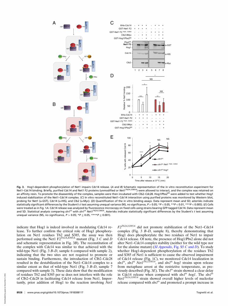
indicate that Hog1 is indeed involved in modulating Cdc14 re-lease. To further confirm the critical role of Hog1 phosphory-lation on Net1 residues T62 and S385, the assay was thenperformed using the Net1 F2T62A,S385A mutant (Fig. 3 C and Dand schematic representation in Fig. 3B). The reconstitution ofthe complex with Cdc14 was similar to that achieved with thewild-type Net1 (Fig. 3 B–D, sample 6 compared with sample 2),indicating that the two sites are not required to promote orsustain binding. Furthermore, the introduction of Clb2–Cdc28resulted in the destabilization of the Net1–Cdc14 complex to asimilar extent as that of wild-type Net1 (Fig. 3 B–D, sample 7compared with sample 3). These data show that the modificationof residues T62 and S385 per se does not interfere with the roleof Clb2–Cdc28 in facilitating Cdc14 release from Net1. Impor-tantly, prior addition of Hog1 to the reaction involving Net1
F2T62A,S385A did not promote stabilization of the Net1–Cdc14complex (Fig. 3 B–D, sample 8), thereby demonstrating thatHog1 does phosphorylate the two residues of Net1 to impairCdc14 release. Of note, the presence of Hog1/Pbs2 alone did notalter Net1–Cdc14 complex stability (neither for the wild type norfor the alanine mutant) (SI Appendix, Fig. S5 C and D). To studywhether Hog1-dependent phosphorylation of the residues T62and S385 of Net1 is sufficient to cause the observed impairmentof Cdc14 release (Fig. 1C), we monitored Cdc14 localization insln1ts, sln1ts Net1T62,S385A, and sln1ts hog1 strains upon releasefrom metaphase arrest at the restrictive temperature, as pre-viously described (Fig. 3E). The sln1ts strain showed a clear delayin Cdc14 release when compared with sln1ts hog1. The sln1ts
Net1T62A,S385A strain showed overall higher levels of nucleolarrelease compared with sln1ts and promoted a prompt increase in
D
E
0
20
40
60
80
100
0 5 10 15 20
% c
ells
with
rele
ased
Cdc
14
Time after release at 37°C (min)
sln1ts
sln1ts Net1 T62A, S385A
sln1ts hog1
****
*NS NS
6His-Cdc14 - + ++ - + ++
GST-Hog1/Pbs2EE - - +- - - +-Clb2-9Myc - - ++ - - ++
Net1 F2 -Pbs2EE -
Hog1 -
Cdc14 -
Clb2 -
α-GST
α-HIS
α-Myc
GST-Net1 F2 + + ++ - - --GST-Net1 F2 T62A, S385A + + ++- - --
1 2 43 5 6 87
CA
B
0
50
100
150
200
Rel
ativ
e%
Net
1 bi
ndin
g
***
*
**
NS
NS*
6His-Cdc14 - + ++ - + ++
GST-Hog1/Pbs2EE - - +- - - +-Clb2-9Myc - - ++ - - ++
GST-Net1 F2 + + ++ - - --GST-Net1 F2 T62A, S385A + + ++- - --
Fig. 3. Hog1-dependent phosphorylation of Net1 impairs Cdc14 release. (A and B) Schematic representation of the in vitro reconstitution experiment forNet1–Cdc14 binding. Briefly, purified Cdc14 and Net1 F2 proteins (unmodified or Net1T62A,S385A) were allowed to interact, and the complex was retained onan affinity resin. To promote the disassembly of the complex, samples were then incubated with Clb2–Cdc28. Hog1/Pbs2EE were added to test whether Hog1induced stabilization of the Net1–Cdc14 complex. (C) In vitro reconstituted Net1–Cdc14 interaction using purified proteins was monitored by Western blot,probing for Net1 (α-GST), Cdc14 (α-HIS), and Clb2 (α-Myc). (D) Quantification of the in vitro binding assays. Data represent mean and SD; asterisks indicatestatistically significant differences by the Student’s t test assuming unequal variance (NS, no significance, P > 0.05; *P < 0.05, **P < 0.01, ***P < 0.005). (E) Cellswere treated as in Fig. 1A. Cdc14 release was analyzed by fluorescence microscopy on fixed cells using strains bearing GFP-tagged Cdc14. Data represent meanand SD. Statistical analysis comparing sln1ts with sln1ts Net1T62A,S385A. Asterisks indicate statistically significant differences by the Student’s t test assumingunequal variance (NS, no significance, P > 0.05; *P ≤ 0.05, ****P ≤ 0.001).
8928 | www.pnas.org/cgi/doi/10.1073/pnas.1918308117 Tognetti et al.
Dow
nloa
ded
by g
uest
on
Dec
embe
r 6,
202
1
Cdc14 release already at 5 min after exit from metaphase arrest,as occurred in the sln1ts hog1 strain.Altogether, these data indicate that Hog1-dependent phos-
phorylation of Net1 is required for the stabilization of Cdc14binding and thus the prevention of Cdc14 release.
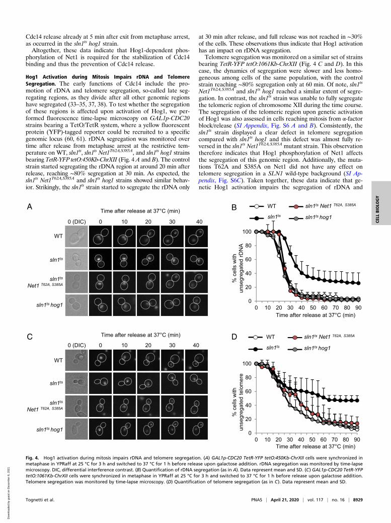
Hog1 Activation during Mitosis Impairs rDNA and TelomereSegregation. The early functions of Cdc14 include the pro-motion of rDNA and telomere segregation, so-called late seg-regating regions, as they divide after all other genomic regionshave segregated (33–35, 37, 38). To test whether the segregationof these regions is affected upon activation of Hog1, we per-formed fluorescence time-lapse microscopy on GAL1p-CDC20strains bearing a TetO/TetR system, where a yellow fluorescentprotein (YFP)-tagged reporter could be recruited to a specificgenomic locus (60, 61). rDNA segregation was monitored overtime after release from metaphase arrest at the restrictive tem-perature on WT, sln1ts, sln1ts Net1T62A,S385A, and sln1ts hog1 strainsbearing TetR-YFP tetO:450Kb-ChrXII (Fig. 4 A and B). The controlstrain started segregating the rDNA region at around 20 min afterrelease, reaching ∼80% segregation at 30 min. As expected, thesln1ts Net1T62A,S385A and sln1ts hog1 strains showed similar behav-ior. Strikingly, the sln1ts strain started to segregate the rDNA only
at 30 min after release, and full release was not reached in ∼30%of the cells. These observations thus indicate that Hog1 activationhas an impact on rDNA segregation.Telomere segregation was monitored on a similar set of strains
bearing TetR-YFP tetO:1061Kb-ChrXII (Fig. 4 C and D). In thiscase, the dynamics of segregation were slower and less homo-geneous among cells of the same population, with the controlstrain reaching ∼80% segregation only at 60 min. Of note, sln1ts
Net1T62A,S385A and sln1ts hog1 reached a similar extent of segre-gation. In contrast, the sln1ts strain was unable to fully segregatethe telomeric region of chromosome XII during the time course.The segregation of the telomeric region upon genetic activationof Hog1 was also assessed in cells reaching mitosis from α-factorblock/release (SI Appendix, Fig. S6 A and B). Consistently, thesln1ts strain displayed a clear defect in telomere segregationcompared with sln1ts hog1 and this defect was almost fully re-versed in the sln1ts Net1T62A,S385A mutant strain. This observationtherefore indicates that Hog1 phosphorylation of Net1 affectsthe segregation of this genomic region. Additionally, the muta-tions T62A and S385A on Net1 did not have any effect ontelomere segregation in a SLN1 wild-type background (SI Ap-pendix, Fig. S6C). Taken together, these data indicate that ge-netic Hog1 activation impairs the segregation of rDNA and
40
sln1ts
Net1 T62A, S385A
Time after release at 37°C (min)
0 (DIC) 0 10 20 30
WT
sln1ts
sln1ts hog1
BA
DC Time after release at 37°C (min)
0 (DIC) 0 10 20 30 40
WT
sln1ts
sln1ts
Net1 T62A, S385A
sln1ts hog1
0
20
40
60
80
100
0 10 20 30 40 50 60 70 80 90
WT
sln1ts
sln1ts Net1 T62A, S385A
sln1ts hog1
Time after release at 37°C (min)
% c
ells
with
unse
greg
ated
rDN
A
Time after release at 37°C (min)
% c
ells
with
unse
greg
ated
telo
mer
e
0
20
40
60
80
100
0 10 20 30 40 50 60 70 80 90
WT
sln1ts
sln1ts Net1 T62A, S385A
sln1ts hog1
Fig. 4. Hog1 activation during mitosis impairs rDNA and telomere segregation. (A) GAL1p-CDC20 TetR-YFP tetO:450Kb-ChrXII cells were synchronized inmetaphase in YPRaff at 25 °C for 3 h and switched to 37 °C for 1 h before release upon galactose addition. rDNA segregation was monitored by time-lapsemicroscopy. DIC, differential interference contrast. (B) Quantification of rDNA segregation (as in A). Data represent mean and SD. (C) GAL1p-CDC20 TetR-YFPtetO:1061Kb-ChrXII cells were synchronized in metaphase in YPRaff at 25 °C for 3 h and switched to 37 °C for 1 h before release upon galactose addition.Telomere segregation was monitored by time-lapse microscopy. (D) Quantification of telomere segregation (as in C). Data represent mean and SD.
Tognetti et al. PNAS | April 21, 2020 | vol. 117 | no. 16 | 8929
CELL
BIOLO
GY
Dow
nloa
ded
by g
uest
on
Dec
embe
r 6,
202
1
telomeres. To measure whether these segregation defects com-promise genomic integrity, we measured Rad52-YFP foci toassess the presence of recombination foci. Net1T62A,S385A cellsdisplayed significantly higher levels of Rad52 foci compared withNet1 wild-type cells (SI Appendix, Fig. S6D). Finally, to assesswhether segregation defects could ultimately affect bud-neckstructures, we monitored by time-lapse microscopy the localiza-tion of Myo1, a protein involved in bud-neck morphogenesis andcytokinesis (SI Appendix, Fig. S6 E and F). The sln1ts strain re-leased at the restrictive temperature displayed a clear defect inMyo1 localization compared with the WT. This delay in theactomyosin ring disassembly could be accountable for thepronounced defect, in contrast with the delay observed inCdc14 release, in cell-cycle completion observed upon Hog1activation.
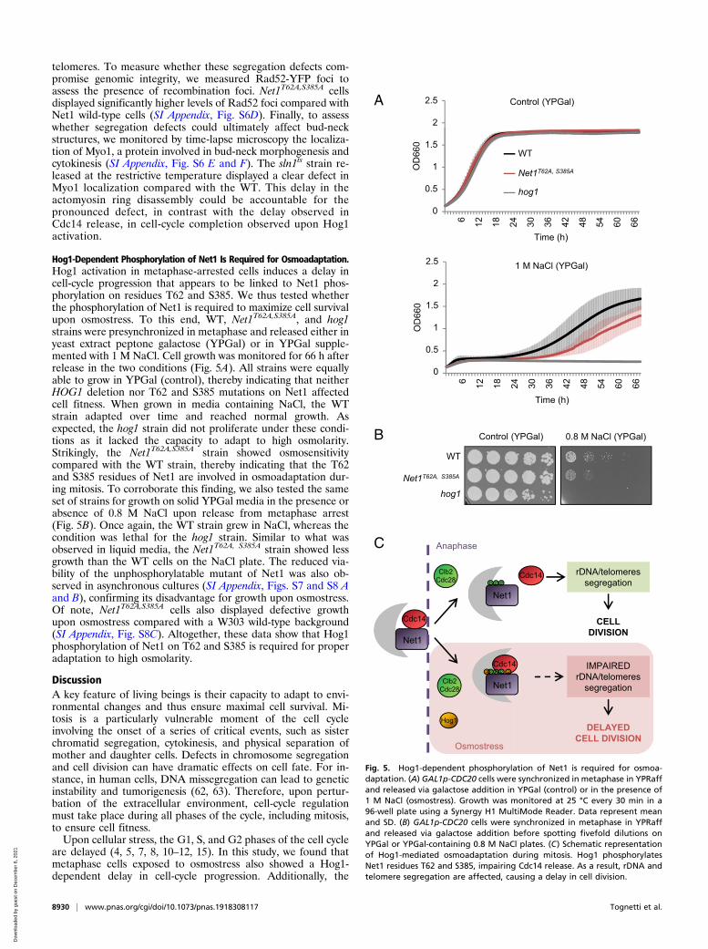
Hog1-Dependent Phosphorylation of Net1 Is Required for Osmoadaptation.Hog1 activation in metaphase-arrested cells induces a delay incell-cycle progression that appears to be linked to Net1 phos-phorylation on residues T62 and S385. We thus tested whetherthe phosphorylation of Net1 is required to maximize cell survivalupon osmostress. To this end, WT, Net1T62A,S385A, and hog1strains were presynchronized in metaphase and released either inyeast extract peptone galactose (YPGal) or in YPGal supple-mented with 1 M NaCl. Cell growth was monitored for 66 h afterrelease in the two conditions (Fig. 5A). All strains were equallyable to grow in YPGal (control), thereby indicating that neitherHOG1 deletion nor T62 and S385 mutations on Net1 affectedcell fitness. When grown in media containing NaCl, the WTstrain adapted over time and reached normal growth. Asexpected, the hog1 strain did not proliferate under these condi-tions as it lacked the capacity to adapt to high osmolarity.Strikingly, the Net1T62A,S385A strain showed osmosensitivitycompared with the WT strain, thereby indicating that the T62and S385 residues of Net1 are involved in osmoadaptation dur-ing mitosis. To corroborate this finding, we also tested the sameset of strains for growth on solid YPGal media in the presence orabsence of 0.8 M NaCl upon release from metaphase arrest(Fig. 5B). Once again, the WT strain grew in NaCl, whereas thecondition was lethal for the hog1 strain. Similar to what wasobserved in liquid media, the Net1T62A, S385A strain showed lessgrowth than the WT cells on the NaCl plate. The reduced via-bility of the unphosphorylatable mutant of Net1 was also ob-served in asynchronous cultures (SI Appendix, Figs. S7 and S8 Aand B), confirming its disadvantage for growth upon osmostress.Of note, Net1T62A,S385A cells also displayed defective growthupon osmostress compared with a W303 wild-type background(SI Appendix, Fig. S8C). Altogether, these data show that Hog1phosphorylation of Net1 on T62 and S385 is required for properadaptation to high osmolarity.
DiscussionA key feature of living beings is their capacity to adapt to envi-ronmental changes and thus ensure maximal cell survival. Mi-tosis is a particularly vulnerable moment of the cell cycleinvolving the onset of a series of critical events, such as sisterchromatid segregation, cytokinesis, and physical separation ofmother and daughter cells. Defects in chromosome segregationand cell division can have dramatic effects on cell fate. For in-stance, in human cells, DNA missegregation can lead to geneticinstability and tumorigenesis (62, 63). Therefore, upon pertur-bation of the extracellular environment, cell-cycle regulationmust take place during all phases of the cycle, including mitosis,to ensure cell fitness.Upon cellular stress, the G1, S, and G2 phases of the cell cycle
are delayed (4, 5, 7, 8, 10–12, 15). In this study, we found thatmetaphase cells exposed to osmostress also showed a Hog1-dependent delay in cell-cycle progression. Additionally, the
A
B
C
Hog1
Clb2Cdc28
Osmostress
Cdc14
Net1
Cdc14
Net1
Cdc14
Net1P
Clb2Cdc28
Anaphase
IMPAIREDrDNA/telomeres
segregation
rDNA/telomeressegregation
CELLDIVISION
DELAYEDCELL DIVISION
PP
P P P P P
6 12 18 24 30 36 42 48 54 60 66
OD
660
Time (h)
0
0.5
1
1.5
2
2.5
6 12 18 24 30 36 42 48 54 60 66
OD
660
Time (h)
1 M NaCl (YPGal)
2
1
0.5
2.5
0
1.5
Control (YPGal)
2
1
0.5
2.5
0
1.5WT
Net1T62A, S385A
hog1
0.8 M NaCl (YPGal)Control (YPGal)
WT
hog1
Net1T62A, S385A
Fig. 5. Hog1-dependent phosphorylation of Net1 is required for osmoa-daptation. (A) GAL1p-CDC20 cells were synchronized in metaphase in YPRaffand released via galactose addition in YPGal (control) or in the presence of1 M NaCl (osmostress). Growth was monitored at 25 °C every 30 min in a96-well plate using a Synergy H1 MultiMode Reader. Data represent meanand SD. (B) GAL1p-CDC20 cells were synchronized in metaphase in YPRaffand released via galactose addition before spotting fivefold dilutions onYPGal or YPGal-containing 0.8 M NaCl plates. (C) Schematic representationof Hog1-mediated osmoadaptation during mitosis. Hog1 phosphorylatesNet1 residues T62 and S385, impairing Cdc14 release. As a result, rDNA andtelomere segregation are affected, causing a delay in cell division.
8930 | www.pnas.org/cgi/doi/10.1073/pnas.1918308117 Tognetti et al.
Dow
nloa
ded
by g
uest
on
Dec
embe
r 6,
202
1
dynamics of Cdc14 release from the nucleolus were delayedupon genetic activation of the kinase Hog1. Cdc14 is a masterregulator of mitosis and its localization-dependent activation iscrucial for cell-cycle completion (reviewed in refs. 39 and 64).Cdc14 nucleolar release during anaphase is promoted byClb2–Cdc28–dependent phosphorylation on the N-terminal re-gion of Net1 (22, 27). We therefore hypothesized that Hog1alters the Net1–Cdc14 complex association via phosphorylationof one of the two proteins. We found that, at least in vitro, Hog1phosphorylated Net1 on residues T62 and S385, which are inclose proximity to the Cdc14-binding domain (59). Consistently,in vitro reconstitution of the Net1–Cdc14 interaction indicatedthat Hog1 interferes with the ability of Clb2–Cdc28 to promotedisruption of the complex. In an unperturbed cell cycle, timelyregulated accumulation of Clb2–Cdc28 facilitates Cdc14 release,which in turn promotes dephosphorylation of a number of tar-gets and segregation of late genomic regions to finally allow forcell division. We speculate that, upon activation during mitosis,Hog1 phosphorylates Net1 on residues T62 and S385 and, by thismeans, transiently increases Net1 affinity for Cdc14 renderingthe Net1–Cdc14 complex more resistant to dissolution uponClb2–Cdc28 phosphorylation. This event affects Cdc14 timelyrelease and in turn rDNA/telomere segregation, ultimatelydelaying cell division (see the schematic diagram in Fig. 5C). Wetherefore propose that cells execute a program to ensureosmoadaptation during early stages of mitosis via Hog1-dependent phosphorylation of Net1 on residues T62 and S385.An important question regards the biological reason un-
derlying a transient arrest of cell-cycle progression during theearly stages of mitosis upon osmostress. Previously reportedosmoadaptive responses that led to a delay in the transitionthrough other cell-cycle phases could be explained by the needfor the cell to prevent or regulate DNA replication (G1 and Sphase, respectively) and postpone mitosis (G2) to avoid sub-optimal execution of these crucial steps while adapting to envi-ronmental change. When cells are forced to adapt to osmostresswhile already committed to division, as occurs in metaphase, theymight sustain the osmoadaptive program to maximize survival.Therefore, a delay in mitosis might be required in order toprevent defects in DNA segregation. In fact, the Net1 mutantthat failed to delay Cdc14 release became osmosensitive. Al-though Cdc14 requirement and functions appear to differ slightlybetween organisms, all living beings most likely require the de-phosphorylation of Cdk substrates to end a cell cycle and enter anew G1 phase. Of note, human Cdc14B rescues cdc14 lethality inbudding and fission yeast, thereby suggesting functional conser-vation (65, 66). Interestingly, disruption of the Cdc14B locus inhuman cells causes an increased incidence of rDNA anaphasebridges, thus pointing to a defect in segregation, despite no ap-parent impairment of cell-cycle progression (67). Given the in-creased complexity of human compared with budding yeast cells,a scenario in which additional factors complement or facilitateCdc14 functions can be envisioned.Budding yeast Cdc14 promotes the dephosphorylation of
many proteins that are phosphorylated by cyclin-dependent ki-nases throughout the cell cycle (reviewed in refs. 42–44 and 56).It has been proposed that, in addition to Cdc14 activation, theClb2–Cdc14 balance is important for execution of mitotic exitevents (68, 69). Along this line, even nondramatic changes in thetiming or in the number of Cdc14 molecules released can havenoticeable effects on cell-cycle progression. Among the mostextensively studied effects of Cdc14 release is the promotion ofrDNA and telomere segregation via inhibition of transcriptionfor completion of the cell cycle (33–35, 38). Consistently, here wereport that Hog1 activation during metaphase not only causes animpairment in Cdc14 nucleolar release but is also linked to adefect in the timely segregation of the late segregating regions.This finding suggests that these cells do not have fully divided
nuclei but might instead arrest with residual genomic materialbelonging to the late segregating regions stretched in-betweenmostly separated nuclei. Additionally, studies that analyzed thephenotype of thermosensitive mutants of Cdc14 (cdc14-1 andcdc14-3) at the restrictive temperature reported cell-cycle arrestin late anaphase/telophase, with generally separated nuclearmasses but unsegregated telomeres and rDNAs (34, 35, 60). Thephenotype observed in the event of Hog1 genetic activationduring metaphase, with initial impairment of the timely releaseof nucleolar Cdc14 and cells arrested in late mitosis, thereforeresembles that reported in the literature for thermosensitiveCdc14 mutants. In contrast, the delay in metaphase observed inthe present study differs from what was observed when mitoticcells bearing MEN mutants were driven to exit mitosis in re-sponse to hyperosmotic stress (14). We propose that this ap-parent discrepancy lies in the differential biochemical state inwhich cells encounter stress. MEN mutant cells should arrest inanaphase, presumably after Cdc14 has already been transientlyreleased from the nucleolus into the nucleus as a function ofFEAR activation (reviewed in refs. 18, 19, and 26). This condi-tion is strikingly different from the metaphase arrest/releaseanalyzed in our study, in which cells still have Cdc14 bound toNet1 and sequestered into the nucleolus. Under the conditionsanalyzed in their study (14), Hog1 might overrule the MEN re-quirement for Cdc14 release in order to maximize survival. Ac-cordingly, Reiser and colleagues (14) found that the hypertonicenvironment per se does not accelerate exit from mitosis andthat cells treated with sorbitol progressed through mitosis lessefficiently than untreated cells. These data are fully consistentwith our observations that mitotic exit is delayed in cells that facean osmotic shock before or during metaphase (with Cdc14 stillsequestered in the nucleolus) and that Net1 is involved in theprocess of osmoadaptation. However, we find plausible thatHog1 may phosphorylate targets other than Net1 to modulatecell-cycle progression in mitosis.At present, we also cannot rule out that the direct action of
Cdc14 on promoting segregation of the late segregating regionsis paired with additional functions of the phosphatase on otherfactors required for successful mitotic exit (i.e., Aurora B, asreported in ref. 35). On the other hand, upon hyperosmoticshock, Hog1-dependent phosphorylation of Net1 could not onlyimpair Cdc14 release but also alter other functions of Cdc14 orNet1, functions that could be related to the stability of the nu-cleolus and rDNA silencing, or to additional activities of thephosphatase Cdc14. In the absence of additional evidence, wespeculate that the Cdc14-related defect in full timely sisterchromatid segregation could be reflected in a delay in organizedseptum formation by physical means, having unsegregatedstretches of DNA lying in-between mother and daughter cellsand ultimately obstructing proper septum formation. The exactmechanisms by which Cdc14 release, together with rDNA andtelomere segregation defects, mediates the delay in cell-cycleprogression deserves further attention.In summary, here we report that activated Hog1 phosphory-
lates Net1, thus impairing Cdc14 nucleolar release and rDNAand telomere segregation, ultimately delaying cell-cycle pro-gression. Here we identify a regulatory mechanism, throughphosphorylation of Net1, by which cells maximize survival via theregulation of mitosis upon environmental changes.
Materials and MethodsSynchronization of GAL1p-CDC20 cells in all experiments was achieved byincubating exponential cells in yeast extract peptone raffinose (YPRaff) for 2to 4 h and released by inducing Cdc20 expression by galactose addition (1%).Activation of the HOG pathway was achieved by incubating cells at the in-dicated NaCl concentration or by genetic means using the sln1ts allele at37 °C. For immunofluorescence (IF) and DAPI staining experiments, cellswere fixed overnight with 3.7% formaldehyde in IF buffer (0.1 M K2HPO4,
Tognetti et al. PNAS | April 21, 2020 | vol. 117 | no. 16 | 8931
CELL
BIOLO
GY
Dow
nloa
ded
by g
uest
on
Dec
embe
r 6,
202
1

0.1 M KH2PO4, 0.5 mM MgCl2, pH 6.4). Cells were then washed twice with IFbuffer and once with sorbitol buffer (0.1 M K2HPO4, 0.1 M KH2PO4, 0.5 mMMgCl2, 1.2 M sorbitol, pH 7.4). Each sample was digested in sorbitol bufferwith 2 μL β-mercaptoethanol and 40 μg/μL zymolase T100 (AttendBio) for30 min at 30 °C. For Cdc14 localization analysis, samples were collected every5 min after release at 37 °C and fixed for 10 min on ice with 3.7% formal-dehyde in phosphate-buffered saline (PBS). Cells were spun down, resus-pended with a solution of 1% formaldehyde in PBS, and incubated for10 min before extensive washes with 40 mM Tris (pH 8.8). For rDNA andtelomere segregation analysis in metaphase-arrested cultures, cells weresynchronized at the onset of metaphase, switched to 37 °C, and let attach toeight-well chambers pretreated with Con A, and unattached cells werewashed away before release by galactose addition. For α-factor synchroni-zation, cultures were arrested with 20 μg/mL α-factor for 3 to 4 h at 25 °Cand released at 25 °C for 60 min before switching to 37 °C to promote ge-netic activation of Hog1 in mitosis. During this time, cells were let attach asbefore. For Myo1 localization analysis, cells were synchronized at the onsetof metaphase, switched to 37 °C, and let attach to eight-well chamberspretreated with Con A, and unattached cells were washed away beforeanalysis. In either case, images were acquired every 10 min for at least90 min at 37 °C with a 100× oil objective lens in the YFP or Cherry spectrum.Analysis of electromobility shifts in Phos-tag gels was achieved in a 6%polyacrylamide gel supplemented with 10 μM Phos-tag (Wako) and 20 μMMnCl2 for 3 h at 100 V on ice. Basic Hog1 kinase assays were performed using1 μg of GST–Hog1 activated with 0.5 μg of GST–Pbs2EE in the presence of
kinase buffer (SI Appendix) and 100 μM ATP. After 30 min at 30 °C, 2 μg ofGST–Cdc14, GST–Net1, GST–Net1 F2, and GST–Net1 F2T62A,S385A was added tothe Hog1/Pbs2EE mixture together with [32P]ATP (0.1 mCi/mL; PerkinElmer)and incubated for 30 min at 30 °C. Full lists of reagents, strains, and plasmidsand detailed protocols used in this article are provided in SI Appendix,Methods.
Data Availability. All data discussed in this study are included in the main textand SI Appendix.
ACKNOWLEDGMENTS. We thank Dr. Josep Clotet and Dr. Jordi Torres-Rosellfor strains; and Dr. Berta Canal and Dr. Gerhard Seisenbacher for helpfuldiscussions and suggestions. S.T. was the recipient of a contract forInvestigador Doctor Junior (PDJ 2014, Generalitat de Catalunya) and a Juande la Cierva fellowship. The study was supported by grants from the SpanishMinistry of Economy and Competitiveness (PGC2018-094136-B-I00 to F.P.;BFU2017-85152-P and FEDER to E.d.N.; BFU2016-77975-R and FEDER to E.Q.),the Catalan Government (2017 SGR 799), and Fundación Botín, by BancoSantander through its Santander Universities Global Division (to F.P.). Wegratefully acknowledge institutional funding from the Spanish Ministry ofEconomy, Industry and Competitiveness (MINECO) through the Centres ofExcellence Severo Ochoa Award, and from the CERCA Programme of theCatalan Government and the Unidad de Excelencia María de Maeztu,funded by the AEI (CEX2018-000792-M). F.P. is the recipient of an ICREAAcadèmia Award (Generalitat de Catalunya).
1. A. Duch, E. de Nadal, F. Posas, The p38 and Hog1 SAPKs control cell cycle progression
in response to environmental stresses. FEBS Lett. 586, 2925–2931 (2012).2. E. de Nadal, G. Ammerer, F. Posas, Controlling gene expression in response to stress.
Nat. Rev. Genet. 12, 833–845 (2011).3. H. Saito, F. Posas, Response to hyperosmotic stress. Genetics 192, 289–318 (2012).4. M. A. Adrover et al., Time-dependent quantitative multicomponent control of the G1-
S network by the stress-activated protein kinase Hog1 upon osmostress. Sci. Signal. 4,
ra63 (2011).5. M. R. Alexander et al., Regulation of cell cycle progression by Swe1p and Hog1p
following hypertonic stress. Mol. Biol. Cell 12, 53–62 (2001).6. Y. L. Chang et al., Yeast Cip1 is activated by environmental stress to inhibit Cdk1-G1
cyclins via Mcm1 and Msn2/4. Nat. Commun. 8, 56 (2017).7. J. Clotet et al., Phosphorylation of Hsl1 by Hog1 leads to a G2 arrest essential for cell
survival at high osmolarity. EMBO J. 25, 2338–2346 (2006).8. A. Duch et al., Coordinated control of replication and transcription by a SAPK protects
genomic integrity. Nature 493, 116–119 (2013).9. A. Duch et al., Multiple signaling kinases target Mrc1 to prevent genomic instability
triggered by transcription-replication conflicts. Nat. Commun. 9, 379 (2018).10. X. Escoté, M. Zapater, J. Clotet, F. Posas, Hog1 mediates cell-cycle arrest in G1 phase
by the dual targeting of Sic1. Nat. Cell Biol. 6, 997–1002 (2004).11. A. González-Novo et al., Hog1 targets Whi5 and Msa1 transcription factors to
downregulate cyclin expression upon stress. Mol. Cell. Biol. 35, 1606–1618 (2015).12. M. Nadal-Ribelles et al., Control of Cdc28 CDK1 by a stress-induced lncRNA. Mol. Cell
53, 549–561 (2014).13. E. Radmaneshfar et al., From START to FINISH: The influence of osmotic stress on the
cell cycle. PLoS One 8, e68067 (2013).14. V. Reiser, K. E. D’Aquino, L. S. Ee, A. Amon, The stress-activated mitogen-activated
protein kinase signaling cascade promotes exit from mitosis. Mol. Biol. Cell 17, 3136–
3146 (2006).15. G. Yaakov et al., The stress-activated protein kinase Hog1 mediates S phase delay in
response to osmostress. Mol. Biol. Cell 20, 3572–3582 (2009).16. G. Bellí, E. Garí, M. Aldea, E. Herrero, Osmotic stress causes a G1 cell cycle delay and
downregulation of Cln3/Cdc28 activity in Saccharomyces cerevisiae. Mol. Microbiol.
39, 1022–1035 (2001).17. K. King, H. Kang, M. Jin, D. J. Lew, Feedback control of Swe1p degradation in the
yeast morphogenesis checkpoint. Mol. Biol. Cell 24, 914–922 (2013).18. A. Mocciaro, E. Schiebel, Cdc14: A highly conserved family of phosphatases with non-
conserved functions? J. Cell Sci. 123, 2867–2876 (2010).19. E. Queralt, F. Uhlmann, Cdk-counteracting phosphatases unlock mitotic exit. Curr.
Opin. Cell Biol. 20, 661–668 (2008).20. W. Shou et al., Exit from mitosis is triggered by Tem1-dependent release of the
protein phosphatase Cdc14 from nucleolar RENT complex. Cell 97, 233–244 (1999).21. R. Visintin, E. S. Hwang, A. Amon, Cfi1 prevents premature exit from mitosis by an-
choring Cdc14 phosphatase in the nucleolus. Nature 398, 818–823 (1999).22. R. Azzam et al., Phosphorylation by cyclin B-Cdk underlies release of mitotic exit
activator Cdc14 from the nucleolus. Science 305, 516–519 (2004).23. W. Shou et al., Cdc5 influences phosphorylation of Net1 and disassembly of the RENT
complex. BMC Mol. Biol. 3, 3 (2002).24. S. Yoshida, A. Toh-e, Budding yeast Cdc5 phosphorylates Net1 and assists Cdc14 re-
lease from the nucleolus. Biochem. Biophys. Res. Commun. 294, 687–691 (2002).25. R. Visintin, F. Stegmeier, A. Amon, The role of the polo kinase Cdc5 in controlling
Cdc14 localization. Mol. Biol. Cell 14, 4486–4498 (2003).26. J. M. Rock, A. Amon, The FEAR network. Curr. Biol. 19, R1063–R1068 (2009).
27. E. Queralt, C. Lehane, B. Novak, F. Uhlmann, Downregulation of PP2A(Cdc55) phos-phatase by separase initiates mitotic exit in budding yeast. Cell 125, 719–732 (2006).
28. L. J. Holt et al., Global analysis of Cdk1 substrate phosphorylation sites provides in-sights into evolution. Science 325, 1682–1686 (2009).
29. A. I. de Los Santos-Velázquez, I. G. de Oya, J. Manzano-López, F. Monje-Casas, LaterDNA condensation ensures timely Cdc14 release and coordination of mitotic exitsignaling with nucleolar segregation. Curr. Biol. 27, 3248–3263.e5 (2017).
30. T. Higuchi, F. Uhlmann, Stabilization of microtubule dynamics at anaphase onsetpromotes chromosome segregation. Nature 433, 171–176 (2005).
31. M. Roccuzzo, C. Visintin, F. Tili, R. Visintin, FEAR-mediated activation of Cdc14 is thelimiting step for spindle elongation and anaphase progression. Nat. Cell Biol. 17, 251–261 (2015).
32. K. E. Ross, O. Cohen-Fix, A role for the FEAR pathway in nuclear positioning duringanaphase. Dev. Cell 6, 729–735 (2004).
33. A. Clemente-Blanco et al., Cdc14 inhibits transcription by RNA polymerase I duringanaphase. Nature 458, 219–222 (2009).
34. D. D’Amours, F. Stegmeier, A. Amon, Cdc14 and condensin control the dissolution ofcohesin-independent chromosome linkages at repeated DNA. Cell 117, 455–469(2004).
35. M. Sullivan, T. Higuchi, V. L. Katis, F. Uhlmann, Cdc14 phosphatase induces rDNAcondensation and resolves cohesin-independent cohesion during budding yeastanaphase. Cell 117, 471–482 (2004).
36. J. Torres-Rosell, F. Machín, A. Jarmuz, L. Aragón, Nucleolar segregation lags behindthe rest of the genome and requires Cdc14p activation by the FEAR network. CellCycle 3, 496–502 (2004).
37. A. Clemente-Blanco et al., Cdc14 phosphatase promotes segregation of telomeresthrough repression of RNA polymerase II transcription. Nat. Cell Biol. 13, 1450–1456(2011).
38. J. Torres-Rosell, F. Machín, L. Aragón, Cdc14 and the temporal coordination betweenmitotic exit and chromosome segregation. Cell Cycle 4, 109–112 (2005).
39. B. Baro, E. Queralt, F. Monje-Casas, Regulation of mitotic exit in Saccharomyces cer-evisiae. Methods Mol. Biol. 1505, 3–17 (2017).
40. M. Segal, Mitotic exit control: A space and time odyssey. Curr. Biol. 21, R857–R859(2011).
41. D. A. Mohl, M. J. Huddleston, T. S. Collingwood, R. S. Annan, R. J. Deshaies, Dbf2-Mob1 drives relocalization of protein phosphatase Cdc14 to the cytoplasm during exitfrom mitosis. J. Cell Biol. 184, 527–539 (2009).
42. J. Bloom et al., Global analysis of Cdc14 phosphatase reveals diverse roles in mitoticprocesses. J. Biol. Chem. 286, 5434–5445 (2011).
43. L. Kao et al., Global analysis of Cdc14 dephosphorylation sites reveals essential reg-ulatory role in mitosis and cytokinesis. Mol. Cell. Proteomics 13, 594–605 (2014).
44. T. Kuilman et al., Identification of Cdk targets that control cytokinesis. EMBO J. 34,81–96 (2015).
45. R. Visintin et al., The phosphatase Cdc14 triggers mitotic exit by reversal of Cdk-dependent phosphorylation. Mol. Cell 2, 709–718 (1998).
46. J. Brace, J. Hsu, E. L. Weiss, Mitotic exit control of the Saccharomyces cerevisiae Ndr/LATS kinase Cbk1 regulates daughter cell separation after cytokinesis. Mol. Cell. Biol.31, 721–735 (2011).
47. P. R. Dohrmann et al., Parallel pathways of gene regulation: Homologous regulatorsSWI5 and ACE2 differentially control transcription of HO and chitinase. Genes Dev. 6,93–104 (1992).
48. M. A. Juanes, S. Piatti, The final cut: Cell polarity meets cytokinesis at the bud neck inS. cerevisiae. Cell. Mol. Life Sci. 73, 3115–3136 (2016).
8932 | www.pnas.org/cgi/doi/10.1073/pnas.1918308117 Tognetti et al.
Dow
nloa
ded
by g
uest
on
Dec
embe
r 6,
202
1
49. F. Meitinger, S. Palani, G. Pereira, The power of MEN in cytokinesis. Cell Cycle 11, 219–228 (2012).
50. E. L. Weiss, Mitotic exit and separation of mother and daughter cells. Genetics 192,1165–1202 (2012).
51. R. Visintin, S. Prinz, A. Amon, CDC20 and CDH1: A family of substrate-specific acti-vators of APC-dependent proteolysis. Science 278, 460–463 (1997).
52. S. Prinz, E. S. Hwang, R. Visintin, A. Amon, The regulation of Cdc20 proteolysis revealsa role for APC components Cdc23 and Cdc27 during S phase and early mitosis. Curr.Biol. 8, 750–760 (1998).
53. X. Mao, Y. Hu, C. Liang, C. Lu, MET3 promoter: A tightly regulated promoter and itsapplication in construction of conditional lethal strain. Curr. Microbiol. 45, 37–40(2002).
54. T. Maeda, S. M. Wurgler-Murphy, H. Saito, A two-component system that regulatesan osmosensing MAP kinase cascade in yeast. Nature 369, 242–245 (1994).
55. F. Posas et al., Yeast HOG1 MAP kinase cascade is regulated by a multistep phos-phorelay mechanism in the SLN1-YPD1-SSK1 “two-component” osmosensor. Cell 86,865–875 (1996).
56. F. Stegmeier, A. Amon, Closing mitosis: The functions of the Cdc14 phosphatase andits regulation. Annu. Rev. Genet. 38, 203–232 (2004).
57. M. Sullivan, D. O. Morgan, Finishing mitosis, one step at a time. Nat. Rev. Mol. CellBiol. 8, 894–903 (2007).
58. B. Soufi et al., Global analysis of the yeast osmotic stress response by quantitativeproteomics. Mol. Biosyst. 5, 1337–1346 (2009).
59. E. E. Traverso et al., Characterization of the Net1 cell cycle-dependent regulator ofthe Cdc14 phosphatase from budding yeast. J. Biol. Chem. 276, 21924–21931 (2001).
60. F. Machín, J. Torres-Rosell, A. Jarmuz, L. Aragón, Spindle-independent condensation-mediated segregation of yeast ribosomal DNA in late anaphase. J. Cell Biol. 168, 209–219 (2005).
61. C. Michaelis, R. Ciosk, K. Nasmyth, Cohesins: Chromosomal proteins that preventpremature separation of sister chromatids. Cell 91, 35–45 (1997).
62. P. V. Jallepalli, C. Lengauer, Chromosome segregation and cancer: Cutting throughthe mystery. Nat. Rev. Cancer 1, 109–117 (2001).
63. B. N. Zhang, A. Bueno Venegas, I. D. Hickson, W. K. Chu, DNA replication stress and itsimpact on chromosome segregation and tumorigenesis. Semin. Cancer Biol. 55, 61–69(2019).
64. F. Machín, O. Quevedo, C. Ramos-Pérez, J. García-Luis, Cdc14 phosphatase: Warning,no delay allowed for chromosome segregation! Curr. Genet. 62, 7–13 (2016).
65. L. Li, B. R. Ernsting, M. J. Wishart, D. L. Lohse, J. E. Dixon, A family of putative tumorsuppressors is structurally and functionally conserved in humans and yeast. J. Biol.Chem. 272, 29403–29406 (1997).
66. M. D. Vázquez-Novelle, V. Esteban, A. Bueno, M. P. Sacristán, Functional homologyamong human and fission yeast Cdc14 phosphatases. J. Biol. Chem. 280, 29144–29150(2005).
67. E. Berdougo, M. V. Nachury, P. K. Jackson, P. V. Jallepalli, The nucleolar phosphataseCdc14B is dispensable for chromosome segregation and mitotic exit in human cells.Cell Cycle 7, 1184–1190 (2008).
68. C. Bouchoux, F. Uhlmann, A quantitative model for ordered Cdk substrate de-phosphorylation during mitotic exit. Cell 147, 803–814 (2011).
69. B. J. Drapkin, Y. Lu, A. L. Procko, B. L. Timney, F. R. Cross, Analysis of the mitotic exitcontrol system using locked levels of stable mitotic cyclin.Mol. Syst. Biol. 5, 328 (2009).
Tognetti et al. PNAS | April 21, 2020 | vol. 117 | no. 16 | 8933
CELL
BIOLO
GY
Dow
nloa
ded
by g
uest
on
Dec
embe
r 6,
202
1