Embed Size (px)
Citation preview

3
2 HISTORY OF THE TREATMENT AND LITERATURE
2.1 History of the treatment
The first positive results of treatment of diabetic retinopathy with the xenon arc coagulator were presented by Meyer-Schwickerath in 1959, who stated: "In einem Fall, in dem nur Mikroaneurysmen und helle Herde sicht- bar waren, verschwanden die hellen Herde teilweise nach der Koagulation der Mi kroaneurysmen" . During the sixties and early seventies several positive reports were pub1 ished on xenon arc treatment (Larsen 1969, Wessing & Meyer-Schwicke- rath 1969, Wetzig & Jepson 1969, Spalter 1971, among others). Direct coagulation of microvascular lesions was said to have a positive effect on lipid deposits and macular oedema. Destruction of new vessels seemed to reduce the rate of haemorrhages and prevent the severe forms of reti- nopathy and blindness. Extensive photocoagulation of the fundus was seen to be followed by regression of new vessels (Meyer-Schwickerath & Schott 1968, Okun 1968).
Some authors had used a laser for treatment. Aiello et al. (19691, Beet- ham et al. (1970) and Geltzer (1972) used the ruby laser. L'Esperance (1969, 1973) and Zweng et al. (1971) reported on the effect of treatment with the argon laser. The latter was well suited for treatment of vascu- lar lesions owing to the good absorption of its green emission in haemo- globin (L'Esperance 1968).
In the early seventies there was an increasing demand for evaluation of the treatment (Irvine & Norton 1971, Ederer & Hiller 1975). The varying course of the disease and the different therapeutic techniques that had been employed made it difficult to interpret the results. Few studies had included a control material (Okun & Johnston 1969, Beetham et al. 1970, Krill et a1 . 1971, Irvine & Norton 1971, Geltzer 1972, Zetterstrom 1972) and only one had randomized the choice of treatment eye (Patz et al. 1973). Several questions needed to be answered. For example: Did the treatment improve the visual prognosis? Could progression of the disease be arrested? What stage of the retinopathy benefited from the treatment?

4
What was the i dea l l i g h t source? What were the long-term r e s u l t s ? What were the compl icat ions f o l l o w i n g treatment? Several p rospec t ive c o n t r o l l e d s tud ies were i n i t i a t e d i n t h e e a r l y seven- t i e s w i t h the a i m o f answering these questions. The r e s u l t s of some of these s tud ies nowadays c o n s t i t u t e the gu ide l i nes f o r the t reatment and are summarized be1 ow.
2.2 Resul ts o f s tud ies w i t h a randomized c o n t r o l group
2.2.1 Non-p ro l i f e ra t i ve d i a b e t i c re t i nopa thy
Patz e t a l . (1973) presented the r e s u l t s o f t reatment o f background r e - t inopathy. Pat ien ts w i t h matur i t y -onset diabetes and v i s u a l a c u i t y o f
20/40, o r poorer, a t the Snel len t e s t were se lec ted f o r t he study. No pa t ien ts w i t h p r o l i f e r a t i v e changes were included. Treatment eyes were
se lec ted a t random. D iabe t i c l e s i o n s were t r e a t e d w i t h an argon lase r , using the d i r e c t technique. S i x t y - th ree p a t i e n t s were fo l l owed up du r ing a per iod ranging from 9 months t o 3 years. The t r e a t e d eyes had s i g n i f i - c a n t l y b e t t e r v i s u a l a c u i t y than the un t rea ted eyes a t t he fo l low-up examinations.
The B r i t i s h M u l t i c e n t r e Study gave repo r t s on the treatment o f d i a b e t i c
background re t i nopa thy i n 1975 and 1980 ( B r i t i s h M u l t i c e n t r e Study 1975, Townsend e t a l . 1980). Pa t ien ts w i t h both eyes s i m i l a r l y a f f e c t e d by macular oedema i n assoc ia t ion w i t h haemorrhages, microaneurysms and hard exudates were included i n the t r i a l . Newly formed vessels were al lowed i f present i n both eyes. D iabe t i c l es ions were t r e a t e d w i t h focal xenon a rc
coagulat ion. Panre t ina l t reatment was used i n n ine eyes which developed new vessels on the o p t i c d i sc . Ninety-nine p a t i e n t s were fo l l owed up f o r a t l e a s t one year, and 23 f o r a t l e a s t 5 years. The t r e a t e d eyes main- t a ined t h e i r v i sua l acu i t y , whereas the v i s i o n o f un t rea ted eyes d e t e r i - orated. The d i f f e rence between the mean v i s u a l a c u i t y i n t r e a t e d and untreated eyes gradua l ly increased. No d i f f e r e n c e was found between the mean v i sua l a c u i t y i n those t r e a t e d and un t rea ted eyes which i n i t i a l l y had had a v i sua l a c u i t y o f l e s s than 6/36. I n n ine t r e a t e d and 25 untrea- ted eyes the v i s i o n was 6/60 o r l e s s a t two consecut ive y e a r l y fo l low-up

5
examinations. The d i f f e r e n c e was s t a t i s t i c a l l y s i g n i f i c a n t .
The r e s u l t s o f a p rospec t ive randomized study on argon l a s e r t reatment o f d i a b e t i c macular oedema were pub l ished i n 1979 by Blankenship. T h i r t y - n ine p a t i e n t s w i t h symmetrical d i a b e t i c macular oedema were fo l lowed up. Areas of f o c a l f l u o r e s c e i n leakage were t r e a t e d and a g r i d p a t t e r n was placed on the temporal s ide o f t he macula. A f t e r 2 years the t rea ted eyes had b e t t e r v i s u a l a c u i t y than the un t rea ted ones, b u t t he d i f f e r e n c e was n o t s t a t i s t i c a l l y s i g n i f i c a n t . I t was observed t h a t macular oedema and hard exudates, which a f t e r t reatment became absorbed, tended t o progress i n un t rea ted eyes.
2.2.2 P r o l i f e r a t i v e d i a b e t i c re t i nopa thy
A randomized study on the e f f e c t o f photocoagulat ion i n p r o l i f e r a t i v e d i a b e t i c re t i nopa thy was i n i t i a t e d i n the USA i n 1971. Pa t ien ts w i t h pro1 i f e r a t i v e re t i nopa thy i n a t l e a s t one eye o r severe non-pro1 i f e r a t i v e re t i nopa thy i n both eyes (93 p a t i e n t s ) were selected. A t o t a l number o f 1 7 5 8 p a t i e n t s entered the t r i a l , Visual a c u i t y o f a t l e a s t 20/100 i n bo th eyes was required. One eye was assigned randomly f o r t reatment and
the o the r was observed as a c o n t r o l . Photocoagulat ion was performed w i t h e i t h e r argon l a s e r o r a xenon a rc coagulator. The choice between the two was randomized. Both types o f t reatment inc luded ex tens ive panre t i na l and f o c a l coagu la t ion o f new vessels. With the argon technique new vessels on the o p t i c d i sc were t rea ted . The p a t i e n t s were fo l lowed up every 4 months Stereoscopic fundus photographs were taken i n i t i a l l y and a t the fo l low-up v i s i t s . The photographs were graded according t o a mod i f ied A i r l i e House c l a s s i f i c a t i o n system (D iabe t i c Ret inopathy Study Research Group 1981: Report no. 7). The design, methods and base l i ne r e s u l t s o f the t r i a l were presented i n 1981 (Report no. 6). I n 1976 i t was repor ted t h a t coagu la t ion reduced the r a t e o f severe v i - sual loss and i n h i b i t e d progression o f t h e disease (D iabe t i c Retinopathy Study Research Group 1976). A f t e r 2 years v i s u a l a c u i t y o f l ess than 5/200 a t two o r more consecut ive ly completed fo l low-up v i s i t s was found
i n 16.3 % o f t he un t rea ted eyes and i n 6.4 % o f t h e t r e a t e d ones. The
d i f fe rence was h i g h l y s t a t i s t i c a l l y s i g n i f i c a n t . E a r l y losses o f v i sua l

6
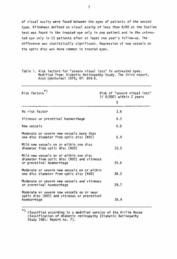
acuity were more frequent in treated t h a n in untreated eyes, especially in xenon-treated eyes. This early decrease in vision persisted a t l a t e follow-ups in eyes treated w i t h the xenon arc technique. Analysis of the visual f ie lds gave clear evidence t h a t photocoagulation, especially with the xenon arc , resulted i n loss of peripheral visual f ie ld . The resul ts of the therapy were found t o be convincing and in 1976 i t was recommended t h a t those control eyes which showed newly formed vessels, especially in the presence of a fresh preretinal or vitreous haemorrhage, also be t rea- ted. I n l a t e r reports (Diabetic Retinopathy Study Research Group 1978, 1980 and 1981: Reports no. 5 and 8 ) the resul ts were confirmed. I t was not cer ta in , however, whether treatment a t early stages of the disease was preferable t o t h a t a t more advanced stages. A special report (Diabetic Retinopathy Study Research Group 1979) pre- sented the "high-risk character is t ics" of diabetic retinopathy (see Table 1). Four retinopathy factors were found t o indicate a high risk of "severe visual loss" (visual acuity of less t h a n 5/200 a t two or more consecutively completed follow-up v i s i t s scheduled a t 4-month in tervals) within 2 years. These were: Vitreous or preretinal haemorrhage, new ves- s e l s , new vessels on or near the optic disc , and moderate or severe new vessels. Eyes with three or more risk factors were found t o be a t a much higher risk of developing "severe visual loss" than eyes with two or fewer factors.
The effect of xenon arc coagulation i n patients w i t h prol i ferat ive re- tinopathy was reported on in 1977 (Bri t ish Multicentre Study 1977) . Pa- t i en ts with symmetrical prol i ferat ive retinopathy were selected fo r the study. The treatment eyes were chosen a t random. New vessels and micro- aneurysms were treated direct ly . Non-perfusion areas were treated in some eyes with new vessels on the optic disc. One hundred patients were fol lo- wed u p for a t l eas t one year, 58 for a t l eas t 2 years and 23 for a t l eas t 3 years. The mean visual acuity of the treated eyes was bet ter than that of the untreated ones a t the yearly control v i s i t s . The difference was s t a t i s t i ca l ly s ignif icant a f t e r 3 years. Patients who i n i t i a l l y had new vessels on bo th optic discs and those with only peripheral new vessels were analysed separately. Patients of the f i r s t type had bet ter mean visual acuity in the treated eyes a f t e r 1, 2 and 3 years. The difference was s t a t i s t i ca l ly s ignif icant . Only minor differences between the means
7
o f v i s u a l a c u i t y were found between t h e eyes o f p a t i e n t s o f t h e second
t ype . B l i n d n e s s d e f i n e d as v i s u a l a c u i t y o f l e s s t h a n 6/60 a t t h e S n e l l e n t e s t was found i n t h e t r e a t e d eye o n l y i n one p a t i e n t and i n t h e u n t r e a - t e d eye o n l y i n 13 p a t i e n t s a f t e r a t l e a s t one y e a r - s f o l l o w - u p . The
d i f f e r e n c p was s t a t i s t i c a l l y s i g n i f i c a n t . Regress ion o f new v e s s e l s on t h e o p t i c d i s c was more common i n t r e a t e d eyes.
Table 1. R i s k f a c t o r s f o r " seve re v i s u a l l o s s " i n u n t r e a t e d eyes. M o d i f i e d f rom: D i a b e t i c R e t i n o p a t h y Study. The t h i r d r e p o r t . Arch Ophthalmol 1979; 97: 654-5.
R i s k f a c t o r s *) R i s k o f " seve re v i s u a l l o s s " (< 5/200) w i t h i n 2 y e a r s
%
No r i s k f a c t o r
V i t r e o u s o r p r e r e t i n a l haemorrhage
New v e s s e l s
Moderate o r severe new v e s s e l s more t h a n one d i s c d iamete r f r o m o p t i c d i s c (NVE)
M i l d new vesse ls on o r w i t h i n one d i s c d iamete r f r o m o p t i c d i s c (NVD)
M i l d new v e s s e l s on o r w i t h i n one d i s c d iamete r f r o m o p t i c d i s c (NVD) and v i t r e o u s o r p r e r e t i n a l haemorrhage
Moderate o r severe new v e s s e l s on o r w i t h i n one d i s c d i a m e t e r f r o m o p t i c d i s c (NVD)
Moderate o r seve re new v e s s e l s and v i t r e o u s o r p r e r e t i n a l haemorrhage
Moderate o r severe new v e s s e l s on o r n e a r o p t i c d i s c (NVD) and v i t r e o u s o r p r e r e t i n a l haemo r r h a g e
3.6
4.2
6.8
6.9
10.5
25.6
26.2
29.7
36.9
* ) C l a s s i f i e d a c c o r d i n g t o a m o d i f i e d v e r s i o n o f t h e A i r l i e House c l a s s i f i c a t i o n o f d i a b e t i c r e t i n o p a t h y ( D i a b e t i c Re t inopa thy Study 1981: Repor t no. 7 ) .

a
Hercules et al. (1977) reported on the treatment of 94 patients with proliferative diabetic retinopathy involving the optic disc. One eye was randomly selected for treatment with panretinal argon laser coagulation. The patients were followed up for 3 years. Regression of newly formed vessels on the optic disc was found after treatment, especially in eyes which initially had had mild or moderate changes. There was a highly significant difference in the mean cumulative deterioration of visual acuity between treated and untreated eyes in patients who initially had had newly formed vessels of moderate severity on the optic disc. The treated eyes had better visual function.
The effect of photocoagulation on optic disc neovascularization in dia- betes was presented in 1980 (Yassur et al. 1980). Sixty-seven patients with new vessels on the optic discs were recruited and one eye was se- lected at random for panretinal photocoagulation with argon laser or a xenon arc coagulator. Forty-five patients had argon laser and 22 xenon arc treatment. The patients were followed up for 4 years. The disc pro- liferations were graded and evaluated at the follow-up visits. The treat ment was found to prevent the occurrence and shorten the duration of optic disc changes.