Embed Size (px)
Citation preview

HISTOLO
GY AND H
ISTOPATHOLO
GY
(non-e
dited
man
uscri
pt)
ONLINEFIRST
ThisisaprovisionalPDFonly.Copyeditedandfullyformattedversiónwillbemadeavailableatfinalpublication
Thisarticlehasbeenpeerreviewedandpublishedimmdediatelyuponacceptance.Articlesin“HistologyandHistopathology”arelistedinPubmed.
Pre-printauthor´sversion
ISSN:0213-3911e-ISSN:1699-5848
SubmityourarticletothisJournal(http://www.hh.um.es/Instructions.htm)
Characterization of the early pathology of cochlear stereocilia in four inbred mouse strains with progressive hearing loss
Authors:Xiang Liu, Yi Xie, Shanshan Huang, Ang Xu, Mengmeng Zhao, Xiaoxia Kang, Aiwei Yan, Ping Li, Changzhu Jin and Fengchan Han
DOI:10.14670/HH-18-086Articletype:ORIGINALARTICLEAccepted:2019-01-24Epubaheadofprint:2019-01-24

HISTOLO
GY AND H
ISTOPATHOLO
GY
(non-e
dited
man
uscri
pt)
Characterization of the early pathology of cochlear stereocilia in four
inbred mouse strains with progressive hearing loss
Xiang Liu1,3, Yi Xie1,2, Shanshan Huang1, Ang Xu1 , Mengmeng Zhao1,2,
Xiaoxia Kang1, Aiwei Yan1, Ping Li1, Changzhu Jin1,3,**, Fengchan Han1,2,*
1Key Laboratory for Genetic Hearing Disorders in Shandong, Binzhou Medical
University, 346 Guanhai Road, Yantai264003, Shandong, P. R. China 2Department of Biochemistry and Molecular Biology, Binzhou Medical
University, 346 Guanhai Road, Yantai264003, Shandong, P. R. China 3Department of Human Anatomy and Histology and Embryology, Binzhou
Medical University, 346 Guanhai Road, Yantai264003, Shandong, P. R.
China
*The First Corresponding Author: [email protected]
**The Second Corresponding Author: [email protected]
Key Laboratory for Genetic Hearing Disorders in Shandong, Department of
Biochemistry and Molecular Biology, Binzhou Medical University, 346 Guanhai
Road, Yantai264003, Shandong, P. R. China

HISTOLO
GY AND H
ISTOPATHOLO
GY
(non-e
dited
man
uscri
pt)
Abstract Objective: Inbred strains of mice offer promising models for understanding
the genetic basis of age-related hearing loss (AHL). NOD/LtJ, A/J, DBA/2J
and C57BL/6J mice are classical models of age-related hearing loss and
exhibit early onset of pathology of AHL. This study was carried out to
characterize the early pathology of cochlear stereocilia in the four mouse
strains with age-related hearing loss.
Methods: The structural features of stereocilia in NOD/LtJ, A/J, DBA/2J and
C57BL/6J mice were observed by scanning electron microscopy (SEM) at
age 2, 4, 6 or 8, and 10 or 12 weeks. Meanwhile, auditory-evoked brainstem
response (ABR) and distortion product otoacoustic emission (DPOAE)
amplitudes of the mice were measured at various intervals (3, 4, 6, 8, 10 and
12 weeks of age).
Results: The ABR thresholds in NOD/LtJ, A/J and DBA/2J mice increased
with age from 3 to 12 weeks. DPOAE amplitudes in NOD/LtJ, A/J, DBA/2J
mice were very low at 4 weeks and became negative at 8 weeks at f2
frequency of 17 672 Hz. In addition to the progressive hearing loss, the four
mouse strains displayed early onset (at 2 weeks of age) and progressive
degeneration of stereocilia in hair cells.
Conclusion: Early degeneration of stereocilia contributes to the functional
impairment of hair cells and hearing loss in NOD/LtJ, A/J, DBA/2J and
C57BL/6J mice.
Keywords
Age-related hearing loss, mouse model, hair cells, stereocilia, scanning
electron microscope

HISTOLO
GY AND H
ISTOPATHOLO
GY
(non-e
dited
man
uscri
pt)
Introduction
Age-related hearing loss (AHL), one of the most common perceptive diseases
among the elderly population, causes both communication disorders and
psychological problems (Gates and Mills, 2005; Yamasoba et al., 2007;
Yamasoba et al., 2013). In addition to environmental and social factors,
genetic aspects are involved in pathogenesis in about 50%-60% of people
with age-related hearing loss (Gates and Mills, 2005; Liu and Yan, 2007;
Angeli et al., 2012). Therefore, it is of great significance to study the
mechanism of genetic variance that leads to progressive hearing loss. As
mice and humans share similar genetic components, anatomic structures, and
pathological characteristics, mouse models play a crucial role in
understanding the pathogenesis associated with these genes (Noben-Trauth
and Johnson, 2009; Angeli et al., 2012; Fujinami et al., 2012). Inbred strains
of mice offer promising models for understanding the genetic basis of human
presbycusis or age-related hearing loss (AHL)(Johnson et al., 2000). NOD/LtJ,
A/J, DBA/2J and C57BL/6J mice are classical models of age-related hearing
loss (Zheng et al., 1999). They share the ahl allele (Noben-Trauth and
Johnson, 2009) and exhibit early onset of pathology of AHL (Fetoni et al.,
2011).
NOD/LtJ mouse strain exhibits very early onset hearing loss, showing 30
dB threshold elevations at 3 weeks of age, which progresses rapidly to near
complete deafness by 9 weeks of age (Zheng et al., 1999; Noben-Trauth and
Johnson, 2009). Mice of the A/J strain exhibit an early-onset progressive
hearing loss that was first reported in 1982 (Henry, 1982). They exhibit
elevated ABR thresholds by 25 days of age, and hearing loss progresses to
near deafness by 3 months of age (Zheng et al., 2009; Yang et al., 2015).
DBA/2J mouse strain also develops early-onset hearing loss (Zheng et al.,
1999; Yang et al., 2015). This hearing loss is profound but not quite as
pronounced as in NOD/LtJ mouse; hearing thresholds at 3 weeks of age are

HISTOLO
GY AND H
ISTOPATHOLO
GY
(non-e
dited
man
uscri
pt)
elevated by 15-20 dB and reach near deafness levels by 14 weeks (Zheng et
al., 1999). The C57BL/6J (B6) mouse strain is the most widely used mouse
model for the study of aging and age-associated diseases. It is well known
that hearing loss occurs at about 9 to 12 months of age (Johnson et al., 1997;
Johnson et al., 2000; Han et al., 2012).
Schuknecht’s seminal work on the pathology of AHL, beginning in the
1950s, proposed four pathological subtypes in presbycusis: sensory, involving
hair cell loss; strial (or metabolic), involving degeneration of the stria
vascularis and reduction in endocochlear potential; neural, involving loss of
spiral ganglion neurons, and mechanical, involving stiffening of the basement
membrane (Schuknecht and Gacek, 1993). Previous studies have shown that
hair cell loss is involved in the hearing loss of all 4 mouse strains (Han et al.,
2012; Han et al., 2015; Yang et al., 2015; Sang et al., 2017). As we all know,
cochlear hair cells for mammals are not renewable. It is important to prevent
or delay the loss of cochlear hair cells to maintain the normal function of the
hair cells. The cochlear stereocilia are at the top of the hair cells and are
essential to the perception of sound and motion (Sekerkova et al., 2011). In
this study, early alterations of the stereocilia in the cochleae in four inbred
mouse strains with age-related hearing loss were characterized by scanning
electron microscopy (SEM). We found that degeneration of cochlear
stereocilia at the early stage may be responsible for the early hearing loss of
these mouse strains.
Materials and methods
Animals
Experimental mice (NOD/LtJ, A/J, DBA/2J and C57BL/6J mice) were bred in a
specific pathogen-free animal facility at Binzhou Medical University. The
animal studies were conducted in accordance with the principles set forth in

HISTOLO
GY AND H
ISTOPATHOLO
GY
(non-e
dited
man
uscri
pt)
the Guide for the Care and Use of Laboratory Animals of Binzhou Medical
University and were approved by that university’s Institutional Animal Use and
Care Committee (protocol 14-0514). A total of 56 mice with ages from 2 to 24
weeks were included in this study.
Measurement of DPOAE amplitudes and ABR thresholds
The mice were anesthetized with 2% tribromoethanol(0.2 mL per 10 g of body
weight) and then placed on a heating pad to maintain a temperature of 37°C.
All operations were carried out in a soundproof and electromagnetic shielding
room. A computer-aided evoked potential system (Intelligent Hearing Systems,
Miami, FL, USA) was used to test the mice for auditory-evoked brainstem
response (ABR) thresholds as described previously (Zheng et al., 1999). The
ABR thresholds in NOD/LtJ, A/J, DBA/2J and C57BL/6J mice at various
intervals (3, 4, 6, 8, 10 weeks of age) were obtained for stimuli of click by
reducing the sound pressure level (SPL) to identify the lowest level at which
an ABR pattern could be recognized. The IHSS Smart EP 3.30 and USB ez
Software (Intelligent Hearing Systems, Miami, FL, USA) were used to
measure the distortion product oto-acoustic emission (DPOAE), which is the
response generated when the cochlea is stimulated simultaneously by two
pure tone frequencies (f1 and f2). For frequencies ranging from 2 to16 kHz,
an Etymotic ER2 Stimulator (Etymotic Research, Inc., Elk Grove Village,
IL,USA) was used and for frequencies ranging from 16 to 30 kHz, a HIS high-
frequency transducer was used. Stimulus response signals were sampled at a
rate of 128 kHz using a 16-bit D/A converter; L1 and L2 amplitudes were set
to the same level. Frequencies were acquired with an F2:F1 ratio of 1.22. The
stimuli were presented starting from the lowest frequencies tested and
increasing to the highest frequencies tested. (Han et al., 2012; Han et al.,
2013).

HISTOLO
GY AND H
ISTOPATHOLO
GY
(non-e
dited
man
uscri
pt)
Scanning electron microscopy
Scanning electron microscopy(SEM)was carried out following the methods
described previously (Shin et al., 2010; Men et al., 2015; Liu et al., 2018). The
mice in the four mouse strains were observed at ages 2, 4, 6, 8, 10 and 12
weeks (n = 4 in each group). Briefly, the inner ears of the mice were dissected
outside of the skull, fixed in 2.5% glutaraldehyde phosphate buffer (0.1 M PBS)
at 4°C overnight, decalcified, and then washed 3 times in 0.1 M PBS (10
minutes each time). The organs of Corti were exposed after the overlying
bones and membranes were carefully cut off. The mouse cochleae were
processed in 1% osmium tetroxide acid (post-fixation) for 40 minutes,
dehydrated with gradient alcohol (50%, 70%, 80%, 95%, and 100%), and
dried to a critical point with liquid CO2. The samples were then mounted onto
round nails made of pure copper and sputter-coated to produce a gold coat of
10-15 nm. Finally, the samples were examined at 10 kV with an
EVO MA 15/LS scanning electron microscope (Carl Zeiss, Oberkochen
Germany).
Statistical analysis
ANOVA (SPSS 16 Software) was used to analyse the ABR thresholds and
EPOAE amplitudes. P < 0.05 was considered to be significant.
Results
Early onset of progressive hearing loss in the four mouse strains
To evaluate hearing loss in NOD/LtJ, A/J, DBA/2J and C57BL/6J mice, ABR
thresholds at click were measured in a time-course manner. The ABR
thresholds in NOD/LtJ, A/J and DBA/2J mice increased with age from 3 to 12
weeks. However, the ABR thresholds in the C57BL/6J mice did not change
with age until after 9 months of age (Han et al., 2012). Unlike the C57BL/6J

HISTOLO
GY AND H
ISTOPATHOLO
GY
(non-e
dited
man
uscri
pt)
mice, the ABR thresholds in NOD/LtJ mice had already risen to 80 dB SPL at
3 weeks, which indicated an early onset of most severe hearing impairment.
At 8 weeks, ABR thresholds in NOD/LtJ mice were about 120 dB SPL which
indicated deafness for the mice. The hearing loss in A/J mice was similar to
that in DBA/2J mice. Hearing impairment was progressive in the 12 weeks
period of observation (Fig 1A). Compared with C57BL/6J mice, ABR
thresholds in A/J and DBA/2J mice were elevated by 35-40 dB SPL by 8
weeks (Fig 1A; Supplementary Fig. 2).
Functional impairment of outer hair cells in NOD/LtJ, A/J and DBA/2J mice
As a secondary screen of mice with known hearing impairment, DPOAE may
provide additional information about outer hair cell (OHC) function (Noben-
Trauth and Johnson, 2009). Therefore, DPOAE amplitudes were measured in
four inbred mice at ages 4 and 8 weeks at f2 frequencies from 8844 to 35 344
Hz (Fig. 1B-C). Typically, DPOAE amplitudes in C57BL/6J mouse littermates
at 4 and 8 weeks showed an inverted V curve, with the height corresponding
to an f2 frequency of 17 672 Hz. However, the DPOAE amplitudes in NOD/LtJ,
A/J, DBA/2J mice at age of 4 weeks were lower than those of the C57BL/6J
mice at f2 frequencies from 8844 to 35 344 Hz (Fig 1B). DPOAE amplitudes in
NOD/LtJ, A/J, DBA/2J mice became negative at age of 8 weeks at f2
frequency of 17 672 Hz (Fig. 1C). These results indicate an early progressive
functional impairment of OHCs in NOD/LtJ, A/J and DBA/2J mice.
Progressive degeneration of hair cell stereocilia in cochleae of the four
mouse strains
Alterations of the stereocilia in cochlear basal turns in the four mouse strains
were observed using SEM (Fig. 3-6). C57BL/6J mice showed almost normal
appearance of OHC stereocilia in the basal turns of cochleae at 2 and 24
weeks (Fig. 3 A,C). The alignment of OHCs and the characteristic array of
stereocilia, which is inverted V-shaped in OHCs, were maintained normally
HISTOLO
GY AND H
ISTOPATHOLO
GY
(non-e
dited
man
uscri
pt)
(Fig. 3 A,C). However, C57BL/6J mice showed defects of single stereocilia at
2 weeks and fusions and bending of stereocilia at 24 weeks under 10 K times
magnification (Fig. 3 B,D). NOD/LtJ mice showed loss of inverted V-shaped
OHC bundles at 2 weeks (Fig. 4A). Defects of single bundle of stereocilia in
the residual OHCs in NOD/LtJ mice were visible at 2 weeks under 10 K times
magnification (Fig. 4B). With the increase of age, NOD/LtJ mice showed more
severe defects and fusions of inverted V-shape OHC bundles at 4 weeks (Fig.
5A,B) and 6 weeks (Fig. 6A,B). At 12 weeks of age, most stereocilia of OHCs
in NOD/LtJ mice were lost (Table 1). The stereocilia in A/J mice were already
irregular at 2 weeks (Fig. 4C,D), disrupted at ages 4 and 8 weeks (Fig. 5C,D;
Fig. 6C,D), and lost for more than half at 12 weeks (Table 1). Particularly, the
stereocilia of OHCs in A/J mice displayed abnormal U-shaped images under
10 K times magnification (Figs. 4C,D, 5C,D;). DBA/2J mice showed bending
of OHC stereocilia or single OHC bundle defects in the basal turns of
cochleae at 2 weeks (Fig. 34E,F). Moreover, DBA/2J mice displayed multiple
deformations such as fusions, bulging or bending of inverted V-shape OHC
bundles at 4 weeks (Fig. 5 E,F). Defects or disruption of stereocilia in DBA/2J
mice were remarkable at 8 weeks (Fig. 6E,F), and about 56% of the
stereocilia were lost at 12 weeks (Table 1). Degeneration of inner hair cell
(IHC) was not so severe as that of OHC at the same time (Fig. 6A,C,E).
Discussion
NOD/LtJ, A/J, DBA/2J and C57BL/6J mice showed early onset progressive
degeneration of cochlear stereocilia
NOD/LtJ, A/J, DBA/2J and C57BL/6J are mouse models of genetic hearing
loss. In addition to the earlier report of Zheng’s group (Zheng et al., 1999), the
features of hearing loss in these strains have also been described recently
(Han et al., 2012; Han et al., 2015; Yang et al., 2015; Sang et al., 2017; Liu et
al., 2018). All the mouse strains developed progressive degeneration of OHCs

HISTOLO
GY AND H
ISTOPATHOLO
GY
(non-e
dited
man
uscri
pt)
and progressive hearing loss. Though hearing loss occurs primarily for high
stimulus frequencies (OHC loss starts at the basal turns), ABR thresholds at
low stimulus frequencies (click) were also elevated in NOD/LtJ, A/J, DBA/2J at
early stage (3-16 weeks). In this study, the NOD/LtJ, A/J, and DBA/2J mice
showed elevation of ABR thresholds at click from 3 to 12 weeks of age,
compared with the thresholds of C57BL/6J mice. There were reports that
C57BL/6J mice presented hearing loss at about 9 to 12 months of age (Han et
al., 2012), and that a significant hearing loss for high-frequencies occurred at
6 months of age (Fetoni et al., 2011). Overall, ABR thresholds (at click) in the
4 mouse strains were basically in accordance with the previous reports. The
function impairment of OHCs was also confirmed by DPOAE at 4 and 8
weeks of age.
Previous studies conclude that the NOD/LtJ, A/J, DBA/2J and C57BL/6J
mice exhibit early onset of pathology in the cochleae. The early, intermediate
and late onset of pathology of AHL is respectively referred to <12 months, 13-
23 months and >24 months of age (Fetoni et al., 2011). In the present study,
the early pathology of the OHC stereocilia in the basal turns of the cochleae in
the 4 mouse strains was further observed under SEM. At 2 weeks of age,
malformation or loss of the OHC stereocilia occurred, even in C57BL/6J mice,
which has not previously been reported; at 4 weeks of age, degeneration of
the bundles of stereocilia was evident in NOD/LtJ, A/J, and DBA/2J mice; at 6-
8 weeks of age, disruption or loss of stereocilia was remarkable in NOD/LtJ,
A/J, and DBA/2J mice; at 12 weeks of age, most stereocilia were lost for
NOD/LtJ, A/J, and DBA/2J mice. In a recent study, the C57BL/6J mice were
used as the “normal controls” for SEM (Liu et al., 2018). It was reported that,
at 1 month and 3 months of age for DBA/2J mice, most OHC bundles already
showed signs of degeneration and some were missing (Shin et al., 2010). Our
present study showed that degeneration of stereocilia bundles occurred at as
early as 2 weeks of age in the 4 mouse strains, though diverse forms of
degeneration were observed. It is well known that the cochlear stereocilia are

HISTOLO
GY AND H
ISTOPATHOLO
GY
(non-e
dited
man
uscri
pt)
essential to the perception of sound and motion (Sekerkova et al., 2011).
Early degeneration of OHC stereocilia may play an important role in the early
onset of hearing loss in these mouse strains.
Degeneration of stereocilia may be caused primarily by gene mutations in
NOD/LtJ, A/J, DBA/2J and C57BL/6J mice
The ahl allele is a major predisposing factor to hearing loss not only in
C57BL/6J strain but also in strains such as A/J, DBA/2J and NOD/LtJ (Noben-
Trauth and Johnson, 2009). It is a mutation in cadherin23 gene (Cdh23),
resulting in skip of exon 7 during RNA splicing. The Cadherin23 (CDH23) is a
component of the tip link in hair cell stereocilia and the corresponding mutant
CDH23 (from ahl) may weaken the stiffness of the cell stereocilia
(Kazmierczak et al., 2007; Noben-Trauth and Johnson, 2009). Meanwhile, the
misfolded CDH23 protein would be retained in the endoplasmic reticulum,
causing a constitutive shortage of functional cadherin 23 and ER stress (Hu et
al., 2016). The above factors may contribute to the early degeneration of
stereocilia in C57BL/6J. With the reduction of stiffness of the stereocilia,
fusion or bending of the stereocilia is evident at 24 weeks of age.
A/J mice also share the ahl allele and have another AHL locus (named
ahl4) in chromosome 10 (Zheng et al., 2009). The ahl4 locus, which could
explain about 40% of the ABR threshold variation in these mice, was identified
as a mutation in the gene of Critrate synthetase (Cs) (Johnson et al., 2012),
which was an essential enzyme and the first rate-limiting enzyme in the
tricarboxylic acid cycle (TCA). Reduction of Cs gene causes excess reactive
oxygen species (ROS) production and cell apoptosis in vitro (Cai et al., 2017).
A single nucleotide insertion in the tRNA-Arg gene (mt-Tr) may also contribute
to the phenotypic effect(Johnson et al., 2001). This same mtDNA variant was
later shown to cause an increase of ROS production in cell lines (Moreno-

HISTOLO
GY AND H
ISTOPATHOLO
GY
(non-e
dited
man
uscri
pt)
Loshuertos et al., 2006). Excess ROS production caused by this mtDNA
variant could exacerbate the stressful effects of the Cdh23 variant and
increase the rate of hair cell degeneration. Probably, pathological alterations
of stereocilia in A/J mice were caused by interaction of the mutations in Cdh23,
Cs and mt-Tr gene.
Apart from ahl, a locus named ahl8 on Chromosome 11 was identified as
the main contributor to the early onset of hearing loss in DBA/2J mice
(Johnson et al., 2008). The ahl8-causative gene was recently identified as
Fascin2 (Fscn2), which encoded an actin crosslinking protein FSCN2 (Shin et
al., 2010). FSCN2 is abundant in stereocilia and it plays a critical role in
stabilizing stereocilia after development (Shin et al., 2010) . In addition to the
mutations in ahl and ahl8, a quantitative trait locus on chromosome 5 may
also be responsible for the severe hearing loss in DBA/2J mice (Johnson et
al., 2015; Suzuki et al., 2015). DBA/2J mice showed fusions and lodging of
inverted V-shaped OHC bundles at 2 and 4 weeks in this study. Early
degeneration of stereocilia in DBA/2J mice may mainly result from mutations
in Cdh23, Fscn2 and other gene(s).
Inbred strains of NOD/LtJ mice are susceptible to early-onset AHL. The
major contributor to the difference in hearing loss compared with C57BL/6J
mice is the ahl2 locus on mouse chromosome 5 in NOD/LtJ mice (Johnson
and Zheng, 2002). The ahl2 locus exacerbates the effects of the ahl locus,
resulting in earlier onset and rapider progression of hearing loss (Noben-
Trauth and Johnson, 2009). NOD/LtJ mice showed loss of inverted V-shape
OHC bundles at 2 weeks and became more severe at 4 weeks of age.
Interaction of ahl and ahl2 contributed to the results.
It should be mentioned that the effects of ahl2, ahl4, and ahl8 locus on
hearing loss in the backcrossed mice were manifested only in mice with
ahl/ahl genotype (Noben-Trauth and Johnson, 2009). That means impairment

HISTOLO
GY AND H
ISTOPATHOLO
GY
(non-e
dited
man
uscri
pt)
of tip links is essential for the effects of ahl2, ahl4, and ahl8. Interaction of ahl
and ahl2, ahl4, ahl8 or other factors determines the cochlear pathology or the
severity of hearing loss in these mice. Genetic defects in these mouse strains
are the main factors of cochlear pathology, leading to progressive hearing loss.
A summary of the hearing loss and stereociliary malformations in the 4 mouse
strains are listed in Table 2.
Multiple factors contribute the early onset hearing loss in NOD/LtJ, A/J,
DBA/2J and C57BL/6J mice
AHL is associated with an age-dependent loss of sensory hair cells, spiral
ganglion neurons and stria vascularis cells in the inner ears (Gates and Mills,
2005; Yamasoba et al., 2013). Previous studies have shown that
degeneration of organ of Corti and afferent neurons are involved in the
hearing loss of C57BL/6J mice (Johnson et al., 1997). Hearing loss in DBA/2J
mice is paralleled by degeneration of the organ of Corti and spiral ganglia
(Willott et al., 2005; Johnson et al., 2008; Yang et al., 2015). Results also
demonstrated that A/J mice displayed all the three features of degeneration of
the sensory hair cells, spiral ganglion neurons and stria vascularis cells (Han
et al., 2015). The NOD/LtJ mice show a typical metabolic hearing loss with
strial capillary degeneration and subsequent strial atrophy (Ohlemiller et al.,
2008; Fetoni et al., 2011). In this study, only the stereocilia defects of OHC in
the 4 mouse strains were observed. In addition to the earlier degeneration of
stereocilia, other factors such as spiral ganglion neurons and strial atrophy
should also be taken into account for the hearing loss.
In summary, NOD/LtJ, A/J, DBA/2J and C57BL/6J mice showed early
onset progressive degeneration of stereocilia, as well as progressive hearing
loss. Our results indicate that early degeneration of stereocilia is an important

HISTOLO
GY AND H
ISTOPATHOLO
GY
(non-e
dited
man
uscri
pt)
factor for the functional impairment in hair cells and progressive hearing loss
in NOD/LtJ, A/J, DBA/2J and C57BL/6J mice.
Declaration of interests
The authors declared no potential conflicts of interest with respect to the
research, authorship, and/or publication of this article.
Acknowledgements
This study is financially supported by the National Natural Science Foundation
of China (81771020, 81570927 and 81271092) and sponsored by Shandong
Scientific and Technological Development Foundation (2014GSF118083) and
Research Initiation Grant of Binzhou Medical University (BY2012KYQD01).
We thank the Taishan Scholar Program of Shandong Province (tshw20110515)
for platform support.

HISTOLO
GY AND H
ISTOPATHOLO
GY
(non-e
dited
man
uscri
pt)
References
AngeliS., LinX.andLiuX.Z. (2012).Geneticsofhearinganddeafness.Anat.Rec.(Hoboken)295,1812-1829.
Cai Q., ZhaoM., Liu X., Wang X., Nie Y., Li P., Liu T., Ge R. and Han F. (2017).Reduced expression of citrate synthase leads to excessive superoxideformation and cell apoptosis.Biochem. Biophys. Res. Commun. 485, 388-394.
Fetoni A.R., Picciotti P.M., Paludetti G. and Troiani D. (2011). Pathogenesis ofpresbycusisinanimalmodels:areview.Exp.Gerontol.46,413-425.
FujinamiY.,MutaiH.,MizutariK.,NakagawaS.andMatsunagaT.(2012).Anovelanimalmodelofhearinglosscausedbyacuteendoplasmicreticulumstressinthecochlea.J.Pharmacol.Sci.118,363-372.
GatesG.A.andMillsJ.H.(2005).Presbycusis.Lancet366,1111-1120.
HanF.,YuH.,TianC.,ChenH.E.,Benedict-AlderferC.,ZhengY.,WangQ.,HanX.andZhengQ.Y.(2012).AnewmousemutantoftheCdh23genewithearly-onset hearing loss facilitates evaluation of otoprotection drugs.PharmacogenomicsJ.12,30-44.
HanF.,YuH., ZhengT.,MaX., ZhaoX., LiP., Le L., SuY.andZhengQ.Y. (2013).OtoprotectiveeffectsoferythropoietinonCdh23erl/erlmice.Neuroscience237,1-6.
HanX.,GeR.,XieG.,LiP.,ZhaoX.,GaoL.,ZhangH.,WangO.,HuangF.andHanF.(2015).Caspase-mediatedapoptosisinthecochleaecontributestotheearlyonset of hearing loss in A/J mice. ASN. Neuro. 7. doi:10.1177/1759091415573985
HenryK.R.(1982).Age-relatedauditorylossandgenetics:anelectrocochleographiccomparisonofsixinbredstrainsofmice.J.Gerontol.37,275-282.
HuJ.,LiB.,ApisaL.,YuH.,EntenmanS.,XuM.,StepanyanR.,GuanB.J.,MullerU.,HatzoglouM.andZhengQ.Y.(2016).ERstressinhibitorattenuateshearingloss and hair cell death in Cdh23erl/erl mutant mice. Cell Death Dis. 7,e2485.
Johnson K.R., Erway L.C., Cook S.A.,Willott J.F. and Zheng Q.Y. (1997). Amajorgeneaffectingage-relatedhearinglossinC57BL/6Jmice.Hear.Res.114,83-92.
HISTOLO
GY AND H
ISTOPATHOLO
GY
(non-e
dited
man
uscri
pt)
JohnsonK.R.,GagnonL.H.,Longo-GuessC.andKaneK.L. (2012).Associationofacitrate synthase missense mutation with age-related hearing loss in A/Jmice.Neurobiol.Aging33,1720-1729.
JohnsonK.R.,Longo-GuessC.,GagnonL.H.,YuH.andZhengQ.Y.(2008).Alocusondistalchromosome11(ahl8)anditsinteractionwithCdh23ahlunderlietheearlyonset,age-relatedhearinglossofDBA/2Jmice.Genomics92,219-225.
JohnsonK.R.,Longo-GuessC.M.andGagnonL.H.(2015).AQTLonChr5modifieshearing loss associatedwith the fascin-2 variant ofDBA/2Jmice.Mamm.Genome26,338-347.
Johnson K.R. and Zheng Q.Y. (2002). Ahl2, a second locus affecting age-relatedhearinglossinmice.Genomics80,461-464.
Johnson K.R., Zheng Q.Y., Bykhovskaya Y., Spirina O. and Fischel-Ghodsian N.(2001). A nuclear-mitochondrial DNA interaction affecting hearingimpairmentinmice.Nat.Genet.27,191-194.
JohnsonK.R.,ZhengQ.Y.andErwayL.C.(2000).Amajorgeneaffectingage-relatedhearinglossiscommontoatleastteninbredstrainsofmice.Genomics70,171-180.
KazmierczakP.,SakaguchiH.,TokitaJ.,Wilson-KubalekE.M.,MilliganR.A.,MullerU.andKacharB.(2007).Cadherin23andprotocadherin15interacttoformtip-linkfilamentsinsensoryhaircells.Nature449,87-91.
LiuX.Z.andYanD.(2007).Ageingandhearingloss.J.Pathol.211,188-197.
LiuX.,ZhaoM.,XieY.,LiP.,WangO.,ZhouB.,YangL.,NieY.,ChengL.,SongX.,JinC.,andHanF.(2018).Nullmutationofthefascin2GenebyTALENleadingtoprogressive hearing loss and retinal degeneration in C57BL/6J mice. G3(Bethesda)8:3221-3230.
MenY.,ZhangA.,LiH.,ZhangT.,JinY.,ZhangJ.andGaoJ.(2015).LKB1IsRequiredfortheDevelopmentandMaintenanceofStereociliainInnerEarHairCellsinMice.PLoSOne10,e0135841.
Moreno-LoshuertosR.,Acin-PerezR.,Fernandez-SilvaP.,MovillaN.,Perez-MartosA., Rodriguez de Cordoba S., Gallardo M.E. and Enriquez J.A. (2006).Differences in reactive oxygen species production explain thephenotypesassociatedwith commonmousemitochondrial DNA variants.Nat. Genet.38,1261-1268.

HISTOLO
GY AND H
ISTOPATHOLO
GY
(non-e
dited
man
uscri
pt)
Noben-Trauth K. and Johnson K.R. (2009). Inheritance patterns of progressivehearinglossinlaboratorystrainsofmice.BrainRes.1277,42-51.
OhlemillerK.K.,RiceM.E.andGagnonP.M.(2008).Strialmicrovascularpathologyandage-associatedendocochlearpotential decline inNOD congenicmice.Hear.Res.244,85-97.
SangL.,ZhengT.,MinL.,ZhangX.,MaX.,EntenmanS.,SuY.andZhengQ.(2017).Otoprotective effects of ethosuximide in NOD/LtJ mice with age-relatedhearingloss.Int.J.Mol.Med.40,146-154.
Schuknecht H.F. and GacekM.R. (1993). Cochlear pathology in presbycusis. TheAnnalsofotology,rhinology,andlaryngology102,1-16.
SekerkovaG.,RichterC.P.andBartlesJ.R.(2011).Rolesoftheespinactin-bundlingproteins in the morphogenesis and stabilization of hair cell stereociliarevealedinCBA/CaJcongenicjerkermice.PLoSgenetics7,e1002032.
Shin J.B., Longo-GuessC.M.,GagnonL.H.,SaylorK.W.,DumontR.A.,SpinelliK.J.,Pagana J.M., Wilmarth P.A., David L.L., Gillespie P.G. and Johnson K.R.(2010). The R109H variant of fascin-2, a developmentally regulated actincrosslinker in hair-cell stereocilia, underlies early-onset hearing loss ofDBA/2Jmice.J.Neurosci.30,9683-9694.
SuzukiS., IshikawaM.,UedaT.,OhshibaY.,MiyasakaY.,OkumuraK.,YokohamaM.,TayaC.,MatsuokaK.andKikkawaY. (2015).Quantitativetrait locionchromosome5forsusceptibilitytofrequency-specificeffectsonhearinginDBA/2Jmice.Exp.Anim.64,241-251.
Willott J.F., Bross L.S. andMcFadden S. (2005). Ameliorative effects of exposingDBA/2J mice to an augmented acoustic environment on histologicalchanges in the cochlea and anteroventral cochlear nucleus. J. Assoc. Res.Otolaryngol.6,234-243.
Yamasoba T., Lin F.R., Someya S., Kashio A., Sakamoto T. and Kondo K. (2013).Currentconceptsinage-relatedhearingloss:epidemiologyandmechanisticpathways.Hear.Res.303,30-38.
YamasobaT., Someya S., YamadaC.,WeindruchR., Prolla T.A. and TanokuraM.(2007)RoleofmitochondrialdysfunctionandmitochondrialDNAmutationsinage-relatedhearingloss.Hear.Res.226,185-193.
YangL.,ZhangH.,HanX.,ZhaoX.,HuF.,LiP.,XieG.,GaoL.,ChengL.,SongX.andHanF.(2015).AttenuationofhearinglossinDBA/2Jmicebyanti-apoptotictreatment.Hear.Res.327,109-116.

HISTOLO
GY AND H
ISTOPATHOLO
GY
(non-e
dited
man
uscri
pt)
Zheng Q.Y., Ding D., Yu H., Salvi R.J. and Johnson K.R. (2009). A locus on distalchromosome 10 (ahl4) affecting age-related hearing loss in A/J mice.Neurobiol.Aging30,1693-1705.
ZhengQ.Y.,JohnsonK.R.andErwayL.C.(1999).Assessmentofhearingin80inbredstrainsofmicebyABRthresholdanalyses.Hear.Res.130,94-107.

HISTOLO
GY AND H
ISTOPATHOLO
GY
(non-e
dited
man
uscri
pt)
Table 1. Percentages of stereociliary loss in basal turns of cochleae of the 4 mouse strains (n = 4 for each strain at each time point)
2 weeks 4 weeks 8 weeks 12 weeks C57BL/6J 0±0 0±0 0±0 0±0 DBA/2J 0±0 1.11±1.52 46.93±4.77 55.92±8.22
A/J 0±0 10.77±2.57 47.07±6.18 66.57±7.48 NOD/LtJ 11.38±6.40 44.34±4.66 63.94±9.51 89.84±4.77

HISTOLO
GY AND H
ISTOPATHOLO
GY
(non-e
dited
man
uscri
pt)
Table 2. Summary of hearing loss and stereociliary malformations in the 4 mouse strains
Strain Defective genes
Age of HL in our studies
(click)
Age of HL in previous
studies
Frequencies lost in previous
studies
Previous analysis by SEM
Age of stereociliary
malformations by SEM in this
study
NOD/LtJ Cdh23 + unknown 3-12 weeks 3-9 weeks all frequencies unknown 2-12 weeks
A/J Cdh23 + Cs 3-12 weeks 3-12 weeks all equally unknown 2-12 weeks
DBA/2J Cdh23 + Fscn2 + QTL(Chr5) 3-12 weeks 3-14 weeks all equally yes 2-12 weeks
C57BL6/J Cdh23 36-102 weeks
(Han et al, 2012)
24-100 weeks higher ones first yes 2-24 weeks

HISTOLO
GY AND H
ISTOPATHOLO
GY
(non-e
dited
man
uscri
pt)
Figure legends
Fig. 1. ABR thresholds and DPOAE amplitudes in NOD/LtJ, A/J, DBA/2J and
C57BL/6J mice. (A) ABR thresholds tested in NOD/LtJ, A/J, DBA/2J and
C57BL/6J mice from 3 to 12 weeks of age at stimulus frequencies of click.
ABR thresholds are presented by mean and standard errors. (B)-(C) The
DPOAE amplitudes measured in NOD/LtJ, A/J, DBA/2J and C57BL/6J mice at
f2 frequencies from 8844 Hz to 35 344 Hz at 4 and 8 weeks. DPOAE
amplitudes are presented by mean and standard errors. Asterisks on the top
of a line indicate the significance of the ABR thresholds or DPOAE
amplitudes in the corresponding strain (NOD/LtJ or C57BL/6J)
compared with any one of the other strains at the same of age. * P < 0.05;
**P < 0.01
Fig. 2. ABR thresholds in NOD/LtJ, A/J, DBA/2J and C57BL/6J mice. (A)-(D)
The typical illustrations of ABR tested in NOD/LtJ, A/J, DBA/2J and C57BL/6J
mice at 8 weeks at click. ABR thresholds are indicated by a black asterisk.
Fig. 3. Images of SEM to show the stereocilia of cochlear hair cells in
C57BL/6J mice. C57BL/6J mice show almost normal appearance of
stereocilia in OHCs in the basal turns of cochleae at 2 weeks (A) and 24
weeks(C) under 2 K times magnification. However, defects (or gaps) of single
stereocilia occurred in C57BL/6J mice at 2 weeks and 24 weeks under 10 K
times magnification as indicated by arrows (B, D). The fusion of stereocilia
was evident at 24 weeks as indicated by asterisks (D). Scale bars = 2 μm.
Fig. 4. Images of hair cell stereocilia by SEM in the basal turns of cochleae of
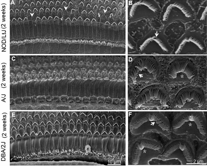
NOD/LtJ, A/J and DBA/2J mice at 2 weeks of age. (A) and (B) Hair cell
stereocilia in the cochleae of NOD/LtJ mice. (C) and (D) Hair cell stereocilia of
cochleae in A/J mice. (E) and (F) Hair cell stereocilia of cochleae in DBA/2J
mice. Loss of entire stereocilia in an OHC in NOD/LtJ mice occurred at 2

HISTOLO
GY AND H
ISTOPATHOLO
GY
(non-e
dited
man
uscri
pt)
weeks of age, as indicated by arrows (A). Defects of stereocilia bundles are
indicated by arrows and fusion or bending of stereocilia is indicated by
asterisks (B, D, F). (A), (C) and (E) Images of stereocilia observed by SEM
under 1 K times magnification. Scale bars = 10 μm. (B), (D) and (F) Images of
stereocilia observed by SEM under 10K times magnification. Scale bars = 2
μm.
Fig. 5. Images of hair cell stereocilia by SEM in the basal turns of cochleae of
NOD/LtJ, A/J and DBA/2J mice at age of 4 weeks. (A) and (B) Hair cell
stereocilia of cochleae in NOD/LtJ mice. (C) and (D) Hair cell stereocilia of
cochleae in A/J mice. (E) and (F) Hair cell stereocilia of cochleae in DBA/2J
mice. Loss of stereocilia in NOD/LtJ and A/J mice at 4 weeks of age is
indicated by arrows (A, C). Defects of stereocilia bundles are indicated by
arrows and fusion or bending of stereocilia is indicated by asterisks ( B, D, F).
(A), (C) and (E) Images of stereocilia observed by SEM under 1K times
magnification. Scale bar = 10 μm. (B), (D) and (F) Images of stereocilia
observed by SEM under 10K times magnification. Scale bar = 2 μm.
Fig. 6. Images of hair cell stereocilia by SEM in the basal turns of cochleae of
NOD/LtJ, A/J and DBA/2J mice at age of 6 or 8 weeks. (A) and (B) Hair cell
stereocilia of cochleae in NOD/LtJ mice. (C) and (D) Hair cell stereocilia of
cochleae in A/J mice. (E) and (F) Hair cell stereocilia of cochleae in DBA/2J
mice. Loss of stereocilia in NOD/LtJ, A/J and DBA/2J mice at 6 or 8 weeks of
age was remarkable (A, C, E). Defects of stereocilia bundles are indicated by
arrows and fusion or bending of stereocilia is indicated by asterisks (B, D, F).
(A), (C) and (E) Images of stereocilia observed by SEM under 2 K times
magnification. Scale bar = 2 μm. (B), (D) and (F) Images of stereocilia
observed by SEM under 5 K times magnification. Scale bar = 2 μm.

HISTOLO
GY AND H
ISTOPATHOLO
GY
(non-e
dited
man
uscri
pt)

HISTOLO
GY AND H
ISTOPATHOLO
GY
(non-e
dited
man
uscri
pt)

HISTOLO
GY AND H
ISTOPATHOLO
GY
(non-e
dited
man
uscri
pt)
HISTOLO
GY AND H
ISTOPATHOLO
GY
(non-e
dited
man
uscri
pt)

HISTOLO
GY AND H
ISTOPATHOLO
GY
(non-e
dited
man
uscri
pt)

HISTOLO
GY AND H
ISTOPATHOLO
GY
(non-e
dited
man
uscri
pt)