Embed Size (px)
Citation preview
Histopathological Effects of Hexavalent Chromium in the Ovaryof a Fresh Water Fish, Channa punctatus (Bloch)
Ashish Kumar Mishra Æ Banalata Mohanty
Received: 27 September 2007 / Accepted: 17 March 2008 / Published online: 6 April 2008
� Springer Science+Business Media, LLC 2008
Abstract The histopathological effects of hexavalent
chromium (Cr VI) in the ovary of a fresh water teleost,
Channa punctatus were investigated. An exposure-depen-
dent alteration in ovarian histology is reported. For both
acute and chronic exposures to Cr (VI), the percentages of
atretic oocytes were increased; this increase was more
pronounced in the acute exposure group. A decrease in
percentage of vitellogenic oocytes was observed in the
chronic exposure group indicating impairment of vitello-
genesis. The hepatocellular vacuolization and atrophy along
with pyknotic nuclei in both acute and chronic chromium
exposed fish liver supports the vitellogenic impairment. The
observed alterations may be due to both direct cytotoxic
effect of Cr (VI) on the ovary as well as mediation by
overall systemic toxicity affecting other vital organs.
Keywords Hexavalent chromium �Ovarian histopathology � Channa punctatus
Chromium (Cr), one of the important toxic heavy metals, is
released to water bodies through the effluents of various
industries. Its indiscriminate introduction into aquatic
ecosystem may pose major threat to growth and survival of
fish populations. Effects of chromium on the hematological
(Gautam and Gupta 1989), biochemical (Jha and Jha 1995),
and immune (Arunkumar et al. 2000) parameters as well as
histological gill lesions (Nath et al. 1997; Begum et al.
2006) of fish have been reported. However, scientific evi-
dence of toxicological impacts of chromium on the fish
reproductive system is completely lacking. Recent studies
in a few mammal species like mice (Pereira et al. 2005;
Acharya et al. 2006), monkey (Aruldhas et al. 2005;
Subramanian et al. 2006) and human (Li et al. 2001;
Danadevi et al. 2003) determined that chromium acts as a
reproductive toxicant. The focus of the present study was
therefore, to investigate the Cr (VI) induced ovarian his-
topathology of a teleost fish, Channa punctatus both on
acute and chronic exposures during the preparatory phase
of the reproductive cycle. The vitellogenic growth (vitel-
logenin incorporation) of the ovarian follicles takes place
during the preparatory phase. Since vitellogenin is syn-
thesized in the liver and transported via blood to the ovary
to be taken up by growing oocytes (Wallace 1985;
Mommsen and Walsh 1988), the impact of chromium on
the liver was also evaluated for the correlative assessment.
Materials and Methods
Adult female specimens of the Channa punctatus (Order:
Ophiocephaliformes, Family: Ophiocephalidae) weighing
50 ± 5 g, (length 18 ± 2 cm) were collected from clean
and unpolluted local freshwater pond used for fish culture
in March 2006, bath treated with 0.1% KMnO4 solution
and acclimatized to laboratory conditions for 2 weeks
before experimentation. Fish were maintained in glass
aquaria containing seasoned tap water (pH 7.3 ± 0.05, DO
7.5 ± 1.0 mg/L, total hardness 215.3 ± 7.0 mg/L as
CaCO3 and alkalinity 133.2 ± 5.0 mg/L as CaCO3) under
natural photoperiod (13L: 11D) and ambient temperature
(18–21�C). Fish were provided with commercial dry fish
feed pellets ad libitum (Hello fish dry pellets; CVM
products, Beijing, China) at approximately 2%–3% of body
weight of the fish/day.
A. K. Mishra � B. Mohanty (&)
Department of Zoology, University of Allahabad,
Allahabad 211002, India
e-mail: [email protected]
123
Bull Environ Contam Toxicol (2008) 80:507–511
DOI 10.1007/s00128-008-9406-9

The fish were divided into three groups of 12 individ-
uals. Group I was the unexposed control, Group II and III
were exposed to hexavalent chromium salt, potassium
dichromate (K2Cr2O7; MERCK, Mumbai, India). The LC50
value of K2Cr2O7 was determined to be 41.75 mg/L using
arithmetic method of Karber as adopted by Dede and Kaglo
(2001). The chronic exposure to a sublethal concentration
of 4 mg/L (&10% of 96 h LC50) was applied for 1 month
to Group II and the acute exposure of 20 mg/L (&50% of
96 h LC50) was applied to Group III for 4 days. The acute
exposure to Group III fish was begun on the 26th day of the
experiment. The whole exposure medium was changed
every other day in both the treatment groups to maintain
the desired concentration of chromium salt. The water in
control group was also changed at the same time. Fish were
killed by decapitation; ovaries and liver were dissected out,
ovaries were weighed, the tissues were then fixed in Bou-
in’s solution for 24 h, and processed via standard
histological procedures. Paraffin blocks were cut in trans-
verse plane at 6-lm thickness and stained with
haematoxylin and eosin for microscopic analysis.
Morphometric analyses of whole ovary as well as indi-
vidual oocytes were carried out. The gonadosomatic index
(GSI) was calculated in percentage as weight of the ovary
(g)/weight of the fish (g) 9 100. The diameter of individ-
ual oocyte stage was determined for 50 oocytes randomly
selected from three tissue sections from each fish. For
oocyte staging, 100 oocytes were randomly selected from
each of the three different sections along the cranial–caudal
axis of the ovary of each fish. The data obtained were
expressed as means ± SEM and were analyzed with Stu-
dent’s t-test for statistical significance between
experimental and control groups. The differences of the
means were considered significant when p \ 0.05.
Results and Discussion
In the control and chronically chromium (VI) exposed
group no fish died while in the group exposed acutely to Cr
(VI) two fish died. The death of fish in acute exposure
group might be due to the systemic toxicity caused by high
dose of chromium.
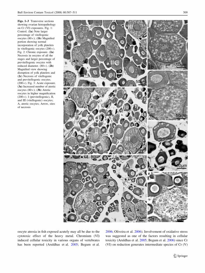
Fish exposed chronically for 1 month to 4 mg/L Cr (VI)
showed retarded growth and development of the ovary as
compared to control fish (Tables 1 and 2, Figs. 1a and b,
2a–c). The gonadosomatic index was significantly lower in
comparison to control (Table 1). Unlike the uniform
extension into the ovarian stroma in the control fish
(Fig. 1a), the growth of the ovigerous lamellae were
stunted in chronic exposed group (Fig. 2a). The diameters
of all the three stages of oocytes of the preparatory phase
ovary, previtellogenic non-yolky stage I oocytes and
vitellogenic oocytes of stage II, III were reduced (Table 2)
which resulted in a less compact arrangement (Fig. 2a). A
lower percentage of vitellogenic oocytes were observed in
the chronically exposed fish as compared to control fish
(Table 2). Necrosis was observed in oocytes of all the three
categories; previtellogenic stage I and vitellogenic stage II,
III categories (Fig. 2a–c). The vitellogenic oocytes showed
disrupted yolk platelets (Fig. 2b). Disruption of ovarian
stroma was also pronounced (Fig. 2c). Increase in atretic
oocytes was also evident in this group (Table 2).
Exposure for 96 h to 20 mg/L Cr (VI) did not alter the
GSI or ovarian diameter as compared to control (Table 1).
The percentage of previtellogenic oocytes of this group
was also not different from the control group (Table 2). A
significantly higher percentage of atretic oocytes were
observed in ovaries from the acute Cr (VI) exposure group
(Fig. 3a, Table 2). Atresia was mostly observed in the
vitellogenic stage II and III oocytes (Fig. 3a and b).
The necrosis of oocytes and ovarian stroma on chronic
exposure to sublethal levels of Cr (VI) and increased
Table 1 Effect of hexavalent chromium on the GSI and ovarian
diameter in teleost fish, Channa punctatus
Parameters Control Acute Chronic
GSI (%) 1.4 ± 0.12 1.28 ± 0.03 0.96 ± 0.04*
Ovarian diameter (mm) 2.4 ± 0.03 2.3 ± 0.04 2.1 ± 0.02**
* p \ 0.05, ** p \ 0.001, N = 10–12
Table 2 Effect of hexavalent chromium on the number and size of Oocytes in teleost fish, Channa punctatus
Stages of oocytes Control Acute Chronic
Number Diameter Number Diameter Number Diameter
Stage I 57.5 ± 0.92 20–110 54.3 ± 1.3 20–110 76.0 ± 1.03** 15–75
Stage II 15.67 ± 0.49 120–160 15.0 ± 0.37 120–160 12.3 ± 0.6* 110–140
Stage III 25.3 ± 0.5 210–360 25.7 ± 1.4 210–360 9.3 ± 0.62** 180–360
Atretic 1.3 ± 0.2 160–275 4.5 ± 0.22* 160–275 2.0 ± 0.4 160–250
Numbers of oocytes are expressed in means ± SEM and diameters are in lm
* p \ 0.05, ** p \ 0.001, N = 10–12
508 Bull Environ Contam Toxicol (2008) 80:507–511
123
oocyte atresia in fish exposed acutely may all be due to the
cytotoxic effect of the heavy metal. Chromium (VI)
induced cellular toxicity in various organs of vertebrates
has been reported (Aruldhas et al. 2005; Begum et al.
2006; Oliveira et al. 2006). Involvement of oxidative stress
was suggested as one of the factors resulting in cellular
toxicity (Aruldhas et al. 2005; Begum et al. 2006) since Cr
(VI) on reduction generates intermediate species of Cr (V)
Figs. 1–3 Transverse sections
showing ovarian histopathology
on Cr (VI) exposures. Fig. 1:
Control. (1a) Note larger
percentage of vitellogenic
oocytes (809); (1b) Magnified
portion showing normal
incorporation of yolk platelets
in vitellogenic oocytes (2009).
Fig. 2: Chronic exposure. (2a)
Necrosis in oocytes of all the
stages and larger percentage of
previtellogenic oocytes with
reduced diameter. (809). (2b)
Magnified view showing
disruption of yolk platelets and
(2c) Necrosis of vitellogenic
and previtellogenic oocytes.
(2009). Fig. 3: Acute exposure.
(3a) Increased number of atretic
oocytes (809); (3b) Atretic
oocytes in higher magnification
(2009). I (previtellogenic), II,
and III (vitellogenic) oocytes;
A, atretic oocytes; Arrow, sites
of necrosis
Bull Environ Contam Toxicol (2008) 80:507–511 509
123
and Cr (IV) that further react with H2O2 to generate
reactive oxygen species (ROS). It is likely that these ROS
may interact with various tissues resulting in their damage.
The inhibition of vitellogenic growth of the oocytes was
evident in the chronic Cr (VI) treatment group through the
higher percentage of previtellogenic oocytes in comparison
to the control group. The Cr (VI) mediated toxic effects
may disrupt vitellogenesis by directly acting on the liver.
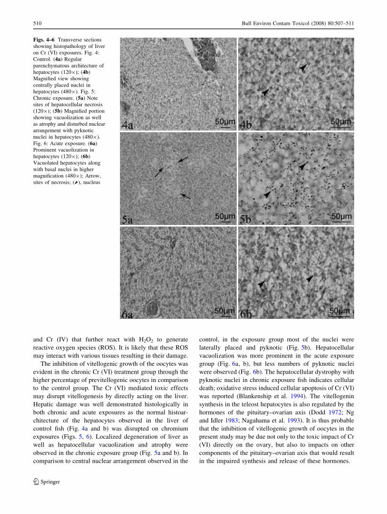
Hepatic damage was well demonstrated histologically in
both chronic and acute exposures as the normal histoar-
chitecture of the hepatocytes observed in the liver of
control fish (Fig. 4a and b) was disrupted on chromium
exposures (Figs. 5, 6). Localized degeneration of liver as
well as hepatocellular vacuolization and atrophy were
observed in the chronic exposure group (Fig. 5a and b). In
comparison to central nuclear arrangement observed in the
control, in the exposure group most of the nuclei were
laterally placed and pyknotic (Fig. 5b). Hepatocellular
vacuolization was more prominent in the acute exposure
group (Fig. 6a, b), but less numbers of pyknotic nuclei
were observed (Fig. 6b). The hepatocellular dystrophy with
pyknotic nuclei in chronic exposure fish indicates cellular
death; oxidative stress induced cellular apoptosis of Cr (VI)
was reported (Blankenship et al. 1994). The vitellogenin
synthesis in the teleost hepatocytes is also regulated by the
hormones of the pituitary–ovarian axis (Dodd 1972; Ng
and Idler 1983; Nagahama et al. 1993). It is thus probable
that the inhibition of vitellogenic growth of oocytes in the
present study may be due not only to the toxic impact of Cr
(VI) directly on the ovary, but also to impacts on other
components of the pituitary–ovarian axis that would result
in the impaired synthesis and release of these hormones.
Figs. 4–6 Transverse sections
showing histopathology of liver
on Cr (VI) exposures. Fig. 4:
Control. (4a) Regular
parenchymatous architecture of
hepatocytes (1209); (4b)
Magnified view showing
centrally placed nuclei in
hepatocytes (4809). Fig. 5:
Chronic exposure. (5a) Note
sites of hepatocellular necrosis
(1209); (5b) Magnified portion
showing vacuolization as well
as atrophy and disturbed nuclear
arrangement with pyknotic
nuclei in hepatocytes (4809).
Fig. 6: Acute exposure. (6a)
Prominent vacuolization in
hepatocytes (1209); (6b)
Vacuolated hepatocytes along
with basal nuclei in higher
magnification (4809); Arrow,
sites of necrosis; ( ), nucleus
510 Bull Environ Contam Toxicol (2008) 80:507–511
123
In conclusion, the present study elucidated the adverse
effects of hexavalent chromium in the ovary of a teleost,
C. punctatus; results also suggest that the overall toxic
impacts occur at multiple organ sites. The lower percentage
of vitellogenic oocytes in chronically exposed fish indi-
cated that the long-term exposures to this heavy metal
might pose a potential risk to fish populations in the
vicinity of waters contaminated with hexavalent chromium.
References
Acharya UR, Mishra M, Tripathy RR, Mishra I (2006) Testicular
dysfunction and antioxidative defense system of Swiss mice after
chromic acid exposure. Reprod Toxicol 22:187–191
Aruldhas MM, Subramanian S, Sekar P, Vengatesh G, Chandrahasan
G, Govindarajulu P, Akbarsha MA (2005) Chronic chromium
exposure-induced changes in testicular histoarchitecture are
associated with oxidative stress: study in a non-human primate
(Macaca radiata Geoffroy). Hum Reprod 20:2801–2813
Arunkumar RI, Rajasekaran P, Michael RD (2000) Differential
effects of chromium compounds on the immune responses of the
African mouth breeder, Oreochromis mossambicus (Peters.).
Fish Shellfish Immunol 10:667–676
Begum G, Venkateswara RJ, Srikanth K (2006) Oxidative stress and
changes in locomotor behavior and gill morphology of Gambu-sia affinis exposed to chromium. Toxicol Environ Chem 88:355–
365
Blankenship LJ, Manning FC, Orenstin JM, Patierno SR (1994)
Apoptosis is the mode of cell death caused by carcinogenic
chromium. Toxicol Appl Pharmacol 126:75–83
Danadevi K, Rozati R, Reddy PP, Grover P (2003) Semen quality of
Indian welders occupationally exposed to nickel and chromium.
Reprod Toxicol 17:451–456
Dede EB, Kaglo HD (2001) Aqua-toxicological effects of water
soluble fractions (WSF) of diesel fuel on O. niloticus fingerlings.
J Appl Sci Environ Manag 5:93–96
Dodd JM (1972) The endocrine regulation of gametogenesis and gonad
maturation in fishes. Gen Comp Endocrinol Supp 3:675–687
Gautam AK, Gupta ML (1989) Chromium induced hematological
anomalies in a freshwater fish, Channa punctatus (Bl.). J
Environ Biol 10:239–243
Jha BS, Jha AK (1995) Biochemical profiles of liver, muscle and
gonads of the freshwater fish, Heteropneustes fossilis, under
chromium stress. In: Prakash R (ed) Toxicity and monitoring of
xenobiotics. Venus Publishing House, New Delhi, pp 127–137
Li H, Chen Q, Li S, Yao W, Li L, Shi X, Wang L, Castranova V,
Vallyathan V, Ernst E, Chen C (2001) Effect of Cr VI exposure
on sperm quality: human and animal studies. Ann Occup Hyg
45:505–511
Mommsen TP, Walsh PL (1988) Vitellogenesis and oocyte assembly.
In: Hoar WS, Randall DJ, Donaldson EM (eds) Fish physiology,
vol 11A. Academic Press, New York, pp 347–406
Nagahama Y, Yoshikuni M, Yamashita M, Sakai N, Tanaka M (1993)
Molecular endocrinology of oocyte growth and maturation in
fish. Fish Physiol Biochem 11:3–14
Nath K, Kumar N, Srivastav AK (1997) Chromium induced
histological alterations in the gills of a freshwater teleost, Colisafasciatus. Fish Biol J Medaka 9:37–40
Ng TB, Idler DR (1983) Yolk formation and differentiation in teleost
fishes. In: Hoar WS, Randall DJ, Donaldson EM (eds) Fish
physiology, vol 9A. Academic Press, New York, p 373
Oliveira H, Santos TM, Ramalho-Santos J, Pereira ML (2006)
Histopathological effects of hexavalent chromium in mouse
kidney. Bull Environ Contam Toxicol 76:977–983
Pereira ML, Pires das Neves R, Oliveira H, Santos TM, Jesus JP
(2005) Effect of Cr (V) on reproductive organ morphology and
sperm parameters: an experimental study in mice. Environ
Health 4:9
Subramanian S, Rajendiran G, Sekhar P, Chandrahasan G, Govind-
arajulu P, Aruldhas MM (2006) Reproductive toxicity of
chromium in adult bonnet monkeys (Macaca radiata Geoffroy).
Reversible oxidative stress in the semen. Toxicol Appl Pharma-
col 215(3):237–249
Wallace RA (1985) Vitellogenesis and oocyte growth in non-
mammalian vertebrates. In: Browder LW (eds) Developmental
biology. Plenum Press, New York, pp 127–177
Bull Environ Contam Toxicol (2008) 80:507–511 511
123