Embed Size (px)
Citation preview
HISTOLOGY REVIEWHISTOLOGY REVIEWMuscle TissueMuscle Tissue
Dr. Tim BallardDr. Tim Ballard
Department of Biology and Marine BiologyDepartment of Biology and Marine Biology
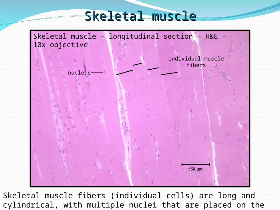
Skeletal muscle – longitudinal section – H&E – 10x objective
Skeletal muscle fibers (individual cells) are long and cylindrical, with multiple nuclei that are placed on the perimeter of the fibers.
Skeletal muscleSkeletal muscle
individual muscle fibers
nucleus
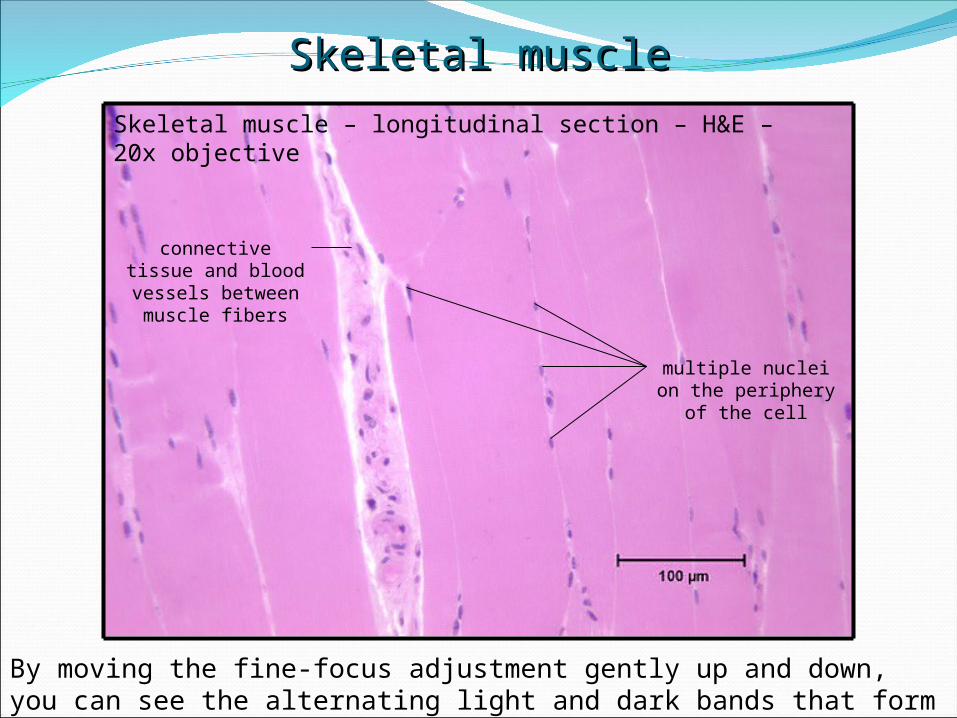
Skeletal muscle – longitudinal section – H&E – 20x objective
By moving the fine-focus adjustment gently up and down, you can see the alternating light and dark bands that form the striations.
Skeletal muscleSkeletal muscle
multiple nuclei on the periphery of
the cell
connective tissue and blood vessels between muscle
fibers
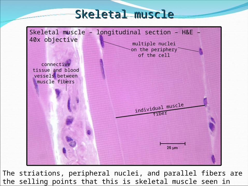
Skeletal muscle – longitudinal section – H&E – 40x objective
The striations, peripheral nuclei, and parallel fibers are the selling points that this is skeletal muscle seen in longitudinal section.
Skeletal muscleSkeletal muscle
multiple nuclei on the periphery of
the cell
connective tissue and blood vessels between muscle
fibers
individual muscle
fiber
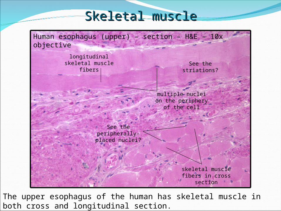
Human esophagus (upper) – section – H&E – 10x objective
The upper esophagus of the human has skeletal muscle in both cross and longitudinal section.
Skeletal muscleSkeletal muscle
multiple nuclei on the periphery of
the cell
longitudinal skeletal muscle
fibersSee the striations?
skeletal muscle fibers in cross
section
See the peripherally-
placed nuclei?

Skeletal muscle – cross section – H&E – 40x objective
Why can’t you see the striations in a cross-section of skeletal muscle?
Skeletal muscleSkeletal muscle
skeletal muscle fibers in cross
section
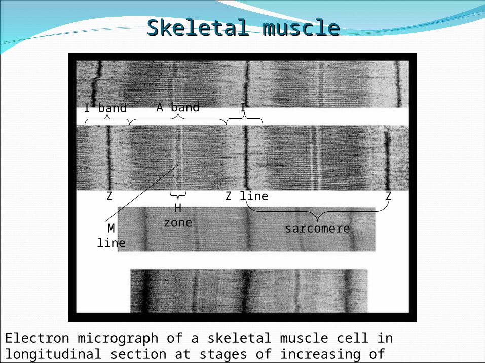
Electron micrograph of a skeletal muscle cell in longitudinal section at stages of increasing of contraction.
Skeletal muscleSkeletal muscle
Z Z line
I band IA band
Z
sarcomere
H zoneM
line

Cardiac muscle – section – H&E – 10x objective
Compare this section with skeletal muscle. Look at the lack of parallel organization of the fibers compared to that of skeletal muscle.
Cardiac muscleCardiac muscle
Cardiac muscle cells run parallel to one another, but unlike skeletal muscle cells, the individual fibers branch to meet other cells, forming a meshwork or network of cells.
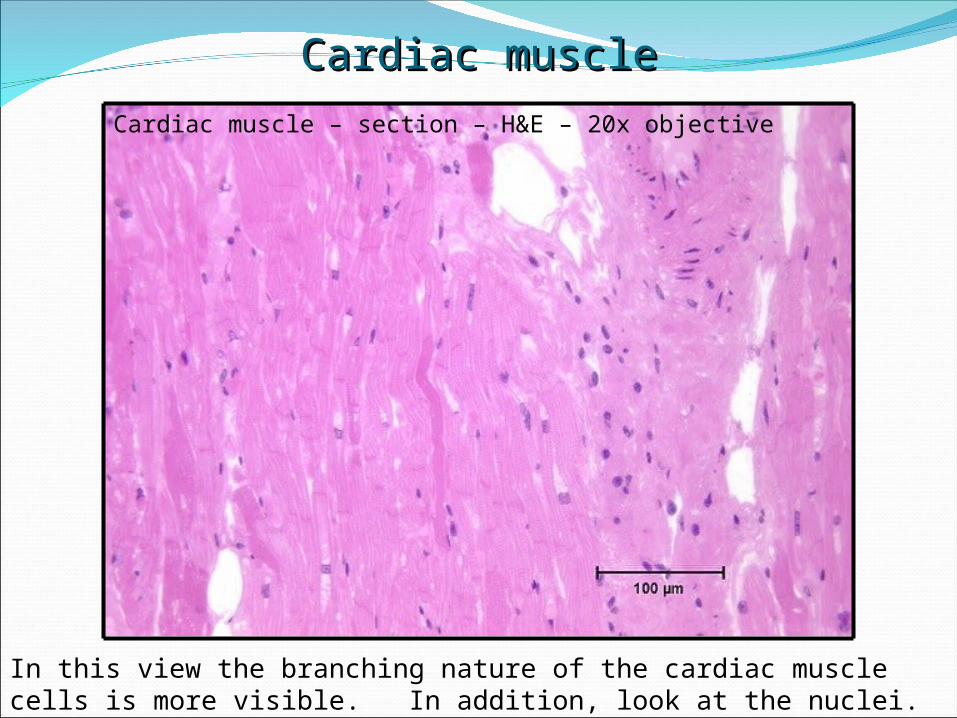
In this view the branching nature of the cardiac muscle cells is more visible. In addition, look at the nuclei. How many are there and where are they?
Cardiac muscleCardiac muscle
Cardiac muscle – section – H&E – 20x objective
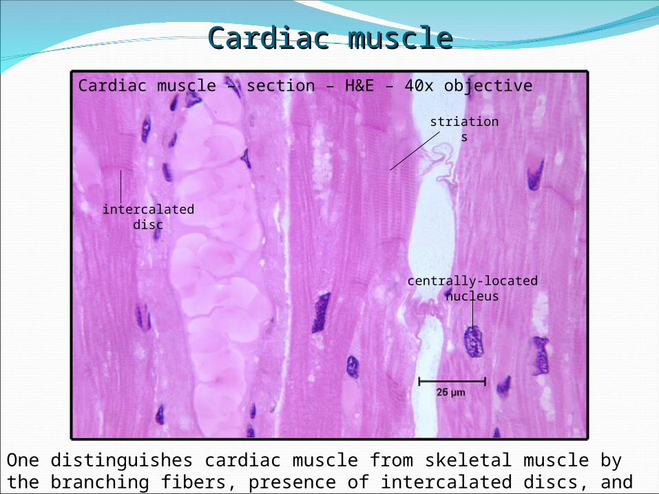
One distinguishes cardiac muscle from skeletal muscle by the branching fibers, presence of intercalated discs, and centrally-placed single nuclei/cell.
Cardiac muscleCardiac muscle
intercalated disc
Cardiac muscle – section – H&E – 40x objective
centrally-located nucleus
striations

Cardiac muscle –section – silver – 20x objective
This stain clearly shows the single central nucleus, branching fibers, intercalated discs, and striations.
Cardiac muscleCardiac muscle
branching
nucleus
intercalated disc
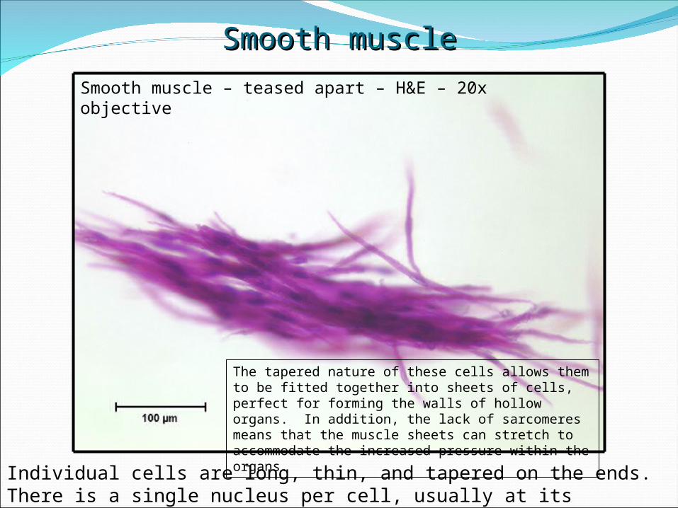
Smooth muscle – teased apart – H&E – 20x objective
Individual cells are long, thin, and tapered on the ends. There is a single nucleus per cell, usually at its longitudinal center. There are no striations.
Smooth muscleSmooth muscle
The tapered nature of these cells allows them to be fitted together into sheets of cells, perfect for forming the walls of hollow organs. In addition, the lack of sarcomeres means that the muscle sheets can stretch to accommodate the increased pressure within the organs.
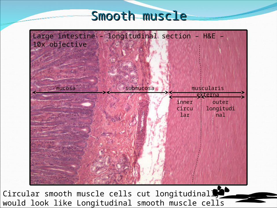
Large intestine – longitudinal section – H&E – 10x objective
Circular smooth muscle cells cut longitudinally would look like Longitudinal smooth muscle cells cut longitudinally would look like
Smooth muscleSmooth muscle
mucosa submucosa muscularis externa
inner circul
ar
outer longitudin
al
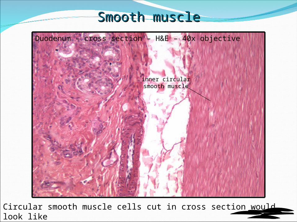
Duodenum – cross section – H&E – 40x objective
Circular smooth muscle cells cut in cross section would look like
Smooth muscleSmooth muscle
inner circular smooth muscle
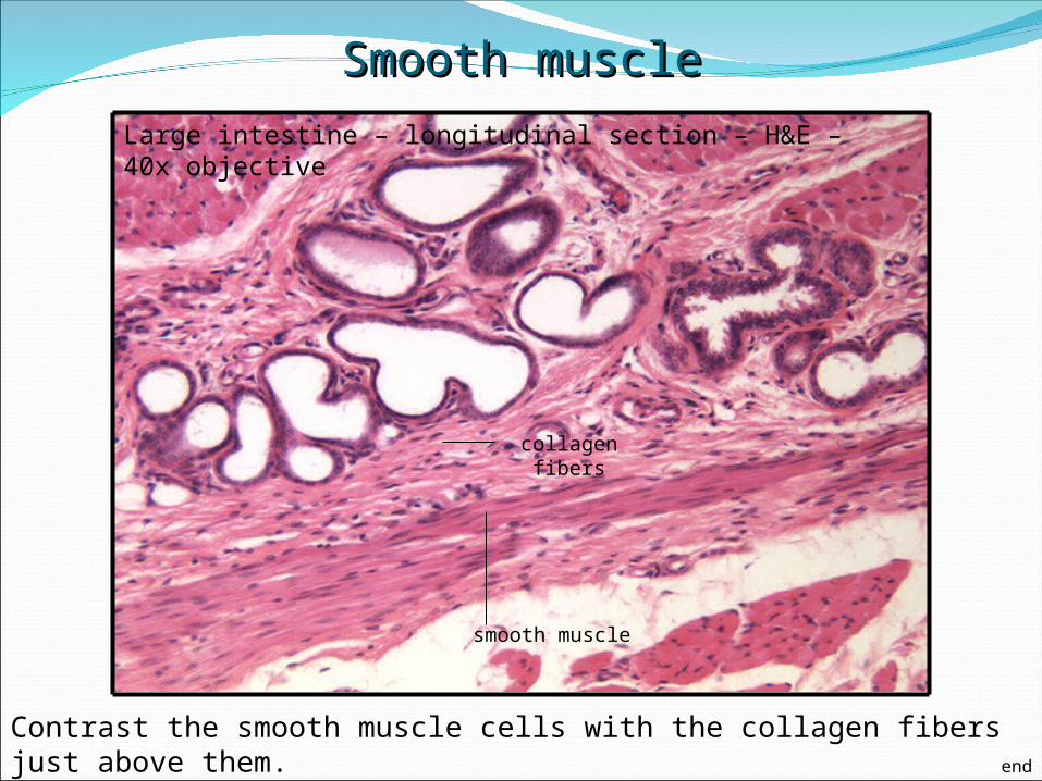
Large intestine – longitudinal section – H&E – 40x objective
Contrast the smooth muscle cells with the collagen fibers just above them.
Smooth muscleSmooth muscle
smooth muscle
collagen fibers
end


![[Oral Biology]Oral Histology Slides_American Corner Family [ACFF @AmCoFam]](https://img.dokumen.tips/doc/110x75/5571f7a349795991698bb904/oral-biologyoral-histology-slidesamerican-corner-family-acff-amcofam.jpg)