Embed Size (px)
Citation preview
His86 from the N‑Terminus of Frataxin Coordinates Iron and IsRequired for Fe−S Cluster SynthesisLeslie E. Gentry, Matthew A. Thacker, Reece Doughty,‡ Russell Timkovich, and Laura S. Busenlehner*
Department of Chemistry, The University of Alabama, Tuscaloosa, Alabama 35487-0336, United States
*S Supporting Information
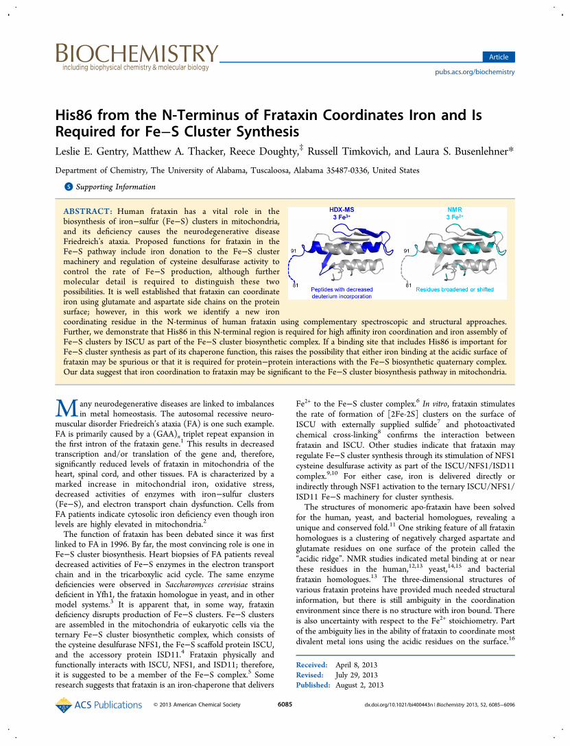
ABSTRACT: Human frataxin has a vital role in thebiosynthesis of iron−sulfur (Fe−S) clusters in mitochondria,and its deficiency causes the neurodegenerative diseaseFriedreich’s ataxia. Proposed functions for frataxin in theFe−S pathway include iron donation to the Fe−S clustermachinery and regulation of cysteine desulfurase activity tocontrol the rate of Fe−S production, although furthermolecular detail is required to distinguish these twopossibilities. It is well established that frataxin can coordinateiron using glutamate and aspartate side chains on the proteinsurface; however, in this work we identify a new ironcoordinating residue in the N-terminus of human frataxin using complementary spectroscopic and structural approaches.Further, we demonstrate that His86 in this N-terminal region is required for high affinity iron coordination and iron assembly ofFe−S clusters by ISCU as part of the Fe−S cluster biosynthetic complex. If a binding site that includes His86 is important forFe−S cluster synthesis as part of its chaperone function, this raises the possibility that either iron binding at the acidic surface offrataxin may be spurious or that it is required for protein−protein interactions with the Fe−S biosynthetic quaternary complex.Our data suggest that iron coordination to frataxin may be significant to the Fe−S cluster biosynthesis pathway in mitochondria.
Many neurodegenerative diseases are linked to imbalancesin metal homeostasis. The autosomal recessive neuro-
muscular disorder Friedreich’s ataxia (FA) is one such example.FA is primarily caused by a (GAA)n triplet repeat expansion inthe first intron of the frataxin gene.1 This results in decreasedtranscription and/or translation of the gene and, therefore,significantly reduced levels of frataxin in mitochondria of theheart, spinal cord, and other tissues. FA is characterized by amarked increase in mitochondrial iron, oxidative stress,decreased activities of enzymes with iron−sulfur clusters(Fe−S), and electron transport chain dysfunction. Cells fromFA patients indicate cytosolic iron deficiency even though ironlevels are highly elevated in mitochondria.2
The function of frataxin has been debated since it was firstlinked to FA in 1996. By far, the most convincing role is one inFe−S cluster biosynthesis. Heart biopsies of FA patients revealdecreased activities of Fe−S enzymes in the electron transportchain and in the tricarboxylic acid cycle. The same enzymedeficiencies were observed in Saccharomyces cerevisiae strainsdeficient in Yfh1, the frataxin homologue in yeast, and in othermodel systems.3 It is apparent that, in some way, frataxindeficiency disrupts production of Fe−S clusters. Fe−S clustersare assembled in the mitochondria of eukaryotic cells via theternary Fe−S cluster biosynthetic complex, which consists ofthe cysteine desulfurase NFS1, the Fe−S scaffold protein ISCU,and the accessory protein ISD11.4 Frataxin physically andfunctionally interacts with ISCU, NFS1, and ISD11; therefore,it is suggested to be a member of the Fe−S complex.5 Someresearch suggests that frataxin is an iron-chaperone that delivers
Fe2+ to the Fe−S cluster complex.6 In vitro, frataxin stimulatesthe rate of formation of [2Fe-2S] clusters on the surface ofISCU with externally supplied sulfide7 and photoactivatedchemical cross-linking8 confirms the interaction betweenfrataxin and ISCU. Other studies indicate that frataxin mayregulate Fe−S cluster synthesis through its stimulation of NFS1cysteine desulfurase activity as part of the ISCU/NFS1/ISD11complex.9,10 For either case, iron is delivered directly orindirectly through NSF1 activation to the ternary ISCU/NFS1/ISD11 Fe−S machinery for cluster synthesis.The structures of monomeric apo-frataxin have been solved
for the human, yeast, and bacterial homologues, revealing aunique and conserved fold.11 One striking feature of all frataxinhomologues is a clustering of negatively charged aspartate andglutamate residues on one surface of the protein called the“acidic ridge”. NMR studies indicated metal binding at or nearthese residues in the human,12,13 yeast,14,15 and bacterialfrataxin homologues.13 The three-dimensional structures ofvarious frataxin proteins have provided much needed structuralinformation, but there is still ambiguity in the coordinationenvironment since there is no structure with iron bound. Thereis also uncertainty with respect to the Fe2+ stoichiometry. Partof the ambiguity lies in the ability of frataxin to coordinate mostdivalent metal ions using the acidic residues on the surface.16
Received: April 8, 2013Revised: July 29, 2013Published: August 2, 2013
Article
pubs.acs.org/biochemistry
© 2013 American Chemical Society 6085 dx.doi.org/10.1021/bi400443n | Biochemistry 2013, 52, 6085−6096

Additionally, reducing agents and some buffers also chelatemetal ions, and many frataxin studies employed thesecompetitors in excess over the frataxin concentration to keepiron in a reduced state. Thus, the stoichiometries and measureddissociation constants could be affected by these chemicalcomponents. From various experiments, it appears that humanfrataxin contains 1−2 Fe2+ binding sites with nanomolar tomicromolar dissociation constants.17 The locations of the actualbinding sites are assumed to be along the acidic ridge and intothe β1-strand, based on NMR iron titrations of frataxin.13,14,18
However, mutation of specific aspartate and glutamate residueswithin the acidic ridge affect frataxin binding with ISCU, thescaffold for Fe−S cluster synthesis, but does not change thetotal number of Fe2+ binding sites.9 This may indicate thatmetal binding at the acidic ridge is superfluous and that theremay be additional, specific Fe2+ binding sites that are not readilyidentifiable by current techniques.Despite many years of research, there are still three main
questions about frataxin iron binding: (1) what is thestoichiometry of Fe2+ binding to human frataxin?; (2) whatresidues are involved in coordination?; and (3) which of theseFe2+ sites are functionally relevant for Fe−S cluster formationby ISCU? To address these questions, we characterized themetal coordination environment and stoichiometries usingFe2+, Fe3+, and Co2+, which are common surrogates to probeFe2+ sites in proteins because they have characteristic electronicabsorptions based on their environment and are not sensitive toaerobic conditions.19 These properties make Co2+ and Fe3+
amenable to UV−visible and NMR spectroscopies. Sinceconformational or dynamics changes in frataxin shouldaccompany metal binding, backbone amide hydrogen/deute-rium exchange mass spectrometry (HDX−MS) was used tocharacterize structural perturbations in solution. Herein, wereport that frataxin binds ∼3 metal ions and that one of thesebinding sites uses a previously unknown residue (His86) fromthe disordered N-terminal tail, which is functionally requiredfor Fe2+ delivery to ISCU for Fe−S synthesis in vitro.
■ EXPERIMENTAL PROCEDURES
Materials. ACS-grade HEPES, dioxane-free IPTG, HPLCgrade water, and HPLC grade acetonitrile were obtained fromEMD Millipore (Billerica, MA). Deuterium oxide (99.99% at.D), ACS-grade Bis-Tris, and ferrozine were from AcrosOrganics (Morris Plains, NJ). Ultrapure magnesium chlorideand ferric chloride were by Fisher Chemical (Fairlawn, NJ).Deuterated d18-HEPES and 15N-ammonium sulfate wereobtained from Cambridge Isotopes (Andover, MA). Otherreagents included porcine pepsin (3200−4500 units/mg) fromSigma-Aldrich (St. Louis, MO), HPLC grade 2-propanol fromHoneywell (Franklin, PA), cobaltous chloride from MacronChemicals (Center Valley, PA), ferrous ammonium sulfatefrom MP Biomedical (Santa Ana, CA), and EDTA from BDHMerck (Radnor, PA).Purification of Human Frataxin. Human frataxin
corresponding to residues 81−210, or the mature form, wasexpressed in Escherichia coli BL21(DE3)pLysS from pET81−210Fxn, as described.8 Purification was performed in thepresence of 2 mM EDTA in Chelex-treated, metal-free water.All glassware was rinsed with 10% nitric acid and metal-freewater to remove trace metals. Frataxin concentrations weredetermined spectroscopically using ε280 nm = 26.93 mM−1 cm−1.The amount of zinc and iron in purified frataxin was
determined via an AAnalyst 400 atomic absorption spectrom-eter (Perkin-Elmer, Waltham, MA) and was negligible.
UV−Visible Titrations. Titrations were performed on anAgilent 8453 spectrophotometer (Santa Clara, CA). For cobalttitrations, 300−400 μM frataxin in Chelex-treated 50 mMHEPES (pH 7.4), 400 mM NaCl, and 5% glycerol was titratedwith a cobalt chloride stock solution in the same buffer. Co2+
was added in 5−10 μL increments, incubated for 5 min at 23°C, and UV−visible spectra were collected from 200−800 nm.Each spectrum was corrected for dilution.Colorimetric Fe2+ titrations required ferrozine, a Fe2+ specific
chelator. All solutions were degassed and prepared in a VacuumAtmosphere anaerobic glovebox. A 0.53 M stock of Fe2+ wasmade in 25 mM HEPES (pH 7.4), 150 mM NaCl. A 6.5 mMferrozine stock was prepared in the same buffer with 2.5 Mammonium acetate. Samples of 108 μM with and without 13μM apo-frataxin were made with increasing Fe2+ concentrationsand spectra were collected from 240−800 nm. The Fe2+−ferrozine3 complex has a molar extinction at 562 nm of 27.9mM−1 cm−1 and a formation constant (Kf) of 3.65 × 10¹⁵
M−3.20
Fluorescence Titrations. Intrinsic tryptophan fluorescencetitrations were performed at 23 °C using a Spex Fluoromax-3fluorimeter (Edison, NJ) with ∼5 μM frataxin in either 50 mMHEPES (pH 7.4), 400 mM NaCl, 5% glycerol (Co2+ titrations),or 50 mM Bis-Tris (pH 7.4), 400 mM NaCl, and 5% glycerol(Fe3+ titrations) using an excitation wavelength of 295 nm. Themetal titrant concentrations were determined by atomicabsorption. After each addition of metal, the sample wasequilibrated for 3 min with stirring at 23 °C. Fluorescenceemission spectra were collected from 270−450 nm andcorrected for dilution. Since the addition of Fe3+ leads toincreased absorbance in this region of the spectrum, thefluorescence data were corrected for inner filter effect with anequivalent UV−vis titration of frataxin under the sameconditions. This procedure was repeated with Co2+ forexperimental continuity. The corrected fluorescence intensitywas determined using eq 1.21
= ×+⎛
⎝⎜⎞⎠⎟F F 10
Abs Abs2corrected i
295nm 343nm
(1)
The initial fluorescence intensity (F0) at the emissionmaximum wavelength was subtracted from each titrationpoint (Fi − F0), which was plotted against total metalconcentration. The binding curves are stoichiometric (i.e., no“free” metal exists during the titration); therefore, theequilibrium binding constants obtained from fitting are notrepresentative of the true Kd values and are not reported.21
Nuclear Magnetic Resonance Spectroscopy. One literof M9 minimal media supplemented with 1 g/L 86% 15N-ammonium sulfate was inoculated, as described, and grown at37 °C to OD600 = 0.8, and then induced with 1 mM IPTG for 4h. Cells were harvested at OD600 1.8−2.0 and purified asdescribed except the final buffer was Chelex-treated 25 mMHEPES-d18 (pH 7.0) with 100 mM NaCl. MALDI−TOF massspectrometry confirmed ∼85% 15N-enrichment in the purifiedNMR sample. Co2+, Mg2+, and Fe2+ titrant stocks were made inthe HEPES-d18 buffer at pH 7.0, and their concentrations weredetermined by atomic absorption.
1H−15N HSQC spectra were collected for 0.56 mM apo-15N-frataxin with 5% (v/v) D2O. Resonances were assignedaccording to ref 22. Titrations from 0.2−6.0 mol equiv of
Biochemistry Article
dx.doi.org/10.1021/bi400443n | Biochemistry 2013, 52, 6085−60966086
Co2+ and Mg2+ were performed aerobically. Individual NMRsamples containing 1.0−4.0 mol equiv of Fe2+ were preparedanaerobically. NMR data were collected at 298 K on a BrukerAvance 600 MHz spectrometer (Fremont, CA) with a tripleresonance 1H/13C/15N probe equipped with z-axis pulsed fieldgradients. The fingerprint main chain amide region wasrecorded by the two-dimensional 1H−15N-HSQC experiment23
using the standard Bruker pulse program. Data collection timeper spectrum ranged from 1.5 to 20 h to improve signal-to-noise ratios. Data were processed using NMRPipe24 andSPARKY (T. D. Goddard and D. G. Kneller, University ofCalifornia, San Francisco). The data matrix consisted of 2048points in the proton dimension centered on the waterresonance and 128 points in the nitrogen dimension centeredat 117 ppm. Data in both dimensions were apodized with ashifted squared sine bell function and zero-filled prior toFourier transformation. After transformation, data in bothdirections were baseline corrected with the default polynomialfunction of NMRPipe. Normalized amide chemical shifts (δNH)were calculated using the change in chemical shift from apo-frataxin values of the 1H (δH) and 15N (δN) resonances uponeach addition of metal as shown in eq 2, modified from ref 25.
δ δδ
= +⎛⎝⎜
⎞⎠⎟( )
5NH H2 N
2
(2)
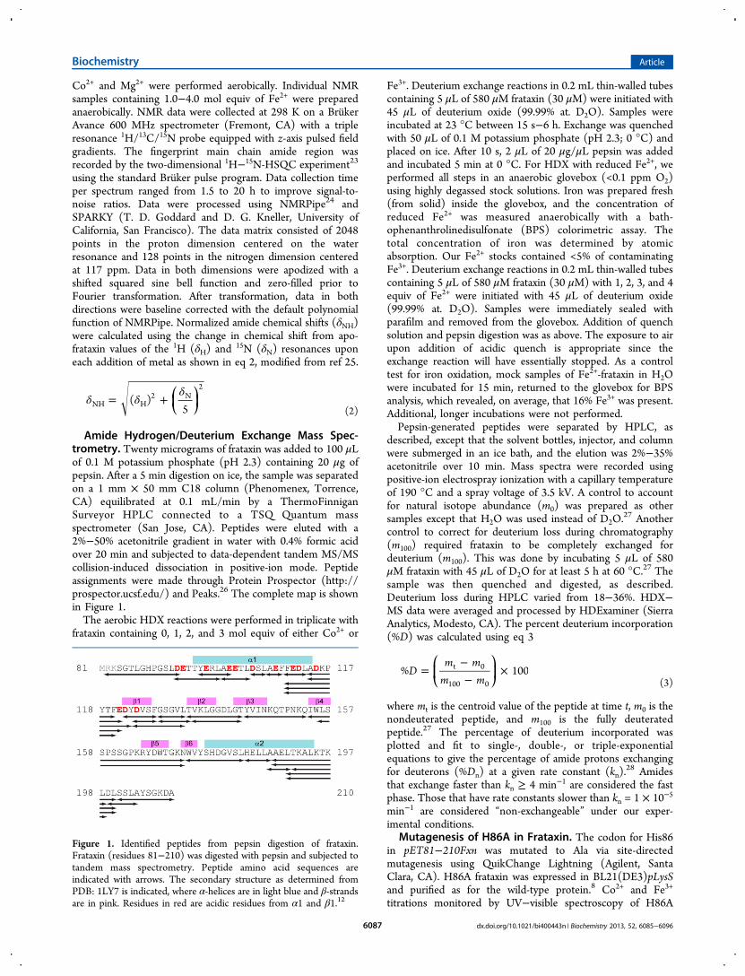
Amide Hydrogen/Deuterium Exchange Mass Spec-trometry. Twenty micrograms of frataxin was added to 100 μLof 0.1 M potassium phosphate (pH 2.3) containing 20 μg ofpepsin. After a 5 min digestion on ice, the sample was separatedon a 1 mm × 50 mm C18 column (Phenomenex, Torrence,CA) equilibrated at 0.1 mL/min by a ThermoFinniganSurveyor HPLC connected to a TSQ Quantum massspectrometer (San Jose, CA). Peptides were eluted with a2%−50% acetonitrile gradient in water with 0.4% formic acidover 20 min and subjected to data-dependent tandem MS/MScollision-induced dissociation in positive-ion mode. Peptideassignments were made through Protein Prospector (http://prospector.ucsf.edu/) and Peaks.26 The complete map is shownin Figure 1.The aerobic HDX reactions were performed in triplicate with
frataxin containing 0, 1, 2, and 3 mol equiv of either Co2+ or
Fe3+. Deuterium exchange reactions in 0.2 mL thin-walled tubescontaining 5 μL of 580 μM frataxin (30 μM) were initiated with45 μL of deuterium oxide (99.99% at. D2O). Samples wereincubated at 23 °C between 15 s−6 h. Exchange was quenchedwith 50 μL of 0.1 M potassium phosphate (pH 2.3; 0 °C) andplaced on ice. After 10 s, 2 μL of 20 μg/μL pepsin was addedand incubated 5 min at 0 °C. For HDX with reduced Fe2+, weperformed all steps in an anaerobic glovebox (<0.1 ppm O2)using highly degassed stock solutions. Iron was prepared fresh(from solid) inside the glovebox, and the concentration ofreduced Fe2+ was measured anaerobically with a bath-ophenanthrolinedisulfonate (BPS) colorimetric assay. Thetotal concentration of iron was determined by atomicabsorption. Our Fe2+ stocks contained <5% of contaminatingFe3+. Deuterium exchange reactions in 0.2 mL thin-walled tubescontaining 5 μL of 580 μM frataxin (30 μM) with 1, 2, 3, and 4equiv of Fe2+ were initiated with 45 μL of deuterium oxide(99.99% at. D2O). Samples were immediately sealed withparafilm and removed from the glovebox. Addition of quenchsolution and pepsin digestion was as above. The exposure to airupon addition of acidic quench is appropriate since theexchange reaction will have essentially stopped. As a controltest for iron oxidation, mock samples of Fe2+-frataxin in H2Owere incubated for 15 min, returned to the glovebox for BPSanalysis, which revealed, on average, that 16% Fe3+ was present.Additional, longer incubations were not performed.Pepsin-generated peptides were separated by HPLC, as
described, except that the solvent bottles, injector, and columnwere submerged in an ice bath, and the elution was 2%−35%acetonitrile over 10 min. Mass spectra were recorded usingpositive-ion electrospray ionization with a capillary temperatureof 190 °C and a spray voltage of 3.5 kV. A control to accountfor natural isotope abundance (m0) was prepared as othersamples except that H2O was used instead of D2O.
27 Anothercontrol to correct for deuterium loss during chromatography(m100) required frataxin to be completely exchanged fordeuterium (m100). This was done by incubating 5 μL of 580μM frataxin with 45 μL of D2O for at least 5 h at 60 °C.27 Thesample was then quenched and digested, as described.Deuterium loss during HPLC varied from 18−36%. HDX−MS data were averaged and processed by HDExaminer (SierraAnalytics, Modesto, CA). The percent deuterium incorporation(%D) was calculated using eq 3
=−−
×⎛⎝⎜
⎞⎠⎟D
m mm m
% 100t 0
100 0 (3)
where mt is the centroid value of the peptide at time t, m0 is thenondeuterated peptide, and m100 is the fully deuteratedpeptide.27 The percentage of deuterium incorporated wasplotted and fit to single-, double-, or triple-exponentialequations to give the percentage of amide protons exchangingfor deuterons (%Dn) at a given rate constant (kn).
28 Amidesthat exchange faster than kn ≥ 4 min−1 are considered the fastphase. Those that have rate constants slower than kn = 1 × 10⁻⁵
min−1 are considered “non-exchangeable” under our exper-imental conditions.
Mutagenesis of H86A in Frataxin. The codon for His86in pET81−210Fxn was mutated to Ala via site-directedmutagenesis using QuikChange Lightning (Agilent, SantaClara, CA). H86A frataxin was expressed in BL21(DE3)pLysSand purified as for the wild-type protein.8 Co2+ and Fe3+
titrations monitored by UV−visible spectroscopy of H86A
Figure 1. Identified peptides from pepsin digestion of frataxin.Frataxin (residues 81−210) was digested with pepsin and subjected totandem mass spectrometry. Peptide amino acid sequences areindicated with arrows. The secondary structure as determined fromPDB: 1LY7 is indicated, where α-helices are in light blue and β-strandsare in pink. Residues in red are acidic residues from α1 and β1.12
Biochemistry Article
dx.doi.org/10.1021/bi400443n | Biochemistry 2013, 52, 6085−60966087
frataxin were performed as for wild-type. The Fe2+ titration ofH86A frataxin in the presence of ferrozine chelator was also asdescribed for the wild-type frataxin protein.Purification of D37A ISCU. The pET-mISCU plasmid29
was graciously obtained from Dr. Tracey Rouault (NationalInstitutes of Health, Bethesda, MD). The codon for Asp37 wasmutated to that for Ala, and a stop codon was introduced afterthe codon for Lys133 using QuikChange mutagenesis (Agilent,Santa Clara, CA). D37A ISCU was expressed in BL21(DE3)pLysS and purified as described for native ISCU.8 D37A ISCUsamples were anaerobically dialyzed into Chelex-treated 50 mMHEPES (pH 7.4), 150 mM NaCl with 10% glycerol. Theconcentration of ISCU was determined spectroscopically usingε280 nm = 9.97 mM−1 cm−1.Fe−S Cluster Assembly Assays. All reactions were
performed in an anaerobic cuvette fitted with a gastight syringebefore removal from the glovebox. The reaction buffer was 25mM HEPES (pH 7.4) with 150 mM NaCl. Frataxin or H86Afrataxin (100 μM) was incubated with 200 μM ferrousammonium sulfate (2 Fe2+:1 frataxin) for 30 min at 25 °C.The reaction containing 100 μM D37A ISCU and 2.4 mMsodium sulfide was initiated by frataxin with 2 equiv of Fe2+ andwas monitored at 426 nm using an Agilent 8453 spectropho-tometer (Santa Clara, CA) in kinetics mode.7 Controlsincluded D37A ISCU with 200 μM ferrous ammonium sulfate(no frataxin), as well as with 200 μM Fe2+ bound to bovineserum albumin (BSA). The data were fit to a first-order rateequation (eq 4) to determine the initial rate of Fe−S clusterassembly,
= −Arate e k t1
1 (4)
where A1 is the amplitude of the first-order rate, k1, as afunction of time, t.
■ RESULTS
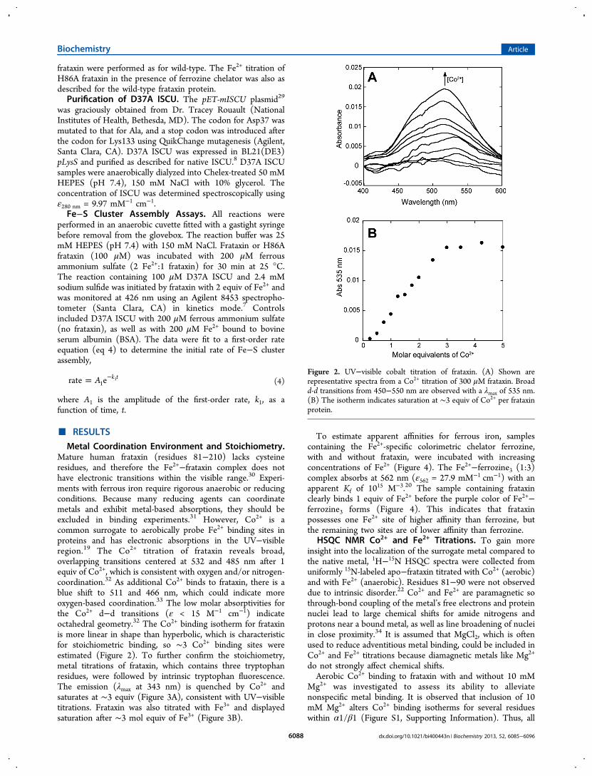
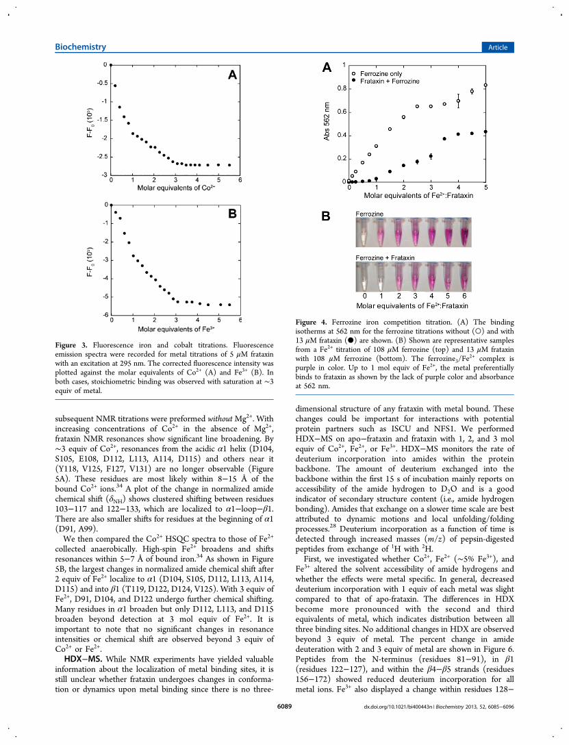
Metal Coordination Environment and Stoichiometry.Mature human frataxin (residues 81−210) lacks cysteineresidues, and therefore the Fe2+−frataxin complex does nothave electronic transitions within the visible range.30 Experi-ments with ferrous iron require rigorous anaerobic or reducingconditions. Because many reducing agents can coordinatemetals and exhibit metal-based absorptions, they should beexcluded in binding experiments.31 However, Co2+ is acommon surrogate to aerobically probe Fe2+ binding sites inproteins and has electronic absorptions in the UV−visibleregion.19 The Co2+ titration of frataxin reveals broad,overlapping transitions centered at 532 and 485 nm after 1equiv of Co2+, which is consistent with oxygen and/or nitrogen-coordination.32 As additional Co2+ binds to frataxin, there is ablue shift to 511 and 466 nm, which could indicate moreoxygen-based coordination.33 The low molar absorptivities forthe Co2+ d−d transitions (ε < 15 M−1 cm−1) indicateoctahedral geometry.32 The Co2+ binding isotherm for frataxinis more linear in shape than hyperbolic, which is characteristicfor stoichiometric binding, so ∼3 Co2+ binding sites wereestimated (Figure 2). To further confirm the stoichiometry,metal titrations of frataxin, which contains three tryptophanresidues, were followed by intrinsic tryptophan fluorescence.The emission (λmax at 343 nm) is quenched by Co2+ andsaturates at ∼3 equiv (Figure 3A), consistent with UV−visibletitrations. Frataxin was also titrated with Fe3+ and displayedsaturation after ∼3 mol equiv of Fe3+ (Figure 3B).
To estimate apparent affinities for ferrous iron, samplescontaining the Fe2+-specific colorimetric chelator ferrozine,with and without frataxin, were incubated with increasingconcentrations of Fe2+ (Figure 4). The Fe2+−ferrozine3 (1:3)complex absorbs at 562 nm (ε562 = 27.9 mM−1 cm−1) with anapparent Kf of 1015 M−3.20 The sample containing frataxinclearly binds 1 equiv of Fe2+ before the purple color of Fe2+−ferrozine3 forms (Figure 4). This indicates that frataxinpossesses one Fe2+ site of higher affinity than ferrozine, butthe remaining two sites are of lower affinity than ferrozine.
HSQC NMR Co2+ and Fe2+ Titrations. To gain moreinsight into the localization of the surrogate metal compared tothe native metal, 1H−15N HSQC spectra were collected fromuniformly 15N-labeled apo−frataxin titrated with Co2+ (aerobic)and with Fe2+ (anaerobic). Residues 81−90 were not observeddue to intrinsic disorder.22 Co2+ and Fe2+ are paramagnetic sothrough-bond coupling of the metal’s free electrons and proteinnuclei lead to large chemical shifts for amide nitrogens andprotons near a bound metal, as well as line broadening of nucleiin close proximity.34 It is assumed that MgCl2, which is oftenused to reduce adventitious metal binding, could be included inCo2+ and Fe2+ titrations because diamagnetic metals like Mg2+
do not strongly affect chemical shifts.Aerobic Co2+ binding to frataxin with and without 10 mM
Mg2+ was investigated to assess its ability to alleviatenonspecific metal binding. It is observed that inclusion of 10mM Mg2+ alters Co2+ binding isotherms for several residueswithin α1/β1 (Figure S1, Supporting Information). Thus, all
Figure 2. UV−visible cobalt titration of frataxin. (A) Shown arerepresentative spectra from a Co2+ titration of 300 μM frataxin. Broadd-d transitions from 450−550 nm are observed with a λmax of 535 nm.(B) The isotherm indicates saturation at ∼3 equiv of Co2+ per frataxinprotein.
Biochemistry Article
dx.doi.org/10.1021/bi400443n | Biochemistry 2013, 52, 6085−60966088
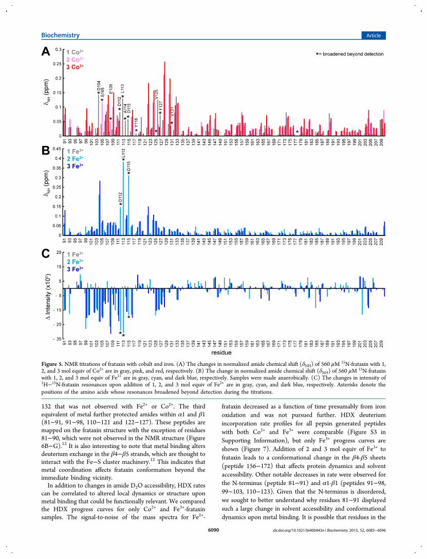
subsequent NMR titrations were preformed withoutMg2+. Withincreasing concentrations of Co2+ in the absence of Mg2+,frataxin NMR resonances show significant line broadening. By∼3 equiv of Co2+, resonances from the acidic α1 helix (D104,S105, E108, D112, L113, A114, D115) and others near it(Y118, V125, F127, V131) are no longer observable (Figure5A). These residues are most likely within 8−15 Å of thebound Co2+ ions.34 A plot of the change in normalized amidechemical shift (δNH) shows clustered shifting between residues103−117 and 122−133, which are localized to α1−loop−β1.There are also smaller shifts for residues at the beginning of α1(D91, A99).We then compared the Co2+ HSQC spectra to those of Fe2+
collected anaerobically. High-spin Fe2+ broadens and shiftsresonances within 5−7 Å of bound iron.34 As shown in Figure5B, the largest changes in normalized amide chemical shift after2 equiv of Fe2+ localize to α1 (D104, S105, D112, L113, A114,D115) and into β1 (T119, D122, D124, V125). With 3 equiv ofFe2+, D91, D104, and D122 undergo further chemical shifting.Many residues in α1 broaden but only D112, L113, and D115broaden beyond detection at 3 mol equiv of Fe2+. It isimportant to note that no significant changes in resonanceintensities or chemical shift are observed beyond 3 equiv ofCo2+ or Fe2+.HDX−MS. While NMR experiments have yielded valuable
information about the localization of metal binding sites, it isstill unclear whether frataxin undergoes changes in conforma-tion or dynamics upon metal binding since there is no three-
dimensional structure of any frataxin with metal bound. Thesechanges could be important for interactions with potentialprotein partners such as ISCU and NFS1. We performedHDX−MS on apo−frataxin and frataxin with 1, 2, and 3 molequiv of Co2+, Fe2+, or Fe3+. HDX−MS monitors the rate ofdeuterium incorporation into amides within the proteinbackbone. The amount of deuterium exchanged into thebackbone within the first 15 s of incubation mainly reports onaccessibility of the amide hydrogen to D2O and is a goodindicator of secondary structure content (i.e., amide hydrogenbonding). Amides that exchange on a slower time scale are bestattributed to dynamic motions and local unfolding/foldingprocesses.28 Deuterium incorporation as a function of time isdetected through increased masses (m/z) of pepsin-digestedpeptides from exchange of 1H with 2H.First, we investigated whether Co2+, Fe2+ (∼5% Fe3+), and
Fe3+ altered the solvent accessibility of amide hydrogens andwhether the effects were metal specific. In general, decreaseddeuterium incorporation with 1 equiv of each metal was slightcompared to that of apo-frataxin. The differences in HDXbecome more pronounced with the second and thirdequivalents of metal, which indicates distribution between allthree binding sites. No additional changes in HDX are observedbeyond 3 equiv of metal. The percent change in amidedeuteration with 2 and 3 equiv of metal are shown in Figure 6.Peptides from the N-terminus (residues 81−91), in β1(residues 122−127), and within the β4−β5 strands (residues156−172) showed reduced deuterium incorporation for allmetal ions. Fe3+ also displayed a change within residues 128−
Figure 3. Fluorescence iron and cobalt titrations. Fluorescenceemission spectra were recorded for metal titrations of 5 μM frataxinwith an excitation at 295 nm. The corrected fluorescence intensity wasplotted against the molar equivalents of Co2+ (A) and Fe3+ (B). Inboth cases, stoichiometric binding was observed with saturation at ∼3equiv of metal.
Figure 4. Ferrozine iron competition titration. (A) The bindingisotherms at 562 nm for the ferrozine titrations without (○) and with13 μM frataxin (●) are shown. (B) Shown are representative samplesfrom a Fe2+ titration of 108 μM ferrozine (top) and 13 μM frataxinwith 108 μM ferrozine (bottom). The ferrozine3/Fe
2+ complex ispurple in color. Up to 1 mol equiv of Fe2+, the metal preferentiallybinds to frataxin as shown by the lack of purple color and absorbanceat 562 nm.
Biochemistry Article
dx.doi.org/10.1021/bi400443n | Biochemistry 2013, 52, 6085−60966089
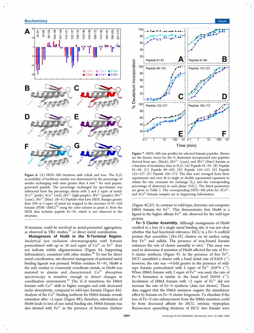
132 that was not observed with Fe2+ or Co2+. The thirdequivalent of metal further protected amides within α1 and β1(81−91, 91−98, 110−121 and 122−127). These peptides aremapped on the frataxin structure with the exception of residues81−90, which were not observed in the NMR structure (Figure6B−G).12 It is also interesting to note that metal binding altersdeuterium exchange in the β4−β5 strands, which are thought tointeract with the Fe−S cluster machinery.12 This indicates thatmetal coordination affects frataxin conformation beyond theimmediate binding vicinity.In addition to changes in amide D2O accessibility, HDX rates
can be correlated to altered local dynamics or structure uponmetal binding that could be functionally relevant. We comparedthe HDX progress curves for only Co2+ and Fe3+-frataxinsamples. The signal-to-noise of the mass spectra for Fe2+-
frataxin decreased as a function of time presumably from ironoxidation and was not pursued further. HDX deuteriumincorporation rate profiles for all pepsin generated peptideswith both Co2+ and Fe3+ were comparable (Figure S3 inSupporting Information), but only Fe3+ progress curves areshown (Figure 7). Addition of 2 and 3 mol equiv of Fe3+ tofrataxin leads to a conformational change in the β4-β5 sheets(peptide 156−172) that affects protein dynamics and solventaccessibility. Other notable decreases in rate were observed forthe N-terminus (peptide 81−91) and α1-β1 (peptides 91−98,99−103, 110−123). Given that the N-terminus is disordered,we sought to better understand why residues 81−91 displayedsuch a large change in solvent accessibility and conformationaldynamics upon metal binding. It is possible that residues in the
Figure 5. NMR titrations of frataxin with cobalt and iron. (A) The changes in normalized amide chemical shift (δNH) of 560 μM15N-frataxin with 1,
2, and 3 mol equiv of Co2+ are in gray, pink, and red, respectively. (B) The change in normalized amide chemical shift (δNH) of 560 μM15N-frataxin
with 1, 2, and 3 mol equiv of Fe2+ are in gray, cyan, and dark blue, respectively. Samples were made anaerobically. (C) The changes in intensity of1H−15N-frataxin resonances upon addition of 1, 2, and 3 mol equiv of Fe2+ are in gray, cyan, and dark blue, respectively. Asterisks denote thepositions of the amino acids whose resonances broadened beyond detection during the titrations.
Biochemistry Article
dx.doi.org/10.1021/bi400443n | Biochemistry 2013, 52, 6085−60966090
N-terminus could be involved in metal-promoted aggregation,as observed in Yfh1 studies,35 or direct metal coordination.Mutagenesis of His86 in the N-Terminal Region.
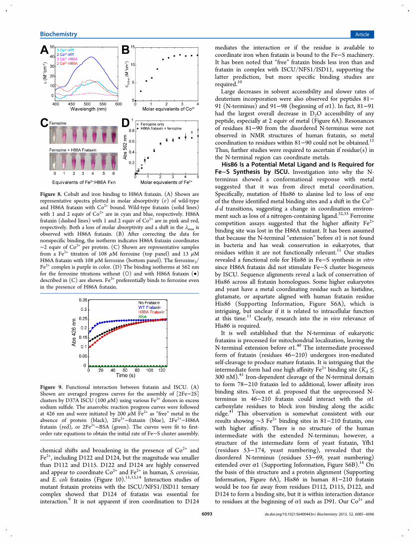
Analytical size exclusion chromatography with frataxinpreincubated with up to 10 mol equiv of Co2+ or Fe3+ doesnot indicate stabile oligomerization (Figure S4, SupportingInformation), consistent with other studies.36 To test for directmetal coordination, site-directed mutagenesis of potential metalbinding ligands was pursued. Within residues 81−91, His86 isthe only residue to commonly coordinate metals, so His86 wasmutated to alanine and characterized. Co2+ absorptionspectroscopy is sensitive enough to detect changes incoordination environment.32 The d−d transitions for H86Afrataxin with Co2+ shift to higher energies and with decreasedmolar absorptivity, compared to wild-type frataxin (Figure 8A).Analysis of the Co2+ binding isotherm for H86A frataxin revealssaturation after ∼2 equiv (Figure 8B); therefore, substitution ofHis86 leads to loss of one metal binding site. H86A frataxin wasalso titrated with Fe2+ in the presence of ferrozine chelator
(Figure 8C,D). In contrast to wild-type, ferrozine out-competesH86A frataxin for Fe2+. This demonstrates that His86 is aligand in the higher affinity Fe2+ site observed for the wild-typeprotein.
Fe−S Cluster Assembly. Although mutagenesis of His86resulted in a loss of a single metal binding site, it was not clearwhether this had functional relevance. ISCU is a Fe−S scaffoldprotein that assembles [2Fe-2S] clusters on its surface usingfree Fe2+ and sulfide. The presence of iron-bound frataxinenhances the rate of cluster assembly in vitro.7 This assay wasused to determine if mutation of His86 affected the rate of Fe−S cluster synthesis (Figure 9). In the presence of free Fe2+,ISCU assembled a cluster with a basal initial rate of 0.018 s−1;however, the rate was ∼4-fold greater in the presence of wild-type frataxin preincubated with 2 equiv of Fe2+ (0.074 s−1).When H86A frataxin with 2 equiv of Fe2+ was used, the rate ofFe−S formation is similar to the basal level (0.018 s−1).Incubation of H86A frataxin with >2 equiv of Fe2+ did notincrease the rate of Fe−S synthesis (data not shown). Thesedata suggest that the H86A mutation negates the stimulatoryeffect by frataxin on Fe−S cluster biogenesis. To elucidate if theloss of Fe−S rate enhancement from the H86A mutation couldbe from decreased affinity for ISCU, intrinsic tryptophanfluorescence quenching titrations of ISCU into frataxin were
Figure 6. (A) HDX−MS titrations with cobalt and iron. The D2Oaccessibility of backbone amides was determined by the percentage ofamides exchanging with rates greater than 4 min−1 for each pepsin-generated peptide. The percentage exchanged for apo-frataxin wassubtracted from the percentage obtain with 2 and 3 equiv of metal:2Co2+ (pink); 3Co2+ (red); 2Fe2+ (light purple); 3Fe2+ (purple); 2Fe3+
(cyan); 3Fe3+ (blue). (B−G) Peptides that have HDX changes greaterthan 10% at 3 equiv of metal are mapped to the structure of 91−210frataxin (PDB: 1EKG)42 using the color scheme in panel A. Note theHDX data includes peptide 81−91, which is not observed in thestructure.
Figure 7. HDX−MS rate profiles for selected frataxin peptides. Shownare the kinetic traces for the % deuterium incorporated into peptidesderived from apo- (black), 2Fe3+- (cyan), and 3Fe3+-(blue) frataxin asa function of incubation time in D2O. (A) Peptide 81−91. (B) Peptide91−98. (C) Peptide 99−103. (D) Peptide 110−123. (E) Peptide122−127. (F) Peptide 156−172. The data were averaged from threeexperiments and were fit to single or double exponential equations toobtain the rate constants for exchange (kn) and the correspondingpercentage of deuterons in each phase (%Dn). The fitted parametersare given in Table 1. The corresponding HDX−MS plots for 2Co2+-and 3Co2+-frataxin samples are in Supporting Information.
Biochemistry Article
dx.doi.org/10.1021/bi400443n | Biochemistry 2013, 52, 6085−60966091

performed with and without Fe3+, as described.37 Thefluorescence of wild type and H86A frataxin was quenchedby ISCU only in the presence of Fe3+ (Figure S5, SupportingInformation). H86A only had a 2-fold lower KD for ISCU thanfor wild type frataxin. Together, these studies show that His86is important for iron binding and in the formation of Fe−Sclusters by ISCU in vitro but does not seem to play a direct rolein ISCU binding.
■ DISCUSSION
Human frataxin is a unique Fe2+-binding protein in that themature form does not contain cysteine residues, which arepreferred ligands based on hard−soft acid base theory.38
Instead, a cluster of aspartate and glutamate residues along oneface of α1 helix creates multiple binding sites for Fe2+ (Figure1).11 This region has hampered direct determination of frataxinmetal stoichiometry from a variety of approaches since itsinteractions with metals could also be nonspecific. For a betterunderstanding of frataxin molecular interactions, metalcoordination as related to structure and function wasinvestigated. From this study, we propose that His86 is apreviously unidentified iron ligand and that it plays a functionalrole in Fe−S synthesis with the scaffold protein ISCU.Localization of Metal Binding Sites in Frataxin Using
Paramagnetic NMR and HDX-MS. Our studies cumulativelysuggest that mature frataxin can bind up to three metal ions.One Fe2+ binding site has higher affinity than the other twosites based on chelator competition assays. In these studies, asignificant population of “free” metal exists so differences inaffinity can be discerned. In other experiments, the frataxinconcentration is high enough that stoichiometric binding wasobserved. It also led to metal distribution among all three sites.
At this time, it is not possible to determine if the two weakermetal binding sites observed in this study are nonspecificbecause actual equilibrium binding constants have not beendetermined. Further functional roles for these residues must beascertained. The HSQC NMR metal titrations in the presenceof Mg2+ attempted to control nonspecific metal coordination,but Mg2+ competed with Co2+ for binding at or near acidicresidues within α1−loop−β1 (Figure S1, Supporting Informa-tion). This suggests that electrostatic interactions with theacidic α1 helix are possible.18 Some of these acidic residues areimportant for interaction with ISCU and other components ofthe Fe−S machinery and not for iron binding.9
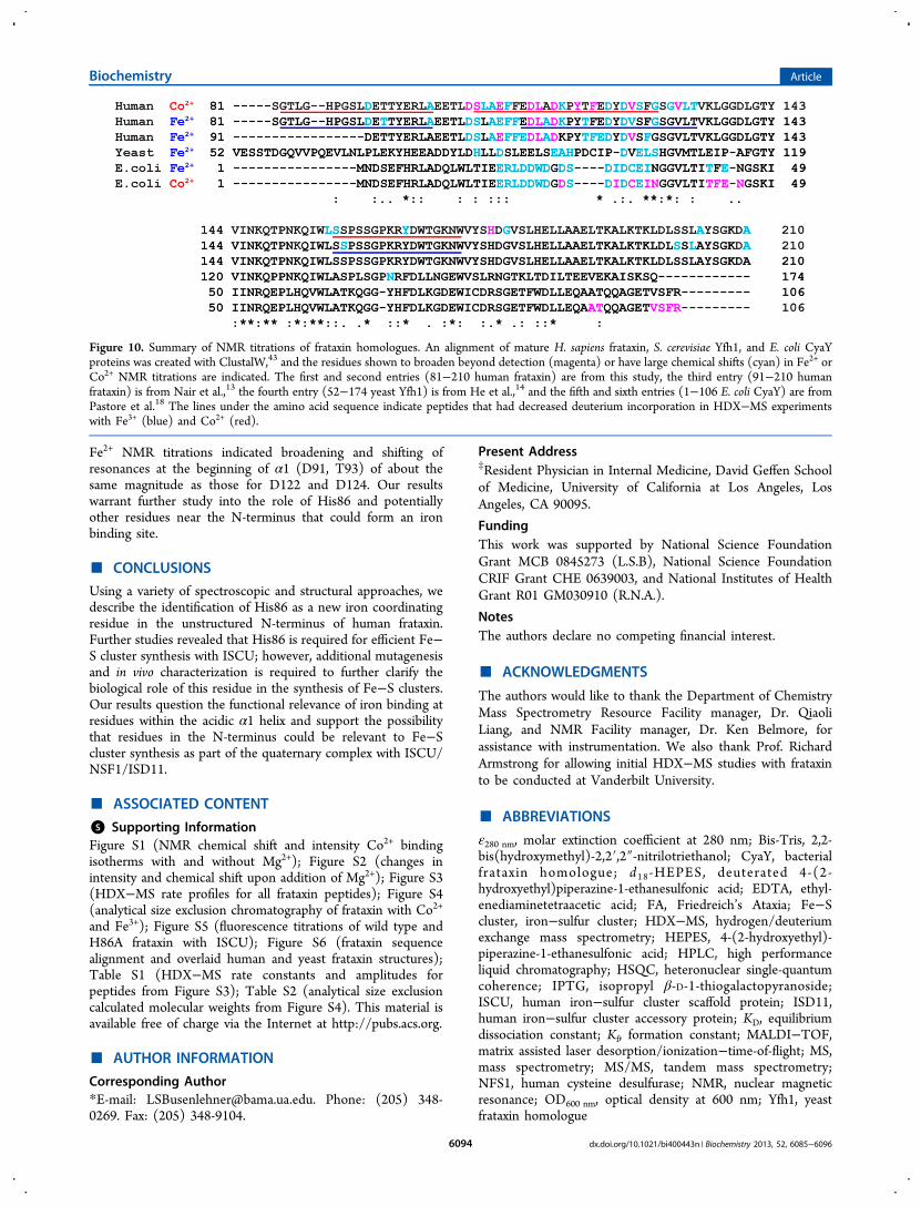
When the Co2+ and Fe2+ NMR results with 81−210 frataxinare compared with those for truncated human frataxin (residues91−210), S. cerevisiae Yfh1, and E. coli CyaY, similarities areobserved that allow for the assignment of several potentialligands (Figure 10). D112 and D115 from the α1 helix arelikely metal coordinating residues since their NMR resonanceswere broadened beyond detection with Co2+ and Fe2+ in ourexperiments and in other homologues (Figure 10).13,14,18
Acidic residues at positions 112 and 115 are conserved in mosteukaryotic frataxins.11 HDX−MS studies showed that thepeptide containing D112 and D115 (110−123) incorporatedless deuterium into the backbone in the presence of metal(Figure 7D). In combination with NMR results, this isconsistent with reduced solvent accessibility and dynamicsfrom metal coordination.39
HDX−MS also revealed that amides within residues 122−127 in β1 were protected from D2O in the presence of metalwithout a measurable change in the rate of uptake (Figure 7E).In other words, metal binding does not significantly perturbstructure in β1. Several residues in this region had altered NMR
Table 1. HDX−MS Rate Constants and Amplitudes for Peptides from Figure 7a
peptide %DPre‑Exb %D1 k1 (min
−1) “fast” %D2 k2 (min−1) “intermediate” %D3 k3 (min‑1) “slow”
81−91apo ∼1002 Fe3+ ∼81 17.8 (±1.0) ≤1 × 10⁻⁴
3 Fe3+ ∼73 27.0 (±1.4) ≤1 × 10⁻⁴
91−98apo ∼47 35.7 (±4.5) 2.2 (±0.4) 17.5 (±1.3) 0.040 (±0.007)2 Fe3+ ∼47 31.5 (±11.2) 2.7 (±1.2) 21.9 (±2.2) 0.031 (±0.007)3 Fe3+ ∼26 53.3 (±8.8) 1.4 (±0.4) 20.3 (±2.4) 0.007 (±0.002)99−103apo 0 100 (±1.2) 0.022 (±0.001)2 Fe3+ 0 67.1 (±2.6) 0.032 (±0.002) ∼33 ≤1 × 10⁻⁴
3 Fe3+ 0 76.9 (±1.5) 0.014 (±0.001) ∼23 ≤1 × 10⁻⁴
110−123apo ∼23 36.8 (±5.0) 1.1 (±0.3) 40.1 (±2.3) 0.018 (±0.002)2 Fe3+ ∼12 34.3 (±5.0) 1.4 (±0.4) 38.1 (±3.6) 0.06 (±0.01) 15.6 (±3.3) 0.002 (±0.001)3 Fe3+ ∼2 28.7(±7.5) 2.0 (±1.0) 32.5 (±3.9) 0.10 (±0.02) 37.4 (±2.4) 0.0030 (±0.0005)122−127apo ∼29 31 (±4) 2.2 (±0.4) 40 (±0.5) ≤1 × 10⁻⁴
2 Fe3+ ∼18 30 (±4) 0.9 (±0.2) 52 (±1) ≤1 × 10⁻⁴
3 Fe3+ ∼18 30 (±3) 0.5 (±0.1) 52 (±1) ≤1 × 10⁻⁴
156−172apo ∼33 28 (±7) 1.4 (±0.6) 39 (±2) 0.010 (±0.002)2 Fe3+ ∼25 24 (±9) 2.1 (±0.5) 19 (±4) 0.06 (±0.04) 32 (±4) 0.0017 (±0.0005)3 Fe3+ ∼29 21 (±2) 2.3 (±0.5) 22 (±3) 0.05 (±0.01) 28 (±3) 0.0012 (±0.0005)
aParameters obtained from fitting the H/D-exchange kinetics of frataxin peptides with and without metal (Figure 7A−F) according to a single,double or triple exponential equations. The rates have been loosely grouped in to fast, intermediate and slow exchange. bThe amount of exchangebefore the first time point is estimated from the fit parameters and is assigned a rate of exchange >4 min−1.28
Biochemistry Article
dx.doi.org/10.1021/bi400443n | Biochemistry 2013, 52, 6085−60966092
chemical shifts and broadening in the presence of Co2+ andFe2+, including D122 and D124, but the magnitude was smallerthan D112 and D115. D122 and D124 are highly conservedand appear to coordinate Co2+ and Fe2+ in human, S. cerevisiae,and E. coli frataxins (Figure 10).11,13,14 Interaction studies ofmutant frataxin proteins with the ISCU/NFS1/ISD11 ternarycomplex showed that D124 of frataxin was essential forinteraction.9 It is not apparent if iron coordination to D124
mediates the interaction or if the residue is available tocoordinate iron when frataxin is bound to the Fe−S machinery.It has been noted that “free” frataxin binds less iron than andfrataxin in complex with ISCU/NFS1/ISD11, supporting thelatter prediction, but more specific binding studies arerequired.10
Large decreases in solvent accessibility and slower rates ofdeuterium incorporation were also observed for peptides 81−91 (N-terminus) and 91−98 (beginning of α1). In fact, 81−91had the largest overall decrease in D2O accessibility of anypeptide, especially at 2 equiv of metal (Figure 6A). Resonancesof residues 81−90 from the disordered N-terminus were notobserved in NMR structures of human frataxin, so metalcoordination to residues within 81−90 could not be obtained.12Thus, further studies were required to ascertain if residue(s) inthe N-terminal region can coordinate metals.
His86 Is a Potential Metal Ligand and Is Required forFe−S Synthesis by ISCU. Investigation into why the N-terminus showed a conformational response with metalsuggested that it was from direct metal coordination.Specifically, mutation of His86 to alanine led to loss of oneof the three identified metal binding sites and a shift in the Co2+
d-d transitions, suggesting a change in coordination environ-ment such as loss of a nitrogen-containing ligand.32,33 Ferrozinecompetition assays suggested that the higher affinity Fe2+
binding site was lost in the H86A mutant. It has been assumedthat because the N-terminal “extension” before α1 is not foundin bacteria and has weak conservation in eukaryotes, thatresidues within it are not functionally relevant.11 Our studiesrevealed a functional role for His86 in Fe−S synthesis in vitrosince H86A frataxin did not stimulate Fe−S cluster biogenesisby ISCU. Sequence alignments reveal a lack of conservation ofHis86 across all frataxin homologues. Some higher eukaryotesand yeast have a metal coordinating residue such as histidine,glutamate, or aspartate aligned with human frataxin residueHis86 (Supporting Information, Figure S6A), which isintriguing, but unclear if it is related to intracellular functionat this time.11 Clearly, research into the in vivo relevance ofHis86 is required.It is well established that the N-terminus of eukaryotic
frataxins is processed for mitochondrial localization, leaving theN-terminal extension before α1.40 The intermediate processedform of frataxin (residues 46−210) undergoes iron-mediatedself-cleavage to produce mature frataxin. It is intriguing that theintermediate form had one high affinity Fe2+ binding site (Kd ≤300 nM).41 Iron-dependent cleavage of the N-terminal domainto form 78−210 frataxin led to additional, lower affinity ironbinding sites. Yoon et al. proposed that the unprocessed N-terminus in 46−210 frataxin could interact with the α1carboxylate residues to block iron binding along the acidicridge.41 This observation is somewhat consistent with ourresults showing ∼3 Fe2+ binding sites in 81−210 frataxin, onewith higher affinity. There is no structure of the humanintermediate with the extended N-terminus; however, astructure of the intermediate form of yeast frataxin, Yfh1(residues 53−174, yeast numbering), revealed that thedisordered N-terminus (residues 53−69, yeast numbering)extended over α1 (Supporting Information, Figure S6B).14 Onthe basis of this structure and a protein alignment (SupportingInformation, Figure 6A), His86 in human 81−210 frataxinwould be too far away from residues D112, D115, D122, andD124 to form a binding site, but it is within interaction distanceto residues at the beginning of α1 such as D91. Our Co2+ and
Figure 8. Cobalt and iron binding to H86A frataxin. (A) Shown arerepresentative spectra plotted in molar absorptivity (ε) of wild-typeand H86A frataxin with Co2+ bound. Wild-type frataxin (solid lines)with 1 and 2 equiv of Co2+ are in cyan and blue, respectively. H86Afrataxin (dashed lines) with 1 and 2 equiv of Co2+ are in pink and red,respectively. Both a loss of molar absorptivity and a shift in the λmax isobserved with H86A frataxin. (B) After correcting the data fornonspecific binding, the isotherm indicates H86A frataxin coordinates∼2 equiv of Co2+ per protein. (C) Shown are representative samplesfrom a Fe2+ titration of 108 μM ferrozine (top panel) and 13 μMH86A frataxin with 108 μM ferrozine (bottom panel). The ferrozine3/Fe2+ complex is purple in color. (D) The binding isotherms at 562 nmfor the ferrozine titrations without (○) and with H86A frataxin (•)described in (C) are shown. Fe2+ preferentially binds to ferrozine evenin the presence of H86A frataxin.
Figure 9. Functional interaction between frataxin and ISCU. (A)Shown are averaged progress curves for the assembly of [2Fe−2S]clusters by D37A ISCU (100 μM) using various Fe2+ donors in excesssodium sulfide. The anaerobic reaction progress curves were followedat 426 nm and were initiated by 200 μM Fe2+ as “free” metal in theabsence of protein (black), 2Fe2+−frataxin (blue), 2Fe2+−H86Afrataxin (red), or 2Fe2+−BSA (green). The curves were fit to first-order rate equations to obtain the initial rate of Fe−S cluster assembly.
Biochemistry Article
dx.doi.org/10.1021/bi400443n | Biochemistry 2013, 52, 6085−60966093
Fe2+ NMR titrations indicated broadening and shifting ofresonances at the beginning of α1 (D91, T93) of about thesame magnitude as those for D122 and D124. Our resultswarrant further study into the role of His86 and potentiallyother residues near the N-terminus that could form an ironbinding site.
■ CONCLUSIONS
Using a variety of spectroscopic and structural approaches, wedescribe the identification of His86 as a new iron coordinatingresidue in the unstructured N-terminus of human frataxin.Further studies revealed that His86 is required for efficient Fe−S cluster synthesis with ISCU; however, additional mutagenesisand in vivo characterization is required to further clarify thebiological role of this residue in the synthesis of Fe−S clusters.Our results question the functional relevance of iron binding atresidues within the acidic α1 helix and support the possibilitythat residues in the N-terminus could be relevant to Fe−Scluster synthesis as part of the quaternary complex with ISCU/NSF1/ISD11.
■ ASSOCIATED CONTENT
*S Supporting InformationFigure S1 (NMR chemical shift and intensity Co2+ bindingisotherms with and without Mg2+); Figure S2 (changes inintensity and chemical shift upon addition of Mg2+); Figure S3(HDX−MS rate profiles for all frataxin peptides); Figure S4(analytical size exclusion chromatography of frataxin with Co2+
and Fe3+); Figure S5 (fluorescence titrations of wild type andH86A frataxin with ISCU); Figure S6 (frataxin sequencealignment and overlaid human and yeast frataxin structures);Table S1 (HDX−MS rate constants and amplitudes forpeptides from Figure S3); Table S2 (analytical size exclusioncalculated molecular weights from Figure S4). This material isavailable free of charge via the Internet at http://pubs.acs.org.
■ AUTHOR INFORMATION
Corresponding Author*E-mail: [email protected]. Phone: (205) 348-0269. Fax: (205) 348-9104.
Present Address‡Resident Physician in Internal Medicine, David Geffen Schoolof Medicine, University of California at Los Angeles, LosAngeles, CA 90095.
FundingThis work was supported by National Science FoundationGrant MCB 0845273 (L.S.B), National Science FoundationCRIF Grant CHE 0639003, and National Institutes of HealthGrant R01 GM030910 (R.N.A.).
NotesThe authors declare no competing financial interest.
■ ACKNOWLEDGMENTS
The authors would like to thank the Department of ChemistryMass Spectrometry Resource Facility manager, Dr. QiaoliLiang, and NMR Facility manager, Dr. Ken Belmore, forassistance with instrumentation. We also thank Prof. RichardArmstrong for allowing initial HDX−MS studies with frataxinto be conducted at Vanderbilt University.
■ ABBREVIATIONS
ε280 nm, molar extinction coefficient at 280 nm; Bis-Tris, 2,2-bis(hydroxymethyl)-2,2′,2″-nitrilotriethanol; CyaY, bacterialfrataxin homologue; d18-HEPES, deuterated 4-(2-hydroxyethyl)piperazine-1-ethanesulfonic acid; EDTA, ethyl-enediaminetetraacetic acid; FA, Friedreich’s Ataxia; Fe−Scluster, iron−sulfur cluster; HDX−MS, hydrogen/deuteriumexchange mass spectrometry; HEPES, 4-(2-hydroxyethyl)-piperazine-1-ethanesulfonic acid; HPLC, high performanceliquid chromatography; HSQC, heteronuclear single-quantumcoherence; IPTG, isopropyl β-D-1-thiogalactopyranoside;ISCU, human iron−sulfur cluster scaffold protein; ISD11,human iron−sulfur cluster accessory protein; KD, equilibriumdissociation constant; Kf, formation constant; MALDI−TOF,matrix assisted laser desorption/ionization−time-of-flight; MS,mass spectrometry; MS/MS, tandem mass spectrometry;NFS1, human cysteine desulfurase; NMR, nuclear magneticresonance; OD600 nm, optical density at 600 nm; Yfh1, yeastfrataxin homologue
Figure 10. Summary of NMR titrations of frataxin homologues. An alignment of mature H. sapiens frataxin, S. cerevisiae Yfh1, and E. coli CyaYproteins was created with ClustalW,43 and the residues shown to broaden beyond detection (magenta) or have large chemical shifts (cyan) in Fe2+ orCo2+ NMR titrations are indicated. The first and second entries (81−210 human frataxin) are from this study, the third entry (91−210 humanfrataxin) is from Nair et al.,13 the fourth entry (52−174 yeast Yfh1) is from He et al.,14 and the fifth and sixth entries (1−106 E. coli CyaY) are fromPastore et al.18 The lines under the amino acid sequence indicate peptides that had decreased deuterium incorporation in HDX−MS experimentswith Fe3+ (blue) and Co2+ (red).
Biochemistry Article
dx.doi.org/10.1021/bi400443n | Biochemistry 2013, 52, 6085−60966094

■ REFERENCES(1) Campuzano, V., Montermini, L., Molto, M. D., Pianese, L.,Cossee, M., Cavalcanti, F., Monros, E., Rodius, F., Duclos, F.,Monticelli, A., Zara, F., Canizares, J., Koutnikova, H., Bidichandani, S.I., Gellera, C., Brice, A., Trouillas, P., De Michele, G., Filla, A., DeFrutos, R., Palau, F., Patel, P. I., Di Donato, S., Mandel, J. L., Cocozza,S., Koenig, M., and Pandolfo, M. (1996) Friedreich’s ataxia: autosomalrecessive disease caused by an intronic GAA triplet repeat expansion.Science 271, 1423−1427.(2) Schmucker, S., and Puccio, H. (2010) Understanding themolecular mechanisms of Friedreich’s ataxia to develop therapeuticapproaches. Hum. Mol. Genet. 19, R103−110.(3) Gonzalez-Cabo, P., Llorens, J. V., Palau, F., and Molto, M. D.(2009) Friedreich ataxia: an update on animal models, frataxinfunction and therapies. Adv. Exp. Med. Biol. 652, 247−261.(4) Rouault, T. A., and Tong, W. H. (2008) Iron-sulfur clusterbiogenesis and human disease. Trends Genet. 24, 398−407.(5) Ye, H., and Rouault, T. A. (2010) Human iron-sulfur clusterassembly, cellular iron homeostasis, and disease. Biochemistry 49,4945−4956.(6) Muhlenhoff, U., Richhardt, N., Ristow, M., Kispal, G., and Lill, R.(2002) The yeast frataxin homolog Yfh1p plays a specific role in thematuration of cellular Fe/S proteins. Hum. Mol. Genet. 11, 2025−2036.(7) Kondapalli, K. C., Kok, N. M., Dancis, A., and Stemmler, T. L.(2008) Drosophila frataxin: an iron chaperone during cellular Fe-Scluster bioassembly. Biochemistry 47, 6917−6927.(8) Watson, H. M., Gentry, L. E., Asuru, A. P., Wang, Y., Marcus, S.,and Busenlehner, L. S. (2012) Heterotrifunctional chemical cross-linking mass spectrometry confirms physical interaction betweenhuman frataxin and ISU. Biochemistry 51, 6889−6891.(9) Schmucker, S., Martelli, A., Colin, F., Page, A., Wattenhofer-Donze, M., Reutenauer, L., and Puccio, H. (2011) Mammalianfrataxin: an essential function for cellular viability through aninteraction with a preformed ISCU/NFS1/ISD11 iron-sulfur assemblycomplex. PLoS One 6, e16199.(10) Colin, F., Martelli, A., Clemancey, M., Latour, J. M., Gambarelli,S., Zeppieri, L., Birck, C., Page, A., Puccio, H., and Ollagnier deChoudens, S. (2013) Mammalian Frataxin Controls Sulfur Productionand Iron Entry during de Novo Fe(4)S(4) Cluster Assembly. J. Am.Chem. Soc. 135, 733−740.(11) Pandolfo, M., and Pastore, A. (2009) The pathogenesis ofFriedreich ataxia and the structure and function of frataxin. J. Neurol.256 (Suppl 1), 9−17.(12) Musco, G., Stier, G., Kolmerer, B., Adinolfi, S., Martin, S.,Frenkiel, T., Gibson, T., and Pastore, A. (2000) Towards a structuralunderstanding of Friedreich’s ataxia: the solution structure of frataxin.Structure 8, 695−707.(13) Nair, M., Adinolfi, S., Pastore, C., Kelly, G., Temussi, P., andPastore, A. (2004) Solution structure of the bacterial frataxin ortholog,CyaY: mapping the iron binding sites. Structure 12, 2037−2048.(14) He, Y., Alam, S. L., Proteasa, S. V., Zhang, Y., Lesuisse, E.,Dancis, A., and Stemmler, T. L. (2004) Yeast frataxin solutionstructure, iron binding, and ferrochelatase interaction. Biochemistry 43,16254−16262.(15) Cook, J. D., Bencze, K. Z., Jankovic, A. D., Crater, A. K., Busch,C. N., Bradley, P. B., Stemmler, A. J., Spaller, M. R., and Stemmler, T.L. (2006) Monomeric yeast frataxin is an iron-binding protein.Biochemistry 45, 7767−7777.(16) Lane, D. J., and Richardson, D. R. (2010) Frataxin, a moleculeof mystery: trading stability for function in its iron-binding site.Biochem. J. 426, e1−3.(17) Huang, J., Dizin, E., and Cowan, J. (2008) Mapping iron bindingsites on human frataxin: implications for cluster assembly on the ISUFe−S cluster scaffold protein. J. Biol. Inorg. Chem 13, 825−836.(18) Pastore, C., Franzese, M., Sica, F., Temussi, P., and Pastore, A.(2007) Understanding the binding properties of an unusual metal-binding protein–a study of bacterial frataxin. FEBS. J. 274, 4199−4210.(19) Maret, W., and Vallee, B. L. (1993) Cobalt as probe and label ofproteins. Methods Enzymol. 226, 52−71.
(20) Thompsen, J. C., and Mottola, H. A. (1984) Kinetics of theComplexation of Iron(II) with Ferrozine. Anal. Chem. 56, 755−757.(21) van de Weert, M. (2010) Fluorescence quenching to studyprotein-ligand binding: common errors. J. Fluoresc. 20, 625−629.(22) Musco, G., de Tommasi, T., Stier, G., Kolmerer, B., Bottomley,M., Adinolfi, S., Muskett, F. W., Gibson, T. J., Frenkiel, T. A., andPastore, A. (1999) Assignment of the 1H, 15N, and 13C resonances ofthe C-terminal domain of frataxin, the protein responsible forFriedreich ataxia. J. Biomol. NMR 15, 87−88.(23) Kay, L., Keifer, P., and Saarinen, T. (1992) Pure absorptiongradient enhanced heteronuclear single quantum correlation spectros-copy with improved sensitivity. J. Am. Chem. Soc. 114, 10663−10665.(24) Delaglio, F., Grzesiek, S., Vuister, G. W., Zhu, G., Pfeifer, J., andBax, A. (1995) NMRpipe: A multidimensional spectral processingsystem based on Unix pipes. J. Biomol. NMR 6, 227−293.(25) Grzesiek, S., Stahl, S. J., Wingfield, P. T., and Bax, A. (1996) TheCD4 determinant for downregulation by HIV-1 Nef directly binds toNef. Mapping of the Nef binding surface by NMR. Biochemistry 35,10256−10261.(26) Ma, B., Zhang, K., Hendrie, C., Liang, C., Li, M., Doherty-Kirby,A., and Lajoie, G. (2003) PEAKS: powerful software for peptide denovo sequencing by tandem mass spectrometry. Rapid Commun. MassSpectrom. 17, 2337−2342.(27) Engen, J. R., and Smith, D. L. (2001) Investigating proteinstructure and dynamics by hydrogen exchange MS. Anal. Chem. 73,256A−265A.(28) Busenlehner, L. S., and Armstrong, R. N. (2005) Insights intoenzyme structure and dynamics elucidated by amide H/D exchangemass spectrometry. Arch. Biochem. Biophys. 433, 34−46.(29) Tong, W. H., and Rouault, T. A. (2006) Functions ofmitochondrial ISCU and cytosolic ISCU in mammalian iron-sulfurcluster biogenesis and iron homeostasis. Cell Metab. 3, 199−210.(30) Solomon, E. I., Brunold, T. C., Davis, M. I., Kemsley, J. N., Lee,S. K., Lehnert, N., Neese, F., Skulan, A. J., Yang, Y. S., and Zhou, J.(2000) Geometric and electronic structure/function correlations innon-heme iron enzymes. Chem. Rev. 100, 235−350.(31) Krezel, A., Lesniak, W., Jezowska-Bojczuk, M., Mlynarz, P.,Brasun, J., Kozlowski, H., and Bal, W. (2001) Coordination of heavymetals by dithiothreitol, a commonly used thiol group protectant. J.Inorg. Biochem. 84, 77−88.(32) Bertini, I., and Luchinat, C. (1984) High spin cobalt(II) as aprobe for the investigation of metalloproteins. Adv. Inorg. Biochem. 6,71−111.(33) Dian, C., Vitale, S., Leonard, G. A., Bahlawane, C., Fauquant, C.,Leduc, D., Muller, C., de Reuse, H., Michaud-Soret, I., and Terradot,L. (2011) The structure of the Helicobacter pylori ferric uptakeregulator Fur reveals three functional metal binding sites. Mol.Microbiol. 79, 1260−1275.(34) Otting, G. (2010) Protein NMR using paramagnetic ions. Annu.Rev. Biophys. 39, 387−405.(35) Li, H., Gakh, O., Smith, D. Y. t., and Isaya, G. (2009)Oligomeric yeast frataxin drives assembly of core machinery formitochondrial iron-sulfur cluster synthesis. J. Biol. Chem. 284, 21971−21980.(36) Gakh, O., Bedekovics, T., Duncan, S. F., Smith, D. Y. t.,Berkholz, D. S., and Isaya, G. (2010) Normal and Friedreich ataxiacells express different isoforms of frataxin with complementary roles iniron-sulfur cluster assembly. J. Biol. Chem. 285, 38486−38501.(37) Yoon, T., and Cowan, J. A. (2003) Iron-sulfur clusterbiosynthesis. Characterization of frataxin as an iron donor for assemblyof [2Fe-2S] clusters in ISU-type proteins. J. Am. Chem. Soc. 125,6078−6084.(38) Pearson, R. G. (1968) Hard and Soft Acids and Bases HSAB 1.Fundamental Principles. J. Chem. Educ. 45, 581−.(39) Asuru, A. P., and Busenlehner, L. S. (2011) Analysis of humanferrochelatase iron binding via amide hydrogen/deuterium exchangemass spectrometry. Intl. J. Mass Spectrom. 302, 76−84.(40) Cavadini, P., Adamec, J., Taroni, F., Gakh, O., and Isaya, G.(2000) Two-step processing of human frataxin by mitochondrial
Biochemistry Article
dx.doi.org/10.1021/bi400443n | Biochemistry 2013, 52, 6085−60966095
processing peptidase. Precursor and intermediate forms are cleaved atdifferent rates. J. Biol. Chem. 275, 41469−41475.(41) Yoon, T., Dizin, E., and Cowan, J. A. (2007) N-terminal iron-mediated self-cleavage of human frataxin: regulation of iron bindingand complex formation with target proteins. J. Biol. Inorg. Chem. 12,535−542.(42) Dhe-Paganon, S., Shigeta, R., Chi, Y. I., Ristow, M., andShoelson, S. E. (2000) Crystal structure of human frataxin. J. Biol.Chem. 275, 30753−30756.(43) Larkin, M. A., Blackshields, G., Brown, N. P., Chenna, R.,McGettigan, P. A., McWilliam, H., Valentin, F., Wallace, I. M., Wilm,A., Lopez, R., Thompson, J. D., Gibson, T. J., and Higgins, D. G.(2007) Clustal W and Clustal X version 2.0. Bioinformatics 23, 2947−2948.
Biochemistry Article
dx.doi.org/10.1021/bi400443n | Biochemistry 2013, 52, 6085−60966096


![Interpolation via Barycentric Coordinates · • Moving least squares coordinates [Manson and Schaefer, 2010] • Cubic mean value coordinates [Li and Hu, 2013] • Poisson coordinates](https://img.dokumen.tips/doc/110x75/6062738927364e51e610e629/interpolation-via-barycentric-coordinates-a-moving-least-squares-coordinates-manson.jpg)
![[edycja, skład i pdf – [edycja, skład i pdf ––– terminus ... · [edycja, skład i pdf –[edycja, skład i pdf ––– terminus] terminus] terminus] 2 Otwierająca - Al-Fatiha](https://img.dokumen.tips/doc/110x75/5c4e258f93f3c34aee575184/edycja-sklad-i-pdf-edycja-sklad-i-pdf-terminus-edycja.jpg)