Embed Size (px)
Citation preview

Hip Arthroscopy Using a Limited Anterior Exposure:An Alternative Approach for Arthroscopic Access
Jon K. Sekiya, M.D., Edward M. Wojtys, M.D., Randall T. Loder, M.D.,and Robert N. Hensinger, M.D.
Summary: Hip arthroscopy is a technically difficult procedure to perform. Alimited anterior approach to the joint has made hip arthroscopy technically lessdifficult in our hands and has enabled us to treat a wide range of hip pathology. Fivehip arthroscopies were performed using a modified 4-cm Smith-Petersen anteriorapproach to the hip exposing the joint capsule as manual traction is applied. Thearthroscope is then easily introduced making visualization of the hip joint possible.The 5 hip arthroscopies resulted in either removal of loose bodies or debridementof an osteochondral fragment, synovitis, or cartilaginous debris. There were nocomplications postoperatively. We believe that hip arthroscopy through a limitedanterior approach provides an easy and safe alternative method for arthroscopicaccess to the hip joint. Importantly, there is a decreased risk of neurovasculartrauma and iatrogenic damage to the articular cartilage and acetabular labrumwhen introducing instruments into the hip joint.Key Words: Hip arthroscopy—Hip joint—Anterior approach—Anterior exposure—Loose bodies
H ip arthroscopy can be a technically difficultprocedure to perform. Yet, in the proper clinical
setting several indications exist for the procedure.Unfortunately, a limited number of opportunities trans-lates into an infrequently performed procedure formost arthroscopic surgeons. Because of this limitedopportunity, methods making this procedure less tech-nically demanding would be welcome. Additionally,certain hip joint pathology might not be best treatedsolely by arthroscopy. Consequently, we devised analternative approach that allows easy, safe arthroscopicaccess to the hip joint capsule through a small,cosmetic anterior approach. This may be easily con-verted into a hip arthrotomy if the situation warrants.This limited anterior approach to the hip joint has
made hip arthroscopy technically less difficult in ourhands, with a low risk of neurovascular and articularcartilage injury, and has enabled us to treat a widerange of hip pathology.
MATERIALS AND METHODS
The patient is placed supine with a small bumpunder the affected hip; the entire leg and hip is thenprepped and draped in the standard sterile fashion. Asmall, modified, Smith-Petersen anterior approach tothe hip is used to expose the hip joint capsule.1,2 A4-cm bikini skin incision (part of a Salter osteotomyincision) is made anteriorly in the line of the skincrease 1 cm below the level of the anterior inferior iliacspine (Fig 1). This incision is then carried downsharply through the dermis, subcutaneous tissue, andfat to the fascial layer. The lateral femoral cutaneousnerve is then identified and retracted medially andprotected throughout the remainder of the dissection.The internervous plane between the tensor fascia lataeand the sartorius is dissected bluntly and retractedlaterally and medially, respectively (Fig 2). The rectus
From the Section of Orthopaedic Surgery (J.K.S., R.T.L., R.N.H.)and MedSport (E.M.W.), University of Michigan Hospitals, AnnArbor, Michigan, U.S.A.
Address correspondence and reprint requests to Jon K. Sekiya,M.D., Section of Orthopaedic Surgery, University of MichiganHostpitals, 1500 E. Medical Center Dr, TC 2912, Ann Arbor, MI48109-0328, U.S.A. E-mail: [email protected]
r 2000 by the Arthroscopy Association of North America0749-8063/00/1601-1920$3.00/0
16 Arthroscopy: The Journal of Arthroscopic and Related Surgery, Vol 16, No 1 (January-February), 2000: pp 16–20

femoris muscle is then identified, including both thedirect and reflected heads arising from the anteriorinferior iliac spine and superior lip of the acetabulum,respectively. These tendons are preserved wheneverpossible, but it is sometimes necessary to detach one orboth heads from their origin to gain access to the hipcapsule (Fig 3). Blunt dissection is then carried downto the iliopsoas tendon, which is retracted medially.The hip capsule is then bluntly exposed (Fig 4).
Manual traction is then applied and the hip jointdistended with saline. The first arthroscopic portal ismade along the anterior capsule either medial or lateraldepending on the location of the joint pathology. Careis taken to place the arthroscope distal to the pathologywhile the more proximal portal is reserved for the
operating instruments. The arthroscope is usuallyeasily introduced with this exposure making visualiza-tion of the femoral head and acetabulum, fovea, andlabrum possible. A second or third portal may then beplaced under direct visualization after approximationwith a spinal needle.
At the completion of this procedure, if only arthros-copy is performed and an open arthrotomy is not, theportals in the hip capsule are closed and the direct headof the rectus femoris is repaired (if incised). Theinterval between the tensor fascia lata and sartorius isreapproximated with direct visualization and protec-tion of the lateral femoral cutaneous nerve. Thesubcutaneous tissues and skin are then closed instandard fashion.
Between July 1995 and February 1997, 5 hiparthroscopies using this modified anterior exposurewere performed at the University of Michigan MedicalCenter (Table 1). The patients’ ages ranged from 9 to26 years, with a mean of 16 years. There were 4 male
FIGURE 1. A 4-cm bikini skin incision is made anteriorly in theline of the skin crease.
FIGURE 2. Plane between the tensor fascia latae and sartorius isidentified and developed with attention to protecting the lateralfemoral cutaneous nerve, which is retracted medially.
FIGURE 3. The rectus femoris muscle is identified and the directhead is incised and retracted.
FIGURE 4. The iliopsoas muscle is retracted medially and the hipcapsule is bluntly exposed.
17ALTERNATIVE ACCESS FOR HIP ARTHROSCOPY

and 1 female patients. All 5 patients were evaluatedpreoperatively with plain radiographs and either com-puted tomographic (CT) arthrography and/or magneticresonance imaging (Fig 5). Three of the patients hadPerthes’ disease: 1 had a preoperative diagnosis of aloose body, 1 of an osteochondral fragment, and 1 hadboth. Two were patients with previous hip trauma: 1had a preoperative diagnosis of a loose body and 1 ofan osteochondral fragment versus a loose flap ofhyaline cartilage. All 5 presented with complaints ofgroin pain, 2 with a limp, and 1 with a significant lossof motion.
RESULTS
At 3 of the arthroscopies, debridement of either anosteochondral fragment, synovitis, or cartilaginous
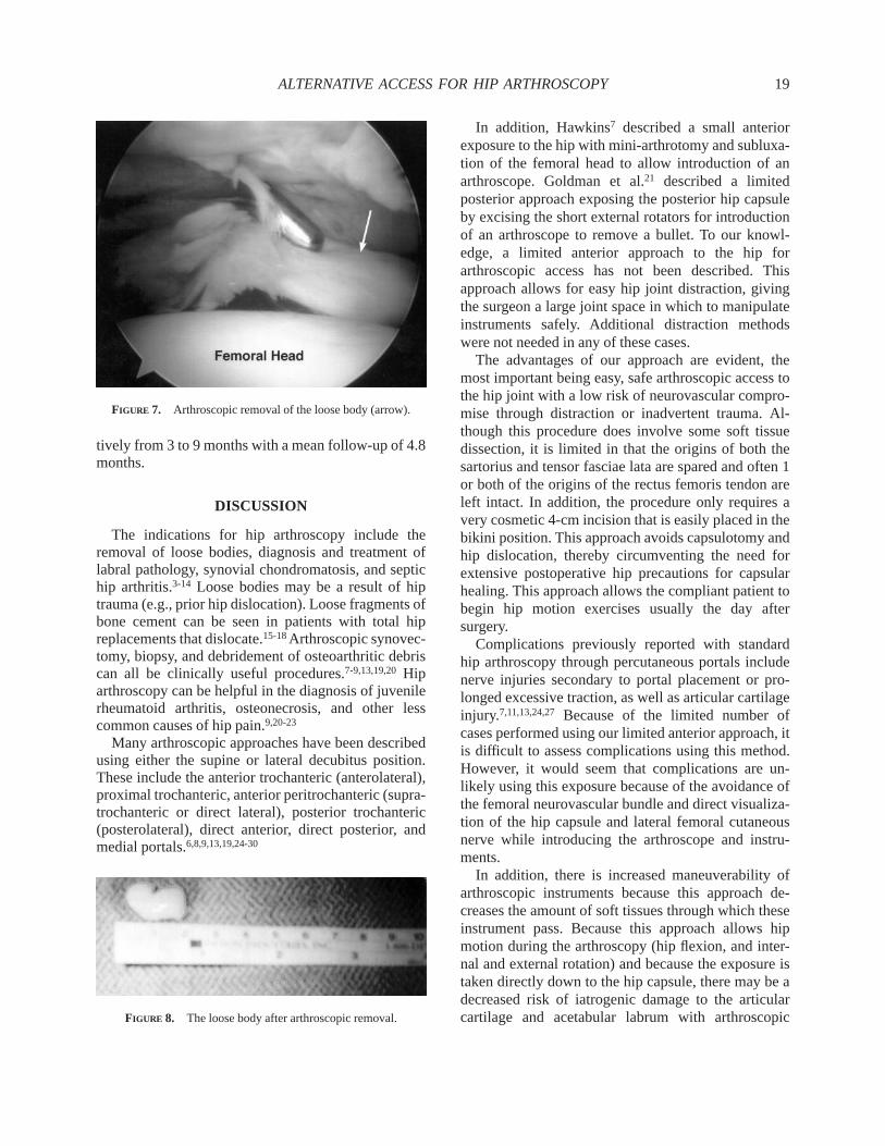
debris was performed (Table 1). Two of the arthrosco-pies involved removal of loose bodies (cases 3 and 4,Figs 6-8). In 1 case (case 2), a shelf procedure with agreater trochanteric osteotomy and advancement wasalso performed. In another case (case 5), exuberantsynovitis caused poor visualization of the joint, and thearthroscopy was easily converted to an open ar-throtomy. In all 5 cases, there were no complicationsand the patients have been followed-up postopera-
TABLE 1. Summary of Cases
Case Age Sex Symptoms Diagnosis TreatmentOther
Procedures Complications
1 12 yr M Pain Perthes*OCD*
Debridement None None
2 17 yr M Pain, limp Perthes*Multiple loose fragments
Debridement ShelfGTA
None
3 17 yr F Pain Loose body Removal of loose body None None4 9 yr M Pain, loss of motion, limp,
subluxationPerthesLoose bodyOCD
DebridementRemoval of loose body
None None
5 26 yr M Pain Crush injuryOCD
Debridement Arthrotomy None
Abbreviations: Perthes, Legg-Calves-Perthes disease; Shelf, shelf procedure; OCD, osteochondral defect/lesion; GTA, greater trochantericadvancement.
FIGURE 5. CT arthrogram of the hip in case 3 showing anintra-articular loose body (arrow).
FIGURE 6. Arthroscopic view of the hip displaying the loose body(arrow) seen earlier with CT arthrogram.
18 J. K. SEKIYA ET AL.
tively from 3 to 9 months with a mean follow-up of 4.8months.
DISCUSSION
The indications for hip arthroscopy include theremoval of loose bodies, diagnosis and treatment oflabral pathology, synovial chondromatosis, and septichip arthritis.3-14 Loose bodies may be a result of hiptrauma (e.g., prior hip dislocation). Loose fragments ofbone cement can be seen in patients with total hipreplacements that dislocate.15-18Arthroscopic synovec-tomy, biopsy, and debridement of osteoarthritic debriscan all be clinically useful procedures.7-9,13,19,20Hiparthroscopy can be helpful in the diagnosis of juvenilerheumatoid arthritis, osteonecrosis, and other lesscommon causes of hip pain.9,20-23
Many arthroscopic approaches have been describedusing either the supine or lateral decubitus position.These include the anterior trochanteric (anterolateral),proximal trochanteric, anterior peritrochanteric (supra-trochanteric or direct lateral), posterior trochanteric(posterolateral), direct anterior, direct posterior, andmedial portals.6,8,9,13,19,24-30
In addition, Hawkins7 described a small anteriorexposure to the hip with mini-arthrotomy and subluxa-tion of the femoral head to allow introduction of anarthroscope. Goldman et al.21 described a limitedposterior approach exposing the posterior hip capsuleby excising the short external rotators for introductionof an arthroscope to remove a bullet. To our knowl-edge, a limited anterior approach to the hip forarthroscopic access has not been described. Thisapproach allows for easy hip joint distraction, givingthe surgeon a large joint space in which to manipulateinstruments safely. Additional distraction methodswere not needed in any of these cases.
The advantages of our approach are evident, themost important being easy, safe arthroscopic access tothe hip joint with a low risk of neurovascular compro-mise through distraction or inadvertent trauma. Al-though this procedure does involve some soft tissuedissection, it is limited in that the origins of both thesartorius and tensor fasciae lata are spared and often 1or both of the origins of the rectus femoris tendon areleft intact. In addition, the procedure only requires avery cosmetic 4-cm incision that is easily placed in thebikini position. This approach avoids capsulotomy andhip dislocation, thereby circumventing the need forextensive postoperative hip precautions for capsularhealing. This approach allows the compliant patient tobegin hip motion exercises usually the day aftersurgery.
Complications previously reported with standardhip arthroscopy through percutaneous portals includenerve injuries secondary to portal placement or pro-longed excessive traction, as well as articular cartilageinjury.7,11,13,24,27 Because of the limited number ofcases performed using our limited anterior approach, itis difficult to assess complications using this method.However, it would seem that complications are un-likely using this exposure because of the avoidance ofthe femoral neurovascular bundle and direct visualiza-tion of the hip capsule and lateral femoral cutaneousnerve while introducing the arthroscope and instru-ments.
In addition, there is increased maneuverability ofarthroscopic instruments because this approach de-creases the amount of soft tissues through which theseinstrument pass. Because this approach allows hipmotion during the arthroscopy (hip flexion, and inter-nal and external rotation) and because the exposure istaken directly down to the hip capsule, there may be adecreased risk of iatrogenic damage to the articularcartilage and acetabular labrum with arthroscopic
FIGURE 7. Arthroscopic removal of the loose body (arrow).
FIGURE 8. The loose body after arthroscopic removal.
19ALTERNATIVE ACCESS FOR HIP ARTHROSCOPY

instrument placement and manipulation as well asimproved visualization of the femoral head.
The need for prolonged, excessive traction, whichcan compromise the sciatic nerve, is obviated asmanual traction has been sufficient to provide adequatedistraction of the hip joint. This is due to the limiteddissection and release of soft tissues around and downto the hip capsule which, when intact, provide resis-tance to longitudinal traction. While manual tractionhas been sufficient in our small series, it is possible thatin excessively tight hips additional traction may beneccessary. In such cases, this approach could beperformed supine on a fracture table in conjunctionwith traction.
There was 1 case (case 5) that was converted to anarthrotomy as a result of poor visualization of the jointcaused by exuberant synovitis. It was felt that a morethorough synovectomy could be performed through anopen approach, and the arthroscopy was easily con-verted to an arthrotomy without the need to repositionthe patient or reprep and drape.
Arthroscopic surgery of the hip through this limitedanterior approach combines the benefits of an arthro-scopic and an open procedure while minimizing opera-tive risk. As a result, it provides the orthopaedicsurgeon with basic arthroscopic skills, familiar withthe anterior approach to the hip, an alternative methodfor performing this otherwise technically difficultprocedure.
REFERENCES
1. Hoppenfeld S, deBoer P. Surgical exposures in orthopaedics:The anatomic approach. Ed 3. Philadelphia: JB Lippincott,1994; 325-338.
2. Smith-Petersen MN. Approach to and exposure of the hip jointfor mold arthroplasty.J Bone Joint Surg Am1949;31:40.
3. Blitzer CM. Arthroscopic management of septic arthritis of thehip. Arthroscopy1993;9:414-416.
4. Bould M, Edwards D, Villar RN. Case report—Arthroscopicdiagnosis and treatment of septic arthritis of the hip joint.Arthroscopy1993;9:707-708.
5. Byrd JWT. Labral lesions: An elusive source of hip pain—Casereports and literature review.Arthroscopy1996;12:603-612.
6. Chung WK, Slater GL, Bates EH. Treatment of septic arthritisof the hip by arthroscopic lavage.J Pediatr Orthop1993;13:444-446.
7. Hawkins RB. Arthroscopy of the hip.Clin Orthop 1989;249:44-47.
8. Ide T, Akamatsu N, Nakajima I. Arthroscopic surgery of the hipjoint. Arthroscopy1991;7:204-211.
9. Johnson LL. Hip joint. In: Johnson LL, ed. Diagnosis andsurgical arthroscopy.Ed 3. St. Louis: CV Mosby, 1986;1491-1516.
10. Okada Y, Awaya G, Ideka T, et al. Arthroscopic surgery forsynovial chondromatosis of the hip.J Bone Joint Surg Br1989;71:198-199.
11. Schindler A, Lechevallier JJC, Rao NS, et al. Diagnostic andtherapeutic arthroscopy of the hip in children and adolescents:Evaluation of results. J Pediatr Orthop1995;15:317-321.
12. Suzuki S, Awaya G, Okada Y, et al. Arthroscopic diagnosis ofruptured acetabular labrum.Acta Orthop Scand1986;57:513-515.
13. Villar RN. Hip arthroscopy.Br J Hosp Med1992;47:763-766.14. Witwity T, Uhlmann RD, Fischer J. Arthroscopic management
of chondromatosis of the hip joint.Arthroscopy1988;4:55-56.15. Keene GS, Villar RN. Arthroscopic loose body retrieval
following traumatic hip dislocation.Injury 1994;25:507-510.16. Mah ET, Bradley CM. Arthroscopic removal of acrylic cement
from unreduced hip prosthesis.Aust N Z J Surg 1992;62:508-510.
17. Nordt W, Giangarra CE, Levy IM, et al. Arthroscopic removalof entrapped debris following dislocation of a total hiparthroplasty.Arthroscopy1987;3:196-198.
18. Shifrin LZ, Reis ND. Arthroscopy of a dislocated hip replace-ment: A case report.Clin Orthop1980;146:213-214.
19. Eriksson E, Arvidsson I, Arvidsson H: Diagnostic and opera-tive arthroscopy of the hip.Orthopedics1986;9:169-176.
20. Frich LH, Lauritzen J, Juhl M. Arthroscopy in diagnosis andtreatment of hip disorders.Orthopedics1989;12:389-392.
21. Goldman A, Minkoff J, Price A, et al. A posterior arthroscopicapproach to bullet extraction from the hip.J Trauma1987;27:1294-1300.
22. Holgersson S, Brattstrom H, Mogensen B, et al. Arthroscopy ofthe hip in juvenile chronic arthritis.J Pediatr Orthop1981;1:273-278.
23. Sekiya JK, Ruch DS, Hunter M, et al. The role of arthroscopyin the staging and diagnosis of osteonecrosis. In: Urbaniak JR,Jones JP, eds.Osteonecrosis—Etiology, diagnosis, and treat-ment. Chicago: American Academy of Orthopaedic Surgeons,1997;253-260.
24. Byrd JWT, Pappas JN, Pedley J. Hip arthroscopy: An anatomicstudy of portal placement and relationship to the extra-articularstructures.Arthroscopy1995;11:418-423.
25. Dorfmann H, Boyer T, Henry P, De Bie B. A simple approach tohip arthroscopy.Arthroscopy1988;4:141-142.
26. Dvorak M, Duncan CP, Day B. Arthroscopic anatomy of thehip. Arthroscopy1990;6:264-273.
27. Funke EL, Munzinger U. Complications in hip arthroscopy.Arthroscopy1996;12:156-159.
28. Glick JM, Sampson TG, Gordon RB, et al. Hip arthroscopy bythe lateral approach.Arthroscopy1987;3:4-12.
29. Keene GS, Villar RN. Arthroscopic anatomy of the hip: An invivo study.Arthroscopy1994;10:392-399.
30. Byrd JWT. Hip arthroscopy utilizing the supine position.Arthroscopy1994;10:275-280.
20 J. K. SEKIYA ET AL.