Embed Size (px)
Citation preview

Hints and Tips to
Biliary Ultrasound
Pamela Parker
Consultant Sonographer

Aims
• Review what is the biliary system
• Review what is normal
• Review our understanding of common
pathologies

The Biliary System

The Gallbladder
• The ultrasound
appearance of the GB
are of a elongated
pear-shaped cystic
structure.
• The gallbladder is
well delineated and
has smooth thin walls

The Gallbladder
• Sonography was found to be accurate in
determining wall thickness to within 1 mm
in 93% of patients and 1.5 mm in 100%.
• Wall thickness greater than 3.5 mm is
highly accurate in predicting disease;
however, a wall thickness 3 mm or less
does not rule out cholecystitis.

The Gallbladder35-year-old healthy male volunteer with normal gallbladder.
A, Longitudinal sonogram of gallbladder, obtained after patient fasted for 12 hours, shows wall (arrow)
as pencil thin echogenic line.
B, Longitudinal sonogram in postprandial state shows pseudothickening of gallbladder wall (arrow)
due to physiological contraction

Case 1
• 45 year old female
• BMI 28
• RUQ pain

Case 1
• Presence of stones?
• Wall thickening?
• Secondary features?

Case 1

Case 1
• Presence of stones √
• Wall thickening? X
• Secondary features? X

Secondary Features

Case 1 - Biliary colic• Caused by a gallstone impacting in the cystic duct or the
ampulla of Vater.
• The pain starts suddenly in the epigastrium (RUQ) and
may radiate around to the back in the interscapular
region.
• Pain persists from 15 minutes up to 24 hours, subsiding
spontaneously or with analgesics.
• Nausea or vomiting often accompanies the pain,
• Occurs as a result of distension of the gallbladder due to
an obstruction or to the passage of a stone through the
cystic duct

Gallstones - Presentation
• Up to 70% of patients with gallstones are
asymptomatic at the time of diagnosis.
• Gallstones may cause biliary colic, acute
or chronic cholecystitis, pancreatitis or
obstructive jaundice.
• Biliary colic is the most common
presentation.
• The second most common presentation is
acute cholecystitis.

Case 2
• 59 year old female
• BMI 30
• RUQ pain
• Pyrexial
• Raised WCC

Case 2
• Presence of stones?
• Wall thickening?
• Secondary features?

Case 2
• Presence of stones √
• Wall thickening √
• Secondary features √

Case 2Fig. 3—59-year-old woman with diffuse gallbladder wall thickening.
A, Longitudinal sonogram shows layered appearance of thickened gallbladder wall, with
relatively hypoechoic region (arrowhead) between echogenic lines.
B, Contrast-enhanced CT scan shows thick-walled gallbladder contains hypodense outer
layer (arrow) that corresponds to subserosal edema, which may simulate pericholecystic
fluid.

Secondary Features

Cholecystitis Investigations
• FBC - the WCC is likely to be raised.
• Liver enzymes are often mildly abnormal.
• Ultrasound findings for cholecystitis:
– Include a thickened GB wall (greater than 3
mm) and may also include pericholecystic
fluid or air in the GB or the GB wall.
– If the GB wall is thickened, with positive
clinical presentation, but there are no
gallstones present then the diagnosis could
still be acalculous cholecystitis

Mural oedema, but not cholecystitis

Causes of GB wall oedema
• Physiological
o post prandial
• Inflammatory
• Adjacent disease
• Non-inflammatory
o adenomyomatosis, cancer, leukaemia, mets
• Generalised oedema o Ascites, organ failure, portal hypertension
• Varices
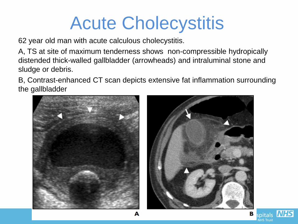
Acute Cholecystitis62 year old man with acute calculous cholecystitis.
A, TS at site of maximum tenderness shows non-compressible hydropically
distended thick-walled gallbladder (arrowheads) and intraluminal stone and
sludge or debris.
B, Contrast-enhanced CT scan depicts extensive fat inflammation surrounding
the gallbladder

Perforated Cholecystitis
? Gb Mass on
CT

Tops Tips to GB imaging
• Correct Preparation
• Clinical presentation
• Secondary features
• Previous imaging

The Bile Duct
• What is normal?
• Does size matter?
• Enlarges with age,
bile duct disease
(?cholecystectomy)
• Should taper to
pancreatic head
• Symptomatic vs
Incidental

Case 3
• 79 year old male
• BMI 22
• Bilirubin 51 μmol/L
(normal 1.71 to 20.5
µmol/L)

Case 3
• Presence of gallstones?
• Presenting symptoms?
• Secondary features?

Case 3

Case 3
• Presence of gallstones √
• Presenting symptoms √
• Secondary features X

Secondary Features

Think - Ascending Cholangitis
• Charcot's triad:
Infected CBD leading
to jaundice and high
swinging fevers with
rigors and chills
• Retrograde infection
up the CBD as a
result of acute
cholecystitis or ERCP

Think - Jaundice
• Jaundice most often
happens as a result of an
underlying disorder that
either causes the production
of too much bilirubin or
prevents the liver from
getting rid of it.
• Both of these result in
bilirubin being deposited in
tissues

Jaundice
• Acute inflammation of the liver:
– impairs the ability of the liver to conjugate and
secrete bilirubin, resulting in a build up.
• Inflammation of the bile duct:
– This can prevent the secretion of bile and
removal of bilirubin, causing jaundice.
• Obstruction of the bile duct:
– This prevents the liver from disposing of
bilirubin
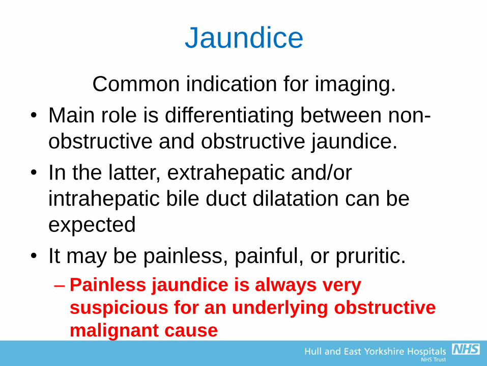
Jaundice
Common indication for imaging.
• Main role is differentiating between non-
obstructive and obstructive jaundice.
• In the latter, extrahepatic and/or
intrahepatic bile duct dilatation can be
expected
• It may be painless, painful, or pruritic.
– Painless jaundice is always very
suspicious for an underlying obstructive
malignant cause

Causes of Jaundice
Obstructive
• post-hepatic causes
– choledocholithiasis
– strictures, e.g. post-
inflammatory/infectious
– external biliary tree
compression, Mirizzi syndrome
– malignant causes
– portal lymphadenopathy
– cholangiocarcinoma
– carcinoma of head of pancreas
– hepatocellular carcinoma
– gallbladder carcinoma
Non-Obstructive
• pre-hepatic and hepatic
causes
– haemolytic anaemia
– mechanical heart valve
– hypersplenism
– hepatic
– acute hepatitis / acute liver
failure
– cirrhosis
– Gilbert syndrome

Pablo Luis Mirizzi, an Argentinian physician 1893 - 1964
Mirizzi Syndrome

Courvoisier sign
• Courvoisier sign
– a patient with painless jaundice and an
enlarged gallbladder (or right upper quadrant
mass), the cause is unlikely to be gallstones
and therefore presumes the cause to be an
obstructing pancreatic or biliary neoplasm
until proven otherwise.
Swiss surgeon Ludwig Georg Courvoisier in 1890

Case 4
69 year old female
Clinical details:
Nausea and vomitting. Raised liver enzymes
(very high).

Case 4
• Presence of gallstones ?
• Presenting symptoms ?
• Secondary features ?

Case 4

Case 4
• Presence of gallstones √ - but distended
GB with thin walls – no cholecystitis
• Presenting symptoms √ - N&V no pain,
abnormal LFT’s
• Secondary features √ - intra & extra
hepatic duct dilatation, no tapering of CBD


Segmental Intrahepatic Biliary
Dilatation

Cholangiocarcinoma• Originally referred
only to primary
tumours of the
intrahepatic bile ducts
• Term is now regarded
as inclusive of
intrahepatic, perihilar,
and distal
extrahepatic tumours
of the bile ducts

Cholangiocarcinoma
• 20–25% are intrahepatic.
• 50–60% of all cases of cholangiocarcinoma are
perihilar tumours (those involving the bifurcation
of the ducts are “Klatskin” tumours).
• Most Klatskin tumours may have been coded as
intrahepatic tumours for purposes of death
certification.
• 20–25% are distal extrahepatic tumours.
• About 5% of tumours may be multifocal.

US of Cholangiocarcinoma• Remains the first line investigation for suspected biliary
obstruction.
• Diagnosis should be suspected when intrahepatic, but
not extrahepatic, ducts are dilated.
• Intrahepatic cholangiocarcinoma may be seen as a mass
lesion but this is unusual.
• Gall stones / cholecystitis is excluded.
• Often misses small perihilar, extrahepatic, and
periampullary tumours and not good at defining the
extent of the tumour.
• Colour Doppler can detect tumour induced
compression/thrombosis of the portal vein or hepatic
artery.
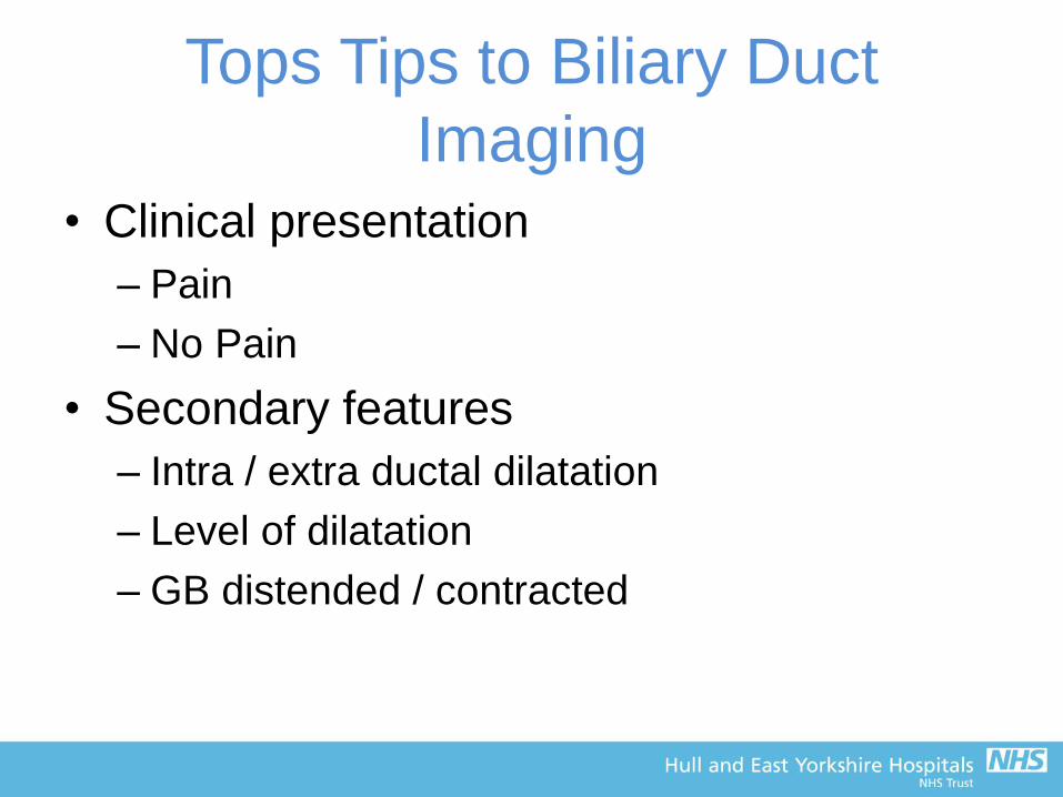
Tops Tips to Biliary Duct
Imaging• Clinical presentation
– Pain
– No Pain
• Secondary features
– Intra / extra ductal dilatation
– Level of dilatation
– GB distended / contracted

The Pancreas – 50 Shades of
Grey

US of the Pancreas
• variable echogenicity
– in young patients, the pancreas is generally
less fatty and therefore usually hypoechoic
– with age, fatty replacement of pancreas can
result in echogenicity similar to surrounding
mesenteric fat

US of the Pancreas
• Pancreatic ultrasound can be used to
assess for pancreatic malignancy,
pancreatitis and its complications, as well
as for other pancreatic pathologyhttps://radiopaedia.org/articles/pancreatic-ultrasound
• Pancreatic duct: ≤3 mm

US of the Pancreas
• Pancreatic duct: ≤3 mm
What does this mean?
9/360 cases PD ≤3 mm
2/9 cases confirmed PD dilatation at MRCP
Should we measure at all?

Case 5
71 Year old female
Clinical Details :
known gallstone,
?pancreatitis, ?CBD
stone

Case 5

Case 5

Pancreatitis
Secondary signs• Raised amylase
(particularly in first 24
hours)
• Relatively hypoechoic
pancreas
• Peri-pancreatic
oedema
• Enlarged pancreas
(?what is normal)
• FF in LUQ / Pelvis

Case 6
66 year old female
Clinical details:
Lap Cholecystectomy 6/7
days ago. Now presented
with abdo pain, vomiting
and jaundice. Amylase
2200, LFTs deranged.
clinically pancreatitis and
cholangitis. Kindly assess
the CBD for retained stones

Case 6

Case 6

Pancreatic Cancer?

Dilated PD?Atrophic pancreas with
stricture from previous
pancreatitisPancreatic head mass

Pancreatic Cancer
Secondary signs• Weight loss
• Painless obstruction
• Partial PD dilatation
• Previous history of
pancreatitis
• EUS imaging modality
of choice
Tops Tips to Pancreatic Imaging
• Clinical presentation
• Measure the duct with caution
• Secondary features
– weight loss
– Peri-pancreatic oedema
– Free fluid LUQ / Pelvis

Favourite Top Tips!
• Clinical presentation
• PMH
• Secondary Signs