Embed Size (px)
Citation preview

Highlights onRevised Recommendations on
Prevention of CABSI
Infection Control BranchCentre for Health Protection
18 January 2019
Recommendation on Prevention of Intravascular Catheter Associated Bloodstream Infection
• The recommendations was first published in 2010.
• Relevant overseas guidelines have been updated since.
• The recommendation was updated in March 2018 https://www.chp.gov.hk/files/pdf/recommendations_on_prevention_of_intravascular_catheter_associated_bloodstream_infection.pdf
2
Update Recommendation
1. Survey of local practice (public and private hospitals) to understand the current situation
2. Literature review to identify changes in overseas guidelines– taking scientific evidence, cost‐effectiveness and current frontline practice into consideration
3. Filled gaps in recommendation so that all major steps for most commonly used catheters are covered
3

Survey on Local Practice
4

Methods
• Survey period: April ‐May 2017• Self‐administrated questionnaire
– constructed and piloted with the help of ICNs;– sent to 27 public and 12 private hospitals;– to be completed by ICN and WM/DOM of up to two departments (pre‐selected from Medical, Surgical, O&G, Paed, O&T, Oncology and ICU) from each hospital.
5

Questionnaire for Survey
1. Go through major guidelines and peer review papers
2. Identify areas need to be reviewed 3. Develop questionnaire to understand more on the current
practice of both public and private hospitals
6
Year
CDC 2002 2011
NICE 2012
NHS 2013
Cochrane Database Systemic Review 2005 2013
Microsoft Word Document

Response Rate
• Public:– 27/27 hospitals (100%) with 72 departments completed questionnaires
• Private:– 12/12 hospitals (100%) with 26 departments completed questionnaires
• Departments:– ICT, M&G, SUR, ORTH, O&G, PAED, ICU, ONC, HD
7

Literature Review
8
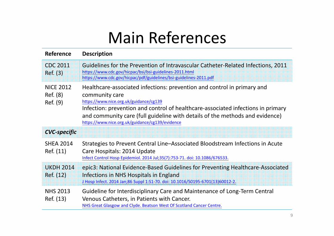
Main ReferencesReference Description
CDC 2011Ref. (3)
Guidelines for the Prevention of Intravascular Catheter‐Related Infections, 2011https://www.cdc.gov/hicpac/bsi/bsi‐guidelines‐2011.htmlhttps://www.cdc.gov/hicpac/pdf/guidelines/bsi‐guidelines‐2011.pdf
NICE 2012Ref. (8)Ref. (9)
Healthcare‐associated infections: prevention and control in primary andcommunity carehttps://www.nice.org.uk/guidance/cg139Infection: prevention and control of healthcare‐associated infections in primary and community care (full guideline with details of the methods and evidence)https://www.nice.org.uk/guidance/cg139/evidence
CVC‐specific
SHEA 2014Ref. (11)
Strategies to Prevent Central Line–Associated Bloodstream Infections in Acute Care Hospitals: 2014 UpdateInfect Control Hosp Epidemiol. 2014 Jul;35(7):753‐71. doi: 10.1086/676533.
UKDH 2014Ref. (12)
epic3: National Evidence‐Based Guidelines for Preventing Healthcare‐Associated Infections in NHS Hospitals in EnglandJ Hosp Infect. 2014 Jan;86 Suppl 1:S1‐70. doi: 10.1016/S0195‐6701(13)60012‐2.
NHS 2013Ref. (13)
Guideline for Interdisciplinary Care and Maintenance of Long‐Term Central Venous Catheters, in Patients with Cancer.NHS Great Glasgow and Clyde. Beatson West Of Scotland Cancer Centre.
9
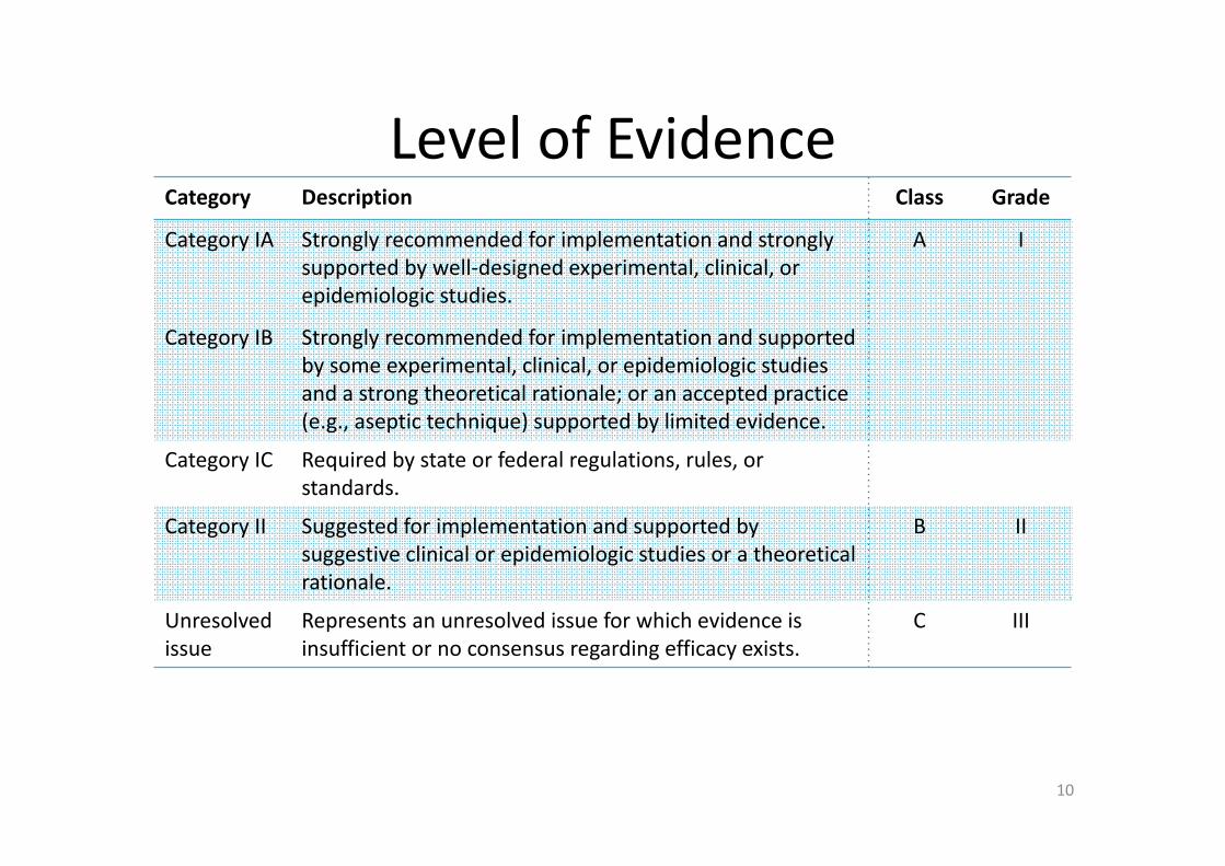
Level of EvidenceCategory Description Class Grade
Category IA
Category IB
Strongly recommended for implementation and strongly supported by well‐designed experimental, clinical, or epidemiologic studies.
Strongly recommended for implementation and supported by some experimental, clinical, or epidemiologic studies and a strong theoretical rationale; or an accepted practice (e.g., aseptic technique) supported by limited evidence.
A I
Category IC Required by state or federal regulations, rules, or standards.
Category II Suggested for implementation and supported by suggestive clinical or epidemiologic studies or a theoretical rationale.
B II
Unresolved issue
Represents an unresolved issue for which evidence is insufficient or no consensus regarding efficacy exists.
C III
10

Recommendation on Prevention of Intravascular Catheter Associated Bloodstream Infection
1. Education, Quality Assurance and Surveillance2. General Aspects
2.1 Hand hygiene2.2 Aseptic technique2.3 Catheter and site care
3. Care of Specific Catheter3.1 Central venous catheters3.2 Peripheral venous catheters3.3 Peripheral arterial catheters3.4 Pressure monitoring systems3.5 Umbilical catheters
4. Maintenance of Administration Sets5. Care of infusate, IV medication and Admixture6. Needleless intravascular catheter systems7. Special consideration for the prevention of CABSI
11
Adobe Acrobat Document

2.1 Hand Hygiene
12

2010 2018 References Level of evidence
Justifications / Remarks
Perform hand antisepsis with water and antiseptic soap or with alcohol hand rub for catheter site care and accessing the system, including before and after catheter insertion, touching the insertion sites, dressing and the infusion system.
2.1Perform hand hygiene procedures, either by washing hands with soap and water or with alcohol‐based hand rubs (ABHR), before and after inserting, accessing, dressing catheters and palpating catheter insertion sites.
CDC 2011 Category IB Proper hand hygiene can be achieved through the use of either an alcohol‐based product or with soap and water with adequate rinsing.
NICE 2012 Category IB The additional benefit achieved with the use of alcohol handrubs compared tohandwashing with non‐antiseptic soap is statistically significant but of uncertain clinicalimportance (3).
13

Ref. Original text of findings / conclusion
CDC 2011
P.12 –
Perform hand hygiene procedures, either by washing hands with conventional soap and water or with alcohol‐based hand rubs (ABHR). Hand hygiene should be performed before and after palpating catheter insertion sites as well as before and after inserting, replacing, accessing, repairing, or dressing an intravascular catheter. Palpation of the insertion site should not be performed after the application of antiseptic, unless aseptic technique is maintained. Category IB
P.30 –
Proper hand hygiene can be achieved through the use of either an alcohol‐based product or with soap and water with adequate rinsing.
NICE 2012
P.77 –
There is a statistically significant reduction of uncertain clinically importance in mean log change in CFUs and it is unlikely that there is any difference in log 10 CFUs after use of alcohol handrubs compared to handwashing with non‐antiseptic soap and water (LOW QUALITY).
There is a statistically significant, but not clinically important, reduction in log 10 CFUs after use of antiseptic soap compared to non‐antiseptic soap and water (LOW QUALITY).
14

2.2 Skin Antisepsis
15

2010 2018 References Level of evidence
Justifications / Remarks
Disinfect skin properly before catheter insertion, with sufficient contact time. For example, 70% alcohol for peripheral line insertion. Chlorhexidine‐based preparation is preferred for central line insertion.
2.2.1Prepare skin with an antiseptic, e.g.70% alcohol for peripheral venous catheter insertion,
CDC 2011 Category IB
2.2.1 2% chlorhexidine in alcohol for CVC and peripheral arterial catheter insertion and dressing changes.
CDC 2011SHEA 2014NICE 2012UKDH 2014NHS 2013
Category IAGrade INot specifiedClass ACategory IA
CDC 2011: lower incidence of arterial catheter colonization, catheter‐related local infection and bacteraemia (but similar rate of arterial catheter‐related sepsis).
16
Ref. Original text of findings / conclusion
CDC 2011
P.13 –
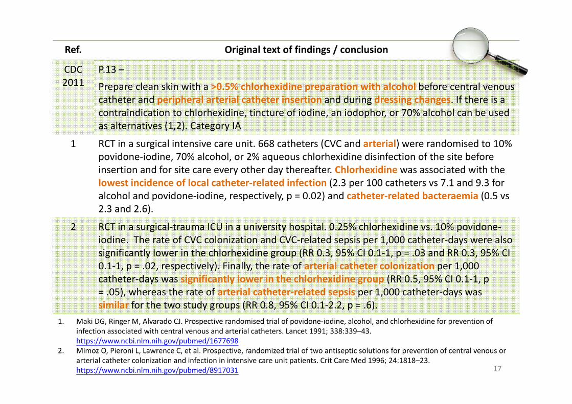
Prepare clean skin with a >0.5% chlorhexidine preparation with alcohol before central venous catheter and peripheral arterial catheter insertion and during dressing changes. If there is a contraindication to chlorhexidine, tincture of iodine, an iodophor, or 70% alcohol can be used as alternatives (1,2). Category IA
1 RCT in a surgical intensive care unit. 668 catheters (CVC and arterial) were randomised to 10% povidone‐iodine, 70% alcohol, or 2% aqueous chlorhexidine disinfection of the site before insertion and for site care every other day thereafter. Chlorhexidine was associated with the lowest incidence of local catheter‐related infection (2.3 per 100 catheters vs 7.1 and 9.3 for alcohol and povidone‐iodine, respectively, p = 0.02) and catheter‐related bacteraemia (0.5 vs 2.3 and 2.6).
2 RCT in a surgical‐trauma ICU in a university hospital. 0.25% chlorhexidine vs. 10% povidone‐iodine. The rate of CVC colonization and CVC‐related sepsis per 1,000 catheter‐days were also significantly lower in the chlorhexidine group (RR 0.3, 95% CI 0.1‐1, p = .03 and RR 0.3, 95% CI 0.1‐1, p = .02, respectively). Finally, the rate of arterial catheter colonization per 1,000 catheter‐days was significantly lower in the chlorhexidine group (RR 0.5, 95% CI 0.1‐1, p = .05), whereas the rate of arterial catheter‐related sepsis per 1,000 catheter‐days was similar for the two study groups (RR 0.8, 95% CI 0.1‐2.2, p = .6).
17
1. Maki DG, Ringer M, Alvarado CJ. Prospective randomised trial of povidone‐iodine, alcohol, and chlorhexidine for prevention of infection associated with central venous and arterial catheters. Lancet 1991; 338:339–43. https://www.ncbi.nlm.nih.gov/pubmed/1677698
2. Mimoz O, Pieroni L, Lawrence C, et al. Prospective, randomized trial of two antiseptic solutions for prevention of central venous or arterial catheter colonization and infection in intensive care unit patients. Crit Care Med 1996; 24:1818–23. https://www.ncbi.nlm.nih.gov/pubmed/8917031

2010 2018 References Level of evidence
Justifications / Remarks
Disinfect skin properly before catheter insertion, with sufficient contact time (alcohol‐based antiseptics require contact time about 30 seconds; non‐alcohol‐based antiseptics require longer contact time usually around 2 min)…
2.2.4 Antiseptics should be allowed to dry.
CDC 2011SHEA 2014NICE 2012NHS 2013
Category IBGrade INot specifiedNot specified
No strong evidence on “contact time” in previous references. Current practice:Peripheral: << 30 secCVC/HD: 30 sec ‐ 2 min
2.2.3 For CVC insertion and dressing changes, apply repeated up and down, back and forth strokes for at least 30 seconds starting at the insertion site and working outward to the periphery.
NHS 2013 Not specified The only reference mentioning concept of “contact time”.
18

Ref. Original text of findings / conclusion
Previous references
3 Mimoz et al. showed a reduction in the blood culture contamination rate with the use of chlorhexidine, compared with povidone‐iodine. However, the short antiseptic drying time of 15‐30 seconds may have biased the results in favor of chlorhexidine, because it takes approximately 2 minutes for povidone‐iodine (2) to reach its maximal antimicrobial effect.
4 Paper published in 1963; full text not found.
19
3. Review of clinical trials of skin antiseptic agents used to reduce blood culture contamination. Infect control and Hosp. Epidemol2007;28(7):892‐89. https://www.ncbi.nlm.nih.gov/pubmed/17564999
4. An evaluation of iodophors as skin antiseptics. Surg Gynecol Obstet 1963; 116:361‐365.
Ref. Original text of findings / conclusion
New references
CDC 2011
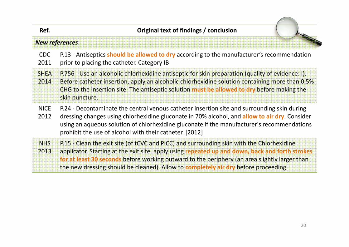
P.13 ‐ Antiseptics should be allowed to dry according to the manufacturer’s recommendation prior to placing the catheter. Category IB
SHEA 2014
P.756 ‐ Use an alcoholic chlorhexidine antiseptic for skin preparation (quality of evidence: I).Before catheter insertion, apply an alcoholic chlorhexidine solution containing more than 0.5% CHG to the insertion site. The antiseptic solution must be allowed to dry before making the skin puncture.
NICE 2012
P.24 ‐ Decontaminate the central venous catheter insertion site and surrounding skin during dressing changes using chlorhexidine gluconate in 70% alcohol, and allow to air dry. Consider using an aqueous solution of chlorhexidine gluconate if the manufacturer's recommendations prohibit the use of alcohol with their catheter. [2012]
NHS 2013
P.15 ‐ Clean the exit site (of tCVC and PICC) and surrounding skin with the Chlorhexidine applicator. Starting at the exit site, apply using repeated up and down, back and forth strokes for at least 30 seconds before working outward to the periphery (an area slightly larger than the new dressing should be cleaned). Allow to completely air dry before proceeding.
20

Local Practice ‐ Skin Antisepsis
21
Type of Hospital Peripheral venous linen (%)
Peripheral arterial linen (%)
Central linen (%)
Public N=72 N=43 N=59
70% alcohol 70 (97.2) 36 (83.7) 6 (10.2)
2% chlorhexidine 10 (13.9) 8 (18.6) 49 (83.1)
Povidone‐iodine 0 (0) 2 (4.7) 15 (25.4)
Others 0 (0) 0 (0) 1^ (1.7)
Private N=25 N=15 N=22
70% alcohol 24 (96.0) 12 (80.0) 5 (22.7)
2% chlorhexidine 1 (4.0) 2 (13.3) 16 (72.7)
Povidone‐iodine 0 (0) 2 (13.3) 12 (54.5)
Others 0 (0) 1* (6.7) 1* (4.5)
*0.5% chlorhexidine gluconate; ^skin prep in OT
OK Mostly OKPractice varied

Local Practice ‐ Skin Antisepsis (Paed)
22
Type of Hospital Peripheral venous linen (%)
Peripheral arterial linen (%)
Central linen (%)
Public N=7 N=6 N=6
70% alcohol 7 (100.0) 5 (83.3) 1 (16.7)
2% chlorhexidine 2 (28.6) 3 (50.0) 4 (66.7)
Povidone‐iodine 0 (0) 0 (0) 2 (33.3)
Others 0 (0) 0 (0) 0 (0)
Private N=2 NA NA
70% alcohol 2 (100.0) NA NA
2% chlorhexidine 0 (0) NA NA
Povidone‐iodine 0 (0) NA NA
Others 0 (0) NA NA

2.3 Frequency of Dressing Change
23

2010 2018 References Level of evidence
Justifications / Remarks
Change dressings at least weekly or when clinically indicated (removal or replacement of catheter; damp, loosened or visibly soiled dressings).
2.3.4 Leave the transparent semipermeable membrane dressing applied to a peripheral cannula insertion site in situ for the life of the cannula, provided that the integrity of the dressing is retained.
NICE 2012 Not specified No strong evidence for “at least weekly” in previous reference. Reduce risk of infection. Reduce patientdiscomfort. Reduce cost.
Replace gauze dressing every 2 days and transparent dressing every 7 days for short‐term CVC.
No change
24

Ref. Original text of findings / conclusion
Previous reference
CDC 2002
P.14 –
Change dressings at least weekly for adult and adolescent patients depending on the circumstances of the individual patient (1). Category II
1 Patients in both Groups (Group A: tCVC, N=230, Q5d vs. Q10d. Group B: nCVC, N=169, Q2d vs.Q5d) receiving CVC dressing changes at longer intervals did not show a significant increase in the rate of local infections, while those who received dressing every 2 days had a significant increase in local skin toxicity. Longer intervals were accompanied by a reduction in costs. The results of this study demonstrate that the increase in time interval between CVC dressing changes in BMT patients did not raise the risk of local infections, while significantly reducing patient discomfort and costs.
25
1. Comparison of two different time interval protocols for central venous catheter dressing in bone marrow transplant patients: results of a randomized, multicenter study. The Italian Nurse Bone Marrow Transplant Group (GITMO). Haematologica. 2000 Mar;85(3):275‐9. https://www.ncbi.nlm.nih.gov/pubmed/10702816

Ref. Original text of findings / conclusion
New reference
2 P.189 –
The advantage of leaving insertion sites intact is that the risk of infection is reduced. No harms were identified, but dressings that are no longer intact should be replaced as soon as possible to reduce the risk of infection. It was the opinion of the GDG that less frequent dressing changes would be cost saving in terms of staff time, resource use, and infection prevention compared to more frequent dressing changes.
3 Arterial Line Management ‐ The site should be secured with a sterile, moisture permeable, dressing e.g. Tegaderm. Dressings should only be changed when soiled or when the line is being changed.
26
2. Infection: prevention and control of healthcare‐associated infections in primary and community care. NICE. https://www.nice.org.uk/guidance/cg139/evidence/control‐full‐guideline‐185186701
3. NHS Salisbury > ICID > Clinical Management > Intensive Care > Arterial Line Management http://www.icid.salisbury.nhs.uk/ClinicalManagement/IntensiveCare/Pages/ArterialLineManagement.aspx

Local Practice – Frequency of change for sterile transparent semipermeable dressing
Type of Hospital Peripheral venous linen (%)
Peripheral arterial linen (%)
Central linen (%)
Public N=71 N=41 N=60
Daily 1 (1.4%) 2 (4.9%) 4 (6.7%)
Every 72 hours 12 (16.9%) 4 (9.8%) 3 (5.0%)
Every 96 hours 26 (36.6%) 8 (19.5%) 7 (11.7%)
Weekly 4 (5.6%) 8 (19.5%) 21 (35.0%)
PRN 27 (38.0%) 17 (41.5%) 17 (28.3%)
Private N=24 N=14 N=21
Daily 0 1 (7.1%) 0
Every 72 hours 4 (16.7%) 4 (28.6%) 4 (19.0%)
Every 96 hours 6 (25.0%) 2 (14.3%) 1 (4.8%)
Weekly 4 (16.7%) 1 (7.1%) 9 (42.9%)
PRN 8 (33.3%) 5 (35.7%) 5 (23.8%)
Mainly during change of catheter

2010 2018 References Level of evidence
Justifications / Remarks
Observe the catheter insertion site daily by palpation and inspection if transparent dressing is used. Visual inspection may be necessary for opaque dressing if patient has unexplained fever, pain, local tenderness, other signs of bloodstream infection or patients cannot communicate.
2.3.6 Evaluate the catheter insertion site daily by palpation to discern tenderness and by inspection if a transparent dressing is in use. Gauze and opaque dressings should not be removed if the patient has no clinical signs of infection. If the patient has local tenderness or other signs of possible CABSI, an opaque dressing should be removed and the site inspected.
CDC 2011 Category II No routine removal of gauze dressing for inspection based only on patients’ inability to communicate.
28
Ref. Original text of findings / conclusion
Previous reference
CDC 2002
P.13 –
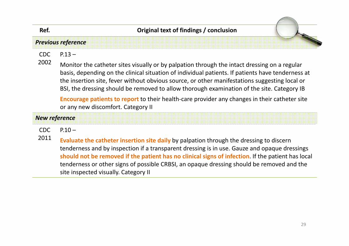
Monitor the catheter sites visually or by palpation through the intact dressing on a regular basis, depending on the clinical situation of individual patients. If patients have tenderness at the insertion site, fever without obvious source, or other manifestations suggesting local or BSI, the dressing should be removed to allow thorough examination of the site. Category IB
Encourage patients to report to their health‐care provider any changes in their catheter site or any new discomfort. Category II
New reference
CDC 2011
P.10 –
Evaluate the catheter insertion site daily by palpation through the dressing to discern tenderness and by inspection if a transparent dressing is in use. Gauze and opaque dressings should not be removed if the patient has no clinical signs of infection. If the patient has local tenderness or other signs of possible CRBSI, an opaque dressing should be removed and the site inspected visually. Category II
29

Local Practice – Frequency of change for sterile gauze dressing
Type of Hospital Peripheral venous linen (%)
Peripheral arterial linen (%)
Central linen (%)
Public N=21 N=16 N=33
Daily 3 (14.3%) 2 (12.5%) 8 (24.2%)
Every 2 days 4 (19.0%) 4 (25.0%) 7 (21.2%)
Every 72 hours 1 (4.8%) 1 (6.3%) 2 (6.1%)
Weekly 0 0 5 (15.2%)
PRN 12 (57.1%) 7 (43.8%) 7 (21.2%)
Private N=5 N=3 N=6
Daily 0 0 0
Every 2 days 2 (40.0%) 0 3 (50.0%)
Every 72 hours 0 0 0
Weekly 1 (20.0%) 1 (33.3%) 0
PRN 1 (20.0%) 1 (33.3%) 1 (16.7%)
Mainly when damp or soiled, so every 2 days or more frequent (daily, PRN)
3.1 Central Venous CatheterSite Selection
31
2010 2018 References Level of evidence
Justifications / Remarks
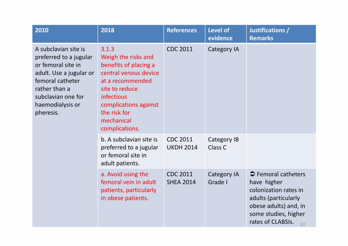
A subclavian site is preferred to a jugular or femoral site in adult. Use a jugular or femoral catheter rather than a subclavian one for haemodialysis or pheresis.
3.1.3Weigh the risks and benefits of placing a central venous device at a recommended site to reduce infectious complications against the risk for mechanical complications.
CDC 2011 Category IA
b. A subclavian site is preferred to a jugular or femoral site in adult patients.
CDC 2011UKDH 2014
Category IBClass C
a. Avoid using the femoral vein in adult patients, particularly in obese patients.
CDC 2011SHEA 2014
Category IAGrade I
Femoral catheters have higher colonization rates in adults (particularlyobese adults) and, in some studies, higher rates of CLABSIs. 32
Ref. Original text of findings / conclusion
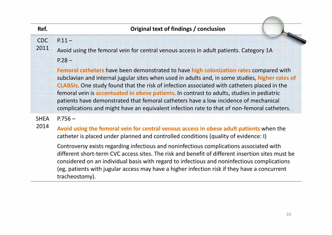
CDC2011
P.11 –
Avoid using the femoral vein for central venous access in adult patients. Category 1A
P.28 –
Femoral catheters have been demonstrated to have high colonization rates compared with subclavian and internal jugular sites when used in adults and, in some studies, higher rates of CLABSIs. One study found that the risk of infection associated with catheters placed in the femoral vein is accentuated in obese patients. In contrast to adults, studies in pediatric patients have demonstrated that femoral catheters have a low incidence of mechanical complications and might have an equivalent infection rate to that of non‐femoral catheters.
SHEA2014
P.756 –
Avoid using the femoral vein for central venous access in obese adult patients when the catheter is placed under planned and controlled conditions (quality of evidence: I)
Controversy exists regarding infectious and noninfectious complications associated with different short‐term CVC access sites. The risk and benefit of different insertion sites must be considered on an individual basis with regard to infectious and noninfectious complications (eg, patients with jugular access may have a higher infection risk if they have a concurrent tracheostomy).
33
3.1 Central Venous CatheterCatheter Change
34

2010 2018 References Level of evidence
Justifications / Remarks
Routine replacement of intravascular catheters is not necessary if they are functioning and have no evidence of causing local or systemic complications.
No change
3.1.11When adherence to aseptic technique cannot be ensured (i.e. catheters inserted during a medical emergency), replace the catheter as soon as possible, i.e. within 48 hours.
CDC 2011 Category IB Reduce risk of infection
35

Ref. Original text of findings / conclusion
CDC 2011
P.11 –
When adherence to aseptic technique cannot be ensured (i.e catheters inserted during a medical emergency), replace the catheter as soon as possible, i.e, within 48 hours (1). Category IB
1 To delineate the pathogenesis and epidemiology of catheter‐related infection with Swan‐Ganzpulmonary artery (PA) catheters, a prospective clinical study of hospitalized adult medical and surgical patients was done.
Overall, 65 (22%) of 297 Swan‐Ganz catheters showed local infection of the introducer (58 catheters) or the intravascular portion of the PA catheter (20 catheters); only two catheters (0.7%) caused bacteremia. Cutaneous colonization of the insertion site with > 102 cfu/10 cm2
(relative risk [RR] 5.5; p < 0.001), insertion into an internal jugular vein (RR 4.3; p < 0.01), catheterization >3 days (RR 3.1; p < 0.01), and insertion in the operating room using less stringent barrier precautions (RR 2.1; p = 0.03) were each associated with a significantly increased risk of catheter‐related infection.
Heavy colonization of the insertion site, percutaneous insertion in the internal jugular vein rather than subclavian vein, catheterization longer than 3 days, and insertion with less stringent barrier precautions significantly increase the risk of catheter‐related infection.
36
1. Mermel LA, McCormick RD, Springman SR, Maki DG. The pathogenesis and epidemiology of catheter‐related infection with pulmonary artery Swan‐Ganz catheters: a prospective study utilizing molecular subtyping. Am J Med 1991; 91:197S–205.http://www.sciencedirect.com/science/article/pii/0002934391903699
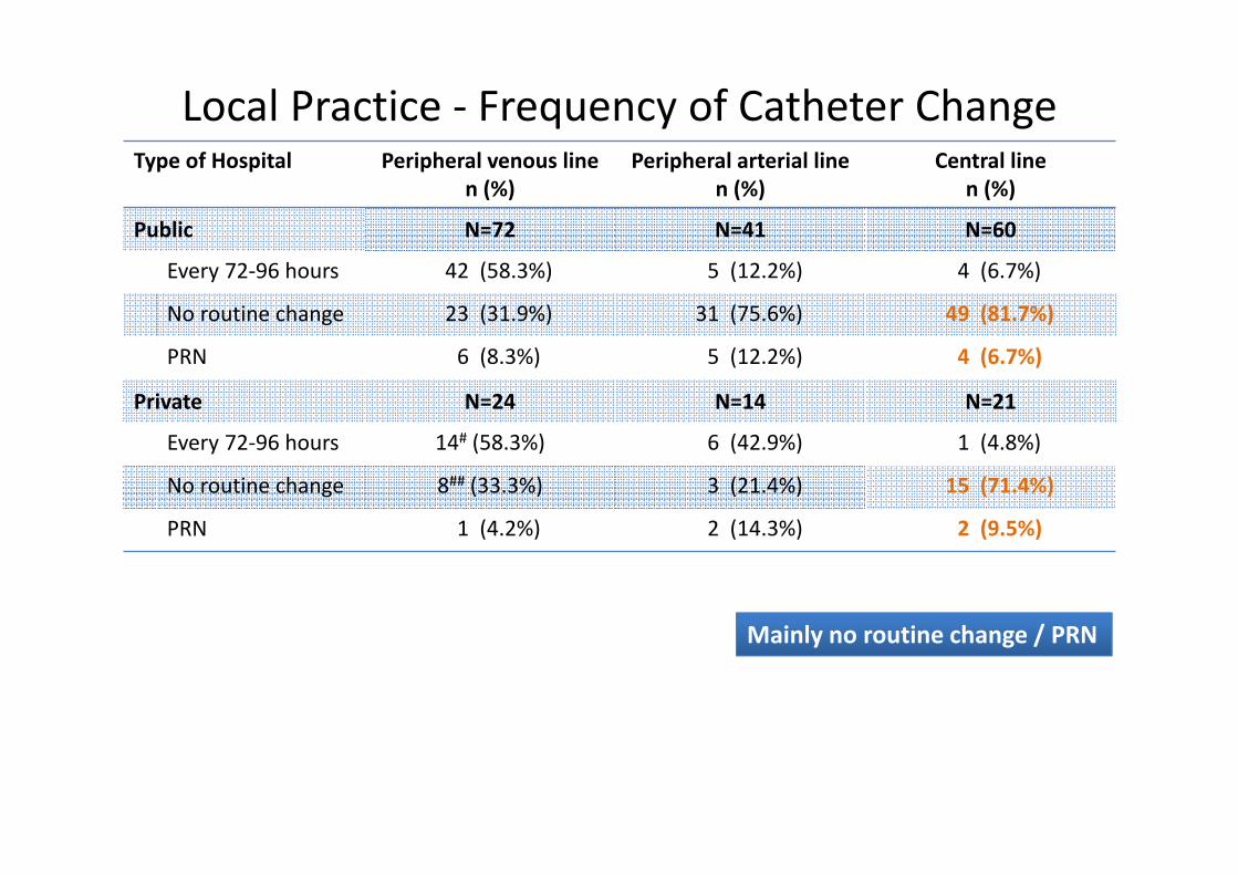
Type of Hospital Peripheral venous linen (%)
Peripheral arterial linen (%)
Central linen (%)
Public N=72 N=41 N=60
Every 72‐96 hours 42 (58.3%) 5 (12.2%) 4 (6.7%)
No routine change 23 (31.9%) 31 (75.6%) 49 (81.7%)
PRN 6 (8.3%) 5 (12.2%) 4 (6.7%)
Private N=24 N=14 N=21
Every 72‐96 hours 14# (58.3%) 6 (42.9%) 1 (4.8%)
No routine change 8## (33.3%) 3 (21.4%) 15 (71.4%)
PRN 1 (4.2%) 2 (14.3%) 2 (9.5%)
Local Practice ‐ Frequency of Catheter Change
Mainly no routine change / PRN

Type of Hospital Peripheral venous linen (%)
Peripheral arterial linen (%)
Central linen (%)
Public N=7 N=6 N=6
Every 72‐96 hours 3 (42.3%) 0 0
No routine change 4 (57.1%) 6 (100%) 6 (100%)
PRN 0 0 0
Private N=2 NA NA
Every 72‐96 hours 1 (50.0%) NA NA
No routine change 1 (50.0%) NA NA
PRN 0 NA NA
Local Practice ‐ Frequency of Catheter Change (Paed)
3.1 Haemodialysis CatheterMRSA Carriers
39
2010 2018 References Level of evidence
Justifications / Remarks
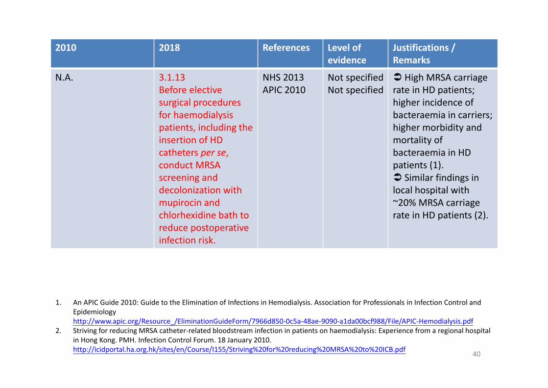
N.A. 3.1.13 Before elective surgical proceduresfor haemodialysispatients, including the insertion of HD catheters per se, conduct MRSA screening and decolonization with mupirocin and chlorhexidine bath to reduce postoperative infection risk.
NHS 2013APIC 2010
Not specifiedNot specified
High MRSA carriage rate in HD patients;higher incidence of bacteraemia in carriers;higher morbidity and mortality of bacteraemia in HD patients (1). Similar findings in local hospital with ~20% MRSA carriage rate in HD patients (2).
1. An APIC Guide 2010: Guide to the Elimination of Infections in Hemodialysis. Association for Professionals in Infection Control and Epidemiologyhttp://www.apic.org/Resource_/EliminationGuideForm/7966d850‐0c5a‐48ae‐9090‐a1da00bcf988/File/APIC‐Hemodialysis.pdf
2. Striving for reducing MRSA catheter‐related bloodstream infection in patients on haemodialysis: Experience from a regional hospital in Hong Kong. PMH. Infection Control Forum. 18 January 2010. http://icidportal.ha.org.hk/sites/en/Course/I155/Striving%20for%20reducing%20MRSA%20to%20ICB.pdf 40

Ref. Original text of findings / conclusion
NHS 2013
P.13 –
For each in‐patient episode of care, a swab must be obtained for MRSA surveillance on admission.
APIC 2010
P.38 –
Presurgical (HD Access) Infection Prevention > Active Surveillance Screening for MRSA and Decolonization > Although there is not Category I level evidence regarding efficacy, some medical centers are culturing patients preoperatively for MRSA. For those who test positive, nasal mupirocin is applied to decolonize the nasal passages before surgery as an additional measure to reduce postoperative infection risk on top of preoperative bathing or showeringwith an antiseptic agent such as CHG.
P.67 –
A major concern with decolonization is the emergence of mupirocin resistance, which occurs more frequently with long‐term use. Of special concern are reports of outbreaks of mupirocin‐resistant MRSA in hospitals following widespread mupirocin use. Therefore, the benefit of either short‐term or long‐term treatment with intranasal mupirocin must be considered very cautiously. The safest approach may be to use mupirocin eradication of
S. aureus nasal carriage for the individual HD patient for a single incident of elective surgery only.
41

3.1 Haemodialysis CatheterTopical Antiseptic or Antibiotic
42

2010 2018 References Level of evidence
Justifications / Remarks
N.A. 3.1.14For haemodialysis, use povidone iodine antiseptic ointment or bacitracin/gramicidin/ polymyxin B ointment at the hemodialysis catheter exit site after catheter insertion and at the end of each dialysis session only if this ointment does not interact with the material of the hemodialysis catheter per manufacturer’s recommendation.
CDC 2011SHEA 2014
Category IBGrade I
10% povidone iodine reduce risk of colonization, exit‐site infection, bloodstream infection. bacitracin/ gramicidin/ polymyxinB ointment reduce risk of bacteraemia, infection and death.
43
Ref. Original text of findings / conclusion
New references
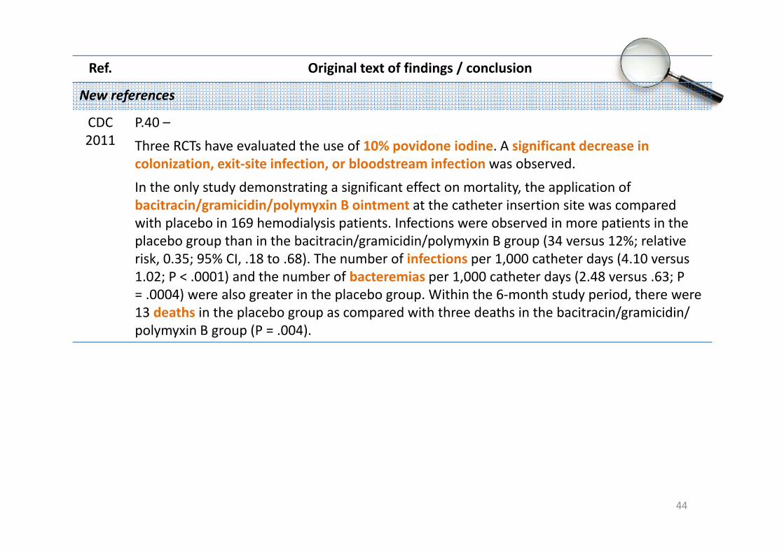
CDC 2011
P.40 –
Three RCTs have evaluated the use of 10% povidone iodine. A significant decrease in colonization, exit‐site infection, or bloodstream infection was observed.
In the only study demonstrating a significant effect on mortality, the application of bacitracin/gramicidin/polymyxin B ointment at the catheter insertion site was compared with placebo in 169 hemodialysis patients. Infections were observed in more patients in the placebo group than in the bacitracin/gramicidin/polymyxin B group (34 versus 12%; relative risk, 0.35; 95% CI, .18 to .68). The number of infections per 1,000 catheter days (4.10 versus 1.02; P < .0001) and the number of bacteremias per 1,000 catheter days (2.48 versus .63; P = .0004) were also greater in the placebo group. Within the 6‐month study period, there were 13 deaths in the placebo group as compared with three deaths in the bacitracin/gramicidin/ polymyxin B group (P = .004).
44

Ref. Original text of findings / conclusion
New references
SHEA 2014
P.757 –
Use antimicrobial ointments for hemodialysis catheter insertion sites (quality of evidence: I).
a. Polysporin “triple” (where available) or povidone‐iodine ointment should be applied to hemodialysis catheter insertion if compatible with the catheter material. Certain manufacturers have indicated that the glycol constituents of ointments should not be used on their polyurethane catheters.
b. Mupirocin ointment should not be applied to the catheter‐insertion site due to the risks of facilitating mupirocin resistance and the potential damage to polyurethane catheters.
45
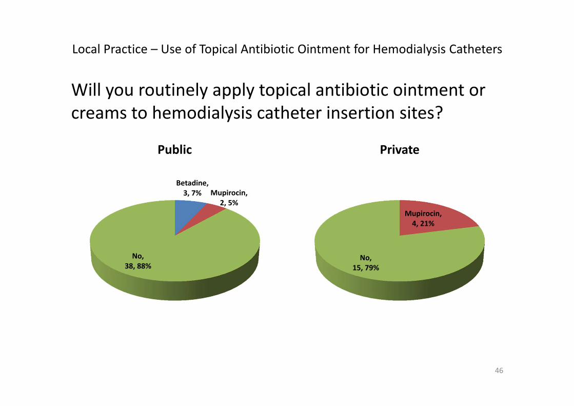
Local Practice – Use of Topical Antibiotic Ointment for Hemodialysis Catheters
46
Will you routinely apply topical antibiotic ointment or creams to hemodialysis catheter insertion sites?
Betadine, 3, 7% Mupirocin,
2, 5%
No,38, 88%
Public
Mupirocin, 4, 21%
No,15, 79%
Private

3.2 Peripheral Venous CatheterSite Selection
47

2010 2018 References Level of evidence
Justifications / Remarks
Use the upper extremity for catheter insertion in adults.
3.2.1For adults, use an upper‐extremity site for catheter insertion. Replace a catheter inserted in a lower extremity site to an upper extremity site as soon as possible.
CDC 2011 Category II
3.2.1 For pediatric patients, the upper or lower extremities or the scalp (in neonates or young infants) can be used as the catheter insertion site.
CDC 2011 Category II
48

3.2 Peripheral Venous CatheterCatheter Change
49
2010 2018 References Level of evidence
Justifications / Remarks
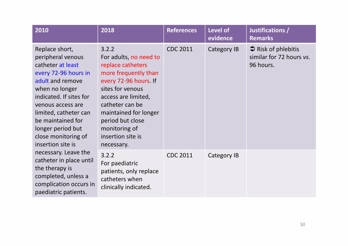
Replace short, peripheral venous catheter at least every 72‐96 hours in adult and remove when no longer indicated. If sites for venous access are limited, catheter can be maintained for longer period but close monitoring of insertion site is necessary. Leave the catheter in place until the therapy is completed, unless a complication occurs in paediatric patients.
3.2.2For adults, no need to replace cathetersmore frequently than every 72‐96 hours. If sites for venous access are limited, catheter can be maintained for longer period but close monitoring of insertion site is necessary.
CDC 2011 Category IB Risk of phlebitis similar for 72 hours vs.96 hours.
3.2.2For paediatricpatients, only replace catheters when clinically indicated.
CDC 2011 Category IB
50
Ref. Original text of findings / conclusion
Previous references
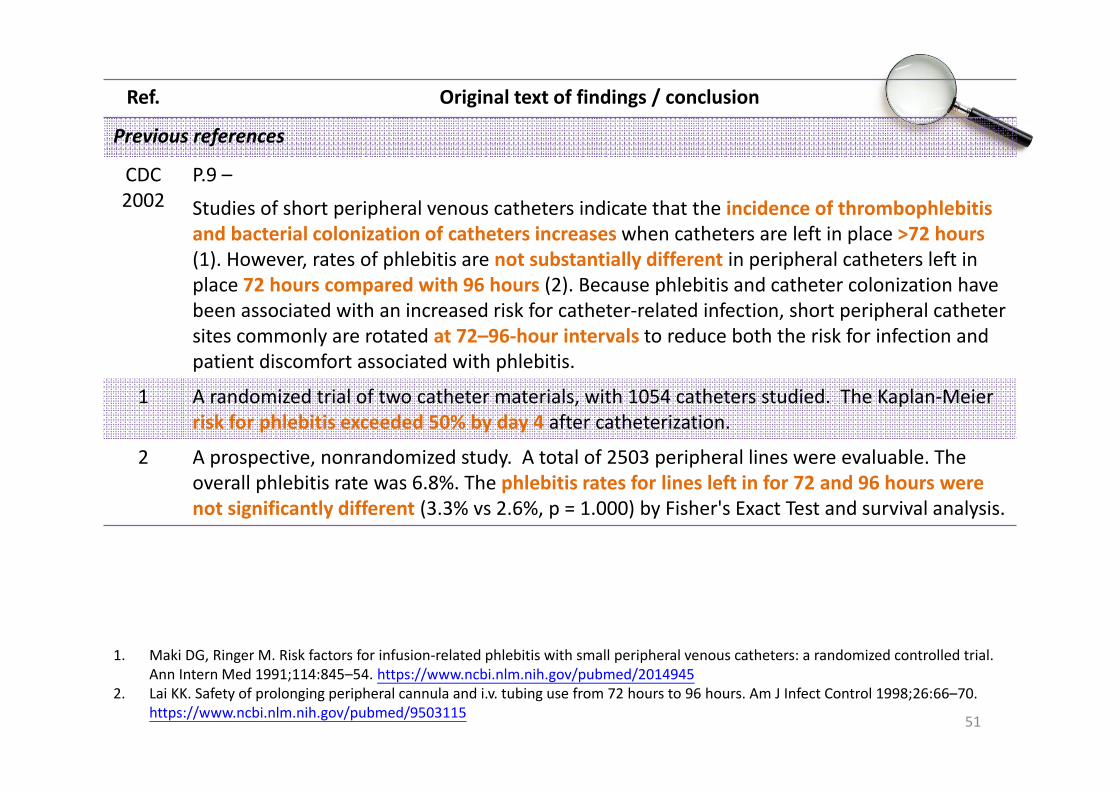
CDC 2002
P.9 –
Studies of short peripheral venous catheters indicate that the incidence of thrombophlebitis and bacterial colonization of catheters increases when catheters are left in place >72 hours(1). However, rates of phlebitis are not substantially different in peripheral catheters left in place 72 hours compared with 96 hours (2). Because phlebitis and catheter colonization have been associated with an increased risk for catheter‐related infection, short peripheral catheter sites commonly are rotated at 72–96‐hour intervals to reduce both the risk for infection and patient discomfort associated with phlebitis.
1 A randomized trial of two catheter materials, with 1054 catheters studied. The Kaplan‐Meier risk for phlebitis exceeded 50% by day 4 after catheterization.
2 A prospective, nonrandomized study. A total of 2503 peripheral lines were evaluable. The overall phlebitis rate was 6.8%. The phlebitis rates for lines left in for 72 and 96 hours were not significantly different (3.3% vs 2.6%, p = 1.000) by Fisher's Exact Test and survival analysis.
51
1. Maki DG, Ringer M. Risk factors for infusion‐related phlebitis with small peripheral venous catheters: a randomized controlled trial. Ann Intern Med 1991;114:845–54. https://www.ncbi.nlm.nih.gov/pubmed/2014945
2. Lai KK. Safety of prolonging peripheral cannula and i.v. tubing use from 72 hours to 96 hours. Am J Infect Control 1998;26:66–70. https://www.ncbi.nlm.nih.gov/pubmed/9503115

Ref. Original text of findings / conclusion
New references
CDC 2011
P.45 –
There is no need to replace peripheral catheters more frequently than every 72–96 hours to reduce risk of infection and phlebitis in adults (1,2,3). Category 1B
1 A randomized trial of two catheter materials, with 1054 catheters studied. The Kaplan‐Meier risk for phlebitis exceeded 50% by day 4 after catheterization.
2 A prospective, nonrandomized study. A total of 2503 peripheral lines were evaluable. The overall phlebitis rate was 6.8%. The phlebitis rates for lines left in for 72 and 96 hours were not significantly different (3.3% vs 2.6%, p = 1.000) by Fisher's Exact Test and survival analysis.
3 The authors studied 3094 patients with 5161 total episodes of peripheral intravenous catheters from day of admission until day of discharge. Analysis of day‐specific risk of phlebitis indicated that, for patients with low risk diagnoses, initial peripheral intravenous catheters might be left in place with relative safety for up to 96 hours.
52
1. Maki DG, Ringer M. Risk factors for infusion‐related phlebitis with small peripheral venous catheters: a randomized controlled trial. Ann Intern Med 1991;114:845–54. https://www.ncbi.nlm.nih.gov/pubmed/2014945
2. Lai KK. Safety of prolonging peripheral cannula and i.v. tubing use from 72 hours to 96 hours. Am J Infect Control 1998;26:66–70. https://www.ncbi.nlm.nih.gov/pubmed/9503115
3. Tager IB, Ginsberg MB, Ellis SE, et al. An epidemiologic study of the risks associated with peripheral intravenous catheters. Am J Epidemiol 1983; 118:839–51. https://www.ncbi.nlm.nih.gov/pubmed/6650485

Type of Hospital Peripheral venous linen (%)
Peripheral arterial linen (%)
Central linen (%)
Public N=72 N=41 N=60
Every 72‐96 hours 42 (58.3%) 5 (12.2%) 4 (6.7%)
No routine change 23 (31.9%) 31 (75.6%) 49 (81.7%)
PRN 6 (8.3%) 5 (12.2%) 4 (6.7%)
Private N=24 N=14 N=21
Every 72‐96 hours 14# (58.3%) 6 (42.9%) 1 (4.8%)
No routine change 8## (33.3%) 3 (21.4%) 15 (71.4%)
PRN 1 (4.2%) 2 (14.3%) 2 (9.5%)
Local Practice ‐ Frequency of Catheter Change
~60% every 72‐96 hours; ~40% no routine change / PRN
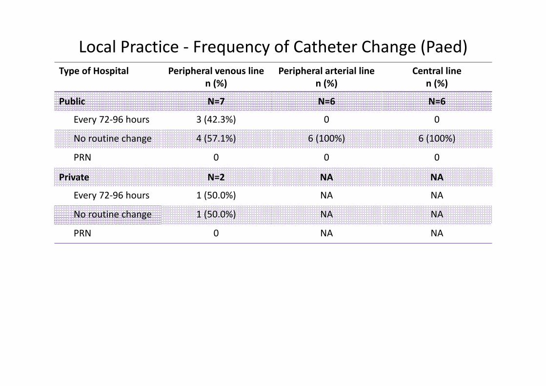
Type of Hospital Peripheral venous linen (%)
Peripheral arterial linen (%)
Central linen (%)
Public N=7 N=6 N=6
Every 72‐96 hours 3 (42.3%) 0 0
No routine change 4 (57.1%) 6 (100%) 6 (100%)
PRN 0 0 0
Private N=2 NA NA
Every 72‐96 hours 1 (50.0%) NA NA
No routine change 1 (50.0%) NA NA
PRN 0 NA NA
Local Practice ‐ Frequency of Catheter Change (Paed)

3.3 Peripheral Arterial CatheterAttire
55
2010 2018 References Level of evidence
Justifications / Remarks
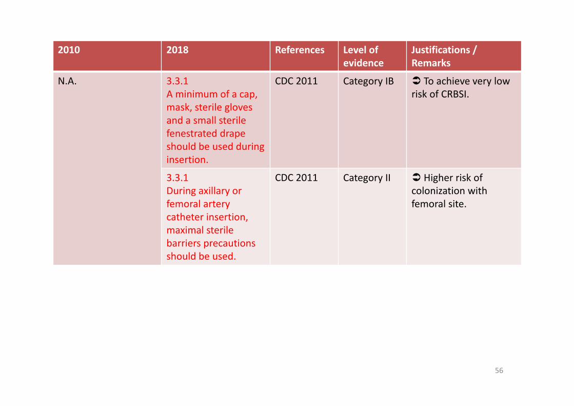
N.A. 3.3.1A minimum of a cap, mask, sterile gloves and a small sterile fenestrated drape should be used during insertion.
CDC 2011 Category IB To achieve very low risk of CRBSI.
3.3.1During axillary or femoral artery catheter insertion, maximal sterile barriers precautions should be used.
CDC 2011 Category II Higher risk of colonization with femoral site.
56

Ref. Original text of findings / conclusion
CDC 2011
P.52 –
Unlike CVCs, use of full barrier precautions during arterial cannulaton does not appear to reduce the risk of arterial CRBSI (1,2). Nonetheless, when arterial catheters are inserted using a protocol which includes maximum barrier precautions, a very low risk of CRBSI(0.41/1,000 catheter days) can be achieved (3).
1 An RCT of patients undergoing radial or dorsalis pedis arterial catheterization with either sterile barrier precautions (SBPs) or standard‐of‐care (hand washing, sterile gloves, skin antisepsis with 0.5% chlorhexidine in 70% alcohol). N = 272. The colonization incidence and AC‐related infection did not differ significantly.
2 Prospective 24‐month cohort study in ICU/HDU in a teaching hospital. 252 patients with 1,082 catheter‐days for AC and 410 patients with 4,040 catheter‐days for CVC. Under aseptic conditions. AC colonization was not significantly different than that in CVC. Femoral ACs were colonized more often than radial ACs (HR 5.08; 95% CI 0.85‐30.3; p = .075).
3 Prospective study in ICU in a teaching hospital. 212 patients with 308 CVCs and 299 ACs. Among severely ill patients with both CVCs and ACs, the epidemiology of positive quantitative cultures of CVCs and ACs is comparable when the same infection control measures are used for the insertion and maintenance of both types of catheters.
57
1. Rijnders BJ, Van Wijngaerden E, Wilmer A, Peetermans WE. Use of full sterile barrier precautions during insertion of arterial catheters: a randomized trial. Clin Infect Dis 2003; 36:743–8. https://www.ncbi.nlm.nih.gov/pubmed/12627358
2. Koh DB, Gowardman JR, Rickard CM, Robertson IK, Brown A. Prospective study of peripheral arterial catheter infection and comparison with concurrently sited central venous catheters. Crit Care Med 2008; 36:397–402. https://www.ncbi.nlm.nih.gov/pubmed/18216598
3. Traore O, Liotier J, Souweine B. Prospective study of arterial and central venous catheter colonization and of arterial‐and central venous catheter‐related bacteremia in intensive care units. Crit Care Med 2005; 33:1276–80. https://www.ncbi.nlm.nih.gov/pubmed/15942344

3.3 Peripheral Arterial CatheterSite Selection
58
2010 2018 References Level of evidence
Justifications / Remarks
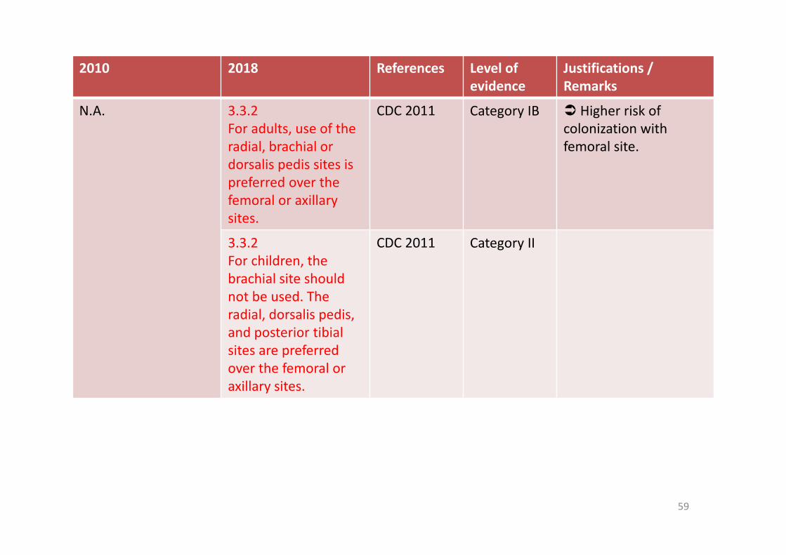
N.A. 3.3.2For adults, use of the radial, brachial or dorsalis pedis sites is preferred over the femoral or axillary sites.
CDC 2011 Category IB Higher risk of colonization with femoral site.
3.3.2For children, the brachial site should not be used. The radial, dorsalis pedis, and posterior tibialsites are preferred over the femoral or axillary sites.
CDC 2011 Category II
59
Ref. Original text of findings / conclusion
CDC 2011
P.52 –
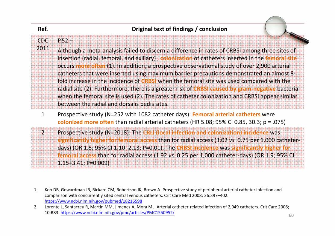
Although a meta‐analysis failed to discern a difference in rates of CRBSI among three sites of insertion (radial, femoral, and axillary) , colonization of catheters inserted in the femoral site occurs more often (1). In addition, a prospective observational study of over 2,900 arterial catheters that were inserted using maximum barrier precautions demonstrated an almost 8‐fold increase in the incidence of CRBSI when the femoral site was used compared with the radial site (2). Furthermore, there is a greater risk of CRBSI caused by gram‐negative bacteria when the femoral site is used (2). The rates of catheter colonization and CRBSI appear similar between the radial and dorsalis pedis sites.
1 Prospective study (N=252 with 1082 catheter days): Femoral arterial catheters were colonized more often than radial arterial catheters (HR 5.08; 95% CI 0.85, 30.3; p = .075)
2 Prospective study (N=2018): The CRLI (local infection and colonization) incidence was significantly higher for femoral access than for radial access (3.02 vs. 0.75 per 1,000 catheter‐days) (OR 1.5; 95% CI 1.10–2.13; P=0.01). The CRBSI incidence was significantly higher for femoral access than for radial access (1.92 vs. 0.25 per 1,000 catheter‐days) (OR 1.9; 95% CI 1.15–3.41; P=0.009)
60
1. Koh DB, Gowardman JR, Rickard CM, Robertson IK, Brown A. Prospective study of peripheral arterial catheter infection and comparison with concurrently sited central venous catheters. Crit Care Med 2008; 36:397–402. https://www.ncbi.nlm.nih.gov/pubmed/18216598
2. Lorente L, Santacreu R, Martin MM, Jimenez A, Mora ML. Arterial catheter‐related infection of 2,949 catheters. Crit Care 2006; 10:R83. https://www.ncbi.nlm.nih.gov/pmc/articles/PMC1550952/

3.3 Peripheral Arterial CatheterCather Change
61
2010 2018 References Level of evidence
Justifications / Remarks
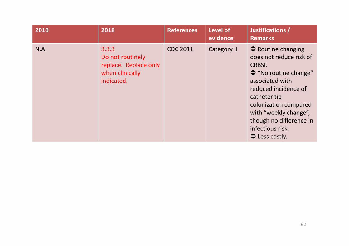
N.A. 3.3.3Do not routinely replace. Replace only when clinically indicated.
CDC 2011 Category II Routine changing does not reduce risk of CRBSI. “No routine change” associated with reduced incidence of catheter tip colonization compared with “weekly change”, though no difference in infectious risk. Less costly.
62

Ref. Original text of findings / conclusion
CDC 2011
P.53 –
The risk of developing a CRBSI increases with the duration of catheterization (1); however, the routine changing of arterial catheters at scheduled times does not result in a diminution of the risk of CRBSI (2). Catheters that need to be in place for >5 days should not be routinely changed if no evidence of infection is observed.
1 1‐year prospective study in 310 children with 340 ACs: The duration of arterial catheterization was longer in group 3 (with CRBSI) than in group 1 (sterile). The risk of infection was non‐existent in the first 48 hours of catheterization. Thereafter it was calculated as being 6.2%, but it correlated poorly with the duration of arterial catheterization. These results confirm the very low incidence of infection related to arterial catheterization in children. Thus, routine catheter reinsertion is, in our opinion, unjustified.
2 RCT in surgical ICU (N=112): 3 management groups: a) percutaneous puncture with every 7‐day catheter change at a new site, b) no weekly change (NWC) with a new site when changed, or c) guidewire exchange with every 7‐day catheter change at the same site. The NWC group demonstrated… a reduced incidence of catheter tip colonization. We conclude that there is no difference in infectious risk between these three methods of long‐term catheter management. The method with the least complications and expense should be used.
63
1. Furfaro S, Gauthier M, Lacroix J, Nadeau D, Lafleur L, Mathews S. Arterial catheter‐related infections in children. A 1‐year cohort analysis. Am J Dis Child 1991; 145:1037–43. https://www.ncbi.nlm.nih.gov/pubmed/1877564
2. Eyer S, Brummitt C, Crossley K, Siegel R, Cerra F. Catheter‐related sepsis: prospective, randomized study of three methods of long‐term catheter maintenance. Crit Care Med 1990; 18:1073–9. https://www.ncbi.nlm.nih.gov/pubmed/2209033

Type of Hospital Peripheral venous linen (%)
Peripheral arterial linen (%)
Central linen (%)
Public N=72 N=41 N=60
Every 72‐96 hours 42 (58.3%) 5 (12.2%) 4 (6.7%)
No routine change 23 (31.9%) 31 (75.6%) 49 (81.7%)
PRN 6 (8.3%) 5 (12.2%) 4 (6.7%)
Private N=24 N=14 N=21
Every 72‐96 hours 14# (58.3%) 6 (42.9%) 1 (4.8%)
No routine change 8## (33.3%) 3 (21.4%) 15 (71.4%)
PRN 1 (4.2%) 2 (14.3%) 2 (9.5%)
Local Practice ‐ Frequency of Catheter Change
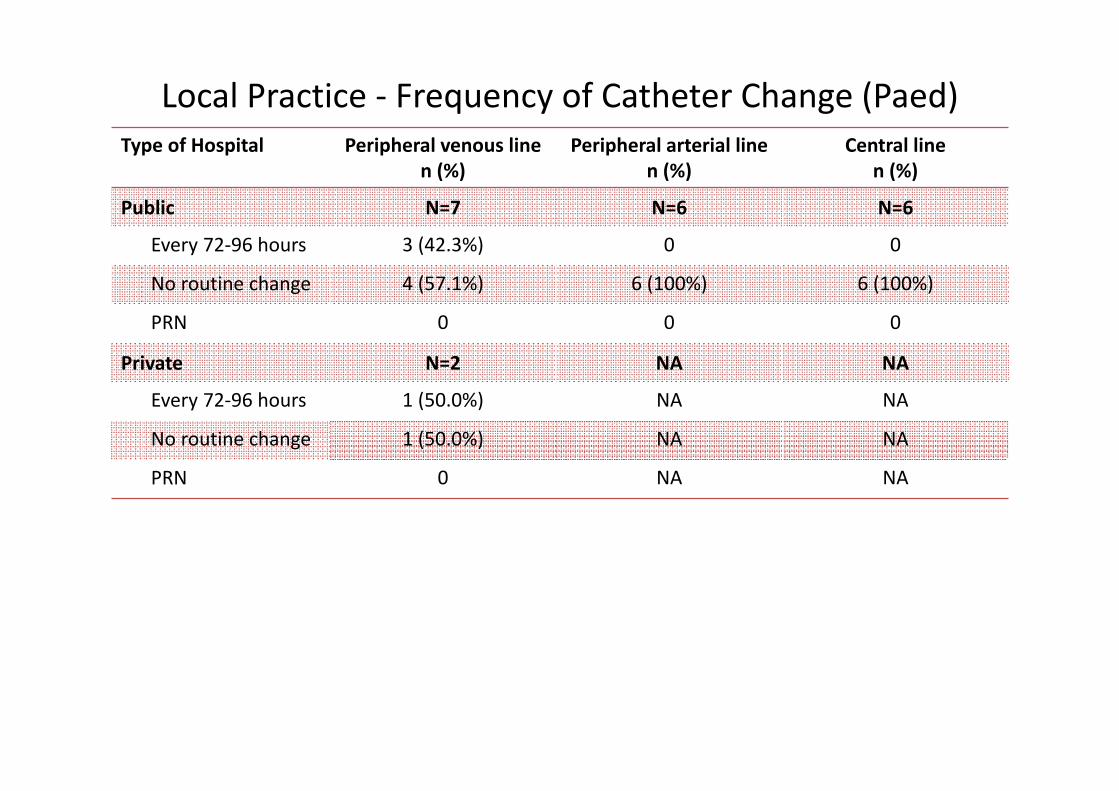
~90% no routine change / PRN in public; while 43% every 72‐96 hours in private
Type of Hospital Peripheral venous linen (%)
Peripheral arterial linen (%)
Central linen (%)
Public N=7 N=6 N=6
Every 72‐96 hours 3 (42.3%) 0 0
No routine change 4 (57.1%) 6 (100%) 6 (100%)
PRN 0 0 0
Private N=2 NA NA
Every 72‐96 hours 1 (50.0%) NA NA
No routine change 1 (50.0%) NA NA
PRN 0 NA NA
Local Practice ‐ Frequency of Catheter Change (Paed)

4.1 Frequency of Change of Administration Set
66

2010 2018 References Level of evidence
Justifications / Remarks
Replace administration sets including extension tubings, add‐on devices no more frequently than every 72 hours, unless CABSIis suspected or confirmed.
4.1Replace administration sets including extension tubings, add‐on devices no more frequently than every 96 hours unless CABSI is suspected or confirmed,
CDC 2011SHEA 2014NICE 2012UKDH 2014
Category IAGrade IINot specifiedClass A
In CDC 2002, studies compared 48‐hour, 72‐hour, 96‐hour change and no change; no significant difference. In CDC 2011, a systematic review, Cochrane 2005, was included: safe up to 96 hours for both central and peripheral catheters Also supported by SHEA and Cochrane 2013; extended the recommendation to peripheral arterial catheters.
but at least every 7 days.
CDC 2011 Category IA Safe and more cost‐effective.
67

Ref. Original text of findings / conclusion
Previous References (on ≥48 hours / ≥72 hours / ≥96 hours / no change)
CDC 2002
P.11 –
Data from each of these studies reveal that replacing administration sets no more frequently than 72 hours after initiation of use is safe and cost‐effective (1,2,3). Data from a more recent study demonstrated that rates of phlebitis were not substantially different if administration sets were left in place 96 hours compared with 72 hours (4).
4 Prospective study (2503 peripheral lines) cf. 72‐hour vs. 96‐hour change: The phlebitis rates for lines left in for 72 and 96 hours were not significantly different (3.3% vs 2.6%, p = 1.000). If intravenous cannulas and lines were prolonged to 96 hours, a potential cost saving of $61,200 per year could be realized.
68
1. Josephson A, Gombert ME, Sierra MF, Karanfil LV, Tansino GF. The relationship between intravenous fluid contamination and the frequency of tubing replacement. Infect Control 1985;6:367–70. https://www.ncbi.nlm.nih.gov/pubmed/3932250
2. Maki DG, Botticelli JT, LeRoy ML, Thielke TS. Prospective study of replacing administration sets for intravenous therapy at 48‐ vs 72‐hour intervals: 72 hours is safe and cost‐effective. JAMA 1987;258:1777–81. http://jamanetwork.com/journals/jama/article‐abstract/368478
3. Snydman DR, Donnelly‐Reidy M, Perry LK, Martin WJ. Intravenous tubing containing burettes can be safely changed at 72 hour intervals. Infect Control 1987;8:113–6. https://www.ncbi.nlm.nih.gov/pubmed/3646182
4. Lai KK. Safety of prolonging peripheral cannula and i.v. tubing use from 72 hours to 96 hours. Am J Infect Control 1998;26:66–70. https://www.ncbi.nlm.nih.gov/pubmed/9503115

Ref. Original text of findings / conclusion
New References (on ≥96 hours)
CDC 2011
P.53 –
In patients not receiving blood, blood products or fat emulsions, replace administration sets that are continuously used, including secondary sets and add‐on devices, no more frequently than at 96‐hour intervals, but at least every 7 days. Category IA
The optimal interval for routine replacement of IV administration sets has been examined in a number of well‐controlled studies and meta‐analyses. Data from these studies reveal that replacing administration sets no more frequently than 72–96 hours after initiation of use is safe and cost‐effective (1,2,3,4,5).
5 Cochrane 2005 (13 RCTs/quasi‐RCTs, N=4783, cf. 24 vs. ≥48 hrs; 48 vs. ≥72 hrs; 72 vs. ≥96 hrs): No significant difference in the risk of catheter colonization (RR 1.13; 95% CI 0.99‐1.28), infusate colonization (RR 1.07; 95% CI 0.65‐1.76), catheter‐related BSI (RR 1.06; 95% CI 0.66‐1.68), infusate‐related BSI (RR 0.58; 95% CI 0.25‐1.35), all‐cause BSI (RR 0.82; 95%CI 0.48‐1.40) and BSI‐related mortality (RR 14.18; 95% CI 0.86‐234.13) when the interval between administration set changes was increased overall or in each comparison.
Overall, there is good evidence that changing intravenous administration sets, that do not contain lipids, blood or blood products, at an interval of up to 96 hours does not affect the risk of infusate‐related BSI or catheter‐related BSI in participants with central or peripheral catheters.
69
5. Gillies D, Wallen MM, Morrison AL, Rankin K, Nagy SA, O’Riordan E. Optimal timing for intravenous administration set replacement. Cochrane Database of Systematic Reviews 2005; Issue 4. Art. No.: CD003588. DOI: 10.1002/14651858.CD003588.pub2. https://www.ncbi.nlm.nih.gov/pubmed/16235329
Ref. Original text of findings / conclusion
New References (on ≥96 hours)
SHEA 2014
P.757 –
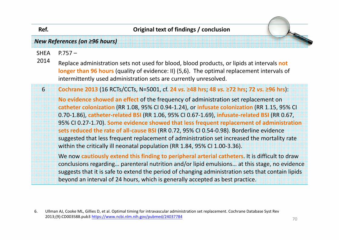
Replace administration sets not used for blood, blood products, or lipids at intervals not longer than 96 hours (quality of evidence: II) (5,6). The optimal replacement intervals of intermittently used administration sets are currently unresolved.
6 Cochrane 2013 (16 RCTs/CCTs, N=5001, cf. 24 vs. ≥48 hrs; 48 vs. ≥72 hrs; 72 vs. ≥96 hrs):
No evidence showed an effect of the frequency of administration set replacement on catheter colonization (RR 1.08, 95% CI 0.94‐1.24), or infusate colonization (RR 1.15, 95% CI 0.70‐1.86), catheter‐related BSI (RR 1.06, 95% CI 0.67‐1.69), infusate‐related BSI (RR 0.67, 95% CI 0.27‐1.70). Some evidence showed that less frequent replacement of administration sets reduced the rate of all‐cause BSI (RR 0.72, 95% CI 0.54‐0.98). Borderline evidence suggested that less frequent replacement of administration set increased the mortality rate within the critically ill neonatal population (RR 1.84, 95% CI 1.00‐3.36).
We now cautiously extend this finding to peripheral arterial catheters. It is difficult to draw conclusions regarding… parenteral nutrition and/or lipid emulsions… at this stage, no evidence suggests that it is safe to extend the period of changing administration sets that contain lipids beyond an interval of 24 hours, which is generally accepted as best practice.
70
6. Ullman AJ, Cooke ML, Gillies D, et al. Optimal timing for intravascular administration set replacement. Cochrane Database Syst Rev 2013;(9):CD003588.pub3 https://www.ncbi.nlm.nih.gov/pubmed/24037784

Ref. Original text of findings / conclusion
Other References (UK)
UKDH 2014
P.S50 –
Administration sets in continuous use do not need to be replaced more frequently than every 96 h, unless device‐specific recommendations from the manufacturer indicate otherwise, they become disconnected or the intravascular access device is replaced. Class A
NICE 2012
P.205 –
In general, administration sets in continuous use need not be replaced more frequently than at 72 hour intervals unless they become disconnected or if a catheter‐related infection is suspected or documented. [2003]
The optimal interval for the routine replacement of intravenous (IV) administration sets has been examined in three well‐controlled studies reviewed by HICPAC. Data from each of these studies reveal that replacing administration sets no more frequently than 72 hours after initiation of use is safe and cost‐effective. When a fluid that enhances microbial growth is infused, e.g., lipid emulsions, blood products, more frequent changes of administration sets are indicated as these products have been identified as independent risk factors for CRBSI. (CDC 2002)
71

Ref. Original text of findings / conclusion
New References (on 4‐7 days)
CDC 2011
P.53 –
In patients not receiving blood, blood products or fat emulsions, replace administration sets that are continuously used, including secondary sets and add‐on devices, no more frequently than at 96‐hour intervals, but at least every 7 days. Category IA
More recent studies suggest that administration sets may be used safely for up to 7 days if used with antiseptic catheters or if fluids that enhance microbial growth (e.g. parenteral nutrition or blood) have not been used (7,8).
7 RCT (N=512) cf. 72‐hour vs. 4‐7 day change: When the 84 patients who received total parenteral nutrition, blood transfusions, or interleukin‐2 through the IV tubing were excluded, the two groups had a comparable rate of colonization (0.4% vs 0.5%), with no catheter‐ or infusion‐related BSIs in either group. In patients at low risk for infection… delaying the replacement of IV tubing up to 7 days may be safe, as well as cost‐effective.
8 RCT (N=404 CHG‐ and SSD‐coated multi‐lumen CVCs) cf. 4‐day vs. 7‐day change: No statistically significant difference in colonization and catheter‐related bacteraemia. IV administration sets can be used for 7 days in patients with short‐term, antiseptic‐coated CVCs.
72
7. Raad I, Hanna HA, Awad A, et al. Optimal frequency of changing intravenous administration sets: is it safe to prolong use beyond 72 hours? Infect Control Hosp Epidemiol 2001; 22:136–9. https://www.ncbi.nlm.nih.gov/pubmed/11310690
8. Rickard CM, Lipman J, Courtney M, Siversen R, Daley P. Routine changing of intravenous administration sets does not reduce colonization or infection in central venous catheters. Infect Control Hosp Epidemiol 2004; 25:650–5. https://www.ncbi.nlm.nih.gov/pubmed/15357156

Ref. Original text of findings / conclusion
New References (on blood / nutrition infusates)
Blood Nutrition
CDC 2011
P.19 –
Replace tubing used to administer blood, blood products, or fat emulsions (those combined with amino acids and glucose in a 3‐in‐1 admixture or infused separately)
within 24 hours of initiating the infusion. Category IB
NICE 2012
P.27 –
Administration sets for blood and blood components should be changed every
12 hours, or according to the manufacturer's recommendations. [2003]
P.27 –
Administration sets used for
total parenteral nutrition infusions should generally be changed every 24 hours.
If the solution contains only glucose and amino acids, administration sets in continuous use do not need to be replaced more frequently than every 72 hours. [2003]
UKDH2014
P.S10 –
Administration sets for blood and blood components should be changed when the transfusion episode is complete or every 12 h (whichever is sooner). Class D
P.S10 –
Administration sets used for lipid‐containing parenteral nutrition should be changed every 24 h. Class D
73

Local Practice – Frequency of Change of Administration SetType of Hospital Peripheral venous line
n (%)Peripheral arterial line
n (%)Central line
n (%)
Public N=71 N=42 N=59
<=4 days (PVL) /<4 days (Others) 67 (94.4%) 31 (73.8%) 55 (93.2%)
5‐7 days (PVL) /4‐7 days (Others) 1 (1.4%) 7 (16.7%) 2 (3.4%)
PRN 0 1 (2.4%) 0
Private N=24 N=14 N=21
<=4 days (PVL) /<4 days (Others) 24 (100.0%) 12 (85.7%) 18 (85.7%)
5‐7 days (PVL) /4‐7 days (Others) 0 2 (14.3%) 2 (9.5%)
PRN 0 0 0
~70%‐90% within 4 days; ~3%‐17% up to 4‐7 days

7.4 Antimicrobial‐ or Antiseptic‐impregnated Catheters
75

2010 2018 References Level of evidence
Justifications / Remarks
Antiseptic coated catheter is more preferable as it does not carry the risk of anaphylactoidreactions, superinfection with yeast and promoting antibiotic resistant pathogen as in antibiotic one.
7.4When using antimicrobial‐ or antiseptic‐impregnated catheters, monitor patients for untoward effects, such asanaphylaxis.
CDC 2011SHEA 2014UKDH 2014
Not specifiedNot specified
No recommendation in current references to preferably use antiseptic‐coated over antimicrobial‐coated catheters. New references caution about risk of anaphylaxis due to chlorhexidine.
76

Ref. Original text of findings / conclusion
Previous References
Old references are either from textbook(1), in‐vitro or in‐vivo (rats) studies (2)(3), or results not statistically significant (4).
4 Multicenter cohort study (N=4535 patients, 56627 CVC‐days): Of the 8,593 CVCs, 240 (2.8%) were associated with CVC‐associated BSI. No significant differences were observed in the distribution of BSI pathogens isolated when antimicrobial‐impregnated CVCs were used with the exception that fungi were more common when an antimicrobial‐impregnated CVC was used (20% vs 9%; P = .06).
Limitations: all of the antimicrobial‐impregnated CVCs used by participating units were impregnated with chlorhexidine and silver sulfadiazine. Therefore, we could not assess the impact of other types of impregnated CVCs.
77
1. Farr BM. Nosocomial infections related to use of intravascular devices inserted for short‐term vascular access. In: Mayhall CG, editor. Hospital epidemiology and infection control. 3rd ed. Baltimore: Williams & Wilkins; 2004:231‐40.
2. Tambe SM, Sampath L, Modak SM. In vitro evaluation of the risk of developing bacterial resistance to antiseptics and antibiotics used in medical devices. J Antimicrob Chemother. 2001 May;47(5):589‐98.
3. Sampath LA, Tambe SM, Modak SM. In vitro and in vivo efficacy of catheters impregnated with antiseptics or antibiotics: evaluation of the risk of bacterial resistance to the antimicrobials in the catheters. Infect Control Hosp Epidemiol. 2001;22(10):640‐6.
4. Alonso‐Echanove J, Edwards JR, Richards MJ et al. Effect of nurse staffing and antimicrobial‐impregnated central venous catheters on the risk for bloodstream infections in intensive care units. Infect Control Hosp Epidemiol. 2003;24(12):916‐25.

Ref. Original text of findings / conclusion
New References
CDC2011
P.36 –
Chlorhexidine/Silver Sulfadiazine: … Although rare, anaphylaxis with the use of these chlorhexidine/silver sulfadiazine catheters has been observed.
Minocycline/Rifampin: … Although there have been concerns related to the potential for development of resistance, several prospective clinical studies have shown that the risk is low. Further, no resistance to minocyline or rifampin related to the use of the catheter has been documented in the clinical setting.
SHEA 2014
P.757 –
Monitor patients for untoward effects, such as anaphylaxis (referring to chlorhexidine).
UKDH 2014
P.S42 –
Be aware of patient sensitivity to chlorhexidine gluconate.
78
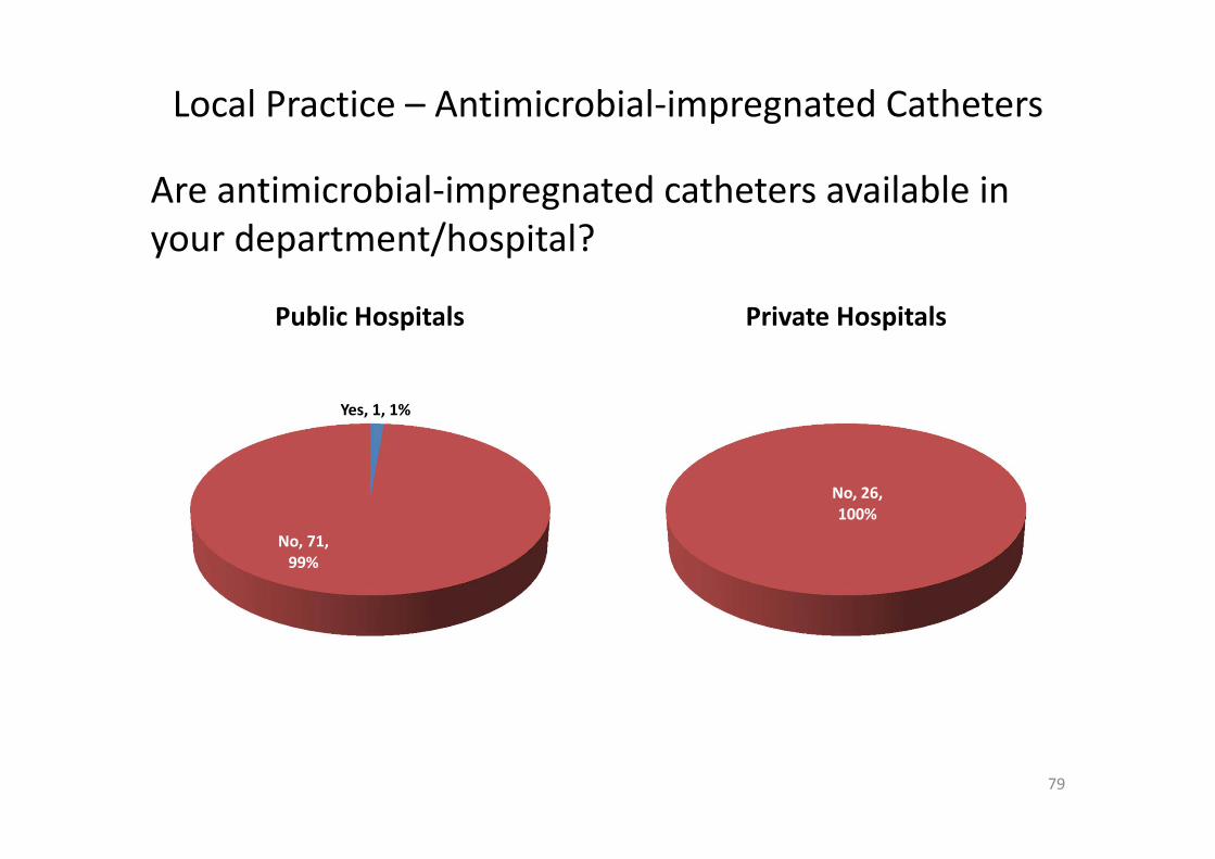
Local Practice – Antimicrobial‐impregnated Catheters
79
Are antimicrobial‐impregnated catheters available in your department/hospital?
Yes, 1, 1%
No, 71, 99%
Public Hospitals
0
No, 26, 100%
Private Hospitals

Local Practice – Antiseptic‐impregnated Catheters
80
Are antiseptic‐impregnated catheters available in your department/hospital?
Yes, 4, 6%
No, 68, 94%
Public Hospitals
0, 0%
No, 26, 100%
Private Hospitals
Antiseptic‐impregnated catheters use: Chlorhexidine/Silver Sulfadiazine Catheter

81

THANK YOU
82