Embed Size (px)
Citation preview

0
Higher Human Biology
Unit 3: Neurobiology and
Communication
Course notes
CURRICULUM FOR EXCELLENCE

1
The nervous system
The nervous system analyses sensory information from the body and the
external environment. Some of this information is stored for possible future
use. Appropriate voluntary and involuntary motor responses are made by
initiating muscular contractions or glandular secretions.
Divisions of the nervous system
Figure 1 below shows that the nervous system can be divided into two areas:
1 the central nervous system (CNS) and
2 the peripheral nervous system (PNS).
Sensory and Motor Pathways
The peripheral nervous system contains two pathways:
1 the sensory pathway consisting of sensory nerve cells (or neurons)
2 the motor pathway consisting of motor nerve cells (or neurons).
Sensory nerve cells carry nerve impulses to the CNS (i.e. to the brain or spinal
Figure 1 Division of the nervous system

2
cord) from receptors. Some receptors are located in external sense organs e.g.
the skin, eye etc, others are found in internal organs e.g. the pancreas which
contains receptors which detect the level of glucose in our blood. The function
of the sensory pathway is to keep the brain in touch with the body’s external
and internal environments.
The brain analyses, processes and stores some of this information. It’s job is
to then act on this information by sending nerve impulses via the motor pathway
to the body’s effectors ( muscles or glands). An appropriate response is then
occurs e.g. enzyme or hormone secretion or muscle contraction. See Figure 2
below.
Further division of the nervous system
The peripheral nervous system can be divided into the:
1 Somatic nervous system (SNS)
2 Autonomic nervous system (ANS)
1. Somatic nervous system
This system controls the voluntary movement of the body’s skeletal
muscles i.e. regular body movements such as moving an arm picking up a
pencil etc. So, this is a system we are in control of as it always involves
conscious thought. This is achieved via sensory and motor pathways as
outlined in Figure 2. However, the somatic nervous system is also
responsible for certain reflex actions.
Example of an event involving the somatic nervous system
Imagine, for example for you are selecting four of your favourite
chocolates from a large box of chocolates. The chocolates act as a visual
Sensory pathway Motor pathway
Effector (e.g. muscle) Receptor (e.g. skin)
Figure 2 Flow of information through the nervous system
Web site
http://www.bbc.co.uk/schools/gcsebitesize/science/aqa_pre_2011/human/thenervoussystemact.shtml

3
stimulus. Nerve impulses pass from the eye via sensory nerve cells to the
brain. Decisions/choices are made and nerve impulses are then sent via
motor nerve cells to the skeletal muscles of the arm and hand to allow
the voluntary response needed to pick up the chocolates.
2. Autonomic nervous system `
This part of the nervous system regulates the internal environment
(homeostasis) by controlling structures and organs like the heart,
bronchioles, and blood vessels. This control is involuntary because it
normally automatically without the person consciously having to think
about it – we don’t notice when our blood vessels change in size or when
food is pushed through our digestive system.
The autonomic nervous system can itself be divided into two different
parts (divisions):
1 sympathetic division
2 parasympathetic division
These two divisions are described as being antagonistic to each other.
Both divisions can affect many of the same structures but they have an
opposite effect on them. This is shown in Figure 3 below.
Figure 3 Antagonistic nature of the sympathetic and parasympathetic divisions
“Rest and digest” “Fight or flight”
Web site
http://www.google.co.uk/url?sa=t&rct=j&q=somatic+nervous+system+site%3Ayoutube.com&source=web&cd=4&cad=rja&uact=8&ved=0CDQQtwIwAw&url=http%3
A%2F%2Fwww.youtube.com%2Fwatch%3Fv%3DOrzKEKI4x6I&ei=TLfxU9WWCOem0AXoyYHgBQ&usg=AFQjCNFuM4cpCHuEPmzbSp1xb0pORZoU1A

4
Note particularly, the antagonistic action of the sympathetic “ fight or flight”
as opposed to the parasympathetic “rest and digest” responses on heart rate,
breathing rate, peristalsis and intestinal secretions.
Parts of the Brain
The brain is organised into three interconnected layers:
the central core
the limbic system
the cerebral cortex
The central core
The central core contains the:
1. Medulla that regulates the basic life processes of breathing, heart rate,
arousal (the state of being awake and aware of our external environment)
and sleep. These processes are involuntary and happen automatically.
2. Cerebellum which is responsible for controlling balance and posture and
movement.
1 medulla 2 cerebellum
Web site - up to 6.05min
http://www.youtube.com/watch?v=x4PPZCLnVkA
5
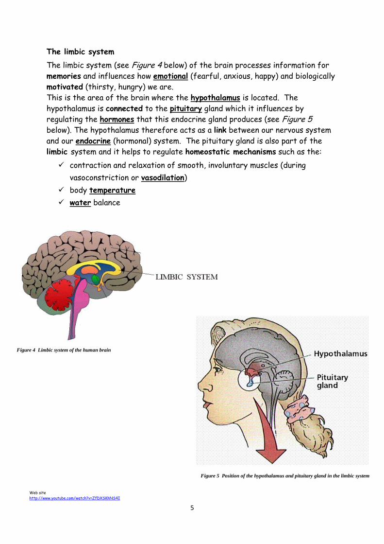
The limbic system
The limbic system (see Figure 4 below) of the brain processes information for
memories and influences how emotional (fearful, anxious, happy) and biologically
motivated (thirsty, hungry) we are.
This is the area of the brain where the hypothalamus is located. The
hypothalamus is connected to the pituitary gland which it influences by
regulating the hormones that this endocrine gland produces (see Figure 5
below). The hypothalamus therefore acts as a link between our nervous system
and our endocrine (hormonal) system. The pituitary gland is also part of the
limbic system and it helps to regulate homeostatic mechanisms such as the:
contraction and relaxation of smooth, involuntary muscles (during
vasoconstriction or vasodilation)
body temperature
water balance
Figure 5 Position of the hypothalamus and pituitary gland in the limbic system
Figure 4 Limbic system of the human brain
Web site
http://www.youtube.com/watch?v=ZfDXSKhNS4I

6
The cerebral cortex
The cerebral cortex is the outer layer of the cerebrum, and it is the centre of
conscious thought. It also:
receives sensory information (from receptors)
co-ordinates voluntary movements
makes decisions
recalls memories
alters our behaviour in the light of experience
The cerebrum is split by a deep cleft into two halves called the cerebral
hemispheres. There are several distinct areas of these cerebral hemispheres.
Figure 6 below shows some of these areas on left cerebral hemisphere – the
right cerebral hemisphere is a mirror image of this. Each of these distinct
areas performs a particular function.

7
The two main areas, are the sensory and motor areas. There are other areas
that are associated (linked) with hese two main areas that deal with:
thought processes
langauge
personality
imagination
intelligence
The left cerebral hemisphere deals with information from the right visual field
and controls the right side of the body. The right cerebral hemisphere deals
with information from the left visual field and controls the left side of the
body. . Infromation is transferred through an area at the centre of the
cerebrum called the corpus callosum which is shown in Figure 7 below.
The corpus callosum is a large bunch of nerve fibres that link the two sides of
the brain. Whatever happens on one side of the brain is quickly communicated
to the other side via the corpus callosum
The role of corpus callosum then, is to get the two halves of the brain to work
together as an integrated whole.
Figure 7 Position of the corpus callosum in the brain
Web site - up to 6.05min
http://www.youtube.com/watch?v=zx53Zj7EKQE

8
Perception Perception is the process by which the brain analyses and then makes sense out
of the sensory information we receive from our surroundings. Although many
perceptual experiences depend on information from our sense organs other
than our eyes, the following notes concentrate of visual perception. Visual
perception allows us to:
1 segregate objects from one another and their background
2 recognise what different objects are
3 jugde how near or far away (i.e. distance) objects are from us
1 Segrgation of Objects
The first stage in the development of visual perception is the
appreciation of an object’s shape. Perception allows us to segregate
objects from one another and their background.
Here the triangle appears to stand out from the background (a white
circle) in a obvious manner. This form of perceptual organisation is called
the “figure-ground” phenomenon. The part seen as the “figure” – in this
example the triangle – stands out from (is segreated from) the white
background – the”ground”, even although they are printed on the same
two-dimensional piece of paper. Sometimes we can switch between the
two and this causes an alteration between the figure and the ground, and
this is what happens when we view optical illusions.
Web site
http://www.sserc.org.uk/images/Biology/Higher_Human/Perception/Perception_ppt.ppt

9
The brain can also perceive shapes once the gap has been filled in – this is
called “closure”. An example of closure is chown below in Figure 8.
2 Recognition
Shape is extremely important when recognising objects – more important
than colour or texture.
Figure 9 above represents five different types of fruit based on their
colour. It is not possible to identify the fruits based on their colour as
the only visual cue. Figure 10 represents the same five fruits, but this
time their shape is used as a visual cue. It is now possible to recognise
the apple, banana, pear and orange. To reciognise whether it is a lemon or
a lime, colour would also be needed.
We use shape to characterise and differentiate objects from one
another during early learning. This information is then stored in our long-
term memory. The most important feature of an object’s shape is its
general outline.
Figure 8 Closure activity
Figure 9 Colour as a recognition cue Figure 10 Shape as a recognition cue
web site
http://www.youtube.com/watch?v=ORoTCBrCKIQ
web site
http://www.youtube.com/watch?v=PKeuhXQj3MM

10
“Perceptual set” is brought about by previous experience, and this can
influence which sensory data we perceive or ignore. In an investigation,
group A were shown pictures of small mammals including rodents. Group B
were shown pictures of humans – some of whom were bald and wore
glasses. Each group was then shown the ambiguous “ratman” diagram
shown in Figure 11 below.
Most people in group A perceived a mouse or a rat, whereas most people
in group B perceived a man with a bald head wearing glasses.
3 Judging distance (or perception of distance)
The distance of one or more objects from the eye is indicated by the
presence of one or more visual cues that can be seen. Visual cues include:
(i) the size of an object in relation to another – the further away an
object is from the eye, the smaller it is perceived to be as shown
by the sleepers on the railway line in Figure 12 below.
(ii) the height of an object in relation to another – again, the further
away an object is from the eye, the smaller it is perceived to be as
shown by the telegraph poles above.
Figure 11 “Ratman”
Figure 12 Visual cues

11
(iii) superimposition – this is when one object partially blocks the view of
another. The blocked object is perceived to be further away.
Binocular disparity
Each eye views an object from a slightly different angle. This mean that there
is a slight difference (disparity) between the images of the same object
formed by the two eyes. The closer the object is to the viewer, the greater
the disparity between the two images. When nerve impulses from the two eyes
reach the brain, they are superimposed and processed into a single picture with
depth. As a result, we get a 3D picture and are able to judge distances well.
Perceptual constancy
A person’s visual perception of their surroundings remains the same (shape,
and size ) no matter how far away or close an object is. Size constancy is
thought to depend, in part, on past experience and stored knowledge, and, in
part, on the cue of relative size.
Imagine that you move towards a shelf to fetch a knife from a knife block. The
image of the knife in the eye becomes bigger the nearer you get to it.
However, the image of the shelf and the knife block also get bigger, so the
relative size of the various objects to one another doesn’t change.
Memory
Memory is the storage, retention and retrieval of information. This includes
pasts experiences, knowledge and thoughts. Our memory enables us to deal
with future situations in the light of past experiences. In the absence of
memory, we would be unable to manage even the slightest task without having to
to relearn it first. All information entering the brain passes through sensory
memory and then enters short term memory. Information is then passed to
long term memory or it is discarded.
(i) Sensory memory (level 1)
Stimuli from our surroundings are perceived continuously as sensory
images by the brain. Sensory memory (SM) lasts a few seconds (i.e. it is
short-lived). Only a few are selected and transferred to level 2.
(ii) Short term memory (level 2)
Most of the information that is transferred into this second level of the
system consists of visual and auditory images. However, our short term
memory (STM) holds only a limited amount of information – about seven

12
items at any one time. Our STM therefore has a limited capacity and items are
held for a very short time. However, during this time the retrieval of items is
very accurate. After this, they are either transferred to level 3 or they are
lost by displacement (pushing out old information by replacing it with new
incoming information), or by decay (when a group of neurons briefly become
activiated, fragile “memory tarces” that have form are broken down).
Chunking
STM can be improved by “chunking”. A “chunk” is a meaningful piece of
information made from several smaller pieces. To most people who are familiar
with the date of the Scottish Referdum on Independence in 2014, 2014 is one
chunk of information and not four. However to most people 4021 is four chunks
of information (unless it happens to be your PIN number!!!).
Since our short term memory can only hold about seven items at one time,
chunking is a useful method to help to increase this. We use chunking to help us
to remember phone numbers.
Code for Glasgow Area Code Private Number
Working memory
Our working memory is an extension of our shrot-term memory. It processes,
manipulates and controls information while it is held in the STM. This then
allows us to perform simple cognitive tasks. For example, you have been asked
to think about all the rooms in your house that have lamps in them and then
calculate the total number of lamps. To do this, you form a mental image of
your home and then do a visuo-spatial tour of each room. You then use your
working memory to count the lamps, and then add this to a running total in your
STM.
Long term memory (LTM) – Level 3
The transfer of information from our STM to our LTM is due to rehearsal.
Rehearsal is when you repeat something that you are trying to memorise over
and over to yourself. This process helps to lengthen the time we retain this
information in our STM. In many cases, rehearsal then allows information to
become encoded and transferred to our LTM from where it can be retrieved at
a later date.
0141 3810 629

13
During encoding, information is organised into categories such as personal
information and problem solving skills. Information is ecoded in our STM either
by:
repitition - referred to as shallow encoding or
linking it with previous memories - referred to as elaborative encoding
It is thought that our long-term memory is able to hold an unlimited amount of
information which is stored for a long time, perhaps even permanently. The
transfer of information between our STM and LTM is summarised below in
Figure 13.
This model is an oversimplification of how our memory works. The three levels
of memory should not be thought of as occupying three distinct regions of the
brain.
Figure 13 Transfer of information from STM to LTM

14
Location of memory in the brain
Several different types of memory exist within our LTM. What they are
concerned with and their locations in the brain, are summarised in the table
below.
Type of memory Concerned with Possible location in the brain
episodic remembering events and
experiences
region of cerebral cortex where
the sensory information was first
received and encoded semantic remembering facts and concepts
procedural remembering skills (e.g. how to ride
a bicycle) motor region of cerebral cortex
emotional remebering positive or negative
associations with certain stimuli
links between limbic system and
cerebral cortex
spatial remembering where things are
placed in relation to other things
limbic system
The cells of the nervous system
Neurons are specialised cells that receive and transmit electrical impulses.
There are three types of neuron:
1. sensory neurons
2. inter neurons
3. motor neurons
See Figure 14 below.
Figure 14 A neural pathway – a reflex arc
15
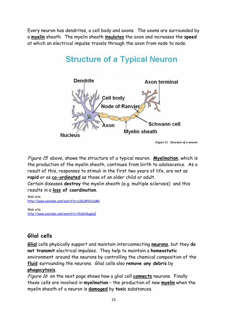
Every neuron has dendrites, a cell body and axons. The axons are surrounded by
a myelin sheath. The myelin sheath insulates the axon and increases the speed
at which an electrical impulse travels through the axon from node to node.
Figure 15 above, shows the structure of a typical neuron. Myelination, which is
the production of the myelin sheath, continues from birth to adolescence. As a
result of this, responses to stimuli in the first two years of life, are not as
rapid or as co-ordinated as those of an older child or adult.
Certain diseases destroy the myelin sheath (e.g. multiple sclerosis) and this
results in a loss of coordination.
Web site
http://www.youtube.com/watch?v=cUGuWh2UeMk
Web site
http://www.youtube.com/watch?v=VIoDr8ugbqI
Glial cells
Glial cells physically support and maintain interconnecting neurons, but they do
not transmit electrical impulses. They help to maintain a homeostatic
environment around the neurons by controlling the chemical composition of the
fluid surrounding the neurons. Glial cells also remove any debris by
phagocytosis.
Figure 16 on the next page shows how a glial cell connects neurons. Finally
these cells are involved in myelination – the production of new myelin when the
myelin sheath of a neuron is damaged by toxic substances.
Figure 15 Structure of a neuron

16
Web site
http://www.youtube.com/watch?v=52NVc9Lku4o
Neurotransmitters
A gap called a syanapse exists between two inter-connecting neurons as shwon
in Figure 17 below. This “gap” is called a synaptic cleft.
Figure 16 Interconnection between glial cell and neurons
axon
glial cell
myelin sheath neuron
Figure 17 Synaptic cleft between axon of one neuron and dendrite of next neuron
pre-synaptic neuron
post-synaptic neuron
web site
http://www.youtube.com/watch?v=xF2UFV6EKt0

17
Chemical messengers called neurotransmitters are released at the axon of one
neuron, then the pass across the synaptic cleft and trigger an electrical impulse
in the dendrite of the next neuron in the pathway. This is how messages are
relayed from neuron to neuron both within and outwith the brain and how
neurons therefore connect with other neurons, muscle fibres and endocrine
glands.
As the diagram on the previous page shows, neurotransmitters are stored in
vesicles. They are then released into the synaptic cleft on the arrival of an
electrical impluse. The neurotransmitters then diffuse across the synaptic
cleft and bind to receptor sites on the dendrites on the next neuron in the
pathway. There are many different receptors and it is the receptor that
determines whether or not the signal is excitatory or inhibitory.
An example of a neurotransmitter is acetylcholine. This chemical can have both
an excitatory or inhibitory effect. If acetylcholine is released into the synaptic
cleft between a motor neuron and a skeletal muscle cell it makes the cell
contract – this is an example of an excitatory signal. However, if acetylcholine
is released into the synaptic cleft between a motor neuron and a heart muscle
cell it reduces the rate and stength of contraction of this cell - this is an
example of an inhibitory signal.
Neurotransmitters must be removed from the synaptic cleft in order to prevent
continuous stimulation of post synaptic neurons. Neurotransmitters are
removed by enzymes and then re-uptake occurs. (The enzyme breaks down the
neurotransmitter into non-active products which are then reabsorbed by the
presynaptic neuron. These non-active products are then used to
resynthsise the active form of the neurotransmitter again. The
neurotransmitter is then stored in a vesicle for re-use).
Filtering out weak stimuli
An impulse can only be transmitted across a synapse and then on through the
post-synaptic neuron when a certain miniumum number of neurotransmitter
molecules have been released. A postsynaptic cell may receive information from
several neighbouring neurons. Synapses are abe to filter out weak stimuli that
arise from insufficient secretions of neurotransmitters. In this instance, the
synapse acts as an unbridgeable gap. However, many weak stimuli can trigger
Web site
http://www.youtube.com/watch?v=p5zFgT4aofA http://www.youtube.com/watch?v=LT3VKAr4roo

18
enough neurotransmitter molecules to be secreted from many presynaptic
neurons simultaneously or in rapid succession, and this is enough to fire off an
electrical impulse.
This series of of weak stimuli which can trigger enough neurotransmitter to fire
off an impulse is called summation.
Types of neural pathways
Neurons are found to be connected to one another in many different ways in the
CNS. This allows many complicated interactions to occur between them, and
thus allows the nervous system to carry out its many complex functions. The
following notes are on these different neural pathways.
a) Converging neural pathways
Converge: to come together and meet at a common point
In a convergent neural pathway, impulses from several sources are
channelled towards, and then meet at a common destination as shown in
Figure 18 below.
This brings about a high concentration of excitatory or inhibitory signals
at a common neuron (labelled 4 in the above diagram) and this helps to
increase their sensitivity to these signals.
b) Diverging neural pathways
Dinverge: to branch out from common point
In a divergent neural pathway, the route along which an impuse is
travelling divides. This type of pathway therefore influeces several
neurons at the same time. This is shown by Figure 19 on the next page.
Figure 18 Converging neural pathway
Neural
convergence

19
c) Reverberating neural pathways
Reverberation: a sound that occurs repeatedly
In a reverberating neural pathway, neurons later on in the pathway have
axon branches that form synapses with neurons that occur earlier in the
pathway. This arrangement enables nerve impulses to be recycled and so
the presynaptic neurons are repeatedly stimulated. This is shown in
Figure 20 below.
Development of new neural pathways
New neural pathways can be developed to create new responses, bypass areas of
brain damage, or suppress reflexes or responses to sensory impulses. This
remarkable ability of brain cells to become altered as a result of new
Figure 19 Diverging neural pathway
Neural
divergence
Neural
reverberation
Figure 20 Reverberating neural pathway

20
environmental experiences is called plasticity of response.
Neurotransmitters, mood and behaviour.
1. Endorphins
Endorphins are neurotransmitters that stimulate neurons that are
involved in reducung pain intensity – in other words, endorphins are our
natural pain killers. They work by combining with receptors at synapses
and so block the transmission of pain signals. The level of endorphin
secretion increases in response to:
o severe injury
o prolonged and continuous exercise
o stress
o certain foods (e.g. chocolate)
Increased levels of endorphins are also connected with feelings of
euphoria, regulation of appetite and the release of sex hormones.
2. Dopamine and the reward pathway
Dopamine, which is produced in several regions of the brain, is also a
neurotransmitter. Neurons which secrete or respond to dopamine are
involved in the reward pathway. This pathway is activated when we are
involved in beneficial behaviours linked with survival, such as eating when
we are hungry, or drinking when we are thirsty. Dopamine induces the
feeling of pleasure and it also reinforces particular behaviours.
Neurotransmitter related disorders and their treatment
Many drugs used to treat neurotransmitter related disorders are very similar to
the neurotransmitters themselves.
Agonists are chemicals (drugs) that bind to and stimulate specific receptors on
the membrane of postsynaptic neurons in a neural pathway. They therfore
mimic a neurotransmitter. When agonists bind to these specific receptors, they
Web site
http://www.youtube.com/watch?v=TGpEl0PD02c
Web site
http://www.youtube.com/watch?v=kVoYpiiy7jg

21
block the action of the naturally occurring neurotransmitter, but agonists still
trigger the normal cellular response that would have been brought about by the
neurotransmitter. See Figure 21.
Antagonists are chemicals (drugs) that bind to and then block specific
receptors on the membrane of postsynaptic neurons in a neural pathway. By
blocking the receptor sites, an antagonist prevents the normal neurotransmitter
from acting on them. This results in the normal transmission of nerve
impulses in that neural pathway being greatly reduced or even brought to a
halt. See Figure 21.
Other drugs can disrupt a neural pathway because they inhibit the enzymes
that degrade (break down) neurotransmitters into non-active products or they
inhibit re-uptake. In other words they prevent the removal of
neurotransmitters from the synaptic cleft.
Mode of action of recreational drugs
Many recreational drugs affect the transmission of nerve impulses in the
reward pathway of the brain, and, in turn, this then affects a person’s state of
consciousness. This is because these drugs alter a person’s neurochemistry,
which can lead to changes in:
mood the person might feel happier, more confident,
depressed, aggressive etc
cognitive thinking the person is unable to carry out complex mental tasks
such as making decisions or solving problems
perception the person misinterprets environmental stimuli.
Sounds, colours and /or a sense of time seem altered
behaviour the person is able to stay awake for longer and can
talk endlessly about themself
normal response
no response or reduced response
Figure 21 Action of agonistic and antagonistic drugs on neural receptors
agonist
antagonist
Web site http://www.youtube.com/watch?v=XVYyQeOJV3U

22
Recreational drugs interact with neurotransmitters in different ways. They can
either:
stimulate the release of a neurotransmitter
initiate the action of a neurotransmitter by acting as an agonist
prevent (block) a neurotransmitter from binding to receptors by acting as
an antagonist
inhibit the degradation (breakdown) of a neurotransmitter by an enzyme
inhibit the re-uptake of neutransmitters from a synaptic cleft
Drug addiction and tolerance
Drug addiction: a chronic disease that causes the sufferer to compulsively
seek out and use the drug regardless of the consequences.
Drug tolerance: drug tolerance is said to have built up when a users
(or desensitisation) reaction to an additive drug is found to have
decreased in intensity compared to previous times,
even although the concentration of the drug has
remained unaltered. (In these circumstances, a larger
dose will be required to bring about the original
effect.)
Drug desensitisation and sensitisation
Drug desensitisation
Desensitisation is due to the repeated use of a drug. Repeated use of a drug
that acts as an agonist, results in certain neuroreceptors (e.g. those that
promote the release of the neurotransmitter dopamine) being repeatedly
stimulated. This then causes heightened feelings of well-being or euphoria in
the drug-taker.
The nervous sytem however, compensates for
the overstimulation of these receptors, by
reducing the number of these receptors.
However, the remaining receptors then become
less sensitive to the agonist drug. This leads to
drug tolerance (also called desensitisation)
because a larger dose of the drug will now be
required to stimulate the reduced number of
these less sensitive receptors as shown in
Figure 22 opposite.
Figure 22 Desensitisation
Web site http://www.youtube.com/watch?v=5f1nmqiHIII
23
Drug sensitisation
Repeated use of a drug that acts as an antagonist, blocks certain
neuroreceptors and so prevents the normal neurotransmitter from acting on
these receptors. The nervous system compensates for the reduced stimulation
of these receptors by increasing the number of receptors. In addition, the
receptors become sensitive to the antagonistic drug.
When the number and sensitivity of receptors is increased as a result of
repeated exposure to a drug acting as an antagonist, this is called sensitisation.
This can result in excessive drug-craving and ultimately drug addiction.
Web site
http://www.youtube.com/watch?v=ukFjH9odsXw

24
Communication and social behaviour
The effect of infant attachment
Humans are social animals. This is because the vast majority of humans prefer
not to lead a solitary existence. To operate successfully, memebers of a
community must be able to communicate with each. Communication can be
verbal, written or non-verbal (e.g. body language and facial expressions).
Communication between human begins at birth and continues throughout life.
Web site
http://www.youtube.com/watch?v=KC73tDFpHQA
Infant attachment
Early infant attachment, usually with a mother or other primary carer, is very
important for a child to be able to develop stable relationships in the future.
The tie that binds a baby to the carer is called infant attachment and this
becomes evident between 6 and 9 months.
The first infant attachment is indiscriminate as far as the baby is concerned,
but as the months go by, the baby is only interested in selected people.
The “strange situation”
The “strange situation” is a research tool that has been devised to investigate
infant attachment. This tool is used by experts to determine if a child is
demonstrating secure or insecure attachment to their primary carer. The
“stange situation” allows a hidden observer to study the behaviour of a baby:
with the mother/primary carer
with a stranger
alone
Secure attachments
Signs of a child having secure attachments include:
the infant plays with toys and investigates freely when the
mother/primary carer is present
the infant displays major distress when the mother leaves them
the infant goes to it’s mother immediately for comfort when she returns
and then calms down quickly
the infant is more attached to it’s mother than to a stranger
Web site
http://www.youtube.com/watch?v=s608077NtNI

25
The more secure the attachment of an infant to their mother/primary carer
the more likely they are to investigate their immediate environment since they
feel safe to do so. This, in turn, gives them an opportunity to learn and develop
their cognitive abilities. A securely attached infant is more likely to benefit
from these opportunities than one that is insecurely attached.
Insecure attachments
Signs of a child having insecure attachments include:
the infant does not play with toys or investigate freely even when the
mother/primary carer is present
the infant displays indifferent, or mild distress when the mother leaves
them
the infant resists comfort from a stranger in the absence of it’s mother
the infant displays inconsistent behaviour e.g. wanting and resisting
comfort at the same time after the mother returns
the infant may show signs of anger or try to hit the mother after she
returns.
Humans depend on adults for a long period of their life. This provides time for
socialisation and and learning to occur and for social competence to develop.
Social competence is necessary if interaction with other people is to be
successful.
Methods of control
The method of control adopted by parents (and other influential adults in their
life) affects a child’s social competence. There are three main methods of
control:
1. Authoritative (“unreasonably strict”)
Reasons for rules are never explained. The child is expected to obey
without question. Little or no warmth is demonstrated towards the child.
2. Authoritative (“demanding but responsive”)
Warm, nurturing and emotionally supportive towards the child. Reasons
for rules are explained. Gives direction and expects responsible
behaviour and explains what the consequences of unacceptable behaviour
will be. Respectful of the child.
3. Permissive (excessively lenient)
Warm, and nurturing towards the child. Responds to child’s needs and
wishes. Limits are not set, rules are not laid down. Responsibilities are
not assigned. Adopts a “no discipline “ approach and doesn’t try to
keep the child under control. Allows the child to regulate their own
behaviour.

26
The effect of communication
1. Non-verbal communication
Non-verbal communication is sending or receiving wordless messages, and
it plays an important role in forming relationships. On some
occasions, it can be used to reinforce verbal messages or it can be used
to add to information that is being transmitted verbally. Non-verbal
communication can signal attitudes and emotions. Facial expressions and
hand signals are examples of non-verbal communication.
Web site
http://www.youtube.com/watch?v=9cX6VaIy2yA
2. Verbal communication
Language combines basic sounds into spoken words. These words are then
represented by written symbols like those shown in Figure 23 below.
These sounds and symbols represent information that can be arranged
into simple categories (words) and more comple hierarchies (phrases,
sentences and paragraphs). The fact that humans are able to
communicate verbally has resulted in the sharing and transmission of
knowledge, the development of cultures and social evolution. Language
therefore helps to accelerate learning and intellectual development.
Language makes much of our behaviour unique and it sets us apart from
other animals.
The effect of experience on the learning process
Learning is defined as a permanent change in behaviour that occurs as a direct
result of experience.
1. Practising
Practising by repeatedly using the same motor skills results in a motor
pathway being established. Hence the saying “practice makes perfect”,
e.g. riding a bicycle, playing the piano. Practice also improves
performance, and lack of practice results in the skill becoming “rusty”.
Web site Web site
http://www.youtube.com/watch?v=29Woge-u9ns http://www.youtube.com/watch?v=BjLzzLq765Q
Figure 23 Language as written symbols

27
Results from a finger maze investigation can be recorded and then displayed as
a learning curve graph like the one shown on Figure 24 below.
2. Imitating
A great deal of human behaviour is learned by observing and imitating the
behaviour of others.
Web sites
http://www.youtube.com/watch?v=t_Me5znI0NY http://www.youtube.com/watch?v=orxgu_NSwGA
3. Trial and error
Trial and error is a learning tool that is used to solve problems by doing
something over and over again until we get it right. This means that we
learn trying, but if our try is an error, we learn something too. This type
of learning can be demonstrated using a rat.
A rat is put into a specially designed box as shown in Figure 25 on the
following page. It explores the box, touches the floor and leans against
Tim
e t
ake
n (
seco
nd
s)
10
40
30
20
trial number
1 5 4 3 2 6 7 8
Figure 24 Learning curve of typical set of results

28
the sides of the box. At some point, the rat pushes the lever and food
immediately appears in the food tray.
If the rat is only rewarded with food when it presses the lever. It soon learns
to link pressing the lever with the delivery of food (it’s reward). This is why
teachers use rewards a learning strategy.
Web site
http://www.youtube.com/watch?v=kzNIV0mTROU
4. Motivation
Motivation is the desire that we have in us that makes us want to
participate in the learning process. Animals, including humans, are
motivated by many factors such as thisrt, hunger and curiosity.
Scientists have investigated the effect of motivation on an animal’s
ability to learn by comparing how well hungry and well-fed rats can
negotiate a maze as shown below in Figure 26. They are then given food
as a reward at the end of the maze.
Figure 25 Trial and error learning in rats
lever
food tray
food
hungry rat
Figure 26 Maze used to research the effect of motivation on learning

29
Below is a set of results using such a maze with a hungry and well fed rat.
Trial number Average number of errors
well fed rat hungry rat
1 135 135
2 145 150
3 135 75
4 145 80
5 130 50
6 150 60
7 135 50
8 140 45
9 125 55
10 120 25
11 130 15
The reliability of the results of the above experimet could be improved by using
many hungry or well fed rats rather than just one of each. The fact that many
trials were done however with each rats helps to make the results more
reliable.

30
5. Reinforcemnet of behaviour
When behaviour is repeated and the behaviour is rewarded, it becomes
reinforced. Reinforcement is a consequence that will strengthen an
organism's future behaviour as it tends to make an organism repeat a
certain piece of behaviour. During reinforcement, the reinforcer (such a
as a reward or praise) increases the probability of the response being
repeated.
Shaping is the rewarding of behaviour that approximates to the desired
behaviour.
Web sites
http://www.youtube.com/watch?v=Nd6rUQzMA2o http://www.youtube.com/watch?v=g6gE4z4yIzU
If behaviour patterns are not rewarded, they are likely to disappear.
This is called extinction.
6. Generalisation and discrimination
Generalisation is the tendency to respond to stimuli which are similar.
For example a child who has been bitten by a large dog will then be afraid
of all dogs – this is called generalisation. However, the same child
might fear large dogs only – this is called discrimination. So,
discrimination allows the learner to distinguish between different
but related stimuli. Learning to discriminate is essential for a child to
prepare itself to cope with everyday life.

31
Social influence
1. Social groups
Humans are social animals which means that we prefer to interact
(socialise) with other people rather than being on our own.
A Social facilitation
Many people are motivated by their need for status. Many people
feel the need to be admired and impress other people in their
social group. Given this, perhaps the presence of other people can
affect an individual’s performance.
Research has shown that people work at a faster rate and achieve
more when they are placed in competititve situations. This
increased performance in the presence of others (especially if it
is competitive) is called social facilitation.
B Deindividuation
Group pressure is a powerful force. People find it difficult not to
participate in the actions of their social group and sometims end
up behaving in a manner which is out of character. This loss of
personal identity in a group tends to lead to diminished restriants
on behaviour. This is called deindividuation.
Web site – start at 2.05min
http://www.youtube.com/watch?v=OFs0b97sqg4
Deindividuation is often used to explain
the anti-social behaviour of some groups
which would not be demonstrated by
individuals from these groups if they were
on their own. Once under group pressure,
individuals think and act differently. This
often takes the form of an anti-social mob,
the members of which have temporarily lost awareness of their
own individuality and responsibilities.
Influences that change beliefs
1. Inernalisation
As a result of persuasion, an individual might change their beliefs about

32
something. This is called internalisation. Supermarkets and politicians
and try to effect internalisation. Politicians tried to persuade the people
to vote “yes” or “no” in the Scottish referendum campaign in 2104. Some
of the debates, speeches, posters and newspaper resulted in people being
persuaded to vote one way or the other.
Web site
http://www.youtube.com/watch?v=9_heYiAIaTI
Identification
If a person changes their beliefs just to be like someone else, this
prosess is called identification. The person that they want to identify
with is someone they either admire or they are influenced by. This is
why “celebrities” are used to endorse products in TV adverts.
Web site
http://www.youtube.com/watch?v=gLTQJKIyraQ
http://www.youtube.com/watch?v=rVL7wAcZ3wY
http://www.youtube.com/watch?v=N0pYvCsajDk

33
Slabs on ground
http://www.youtube.com/watch?v=z9Sen1HTu5o
Scientific method
http://www.youtube.com/watch?v=wlb7tLJy5AI
Chemical reactions
http://www.youtube.com/watch?v=EWOwAu7XWJs
http://www.youtube.com/watch?v=kr7dGPOwzsA

34