Embed Size (px)
Citation preview
Chemical Physics Letters 381 (2003) 634–641
www.elsevier.com/locate/cplett
High-resolution NMR spectroscopy in inhomogeneousB0 and B1 fields by two-dimensional correlation
Sasa Antonijevic, Stephen Wimperis *
School of Chemistry, University of Exeter, Stocker Road, Exeter EX4 4QD, UK
Received 29 July 2003; in final form 8 September 2003
Published online: 4 November 2003
Abstract
Recently, there has been much interest in methods for obtaining high-resolution NMR spectra in inhomogeneous B0
and B1 fields and in their application to so-called �ex situ� spectroscopy, where the sample and magnet/probe assembly
are spatially separated. Here we discuss the implementation of the well-known two-dimensional nutation experiment as
a method for correlating B0 and B1 inhomogeneities and, hence, achieving a high-resolution NMR spectrum. The
advantages of this approach lie in its simplicity, its spatial (�depth�) resolution, and in its not requiring a linear cor-
relation of fields, i.e., B1 ¼ aB0 þ k, across the sample.
� 2003 Elsevier B.V. All rights reserved.
1. Introduction
High-resolution NMR spectroscopy is normallyperformed in a B0 field that is spatially homoge-
neous. It has always been recognised, however,
that the use of such a magnetic field is not always
feasible. One example that has aroused interest
recently is that of so-called �ex situ� NMR spec-
troscopy, where the sample and magnet/probe as-
sembly are spatially separated [1]. An inevitable
feature of this experimental arrangement is thatthe radiofrequency or B1 field will also be spatially
inhomogeneous.
* Corresponding author. Fax: +44-1392-263434.
E-mail address: [email protected] (S. Wimperis).
0009-2614/$ - see front matter � 2003 Elsevier B.V. All rights reserv
doi:10.1016/j.cplett.2003.09.116
Pines and coworkers have advocated an ap-
proach to recording high-resolution NMR spectra
that exploits a spatial correlation of the inhomo-geneous B0 and B1 fields [1–3]. Applications to the
NMR �logging� of oil wells [4] and in mobile, sur-
face-scanning NMR spectrometers [5] have been
suggested. One implementation involves the use of
a train of composite z-rotation pulses interleaved
with data sampling and allows direct acquisition of
a high-resolution spectrum [1–3]. However, a sec-
ond implementation uses two-dimensional NMRcorrelation of the frequency dispersions produced
by the inhomogeneous B0 and B1 fields [2] and is
identical to the well-known two-dimensional nu-
tation experiment [6–8] or, equivalently, to the so-
called �rotating-frame� imaging experiment [9].
The purpose of this Letter is to present a de-
tailed discussion of the two-dimensional nutation
ed.
S. Antonijevic, S. Wimperis / Chemical Physics Letters 381 (2003) 634–641 635
experiment as an approach to high-resolution
NMR spectroscopy in correlated inhomogeneous
B0 and B1 fields. Although slower than direct ac-
quisition, the method will be shown to have the
same sensitivity and to possess a number advan-
tages, including simplicity and not requiring alinear correlation of fields, i.e., B1 ¼ aB0 þ k,across the sample.
2. Experimental details
Experiments were performed in a 100-mm ver-
tical bore superconducting NMR magnet gener-ating a magnetic field of B0 ¼ 4:7 T (m0 ¼ 200:06MHz for 1H). To mimic the B0 inhomogeneity
encountered in �ex situ� NMR, a B0 gradient was
produced along the laboratory-frame x axis by
passing a constant 0–10 A current through an x-gradient coil wound as part of a room-temperature
imaging gradient/shim set. Up to 60 W of power
was dissipated and generous quantities of coolingair were applied to the bore, probe and shims.
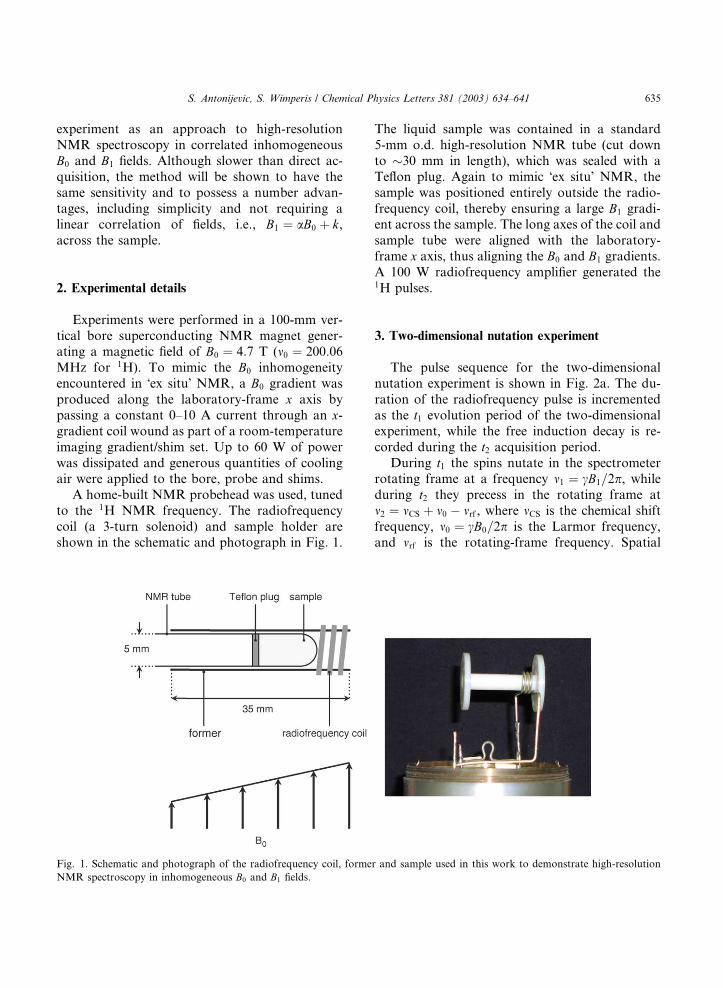
A home-built NMR probehead was used, tuned
to the 1H NMR frequency. The radiofrequency
coil (a 3-turn solenoid) and sample holder are
shown in the schematic and photograph in Fig. 1.
Fig. 1. Schematic and photograph of the radiofrequency coil, forme
NMR spectroscopy in inhomogeneous B0 and B1 fields.
The liquid sample was contained in a standard
5-mm o.d. high-resolution NMR tube (cut down
to �30 mm in length), which was sealed with a
Teflon plug. Again to mimic �ex situ� NMR, the
sample was positioned entirely outside the radio-
frequency coil, thereby ensuring a large B1 gradi-ent across the sample. The long axes of the coil and
sample tube were aligned with the laboratory-
frame x axis, thus aligning the B0 and B1 gradients.
A 100 W radiofrequency amplifier generated the1H pulses.
3. Two-dimensional nutation experiment
The pulse sequence for the two-dimensional
nutation experiment is shown in Fig. 2a. The du-
ration of the radiofrequency pulse is incremented
as the t1 evolution period of the two-dimensional
experiment, while the free induction decay is re-
corded during the t2 acquisition period.
During t1 the spins nutate in the spectrometerrotating frame at a frequency m1 ¼ cB1=2p, whileduring t2 they precess in the rotating frame at
m2 ¼ mCS þ m0 � mrf , where mCS is the chemical shift
frequency, m0 ¼ cB0=2p is the Larmor frequency,
and mrf is the rotating-frame frequency. Spatial
r and sample used in this work to demonstrate high-resolution
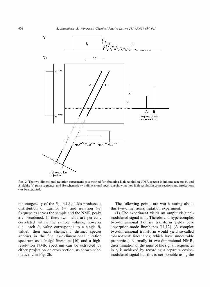
Fig. 2. The two-dimensional nutation experiment as a method for obtaining high-resolution NMR spectra in inhomogeneous B0 and
B1 fields: (a) pulse sequence, and (b) schematic two-dimensional spectrum showing how high-resolution cross sections and projections
can be extracted.
636 S. Antonijevic, S. Wimperis / Chemical Physics Letters 381 (2003) 634–641
inhomogeneity of the B0 and B1 fields produces a
distribution of Larmor (m0) and nutation (m1)frequencies across the sample and the NMR peaksare broadened. If these two fields are perfectly
correlated within the sample volume, however
(i.e., each B1 value corresponds to a single B0
value), then each chemically distinct species
appears in the final two-dimensional nutation
spectrum as a �ridge� lineshape [10] and a high-
resolution NMR spectrum can be extracted by
either projection or cross section, as shown sche-matically in Fig. 2b.
The following points are worth noting about
this two-dimensional nutation experiment:
(1) The experiment yields an amplitude(sine)-modulated signal in t1. Therefore, a hypercomplex
two-dimensional Fourier transform yields pure
absorption-mode lineshapes [11,12]. (A complex
two-dimensional transform would yield so-called
�phase-twist� lineshapes, which have undesirable
properties.) Normally in two-dimensional NMR,
discrimination of the signs of the signal frequencies
in t1 is achieved by recording a separate cosine-modulated signal but this is not possible using the
S. Antonijevic, S. Wimperis / Chemical Physics Letters 381 (2003) 634–641 637
pulse sequence in Fig. 2a. However, this is not a
problem here since we can be confident that the
nutation frequency m1 is always positive (in fact, we
only display this part of the spectrum).
(2) An amplitude-modulated signal can be
viewed as the sum of counter-rotating echo andantiecho signals [11,12]. If we assume a linear
correlation of the two inhomogeneous fields,
B1 ¼ aB0 þ k, then the two inhomogeneities are
refocussed in the echo signal during the acquisition
period at t2 ¼ at1 [2]. The antiecho signal does not
refocus and is weak but is nevertheless an essential
part of the overall modulation and allows pure
absorption-mode lineshapes to be obtained [11,12].(3) It is not advisable to use �delayed acquisi-
tion� [11] in a nutation experiment, i.e., to redefine
the t1 and t2 periods so that t01 ¼ ð1þ aÞt1 and
t02 ¼ t2 � at1. If this is done, the effect is to �shear�the echo signal such that the inhomogeneous
broadening lies only in the m2 frequency dimension
and a high-resolution spectrum can be obtained
simply by projection onto the m01 axis. The weakantiecho signal, however, is sheared in the wrong
direction by this procedure and still lies across
both dimensions. Worse still, because the echo and
antiecho signals are no longer coincident in the
frequency domain, both signals consist of unde-
sirable phase-twist lineshapes.
(4) Similarly, shearing the nutation experiment
data set after the Fourier transformation with re-spect to t2 [11,13] also produces echo and antiecho
signals that are noncoincident. This problem is
normally dealt with in two-dimensional NMR by
separating the echo and antiecho signals and (ef-
fectively) shearing them individually in opposite
senses. However, this separation is not possible
with the experiment in Fig. 2a because a cosine-
modulated signal is not available.(5) If shearing of the two-dimensional nutation
experiment data set is required, so that a high-res-
olution NMR projection can be obtained, then the
optimum way to achieve it is to record pure ab-
sorption-mode lineshapes and to shear these in the
frequency domain, using interpolation if necessary.
(6) It is possible to convert the experiment in
Fig. 2a into one which yields phase modulation int1, rather than amplitude modulation, by adding a
90� pulse with orthogonal phase in the rotating
frame to the end of the t1 nutation pulse [9]. There
is a factor of 2 increase in signal intensity and,
after a complex Fourier transformation (as ap-
propriate for phase-modulated data [11]), a factor
of 2p2 increase in signal-to-noise ratio compared
with the hypercomplex amplitude-modulated ex-periment. The absence of an antiecho signal,
however, means that this experiment yields unde-
sirable phase-twist lineshapes [9].
(7) By using a second, complementary phase-
modulated experiment, where the phase of the
additional 90� pulse is shifted by a further 180�,the weak antiecho signal can be recorded [9]. Pure
absorption-mode lineshapes can now be obtainedvia hypercomplex Fourier transformation of the
two data sets. The gain in signal-to-noise relative
to the amplitude-modulated experiment is reduced
to a factor of 2.
(8) The severe drawback of these phase-modu-
lated experiments is the difficulty of producing a
90� pulse that is uniform across the sample when,
by the very nature of the experiment, the B1 field ishighly inhomogeneous. Both composite [14] and
adiabatic 90� [15,16] pulses could possibly be used
to alleviate this problem, but in this Letter we will
concentrate on the implementation of the simpler,
if less sensitive, amplitude-modulated nutation
experiment in Fig. 2a.
4. Results and discussion
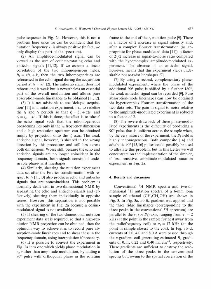
Conventional 1H NMR spectra and two-di-
mensional 1H nutation spectra of a 6-mm long
sample of ethanol (CH3CH2OH) are shown in
Fig. 3. In Fig. 3a, no B0 gradient was applied and
the three ridge lineshapes (corresponding to the
three peaks in the conventional 1H spectrum) areparallel to the m1 (or B1) axis, ranging from m1 ¼ 2
kHz (at the point in the sample furthest away from
the radiofrequency coil) to m1 ¼ 17 kHz (at the
point in sample closest to the coil). In Fig. 3b–d,
currents of 2.0, 4.0 and 8.0 A were passed through
the x-gradient coil generating estimated B0 gradi-
ents of 0.11, 0.22 and 0.40 mT cm�1, respectively.
These gradients are sufficient to destroy the reso-lution of the three peaks in the conventional
spectra but, owing to the spatial correlation of the
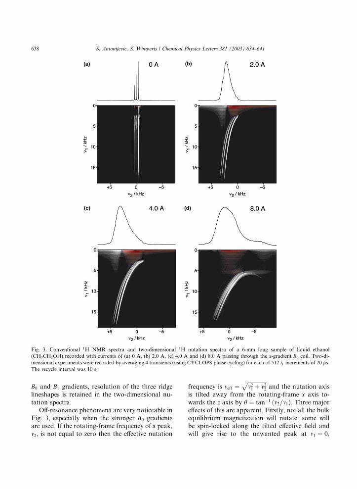
Fig. 3. Conventional 1H NMR spectra and two-dimensional 1H nutation spectra of a 6-mm long sample of liquid ethanol
(CH3CH2OH) recorded with currents of (a) 0 A, (b) 2.0 A, (c) 4.0 A and (d) 8.0 A passing through the x-gradient B0 coil. Two-di-
mensional experiments were recorded by averaging 4 transients (using CYCLOPS phase cycling) for each of 512 t1 increments of 20 ls.The recycle interval was 10 s.
638 S. Antonijevic, S. Wimperis / Chemical Physics Letters 381 (2003) 634–641
B0 and B1 gradients, resolution of the three ridge
lineshapes is retained in the two-dimensional nu-
tation spectra.
Off-resonance phenomena are very noticeable in
Fig. 3, especially when the stronger B0 gradients
are used. If the rotating-frame frequency of a peak,m2, is not equal to zero then the effective nutation
frequency is meff ¼ffiffiffiffiffiffiffiffiffiffiffiffiffiffim21 þ m22
pand the nutation axis
is tilted away from the rotating-frame x axis to-
wards the z axis by h ¼ tan�1 ðm2=m1Þ. Three major
effects of this are apparent. Firstly, not all the bulk
equilibrium magnetization will nutate: some will
be spin-locked along the tilted effective field andwill give rise to the unwanted peak at m1 ¼ 0.
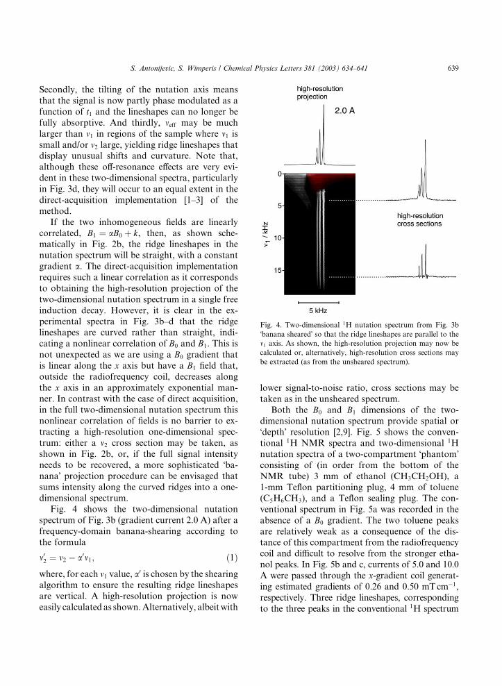
Fig. 4. Two-dimensional 1H nutation spectrum from Fig. 3b
�banana sheared� so that the ridge lineshapes are parallel to the
m1 axis. As shown, the high-resolution projection may now be
calculated or, alternatively, high-resolution cross sections may
be extracted (as from the unsheared spectrum).
S. Antonijevic, S. Wimperis / Chemical Physics Letters 381 (2003) 634–641 639
Secondly, the tilting of the nutation axis means
that the signal is now partly phase modulated as a
function of t1 and the lineshapes can no longer be
fully absorptive. And thirdly, meff may be much
larger than m1 in regions of the sample where m1 issmall and/or m2 large, yielding ridge lineshapes thatdisplay unusual shifts and curvature. Note that,
although these off-resonance effects are very evi-
dent in these two-dimensional spectra, particularly
in Fig. 3d, they will occur to an equal extent in the
direct-acquisition implementation [1–3] of the
method.
If the two inhomogeneous fields are linearly
correlated, B1 ¼ aB0 þ k, then, as shown sche-matically in Fig. 2b, the ridge lineshapes in the
nutation spectrum will be straight, with a constant
gradient a. The direct-acquisition implementation
requires such a linear correlation as it corresponds
to obtaining the high-resolution projection of the
two-dimensional nutation spectrum in a single free
induction decay. However, it is clear in the ex-
perimental spectra in Fig. 3b–d that the ridgelineshapes are curved rather than straight, indi-
cating a nonlinear correlation of B0 and B1. This is
not unexpected as we are using a B0 gradient that
is linear along the x axis but have a B1 field that,
outside the radiofrequency coil, decreases along
the x axis in an approximately exponential man-
ner. In contrast with the case of direct acquisition,
in the full two-dimensional nutation spectrum thisnonlinear correlation of fields is no barrier to ex-
tracting a high-resolution one-dimensional spec-
trum: either a m2 cross section may be taken, as
shown in Fig. 2b, or, if the full signal intensity
needs to be recovered, a more sophisticated �ba-nana� projection procedure can be envisaged that
sums intensity along the curved ridges into a one-
dimensional spectrum.Fig. 4 shows the two-dimensional nutation
spectrum of Fig. 3b (gradient current 2.0 A) after a
frequency-domain banana-shearing according to
the formula
m02 ¼ m2 � a0m1; ð1Þwhere, for each m1 value, a0 is chosen by the shearing
algorithm to ensure the resulting ridge lineshapes
are vertical. A high-resolution projection is now
easily calculated as shown.Alternatively, albeitwith
lower signal-to-noise ratio, cross sections may be
taken as in the unsheared spectrum.
Both the B0 and B1 dimensions of the two-
dimensional nutation spectrum provide spatial or
�depth� resolution [2,9]. Fig. 5 shows the conven-
tional 1H NMR spectra and two-dimensional 1Hnutation spectra of a two-compartment �phantom�consisting of (in order from the bottom of the
NMR tube) 3 mm of ethanol (CH3CH2OH), a
1-mm Teflon partitioning plug, 4 mm of toluene
(C5H6CH3), and a Teflon sealing plug. The con-
ventional spectrum in Fig. 5a was recorded in the
absence of a B0 gradient. The two toluene peaks
are relatively weak as a consequence of the dis-tance of this compartment from the radiofrequency
coil and difficult to resolve from the stronger etha-
nol peaks. In Fig. 5b and c, currents of 5.0 and 10.0
A were passed through the x-gradient coil generat-ing estimated gradients of 0.26 and 0.50 mTcm�1,
respectively. Three ridge lineshapes, corresponding
to the three peaks in the conventional 1H spectrum
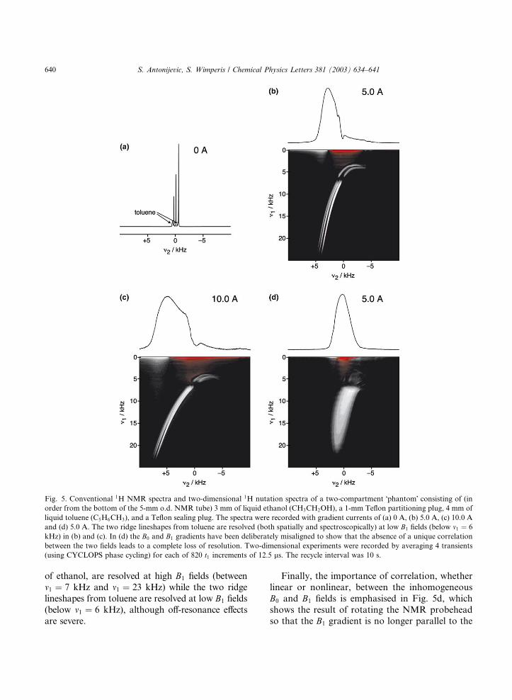
Fig. 5. Conventional 1H NMR spectra and two-dimensional 1H nutation spectra of a two-compartment �phantom� consisting of (in
order from the bottom of the 5-mm o.d. NMR tube) 3 mm of liquid ethanol (CH3CH2OH), a 1-mm Teflon partitioning plug, 4 mm of
liquid toluene (C5H6CH3), and a Teflon sealing plug. The spectra were recorded with gradient currents of (a) 0 A, (b) 5.0 A, (c) 10.0 A
and (d) 5.0 A. The two ridge lineshapes from toluene are resolved (both spatially and spectroscopically) at low B1 fields (below m1 ¼ 6
kHz) in (b) and (c). In (d) the B0 and B1 gradients have been deliberately misaligned to show that the absence of a unique correlation
between the two fields leads to a complete loss of resolution. Two-dimensional experiments were recorded by averaging 4 transients
(using CYCLOPS phase cycling) for each of 820 t1 increments of 12.5 ls. The recycle interval was 10 s.
640 S. Antonijevic, S. Wimperis / Chemical Physics Letters 381 (2003) 634–641
of ethanol, are resolved at high B1 fields (between
m1 ¼ 7 kHz and m1 ¼ 23 kHz) while the two ridge
lineshapes from toluene are resolved at low B1 fields
(below m1 ¼ 6 kHz), although off-resonance effectsare severe.
Finally, the importance of correlation, whether
linear or nonlinear, between the inhomogeneous
B0 and B1 fields is emphasised in Fig. 5d, which
shows the result of rotating the NMR probeheadso that the B1 gradient is no longer parallel to the
S. Antonijevic, S. Wimperis / Chemical Physics Letters 381 (2003) 634–641 641
B0 gradient. Once B1 is no longer a single-valued
function of B0 across the sample volume, there is a
complete loss of resolution in the two-dimensional
nutation spectrum.
5. Conclusions
As suggested by Pines and coworkers [2], the
two-dimensional nutation experiment can be used
to obtain high-resolution NMR spectra in corre-
lated inhomogeneous B0 and B1 fields. The method
is very simple (avoiding the need for sophisticated
pulses interleaved with data acquisition), providesspatial resolution in one dimension, and has the
great advantage of not requiring a linear correla-
tion of the two fields, making it perhaps more
generally applicable than the direct-acquisition
approach [1–3]. The latter has the advantage of
speed (i.e., the minimum experiment duration is
much shorter) but, per unit experiment time, will
have the same sensitivity as a projection of thetwo-dimensional nutation spectrum. To date, both
methods have only been demonstrated using one-
dimensional B0 and B1 gradients and the lifting of
this restriction is likely to be an important goal in
future work.
Acknowledgements
We are grateful to EPSRC for their generous
support from October 2000 to September 2003
(grant no. GR/N07622) and to Mike Jones and
Neville England for technical assistance.
References
[1] C.A. Meriles, D. Sakellariou, H. Heise, A.J. Moul�ee, A.
Pines, Science 293 (2001) 82.
[2] H. Heise, D. Sakellariou, C.A. Meriles, A. Moul�ee, A.
Pines, J. Magn. Reson. 156 (2002) 146.
[3] D. Sakellariou, C.A. Meriles, A. Moul�ee, A. Pines, Chem.
Phys. Lett. 363 (2002) 25.
[4] R.L. Kleinberg, A. Sezginer, D.D. Griffin, M. Fukuhara,
J. Magn. Reson. 97 (1992) 466.
[5] G. Eidmann, R. Savelsberg, P. Bl€uumler, B. Bl€uumich,
J. Magn. Reson. A 122 (1996) 104.
[6] A.P.M. Kentgens, J.J.M. Lemmens, F.M.M. Geurts, W.S.
Veeman, J. Magn. Reson. 71 (1987) 62.
[7] A. Samoson, E. Lippmaa, J. Magn. Reson. 79 (1988) 255.
[8] B.C. Gerstein, in: D.M. Grant, R.K. Harris (Eds.),
Encyclopedia of Nuclear Magnetic Resonance, vol. 5,
Wiley, Chichester, 1996, p. 3360.
[9] P. Styles, in: D.M. Grant, R.K. Harris (Eds.), Encyclope-
dia of Nuclear Magnetic Resonance, vol. 5, Wiley, Chich-
ester, 1996, p. 2847.
[10] S.P. Brown, S. Wimperis, Chem. Phys. Lett. 237 (1995)
509.
[11] R.R. Ernst, G. Bodenhausen, A. Wokaun, Principles of
Nuclear Magnetic Resonance in One and Two Dimensions,
Clarendon Press, Oxford, 1987.
[12] J. Keeler, D. Neuhaus, J. Magn. Reson. 63 (1985) 454.
[13] P.J. Grandinetti, J.H. Baltisberger, A. Llor, Y.K. Lee, U.
Werner, M.A. Eastman, A. Pines, J. Magn. Reson. A 103
(1993) 72.
[14] S. Wimperis, J. Magn. Reson. A 109 (1994) 221.
[15] K. Hendrich, H. Merkle, S. Weisdorf, W. Vine, M.
Garwood, K. Ugurbil, J. Magn. Reson. 92 (1991) 258.
[16] M. Garwood, K. Yong, J. Magn. Reson. 94 (1991) 511.