Embed Size (px)
Citation preview
High-resolution native and complex structures of thermostableb-mannanase from Thermomonospora fusca — substratespecificity in glycosyl hydrolase family 5Mark Hilge1†*, Sergio M Gloor1, Wojciech Rypniewski2, Oliver Sauer3,Tom D Heightman4, Wolfgang Zimmermann5, Kaspar Winterhalter1
and Klaus Piontek1*
Background: β-Mannanases hydrolyse the O-glycosidic bonds in mannan, ahemicellulose constituent of plants. These enzymes have potential use in pulpand paper production and are of significant biotechnological interest.Thermostable β-mannanases would be particularly useful due to their hightemperature optimum and broad pH tolerance. The thermophilic actinomyceteThermomonospora fusca secretes at least one β-mannanase (molecular mass38 kDa) with a temperature optimum of 80°C. No three-dimensional structure ofa mannan-degrading enzyme has been reported until now.
Results: The crystal structure of the thermostable β-mannanase from T. fuscahas been determined by the multiple isomorphous replacement method andrefined to 1.5 Å resolution. In addition to the native enzyme, the structures of themannotriose- and mannohexaose-bound forms of the enzyme have beendetermined to resolutions of 1.9 Å and 1.6 Å, respectively.
Conclusions: Analysis of the –1 subsite of T. fusca mannanase reveals neither afavourable interaction towards the axial HO–C(2) nor a discrimination against theequatorial hydroxyl group of gluco-configurated substrates. We propose thatselectivity arises from two possible mechanisms: a hydrophobic interaction of thesubstrate with Val263, conserved in family 5 bacterial mannanases, whichdiscriminates between the different conformations of the hydroxymethyl group innative mannan and cellulose; and/or a specific interaction between Asp259 andthe axial hydroxyl group at the C(2) of the substrate in the –2 subsite. Comparedwith the catalytic clefts of family 5 cellulases, the groove of T. fusca mannanasehas a strongly reduced number of aromatic residues providing platforms forstacking with the substrate. This deletion of every second platform is in goodagreement with the orientation of the axial hydroxyl groups in mannan.
IntroductionHemicelluloses are linear or branched polysaccharides thatare mostly found as heteroglycans in higher terrestrial plants.Depending on their sugar backbone composition they areclassified as either xylans, mannans, arabinogalactans orarabinans. The two most important representative hemi-celluloses are the hetero-1,4-β-D-xylans and the hetero-1,4-β-D-mannans. In hetero-1,4-β-D-mannans, the primaryhydroxyl group of the backbone residues is substituted byα-linked galactose; the degree of substitution is depen-dent on the source. Together with cellulose and lignin,hemicelluloses form the most abundant structural compo-nents of plant cell walls [1].
Microorganisms that degrade hemicelluloses frequentlyappear in soil, compost, and in the rumen of animals. Tocompletely hydrolyse (hetero)mannans, fungi and bacteria
have to produce at least one mannanase (EC 3.2.1.78),one β-mannosidase (EC 3.2.1.25) and an α-galactosidase(EC 3.2.1.22) [2]. In the process of degradation mannanaseshydrolyse the 1,4-β-mannopyranoside bond in galacto-,gluco- and galactoglucomannans to mannooligomers, man-nobiose and mannose, respectively. The degree of hydroly-sis of galactomannan decreases with increasing substitutionby galactose, suggesting that galactose residues obstruct theenzymic cleavage of the mannan backbone [2].
On the basis of sequence comparisons by homology andhydrophobic cluster analysis [3,4], the catalytic domains ofglycosyl hydrolases have been classified into 64 families(http://expasy.hcuge.ch/cgi-bin/lists?glycosid.txt). Despiteacting on three different substrates, endo-1,3-β-glucanases,endo-1,4-β-glucanases and endo-1,4-β-mannanases have sofar all been assigned to glycosyl hydrolase family 5. As the
Addresses: 1Laboratorium für Biochemie, ETHZentrum, Universitätstrasse 16, CH-8092 Zürich,Switzerland, 2European Molecular BiologyLaboratory, c/o DESY, Notkestrasse 85, D-22603Hamburg, Germany, 3Abteilung für Strukturbiologie,Institut für Physikalische Chemie, Karl-Franzens-Universität Graz, A-8010 Graz, Austria,4Laboratorium für Organische Chemie, ETHZentrum, Universitätstrasse 16, CH-8092 Zürich,Switzerland and 5Biotechnology Laboratory,Aalborg University, Sohngaardsholmsvej 57, DK-9000 Aalborg, Denmark.
†Present address: Biophysical Structural Chemistry,Gorlaeus Laboratories, Leiden University, PO Box9502, 2300 RA Leiden, The Netherlands.
*Corresponding authors.E-mail: [email protected]
Key words: crystal structure, family 5, glycosylhydrolase, mannanase, Thermomonospora fusca
Received: 12 August 1998Revisions requested: 9 September 1998Revisions received: 17 September 1998Accepted: 18 September 1998
Structure 15 November 1998, 6:1433–1444http://biomednet.com/elecref/0969212600601433
© Current Biology Ltd ISSN 0969-2126
Research Article 1433

sequence identity among the members of this family israrely higher than 20%, an additional classification into sixsubfamilies, A1–A6, has been introduced [5,6]. Enzymeswithin subfamilies show at least 25% sequence identity andmay display similar substrate specificities. For instance,many enzymes of subfamily A3 additionally reveal lichenaseactivity, whereas some members of subfamily A4 show con-siderable xylanase activity [5].
So far the crystal structures of four family 5 cellulases,three in combination with a substrate, have been solved:Clostridium thermocellum cellulase (CelC) which belongsto subfamily A3 [7,8]; Clostridium cellulolyticum endo-glucanase A (CelCCA), subfamily A4 [9]; Acidothermuscellulolyticus endocellulase (E1cd), subfamily A1 [10]; and
Bacillus agaradherans endoglucanase (Cel5A), subfamilyA2 [11]. With the enzymes of the glycosyl hydrolasefamilies 1, 2, 5, 10, 17, 26, 30, 35, 39 and 42 they formthe GH-A clan [12]. Enzymes of this clan share an(α/β)8-barrel fold and with the exception of family 26three conserved active-site residues (one asparagine andtwo glutamate residues), and follow a retaining cleavagemechanism (Figure 1).
The few mannanase sequences reported permit assignmentof these enzymes to either family 5 or 26. Recent workusing hydrophobic cluster analysis has identified four con-served residues in family 26, and suggests that these man-nanases also belong to the GH-A clan [13]. Mannanaseswithin family 5 share eight strictly conserved amino acid
1434 Structure 1998, Vol 6 No 11
Figure 1
O
O
HO
HO
OH
HO
O
O O
HO
OH
OHO
HO
OH
HO
O
O
HO
OH
O
OHO
HO
OH
HO
O
O
HO
OH
(a)
–1 +2–2–3–4
ReducingNon-reducing
+1
O– O
Glu225
O
O
Glu128
H
OHO
HO
OH
OO
HO
O
O
HO
OH
OO
Glu225
O
– O
Glu128
OHO
HOOHO
HOHO
O
O
HO
OH
OO
Glu225
O
– O
Glu128
OHO
HOOHO H
OH
O– O
Glu225
O
O
Glu128
H
OHO
HO
OH
OOH
–1
3.5 Å
+1
(b)
Structure
The reaction catalysed by β-mannanase. (a) The nomenclature forsugar-binding subsites in glycosyl hydrolases [15]. (b) The retainingmechanism for T. fusca mannanase, in which the glycosidic oxygen isprotonated by Glu128 (proton donor) and the anomeric carbon atom is
attacked by Glu225 (nucleophile). The resulting mannosyl–mannanaseintermediate is then hydrolysed by a water molecule, generating aproduct with the same anomeric configuration as the substrate [17].

residues with more than 60 cellulases [10,14]. As deducedfrom the available cellulase crystal structures, it is obviousthat all these conserved residues are located at or close tothe active site. They either participate in binding of thesugar in the –1 subsite [15] or stabilise the position and pro-tonation state of the catalytic glutamate residues [7–10].
Despite the lack of the major part of the primary structure,the 42 N-terminal amino acids of Thermomonospora fuscaβ-mannanase Q1.1 allowed the assignment of this enzymeto glycosyl hydrolase family 5 [16]. Here we report the high-resolution structures of Q1.1, in native and complexedform, and provide a rationale for the substrate specificity ofthis, as yet, structurally uncharacterised class of enzymes. Inaddition, we propose two new subfamilies for family 5.
Results and discussionOverall structure descriptionThe crystal structure of T. fusca β-mannanase Q1.1 wasdetermined by the multiple isomorphous replacement(MIR) method at 2.2 Å resolution and experimental phaseswere extended to 1.5 Å. The final model, consisting of 302amino acids, has a crystallographic R factor of 11.9% and anR free value of 17.6%. The overall structure of β-man-nanase Q1.1 is presented in Figure 2a. The protein showsthe architecture of the classical (α/β)8-barrel motif. Amongthe 28 families of glycosyl hydrolases for which a three-dimensional structure has been determined, families 1, 2,5, 6, 10, 13, 14, 17, 18 and 20 display this (α/β)8-barrelmotif [17]. The molecule has approximate dimensions of45 × 45 × 40 Å and its most obvious feature is a prominentcleft which is about 15 Å deep, 15 Å wide and 30 Å long(Figure 2b). The surface electrostatic potential is domi-nated by negative charges, which is consistent with therather acidic pI (3.9) of the protein. This may relate to thestability of the enzyme in alkaline solutions.
Aside from the structural elements of the barrel, two shortβ strands at the N terminus form the bottom of the barrel(blue arrows; Figure 2a). These two β strands are alsofound in endocellulase E1cd from A. cellulolyticus [10]. Theconnections between the αβ repeats (at the bottom of thebarrel) are generally very short (4–6 residues). The onlydisulphide bond of Q1.1, between Cys74 and Cys81, islocated at the end of helix 2 and the beginning of strand 3,and might permit the tight turn from α2 to β3. This disul-phide bond does not exist in the structures of CelC,CelCCA and E1cd; although Cys74 is conserved inCelCCA and CelC, there is no counterpart for Cys81.
Two new family 5 subfamiliesAn alignment of all the available bacterial and eukaryoticmannanase amino acid sequences in family 5 is shown inFigure 3. The alignment reveals the existence of two dis-tinct groups of mannanases, which we propose as sub-families A7 (eukaryotic mannanases) and A8 (bacterialmannanases). Among subfamily A8 members the sequenceidentity is above 43% (i.e. substantially higher than thehomology within other subfamilies of glycosyl hydrolasefamily 5. Between members of subfamilies A7 and A8the sequence identity is below 20%. In a pairwise align-ment of T. fusca mannanase Q1.1 with all family 5sequences deposited in the SWISS-PROT data bank, thehighest identities were shown with the endo-1,4-β-glu-canases from Cellulomonas fimi (23.2%; SWISS-PROTcode P50400), Ruminococcus albus (22.7%; SWISS-PROTcode Q07940) and Streptomyces lividans (21.6%; SWISS-PROT code P27035).
Comparison with other glycosyl hydrolasesThe structure of T. fusca β-mannanase was compared withstructures deposited in the Protein Data Bank (PDB)using the program DEJAVU [18]. Thereafter, potentially
Research Article Structures of a thermostable b-mannanase Hilge et al. 1435
Figure 2
The overall fold of T. fusca β-mannanase. (a) Schematic representation of the structureof T. fusca β-mannanase. β Strands β1 andβ0, which form the bottom of the barrel, areshown as blue arrows; all other β strands areshown as red arrows. Helices are depicted asgreen spirals. The figure was generated usingthe program MOLSCRIPT [43]. (b) Surfaceelectrostatic potential distribution for T. fuscaβ-mannanase. Positive potential is shown inblue and negative potential in red. The figureshows the catalytic cleft at the top right of themolecule. The figure was prepared using theprogram GRASP [44].
(a) (b)
Catalytic cleft
Structure

related structures were more precisely analysed withLSQMAN [19] (Table 1), which eliminated false positivehits. The overall fold of β-mannanase Q1.1 was, asexpected, most similar to those of endocellulase E1cd[10], endoglucanase CelCCA [9] and endocellulase CelC[7,8]. Lower similarity could be detected to the catalyticdomains of β-1,4-glycanase CEX from C. fimi [20] (PDBcode 1EXP), 1,3-β-D-glucanase and 1,3;1,4-β-D-glucanase
from plants [21] (PDB codes 1GHR and 1GHS), xylanaseA from S. lividans [22] (PDB code 1XAS) and xylanasefrom C. thermocellum [8] (PDB code 1XYZ), which are allmembers of the GH-A clan. Besides the three key cat-alytic residues (Asn127, Glu128 and Glu 225), 1EXP,1GHR and 1GHS also show the conserved Arg50 residue,which is replaced in family 10 xylanases by a valine orthreonine. The sugar stacking in the –1 subsite of family
1436 Structure 1998, Vol 6 No 11
Figure 3
(a) MANA_THEFU ----------------------------------------MANA_STRLI ----------------------------------------MANA_VIBRI ----------------------------------------MANB_CALSA ----------------------------------------
MANA_THEFU -------------------------ATGLHVKNGRLYEANMANA_STRLI ------------------------AAGGIHVSNGRVVEGNMANA_VIBRI MKFT--KAIFSLLLFIWAS----CAHAGFYVSNGVLYEANMANB_CALSA -------------------------------NDG-VVKID
MANA_THEFU GQEFIIRGVSHPHNWYPQH--------TQAFADIKSHGANMANA_STRLI GSAFVMRGVNHAYTWYPDR--------TGSIADIAAKGANMANA_VIBRI GSAFKIRGINHAHTWYTDKL-------SVALSGIAATGANMANB_CALSA TS--TLIGTNHAHCWYRDRL-------DTALRGIRSWGMN
MANA_THEFU TVRVVLSN--------G---VRWS---KNGPSDVANVISLMANA_STRLI TVRVVLSS--------G---GRWT---KTSASEVSALIGQMANA_VIBRI TVRVVLSN--------G---YRWT---KNDVSDVTNIINLMANB_CALSA SVRVVLSN--------G---YRWT---KIPASEVANIISL
MANA_THEFU CK-QNRLICMLEVH-------------D-------TTGYGMANA_STRLI CK-ANKVICVLEVH-------------D-------TTGYGMANA_VIBRI AK-ANNLIAILEVH-------------D-------TTGYGMANB_CALSA SRSLGFKAIILEVH-------------D-------TTGYG
MANA_THEFU EQSGA-----STLDQAVDYWIELKSVLQ-GEEDYVLIN--MANA_STRLI KD-GA-----TSLDQAGDYWVGVKSAAWRAQEDYVVVN--MANA_VIBRI EESSA-----ASLDSAADYWIELKNELI-GQEDYVIIN--MANB_CALSA EDGAA-----CSLAQAVEYWKEIKSVLD-GNEDFVIIN--
MANA_THEFU IGNEP--YG--------NDSATVAAWASDTSAAIQRLRAAMANA_STRLI IGNEP--FG--------N--TNYAAWTDATKSAIGKLRGAMANA_VIBRI LGNEP--FG--------NN-NDAVAWVNDHVSAIQRLRSAMANB_CALSA IGNEP--YG--------N--NNYQNWVNDTKNAIKALRDA
MANA_THEFU GFEHTLVVDAPNWGQDWT---N---TMRNNADQVYASDPTMANA_STRLI GLGHALMVDAPNWGQDWS---G---TMRSNAASVFASDPDMANA_VIBRI GINHTIMVDAPNWGQDWK---G---FMLNNAQFVFNSDPKMANB_CALSA GFKHTIMVDAPNWGQDWS---N---TMRDNAQSIMEADPL
MANA_THEFU GNTVF-SIHMY------GV-----YSQAS-TITSYLEHFVMANA_STRLI RNTVF-SIHMY------GV-----YDTAA-EVRDYLNAFVMANA_VIBRI LNTIF-SVHMY------EV-----YSSYN-SVNDYISSFTMANB_CALSA RNLVF-SIHMY------GV-----YNTAS-KVEEYIKSFV
MANA_THEFU NAG-LPLIIGEFGHDHS-DGNPDEDT--------IMAEAEMANA_STRLI GNG-LPIVVGEFGDQHS-DGNPDEDA--------IMATAQMANA_VIBRI NNG-LVLVIGEFASTHK-GADVDEGS--------IMERSEMANB_CALSA DKG-LPLVIGEFGHQHT-DGDPDEEA--------IVRYAK
MANA_THEFU RLKLGYIGWSWSGN-GGGVEYL-DMVYNFDGDNLSPWGERMANA_STRLI SLGVGYLGWSWSGN-GGGVEYL-DMVNGFDPNSLTSWGNRMANA_VIBRI TLSLGYIGWSWSGN-DTTTSDL-DIVNNWDNNSYSTWGNVMANB_CALSA QYKIGLFSWSWCGN-SSYVGYL-DMVNNWDPNNPTPWG--
MANA_THEFU IFYGPNGIASTAKEAVIFG---------------------MANA_STRLI ILYGSNGIAATSRTATVYGGGGGSTGGTAPNGYPYCVNGGMANA_VIBRI LINGQNGIKSTSTLATVFTCGNDCNDDSSGE-YPICSSSAMANB_CALSA QWYKTNAIGTSS----------------------------
MANA_THEFU ----------------------------------------MANA_STRLI AVRPGR----------------------------------MANA_VIBRI VDPDGDGWGWENNQSCIVQDSSDTAPNGYPYCSQESSDPDMANB_CALSA ----------------------------------------
MANA_THEFU --------------MANA_STRLI --------------MANA_VIBRI GDGWGWENNASMCCMANB_CALSA --------------
(b) AAMANNA -----------------------------------MKLSHTRBEMA ----------------------------------------ABCEL4MR DVPVWGQCGGRDWTGETACASGSSCVVQNEWYSQCLPGSTTOMATO ----------------------------------------
AAMANNA MLLS--LASLGVATALPRTPNHNAATTAFPSTSGLHFTIDTRBEMA ----------AVLQPVPR-------ASSFVTISGTQFNIDABCEL4MR TPTNPPPATTTSQTTAPPTTSHP-VSTGFVKASGTRFTLNTOMATO MSYARRSCICGLFLLFLALVC--EANSGFIGVKDSHFELN
AAMANNA GKTGYFAGTNSYWIGFLTNND-D---VDLVMSQLAASDLKTRBEMA GKVGYFAGTNCYWCSFLTNHA-D---VDSTFSHISSSGLKABCEL4MR GQKYTVVGGNSYWVGLTGLSTSA---MNQAFSDIANAGGTTOMATO GSPFLFNGFNSYWLMHVAADPTERVKVTIVLKDASVAGLS
AAMANNA ILRVWGFNDVNTKPTDG---TVWYQLHANGTSTINTGADGTRBEMA VVRVWGFNDVNTQPSPG---QIWFQKLSATGSTINTGADGABCEL4MR TVRTWGFNEVTS-PN-GNYYQSWS----GARPTINTGASGTOMATO VCRTWAFSD-------G---GDRA----LQISPGIYDERV
AAMANNA LQRLDYVVTSAEKYGVKLIINFVNEWTDYGGMQAYVTAYGTRBEMA LQTLDYVVQSAEQHNLKLIIPFVNNWSDYGGINAYVNAFGABCEL4MR LLNFDNVIAAAKANGIRLIVALTNNWADYGGMDVYVNQMVTOMATO FQGLDFVIAEAKKYGAQIS----N---D-------DEFYT
AAMANNA AAAQTD--FYTNTAIQAAYKNYIKAVVSRYSSSAAIFAWETRBEMA GNATT---WYTNTAAQTQYRKYVQAVVSRYANSTAIFAWEABCEL4MR GNGQPHDLFYTNTAIKDAFKSYGRAFVSRYANEPTVMAWETOMATO HPMLKK--YLKNHIEKVVTRLNSITKVA-YKDDPTIMAWE
AAMANNA LANEPRCQG--------CDTSVLYNWISDTSKYIKSLDS-TRBEMA LGNEPRCNG--------CSTDVIVQWATSVSQYVKSLDS-ABCEL4MR LANEPRCKGSTGTTSGTCTTTTVTNWAKEMSAFIKTIDS-TOMATO LMNEPRDQAD-------YSGKTVNGWVQEMASFVKSLDN-
AAMANNA --KHLVTIGDEGFGLDVDSD-GSYPYTYGEGLNFTKNLGITRBEMA --NHLVTLGDEGLGLSTG-D-GAYPYTYGEGTDFAKNVQIABCEL4MR --NHLVAIGDEGFYNQPG-A-PTYPYQGSEGVDFEANLAITOMATO --KHLLEVGMEGFYGDSIPIRKSVNPGYQVGTDFISNHLI
AAMANNA STIDFGTLHLYPDSW--GTS----YDWGNGWITAHAAACKTRBEMA KSLDFGTFHLYPDSW--GTN----YTWGNGWIQTHAAACLABCEL4MR SSVDFATFHSYPEPW--GQGA-DAKAWGTQWITDHAASMKTOMATO NEIDFATIHAYTDQWVSGQSDDAQLVWMEKWITSHWEDAR
AAMANNA AVG-KPCLLEEYGVTSNHCAVESPWQ--------QTAGNATRBEMA AAG-KPCVFEEYGAQQNPCTNEAPWQ--------TTSLTTABCEL4MR RVN-KPVILEEFGVTTNQPDTYAEWF--------NEIES-TOMATO NILKKPLVLAEFGKSSRGQGSRDIFMSSVYRNVYNLAKEG
AAMANNA TGISGDLYWQYGTTFSWGQSPN-DGNTFYYNT--SDFTCLTRBEMA RGMGGDMFWQWGDTFANGAQSNSDPYTVWYNS--SNWQCLABCEL4MR SGLTGDLIWQAGSHLSTGDTPN-DGYAVYPDG--PVYP-LTOMATO GTMAGSLVWQLMAH--GMENYD-DGYCIVLGQTPSTTQ-I
AAMANNA VTDHVAAINAQSK---TRBEMA VKNHVDAINGG-----ABCEL4MR VKSHASAMKNRA----TOMATO ISDQAHVMTALARSLN
Structure
Amino acid sequence alignment of family 5 β-mannanases. Identicalamino acids are indicated by black shading and similar amino acidsby grey shading. (a) The sequences of bacterial β-mannanases:MANA_THEFU from Thermomonospora fusca (EMBL code,AJ006227); MANA_STRLI from Streptomyces lividans (SWISS-PROT code, P51529); MANA_VIBRI from Vibrio sp. (EMBL code,D86329); and MANB_CALSA from Caldocellum saccharolyticum(SWISS-PROT code, P22533). (b) The sequences of eukaryotic
β-mannanases: AAMANNA from Aspergillus aculeatus (EMBL code,L35487); TRBEMA from Trichoderma reesei (EMBL code, L25310);ABCEL4MR from Agaricus bisporus (EMBL code, Z50095); andTOMATO from Lycopersicon esculentum (EMBL code, AF 017144).To allow comparison between (a) and (b) gaps were introduced intothe bacterial sequences. The alignment was generated using theCLUSTALW algorithm, and shading was carried out withBOXSHADE.
10 enzymes is still maintained by a tryptophan residue,whereas in 1EXP, 1GHR, 1GHS this function is achievedby a phenylalanine.
As in the other structures of enzymes from families 1, 2, 5,17 and 18 (but surprisingly not 10), a rare nonproline cispeptide bond [23] occurs following the aromatic residueinteracting with the –1 sugar. This cis peptide bondappears to be essential for the enzyme function because itconstrains the position of the preceding tryptophan orphenylalanine residue [7]. In the T. fusca mannanase thiscis peptide bond is formed by a serine, as it is for E1cd, therepresentative for subfamily 1.
The active siteIn the –1 subsite, the arrangement of residues involved inthe attack of the glycosidic bond is well maintained amongQ1.1 and the three other family 5 enzyme structures
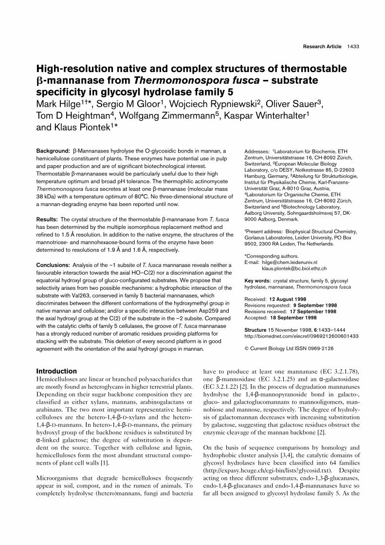
(Figure 4a). In particular, Arg50, His86, Asn127, Glu128,His196, Tyr198, Glu225 and Trp254 are strictly conservedin family 5 enzymes.
By analogy with the substrate complexes of E1cd andCelC, Glu128 and Glu225 perform the roles of catalyticproton donor and nucleophile, respectively. His86 isresponsible for the hydrogen bond mediated recognitionof the HO–C(3), and Trp254 provides a hydrophobicsurface for the α-face of the pyranoside ring (Figure 4b).The other conserved residues, Arg50, Asn127, His196 andTyr198, stabilise the active-site environment and arelikely to influence the protonation state of the two cat-alytic glutamate residues. Arg50 forms a salt bridge withGlu225 and a hydrogen bond with Asn127. Interestingly,Asn127, which may interact with the equatorial HO–C(2)in cellulases [7,10], is also present at an identical positionin Q1.1, which raises the question of the role of this
Research Article Structures of a thermostable b-mannanase Hilge et al. 1437
Figure 4
The active site of β-mannanase. (a) Structuralsuperposition of the active-site residues offamily 5 glycanases: β-mannanase Q1.1 isdepicted in yellow, endoglucanase CelCCA inred, endocellulase E1cd in blue, andendocellulase CelC in green. Thesuperposition was made with the programLSQMAN [19] and displayed in the graphicsprogram O [45]. Residues Arg50 and His196are omitted for clarity. (b) 3Fo–2Fc electron-density map contoured at 1.5σ, displaying theβ-mannanase active site. The labelscorrespond to the numbering in T. fuscaβ-mannanase.
(b)
Trp254
Glu225
Glu128
His86
(a)
Trp254 Tyr198
His86
Asn127
Glu128
Glu225
Structure
Table 1
Structural alignment of Thermomonospora fusca b-mannanase and related enzymes.
1MAN 1EDG 1ECE 1EXP 1GHRProtein Family Nr Nm rmsd Nc Nm rmsd Nc Nm rmsd Nc Nm rmsd Nc Nm rmsd Nc
1MAN 5 3021EDG 5 380 92 1.085 291ECE 5 358 102 0.996 32 115 1.104 341CEN 5 340 112 1.011 25 162 0.923 42 143 1.020 351EXP 10 312 68 1.232 111GHR 17 306 64 0.947 13 53 1.061 61XAS 10 299 63 1.181 11 264 0.810 144 59 1.096 6
Pairwise root mean square deviations (rmsds; in Å) on Cα atoms forT. fusca β-mannanase Q1.1 (PDB code, 1MAN); C. cellulolyticumendoglucanase CelCCA [9] (PDB code, 1EDG); A. cellulolyticusendocellulase E1cd [10] (PDB code, 1ECE); C. thermocellumendocellulase CelC [7] (PDB code, 1CEN); C. fimi β-1,4-glycanaseCEX [20] (PDB code, 1EXP); Hurdeum vulgare 1,3;1,4-β-D-glucanase
[21] (PDB code, 1GHR); and S. lividans xylanase A [22] (PDB code,1XAS). The alignments were carried out with the program LSQMAN[19]. A 2.0 Å cut-off was used to define structurally equivalentresidues. Nr, number of residues in the protein; Nm, number ofmatched residues; Nc, number of matched residues of identical typein both proteins.
residue in substrate binding and/or transition-state stabili-sation (see below). His196 and Tyr198 are hydrogenbonded to Glu128 and Glu225, respectively. In contrast tothe three cellulases, the highly similar arrangement of thestrictly conserved sidechains in Q1.1 is achieved by anenergetically unfavourable course of the backbone. This isemphasised by the Ramachandran plot where Glu128 andTyr198 lie in the generously allowed regions.
Substrate bindingHigh-resolution data were collected from crystals soakedin mannotriose and mannohexaose (Table 2). The inter-actions between the substrate residues and T. fusca man-nanase are shown in Figure 5. Both substrate complexesshow well defined electron density, revealing an identicaloccupation of the –2 and –3 subsites and a potential –4subsite (Figure 6). Similar binding to the –2 and –3 sub-sites was found for cellobiose in cellulase Cel5A fromBacillus agaradherans, unlike the cellobiose complexes ofE1cd and CelC where the –1 subsite is occupied. Thisvacancy is consistent with the view that the –1 subsiteshould favour the transition state rather than substratebinding. As E1cd and CelC do not have a –3 subsite, cel-lobiose binding equivalent to that seen in Q1.1 and Cel5Awould be unfavourable.
Whereas the –1 subsites of the three cellulases and Q1.1have very similar structures, the –2 subsites differ consid-erably. In Q1.1, the groove narrows at the –2 subsite andlacks an aromatic platform compared to the cellulase struc-tures in family 5. The only direct interactions between the–2 mannosyl residue and the protein are a 2.7 Å hydrogenbond formed between Asn259 and the axial HO–C(2)
(Figure 6c) and a 3.0 Å hydrogen bond between HO–C(3)and the backbone nitrogen atom of Gly260. In E1cd,Phe29 forms a hydrophobic platform at the bottom of therelatively wide –2 subsite, so that the –2 and –1 glucosylrings are approximately coplanar. The phenyl ring plane isnearly parallel to the –2 sugar ring, at a distance of ~5 Å. InCelC the substrate-binding groove also narrows at the –2subsite forcing the glucose residue to twist such that itsring plane is approximately 90° to that of the –1 residue.The platform Trp212 in CelC is appropriately positionedto the side of the cleft forming van der Waals contactswith the α-face of the –2 residue.
Similarly, only a few interactions are present at the –3subsite. The mannosyl residue is primarily bound throughhydrophobic stacking with Trp30 and is laterally restrictedthrough Trp59. In CelCCA, Trp57 shows a similar orien-tation to Trp30 in Q1.1. The hydrogen bond betweenNε1 of Trp59 and HO–C(3) seems to be the only inter-action with the protein in the 1.9 Å mannotriose complex.However, in the mannohexaose complex the hydrox-ymethyl group has a double conformation: in the gauche–trans conformation it forms a hydrogen bond with Tyr31,whereas in the gauche–gauche conformation it interacts viaa water-mediated hydrogen bond with the axial hydroxylgroup of the –3 mannosyl residue.
A novel feature in the mannotriose and mannohexaosecomplexes is the existence of a –4 subsite. Compared tothe –3 and –2 subsites the electron density is weaker,presumably due to even fewer interactions. The 2.9 Åhydrogen bond between the axial HO–C(2) and the back-bone carbonyl oxygen of Trp30 and the water-mediated
1438 Structure 1998, Vol 6 No 11
Table 2
Data collection and MIR phasing statistics.
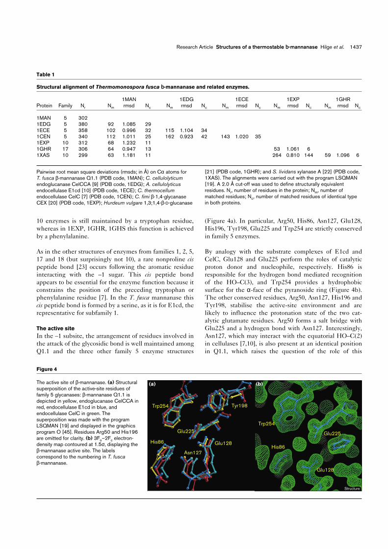
Native 1 Mannotriose Mannohexaose TMLA HgI4 Baker IrCl6 Native 2* Xenon*
λ (Å) 0.99 1.00 0.83 1.00 0.90 1.00 0.90 0.9 1.54Soak concentration (mM) 100 100 100 1 2 5
Soak pressure(bar) – – – – – – – – 50
Soak time 1.5 h 30 h 40 h 60 h 72 h 84 h 3 minNo. of sites(major/minor) 4/10 4/– 1/4 –/5 2/2
Resolution (Å) 1.80 1.9 1.6 2.40 2.35 3.0 2.50 1.5 2.2Redundancy 2.90 3.34 3.65 3.32 3.22 3.12 2.91 3.22 2.98Completeness (%)† 93.4 99.3 83.9 98.8 98.8 98.3 97.8 97.2 97.4
(84.8) (97.0) (84.5) (97.2) (99.2) (98.8) (95.9) (92.2) (95.8)I/σ† 12.4 10.6 18.0 13.9 16.74 12.8 11.6 17.2 16.9
(3.5) (2.9) (2.6) (6.9) (9.1) (6.9) (4.9) (2.9) (10.5)Rmerge (I)† 7.3 10.9 6.2 8.4 6.1 9.0 9.5 6.2 6.2
(30.9) (39.9) (47.2) (17.6) (12.2) (17.2) (23.5) (33.1) (10.8)Phasing power(iso/ano) 2.18/0.80 1.18/1.17 1.37/1.01 0.70/0.43 1.19/0.72
*Data collected under cryogenic conditions; all other data were collected at room temperature. †Values in parentheses correspond to those for thehighest resolution shell.

interaction of HO–C(3) with Glu33 present the only con-tacts. Depending on the crystal form, additional contactsare established through interactions with a symmetry-related molecule. As a consequence the electron densitybeyond C(3) is improved.
The +1 and +2 subsites of all three cellulases and Q1.1 aresimilar, suggesting that in Q1.1 the fissile glycosidic linkageis twisted in an equivalent way to the one in E1cd. In Q1.1Trp167 fulfils the function of Trp213 of E1cd and Tyr176in CelC, but there is no counterpart to Tyr245 in E1cdand Phe203 in CelC. Trp171 in Q1.1 could form the +3subsite, allowing the mannosyl residues to adopt a confor-mation similar to unbound mannan. Trp57 and Trp181 arecomparably positioned in CelCCA and in CelC, Phe244provides a potential platform for the +3 subsite.
The –1 subsiteThe mannohexaose complex does not reveal a clear viewof the –1 subsite, although strong difference densitypeaks indicate positions expected for C(2), C(3) and theircorresponding hydroxyl groups. The absence of densityfor the glycosidic bond between the –2 and –1 mannosylresidues and the lack of reliable density for the rest of thepyranoside ring prevented satisfactory construction of the–1 sugar. Superimposing the native structure with the
mannotriose and mannohexaose structures revealed awell-matched pattern of electron density that may repre-sent the water structure in the –1 subsite. Indeed, themost prominent water molecules, which probably substi-tute the positions of HO–C(2) and HO–C(3), are withinhydrogen-bonding distance of Asn127 (3.0 Å) and His86(3.1 Å), respectively.
The asparagine preceding the proton donor is strictly con-served in all glycanases of the GH-A clan and mutations ofthis residue lead to complete loss of activity [24]. As previ-ously suggested [7,10], this residue might play a moreimportant role in transition-state binding than in substraterecognition, as the interatomic distance between the equa-torial HO–C(2) of the substrate and the δN and δO atomsof asparagine is relatively long (~3.3 Å [10]). On formingthe glycosyl ester intermediate this distance is reduced,as observed in the 2′′,4′′-dinitrophenyl-2-deoxy-2-fluoro-β-cellobioside–xylanase/glucanase complex from C. fimi(2.8 Å) [25]. Consistent with this assumption is the conser-vation of Asn127 in Q1.1, although the distance to theaxial HO–C(2) is even longer (~4 Å). In the transitionstate, via which the glycosyl ester is formed, the –1 sugarresidue must approach the catalytic nucleophile, and theconcomitant ring distortion would place the HO–C(2) in apseudo-equatorial position. Both the approach of the –1
Research Article Structures of a thermostable b-mannanase Hilge et al. 1439
Figure 5
HO
O
OH
HO
OH
O
O
HO
OH
O
OH
O
HO
OH
NH
HO
N
NHNδ2
Oδ1
O
OH
O
ON
O
OH
OHOH
Tyr31
–4 –3 –2 –1
Trp59
Val263
His86
3.1Å
Asn259
Glu33
Glu128
Glu225
Asn127
3.1Å
3.1Å
2.9Å
2.7Å
Hydrophobic platformTrp30
3.1Å
3.1Å
2.9Å
3.0Å
3.1Å
2.7Å
Gly260
3.0Å
Hydrophobic platformTrp 254
2.9Å
O Trp302.8Å
Tyr198
Structure
Schematic representation of the T. fusca mannanase–mannotriosecomplex interactions. The positions of the four subsites are indicatedbelow. As the –1 mannosyl residue is not clearly visible in the electron-density map, it is depicted with dashed lines at the expected position.
The five water molecules are represented by spheres. The two watermolecules in the –1 subsite have low B factors and may represent thepositions of HO–C(3) and HO–C(2). In the –3 subsite the doubleconformation of the primary hydroxyl group is shown.

sugar and its ring distortion reduce the distance betweenHO–C(2) and Asn127. In this way an additional hydrogenbond is formed which contributes to the catalytic effec-tiveness of the enzyme.
Substrate specificityDespite the rather limited number of interactions with theprotein, the mannosyl residues bind specifically into thecatalytic cleft, as indicated by the equivalent sugar atompositions in the mannotriose and mannohexaose com-plexes (root mean square deviation [rmsd] 0.1 Å). Asexpected, for mannose the reducing end of the substrate ispresent in the preferred α-configuration (Figure 6c). Inaddition, there is some evidence for an α-anomer in the –4subsite, although not as prominent as for the –2 subsite.The α-anomer seems to be shifted by 0.2 Å from itsexpected position suggesting that the substrate is not yetproperly positioned in the –4 subsite.
Among the various substrates hydrolysed by members ofthe GH-A clan, mannan is the only compound having theHO–C(2) group in the axial position. As mannan appearsat first glance to be otherwise identical to cellulose, thepreference of Q1.1 for mannan might be expected toresult from either a stabilising interaction towards theaxial hydroxyl group at C(2) of the pyranose ring, or a dis-crimination against the equatorial hydroxyl group ofgluco-configurated substrates. At sugar subsite –1, pro-posed as the catalytic subsite, neither of these criteria arefulfiled in the Q1.1 structure (results based on a cello-tetraose model, described below), suggesting that Q1.1should also hydrolyse gluco-configurated substrates.Indeed, Q1.1 shows activity towards xylan (below 10%compared with mannan), and to an even lesser extenttowards carboxymethylcellulose. This is in contrast to theβ-mannanase from S. lividans, where no xylanase, endo-cellulase or α-galactosidase activity could be detected[26]. Mannan is still the preferred substrate, however,suggesting that the substrate selectivity may arise fromconformational, rather than configurational, differencesbetween mannan and its gluco-configurated analogues.Electron diffraction studies of mannan [27,28] show aconformation similar to that of crystalline cellulose, inwhich alternating rings are oriented ~180° to each other.In cellulose this conformation is stabilised by hydrogenbonds between HO–C(3) of one residue and O5 of theneighbouring residue, and between the hydroxymethylgroup of the first residue and HO–C(2) of the second(Figure 7a). In mannan this second hydrogen bond is notpossible because HO–C(2) is axial. The conformations ofthe glycosidic linkage and the hydroxymethyl groupchange to accommodate an alternative hydrogen-bondingscheme in which HO–C(3) is hydrogen bonded to bothO5 and the hydroxymethyl group of the neighbouringresidue (Figure 7b). The main distinguishing featuresbetween mannan and cellulose are thus the slightly
1440 Structure 1998, Vol 6 No 11
Figure 6
Fo–Fc electron-density maps contoured at 3σ before the incorporationof the carbohydrate compound. The electron density for (a) themannotriose and (b) the mannohexaose complex. (c) The positivedifference density (in green) for the α-anomer above C(1) in themannohexaose complex. In addition, the 2.7 Å hydrogen bond betweenAsp259 and the axial HO–C(2) is shown.
(b)
–4 –3 –2
(a)
–4 –3 –2
HO-C(2)
C(1)
Asp259
2.7 Å
(c)
Structure

shifted torsion angles (in cellulose I φ = –98°, ψ = –143°;in mannan φ = –90°, ψ = –149° [29]) of the glycosidiclinkage, and the conformation of the hydroxymethylgroup (trans–gauche in cellulose I, χ = –81°; gauche–trans inmannan, χ = 175°).
On the basis of the structure of bound mannotriose, cello-tetraose of E1cd was docked into the active site of Q1.1,superimposing the –2 glucosyl residue exactly on the equiv-alent residue of mannotriose (Figures 7c and 7d). In thisposition the hydrogen bonds between the catalytic protondonor Glu128 and the glycosidic oxygen and betweenHis86 and HO–C(3) are preserved, as is the hydrophobicinteraction with Trp254. In E1cd the hydroxymethyl groupof the –1 glucose residue is bound in the gauche–gauche con-formation and is hydrogen bonded to Asp259. In Q1.1 thisposition is occupied by Val263, forcing the hydroxymethylgroup to bind in the gauche–trans conformation which is pre-ferred in the unbound mannan structure (Figures 7c and7d). Thus, a cellulose chain would have to undergo consid-erably more conformational distortion and disruption of the
hydrogen-bond network in order to be bound to the activesite of Q1.1. Xylan, which does not have the hydroxymethylgroup, would undergo less hydrogen-bond disruption onbinding to Q1.1, as reflected in its higher rate of hydrolysis.
Interestingly, three consecutive glycine residues immedi-ately preceding Val263 are found in a double conformation,forming a flexible loop. Furthermore, the B factors ofVal263 are 4 Å2 lower in the room temperature complexstructures than compared to the cryo structure without sub-strate, suggesting a more fixed position in the complex.
A mannanase-specific feature is the relative paucity, com-pared to cellulases, of hydrophobic platforms undergoingvan der Waals interactions with the substrate. In fact, in T.fusca mannanase there are only stacking partners for the –3,–1 and perhaps +1 and +3 subsites, and these are exclu-sively maintained by tryptophan residues. This is in goodagreement with the axial hydroxyl group pointing in theopposite direction of the aromatic residues. In all the othersubsites the axial hydroxyl group would be directed towards
Research Article Structures of a thermostable b-mannanase Hilge et al. 1441
Figure 7
O
OHO
HO OH
OH
O
O
OHHO
O
OHO
HO OH
OH
O
O
OHHO
O
–2
–1
+1
+2
O
OOH
HO
OH
HO
O
O
HO
HO
O
O
O
OH
HO
OH
HO
O
O
HO
HO
O
OOH
HO
OH
HO
O
O
OH
HO
O
O
O
OH
HO
OH
HO
O
O
HO
HO
O–
O
O
OH
N
HN
NH2
O
NH
Glu128
Glu225
Val263His86
Trp30
Asn127
Val263
Trp30
Asn127
Glu128
Glu225
His86
(a) (b) (c) (d)
Structure
The substrate specificity of mannanase Q1.1. A comparison between(a) cellulose and (b) mannan illustrates the different hydrogen-bondnetworks and the differing conformations of the hydroxymethyl group.Illustrations in (c) and (d) show a model of mannotetraose (bonds in
white) docked into the active site of Q1.1 (bonds in yellow) based onthe mannotriose and mannohexaose complexes. Atoms are shown instandard colours.

the aromatic ring. Thus, in cellulases, the axial hydroxylgroup of mannan could cause a negative selection andprevent proper binding to the enzyme. On the other hand, alower binding affinity for mannanase has to be expected forsubstrates lacking the axial hydroxyl group, due to thereduced number of hydrophobic platforms.
Finally, the mannanase–substrate complexes indicate thatthe distinction between mannan and other substratesarises not only in the –1 subsite, but may also result frominteractions with the axial HO–C(2) in subsites lacking ahydrophobic platform (Figure 5). Among the three axialhydroxyl groups visible in the electron density, HO–C(2)in the –2 subsite is of special interest, as it forms a stronghydrogen bond with Asp259 (Figure 6c). The hydrogenbond between HO–C(2) and the Nε1 of the strictly con-served Trp254, as found for family 5 cellulases, is not pos-sible for Q1.1. These considerations make Val263 andAsp259 prime candidates for mutations.
ThermostabilityEnzymes from thermophiles have considerable potentialin biotechnological applications because of their resistanceto heat denaturation and consequently lower replacementcosts when integrated into high temperature processes.The structural basis of thermophilic properties is stillpoorly understood, however. As deduced from structuresof hyperthermophilic organisms with a growth tempera-ture optimum above 80°C, oligomerisation of the protein,ionic interactions at the protein surface and multiple inter-actions among different segments appear to be the mostimportant determinants for thermostability [30]. In asurvey of 38 high-resolution protein structures it wasfound that typical mesophilic proteins undergo on average0.04 ion-pair interactions per residue. This value is up tothree times higher for thermostable proteins [31] and is0.07 for Q1.1. There are also indications that the utilisa-tion of arginine in such ion pairs is strongly preferred.Arginine residues participating in ion pairs show multiplehydrogen bonds between the guanidinium group of thearginine and the carboxylate group of the acidic partner inthe ion pair. In ion pairs involving lysine, no equivalentbidentate interactions are observed [32].
Like the endocellulase E1cd from A. cellolyticus [10], Q1.1β-mannanase has a temperature optimum of 80°C. Giventheir low sequence identity, it is rather difficult to comparethese two structures in terms of interactions responsiblefor thermostability. The S. lividans β-mannanase has 54%sequence identity to T. fusca β-mannanase, but shows aconsiderably lower temperature optimum of 58°C. Assum-ing that mainly salt bridges are responsible for thermo-stability, the aligned sequences were investigated fordifferences in their charge: 17 charged residues not presentin S. lividans mannanase and nine charged residues notpresent in T. fusca mannanase were found. Expecting high
structural similarity these charge differences were checkedin the atomic model, which revealed that Q1.1 might formten salt bridges more than S. lividans mannanase. In addi-tion, Q1.1 displays four ion triplets, two of them includingan arginine forming hydrogen bonds. Site-directed muta-genesis experiments could verify the role of these residuesin thermostability.
Biological implicationsThermostable and broad pH-tolerant enzymes would beparticularly suited for pulp and paper processing per-formed at elevated temperature and alkaline pH condi-tions. Glycosyl hydrolases, such as xylanases, enhancethe bleaching, thereby reducing the consumption of chlo-rine, peroxide and ozone-based bleaching chemicals.The addition of mannanases could improve the bleach-ing results. Thus Thermomonospora fusca β-mannanaseQ1.1 is of much interest due to its tolerance to a broad,alkaline pH range and high temperature optimum of80°C. Knowledge of the tertiary structure of this man-nanase, together with the availability of the genesequence, is essential for the development of engineeredenzymes for use in biotechnological processes.
The three-dimensional structures of Q1.1 and its sub-strate complexes provide insight into the mechanism ofthis, as yet, uncharacterised class of enzymes and con-tribute to an understanding of substrate specificities infamily 5 glycosyl hydrolases. The arrangement of aminoacid sidechains in subsite –1 of the active site is almostidentical to those of family 5 cellulases and no specificinteractions with the axial HO–C(2) group could befound, suggesting that the enzyme is not specific. Indeed,the Q1.1 mannanase shows considerable activitytowards xylan and to a much lesser extent towards cel-lulose. Mannan, however, is the preferred substrate.The mannotriose complex and a model for the –1 subsitesuggest that Val263, conserved in bacterial family 5mannanases, and a specific hydrogen bond towards theaxial hydroxyl group at the C(2) in the –2 subsite dis-criminate against cellulose. Enzyme-bound mannan is ina conformation close to that of unbound mannan, allow-ing the conservation of its hydrogen-bonding system,whereas the binding of cellulose would require anunfavourable disruption of its hydrogen-bond network.Site-directed mutagenesis will now be required to testthese assumptions experimentally and to further clarifythe understanding of substrate specificity.
Materials and methodsProtein purification and crystallisationβ-Mannanases from T. fusca were purified by anion-exchange and affin-ity chromatography. Crystals were grown by the hanging-drop vapourdiffusion method at room temperature as described previously [16],using a reservoir solution of 1.85 M ammonium sulphate, 0.1 M HEPES(pH 7.0). The protein crystallised in the orthorhombic space groupP212121 with cell dimensions of either a = 43.70 Å, b = 46.06 Å and
1442 Structure 1998, Vol 6 No 11

c = 132.51 Å (native) or a = 46.8 Å, b = 71.6 Å and c = 98.1 Å (man-notriose and mannohexaose complex). Both crystal forms have onemolecule in the asymmetric unit.
Data collectionFor phase determination the method of MIR was used. By soakingnative protein crystals in microbridges, heavy-atom derivatives wereobtained with trimethyl lead acetate (TMLA), HgI4, Baker’s dimercurialand IrCl6. In addition, a xenon derivative was prepared by applying 50bar xenon pressure for 3 min [33]. Data sets for native 2 and the xenonderivatives were collected at 100K; data sets for native 1, all the otherderivatives and the substrate complexes were collected at room tem-perature. With the exception of the xenon derivative (for which a rotat-ing anode was used), all data were collected at the EMBL outstation inHamburg using synchrotron radiation. Data processing and reductionwas performed with the HKL package [34]. A summary of data collec-tion statistics is shown in Table 2.
Structure determinationPatterson maps for all derivatives were calculated using the programsin the CCP4 package [35]. Only sites in the xenon and TMLA Patter-son maps were unambiguously interpretable. Minor sites in the TMLAderivative and the sites in the other room temperature derivatives werelocated by means of residual maps implemented in SHARP [36]. Theanomalous differences allowed the decision for the correct hand andcontributed a considerable amount of phasing power. The statisticsfor the heavy-atom refinement are given in Table 2. With a SOLOMON[37] solvent-flattening procedure implemented in SHARP, phaseswere directly extended from 2.44 to 1.5 Å, revealing a map of highquality. Afterwards an electron-density map on a fine grid (0.2 Å) wascalculated for input in wARP [38] and a total of 480 cycles were run.A major obstacle in solving the β-mannanase structure was theabsence of the complete amino acid sequence. However, the 1.5 ÅwARP map allowed for the automatic building of the backbone for292 of the final 302 residues visible in the electron density; the build-ing was performed using an unpublished program by A Perrakis and VLamzin. The density was not continuous for only two loop regions(residues 229–233 and residues 260–264) and the C terminus.About 260 amino acids could be assigned to the electron density withhigh probability. An unambiguous stretch of eight amino acids at theC terminus permitted the design of degenerate downstream primers.Together with a specific upstream primer, approximately 90% of themannanase gene sequence could be obtained by use of the poly-merase chain reaction (PCR) and DNA sequencing. The polypeptidesequence previously assigned from the electron density was thenchecked and, where necessary, corrected.
Structure refinementTo obtain an unbiased indicator of the refinement progress, 5% of thereflections were set aside for free R factor calculations. Prior to everyrebuilding step, the Ramachandran plot was inspected and real-spacefit values were calculated with OOPS [39] to highlight trouble spots.The model, consisting of 292 residues, was refined in a combined pro-cedure using REFMAC [40] and ARP [41]. After several rounds ofmodel building and REFMAC/ARP refinement the two loops, whichturned out to form double conformations, could be built at least for thehigher occupied species, resulting in a model containing 302 aminoacids. Extensive trials to build the C terminus failed.
Solutions for the second crystal form, used for the mannotriose and man-nohexaose soaks, were easily obtained by molecular replacement withAMORE [42]. The refinement of the substrate complexes revealed thesame troublesome regions as found for the native structure. The loopcontaining the three glycine residues in double conformation could bebuilt, whereas the region of residues 229–233 was too disordered toallow reliable modelling. Finally, the temperature factors for atoms of the1.5 Å native and 1.6 Å mannohexaose structure were refined anisotropi-cally (REFMAC version 4.0.1 [40]), reducing the free R factor by 2.5 and3.1%, respectively. For all the structures, no residues were detected in
the disallowed regions of the Ramachandran plot; two residues (Glu128and Tyr198, both involved in catalysis) lie in the generously allowedregion. Superposition of the native structure with the two complex struc-tures revealed rms distance values of 0.21 Å for both complexes, thelargest differences were found in the two flexible loop regions. Asummary of refinement and model statistics is given in Table 3.
Accession numbersThe atomic coordinates of native β-mannanase Q1.1 and the twocomplexes have been deposited with the Protein Data Bank (acces-sion codes 1MAN, 2MAN and 3MAN, respectively). The DNA sequencehas been deposited with the EMBL data bank (accession numberAJ006227).
AcknowledgementsWe are very grateful to Eric de La Fortelle, Anastassis Perrakis and VictorLamzin for their extensive help and enthusiasm with the programs SHARP,wARP and ARP, respectively. We would also like to thank Christoph Kratkyfor generously assigned beam time on his rotating anode and GerlindSulzenbacher for fruitful discussions on substrate complexes. We particu-larly thank the people at the EMBL outstation at DESY in Hamburg and atthe Swiss Norwegian Beamline at ESRF in Grenoble for their support.Alexander Kraev, Lei Li and Fredi Stutz are thanked for competent help inDNA sequencing. This work was funded by an ETH grant 0-20-158-96 andthe company Dr W Kolb AG, CH-8903 Hedingen.
References1. Zimmermann, W. (1991). Bacterial degradation of hemicelluloses. In
Degradation of Environmental Pollutants by Microorganisms and theirMetalloenzymes. (Sigel, H. & Sigel, A eds), pp. 357-398, MarcelDekker, New York.
2. McCleary, B.V. (1983). Enzymic interactions in the hydrolysis ofgalactomannan in germinating guar: the role of exo-β-mannanase.Phytochemistry 22, 649-658.
Research Article Structures of a thermostable b-mannanase Hilge et al. 1443
Table 3
Refinement and model statistics.
Native 2 Mannotriose Manno-hexaose
Resolution range (Å) 32–1.5 30–1.9 18–1.6Number of reflections work/test 40,351/2146 25,233/1344 36,145/1159
Final R factor(all data) (%) 11.9 17.8 12.8
Last recorded value of R free (all data) (%) 17.6 21.7 16.6
No. of protein atoms 2328 2324 2324No. of water molecules 426 197 328Average B factors protein atoms (Å2) 12.4 18.9 18.5water molecules (Å2) 31.0 35.4 42.8–2 mannosyl residue (Å2) – ~28 ~29–3 mannosyl residue (Å2) – ~28 ~28–4 mannosyl residue (Å2) – – ~26
Rms deviation bond length (Å) 0.007 0.010 0.009bond angles (Å) 0.022 0.027 0.027
Ramachandran plot outliers none none noneResidues with unusual peptide orientations Ser255 Ser255 Ser255
Restraints used in the isotropic and anisotropic refinement were0.02 Å for the bond lengths and 0.04 Å for the angles. Anisotropictemperature factors were restrained according to the standard criteriain REFMAC and the weights were optimised to achieve the maximumdrop in free R factor.

3. Henrissat, B. (1991). A classification of glycosyl hydrolases based onamino acid sequence similarities. Biochem. J. 280, 309-316.
4. Henrissat, B. & Bairoch, A. (1993). New families in the classification ofglycosyl hydrolases based on amino acid similarities. Biochem. J. 293,781-788.
5. Béguin, P. (1990). Molecular biology of cellulose degradation. InAnnu. Rev. Microbiol. 44, (Ornston, N., ed), pp. 219-248, AnnualReviews Inc., Palo Alto.
6. Lo Leggio, L., Parry, N.J., van Beeumen, J., Claeyssens, M., Bhat, M.K.& Pickersgill, R.W. (1997). Crystallization and preliminary X-rayanalysis of the major endoglucanase from Thermoascus aurantiacus.Acta Cryst. D 53, 599-604.
7. Dominguez, R., Souchon, H., Lascombe, M.-B. & Alzari, P.M. (1996).The crystal structure of a family 5 endoglucanase mutant in complexedand uncomplexed forms reveals an induced fit activation mechanism.J. Mol. Biol. 257, 1042-1051.
8. Dominguez, R., et al., & Alzari, P.M. (1995). A common protein foldand similar active site in two distinct families of β-glycanases. Nat.Struct. Biol. 2, 569-576.
9. Ducros, V., et al., & Haser, R. (1995). Crystal structure of the catalyticdomain of a bacterial cellulase belonging to family 5. Structure 3, 939-949.
10. Sakon, J., Adney, W.S., Himmel, M.E., Thomas, S.R. & Karplus, P.A.(1996). Crystal structure of thermostable family 5 endocellulase E1from Acidothermus cellulolyticus in complex with cellotetraose.Biochemistry 35, 10648-10660.
11. Davies, G.J., Dauter, M., Brzozowski, A.M., Bjørnvad, M.E., Andersen,K.V. & Schülein, M. (1998). Structure of the Bacillus agardheransfamily 5 endoglucanase at 1.6 Å and its cellobiose complex at 2.0 Åresolution. Biochemistry 37, 1926-1932.
12. Henrissat, B. & Davies, G. (1997). Structural and sequence-basedclassification of glycoside hydrolases. Curr. Opin. Struct. Biol. 7, 637-644.
13. Bolam, D.N., et al., & Gilbert, H.J. (1996). Mannanase A fromPseudomonas fluorescens ssp. cellulosa is a retaining glycosylhydrolase in which E212 and E320 are the putative catalytic residues.Biochemistry 35, 16195-16204.
14. Wang, Q., et al., & Withers, S.G. (1993). Glu 280 is the nucleophile inthe active site of Clostridium thermocellum CelC, a family A endo-β-1,4-glucanase. J. Biol. Chem. 268, 14096-14102.
15. Davies, G.J., Wilson, K.S. & Henrissat, B. (1997). Nomenclature forsugar-binding subsites in glycosyl hydrolases. Biochem. J. 321, 557-559.
16. Hilge, M., Gloor, S., Winterhalter, K., Zimmermann, W. & Piontek, K.(1996). Crystallization and preliminary crystallographic analysis of twoβ-mannanase isoforms from Thermomonospora fusca KW3. ActaCryst. D 52, 1224-1225.
17. Davies, G. & Henrissat, B. (1995). Structures and mechanisms ofglycosyl hydrolases. Structure 3, 853-859.
18. Kleywegt, G.J. & Jones, T.A. (1997). Detecting folding motifs andsimilarities in protein structures. In Methods in Enzymology Vol. 277.(Carter, C.W.J. & Sweet, R.M., eds), pp. 525-545, Academic Press,New York.
19. Kleywegt, G.J. & Jones, T.A. (1994). A super position. ESF/CCP4Newsletter 31, 9-14.
20. White, A., Withers, S.G., Gilkes, N.R. & Rose, D.R. (1994). Crystalstructure of the catalytic domain of the β-1,4-glycanase Cex fromCellulomonas fimi. Biochemistry 33, 12546-12552.
21. Varghese, J.N., Garrett, T.P.J., Colman, P.M., Chen, L., Høj, P.B. &Fincher, G.B. (1994). Three-dimensional structures of two plant β-glucan endohydrolases with distinct substrate specificities. Proc. NatlAcad. Sci. USA 91, 2785-2789.
22. Derewenda, U., et al., & Derewenda, Z.S. (1994). Crystal structure, at2.6 Å resolution, of the Streptomyces lividans xylanase A, a member ofthe F family of β-1,4-D-glycanases. J. Biol. Chem. 269, 20811-20814.
23. Stewart, D.E., Sarkar, A. & Wampler, J.E. (1990). Occurrence and roleof cis-peptide bonds in protein structures. J. Mol. Biol. 214, 253-260.
24. Bortoli-German, I., Haiech, J., Chippaux, M. & Barras, F. (1995).Informational suppression to investigate structural functional andevolutionary aspects of the Erwinia chrysanthemi cellulase EGZ. J. Mol. Biol. 246, 82-94.
25. White, A., Tull, D., Johns, K., Withers, S.G. & Rose, D.R. (1996).Crystallographic observation of a covalent catalytic intermediate in aβ-glycosidase. Nat. Struct. Biol. 3, 149-154.
26. Arcand, N., Kluepfel, D., Paradis, F.W., Morosoli, R. & Shareck, F.(1993). β-Mannanase of Streptomyces lividans 66: cloning and DNAsequence of the manA gene and characterization of the enzyme.Biochem. J. 290, 857-863.
27. Chanzy, H., Pérez, S., Miller, D.P., Paradossi, G. & Winter, W.T.(1987). An electron diffraction study of mannan I. Crystal andmolecular structure. Macromolecules 20, 2407-2413.
28. Dorset, D.L. & McCourt, M.P. (1993). Electron crystallographicanalysis of a polysaccharide structure — direct phase determinationand model refinement for mannan I. J. Struct. Biol. 111, 118-124.
29. Chandrasekaran, R. (1997). Polysaccharide helices in oriented fibres.Adv. Carbohydr. Chem. Biochem. 52, 326-333.
30. Ermler, U., Merckel, M.C., Thauer, R.K. & Shima, S. (1997).Formylmethanofuran: tetrahydromethanopterin formyltransferase fromMethanopyrus kandleri — new insights into salt-dependence andthermostability. Structure 5, 635-646.
31. Barlow, D.J. & Thornton, J.M. (1983). Ion-pairs in proteins. J. Mol. Biol.168, 857-885.
32. Yip, K.S.P., et al., & Rice, D.W. (1995). The structure of Pyrococcusfuriosus glutamate dehydrogenase reveals a key role for ion-pairnetworks in maintaining enzyme stability at extreme temperatures.Structure 3, 1147-1158.
33. Sauer, O., Schmidt, A. & Kratky, C. (1997). Freeze-trappingisomorphous xenon derivatives of protein crystals. J. Appl. Cryst. 30,476-486.
34. Otwinowski, Z. & Minor, W. (1997). Processing of X-ray diffractiondata collected in oscillation mode. In Methods in EnzymologyVol. 276. (Carter, C.W.J. & Sweet, R.M., eds), pp. 307-326, AcademicPress, New York.
35. Collaborative Computational Project, No. 4 (1994). The CCP4 suite:programs for protein crystallography. Acta Cryst. D 50, 760-763.
36. de La Fortelle, E. & Bricogne, G. (1997). Maximum-likelihood heavy-atom parameter refinement in the MIR and MAD methods. In Methodsin Enzymology Vol. 276. (Carter, C.W. & Sweet, R.M., eds), pp. 472-494, Academic Press, New York.
37. Abrahams, J.P. & Leslie, A.G.W. (1996). Methods used in thestructure determination of bovine mitochondrial F1 ATPase. ActaCryst. D 52, 30-42.
38. Perrakis, A., Sixma, T.K., Wilson, K.S. & Lamzin, V.S. (1997). wARP:improvement and extension of crystallographic phases by weightedaveraging of multiple-refined dummy atomic models. Acta Cryst. D 53,448-455.
39. Kleywegt, G.J. & Jones, T.A. (1996). Efficient rebuilding of proteinstructures. Acta Cryst D 52, 829-832.
40. Murshudov, G., Vagin, A. & Dodson, E. (1997). Refinement ofmacromolecular structures by the maximum-likelihood method. ActaCryst D 53, 240-255.
41. Lamzin, V.S. & Wilson, K.S. (1993). Automated refinement of proteinmodels. Acta Cryst. D 49, 129-147.
42. Navaza, J. (1994). AMoRe: an Automated Package for MolecularReplacement. Acta Cryst. A 50, 157-163.
43. Kraulis, P.J. (1991). MOLSCRIPT: a program to produce both detailedand schematic plots of protein structures. J. Appl. Cryst. 24, 946-950.
44. Nicholls, A., Bharadwaj, R. & Honig, B. (1993). GRASP: graphicalrepresentation and analysis of surface properties. Biophys. J. 64, 166.
45. Jones, T.A., Zou, J.-Y., Cowan, S.W. & Kjeldgaard, M. (1991).Improved methods for building protein models in electron densitymaps and the location of errors in these models. Acta Cryst. A 47,110-119.
1444 Structure 1998, Vol 6 No 11