Embed Size (px)
Citation preview
Journal of Asian Scientific Research, 2014, 4(2): 70-82
70
HIGH LEVEL OF SERUM CALCIUM AND IRON INFLUENCES THE RISK OF
TYPE 2 DIABETES MELLITUS WITH PERIODONTITIS
D.S. Pushparani
Department of Biochemistry, SRM Dental College, Ramapuram, Chennai, India
S. Nirmala
Department of Biochemistry, SRM Dental College, Ramapuram, Chennai, India
ABSTRACT
The purpose of the study was to evaluate the serum level of calcium (Ca) and iron (Fe) in type 2
diabetes mellitus (T2DM) with periodontitis subjects. A total of 450 subjects participated in the
study, who were all distributed equally into three groups as control healthy individuals (group I),
T2DM without periodontitis (group II), and T2DM with periodontitis (group III), matched for age,
sex, and duration of diabetes. Serum concentrations of glucose, HbA1c, Ca, and Fe were measured
using various methods. Student’s t-test, Pearson correlations, and analysis of variance (ANOVA)
tests were used for statistical analysis. The serum Ca and Fe level was found to be significantly
increased in T2DM with periodontitis (group III) and lowered in T2DM without periodontitis
(group II) (P < 0.0001) when compared to control. Patients with T2DM with periodontitis has
increased serum Ca level which are mainly released from the alveolar bone loss and increased Fe
level further induces more complications in T2DM with periodontitis.
Keywords: Alveolar bone loss, Calcium, Iron, Oxidative stress, Periodontitis, Type 2 diabetes
mellitus.
1. INTRODUCTION
Periodontitis is a common disease affecting the tooth-supporting structures of millions of
people worldwide. It is an inflammatory response to a bacterial challenge at the gingival margin
and represents a portal of entry for periodontal pathogens, bacterial endotoxins and
proinflammatory cytokines [1]. Periodontal biofilm formation is a stepwise and continuous
process. In the initial phase, the gram-positive and aerobic bacteria dominate. Later, the gram-
negative anaerobic periodontal pathogens increase in the biofilm. Clinically, the course of
periodontitis leads to increased subgingival inflammation and formation of periodontal pockets. If
left untreated, it leads to deterioration of the supportive tissue of the teeth and eventually to tooth
loss [2]. Thus, the local oral inflammatory disease, periodontitis, may induce and perpetuate a
Journal of Asian Scientific Research
journal homepage: http://www.aessweb.com/journals/5003
Journal of Asian Scientific Research, 2014, 4(2): 70-82
71
systemic inflammation that may aggravate systemic diseases such as cardiovascular disease,
pulmonary disease, rheumatoid arthritis and diabetes mellitus [3].
Current smokers and diabetics are additional recognizable factors at risk for tooth loss [4]. The
number of tooth loss is more severe in poorly controlled diabetics due to increased loss of
periodontal attachment and alveolar bone from periodontal diseases. Individuals with diabetes are
at risk for a number of debilitating disease processes and periodontal diseases are the most common
of these chronic disorders. The past decade has produced a growing number of investigations
surrounding the idea that infection and inflammation as a result of oral pathogenesis can lead to
progression of systemic disease [5, 6]. It is believed that infection-mediated upregulation of
cytokines and other inflammatory mediators play a central role in this pathological process.
Epidemiologic studies have consistently demonstrated an association between periodontal disease
and diabetes [7-9].
Calcium (Ca) mineral is the fifth most abundant element next to oxygen, carbon, hydrogen,
and nitrogen. It is an essential element present in the bones and teeth, offering a structural function.
Ca acts as an intracellular “second messengers” impacting enzyme activity and release of many
hormones such as insulin, aldosterone, vasopressin and rennin. Ca is crucial for the maintenance of
cell metabolic rate, nerve transmission, and muscle contraction [10]. In addition, it plays many
essential tasks in the synthesis, release, and receptor responsiveness to neurotransmitters [11].
Iron (Fe) has a significant function in the development of diabetes and its complications.
Studies suggest that increased Fe stores tend to be firmly associated with the growth of diabetes
and periodontal disease [12, 13]. Furthermore, anemia is a typical finding in patients with T2DM
[14]. Excessive Fe, however, may be dangerous. It has been established in vitro that free Fe or Fe
overload excess can cause free radical formation, lipid peroxidation, and neuronal damage [15]. It
is progressively more renowned that Fe influences glucose metabolism, even in the lack of
essential Fe overload. In the general population, body Fe stores are positively associated with the
development of glucose intolerance and T2DM [16, 17]. Modifications in the plasma
concentrations of several trace elements have been suspected in diabetic patients and may be
involved in some of the metabolic dysfunctions. Interconnecting systems of minerals accomplish
the body’s defense against oxidative stress. This study was performed in order to find a link
between serum Ca and Fe in T2 DM with periodontitis.
2. MATERIALS AND METHODS
A total of 450 subjects of either sex aged >25 years were selected from SRM speciality
hospital and department of Periodontology, SRM Dental College, Chennai. All subjects were
distributed into 3 groups each having 150 individuals. All the subjects were free from any systemic
illness other than diabetes and are not taking any Ca and Fe tablets. Exclusion criteria included
anemia, or drug that could affect serum Ca and Fe levels. Subjects who gave improper history
about missed tooth or having any oral inflammatory problems were not included in the study. All
patients with T2DM who had been referred to SRM Speciality Hospital were enrolled in the study.
Patients with a history of ketoacidosis and other diabetic complications were excluded from the

Journal of Asian Scientific Research, 2014, 4(2): 70-82
72
study. Our criteria for the diagnosis of anemia were based on clinical examination and a
hemoglobin level of less than 12 g/dl.
Dental examination was done with the assistance of dentist in the Department of
Periodontology, SRM Dental College, Chennai, under natural light source. Mean pocket probing
depth (PPD), and clinical attachment loss (CAL) were measured using mouth mirror and William’s
periodontal probe to assess the periodontal status. PPD was measured as the distance from the
gingival margin to the bottom of the probed pocket. Probing depths were recorded at six sites per
tooth, rounded up to the nearest millimetre. Periodontitis was confirmed by bone loss evident on
radiographic examination. The healthy controls were not on any kind of prescribed medication or
dietary restrictions.
Five ml of venous blood sample was drawn after applying a tourniquet. Blood sample was
centrifuged for 15 minutes at 3000 rpm. The hemolyzed samples were discarded. Hemoglobin,
HbA1c, and fasting blood sugar were measured in blood samples. Glucose was estimated by using
commercially available kit (Glucose oxidase and Peroxidase) method. Hemoglobin is estimated by
using cyanomethemoglobin colorimetric method. Glycosylated hemoglobin (HbA1c) was analysed
by high performance chromatography method (Biosystems S.A, Costa Brava, Spain). The serum
Ca was estimated colorimetrically using OCPC (o-Cresolphthalein Complexone) method by using
the kit supplied by Crest Biosystems (a division of Coral Clinical Systems), Goa, India. The
absorbance was read at 570 nm and the values are expressed in mg/dl.
Serum Fe was determined using [18] dipyridyl method. Mixed equal volumes of serum, 0.1 M
sodium sulphite and dipyridyl reagent in a glass stoppered tube and centrifuged. The supernatant is
heated in boiling water for 5 minutes. After cooling, added 1ml. of chloroform, stoppered and
shaken vigorously for 30 seconds. Centrifuge for five minutes at 300 rpm to get a clear supernatant
fluid. The readings were measured in an UV visible spectrophotometer (SL159-ELICO) at the
wavelength of 520 nm.
3. STATISTICAL ANALYSIS
Data were presented as mean ± SD (standard deviation). An unpaired Student’s t test and
Newman-Keuls multiple comparison test were used to evaluate the differences between groups.
Correlations between various variables are done using Pearson’s correlation equations. Statistical
significance was taken as p <0.05. All statistical analysis was performed using Winks SDA 7.0.5
(Windows Kwik Stat) statistical software package.
4. RESULTS
The demographic data and descriptive statistics of each parameter within group I (healthy
controls), group II (T2DM without periodontitis), and group III (T2DM with periodontitis) were
shown in Table 1. There were highly statistical differences in the means of age, BMI, systolic
blood pressure, diastolic blood pressure, and clinical attachment level (CAL) among the three
groups. As expected the mean levels of periodontal probing depth (PD) and clinical attachment
level (CAL), were significantly greater than 4mm in T2DM with periodontitis when compared to

Journal of Asian Scientific Research, 2014, 4(2): 70-82
73
healthy subjects. Comparing to control, group II and group III subjects presented mean percentage
of HbA1c levels 7.74 ± 1.31 and 8.38 ± 1.17 respectively.T2DM with periodontitis, had a higher
mean BMI and systolic blood pressure, and diastolic blood pressure than those in group I, and
group II. The fasting blood glucose (FBG) and hemoglobin levels were elevated significantly in
group III when compared to groups I, and II.
Table-1. Demographic and descriptive statistics of each parameter within the three groups
Parameters
Control
Group I
T2DM without
periodontitis
Group II
T2DM with
periodontitis
Group III
No of samples 150 150 150
Gender (M/F)
80/70 78/72 77/73
Age, years 35.46 ± 10.74 46.26 ± 10.02*** 44.42 ± 10.37***
Duration of diabetes,
years
-
8.39 ± 5.35
8.70 ± 4.82
HbA1c %
5.20 ± 0.51
7.74 ± 1.31***
8.38 ± 1.17***
Hemoglobin, g/dl
12.8 ± 1.85
13.16 ± 1.00***
13.75 ± 1.62***
BMI, kg/m2
22.72 ± 1.5
23.32 ± 1.49**
24.07 ± 1.51**
Systolic blood
pressure(mm Hg)
119.5 ± 4.65
126.4 ± 5.70**
128.8 ± 5.09**
Diastolic blood
pressure(mm Hg)
72.93 ± 2.10
75.14 ± 1.78**
79.05 ± 3.03**
FBG, mg/dl
95.28 ± 12.51
183.7 ± 57.16***
176.7 ± 59.12***
PD (mm) 1.45 ± 0.13
1.42 ± 0.17 NS
4.61 ± 0.51***
CAL (mm) 0.70 ± 0.27 0.64 ± 0.15** 4.91 ± 0.37***
Values are expressed as Mean ± SD; except for gender (Male, M / Female, F). Glycosylated hemoglobin, HbA1c; Body
mass index, BMI; Fasting blood glucose, FBG; Probing depth, PD; Clinical attachment level, CAL. Differences were
considered significant level at *** p <0.0001; ** p <0.001; for parameters of group II, III vs group I and NS, non-
significant
4.1. Serum Calcium and Iron Level in the Studied Population
The serum concentration of Ca in group I, II, and III was shown in Fig 1. According to
Newman-Keuls Multiple Comparison test, the means levels of serum Ca of group II was lesser than
the means of all other groups. At the 0.05 significance level, the means of group III is significantly
different when compared to other groups. The mean calcium level in T2DM with periodontitis
Journal of Asian Scientific Research, 2014, 4(2): 70-82
74
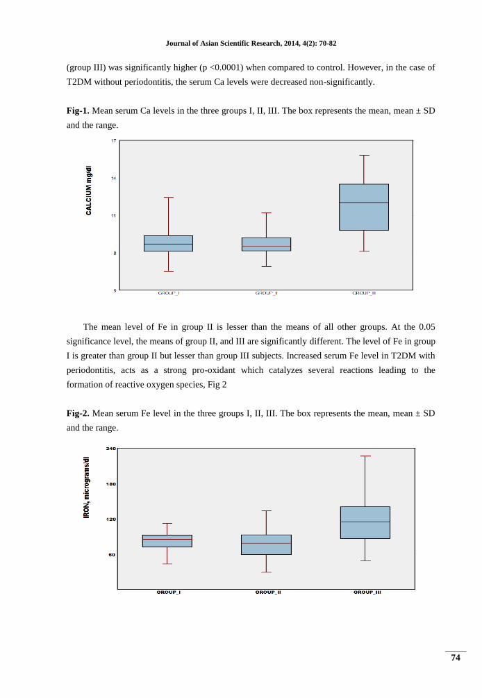
(group III) was significantly higher (p <0.0001) when compared to control. However, in the case of
T2DM without periodontitis, the serum Ca levels were decreased non-significantly.
Fig-1. Mean serum Ca levels in the three groups I, II, III. The box represents the mean, mean ± SD
and the range.
The mean level of Fe in group II is lesser than the means of all other groups. At the 0.05
significance level, the means of group II, and III are significantly different. The level of Fe in group
I is greater than group II but lesser than group III subjects. Increased serum Fe level in T2DM with
periodontitis, acts as a strong pro-oxidant which catalyzes several reactions leading to the
formation of reactive oxygen species, Fig 2
Fig-2. Mean serum Fe level in the three groups I, II, III. The box represents the mean, mean ± SD
and the range.
Journal of Asian Scientific Research, 2014, 4(2): 70-82
75
4.2. Pearson Correlation between Serum Ca and Fe with the Descriptive Parameters in the
Three Groups
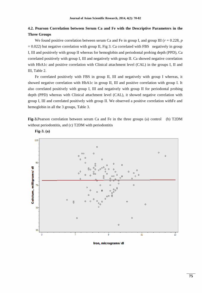
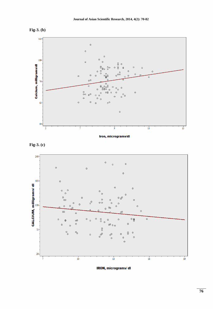
We found positive correlation between serum Ca and Fe in group I, and group III (r = 0.228, p
= 0.022) but negative correlation with group II, Fig 3. Ca correlated with FBS negatively in group
I, III and positively with group II whereas for hemoglobin and periodontal probing depth (PPD), Ca
correlated positively with group I, III and negatively with group II. Ca showed negative correlation
with HbA1c and positive correlation with Clinical attachment level (CAL) in the groups I, II and
III, Table 2.
Fe correlated positively with FBS in group II, III and negatively with group I whereas, it
showed negative correlation with HbA1c in group II, III and positive correlation with group I. It
also correlated positively with group I, III and negatively with group II for periodontal probing
depth (PPD) whereas with Clinical attachment level (CAL), it showed negative correlation with
group I, III and correlated positively with group II. We observed a positive correlation withFe and
hemoglobin in all the 3 groups, Table 3.
Fig-3.Pearson correlation between serum Ca and Fe in the three groups (a) control (b) T2DM
without periodontitis, and (c) T2DM with periodontitis
Fig-3. (a)
Journal of Asian Scientific Research, 2014, 4(2): 70-82
76
Fig-3. (b)
Fig-3. (c)
Journal of Asian Scientific Research, 2014, 4(2): 70-82
77
Table-2.Pearson correlation between Calcium and other independent variables involved in the
study in the 3 groups
Parameters Group I Group II Group III
HbA1c r
p
-0.022
0.83
-0.09
0.371
-0.253
0.011*
Hemoglobin r
p
0.040
0.693
-0.087
0.387
0.008
0.935
FBS r
p
-0.430
0.668
0.199
0.047*
-0.086
0.397
PPD r
p
0.003
0.977
0.000
0.997
-0.121
0.231
CAL r
p
0.018
0.86
0.16
0.112
0.136
0.178
Glycosylated hemoglobin, HbA1c; Fasting blood glucose, FBG; Periodontal probing depth, PPD; Clinical attachment level,
CAL; Pearson’s coefficient, r; * significant p value.
Table-3.Pearson correlation between iron and other independent variables involved in the study in
the 3 groups
Parameters Group I Group II Group III
HbA1c r
p
0.002
0.982
-0.103
0.306
-0.05
0.620
Hemoglobin r
p
0.145
0.149
0.126
0.213
0.079
0.434
FBS r
p
-0.128
0.204
0.205
0.004*
0.229
0.022*
PPD r
p
0.024
0.813
-0.048
0.634
0.056
0.579
CAL r
p
-0.063
0.533
0.050
0.618
-0.183
0.069*
Glycosylated hemoglobin, HbA1c; Fasting blood glucose, FBG; Periodontal probing depth, PPD; Clinical attachment level,
CAL; Pearson’s coefficient, r; * significant p value.
5. DISCUSSIONS
Diabetes mellitus is a systemic disease with a number of major complications that often
adversely affects the quality and length of life, particularly as it relates to cardiovascular events and
sudden death. Many studies have reported a positive association between oral infection, coronary
heart disease, and diabetes mellitus. Micronutrients like Ca and Fe play an essential role in
regeneration, for coping with oxidative stress and for an adequate immune response. Hence, these
elements are essential for maintaining health throughout life. Micronutrients can cause diseases
through deficiency, imbalance, or toxicity. Studies have shown that elevated Ca and Fe levels may
be a contributing factor in many inflammatory conditions. The results of our study demonstrated
higher level of Ca and Fe in the serum of T2DM with periodontitis individuals. Periodontal
infection represents one of the diabetes-associated complications that may be involved in altering
vascular pathology in the diabetic patient. It is currently recognized that T2DM is associated with
Journal of Asian Scientific Research, 2014, 4(2): 70-82
78
systemic inflammation. Both animal and human studies have suggested a possible role of altered
micronutrient status in the pathogenesis of T2DM and its complications.
The interrelationships between diabetes and periodontal disease provide an example of
systemic disease (diabetes) predisposing to oral infection, and once that infection is established, the
oral infection may exacerbate systemic disease. In addition, it is also possible for oral infection to
predispose to systemic disease in the case of impaired glucose tolerance or as is evident in
increased prevalence of cardiovascular disease in the diabetic group [19, 20]. Glucose is the main
physiological stimulus for pancreatic beta cells to release insulin. Glucose increases cytosolic Ca2+
activity, which is a major trigger to promote the release of secretory granules. cAMP is one of the
central signalling molecules that has been identified to regulate release of various neurotransmitters
and hormones, including insulin from pancreatic beta cells [21, 22].
It is of interest to note that very high serum Ca concentration in T2DM with periodontitis
indicates a strong correlation found between the pocket depth and alveolar bone loss. Ca and
insulin are important for the formation of bone collagen [23, 24] and depletion of Ca bone mineral
content leads to alveolar bone loss, bone fractures, and tooth loss in periodontitis patients with
diabetes. More recently, there is accumulating evidence to suggest that altered Ca homeostasis may
also play a role in the development of T2DM with periodontitis. Ca is essential for insulin-
mediated intracellular processes in insulin-responsive tissues such as skeletal muscle and adipose
tissue [25] with a very narrow range of intracellular [Ca2+
] needed for optimal insulin-mediated
functions. Changes in intracellular [Ca2+
] in primary insulin target tissues may contribute peripheral
insulin resistance [26].
In order to understand cellular and molecular mechanisms, one must identify common
physiological changes associated with diabetes and periodontitis that produce a synergy or additive
effect when the conditions coexist. Accumulation of advanced glycation end products, due to the
chronic hyperglycemic state or diabetes, combined with the presence of infection/inflammation
may provide a viable explanation. Reactive oxygen species (ROS) can directly modulate both the
activity and expression of several key proteins involved in Ca2+
homeostasis. The primary means
whereby ROS affects protein function are via modifications of the sulfhydryl (SH) moieties within
cysteine residues leading to formation of disulfide bonds and/or oxidation of reduced glutathione
leading to S-glutathionylation and, the initial response to elevated ROS is elevated Ca2+
entry [27].
In addition, H2O2-induced decreases L-type Ca2+
channel activity may also partially
compensate for ROS-induced Ca2+
elevation in excitable cells [28]. This is particularly relevant in
muscle, where elevated ROS is a relatively common event during exercise [29]. A priming effect
delivered by AGE accumulation in the tissues coupled with resultant oxidative stress and an
infectious challenge such as bacterial endotoxin (lipopolysaccharide or LPS) could possibly
account for the accelerated periodontal disease often seen in patients with diabetes.
Serum Fe concentration is significantly higher in T2DM with periodontitis when compared
with other groups. It is well established that people with haemochromatosis, a genetic condition
that causes extremely high Fe levels in the body are at increased risk for developing diabetes. But a
new study suggests even a moderately elevated Fe levels may be associated with diabetes [30, 31].

Journal of Asian Scientific Research, 2014, 4(2): 70-82
79
Fe chelation has been shown to increase the expression of LPS-related genes which leads to
increased biofilm formation. The most studied periodontal pathogen, regarding the need and effects
of Fe on the bacterium, is likely P. gingivalis. In agreement with being an essential growth factor
and important cause of oxidative stress [32], Fe limitation up regulates the genes involved in Fe
uptake and down regulates the genes associated with the storage of Fe as well as the oxidative
stress response of P. gingivalis. High concentration of Fe might play an important role in
enhancement of growth and virulence of microorganisms of the subgingival plaque and the
initiation of active periodontitis.
In periodontitis, during the active phases of the tissue destruction, the periodontal tissue and
gingival fluid typically contain high levels of proinflammatory mediators such as interleukin (IL)-
1β [33]. The first-line host defense is initiated by the polymorphonuclear (PMN) leukocytes that
are recruited to the site by chemotactic factors. In the subgingival area, the inflammatory reaction
causes the release of reactive oxygen species (ROS), such as hydrogen peroxide (H2O2) and
superoxide (O2 •−), and cytokines from the host cells to destroy the bacteria. Because free iron
catalyses the formation of toxic free radicals from H2O2 and is essential for the function of the host
and the pathogenic bacteria, human organs have developed complex ways to limit the availability
of free Fe in the environment [32]. Thus, the environment that surrounds potential colonisers when
they enter a human host is suboptimal in terms of free Fe concentration.
Most of the host Fe is bound to Fe-binding proteins such as transferrin, ferritin, lactoferrin and
haemoglobin that contain haem. However, the situation may change during the progression of
periodontitis. It has been hypothesised that the concentration of Fe may increase due to the
increasing concentration of haemoglobin leaking from vascular ulcers of the gingival pocket. Some
periodontal pathogens, such as P. gingivalis, T. denticola and A. actinomycetemcomitans, have
been demonstrated to express haem-binding proteins on their surfaces [34] that may facilitate Fe
acquisition in free Fe-limited conditions.
Severe chronic forms of periodontitis can result in systemic response to the bacteria and
bacterial products that are disseminated due to breakdown of the periodontal apparatus in the oral
cavity. Tonetti, et al. [35] concluded that intensive periodontal treatment resulted in acute, short-
term systemic inflammation and endothelial dysfunction. In addition, periodontal disease has been
shown to be a strong predictor of mortality from ischemic heart disease and diabetic nephropathy in
Pima Indians with type 2 diabetes. This effect was found to be independent of the effects of
traditional risk factors [20]. Elevated Ca and Fe level in the serum are associated with an increased
alveolar bone loss and oxidative stress which can predispose an individual to the risk of developing
periodontitis leading to various diabetes complications including periodontitis.
Our study cannot support the hypothesis that calcium deficiency is a main cause of destructive
periodontal disease. Excessive Ca and Fe in serum may promote development and progression of
oxidative stress, altered immunity and altered insulin secretion or its action. An abundance of
information accumulated from studies on the complication of diabetes and periodontal disease have
revealed that altered immune response may be antecedent of both diseases, which may have a
synergistic effect when they coexist in the host.

Journal of Asian Scientific Research, 2014, 4(2): 70-82
80
6. CONCLUSIONS
Periodontal diseases are inflammatory in nature; as such, they may alter glycemic control in a
similar manner to obesity, another inflammatory condition. Oxidative stress plays an important role
in the development of diabetes complications, both microvascular and cardiovascular. Minerals in
the blood will show a reduction or exaggeration according to the intensity of pathogenicity. This
study has shown increased serum Ca and Fe level in T2DM with periodontitis which emphasis on
the inflammatory nature of periodontal diseases and the potential systemic effects of periodontal
infection.
REFERENCES
[1] D. Noda, T. Hamachi, K. Inoue and K. Maeda, "Relationship between the presence of
periodontopathic bacteria and the expression of chemokine receptor MRNA in inflamed gingival
tissues," J Periodontal Res., vol. 42, pp. 566-571, 2007.
[2] M. H. Mohammad, M. G. D. Azmi and M. I. Majdy, "The effect of glycemic control on Candida
colonization of the tongue and the subgingival plaque in patients with type II diabetes and
periodontitis," Oral Surgery, Oral Medicine, Oral Pathology and Oral Radiology, vol. 116, pp. 321-
326, 2013.
[3] R. S. Vanessa, A. L. Jadson, S. M. Tamires and F. Magda, "Relationship between glycemic subsets
and generalized chronic periodontitis in type 2 diabetic Brazilian subjects," Archives of Oral
Biology, vol. 57, pp. 293-299, 2012.
[4] G. Taylor, "Bidirectional interrelationships between diabetes and periodontal diseases: An
epidemiologic perspective," Ann Periodontol, vol. 6, pp. 99–112, 2012.
[5] K. Jemin and A. Salomon, "Periodontal disease and systemic conditions: A bidirectional
relationship," Odontology, vol. 94, pp. 10–21, 2006.
[6] L. Xiaojing, M. K. Kristin, T. Leif and O. Ingar, "Systemic diseases caused by oral infection," Clin
Microbiol Rev., vol. 13, pp. 547–558, 2000.
[7] J. Monik, B. H. Frank, M. Miguel, L. Yi and J. J. Kaumudi, "Type 2 diabetes mellitus and 20 year
incidence of periodontitis and tooth loss," Diabetes Research and Clinical Practice, vol. 98, pp.
494-500, 2012.
[8] C. Po-Chun and P. L. Lum, "Interrelationships of periodontitis and diabetes: A review of the current
literature," Journal of Dental Sciences, vol. 7, pp. 272-282, 2012.
[9] S. R. Neelimak, M. K. Ramesh, G. C. Viren and H. M. Nilkanth, "A clinical study of the
relationship between diabetes mellitus and periodontal disease," Journal of Indian Society of
Periodontology, vol. 15, pp. 388-392, 2011.
[10] S. Edwards, "Maintaining calcium balance: Physiology and implications," Nursing times, vol. 101,
pp. 58-61, 2005.
[11] L. Robinson, H. Blair and J. Barnett, "Regulation of bone turnover by calcium-regulated calcium
channels," Annals of the NewYork Academy of Sciences, vol. 1192, pp. 351-357, 2010.
[12] J. Fernandez-Real, A. Lopez-Bermejo and W. Ricart, "Cross-talk between iron metabolism and
diabetes," Diabetes, vol. 51, pp. 2348-2354, 2002.
Journal of Asian Scientific Research, 2014, 4(2): 70-82
81
[13] O. Teresa, S. Waltena, L. Xinyan and A. G. Caroline, "Iron and heme utilization in Porphyromonas
gingivalis," FEMS Microbiology Reviews, vol. 29, pp. 119-144, 2005.
[14] B. M. Janet and S. H. B. David, "Anemia and the role of erythropoietin in diabetes," Journal of
Diabetes and its Complications, vol. 20, pp. 262-272, 2006.
[15] C. Hernandez, J. Genesca, J. Ignasi Esteban, L. Garcia and R. Simo, "Relationship between iron
stores and diabetes mellitus in patients infected by hepatitis C virus: A case-control study," Med Clin
(Barc)., vol. 15, pp. 21-22, 2000.
[16] B. Pierre, R. Martine, L. L. Caroline and L. Olivier, "Non-transferrin bound iron: A key role in iron
overload and iron toxicity," Biochimica et Biophysica Acta (BBA) - General Subjects, vol. 1820, pp.
403-410, 2012.
[17] A. S. Christine, "Iron intake and regulation: implications for iron deficiency and iron overload,"
Alcohol, vol. 30, pp. 99-102, 2003.
[18] W. Ramsay, "Determination of serum iron," Biochem. J., vol. 57, p. 17, 1954.
[19] E. Lalla, I. Lamster, S. Drury, C. Fu and A. Schmidt, "Hyperglycemia, Glycoxidation and receptor
for advanced glycation endproducts: Potential mechanisms underlying diabetic complications,
Including diabetes-associated periodontitis," Periodontol, vol. 23, pp. 50–62, 2000.
[20] A. Saremi, R. Nelson and M. Tulloch-Reid, "Periodontal disease and mortality in type 2 diabetes,"
Diabetes Care, vol. 28, pp. 27–32, 2005.
[21] E. Renstrom, L. Eliasson and P. Rorsman, "Protein kinase A-dependent and independent stimulation
of exocytosis by CAMP in mouse pancreatic B-cells," J. Physiol, vol. 502, pp. 105–118, 1997.
[22] R. Burgoyne and A. Morgan, "Secretory granule exocytosis," Physiol. Rev., vol. 83, pp. 581–632,
2003.
[23] E. Canalis, J. Dietrich, D. Maina and L. Raisz, "Hormonal control of bone collagen synthesis in
vitro- effects of insulin and glucagon," Endocrinology, vol. 100, pp. 668-74, 1977.
[24] I. Dickson, D. Eyre and E. Kodicek, "Influence of plasma calcium and vitamin D on bone collagen.
Effects of lysin hydroxylation and crosslink formation," Biochim Biophys Acta., vol. 588, pp. 169-
73, 1979.
[25] D. Wright, K. Hucker, J. Holloszy and D. Han, "Ca2+ and AMPK both mediate stimulation of
glucose transport by muscle contractions," Diabetes, vol. 53, pp. 330–335, 2004.
[26] S. Segal, S. Lloyd, N. Sherman, K. Sussman and B. Draznin, "Postprandial changes in cytosolic free
calcium and glucose uptake in adipocytes in obesity and non-insulin-dependent diabetes mellitus,"
Horm Res., vol. 34, pp. 39–44, 1990.
[27] B. Hawkins, K. Irrinki, K. Mallilankaraman and Y. Lien, "S-glutathionylation activates STIM1 and
alters mitochondrial homeostasis," J. Cell. Biol., vol. 190, pp. 391–405, 2010.
[28] H. Matsuura and M. Shattock, "Membrane potential fluctuations and transient inward currents
induced by reactive oxygen intermediates in isolated rabbit ventricular cells," Circ. Res., vol. 68, pp.
319–329, 1991.
[29] W. Aoi, Y. Naito, Y. Takanami and Y. Kawai, "Oxidative stress and delayed-onset muscle damage
after exercise," Free Radic. Biol. Med., vol. 37, pp. 480–487, 2004.
Journal of Asian Scientific Research, 2014, 4(2): 70-82
82
[30] T. Buchanan, A. Xiang, R. Peters, S. Kjos and K. Berkowitz, "Response of pancreatic beta-cells to
improved insulin sensitivity in women at high risk for type 2 diabetes," Diabetes, vol. 49, pp. 782-
788, 2000.
[31] A. Helin, T. Kinnunen, J. Raitanen, S. Ahonen and S. Virtanen, "Iron intake, haemoglobin and risk
of gestational diabetes: A prospective cohort study," BMJ Open, vol. 2, 2012.
[32] J. Lewis, "Metal uptake in host-pathogen interactions: Role of iron in porphyromonas gingivalis
interactions with host organisms," Periodontol 2000, vol. 52, pp. 94–116, 2010.
[33] B. Rescala, W. Rosalem and R. Teles, "Immunologic and microbiologic profiles of chronic and
aggressive periodontitis subjects," J. Periodontol, vol. 81, pp. 1308–1316, 2010.
[34] E. Rhodes, S. Menke and C. Shoemaker, "Iron acquisition in the dental pathogen Actinobacillus
actinomycetemcomitans: What does it use as a source and how does it get this essential metal?,"
Biometals, vol. 20, pp. 365–377, 2007.
[35] M. Tonetti, F. D’Aiuto and L. Nibali, "Treatment of periodontitis and endothelialfunction," N Engl J
Med., vol. 356, pp. 911–920, 2007.


![Possible Correlation between INR and Serum Calcium · T. A. Helin et al. 1183 prothrombin time, and vitro in duodenal preparations reduced calcium absorption. Our recent workin [25]](https://img.dokumen.tips/doc/110x75/5cab498688c99319398cf413/possible-correlation-between-inr-and-serum-calcium-t-a-helin-et-al-1183-prothrombin.jpg)