Embed Size (px)
Citation preview

4000 Jones Bridge Road Chevy Chase, Maryland 20815-6789 www.hhmi.org
nonprofit org.us postagepaidHyattsville, mdpermit no. 61
HH
MI B
UL
LE
TIN
• Ho
wa
rd H
ug
he
s M
ed
ica
l Institu
te • ww
w.h
hm
i.org
vo
l. 19
/ n
o. 0
4
Pho
tota
ke/©
Jea
n-C
laud
e R
evy
Change Service Requested
N o v. ’0 6 vo l . 1 9 • N o. 0 4
HHMI BULLETIN
Although thiS SmAll, weedy plAnt hAS little AgRiCultuRAl vAlue,
it hAS huge fAnS Among plAnt biologiStS And genetiCiStS (See pAge
14). ArAbidopsis thAliAnA iS vAlued foR itS eASe of gRowth (heRe it
gRowS without Soil in A petRi diSh) And SmAll genome. At leASt 139
of itS geneS ARe SimilAR to humAn geneS linked to diSeASeS SuCh AS
heReditARy deAfneSS, blindneSS, And CAnCeR.
superhero of the plant World
plants are UsA shared biochemistry makes
plants a beautiful model for studying human health and disease.
*In thIs Issue
Sperm Secrets/A Parent’s Perspective Drosophila Envy

A close-up view within a focal adhesion— a footprint of a cell—as seen by overlaying images from a conventional microscope (blue) and images from the new photoactivated localization microscope (magenta) developed by Janelia Farm scientists Eric Betzig and Harald Hess and their colleagues.
12
Imag
e co
urte
sy o
f Eric
Bet
zig
and
Har
ald
Hes
s (H
HM
I) a
nd M
icha
el D
avid
son
(Flo
rida
Stat
e U
nive
rsity
)

vol. 19 november ’o6 no. o4D E P A R T M E N T S
V I S I T T H E B U L L E T I N O N L I N E F O R A D D I T I O N A L C O N T E N T A N D A D D E D F E AT U R E S : http:// www.hhmi.org/bulletin C O V E R I M A G E : A N I TA K U N Z
P R E S I D E N T ’ S L E T T E R
03 A Transforming Energy
C E N T R I F U G E
04 A Cast of Thousands 05 Dreams About Science and Hoops in America 06 A Family Grows in Baltimore
U P F R O N T
08 When Sperm Give Up Their Secrets 10 Yeast for Thought 12 Sharper Image
P E R S P E C T I V E S A N D O P I N I O N S
38 Rafael Viñoly 40 Monica Coenraads 42 Q&A – What challenges do young scientists face today?
C H R O N I C L E
J a n e l i a F a r m 44 A New Scientific Community 46 Let the Science Begin 48 Janelia Farm Team Grows 49 Search Is On for Janelia’s First Grad Students I n s t i t u t e N e w s 50 HHMI Broadens Scientific Leadership Briggs and McCleskey Join Institute 51 Latin American, Canadian Science Stars Awarded $19 Million S c i e n c e E d u c a t i o n 52 Expanding Her Horizons 53 The FARM Project: A Summer Science Workshop L a b B o o k 54 A Sweet Solution to a Sticky Problem 55 Sugar Code-Busters 56 Monkey Feel, Monkey Do I n M e m o r i a m 57 David L. Garbers N o t a B e n e 58 News of recent awards and other notable achievements
O B S E R V A T I O N S
On Creativity
F E A T U R E S
Plants Are Us Scientists are gaining critical knowledge of human biology from the study of—no kidding—plants. [ C OV E R S T O R Y ]
Man’s Best ModelIt may not seem strange that dogs can enlighten us about behaviors deemed uniquely human. But apparently, so can other animals: mice, fish, flies—even snails.
Learning From PatientsPatients have much to learn from scientific advances. Often, though, it’s also the scientists who benefit from the patients.
Drosophila EnvyA maverick scientist is risking all to study the behavior of fruit flies, convinced that they have much to teach us about emotions.
20 26 32

H H M I T R U S T E E S
James A. Baker, III, Esq.Senior Partner / Baker & Botts
Richard G. DarmanPartner / The Carlyle Group Chairman of the Board / AES Corp.
Frank William GayFormer President & CEO / SUMMA Corporation
Joseph L. Goldstein, M.D.Professor & Chairman, Department of Molecular Genetics University of Texas Southwestern Medical Center at Dallas
Hanna H. Gray, Ph.D., ChairmanPresident Emeritus & Harry Pratt Judson Distinguished Service Professor of History The University of Chicago
Garnett L. KeithSeaBridge Investment Advisors, L.L.C. Former Vice Chairman & Chief Investment Officer The Prudential Insurance Company of America
Jeremy R. Knowles, D.Phil.Dean Emeritus & Amory Houghton Professor of Chemistry & Biochemistry / Harvard University
William R. Lummis, Esq.Former Chairman of the Board of Directors & CEO The Howard Hughes Corporation
Paul M. Nurse, Ph.D.President / The Rockefeller University
Kurt L. Schmoke, Esq.Dean / Howard University School of Law
Anne M. TatlockChairman & CEO / Fiduciary Trust Company International
H H M I O F F I C E R S & S E n I O R a d v I S O R S
Thomas R. Cech, Ph.D. / PresidentCraig A. Alexander / V.P. & General Counsel
Peter J. Bruns, Ph.D. / V.P. for Grants & Special Programs
David A. Clayton, Ph.D. / V.P. & Chief Scientific Officer Joseph D. Collins / V.P. for Information Technology
Joan S. Leonard, Esq. / Senior Counsel to the President
Avice A. Meehan / V.P. for Communications & Public Affairs
Edward J. Palmerino / V.P. for Finance & Treasurer Gerald M. Rubin, Ph.D. / V.P. & Director, Janelia Farm Research Campus
Landis Zimmerman / V.P. & Chief Investment Officer
H H M I B U L L E T I n S Ta F F
Mary Beth Gardiner / Editor Cori Vanchieri / Story Editor Jim Keeley / Science Editor Jennifer Donovan / Education Editor
Patricia Foster / Associate Director of Communications for Web & Special ProjectsJacqueline Ruttimann / Assistant EditorDean Trackman, Maya Pines / Contributing Editors
A D D I T I o N A L C o N T R I B u T o R S
Cay Butler, Michelle Cissell, Nicole Kresge,
Steven Marcus, Heather McDonald, Kathy SavoryVSA Partners, NYC / Concept & Design
Phot
os c
ourt
esy
of M
icha
el M
ason
, A
lici
a A
ult,
and
Jac
quel
ine
Rut
tim
ann.
D
raw
ing
by P
eter
Ark
le.
Telephone (301) 215 .8855 • Fax (301) 215 .8863 • www.hhmi .org
©2006 Howard Hughes Medical Inst i tute
The opinions, beliefs, and viewpoints expressed by authors in the
HHMI Bulletin do not necessarily reflect the opinions, beliefs, viewpoints,
or official policies of the Howard Hughes Medical Institute.
2 h h m i b u l l e t i n | November 2006
contributors
Michael Mason has reported for The New York Times, Newsweek, Health, National Geographic, and a wide variety of other national publications. When not at the computer keyboard, he’s camping in the Sierras, where cell phone coverage is blessedly spotty and e-mail is nonexistent. He lives in San Fran-cisco with his partner and their obese Corgi, who strenuously objects to camping. (1)
For two decades, alicia ault has written about health and medicine, covering everythingfrom Nobel Prize winners to FDA politics to organ transplant techniques. As a freelancer, she’s covered multisport adventure races in Borneo, bird preserves in Louisiana, Jamaican hideaways, and pop culture fads (for example, custom-sized electronics for boats, motorcycles, and golf carts). Her stories have appeared in The New York Times and the Washington Post. (2)
Peter arkle grew up ten miles south of Edinburgh, Scotland, in the town of Penicuik. He moved to London in 1987 to attend The Royal College of Art and in 1995 relocated to New York City, where he and his wife, Amy, now reside. Arkle writes and illustrates a regular column, The Stalker, for Print Magazine, and draws all sorts of odd things for Food and Wine Magazine, The New York Times, and a variety of other clients, from hair care companies to law firms. In his spare time, he enjoys reading about science, particularly insects, which he likes to draw or paint whenever he gets the chance. (3)
After two tours of graduate school, one for a doctorate in tumor biology from Georgetown university, the other for a master’s in journalism from the university of Maryland at College Park, Jacqueline ruttiMann is happily settling in as assistant editor of the HHMI Bulletin. Since trading the lab bench for the news desk, she has written for Nature, FoxNews.com, and WTOP. (4)
(3)
(1)
(4)
(2)

“Janelia...will rely on creative, daring individuals given the freedom and support to explore the big scientific problems of our time.
thomas cech
At university And college cAmpuses Around the
country, early autumn brings the energy of new beginnings sparked by enthusiastic students, fresh intellectual challenges, and renewed collegial interactions. Measured against this standard, HHMI began the season in a particularly dynamic and eventful fashion. I’d even call it joyful.
After nearly seven years of thinking and planning—concurrent with four solid years of construction—the Institute opened its Janelia Farm Research Campus. With laboratories and offices still filled with moving boxes and equipment crates, Director Gerry Rubin and his colleagues hosted their first meeting of HHMI inves-tigators immediately after Labor Day. Everyone lived to tell the tale—as the saying goes—and the Janelia team immediately began readying the campus for the biennial gathering of HHMI’s International Research Scholars, who came from every continent save Antarctica. It’s fair to say that these inaugural meetings will be remembered for years to come, both for their scientific content and for the palpable sense of excitement generated by Janelia Farm.
The week of activities that marked the official opening of Janelia Farm began on an extraordinary note with the announce-ment of the 2006 Nobel Prize in Physiology or Medicine. Craig Mello, an HHMI investigator at the University of Massachusetts Medical School in Worcester, received the early-morning phone call from Sweden along with his colleague, Andrew Fire, who was at the Carnegie Institution in Baltimore when the two collaborated. Their discovery, called RNA interference (RNAi), is a normal regulatory mechanism that allows cells to shut down individual genes during embryonic development. What really made RNAi famous was its utility as a research tool. In less than a decade, it became widely used to selectively silence specific genes in plants and animals. Several companies are exploring its potential in new therapeutic approaches. Because most human ailments, from viral infections to cancer and immunological diseases, involve specific RNA molecules, the list of possible applications is long.
Mello and Fire (now at Stanford University School of Medicine) wanted to find a better way to block gene expression during embry-onic development. So they inserted RNA into their model organism, the microscopic roundworm Caenorhabditis elegans—a blocking maneuver that worked all too well. Interference spread from cell to cell, from generation to generation. Undeterred by these puzzling results, the two scientists persisted in their efforts to understand what they were seeing. They determined that the active agent was double-stranded RNA—a piece of the messenger and its pairing partner. It was a fundamental, transforming insight.
The path to discovery followed by Mello and Fire offers a frame-work for thinking about science and for the evolving research community that is taking shape at Janelia Farm. We intend Janelia Farm to be a place that fosters risk taking and collaboration. To help
shape its scientific culture, we studied salient historical models, among them Bell Labs in Murray Hill, New Jersey, and England’s Laboratory of Molecular Biology. The initial areas of research have been identified: understanding how the brain processes information and inventing imaging technologies and computational methods for image analysis. Scientists have been recruited. The first laboratories are up and func-tioning. An ambitious program of meetings and workshops has been scheduled. Graduate students will soon be identified through our joint program with the University of Cambridge and the University of Chicago. In short, this experiment is starting to take on a life of its own.
As Janelia Farm opens for discovery, it is impossible to predict what will unfold in the coming years. Will this vision—that physicists, computer scientists, engineers, and chemists can work shoulder-to-shoulder with biologists to explore the frontiers of biology—be as robust as we claim? We have talked about risk, but will the risk pay off in terms of discoveries? These are heady questions given the success of HHMI’s existing approach to supporting biomedical research. Our investiga-tors—as Craig Mello illustrates—are leaders in discovery. Janelia is a different model, with different expectations, but it too will rely on creative, daring individuals given the freedom and support to explore the big scientific problems of our time. As the Janelia scientists set down roots and begin their work throughout this fall season, we await, with great anticipation, the fresh new ideas, new approaches, and new findings that no doubt will arise this spring and in the years to come.
A Transforming Energy
Paul
Fet
ters
�November 2006 | h h m i b u l l e t i n
president’s letter
”

A Cast of ThousandsJose Izquierdo is not a medical researcher, but he contributes to studies that one day could provide cures for Alzheimer’s disease, cancer, malaria, and HIV/AIDS. Izquierdo, a retiree in Ponce, Puerto Rico, is one of the more than 75,000 “armchair scientists” involved in a project called Rosetta@home, led by HHMI investigator David Baker at the University of Washington, Seattle. Rosetta enlists ordinary people who, whether sitting at their computers or not, allow their machines’ otherwise-dormant processing power to help predict the three-dimensional shapes of proteins—keystone to understanding disease and designing targeted drugs.
When Izquierdo became involved, he was physically ill and suffering from severe depression. “My spirit was ready to give up,” he says. “Then I realized
that my little computer could join me with so many people who were really doing something.” Now he devotes as much passion to conducting his piece of the research as he gives to cultivating award-winning orchids—Izquierdo once transported a single cut flower in a Samsonite from his Puerto Rico home to a competition in Philadelphia.
Rosetta relies on “distributed computing,” a process that takes a massive calculation and breaks it into chunks to be sent across the Internet and processed by home computers worldwide. While the Izquierdo “lab” crunches numbers, the computer of Thomas Dooley, for instance, way up in Alaska, does likewise. Kathryn Marks’s computer ran Rosetta virtually full time while she was teaching English in South Korea. Also not a white-coat type, she would “check in” every evening while in her “jammies, listening to country music.”
Participants are organized into far-fl ung teams, and Izquierdo is the statistician of his team, meaning he keeps track of “credits” earned by each volunteer. The team, called Xtreme-Systems, is one of Rosetta’s highest performers. Its 500-plus members worldwide include “grandmothers, kids, rich people, poor people, Ph.D.s, and high school dropouts,” says Izquierdo.
Volunteers pride themselves on their team’s clever monikers, such as Dutch Power Cows, The Knights Who Say Ni!, and The Final Front Ear. Some even name their computers, with “Clark Kent” reputed to be the most powerful machine.
Rosetta participants admit to being both obsessive and competitive. “Credits essentially have no value except for bragging rights, which do happen to motivate a lot of people,” says volunteer Mark Pottorff. Some participants “crunch” nonstop, and several have invested in auxiliary machines just to contribute to the cause.
Darin Steffl , a Minnesota high school student who runs his own computer
“My spirit was ready to give up.
Then I realized that my lit t le
computer could join me with so
many people who were really doing
something. ”JOSE IZQUIERDO
4 H H M I B U L L E T I N | November 2006
Pete
r A
rkle
centrifuge

business, uses seven PCs for Rosetta. “Most people’s computers are just sitting there doing nothing,” he says. “Why not have it do something useful?” Taking that notion even further, one Team XtremeSystems member in Hong Kong devotes hundreds of computers from his business to running Rosetta full time. “We call it ‘The Farm,’” Izquierdo says.
Many volunteers were drawn to Baker’s program by the loss of a loved one. Marc Abrams, a businessman from New York, explains, “My mother died of cancer when I was 18, and my wife died of melanoma fi ve years ago. This is a way for me to help solve the mysteries of those diseases.”
No matter what motivates Rosetta volunteers, almost all of them cite their camaraderie with other participants as a main attraction. Many chat with one another on a daily basis, using the Rosetta message boards. “But we are not nerds,” Izquierdo maintains. “We trade barbs and jokes. We can discuss anything from how to improve the computer to how to make an easy-bake cake.”
They also cite Baker’s accessibility as a factor that drives their zeal. “Dr. Baker and his team take the time to explain what they’re doing. Even if you don’t always understand the science, you’re made to feel like part of a family,” says Izquierdo. “When David Baker calls for help, it will come, because people have such loyalty toward him. He’s kind of like a cult fi gure.” —Lindsay Moran
Growing up in East Germany when it was still a stalwart of the Communist bloc, Dorothee Kern was an outstanding student with a passion for science. But what she really had going for her was a talent for sports. The government was strenuously trying to identify potential athletic superstars who could put this small nation on the map. “The offi cials went to all the schools,” says Kern, now a biochemist at Brandeis University and a newly minted HHMI investigator. “In second and third grade they measured your fi ngers and toes, asked how tall your parents were. They were trying to see how the genes fell and look for athletic talents.” Young swimmers, track and fi eld athletes, gymnasts, and others with Olympic potential were intensively trained in special schools—almost to the exclusion of academic studies. And as became well known after German reunifi cation, many were given steroids, growth hormones, and other performance-enhancing drugs, with often-tragic medical consequences. “You were not a free person,” says Kern. When she was in fi fth grade, Kern was chosen for a special sports school in swimming. But her father, wary of a perilous future for his daughter, said no. Instead, she chose basketball, also one of her early loves. “Basketball wasn’t an Olympic sport for East German athletes, so I could go to a top academic school instead and wasn’t pressured to take drugs.” By the age of 12, Kern was playing on the junior national team—her entry ticket for high school. Despite receiving top scores in middle school, Kern was not nominated for high school because her family was not “in line” with the communistic society. However, playing on the national team was her way to “support” the country, opening the door to high school followed by college at the Martin-Luther University in Halle. Sports continued to be a major part of her school life during her biochemistry studies at the university, where Kern juggled academic work with travels for the basketball team. She persisted in her studies and sport alike, ultimately earning a Ph.D. and captaining the German national basketball team for 8 years. Nevertheless, the repressive government and hidebound structure of the East German science establishment rankled her, as they did so many of her colleagues. It was good-bye to all that when the Wall came down in 1989. “That was big,” she says. “You could take your life in your own hands, go to another country, have freedom of speech.” When the opportunity came up, she quit pro basketball and accepted a post-doctoral fellowship at the University of California, Berkeley, where she loved the scientifi c environment and the scrappy style of American pickup basketball. In 1998, she moved to Waltham, Massachusetts, just outside Boston, to take a tenured position at Brandeis. Her husband is a scientist at a major pharmaceutical company, and her daughters, who attend Waltham public schools, are outdoorsy and athletic and want to become scientists. On a recent summer day, they bounced into Kern’s offi ce ready to play basketball with mom on her lunch hour. “One of the best things in life is to have big dreams,” she says refl ectively. “Mine was to become a professor in science, to have a family, to play basketball, and live in America. And it has all come true.” —Richard Saltus
5November 2006 | H H M I B U L L E T I N
Jaso
n G
row
Dreams About Science and Hoops in America
F O R M O R E I N F O R M A T I O N : To learn more about Rosetta@home, visit http://boinc.bakerlab.org/rosetta/.

A Family Grows in Baltimore
When it comes to cats, says Nancy L. Craig, three is the magic number—the most that a person may bring home before eyebrows start being raised.
Craig once stepped into an elevator only to have a woman nearby spy tell-tale tufts of fur clinging to her black wool coat and ask, “How many cats do you have?” “Five,” Craig replied, bracing for The Look. “When you tell people you have two or three cats, they understand and think you just love animals. There’s a distinct change in attitude when you tell them you have more than that,” Craig laughs.
Craig, an HHMI investigator at the Johns Hopkins University School of Medicine, focuses on the intricacies of mobile bits of DNA called transposons that hop about the genomes of all organisms, from bacteria to humans.
A lifelong cat lover, Craig started her current brood innocently in 1997 when she found herself without a pet. She launched the effort to fi ll that void, like any dedicated researcher, by gathering as much information as possible—in this case, by visiting the rarifi ed world of cat shows.
Amid the spectacle of pampered felines resting in opulent cages, Craig discovered the large, regal Maine coon. The breed’s name arises from the biologically impossible legend that it
resulted from the mating of semiwild cats and raccoons—a myth bolstered by the breed’s bushy tail and raccoon-like coloring. Long-haired with tufted ears and leonine “ruffs” around their necks, these cats, which can reach nose-to-tail lengths of 48 inches, are so smart, easily trained, and people-oriented that they’re often called “doglike.” The big, amiable felines fi t the statuesque Craig, who notes, “I am not a girl who needs a small cat.”
Craig’s search for a Maine coon to call her own resulted in Rufus, a one-year-old classic brown tabby from a Maryland breeder. Craig then adopted a red tabby Maine coon kitten she named Dexter. Her third cat, a short-haired jet-black cat named Zuzu,
joined the clan after she saw him in a pet rescue center.
Though content with her three cats, Craig saw an advertisement one day in the Baltimore Sun for a Maine coon in need of rescue. Seeing it as “a sign” she rushed to the cat’s aid only to fi nd she was too late. Instead, Craig adopted a gray mixed-breed named Granger.
Happy as one of three, however, Dexter became irritable, territorial, and destructive with the addition of Granger. Reluctantly, Craig found the newcomer a one-cat home where he is much happier.
Despite Dexter’s resistance, Craig decided she wanted Maine coon kittens. Her quest ended 750 miles away in Owensboro, Kentucky, at the Verismo Cattery. In order to take Ajax—a cameo, or light red, kitten—and Oliver—a silver tabby—home with her, she had to promise owner Tom Odom that she would keep the top up on her BMW Z4 convertible.
With fi ve cats, Craig began to accept the ribbing and questioning from friends, and an occasional aghast stranger. The menagerie expanded further earlier this year after Craig and her partner Helen McComas were married. McComas brought to the household her aging shorthaired cat, Kitty, and sweet-tempered keeshond, Eli.
Despite her passion for her cats, Craig has no intention of entering the show world. She and McComas are satisfi ed just living with their ample animal family. —Lisa Chiu
“ I am not a girl who needs a small cat. ”NANCY CRAIG
When it comes to cats, says Nancy L. Craig, three is the magic number—the
6 H H M I B U L L E T I N | November 2006
Illu
stra
tion
: Pe
ter
Ark
le
Phot
o: P
aul
Fett
ers
centrifuge

7November 2006 | h h m i b u l l e t i n
upfront
Scientists are rarely satisfied with an incomplete view of how things work in the body. They always want a more intimate picture of the inside of the cell. The HHMI researchers who aimed at two very different targets and achieved unprecedented clarity are no exception. A team of microscopists now based at Janelia Farm has devised a technique to produce ultrasharp snapshots of proteins within cells. Other HHMI scientists overcame technological barriers to determine how sperm abruptly convert their tail motion from a steady undulation to the whip-cracking snap necessary to thrust into an egg. Can a new male contraceptive be far behind?
08 When Sperm Give Up Their Secrets
A new window on sperm cells’ ion channels could ultimately help control populations, human and otherwise.
10 Yeast for Thought
First in yeast, and then in laboratory creatures with nerve cells, researchers have discovered how the Parkinson’s disease process may be curtailed.
12 Sharper Image
No consumer toy, here is a researcher’s dream—a light microscope that goes where none has gone before, down to the level of individual protein molecules.

� h h m i b u l l e t i n | November 2006
upfront
Few biological images have worked their way into the popular imagination
more successfully than the sperm cell. You needn’t have ever opened a science
textbook to recognize that formidable little swimmer propelled by a tadpole-like
tail. ¶ But until recently, imagination (as opposed to genuine knowledge) was
all scientists could bring to bear on the understanding of spermatozoa, because
the intimate details of their lives were hidden within “ion channels”—networks
of pore-forming proteins that help regulate the cells’ electrochemical activity.
While standard laboratory techniques used to eavesdrop on ion-channel
functioning have worked fine for most other types of cells, sperm cells proved
When Sperm Give Up Their Secrets A new window on sperm cells’ ion channels could ultimately
help control populations, human and otherwise.
resistant—until a research team led by HHMI investigator David E. Clapham, a neurobiologist at Harvard Medical School, found a way around the problem.
In 2001, Clapham discovered a partic-ular ion channel, which he named CatSper1, unique to the sperm’s tail. This channel appeared to be necessary for the sperm to enter into “hyperactivation,” the process that enables it to penetrate the egg.
Young, immature sperm cells tend to swim in an orderly, highly symmetrical pattern. But as they travel farther into the vaginal canal and enter the alkaline envi-ronment housing the egg, their pattern changes. As hyperactivation begins, the sperm’s swimming pattern becomes less symmetrical. The tail assumes a whip-like motion, enabling it to strike the egg’s surface with greater force. When Clapham “knocked out”—that is, inactivated—the gene for CatSper1 in mouse sperm cells, 100 percent of the mice became infertile.
Normally, when researchers want a closer look at an ion channel of interest,
they use a technique called patch-clamp recording. In this routine procedure, an electrode provides a window through the membrane into the cell’s machinations. In addition, researchers can introduce various chemicals and substrates through the probe to manipulate and study the cell’s properties.
“We tried this [patch-clamp] tech-nique over a thousand times with sperm cells, and failed at every attempt,” says Clapham. “We found that sperm cells aren’t amenable to it.” This may result, at least in part, from their rigid membrane and constant wriggling.
Eventually, Yuriy Kirichok, a postdoc-toral fellow in Clapham’s lab, discovered that the sperm’s cytoplasmic droplet, a remnant of the precursor germ cell cyto-plasm, was the ideal entry point for the patch-clamp probe, providing a way around sperm’s tough skin and unruly movements. Then, Clapham and his team found that CatSper1 enables calcium to enter into the sperm, which changes the swimming pattern into the whip-like motion needed for penetrating the egg. “This has opened up a whole new pathway for probing these electrical currents and finding out exactly what they do,” says Clapham. “I feel like we’ve discovered the entrance into an ancient pyramid that no one has ever been inside of before.”
“I feel l ike we’ve discovered the
entrance into an ancient pyramid
that no one has ever been inside
of before. ”DaviD Clapham
John
Bri
dges

�November 2006 | h h m i b u l l e t i n
Illu
stra
tion
: Je
ffre
y D
ecos
ter
Pho
to:
UT
Sou
thw
este
rn N
ews
& P
ubli
cati
ons
The researchers published their findings in the February 9, 2006, issue of Nature.
At roughly the same time that Clapham identified CatSper1, David L. Garbers, an HHMI investigator at the University of Texas Southwestern Medical Center at Dallas, discovered two other sperm-specific proteins that affect fertility: CatSper2 and sNHE. Mice in which CatSper2 was knocked out had much the same profile as Clapham’s CatSper1 knock-outs—that is, they were unable to achieve a hyperacti-vated state. In sNHE knock-outs, by contrast, the sperm were unable to swim at all.
“These findings have changed our whole outlook on fertility,” says Garbers, who authored a review paper on CatSpers and sNHE in the May 16, 2006, issue of Molecular and Cellular Endocrinology. “We used to think that as long as sperm can move,
the male is fertile. Now, with these CatSper studies, we’re seeing that motility isn’t enough. Sperm cells need hypermotility.”
Both Clapham and Garbers are involved with the biotech company Hydra Biosciences, Inc., in Cambridge, Massachusetts (Clapham as cofounder, Garbers as scientific advisor). The company is seeking to translate these CatSper findings into a male contraceptive pill.
“CatSper proteins are unique to the sperm cell,” says Clapham. “So theoretically, a pill that targets and disables a CatSper protein should have no adverse side effects
whatsoever. And because such a drug wouldn’t work by affecting hormone levels, it would only be taken as needed.”
Garbers, however, is thinking beyond simply adding to the armamentarium of human contraceptives. He also sees these findings as a potential way to control certain animal populations.
“Because fertilization is species-specific,” he says, “in theory we could make drugs that target just one kind of animal. You could set up rodent baits that do nothing other than render the rodents who eat it infertile. Similarly for the deer population.”
And if, for some reason, the CatSper proteins don’t work as targets, there are a lot of other candidates. “Sperm cells contain somewhere between 1,000 and 2,000 unique proteins,” says Garbers. “And for a male contraceptive, that’s perfect. There will no doubt be many, many targets from which to choose.” p – D av i D C a m e r o n
David Garbers passed away on September 5. A remembrance can be found on page 57.
F O R M O R E I N F O R M AT I O N : For more about how the body exploits ion channels, visit the Online Bulletin.
“ These findings have changed our whole outlook on fertility. ”DaviD Garbers

10 h h m i b u l l e t i n | November 2006
upfront
Yeast for ThoughtFirst in yeast, and then in laboratory creatures with nerve cells, researchers have discovered how the Parkinson’s disease process may be curtailed.
Susan Lindquist called in colleagues to see if her findings in yeast
would hold up in animals with neurons and brains. Jaso
n G
row

11November 2006 | h h m i b u l l e t i n
Y E A S T M I Gh T N O T B E T h E M O S T O Bv I O U S E x P Er I M E N TAl M O D El S fO r N E UrO-
degenerative diseases. for one thing, they don’t have brains. ¶ But these single-
celled creatures get sick and die from the same toxic culprit that mucks up
dopamine-producing neurons in Parkinson’s disease. Now, a multi-institutional
team led by hhMI investigator Susan lindquist has found a way to reverse
the damage in yeast. Even better, the team confirmed the same defect and
cure in dopamine-producing neurons of fruit flies, roundworms, and rats.
The findings reveal how simple yeast may speed up the search for new therapeutics for complex brain diseases that are hard to study in people. “We put a human gene into an organism that separated from us in evolution one billion years ago, and we found the same biochemical activity,” Lindquist says. “This is a new way to understand the biology and a potential mechanism for discovering drugs.”
Three years ago, researchers in Lindquist’s lab at the Whitehead Institute for Biomedical Research (Cambridge, Massachusetts) showed how yeast can serve as “living test tubes” by supplementing them with the gene encoding the human protein alpha-synuclein—a major contributor to compromised brain function in people with Parkinson’s disease. One copy of the gene didn’t hurt the yeast, but two copies proved fatal. “That’s when we decided to use the yeast for genetics and for drug screening,” says Lindquist, who also has an appointment at the Massachusetts Institute of Technology.
In work reported in the July 21, 2006, issue of Science, Lindquist and her colleagues investigated whether extra amounts of any yeast gene could offset the effects of excess alpha-synuclein. They set about testing 5,000 yeast genes one by one.
The sought-after response emerged after they had tested a third of the genes in the yeast genome. Yeast bogged down by alpha-synuclein perked up when they had extra copies of genes associated with the movement of proteins from one cellular compartment to another. More specifically, these genes affect the flow of tiny fatty bubbles known as vesicles from the endoplasmic reticulum (ER), where newly made proteins are customized for special duties, to the Golgi complex, where the proteins are further modified, repackaged,
and addressed for delivery. An extra copy of one particular gene rescued the yeast from alpha-synuclein overload—and, later, its counterpart did the same for roundworms, fruit flies, and rat neurons.
“It’s sort of like traffic on city streets, which is normally controlled by stoplights,” says Lindquist. “Here, it’s like someone crashed at the intersection and nothing is getting through.” The extra ER-Golgi trafficking gene acts like a police officer directing cars past the wreck. “Our idea is that [the extra alpha-synuclein] is doing something generally toxic to all cells,” she says. “It’s just that the dopamine-producing neurons are more sensitive and die earlier.”
One hazard for these cells is the dopamine. As soon as the unstable neurotransmitter is made, vesicles must quickly package it and shuttle it out of the neuron. If dopamine accumulates inside the neuron, it can degrade into destructive by-products, such as the reactive oxygen species found in Parkinson’s patients.
Collaborator (and first author of the Science paper) Antony Cooper at the University of Missouri–Kansas City determined that the first signs of blocked ER-Golgi traffic happen early on in yeast with an overabundance of alpha-synuclein. He also noted that the genetic boosts were rescuing yeast by, in essence, turbocharging ER-Golgi traffic to override obstruction caused by the protein.
“At that point it became really interesting,” Lindquist says, “but it was just yeast.”
“ This is a new way to understand the biology and a potential mechanism for discovering drugs.” ”susan linDquist
So Lindquist called fruit fly neurogeneticist Nancy M. Bonini, an HHMI investigator at the University of Pennsylvania, to see if the findings would hold up in animals with neurons and brains. Bonini had developed a Parkinson’s model by overexpressing alpha-synuclein in dopamine-producing fly neurons. She found that the gene that made the most difference in the yeast also appeared to suppress toxicity in the fly model.
“Although a yeast cell is not a neuron,” Bonini says, “and nothing takes the place of [studies in] humans, this is an example of funda-mental cell biology leading to a new insight that puts us in a much better position to pioneer a foundation for new therapeutic approaches.”
Lindquist brought in two more collabo-rators late last year. Jean-Christophe Rochet at Purdue University tested the gene in midbrain neurons cultured from rat embryos, with the same results. University of Alabama researchers tried identical experiments in a roundworm model of Parkinson’s disease that had been developed in the lab of Guy A. Caldwell, coordinator of an HHMI under-graduate science program.
“Lo and behold, it worked like a charm,” says Caldwell, whose work is also funded by The Michael J. Fox Foundation for Parkinson’s Research. “It’s a beautiful continuum going from a single cell to a mammalian system. It tells us this pathway is evolutionarily conserved.”
“Now we’re off to the races,” says Lindquist. Participating researchers are following up on promising results in their respective animal models, exploring additional features of the biology in the more complex organisms and testing small molecules from the yeast drug screen as potential new drugs. p – C a r o l C r u z a n m o r t o n
FOR MORE INFORMATION: For more about the screening method developed by Lindquist and colleagues, visit the Online Bulletin.

12 h h m i b u l l e t i n | November 2006
upfront
the microscopic world. Particles inside cells distort light waves in much the same way that raindrops falling into a pond form concentric ripples. With so many structures packing cells, high-magnification micro-scope images become torrents of overlapping light patterns.
As a graduate student in the 1980s, Eric Betzig wasn’t satisfied living within Abbe’s constraints, so he developed an imaging technique called near-field microscopy to circumvent them. Honing the method at
Bell Labs, Betzig achieved unprecedented resolutions of 30–50 nanometers.
But even at that level, images weren’t sharp enough to meet what Betzig calls the First Holy Grail of optical microscopy: imaging individual protein molecules within cells. He and his Bell Labs colleague Harald Hess realized that, by discerning individual proteins, cell biologists could keep more intimate tabs on what those proteins were doing and how they interacted.
Last year, the two inventors—both unem-ployed at the time—developed an elegant procedure out of an idea they had begun exploring together in the 1990s. Instead of having a muddle of fluorescently labeled proteins glowing at once, sloshing light waves everywhere, they found a way to turn on just a few molecules at a time. That way, the concentric waves surrounding each molecule wouldn’t overwhelm the image.
It’s easy to pinpoint where a pebble falls in a pond by looking at the center of the ripples. Likewise, Betzig and Hess reasoned, by making just a few molecules in the cell visible at a time, they could pinpoint those mole-cules and computationally erase the ripples from the image. Then they could do another exposure on a different set of molecules in the same field. With automated optics and electronics, they could repeat the operation, snapping thousands of exposures, until they captured virtually every labeled molecule. The composite image that emerged by
overlaying these pictures would reveal the locations of all the individual molecules.
The key to making the method work is a new generation of photoactivatable fluores-cent protein labels developed by Jennifer Lippincott-Schwartz and George Patterson at the National Institutes of Health (NIH), with whom Hess and Betzig collaborated. Before they can fluoresce, the probes must be activated by exposure to violet light. With genetic engineering techniques, any protein can be tagged with the photoactivatable labels. By shining just a small amount of violet light on cells, the researchers can acti-vate and image a different random sample of labeled protein molecules in every snapshot.
The whole discovery was inspired by a visit to researchers at Florida State University. “A new photoactivatable fluorescent label that we learned about from Michael Davidson at Florida State was the missing element,” says Hess. “It dovetailed with work that we did years ago at Bell Labs and sparked the PALM concept.”
After two months in a makeshift lab constructing the photoactivated localization microscope (PALM), the researchers shifted focus to Lippincott-Schwartz’s lab in Bethesda, Maryland, where the team soon saw the images they had hoped for. The pictures show cellular structures at a reso-lution comparable to that of electron microscopy, but with the added benefit of being specific to only the desired target protein localized to 2- to 25-nanometer precision. “It knocks the socks off of standard fluorescence microscopy from a resolution perspective,” Betzig says.
The team revealed the technique at an April meeting at NIH, and their paper,
“A new photo-activatable
fluorescent label was the missing
element that sparked the PALM
concept. ”haralD hess
No consumer toy, here is a researcher’s dream—a light microscope that goes where none has gone before, down to the level of individual protein molecules.
Sharper Image
more than a century ago, german physicist ernst abbe discovered a Funda-
mental limitation on how sharply a conventional light microscope can
focus on extremely small objects. Because of the way light moves in the
realm of the infinitesimal, no matter how powerful the microscope, it
cannot distinguish particles closer than about 200 nanometers (one-
quarter the diameter of a typical bacterium) as separate objects. ¶ The
problem is that the wave nature of light becomes more apparent in
Mar
k h
arm
el

13November 2006 | h h m i b u l l e t i n
Imag
es f
rom
Sci
ence
200
6 Se
p15;
313
(579
3):1
642-
5. E
pub
2006
Aug
10.
rep
rint
ed w
ith
perm
issi
on f
rom
AA
AS
Ph
oto:
Pau
l fe
tter
s
with its spectacular images, appeared August 10, 2006, in the advance online edition of Science, and in the print version on September 15.
Securing the First Holy Grail wasn’t the only cause for celebration for Hess and Betzig, who are no longer unemployed. Last October, Betzig joined HHMI’s Janelia Farm Research Campus (JFRC) as a group leader. Hess was appointed shortly thereafter as director of Janelia’s applied physics and instrumentation group. Revolutionary as PALM is, Betzig says, “We have a long way to go to apply it routinely to biology.” For example, they want to make PALM compatible with a larger palette of
photoactivatable dyes so researchers can label multiple proteins at once. “Knowing [the intracellular location of] one protein is nice,” Betzig says, “but what you really want to know is how protein A is interacting with protein B.”
Capturing 10,000 frames to create a single picture takes hours, so Betzig and his JFRC colleagues are now designing another microscope in pursuit of his Second Holy Grail: high-resolution video images of live cells, captured noninvasively, in real time. That goal poses major hurdles. First, there’s the light problem. “You can only get so many photons out of a cell before it won’t give up any more,” Betzig says. And collecting multiple video frames quickly drains the photon supply. Then there’s the data problem: high-resolution video microscopy requires monumental computing power and software savvy. But Betzig savors the challenge. On returning from his first visit to Janelia Farm, he told Hess, “I’ve just been to heaven.” p – P a u l m u h l r a D
Left: Viewing a mitochondrion using conventional diffraction-limited microscopy offers a resolution (200 nanometers) barely
sufficient to visualize the mitochondrial internal membranes. Right: Viewing the same mitochondrion by imaging sparsely
activated fluorescent molecules one at a time—using PALM—provides much better resolution (20 nanometers), producing a
detailed picture of the mitochondrion’s internal membranes.
“ It knocks the socks off of standard fluorescence microscopy from a resolution perspective. ” eriC betziG


by kathryn brown • illustration by anita kunz
Scientists are gaining critical knowledge of human biology from the study of—no kidding—plants. Fact is, we have lots
of similarities, and plants are easier to deal with.
~

16 h h m i b u l l e t i n | November 2006
Two centuries after the Augustinian monk crossed pea plants to probe heredity, an increasing number of molecular biologists are turning to plant models to unravel basic human biochemistry, understand disease, and improve nutrition worldwide. Several HHMI investigators are at the forefront of this green trend.
“About 20 years ago, most biomedical scientists were lukewarm on the benefits of plant research,” says HHMI investigator Joanne Chory, a plant biologist at the Salk Institute for Biological Studies. “But we’ve since found that we can freely manipulate genetics in plants, making transgenic varieties. I like to say that we can actually do gene therapy in plants because the plant genome can stably integrate and express introduced genes—and that has opened the way to new molecular insights.”
their surface. With this trick, plants regulate gene expression to control cell growth and development. DNA methylation is a primary example of what’s called epigenetic gene regulation—or heritable changes in gene expression that do not modify the funda-mental gene sequence.
In fact, what Jacobsen witnessed at the lab bench—the difference between ordinary flowers and blossoms bearing extra stamens—resembles a devastating biochemical process in humans: cancer. Like plant cells, human cells also methylate DNA. And some cancer cells inappropriately methylate tumor-suppressor genes in particular, effectively silencing them and allowing the cancer cells to grow unchecked.
“I realized then,” Jacobsen recalls, “that these plant mutations could give us a real genetic handle on DNA methylation, explaining the biochemistry with experiments that would be impossible to do in other models.” Key biomedical model organisms, such as the fruit fly, worm, and yeast, wouldn’t cut the mustard, so to speak, because they lack the DNA methylation feature. And “experimentally reducing DNA methylation kills embryos in other mammalian systems,” says Jacobsen. “But in plants, with their
This bloom has emerged, in large part, because of the model mustard plant Arabidopsis thaliana. Its sequenced genome, published in 2000, showed that mustard and humans share some important biology. At least 139 known human disease genes have homo-logues, or counterparts, in Arabidopsis. Even more basic, perhaps, is a shared biochemistry that makes life possible. “Like almost all higher organisms, humans and plants have much in common,” says HHMI investigator Steven E. Jacobsen, a plant biologist at the University of California, Los Angeles. “Thanks to work in a number of labs, plant research is rapidly gaining more respect.”
Jacobsen, whose lab is one of those pushing plants to the forefront, grew up on a farm in Merced, California, and had originally planned to work in plant development. As a postdoctoral fellow at the California Institute of Technology, however, he stumbled upon a set of unusual mutations that changed devel-oping flowers. Flowers with these mutations sprouted extra male sexual organs, or stamens. Yet, sister plants, sharing the same basic DNA, remained perfectly ordinary, with the usual six stamens.
Puzzled, Jacobsen compared his mutant and ordinary plants. He found a telling difference in a key gene, called SUPERMAN, that regulates flower development. The difference—and the launch of his career—was DNA methylation. As Jacobsen’s subsequent work has illustrated in detail, plant cells can turn off, or silence, particular genes by chemically attaching methyl groups to
Gregor Mendel would no doubt be pleased.

17November 2006 | h h m i b u l l e t i n
built-in biological redundancy, you can knock out various genes and the plants still thrive.”
Since his postdoctoral surprise, Jacobsen and colleagues have been systematically deconstructing DNA methylation. They used the hypermethylated SUPERMAN mutants (called clark kent mutants) to screen for genes that maintain proper patterns of DNA methylation. Using this trick, Jacobsen and colleagues cloned three genes—CHROMOMETHYLASE3, KRYPTONITE, and ARGONAUTE4—that further clarify methylation mechanics and the interaction of methylated DNA with modified histones (proteins that help wrap the DNA into tightly packed chromosomes) as well as with small RNA molecules that target methylation. Last summer, Jacobsen’s team described how plants use RNA interference
(RNAi), or RNA silencing, to target specific genes for DNA methylation.
Now, Jacobsen’s team is working to pinpoint every instance of DNA methylation in the Arabidopsis genome. The lab’s initial efforts were reported in the August 31, 2006, online edition of the journal Cell. With this model organism map, Jacobsen says, scientists will be able to find similar activity in the more complex human genome. “Although not identical, DNA methylation in plants and humans is sufficiently similar that we can glean useful information,” he says.
Scientists also have sidled up to the potting bench to analyze regulatory RNA. Over the past decade, regulatory RNA has won recognition as a major coordinator of gene expression in cells of various species. Indeed, the petunia,
Clockwise from left: Joanne Chory, Steven Jacobsen, David Bartel, and Bonnie Bartel are all studying and manipulating plant genetics to answer some core questions about basic biochemical processes and regulatory factors in humans.
“Like almost all higher
organisms, humans and
plants have much in
common.” —steven jacobsen
streaked in different colors, provided some of the first evidence that regulatory RNA can silence pigment genes during development.
Today, HHMI investigator David Bartel, a biologist at the Massachusetts Institute of Technology, is studying plant regulatory RNA for insight into animal development. Bartel’s lab studies in particular how microRNAs (miRNAs) silence genes. In 2002, having found many miRNAs in animals, Bartel wondered whether these molecules might also exist in diverse species—namely, plants. He contacted an inside resource for Arabidopsis plants: his sister, Bonnie Bartel, a plant biologist at Rice University who became an HHMI professor this year. Teaming up, the sibling duo set about analyzing mustard plants for regulatory RNAs. They hit the jackpot.
“At the time, only three known targets of animal miRNA existed,” says David Bartel. Ja
cobs
en /
Joh
n H
ayes
/AP,
©H
HM
I
Cho
ry /
Ken
t Sc
hnoe
ker/
Salk
Ins
titu
te
B.
Bar
tel
/ D
onna
Car
son/
AP,
©H
HM
I
D.
Bar
tel
/ R
ober
t K
lein
/AP,
©H
HM
I

18 h h m i b u l l e t i n | November 2006
A Cabbage Kids Will LikeHHMI professor Richard M. Amasino,
a biochemist at the University of
Wisconsin–Madison, envisions whole
classrooms of green thumbs. He sees
students, from elementary school to
college, breeding inexpensive and
easy-to-grow plants to study flower
development, plant size, and other
basic processes. Amasino is working
to adapt fast-flowering cabbages into
a series of genetics lessons for
students of different ages.
“If you really want kids to get it, they
need to see genetics in action, without
fancy equipment,” says Amasino.
“They need to see interesting mutations
that not only look cool, but also can
be studied further. But while science
students dissect frogs to understand
anatomy or maybe worms to study
development, how can they easily
learn about genetics? Arabidopsis
and Drosophila are challenging for
most educators to use.”
Developing a classroom model of
genetics from scratch is a tall order.
Amasino is recruiting teams of under-
graduates and his own lab members
to dip seeds into mutation-causing
solutions, hand-pollinate and water
thousands of experimental plants, and
monitor future generations of the plants
to identify interesting variants. The
teams will also help develop hands-on
class activities to show K–12 students
how genes pass from one generation
to the next.
“My ultimate goal is to find a range
of experiments so that advanced
students could grow generations of
plants, say, while third-graders breed
simple mutants,” Amasino says. “The
beauty of this project is that even if
we produce just a handful of mutants,
we’ll be ready to start developing
classroom exercises.” — K.B.
“When we mapped just one miRNA (miR171) to the Arabidopsis genome, we turned up three different targets. That’s when it got exciting. Soon we had about 50 confidently identified targets and could begin to consider the broad functions of these regulatory RNAs.” Bartel, who had never worked with plants before, promptly hired several scientists to study plant miRNAs full-time.
The team has identified miRNAs in Arabidopsis that, for example, regulate embry-onic and organ development, and analogous targets in animal models. The team also honed lab techniques for plants, such as high-throughput sequencing methods, before adapting them to animal studies. Interestingly, Bartel notes that although miRNAs in plants and animals have the same silencing effect, their mechanisms usually differ considerably. He speculates that miRNAs evolved at least twice, once in early plants and again in early animals. Today, Arabidopsis has at least 100 known miRNA genes, while humans have more than 400.
Beyond their service in elucidating the biology of other life forms, plants are of course worth study in their own right. Breakfast, lunch, and dinner come to mind. On the food front, HHMI supports plant research that could lead to hardier, more abundant crops—particularly in developing countries. With the world population expected to grow by 2–3 billion over the next 30 years, many regions could struggle to feed residents. “We urgently need more and better food,” says HHMI international research scholar Luis Herrera Estrella, head of Mexico’s National Laboratory of Genomics for Biodiversity of the Center for Research and Advanced
Studies at the National Polytechnic Institute in Irapuato, Mexico.
Estrella is working to develop transgenic plants that grow in acid soils, which represent roughly a third of the world’s arable land. In Latin America, he says, more than 450 million hectares of land are highly acidic yet otherwise prime for farming, with a good growing climate and available water. Acid soil contains toxic levels of aluminum, which inhibits the roots of growing plants, and low amounts of phosphorous, an essential plant nutrient. Rather than fight the poor soil, most farmers in the region simply allow cattle to graze on marginal pastures. Those that do plant crops such as corn and soybeans must apply costly amounts of lime and fertilizer.
If growers could cultivate crops on this acid soil, Estrella says, food production in Latin America could jump by 50 percent. With that goal, he and his colleagues have studied plants naturally adapted to acid soils, pinpointing their molecular talents for tolerating aluminum and efficiently using phosphorous. His lab is also working to design plants with shallow, highly branched root systems that can more efficiently use fertilizer in diverse kinds of soils. “If we can produce new plant varieties or hybrids that require less fertilizer,” he says, “we could cut farm costs, make agriculture more sustain-able, and reduce harm to the environment.” The team is headed in that direction, with recent studies that identified key regula-tory and metabolic genes that allow some varieties of maize and Arabidopsis to thrive in low-phosphate soils.
Despite their prodigious value to humans, sometimes plants can cause human health problems. HHMI investigator Daphne Preuss, who is one of HHMI’s first plant biologists, and her colleagues at the University of Chicago have recently identified genes that code for proteins that coat the pollen of Arabidopsis, a finding that is helping scientists learn how plants recognize pollen from their own species. Building on that research, Preuss is
Richard Amasino
Bri
an E
bner
/AP,
©H
HM
I

19November 2006 | h h m i b u l l e t i n
working to identify the molecules within pollen that trigger allergies. This line of research, funded in part by the Arabidopsis 2010 Project (see sidebar), could lead to more effective treatments for the beleaguered millions who are allergic to airborne pollen.
Back at Salk, Joanne Chory shifts her gaze skyward to explore how plants respond to different light environments. In particular, Chory focuses on signaling by brassino-steroids, plant-steroid hormones that are critical regulators of development in virtually all plant cells, including seeds, flowers, roots, leaves, stems, pollen, and young vegetative tissue. Because brassinosteroids control cell growth, they help determine whether a plant becomes a dwarfed knot of green, say, or a sleek stalk stretching high.
In two studies reported in the journals Nature and Science this year, Chory and
colleagues diagrammed how brassinosteroids trigger a cascade of biochemical events that ultimately activate the expression of diverse genes involved in plant development, from cell-wall metabolism to reproductive organs. “We are now in a position to modify steroid levels or the signaling pathway to either miniaturize or grow the plant bigger,” says Chory. The Nature study, in particular, generated significant media coverage, with the vision of grass that never needs mowing. Even more importantly, Chory adds, these findings could improve plant yield for agriculture worldwide.
In an earlier study, Chory and colleagues identified a gene that controls flowering when “shade avoidance” is triggered. Shade avoidance syndrome is a novel molecular mechanism activated when sun-loving plants, such as Arabidopsis, get stuck in the shade. Short on sunlight, the plant taps this pathway to elongate its stem, restrict leaf and root development, and—if all else fails—produce what Chory calls a “desper-ation flower” that allows the shade-stressed plant to set seeds and ensure survival of at least some offspring.
“All our studies—on brassinosteroids, auxin [another hormone that controls plant development], and the quality and quantity of light—suggest how plants grow in different environments,” says Chory. “In the next five years, we want to understand how these different systems interact.” During that time, she notes, basic research on plants will contribute to biofuels and to the creation of hardier, higher-yielding crop varieties with added nutritional content. “In fact, the study of plant genomes might contribute more to human health and well-being than the study of any animal genome.” p
“In fact, the study of plant
genomes might
contribute more to human
health and well-being
than the study of
any animal genome.”
—joanne chory
Arabidopsis 2010: Big Plans for a Little GenomeEven as biologists released the
Arabidopsis thaliana genome sequence
in 2000 they were launching another
ambitious endeavor: Arabidopsis 2010,
a project to map and explain by the
year 2010 the function of each of the
mustard plant’s roughly 25,000 genes.
Their goal is to outline nothing less
than the complete workings of a
flowering plant.
“The Arabidopsis genome is small
enough that we can understand every
gene,” says HHMI investigator Joanne
Chory, a plant biologist at the Salk
Institute and a principal author of the
project’s planning document. Ultimately,
Arabidopsis 2010’s goal is to build a
body of knowledge—a kind of modern
encyclopedia, with interactive tools—
that plant biologists can tap as a
central reference. Through 2005,
Arabidopsis 2010 had funded 86 indi-
vidual projects, primarily with support
from the National Science Foundation.
Participants, including HHMI investiga-
tors Steve Jacobsen, David Bartel, and
Daphne Preuss, are building a toolkit
of sorts, including valuable seed banks
of modified Arabidopsis lines such as
“knock-outs,” which lack a given gene.
Other projects range from cell wall
studies to microarray experiments that
test gene function.
Biologists have already begun talk
of Arabidopsis 2020, a program to
build on this basic toolkit, with projects
that not only extend basic research
but may also serve practical applica-
tions in agriculture, environmental
science, and other fields. — K.B.
For more information, see the
“Mid-Course Assessment of the
Arabidopsis 2010 Project” at
www.nsf.gov/pubs/2006/bio0601/
bio0601.pdf.


TS
LING
FROM
EARN atients have much to gain
from scientific advances.Often, though, it ’s also the scientists
who benefit from the patients.
Pby marc wortman
photography by danny lyon
PATIENTS HAVE MUCH TO GAIN FROM SCIENTIFIC ADVANCES. OFTEN, THOUGH, IT’S ALSO THE SCIENTISTS WHO BENEFIT FROM THE PATIENTS.
photography by danny lyon
Marc Wortman
By
PATIEN

22 h h m i b u l l e t i n | November 2006
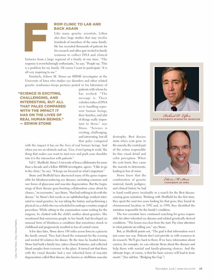
The paper had nothing to do with learning the breast stroke, but it did give him a likely reason why he sank as a child when the other kids floated. A rare inherited genetic mutation was probably what made his otherwise normal bones grow many times beyond typical density–so thick and heavy, in fact, that he sank in water like a sack of stones. The realization spurred him to contact the study’s director, Richard P. Lifton, an HHMI investigator at Yale University School of Medicine. Packard figured his condition might aid in Lifton’s effort to find a cure for the opposite situation, the loss of bone density seen in osteoporosis, a disease that afflicts some 28 million, mostly elderly, Americans and is responsible for 1.5 million frac-tures each year, many of them linked to an increased risk of death. Packard, of course, had never fractured a bone in his life.
Lifton was grateful for Packard’s initiative. “When a patient can tell you ‘I’ve got a genetic disease, and these are the people in my family who are likely to have it,’ it’s tremendously helpful.”
In the stereotypical research experience, the flow of knowl-edge runs from scientist to patient through discoveries leading to health care advances. But in reality, the reverse very often proves
true. Many scientists who study the biomed-ical basis for disease have had chance or purposeful encounters with patients that have changed the course of their thinking and their research.
Many other scientists draw direct inspiration from patients, which keeps them returning to
the lab day after day to push knowledge of biological mechanisms in projects that may take years to complete. And the efforts that indi-viduals and advocacy groups make to encourage research and build financial and political support for it often prove crucial for tackling particular diseases (See Perspectives & Opinions, page 40).
The NEJM paper Packard had read described an inherited syndrome in members of a Connecticut family; they had come to Lifton’s attention after a man in a bad car wreck escaped without a single broken bone, much to the amazement of the emergency-room physicians who treated
him. Tests later revealed his otherwise normal bones to be among the densest ever recorded. This condition intrigued Lifton, as his laboratory is widely recognized for its success in mapping genes involved in common inherited diseases–such as hypertension and osteoporosis–by analyzing outliers. Such rare types of disorders, running in families, help researchers to identify causes of the related but genetically and environmentally more complex common forms. This also allows them to study far fewer patients than the many thousands needed for large-scale genomic analysis.
When Lifton and his colleagues analyzed DNA from 20 members of the man’s family, they discovered that the cause was an inherited mutation of the gene coding for “low density lipoprotein receptor–related protein 5” (LRP5). A different mutation of the LRP5 gene was already known to be associated with just the opposite effect, the increasingly porous and brittle bones of osteoporosis.
Part of Packard’s family traced its roots to Connecticut and, like the family in the NEJM paper, tended to have exceptionally large, square jaws. He figured he must be distantly related to that family and had inherited the same LRP5 mutation. Finally, he understood why he–and two of his sisters, two of his sons, and each of their sons–could never float.
Scheduled to soon have a hip replacement, he offered Lifton some bone samples, which were accepted with great appreciation. “Being able to study gene expression on living bone is a real rarity,” says Lifton. Those bone chips harvested during the surgery are already showing promise for developing therapies that would mimic the LRP5 mutation’s mechanism of action.
Ironically, Packard’s wife has osteoporosis. “It’s just a miserable condition,” he says. If Lifton, other scientists, and drug developers at pharmaceutical companies can develop a medication that affects osteoporosis patients much like the LRP5 mutation does Packard, the damage experienced by millions, including Packard’s wife, could potentially be repaired. Today’s therapies, by contrast, can only slow the disease’s progression.
B
EVEN IN RETIREMENT, JOHN PACKARD, A FORMER CARDIOLOGIST AND MEDICAL EDUCATOR AT THE UNIVERSITY OF ALABAMA SCHOOL OF MEDICINE, CONTINUES TO READ SCIENTIFIC JOURNALS. IN MAY 2002, WHILE HE WAS SCANNING THE NEW ENGLAND JOURNAL OF MEDICINE (NEJM), ONE OF THE ARTICLES BROUGHT HIM A SHOCK OF RECOGNITION. SUDDENLY, THE 83-YEAR-OLD REALIZED WHY, DESPITE HAVING ALWAYS BEEN VIGOROUS AND ATHLETIC, HE HAD NEVER BEEN ABLE TO FLOAT.
ONING UP ON OSTEOPOROSIS
“WHEN A PATIENT CAN TELL YOU ‘I’VE GOT A GENETIC DISEASE, AND THESE ARE THE PEOPLE IN MY FAMILY WHO ARE LIKELY TO HAVE IT,’ IT’S TREMENDOUSLY HELPFUL.” — RICHARD LIFTON
23November 2006 | h h m i b u l l e t i n
F Like many genetic scientists, Lifton also does large studies that may involve hundreds of members of the same family. He has recruited thousands of patients for his research and often gets invited to family reunions to collect DNA and clinical
histories from a large segment of a family at one time. “The response is overwhelmingly enthusiastic,” he says. “People say, ‘This is a problem for my family. Of course I want to participate.’ It is all very inspiring to me.”
Similarly, Edwin M. Stone–an HHMI investigator at the University of Iowa who studies eye disorders and other related genetic syndromes–keeps pictures posted in his laboratory of
patients with whom he has worked. “The message is: These colorless tubes of DNA we’re handling repre-sent human beings, their families, and alot of things really impor-tant to them,” says Stone. “Science is exciting, challenging, and interesting, but all that pales compared
with the impact it has on the lives of real human beings. And when you see an obstacle and say, ‘Gee, it isn’t going to work,’ the thing that makes you want to start over and push your shoulder into it is the interaction with patients.”
Val C. Sheffield, Stone’s University of Iowa collaborator for more than a decade and a fellow HHMI investigator, agrees. “I like to go to the clinic,” he says. “It keeps me focused on what’s important.”
Stone and Sheffield have discovered many of the genes respon-sible for blindness-inducing eye diseases, including common and rare forms of glaucoma and macular degeneration. But the begin-nings of their disease gene-hunting collaboration came about by chance, “an encounter,” says Stone, “that had nothing to do with the disease.” In Stone’s first weeks as an ophthalmology resident inter-ested in visual genetics, he was taking the history and performing a physical on a child who was scheduled to undergo a routine surgical procedure. While sitting in the examination room waiting for the surgeon, he chatted with the child’s mother about genetics. She mentioned that numerous people in her family had developed an unusual form of blindness–a condition that sometimes struck in childhood and progressively resulted in loss of central vision.
A few days later, Stone drove 100 miles across Iowa to a pizzeria the family owned. They had closed the restaurant for the evening and invited 60 relatives for dinner. By the time he headed home, Stone had built a family tree, taken clinical histories, and collected blood samples from everyone in the place. It turned out that those with the visual disorder had a rare inherited form of macular degeneration called Best disease, also known as vitelliform macular
dystrophy. Best disease starts when cysts grow in the macula, the central part of the retina responsible for fine visual detail and color perception. When the cysts burst, they cause the macula to deteriorate, leading to loss of vision.
Stone knew that the combination of genetic material, family pedigree, and clinical history he had in hand could prove invaluable in a search for the Best disease-causing gene mutation. Working with Sheffield for the first time, they spent the next two years looking for that gene; they found its chromosomal location in 1992 and, in 1998, they identified the mutation responsible for the family’s condition.
The two scientists have continued searching for genes respon-sible for other inherited eye diseases and related genetically derived conditions. “The lesson was clear from the start: Pay close attention to what patients are telling you,” says Stone.
But, as Sheffield points out, “The goal is that information won’t just come one way. Patients don’t just provide us with resources to do research. We’ll give back to them. If we have information about carriers, for example, we can educate them about the diseases and help them with marital and family-planning choices. And the ultimate hope, of course, is that the basic science will lead to treat-ments.” [See sidebar, “Bridging the Gap.”]
ROM CLINIC TO LAB AND BACK AGAIN
“SCIENCE IS EXCITING, CHALLENGING, AND INTERESTING, BUT ALL THAT PALES COMPARED WITH THE IMPACT IT HAS ON THE LIVES OF REAL HUMAN BEINGS.” — EDWIN STONE
yale university school of medicineRichard P. Lifton
university of iowaEdwin M. Stone
Lif
ton:
Pau
l Fe
tter
s S
tone
: E
dwar
d H
effr
on

24 h h m i b u l l e t i n | November 2006
Patient advocates play an important role in driving biomedical science to find cures, often by helping scientists interested in fundamental biological mechanisms to see that their work has important clinical applications. Consider, for example, HHMI investigator Thomas M. Jessell at
Columbia University’s College of Physicians and Surgeons, who studies the assembly of neural circuitry in the central nervous system. Within the last few years he began experiments to turn mouse embryonic stem cells into motor neurons (which connect to muscles for movement and locomotion) to study the course of their normal development. It turns out that this research has also been fruitful for better understanding neurodegenerative diseases such as amyotrophic lateral sclerosis (ALS, also known as Lou Gehrig’s disease) and spinal muscular atrophy (SMA), both of which cause fatal loss of neuromuscular control. Jessell now works closely with Project ALS and the SMA Foundation to study stem cells as potential treatments for these diseases, in conjunction with HHMI investigator Douglas A. Melton at Harvard University.
Jessell’s turn toward finding clinical applications for his basic-science studies came about because individual philanthropists
and disease advocacy groups began seeking him out. At their urging, after build-ing a career largely devoted to basic science, he has found himself increasingly thinking about the relationship between his research and its potential for clinical applications. “It’s not
that the work in the laboratory has changed to become clinical,” he says, “but with the perspective such alliances bring, it’s easier to think about applications.”
To deepen the connection between basic and clinical research, Jessell helped to set up the Motor Neuron Center at Columbia University—funded with large gifts from Leonard Tow, a New Yorker whose wife has ALS, and the SMA Foundation—to bring together and support 40 different laboratories, both basic and clinical, in potentially productive collaborations. “This has changed the way I think about the science we do,” says Jessell.
ADVOCACY FOR CURES
While large numbers of patients are often needed for biomedical studies, some-times an encounter with a single patient can also contribute to, even radically alter, a scientist’s thinking. In 1999, HHMI investigator Huda Y. Zoghbi announced to a group of reporters gathered
at Baylor College of Medicine that she had found the gene mutation responsible for Rett syndrome, an inherited disease that strikes young girls and leaves them severely disabled for life. Sitting in the first row at the news conference was Ashley Fry, by then a bright-eyed, dark-haired young woman. Zoghbi made sure to invite Ashley, even though she could not understand the proceedings. “The person who put me on the road to that discovery needed to be there,” says Zoghbi.
Years before that day, as a pediatric neurology resident in Houston, in 1983, Zoghbi fully intended to go into clinical prac-tice. “I was not planning on becoming a scientist,” she recalls. But then she encountered three-year-old Ashley and her parents, and Zoghbi’s life changed.
Ashley had been developing normally until, at age 18 months, she suddenly lost control of her hands and had trouble standing and walking. Then she regressed in other ways, including losing most of her communication skills, and she began having seizures. Eventually, she developed severe scoliosis–curvature of the spine–which required several surgeries. Her deterioration stabilized, but her developmental progress came to a halt. “How,” Zoghbi asked, “could something going so well suddenly change so terribly?”
Other neurologists had told Ashley’s parents that they must have been wrong about their daughter’s seemingly abrupt deterioration. She likely had cerebral palsy, they insisted, a disorder that begins at birth, never permits normal development, and causes continuing deterioration. Convinced that her symptoms did not fit this diagnosis, her parents had become increasingly frustrated and disenchanted by the time they encountered Zoghbi.
“To me, at the first meeting,” recalls Ashley’s father, Clifford Fry, an economics consultant based in College Station, Texas, “Huda Zoghbi was just another doctor in the room.”
But she listened to the parents’ report on their daughter’s devel-opment and sudden change. “I knew from talking with them that it wasn’t there at birth, and I was intrigued.” The same month Zoghbi met the Fry family, a group of European investigators published a clinical paper describing other young girls with a condition exactly like Ashley’s. It was called Rett syndrome. “I realized that what Ashley was experiencing must be Rett syndrome and that the syndrome must be caused by a genetic defect, given its occurrence in girls only.” After that visit, Zoghbi and Ashley’s parents knew they were dealing
LIFE-CHANGING PATIENTS
“WITH THE PERSPECTIVE SUCH ALLIANCES [WITH PHILANTHROPISTS AND ADVOCACY GROUPS] BRING, IT’S EASIER TO THINK ABOUT APPLICATIONS.” — THOMAS JESSELL

25November 2006 | h h m i b u l l e t i n
Longtime collaborators in the search for
genetic causes of eye diseases, HHMI investi-
gators Edwin M. Stone and Val C. Sheffield at
the University of Iowa have recently teamed
up to launch The John and Marcia Carver
Nonprofit Genetic Testing Laboratory, which
seeks to expand the availability and reduce
the cost of tests for rare genetic disorders.
Though the initial emphasis is on eye disorders,
the scientists’ goal is to expand the program
to encourage widespread molecular diagnosis
for as many genetic disorders as possible.
“Testing,” says Stone, “will accelerate progress
toward cures of rare diseases.”
The vast majority of people do not seek addi-
tional help, he says, when they learn that they
or their children have an inherited disease for
which testing is not widely available. As a result,
most never receive a molecular diagnosis. “No
blood sample is drawn, and no data are made
available to any researchers,” says Stone. But
with increased availability of low-cost genetic
testing, molecular diagnoses can become
more the norm; and in the Internet age, the
rest of the research world can get involved.
With permission from patients, the laboratory
makes data from the tests, masked to protect
the privacy of the patients and their families,
available to researchers globally.
Patients can in turn find out about opportunities
to participate in research studies and try
emerging treatments. “It works in both direc-
tions,” says Stone. “Patients and researchers
will benefit.” — M.W.
for more information, go to www.carverlab.org.
BRIDGING THE GAP
with a disease with identifi-able symptoms. With that meeting, Zoghbi’s plans for her future changed. She redirected her training to obtain research skills in molecular genetics. Today, Zoghbi runs a large neuro-genetics research laboratory at Baylor and is one of the world’s leaders in the study of childhood neurological disorders.
Zoghbi’s goal was to identify the gene muta-tion responsible for Rett syndrome; it took her another 16 years. In 1999, she finally announced that she had found the culprit, a gene encoding a protein called “methyl-CpG-binding protein 2,”or MECP2. The protein normally silences several genes during certain periods of central nervous system maturation. But in Rett syndrome, a child has a mutated form of the MECP2 gene that prevents the protein from silencing genes at the appro-priate times in the maturing nervous system. This results in normal development for a period followed by sudden nervous-system dete-rioration at a specific moment in infancy.
Until that discovery, Rett syndrome generally could not be diagnosed until a child reached five years of age and then only by physician observation. With identification of the gene mutation, a molecular diagnosis can now be made as early as 18 months and sometimes even sooner. Consequently, physical and other therapies can begin much earlier in life, thereby improving chances for pres-ervation of some skills.
“These are small steps so far,” says Zoghbi. Better treatments remain a distant hope. “That is what drives me to get up early in the morning to get to work.”
Witnessing that 1999 news conference announcing the discovery of the MECP2 mutation was as meaningful for Ashley’s family as it was for Zoghbi. “We were proud to be there,” says Ashley’s father. “Everything Dr. Zoghbi and other researchers can find out is a step forward. It may help Ashley and it may help others. Ashley would be happy to know that she has helped.” p
baylor college of medicineHuda Y. Zoghbi
columbia universityThomas M. Jessell
Jess
ell:
Mar
c B
ryan
-Bro
wn
Zog
hbi:
Pam
Fra
ncis

26 h h m i b u l l e t i n | November 2006

27November 2006 | h h m i b u l l e t i n
It may not seem strange that dogs,
by Michael Mason
so often anthropomorphized, can enlighten molecular biologists about
behaviors deemed uniquely human.
But apparently, so can other animals:
mice, fish, flies–even snails.
Model
Man’s
ILLUSTRATION BY JONATHON ROSEN

28 h h m i b u l l e t i n | November 2006
Increasingly, scientists see in animals the underpinnings of behav-ioral responses once thought to be uniquely human: depression, happiness, even lust. Whether the researchers’ experiments lead to new therapies, the very fact that many of these behaviors can be modeled in animals comes as something of a shock.
A depressed mouse? An insomniac fruit fly? C’mon.Yet these beasts do exist. Scientists have had only to look for
them or, more recently, to create them. In the early 1970s, at an American Medical Association conference in San Francisco, sleep researcher William C. Dement of Stanford University School of Medicine described to his audience the bizarre symptoms
In a laboratory hallway at Stanford University, the neurologist and HHMI investigator lays out a small meal for Bear, a fluffy 8-year-old Belgian schipperke. Food is one of Bear’s favorite things, along with Mignot himself, and today the combination proves over–whelming. As he tucks into his grub, Bear, the last narcoleptic dog remaining at the university’s famous multibreed colony, keels over.
This time the interlude lasts just a moment. After a rousing pat on the head, Bear resumes his meal, apparently none the worse. “They become overstimulated by food, or play, or sex,” sighs Mignot. “You can imagine what it was like trying to breed a colony of narcoleptic dogs.”
Well, no, actually. Still, while every animal model of human behavior has its own special challenges, they remain essential to inves-tigators. Molecular biologists depend on our evolutionary relatives to illumine the biochemical circuitry that drives us.
“Neurology has been revolutionized by the existence of animal models,” says neurobiologist Eric R. Kandel, a Nobel laureate and HHMI investigator at Columbia University. “The brain of a mouse is a prototypic mammalian brain. Evolution has been quite conservative across mammalian species.”
Emmanuel Mignot is not prepared to let sleeping dogs lie.
of narcolepsy: waves of unstoppable drowsiness, fragmented sleep patterns, episodes of quasi-paralysis at times of strong emotion. An estimated 150,000 people have the disease, he noted, and for them daily life is a perilous obstacle course.
After the talk, a doctor visiting from Canada told Dement that he had seen this syndrome all too recently–in his dog. Far from finding a potential connection laughable, Dement scouted among local breeders for similarly affected animals, ultimately establishing a colony of narcoleptic Doberman pinschers and Labrador retrievers at the university. As it turned out, he could not have picked a better way to study the disease.
Because they are highly inbred and often geographically isolated, dogs harbor more than 300 genetic diseases, researchers now know–more than any other species besides humans. At least half of these diseases are believed to be analogous to specific human disorders. Seventeen breeds, including dachshunds and poodles, are known to develop narcolepsy.
“The dog genome is very similar to the human genome,” says Mignot. “They really have not been used enough as models.”
In 1999, relying on results obtained from the Stanford colony, Mignot and his colleagues discovered that narcolepsy in Dobermans and other breeds results from a mutation in Hcrtr2, a gene that encodes a receptor for hypocretin, a neuropeptide produced in two forms in neurons of the dorsolateral hypothalamus and dissemi-nated through the brain stem, spinal cord, and forebrain.
Still, the link from animal model to human behavior is seldom direct. Dobermans and Labradors have slightly different mutations of the Hcrtr2 gene, Mignot and his colleagues found. And humans appear to develop narcolepsy for a completely different reason: They simply don’t produce hypocretins at all.
Interestingly, some dogs, such as Bear, also suffer from this form of narcolepsy. “Bear is especially interesting for us to keep for the future,” Mignot says. “Because he lacks hypocretin, like human patients, he could be an ideal model to test a new medication aimed at replacing hypocretins in humans.”
Earlier this year, Stanford University began disbanding its renowned colony of narcoleptic dogs, as functional genetic studies are more easily done in other models, and most of the dogs have been adopted. Lately, Mignot has been thinking that Bear will need a very special retirement home: the scientist’s own.
An Model
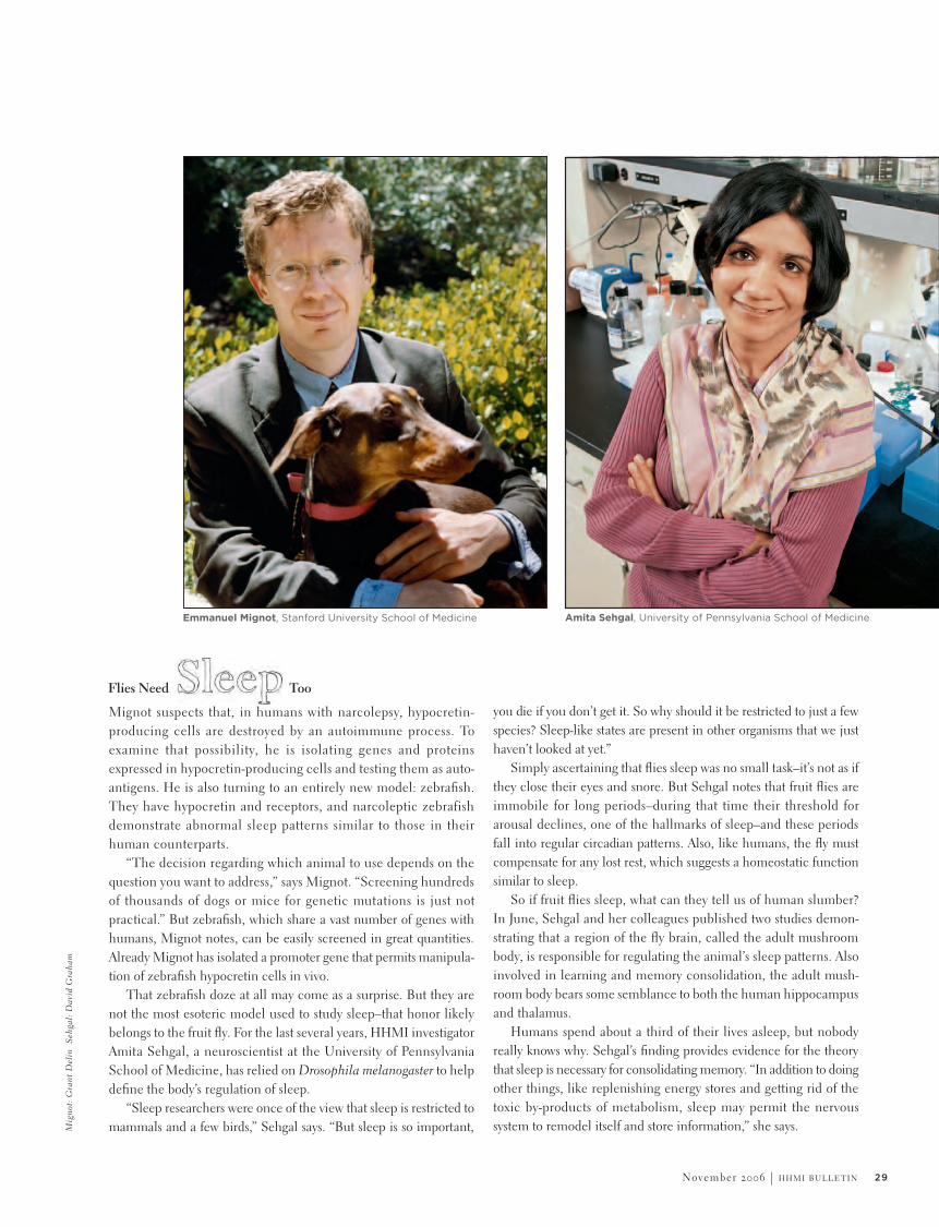
29November 2006 | h h m i b u l l e t i n
Amita Sehgal, University of Pennsylvania School of Medicine
Mignot suspects that, in humans with narcolepsy, hypocretin-producing cells are destroyed by an autoimmune process. To examine that possibility, he is isolating genes and proteins expressed in hypocretin-producing cells and testing them as auto-antigens. He is also turning to an entirely new model: zebrafish. They have hypocretin and receptors, and narcoleptic zebrafish demonstrate abnormal sleep patterns similar to those in their human counterparts.
“The decision regarding which animal to use depends on the question you want to address,” says Mignot. “Screening hundreds of thousands of dogs or mice for genetic mutations is just not practical.” But zebrafish, which share a vast number of genes with humans, Mignot notes, can be easily screened in great quantities. Already Mignot has isolated a promoter gene that permits manipula-tion of zebrafish hypocretin cells in vivo.
That zebrafish doze at all may come as a surprise. But they are not the most esoteric model used to study sleep–that honor likely belongs to the fruit fly. For the last several years, HHMI investigator Amita Sehgal, a neuroscientist at the University of Pennsylvania School of Medicine, has relied on Drosophila melanogaster to help define the body’s regulation of sleep.
“Sleep researchers were once of the view that sleep is restricted to mammals and a few birds,” Sehgal says. “But sleep is so important,
Emmanuel Mignot, Stanford University School of Medicine
you die if you don’t get it. So why should it be restricted to just a few species? Sleep-like states are present in other organisms that we just haven’t looked at yet.”
Simply ascertaining that flies sleep was no small task–it’s not as if they close their eyes and snore. But Sehgal notes that fruit flies are immobile for long periods–during that time their threshold for arousal declines, one of the hallmarks of sleep–and these periods fall into regular circadian patterns. Also, like humans, the fly must compensate for any lost rest, which suggests a homeostatic function similar to sleep.
So if fruit flies sleep, what can they tell us of human slumber? In June, Sehgal and her colleagues published two studies demon-strating that a region of the fly brain, called the adult mushroom body, is responsible for regulating the animal’s sleep patterns. Also involved in learning and memory consolidation, the adult mush-room body bears some semblance to both the human hippocampus and thalamus.
Humans spend about a third of their lives asleep, but nobody really knows why. Sehgal’s finding provides evidence for the theory that sleep is necessary for consolidating memory. “In addition to doing other things, like replenishing energy stores and getting rid of the toxic by-products of metabolism, sleep may permit the nervous system to remodel itself and store information,” she says.
Flies Need Too
Mig
not:
Gra
nt D
elin
Se
hgal
: D
avid
Gra
ham

30 h h m i b u l l e t i n | November 2006
Findings in animals often lead scientists to reconsider not just basic human functions like sleep but also traits once considered quintessentially human, such as how we learn and store what we have learned. Columbia’s Kandel asserts that animal-based molecular biology soon will affect psychiatry as profoundly as it has neurology.
Nearly 50 years ago, against the advice of his mentors, Kandel elected to study fear and learned memory of fear in a highly unlikely subject: a giant marine snail called Aplysia californica, which grows up to a foot long and can weigh several pounds. (Kandel nonetheless refers to Aplysia as “beautiful.”)
Many scientists of the day thought the neurobiology of an inver-tebrate could have little relevance to the mental functions of humans. Chief among the doubters, Kandel recalls, were students of behavior: psychiatrists, psychoanalysts, and psychologists. At that time only two scientists in the world bothered to study this snail at all.
But in a body of work spanning decades, Kandel and other researchers used Aplysia, and later mice, to establish a molecular basis for memory storage and learning in the brain. Scientists working with rats eventually traced the memory of fear to the amygdala and the body’s expression of fear to the hypothalamus. Using these findings to explore the molecular underpinnings for
Li-Huei Tsai, Massachusetts Institute of Technology
Insights, of Snails
Of all the human pathologies that scientists have
attempted to model in animals, perhaps none is as
difficult as schizophrenia. Striking about 1 percent of
the population, the disease is marked by three broad
types of symptoms: positive (delusions, hallucinations),
negative (withdrawal, lack of expression), and cognitive
(poorworking memory, short attention span).
It’s a formidable recipe. Against these considerable
odds, however, two HHMI investigators have had success
in approximating certain aspects of the disease in
animal models.
Over the years several studies have reported that
antipsychotic drugs ameliorate schizophrenia by
blocking excessive dopamine transmission in the
brain. In 2004, researchers learned that these patients
often have large numbers of D2 dopamine receptors
in the striatum, an area deep within the brain that
controls movement and balance.
In an attempt to extend this finding, Eric R. Kandel’s
Columbia University lab created mice that also over-
expressed these receptors, and his team discovered
that the mice suffered a hallmark of schizophrenia:
impaired working memory. This deficit persisted even
when the researchers allowed production of D2
receptors to return to normal.
Early in an animal’s development, Kandel reasoned,
the damage caused by increased dopamine trans-
mission may produce irreversible changes in other
parts of the brain.
But dopamine may not be the only culprit.
Calcineurin is an enzyme involved in the regulation of
the immune response and in learning. In 2001, HHMI
investigator Susumu Tonegawa and his colleagues at
the Massachusetts Institute of Technology found
that knock-out mice missing calcineurin in the fore-
brain also suffer impaired working memory. And
the mutants display other symptoms reminiscent of
schizophrenia: They are hyperactive and withdrawn
from other animals, for example, and they are more
easily startled than normal mice.
“Based on our mouse study, we were able to go
on to obtain direct genetic evidence that variation in
a human calcineurin gene is associated with schizo-
phrenia susceptibility in humans,” says Tonegawa.
“These data provide strong support for the involvement
of calcineurin-related genes in schizophrenia etiology.”
Precisely which of these are involved is still a
mystery, and in any event the disease is likely to
depend on interaction with the environment. Adds
Tonegawa, “In recent years, real progress has been
made in identifying disease genes. We hope that
advances like our calcineurin findings will ultimately
lead to better therapies.” — M.M.
MODELING EMOTIONAL COMPLEXITY
Joel
Vea
k

31November 2006 | h h m i b u l l e t i n
Fortunately, the gap between human anxiety and animal fear may prove smaller than once thought. Scientists have yet to discover an Aplysia with full-blown panic disorder, of course, but rodent models of depression have led directly to a number of useful medications. Better models, though, may soon bring better therapies.
Scientists studying depression, along with drugs to reduce it, have relied largely on wild-type mice that were “socially defeated.” Submissive mice were housed in containers with dominant mice, and the behavior developing in the submissives was judged to be as close to depression as researchers could get.
But last year HHMI investigator Li-Huei Tsai, a neurobiologist now at the Massachusetts Institute of Technology, led a team that developed mutant mice whose disease may more closely mimic that of humans.
Tsai and her colleagues found that a protein called Par4 binds to a type of dopamine receptor and that mice with mutated Par4 demonstrate something akin to depression. Dopamine is a neurotransmitter active in the brain pathways governing mood, reward, and motivation.
In one standard test, mutant mice placed in a clear plastic container filled with water stopped swimming long before wild-type mice. “They are fully conscious, but they quickly become immo-bile,” says Tsai. “They don’t struggle. There is a loss of motivation.”
In another experiment, hungry mutant and wild-type mice are placed in an open environment with food at the far end. Mice are averse to open spaces, so this set-up presumably creates some anxiety. Wild-type mice are quick to surmount their discomfort and find the food. But the depressed mice take a very long time to follow suit.
Closer examination of animal neurobiology doesn’t just tell us about disease—it tells us much about normal human behavior, too. “If you want to study behavioral genetics,” says HHMI investigator Catherine Dulac, a molecular biologist at Harvard University, “you need something robust so that the changes you introduce will be observable.”
Few behaviors are more robust and observable than reproduc-tion, Dulac adds. In mice, as in many animals, certain behaviors hard-wired into the brain—mating and aggression, for example—are strongly influenced by airborne molecules, called pheromones, released by one animal and perceived by another.
Although the vomeronasal organ (VNO), the structure a mouse uses to sense pheromones, resides in the nasal cavity, researchers have long believed that the animal’s pheromone-sensing apparatus is completely separate from the olfactory system, which regulates smell. (continued on page 60)
Humans spend about a third of their lives asleep, but nobody really knows why. Sehgal’s finding provides evidence for the theory that sleep is necessary for consolidating memory.
fear in mice, Kandel discovered that they bear great resemblance to the mechanisms observed in Aplysia.
Now it appears that happiness may be similarly discernible in the brain. In a recently published study, Kandel and Columbia colleague Michael Rogan showed that mice conditioned to associate a sound with safety showed reduced electrophysiological activity in the amygdala, perhaps representing a decrease in fear. At the same time, they showed increased activity in parts of the dorsal striatum involved in positive emotion and reward. “These mice just walk around unafraid, as if they own the place,” says Kandel.
Nearly 30 percent of Americans suffer an anxiety disorder at some point in their lives, and the finding has made Kandel wonder if certain of these disorders might result from defects in the neural systems that communicate security.
Pheromone-Driven
“The mutant animals fail to do what they need to do,” says Tsai. “They find it more difficult to overcome their anxiety and fear.” Most current medications for depression, the SSRIs (selec-tive serotonin reuptake inhibitors) and MAOIs (monoamine oxidase inhibitors), modify synaptic levels of serotonin or norepi-nephrine. Tsai’s experiments with depressed mice suggest that dopamine may also provide an excellent target for intervention.
Mice

by jeff miller • photographs by misha gravenor
droSoPhilaenvY A maverick scientist is risking all to study the behavior
of fruit fl ies, convinced that they have much to teach us about the evolution and neurobiology of emotions.


34 h h m i b u l l e t i n | November 2006
Sean Morrison, an HHMI investigator at the University of Michigan who was a post-doctoral fellow in Anderson’s lab from 1996 to 1999. “He discovered many of the genes that regulate neurogenesis. And his lab is the place where neural crest stem cells were identified and characterized.”
Anderson’s discovery of three classes of master genes inside neural stem cells—genes that give rise to neurons and related cells that insulate nerve fibers—was one of the first to answer the larger question of how stem cells differentiate. He also discovered some fundamental properties of angiogenesis (the formation of blood vessels)—namely, that arteries, but not veins, are aligned with nerves in embryonic skin and that the nerves are required for arterial differentiation.
But Anderson needed a change.Was it the typical lag time of 12 to 18
months between testing a hypothesis in mice and getting a result that agitated him? “I can get very impatient,” he concedes.
Was it the need to recharge his research batteries by challenging himself? Yes, he agrees. “One of my biggest self criticisms is that I tend to identify problems and get into areas a little too early—which is also good because I’m ahead of the pack.”
Or was it the thrill of the hunt, the same feeling that once tempted him toward archaeology when he was a boy? Perhaps. “The happiest moments of my scientific life have come when I’ve made that initial discovery and I suddenly glimpse some-thing about nature that, at that moment, no one else knows.”
their conventional ways,” says HHMI inves-tigator David J. Anderson, “for making a continual set of contributions to one problem.” But Anderson, a neurobiologist at the California Institute of Technology, who admits to being “occasionally crazy and easily bored,” has chosen a different course.
He traded continued success studying the biology of neural stem cells for the uncharted world of fruit fly emotion. Anderson likens his path to the one pursued by Caltech colleague Seymour Benzer. “Starting out in solid-state physics, he switched to molecular biology, then made a 180-degree turn and founded the field of neurobiology and neurogenetics,” says Anderson, with obvious admiration.
Benzer is motivated by the answer to a single question. “Isn’t it more crazy not to follow the driving force of your curiosity and passion?” he asks, rooting for Anderson. “David has the courage, energy, and intelligence to make a go of it.”
Although Anderson has an obvious love of risk, he has certainly shown himself capable of playing by the usual rules. After completing his postdoctoral studies in the Columbia University laboratory of HHMI investigator Richard Axel (2004 Nobel Prize winner, and his mentor), Anderson spent more than 20 years helping to pioneer the study of the developmental biology of stem cells in mice.
“He made seminal contributions to our understanding of neural development,” says
“ScientiStS are rewarded for There was something else, too, Anderson
acknowledges. Drosophila envy. “I’ve had it my whole career. Fruit flies are such a powerful system, and they are simpler. You can understand mechanisms that govern processes more easily than you can in mice.”
Other scientists have broken ranks before, of course. “But what makes Anderson’s move more unusual is that he moved out of a very active field of which he was a leader,” says Gerald M. Rubin, HHMI vice president and long-time Drosophila researcher.
And he did it just a few years before California voters agreed to spend $3 billion to underwrite stem cell research. That a tiny fruit fly proved irresistible to Anderson just as support for his core area of research was mounting illuminates another of Anderson’s key beliefs. Put simply, packing up the furniture—if not exactly chopping it into kindling—is sometimes necessary to break new scientific ground, especially when herds of fellow scientists begin warming themselves at your campfire.
Morrison was just finishing his postdoc-toral work when Anderson began to talk about shifting to studies on the molecular regulation of behavior. “My reaction was that it sounded like a really hard problem that was obviously of great interest to solve,” he recalls, “but I worried how much headway they’d be able to make experimentally.”
Anderson’s the kind of standout scientist, though, who picks problems that everyone agrees are important but no one else knows how to solve, just like Richard Axel and his olfactory research, Morrison says. “Everyone

35November 2006 | h h m i b u l l e t i n
“ScientiStS are rewarded for else looked at the problem and agreed it
would be cool to understand how we smell, but no one knew how to tackle it.”
Given Anderson’s success in the five years since he began his scientific walkabout, few would now call him shortsighted. Morrison calls the move heroic. “As this new line of work matures in his lab, it will be a force in shaping our understanding of how behavior is regulated.”
Anderson expects to learn from his mistakes more rapidly and less expensively in fruit flies than he would in mice. But since he longs to get inside a fly’s head to study the connections between neurons, genes, and behavior—Anderson’s current passion—he’ll need to be patient too. After all, he says, “It is extremely difficult for all but the most talented electrophysiologists—such as my Caltech colleague Gilles Laurent—to stick an electrode into a fly’s brain and measure the electrical activity of its neurons.”
Researchers can alter flies genetically, however, and use various imaging tech-niques and special microscopes to see which brain cells fluoresce or react in response to different stimuli or during various activities. Such processes, Anderson says, are not only informative but downright thrilling. “It’s like flying over a city at night and watching the lights of the houses go on and off.”
But, he cautions, “Just because there is a correlation between neurons and an activity doesn’t mean those neurons alone are respon-sible for the behavior. To test that hypothesis,
The Smell of fear?
you have to turn off the neurons, and see if the behavior goes away.” Using one such genetic technique, Anderson, with a team including Axel and Benzer, was able to engi-neer temperature-sensitive neurons that shut down at 90°F. In a series of experiments that pivoted around this temperature sensitivity, they discovered that “traumatized” fruit flies emit a stress odor containing carbon dioxide, repelling other flies, and that a single type of neuron in the nose of the fly is responsible for detecting the noxious gas.
Were the normal flies reacting to a “smell of fear”? Maybe, and maybe not, says
Anderson. What he learned in additional experiments—that the target olfactory neuron is both activated by carbon dioxide and neces-sary for the response to carbon dioxide—still did not prove what actually triggered the avoidance behavior. So in a clever case of genetic manipulation, Anderson added a bit of algae DNA to the fruit fly’s carbon-dioxide-sensitive neurons: specifically, an ion-channel gene that is activated by blue light.
The next step was to put these altered flies into an apparatus and give them a choice between flying into an empty tube or one illuminated with blue light. “We wanted to
“david anderson sets a great example for the people who train in his lab—it’s
not about ego or reputation, it’s about getting to the truth,” says Sean Morrison.

36 h h m i b u l l e t i n | November 2006
know if flies exposed to this light would think they smelled carbon dioxide and act to avoid it,” Anderson explains. Sure enough, when he flicked on the switch, the altered flies shunned the blue light, proving the primary link between neuronal activity and behavior.
In comparable research with mice, Anderson has also narrowed the search for genes that control whether a stressed mouse
freezes or flees when hearing the mouse version of an alarm bell. But be they fruit flies or mice, is their behavior emotional behavior, and if so, so what?
Anderson’s knowledge is encyclopedic, according to Morrison. “He has a truly remarkable ability to bring
together diverse facts from different areas and synthesize them into incredibly creative ideas.” That brainpower may come from Anderson’s parents. His father is a retired theoretical astrophysicist, who lettered in chess at the University of Chicago. Every year, he traveled the family from Teaneck, New Jersey, to Woods Hole, Massachusetts, because he was interested in learning more about fluid mechanics. Anderson’s mother, a professor of Latin American literature, still teaches at New York University.
The younger Anderson says he takes inspiration from many sources, among them Charles Darwin’s 1872 monograph, “The Expression of Emotion in Man and Animals.” This dense work, overshadowed by Darwin’s writings on natural selection, asks why emotions take on physical expressions—why people smile when they’re happy or cringe when they’re afraid. Darwin posited that opposite emotions produce opposite behav-ioral manifestations, as well: a smile will turn up the corners of the mouth; a frown will turn them down.
For most principal investigators, the moments for real science are crowded by constant meetings and nagging adminis-trative duties, articles to read, lab notes to review, grants to write, and both scheduled and unscripted mentoring moments.
Anderson loves his time with students and colleagues—“I would not do well in a scientific monastery”—but he also has a deep longing for his lab bench, where he can work alone, dirty his hands, and fill his own lab notebook with observations and results. In early 2006, he got his wish, taking a six-month sabbatical at Germany’s University of Würzburg.
In Germany, Anderson was able to immerse himself in the lives and biology
InSpIraTIon and darwIn
alone wITh The flIeS
Ann
e H
erga
rden
and
Tim
othy
Tay
ler
in this larval fly nervous system, major structures are stained red and neurons
expressing a specific neuropeptide are stained green. in the mammalian
brain, many neuropeptides are produced in the hypothalamus to play a
role in the control of emotional behaviors. of the approximately 100,000
neurons in the fly’s nervous system, only about 10 express this neuropeptide,
suggesting that they have a very specific function.

37November 2006 | h h m i b u l l e t i n
been conserved, perhaps emotional responses have been as well.
Rubin, for one, already is convinced that flies have emotions. “What is not clear is how analogous fly ‘fear’ and human ‘fear’ are in the way the fly or human feels them and responds behaviorally.”
Answering that question could lead to novel antidepressants with, for example, fewer side effects. But these would be consequences, Anderson insists, not objectives. Indeed, he is openly scornful of the current funding fashion that dresses up the “best” and most attractive scientific research as “translational.” Scientists need to be free to follow their curiosity, he believes, even when any ultimate application is not yet evident and even though they essentially have to start their careers all over again. HHMI enabled that freedom, Anderson says. “Its financial support has made every-thing I’ve done possible.”
Not surprisingly, support and understanding from many of his peers has been grudging, Anderson confesses. He had little formal training in Drosophila neuroanatomy and, when he started, precious little intuition about the most productive experiments that such training and experience provide. “I basically had to teach myself,” he acknowledges. “I felt like a graduate student again.”
Moreover, the switch in research direc-tion from development of the nervous system to behavior—“like turning around an oil tanker”—jolted the academic lives of the graduate students and postdocs who had
of fruit flies. Period. There were leads to ponder, behavioral experiments to design, and always a single question to answer: “How can we determine in an objective way whether flies have emotions or not?”
The problem was—and remains—vexing. For example, when flies scatter as a shadow passes over them, are they truly afraid?
Of course, to detect an emotion requires first defining the term, a task that Anderson agrees can be subjective, if not downright emotional. His choice? Emotion is some-thing that places value on behavioral options and that reveals itself in grades of intensity and that produces internal states with similar ranges—from mild apprehension to terror-izing fear, for example.
Studying fruit flies long enough and hard enough, as Anderson did in Germany, enables one to detect some intriguing emotion-like clues, he contends. During courtship, for example, male fruit flies extend their wings horizontally as they sing to their mates. Flies engaged in aggressive behavior, on the other hand, elevate their wings straight up in the air.
“When you look at their wings and the two perpendicular axes associated with two activities of opposite emotional valence, it makes me wonder,” says Anderson. “Maybe the wings of the fly are the best way to read its internal state.”
Adding weight to his hypothesis is work by Axel and others that has found a similar structural scheme in both mice and fruit flies. In short, all neurons that express a common olfactory receptor in these crea-tures have axons that converge on a single point in the brain.
“In over 400 million years of evolution, the same neural circuit organization has been conserved, even when the molecules themselves have not,” Anderson relates with genuine awe. And if this circuitry has
allied themselves to his previous scientific interests. Neglect was never an issue, however. In each case, Anderson stayed the course, supervising experiments in angiogenesis, and teaching, guiding, and mentoring until the cycle of students and fellows could finish their work and move on with their careers.
What stirs Anderson now is to nail down, once and for all, the genetic controls and neuronal circuitry behind fear and anxiety in fruit flies, no matter that these creatures lack an amygdala and hippocampus and no matter the ultimate relevance to human health.
Where his present research will lead is unclear, although, in one experiment, when he put a black patch on the surface of their vial, the flies congregated under it when a shadow was passed over the vial. They appeared, at least superficially, to be hiding—as if from a predator.
“If an animal suddenly becomes, say, very aggressive, what’s going on in its brain—and where—to make it act that way?” Anderson asks. Wouldn’t it be great, he adds, if it were possible to derive certain universal rules that could definitively link a particular behavior to an emotion?
“Emotions just didn’t pop up out of nowhere. When did they begin? What are the building blocks?” He pauses. The corners of his mouth turn up. He smiles. “There is a rich vein to be mined in fruit flies. And in the end, I’m convinced that, if nothing else, fruit flies are going to teach us about the evolution of emotions.” p
STayIng ThecourSe
“there iS a rich vein to be Mined in fruit
flieS. and in the end, i’M convinced that, if nothing elSe, fruit flieS are going to teach uS about the evolution of eMotionS.” —david anderSon
W E B E X T R A : For details of Anderson’s studies on fear and pain, visit the Online Bulletin.

38 h h m i b u l l e t i n | November 2006
p e r s p e c t i v e s & o p i n i o n s
Rafael Viñoly
ColleCtive Soul an insightful critique helped janelia farm’s architect find his vision.
Paul
Fet
ters

39November 2006 | h h m i b u l l e t i n
The heart of the Janelia Farm campus–known as the “landscape building”–is a unique architectural surprise. Most of the massive structure is not even visible from the entrance, yet from within it is flooded with natural light and offers stunning views of the Potomac River and the countryside beyond. Internationally renowned architect Rafael Viñoly designed the structure to conform to the contours of the land. The result is a building at once entirely modern yet wholly inseparable from its natural setting. That concept was also a surprise for Viñoly. He started out with an entirely different vision.
At the groundbreaking, you said, “Nature is the centerpiece of research at Janelia Farm and the building follows that idea.” Explain nature’s place in your design.We came up with this notion of integrating the building entirely into the landscape. When complete, the site should remain essentially “untouched.” Everything in this idea led us to enhance the view of nature. From an architectural perspective, things like the enormous amount of glass, the rooftop gardens, and placing the offices and corridors towards the open view work, but they are also completely intercon-nected with this idea. The structure is basically a series of steps terraced into the hillside and then a series of environments that undulate along the length of the hill’s curves where scien-tists work in connection with nature. I think that’s a pretty powerful idea. The entire composition looks like a natural thing. It looks like it has been there for many, many years.
How did you generate this idea of using the existing landscape as the guiding concept for the building?At the beginning we had exactly the opposite idea; we planned to impose a grid on the landscape. Then we had an interview with [HHMI architect] Bob McGhee and the others in the competition interview committee, and they told us, ‘You’re completely wrong.’ I didn’t think so at the time, but the critique was so good it forced me to challenge our assumptions about how a building that satisfies the Institute’s objectives would look. This is so typical in architecture–really in life—that you try to do something exactly against the current until you realize you’d better go the other way. The final concept proved to be amazingly self-supporting. You know you’re on a good track in architecture when you don’t need to push too much. The thing sails on its own.
Is there a tension between the notion that this is a building about nature and about high-tech, advanced science?At the end of the day, Janelia Farm is a highly technical structure. The requirements for services, air exchange, equipment, programmatic space, and flexibility in the lab configuration were enormously demanding. How do you design this huge, intricate machine to be part of nature? The drive to resolve that tension was built into the very idea of Janelia Farm. If you’re HHMI and you decide to make this enormous leap into the future, you could have chosen any place in the world to do it. Choosing this particular site determines something. You want all of these top scientists to move to the country. What are you going to do? Try to transform the country into a city? No, it’s the country. The Institute’s fundamental idea at Janelia Farm was that you are in an unconstrained connection through your work with nature. If the building is any good, it’s because this idea behind Janelia Farm is very good.
How do you as the architect measure the success of Janelia Farm?Janelia Farm doesn’t need anything other than to produce great work and to provide a setting to produce that level of thinking. So what matters here above all is the build-ing’s capacity to generate some level of happiness. When you’ve done this right, it’s not because the building becomes famous or noticeable. It’s because people take possession of it. I believe that a building’s audience, not the critics, is always right. If that audience at Janelia Farm, mainly the people who live and work there, is happy, then we were right in our idea. You can’t fake that.
I n t e r v I e w b y M a r c w o r t M a n . A native of Uruguay, Rafael Viñoly founded Rafael Viñoly Architects, P.C., in 1983. Its offices are in New York City and London.

40 h h m i b u l l e t i n | November 2006
Monica Coenraads
MoTHER oF INVENTIoN
p e r s p e c t i v e s & o p i n i o n s
Paul
Fet
ters

41November 2006 | h h m i b u l l e t i n
The birth of a first child is always a momentous event, as it was for Monica Coenraads when her daughter Chelsea arrived in october 1996. By Chelsea’s first birthday, however, it was clear she was not developing normally. Six months later, Chelsea was diagnosed with Rett syndrome. But there was no definitive test for Rett syndrome, no known cause, and no treatments. Coenraads dedicated herself to seek out and work with scientists willing to study the disease. A cofounder of the Rett Syndrome Research Foundation (RSRF) and its current director of research, Coenraads has helped provide more than $11 million in research grants.
We can be nimble in ways that large federal agencies aren’t, funding risky projects that the government isn’t willing to take on. We have an esteemed scientific advisory board, chaired by Adrian Bird of the University of Edinburgh, and many ad hoc advisors who ensure that we fund high-impact projects directly relevant to Rett syndrome.
RSRF funding has played a part in nearly every break-through that’s happened in the field since the MECP2 gene mutations were discovered. For instance, we funded the development of an animal model of Rett that has proved crucial. Currently, we’re funding collaborative work between HHMI investigators Huda Zoghbi and Nathaniel Heintz, targeting genes whose expression is up- or down-regulated because of MECP2 dysfunction.
My style is very hands-on. I comb through scientific jour-nals and press releases on a daily basis and regularly recognize work that could be synergistic with Rett syndrome. When I contact these scientists and engage them in discussion, they are intrigued by the scientific challenge of Rett.
In working with these researchers, I have definitely devel-oped personal relationships. I think the fact that I’m a mother with a child with Rett personalizes it. Recently, we’ve created opportunities for researchers to meet our children, which I think helps them understand the urgency of the disease. It’s true that science takes time and that good science takes even more time, but as a mother of a child with this syndrome, I know that we don’t have time. And I think they get that.
During the last 7 years I’ve witnessed Rett syndrome rise from obscurity to become a high-profile disorder with links to autism, schizophrenia, and a host of other neurological diseases. It is my hope that Rett syndrome will prove to be the first treatable childhood neurological disorder.
After Chelsea’s diagnosis, I started to learn all I could about Rett syndrome. It was a pretty depressing scenario: The girls lost most if not all acquired language and motor skills, became autistic-like, and suffered seizures; some appeared to be in terrible pain. Even more depressing, the search for the gene had been ongoing for 15 years.
It was evident that an organization able to focus its energy on research would be beneficial. At the beginning, I just wanted to link scientists to speed discovery. I talked to top-notch scientists and directors of research organizations about what was needed. As it turned out, just as we were getting RSRF off the ground, the Rett-related gene MECP2 was discovered, so we immediately began brainstorming about the next steps.
I never expected to fall in love with the science, but the neurobiology of Rett is fascinating. MECP2 is involved in regulating the expression of other genes, so the question is which genes are responsible for the diverse neurological symp-toms. MECP2 has lured all sorts of researchers into the Rett field, because it touches on so many different areas of science.
Before the Foundation, I was running a successful restaurant. I have an MBA, but I really fell into the business. My father had opened a restaurant in Stamford, Connecticut, and soon after became very ill. My brother and I took over and ran it for 10 years. I figured that when life had thrown me lemons I had made lemonade once. When Rett syndrome reared its ugly head, I thought perhaps I could do it again.
I’m not so naïve to think that we have the resources to single-handedly cure Rett syndrome–that’s bigger than any one organization, lab, or investigator. But we do have passion. Founded by six concerned parents of children with Rett, our organization is extremely motivated and a lot of what we do doesn’t involve huge amounts of money. We can bring researchers together to share ideas. It’s about engaging world-class talent. It’s about forging relationships and pooling resources.
I n t e r v I e w b y a l I c I a au lt. Monica Coenraads, her husband, Pieter, daughter Chelsea, and sons Alex, age 8, and Tyler, age 6, live in Trumbull, Connecticut.

42 h h m i b u l l e t i n | November 2006
What challenges do young scientists face today?
Many young scientists dream of heading a lab of their own. According to a few early and mid-career HHMI-supported researchers, today’s up-and-coming scientists experience certain
realities their mentors never faced. — e D I t e D b y J a c Q u e l I n e r u t t I M a n n
p e r s p e c t i v e s & o p i n i o n s
Q & A
alfredo Quiñones-HinojosaH H M I P H y s I c I a n -
s c I e n t I s t e a r l y c a r e e r
awa r d r e c I P I e n t a n d
a s s I s ta n t P r o f e s s o r
o f n e u r o s u r g e r y a n d
o n c o l o g y, t H e J o H n s
H o P k I n s u n I v e r s I ty
s c H o o l o f M e d I c I n e
“one of the main challenges I face daily, in addition to trying to keep up with the most updated literature, is trying to find sources of funding for my laboratory. It is devastating to hear other young scientists, who have not been as lucky as I have been, talk about their inability to stay in the field and their subsequent move to the private sector.”
Ster
nson
: M
atth
ew S
epti
mus
H
a: J
ohn
Dix
on/A
P, ©
HH
MI
New
man
: Jo
hn H
ayes
/AP,
©H
HM
I Q
uiño
nes-
Hin
ojos
a: K
eith
Wel
ler/
John
s H
opki
ns M
edic
ine
Scott M. Sternsong r o u P l e a d e r ,
J a n e l I a fa r M r e s e a r c H
c a M P u s
“Young scientists, at the height of their research productivity, are expected to become lab managers, fundraisers, and adminis-trators when they should be in the lab cranking out results in experiments. Ultimately, you have to grow up and deal with these responsibilities, but I think we should nurture newly independent scien-tists so that success at this level is about more than just getting your grants.”
taekjip HaH H M I I n v e s t I g at o r
a n d a s s o c I at e P r o f e s s o r
o f P H y s I c s , u n I v e r s I ty
o f I l l I n o I s at
u r b a n a - c H a M Pa I g n
“our generation grew up scientifically during the advent of the Internet and cell phones. As a result, we suffer from so-called ‘constant partial attention’ as our line of thoughts are continually interrupted by demand from colleagues, students, vendors, publishers, and of course sports scores. Instead of spending time in the library, we compulsive-ly download and print papers from online jour-nals and never read them. I need more time ‘off line’.”
Dianne K. newmanH H M I I n v e s t I g at o r a n d
P r o f e s s o r o f g e o b I o l o g y,
c a l I f o r n I a I n s t I t u t e
o f t e c H n o l o g y
“Perhaps it is nostalgia, but it strikes me that in the pre-vious generation the pace of research was much more conducive to scholarship. To achieve a bold, long-term research vision, one might not want to publish anything for several years in order to lay the groundwork for something more pro-found. But the tempo and structure of the grants and tenure process make this impractical for most young scientists.”

43November 2006 | h h m i b u l l e t i n
chronicle
44 Janelia Farm — Open for Discovery
A New Scientific Community / Let the Science Begin / Janelia Farm Team Grows / Search Is On for Janelia’s First Grad Students
50 Institute News
HHMI Broadens Scientific Leadership / Briggs and McCleskey Join Institute / Latin American, Canadian Science Stars Awarded $19 Million
52 Science Education
Expanding Her Horizons / The FARM Project: A Summer Science Workshop
54 Lab Book
A Sweet Solution to a Sticky Problem / Sugar Code-Busters / Monkey Feel, Monkey Do
57 In Memoriam
David L. Garbers
58 Nota Bene
Szostak Receives Lasker Award / Jan Lab Jamboree / Alt to Receive Knudson Award / Steitz Wins Keio Medical Science Prize
Paul
Fet
ters

44 h h m i b u l l e t i n | November 2006
Janelia Farm—Open for Discovery
Galaoctober 3
Nearly 300 scientific, civic, and business leaders converged to formally open the Janelia Farm campus. clockwise from the upper right: Janelia Farm scientists Sean eddy and elena rivas stride along the second floor glass corridor; behind them are Winfred Denk (left) of Germany’s Max Planck Institute for Medical research and bruce Stillman, president and ceo of the cold Spring Harbor Laboratory and a member of HHMI’s Medical Advisory board. Hanna H. Gray, chairman of the HHMI trustees, speaks with Virginia Gov. timothy M. Kaine, whose remarks capped the evening. Gerald M. rubin, director of Janelia Farm, applauds the team that made the campus operational. Architect rafael Viñoly takes in the view from the signature glass-enclosed staircase. trustee James A. baker III (left) and HHMI President thomas r. cech chat before dinner.
A New Scientific Community A weeklong celebration of HHMI’s Janelia Farm Research Campus began with a gala dinner and concluded with tours for the public.
Phot
os b
y: J
ames
Keg
ley
and
Paul
Fet
ters

45November 2006 | h h m i b u l l e t i n
Symposiumoctober 4
the opening symposium focused on major challenges and opportunities in neuroscience. Speakers included Janelia Farm scientist Karel Svoboda and HHMI investigators cornelia bargmann of the rockefeller University and richard Axel of columbia University. Nobel laureates gathered for the occasion included (from left) HHMI investigator roderick MacKinnon of the rockefeller University; richard Axel; Janelia Farm senior fellow Sydney brenner; HHMI trustees Sir Paul Nurse, also president of the rockefeller University, and Joseph Goldstein of the University of texas Southwestern Medical center; thomas r. cech, and HHMI investigators Linda buck of the Fred Hutchinson cancer research center and eric Kandel of columbia University.
Receptionoctober 5
Loudoun county’s role took center stage on thursday. Gathered are, from left to right, cheryl Moore, Janelia Farm’s chief operating officer; director Gerry rubin and his wife, Lynn; Lori Waters, Loudoun county board of Supervisors, and Scott York, chairman of the board of Supervisors.
Staff Open Houseoctober 6
An open house and lunch brought together personnel from HHMI’s headquarters in chevy chase, MD, with their counterparts at Janelia Farm.
Public Open House october 7
More than 3,000 people braved wet, blustery weather for a day of talks and tours. From left, visitors enter a welcome center where HHMI employees and volunteers, including librarian Megumi Lincoln, provide an orientation and refreshments. Director Gerald M. rubin talks to a packed auditorium. Nearly two dozen students from the Loudoun county Academy of Science volunteered; here, carlo Del Mundo, in the blue t-shirt, offers information about research at Janelia Farm.
All
pho
tos:
Pau
l Fe
tter
s

46 h h m i b u l l e t i n | November 2006
Janelia Farm—Open for Discovery
a moment of satisfaction. “It’s working,” he says with a tinge of pride. “It’s nice to see the campus cleaned up and being used. You think about things for a long time and you envision how things are going to work, but you never know for sure. I’m pleased.”
An architect and an interior designer, McGhee wrote the detailed architectural program for Janelia Farm, including how many labs, offices, guest rooms, and meeting spaces were needed, and then went on to develop designs and specifications for the furniture and fixtures that fill Janelia’s interior spaces—even the color scheme that runs throughout. McGhee received lots of input from HHMI trustees and leadership, along with an advisory committee and other experts, but he brought decades of experience to the task.
As the Institute architect and facilities officer since 1985, McGhee has designed new facilities and directed renovations for HHMI investigator sites around the country. McGhee’s interiors work hand-in-hand with the architectural plans created by principal architect Rafael Viñoly (see page 38). Viñoly’s visionary design includes enormous stretches of glass, creating corridors and labs that look out onto rooftop gardens and the hillsides beyond. The link with nature is felt throughout the building, with a mix of natural and recycled materials.
“The design of this place is phenomenal. Every detail that really matters has been thought out,” says Sean R. Eddy, a Janelia
Farm group leader and computational biologist who was previ-ously an HHMI investigator at Washington University School of Medicine in St. Louis. “Bob McGhee spent hours working with me on the design of our offices and computer spaces. At every other place I’ve worked, computational biology labs were designed as an afterthought, placed in converted wet labs or in cubicles.” That doesn’t work well, says Eddy, for people who spend 12-plus hours a day thinking hard and working at a computer. “People need windows and sunlight and a much more human environment or they stop thinking efficiently.”
Eddy likens the feeling of connectivity from the open spaces and glass walls of Janelia’s landscape building to being at home. “You might think it’s a Big Brother intrusive thing, but it’s not like that,” he says, sitting at his desk and waving through a glass wall to lab members on their way to a ping-pong break. “It’s like being in a house with your family: You know where everyone is even though they’re off in different rooms.”
The social engineering aspects of the design plan have already made a difference for Rex Kerr, a fellow who just moved to Janelia from the Salk Institute in September. Kerr aims to develop imaging methods to look at neural activity in the worm model Caenorhabditis elegans. “Even though I’m still sort of half-camping in my lab, I’ve already had interesting conversa-tions with [group leaders] Eric Betzig and Gene Myers. They
Terraced: the front entrance to the landscape building.
Let the Science Begin After six years and “hundreds and hundreds” of drawings, Janelia Farm design wizard Robert H. McGhee can finally allow himself
All
pho
tos:
Pau
l Fe
tter
s
Light: there’s nothing dark or dreary about the staircases.
Flexible workspaces: Specially designed bollards enable lab benches to easily take on different functions.

47November 2006 | h h m i b u l l e t i n
build microscopes and tools to do analysis and I have a cool problem to work on, so it makes for a very good interaction.” Kerr is anxious to begin work and expects to have his worm colony established and his microscopes and other equipment fully in place within six months.
McGhee says the major challenge at Janelia Farm was antici-pating what scientists would need five years in advance. “Because we didn’t know what the scientific questions to be asked at Janelia would be, we gave the building a lot of flexibility to move from one function to another, without great cost or loss of time because of construction,” he says.
In the laboratories, for example, McGhee felt strongly that the ceilings had to be kept free of shelving, wiring, and piping. Instead, these services are brought up from the floor via stainless steel bollards. Shelves, tables, and drawers back up the bollards and can be removed with a minimum of fuss. “We wanted the labs to have other lives to them than strictly biochemistry,” says McGhee. “You can move the benchwork and change a lab in just hours.”
The extra large “back of house” support space sets Janelia apart as well. Many of the things that usually cramp a scientist’s lab area—fume hoods, freezers, and centrifuges, for example—have their own space near the lab but out of the way. “As we bring scientists on, we determine how that back of house space will be used,” says McGhee.
Eddy is downright giddy about Janelia’s data-processing capacity. “We have 1,000 processors here—more power here than we had in St. Louis—and the capacity to add 10 times more. And the system is running smoothly,” he says. “When I was in academia, we had our own fiefdoms and had to make alliances to build infrastructure, which was a tremendous time drag.” Here, he doesn’t have to think about civil engineering or building an IT team because a strong system already exists. “This place could be a model for universities on how to run a high-performance computing system.”
Entering the landscape building, visitors first see the meeting and break out spaces. “It’s somewhat akin to walking into a conference facility, one that is very gracious and open—and has laboratories in it,” says McGhee. “But you would never have a lab building with those elements if it didn’t have a larger mission.”
Part of what this building does is reach outside, says McGhee. “Bringing scientists in for meetings makes the campus more scientifically robust. That’s why there are very good meeting facil-ities and housing for guests, and why it doesn’t feel, when you walk in, like a research building. It feels like something else.”
Judging by the positive buzz heard at the first scientific meeting, held at Janelia in September, the “feel” of the building is right on the mark. Says McGhee, “We’ve all worked hard on this, not because we thought we had to design a building but because it has to be right.” p – M A r y B e t h G A r d i N e r
Scientific presentations: the main auditorium seats 250.Guest housing: beyond the main entrance’s circular driveway are 96 guest rooms.
Making Connections: common areas that encourage interaction among scientists are plentiful.

48 h h m i b u l l e t i n | November 2006
Janelia Farm—Open for Discovery
“The highly creative scientists we have selected have all taken unique paths through academic or industrial research environ-ments. They bring different but complementary skills to Janelia Farm,” says Janelia Farm director Gerald M. Rubin, who led the recruitment. “There are three physicists, two chemists, three biologists, and two engineers.”
Resident investigators at Janelia Farm generally fall into two main categories: group leaders and fellows. Group leaders receive an initial appointment of six years. They each direct research groups of two to six lab members. The fellows, who receive a five-year appointment, are independent researchers not formally associated with a group leader. They are free to form their own collaborations and will lead a research group of up to two lab members.
At Janelia Farm, the new recruits join the existing six group leaders, selected last year, and their lab members. The researchers are working on two broad goals: discovering the basic rules and mechanisms of the brain’s information-processing system, and developing biological and computational techniques for creating and interpreting biological images.
The scientists have already begun sketching out ideas for their ambitious projects, including building new types of micro-scopes to observe living cells in unprecedented detail, learning how the brain adapts and rewires in response to new experiences,
deciphering which neurons control specific behaviors in the fruit fly Drosophila melanogaster, using protein design and computa-tion techniques to create novel nanosensors that can be used in living organisms and cells, and understanding the physical mechanisms neurons use to process and store information.
Janelia Farm team Grows Ten research leaders have joined the expanding scientific community at the Janelia Farm Research Campus.
Search Is On for Janelia’s First Grad Students
HHMI is recruiting students who are pursuing a Ph.D. to do their disserta-tion research at Janelia Farm.
“the kinds of students we are seeking are much like our group leaders and fellows—driven and passionate about research,” says Kevin Moses, associate director for science and training at Janelia Farm. “Students can provide the intellec-
tual glue that binds a research community together.”
Developed in partnership with the University of chicago and the Univer-sity of cambridge, the new Janelia Farm Graduate Program will allow students to benefit from Janelia Farm’s unique research environment as well as both universities’ outstanding academic resources. those who complete the joint training partner-ship will receive a Ph.D. in biology.
to identify qualifying students, initial recruitment efforts will be
supplemented with referrals from HHMI’s network of researchers.
Applicants will submit a research project proposal matched to the focus of a particular lab at Janelia Farm. Matriculating students will have the choice of earning their Ph.D. from either the University of chicago or the University of cambridge, where they will gener-ally spend their first year. For the remaining three to four years, students will conduct research alongside Janelia’s scientists. each
As HHMI’s first freestanding campus, Janelia Farm is a setting where small research groups can explore fundamental biomed-ical questions in a highly collaborative, interdisciplinary culture. The $500 million campus officially opened October 2006.
“the highly creative scientists we have selected have all taken unique paths through academic or industrial research environments. they bring different but comple-mentary skills to Janelia Farm.”
–Gerald Rubin

49November 2006 | h h m i b u l l e t i n
If You Build It
Another open competition for Janelia Farm group leaders runs from September 1 to
December 15. recruitment of new fellows is ongoing.
Interested scientists are invited to apply through the Janelia
Farm Web site (http://www.hhmi.org/janelia/).
Group Leader
Cold Spring
Harbor Laboratory
MoLecULAr
bIoLoGISt
develops tools for selectively switching off neurons to study neural circuits.
Loren L. Looger, Ph.D.Alla Y. Karpova, Ph.D. Jeffrey C. Magee, Ph.D. Scott M. Sternson, Ph.D. Harald F. Hess, Ph.D.
Group Leader
Stanford University
bIocHeMISt
engineers protein reagents to reveal the workings of neural circuits.
Group Leader
Louisiana State
University
PHYSIoLoGISt
Studies how the physical properties of neurons allow them to process and store information.
Group Leader
The Rockefeller
University
cHeMISt AND
NeUroScIeNtISt
Combines chemistry with genetics to study how the mouse brain controls behavior.
Director of the Applied
Physics and
Instrumentation Group
Nuquest Research, LLC
PHYSIcISt
develops technological tools to advance biological research.
Vivek Jayaraman, M.S. Elena Rivas, Ph.D.Dmitry Rinberg, Ph.D.Michael B. Reiser, M.S.Rex Kerr, Ph.D.
Fellow
California Institute
of Technology
eNGINeer AND
NeUroScIeNtISt
Uses experimental and computational techniques to study brain circuits behind decision making.
Fellow
Salk Institute
NeUrobIoLoGISt
AND bIoPHYSIcISt
Uses calcium imaging to model brain activity in normal and abnormal behavior.
Fellow
California Institute
of Technology
eNGINeer AND
NeUroScIeNtISt
Uses computational and engineering techniques to study the sensory control of fruit fly flight.
Fellow
Monell Chemical
Senses Center
NeUroScIeNtISt
AND PHYSIcISt
develops new tools to study olfaction in mice.
Fellow
Washington University
School of Medicine
coMPUtAtIoNAL
bIoLoGISt
hunts for genes with compu-tational tools originally used for linguistics research.
student will be mentored by a Janelia Farm group leader and a faculty member at the partner university. the program will accept up to 12 graduate students each year, Moses says. HHMI will fully fund each student’s academic costs and health insurance. Students will also receive an annual stipend of $30,000 (or the U.K. equivalent).
“the combination of small labs, an intense research atmosphere, unique resources, and creative people will make Janelia an out-standing place to train as a new
scientist,” says Janelia Farm director Gerald M. rubin. “If I were going to graduate school now, I wouldn’t want to be anywhere else.”
Interested students must complete the online application at www.hhmi.org/janelia and submit all related materials by December 15, 2006. Janelia Farm scientists and faculty from the two partner universities will interview candidates during a symposium at Janelia Farm from March 1 to 4, 2007. the inau-gural class will begin studies in the fall of 2007.
The names and current affiliations of the scientists newly selected to join Janelia Farm are as follows:
Kar
pova
: Su
san
Doo
ley
Loo
ger:
Bar
bara
Rie
s M
agee
: Tom
Kle
indi
nst
Ster
nson
: Mat
thew
Sep
tim
us
Hes
s, Ja
yara
man
, Ker
r, R
eise
r: M
ark
Har
mel
Rin
berg
: Dav
id G
raha
m R
ivas
: Pau
l Fet
ters

50 h h m i b u l l e t i n | November 2006
Dix
on /
Mar
k H
arm
el
Cla
yton
/ P
aul
Fett
ers
T h e T r u s T e e s o f T h e h o wa r d h u g h e s
Medical Institute have elected Jack E. Dixon, now Dean of Scientific Affairs at the University of California, San Diego (UCSD), School of Medicine, as vice president and chief scientific officer, a posi-tion previously held by David A. Clayton. Clayton will become vice president for research operations. The appointments will become effective February 1, 2007.
Currently a member of the Institute’s Medical Advisory Board, Dixon will play major roles in HHMI’s flagship investigator program and in identifying opportunities that capitalize on the Institute’s expertise in biomedical research and science education.
Clayton, who joined HHMI from Stanford University in 1996, will manage the Institute’s research operations, including budgets, salaries, and major equipment purchases. He will continue to oversee the Institute’s investments in research facilities, such as the synchrotron beamlines built by HHMI at Lawrence Berkeley National Laboratory in Berkeley, California.
Dixon plans to maintain his laboratory at UCSD, where he is also a professor of pharmacology, cellular and molecular medicine, and chemistry and biochemistry. “I think you can be a better scientific leader if your feet are on the ground and you are dealing with the same things as the investi-gators,” he says.
A member of the Institute of Medicine and the National Academy of Sciences,
Dixon has had a distinguished scientific career. Trained as a chemist, he received a Ph.D. in chemistry from the University of California, Santa Barbara, in 1971 and joined the biochemistry faculty at Purdue University in 1973. He became co-director of Michigan’s Life Sciences Institute in 2001 and then joined UCSD in 2003 as dean of scientific affairs.
Dixon’s research has focused on protein tyrosine phosphatases, a group of proteins that govern an essential biochemical reac-tion in which a phosphate group is added
institute news
Briggs and McCleskey Join Institute
h h M I h a s a p p o I n T e d r e s e a r c h e r a n d p h y s I c I a n Josephine P. Briggs as senior scientific officer and neuroscientist Edwin W. McCleskey as scientific officer, effec-tive September 1, 2006. Both will work from the Institute’s headquarters in Chevy Chase, Maryland, to support the research of HHMI investigators across the country.
Briggs comes to HHMI from the National Institute of Diabetes and Digestive and Kidney Diseases. She served as co-chair of a National Institutes of Health (NIH) working group responsible for implementing key elements of the NIH Roadmap, which aims to facilitate the translation of basic discoveries into clinical applications. Briggs has played a leading role in developing genomic resources for zebrafish, an important model organism for the study of development and disease. Her research on kidney physiology resulted in more than 100 peer-reviewed publications and led to a greater understanding of progressive renal disease, hypertension, and congestive heart failure.
Briggs received her M.D. from Harvard Medical School and did her residency training at Mount Sinai School of Medicine. Before moving to NIH in 1997, she spent 12 years at the University of Michigan Medical School as a faculty member in
Jack Dixon to Join institute from ucsD
HHMI Broadens Scientific Leadership
to another protein. The reaction, called phosphorylation, serves as a signaling mech-anism between cells and could play a role in elucidating the uncontrolled growth typical of cancer, the establishment and wiring of neurons, and the ability of bacteria and viruses to thwart the immune system.
Clayton is a molecular biologist who is a leader in the study of mitochondrial genes in health and in disease. A member of the Institute of Medicine, Clayton received his Ph.D. from the California Institute of Technology in 1970. He was a professor in the pathology and developmental biology departments at Stanford as well as asso-ciate director of the Beckman Center for Molecular and Genetic Medicine.
Clayton became an HHMI vice presi-dent in 2000. Since then, he has played a crucial role in early planning for the Janelia Farm Research Campus and in heading HHMI’s science program. p
Jack Dixon (left) and David Clayton (right)
will lead the Institute’s science program and
research operations, respectively.

51November 2006 | h h m i b u l l e t i n
Paul
Fet
ters
Latin American, Canadian Science Stars Awarded $19 MillionThirty-nine scientists in Latin America and Canada have been named HHMI international research scholars for their accomplishments and promising biomedical research. Chosen from 546 applicants, the 18 new and 21 renewing scholars will each receive a five-year grant totaling $250,000 to $500,000. ¶ Launched in 1991, HHMI’s international research scholar program supports outstanding scientists in their own countries, linking them with each other and with other HHMI scientists to create an international network of scientific excellence. The Institute has awarded more than $100 million in grants to scientists in 32 countries on every continent. ¶ The new (indicated by an asterisk) and renewing international research scholars are:
Argentina
Diego de Mendoza, Institute of Molecular and Cellular Biology of
Rosario, CONICET
A. Belén Elgoyhen, Institute for Research on Genetic Engineering and
Molecular Biology, CONICET
A. Carlos Frasch, Institute for Research in Biotechnology, National
University of General San Martin
Fernando Goldbaum, Leloir Institute Foundation
Alberto Kornblihtt, University of Buenos Aires
Javier Palatnik*, Institute of Molecular and Cellular Biology of
Rosario, CONICET
Armando Parodi, Leloir Institute Foundation
Marcelo Rubinstein, Institute for Research on Genetic Engineering
and Molecular Biology, CONICET
Alejandro Schinder*, Leloir Institute Foundation
Alejandro Vila, Institute of Molecular and Cellular Biology of Rosario,
CONICET
Pablo Wappner*, Leloir Institute Foundation
Marcelo Yanovsky*, Institute for Agricultural Plant Physiology and
Ecology, CONICET
Brazil
Marie Leite*, Federal University of Minas Gerais
Pedro Oliveira, Federal University of Rio de Janeiro
Alexandre Peixoto, Oswaldo Cruz Foundation
Canada
Lea Harrington*, Ontario Cancer Institute
Philip Hieter*, University of British Columbia
Timothy Hughes*, University of Toronto
Freda Miller*, The Hospital for Sick Children
Dana Philpott*, University of Toronto
Richard Rachubinski, University of Alberta
Michael Rudnicki, Ottawa Health Research Institute
Michael Salter*, The Hospital for Sick Children
Erwin Schurr*, McGill University
Eric Shoubridge, McGill University
Nahum Sonenberg, McGill University
Peter St George-Hyslop, University of Toronto
Natalie Strynadka, University of British Columbia
Michael Tyers*, Samuel Lunenfeld Research Institute
André Veillette*, Clinical Research Institute of Montreal
Yu Tian Wang, University of British Columbia
Richard Wozniak*, University of Alberta
Jeffrey Wrana, Samuel Lunenfeld Research Institute
Chile
Miguel Concha*, Institute of Biomedical Sciences, University of Chile
Mexico
Luis Brieba-De Castro*, Center for Research and Advanced Studies of
the National Polytechnic Institute
Luis Herrera Estrella, Center for Research and Advanced Studies of the
National Polytechnic Institute
Ranulfo Romo, Institute of Cellular Physiology, National Autonomous
University of Mexico
Jean-Philippe Vielle-Calzada, Center for Research and Advanced
Studies of the National Polytechnic Institute
Venezuela
Raúl Padrón, Venezuelan Institute for Scientific Research (IVIC)
the departments of internal medicine and physiology. Before that, she was a research scientist at the University of Munich.
McCleskey will remain a senior scientist and professor at the Vollum Institute for Advanced Biomedical Research, Oregon Health & Science University (OHSU). He was director of the Neuroscience Graduate Program at OHSU
Senior scientific officer,
Josephine Briggs (left),
and scientific officer,
Edwin McCleskey (below),
join the team supporting
HHMI investigators across
the country.
from 1996 to 2002 and has served as co-director of the neurobiology course at the Marine Biological Laboratory in Woods Hole, Massachusetts, since 2004. McCleskey has made significant contributions to our knowledge of ion channel function and how opiates suppress pain. His work helped discern the role of acid-sensing ion channels in ischemic heart pain, an important signal in cardiovascular disease and myocardial infarction.
McCleskey received his Ph.D. in physiology from the University of Washington, and did postdoctoral training at Yale University School of Medicine. He was a faculty member at Washington University School of Medicine from 1987 to 1993, before joining the Vollum Institute. p

52 h h m i b u l l e t i n | November 2006
science education
s o o n a f T e r s Ta r T I n g h e r f r e s h M a n y e a r at Dartmouth College, in September 2005, Cinnamon Spear received a care package with the usual fare: chocolate-covered coffee beans, a teddy bear, a postcard from her home state of Montana—and PCR tubes.
The slightly quirky gift, an essential tool for molecular biologists, came from Spear’s labmates at the Center for Biofilm Engineering (CBE) at Montana State University (MSU), where she had spent the two previous summers doing research through the HHMI-supported Montana Apprenticeship Program. Not only did Spear enjoy the research—which led to her first published paper, in the May 2006 issue of Microbial Ecology—she also left a lasting impression.
“It’s like we’ve adopted her, and she’s adopted us,” says research scientist Mark Burr, Spear’s CBE lab mentor.
Spear, age 19, returned to Burr’s MSU lab this past summer through a separate HHMI-supported program, the Undergraduate Science Education Program, spending 10 weeks studying water-quality issues in the campus wetlands.
Despite strong ties to her MSU colleagues and to Montana—Spear is Northern Cheyenne and grew up on the reservation in Lame Deer—when it came time for college she chose a school 2,000 miles and two time zones away.
“It was a hard decision,” she allows. With plans to become a pediatrician, Spear was attracted to MSU’s participation in the WWAMI program, which aims to provide high-quality medical education for the states of Washington, Wyoming, Alaska, Montana, and Idaho. By attending Montana State, she would virtually have been guaranteed admis-sion to the University of Washington School of Medicine. But in the end, she couldn’t pass up the opportunity to attend an Ivy League school.
Prestige was not the only factor. “I want to come back to the reservation for my career,” she says. “But I wouldn’t want to spend my whole life in Montana when there’s so much more out there.” Also, Dartmouth has a strong Native American Studies program, and Spear is considering a major in that field.
Distance can have its advantages, as well. “I’ve taken care of my brothers and sisters a lot, so I know if I were close to home I’d be leaving school to go home,” says Spear, who looks out for a cousin and three siblings, ages 18, 16, 14, and 11. “As it is, I still worry about them because I feel like they’re my kids. I’m always calling and checking up on them.” Ja
son
Gro
w
an aspiring pHysician-scientist Leaves tHe
reservation anD goes east to tHe ivy Leagues—
for HerseLf anD tHe foLks back Home.
Expanding Her Horizons
“�I�wasn’t�sure�I�could�survive�at�Dartmouth,�coming�from�Lame�Deer�High�School.�But�I�did,�and�I�want�to�tell�everybody�they�can�do�it�too.” cinnamon spear

53November 2006 | h h m i b u l l e t i n
Chr
is H
ildr
eth
In high school, Spear maintained a 4.0 average, took every science course the school offered, and participated in as many extra-curricular activities as she could. At her high school graduation, attended by Burr and two other lab members, Spear received more than half of the 20 scholarships and awards given out.
Teacher Deeanna Williams got special permission for Spear to take her statistics course at the local tribal college when Spear was still a high school freshman. Spear aced the course. But Williams says she admires Spear for more than her intellect. Though her family has struggled with alcoholism and other challenges, “She’d stay so positive,” says Williams. “Sometimes students were mean to Cinnamon because she doesn’t look Native American”—she has light-colored hair and fair skin—but when those same students needed help, say, with college applications, she’d help them. “I almost couldn’t believe she would do that, but she’d say, ‘Well, this is what’s important.’”
It was also important to Spear that she take the risk to leave the reservation and attend a school on the East Coast, in a state she’d never visited, to expand horizons—for herself, and for many others back home. As she reminds herself with a quote stuck to her computer monitor: I’m doing what I’m doing for those who can’t. One of those who can’t is her best friend. They swore they’d attend college together. “Now she works at the casino and has a baby.” Another is her older brother, whose summer research experience in 2001 inspired Spear to apply to a program herself. “Now he has a baby and no school and no job.”
She also wants to inspire the kindergart-ners on the reservation, who are less than half as likely as their white counterparts to get a college degree one day. “I want to get them thinking early about what they want to be when they grow up.” More broadly, she wants to allay the fears of any kid who is anxious about leaving home. “I was scared,” she admits. “I wasn’t sure I could survive at Dartmouth, coming from Lame Deer High School. But I did, and I want to tell every-body they can do it too.”
Burr is confident that Spear will realize her ultimate career goal and, quite likely, all others. “I introduced her to someone the other day, noting that Cinnamon wants to be a doctor. She corrected me, saying, ‘Cinnamon is going to be a doctor.’” p – n a n c y V o l k e r s
for a week in July, 19 middle school students from Durham, north
carolina; rochester, new york; albuquerque, new mexico; and cincinnati,
ohio, gathered at Duke university to participate in the HHmi-sponsored
farm (finding and researching mycobacteriophages) program. the
students, most from disadvantaged backgrounds or underrepresented
minority groups, filled their days with scientific—and not so scientific—
activities. 1) they donned 3-D glasses to “dive” into a Dna molecule in a
The FARM Project: A Summer Science Workshop
virtual reality chamber and later dived into a real swimming pool. 2) they
isolated Dna from strawberries and got to wear the encapsulated goo
on a string necklace. 3) and, in the ultimate quest, they isolated bacte-
riophages (viruses that infect bacteria) from soil samples brought from
home. the students agreed they learned a lot about science, including
that it can be both exciting and frustrating—“i still can’t find my bacterio-
phage,” lamented one student—while also learning a lot about each other.
(1) (2) (3)
cinnamon spear is a gifted runner—she ran every event on her high school track team. Last
year, she participated in the fort robinson memorial breakout run, held to honor 149
cheyenne ancestors who escaped from captivity in fort robinson, nebraska, in January 1879,
to return home. most were captured or killed; only a few made the 400-mile journey back to
montana. spear and about 90 others ran the route in relays over five bitter-cold days. a history
of the run, and spear’s thoughts on it, are recounted in chapter 5 of Sole Sisters: Stories of Women and Running, published in paperback (andrews mcmeel) in march 2006.
For Those Who Couldn’t

54 h h m i b u l l e t i n | November 2006
lab book
Human Dna on tHe Fast track
Since completing the sequencing of the
chimpanzee genome last year, geneticists
have spent hours comparing human DNA
sequences to those of our closest evolutionary
relative, looking for the differences that
distinguish the two species. Now a team of
researchers led by HHMI investigator David
Haussler of the University of California, Santa
Cruz, has found the human DNA sequence
with the most dramatically increased rate
of change. Published in an August 16, 2006,
advance online publication of Nature, the
work looks in depth at a region called
HAR1 (for human accelerated region 1).
Incredibly, since the human lineage
separated from that of the chimp, 18 of the
118 nucleotides within this region have
changed. The researchers determined that
HAR1 is part of a larger DNA that is tran-
scribed into RNA in the brain and that this
first occurs in human embryos between the
7th and 9th weeks of gestation. Furthermore,
the RNA is produced by a Cajal-Retzius
neuron, a particular type of cell that plays
a critical role in creating the six layers of
neurons in the human cortex by producing
a protein called reelin.
According to Haussler, the possibility
that the HAR1 regions may play a role in the
function of reelin is especially interesting
since defects in reelin expression have been
associated with schizophrenia and other
mental disorders.
“sticky” mice ProviDe
clues to toxic sluDge
Researchers have long known that neurode-
generative disorders can be caused by the
accumulation of misfolded proteins in
neurons, which eventually triggers cell death.
Yet new studies by HHMI investigator Susan
L. Ackerman and colleagues at The Jackson
Laboratory point to a novel mechanism
behind the toxic protein buildup in neurons:
rather than resulting from a faulty gene, the
diseases could be caused by errors in
genetic instruction.
The researchers studied a strain of mutant
mice called sticky (sti). Named for their fur’s
unkempt appearance, these mice also exhibit
poor muscle control due to death of Purkinje
cells in the cerebellum. To learn why these
cells were dying, they searched for the gene
disrupted by the sti mutation. Instead, they
found a subtle defect in a gene that codes
for part of the cell's protein synthesis
machinery—an enzyme called alanyl tRNA
synthetase, which loads or “charges” the
amino acid alanine onto protein-building
molecules called transfer RNAs (tRNAs).
The mutant enzyme charges an incorrect
amino acid—serine—to tRNAs, leading to
the incorrect amino acid being incorporated
into proteins. Proteins resulting from this
substitution folded improperly, clogging and
eventually killing the Purkinje cells in sticky
mouse mutants.
In their August 13, 2006, advance online
article in Nature, the researchers showed
that the problem could be corrected by
inserting a normal version of the tRNA
synthetase gene.
movie sPies on malaria
Parasite's sneaky BeHavior
Malaria has been outsmarting the human
immune system for centuries. Now,
researchers have discovered one of the
parasite's sneakiest tricks—using dead liver
cells to cloak and transport itself back into
the bloodstream after leaving the liver.
Robert Ménard, an HHMI international
research scholar, and his postdoctoral
fellow, Rogerio Amino, at the Pasteur
Institute in Paris, caught the behavior on
film. Their images—published online in
Science Express on August 3, 2006—clear
up a perennial puzzle about the malaria
parasite's life cycle.
Cou
rtes
y of
St
Geo
rge-
Hys
lop
lab
A Sweet Solution to a Sticky ProblemALzHEIMER’S DISEASE MAY oNE DAY bE TREATAbLE bY DRUgS bASED
oN A SUgAR ALREADY PRESENT IN THE HUMAN boDY.
reduction and prevention of plaque formation and increased cognitive func-tion and longevity. Two other isomers were less effective: epi-inositol worked transiently only when it was used before the disease’s progression; myo-inositol, found in supplements in nutritional stores, had no effect.
The team’s research appeared in the July 2006
Amyloid-beta peptide plaques (orange) in brain tissue are cleared by the sugar alcohol inositol.
issue of Nature Medicine. The results, according to St George-Hyslop, are the final missing element in the evidence that amyloid beta plaques play a key role in causing Alzheimer’s disease.
Because scyllo-inositol is already present in humans and can cross the blood-brain barrier to reach the brain, it holds therapeutic promise. Transition Therapeutics, Inc. (Toronto, Ontario) in which St George-Hyslop has a small financial interest, is already performing human clinical trials with the compound. p – Jacqueline Ruttimann
Sugar might be bad for your teeth and waistline, but some sugars just might be good for your brain. A research team led by HHMI international research scholar Peter St George-Hyslop of the University of Toronto has discovered that certain forms of the sugar alcohol inositol may rid the brain of amyloid beta plaques implicated in Alzheimer’s disease.
Alzheimer’s is literally a sticky problem, in which small proteins, called amyloid beta, adhere to each other to form plaques in the brain. These masses cause the choking and death of neurons, resulting in behavioral changes and—Alzheimer’s most notable symptom—memory loss.
The group screened various compounds for their potential to unclog these clumps and ultimately chose inositol, which is present at the tips of certain cell-membrane molecules. Like many sugars, inositol has a ring-like structure. It also contains groups of paired hydrogen and oxygen molecules, typical of alcohols, which contort the structure into several distinct conformations called isomers.
St George-Hyslop and colleagues combined different inositol isomers with amyloid beta. Those that kept the sticky protein from clumping they fed to mice engineered to have Alzheimer’s-like disease; the mice were given the isomers either before or after the onset of symptoms. Regardless of when it was administered, one isomer—scyllo-inositol—produced substantial improvements:
I N B R I E F

55 November 2006 | h h m i b u l l e t i n
Researchers had long assumed that the
parasite form that infects red blood cells,
called a merozoite, was released from a
ruptured liver cell and moved on its own
back to the bloodstream. but laboratory
studies have shown that the free-moving
merozoites are typically destroyed by the
liver's resident macrophage immune cells.
The movies reveal that the parasites shroud
themselves in a liver cell membrane struc-
ture called a merosome, allowing them to
sneak past macrophages.
The merosomes, which begin as irregular
protrusions on the surface of malaria-infected
liver cells, were visible in a series of fluores-
cent images taken at one-second intervals
inside living mice. The researchers were able
to watch as the protrusions pinched off and
carried the parasite into blood vessels. In the
studies they also learned that the parasites
prevented the dying liver cell from broad-
casting a chemical "death signal" that would
normally tell a macrophage to ingest it.
scientists WatcH
Hiv target FliP out
Michael F. Summers, an HHMI investigator
at the University of Maryland, baltimore
County, and colleagues have identified a
new drug target that could defeat HIV's
rapid evolution, the main mechanism of
drug resistance. In their July 25, 2006,
article in the Proceedings of the National Academy of Sciences, the researchers
provide evidence for how new HIV particles
assemble at the cell membrane before
popping out to infect others.
The main HIV protein involved in making
viral shells, called gag, surreptitiously scans
the cell's internal signposts and upon finding
the right address on the membrane, latches
on. Through nuclear magnetic resonance
(NMR) studies, the group found that one of
the cellular signposts is phosphatidylinositol
4,5-bisphosphate (PIP2). PIP2 binds to a
specific end of the gag protein called the
matrix and in a two-step binding process,
locks gag to the cellular membrane.
First, the matrix grabs onto PIP2, which
serves as a bridge to the membrane. This
binding triggers the matrix to “flip out,”
exposing a fatty acid tail that drives into the
membrane. A tight embrace results: one
arm of PIP2 locks deep into the matrix, and
the matrix tail juts into the membrane.
Finding a drug that binds PIP2 could
block gag from attaching to the cellular
membrane and facilitating assembly of
mature HIV virions. Since the segment of the
matrix that binds to PIP2 does not rapidly
mutate, the drug might also be immune to
new HIV mutations, which have decreased
the effectiveness of current anti-viral drugs.
tecHnique yielDs neW
mouse moDel oF liver cancer
Researchers have greatly shortened the time
it takes to create a mouse model of human
liver cancer—going from about a year with
standard techniques down to about one
month with the new approach. Scott W.
Lowe, an HHMI investigator at the Cold
Spring Harbor Laboratory, and his colleagues
described the new model in the June 30,
2006, issue of Cell. Unlike traditional mouse
models, which involve laboriously creating
mutations in the whole mouse and then
cross-breeding strains of mice to combine
mutations, the new technique begins with
the isolation of liver progenitor cells from
fetal mice. The researchers can induce
precise cancer-causing mutations in the fetal
cells and then transplant them into adult
mice, where the fetal cells colonize the liver
and produce cancers.
The tumors arising in the mice are
detected using a genome-wide scanning
technique called RoMA (representational
oligonucleotide microarray analysis). The
researchers say their approach will help Rep
rint
ed w
ith
perm
issi
on f
rom
Jou
rnal
of
the
Am
eric
an C
hem
ical
Soc
iety
200
4:12
6, 7
736-
7737
. C
opyr
ight
200
4 A
mer
ican
Che
mic
al S
ocie
ty.
Sugar Code-BustersTHE SULFATE gRoUPS THAT ADoRN SUgAR CHAINS oN CELL SURFACES
APPEAR To HAVE FUNCTIoNS boTH WoRkADAY AND PRoFoUND.
The chains of sugars that dot the surfaces of cells have been linked to vital processes—such as cell motility, blood clotting, and nerve regeneration—but their precise structures and functions have remained elusive, in large part because of their chemical complexity.Now the secret is out: These chains, called glycosaminoglycans, have a structural code analogous to that of DNA and proteins.
HHMI investigator Linda C. Hsieh-Wilson at the California Institute of Technology and colleagues have taken the first steps toward cracking this code by looking at chondroitin sulfate, a carbo-hydrate found in the brain and joints—and in many over-the-counter arthritis medications. Chondroitin sulfate consists of two alternating sugars (N-acetylgalactosamine and glucuronic acid) that form the chain’s “links.” Adding to its complexity are sulfate groups that adorn the chain along its length in a seemingly random fashion.
The conventional view was that the sulfate groups attract water and provide much of the shock-absorbing properties of cartilage. But Hsieh-Wilson wondered if their positions along the sugar back-bone could direct the three-dimensional structures of the molecules, thus encoding protein-binding sites and other information.
To test this idea, she developed a method to synthesize four molecules of chondroitin sulfate, each with a different pattern of sulfate groups. Hsieh-Wilson then used microarrays to look at the ability of different neuronal growth factors to bind to the synthetic
molecules. She also investigated whether the different forms of chondroitin sulfate would promote neuronal cell growth.
Only one of the four sulfated molecules bound to growth factors and stimulated nerve growth, suggesting that a sequence-specific code on chondroitin sulfate enables protein binding. The work was published in July 2006 in the advance online version of Nature
Special arrangements of sulfate groups on certain molecules encourage branching and outgrowth of neurons.
Chemical Biology, and appears in the September 2006 print version of the journal.
“This is the first direct evidence that the sulfation patterns of chondroi t in sul fate encode specific informa-tion,” says Hsieh-Wilson. “We’re at the early stages of learning the grammar of glycosaminoglycans, but these experiments suggest that they have much to teach us.” p –Jacqueline Ruttimann
I N B R I E F

Responding to sensory information may take more than a simple flip of a switch in one area of the brain, according to recent research performed in monkeys. HHMI international research scholar Ranulfo Romo and his colleague Victor de Lafuente have found
Monkey Feel, Monkey DoSENSoRY PERCEPTIoN INVoLVES NoT JUST oNE bUT MANY AREAS oF THE bRAIN.
Ric
hard
T.
Now
itz
/ Ph
oto
Res
earc
hers
, In
c
scientists distinguish “driver genes,” which
are mutated genes that cause cancer, from
“bystander” genes, which are mutated but
do not cause cancer. They identified two
such driver genes, cIAP1, which prevents cell
death, and Yap, which promotes cell prolif-
eration. The new mouse model is also likely
to facilitate development and testing of new
drugs to target liver cancer.
neW insigHt into egFr activation
In a discovery that may help design new
cancer drugs, researchers have provided a
definitive look at how the catalytic center of
the epidermal growth factor receptor
(EgFR)—a protein often implicated in cancer
development—turns itself on to promote cell
growth. The researchers, led by HHMI inves-
tigator John kuriyan at the University of
California, berkeley, published their findings
in the June 15, 2006, issue of Cell.In healthy cells, EgFR triggers growth in
response to a signal coming from outside the
cell. When signaled, EgFR receptors on
the cell surface form pairs, causing a phys-
ical change in the shape of the catalytic
center of the protein, called the tyrosine
kinase domain. The normally inactive kinase
is activated, sending signals into the cell
that trigger cell growth.
kuriyan and colleagues demonstrated
that the kinase domains of EgFRs are
normally in the off state but are switched on
when placed next to one another. Using x-
ray crystallography to determine the kinase
domain structure, the researchers found two
possible conformations—a symmetric form,
in which both units had the same relative
position to each other, and an asymmetric
form, in which one unit took a different posi-
tion relative to the other. The asymmetric
conformation, they determined, is important
for activation.
EgFR is the target of several cancer
drugs currently in development, as well as
several approved therapies. The researchers
say their findings offer clues for the design
of next-generation EgFR inhibitors.
a neW Way to BuilD Bone
Stanford University researchers have found
that they can increase bone mass in mice by
tweaking the shape of a regulatory protein.
HHMI investigator gerald R. Crabtree and
HHMI predoctoral fellow Monte Winslow
report that a slight bump in the activity of a
protein called NFATc1 causes massive bone
accumulation, suggesting that NFATc1 or
other proteins that regulate its activity
will make good targets for drugs to treat
osteoporosis. They report their findings in a
study published in the June 6, 2006, issue of
Developmental Cell.In vertebrates, bone is constantly being
formed and broken down by a balance of
two cells: osteoclasts, which continuously
degrade bone, and osteoblasts, which
replenish it. If the balance is upset and more
bone is destroyed than formed, osteopo-
rosis results, increasing the risk of fractures.
Patients who were treated with the drug
cyclosporine—often given to suppress the
immune system before organ transplants—
tend to lose bone mass like osteoporosis
patients. Cyclosporine inhibits a signaling
protein complex known as calcineurin,
which chemically modifies and changes
the shape of the NFATc family of proteins
that then move to the cell nucleus and
trigger multiple gene activation.
The researchers found that mice with
enhanced NFATc1 activity in their osteoblasts
had many more of these bone-forming
cells and an increased bone mass. They
also found a possible reason for the
increase in bone-destroying osteoclasts:
The altered osteoblasts expressed higher
levels of inflammatory proteins called
chemokines, which promote osteoclast
development.
Neuroscientists are feeling their way around sensory perception with help from rhesus macaques (Macaca mulatta).
that a gradual buildup of information across numerous sections of the brain is needed. The duo’s research findings appeared in the August 21, 2006, online version of the Proceedings of the National Academy of Sciences.
By attaching electrodes to single neurons in different areas of the brain, specifically the cortical areas of the frontal and parietal lobes, Romo and Lafuente, at the Institute of Cellular Physiology, National Autonomous University of Mexico, arrived at this answer by training a pair of monkeys to report the presence or absence of a vibratory stimulus applied to one of their fingertips. Animals pressed on one of two buttons to indicate whether the stimulus was present or not, and were rewarded with a drop of liquid for correct responses. The researchers found that the train of thought needed to pass through numerous stations as neuronal activity spread from the lower somatosensory cortex of the parietal lobe, which sensed or felt the vibration, to the higher premotor area of the frontal lobe, which sent the signal to push the button.
“The action of these cortical areas gradually predicts whether the monkey is going to detect or not going to detect the stimulus,” says Romo, who plans to further confirm his findings by doing simul-taneous recordings of multiple neurons in these brain regions.
Figuring out how sensory experiences arise from activity in the brain is a huge challenge in neurophysiology, says Romo, adding that the paper “helps fill in the gaps.” p – Jacqueline Ruttimann
I N B R I E F
56 h h m i b u l l e t i n | November 2006
lab book

in memoriamDavid L. Garbers
1944–2006
DaviD L. Garbers, a DistinGuisheD scientist anD an hhMi investiGator
for More than 30 years, DieD suDDenLy on septeMber 5. he was 62.
Garbers was a professor of pharmacology and director of the cecil h. and ida Green center for reproductive biology sciences at the university of texas (ut) southwestern Medical center in Dallas, and one of the longest-serving hhMi investigators.
“David was an easygoing, honest, straightforward person,” says collaborator and friend David e. clapham, a neurobiologist and hhMi investigator at harvard Medical school. “he believed that science is about finding the truth, and that you should present data the way they are, warts and all. you don’t have to sell the results, because after all, it will not change how the thing you are studying really works. he also appreciated the humor in the crazy ways scientists behave, and could laugh at himself.”
his interest in how things work was born on the farm where he grew up in La crosse, wisconsin. from there, he studied at the university of wisconsin–Madison, where he earned a bachelor’s degree in animal science in 1966, a master’s in reproductive biology in 1970, and a doctorate in biochemistry in 1972. he completed a postdoctoral research fellowship in physiology at vanderbilt university school of Medicine in nashville. in 1974, he became an assistant professor of physiology at vanderbilt, where he was appointed an hhMi investigator in 1976, and became a full professor in 1982. he joined the ut southwestern faculty in 1990 as a professor of pharmacology, and in 1999 was named the director of the Green center. he was a member of the national academy of sciences, as well as the american academy of arts and sciences and the academy of Medicine, engineering, and science of texas.
“Dave Garbers was a pillar of our department at ut southwest- ern, both as a scientist and mentor,” says David J. Mangelsdorf, also an hhMi investigator who was recruited by Garbers to ut southwestern. “few could match his unpretentious style, wisdom, and knack for taking a research project in the right direction.”
his science focused on sperm biology and signal transduction. studying the sperm cells of sea urchins, he discovered a novel family of receptors that enable the sperm to swim in the right direction. he subsequently found these same receptors on higher organisms, including mammals. More recently, Garbers identified proteins expressed only on sperm cells, including an ion channel that gives a sperm the wiggle it needs to penetrate the egg membrane. Details of that work are on page 8. his research has opened opportunities for new contraceptives and ways to increase fertility. Garbers’s scientific interests also extended to stem cell biology and the development of techniques to grow male germ cells in the laboratory.
for several years, Garbers taught an annual physiology course at the Marine biology Lab at woods hole, Massachusetts. that’s where he reconnected in 2000 with David clapham, whom he’d met a decade earlier at a signal transduction meeting in scotland. “David invited me to come to woods hole to give a talk about calcium signaling,” says clapham. turns out the two had found proteins that were constituents of the same ion channel involved in sperm motility. “we’ve been working together on them ever since.”
Garbers was the father of Lesley fitzGerald, of McKinney, texas, and Michael Garbers, of Lewisville, texas. he loved being a grandfather to his two grandchildren, hailey and austin, according to clapham. “he talked about them often,” his friend recalls. “that fatherly nature of his was also true in his dealing with postdoctoral fellows and graduate students. he had their best interests at heart. he was willing to give advice and was not hard to approach.”
Mangelsdorf agrees, calling Garbers “a superb recruiter and promoter of junior faculty. My first visit with him, which i will never forget, was one of the reasons i chose to start my independent career at ut southwestern, and my many interactions with him thereafter are a main reason i am still here. as a friend, colleague, and confidant, i will miss him.” pU
T S
outh
wes
tern
New
s &
Pub
lica
tion
s
57 November 2006 | h h m i b u l l e t i n

58 h h m i b u l l e t i n | November 2006
nota bene
AngelikA Amon, an HHMI investigator at the Massachusetts Institute of Technology, has been selected to receive the 2007 American Society for Biochemistry and Molecular Biology’s Amgen Award for young investiga-tors. The award recognizes her work on the molecular mechanisms involved in chromo-some segregation during mitosis and meiosis.
Bonnie l. BAssler, an HHMI investigator at Princeton University, received the American Society for Microbiology’s 2006 Eli Lilly and Company Research Award. Started in 1936, the Eli Lilly Award is given to individ-uals on the threshold of their research career in microbiology or immunology. Bassler works on understanding the molecular mechanisms of bacterial communication.
HHMI investigator mArk F. BeAr , a neurobiologist at the Massachusetts Institute of Technology, was awarded the William and Enid Rosen Research Award from The National Fragile X Foundation.
Two HHMI investigators—DAviD e.
ClAphAm, at Harvard Medical School, and huDA Y. ZoghBi, at Baylor College of Medicine in Houston, Texas—received 2006 Bristol-Myers Squibb Freedom to Discover Awards. Zoghbi received the Award for Distinguished Achievement in Neuroscience Research for her work on understanding the molecular basis and pathogenesis of neurodevelopmental diseases. Clapham was honored with the Award for Distinguished Achievement in Cardiovascular Research for
his studies on heart rate control and cardiac G protein-gated potassium channels.
sArAh C.r. elgin, an HHMI professor at Washington University in St. Louis, and A. mAlColm CAmpBell, an HHMI program director at Davidson College, share the 2006 Bruce Alberts Award for Excellence in Science Education from the American Society for Cell Biology. Both have been instrumental in bringing genomics to undergraduate and high school curricula.
sCott D. emr, an HHMI investigator at the University of California, San Diego, will receive the 2007 Avanti Award from the American Society for Biochemistry and Molecular Biology for his contributions in the area of lipid and membrane research.
HHMI international research scholar B. Brett FinlAY, of the University of British Columbia in Vancouver, was dubbed an officer in the Order of Canada, a designation similar to knighthood in the United Kingdom. Finlay will also receive the 2006 Flavelle Medal from the Royal Society of Canada.
elAine FuChs, an HHMI investigator at The Rockefeller University, received the 2006 Steven C. Beering Award for Advancement of Biomedical Science from the Indiana University School of Medicine. The international award recognizes “outstanding research contributions to biomedical or clinical science.”
JAne gitsChier, an alumni HHMI investi-gator at the University of California, San Francisco, received a 2006 John Simon Guggenheim Memorial Foundation Fellowship. The award recognizes individuals from the
Szos
tak:
Mar
k W
ilso
n J
ans:
Fre
d M
ertz
Jac k S zo S ta k
Szostak Receives Lasker Award
the 2006 albert Lasker award for Basic Medical Research was awarded to HHMI investigator Jack W. Szostak of Massachusetts General Hospital, carol W. Greider of the Johns Hopkins University School of Medicine, and Elizabeth H. Blackburn of the University of california, San Francisco, for their research on telomerase, the enzyme responsible for maintaining chromosome length. telomerase malfunctions have been implicated in human cancers and aging. Given by the albert and Mary Lasker Foundation for “outstanding contributions in basic and clinical medical research,” the Lasker award has become known as “america’s Nobel,” as 71 Lasker winners have gone on to win the Nobel Prize.
Jan Lab Jamboree
efforts have led them to discover a potassium channel in fruit flies that has also proven to be an important model for humans, and a key protein that regulates neuronal growth and activation that has possible links to memory formation. Having both been graduate students in the lab of Nobel laureate Max Delbrück, the tradition of great science continues in the Jan lab, as several of their former students and postdocs have moved on to some of the world’s most prestigious academic institutions. Liqun Luo, a former research associate in the Jan lab and now an HHMI investigator at Stanford University, helped organize the event.
Y U H - N U N G a N D L I LY Ja N
over 100 former lab alumni and current lab members paid homage on august 12–13, 2006, to their mentors, Lily and Yuh-Nung Jan, at a symposium celebrating the married couple’s 27 years of neurobio-logical research and their upcoming 60th birthdays. Both HHMI investigators at the University of california, San Francisco, the Jans share the same last name and lab space, but their fields of interest are separate: Lily works on neuronal potas-sium channels and Yuh-Nung on neuronal cell fate and circuitry formation. their joint
S p o t L i g h t
S p o t L i g h t

59November 2006 | h h m i b u l l e t i n
Alt to Receive Knudson Award
the 11th annual alfred G. knudson award for Excellence in cancer Genetics will be presented to HHMI investigator Frederick W. Alt of Harvard Medical School by the National cancer Institute at its annual retreat in January 2007. the award recognizes scientists for contributions to the field of cancer genetics. Working primarily in cancers of the immune system, alt focuses on elucidating gene rearrange-ments within lymphocytes that allow them to bind to foreign substances and launch an immune response.
F R E D E R I c k a Lt
Alt
: C
hris
toph
er J
ones
St
eitz
: H
arol
d Sh
apir
o
natural sciences to the creative arts for “distin-guished achievement in the past and exceptional promise for future accomplishment.”
rAJesh s. gokhAle, an HHMI interna-tional research scholar at the National Institute of Immunology in New Delhi, India, received the 2006 Council of Scientific and Industrial Research’s Shanti Swarup Bhatnagar award for his work on understanding molecular diversity in the bacterium Mycoplasma tuberculosis.
tAekJip hA, an HHMI investigator at the University of Illinois, Urbana-Champaign, will receive the Biophysical Society’s 2007 Michael and Kate Bárány Award for Young Investigators.
Six current and former HHMI international research scholars have been elected into the Royal Society of Canada. They are former scholar lewis e. kAY, University of Toronto, and current scholars miChAel A. ruDniCki, Ottawa Health Research Institute; stephen
w. sCherer, The Centre for Applied Genomics, University of Toronto; nAtAlie
strYnADkA , University of British Columbia; Yu tiAn wAng, Brain Research Centre, University of British Columbia; and JeFFreY wrAnA, The Samuel Lunenfeld Research Institute, University of Toronto.
JAwon lee, a high school student supported by HHMI’s pre-college education program, won a gold medal at the 2006 International Biology Olympiad. An annual world champi-onship biology competition for high school students, the event, held this year in Rio Cuarto,
Argentina, pitted students from 55 countries. Lee spent last summer in the lab of HHMI investigator Joanne Chory at the Salk Institute.
riChArD p. liFton, an HHMI investigator at the Yale University School of Medicine, received the 2006 Merck Sharp & Dohme International Award for his genetic work on high blood pressure and cardiovascular disease.
CrAig C. mello, an HHMI investigator at the University of Massachusetts Medical School, received Johnson & Johnson’s inau-gural Dr. Paul Janssen Award for Biomedical Research. Mello was recognized for his role in the discovery of RNA interference (RNAi) and the elucidation of its biological functions.
Two HHMI investigators—emmAnuel
mignot at Stanford University School of Medicine and mAsAshi YAnAgisAwA at the University of Texas Southwestern Medical Center at Dallas—received the Sleep Research Society’s 2006 Outstanding Scientific Achievement Award for their research on the effect of orexin on regulating sleep.
vAlerie miZrAhi, an HHMI interna-tional research scholar at the University of the Witwatersrand in South Africa, was awarded the Gold Medal Award of the South African Society of Biochemistry and Molecular Biology for her research on the molecular biology of tuberculosis.
eDwArD rAmos, a junior at Amherst College, Amherst, Massachusetts, was named
a 2006 Goldman Sachs Global Leader. Granted to 20 students from leading U.S. and Canadian colleges, the award is given to those who show promise as future world leaders. With support from an HHMI under-graduate education grant, Ramos worked last summer in the lab of Karena McKinney at Amherst investigating the effect of volatile organic compounds on ozone production.
AleJAnDro sánCheZ AlvArADo, an HHMI investigator at the University of Utah School of Medicine, won Hispanic Magazine’s 2006 Science & Technology Achievement Award. A native of Caracas, Venezuela, Sánchez Alvarado has established a model system to study the molecular mechanics of regeneration, using the freshwater flatworm Schmidtea mediterranea.
susAn s. tAYlor, an HHMI investigator at the University of California, San Diego, has been selected to receive the 2007 William C. Rose Award from the American Society for Biochemistry and Molecular Biology for her work on cAMP-dependent protein kinase and for her commitment to training future scientists.
Steitz Wins Keio Medical Science PrizeHHMI investigator thomas A. Steitz of Yale University won the 2006 keio Medical Science Prize from keio University in tokyo, Japan. Valued at 20 million yen (around US$172,000), the award, typically given to one Japanese and one non-Japanese researcher, aims to reward “outstanding achievements in the field of medical science in the hope that it will ultimately contribute to the peace of mankind.” Steitz was recog-nized for his work in determining the structure and function of the large ribosomal subunit.
t H o M a S S t E I t z
S p o t L i g h t
S p o t L i g h t

60 h h m i b u l l e t i n | November 2006
Dulac and her colleagues discovered last year that the conven-tional view is likely to be wrong. The researchers used a modified virus to trace the connections from the VNO and the olfactory system to neurons producing luteinizing hormone-releasing hormone (LHRH), thought to be activated by pheromones.
The neural input into the LHRH neurons did not come primarily from the VNO, as they’d expected; instead, LHRH neurons seemed to receive input from the olfactory epithelium–a sheet of cells inside the nasal cavity that are involved in smell. Mutant male mice without VNOs are able to mate, Dulac has found, but do so indiscriminately with both males and females. Mutant males without functional olfactory epithelia, however, no longer respond to sexual stimuli such as female urine.
continued from page 31
(man’s best model)
these four key components of HHmI’s
work also guide and define the mission
of the Institute’s quarterly magazine,
the HHMI Bulletin.
subscribing is fast, easy, and free.
Visit www.hhmi.org/bulletin and follow
the instructions there to subscribe
online.
While you’re online, read the Web
edition of the Bulletin.
Subscribe! Knowledge discovery Research education
HHMI BULLETIN
The findings raise some interesting questions about human sexual behavior. Despite oft-cited anecdotal evidence, the fact that humans have no VNO has long argued against the possibility that their reproductive habits are influenced by pheromones. Now, it appears, a VNO may not be necessary for pheromones to work in humans.
“Mating and reproduction are animal behaviors, and humans are animals too,” says Dulac. She hears an instinctive confirma-tion of the evolutionary link every time she gives a talk about her work. “It’s very striking to me that, even though I am showing pictures of male and female mice, there are instantly giggles in the room,” she says. “Somehow, everyone senses that this behavior raises questions about human behavior.” p

a close-up view within a focal adhesion— a footprint of a cell—as seen by overlaying images from a conventional microscope (blue) and images from the new photoactivated localization microscope (magenta) developed by Janelia farm scientists eric Betzig and Harald Hess and their colleagues.
12
Imag
e co
urte
sy o
f Eric
Bet
zig
and
Har
ald
Hes
s (H
HM
I) a
nd M
icha
el D
avid
son
(Flo
rida
Stat
e U
nive
rsity
)
Sydney Brenner established Caenorhabditis elegans as a potent model organism while making seminal discoveries about cell differentiation and organ development as a molecular biologist in Cambridge in the 1960s and 70s. He shared the Nobel Prize in 2002 for his groundbreaking work. A distinguished research fellow at the Salk Institute and a senior advisor at Janelia Farm, Brenner extols scientists to pay heed to “daring ideas” – and to persevere if success is not immediate. He has a thing or two to say about 20th century philosopher Karl Popper’s take on science as well.
I think one of the things about creativity is not to be afraid of saying the wrong thing. That’s terribly important. Too many people are brought up, especially in our culture, that everything should be rational, should be worked out, and that daring ideas shouldn’t be uttered simply because they are most likely to be wrong the first time around. And I think the other answer to creativity is that daydreaming is terribly important. But the essence of science is to realize [creativity], to implement it. And I think one of the things about creativity is to absolutely know where and how one could
actually prove [a thing]. Now, I know there’s a lot spoken about Popper’s approach to this, in which one should actually work out ways of disproving one’s theory. Of course, I don’t think Popper is cognizant of the psychology of scientists. He may know something about the philosophy of science, but you find me a scientist who’s actually going to sit down and work out a way of disproving his results. Most of us, realizing the persnicketiness of nature and the unreliability of nature, have to work hard to prove our results. Because the first time an experiment is done it’s likely not to work–not because the theory is wrong, but just because the whole entropic universe is reaching us the instant we start to dabble in it. Part of creativity is actually something which is a bit irrational, which is a feel that this is the right thing. And that one will go to some lengths to actually realize it.
This edited excerpt is from a videotaped interview available through the Peoples Archive Web site (www.peoplesarchive.com).
O b s e r v a t i O n s
Shon
agh
Rae
On Creativity

4000 Jones Bridge Road Chevy Chase, Maryland 20815-6789 www.hhmi.org
nonprofit org.us postagepaidHyattsville, mdpermit no. 61
HH
MI B
UL
LE
TIN
• Ho
wa
rd H
ug
he
s M
ed
ica
l Institu
te • ww
w.h
hm
i.org
vo
l. 19
/ n
o. 0
4
Pho
tota
ke/©
Jea
n-C
laud
e R
evy
Change Service Requested
N o v. ’0 6 vo l . 1 9 • N o. 0 4
HHMI BULLETIN
Although thiS SmAll, weedy plAnt hAS little AgRiCultuRAl vAlue,
it hAS huge fAnS Among plAnt biologiStS And genetiCiStS (See pAge
14). ArAbidopsis thAliAnA iS vAlued foR itS eASe of gRowth (heRe it
gRowS without Soil in A petRi diSh) And SmAll genome. At leASt 139
of itS geneS ARe SimilAR to humAn geneS linked to diSeASeS SuCh AS
heReditARy deAfneSS, blindneSS, And CAnCeR.
superhero of the plant World
plants are UsA shared biochemistry makes
plants a beautiful model for studying human health and disease.
*In thIs Issue
Sperm Secrets/A Parent’s Perspective Drosophila Envy

Spin
e