Embed Size (px)
Citation preview

221 International Journal of Scientifi c Study | September 2015 | Vol 3 | Issue 6
Heterotopic Pancreas in Gastric Antrum: A Report of Two CasesNisha Kaul1, Harsh Kapoor2, Vinod Kaul3
1Professor, Department of Anatomy, Santosh Medical College, Pratap Vihar, Ghaziabad, Uttar Pradesh, India, 2Director and Senior Consultant, Department of Gastroenterology, Primus Super Specialty Hospital, New Delhi, India, 3Senior Consultant & Laparoscopic Surgeon, Department of General & Laparoscopy Surgery, Fortis Aashlok Hospital, New Delhi, India
male predominance, with male to female ratio as 3:1.2 The most recognized locations of ectopic pancreatic tissue includes: (1) Proximal duodenum (17-36%), (2) gastric antrum including gastric duplication cysts - 25-38%, (3) jejunum - 15-21%, (4) Meckel’s diverticulum 5.3%, and (5) ileum 5.8%. Less common regions involved are esophagus, gallbladder, common bile duct, spleen, mesentery, mediastinum, periampullary site of duodenum, and fallopian tube 7%;1,2 least common sites being tongue, submandibular salivary gland, and lymph node.3
Most of the time, the heterotopic pancreas is located in the stomach where it is maximally seen in the antrum; either on the posterior wall or anterior wall, being more common along the greater curvature. The involvement of submucosal layer, muscularis, and subserosal layer is 73%, 17%, and 10%, respectively. The macroscopic appearance is that of a benign fi rm submucosal mass on a broad base, sharply circumscribed from the surrounding tissue. The diagnostic tools include contrast radiography, computerized tomography (CT) scan, and endoscopic ultrasonography (EUS).1 Findings of endoscopy of upper GIT: The typical endoscopic fi nding is a fi rm round or oval subepithelial lesion with a central umbilication or depression, which
INTRODUCTION
Heterotopic or ectopic pancreas is defi ned as the presence of abnormally located pancreatic glandular tissue with no structural and vascular connection with main pancreas. Most of the heterotopic pancreatic lesions are asymptomatic and are found incidentally along gastrointestinal tract (GIT) during endoscopic examinations, laparotomies, and autopsies. However, this anomalous pancreatic tissue may present as various types of acute or chronic gastrointestinal manifestations. Eventually, many serious complications may develop including upper GI bleeding, gastric ulcers, pyloric obstruction, pancreatitis, pseudocysts, abscesses, or even malignant degenerations.1 The incidence of ectopic pancreas seen at autopsies ranges between 5% and 13.7%. It is more common in the age group of 30-50 years, having
Case Report
Abstract
Pancreatic glandular tissue situated outside the normal anatomical site of the human body is called heterotopic pancreas. The incidence is very low as mostly it remains asymptomatic. It is usually found in association with stomach and duodenum, but it may be found in relation with any organ and even outside the abdominal cavity. When symptomatic, it may present as pain in the abdomen, pancreatitis, gastrointestinal tract bleed, abscesses, cysts, or malignancy of the concerned organ. Gastric ectopic pancreas is diagnosed by ultrasonic endoscopy and tissue biopsy. Conservative treatment gives a temporary relief. Local wedge resection of this non-malignant lesion is the ultimate treatment of choice. In the present study, we report cases of ectopic gastric antral pancreas in two young males aging 20 and 16 years who happened to be cousins. They presented with vague symptoms of epigastric pain, nausea, vomiting, etc. One of the patients had h/o melena. The diagnosis was made by endoscopic ultrasonography and confi rmed by endoscopic biopsy. This study further supports the genetic theory of ectopic pancreas as there could be a common type of abnormal genetic signaling pathway leading to trans-commitment of non-pancreatic tissue progenitors to pancreatic lineage in both patients leading to a common type of ectopic pancreatic pathology.
Key words: Abnormal organogenesis, Endoscopic ultrasonography, Gastric antrum, Heterotopic pancreas
Access this article online
www.ijss-sn.com
Month of Submission : 08-2015Month of Peer Review : 09-2015Month of Acceptance : 09-2015Month of Publishing : 09-2015
Corresponding Author: Dr. Nisha Kaul, 23, Double Storey, New Rajinder Nagar, New Delhi – 110 060, India. Phone: +91-9871606960/9990366713. E-mail: [email protected]
DOI: 10.17354/ijss/2015/428

Kaul, et al.: Heterotopic Pancreas in Gastric Antrum
222International Journal of Scientifi c Study | September 2015 | Vol 3 | Issue 6
corresponds to the opening of a duct. This central dimpling or umbilication implies a presumptive diagnosis of ectopic pancreas during preoperative endoscopy.4 The fi ndings may sometimes be also that of polypoid mass (submucosal or muscularis growth) with central umbilication. The tissue area appears as discrete, yellowish gray nodules with well-defi ned lobules of acinar tissues that may be replete with islets of Langerhans as well as exocrine glands. The nodules are small (1-3 cm) in diameter though lesions in the stomach are usually larger than other sites averaging 2.4 cm.5 However, these fi ndings are not present in all the patients. In these patients, EUS would be helpful for predicting ectopic pancreas.4 The characteristic EUS features of ectopic pancreas reported are as: (1) Indistinct borders, (2) heterogeneous echogenicity, (3) the presence of an anechoic area, and its location within the second, third, fourth, or fi fth layers of the stomach.4
Classifi cation of Heterotopic PancreasBased on the sonographic appearance of the layer of origin, ectopic pancreas is classifi ed into two types:1. Superfi cial type (s-type)2. Deep type (d-type).4
Histological classifi cation5
Heinrich classifi ed heterotopic pancreas histologically into three types: Type 1 - Showing all the components of pancreas such as ducts, acini, and endocrine islets, Type 2 - Showing ducts type with acini, Type 3 - Showing ducts with only a few acini or dilated ducts only, the so called adenomyoma.
EtiologyTwo important factors are recognized; they are:
Embryological factorsa. Buds of embryonic tissue penetrate into the wall
of rapidly growing intestine with a consequent of separation from the main pancreas and subsequent autonomous growth1
b. An inappropriate expression of pluripotent embryonic mesenchymal tissue of the gastrointestinal tract with subsequent development of pancreatic tissue1,3
c. According to Derbyshire RC, prior to the fusion of the ventral and dorsal pancreatic buds, small branches from them may become attached to the gut wall at various locations. These branches remain anchored to the gut wall and as the pancreatic gland pulls away from the gut, these remain grafted in its new location on the gut wall and develop as heterotrophic pancreatic tissue.3
Genetic factors1. Hes-1 - Main effectors of Notch signaling pathway
regulates fate and differentiation of many cell types during the development including region - appropriate specifi cation of pancreas in the foregut endoderm
through the regulation of expression of Ptf1a (a transcription factor). Any deviation and abnormality in this pathway is considered to be responsible for the pathogenesis of ectopic pancreas6
2. Pancreatic and duodenal homeobox gene-1 (Pdx1) is region - specifi c transcriptional regulators playing a pivotal role in pancreas organogenesis. Pdx1 - Null mutants have an early block in pancreas formation6
3. Transforming growth factor-B, (TGF-B), fi broblast growth factor (fgf), notch and hedgehog signaling pathways regulate and interact with each other to govern pancreas development; anything going wrong with their interactive activity leads to anomalies7
4. Inhibition of sonic hedgehog (shh) leads to ectopic development of pancreatic tissue6
5. Trans-commitment of non-pancreatic tissue progenitors to pancreatic lineage.6
CASE REPORTS
We present the case reports of two patients who are Afghani nationals and happen to be distant cousins.
Case I (Figures 1-3)A 21-year-old male from Afghanistan visited the Department of Gastroenterology of a private super specialty hospital at New Delhi, with H/O vague pain in the upper abdomen and chronic dyspepsia for a period of 1-year. There was no H/o weight loss, malena, haemetamesis, drug intake for any chronic disease, etc.
General physical examinationAll parameters were within normal limits. Abdominal examination revealed tenderness in epigastric and umbilical region on deep palpation. Routine blood, urine, stool, and all biochemical tests were within the normal range. The
Figure 1: Plain endoscopic images of the stomach (Case I). Gastric antrum showing, (i) A simple umbilicated lesion,
(ii) closed pyloric end of the stomach
Kaul, et al.: Heterotopic Pancreas in Gastric Antrum
223 International Journal of Scientifi c Study | September 2015 | Vol 3 | Issue 6
patient was advised for plain endoscopy followed by EUS of upper gastrointestinal organs.
Endoscopic reportA small umbilicated lesion of the size of 5 mm was noted in the antrum of the stomach.The surface of the lesion appeared smooth without any features of ulceration. Small erosion at the incisura of lesser curvature and the features of gastritis in the mucosa of antrum were noted. Rest of the stomach and duodenal mucosa looked normal. This was followed by EUS of stomach and duodenum.
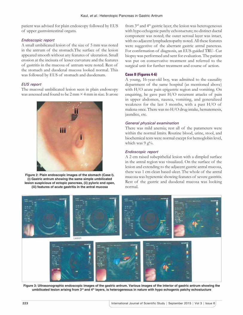
EUS reportThe mucosal umbilicated lesion seen in plain endoscopy was assessed and found to be 2 mm × 4 mm in size. It arose
from 3rd and 4th gastric layer, the lesion was heterogeneous with hypo echogenic patchy echostucture; no distinct ductal component was noted; the outer serosal layer was intact, with no adjacent lymphadenopathy noted. All these features were suggestive of the aberrant gastric antral pancreas. For confi rmation of diagnosis, an EUS-guided TRU- Cut biopsy was performed and sent for evaluation. The patient was put on conservative treatment and referred to the surgical unit for further treatment and course of action.
Case II (Figures 4-6)A young, 16-year-old boy, was admitted to the causality department of the same hospital (as mentioned above) with H/O acute pain epigastric region and vomiting. On enquiring, he gave past H/O recurrent attacks of pain in upper abdomen, nausea, vomiting, and generalized weakness for the last 3 months, with a past H/O of malena once. There was no H/O drug intake, hematemesis, jaundice, etc.
General physical examinationThere was mild anemia; rest all of the parameters were within the normal limits. Routine blood, urine, stool, and biochemical tests were normal except for hemoglobin level, which was 9 g%.
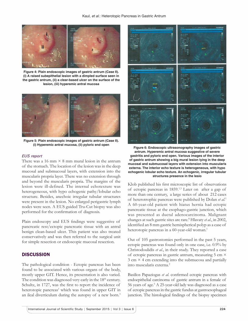
Endoscopic reportA 2 cm raised subepithelial lesion with a dimpled surface in the antral region was visualized. On the surface of the lesion and extending to the adjacent gastric antral mucosa, there was 1 cm clean based ulcer. The whole of the antral mucosa was hyperemic showing features of severe gastritis. Rest of the gastric and duodenal mucosa was looking normal.
Figure 2: Plain endoscopic images of the stomach (Case I). (i) Gastric antrum showing the same simple umbilicated
lesion suspicious of ectopic pancreas, (ii) pyloric end open, (iii) features of acute gastritis in the antral mucosa
Figure 3: Ultrasonographic endoscopic images of the gastric antrum. Various images of the interior of gastric antrum showing the umbilicated lesion arising from 3rd and 4th layers, is heterogeneous in nature with hypo echogenic patchy echostucture
Kaul, et al.: Heterotopic Pancreas in Gastric Antrum
224International Journal of Scientifi c Study | September 2015 | Vol 3 | Issue 6
Klob published his fi rst microscopic list of observations of ectopic pancreas in 1859.1,3 Later on after a gap of more than one century, a large series of about 212 cases of heterotrophic pancreas were published by Dolan et al.1 A 60-year-old patient with hiatus hernia had ectopic pancreatic tissue at the esophago-gastric junction, which was presented as ductal adenocarcinoma. Malignant changes at such gastric sites are rare.8 Hlavaty et al., in 2002, identifi ed an 8 mm gastric hemispherical polyp as a case of heterotopic pancreas in a 60-year-old woman.1
Out of 105 gastrostomies performed in the past 5 years, ectopic pancreas was found only in one case, i.e. 0.9% by Christodoulidis et al., in their study. They reported a case of ectopic pancreas in gastric antrum, measuring 5 cm × 3 cm × 4 cm extending into the submucosa and partially into muscularis externa.2
Basilios Papaziogas et al. confi rmed ectopic pancreas with endoepithelial carcinoma of gastric antrum in a female of 56 years of age.5 A 25-year-old lady was diagnosed as a case of ectopic pancreas in the gastric fundus at gastroesophageal junction. The histological fi ndings of the biopsy specimen
Figure 4: Plain endoscopic images of gastric antrum (Case II). (i) A raised subepithelial lesion with a dimpled surface seen in the gastric antrum, (ii) a clear-based ulcer on the surface of the
lesion, ( iii) hyperemic antral mucosa
Figure 5: Plain endoscopic images of gastric antrum (Case II). (i) Hyperemic antral mucosa, (ii) pyloric end open
Figure 6: Endoscopic ultrasonography images of gastric antrum. Hyperemic antral mucosa suggestive of severe
gastritis and pyloric end open. Various images of the interior of gastric antrum showing a big mural lesion lying in the deep
mucosal and submucosal layers with extension into muscularis externa. The interior echo texture is heterogeneous, with hypo echogenic lobular echo texture. An echogenic, irregular tubular
structures presence in the lesio
EUS reportThere was a 16 mm × 8 mm mural lesion in the antrum of the stomach. The location of the lesion was in the deep mucosal and submucosal layers, with extension into the muscularis propria layer. There was no extension through and beyond the muscularis propria. The margins of the lesion were ill-defined. The internal echotexture was heterogeneous, with hypo echogenic pathy/lobular echo structure. Besides, anechoic irregular tubular structures were present in the lesion. No enlarged perigastric lymph nodes were seen. A EUS guided Tru-Cut biopsy was also performed for the confi rmation of diagnosis.
Plain endoscopy and EUS fi ndings were suggestive of pancreatic rest/ectopic pancreatic tissue with an antral benign clean-based ulcer. This patient was also treated conservatively and was then referred to the surgical unit for simple resection or endoscopic mucosal resection.
DISCUSSION
The pathological condition - Ectopic pancreas has been found to be associated with various organs of the body, mostly upper GIT. Hence, its presentation is also varied. The condition was diagnosed very early in the 18th century. Schultz, in 1727, was the fi rst to report the incidence of heterotopic pancreas1 which was found in upper GIT in an ileal diverticulum during the autopsy of a new born.3

Kaul, et al.: Heterotopic Pancreas in Gastric Antrum
225 International Journal of Scientifi c Study | September 2015 | Vol 3 | Issue 6
of the same patient were reported as gastric heterotopic pancreas with pancreatic intraepithelial neoplasm-2 which is believed to represent a precursor lesion for the development of ductal adenocarcinoma.9 Various sites other than the stomach, where ectopic pancreas was diagnosed by various researchers are: A 5-year-old child with a diagnosis of Meckel’s diverticulum was found to have heterotopic pancreatic tissues in the various parts of the gastrointestinal tract by Baysoy et al.10 An isolated heterotopic pancreas, at the terminal ileum of a 47-year-old male, was found to be the cause of ileo-ileal intussusception by Ahmed Monier et al.11 Heterotopic pancreas in the spleen with malignant degeneration to mucinous cyst adenocarcinoma was reported by Nisar et al.12
A case of heterotopic pancreas adjacent to ampulla of vater mimicking cholangiocarcinoma was reported by Atindriya Biswas et al. in UK.3 Goodarzi et al. reported a case of pancreatic heterotopia in the rectum of a 42-year-old female in whom it had turned into ductal adenocarcinoma.13 An 18-year-old post-pubertal girl having an H/O cholecystitis was diagnosed as a case of heterotopic pancreas in the gallbladder, the ectopic pancreas being responsible for cholecystitis.14 A 45-year-old healthy lady who went for a routine medical check-up was found to have soft tissue mass in the left lobe of liver on CT scan. Left hepatic lobectomy was performed and subsequent histopathologicalstudy of the mass removed revealed the presence of ectopic pancreatic tissue present in the liver. This intrahepatic pancreatic tissue had changed into adenocarcinoma.15 Lizhi-Zhang et al reported two very rare cases of ectopic pancreatic mass in anterior. Mediastinum of thoracic cavity in two young females who were15 and16 years old respectively.16
CONCLUSION
Although pancreatic heterotopia is a rare entity, yet it should always be considered in the differential diagnosis of extra mucosal gastric lesion/GI stromal tumor and any undiagnosed abdominal ailment. The quick and easy diagnostic tool is to do EUS and a simultaneous biopsy.
Such diagnostic procedures are all the most important in the cases where pancreatic heteropenia presents primarily as pancreatitis, hyperinsulinism, Zollinger Ellison syndrome, common bile duct obstruction, etc. It is often impossible to distinguish gastric pancreatic heterogenic tissue from primary or metastatic cancer on endoscopy because endoscopic biopsies are sometimes unremarkable. Hence, a frozen section should be taken at the time of surgery to confi rm
the diagnosis. Surgical excision provides symptomatic relief and treatment also. Surgery is recommended, especially if the diagnosis is uncertain. Our case study of two patients revealed that ectopic pancreas can also be present at a young age as described by a few authors rather than conventionally declared in the third or fourth decade of life. Our patients had the same diagnosis and were also related to each other. This fact further enlightens the role of genetic theory in the pathogenesis of ectopic pancreas. It is presumed that both of them had some common genetic pattern and abnormal genetic signaling pathway which predisposed to the formation of ectopic pancreas in both individuals, that too involving antrum of the stomach in both cases.
REFERENCES
1. Hlavaty T, Lukac L, Vyskocil M, Galbavy S. Heterotopic pancreas in gastric antrum with macroscopic appearance of gastric polyp. Bratisl Lek Listy 2002;103:117-20.
2. Christodoulidis G, Zacharoulis D, Barbanis S, Katsogridakis E, Hatzitheofi lou K. Heterotopic pancreas in the stomach: A case report and literature review. World J Gastroenterol 2007;13:6098-100.
3. Biswas A, Hussain EA, Feakins RM, Abraham AT. Heterotopic pancreas mimicking Cholangiocarcinoma. A case report and literature review. J Pancreas (Online) 2007;8:28-34.
4. Kim GH. EUS fi ndings of gastric ectopic pancreas. Video J Encyclopaedia GI Endosc 2012;1:160-1.
5. Papaziogas B, Koutelidakis I, Tsiaousis P, Panagiotopoulou K, Paraskevas G, Argiriadou H, et al. Carcinoma developing in ectopic pancreatic tissue in the stomach: A case report. Cases J 2008;1:249.
6. Fukuda A, Kawaguchi Y, Furuyama K et al. Ectopic pancreas formation in Hes1-knockout mice reveals plasticity of endodermal progenitors of the gut, bile duct, and pancreas. J Clin Invest 2006;116:1484-93.
7. Hebrok M. Hedgehog signaling in pancreas development. Mech Dev 2003;120:45-57.
8. Guillou L, Nordback P, Gerber C, et al. Ductal adenocarcinoma arising in a heterotopic pancreas situated in a hiatal hernia. Arch Pathol Lab Med 1994;118:568-71.
9. Sadeghi NR, Godambe A, Shienbaum AJ, Alloy A. Premalignant gastric heterotopic pancreas. Gastroenterol Hepatol (N Y) 2008;4:218-21.
10. Baysoy G, Balamtekin N, Uslu N, Karavelioglu A, Talim B, Ozen H. Double heterotopic pancreas and Meckel’s diverticulum in a child: Do they have a common origin? Turk J Pediatr 2010;52:336-8.
11. Monier A, Awad A, Szmigielski W, Muneer M, Alrashid A, Darweesh A, et al. Heterotopic pancreas: A rare cause of ileo-ileal intussusception. Pol J Radiol 2014;79:349-51.
12. Nisar PJ, Zaitoun AM, Lobo DN, Rowlands BJ. Heterotopic pancreas in the spleen: Malignant degeneration to mucinous cystadenocarcinoma. Eur J Gastroenterol Hepatol 2002;14:793-6.
13. Goodarzi M, Rashid A, Maru D. Invasive ductal adenocarcinoma arising from pancreatic heterotopia in rectum: Case report and review of literature. Hum Pathol 2010;41:1809-13.
14. Elhence P, Bansal R, Agrawal N. Heterotopic pancreas in gall bladder associated with chronic cholecystolithiasis. Int J Appl Basic Med Res 2012;2:142-3.
15. Yan ML, Wang YD, Tian YF, Lin Y. Adenocarcinoma arising from intrahepatic heterotopic pancreas: A case report and literature review. World J Gastroenterol 2012;18:2881-4.
16. Zhang L, Peng LQ, Yu JQ, Yuan HM, Chu ZG, Zeng HJ, et al. Ectopic pancreas in the anterior mediastinum: A report of two cases and review of the literature. Oncol Lett 2014;7:1053-1056.
How to cite this article: Kaul N, Kapoor H, Kaul V. Heterotopic Pancreas in Gastric Antrum: A Report of Two Cases. Int J Sci Stud 2015;3(6):221-225.
Source of Support: Nil, Confl ict of Interest: None declared.

![Adenocarcinoma arising from a heterotopic pancreas in the ... · evidence, the incidence of HP is estimated to range from 0.25 to 1.2% [1, 2]. Notably, the incidence of malignant](https://img.dokumen.tips/doc/110x75/5f0ae28d7e708231d42dd194/adenocarcinoma-arising-from-a-heterotopic-pancreas-in-the-evidence-the-incidence.jpg)


![International Journal of Clinical EndocrinologyTh e heterotopic pancreas is histologically divided into three types, according to von Heinrich’s classifi cation [1]. Type I had ducts,](https://img.dokumen.tips/doc/110x75/5f4b2d8546fe527db76dd962/international-journal-of-clinical-endocrinology-th-e-heterotopic-pancreas-is-histologically.jpg)