Embed Size (px)
Citation preview

RESEARCH ARTICLE
Hepatocyte-like cells reveal novel role of SERPINA1 intransthyretin amyloidosisChristoph Niemietz, Lutz Fleischhauer*, Vanessa Sandfort, Sarah Guttmann, Andree Zibertand Hartmut H.-J. Schmidt‡
ABSTRACTTransthyretin (TTR)-related familial amyloid polyneuropathy (ATTR)results from aggregation and extracellular disposition of misfoldedTTR mutants. Growing evidence suggests the importance of hepaticchaperones for the modulation of pathogenesis. We took advantageof induced pluripotent stem cell (iPSC)-derived hepatocyte-like cells(HLCs) from ATTR patients (ATTR-HLCs) to compare chaperonegene expression to that in HLCs from healthy individuals (H-HLCs).From the set of genes analyzed, chaperones that are predominantlylocated extracellularly were differently expressed. Expression of thechaperones showed a high correlation with TTR in both ATTR-HLCsand H-HLCs. In contrast, after TTR knockdown, the correlation wasmainly affected in ATTR-HLCs suggesting that differences in TTRexpression triggers aberrant chaperone expression. Serpin family Amember 1 (SERPINA1) was the only extracellular chaperone thatwas markedly upregulated after TTR knockdown in ATTR-HLCs.Co-immunoprecipitation revealed that SERPINA1 physically interactswith TTR. In vitro assays indicated that SERPINA1 can interferewith TTR aggregation. Taken together, our results suggest thatextracellular chaperones play a crucial role in ATTR pathogenesis, inparticular SERPINA1, which may affect amyloid formation.
KEY WORDS: SERPINA1, Transthyretin, Chaperones, Diseasemodeling, Induced pluripotent stem cells (iPSCs)
INTRODUCTIONTransthyretin amyloidosis (ATTR) is a rare, progressive diseasecaused by mutations of transthyretin (TTR). ATTR is thought toresult from the formation of amyloid fibrils, primarily affecting theperipheral autonomic nervous system and the heart (Conceiçãoet al., 2016). More than 100 disease-causing mutations of the TTRgene have been described. TTR displays extensive β-sheet structuresprone to self-oligomerization (Blake and Serpell, 1996). TTR is atetrameric protein and it is assumed that kinetic instability of mutantproteins favors fibril formation (Foss et al., 2005). TTR synthesisand secretion into the blood predominantly takes place in the liver,while minor sites of synthesis (<5%) include the choroid plexus,retinal pigment epithelium, pancreas, placenta and the Schwanncells (Buxbaum et al., 2014; Cavallaro et al., 1990; Schreiber,2002). TTR functions as a plasma protein responsible for the
transport of thyroxine (T4) and the retinol-binding protein (RBP4),which acts as a carrier for retinol (vitamin A) (Monaco et al., 1995;Neumann et al., 2001). TTR−/− mice, however, do not showsignificant signs of disease and are fertile (Wei et al., 1995).
The impact of the protein quality control (PQC) system has beenassociated to the pathogenesis of amyloidotic diseases, e.g. inamyotrophic lateral sclerosis (ALS), Alzheimer’s and Parkinson’sdisease (Daturpalli et al., 2013; Park et al., 2018; Wang et al., 2014).Several lines of evidence suggest that PQC, operative during thesynthesis of TTR in the hepatocyte, is also an important determinantof ATTR. In a pioneering work, biophysical and secretion assayssuggested the contribution of endoplasmic reticulum (ER)-assistedfolding by cell type-specific chaperones (Sekijima et al., 2005).TTR mutations ATTRD18G and ATTRA25T, both associated withlate-onset ATTR of the central nervous system, are found at lowconcentrations in the blood (Hammarström et al., 2003; Sekijimaet al., 2003). For ATTRD18G, low secretion efficiency has beenobserved in HEK cells, suggesting that ER retention of misfoldedTTR results in the induction of the unfolded protein response(UPR), including upregulation of the ER chaperone glucose-regulated protein 78 (GRP78) (Sato et al., 2007). More recently,proteome analysis (2D-DIGE) following expression of the TTRmutation ATTRL55P in yeast revealed the induction of chaperonesthat mediate protein folding, including members of the HSP70,peptidyl-prolyl-cis-trans isomerase (PPIase) and ubiquitin-likemodifier (SUMO) families (Gomes et al., 2012). Several TTRmutations were shown to be subjected to ER-associated degradation(ERAD) pathways (Sato et al., 2012). Multi-dimensional proteinidentification technology (MudPIT) profiling of adipose tissue hassuggested the overexpression of several chaperones in ATTRpatients (Brambilla et al., 2013). Of note, proteome analyses ofplasma from ATTRV30M patients indicated altered expression ofseveral extracellular chaperones (da Costa et al., 2015).
The protease inhibitor SERPINA is also highly expressed in theliver and strongly secreted into blood (Topic et al., 2012).SERPINA1 deficiency results in a genetic disorder resulting indamage of the liver and the lung due to uncontrolled neutrophilelastase and non-functional SERPINA1 in the liver (Radlovic et al.,2014). Direct interaction of TTR and SERPINA1, however, has notyet been described.
Whereas previous studies analyzed whole liver, human serum,adipose tissue or a variety of standard cell culture lines, such asHEK293 cells, to assess the impact of PQC on gene expression, wetook advantage of hepatocyte-like cells (HLCs) differentiated frominduced pluripotent stem cells (iPSCs). Gene expression ofchaperones related to amyloidosis was explored by real-timeqPCR analysis in HLCs with the intrinsic genetic background ofATTR patients, i.e. in HLCs derived from ATTR patients (ATTR-HLCs). TTR knockdown in HLCs was employed to study therelation of TTR and individual chaperone gene expression. WeReceived 9 May 2018; Accepted 18 September 2018
Medizinische Klinik B fur Gastroenterologie und Hepatologie, UniversitatsklinikumMunster, 48149 Munster, Germany.*Present address: Fakultat fur angewandte Naturwissenschaften und Mechatronik,Hochschule Munchen, 80335 Munchen, Germany.
‡Author for correspondence ([email protected])
C.N., 0000-0002-9393-8748; S.G., 0000-0001-8419-9501; A.Z., 0000-0002-5188-450X; H.H.-J.S., 0000-0002-2402-7764
1
© 2018. Published by The Company of Biologists Ltd | Journal of Cell Science (2018) 131, jcs219824. doi:10.1242/jcs.219824
Journal
ofCe
llScience

identified SERPINA1 as a highly regulated chaperone in ATTR-HLCs. Further analyses revealed a physical interaction ofSERPINA1 with TTR as well as its interference with TTRaggregation, suggesting that SERPINA1 represents a novel targetfor therapy of ATTR.
RESULTSComparison of cells derived from ATTR patients and healthydonorsIn this study, ATTR-HLCs were used to assess chaperone geneexpression. HLCs from healthy individuals (H-HLCs) served ascontrol. The characteristics of the four ATTR cell lines and the fourcell lines derived from healthy individuals are given in Table S1.Weused a protocol for reprogramming and differentiation that has beenshown to be very robust in terms of variability (Niemietz et al.,2016). After reprogramming of primary patient cells, the cellsdisplayed typical iPS cell colony growth (Fig. 1A), highimmunofluorescence staining of pluripotent stem cell markersOCT4, NANOG, SSEA-4, and TRA-1-60 (Fig. 1B), high mRNAlevels of embryonic stem cell-specific markers OCT4, NANOG andSOX2 (Fig. 1C), and expected expression levels in the trilineagehPSC Scorecard™ Assay (Fig. 1D) suggesting that typical iPSCshave been derived from ATTR patients and healthy individuals.After a 14 day differentiation protocol of iPSCs, the cells showedtheir characteristic polygonal shape and high nuclear to cytoplasmicratio that is typical of HLCs (Fig. 1E), overall highimmunofluorescence staining of albumin, HNF4A and TTR(Fig. 1F), high glycogen storage (Fig. 1G), and high expressionlevels of the hepatocyte marker genes serum albumin (ALB),apolipoprotein A1 (APOA1), ATPase copper transporting Beta(ATP7B), nuclear constitutive androstane receptor (NR1I3, alsoknown as CAR) and transferrin (TF) (Fig. 1H). These data indicatethat phenotypes, function as well as expression levels of hepatocytemarker genes are almost identical in iPSCs and HLCs derived fromATTR patients and healthy controls, when cells were subjected tothe reprogramming and differentiation protocols. Importantly,variation was low between expression of hepatic marker genes inHLCs derived from H-HLCs and ATTR-HLCs when using at leasttwo clones per line (±s.e.<1.1) (Fig. S1).
Almost identical TTR expression in HLCs derived from ATTRpatients and healthy individualsSince the relation of TTR to chaperone gene expression was of highimportance to our study, we further assessed whether overall TTRexpression might differ between ATTR-HLCs and H-HLCs. TTRmRNA and TTR protein secreted into the cell culture medium byHLCs did not differ between both groups (Fig. 2A,B). Of note, TTRmRNA levels observed in HLCs were similar to those in primaryhuman hepatocytes, suggesting that HLCs model humanhepatocytes (Niemietz et al., 2016). We also inspected thedifferent forms of TTR by western blot analyses and did notobserve differences between both groups of HLCs (Fig. 2C),suggesting that any deviation that might be observed for chaperonegene expression between ATTR-HLCs and H-HLCs was not biasedby the level of TTR expression.
Gene expression of extracellular chaperones ispredominantly affected in HLCs derived from ATTR patientsHaving verified that TTR appears to be synthesized and secreted in asimilar manner and magnitude in both groups of HLCs, RT-qPCRanalyses of chaperone genes were performed. In total, 39 genes wereinvestigated (Table S2). Selection of genes was based on PubMed
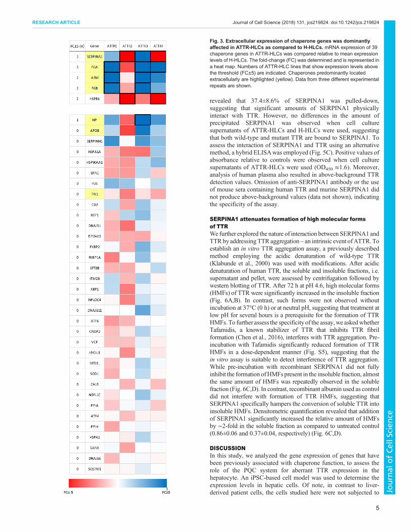
searches for genes reported to be associated with protein folding,degradation, ubiquitylation and translational arrest in amyloidoticdieseases, such as ATTR, amyotrophic lateral sclerosis (ALS),Alzheimer’s disease, amyloid light-chain (AL) amyloidosis andamyloid A (AA) amyloidosis. All genes were first validated inhuman hepatoma HepG2 cells and found to be robustly expressed(data not shown). The expression of the chaperone genes wascategorized according to a threshold of fold-change (FC) that takesinto account the variability of hepatocyte marker expression(FC<±2.5). A FC≥±5 (two times that of the mean hepatic markergene variability) was chosen to indicate significance of chaperonegene expression between groups. Of note, differences in individualchaperone gene expression in the cell lines were low (±s.e.<1.3)(Fig. S2). Five chaperone genes, i.e. SERPINA1, fibrinogen alphachain (FGA), fibrinogen beta chain (FGB), alpha-2-macroglobulin(A2M) and heat shock protein beta-1 (HSPB1), were found to differin at least two ATTR-HLC lines, whereas 34 genes showedexpression levels below the threshold (Fig. 3). Expression levels ofSERPINA1 and FGA differed in three ATTR-HLC lines.Upregulation was found for FGA (3 ATTR-HLC lines),A2M (2 ATTR-HLC lines) and FGB (2 ATTR-HLC lines).Downregulation was observed for HSPB1 (2 ATTR-HLC lines).SERPINA1 displayed upregulation in one ATTR-HLC line anddownregulation in two ATTR-HLC lines. However, variation inSERPINA1 expressionwas very low in the four H-HLC lines suggestingthat variability due to different donors to generate HLCs is generally low(Fig. S3). Of note, four of the genes (SERPINA1, FGA, A2M and FGB)belong to a group of seven chaperones that are predominantly locatedextracellularly (da Costa et al., 2015). In contrast, 32 chaperones thathave been reported to predominantly show intracellular localization didnot exceed a FC≥±5, indicating different expression levels in ATTR-HLCs as compared to H-HLCs (Fig. 3).
SERPINA1 expression is closely related to that of TTRin HLCsChaperone gene expression in HLCs could be associated to theoverall level of TTR expression. We used small interfering RNAs(siRNA) and antisense oligonucleotides (ASO) for knockdown ofTTR expression in the HLCs (Niemietz et al., 2016). As expected,TTR downregulation was almost identical in ATTR-HLCs and H-HLCs resulting in an mRNA knockdown of 75% to 85% and aprotein knockdown of 67% to 85% (Fig. S4). The expression of thefive chaperones (SERPINA1, FGA, A2M, FGB and HSPB1) thatwere found to be differently regulated in at least two ATTR-HLClines (Fig. 3) were examined for the effect of TTR knockdown. First,the correlation coefficient of TTR mRNA before and afterknockdown was determined in H-HLCs and ATTR-HLCs(Fig. 4A,B). Whereas no correlation (r<0.1) was observed forHSPB1, a high correlation (r>0.7) was observed for the otherchaperone genes prior TTR knockdown in H-HLCs and ATTR-HLCs. However, after TTR knockdown, the correlation coefficientsignificantly (r>0.5) dropped in ATTR-HLCs. Inspection of thechaperone mRNA level after TTR knockdown revealed a significantincrease of SERPINA1 in ATTR-HLCs, whereas no induction wasobserved for H-HLCs (Fig. 4C), and the other chaperone genes wereonly marginally affected by TTR knockdown in both HLC groups.Patient-specific analysis of TTR mRNA expression in the fourATTR-HLC lines indicated an upregulation of SERPINA1 mRNAin three out of four patient-specific HLC lines (Fig. 4D). Secretionof SERPINA1 fromHLCs after TTR knockdown was also assessed.No significant change in SERPINA1 levels was observed after TTRknockdown for H-HLCs, whereas SERPINA1 protein levels were
2
RESEARCH ARTICLE Journal of Cell Science (2018) 131, jcs219824. doi:10.1242/jcs.219824
Journal
ofCe
llScience

Fig. 1. See next page for legend.
3
RESEARCH ARTICLE Journal of Cell Science (2018) 131, jcs219824. doi:10.1242/jcs.219824
Journal
ofCe
llScience

elevated in three of four ATTR-HLCs (Fig. 4E), suggesting that – inATTR-HLCs – SERPINA1 represents a chaperone that is inverselycorrelated to expression of TTR.
SERPINA1 physically interacts with TTRHigh levels of SERPINA1 and TTR are found in human serum.However, a physical interaction between SERPINA1 and TTR hasnot been reported. We assessed interaction of SERPINA1 and TTRby co-immunoprecipitation using cell culture supernatants derivedfrom HLCs. First, expression of SERPINA1 was determined bywestern blot analysis of HLC cell culture supernatants. A singleprotein band of ∼55 kDa was observed in the supernatants ofATTR-HLCs and H-HLCs, which is similar to that in humanplasma samples (Fig. 5A). Next, HLC cell culture supernatants wereincubated with anti-TTR antibody for pull-down usingimmunobeads, followed by western blot analysis to detectco-precipitated SERPINA1. A SERPINA1-specific band wasobserved for cell culture supernatants derived from ATTR-HLCsand H-HLCs (Fig. 5B). Of note, SERPINA1 could not beprecipitated from HLC supernatants when non-specific antibodywas used for pull-down. Densitometric quantification of theSERPINA1 co-immunoprecipitates relative to the loading control
Fig. 1. Similar molecular and phenotypical characteristics of cellsobtained from ATTR patients and healthy individuals. (A) Brightfieldimages of cell colonies derived from ATTR and healthy individuals afterreprogramming. Scale bars: 100 µm. (B) Immunofluorescence afterreprogramming. Scale bars: 50 µm. (C) mRNA expression of markersindicating pluripotency. Values derived from four ATTR-HLCs and four H-HLCsare shown (data from three different experimental repeats). (D) Sample scoresof EBs relative to an undifferentiated reference set. ATTR (red, green) anddonor-EBs established from healthy individuals (black, blue) arerepresentatively shown. (E) Brightfield images showing hepatic differentiationof iPSC-derived cells at day 14. Scale bars: 50 µm. (F) Immunofluorescencestains of iPSC-derived cells at day 14 of hepatic differentiation. Scale bars:50 µm. (G) PAS reaction indicating glycogen synthesis of cells at day 14 ofhepatic differentiation. One typical experiment is shown. Scale bars: 50 µm.(H) RT-qPCR results of five liver markers expressed in four ATTR-HLCs at day14 of hepatic differentiation (data from three different experimental repeats).
Fig. 2. Similar TTR expression in HLCs derived from ATTRpatients and healthy individuals. (A) Boxplot representationof TTR mRNA expression in four ATTR-HLCs and four H-HLCs.ΔCt versus GAPDH was determined (data from three differentexperimental repeats). (B) Boxplot representation of TTR proteindetermined in the supernatants of four ATTR-HLCs and fourH-HLCs by ELISA (data from three different experimental repeats).(C) Western blot analysis of TTR expression in HLCs; 15 µl of cellculture supernatant derived from ATTR-HLCs and H-HLCs wassubjected to SDS-PAGE. Glutaraldehyde (GA) of supernatants isindicated. Blots were derived from different gels and assembledaccording tomolecular weight standards. Plasma (1 µl) served as acontrol. One of three typical experiments is shown.
4
RESEARCH ARTICLE Journal of Cell Science (2018) 131, jcs219824. doi:10.1242/jcs.219824
Journal
ofCe
llScience
revealed that 37.4±8.6% of SERPINA1 was pulled-down,suggesting that significant amounts of SERPINA1 physicallyinteract with TTR. However, no differences in the amount ofprecipitated SERPINA1 was observed when cell culturesupernatants of ATTR-HLCs and H-HLCs were used, suggestingthat both wild-type and mutant TTR are bound to SERPINA1. Toassess the interaction of SERPINA1 and TTR using an alternativemethod, a hybrid ELISAwas employed (Fig. 5C). Positive values ofabsorbance relative to controls were observed when cell culturesupernatants of ATTR-HLCs were used (OD450 ≈1.6). Moreover,analysis of human plasma also resulted in above-background TTRdetection values. Omission of anti-SERPINA1 antibody or the useof mouse sera containing human TTR and murine SERPINA1 didnot produce above-background values (data not shown), indicatingthe specificity of the assay.
SERPINA1 attenuates formation of high molecular formsof TTRWe further explored the nature of interaction between SERPINA1 andTTR byaddressing TTR aggregation – an intrinsic event ofATTR. Toestablish an in vitro TTR aggregation assay, a previously describedmethod employing the acidic denaturation of wild-type TTR(Klabunde et al., 2000) was used with modifications. After acidicdenaturation of human TTR, the soluble and insoluble fractions, i.e.supernatant and pellet, were assessed by centrifugation followed bywestern blotting of TTR. After 72 h at pH 4.6, high molecular forms(HMFs) of TTR were significantly increased in the insoluble fraction(Fig. 6A,B). In contrast, such forms were not observed withoutincubation at 37°C (0 h) or at neutral pH, suggesting that treatment atlow pH for several hours is a prerequisite for the formation of TTRHMFs. To further assess the specificityof the assay, we askedwhetherTafamidis, a known stabilizer of TTR that inhibits TTR fibrilformation (Chen et al., 2016), interferes with TTR aggregation. Pre-incubation with Tafamidis significantly reduced formation of TTRHMFs in a dose-dependent manner (Fig. S5), suggesting that thein vitro assay is suitable to detect interference of TTR aggregation.While pre-incubation with recombinant SERPINA1 did not fullyinhibit the formation ofHMFs present in the insoluble fraction, almostthe same amount of HMFs was repeatedly observed in the solublefraction (Fig. 6C,D). In contrast, recombinant albumin used as controldid not interfere with formation of TTR HMFs, suggesting thatSERPINA1 specifically hampers the conversion of soluble TTR intoinsoluble HMFs. Densitometric quantification revealed that additionof SERPINA1 significantly increased the relative amount of HMFsby ∼2-fold in the soluble fraction as compared to untreated control(0.86±0.06 and 0.37±0.04, respectively) (Fig. 6C,D).
DISCUSSIONIn this study, we analyzed the gene expression of genes that havebeen previously associated with chaperone function, to assess therole of the PQC system for aberrant TTR expression in thehepatocyte. An iPSC-based cell model was used to determine theexpression levels in hepatic cells. Of note, in contrast to liver-derived patient cells, the cells studied here were not subjected to
Fig. 3. Extracellular expression of chaperone genes was dominantlyaffected in ATTR-HLCs as compared to H-HLCs. mRNA expression of 39chaperone genes in ATTR-HLCs was compared relative to mean expressionlevels of H-HLCs. The fold-change (FC) was determined and is represented ina heat map. Numbers of ATTR-HLC lines that show expression levels abovethe threshold (FC±5) are indicated. Chaperones predominantly locatedextracellularly are highlighted (yellow). Data from three different experimentalrepeats are shown.
5
RESEARCH ARTICLE Journal of Cell Science (2018) 131, jcs219824. doi:10.1242/jcs.219824
Journal
ofCe
llScience

secondary regulation by other organs or factors present in the blood.In our cohort of chaperone genes chosen for qRT-PCR analysis ofHLCs, we observed that expression of several genes wassignificantly different in ATTR-HLCs as compared to H-HLCs.Notably, chaperones predominantly associated to extracellularlocation were affected in ATTR-HLCs, whereas genes primarilyinvolved in ERAD and UPR pathways were mostly expressedat levels found in cells that had been derived from healthyindividuals. Both last-mentioned systems have been described tofail detection of TTR mutants – i.e. ATTR-L55P and ATTR-V122I –that show high thermodynamic stability resulting in high secretionefficiency (Chen et al., 2014; Sekijima et al., 2005). Accordingly,ATTRV30M, which we investigated here in two ATTR-HLC lines,had been classified to have similar thermodynamic stability as
wild-type TTR (Sato et al., 2007; Sekijima et al., 2005). In addition,ATTRG47A and ATTRR34T, the other two amyloidogenic mutationsstudied, share central clinical parameterswithATTRV30M (González-Duarte et al., 2013; Patrosso et al., 1998). Our observation, therefore,indicates that transcription mediated through ERAD and UPR pathwaysis not significantly affected in the presence of TTR mutants with a highthermodynamic stability.
Microarray analyses of liver biopsy samples, the current goldstandard to assess differently regulated hepatic gene expression inATTR, indicated a modulation of ER-associated proteins in patientscarrying ATTRV30M (Norgren et al., 2014), e.g. gene expression ofFK506 binding protein 2 (FKBP2) was significantly downregulated.In our study, a disparate regulation of this gene was not observed.However, apart from the different processing events of cells and RNA
Fig. 4. SERPINA1 is closely related to TTR expression in HLCs. (A,B) Correlation of TTR with chaperone genes in H-HLCs (A) and ATTR-HLCs (B).Correlation factor (r) was calculated before and after TTR knockdown (data from four different experimental repeats). (C) mRNA expression of chaperone genesafter TTR knockdown. Data were set relative to untreated controls (data from four different experimental repeats). (D,E) SERPINA1 mRNA expression (D) andprotein levels (E) before and after TTR knockdown in the four ATTR-HLC lines (data from three different experimental repeats). *P<±0.05; ns, not significant.
6
RESEARCH ARTICLE Journal of Cell Science (2018) 131, jcs219824. doi:10.1242/jcs.219824
Journal
ofCe
llScience

that were used used to determine gene expression in the twoapproaches, other factors, such as age of patients, influence of blood-borne growth factors and course of disease may affect levels of geneexpression. HLCs derived from iPSC have been previously used forgene expression studies, also involving a few chaperone genes(Leung et al., 2013). The gene encoding heat shock protein family A(Hsp70) member 1A (HSPA1A) was one chaperone that was found tobe slightly overexpressed (∼2-fold). We found HSPA1A to bedifferently expressed in twoATTR-HLCs (FC<−2.7).Wemight have
missed the biological significance of this and other minor mRNAdysregulations of chaperone genes as we have chosen a high internalthreshold (FC<±5) to indicate significance.
In our study, SERPINA1, FGA, A2M, FGB andHSPB1were foundto be highly expressed in ATTR-HLCs as compared to H-HLCs. Ofthese, HSPB1 is the only chaperone that is predominantly foundintracellularly. Gene expression was found to be downregulated intwo ATTR-HLCs, while two other patient-specific cell lines thatdisplayed minor downregulation did not exceed a threshold of FC±5(Fig. 3). HSPB1 is known to induce sequestration of beta-amyloidand is induced in the brains of mice transgenic for the human amyloidprecursor protein (Ojha et al., 2011). Thus, HSPB1 may beoverrepresented in neuronal cells exposed to amyloid fibrils (Santoset al., 2008) but may have similar functions in hepatocytes, althoughTTR amyloid deposition is not observed in the liver.
Of the chaperones identified in this study, fibrinogen is the onlychaperone that has previously been reported to directly interact withTTR (da Costa et al., 2011; Tang et al., 2009b). The chaperoneactivity of FGA is based on the isoform fibrinogen-420 (Tang et al.,2009a), and glycation of FGA results in a loss of function (Fonsecaet al., 2016). Moreover, FGA can cause amyloid deposition as aconsequence of genetic mutation (Yazaki et al., 2018). Also, cross-seeding by FGA has to be considered as one possible mechanism bywhich amyloidic proteins spread, mostly worsening disease (Sakaiet al., 2017; Verma et al., 2018). However, aggregation-proneproteins have also been shown to inhibit crosslinking ofamyloidogenic proteins. Interestingly, this role has been describedfor monomeric TTR, which has been shown to interfere with beta-amyloid oligomerization (Garai et al., 2018; Li et al., 2013). It must,however, be taken into account that beneficial and disease-promotingelements can be closely related.
A2M has been identified to inhibit amyloid fibril formation ofaggregation-prone proteins in other diseases, such as Alzheimer’sdisease (Yerbury et al., 2009). Of note, FGA, FGB and A2M encodefor acute-phase proteins, i.e. proteins whose plasma concentrationincrease or decrease in response to inflammation (Jain et al., 2011),suggesting that upregulation of these genes is a characteristic ofATTR (Suenaga et al., 2017). At least two patient-specificATTR-HLC cell lines exhibited upregulation of FGA, FGB andA2M. In line, previous studies also report an induction of the threechaperones in plasma samples of ATTRV30M patients (da Costaet al., 2015), corroborating the validity of our HLC model.Overexpression of these chaperones in our cell model, therefore,indicates that such regulation is due to the intrinsic synthesis ofmutant TTR in the hepatocyte, rather than to a secondary regulativeresponse. It can be assumed that FGA, FGB and A2M worksynergistically, enabling hepatocytes to cope with amyloidogenicTTR. Although expression of FGA and FGB is correlated to result infunctional fibrinogen, we can only speculate on the role of A2M tocontribute to a complex cluster of chaperones targeting proteinaggregates (Wyatt et al., 2012; Yerbury et al., 2009; Zsila, 2010).Although in our approach several chaperones were identified to beassociated with ATTR pathogenesis, a rather small andheterogeneous patient group, encompassing three differentmutations and early/late onset of disease, was studied. While ourdata suggest that chaperone expression differs in hepatic cells ofATTR patients and of healthy individuals, a genotype- andphenotype-specific analysis needs a higher number of patients.
SERPINA1 was identified as a chaperone in amyloidoses, and ispart of amyloid aggregates in Alzheimer’s disease and ALS(Howlett and Moore, 2006; Zsila, 2010). In ATTR, SERPINA1 wasfound to be upregulated in plasma of ATTR patients (da Costa et al.,
Fig. 5. SERPINA1 is bound by TTR in cell culture supernatants ofHLCs. (A) Western blot analysis of SERPINA1 expressed by HLCs. Cellculture supernatant (15 µl) derived from ATTR-HLCs or H-HLCs wassubjected to SDS-PAGE. Plasma (1 µl per lane) is shown as a control.(B) Co-immunoprecipitation of TTR and SERPINA1. Cell culture supernatantsderived from ATTR-HLCs and H-HLCs were subjected to immunoprecipitationusing anti-TTR antibody followed by western blotting of SERPINA1. Loadingcontrol (15 µl) before immunoprecipitation is shown. As control, unrelatedantibody was used for pull-down. One of three typical experiments is shown.(C) Hybrid ELISA using capture antibody against SERPINA1 and detectionantibody against TTR. OD values were set relative to control (blank) at 1.0. Boxplot representation of four ATTR-HLC cell culture supernatants (diluted 1:10)and three ATTR plasma samples (diluted 1:5000). Non-conditioned cell culturemedium and murine sera were used as controls (data from five differentexperimental repeats). *P<0.05.
7
RESEARCH ARTICLE Journal of Cell Science (2018) 131, jcs219824. doi:10.1242/jcs.219824
Journal
ofCe
llScience

2015). Our findings indicate upregulation of SERPINA1 mRNA inone ATTR-specific hepatic cell line and downregulation in twoother cell lines, while one cell line did not show disparate expressionlevels as compared to healthy individuals. To our knowledgedecreased levels of SERPINA1 have not been reported for ATTR;however, Alzheimer’s patients show low SERPINA1 liquor levels(Hansson et al., 2009). As our ATTR patient cohort was low, thecourse of SERPINA1 regulation in hepatocytes cannot be proposed.However, SERPINA1 represents one of the most-regulated genes inour cohort of hepatic cell0073, suggesting that it is highlyresponsive to aberrant TTR expression in hepatocytes.The important role of SERPINA1 in ATTR is emphasized again
in our study by analysis of TTR knockdown. SERPINA1 expressionwas inversely correlated with TTR levels after knockdown,suggesting a mutual dependence of both genes. Intriguingly,investigations of serum TTR and SERPINA1 levels in cells ofpatients suffering various diseases indicates opposed levels ofexpression (Bag et al., 2014; Fatima et al., 2017; Raju M. et al.,2016). Although the exact molecular regulation of oppositeexpression levels remains to be unraveled, IL-6 driven inductionof gene expression following inflammation might play a role. It is,therefore, tempting to speculate that the process of inflammation inATTRmodulates disease (Gonçalves et al., 2017). Furthermore, ourdata indicate for the first time that TTR directly interacts with
SERPINA1. Although we did not detect any difference in thebinding of SERPINA1 to wild-type or mutant TTR in ourimmunoprecipitation assay, subtle differences might exist in vivo,depending on the TTR mutant expressed by ATTR patients.
A protocol involving acid-mediated TTR aggregation was used inour study and resulted in high-molecular forms (HMFs) of TTRas observed after western blot analysis. Since the detection ofHMFs depended on glutaraldehyde treatment prior to SDS gelelectrophoresis, we assume that HMFs are highly labile. Staining ofHMFs with Congo Red failed, possibly due to interference withglutaraldehyde (data not shown). Our data indicate that SERPINA1interferes with TTR aggregation, suggesting that the chaperone canhamper a conversion of the soluble form into insoluble aggregates.Aggregation into self-assembled fibrils of SERPINA1 has beenreported together with concurrent inhibition of these oligomers bySERPINA1 fragments (Janciauskiene et al., 1995). Several proteins,including serum amyloid P-component (SAP), apolipoprotein,complement binding factors and clusterin, have been found inamyloid deposits of patients (Azevedo et al., 2011; Klein et al.,2011), suggesting that interaction of TTR with serum proteins iscommon. Although binding of serum proteins to TTR may takeplace at different steps of amyloid formation and has diversefunctional consequences, it has previously been proposed thatclusterin preferentially binds to TTR monomers, resulting in the
Fig. 6. SERPINA1 attenuates formation of high molecular forms of TTR. (A) TTR protein (1 µg) was exposed to HMF formation by using acetate buffer(pH 4.6) and incubated for indicated time points. After centrifugation and glutaraldehyde treatment, pellet (P) and supernatant (SN) fractions were subjected toSDS-PAGE followed by western blot analysis of TTR. (B) Relative amounts of P and SN fractions were determined by densitometric quantification (data from threedifferent experimental repeats). (C) Formation of high molecular form (HMF) TTR was determined in the presence of SERPINA1 and albumin at 60 ng/ml.Treatment of TTR at pH 7.4 is shown as control. (D) Densitometric quantification of relative HMF formation in P and SN fractions was determined in presence andabsence of SERPINA1 (data from three different experimental repeats). *P<0.05; ns, not significant.
8
RESEARCH ARTICLE Journal of Cell Science (2018) 131, jcs219824. doi:10.1242/jcs.219824
Journal
ofCe
llScience

blocking of the first molecular events (Greene et al., 2015).However, as shown in our study, clusterin was not differentlyregulated in HLCs of ATTR patients. In the presence of the TTRtetramer stabilizer Tafamidis, we observed a specific pattern ofHMFs that differed from the HMF pattern after addition ofSERPINA1. Thus, we hypothesize that SERPINA1 binds to bothwild-type TTR andmutant TTR, and reduces fibril formation duringa phase other than that of TTR tetramer stabilization. By contrast,low levels of SERPINA1 might be unfavorable, as suggested bystudies on ATTR patients with SERPINA1 deficiency. An earlyonset of disease after liver transplantation was reported (Guttmannet al., 2017).The PQC system might represent an alternative therapy approach
to target amyloidosis. Promising studies, e.g. for Alzheimer’sdisease, focus on proteins of the BRICHOS family (Cohen et al.,2015; Willander et al., 2012b). It has previously been suggested thatdomains proSP-C and Bri2 can delay fibrillation of Aß40 and Aß42(Willander et al., 2012b). Proteins of the heat shock family havebeen demonstrated to efficiently regulate fibril length of α-synuclein, e.g. in Parkinson’s disease (Gao et al., 2015; Nillegodaet al., 2015). For ATTR, it has been shown in cell culture thatinduction of ATF6 results in reduced secretion of aggregation-proneTTR, possibly impeding pathogenic fibril deposition (Chen et al.,2014; Shoulders et al., 2013). Although our work here adds severalchaperones – most importantly SERPINA1 – to the growing searchfor other therapy strategies to treat ATTR, we were unable todecipher the exact biological mechanisms the identified chaperonesuse for pathogenesis.In order to determine chaperone gene expression, we used
iPSC-derived cell lines. As noted earlier, iPSC-based approachescan have shortcomings due to incomplete reprogramming anddifferentiation, most prominently when comparing differentdonors (Kajiwara et al., 2012; Ortmann and Vallier, 2017).Although we addressed variability by using identical protocolsfor iPSC generation and hepatic differentiation and an overall lowvariability of marker gene expression – including TTR andSERPINA1 – was observed, a bias due to the heterogeneity of cellpopulation cannot be excluded. Within the limitations of ourapproach, our data suggest that SERPINA1 has a role in ATTRpathogenesis, possibly by inhibiting the formation of large TTRaggregates. Our data also indicate that a concerted modulation of– mostly extracellular – chaperone genes is induced in ATTRpatients; this might be part of a complex proteostasis network,resulting in an adaptive response to aberrant TTR expression (vanOosten-Hawle and Morimoto, 2014).
MATERIALS AND METHODSEthics statementAll samples were obtained in accordance with ethics committee of theUniversitätsklinikumMünster. Participants were informed regarding the useof data and informed consent was obtained in writing from all individualsenclosed in the study.
Isolation of urine-derived cells12-well cell culture plates (Greiner Bio-One, Kremsmünster, Austria) werecoated with 0.1% gelatin (Merck Millipore, Darmstadt, Germany). Briefly,urine was centrifuged at 400 g for 10 min and cell pellets were washed with50 ml of cold PBS (Sigma-Aldrich, St. Louis, MO), 1% penicillin–streptomycin (Pen–Strep; Thermo Fisher Scientific, Waltham, MA). After asecond centrifugation, cell pellets were resuspended in UC medium[DMEM/F-12 (Life Technologies, Carlsbad, CA) supplemented with 10%FCS (Thermo Fisher Scientific), 1% Pen–Strep, 1×MEM non-essentialamino acid solution (NEAA, Sigma-Aldrich), 1× GlutaMAX-I (Life
Technologies), 0.1 mM 2-mercaptoethanol (Life Technologies) andSingleQuot Kit CC-4127 REGM (Lonza, Basel, Switzerland)]. Cells wereresuspended in 1 ml of UC medium and incubated at 37°C with 5% CO2 for24 h. Half of the medium was changed daily. At confluence, cells wereexpanded for two passages (p2). Typically – and independently from theinitial urine volume – 4×10⁶ cells were obtained after 3 weeks of cell culture.
Generation of iPSCsApproximately 0.5×10⁶ of urine-derived cells at p2 were transfected with1 µg of pCXLE-hOCT3/4-shp53-F, pCXLE-hSK and pCXLE-hUL(Addgene plasmids #27077, #27078 and #27080, respectively) by usingthe Amaxa Basic Nucleofector Kit for Primary Mammalian Epithelial Cells(VPI-1005, Lonza, Basel, Switzerland). Subsequently, cells weretransferred to Matrigel® matrix (Corning, Corning, NY) coated 6-well cellculture plates (Greiner Bio-One) and cultured for 24 h in UC medium. Atday 1, UC medium was replaced with serum-free complete cell culturemedium mTeSR-1 (Stemcell Technologies, Vancouver, BC, Canada)supplemented with 1% Pen–Strep. iPSC colonies were subcultured every7 days by using 1 U/ml Dispase (Stemcell Technologies).
Hepatic differentiationiPSC lines showing stable growth characteristics were transferred intosingle-cell suspensions using Accutase solution (Sigma-Aldrich).Typically, one 6-well cell culture plate with 4–5×10⁶ cells was used forhepatic differentiation. After 24 h, culture medium was replaced for 3 dayswith DMEM/F12, 100 ng/ml recombinant activin-A (PeproTech Germany,Hamburg, Germany), 100 ng/ml fibroblast growth factor-2 (FGF2,PeproTech Germany) and 50 ng/ml recombinant human Wnt3a (R&DSystems, Minneapolis, MN) using increasing concentrations of KnockOutSR Xenofree CTS (KSR, Life Technologies) of 0.2–2.0%. The next 8 days,cells were cultured in DMEM/F12 supplemented with 10% KSR, 1 mMNEAA, 1 mM L-glutamine, 1% dimethyl sulfoxide (Sigma-Aldrich) and100 ng/ml hepatocyte growth factor (HGF, Peprotech). To induce hepaticmaturation, cells were treated for the final 3 days with DMEM/F12supplemented with 10% KSR, 1 mmol/l NEAA, 1 mM L-glutamine and0.1 µM dexamethasone (Sigma-Aldrich).
ImmunocytochemistryFollowing fixation with 4% paraformaldehyde (Electron MicroscopySciences) and permeabilization with 0.5% Triton X-100 (Thermo FisherScientific), cells grown in a 12-well plate (1–2×10⁶ cells) were blocked with3% bovine serum albumin solution (BSA, Sigma-Aldrich) and stored at 4°Cuntil staining. Primary and secondary antibodies were diluted in 1% BSAsolution. Anti-albumin antibody (ab2406, Abcam, Cambridge, UK), anti-TTRantibody (A0002, Dako, Glostrup, Denmark), anti-HNF4a antibody (sc-6556,Santa CruzBiotechnology, Dallas, TX) and anti-NANOGantibody (sc-33759,Santa CruzBiotechnology) were used at 1:100. Anti-Oct4 antibody (60093PE,Stemcell Technologies), anti-SSEA-4 antibody (60062PE, StemcellTechnologies) and anti-TRA-1-60 antibody (60064AD, StemcellTechnologies) were used at 1:200. Antibodies against Alexa Fluor 488(A11001, Invitrogen, Carlsbad, CA), Alexa Fluor 568 (A11057, Invitrogen)and Alexa Fluor 594 (A11012, Invitrogen) were used at 1:500. Samples wereincubated with primary antibody for 2 h, and with secondary antibody for 1 hat 4°C. Photographs were taken using an Olympus CKX41-X10 microscopewith cellSens Standard 1.11 imaging software.
Quantitative real-time PCRCell pellets derived from one 6-well cell culture plate were used to isolatewhole RNA using the RNeasy Mini Kit (Qiagen, Hilden, Germany) and1 µg of RNAwas applied for first-strand synthesis using the SuperScript™III First-Strand Synthesis SuperMix for qRT-PCR kit (Invitrogen). Of thereverse transcriptase product 1 µl was mixed with Takyon ROX SYBRMaster Mix Blue dTTP (Eurogentec, Lüttich, Belgium) and 150 nMof primers (Table S2). qPCR was analyzed on the ABI Prism 7900 HTSequence Detection System (Applied Biosystems, Foster City, CA). Datawere analyzed by SDS 2.4 software (Applied Biosystems). Ct valueswere normalized to GAPDH and evaluated using the comparative Ctmethod (2−ΔΔCt).
9
RESEARCH ARTICLE Journal of Cell Science (2018) 131, jcs219824. doi:10.1242/jcs.219824
Journal
ofCe
llScience

hPSC scorecard assayEmbryoid bodies were established as previously described (Niemietz et al.,2016). 1×10⁶ cells were used to generate embryoid bodies. Briefly, iPSCclusters were cultured for 7 days in 6-well suspension culture plates (GreinerBio-One) according to the manufacturer’s protocol (TaqMan® hPSCScorecard™). The trilineage differentiation potential was examined usingthe 384-well hPSC Scorecard™ Panel (Life Technologies) followed byevaluation using the Thermo Fisher Cloud hPSC Scorecard Analysissoftware.
Periodic acid-Schiff reactionGlycogen synthesis of HLCs was assessed at day 14 of hepaticdifferentiation by periodic acid-Schiff (PAS) reaction using the systemprotocol (395B-1KT, Sigma-Aldrich).
TTR knockdownHLCs at day 14 of hepatic differentiation were transferred to Opti-MEM(Gibco). siTTR1 (Alnylam Pharmaceuticals, Cambridge, MA) was added at0.3 µM to the medium. TTR-ASO (IONIS Pharmaceuticals, Carlsbad, CA)was mixed at 1.5 µM with 2.5 µl Lipofectamine 2000 Reagent (LifeTechnologies). Scrambled oligonucleotides were used as control. Cellculture supernatants were harvested daily for 3 days.
SDS-PAGE and western blottingLysates derived from a 6-well plate were prepared using RIPA buffer (60 mMtris-HCl, 150 mM NaCl, 2% Na-deoxycholate, 2% Triton X-10, 0.2% SDSand 15 mM EDTA) in the presence of protease inhibitors (Roche, CompleteMini, EDTA-free) and separated on 10–12.5% SDS polyacrylamide gels. Forblotting Amersham Protran Premium 0.45 nitrocellulose (GE Healthcare LifeSciences, Buckinghamshire, UK) was used. After overnight blocking in 5%non-fat dry milk (AppliChem, Darmstadt, Germany) blots were incubated inTris-buffered saline (TBS)/0.05% Tween 20 (Sigma-Aldrich). Anti-TTRantibody (A0002, Dako) and anti-SERPINA1 antibody (843285, R&DSystems) were used at 1:1000 in 0.05% TBS-T with 0.5% non-fat milk for1 h. Anti-goat HRP (ab97110, Abcam) and anti-rabbit HRP (ab6721,Abcam)were used at 1:10,000. Amersham ECLWestern Blotting Detection Reagent(GEHealthcare Life Sciences) was applied. All blots were quantified by usingImageJ 1.50i analysis software.
ELISAELISA of human TTR and SERPINA1ELISA of TTR was performed as reported elsewhere (Coelho et al., 2013).Briefly, Nunc 96-well MaxiSorp™ plates (44240421, Thermo FisherScientific) were coated overnight with anti-TTR antibody (A0002, Dako) in50 mM carbonate/bicarbonate buffer (C3041-50CAP, Sigma-Aldrich) at4°C. After washing, samples were diluted in 1× powerblock (HK085-5K,Biogenex, Fremont). Cell culture supernatants were used at 1:10 and humanplasma samples were used at 1:19,600. For standard curve calculation, TTR(P1742-5MG, Sigma-Aldrich) was applied. Detection was by ab9015(Abcam; at 1:2500) and alkaline-phosphate conjugation by A5187-1ML(Sigma-Aldrich). Colorimetric detection was performed by p-Nitrophenylphosphate addition (N2770-50SET, Sigma-Aldrich). Absorbance wasdetermined at 405 nm. Human SERPINA1 DuoSet ELISA (DY1268,R&D Systems) was used according to the manufacturer’s instruction. Cellculture supernatants were used at 1:4000 and in 5% Tween 20 in PBS. TMBsubstrate (555214, BD Biosciences, Franklin Lakes, NJ) was used forcolorimetric detection. Absorbance was determined at 450 nm. HybridELISAwas performed with goat anti-human SERPINA1 antibody (843285,R&D Systems) for capture and with sheep anti-human TTR antibody(ab9015, Abcam) for detection.
In vitro TTR aggregationHuman TTR (P1742, Sigma-Aldrich) was reconstituted in 1× powerblock(BioGenex, Fremont, CA) and incubated under acidic conditions to induceTTR aggregation. 1 µg of TTR was mixed with 1× volume of 200 mMacetate buffer containing 100 mMKCl and 1 mM EDTA (pH 4.6). Sampleswere incubated at 37°C for 72 h prior centrifugation at 20,000 g, 4°C for
30 min. The supernatant was carefully removed from the pellet and bothfractions were treated with 2.5% glutaraldehyde solution (340855, Sigma-Aldrich). Samples were subjected to TTR western blotting thereafter.
Co-immunoprecipitationCell culture supernatant (15 µl) derived from 6-well plates was preclearedwith A/G Plus-Agarose beads (sc2003, Santa Cruz Biotechnology). Beadswere mixed with 2 µg of anti-TTR antibody (A0002, Dako) and incubatedwith precleared samples in non-denaturing RIPA buffer (20 mM Tris HClpH 8, 137 mM NaCl, 1% Triton X-100, 2 mM EDTA) and proteaseinhibitors while shaking at 4°C for 24 h. The next day, samples werewashed5× with PBS, centrifuged at 1000 g for 30 s and subjected to 12.5% PAGE.
Statistical analysesStatistical analyses were performed by using Kruskal–Wallis one-wayANOVA and Student’s t-test using SPSS 25 software. Data are given asmean±standard error (±s.e.m.). Correlation of two genes was determined byusing Pearson’s equation followed by Fisher’s z-transformation.
AcknowledgementsWe are grateful to O. Nadzemova for excellent technical assistance.
Competing interestsThe authors declare no competing or financial interests.
Author contributionsConceptualization: C.N., V.S., A.Z., H.H.-J.S.; Methodology: C.N., L.F., A.Z.;Validation: C.N., A.Z.; Formal analysis: C.N., L.F., A.Z.; Investigation: C.N., L.F.,V.S., S.G.; Data curation: A.Z.; Writing - original draft: C.N., A.Z., H.H.-J.S.; Writing -review & editing: V.S., S.G., A.Z., H.H.-J.S.; Visualization: C.N., A.Z.; Supervision:V.S., A.Z., H.H.-J.S.; Project administration: A.Z., H.H.-J.S.
FundingThis research received no specific grant from any funding agency in the public,commercial or not-for-profit sectors.
Supplementary informationSupplementary information available online athttp://jcs.biologists.org/lookup/doi/10.1242/jcs.219824.supplemental
ReferencesAzevedo, E. P. C., Pereira, H. M., Garratt, R. C., Kelly, J. W., Foguel, D. and
Palhano, F. L. (2011). Dissecting the structure, thermodynamic stability, andaggregation properties of the A25T transthyretin (A25T-TTR) variant involved inleptomeningeal amyloidosis: identifying protein partners that co-aggregate duringA25T-TTR fibrillogenesis in cerebrospinal fluid. Biochemistry 50, 11070-11083.
Bag, A. K., Saha, S., Sundar, S., Saha, B., Chakrabarti, A. andMandal, C. (2014).Comparative proteomics and glycoproteomics of plasma proteins in Indianvisceral leishmaniasis. Proteome Sci. 12, 48.
Blake, C. and Serpell, L. (1996). Synchrotron X-ray studies suggest that the core ofthe transthyretin amyloid fibril is a continuous β-sheet helix. Structure 4, 989-998.
Brambilla, F., Lavatelli, F., Di Silvestre, D., Valentini, V., Palladini, G., Merlini, G.and Mauri, P. (2013). Shotgun protein profile of human adipose tissue and itschanges in relation to systemic amyloidoses. J. Proteome Res. 12, 5642-5655.
Buxbaum, J. N., Roberts, A. J., Adame, A. and Masliah, E. (2014). Silencing ofmurine transthyretin and retinol binding protein genes has distinct and sharedbehavioral and neuropathologic effects. Neuroscience 275, 352-364.
Cavallaro, T., Martone, R. L., Dwork, A. J., Schon, E. A. and Herbert, J. (1990).The retinal pigment epithelium is the unique site of transthyretin synthesis in the rateye. Invest. Ophthalmol. Vis. Sci. 31, 497-501.
Chen, J. J., Genereux, J. C., Qu, S., Hulleman, J. D., Shoulders, M. D. andWiseman, R. L. (2014). ATF6 activation reduces the secretion and extracellularaggregation of destabilized variants of an amyloidogenic protein. Chem. Biol. 21,1564-1574.
Chen, J. J., Genereux, J. C., Suh, E. H., Vartabedian, V. F., Rius, B., Qu, S.,Dendle, M. T. A., Kelly, J. W. andWiseman, R. L. (2016). Endoplasmic reticulumproteostasis influences the oligomeric state of an amyloidogenic protein secretedfrom mammalian cells. Cell Chem. Biol. 23, 1282-1293.
Coelho, T., Adams, D., Silva, A., Lozeron, P., Hawkins, P. N., Mant, T., Perez, J.,Chiesa, J., Warrington, S., Tranter, E. et al. (2013). Safety and efficacy of RNAitherapy for transthyretin amyloidosis. N Engl. J. Med. 369, 819-829.
Cohen, S. I. A., Arosio, P., Presto, J., Kurudenkandy, F. R., Biverstål, H., Dolfe,L., Dunning, C., Yang, X., Frohm, B., Vendruscolo, M. et al. (2015). Amolecular
10
RESEARCH ARTICLE Journal of Cell Science (2018) 131, jcs219824. doi:10.1242/jcs.219824
Journal
ofCe
llScience

chaperone breaks the catalytic cycle that generates toxic Abeta oligomers. Nat.Struct. Mol. Biol. 22, 207-213.
Conceiçao, I., Gonzalez-Duarte, A., Obici, L., Schmidt, H. H.-J., Simoneau, D.,Ong, M.-L. and Amass, L. (2016). “Red-flag” symptom clusters in transthyretinfamilial amyloid polyneuropathy. J. Peripheral Nervous Syst. 21, 5-9.
da Costa, G., Gomes, R. A., Guerreiro, A., Mateus, É., Monteiro, E., Barroso, E.,Coelho, A. V., Freire, A. P. and Cordeiro, C. (2011). Beyond genetic factors infamilial amyloidotic polyneuropathy: protein glycation and the loss of fibrinogen’schaperone activity. PLoS ONE 6, e24850.
da Costa, G., Ribeiro-Silva, C., Ribeiro, R., Gilberto, S., Gomes, R. A., Ferreira,A. N., Mateus, É., Barroso, E., Coelho, A. V., Freire, A. P. et al. (2015).Transthyretin amyloidosis: chaperone concentration changes and increasedproteolysis in the pathway to disease. PLoS ONE 10, 1-17.
Daturpalli, S., Waudby, C. A., Meehan, S. and Jackson, S. E. (2013). Hsp90inhibits alpha-synuclein aggregation by interacting with soluble oligomers. J. Mol.Biol. 425, 4614-4628.
Fatima, I., Sadaf, S., Musharraf, S. G., Hashmi, N. and Akhtar, M. W. (2017). CD5molecule-like and transthyretin as putative biomarkers of chronic myeloidleukemia - an insight from the proteomic analysis of human plasma. Sci. Rep.7, 40943.
Fonseca, D., Gilberto, S., Ribeiro-Silva, C., Ribeiro, R., Guinote, I. B., Saraiva,S., Gomes, R. A., Mateus, E., Viana, A., Barroso, E. et al. (2016). The role offibrinogen glycation in ATTR: evidence for chaperone activity loss in disease.Biochem. J. 473, 2225-2237.
Foss, T. R., Wiseman, R. L. and Kelly, J. W. (2005). The pathway by which thetetrameric protein transthyretin dissociates. Biochemistry 44, 15525-15533.
Gao, X., Carroni, M., Nussbaum-Krammer, C., Mogk, A., Nillegoda, N. B.,Szlachcic, A., Guilbride, D. L., Saibil, H. R., Mayer, M. P. and Bukau, B. (2015).Human Hsp70 disaggregase reverses Parkinson’s-linked alpha-synucleinamyloid fibrils. Mol. Cell 59, 781-793.
Garai, K., Posey, A. E., Li, X., Buxbaum, J. N. and Pappu, R. V. (2018). Inhibitionof amyloid beta fibril formation bymonomeric human transthyretin. Protein Sci. 27,1252-1261.
Gomes, R. A., Franco, C., Da Costa, G. A., Planchon, S. B., Renaut, J., Ribeiro,R. M., Pinto, F., Silva, M. S., Coelho, A. V., Freire, A. P. et al. (2012). Theproteome response to amyloid protein expression in vivo. PLoS ONE 7, e50123.
Gonçalves, N. P., Moreira, J., Martins, D., Vieira, P., Obici, L., Merlini, G.,Saraiva, M. and Saraiva, M. J. (2017). Differential expression of Cathepsin E intransthyretin amyloidosis: from neuropathology to the immune system.J. Neuroinflammation 14, 115.
Gonzalez-Duarte, A., Lem-Carrillo, M. and Cardenas-Soto, K. (2013).Description of transthyretin S50A, S52P and G47A mutations in familialamyloidosis polyneuropathy. Amyloid 20, 221-225.
Greene, M. J., Klimtchuk, E. S., Seldin, D. C., Berk, J. L. and Connors, L. H.(2015). Cooperative stabilization of transthyretin by clusterin and diflunisal.Biochemistry 54, 268-278.
Guttmann, S., Rocken, C., Schmidt, M., Grunewald, I., Zibert, A., Stypmann, J.,Schilling, M. and Schmidt, H. (2017). De novo hereditary (familial) amyloidpolyneuropathy (FAP) in a FAP liver recipient. Amyloid 24, 126-127.
Hammarstrom, P., Sekijima, Y., White, J. T., Wiseman, R. L., Lim, A., Costello,C. E., Altland, K., Garzuly, F., Budka, H. and Kelly, J. W. (2003). D18Gtransthyretin is monomeric, aggregation prone, and not detectable in plasma andcerebrospinal fluid: a prescription for central nervous system amyloidosis?Biochemistry 42, 6656-6663.
Hansson, S. F., Andreasson, U., Wall, M., Skoog, I., Andreasen, N., Wallin, A.,Zetterberg, H. and Blennow, K. (2009). Reduced levels of amyloid-beta-bindingproteins in cerebrospinal fluid from Alzheimer’s disease patients. J. Alzheimer’sDis. 16, 389-397.
Howlett, G. J. and Moore, K. J. (2006). Untangling the role of amyloid inatherosclerosis. Curr. Opin. Lipidol. 17, 541-547.
Jain, S., Gautam, V. and Naseem, S. (2011). Acute-phase proteins: as diagnostictool. J. Pharmacy Bioallied Sci. 3, 118-127.
Janciauskiene, S., Carlemalm, E. and Eriksson, S. (1995). In vitro fibril formationfrom alpha 1-antitrypsin-derived C-terminal peptides. Biol. Chem. Hoppe-Seyler376, 415-424.
Kajiwara, M., Aoi, T., Okita, K., Takahashi, R., Inoue, H., Takayama, N., Endo, H.,Eto, K., Toguchida, J., Uemoto, S. et al. (2012). Donor-dependent variations inhepatic differentiation from human-induced pluripotent stem cells. Proc. Natl.Acad. Sci. USA 109, 12538-12543.
Klabunde, T., Petrassi, H. M., Oza, V. B., Raman, P., Kelly, J. W. and Sacchettini,J. C. (2000). Rational design of potent human transthyretin amyloid diseaseinhibitors. Nat. Struct. Biol. 7, 312-321.
Klein, C. J., Vrana, J. A., Theis, J. D., Dyck, P. J., Dyck, P. J., Spinner, R. J.,Mauermann, M. L., Bergen, H. R., III, Zeldenrust, S. R. and Dogan, A. (2011).Mass spectrometric-based proteomic analysis of amyloid neuropathy type innerve tissue. Arch. Neurol. 68, 195-199.
Leung, A., Nah, S. K., Reid,W., Ebata, A., Koch, C.M., Monti, S., Genereux, J. C.,Wiseman, R. L., Wolozin, B., Connors, L. H. et al. (2013). Induced pluripotentstem cell modeling of multisystemic, hereditary transthyretin amyloidosis. StemCell Rep. 1, 451-463.
Li, X., Zhang, X., Ladiwala, A. R. A., Du, D., Yadav, J. K., Tessier, P. M., Wright,P. E., Kelly, J. W. and Buxbaum, J. N. (2013). Mechanisms of transthyretininhibition of beta-amyloid aggregation in vitro. J. Neurosci. 33, 19423-19433.
Monaco, H. L., Rizzi, M. and Coda, A. (1995). Structure of a complex of two plasmaproteins: transthyretin and retinol-binding protein. Science 268, 1039-1041.
Neumann, P., Cody, V. and Wojtczak, A. (2001). Structural basis of negativecooperativity in transthyretin. Acta Biochim. Pol. 48, 867-875.
Niemietz, C. J., Sauer, V., Stella, J., Fleischhauer, L., Chandhok, G., Guttmann,S., Avsar, Y., Guo, S., Ackermann, E. J., Gollob, J. et al. (2016). Evaluation oftherapeutic oligonucleotides for familial amyloid polyneuropathy in patient-derivedhepatocyte-like cells. PLoS ONE 11, e0161455.
Nillegoda, N. B., Kirstein, J., Szlachcic, A., Berynskyy, M., Stank, A., Stengel,F., Arnsburg, K., Gao, X., Scior, A., Aebersold, R. et al. (2015). Crucial HSP70co-chaperone complex unlocks metazoan protein disaggregation. Nature 524,247-251.
Norgren, N., Olsson, M., Nystrom, H., Ericzon, B. G., de Tayrac, M., Genin, E.,Plante-Bordeneuve, V. and Suhr, O. B. (2014). Gene expression profile inhereditary transthyretin amyloidosis: differences in targeted and source organs.Amyloid 21, 113-119.
Ojha, J., Masilamoni, G., Dunlap, D., Udoff, R. A. and Cashikar, A. G. (2011).Sequestration of toxic oligomers by HspB1 as a cytoprotective mechanism. Mol.Cell. Biol. 31, 3146-3157.
Ortmann, D. and Vallier, L. (2017). Variability of human pluripotent stem cell lines.Curr. Opin. Genet. Dev. 46, 179-185.
Park, S. K., Arslan, F., Kanneganti, V., Barmada, S. J., Purushothaman, P.,Verma, S. C. and Liebman, S. W. (2018). Overexpression of a conserved HSP40chaperone reduces toxicity of several neurodegenerative disease proteins.Prion 12, 16-22.
Patrosso, M. C., Salvi, F., De Grandis, D., Vezzoni, P., Jacobson, D. R. andFerlini, A. (1998). Novel transthyretin missense mutation (Thr34) in an Italianfamily with hereditary amyloidosis. Am. J. Med. Genet. 77, 135-138.
Radlovic, N., Lekovic, Z., Radlovic, V., Simic, D., Topic, A., Ristic, D., Ducic, S.and Baletic, A. (2014). Alpha-1-antitrypsin deficiency in children: clinicalcharacteristics and diagnosis. Srp. Arh. Celok. Lek. 142, 547-550.
Raju M., S., V, J., Kamaraju, R. S., Sritharan, V., Rajkumar, K., Natarajan, S.,Kumar, A. D. and Burgula, S. (2016). Continuous evaluation of changes in theserum proteome from early to late stages of sepsis caused by Klebsiellapneumoniae. Mol. Med. Rep. 13, 4835-4844.
Sakai, K., Asakawa, M., Takahashi, R., Ishida, C., Nakamura, R., Hamaguchi, T.,Ono, K., Iwasa, K. and Yamada, M. (2017). Coexistence of transthyretin- andAbeta-type cerebral amyloid angiopathy in a patient with hereditary transthyretinV30M amyloidosis. J. Neurol. Sci. 381, 144-146.
Santos, S. D., Magalhaes, J. and Saraiva, M. J. (2008). Activation of the heatshock response in familial amyloidotic polyneuropathy. J. Neuropathol. Exp.Neurol. 67, 449-455.
Sato, T., Susuki, S., Suico, M. A., Miyata, M., Ando, Y., Mizuguchi, M., Takeuchi,M., Dobashi, M., Shuto, T. and Kai, H. (2007). Endoplasmic reticulum qualitycontrol regulates the fate of transthyretin variants in the cell. EMBO J. 26,2501-2512.
Sato, T., Sako, Y., Sho, M., Momohara, M., Suico, M. A., Shuto, T., Nishitoh, H.,Okiyoneda, T., Kokame, K., Kaneko, M. et al. (2012). STT3B-Dependentposttranslational N-glycosylation as a surveillance system for secretory protein.Mol. Cell 47, 99-110.
Schreiber, G. (2002). The evolution of transthyretin synthesis in the choroid plexus.Clin. Chem. Lab. Med. 40, 1200-1210.
Sekijima, Y., Hammarstrom, P., Matsumura, M., Shimizu, Y., Iwata, M., Tokuda,T., Ikeda, S. and Kelly, J. W. (2003). Energetic characteristics of the newtransthyretin variant A25T may explain its atypical central nervous systempathology. Lab. Invest. 83, 409-417.
Sekijima, Y., Wiseman, R. L., Matteson, J., Hammarstrom, P., Miller, S. R.,Sawkar, A. R., Balch, W. E. and Kelly, J. W. (2005). The biological and chemicalbasis for tissue-selective amyloid disease. Cell 121, 73-85.
Shoulders, M. D., Ryno, L. M., Genereux, J. C., Moresco, J. J., Tu, P. G., Wu, C.,Yates, J. R., III, Su, A. I., Kelly, J. W. and Wiseman, R. L. (2013). Stress-independent activation of XBP1s and/or ATF6 reveals three functionally diverseER proteostasis environments. Cell Rep. 3, 1279-1292.
Suenaga, G., Ikeda, T., Masuda, T., Motokawa, H., Yamashita, T., Takamatsu, K.,Misumi, Y., Ueda, M., Matsui, H., Senju, S. et al. (2017). Inflammatory stateexists in familial amyloid polyneuropathy that may be triggered by mutatedtransthyretin. Sci. Rep. 7, 1579.
Tang, H., Fu, Y., Zhan, S. and Luo, Y. (2009a). αEC, the C-terminal extension offibrinogen, Has chaperone-like activity. Biochemistry 48, 3967-3976.
Tang, H., Fu, Y., Cui, Y., He, Y., Zeng, X., Ploplis, V. A., Castellino, F. J. and Luo,Y. (2009b). Fibrinogen has chaperone-like activity. Biochem. Biophys. Res.Commun. 378, 662-667.
Topic, A., Ljujic, M. and Radojkovic, D. (2012). Alpha-1-antitrypsin inpathogenesis of hepatocellular carcinoma. Hepatitis Monthly 12, e7042.
van Oosten-Hawle, P. andMorimoto, R. I. (2014). Organismal proteostasis: role ofcell-nonautonomous regulation and transcellular chaperone signaling. GenesDev. 28, 1533-1543.
11
RESEARCH ARTICLE Journal of Cell Science (2018) 131, jcs219824. doi:10.1242/jcs.219824
Journal
ofCe
llScience
Verma, M., Girdhar, A., Patel, B., Ganguly, N. K., Kukreti, R. and Taneja, V.(2018). Q-rich yeast prion [PSI(+)] accelerates aggregation of transthyretin, a non-Q-rich human protein. Frontier. Mol. Neurosci. 11, 75.
Wang, X., Cattaneo, F., Ryno, L., Hulleman, J., Reixach, N. and Buxbaum, J. N.(2014). The systemic amyloid precursor transthyretin (TTR) behaves as aneuronal stress protein regulated by HSF1 in SH-SY5Y human neuroblastomacells and APP23 Alzheimer’s disease model mice. J. Neurosci. 34, 7253-7265.
Wei, S., Episkopou, V., Piantedosi, R., Maeda, S., Shimada, K., Gottesman,M. E. and Blaner, W. S. (1995). Studies on the metabolism of retinol and retinol-binding protein in transthyretin-deficient mice produced by homologousrecombination. J. Biol. Chem. 270, 866-870.
Willander, H., Askarieh, G., Landreh, M.,Westermark, P., Nordling, K., Keranen,H., Hermansson, E., Hamvas, A., Nogee, L. M., Bergman, T. et al. (2012a).High-resolution structure of a BRICHOS domain and its implications for anti-amyloid chaperone activity on lung surfactant protein C. Proc. Natl. Acad. Sci.USA 109, 2325-2329.
Willander, H., Presto, J., Askarieh, G., Biverstål, H., Frohm, B., Knight, S. D.,Johansson, J. and Linse, S. (2012b). BRICHOS domains efficiently delayfibrillation of amyloid beta-peptide. J. Biol. Chem. 287, 31608-31617.
Wyatt, A. R., Yerbury, J. J., Dabbs, R. A. and Wilson, M. R. (2012). Roles ofextracellular chaperones in amyloidosis. J. Mol. Biol. 421, 499-516.
Yazaki, M., Yoshinaga, T., Sekijima, Y., Kametani, F. and Okumura, N. (2018).Hereditary fibrinogen Aalpha-chain amyloidosis in Asia: clinical and molecularcharacteristics. Int. J. Mol. Sci. 19, 320.
Yerbury, J. J., Kumita, J. R., Meehan, S., Dobson, C. M. and Wilson, M. R.(2009). α2-Macroglobulin and haptoglobin suppress amyloid formation byinteracting with prefibrillar protein species. J. Biol. Chem. 284, 4246-4254.
Zsila, F. (2010). Inhibition of heat- and chemical-induced aggregation of variousproteins reveals chaperone-like activity of the acute-phase component and serineprotease inhibitor human α1-antitrypsin. Biochem. Biophys. Res. Commun. 393,242-247.
12
RESEARCH ARTICLE Journal of Cell Science (2018) 131, jcs219824. doi:10.1242/jcs.219824
Journal
ofCe
llScience