Embed Size (px)
Citation preview

Hepatitis
Mohamed Ahmed FouadLecturer f Pediatrics
Jazan Faculty of Medicine

Defention
• Hepatitis, a general term referring to inflammation of the liver, may result from various causes, both infectious (ie, viral, bacterial, fungal, and parasitic organisms) and noninfectious (eg, alcohol, drugs, autoimmune diseases, and metabolic diseases)
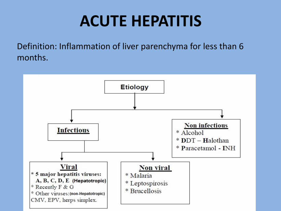
ACUTE HEPATITIS
Definition: Inflammation of liver parenchyma for less than 6 months.

Hepatitis A virus Type of virus: Picorna virus = entero virus, RNA.
Route of infection: Faeco-oral, patients excrete virus in faeces for 2 weeks before onset of illness &for 1 week after.
Incubation Period: 2-6 weeks.
Age: More in Children and young's.
Resolution: It is the rule, fulmination may occur in 0.5% of cases.
Malignancy: No.
Chronic hepatitis: No.
Carrier: No.
Markers: HA Ab (Hepatitis A Antibody)if +ve = infection, - IgM ~ Recent Infection.- IgG ~ Old Infection (may persist for years),

Prophylaxis
• 1. Active immunization (Vaccine), inactivated (HAV) vaccine (Havrix).
• Indications: Traveling to endemic area, patients with chronic liver disease.
• Dose: 0.5ml/IM + booster dose after 6-18 month.
• Validity: long life immunity (protection is 100% for more than 10 years).
• NB : it is obligatory in KSA at 18 months and 24 months
2. Passive immunization (Specific immunoglobulin)
= immune serum globulin (Abs contain HA Ab).
Indication: (Sero prevention, Sero attenuation), efficacy is 80-90%.
- Contact = recent exposure (it is better to be given within 2weeks of exposure).
- Traveling to endemic areas, it gives protection for 6 months

Hepatitis B virus (HBV)*Typoef virus: DNA virus.
*Routeof infection: (HBV is present in high concentration in blood so it is highly infectious)
-Blood, blood products. , saliva. - Transplacental (vertically Perinatal Infants born to HBeAg-positive mothers ).
• Incubation Period: 2-6 months.
• Age:Any (more in adult).
• :Malignancy: may occur.
• "Chronic hepatitis: 5%.
• :Flumination may occur.
• :Carrier:0.5-10% (

Immune response and fate of hepatitis B infectian:
• Resolution: (competent immune system)• Good immune response → acute hepatitis followed by clearance of the
virus and resolution.
• Carrier (poor Immune response to virus B particularlly)• The cytotoxic T cells can't recognize the viral Ag leading to viral
persistence.• There is poor immune response so, no hepatitis and no clearance of the• virus. The virus can proliferate with normal liver biochemistry and
function.
• Chronic hepatitis (5-10%): (Slightly better immune response)• Also the cytotoxic T cells can't recognize the viral Ag leading to viral• persistence. There is better immune response so, chronic hepatitis occurs• with no clearance of the virus with continuing the hapatocellular damage.

Markers:
• Thevirus (Dane particle) consists of
• 1. Protein coat (surface Ag).
• 2. Core: It contains DNA, DNA polymerase, core Ag and eAg.
• HBeAg is a protein subunit (part) of the core and it is
• secreted into plasma during active viral replication.
• SO, there are 3 antigens + 3 Antibodies = 6 markers
• Surface (s) Ag + HBsAb
• Core (c) Ag + HBcAb
• HBeAg + HBeAb

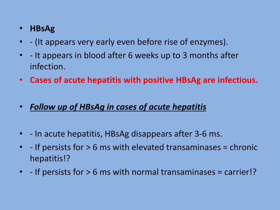
• HBsAg
• - (It appears very early even before rise of enzymes).
• - It appears in blood after 6 weeks up to 3 months after infection.
• Cases of acute hepatitis with positive HBsAg are infectious.
• Follow up of HBsAg in cases of acute hepatitis
• - In acute hepatitis, HBsAg disappears after 3-6 ms.
• - If persists for > 6 ms with elevated transaminases = chronic hepatitis!?
• - If persists for > 6 ms with normal transaminases = carrier!?
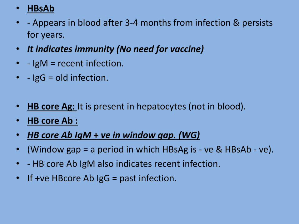
• HBsAb
• - Appears in blood after 3-4 months from infection & persists for years.
• It indicates immunity (No need for vaccine)
• - IgM = recent infection.
• - IgG = old infection.
• HB core Ag: It is present in hepatocytes (not in blood).
• HB core Ab :
• HB core Ab IgM + ve in window gap. (WG)
• (Window gap = a period in which HBsAg is - ve & HBsAb - ve).
• - HB core Ab IgM also indicates recent infection.
• If +ve HBcore Ab IgG = past infection.

• HBeAg
• - If +ve = Active viral replication.
• - Also it indicates high Infectivity (in acute or chronic hepatitis).
• HBeAb
• - = Low infectivity.
• = Convalescence.
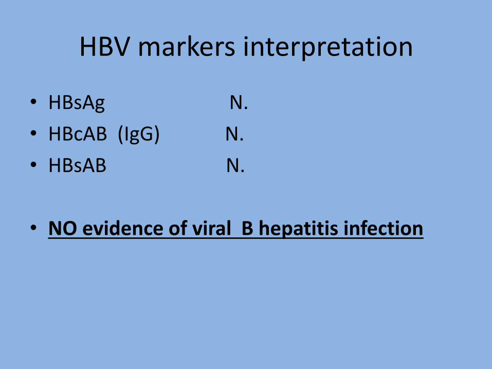
HBV markers interpretation
• HBsAg N.
• HBcAB (IgG) N.
• HBsAB N.
• NO evidence of viral B hepatitis infection
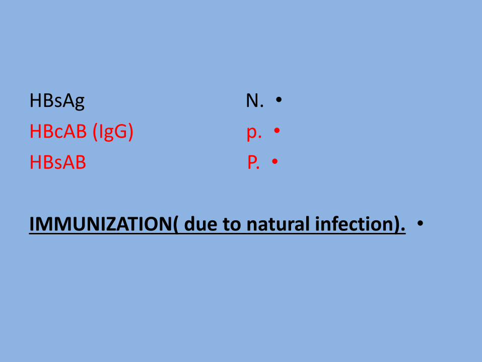
•HBsAg N.
•HBcAB (IgG) p.
•HBsAB P.
•IMMUNIZATION( due to natural infection).

•HBsAg N.
•HBcAB (IgG) N.
•HBsAB P.
•IMMUNIZATION(due to vaccination).

• HBsAg P.
• HBsAB N.
• MAY BE ACUTE OR CHRONIC.
• Order Hep. B Core IgM to clarify.
• The IgM will be positive , If Acute.

• Prophylaxis:
• 1-Vaccine
• It is made by recombinant DNA.
• It is given by IM injection 3 doses over 6 months (0, 1, 6) in deltoid.
• It gives immunity in 90% of patients.
• - To check vaccine, measure HBs Ab at 7-9 months after the initial
• dose.
• In KSA obligatory vaccination schadule given at : 0,2,4,6 months
• Indications: (Risk group)
• Doctors, nurses. Patients with hemophilias.
• - All newborn babies of HBsAg-positive mothers, also they must take HBIG immediately additional to the vaccine.
• Hemodialysis patients.

• 2- Hepatitis B immunoglobulin(HBIG)
• It contains HBsAb.
• Indication: Recent exposure.
• It should be given within 24 hours or at most a week from exposure to infected blood.

Hepatitis C Virus• Mode of infection• • It is a single stranded RNA virus of the flaviviridae family .
• The virus has low concentration in blood than HBV.
• Mainly blood transfusion (90% of cases) i.e post transfusion hepatitis.
• • Sexual transmission can occur but less than HBV, transplacental• transmission is relatively uncommon
• There are at least six genotypes of virus C, genotype I account for 70-801% of cases.
• I.P.: 2-6 m• Chronicity: Occurs in more than 50% of cases.• Malignancy: May occur.• Carriers: May occur.•

• Markers
• HC Ab (by ELlSA or RIBA method}
• If +ve= it mean infection but it doesn’t indicate immunity .
• PCR (polymerase chain reaction)
• If +ve = activity as it detect the RNA of virus in blood.
• (recently PCR titre Le. Ouantitative method is available)
• Screen with HCV IgG antibody after 18 month of age and HCV RNA after 2 month of age.
• No vaccine is available for virus C
Hepatitis E virus• It is an RNA virus (Calcivirus)
• - Mainly faeco oral.
• - I.P.: 2-8 weeks
• - Fulmination with pregnancy.
• Carrier : No
• Chronicity : No
• Markers : HEV AB (IgM ,IgG) , PCR

Hepatitis D Virus (Delta agent)
• Alone is not pathogenic as it is an incomplete RNA particle.
• -It is activated by the presence of HBV, as its RNA particle enclosed in a shell of HBs Ag .
• . Marker: anti Delta (lgM, IgG).
• - Co-infection with HBV leads to acute hepatitis with +ve anti-Delta IgMand HBc Ab IgM.
• . Superinfection results in an acute exacerbation of chronic HBV, this is diagnosed by antiDelta IgM with HBc Ab IgG.
• Fulminant hepatitis is more common with co-infection.
Hepatitis G (HGV)
• - Originally there is GB hepatitis agent which was isolated from a surgeon (GB)
• It is RNA virus (flavivirus)
• HGBV-C is similar to HCV
• HGV is transmitted parenterally and is found in I.V drug users, haemophiliacs and dialysis patients
• - Any rise in serum transaminases after HGV infection can be explained by co-infection with another virus e.g. HCV
• Up till now there is no definite evidence that HGV leading to acute or chronic hepatitis !?

Acute hepatitis due to nonHepatotropic viruses
• CMV: • - It causes acute hepatitis particularly in immunocompromised patients.• - Liver biopsy shows intranuclear inclusions and giant cells.• - The virus can be isolated from the urine.• - It can be treated by ganciclovir
• Infectious mononucleosis: • - It is caused by Epstein-Barr virus.• - Mild jaundice + sore throat.• - Minor abnormalities of liver biochemistry.• - Clinical hepatitis is rare.• - Positive Paul-Bunnell or monospot test.• - The sinusoids and portal tracts are infiltrated with large mononuclear
cells.• - No specific treatment.
• Herpes simplex:
• This virus occasionally causes generalized infection in immunocompromised patients.
• There is marked elevation of liver enzymes.
• Liver biopsy show extensive necrosis.
• It can be treated with Aciclovir.
• * Yellow fever:
• This viral infection is transmitted by mosquite Aedes aegypti leading to acute
• hepatic necrosis. There is no specific treatment.

ACUTE VIRAL HEPATITIS• Pathalogy
• Although some histological features are suggestive of aetiological factor,
• most of changes are essentially similar whatever the cause.
• Features:
• - Portal tract infiltration with inflammatory cells.
• - Swelling of hepatocytes narrowing of bile canaliculi
• intrahepatic cholestasis.
• - Centizonal necrosis (Zone 3).

Clinical picture
• I.Non icteric hepatitis (Anicteric):
• - It is usually a mild form of hepatitis, it may pass unnoticed.
• - S bilirubin < 2.5 mg or < 3 mg.
• - Clinically there is a mild flu-like illness with anorexia, nausea.
• - Increased transaminases.
• Fate
• - Resolution, chronic hepatitis post hepatitis liver cirrhosis !?

• II-Icteric hepatitis:
• A - Pre-icteric phase (for about 1-2 weeks due to viremia)
• - Fever, headache, malaise.
• - Marked anorexia, distaste for cigarettes.
• - Pain in right hypochondrium.
• - If you suspect you can confirm by rise of SGPT.
• B- Icteric phase (for about 3-6 weeks)Cholestatic jaundice
• - Swelling of hepatocytes obliteration of bile canaliculus Intra
• hepatic cholestasis jaundice.
• - Dark urine.
• - Clay stool.
• - Improvement of fever, malaise, headache.
• - Liver: mild++, Soft, tender (acute stretch of liver capsule).
• - Spleen: mild ++ = RES hyperplasia in 10% of patients.

• c- Post-icteric (convalescence)
• Improvement of general condition (symptoms & signs disappear gradually)
• - SGOT with the onset of recovery.
• - S. bilirubin but jaundice persists for a time as bilirubin has a high affinity to collagen fibers in sclera.
• - After about 3-6 ms patients become clinically & biochemically free.

• C/P of acute viral hepatitis almost the same in different types of viral infection but:
• 1- Acute hepatitis A is less severe than acute hepatitis B, and the illness is over within 3-6 weeks. Pronounced cholestasis may occur.
• 2- Acute hepatitis B is more severe than A
• 3- Most infection with acute HCV are .asymptomatic, most of patients are diagnosed years later with evidence of chronic liver disease.

Complication of heaptitis B&C
• -Hepatic complications (2, 3, 4, 5 may occur with hepatitis A)
• 1- Chronic hepatitis (persistent elevation of transaminases for> 6 ms).
• 2- Fulminant hepatitis with rapid development of acute L.C.F. and encephalopathy (associated with prolonged PT).
• 3- Prolonged cholestasis due to unresolved swelling of hepatocytes.
(Normal or mild rise of transaminases, i Alk. P, i bilirubin), spontaneous recovery usually occurs.
• 4- Relapse
• 5- Post hepatitis $:
• • There is normalization of enzymes but the patient still complaining.
• • It may be subjective & psychogenic (not due to liver disease).
• • There is easy fatigue, malaise and pain in right hypochondrium.
• • Treatment is reassurance & follow up.
• 6- Hepatoma.
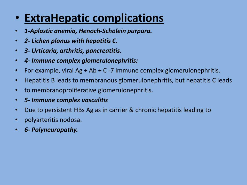
• ExtraHepatic complications• 1-Aplastic anemia, Henoch-Scholein purpura.
• 2- Lichen planus with hepatitis C.
• 3- Urticaria, arthritis, pancreatitis.
• 4- Immune complex glomerulonephritis:
• For example, viral Ag + Ab + C -7 immune complex glomerulonephritis.
• Hepatitis B leads to membranous glomerulonephritis, but hepatitis C leads
• to membranoproliferative glomerulonephritis.
• 5- Immune complex vasculitis
• Due to persistent HBs Ag as in carrier & chronic hepatitis leading to
• polyarteritis nodosa.
• 6- Polyneuropathy.

INVESTIGATIONS• Urine
• - it is a bed side test.
• - Direct bilirubin appears in urine (bilirubinuria) early sign, due to cholestasis.
• - Urobilinogen in urine is variable:
• - Early with marked obstruction -ve urobilinogen in urine.
• - Late with relieve of obstruction +ve urobilinogen in urine

• Liver function tests, Enzymes, markers:
• - SGOT, SGPT showing marked elevation.
• - S. albumin is normal.
• - Alk. P is mildly elevated.
• - ȢGT showing mild elevation.
• PT normal = good prognosis.
• prolonged = bad prognosis.
• - S. bilirubin is elevated (direct more than indirect).
• - Viral markers as before.
• Follow up
• 1) SGOT, its 1/2 life is short, so its decline = onset of recovery.
• 2) Follow up is by SGPT to detect relapse.
• 3) Persistance of HBsAg for example = chronic hepatitis or carrier.

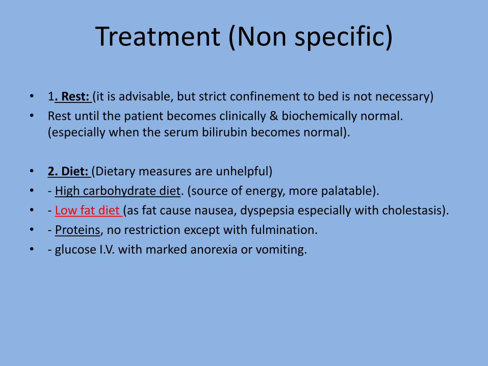
Treatment (Non specific)
• 1. Rest: (it is advisable, but strict confinement to bed is not necessary)
• Rest until the patient becomes clinically & biochemically normal. (especially when the serum bilirubin becomes normal).
• 2. Diet: (Dietary measures are unhelpful)
• - High carbohydrate diet. (source of energy, more palatable).
• - Low fat diet (as fat cause nausea, dyspepsia especially with cholestasis).
• - Proteins, no restriction except with fulmination.
• - glucose I.V. with marked anorexia or vomiting.

• 3. Steroids:
• - No benefit, it may lead to exacerbation!.
• - It may be used in some cases with prolonged cholestasis!?
• 4. Vitamins
• 5. Antiemetics e.g. Domperidone
• .

Chronic Hepatitis

CHRONIC HEPATITIS• Definition• Inflammatory disease within liver parenchyma more than 6 months.
• Chronic viral hepatitis is the major cause of chronic liver disease, cirrhosis and hepatocellular carcinoma in the world.
• Causes• 1. Viral (B,D, C).• 2. Drugs: e.g. a-methyl dopa, INH.• 3. Autoimmune (e.g Lupoid hepatitis).• 4. Inherited diseases e.g. Wilson's disease, α1 antitrypsin deficiency.
• Pathological types• 1- Mild form previously called chronic persistent hepatitis.• 2- Severe form previously called chronic active hepatitis.

Mild form of chronic hepatitis• Common, particularly following H.B.• Clinical picture• - Asymptomatic, it may be discovered accidentally.• - Fatigue & pain in right hypochondrium, fat intolerance ma occur.
• LAB• Mild elevation of transaminases, also positive viral' markers may be present
e.g.HBsAg.
• Biopsy: Portal tract infiltration with inflammatory cells with no loss of architecture.
• Treatment:• 1- Reassurance.• 2- Follow up /6m by enzymes. (SGPT).• 3- Avoid hepatotoxicity - hepatic support e.g. silymarin - antioxidants.

Severe form of Chronic Hepatitis• Clinical picture
• • It ranges from mild asymptomatic cases up to frank LCF & portal H.
• • It will be transformed to cirrhosis.
• *Features of hepatitis for > 6m
• - Jaundice is slight or absent.
• - The liver may be enlarged and tender.
• - Constitutional symptoms (malaise, fatigue, low grade fever, ....).
• - Cutaneous signs of chronic liver disease (see later).
• *Progression to cirrhosis
• - LCF (liver cell failure).
• - Portal HTN.

• Extrahepatic manifestations of chronic viral hepatitis
• Extrahepatic manifestations of autoimmune hepatitis
• - Polyarthritis.
• - Hashimoto's thyroiditis.
• Autoimmune hemolytic anemia.
• - Grave's disease.

Investingations• 1. Enzymes: SGOT and SGPT usually increased up to 3-5 folds or more.
• 2. Serum albumin: usually normal, it is low in advanced cases.
• 3. Markers: for viral or autoimmune hepatitis e.g. HBsAg, HCAB, ANA, LKM.
• 4. Biopsy:
• Thefindings are:
• I- Bridging necrosis
• II- Piece meal necrosis.
• III- Late, rosette appearance then cirrhosis.

Treatment
• Chronic HBV : INF Alfa or lamivudine
• Chronic HCV : INF Alfa and ribavirin
• Auto-immune : steroid