Embed Size (px)
Citation preview
Original Article
Hepatic ERa accounts for sex differences in theability to cope with an excess of dietary lipids
Clara Meda 1, Mara Barone 2,3, Nico Mitro 4, Federica Lolli 4, Silvia Pedretti 4, Donatella Caruso 4,Adriana Maggi 2,3, Sara Della Torre 2,3,*
ABSTRACT
Objective: Among obesity-associated metabolic diseases, non-alcoholic fatty liver disease (NAFLD) represents an increasing public health issuedue to its emerging association with atherogenic dyslipidemia and cardiovascular diseases (CVDs). The lower prevalence of NAFLD in pre-menopausal women compared with men or post-menopausal women led us to hypothesize that the female-inherent ability to counteract thispathology might strongly rely on estrogen signaling.In female mammals, estrogen receptor alpha (ERa) is highly expressed in the liver, where it acts as a sensor of the nutritional status and adaptsthe metabolism to the reproductive needs. As in the male liver this receptor is little expressed, we here hypothesize that hepatic ERa mightaccount for sex differences in the ability of males and females to cope with an excess of dietary lipids and counteract the accumulation of lipids inthe liver.Methods: Through liver metabolomics and transcriptomics we analyzed the relevance of hepatic ERa in the metabolic response of males andfemales to a diet highly enriched in fats (HFD) as a model of diet-induced obesity.Results: The study shows that the hepatic ERa strongly contributes to the sex-specific response to an HFD and its action accounts for oppositeconsequences for hepatic health in males and females.Conclusion: This study identified hepatic ERa as a novel target for the design of sex-specific therapies against fatty liver and its cardio-metabolicconsequences.
� 2019 The Author(s). Published by Elsevier GmbH. This is an open access article under the CC BY-NC-ND license (http://creativecommons.org/licenses/by-nc-nd/4.0/).
Keywords Liver metabolism; Fatty liver; Sex differences; Estrogen; ERa; Amino acids
1. INTRODUCTION
In female mammals, liver estrogen receptor alpha (ERa) plays a centralrole in the adaptive response of hepatic metabolism to the energy re-quirements characterizing the different reproductive stages [1e3]. Inaddition, this receptor was reported to have a key role in the control oflipid and amino acid (AA) metabolism [1,4]. Unlike in females, theexpression of ERa in the adult male liver is very low [2,4], suggestingthat, in males, the role of this receptor in metabolic control might beextremely limited. In view of its prominent role in the female liver, ERaaction might strongly contribute to the well-recognized, but oftenoverlooked, hepatic sexual dimorphism [5e7] and to the sex-specificincidence of hepatic diseases [8,9]. Among them, non-alcoholic fattyliver disease (NAFLD), a syndrome characterized by excess triglycerideaccumulation within hepatocytes [10], represents a paradigmaticexample. Indeed, although obesity is prevalent in females, fertile womenresult to be, to some extent, protected from obesity-associated fattyliver/NAFLD and cardio-metabolic consequences [11e13], suggestingthe involvement of estrogen signaling. This idea is further supported bystudies showing that the lack of estrogens (ovariectomized, OVX, andaromatase knockout mice, ArKO) [1,14e17] or ERa (ERa knockout
1Department of Health Sciences, University of Milan, Italy 2Department of PharmaceutDiseases, University of Milan, Italy 4Department of Pharmacological and Biomolecular
*Corresponding author. Department of Pharmaceutical Sciences, University of Milan, V
Received November 14, 2019 � Revision received December 11, 2019 � Accepted De
https://doi.org/10.1016/j.molmet.2019.12.009
MOLECULAR METABOLISM 32 (2020) 97e108 � 2019 The Author(s). Published by Elsevier GmbH. This is an openwww.molecularmetabolism.com
mice, ERaKO) [16,18,19] is associated with the development of hepaticsteatosis. However, the analysis of the lack of estrogen signaling in OVX,ArKO, and ERaKOmice did not allow to disentangle the systemic effectsof estrogens from those occurring in the liver specifically, preventing theunderstanding of the contribution of hepatic estrogen signaling to thesex-specific prevalence of fatty liver/NAFLD.Considering that, in fertile females, estrogens, acting through thehepatic ERa, might antagonize liver lipid deposition, we hypothesizedthat the low content of ERa and the lack of such a well-tuned regu-lation in males might be at the basis of the higher prevalence of hepaticsteatosis/NAFLD in males, particularly under an unbalanced dietaryregimen. Indeed, although the contribution of liver ERa in preventing orlimiting the diet-induced hepatic steatosis/NAFLD in the two sexes hasnot been investigated, this hypothesis is supported by recent reportshighlighting the negative correlation between hepatic ERa expression/activity and fatty liver [1,20,21].Thus, the present study was undertaken to evaluate the extent towhich the hepatic ERa might account for sex differences in the abilityof the male and female liver to cope with an excess of dietary lipids andcounteract the development of fatty liver/NAFLD in a model of diet-induced obesity.
ical Sciences, University of Milan, Italy 3Center of Excellence on NeurodegenerativeSciences, University of Milan, Italy
ia Balzaretti 9, 20133, Milan, Italy. E-mail: [email protected] (S. Della Torre).
cember 12, 2019 � Available online 24 December 2019
access article under the CC BY-NC-ND license (http://creativecommons.org/licenses/by-nc-nd/4.0/). 97
Original Article
The data obtained show that, besides its sex-biased expression, thehepatic ERa contributes to the sex-specific metabolic adaptation to afat-enriched diet, accounting for opposite consequences for health inmales and females. This study, by unraveling the relevance of hepaticERa in the liver of the two sexes, underlines the necessity to customizepharmacological and dietary interventions aimed to prevent obesity-associated fatty liver/NAFLD and the cardio-metabolic consequences.
2. MATERIAL AND METHODS
2.1. AnimalsSyngenic (ERa floxed, ERaf/f) and LERKO (liver ERa knockout) [3] micewere fed with a low fat diet (LFD, Research diet, D12450B) or a high fatdiet (HFD, Research Diets, D12492) for 16 weeks starting at 4 monthsof age. At the end of the experiment, mice were 8 months old. Roomtemperature was maintained at 22 �Ce25 �C, with a 12-hour light/dark cycle (lights on at 7:00 a.m.). For all the animal studies, both maleand female mice were used. Female mice were collected when in theMetestrus phase of the estrous cycle; to do this, vaginal smears wereperformed at 9:00 a.m. To avoid any possible confounding effect dueto the circadian rhythm or feeding status, the mice were euthanized inthe early afternoon after 6 h of fasting [3]. All animal experimentationwas performed in accordance with the ARRIVE guidelines and theEuropean guidelines for animal care and the use of experimental an-imals, approved by the Italian Ministry of Research and University, andcontrolled by a departmental panel of experts.
2.2. Liver histologyThe left lobe of the liver was fixed in 10% neutral formalin solution(SigmaeAldrich) overnight at 4 �C, cryopreserved in a 30% (w/v)sucrose solution for 24 h at 4 �C, and stored at�80 �C. Liver sections7-mm thick were cut with a refrigerated microtome (Leica), collectedon slides, and stored at �80 �C until staining. Hematoxylineeosin(H&E) staining was performed on the frozen slides with Mayer he-matoxylin (Bio-Optica) for 1 min and, after washing with water, with1% eosin aqueous solution (Bio-Optica) for 4 min. Oil Red O stainingwas performed as previously described [1]. The Accustain Trichromestain kit was used for Masson trichrome staining (SigmaeAldrich).After staining, the slides were cleared in xylenes and cover slippedwith xylene-based mounting medium (Eukitt, Bio-Optica). The liversections were evaluated in a blinded fashion under a light microscope.Semi-quantitative grading of the hepatocellular vacuolar degenerationand portal inflammation (i.e., the infiltration of mononuclear inflam-matory cells) was also performed in a blinded fashion. Images of thestained sections were captured using Microscope Axioscop2 mot plus(Zeiss).
2.3. Biochemical assaysThe triglyceride (TG), free fatty acid (FFA), and cholesterol (CH) levelswere measured with appropriate kits according to the manufacturer’sprotocols (Biovision).
2.4. Metabolomic analysisFor metabolomic analyses, liver tissues were homogenized with atissue lyser. Briefly, tissues were lysed in 250 mL of methanol/acetonitrile 1:1 (v/v) with D-Glucose-13C6 1 ng/mL (internal standard,Sigma Aldrich, 389374) and centrifuged at 4 �C. Supernatant wassaved for subsequent analysis. Amino acid quantification was per-formed through previous derivatization. Samples were incubated withphenylisothiocyanate (PITC) solution for 20 min at room temperature,dried, and resuspended in 5 mM ammonium acetate in MeOH/H2O 1:1
98 MOLECULAR METABOLISM 32 (2020) 97e108 � 2019 The Author(s). Published by Elsevier GmbH. T
(v/v). Metabolomic data were performed on an API-4000 triple quad-rupole mass spectrometer (AB SCIEX) coupled with a high-performance liquid chromatography (HPLC) system (Agilent) and aCTC PAL HTS autosampler (PAL System). The identity of all metaboliteswas confirmed using pure standards. Quantification of different me-tabolites was performed with a liquid chromatography/tandem massspectrometry (LC-MS/MS) method using a C18 column (Biocrates) foramino acids and a cyano-phase LUNA column (50 mm � 4.6 mm,5 mm; Phenomenex). The AAs were analyzed through a 10-minute runin positive ion mode, whereas other metabolites were run for 5 min innegative ion mode. Twenty multiple reaction monitoring (MRM) tran-sitions in positive ion mode and 30 MRM transitions in negative ionmode (all other metabolites) were used, respectively. The mobilephases for positive ion mode analysis were phase A: 0.2% formic acidin water and phase B: 0.2% formic acid in acetonitrile. The gradientwas T0 100% A, T5.5min 5%A, and T7min 100%A with a flow rate of500 mL/min. The mobile phase for negative ion mode analysis (all othermetabolites) was phase A: 5 mM ammonium acetate pH 7.00 in MeOH.The gradient was 100% A for all the analysis with a flow rate of500 mL/min. MultiQuant� software (version 3.0.2) was used for dataanalysis and peak review of chromatograms. Quantitative evaluation ofall metabolites was performed based on calibration curves with purestandards, and data were normalized on micrograms of proteins.Metabolomic data analysis was performed using Metaboanalyst 3.0software (http://www.metaboanalyst.ca/).Plasma FFA extraction was conducted by Folch method and then aspreviously reported [22] using 13C18-labeled linoleic acid and
13C16-labeled palmitic acid (SigmaeAldrich) as internal standards. Ex-tracts were dried under nitrogen and resuspended in methanol/water (1:1) before submission for analyses. For FFA quantification,the MS analysis was conducted with a selective ion monitoring-tandem mass spectrometry (SIM-MS/MS) method. An electro-spray ionization (ESI) source was used and connected with an API4000 triple quadrupole instrument (AB Sciex, USA). The mobilephases were: water:methanol (97:3)/10 mM isopropylethylamine/15 mM acetic acid (phase A) and methanol (phase B). T0 20% A,T20min 1%A, T25min 1%A, T25.1min 20%A, T30min 20%A. TheHypersil GOLD PFP column (100 mm � 2.1 mm, 3 mm) wasmaintained at 40 �C, and the flow rate was 500 mL/min. Multi-Quant� software version 3.0.2 was used for data analysis andpeak review of chromatograms. The quantitative evaluation ofdifferent FFAs was performed based on standard curves. Quanti-tative data were normalized on microliter of plasma extracted.
2.5. Real-time PCR gene expression analysisTotal liver RNA extraction was isolated with TRIzol Reagent (Invitrogen)and purified using the RNeasy mini-kit protocol (Qiagen), according tothe manufacturer’s instructions. For the preparation of cDNA, 1 mg RNAwas denatured at 75 �C for 5 min in the presence of 1.5 mg of randomprimers (Promega) in 15 mL final volume. Deoxynucleotide triphosphate(GE Healthcare) and Moloney murine leukemia virus reverse tran-scriptase (M-MLV RT) (Promega) were added at 0.5mm and 8 U/mL finalconcentration, respectively, in a final volume of 25 mL. The RT reactionwas performed at 37 �C for 1 h; the enzyme was inactivated at 75 �C for5 min. Control reactions without the addition of the RT enzyme wereperformed for each sample. For the real-time polymerase chain reaction(rtPCR) experiments, the reaction mix for each sample was made up of2 mL of pre-diluted cDNA, 5 mL of TaqMan 2� Universal PCRMaster MixNo AmpErase UNG (ThermoFisher/Life Technologies), 0.5 mL of 20xprimers/probes mix, and 2.5 mL of H2O. The primers used for the rtPCRreactions are listed in Table S4. The 36b4 primer was used as the
his is an open access article under the CC BY-NC-ND license (http://creativecommons.org/licenses/by-nc-nd/4.0/).www.molecularmetabolism.com
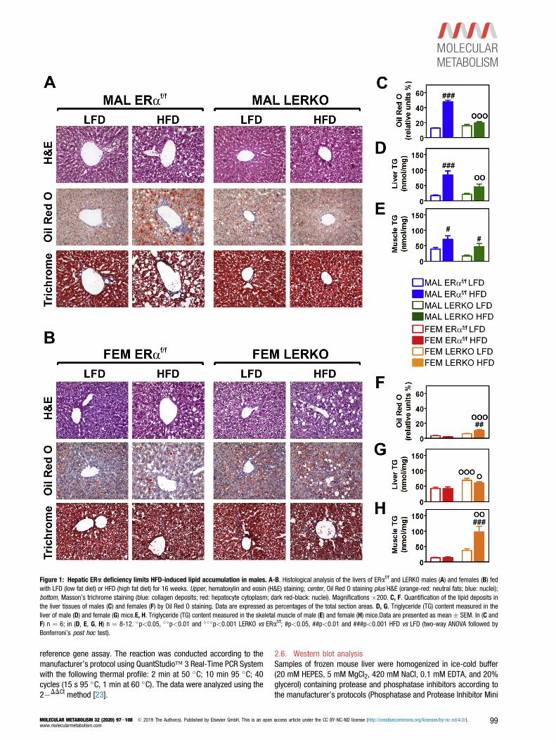
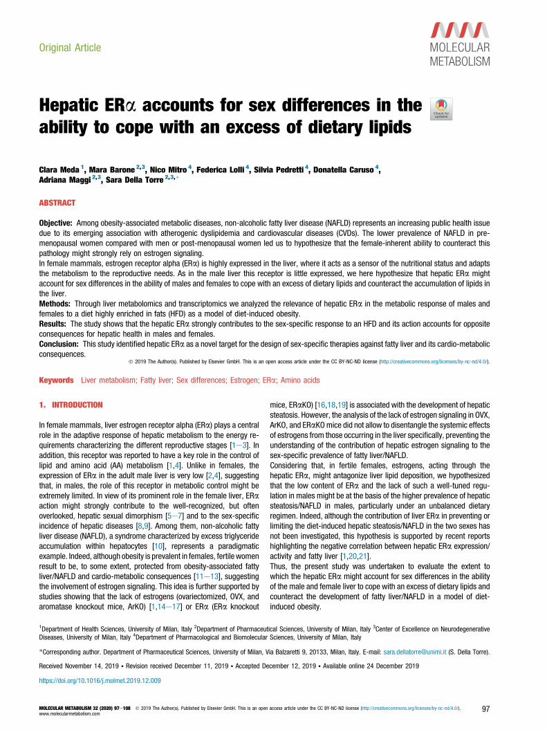
Figure 1: Hepatic ERa deficiency limits HFD-induced lipid accumulation in males. A-B. Histological analysis of the livers of ERaf/f and LERKO males (A) and females (B) fedwith LFD (low fat diet) or HFD (high fat diet) for 16 weeks. Upper, hematoxylin and eosin (H&E) staining; center, Oil Red O staining plus H&E (orange-red: neutral fats; blue: nuclei);bottom, Masson’s trichrome staining (blue: collagen deposits; red: hepatocyte cytoplasm; dark red-black: nuclei). Magnifications �200. C, F. Quantification of the lipid deposits inthe liver tissues of males (C) and females (F) by Oil Red O staining. Data are expressed as percentages of the total section areas. D, G. Triglyceride (TG) content measured in theliver of male (D) and female (G) mice.E, H. Triglyceride (TG) content measured in the skeletal muscle of male (E) and female (H) mice.Data are presented as mean � SEM. In (C andF) n ¼ 6; in (D, E, G, H) n ¼ 8-12. �p<0.05, ��p<0.01 and ���p<0.001 LERKO vs ERaf/f; #p<0.05, ##p<0.01 and ###p<0.001 HFD vs LFD (two-way ANOVA followed byBonferroni’s post hoc test).
reference gene assay. The reaction was conducted according to themanufacturer’s protocol using QuantStudio� 3 Real-Time PCR Systemwith the following thermal profile: 2 min at 50 �C; 10 min 95 �C; 40cycles (15 s 95 �C, 1 min at 60 �C). The data were analyzed using the2�DDCt method [23].
MOLECULAR METABOLISM 32 (2020) 97e108 � 2019 The Author(s). Published by Elsevier GmbH. This is an openwww.molecularmetabolism.com
2.6. Western blot analysisSamples of frozen mouse liver were homogenized in ice-cold buffer(20 mM HEPES, 5 mM MgCl2, 420 mM NaCl, 0.1 mM EDTA, and 20%glycerol) containing protease and phosphatase inhibitors according tothe manufacturer’s protocols (Phosphatase and Protease Inhibitor Mini
access article under the CC BY-NC-ND license (http://creativecommons.org/licenses/by-nc-nd/4.0/). 99

Original Article
Tablets, Pierce). After three repeated cycles of freezing and thawing,the homogenate was centrifuged at 16,100 g for 15 min at 4 �C, andthe supernatant was collected in a new tube. After appropriatequantitative analysis (Bradford assay, Pierce), equal amounts of theprotein samples (25 mg of liver extracts) were resuspended in Laemmlisample buffer and separated in an 8%e10% sodium dodecylsulfate(SDS) polyacrylamide gel system (Biorad). After transfer, the nitrocel-lulose membranes were incubated with specific antibodies overnight at4 �C and then with the secondary antibody conjugated with peroxidasefor 1 h at RT. The primary antibodies used were the following: anti-LDLR (Novus Biological, NB110-57162), anti-CD36 (Abcam,ab133625), and anti-b-actin (Sigma, A1978). Immunoreactivity wasdetected with an ECL Western Blotting Analysis System (Amersham)and acquired and analyzed using an Odissey Fc Imaging system andthe Image Studio� software (LiCor Biosciences).
2.7. Plasma CH profileThe CH distribution in the plasma lipoprotein fractions was determinedvia fast protein liquid chromatography (FPLC) using a Superose 6column (Amersham Biosciences). Fractions of 500 mL were collectedand assayed for CH with an enzymatic kit (Sentinel).
2.8. Quantification and statistical analysisStatistical analyses were performed by the Student t test for thecomparison of two different experimental groups, or two-way analysisof variance (ANOVA) followed by the Bonferroni post hoc test formultiple testing comparisons. All statistical analyses were performedusing GraphPad Prism 5.0 (GraphPad Software). All data are expressedas mean� standard error of the mean (SEM). A p value less than 0.05was considered statistically significant. The statistical parameters canbe found in the figure legends.
3. RESULTS
3.1. The lack of hepatic ERa signaling has opposite consequencesin the liver of males and femalesTo study the relevance of hepatic ERa in the response of male andfemale livers to an unhealthy, fat-enriched diet, syngenic (ERaf/f) andliver-specific ERa knockout (LERKO) [3] mice were fed with a low-fatdiet (LFD) or a 60% high-fat diet (HFD) for 16 weeks starting at 4months of age (see Methods and Tables S1 and S2). We focused onthat time with the aim to study the sex-specific relevance of hepaticERa in the metabolic adaptation to a diet enriched in fats and incounteracting lipid accumulation in the liver of the two sexes. At theend of the dietary treatment, the mice were euthanized in the earlyafternoon after 6 h of fasting [3], and the excised livers were processedfor histological and biochemical analysis.The HFD had a critical effect in ERaf/f males, leading to a majoraccumulation of lipids in the liver as shown by Oil Red O staining(þ280% vs LFD) and by the measurement of TG content (þ395% vsLFD) (Figure 1A, C, D). Lipid deposition was associated with hepato-cellular vacuolar degeneration and portal infiltration of mononuclearleukocytes as indicated by H&E and Masson trichrome stainings.Considering that in the male liver the content of ERa is quite low, weexpected that the liver-specific ablation of this receptor had minimaleffects in LERKO males with respect to their syngenic counterparts.Contrary to our expectations was the observation of a moderate lipiddeposition in the liver of LERKO males after a HFD (Figure 1A, C, D).The effect of both diet and ERa deficiency was very different in fe-males. In ERaf/f females, the HFD did not alter the hepatic lipid content,whereas the mere ablation of ERa was associated with a significant
100 MOLECULAR METABOLISM 32 (2020) 97e108 � 2019 The Author(s). Published by Elsevier GmbH. T
increase in liver lipid deposition (þ50% TG, irrespective of the diet)(Figure 1B, F, G).In males, the HFD also led to lipid accumulation in the skeletal muscleof both ERaf/f (þ81% vs LFD) and LERKO (þ184% vs LFD). In females,the HFD enhanced fat deposition in the skeletal muscle of LERKO(þ162% vs LFD) but not in ERaf/f females (Figure 1EeH). This sug-gested that, in females, the hepatic ERa plays a role in preventingectopic lipid deposition.In both sexes, the described effects were associated with alterations ofthe body weight (BW) that were found to be independent from hepaticERa. Indeed, in the course of the experiment, the changes in BW weresuperimposable in ERaf/f and LERKO mice (Figure 2A) and, at the endof the experiment, HFD-fed males and females weighed significantlymore than their LFD-fed counterparts (Figure 2B).The analysis of visceral white adipose tissue (vWAT) weight and feedefficiency (FE) further demonstrated a sex-specific response to HFD ofERaf/f and LERKO mice. HFD-fed ERaf/f males accumulated morevWAT than LERKO males, whereas the opposite was true for females(Figure 2C). With respect to males, ERaf/f females were able to bettercope with the HFD; indeed, they kept FE relatively low and avoided theaccumulation of fat tissue, an ability lost by LERKO females(Figure 2CeD). Differently from females, hepatic ERa deficiency inmales limited FE after a HFD (�24% vs ERaf/f males) (Figure 2D).Nevertheless, the increased food intake (þ15%) (Figure 2E) suggestedan altered nutrient absorption and/or metabolism in LERKO males.These data demonstrate that hepatic ERa action accounts for a sex-specific response to a HFD with different consequences for malesand females, suggesting that, besides its dimorphic expression(Fig. S1), liver ERa exerts different metabolic functions in the twosexes.
3.2. HFD unmasks the sex-specific impact of hepatic ERa ablationon liver metabolomeTo better comprehend the different roles exerted by ERa in the liver ofthe two sexes, we pursued our characterization of liver metabolismthrough a metabolomic approach. As shown in the heatmap ofFigure 3A, the most relevant liver metabolites (Table S3) were affectedby the diet and by the presence/absence of hepatic ERa in a sex-specific fashion. The principal component analysis (PCA) of liver me-tabolites clearly showed that the diet acts as a clear discriminatingfactor for LERKO males, but less for ERaf/f males that, conversely, arerepresented by partially superimposing ellipses (Figure 3B). In females,the overlap among the four groups indicated that the liver metabolomeof females was less affected by the diet and by ERa deficiency(Figure 3C). Cluster analysis performed on liver metabolites (Figure 3D)clearly showed how the ablation of ERa in the liver of males isresponsible for a metabolic signature that significantly diverged fromthat of all other experimental groups. HFD led to major changes inLERKO males: after the HFD, the metabolic fingerprint of LERKO malesbecame more similar to that of the HFD-fed females, indicating that thehepatic ERa directs the male-specific response to a fat-enriched diet.By considering a fold change jFCj> 1.5 and aeLog10(p-value)> 1.3,we found that, in the liver of ERaf/f males, HFD led to a significantdecrease in the content of AA (in red) and of short-chain acyl-carnitine(C4) and to a greater content of medium-chain acyl-carnitines (C8 andC9) (Figure 4A and table S3). The lack of ERa by itself induced ametabolic rewiring in the liver of LERKO males, which showed lowcontent of AA, intermediates of glycolysis/gluconeogenesis (Gly/Gln:glucose, glucose-6-phosphate, fructose-1,6-biphosphate, lactate),pentose phosphate pathway (PPP: erythrose-4-phosphate), and highcontent of intermediates of TCA cycle (acetyl-CoA, citrate, isocitrate, 2-
his is an open access article under the CC BY-NC-ND license (http://creativecommons.org/licenses/by-nc-nd/4.0/).www.molecularmetabolism.com

Figure 2: Effect of HFD and hepatic ERa deficiency on body weight and food intake. A. Body weight (BW) of ERaf/f and LERKO mice measured weekly along the 16 weekslong experiment and expressed as percentage versus time 0. B. BW of ERaf/f and LERKO mice measured at the end of the 16 weeks long experiment. C. Quantification of thevisceral white adipose tissue (vWAT) normalized over the BW. D. Feeding efficiency expressed as dBW/dFI (dBody weight/dFood intake). E. Food intake (FI) expressed as kcal %.Data are presented as mean � SEM (n ¼ 18). �p<0.05, ��p<0.01 and ���p<0.001 LERKO vs ERaf/f; #p<0.05, ##p<0.01 and ###p<0.001 HFD vs LFD (two-way ANOVAfollowed by Bonferroni’s post hoc test).
oxoglutarate) and energy metabolism [oxidized nicotinamide adeninedinucleotide (NADþ), reduced nicotinamide adenine dinucleotide(NADH), oxidized nicotinamide adenine dinucleotide phosphate(NADPþ), and reduced nicotinamide adenine dinucleotide phosphate(NADPH)]. The already low content of AA further decreased in the liverof LERKO males after HFD; this change was associated with a decreaseof intermediates of Gly/Gln (PEP, pyruvate), TCA cycle (acetyl-CoA,isocitrate, 2-oxoglutarate, succinyl-CoA, succinate), lipid synthesis(malonyl-CoA), and energy metabolism [NADþ, NADH, NADPþ,NADPH, adenosine triphosphate (ATP), adenosine diphosphate (ADP),and adenosine monophosphate (AMP)] and with an increase of short-(C4) and medium-chain (C8, C9) acyl-carnitines.In ERaf/f females, the effect of the HFD was circumscribed to a fewchanges (low content of glucose-6-phosphate, Ribu-5P, citrate, iso-citrate, fumarate, and malate) and, differently from males, did notaffect the AA content. AA homeostasis in females was dependent onhepatic ERa; indeed, compared with ERaf/f females, LERKO femalesshowed higher content of AA, some of which [alanine (Ala), threonine(Thr), glutamine (Gln), glutamate (Glu), methionine (Met), histidine(His), and arginine (Arg)] were significantly decreased after a HFDtogether with some intermediates of the TCA cycle (isocitrate, 2-oxoglutarate, succinate, fumarate, malate) and of energy metabolism(NADPþ, NADPH).A more detailed analysis based on the pathways with the most relevantimpact (impact > 0.6 and eLog(p-value) > 3) indicated that themetabolism of AA and, in particular, of branched-chain AA (BCAA: Val,Leu, Ile) was the most affected by the lack of hepatic ERa and by thenutritional challenge, especially in males (Figure 4B). Interestingly, only
MOLECULAR METABOLISM 32 (2020) 97e108 � 2019 The Author(s). Published by Elsevier GmbH. This is an openwww.molecularmetabolism.com
the liver of ERaf/f females was able to preserve the homeostasis of AAwhen challenged with a HFD, a feature associated with a healthymetabolic phenotype.
3.3. The lack of hepatic ERa in males, but not females, isresponsible for an altered plasma lipid profileNext, we examined the extent to which the lack of hepatic ERa couldaffect the homeostasis of circulating lipids (TG, FFA, and CH), partic-ularly under a HFD regimen.As shown in Figure 5A, plasma TG levels were slightly increased by aHFD in males but not in females, a sex-specific effect that resultedindependently from hepatic ERa presence/absence.With the exception of docosahexaenoic acid (DHA, �40%), the levelsof FFA were unchanged by HFD in the plasma of ERaf/f males(Figure 5B). In males, this homeostasis was ensured by hepatic ERa;indeed, the lack of this receptor in LERKO males led to a significantincrease (or showed a trend to augment) in the majority of FFA, aneffect boosted by the HFD (Figure 5B). In ERaf/f females, the onlysignificant changes driven by HFD were related to stearic acid (þ48%)and to DHA (�41%), whereas no changes were observed in LERKOfemales independent of the diet (Figure 5C). These data suggest thatthe hepatic ERa deficiency impacts on the metabolism of FFA mainly inmales.Having found that hepatic ERa plays a pivotal role in the regulationof CH and lipoprotein metabolism in females [2], we pursued ourstudy by measuring total CH levels and by analyzing the distributionof CH among plasma lipoproteins by fast protein liquid chroma-tography (FPLC) (Figure 6AeD). HFD induced a significant increase
access article under the CC BY-NC-ND license (http://creativecommons.org/licenses/by-nc-nd/4.0/). 101
Figure 3: HFD unmasks the sex-specific impact of hepatic ERa ablation on liver metabolome. A. Hierarchical Clustering Heatmap of biologically major metabolites of theglycolysis/gluconeogenesis (GLY/GNG), pentose phosphate pathway (PPP), tricarboxylic acid cycle (TCA), energy metabolism (En. Met.), amino acids (AA), and acyl-carnitines (Acyl-Carn) measured in the liver. The color-coded scale on the left indicates the normalized metabolite expression. B-C. Principal Component Analysis (PCA) of the metabolitesmeasured in the liver of ERaf/f and LERKO males (B) and females (C) fed with LFD and HFD. Colored ellipses - generated using MetaboAnalyst 3.0 software - represent 95%confidence intervals and highlight the basic clustering/separation between groups. Colored dots represent individual samples (n¼6). D. Hierarchical Clustering (Dendrogram) of thedata showed in A was done using a software available at the web page http://www.wessa.net/rwasp_hierarchicalclustering.wasp.
Original Article
of total CH levels in the plasma of ERaf/f but not in LERKO males,whereas the opposite was true for females (Figure 6A). As aconsequence of the HFD, the distribution of CH in the low-densitylipoprotein (LDL) fraction increased, leading to a higher LDL/high-density lipoprotein (HDL) ratio in both ERaf/f males and females(Figure 6BeD). The sole ablation of liver ERa resulted in higherLDL-CH and LDL/HDL ratio in both LERKO males and females.However, only in LERKO males LDL-CH and LDL/HDL ratio werefurther augmented by HFD, whereas no changes were observed inLERKO females after HFD (Figure 6BeD), despite the higher totalCH levels (Figure 6A). All together, these data suggest that ERaaction in the liver strongly contributes to the different, sex-specific
102 MOLECULAR METABOLISM 32 (2020) 97e108 � 2019 The Author(s). Published by Elsevier GmbH. T
regulation of circulating lipids (FFA and CH, in particular) inresponse to a fat-enriched diet.
3.4. Liver ERa regulates the hepatic lipid metabolism in a sex-specific fashionLipid deposition in the liver might be a consequence of an imbalancebetween the lipids that are taken up or synthesized de novo and thosebeing catabolized or exported [24]. The fact that the lack of hepaticERa prevented lipid deposition in the liver and increased the circulatinglipids in males, but did the opposite in females, led us to hypothesizethat the ERa action may regulate the uptake of lipids differently in thetwo sexes. To investigate on that, we first evaluated the expression of
his is an open access article under the CC BY-NC-ND license (http://creativecommons.org/licenses/by-nc-nd/4.0/).www.molecularmetabolism.com

Figure 4: Hepatic ERa preserves AA homeostasis in the liver of females but not of males challenged with HFD. A. Volcano plot of biologically relevant metabolitesmeasured in the liver of ERaf/f and LERKO males and females fed with LFD and HFD. Only metabolites with a |FC|>1.5 and -Log10(p-value)>1.3 were considered as significant anddisplayed with triangles of different colors depending on the class they belong to (see legend). B. Pathway impact analysis of the biological pathways regulated in the liver of ERaf/f
and LERKO males and females fed with LFD and HFD. Only metabolic pathway with an impact>0.6 and a -Log(p-value)>3 were considered as significant and displayed as coloredcircles (varying from yellow to red).
MOLECULAR METABOLISM 32 (2020) 97e108 � 2019 The Author(s). Published by Elsevier GmbH. This is an open access article under the CC BY-NC-ND license (http://creativecommons.org/licenses/by-nc-nd/4.0/).www.molecularmetabolism.com
103

Figure 5: Lack of hepatic ERa affects the circulating lipids especially in males. A-C. Levels of triglyceride (TG) (A) and free fatty acid (FFA) (B-C) measured in the plasma.Data information: In (AeC), data are presented as mean � SEM (n¼ 8-12). �p<0.05, ��p<0.01 and ���p<0.001 LERKO vs ERaf/f; #p<0.05, ##p<0.01 and ###p<0.001 HFDvs LFD (two-way ANOVA followed by Bonferroni’s post hoc test).
Original Article
receptors relevant for the uptake of lipids, such as low-density lipo-protein receptor (LDLR) and cluster of differentiation 36 (CD36). LDLRand CD36 were induced in the liver of ERaf/f but not LERKO maleswhen exposed to HFD, indicating that, in the absence of hepatic ERa,the male liver lost its ability to promote the lipid uptake (Figure 6EeG).Compared with their male counterparts, ERaf/f and LERKO femalesshowed high basal Ldlr and Cd36 mRNAs and followed a differentstrategy when exposed to HFD: the lipid uptake was unchanged in theliver of ERaf/f females (Figure 6EeG) and promoted in the liver ofLERKO females (þ103% CD36, Figure 6G). These data suggest thatERa might have a sex-specific role in the control of lipid uptake inresponse to HFD.The hepatic content of LDLR [25,26] and CD36 [27] can beregulated through the degradation promoted by proprotein con-vertase subtilisin/kexin type 9 (PCSK9), whose expression isaffected by estrogens [28]. Pcsk9 mRNA was upregulated by HFDin the liver of ERaf/f but not in LERKO males, whereas theopposite was true for females (Figure 6H). Although its relevancein the mouse liver is still debated [29e31], inducible degrader ofthe low-density lipoprotein receptor (IDOL), the expression ofwhich is controlled at the transcriptional level by liver X receptoralpha (LXRa), may mediate the degradation of LDLR, therebylimiting the uptake of CH through the LDLR pathway. The over-expression of Idol (þ32%) and Lxra (þ29%) in the liver ofLERKO males treated with HFD suggested that the low expressionof LDLR protein might be a consequence of an enhanced LDLRdegradation due to the over-activation of the LXRa-IDOL pathway(Figure 6H). Interestingly, such regulation was found to be male-specific, as we did not observe a similar impairment of themodulation of LDLR and of Idol expression in the liver of females(Figure 6F,H).
104 MOLECULAR METABOLISM 32 (2020) 97e108 � 2019 The Author(s). Published by Elsevier GmbH. T
Next, we evaluated the relevance of sex and hepatic ERa in theregulation of lipid metabolism by measuring the changes in theexpression of some key genes involved in: i) the synthesis of FA (Fasn,fatty acid synthase; Elovl6, ELOVL fatty acid elongase 6) and CH(Hmgcr, 3-hydroxy-3-methylglutaryl-CoA reductase; Pmvk, phospho-mevalonate kinase); ii) generation of lipid droplets (Plin2, perilipin-2);iii) mitochondrial FA b-oxidation (mtFAO: Cpt1a, carnitine palmitoyl-transferase 1A; Hadha, hydroxyacyl-Co A dehydrogenase trifunctionalmultienzyme complex subunit alpha, and Acadl, acyl-CoA dehydro-genase long chain); and iv) conversion of CH into bile acids (Cyp7a1and Cyp27a1,cytochrome P450 7A1 and P450 27A1); v) esterification,assembly, and export of lipids (Acat2, acetyl-CoA acetyltransferase 2;Mttp, microsomal triglyceride transfer protein) (Figure 7).In ERaf/f males, HFD feeding was associated with an increased lipidsynthesis (þ188% Fasn, þ21% Elovl6, þ158% Hmgcr, þ76% Pmvkvs LFD) and with the generation of lipid droplets (þ66% Plin2 vs LFD),thus contributing to lipid accumulation in the liver (Figure 7). ThemtFAO (þ35% Cpt1a, þ35% Hadha, þ39% Acadl vs LFD) and thecatabolism of CH (þ73% Cyp7a1 and þ37% Cyp27a1 vs LFD) wereincreased by HFD. However, mtFAO was found to be inefficient, leadingto the accumulation of medium-chain acyl-carnitines (Figure 3A andTable S3) that, in turn, could further promote the degeneration ofhepatic tissue. In the attempt to manage lipid excess, the male liverpromoted the export of lipids by enhancing the expression of Acat2(þ115% vs LFD) and Mttp (þ22% vs LFD), a metabolic adaptation thatmight contribute to the high levels of TG and CH measured in theplasma (Figures 5A and 6A). These data suggested that the fatdeposition in the liver of ERaf/f males might be a consequence ofincreased import and synthesis of lipids and altered mtFAO.In the liver of LERKOmales, the synthesis of lipids was increased, althoughto a lesser extent with respect to ERaf/f males (�99% Fasn, �119%
his is an open access article under the CC BY-NC-ND license (http://creativecommons.org/licenses/by-nc-nd/4.0/).www.molecularmetabolism.com
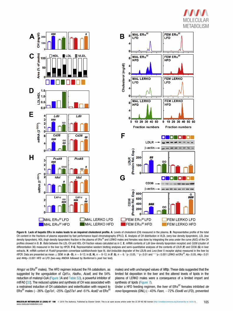
Figure 6: Lack of hepatic ERa in males leads to an impaired cholesterol profile. A. Levels of cholesterol (CH) measured in the plasma. B. Representative profile of the totalCH content in the fractions of plasma separated by fast performance liquid chromatography (FPLC). C. Analysis of CH distribution in VLDL (very-low density lipoprotein), LDL (lowdensity lipoprotein), HDL (high density lipoprotein) fraction in the plasma of ERaf/f and LERKO males and females was done by integrating the area under the curve (AUC) of the CHprofiles showed in B. D. Ratio between the LDL-CH and HDL-CH fraction values calculated as in C. E. mRNA contents of Ldlr (low-density lipoprotein receptor) and Cd36 (cluster ofdifferentiation 36) measured in the liver by rtPCR. F-G. Representative western blotting analyses and semi-quantitative analyses of the contents of LDLR (F) and CD36 (G) in liverextracts. H. mRNA content of Pcsk9 (proprotein convertase subtilisin/kexin type 9), Idol (inducible degrader of the LDLR) and Lxra (liver X receptor alpha) measured in the liver byrtPCR. Data are presented as mean � SEM: in (AeD), n ¼ 8-12; in (E, H), n ¼ 8-12; in (F, G), n ¼ 6. �p<0.05, ��p<0.01 and ���p<0.001 LERKO vs ERaf/f; #p<0.05, ##p<0.01and ###p<0.001 HFD vs LFD (two-way ANOVA followed by Bonferroni’s post hoc test).
Hmgcr vs ERaf/f males). The HFD regimen induced the FA catabolism, assuggested by the upregulation of Cpt1a, Hadha, Acadl, and the 54%reduction of malonyl-CoA (Figure 3A and Table S3), a powerful inhibitor ofmtFAO [32]. The reduced uptake and synthesis of CH was associated witha restrained induction of CH catabolism and esterification with respect toERaf/f males (�26% Cyp7a1, -25% Cyp27a1 and -51% Acat2 vs ERaf/f
MOLECULAR METABOLISM 32 (2020) 97e108 � 2019 The Author(s). Published by Elsevier GmbH. This is an openwww.molecularmetabolism.com
males) and with unchanged values ofMttp. These data suggested that thelimited fat deposition in the liver and the altered levels of lipids in theplasma of LERKO males were a consequence of a limited import andsynthesis of lipids (Figure 7).Under a HFD feeding regimen, the liver of ERaf/f females inhibited denovo lipogenesis (DNL) (�43% Fasn, �72% Elovl6 vs LFD), prevented
access article under the CC BY-NC-ND license (http://creativecommons.org/licenses/by-nc-nd/4.0/). 105

Figure 7: Hepatic ERa confers on liver a sex-specific strategy to cope with the excess of dietary lipids. Heatmaps representing the HFD-induced changes in the hepaticcontent of key genes involved in lipid metabolism. Data are expressed as percentage versus LFD.
Original Article
the synthesis of CH, and promoted the catabolism of FA (þ41%Hadha,þ37% Acadl vs LFD) and the catabolism of CH (þ84% Cyp7a1and þ34% Cyp27a1 vs LFD), without leading to an accumulation ofmedium-chain acyl-carnitines (Figure 3A and Table S3). The lipidexport was unchanged. All together, these changes represented aconcerted metabolic strategy useful to counteract the development offatty liver and the rise of circulating lipids (Figure 7).This female-biased ability to counteract the excess of dietary lipids byadopting protective metabolic adjustments was, at least in part,dependent on hepatic ERa (Figure 7). Indeed, with the lack of hepaticERa, although the synthesis of FA was still inhibited DNL (�43%Fasn, �67% Elovl6 vs LFD), the synthesis of CH increased (þ58%Hmgcr, þ36% Pmvk vs LFD). mtFAO was increased by HFD but to alesser extent with respect to ERaf/f females (�19% of Hadha andAcadl vs ERaf/f females) and led to the accumulation of medium- (C8,C9, C10) and long-chain (C12, C16, C8 and C18:1) acyl-carnitines(Figure 3A and Table S3). The lack of hepatic ERa did not affect theexpression of genes involved in CH catabolism nor in lipid export(Figure 7). These changes suggested that the increased lipid import,the enhanced CH synthesis, and the impaired mtFAO might contributeto higher deposition of lipids in the liver and to higher CH levels in theplasma of LERKO females.These data led us to conclude that hepatic ERa contributes to the sex-specific metabolic adaptation to a diet enriched in fats, by favoring andpreventing lipid deposition in the liver of males and females,respectively.
4. DISCUSSION
By directly comparing siblings of the two sexes, we here underlinemajor, sex-specific, and ERa-dependent differences in male and fe-male liver ability to cope with an excessive dietary intake of fats. The
106 MOLECULAR METABOLISM 32 (2020) 97e108 � 2019 The Author(s). Published by Elsevier GmbH. T
male liver was unable to set up compensatory mechanisms to dealwith the excess of dietary lipids in the long-term: after 16 weeks ofHFD, enhanced lipid uptake, increased lipid synthesis, and impaired FAoxidation led to fatty liver and degeneration of hepatic metabolism,which favored the increase of circulating CH. On the contrary, femaleliver was able to manage the excess lipids, limiting lipid import,avoiding worthless DNL, and efficiently promoting the mtFAO. Suchfemale metabolic flexibility was possibly a consequence of a sex-biased hepatic metabolism geared to satisfy the energy needs ofreproduction [5]. Indeed, the necessity to adapt the hepatic metabolismto different and highly variable reproductive needs might provide thefemale liver with a greater ability to manage dietary stress, includinglipid excess. In females, this metabolic flexibility was dependent onhepatic ERa; indeed, LERKO females were unable to properly processdietary lipids and were affected by fatty liver, increased circulatinglipids, and ectopic fat accumulation, all metabolic alterations generallyoccurring in females after menopause or ovariectomy [1,33,34].Since hepatic ERa activity provides females with the metabolic flexi-bility necessary to antagonize the detrimental effects of a HFD, thefinding of major metabolic changes in LERKO males was quite unex-pected. Indeed, despite the low expression of ERa that led us tospeculate that this receptor might be less relevant for the male hepaticmetabolism, our results showed the opposite: the lack of hepatic ERain LERKO males prevented lipid deposition and the progressivedegeneration of hepatic tissue typical of ERaf/f males exposed to HFD.Therefore, despite its abundance, ERa accomplishes a significantmetabolic function in the male liver that is clearly not the same as infemales. This is particularly relevant with regard to the uptake and denovo synthesis of lipids; indeed, under HFD conditions, the regulationof genes involved in these metabolic pathways is diametricallyopposed in the liver of males and females, depending on the presence/absence of hepatic ERa.
his is an open access article under the CC BY-NC-ND license (http://creativecommons.org/licenses/by-nc-nd/4.0/).www.molecularmetabolism.com
Besides lipid catabolism, the hepatic content of AA (Figure 6AeB) wasthe pathway most affected by HFD regimen in ERaf/f males, confirmingstudies showing a correlation between low AA content and hepaticsteatosis/NAFLD in obesogenic-like conditions [35]. The finding thatthe content of AA was decreased by HFD both in LERKO males andfemales, whereas it was unaffected in the liver of ERaf/f females,suggested that the homeostasis of AA under an unbalanced dietaryregimen is a female-specific prerogative in charge of hepatic ERa.However, the lack of hepatic ERa did not decrease but, otherwise,increased the differences between the two sexes (Fig. S2), suggestingthat ERa might strongly contribute to a different regulation of thehepatic metabolism in the two sexes.How can we explain that the same receptor in the same tissue has adifferent function in the two sexes? In our previous reports, we showedthat sex differences in the hepatic metabolism (and in particular in the AAcontent) depend on an estrogen-dependent sexual differentiation of themale liver that occurs right around birth; at that time, testis-derivedtestosterone is converted by the aromatase enzyme in estrogen that“organizes” and programs the liver in a male-specific manner, by actingthrough hepatic ERa [4]. The lack of such “organization” in the liver ofLERKO males (that do not express hepatic ERa since embryonal day 18.5)might be responsible for a major metabolic rewiring of the liver, evenwhen LERKO males are exposed to LFD (Figures 3A and 4A). As aconsequence of the lack of ERa-dependent programming of male hepaticmetabolism, the metabolic response of the LERKO male liver to HFDregimen was clearly different from that of the ERaf/f male liver and theLERKO female liver. In this view, liver ERa also has specific abilities incontrolling hepatic metabolism in males, despite its low concentration.
5. CONCLUSION
This study highlights the essential role of hepatic ERa in the oppositeregulation of lipid metabolism in the liver of the two sexes when dietarylipids are in excess: in males, hepatic ERa action contributes to liverlipid accumulation mainly by stimulating lipid import and synthesis;conversely, in females, ERa prevents the hepatic lipid deposition bykeeping the lipid uptake low, by inhibiting lipid synthesis, and byefficiently promoting mtFAO.All together, these data led us to conclude that hepatic ERa stronglycontributes to the sex-specific response to a diet enriched in fats, andits action accounts for opposite consequences for hepatic health inmales and females. In view of our observations, a proper liver ERaactivity should be preserved in females to maintain the metabolicflexibility and counteract the metabolic impairments (fatty liver,increased circulating lipids) typical of the post-menopause period[1,33,34]. As the exposure to estrogenic compounds early in life mayhave severe consequences for the hepatic metabolism of males [4],our study questions whether the increased incidence of metabolicdisturbances may be associated with an early exposure to estrogen-like endocrine disruptors [36,37], whose abundance is continuouslygrowing as a result of contaminants of our environment.
FUNDING
This work was supported by a grant from the European Community(ERC-Advanced Grant 322977).
AUTHOR CONTRIBUTIONS
C.M. performed most of the in vivo studies, biochemical assays and theliver histology analysis; C.M. revised the manuscript. M.B. performed
MOLECULAR METABOLISM 32 (2020) 97e108 � 2019 The Author(s). Published by Elsevier GmbH. This is an openwww.molecularmetabolism.com
the western blotting analysis and real-time PCR analysis. N.M.conceived the metabolomic analysis and the FPLC analysis of CHprofile; N.M revised the manuscript. F.L. performed real-time PCRanalysis and revised the manuscript. S.P. performed the metabolomicanalysis and the FPLC analysis of CH profile. D.C. participated in theconception of the metabolomic analysis. A.M. participated in theconception of the study, discussed the results and revised themanuscript. S.D.T. conceived the project, performed the in vivostudies, the biochemical assays, the analysis of metabolomic data;S.D.T. wrote and revised the manuscript.
ACKNOWLEDGMENTS
We are grateful to Valeria Benedusi for helpful discussion and critical reading and to
Monica Rebecchi for technical assistance.
CONFLICT OF INTEREST
The authors do not have any conflicts of interest.
APPENDIX A. SUPPLEMENTARY DATA
Supplementary data to this article can be found online at https://doi.org/10.1016/j.
molmet.2019.12.009.
REFERENCES
[1] Villa, A., Della Torre, S., Stell, A., Cook, J., Brown, M., Maggi, A., 2012.
Tetradian oscillation of estrogen receptor alpha is necessary to prevent liver
lipid deposition. Proceedings of the National Academy of Sciences of the
United States of America 109(29):11806e11811.
[2] Della Torre, S., Mitro, N., Fontana, R., Gomaraschi, M., Favari, E.,
Recordati, C., et al., 2016. An essential role for liver ERalpha in coupling
hepatic metabolism to the reproductive cycle. Cell Reports 15(2):360e371.
[3] Della Torre, S., Rando, G., Meda, C., Stell, A., Chambon, P., Krust, A., et al.,
2011. Amino acid-dependent activation of liver estrogen receptor alpha in-
tegrates metabolic and reproductive functions via IGF-1. Cell Metabolism
13(2):205e214.
[4] Della Torre, S., Mitro, N., Meda, C., Lolli, F., Pedretti, S., Barcella, M., et al.,
2018. Short-term fasting reveals amino acid metabolism as a major sex-
discriminating factor in the liver. Cell Metabolism 28(2):256e267 e5.
[5] Della Torre, S., Maggi, A., 2017. Sex differences: a resultant of an evolutionary
pressure? Cell Metabolism 25(3):499e505.
[6] Khristi, V., Ratri, A., Ghosh, S., Pathak, D., Borosha, S., Dai, E., et al., 2019.
Disruption of ESR1 alters the expression of genes regulating hepatic lipid and
carbohydrate metabolism in male rats. Molecular and Cellular Endocrinology
490:47e56.
[7] Zheng, D., Wang, X., Antonson, P., Gustafsson, J.A., Li, Z., 2018. Genomics of
sex hormone receptor signaling in hepatic sexual dimorphism. Molecular and
Cellular Endocrinology 471:33e41.
[8] Blachier, M., Leleu, H., Peck-Radosavljevic, M., Valla, D.C., Roudot-
Thoraval, F., 2013. The burden of liver disease in Europe: a review of available
epidemiological data. Journal of Hepatology 58(3):593e608.
[9] Maggi, A., Della Torre, S., 2018. Sex, metabolism and health. Molecular
Metabolism 15:3e7.
[10] Michelotti, G.A., Machado, M.V., Diehl, A.M., 2013. NAFLD, NASH and liver
cancer. Nature Reviews. Gastroenterology & Hepatology 10(11):656e665.
[11] Ballestri, S., Nascimbeni, F., Baldelli, E., Marrazzo, A., Romagnoli, D.,
Lonardo, A., 2017. NAFLD as a sexual dimorphic disease: role of gender and
reproductive status in the development and progression of nonalcoholic fatty
access article under the CC BY-NC-ND license (http://creativecommons.org/licenses/by-nc-nd/4.0/). 107

Original Article
liver disease and inherent cardiovascular risk. Advances in Therapy 34(6):
1291e1326.
[12] Della Torre, S., Benedusi, V., Fontana, R., Maggi, A., 2014. Energy metabolism
and fertility: a balance preserved for female health. Nature Reviews. Endo-
crinology 10(1):13e23.
[13] Kautzky-Willer, A., Handisurya, A., 2009. Metabolic diseases and associated
complications: sex and gender matter! European Journal of Clinical Investi-
gation 39(8):631e648.
[14] Amano, A., Kondo, Y., Noda, Y., Ohta, M., Kawanishi, N., Machida, S., et al.,
2017. Abnormal lipid/lipoprotein metabolism and high plasma testosterone
levels in male but not female aromatase-knockout mice. Archives of
Biochemistry and Biophysics 622:47e58.
[15] Chen, K.L., Madak-Erdogan, Z., 2018. Estrogens and female liver health.
Steroids 133:38e43.
[16] Han, S.I., Komatsu, Y., Murayama, A., Steffensen, K.R., Nakagawa, Y.,
Nakajima, Y., et al., 2014. Estrogen receptor ligands ameliorate fatty liver
through a nonclassical estrogen receptor/Liver X receptor pathway in mice.
Hepatology 59(5):1791e1802.
[17] Simpson, E.R., Misso, M., Hewitt, K.N., Hill, R.A., Boon, W.C., Jones, M.E.,
et al., 2005. Estrogen–the good, the bad, and the unexpected. Endocrine
Reviews 26(3):322e330.
[18] Barros, R.P., Gustafsson, J.A., 2011. Estrogen receptors and the metabolic
network. Cell Metabolism 14(3):289e299.
[19] Ribas, V., Nguyen, M.T., Henstridge, D.C., Nguyen, A.K., Beaven, S.W.,
Watt, M.J., et al., 2010. Impaired oxidative metabolism and inflammation are
associated with insulin resistance in ERalpha-deficient mice. American journal
of physiology. Endocrinology and metabolism 298(2):E304eE319.
[20] Erkan, G., Yilmaz, G., Konca Degertekin, C., Akyol, G., Ozenirler, S., 2013.
Presence and extent of estrogen receptor-alpha expression in patients with
simple steatosis and NASH. Pathology, Research and Practice 209(7):429e
432.
[21] Wang, X., Lu, Y., Wang, E., Zhang, Z., Xiong, X., Zhang, H., et al., 2015.
Hepatic estrogen receptor alpha improves hepatosteatosis through upregula-
tion of small heterodimer partner. Journal of Hepatology 63(1):183e190.
[22] Benedusi, V., Della Torre, S., Mitro, N., Caruso, D., Oberto, A., Tronel, C., et al.,
2017. Liver ERalpha regulates AgRP neuronal activity in the arcuate nucleus of
female mice. Scientific Reports 7(1):1194.
[23] Livak, K.J., Schmittgen, T.D., 2001. Analysis of relative gene expression data
using real-time quantitative PCR and the 2(-Delta Delta C(T)) Method. Methods
25(4):402e408.
[24] Hodson, L., Gunn, P.J., 2019. The regulation of hepatic fatty acid synthesis
and partitioning: the effect of nutritional state. Nature Reviews. Endocrinology.
108 MOLECULAR METABOLISM 32 (2020) 97e108 � 2019 The Author(s). Published by Elsevier GmbH. T
[25] Horton, J.D., Cohen, J.C., Hobbs, H.H., 2009. PCSK9: a convertase that co-
ordinates LDL catabolism. Journal of Lipid Research 50(Suppl):S172eS177.
[26] Maxwell, K.N., Breslow, J.L., 2004. Adenoviral-mediated expression of Pcsk9
in mice results in a low-density lipoprotein receptor knockout phenotype.
Proceedings of the National Academy of Sciences of the United States of
America 101(18):7100e7105.
[27] Demers, A., Samami, S., Lauzier, B., Des Rosiers, C., Ngo Sock, E.T., Ong, H.,
et al., 2015. PCSK9 induces CD36 degradation and affects long-chain
fatty acid uptake and triglyceride metabolism in adipocytes and in
mouse liver. Arteriosclerosis, Thrombosis, and Vascular Biology 35(12):2517e
2525.
[28] Ghosh, M., Galman, C., Rudling, M., Angelin, B., 2015. Influence of physio-
logical changes in endogenous estrogen on circulating PCSK9 and LDL
cholesterol. Journal of Lipid Research 56(2):463e469.
[29] Hong, C., Marshall, S.M., McDaniel, A.L., Graham, M., Layne, J.D., Cai, L.,
et al., 2014. The LXR-Idol axis differentially regulates plasma LDL levels in
primates and mice. Cell Metabolism 20(5):910e918.
[30] van Loon, N.M., Ottenhoff, R., Kooijman, S., Moeton, M., Scheij, S., Roscam
Abbing, R.L.P., et al., 2018. Inactivation of the E3 ubiquitin ligase IDOL at-
tenuates diet-induced obesity and metabolic dysfunction in mice. Arterio-
sclerosis, Thrombosis, and Vascular Biology 38(8):1785e1795.
[31] Zelcer, N., Hong, C., Boyadjian, R., Tontonoz, P., 2009. LXR regulates
cholesterol uptake through Idol-dependent ubiquitination of the LDL re-
ceptor. Science 325(5936):100e104.
[32] McGarry, J.D., Mannaerts, G.P., Foster, D.W., 1977. A possible role for
malonyl-CoA in the regulation of hepatic fatty acid oxidation and ketogenesis.
Journal of Clinical Investigation 60(1):265e270.
[33] Auro, K., Joensuu, A., Fischer, K., Kettunen, J., Salo, P., Mattsson, H., et al.,
2014. A metabolic view on menopause and ageing. Nature Communications 5:
4708.
[34] Gutierrez-Grobe, Y., Ponciano-Rodriguez, G., Ramos, M.H., Uribe, M., Mendez-
Sanchez, N., 2010. Prevalence of non alcoholic fatty liver disease in pre-
menopausal, posmenopausal and polycystic ovary syndrome women. The role
of estrogens. Annals of Hepatology 9(4):402e409.
[35] Lake, A.D., Novak, P., Shipkova, P., Aranibar, N., Robertson, D.G., Reily, M.D.,
et al., 2015. Branched chain amino acid metabolism profiles in progressive
human nonalcoholic fatty liver disease. Amino Acids 47(3):603e615.
[36] Foulds, C.E., Trevino, L.S., York, B., Walker, C.L., 2017. Endocrine-disrupting
chemicals and fatty liver disease. Nature Reviews. Endocrinology 13(8):445e
457.
[37] Heindel, J.J., Newbold, R., Schug, T.T., 2015. Endocrine disruptors and
obesity. Nature Reviews. Endocrinology 11(11):653e661.
his is an open access article under the CC BY-NC-ND license (http://creativecommons.org/licenses/by-nc-nd/4.0/).www.molecularmetabolism.com
![Validation of the (anti-) ERα CALUX bioassay ERa Calux...validation of the ERα CALUX bioassay (agonistic and antagonistic mode) according to the OECD guidelines for validation [2]](https://img.dokumen.tips/doc/110x75/5e63fa0741abdf46ef13e550/validation-of-the-anti-er-calux-bioassay-era-calux-validation-of-the-er.jpg)