Embed Size (px)
Citation preview

Hematopoietic stem cell fate isestablished by the Notch–Runx pathwayCaroline Erter Burns,1 David Traver,2 Elizabeth Mayhall,1 Jennifer L. Shepard,1
and Leonard I. Zon1,3
1Stem Cell Program and Division of Hematology/Oncology Children’s Hospital and Dana Farber Cancer Institute, HowardHughes Medical Institute, Harvard Stem Cell Institute, Harvard Medical School, Boston, Massachusetts 02115, USA;2Section of Cell and Developmental Biology, University of California, San Diego, La Jolla, California 92093-0380, USA
Identifying the molecular pathways regulating hematopoietic stem cell (HSC) specification, self-renewal, andexpansion remains a fundamental goal of both basic and clinical biology. Here, we analyzed the effects ofNotch signaling on HSC number during zebrafish development and adulthood, defining a critical pathway forstem cell specification. The Notch signaling mutant mind bomb displays normal embryonic hematopoiesisbut fails to specify adult HSCs. Surprisingly, transient Notch activation during embryogenesis via an inducibletransgenic system led to a Runx1-dependent expansion of HSCs in the aorta-gonad-mesonephros (AGM)region. In irradiated adults, Notch activity induced runx1 gene expression and increased multilineagehematopoietic precursor cells approximately threefold in the marrow. This increase was followed by theaccelerated recovery of all the mature blood cell lineages. These data define the Notch–Runx pathway ascritical for the developmental specification of HSC fate and the subsequent homeostasis of HSC number,thus providing a mechanism for amplifying stem cells in vivo.
[Keywords: Stem cell; Notch; Runx; AGM; zebrafish; irradiation]
Supplemental material is available at http://www.genesdev.org.
Received May 25, 2005; revised version accepted July 25, 2005.
In vertebrates, the adult hematopoietic system is com-posed of distinct cell lineages that undergo progressivedifferentiation from multipotent hematopoietic stemcells (HSCs) (Durand and Dzierzak 2005). The clinicalimportance of HSCs in transplantation protocols raisesthe significance of understanding their anatomical originand fate potential. Hematopoiesis arises during two em-bryonic phases: a brief primitive wave that predomi-nantly generates erythrocytes, followed by a definitivewave that produces long-term hematopoietic stem cells(LT-HSCs) capable of reconstituting the blood system forlife. Definitive hematopoiesis occurs, at least in part,in the aorta–gonad–mesonephros (AGM) region, whereLT-HSCs are associated with the ventral aortic wall. Al-though much is known about the cellular and functionalproperties of HSCs in mammals, relatively little is un-derstood about the genetic pathways regulating their in-duction, expansion, and homeostasis during embryogen-esis and adulthood. One pathway known to play a fun-damental role in regulating a variety of cell fate decisionsin progenitors of various organ systems is the Notch sig-naling pathway.
notch encodes a single pass glycoprotein receptor that
binds integral membrane ligands (Delta and Jagged) totransmit signals between cells in direct contact. Juxta-crine signaling generally occurs between neighbors thatarise from a common precursor and share similar devel-opmental potential (Lewis 1998). Notch is best knownfor its role in a process termed “lateral inhibition,” amodel explaining how one cell is selected from a group ofequivalent precursors to adopt an alternative fate (Green-wald and Rubin 1992). In this scenario, activation of theNotch receptor in a particular cell inhibits ligand pro-duction by that cell (the “signal-receiving cell”). Con-comitantly, cells producing high levels of ligand (the“signal-emitting cell”) force their neighbors to activateNotch and produce less ligand. Lateral inhibitionthrough the Notch pathway modulates lineage decisionsin related cells by inhibiting one fate and promoting an-other (Bray 1998; Lewis 1998).
The mechanism by which Notch determines cell fatehas been studied extensively in a variety of organismsand has led to the following model in which the Notchreceptor undergoes three successive cleavage events tobecome active. Notch is first processed in the Golgi net-work to produce a functional transmembrane receptor.Once bound to its ligand, the second cleavage event oc-curs, which generates two products, the Notch extra-cellular domain (NECD) and a membrane-bound acti-vated form (NEXT). The final cleavage event, mediatedby presenilin-dependent � secretase activity, occurs
3Corresponding author.E-MAIL [email protected]; FAX (617) 730-0222.Article published online ahead of print. Article and publication date areat http://www.genesdev.org/cgi/doi/10.1101/gad.1337005.
GENES & DEVELOPMENT 19:2331–2342 © 2005 by Cold Spring Harbor Laboratory Press ISSN 0890-9369/05; www.genesdev.org 2331
Cold Spring Harbor Laboratory Press on June 28, 2018 - Published by genesdev.cshlp.orgDownloaded from

within the NEXT fragment and results in the release ofthe Notch intracellular domain (NICD), which trans-locates to the nucleus and acts as a transcriptional regu-lator (Le Borgne and Schweisguth 2003).
Post-translational modification, specifically by addi-tion of ubiquitin peptides, plays a critical role in regu-lating Notch–Delta activity (Lai 2002b). Drosophila neu-ralized was shown to encode an E3 ligase that ubiqui-tylates and promotes the endocytosis of the Notch ligandDelta (Deblandre et al. 2001; Lai et al. 2001; Pavlopouloset al. 2001; Yeh et al. 2001). In vertebrate systems, how-ever, Neuralized is not essential for lateral inhibition,suggesting that other E3 ligases may modify Delta. mindbomb, a neurogenic mutant isolated in several zebrafishmutagenesis screens (Haffter et al. 1996; Schier et al.1996; Golling et al. 2002), encodes a highly conservedand previously uncharacterized E3 ligase that functionssimilarly to Neuralized (Itoh et al. 2003; Chen and CaseyCorliss 2004). Mind bomb is required cell nonautono-mously for Notch signaling and lateral inhibition by con-trolling Delta protein trafficking (Itoh et al. 2003; Chenand Casey Corliss 2004).
Notch receptors and their ligands are expressed in he-matopoietic cells and have been implicated in regulatingHSC induction and lineage cell fate decisions (Ohishiet al. 2003). In murine cell culture, constitutive Notch1expression in hematopoietic progenitor and stem cellsestablished immortalized cell lines able to generate prog-eny with either lymphoid or myeloid characteristicsboth in vitro and in long-term mouse reconstitution as-says (Varnum-Finney et al. 2000, 2003). Retroviral acti-vation of Notch1 in recombination activating gene-1(RAG-1)-deficient mouse stem cells resulted in an in-crease of HSCs due to decreased differentiation in vivo(Stier et al. 2002). Similar results were observed whenoverexpressing the Notch effector gene HES-1 in celllines (Kunisato et al. 2003). Increased numbers ofNotch1-expressing HSCs were documented in mice withexpanded osteoblastic niche cells that present Notch li-gands in the bone marrow microenvironment (Calvi etal. 2003). More recently, the Notch signaling pathwaywas shown to be active in native adult HSCs and down-regulated in differentiating progeny (Duncan et al. 2005).Taken together, activation of Notch in marrow-derivedHSCs is likely important in maintaining HSC fate andmay confer a survival advantage following transplanta-tion by promoting stem cell self-renewal.
Loss of Notch signaling in mice and flies has demon-strated that this pathway is required for HSC inductionduring embryogenesis. When the AGM region was sur-gically removed from Notch1−/− mice and grown in cul-ture, AGM-derived cells were unable to produce colonyforming cell (CFC) units in vitro or reconstitute theblood system of irradiated adult mice (Kumano et al.2003). In Drosophila blood cell development, cardiogenicmesoderm is induced and subsequently segregated intovascular (cardioblasts), excretory (nephrocytes), or blood(lymph gland) specific tissues. Consistent with themouse mutant, loss of Notch signaling caused a markeddecrease of excretory and blood progeny with a concomi-
tant expansion of vascular fates (Mandal et al. 2004).These results indicate that Notch signaling is importantin the early generation of blood precursors across widelydivergent phyla. The downstream target genes of Notchsignaling in stem cells, however, remain to be deter-mined.
Here, we used classic genetic experiments to reveal ahierarchical molecular network leading to the inductionand expansion of hematopoietic stem and progenitorcells in the zebrafish embryo and adult. We show thatthe Notch signaling mutant mind bomb develops nor-mal embryonic hematopoiesis but fails to specify defini-tive HSCs normally found associated with the ventralaortic wall. Using an inducible transgenic system, wefound that brief Notch stimulation greatly expandedHSC number in the AGM. Morpholino knock-down ofRunx1 function completely abolished this increase,showing for the first time that Runx1 is a downstreameffector of Notch signaling in HSC induction. In theadult, we examined the effect of activated Notch on mar-row recovery following irradiation. Compared with con-trol siblings, the percentage of multilineage hematopoi-etic precursors increased approximately threefold, fol-lowed by mature myeloid and lymphoid cells. Theexpanded precursor population showed transcriptionalup-regulation of the runx1, lmo2, and scl genes, all mark-ers of stem and progenitor cells. These findings definethe Notch–Runx pathway as critical during both embryo-genesis and adulthood to maintain HSC homeostasis.
Results
Mind bomb, the ubiquitin ligase for Notch ligands,is required for HSC specification
To assess the requirement for the Notch pathway in de-finitive HSC induction, a null allele of the Notch signal-ing mutant mind bomb (mib) was analyzed. Both c-myband runx1 transcripts mark emerging definitive hemato-poietic stem and progenitor cells that are normally con-fined to the ventral wall of the zebrafish dorsal aorta(Burns et al. 2002; Traver and Zon 2002; Gering and Pa-tient 2005). mind bomb mutants lack aortic c-myb andrunx1 expression (Fig. 1d,e) compared with wild-type sib-lings (Fig. 1a,b). Moreover, rag-1 transcripts expressed bydifferentiated thymic T-cells (Fig. 1c) are absent in mindbomb embryos 4 d post-fertilization (dpf) (Fig. 1f). Toexclude the possibility that HSC induction was defectivebecause a dorsal aorta was not specified, we analyzedflk1 expression in mind bomb animals. flk1 transcriptsappear relatively normal in mind bomb mutants (Fig. 1j)compared with wild-type animals (Fig. 1g), although in-tersomitic vessels are somewhat disorganized. The pres-ence of a dorsal aorta with concomitant loss of HSC/progenitor cells and lymphoid progeny suggests thatNotch signaling is required for definitive HSC specifica-tion.
We next analyzed whether Notch signaling is requiredfor primitive hematopoiesis. Blood and endothelialmarkers scl, gata1, fli1, and runx1 are expressed at the
Burns et al.
2332 GENES & DEVELOPMENT
Cold Spring Harbor Laboratory Press on June 28, 2018 - Published by genesdev.cshlp.orgDownloaded from
10-somite stage in all embryos derived from incrossingmind bomb heterozygous adults (data not shown), sug-gesting that Notch signaling is dispensable for primitiveprogenitor cell induction. Primitive erythrocyte num-bers appear normal in the intercellular mass (ICM) of 24h post-fertilization (hpf) mind bomb mutants comparedwith wild-type siblings as seen by gata1 (Fig. 1k,h), glo-bin (Fig. 1l,i), and scl (data not shown) expression. Primi-tive myeloid cells, marked by l-plastin (Fig. 1m,p), Pu.1(Fig. 1n,q), and myeloperoxidase (mpo) (Fig. 1o,r) are alsodetected in mind bomb embryos at levels similar to thatseen in wild type. Our results suggest that Notch signal-ing is dispensable for primitive hematopoiesis, but nec-essary for the definitive hematopoietic wave.
Notch signaling is sufficient for definitive HSCinduction in the embryonic AGM
Given the requirement of Notch signaling for blood stemcell induction and the need to find agents that increasestem cell number, we evaluated whether Notch activitywas sufficient to generate more HSCs in vivo. Since in-jection of an activated form of notch (NICD) mRNA isknown to cause severe dorso-anterior defects (Lawson etal. 2001), we made use of a powerful resource in zebrafishto temporally regulate NICD activation using a Gal4/UAS transgenic system (Scheer and Campos-Ortega1999; Scheer et al. 2002). Two major advantages of thisapproach are that NICD can be induced at any giventime point and that the activation can be brief. Sinceconstitutive Notch activity is known to block differen-tiation of stem cells both in vitro and in vivo, this sys-tem is unique in that NICD induction is reversible anddoes not prevent lineage commitment.
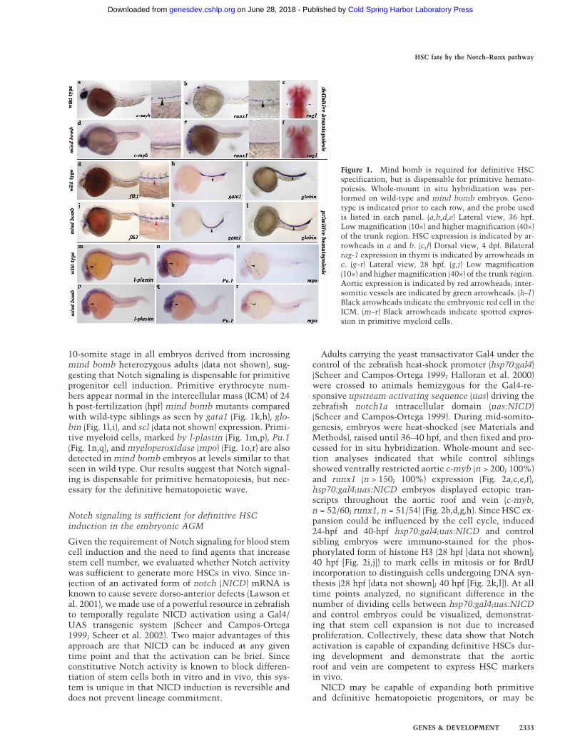
Adults carrying the yeast transactivator Gal4 under thecontrol of the zebrafish heat-shock promoter (hsp70:gal4)(Scheer and Campos-Ortega 1999; Halloran et al. 2000)were crossed to animals hemizygous for the Gal4-re-sponsive upstream activating sequence (uas) driving thezebrafish notch1a intracellular domain (uas:NICD)(Scheer and Campos-Ortega 1999). During mid-somito-genesis, embryos were heat-shocked (see Materials andMethods), raised until 36–40 hpf, and then fixed and pro-cessed for in situ hybridization. Whole-mount and sec-tion analyses indicated that while control siblingsshowed ventrally restricted aortic c-myb (n > 200; 100%)and runx1 (n > 150; 100%) expression (Fig. 2a,c,e,f),hsp70:gal4;uas:NICD embryos displayed ectopic tran-scripts throughout the aortic roof and vein (c-myb,n = 52/60; runx1, n = 51/54) (Fig. 2b,d,g,h). Since HSC ex-pansion could be influenced by the cell cycle, induced24-hpf and 40-hpf hsp70:gal4;uas:NICD and controlsibling embryos were immuno-stained for the phos-phorylated form of histone H3 (28 hpf [data not shown];40 hpf [Fig. 2i,j]) to mark cells in mitosis or for BrdUincorporation to distinguish cells undergoing DNA syn-thesis (28 hpf [data not shown]; 40 hpf [Fig. 2k,l]). At alltime points analyzed, no significant difference in thenumber of dividing cells between hsp70:gal4;uas:NICDand control embryos could be visualized, demonstrat-ing that stem cell expansion is not due to increasedproliferation. Collectively, these data show that Notchactivation is capable of expanding definitive HSCs dur-ing development and demonstrate that the aorticroof and vein are competent to express HSC markersin vivo.
NICD may be capable of expanding both primitiveand definitive hematopoietic progenitors, or may be
Figure 1. Mind bomb is required for definitive HSCspecification, but is dispensable for primitive hemato-poiesis. Whole-mount in situ hybridization was per-formed on wild-type and mind bomb embryos. Geno-type is indicated prior to each row, and the probe usedis listed in each panel. (a,b,d,e) Lateral view, 36 hpf.Low magnification (10×) and higher magnification (40×)of the trunk region. HSC expression is indicated by ar-rowheads in a and b. (c,f) Dorsal view, 4 dpf. Bilateralrag-1 expression in thymi is indicated by arrowheads inc. (g–r) Lateral view, 28 hpf. (g,j) Low magnification(10×) and higher magnification (40×) of the trunk region.Aortic expression is indicated by red arrowheads; inter-somitic vessels are indicated by green arrowheads. (h–l )Black arrowheads indicate the embryonic red cell in theICM. (m–r) Black arrowheads indicate spotted expres-sion in primitive myeloid cells.
HSC fate by the Notch–Runx pathway
GENES & DEVELOPMENT 2333
Cold Spring Harbor Laboratory Press on June 28, 2018 - Published by genesdev.cshlp.orgDownloaded from
exclusive for increasing adult HSCs. Primitive pro-genitor markers runx1, scl, and fli1 were examined3.5 h post-heat shock and were not expanded (Fig. 2m–o).Induced Notch is active at this time since NICDRNA is detected in embryos as early as 1.5 h post-heat shock (Scheer et al. 2002). In our hands, gal4 RNAwas highly expressed in hsp70:gal4 transgenic animals(Fig. 2q) and was absent from control siblings (Fig. 2p).Therefore, Notch-dependent expansion of HSCs in theAGM is independent of primitive progenitor cell induc-tion.
Dose-dependent HSC/progenitor cell expansion isindependent of artery identity
HSC induction could be dependent on arterial fate deci-sions. Work by Lawson and Weinstein (Lawson et al.2001) clearly show that Notch signaling is partially re-quired for artery identity in that mind bomb mutantsfail to express the arterial markers ephrinb2a andnotch3, but maintain arterial expression of tbx20, flk1,and gridlock. Although it was initially determined thatNICD was not sufficient to expand aortic-specific mark-ers to the vein (Lawson et al. 2001), expansion ofephrinb2a was later demonstrated (Lawson et al.2002). These studies suggest that Notch signaling is re-quired in the zebrafish for some aspects of artery identityand is sufficient to expand some arterial markers to thevein.
We tested whether the abnormal expansion of HSCs tothe vein correlated with a conversion of vein to arteryidentity. Embryos from hsp70:gal4 and uas:NICD mat-ings were heat-shocked for varying lengths of time andprocessed by in situ hybridization for ephrinb2a or runx1at 28 hpf or c-myb at 40 hpf. When heat-shocked for 1min, we detected no expansion of c-myb (n = 0/19) orephrinb2a (n = 0/27) transcripts in hsp70:gal4;uas:NICDtransgenics (Fig. 3a,c). Weak expansion of runx1 was de-tectable in the vein (n = 2/9) (Fig. 3b), although mosthsp70:gal4;uas:NICD embryos showed no ectopic tran-scripts. After 10 min of heat shock, hsp70:gal4;uas:NICDembryos showed considerable expansion of c-myb(n = 26/32) and runx1 (n = 4/8) expression throughout theartery and in some regions of the vein, but no expansionof ephrinb2a (n = 0/18) could be visualized (Fig. 3d–f). Ifembryos were exposed to heat for 20 min, c-myb (n = 36/36) and runx1 (n = 10/10) transcripts were hugely ex-panded in the aorta and vein of hsp70:gal4;uas:NICDtransgenics, suggesting that ectopic HSCs had been in-duced (Fig. 3g,h). Conversely, no hsp70:gal4;uas:NICDembryos showed significant expansion of ephrinb2aexpression (n = 0/39) (Fig. 3i). These data demon-strate that artery identity and HSC specification can beuncoupled in vivo, suggesting that Notch signalingacts through separate pathways to regulate induction ofeach cell fate. Additionally, these studies establish adose response for Notch signaling in the derivation ofHSCs.
Runx1 is dispensable for artery identity in vivo
In the mouse, Runx1 is dispensable for primitive hema-topoiesis, but necessary for the definitive wave (Wang etal. 1996; Fujita et al. 2001; Okuda et al. 2001). Morerecently, morpholino knock-down experiments (Ekker2000) revealed a similar function for Runx1 in the devel-oping zebrafish embryo (Kalev-Zylinska et al. 2002). Asthe hematopoietic phenotype displayed by the runx1morphant is strikingly similar to mind bomb, we hy-pothesized that these pathways may converge at thelevel of definitive HSC induction.
To define a genetic relationship between notch signal-ing and runx1, we established a runx1 morphant pheno-
Figure 2. NICD expands HSC/progenitor cells in the AGM.Whole-mount in situ hybridization was performed on controlsibling and hsp70:gal4;uas:NICD embryos (see Materials andMethods for heat-shock conditions). Red arrowheads denoteaorta expression, and black arrowheads show vein expression.The probe used is listed in each panel. (a–d) Lateral view. Lowmagnification (10×) and higher magnification (40×) of the trunkregion. hsp70:gal4;uas:NICD transgenics show expanded HSCmarkers throughout the artery and vein. (e–h) Plastic sectionsthrough the trunk region of 36–40 hpf whole-mount in situhybridized embryos. (e,f) Note ventrally restricted c-myb andrunx1 aortic expression. (g,h) Note ectopic expansion of c-myband runx1 to the aortic roof and vein. pH3 immunostaining at∼40 hpf (i,j) (see Materials and Methods) and BrdU incorpora-tion at ∼40 hpf (k,l). Compare brown stain in dividing cellsin vessel region of wild-type (i,k) to hsp70:gal4;uas:NICD (j,l).(m–q) Approximately 10–12 somite stage. (m–o) Dorsal view;(p,q) lateral view.
Burns et al.
2334 GENES & DEVELOPMENT
Cold Spring Harbor Laboratory Press on June 28, 2018 - Published by genesdev.cshlp.orgDownloaded from

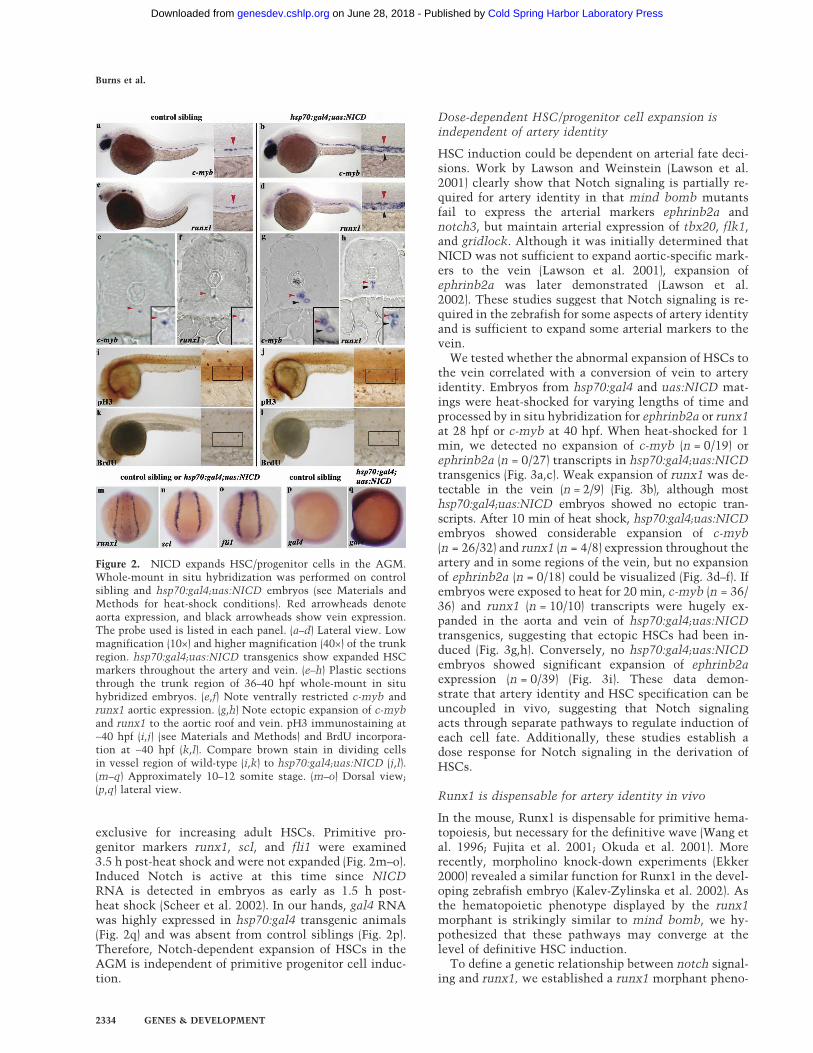
type by coinjecting two morpholinos that block mRNAsplicing (see Materials and Methods). As in previouslypublished reports (Kalev-Zylinska et al. 2002; Gering andPatient 2005), we found that Runx1 is dispensable forprimitive hematopoiesis but required for c-myb express-ing definitive HSCs (Supplementary Fig. 1a). RT–PCRanalysis demonstrated that runxMO injection abolishednormal runx1 transcripts compared with uninjected con-trols (Supplementary Fig. 1b). We next investigated ves-sel integrity and aortic identity by analyzing expressionof the aortic markers notch3, deltaC, and ephrinb2a inrunx morphant animals. One of the hallmarks of properaorta identity is the formation of branching intersomiticvessels (ISV). Although aortic identity is not completelynormal in runx morphants, as seen by the lack of fli1(Fig. 4a,b) (n = 19) and flk1 (Fig. 4c,d) (n = 23) expressingISVs, notch3 (n = 13), deltaC (n = 14), and ephbrinb2a(n = 12) aortic expression is maintained in the absence ofRunx1 function (Fig. 4e–j). These data show that Runx1is dispensable for proper localization of aortic tran-scripts, suggesting that Runx1 function is not requiredfor most aspects of artery identity.
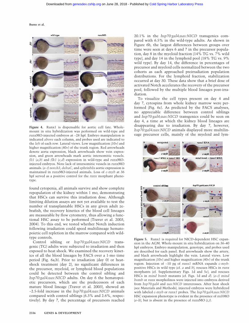
We next tested whether c-myb-expressing HSCs couldbe expanded or rescued by overexpression of runx1 RNAin wild-type, runx1 morphant, or mind bomb mutantanimals (Fig. 5). runx1 RNA was injected at the 1–2-cellstage, and embryos were raised until ∼40 hpf. Comparedwith uninjected sibling controls that show ventrally re-stricted aortic c-myb expression (Figs. 2e, 5a), runx1mRNA expanded c-myb-expressing HSCs ectopicallythroughout the aortic roof and vein (n = 16/24) (Fig. 5b).Interestingly, runx1 mRNA overexpression partially res-cued c-myb-expressing HSCs in the aortic floor of runx1morphants (n = 20/24) (cf. Supplementary Figs. 1d and5c) and mind bomb animals (n = 8/10) (cf. Figs. 1d and5d). We did notice that wild-type embryos injected withrunx1 RNA showed greater numbers of c-myb-positivecells compared with runxMO- and mind bomb-injectedanimals, suggesting that the total amount of Runx1function in the developing embryo is critical for HSCspecification. In summary, our results demonstrate thatrunx1 functions genetically downstream of or in paral-lel to notch signaling and mind bomb. These data sug-gest that Runx1, like NICD, dose-dependently influ-ences the number of c-myb-positive HSCs specified inthe AGM.
Runx1 is required for NICD-dependent expansionof definitive HSCs in the AGM
During embryonic neurogenesis, Mind Bomb is knownto act upstream of NICD production in the Notch sig-naling pathway (Itoh et al. 2003; Chen and Casey Corliss2004). We tested whether NICD would suppress themind bomb definitive HSC phenotype. A mind bomb-specific morpholino (mibMO) (Itoh et al. 2003) was in-jected into embryo clutches derived from hsp70:gal4 anduas:NICD matings. Following exposure to heat shock,control siblings showed complete loss of both c-myb(n = 117/125) and runx1 (n = 97/141) transcripts (Fig.5e,g) in the dorsal aorta, thereby phenocopying the mindbomb mutant. mibMO-injected hsp70:gal4;uas:NICDembryos showed the notch gain-of-function phenotypein that both c-myb (n = 45/45) and runx1 (n = 43/49)transcripts were expanded to the aortic roof and vein(Fig. 5f,h). These data show that the Mind Bomb E3 ubiq-uitin ligase functions upstream of NICD during HSCspecification. To test whether runx1 is required forNICD-dependent expansion of HSCs in the AGM, therunxMO was injected into clutches derived fromhsp70:gal4 and uas:NICD matings (Fig. 5i,j), and the ani-mals were subsequently heat-shocked. Interestingly,runx morphant hsp70:gal4;uas:NICD transgenics (Fig.5j) showed low to no c-myb expression in the aortic floor(n = 9/12), thus phenocopying the runx morphant controlsiblings (Fig. 5i) (n = 30/37). This finding demonstratesthat Runx1 is required for NICD-dependent HSC expan-sion. Together, these results suggest a genetic hierarchyleading to HSC induction in the AGM in which NICD isgenetically downstream of mind bomb and upstream ofrunx1.
NICD activation improves marrow recovery followingirradiation
Our studies in the embryo have demonstrated an impor-tant role for signaling via the Notch–Runx pathway inthe initial specification of HSC fate. We next testedwhether this pathway also plays a role in the adult he-matopoietic system. We have previously shown that asublethal dose of total body irradiation rapidly leads to avirtual depletion of hematopoietic cells in adult wholekidney marrow (Traver et al. 2004). Despite this pro-
Figure 3. NICD dose-dependently expands HSCs inde-pendent of aortic cell fate. Whole-mount in situ hybrid-ization was performed on hsp70:gal4;uas:NICD em-bryos heat-shocked at 40°C for 1, 10, or 20 min. Redarrowheads denote aorta expression, and black arrow-heads show vein expression. The arrowhead size sug-gests the level of transcript present. Low magnification(10×) and higher magnification (40×) of the trunk region.(a,d,g) c-myb HSC expression. (b,e,h) runx1 HSC expres-sion. (c,f,i) ephrinB2a arterial expression.
HSC fate by the Notch–Runx pathway
GENES & DEVELOPMENT 2335
Cold Spring Harbor Laboratory Press on June 28, 2018 - Published by genesdev.cshlp.orgDownloaded from
found cytopenia, all animals survive and show completerepopulation of the kidney within 1 mo, demonstratingthat HSCs can survive this irradiation dose. Althoughlimiting dilution assays are not yet available to test thenumber of transplantable HSCs in any given adult ze-brafish, the recovery kinetics of the blood cell lineagesare measurable by flow cytometry, thus allowing a func-tional HSC assay to be performed (Traver et al. 2003,2004). To this end, we tested whether Notch inductionfollowing irradiation could speed multilineage hemato-poietic cell repletion in the marrow compared with wild-type controls.
Control sibling or hsp70:gal4;uas:NICD trans-genic (TG) adults were subjected to irradiation and thenexposed to heat shock. We examined the recovery kinet-ics of all the blood lineages by FACS over a 1-mo timeperiod (Fig. 6a,b). Prior to irradiation (day 0) or heat-shock treatment (day 2), no significant differences inthe precursor, myeloid, or lymphoid blood populationscould be detected between the control sibling andhsp70:gal4;uas:NICD adults. On day 6 the hematopoi-etic precursors, which are the predecessors of eachmature blood lineage (Traver et al. 2003), showed an∼2.5-fold increase in the hsp70:gal4;uas:NICD animalscompared with control siblings (6.3% and 2.6%, respec-tively). By day 7, the percentage of precursors reached
20.1% in the hsp70:gal4;uas:NICD transgenics com-pared with 6.5% in the wild-type adults. As shown inFigure 6b, the largest differences between groups overtime were seen at days 6 and 7 in the precursor popula-tion, day 8 in the myeloid fraction (14% TG vs. 7% wildtype), and day 14 in the lymphoid pool (18% TG vs. 9%wild type). By day 14, the difference in percentages ofprecursor and myeloid cells normalized between the twocohorts as each approached preirradiation populationdistributions. For the lymphoid fraction, stabilizationoccurred at day 30. These data show that a brief dose ofactivated Notch accelerates the recovery of the precursorpool, followed by the multiple blood lineages post-irra-diation.
To visualize the cell types present on day 4 andday 7, cytospins from whole kidney marrow were per-formed (Fig. 6c). As predicted by the FACS analyses,no appreciable difference between control siblingsand hsp70:gal4;uas:NICD transgenics could be seen onday 4, a time at which the kidney blood lineages aredisappearing due to irradiation. By day 7, however,hsp70:gal4;uas:NICD animals displayed more multilin-eage precursor cells, mainly of the myeloid and lym-
Figure 4. Runx1 is dispensable for aortic cell fate. Whole-mount in situ hybridization was performed on wild-type andrunxMO-injected embryos at ∼28 hpf. Embryo manipulation isindicated above each column, and probes used are indicated tothe left of each row. Lateral views. Low magnification (10×) andhigher magnification (40×) of the trunk region. Red arrowheadsdenote aorta expression, black arrowheads show vein expres-sion, and green arrowheads mark aortic intersomitic vessels.fli1 (a,b) and flk1 (c,d) expression in wild-type and runxMO-injected embryos. Note lack of intersomitic vessels in runxMOanimals. (e–j) notch3, deltaC, and ephrinb2a aortic expression ismaintained in runxMO-injected animals. Loss of c-myb at 36hpf served as a positive control for the runx morphant pheno-type.
Figure 5. Runx1 is required for NICD-dependent HSC expan-sion in the AGM. Whole-mount in situ hybridization on 36–40hpf embryos. Embryo manipulation, genotype, and probes usedare described for each panel. Red arrowheads show the artery,and black arrowheads highlight the vein. Lateral views. Lowmagnification (10×) and higher magnification (40×) of the trunkregion. Injection of ∼10 pg of runx1 mRNA expands c-myb-positive HSCs in wild type (cf. a and b), rescues HSCs in runxmorphants (cf. Supplementary Figs. 1d and 5c), and rescuesHSCs in mind bomb mutants (cf. Figs. 1d and d). (e–j) mindbomb or runx morpholinos were injected into embryos derivedfrom hsp70:gal4 and uas:NICD intercrosses. After heat shock(see Materials and Methods), injected embryos were hybridizedwith labeled c-myb or runx1 probes. The hsp70:gal4;uas:NICDHSC expansion phenotype is evident in the presence of mibMO(e–h), but is absent in the presence of runxMO (i,j).
Burns et al.
2336 GENES & DEVELOPMENT
Cold Spring Harbor Laboratory Press on June 28, 2018 - Published by genesdev.cshlp.orgDownloaded from

phoid lineages than their sibling controls. These findingsdemonstrate that NICD is capable of expanding earlymultilineage hematopoietic precursors at a rate fasterthan that of control siblings, strongly suggesting thatNotch signaling is expanding a common predecessor.
Since precursor cells from multiple lineages were ex-panded in hsp70:gal4;uas:NICD animals on day 6, weanalyzed whether transcripts of known stem and pro-genitor cell markers were transcriptionally up-regulatedprior to the increase of the precursor population. RNAwas harvested from individual control sibling orhsp70:gal4;uas:NICD kidneys on day 3 and quantita-tive PCR was performed. The fold change in gene ex-pression was determined for each kidney and the averagefold change for each gene of interest was calculatedand graphed (Fig. 6d). While expression of flk1, fli1, andgata2 were not affected, runx1 and lmo2 transcripts werefour- and fivefold higher in the hsp70:gal4;uas:NICDanimals, respectively, while scl transcripts were in-creased by twofold. These findings demonstrate thathsp70:gal4;uas:NICD adults up-regulate stem and pro-genitor cell markers in their marrow following heat-
shock exposure, likely reflecting an expansion of theHSC pool as a result of NICD activity. Moreover, limitedNotch signaling accelerates hematopoietic recoverywithout permanently altering the balance of cells in eachblood lineage. Our findings show that the Notch path-way is used during both embryogenesis and adulthood tomaintain proper stem cell homeostasis.
Discussion
Notch signaling has been hypothesized to control stemcell self-renewal. Prior experimental approaches have al-lowed only limited conclusions to be drawn since con-stitutive Notch activation, typically with a retrovirus,renders a maturation defect that prevents the normaldifferentiation of progenitors. Our data demonstrate thatNotch activity is necessary to establish HSC fate andsufficient to expand HSC number in the embryonicAGM. In the adult, a brief pulse of Notch activity ex-pands the HSC pool following sublethal doses of totalbody irradiation and speeds the recovery of multilineagehematopoiesis without permanently altering blood lin-
Figure 6. NICD expands the multilineage pre-cursor cells in adult marrow and acceleratesblood lineage recovery post-irradiation. (a) Car-toon depicting manipulation of adult control sib-ling or hsp70:gal4;uas:NICD fish is outlined.Light scatter profiles of whole kidney marrowfrom adult control sibling or hsp70:gal4;uas:NICDfish showing the effects of sublethal irradiationand heat shock on the relative percentages oferythrocytes (red gate), lymphocytes (blue gate),precursors (purple gate), and myelomonocytes(green gate) over time. Each FACS plot is of onerepresentative fish from each time point, and thepercentages reflect the average number of cells ineach gate. The erythroid population is excludedfrom our analyses due to a marked variability inthe numbers extracted from whole kidney dissec-tions. This inconsistency has been previously re-ported (Traver et al. 2004) and is likely due to theclose association of the kidney with the dorsalaorta. (b) The average percentage of cells in eachgate (Y-axis, shown as percentages in a), with theexception of erythroid, was plotted over time (X-axis). (c) Cytospin preparation of day 4 (top) orday 7 (bottom) whole kidney marrow from con-trol sibling (left) or hsp70:gal4;uas:NICD adults(right). (d) Quantitative PCR analysis from con-trol sibling or hsp70:gal4;uas:NICD whole kidneymarrow on day 3 (see Materials and Methods). Theaverage fold induction of each transcript is repre-sented by the bar graph with standard devia-tions. (WT) Wild type; (TG) hsp70:gal4;uas:NICD;(lp) lymphocyte precursor; (ep) erythroid precur-sor; (mp) myeloid precursor.
HSC fate by the Notch–Runx pathway
GENES & DEVELOPMENT 2337
Cold Spring Harbor Laboratory Press on June 28, 2018 - Published by genesdev.cshlp.orgDownloaded from

eage homeostasis. We further identify runx1 as a majortarget of Notch signaling and demonstrate the require-ment of the Notch–Runx pathway for stem cell fate.
Notch and hematopoietic cell fate determination
Hematopoietic cell fate decisions occur throughout em-bryogenesis and adulthood, many of which are con-trolled by the Notch signaling pathway. Notch1 influ-ences the decision of T versus B lymphoid fate (Pui et al.1999; Radtke et al. 1999), lymphoid versus myeloid dif-ferentiation (Stier et al. 2002), �� versus �� T-cell fate(Washburn et al. 1997), and CD4 versus CD8 T-cell lin-eages (Robey et al. 1996), thus providing a key regulatorysignal in determining the fate of multipotential hemato-poietic precursors. Here, we provide evidence that Notchdrives mesodermal progenitors to the HSC fate.
The process of Notch regulating stem cell fate appearsto be evolutionarily conserved. In flies, Notch is requiredfor specification of dorsal mesoderm to the blood, endo-thelial, and nephrocyte fates (Mandal et al. 2004). In theabsence of Notch, the mesoderm adopts an entirely en-dothelial fate, while in the presence of high Notch ac-tivity, it becomes predominantly blood and nephrocytes.Although the hematopoietic system of a vertebrate ismore elaborate and under distinct control mechanisms,our studies support the Drosophila conclusions and in-dicate a conservation of Notch driving the blood stemcell fate. Mice deficient for Notch1 activity (Kumano etal. 2003; Hadland et al. 2004) or one of its transcriptionalmediators, RBPj� (Robert-Moreno et al. 2005), lack em-bryonic HSC specification in the AGM. Similarly, wefound that mind bomb mutant embryos do not specifyHSCs, establishing the Notch requirement during stemcell induction.
During vertebrate embryogenesis, mesoderm in theAGM region becomes specified to several fates. Manystudies refer to the “hemogenic endothelial cell” as abipotential precursor to the HSC and vascular tree; how-ever, recent studies provide evidence for a subaortic mes-enchymal cell population that independently migratesthrough the endothelial cells to become hematopoietic(Mendes et al. 2005). Additionally, the “mesoangioblast”has been proposed as a common mesodermal precursorto the blood, endothelial, mesenchymal, and smoothmuscle lineages (Cossu and Bianco 2003; Ema et al.2003). The fate decisions imposed on mesodermal pro-genitors within the AGM are clearly influenced by theNotch pathway. For instance, mice deficient in RBPj�show expanded VE-Cadherin and CD31/PECAM endo-thelial cell expression with concomitant loss of defini-tive HSCs (Robert-Moreno et al. 2005). Ablation of theCOUP-TFII transcription factor in endothelial cells en-abled veins to acquire arterial characteristics, includingthe expression of Notch1 and the formation of ectopicHSCs (You et al. 2005). This result would favor Notchacting to induce HSCs from a hemogenic endothelialcell. Our results in zebrafish support a model in whichthe Notch pathway regulates arterial and HSC fatechoice either from distinct mesodermal populations or
over different developmental windows since each deci-sion can be uncoupled in vivo. In support of this hypoth-esis, Notch activity did not expand arterial ephrinb2aexpression to the vein, a location where the HSC tran-scripts c-myb and runx1 were robust. In this case, HSCscould be specified from mesodermal precursors that aredistinct from those committed to the arterial fate. It ispossible that subaortic mesenchymal cells are a target ofNotch activity and a source of HSC precursors indepen-dent of the endothelium. The finding that both aorta andvein express HSC markers in the Notch-activated statewith minimal change in ephrinB2a expression indicatesthat Notch independently regulates mesoderm–HSC andartery–vein cell fate decisions.
Lateral inhibition has been proposed in the central ner-vous system whereby Notch signaling promotes non-neural fates while inhibiting neural development (Lewis1998). HSC fate may be established by a similar mecha-nism whereby Notch activation in an endothelial ormesenchymal cell causes down-regulation of ligand pro-duction. Consequently, a cell that produces more ligandwill force its neighbor to produce less, thus generating asalt-and-pepper pattern of cells containing elevatedNotch activity. In this model, cells containing high lev-els of NICD would become HSCs, while those with lowNICD activity would remain endothelial or mesenchy-mal.
The Notch–Runx1 pathway participatesin self-renewal in the stem cell niche
The adult stem cell niche has been recently character-ized in the mouse bone marrow and consists of an end-osteal (quiescent) and vascular (proliferative) compart-ment (Heissig et al. 2002; Calvi et al. 2003; Zhang et al.2003; Arai et al. 2004; Avecilla et al. 2004). Under steady-state conditions, it is thought that most HSCs reside inthe G0 phase of the cell cycle in close contact with stro-mal cells, including osteoblasts (Calvi et al. 2003; Zhanget al. 2003). The balance between quiescent and cyclingstem cells appears to rely on the amount of soluble cy-tokines, which result in HSCs relocating from the osteo-blastic to the vascular niche (Heissig et al. 2002). Thismobilization of stem cells into peripheral circulationmay be necessary for reconstituting the HSC pool. Manysignaling pathways are thought to contribute to stemcell self-renewal in the marrow niche including Notch(Maillard et al. 2003), Wnt (Reya et al. 2003; Willert et al.2003; Duncan et al. 2005), Hedgehog (Baron 2001; Bhard-waj et al. 2001; Gering and Patient 2005), and factors thatnegatively regulate the cell cycle, such as Tie2/Angio-poietin-1 (Arai et al. 2004). Cooperation of such path-ways is thought to maintain stem cell homeostasis invivo.
Several studies have hypothesized that Notch affectsHSCs, although direct proof of the activity and thedownstream targets have remained to be elucidated. Inmurine cell culture, constitutive Notch1 expression inHSC/progenitor cells established immortalized cell linesable to generate progeny with either lymphoid or my-
Burns et al.
2338 GENES & DEVELOPMENT
Cold Spring Harbor Laboratory Press on June 28, 2018 - Published by genesdev.cshlp.orgDownloaded from

eloid characteristics (Varnum-Finney et al. 2000, 2003).Retroviral Notch1 activation in recombination activat-ing gene-1 (RAG-1)-deficient mouse stem cells resultedin an increase in HSC self-renewal and favored lymphoidover myeloid differentiation (Stier et al. 2002).
The studies presented here differ from others in that abrief pulse of Notch activity was administered and thecells were able to terminally differentiate. Other experi-ments with retroviruses and conditional alleles perma-nently express NICD and thus alter the normal matura-tion of cells. For instance, in our adult assays an increasein the lymphoid cell fate was not concomitant with adecrease in the myeloid lineage, as previously seen (Stieret al. 2002). Based on these results, we propose that ac-tivated Notch expands the stem and progenitor cell com-partment by either influencing undifferentiated cells toadopt a HSC fate or by causing a G0 HSC population toup-regulate runx1-dependent gene expression.
Our findings that the stem cell markers runx1, scl, andlmo2 were transcriptionally increased in response toNICD indicates that stem and progenitor cells were ex-panded in the adult marrow, possibly by increasing stemcell self-renewal. Recently, a conditional allele of runx1was generated in the mouse to study the loss of Runx1function during adult hematopoiesis (Ichikawa et al.2004; Growney et al. 2005). In transplantation studies,Runx1-excised marrow cells showed a reduced competi-tive repopulating ability in long-term engraftment assays(Growney et al. 2005), demonstrating that Runx1 is es-sential for normal stem cell function. We demonstratethat the NICD-induced expansion of HSCs in the AGMis dependent on Runx1. When we examined the proxi-mal and distal promoters of the human runx1 gene(Ghozi et al. 1996), we found no DNA-binding sites forRBPj�, the primary Notch pathway mediator that physi-cally interacts with DNA to modulate target gene tran-scription (Lai 2002a; data not shown). It is still possiblethat Notch directly regulates runx1 transcriptionthrough alternative binding sites, although it may indi-rectly activate runx1 expression. In either case, theNotch–Runx pathway is likely operative in both theAGM and adult marrow and may lead to the activationof downstream targets critical for stem cell homeostasis.
Notch signaling has been extensively linked to theprocess of both normal and aberrant stem cell self-re-newal (Maillard et al. 2003). The human Notch1 recep-tor, TAN-1, was first identified as a partner gene in a (7;9)chromosomal translocation found in <1% of all T-cellacute lymphoblastic leukemias (T-ALL) (Ellisen et al.1991). Recently, >50% of all human T-ALLs were shownto have activating mutations in the notch1 gene (Wenget al. 2004). These data emphasize how dysregulation ofthe Notch signaling pathway can result in uncontrolledself-renewal that ultimately produces malignancy.
Clinical implications
Transplantation of HSCs has been successful in thetreatment of malignancies and other diseases, such asaplastic and sickle-cell anemia (Gaziev and Lucarelli
2003). After irradiation or chemotherapy is given to pa-tients, restoration of normal hematopoiesis is critical toprevent infection and bleeding. In this study, we showedthat a pulse of Notch activity expands stem cell numberin the adult marrow without permanently altering bloodlineage homeostasis. This finding has obvious therapeu-tic implications. Small molecule agonists that induceNotch signaling could be used to pharmacologically ex-pand stem cell numbers and blood progenitors. For in-stance, embryonic cord blood stem cells are often insuf-ficient for adult stem cell transplants. Notch activatorsmay be used to increase mobilization of HSCs for trans-plantation, similar to the clinical activity of G-CSF inperipheral stem cell harvests. Our data provide rationalefor future clinical work to focus on methods that ma-nipulate the Notch signaling pathway to amplify bloodstem cells, and thus multilineage hematopoiesis.
Materials and methods
Fish care and strains
Zebrafish were bred and maintained as described (Solnica-Krezel et al. 1994). The following lines were used: wild-typeAB, wik, and Tu strains, mind bombta56b (Jiang et al. 1996),Tg(uas:notch1a-intra) (Scheer and Campos-Ortega 1999), andTg(hsp70:gal4) (Scheer and Campos-Ortega 1999).
In situ hybridization, morpholinos, and mRNAs
Whole-mount in situ hybridization was performed as described(Thisse et al. 1993). Digoxygenin-labeled antisense RNA probeswere synthesized using a DIG RNA Labeling Kit (SP6/T7;Roche). For histological analysis, embryos were embedded inJB4 plastic resin (Polysciences Inc.). Embedded embryos wereprepared using a Leica microtome and the resulting 8–10-µmsections were mounted onto glass slides and photographed.
The antisense morpholino oligos (Summerton et al. 1997;Ekker 2000; Nasevicius and Ekker 2000) runxMO3 (5�-TGTTAAACTCACGTCGTGGCTCTC-3�) and runxMO5 (5�-AATGTGTAAACTCACAGTGTAAAGC-3�) recognize donor sitesof predicted exon/intron junctions. The final injection solutioncontained 0.6 mM runxMO3, 1 mM runxMO5, 1× Danieau Me-dium, and 1× Phenol Red (Saude et al. 2000). The mind bombmorpholino (Itoh et al. 2003) was injected at a concentration of1 mM in 1× Danieau Medium/1× Phenol Red.
runx1, formerly known as runxa (Burns et al. 2002), was sub-cloned into the pCS2+ expression vector, and capped RNA wassynthesized using mMessage Machine (Ambion). Ten pico-grams to 20 pg of RNA was injected into the yolk of one-cell- totwo-cell-stage embryos.
Immunostaining and BrdU incorporation
Embryos were fixed overnight in 4% paraformaldehyde at 4°C.For staining, embryos were washed twice in PBST, incubated inacetone for 7 min at −20°C, then washed once in water andtwice in PBST at room temperature. Embryos were incubated inpolyclonal anti-pH3 antibody (1:750 dilution; Santa Cruz Bio-technology) for at least 2 h at room temperature, washed inPBS-T, incubated in goat anti-rabbit horseradish peroxidase-conjugated antibody (Jackson Immunoresearch) for 2 h at room
HSC fate by the Notch–Runx pathway
GENES & DEVELOPMENT 2339
Cold Spring Harbor Laboratory Press on June 28, 2018 - Published by genesdev.cshlp.orgDownloaded from

temperature, washed in PBS-T, and then developed in diamino-benzidine/H2O2 (Sigma).
For BrdU incorporation, embryos were chilled on ice for 15min at the 24- and 40-hpf stages and pulsed in a 10 mM BrdU,15% DMSO solution for 20 min on ice. Embryos were thentransferred to warm embryo media, incubated for 5 min at28.5°C, fixed in 4% PFA for 2 h at room temperature, and in-cubated overnight in methanol at −20°C. Embryos were rehy-drated in PBS-T, digested with proteinase K (10 µg/mL), andpost-fixed in 4% PFA for 20 min at room temperature. Embryoswere washed in H2O, incubated in 2 N HCl for 1 h, washed inPBS-T, and placed in a blocking solution for 30 min at roomtemperature. Embryos were incubated in monoclonal anti-BrdUantibody (1:100 dilution; Sigma) for at least 2 h at room tem-perature, incubated in goat anti-mouse horseradish peroxidase-conjugated antibody (Jackson Immunoresearch) for 2 h at roomtemperature, washed in PBS-T, and then developed in diamino-benzidine/H2O2 (Sigma).
Embryo heat-shock experiments
Tg(hsp70:gal4) adults were mated to Tg(uas:notch1a-intra) fishand their embryos were harvested and raised in E3 (Westerfield1995). Between the 8- and 12-somite stages, embryos were col-lected in 50-mL Falcon tubes in ∼5 mL of E3 and submerged ina 37°C waterbath for 30–45 min. Subsequently, embryos wereplaced in Petri dishes, allowed to develop until ∼40 hpf, col-lected in 4% paraformaldehyde, and processed by in situ hybrid-ization. For the results in Figure 3, the experimental conditionsfor timing and heat shock were followed from Lawson et al.(2002).
Adult irradiation, heat shock, and kinetic analysis
Tg(hsp70:gal4) adults were mated to Tg(uas:notch1a-intra)fish and their embryos were harvested and raised. GenomicDNA was extracted from clipped tail fins, and PCR amplifica-tion determined whether each transgene was present. Controlsiblings (wild type or single transgenics) and experimentalhsp70:gal4;uas:NICD double transgenic fish were irradiated at asublethal dose of 2000 rads/20 Gy (day 0), as previously de-scribed (Traver et al. 2004). Fish were returned to the aquaticsfacility until day 2, when they were removed and placed in a37°C dry incubator overnight. The fish were returned to theaquatics facility on day 3.
Whole kidney marrow was dissected from the following num-ber of euthanized adult zebrafish: day 0, wild type = 3,hsp70:gal4;uas:NICD (TG) = 3; day 2, wild type = 4, TG = 3; day4, wild type = 6, TG = 6; day 7, wild type = 6, TG = 7; day 8, wildtype = 4, TG = 4; day 10, wild type = 5, TG = 4; day 15, wildtype = 5, TG = 4; day 30, wild type = 5, TG = 3. Individual kid-neys were placed in a 12-well Petri plate on ice in 2 mL ofice-cold 0.9× Dulbecco’s phosphate buffered saline (PBS) con-taining 5% 0.2-µm-filtered fetal calf serum (FCS). The kidneywas dissociated to a single cell suspension by repeated pipettingwith a P1000. The suspension was then filtered over a 40-µmnylon mesh into a FACS tube on ice. The 2-mL suspension wasthen aliquoted to 1 mL per FACS tube. One-hundred-thousandcells were collected by flow cytometry based on propidium io-dide (PI) exclusion (1 µg/mL; Sigma), forward scatter, and sidescatter using a FACS Vantage flow cytometer (Becton Dickin-son), as previously described (Traver et al. 2004). Total kidneycell counts were performed using a hemocytometer.
Quantitative PCR analysis from whole kidney marrow
Adult fish were sublethally irradiated and heat-shocked as de-scribed above. Fish were removed from the 37°C incubator on
day 3 and kept at room temperature for ∼3–4 h, at which timeindividual kidneys were dissected. Each kidney was placed inTrizol (Invitrogen), total RNA was isolated, and random primedcDNA was generated (Invitrogen, SuperScript III First-StrandSynthesis System). The SYBR green (Invitrogen) method wasused to quantify cDNAs of interest, which are represented asthe fold change in transcript level between control sibling andhsp70:gal4;uas:NICD kidneys. The amount of cDNA startingmaterial for each kidney was normalized in relation to �-actinexpression. For all experiments, cDNA was quantified using anApplied Biosystems Sequence Detection System 7000. Theprimer sequences used were as follows: scl: forward, 5�-CTCGAATGGTGCAGTTGAGTCC-3�, reverse, 5�-GCATCTCCAGCAAAACCACTGT-3�; flk1: forward, 5�-CGAACGTGAAGTGACATACGG-3�, reverse, 5�-CCCTCTACCAAACCATGTGAAA-3�; fli1: forward, 5�-CCCCTCAGCAAGAGTGGATTA-3�,reverse, 5�-CGGCGACTCTTGTGCAGTATAT-3�; runx1: for-ward, 5�-CGTCTTCACAAACCCTCCTCAA-3�, reverse, 5�-GCTTTACTGCTTCATCCGGCT-3�; lmo2: forward, 5�-AAACACTGGAGGCAAATGAGGA-3�, reverse, 5�-AGAAAGAAGCGGTCTCCGATG-3�; gata2: forward, 5�-ACAACGTCAACAGGCCACTGA-3�, reverse, 5�-TCGAAACCCTCACCAGATCGT-3�;�-actin: forward, 5�-GCTGTTTTCCCCTCCATTGTT-3�, re-verse, 5�-TCCCATGCCAACCATCACT-3�.
Acknowledgments
We thank S. Orkin, G. Daley, and W. Goessling for criticalevaluation of the manuscript; C. Belair and B. Barut for animaland laboratory management; A. Flint for assistance with flowcytometry; R. Peterson for �-actin quantitative PCR primers;and B. Appel and A. Latimer for mind bomb mutant lines.C.E.B. was supported by the American Cancer Society (PF-01-255-01-LIB) and currently holds a Research Career Award (1 K01DK067179-01 A1) through the NIH NIDDK division. L.I.Z. issupported by HHMI, and this work was funded by a grant fromthe NIH NHLBI (5 R01 HL48801-13).
References
Arai, F., Hirao, A., Ohmura, M., Sato, H., Matsuoka, S., Takubo,K., Ito, K., Koh, G.Y., and Suda, T. 2004. Tie2/angiopoietin-1signaling regulates hematopoietic stem cell quiescence inthe bone marrow niche. Cell 118: 149–161.
Avecilla, S.T., Hattori, K., Heissig, B., Tejada, R., Liao, F., Shido,K., Jin, D.K., Dias, S., Zhang, F., Hartman, T.E., et al. 2004.Chemokine-mediated interaction of hematopoietic progeni-tors with the bone marrow vascular niche is required forthrombopoiesis. Nat. Med. 10: 64–71.
Baron, M. 2001. Induction of embryonic hematopoietic and en-dothelial stem/progenitor cells by hedgehog-mediated sig-nals. Differentiation 68: 175–185.
Bhardwaj, G., Murdoch, B., Wu, D., Baker, D.P., Williams, K.P.,Chadwick, K., Ling, L.E., Karanu, F.N., and Bhatia, M. 2001.Sonic hedgehog induces the proliferation of primitive humanhematopoietic cells via BMP regulation. Nat. Immunol.2: 172–180.
Bray, S. 1998. Notch signalling in Drosophila: Three ways to usea pathway. Semin. Cell Dev. Biol. 9: 591–597.
Burns, C.E., DeBlasio, T., Zhou, Y., Zhang, J., Zon, L., andNimer, S.D. 2002. Isolation and characterization of runxaand runxb, zebrafish members of the runt family of tran-scriptional regulators. Exp. Hematol. 30: 1381–1389.
Calvi, L.M., Adams, G.B., Weibrecht, K.W., Weber, J.M., Olson,
Burns et al.
2340 GENES & DEVELOPMENT
Cold Spring Harbor Laboratory Press on June 28, 2018 - Published by genesdev.cshlp.orgDownloaded from

D.P., Knight, M.C., Martin, R.P., Schipani, E., Divieti, P.,Bringhurst, F.R., et al. 2003. Osteoblastic cells regulate thehaematopoietic stem cell niche. Nature 425: 841–846.
Chen, W. and Casey Corliss, D. 2004. Three modules of ze-brafish Mind bomb work cooperatively to promote Deltaubiquitination and endocytosis. Dev. Biol. 267: 361–373.
Cossu, G. and Bianco, P. 2003. Mesoangioblasts—Vascular pro-genitors for extravascular mesodermal tissues. Curr. Opin.Genet. Dev. 13: 537–542.
Deblandre, G.A., Lai, E.C., and Kintner, C. 2001. Xenopus neu-ralized is a ubiquitin ligase that interacts with XDelta1 andregulates Notch signaling. Dev. Cell 1: 795–806.
Duncan, A.W., Rattis, F.M., Dimascio, L.N., Congdon, K.L.,Pazianos, G., Zhao, C., Yoon, K., Cook, J.M., Willert, K.,Gaiano, N., et al. 2005. Integration of Notch and Wnt sig-naling in hematopoietic stem cell maintenance. Nat. Immu-nol. 6: 314–322.
Durand, C. and Dzierzak, E. 2005. Embryonic beginnings ofadult hematopoietic stem cells. Haematologica 90: 100–108.
Ekker, S.C. 2000. Morphants: A new systematic vertebratefunctional genomics approach. Yeast 17: 302–306.
Ellisen, L.W., Bird, J., West, D.C., Soreng, A.L., Reynolds, T.C.,Smith, S.D., and Sklar, J. 1991. TAN-1, the human homologof the Drosophila notch gene, is broken by chromosomaltranslocations in T lymphoblastic neoplasms. Cell 66: 649–661.
Ema, M., Faloon, P., Zhang, W.J., Hirashima, M., Reid, T., Stan-ford, W.L., Orkin, S., Choi, K., and Rossant, J. 2003. Combi-natorial effects of Flk1 and Tal1 on vascular and hematopoi-etic development in the mouse. Genes & Dev. 17: 380–393.
Fujita, Y., Nishimura, M., Taniwaki, M., Abe, T., and Okuda, T.2001. Identification of an alternatively spliced form of themouse AML1/RUNX1 gene transcript AML1c and its ex-pression in early hematopoietic development. Biochem. Bio-phys. Res. Commun. 281: 1248–1255.
Gaziev, J. and Lucarelli, G. 2003. Stem cell transplantation forhemoglobinopathies. Curr. Opin. Pediatr. 15: 24–31.
Gering, M. and Patient, R. 2005. Hedgehog signaling is requiredfor adult blood stem cell formation in zebrafish embryos.Dev. Cell 8: 389–400.
Ghozi, M.C., Bernstein, Y., Negreanu, V., Levanon, D., andGroner, Y. 1996. Expression of the human acute myeloidleukemia gene AML1 is regulated by two promoter regions.Proc. Natl. Acad. Sci. 93: 1935–1940.
Golling, G., Amsterdam, A., Sun, Z., Antonelli, M., Maldonado,E., Chen, W., Burgess, S., Haldi, M., Artzt, K., Farrington, S.,et al. 2002. Insertional mutagenesis in zebrafish rapidly iden-tifies genes essential for early vertebrate development. Nat.Genet. 31: 135–140.
Greenwald, I. and Rubin, G.M. 1992. Making a difference: Therole of cell–cell interactions in establishing separate identi-ties for equivalent cells. Cell 68: 271–281.
Growney, J.D., Shigematsu, H., Li, Z., Lee, B.H., Adelsperger, J.,Rowan, R., Curley, D.P., Kutok, J.L., Akashi, K., Williams,I.R., et al. 2005. Loss of Runx1 perturbs adult hematopoiesisand is associated with a myeloproliferative phenotype.Blood 106: 494–504.
Hadland, B.K., Huppert, S.S., Kanungo, J., Xue, Y., Jiang, R.,Gridley, T., Conlon, R.A., Cheng, A.M., Kopan, R., andLongmore, G.D. 2004. A requirement for Notch1 distin-guishes 2 phases of definitive hematopoiesis during develop-ment. Blood 104: 3097–3105.
Haffter, P., Granato, M., Brand, M., Mullins, M.C., Hammer-schmidt, M., Kane, D.A., Odenthal, J., van Eeden, F.J., Jiang,Y.J., Heisenberg, C.P., et al. 1996. The identification of geneswith unique and essential functions in the development of
the zebrafish, Danio rerio. Development 123: 1–36.Halloran, M.C., Sato-Maeda, M., Warren, J.T., Su, F., Lele, Z.,
Krone, P.H., Kuwada, J.Y., and Shoji, W. 2000. Laser-inducedgene expression in specific cells of transgenic zebrafish. De-velopment 127: 1953–1960.
Heissig, B., Hattori, K., Dias, S., Friedrich, M., Ferris, B., Hack-ett, N.R., Crystal, R.G., Besmer, P., Lyden, D., Moore, M.A.,et al. 2002. Recruitment of stem and progenitor cells fromthe bone marrow niche requires MMP-9 mediated release ofkit-ligand. Cell 109: 625–637.
Ichikawa, M., Asai, T., Chiba, S., Kurokawa, M., and Ogawa, S.2004. Runx1/AML-1 ranks as a master regulator of adulthematopoiesis. Cell Cycle 3: 722–724.
Itoh, M., Kim, C.H., Palardy, G., Oda, T., Jiang, Y.J., Maust, D.,Yeo, S.Y., Lorick, K., Wright, G.J., Ariza-McNaughton, L., etal. 2003. Mind bomb is a ubiquitin ligase that is essential forefficient activation of Notch signaling by Delta. Dev. Cell4: 67–82.
Jiang, Y.J., Brand, M., Heisenberg, C.P., Beuchle, D., Furutani-Seiki, M., Kelsh, R.N., Warga, R.M., Granato, M., Haffter, P.,Hammerschmidt, M., et al. 1996. Mutations affecting neu-rogenesis and brain morphology in the zebrafish, Danio re-rio. Development 123: 205–216.
Kalev-Zylinska, M.L., Horsfield, J.A., Flores, M.V., Postlethwait,J.H., Vitas, M.R., Baas, A.M., Crosier, P.S., and Crosier, K.E.2002. Runx1 is required for zebrafish blood and vessel devel-opment and expression of a human RUNX1–CBF2T1 trans-gene advances a model for studies of leukemogenesis.Development 129: 2015–2030.
Kumano, K., Chiba, S., Kunisato, A., Sata, M., Saito, T.,Nakagami-Yamaguchi, E., Yamaguchi, T., Masuda, S.,Shimizu, K., Takahasi, T., et al. 2003. Notch1 but NotNotch2 is essential for generating hematopoietic stem cellsfrom endothelial cells. Immunity 18: 699–711.
Kunisato, A., Chiba, S., Nakagami-Yamaguchi, E., Kumano, K.,Saito, T., Masuda, S., Yamaguchi, T., Osawa, M., Kageyama,R., Nakauchi, H., et al. 2003. HES-1 preserves purified he-matopoietic stem cells ex vivo and accumulates side popu-lation cells in vivo. Blood 101: 1777–1783.
Lai, E.C. 2002a. Keeping a good pathway down: Transcriptionalrepression of Notch pathway target genes by CSL proteins.EMBO Rep. 3: 840–845.
———. 2002b. Protein degradation: Four E3s for the notch path-way. Curr. Biol. 12: R74–R78.
Lai, E.C., Deblandre, G.A., Kintner, C., and Rubin, G.M. 2001.Drosophila neuralized is a ubiquitin ligase that promotes theinternalization and degradation of delta. Dev. Cell 1: 783–794.
Lawson, N.D., Scheer, N., Pham, V.N., Kim, C.H., Chitnis, A.B.,Campos-Ortega, J.A., and Weinstein, B.M. 2001. Notch sig-naling is required for arterial–venous differentiation duringembryonic vascular development. Development 128: 3675–3683.
Lawson, N.D., Vogel, A.M., and Weinstein, B.M. 2002. sonichedgehog and vascular endothelial growth factor act up-stream of the Notch pathway during arterial endothelial dif-ferentiation. Dev. Cell 3: 127–136.
Le Borgne, R. and Schweisguth, F. 2003. Notch signaling: Endo-cytosis makes delta signal better. Curr. Biol. 13: R273–R275.
Lewis, J. 1998. Notch signalling and the control of cell fatechoices in vertebrates. Semin. Cell Dev. Biol. 9: 583–589.
Maillard, I., Adler, S.H., and Pear, W.S. 2003. Notch and theimmune system. Immunity 19: 781–791.
Mandal, L., Banerjee, U., and Hartenstein, V. 2004. Evidence fora fruit fly hemangioblast and similarities between lymph-gland hematopoiesis in fruit fly and mammal aorta-gonadal-
HSC fate by the Notch–Runx pathway
GENES & DEVELOPMENT 2341
Cold Spring Harbor Laboratory Press on June 28, 2018 - Published by genesdev.cshlp.orgDownloaded from

mesonephros mesoderm. Nat. Genet. 36: 1019–1023.Mendes, S.C., Robin, C., and Dzierzak, E. 2005. Mesenchymal
progenitor cells localize within hematopoietic sites through-out ontogeny. Development 132: 1127–1136.
Nasevicius, A. and Ekker, S.C. 2000. Effective targeted gene‘knockdown’ in zebrafish. Nat. Genet. 26: 216–220.
Ohishi, K., Katayama, N., Shiku, H., Varnum-Finney, B., andBernstein, I.D. 2003. Notch signalling in hematopoiesis. Se-min. Cell Dev. Biol. 14: 143–150.
Okuda, T., Nishimura, M., Nakao, M., and Fujita, Y. 2001.RUNX1/AML1: A central player in hematopoiesis. Int. J.Hematol. 74: 252–257.
Pavlopoulos, E., Pitsouli, C., Klueg, K.M., Muskavitch, M.A.,Moschonas, N.K., and Delidakis, C. 2001. neuralized en-codes a peripheral membrane protein involved in delta sig-naling and endocytosis. Dev. Cell 1: 807–816.
Pui, J.C., Allman, D., Xu, L., DeRocco, S., Karnell, F.G., Bak-kour, S., Lee, J.Y., Kadesch, T., Hardy, R.R., Aster, J.C., et al.1999. Notch1 expression in early lymphopoiesis influencesB versus T lineage determination. Immunity 11: 299–308.
Radtke, F., Wilson, A., Stark, G., Bauer, M., van Meerwijk, J.,MacDonald, H.R., and Aguet, M. 1999. Deficient T cell fatespecification in mice with an induced inactivation ofNotch1. Immunity 10: 547–558.
Reya, T., Duncan, A.W., Ailles, L., Domen, J., Scherer, D.C.,Willert, K., Hintz, L., Nusse, R., and Weissman, I.L. 2003.A role for Wnt signalling in self-renewal of haematopoieticstem cells. Nature 423: 409–414.
Robert-Moreno, A., Espinosa, L., de la Pompa, J.L., and Bigas, A.2005. RBPj�-dependent Notch function regulates Gata2 andis essential for the formation of intra-embryonic hematopoi-etic cells. Development 132: 1117–1126.
Robey, E., Chang, D., Itano, A., Cado, D., Alexander, H., Lans,D., Weinmaster, G., and Salmon, P. 1996. An activated formof Notch influences the choice between CD4 and CD8 T celllineages. Cell 87: 483–492.
Saude, L., Woolley, K., Martin, P., Driever, W., and Stemple,D.L. 2000. Axis-inducing activities and cell fates of the ze-brafish organizer. Development 127: 3407–3417.
Scheer, N. and Campos-Ortega, J.A. 1999. Use of the Gal4-UAStechnique for targeted gene expression in the zebrafish.Mech. Dev. 80: 153–158.
Scheer, N., Riedl, I., Warren, J.T., Kuwada, J.Y., and Campos-Ortega, J.A. 2002. A quantitative analysis of the kinetics ofGal4 activator and effector gene expression in the zebrafish.Mech. Dev. 112: 9–14.
Schier, A.F., Neuhauss, S.C., Harvey, M., Malicki, J., Solnica-Krezel, L., Stainier, D.Y., Zwartkruis, F., Abdelilah, S.,Stemple, D.L., Rangini, Z., et al. 1996. Mutations affectingthe development of the embryonic zebrafish brain. Develop-ment 123: 165–178.
Solnica-Krezel, L., Schier, A.F., and Driever, W. 1994. Efficientrecovery of ENU-induced mutations from the zebrafishgermline. Genetics 136: 1401–1420.
Stier, S., Cheng, T., Dombkowski, D., Carlesso, N., and Scad-den, D.T. 2002. Notch1 activation increases hematopoieticstem cell self-renewal in vivo and favors lymphoid over my-eloid lineage outcome. Blood 99: 2369–2378.
Summerton, J., Stein, D., Huang, S.B., Matthews, P., Weller, D.,and Partridge, M. 1997. Morpholino and phosphorothioateantisense oligomers compared in cell-free and in-cell sys-tems. Antisense Nucleic Acid Drug Dev. 7: 63–70.
Thisse, C., Thisse, B., Schilling, T.F., and Postlethwait, J.H.1993. Structure of the zebrafish snail1 gene and its expres-sion in wild-type, spadetail and no tail mutant embryos.Development 119: 1203–1215.
Traver, D. and Zon, L.I. 2002. Walking the walk: Migration andother common themes in blood and vascular development.Cell 108: 731–734.
Traver, D., Paw, B.H., Poss, K.D., Penberthy, W.T., Lin, S., andZon, L.I. 2003. Transplantation and in vivo imaging of mul-tilineage engraftment in zebrafish bloodless mutants. Nat.Immunol. 4: 1238–1246.
Traver, D., Winzeler, A., Stern, H.M., Mayhall, E.A., Langenau,D.M., Kutok, J.L., Look, A.T., and Zon, L.I. 2004. Effects oflethal irradiation in zebrafish and rescue by hematopoieticcell transplantation. Blood 104: 1298–1305.
Varnum-Finney, B., Xu, L., Brashem-Stein, C., Nourigat, C.,Flowers, D., Bakkour, S., Pear, W.S., and Bernstein, I.D.2000. Pluripotent, cytokine-dependent, hematopoietic stemcells are immortalized by constitutive Notch1 signaling.Nat. Med. 6: 1278–1281.
Varnum-Finney, B., Brashem-Stein, C., and Bernstein, I.D. 2003.Combined effects of Notch signaling and cytokines induce amultiple log increase in precursors with lymphoid and my-eloid reconstituting ability. Blood 101: 1784–1789.
Wang, Q., Stacy, T., Binder, M., Marin-Padilla, M., Sharpe, A.H.,and Speck, N.A. 1996. Disruption of the Cbfa2 gene causesnecrosis and hemorrhaging in the central nervous systemand blocks definitive hematopoiesis. Proc. Natl. Acad. Sci.93: 3444–3449.
Washburn, T., Schweighoffer, E., Gridley, T., Chang, D.,Fowlkes, B.J., Cado, D., and Robey, E. 1997. Notch activityinfluences the �� versus �� T cell lineage decision. Cell 88:833–843.
Weng, A.P., Ferrando, A.A., Lee, W., Morris, J.P.T., Silverman,L.B., Sanchez-Irizarry, C., Blacklow, S.C., Look, A.T., andAster, J.C. 2004. Activating mutations of NOTCH1 in hu-man T cell acute lymphoblastic leukemia. Science 306: 269–271.
Westerfield, M. 1995. The zebrafish book, 3rd ed. University ofOregon Press, Eugene, OR.
Willert, K., Brown, J.D., Danenberg, E., Duncan, A.W., Weiss-man, I.L., Reya, T., Yates III, J.R., and Nusse, R. 2003. Wntproteins are lipid-modified and can act as stem cell growthfactors. Nature 423: 448–452.
Yeh, E., Dermer, M., Commisso, C., Zhou, L., McGlade, C.J.,and Boulianne, G.L. 2001. Neuralized functions as an E3ubiquitin ligase during Drosophila development. Curr. Biol.11: 1675–1679.
You, L.R., Lin, F.J., Lee, C.T., DeMayo, F.J., Tsai, M.J., and Tsai,S.Y. 2005. Suppression of Notch signalling by the COUP-TFII transcription factor regulates vein identity. Nature435: 98–104.
Zhang, J., Niu, C., Ye, L., Huang, H., He, X., Tong, W.G., Ross,J., Haug, J., Johnson, T., Feng, J.Q., et al. 2003. Identificationof the haematopoietic stem cell niche and control of theniche size. Nature 425: 836–841.
Burns et al.
2342 GENES & DEVELOPMENT
Cold Spring Harbor Laboratory Press on June 28, 2018 - Published by genesdev.cshlp.orgDownloaded from

10.1101/gad.1337005Access the most recent version at doi: 19:2005, Genes Dev.
Caroline Erter Burns, David Traver, Elizabeth Mayhall, et al. pathway
Runx−Hematopoietic stem cell fate is established by the Notch
Material
Supplemental
http://genesdev.cshlp.org/content/suppl/2005/09/15/gad.1337005.DC1
References
http://genesdev.cshlp.org/content/19/19/2331.full.html#ref-list-1
This article cites 70 articles, 22 of which can be accessed free at:
License
ServiceEmail Alerting
click here.right corner of the article or
Receive free email alerts when new articles cite this article - sign up in the box at the top
Cold Spring Harbor Laboratory Press
Cold Spring Harbor Laboratory Press on June 28, 2018 - Published by genesdev.cshlp.orgDownloaded from