Embed Size (px)
Citation preview
Regular paper
Hemagglutinin stalk domain from H5N1 strain as a potentially universal antigenKarolina Uranowska, Jolanta Tyborowska, Anna Jurek, Bogusław Szewczyk and Beata Gromadzka*
Department of Recombinant Vaccines, Intercollegiate Faculty of Biotechnology, University of Gdansk and Medical University of Gdansk, Gdańsk, Poland
Influenza A virus infections are the major public health concern and cause significant morbidity and mortality each year worldwide. Vaccination is the main strategy of influenza epidemic prevention. However, seasonal vac-cines induce strain-specific immunity and must be re-formulated annually based on prediction of the strains that will circulate in the next season. Thus, it is essential to develop vaccines that would induce broad and per-sistent immunity to influenza viruses. Hemagglutinin is the major surface antigen of the influenza virus. Recent studies revealed the importance of HA stalk-specific an-tibodies in neutralization of different influenza virus strains. Therefore, it is important to design an immu-nogen that would focus the immune response on the HA stalk domain in order to elicit neutralizing antibod-ies. In the present study, we report characterization of a conserved truncated protein, potentially a universal influenza virus antigen from the H5N1 Highly Patho-genic Avian Influenza A virus strain. Our results indicate that exposure of the HA stalk domain containing con-served epitopes results in cross reactivity with different antibodies (against group 1 and 2 HAs). Additionally, we conclude that HA stalk domain contains not only confor-mational epitopes recognized by universal FI6 antibody, but also linear epitopes recognized by other antibodies.
Key words: H5N1, universal influenza antigen, hemagglutinin stalk domain, universal influenza antibodies, FI6
INTRODUCTION
The influenza virus belongs to the Orthomyxoviridae family and is one of the life-threatening pathogens in the world. There are three types of influenza virus identified as A, B and C, while subtype A is predominantly a se-rious public health problem causing acute infections in humans, characterized by high mortality rate. According to the World Health Organization (WHO), each year in-fluenza virus infects up to 15% of the population (ap-proximately 1 billion cases). Epidemiological data show a possibility of emergence of new pandemic strains.
Outbreak of the human pandemic influenza A (pH1N1) has caused the intensification of the scientific research and clinical trials so as to develop pandemic in-fluenza vaccines and therapies. Blood samples were col-lected from donors exposed to pH1N1 and plasma cells
were isolated with the aim of screening for secretion of an antibody that would bind to diverse hemagglutinins. A human monoclonal antibody FI6 was found to recog-nize all subtypes of HAs. FI6 cross-reacts with members of both HAs groups via the attachment to the conserved epitope in hemagglutinin (Corti et al., 2011).
Hemagglutinin (HA) is the major surface glycoprotein of influenza virus. It is a homotrimeric protein molecule, where each monomer consists of two subunits linked by disulfide bonds: HA1 subunit, which forms a globu-lar head and HA2 subunit, which forms a stalk domain (Fig. 1)(Kang et al., 2011). Hemagglutinin plays a crucial role in the viral entry. The globular head domain con-tains a receptor-binding site for sialic acid that enables attachment of the virus to the host cell, while fusion peptide located in the stalk domain is responsible for the pH-induced fusion of the viral envelope with the endo-some membrane (Bouvier & Palese, 2008).
HA proteins of influenza A viruses are divided into at least 17 subtypes and are categorized into two ma-jor phylogenetic groups based on distinct structures in the HA2 stalk domain: group 1 (subtypes H1, H2, H5, H6, H8, H9, H11, H12, H13, H16 and H17) and group 2 (subtypes H3, H4, H7, H10, H14, H15) (Gamblin & Skehel, 2010; Krammer et al., 2012a).
Current seasonal vaccines predominantly induce an-ti-HA antibodies that recognize antigenic sites in the globular head domain and act by blocking its receptor-binding activity (Margine et al., 2013a). However, HA head is highly variable among diverse virus strains which is related to frequent amino-acid changes in this region which allows efficient immune evasion. This leads to the lack of protection against viruses that are different from the strain used for vaccine formulation (Margine et al., 2013b).
Recent studies aiming to characterize conserved epitopes of influenza virus revealed that specific regions in the HA stalk domain are highly preserved both in the structure and the sequence among various subtypes of viruses. It has been discovered that neutralizing antibod-ies to the HA stalk domain can be found after influ-enza infection (Wrammert et al., 2011) and vaccination (Corti et al., 2010). These anti-stalk antibodies demon-strate cross-reactivity between HAs of many strains from group 1 as well as from group 2 and it has been shown
*e-mail: [email protected]: HA, hemagglutinin; PAb, polyclonal antibody; MAb, monoclonal antibody; HPAI, highly pathogenic avian influenza; pH1N1, pandemic strain H1N1; rHA, recombinant hemagglutinin; WT AcNPV, Autographa californica nuclear polyhedrosis virus wild type strain.
Received: 13 June, 2014; revised: 29 August, 2014; accepted: 02 September, 2014; available on-line: 09 September, 2014
Vol. 61, No 3/2014541–550
on-line at: www.actabp.pl
542 2014K. Uranowska and others
that they act by preventing the fusion step of viral entry. It was also confirmed that anti-stalk antibodies elicited by infection with the 2009 pH1N1 have contributed to the disappearance of normally circulating H1N1 influen-za virus strains in the following season (Pica et al., 2012; Sangster et al., 2013).
Antigenically conserved HA stalk domain is thus a promising candidate for preparation of a broadly protec-tive universal influenza vaccine; a potential immunogen that would focus the immune response on the conserved epitopes and would elicit anti-stalk neutralizing antibod-ies, and may probably give higher protection against dif-ferent strains of the virus.
In this study we present the design, expression in a baculovirus system and characterization of a conserved truncated protein, HA stalk from the H5N1 HPAI strain, which might potentially be an universal influenza virus antigen. Such a “headless” HA lacks the region lo-cated between cysteines 52 and 277 on the HA1 subu-nit, and instead it posseses a short linker peptide (Fig. 1). Experiments were conducted to determine, whether the antibodies raised against various Influenza A virus strains can bind to HA stalk domain. In addition, we wanted to explore, whether immunization of rabbits with full length H5N1 HA elicits HA stalk-specific antibodies.
MATERIALS AND METHODS
Virus cDNA synthesis and PCR amplification. The full length HA gene was obtained according to the methods described by Gromadzka et al., 2008. The H5N1 HA stalk domain was designed based on current knowledge of its structure (Steel et al., 2010). Gene cod-ing for the headless hemaglutinin from H5N1 Polish strain was synthesized and cloned into p GEM T-easy vector (accesion number FM163448).
Plasmid construction and sequencing. Products of amplification (full HA and truncated HA) were cloned into pGEM-T easy vector according to the manufac-turer’s manual (Promega). Plasmids were propagated in Escherichia coli cells and purified on an affinity column (A&A Biotechnology). DNA sequencing was performed
to confirm the correct sequence using universal primers for T7 and Sp6 promoter in pGEM-T easy vector.
Generation of recombinant baculoviruses. Ampli-fied DNA of full-length HA was resolved on 1% agarose gel, purified (Gel-out, A&A Biotechnology) and ligated to the pFastBac1 (Invitrogen) donor vector using the BamHI and SpeI restriction enzyme sites. The truncated form of HA gene was cloned into pFastBac1 using the EcoRI and NotI restriction enzyme sites. The resulting recombinant transfer vectors (pFastBac1-HA, pFastBac1-HA stalk), which contained full-length HA and truncated form of HA gene respectively, were introduced into the target bacmids by site-specific recombination in E. coli (DH10Bac) as described in the Bac-to-Bac® protocol (Invitrogen).
Cell culture. Spodoptera frugiperda Sf9 insect cells (Inv-itrogen) were cultured in monolayers at 27°C using HyQ SFX medium (Thermo Scientific). Transfection and re-covery of recombinant baculovirus from Sf9 cultures was performed according to the Bac-to-Bac® protocol and virus infectivity titer was determined using Sf9-Eeasy Titer (Sf9-ET) cell line. The Sf9-ET cells possess plas-mid DNA containing the enhanced green fluorescent protein (eGFP) gene under the control of a baculovirus polyhedrin promoter. When used in the titration assay, the Sf-9ET cells turn green when they are infected with the baculovirus as a result of the activation of the poly-hedrin promoter/eGFP complex by baculovirus gene products expressed during the infection. (Hopkins & Esposito, 2009). Recombinant baculoviruses containing both forms of HA gene were confirmed by a PCR reac-tion with specific primers based on a universal sequence in the bacmid genome according to the manual instruc-tions.
Production and purification of hemagglutinin re-combinant proteins. Sf9 cells were grown as monolay-ers at 27°C in a 12-well culture plate using HyQ SFX medium (106 cells/ml). Cultures were infected with the recombinant baculovirus with a multiplicity of infection of 1 and cells were collected from the culture medium 48h postinfection. Harvested cells and supernatant were separated by centrifugation in a microcentrifuge at 13 300 rpm for 5 min. Both the supernatant and the cell
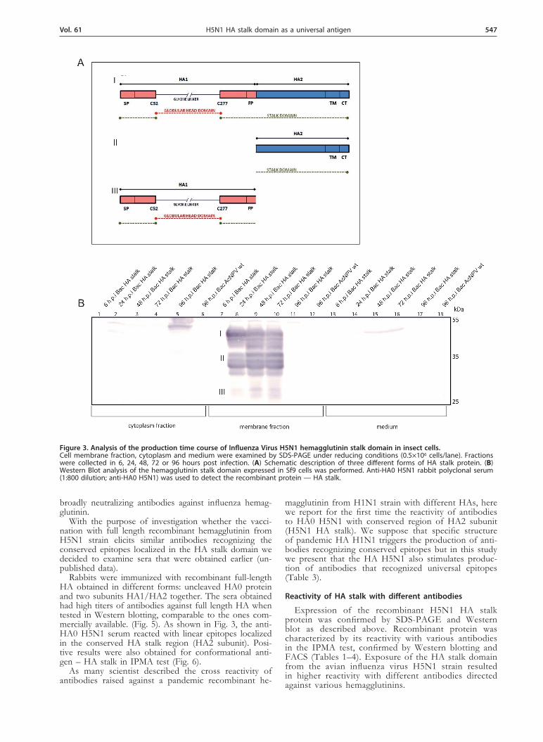
Figure 1. Schematic description of the full length and “headless” hemagglutinin from H5N1 strain. (A) Full length HA (H5) containing globular head and stalk domain. Globular head region is mainly located between cysteines 52 and 277; stalk domain consists of N- and C-terminal parts of HA1 subunit and all of HA2 subunit; SP — signal peptide, FP — fusion peptide, TM — transmembrane domain, CT — cytoplasmic tail (B) HA stalk domain construct from the avian influenza virus H5N1 (“headless” HA) lacks the region located between cysteines 52 and 277 on the HA1 subunit and possesses instead a four glycine peptide linker.
Vol. 61 543H5N1 HA stalk domain as a universal antigen
lysates were examined for recombinant proteins (HA full and HA stalk) by SDS-PAGE and Western blotting.
Time course of recombinant H5N1 HA stalk pro-tein production in Sf9 cells. Sf9 cells were grown as monolayers at 27°C in 12-well culture plate using HyQ SFX medium (106 cells/ml). Cultures were infected with the recombinant baculoviruses at the multiplicity of in-fection of 1 and cells were collected from the culture medium at different times post infection (6 h, 24 h, 48 h, 72 h, 96 h). Supernatants (medium) were separated from the cell pellet by centrifugation at 13300 rpm for 5 minutes. Proteins from the medium were precipitated us-ing 10% trichloroacetic acid (TCA). In parallel, cell pel-lets were treated with lysis buffer on ice (100 mM Tris pH 7.4, 100 mM NaCl and 1% Nonidet P-40) in order to separate cell membranes from the cytoplasm. Result-ing samples were boiled for 10 minutes at 100°C in a loading buffer (Life technologies) prior to SDS-PAGE on 10% gels.
Purification of recombinant HA protein from pol-yvinylidene difluoride (PVDF) membranes. Cultures were infected with the recombinant baculoviruses at the multiplicity of infection of 1 and cells were collected from the culture medium 72 hours post infection. Su-pernatants (medium) were separated from the cell pel-let by centrifugation at 8500 rpm for 10 minutes. Cell
pellets were lysed for 10 min. on ice with a lysis buffer (100 mM Tris pH 7.5, 1% Triton X-100, 150 mM NaCl) in order to separate cell membranes from the cytoplasm. Membrane fraction samples were separated by 8% SDS-PAGE followed by transferring the samples onto PVDF membranes. Electrotransfer was performed in a Tris-gly-cine transfer buffer at 25V overnight. As a control West-ern Blotting was performed before the blotted proteins were visualized using Ponceau S. HA protein was eluted from PVDF membrane with elution buffer containing 50 mM Tris pH 9.5, 2% SDS and 1% Triton X-100. Eluted proteins were precipitated with acetone and analyzed by SDS-PAGE gel and Western blot.
Production of hyperimmune antiserum in rabbits. Rabbits were immunized with 100 µg of different eluted recombinant H5N1 hemmaglutinin (HA0; HA1/HA2) mixed with Freund’s complete adjuvant by intradermal injection followed by subcutaneous booster injection 3 weeks later with the same dose in Freund’s incomplete adjuvant. Rabbits were bled 2 weeks later. The serum was collected and stored at –20ºC until use.
Immunoperoxidase Monolayer Assay (IPMA). Sf9 cells were seeded into 12-well cell culture plates (106 cells/ml) and infected with the recombinant baculovi-ruses at the multiplicity of infection of 1. After incubat-ing for 48 hours, the medium was removed and the cells
Table 1. Description of different antibodies against group 1 hemagglutinins used in this study.
Group 1. Hemagglutinins
Strain Immunogen Antibody description
Human H1N1 HAPurified human-cell derived recombinant H1N1 A/California/04/2009 and A/California/07/2009 HA extracellular domain. Clone ID 9G1G8
Mouse Monoclonal AntibodySino Biological Inc. cat. no 11048-MM08
H1N1 HA Purified influenza virus type A strain H1N1Mouse Monoclonal Antibody Santa Cruz Biotechnology cat. No sc-52025
H1N1 HA Recombinant HA protein eluted from PVDF mem-brane
Monospecific Rabbit Polyclonal Anti-body
Avian/Human H5N1 HA0 Recombinant HA protein eluted from PVDF mem-brane
Monospecific Rabbit Polyclonal Anti-bodies
H5N1 HA1 116–256 aa rHA peptide Monospecific Rabbit Polyclonal Anti-bodies
H5N1 HA1/HA2 Recombinant HA protein eluted from PVDF mem-brane
Monospecific Rabbit Polyclonal Anti-bodies
H5N1 H5N1 — re-ference Whole virus H5N1: A/Ck/Scot/59
Polyclonal Chicken Antibodies - Animal Health and Veterinary Laboratories Agency (VLA) cat. no RAA7002
H5N1 HA Purified human-cell derived recombinant influenza A virus A/Anhui/1/2005/H5N1 HA extracellular do-main. Clone ID 9F2E3F3
Mouse Monoclonal Antibodies. Sino Biological Inc. cat. no 11048-MM01
H5N1 HAPurified human-cell derived recombinant influenza A virus A/Anhui/1/2005/H5N1 HA extracellular do-main. Clone ID 1C5B1A10
Mouse Monoclonal Antibodies. Sino Biological Inc. cat. no 11048-MM06
H5N1 HAPurified human-cell derived recombinant influenza A virus A/Anhui/1/2005/H5N1 HA extracellular do-main. Clone ID 14B1E2G6
Mouse Monoclonal Antibodies. Sino Biological Inc.cat. no 11048-MM10
Avian H5N2 H5N2 — re-ference Whole virus H5N2:A/Ost/Den/72420/96 Chicken Polyclonal Antibody
cat. no RAA7003 1:500
H5Nx H5Nx Whole virus — H5 avian influenzaChicken Polyclonal AntibodyNational Veterinary Research Institute PIWet
544 2014K. Uranowska and others
were fixed with 70% methanol. Cells were then incu-bated with primary antibodies (Tables 1–4) in a binding buffer (PBS – 0.05% Tween 80, 5% FBS) for 1 h, at RT. Cells were then washed three times with PBS — 0.05% Tween 80, horseradish peroxidase conjugated secondary antibody (Santa Cruz Biotechnology, USA) was added at a dilution 1:1000 and plates were incubated for 1 h, at RT. After a final wash, NovaRED Peroxidase Substrate Kit (Vector) was used in order to detect positive red re-action products. The color reaction was allowed to de-velop for 5–15 minutes and the plates were then washed with H2O and examined under a light microscope.
Western blot analysis. Proteins were separated by 10% SDS-PAGE followed by samples transfer to PVDF membranes. After protein electrotransfer in a Tris-glycine buffer at 25V overnight, membranes were blocked with 3% non-fat milk at RT for 1h and in-cubated with different antibodies against HA proteins (Tables 1–4) at RT for 1 h. Subsequently, alkaline phosphatase-conjugated secondary antibody (1:1000, Santa Cruz Biotechnology, USA) was added and it was incubated for 1h at RT. After three washes with TBS, a substrate solution containing NBT and BCIP was added. After developing the bands, the membrane was washed with water.
Flow cytometry. Sf9 cells were seeded into 6-well cell culture plates (1.5×10⁶ cells/ml) and infected with the recombinant baculoviruses at the multiplicity of infection of 1. Presence of full and stalk recombinant HA pro-teins at the cell surface was determined by indirect im-munofluorescence as follows. Cells (5×10⁵ cells/sample) were collected into 100 ml of a PBA buffer (PBS + 1% BSA + 0.02% sodium azide) and placed separately into the wells of 96-well polystyrene V-bottom microplate (Greiner Bio-One, Germany). Next, the cells were cen-trifuged for 3 min at 1300 rpm.
Cells were resuspended in the primary antibody solu-tion in PBA (Tables 3–4) and stained for 1 hour on ice. After two rounds of washing with PBA by 3 min cen-trifugation at 1300 rpm, 4°C, discarding the antibody so-lution and resuspending the cell pellet in the PBA wash-ing buffer, cells were incubated with goat anti-mouse IgG-PE (1:1000, Becton-Dickinson, USA) for 45 min on ice. After double washing with PBA, cells were re-
suspended in 120 ml of PBA and transferred to 0.65 ml tubes. Cells were analyzed using the FACS Calibur flow cytometer (Becton-Dickinson, USA) and the CellQuest software (Becton-Dickinson, USA).
Antibodies recognizing various hemagglutinins. Different monospecific rabbit polyclonal antibodies (anti HA0 H5N1, anti HA1/HA2 H5N1) were obtained in our laboratory (unpublished data). Chicken polyclonal antibodies (anti H5Nx, H1N1, H7Nx) were kindly pro-vided by National Veterinary Research Institute (PIWet). The rest of the used antibodies were purchased from Santa Cruz Biotechnology Inc., Sino Biological Inc. and Veterinary Laboratories Agency (VLA). The details are presented in Tables 1 and 2.
RESULTS
Expression of the hemagglutinin recombinant protein genes in Sf9 insect cells
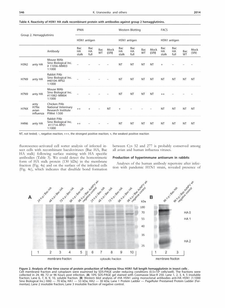
Our aim was to investigate whether the approach based on presentation to the host immune system of a stalk region of hemagglutinin of the human influenza viruses (H1 and H3) described by Steel and coworkers, 2010 can be applied also to the HPAI strains. For this purpose we planned to characterize the conserved, trun-cated form of hemagglutinin (HA stalk) from the Polish H5N1 strain. To compare the results obtained for trun-cated form of HA we decided to express the full length HA gene as well. Recombinant viruses bearing the full length and stalk form of hemagglutinin were used to infect insect cells under optimized conditions. A major polypeptide band of full-length HA0 with a molecular mass of 70 kDa was identified in cell lysates by SDS-PAGE followed by either Coomassie brilliant blue stain-ing or Western blot analysis using monoclonal antibodies anti-HA H5N1 (1:1000, Sino Biological Inc.) (Fig. 2).
All the HPAI hemagglutinins possess multiple arginine residues at the site of cleavage and are cut intracellulary by ubiquitously occurring proteases (Steinhauer, 1999; Gamblin & Skehel, 2010). Apart from recombinant HA0 we could also observe HA1 with molecular mass of 53 kDa and HA2 subunits of 30 kDa which were the re-
Table 2. Description of different antibodies against group 2 hemagglutinins used in this study.
Group 2. Hemagglutinins
Strain Immunogen Antibody description
Human H3N2 HAPurified human-cell derived recombinant H3N2 A/Brisbane/10/2007 HA. Clone ID MM03
Mouse Monoclonal Antibody Sino Biological Inc. cat. no 11056-MM03
Avian/Human H7N9 HAPurified recombinant Influenza A H7N9/A/Shanghai/1/2013 HA 1–524 aa
Rabbit Polyclonal Antibody Sino Biological Inc. cat. no 40104-RP02
Avian/Human H7N9 HAPurified recombinant Influenza A H7N9 /A/Shanghai/1/2013 HA 1–524 aa
Mouse Monoclonal Antibody Sino Biological Inc. cat. no 11082-MM04
Avian H7Nx H7Nx avian influenza Whole virus- H7/Poland Chicken Polyclonal Antibody
National Veterinary Research Institute PIWet
Avian H4N6 HAPurified recombinant Influenza A H4N6/A/mallard/Ohio/657/2002HA 1–524 aa
Rabbit Polyclonal Antibody Sino Biological Inc. Cat.no 11714-RP01
Human FI6 HA H1N1 vHighly specific humanised synthetic antibodies based on neutralizing antibody selected from plasma cells that bind to group 1 and group 2 Influenza A HAs
Vol. 61 545H5N1 HA stalk domain as a universal antigen
sult of a proteolytic cleavage by insect cells protease — furin (Fig. 2b). The recombinant HA stalk protein was also detected using anti-HA0 H5N1 rabbit polyclonal serum (1:800, obtained in our laboratory). We detected HA stalk protein mainly in the membranes which is similar to cell localization of full length HA (Fig. 3). In western blot analysis under reducing conditions we ob-served three major bands of different forms of recom-binant HA stalk protein. The observed three forms of
expressed recombinant protein most likely represent: HA stalk composed of N- and C-terminal parts of HA1 sub-unit plus the whole HA2 subunit with molecular mass of 45 kDa, single HA2 subunit (37 kDa) and N- and C-terminal parts of HA1 subunit linked by four glycines (27 kDa).
We wanted to investigate whether absence of the globular head domain would disrupt transport through the Golgi complex to the cell surface. We performed
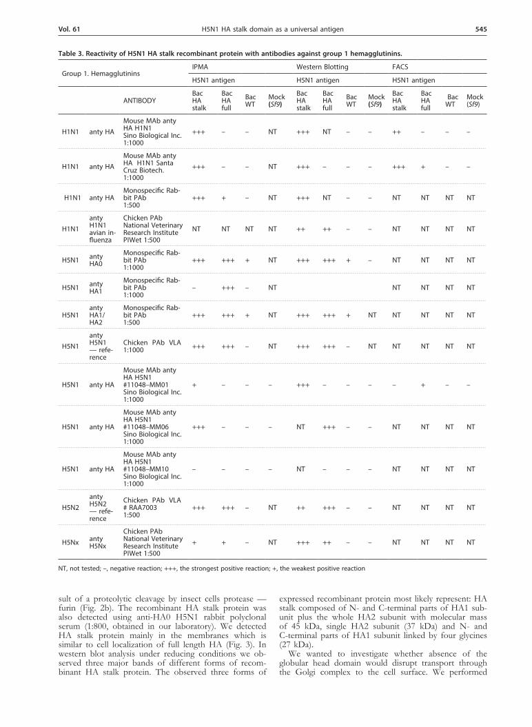
Table 3. Reactivity of H5N1 HA stalk recombinant protein with antibodies against group 1 hemagglutinins.
Group 1. HemagglutininsIPMA Western Blotting FACS
H5N1 antigen H5N1 antigen H5N1 antigen
ANTIBODYBac HA stalk
Bac HA full
Bac WT
Mock (Sf9)
Bac HA stalk
Bac HA full
Bac WT
Mock (Sf9)
Bac HA stalk
BacHA full
Bac WT
Mock (Sf9)
H1N1 anty HAMouse MAb anty HA H1N1 Sino Biological Inc. 1:1000
+++ – – NT +++ NT – – ++ – – –
H1N1 anty HAMouse MAb anty HA H1N1 Santa Cruz Biotech. 1:1000
+++ – – NT +++ – – – +++ + – –
H1N1 anty HAMonospecific Rab-bit PAb 1:500
+++ + – NT +++ NT – – NT NT NT NT
H1N1anty H1N1 avian in-fluenza
Chicken PAb National Veterinary Research Institute PIWet 1:500
NT NT NT NT ++ ++ – – NT NT NT NT
H5N1 anty HA0
Monospecific Rab-bit PAb 1:1000
+++ +++ + NT +++ +++ + – NT NT NT NT
H5N1 anty HA1
Monospecific Rab-bit PAb 1:1000
– +++ – NT NT NT NT NT
H5N1anty HA1/HA2
Monospecific Rab-bit PAb 1:500
+++ +++ + NT +++ +++ + NT NT NT NT NT
H5N1anty H5N1 — refe-rence
Chicken PAb VLA1:1000 +++ +++ – NT +++ +++ – NT NT NT NT NT
H5N1 anty HA
Mouse MAb anty HA H5N1#11048–MM01 Sino Biological Inc. 1:1000
+ – – – +++ – – – – + – –
H5N1 anty HA
Mouse MAb anty HA H5N1#11048–MM06 Sino Biological Inc. 1:1000
+++ – – – NT +++ – – NT NT NT NT
H5N1 anty HA
Mouse MAb anty HA H5N1#11048–MM10 Sino Biological Inc. 1:1000
– – – – NT – – – NT NT NT NT
H5N2anty H5N2 — refe-rence
Chicken PAb VLA# RAA70031:500
+++ +++ – NT ++ +++ – – NT NT NT NT
H5Nx anty H5Nx
Chicken PAb National Veterinary Research Institute PIWet 1:500
+ + – NT +++ ++ – – NT NT NT NT
NT, not tested; –, negative reaction; +++, the strongest positive reaction; +, the weakest positive reaction
546 2014K. Uranowska and others
fluorescence-activated cell sorter analysis of infected in-sect cells with recombinant baculoviruses (Bac HA, Bac HA stalk) following surface staining with HA specific antibodies (Table 3). We could detect the homotrimeric form of HA stalk protein (130 kDa) in the membrane fraction (Fig. 4a) and on the surface of the infected cells (Fig. 4c), which indicates that disulfide bond formation
between Cys 52 and 277 is probably conserved among all avian and human influenza viruses.
Production of hyperimmune antiserum in rabbits
Analyses of the human antibody repertoire after infec-tion with pandemic H1N1 strain, revealed presence of
Table 4. Reactivity of H5N1 HA stalk recombinant protein with antibodies against group 2 hemagglutinins.
Group 2. Hemagglutinins
IPMA Western Blotting FACS
H5N1 antigen H5N1 antigen H5N1 antigen
AntibodyBac HA stalk
Bac HA full
Bac WT
Mock (Sf9)
Bac HA stalk
Bac HA full
Bac WT
Mock (Sf9)
Bac HA stalk
BacHA full
Bac WT
Mock (Sf9)
H3N2 anty HAMouse MAb Sino Biological Inc. # 11056–MM031:1000
– – – – NT NT NT NT + – – –
H7N9 anty HARabbit PAb Sino Biological Inc. #40104–RP021:1000
– – – – NT NT NT NT NT NT NT NT
H7N9 anty HAMouse MAb Sino Biological Inc. #11082–MM041:1000
– – – – NT NT NT NT ++ – – –
H7NXanty H7Nx avian influenza
Chicken PAb National Veterinary Research Institute PIWet 1:500
++ + – NT + – – – NT NT NT NT
H4N6 anty HARabbit PAb Sino Biological Inc. #11714–RP011:1000
++ – – – NT NT NT NT NT NT NT NT
NT, not tested; –, negative reaction; +++, the strongest positive reaction; +, the weakest positive reaction
Figure 2. Analysis of the time course of protein production of Influenza Virus H5N1 full length hemagglutinin in insect cells . Cell membrane fraction and cytoplasm were examined by SDS-PAGE under reducing conditions (0.5×10⁶ cells/well). The fractions were collected in 24, 48, 72 or 96 hours post infection. (A) 10% SDS-PAGE gel stained with Coomassie blue-R 250. Lane 1, 2, 3, 4, 5 insoluble fraction; Lane 6, 7, 8, 9, 10, soluble fraction. (B) Western blot analysis of rHA H5N1 using monoclonal antibodies anti-HA H5N1 (1:1000 Sino Biological Inc.) HA0 — 70 kDa; HA1 — 53 kDa; HA2 — 30 kDa; Lane 1 Protein Ladder — PageRuler Prestained Protein Ladder (Fer-mentas); Lane 2 insoluble fraction; Lane 3 insoluble fraction of negative control.
Vol. 61 547H5N1 HA stalk domain as a universal antigen
broadly neutralizing antibodies against influenza hemag-glutinin.
With the purpose of investigation whether the vacci-nation with full length recombinant hemagglutinin from H5N1 strain elicits similar antibodies recognizing the conserved epitopes localized in the HA stalk domain we decided to examine sera that were obtained earlier (un-published data).
Rabbits were immunized with recombinant full-length HA obtained in different forms: uncleaved HA0 protein and two subunits HA1/HA2 together. The sera obtained had high titers of antibodies against full length HA when tested in Western blotting, comparable to the ones com-mercially available. (Fig. 5). As shown in Fig. 3, the anti-HA0 H5N1 serum reacted with linear epitopes localized in the conserved HA stalk region (HA2 subunit). Posi-tive results were also obtained for conformational anti-gen – HA stalk in IPMA test (Fig. 6).
As many scientist described the cross reactivity of antibodies raised against a pandemic recombinant he-
magglutinin from H1N1 strain with different HAs, here we report for the first time the reactivity of antibodies to HA0 H5N1 with conserved region of HA2 subunit (H5N1 HA stalk). We suppose that specific structure of pandemic HA H1N1 triggers the production of anti-bodies recognizing conserved epitopes but in this study we present that the HA H5N1 also stimulates produc-tion of antibodies that recognized universal epitopes (Table 3).
Reactivity of HA stalk with different antibodies
Expression of the recombinant H5N1 HA stalk protein was confirmed by SDS-PAGE and Western blot as described above. Recombinant protein was characterized by its reactivity with various antibodies in the IPMA test, confirmed by Western blotting and FACS (Tables 1–4). Exposure of the HA stalk domain from the avian influenza virus H5N1 strain resulted in higher reactivity with different antibodies directed against various hemagglutinins.
Figure 3. Analysis of the production time course of Influenza Virus H5N1 hemagglutinin stalk domain in insect cells. Cell membrane fraction, cytoplasm and medium were examined by SDS-PAGE under reducing conditions (0.5×10⁶ cells/lane). Fractions were collected in 6, 24, 48, 72 or 96 hours post infection. (A) Schematic description of three different forms of HA stalk protein. (B) Western Blot analysis of the hemagglutinin stalk domain expressed in Sf9 cells was performed. Anti-HA0 H5N1 rabbit polyclonal serum (1:800 dilution; anti-HA0 H5N1) was used to detect the recombinant protein — HA stalk.
548 2014K. Uranowska and others
We observed that HA stalk recombinant protein reacted with polyclonal antibodies raised against influ-enza virus type A strain H5N1, against HA2 subunit of H5N1, against H5N1 reference strain, H5N2 VLA and H5Nx chicken polyclonal antibody.
HA stalk was able to react with rabbit polyclonal as well as mouse monoclonal antibodies against H1N1 pan-demic strain. In full length hemagglutinin, HA2 subunit is thought to be masked by the membrane-distant por-tion of HA1 subunit, the globular head domain. Thus, epitopes localized in the stalk domain were not recog-nized by anti H1N1 mAbs.
H5N1 HA stalk protein also reacted with antibodies against HAs from phylogenetic group 2 (Tables 2 and 3) which implies that the antigen described in this study contains the conserved, probably universal epitopes.
Reactivity of HA stalk with universal human neutralizing antibody FI6
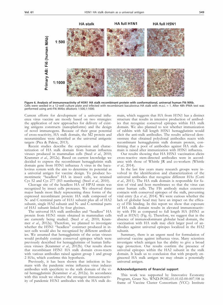
FI6 antibody recognizes all subtypes of the influenza A virus hemagglutinin. As the FI6 antibody recognized only conformational epitopes we decided to investigate its reactivity with HA stalk antigen. In the IPMA test we examined whether recombinant H5N1 HA stalk protein reacts with FI6 human neutralizing antibody. FI6 specifi-cally recognized HA stalk protein (Fig. 6).
DISCUSSION
Influenza virus causes 250 000–500 000 deaths world-wide annually. Potential global pandemic could kill mil-lions (Kang et al., 2012). Current seasonal influenza vac-cines are designed to provide strain-specific protection against two circulating subtypes of influenza A virus (H1N1 and H3N2) and one influenza B virus (Doyle et al., 2013). The rapid spreading of the 2009 pandemic H1N1 influenza virus was a signal that universal influ-enza vaccines are necessary, so that they would broadly protect against many mutated strains (Lu et al., 2014).
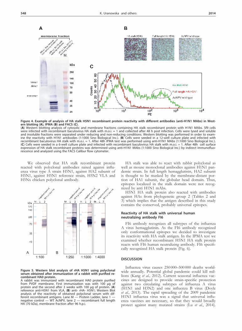
Figure 4. Example of analysis of HA stalk H5N1 recombinant protein reactivity with different antibodies (anti-H1N1 MAbs) in West-ern blotting (A), IPMA (B) and FACS (C).(A) Western blotting analysis of cytosolic and membrane fractions containing HA stalk recombinant protein with H1N1 MAbs. Sf9 cells were infected with recombinant baculovirus HA stalk with m.o.i. = 1 and collected after 48 h post infection. Cells were lysed and soluble and insoluble fractions were separated under reducing and non-reducing conditions. Western blotting was performed in order to exam-ine the reactivity with H1N1 antibodies (1:1000 Sino Biological Inc.). (B) Cells were seeded in a 12-well culture plate and infected with recombinant baculovirus HA stalk with m.o.i. = 1. After 48h IPMA test was performed using anti-H1N1 MAbs (1:1000 Sino Biological Inc.). (C) Cells were seeded in a 6-well culture plate and infected with recombinant baculovirus HA stalk with m.o.i. = 1. After 48h cell surface expression of HA stalk recombinant proteins was determined using anti-H1N1 MAbs (1:1000 Sino Biological Inc.) by indirect immunofluo-rescence and analyzed using the FACS Calibur flow cytometer.
Figure 5. Western blot analysis of rHA H5N1 using polyclonal serum obtained after immunization of a rabbit with purified re-combinant HA0 protein.A rabbit was immunized with recombinant HA0 protein purified from PVDF membrane. First immunization was with 100 μg of protein and the second after 3 weeks with 100 μg of protein. (A) reference anti-H5N1 from VLA, (B) anti- rHA- H5N1; Western Blot analysis of the reactivity of obtained polyclonal serum with dif-ferent recombinant antigens. Lane M — Protein Ladder, lane 1 — negative control — WT AcNPV, lane 2 — recombinant full length HA (70 kDa), membrane fraction after 96 h.p.i.
Vol. 61 549H5N1 HA stalk domain as a universal antigen
Current efforts for development of a universal influ-enza virus vaccine are mostly based on two strategies: the application of new approaches for delivery of exist-ing antigens constructs (nanoplatforms) and the design of novel immunogens. Because of their great potential of cross-reactivity, HA stalk domain, the M2 protein and neuraminidase were identified as the universal antigenic targets (Pica & Palese, 2013).
Recent studies describe the expression and charac-terization of HA stalk domain from human influenza viruses produced in mammalian cells (Steel et al., 2010; Krammer et al., 2012a). Based on current knowledge we decided to express the recombinant hemagglutinin stalk domain gene from H5N1 influenza A virus in the bacu-lovirus system with the aim to determine its potential as a universal antigen for vaccine design. To produce ho-motrimeric “headless” HA in insect cells, we retained Cys 52 and Cys 277 (H3 numbering) (Steel et al., 2010).
Cleavage site of the headless HA of HPAI strain was recognized by insect cells proteases. We observed three major bands most likely representing three forms of the expressed recombinant protein: HA stalk composed of N- and C-terminal parts of HA1 subunit plus all of HA2 subunit, single HA2 subunit and N- and C-terminal parts of HA1 subunit linked by four glycines.
The universal HA stalk antibodies and “headless” HA protein from H1N1 strain obtained in mammalian cells are currently being studied. (Steel et al., 2010; Kram-mer et al., 2012a). Therefore, we planned to investigate whether the H5N1 “headless” construct produced in in-sect cells would also be recognized by different antibod-ies. We assumed that hemagglutinin of the avian viruses would probably contain conserved epitopes which were previously described for hemagglutinins of human Influ-enza viruses (Krammer et al., 2012b). Our results show that recombinant H5N1 HA stalk domain cross-reacts with various antibodies raised against group 1 and group 2 HAs, which confirms this hypothesis.
Previously, it has been shown that infection in hu-mans with the pandemic swine influenza virus induces antibodies with specificity to the stalk domain of the vi-ral hemagglutinin (Krammer et al., 2012a). In accordance with this result we observe the strongest immunoreactiv-ity of pandemic H1N1 antibodies with the HA stalk do-
main, which suggests that HA from H1N1 has a distinct structure that results in intensive production of antibod-ies that recognize conserved epitopes within HA stalk domain. We also planned to test whether immunization of rabbits with full length H5N1 hemagglutinin would elicit the anti-stalk antibodies. The results achieved dem-onstrate that obtained polyclonal antibodies reacts with recombinant hemagglutinin stalk domain protein, con-firming that a pool of antibodies against HA stalk do-main is raised after immunization with H5N1 influenza.
Our results showing that HA H5N1 vaccination elicits cross-reactive stem-directed antibodies were in accord-ance with those of Whittle JR and co-workers (Whittle et al., 2014).
In the last few years many research groups were in-volved in the identification and characterization of the universal antibodies that recognize different HAs (Corti et al., 2011). The HA stalk domain is responsible for fu-sion of viral and host membranes so that the virus can enter human cells. The FI6 antibody makes extensive contacts with conserved epitopes of the stalk, preventing viral entry (Lu et al., 2014). We were curious whether the lack of globular head may have an impact on the effica-cy of FI6 binding. In this report we show that exposure of HA stalk domain results in elevated immunoreactiv-ity with FI6 as compared to full length HA (H5N1 as well as H1N1) (Fig. 6). Therefore, we suggest that in the absence of immunodominant globular head domain, the vaccination with HA stalk may elicit larger pool of an-tibodies against universal epitopes localized in the HA2 subunit.
Summary, there is an urgent need for formulation of universal vaccine against influenza virus. Many scientists investigate which antigen has the ability to give a broad rage protection. Our results confirm the presence of universal epitopes within the HA2 subunit in the stalk domain. It leads us to conclusion that with properly en-gineered HA stalk antigen we may obtain a potentially universal antigen.
Acknowledgements of financial support
This work was supported by Innovative Economy Program, Grant No. WND-POIG.01.01.02-00-007/08 in frame of Vaccine Cluster Consortium (VCC): Institute
Figure 6. Analysis of immunoreactivity of H5N1 HA stalk recombinant protein with conformational, universal human FI6 MAb. Cells were seeded in a 12-well culture plate and infected with recombinant baculovirus HA stalk with m.o.i. = 1. After 48h IPMA test was performed using anti-FI6 MAbs dilutions 1:500,1:1000.
550 2014K. Uranowska and others
of Biochemistry and Biophysics, Polish Academy of Sci-ences, Warsaw, Poland (IBB); Institute of Biotechnology and Antibiotics, Warsaw, Poland (IBA); Department of Recombinant Vaccines, Intercollegiate Faculty of Bio-technology, University of Gdansk and Medical Univer-sity of Gdansk, Gdansk, Poland (UG); Kucharczyk TE sp. z o.o. ,Warsaw, Poland (KTE); Institute of Animal Reproduction and Food Research, Polish Academy of Sciences, Tuwima 10, 10–747 Olsztyn, Poland (IAR); Department of Poultry Diseases, National Veterinary Re-search Institute, Pulawy, Poland (PIWet).
REFRENCES
Bouvier NM, Palese P (2008) The biology of influenza viruses. Vaccine 26 (Suppl 4): D49–D53.
Corti D, Suguitan AL Jr, Pinna D, Silacci C, Fernandez-Rodriguez BM, Vanzetta F, Santos C, Luke CJ, Torres-Velez FJ, Temperton NJ, Weiss RA, Sallusto F, Subbarao K, Lanzavecchia A (2010) Hetero-subtypic neutralizing antibodies are produced by individuals immu-nized with a seasonal influenza vaccine. J Clin Invest 120: 1663–1673.
Corti D, Voss J, Gamblin SJ, Codoni G, Macagno A, Jarros-say D, Vachieri SG, Pinna D, Minola A, Vanzetta F, Silacci C, Fernandez-Rodriguez BM, Agatic G, Bianchi S, Giacchet-to-Sasselli I, Calder L, Sallusto F, Collins P, Haire LF, Tem-perton N, Langedijk JP, Skehel JJ, Lanzavecchia A (2011) A neutralizing antibody selected from plasma cells that binds to group 1 and group 2 Influenza A hemagglutinins. Science 333: 850–856.
Doyle TM, Hashem AM, Li C, Van Domselaar G, Larocque L, Wang J, Smith D, Cyr T, Farnsworth A, He R, Hurt AC, Brown EG, Li X (2013) Universal anti-neuraminidase antibody inhibiting all influ-enza A subtypes. Antiviral Res 100: 567–574.
Gamblin SJ, Skehel JJ (2010) Influenza Hemagglutinin and Neuramini-dase Membrane Glycoproteins. J Biol Chem 285: 28403–28409.
Gromadzka B, Smietanka K, Dragun J, Minta Z, Gora-Sochacka A, Szewczyk B (2008) Detection of changes in avian influenza genome fragments by multitemperature single strand conformational poly-morphism. Molecular and Cellular Probes 22: 301–304.
Hopkins R, Esposito D (2009) A rapid method for titrating baculovirus stocks using the Sf-9 Easy Titer cell line. Biotechniques 47: 785–788.
Kang SM, Kim MC, Compans RW (2012) Virus-like particles as uni-versal influenza vaccines. Expert Rev Vaccines 11: 995–1007.
Kang SM, Song JM, Compans RW (2011) Novel vaccines against influ-enza viruses. Virus Research 162: 31–38.
Krammer F, Margine I, Tan GS, Pica N, Krause JC, Palese P (2012a) A carboxy-terminal trimerization domain stabilizes conformational epitopes on the stalk domain of soluble recombinant hemagglutinin substrates. PLOS One 7: e43603.
Krammer F, Pica N, Hai R, Tan GS, Palese P (2012b) Hemagglutinin stalk-reactive antibodies are boosted following sequential infection with seasonal and pandemic H1N1 Influenza virus in mice. J Virol 86: 10302–10307.
Lu Y, Welsh JP, Swartz JR (2014) Production and stabilization of the trimeric influenza hemagglutinin stem domain for potentially broadly protective influenza vaccines. Proc Natl Acad Sci USA 111: 125–130.
Margine I, Hai R, Albrecht RA, Obermoser G, Harrod AC, Banchere-au J, Palucka K, García-Sastre A, Palese P, Treanor JJ, Krammer F (2013a) H3N2 influenza virus infection induces broadly reactive hemagglutinin stalk antibodies in humans and mice. J Virol 87: 4728–4737.
Margine I, Krammer F, Hai R, Heaton NS, Tan GS, Andrews SA, Runstadler JA, Wilson PC, Albrecht RA, García-Sastre A, Palese P (2013b) Hemagglutinin stalk-based universal vaccine constructs pro-tect against group 2 Influenza A viruses. J Virol 87: 10435–10446.
Pica N, Hai R, Krammer F, Wang TT, Maamary J, Eggink D, Tan GS Krause JC, Moran T, Stein CR, Banach D, Wrammert J, Belshe RB, García-Sastre A, Palese P (2012) Hemagglutinin stalk antibodies elicited by the 2009 pandemic influenza virus as a mechanism for the extinction of seasonal H1N1 viruses. Natl Acad Sci USA 109: 2573–2578.
Pica N, Palese P (2013) Toward a universal influenza virus vaccine: prospects and challenges. Annu Rev Med 64: 189–202.
Sangster MY, Baer J, Santiago FW, Fitzgerald T, Ilyushina NA, Sunda-rarajan A, Henn AD, Krammer F, Yang H, Luke CJ, Zand MS, Wright PF, Treanor JJ, Topham DJ, Subbarao K (2013) B cell response and hemagglutinin stalk-reactive antibody production in different age cohorts following 2009 H1N1 influenza virus vaccina-tion. Clin Vaccine Immunol 20: 867–876.
Steel J, Lowen AC, Wang TT, Yondola M, Gao Q, Haye K, García-Sastre A, Palese P (2010) Influenza virus vaccine based on the con-served hemagglutinin stalk domain. MBio 1: 1–9.
Steinhauer DA (1999) Role of hemagglutinin cleavage for the patho-genicity of influenza virus. Virology 258: 1–20.
Whittle JR, Wheatley AK, Wu L, Lingwood D, Kanekiyo M, Ma SS, Narpala SR, Yassine HM, Frank GM, Yewdell JW, Ledgerwood JE, Wei CJ, McDermott AB, Graham BS, Koup RA, Nabel GJ (2014) Flow cytometry reveals that H5N1 vaccination elicits cross-reactive stem-directed antibodies from multiple Ig heavy chain lineages. J Virol 88: 4047–4057.
Wrammert J, Koutsonanos D, Gui-Mei Li, Edupuganti S, Sui J, Mor-rissey M, McCausland M, Skountzou I, Hornig M, Lipkin WI, Me-hta A, Razavi B, Del Rio C, Zheng NY, Lee JH, Huang M, Ali Z, Kaur K, Andrews S, Amara RR, Wang Y, Das SR, O’Donnell CD, Yewdell JW, Subbarao K, Marasco WA, Mulligan MJ, Compans R, Ahmed R, Wilson PC (2011) Broadly cross-reactive antibodies dominate the human B cell response against 2009 pandemic H1N1 influenza virus infection. J Exp Med 208: 181–193.