Embed Size (px)
Citation preview
Correspondence
Heck's disease (focal epithelial hyperplasia)
To the Editor:The comprehensive review of Heck's disease (focal
epithelial hyperplasia) by Stiefler, Solomon, andShalita, which appeared on page 499 of the December,1979, issue of the JOURNAL, stressed that although thisfascinating condition may be more prevalent than previously assumed, there has been a relative paucity ofreports in the dermatologic literature.
Since the original description of these oral mucosallesions in Navajo Indian children in 1965,1 the oralsurgery, medicine, and pathology literature has been
replete with reports of patients from Abu Dhabi,Argentina, Australia, Bolivia, Brazil, Colombia, Ecuador, El Salvador, France, Guatemala, Iraq, Israel,Mexico, The Netherlands, Paraguay, Peru, Polynesia,Puerto Rico, South Africa, Sweden, and Venezuela.Patients in the Eskimo population of Canada andGreenland have also been reported. Stiefler, Solomon,and Shalita, however, found only a single report published in the American dermatologic literature. 2
There are other studies in the American dermatologic literature which may have been over100ked,3-s and we would like to contribute a report of acase in an elderly North American Caucasian.
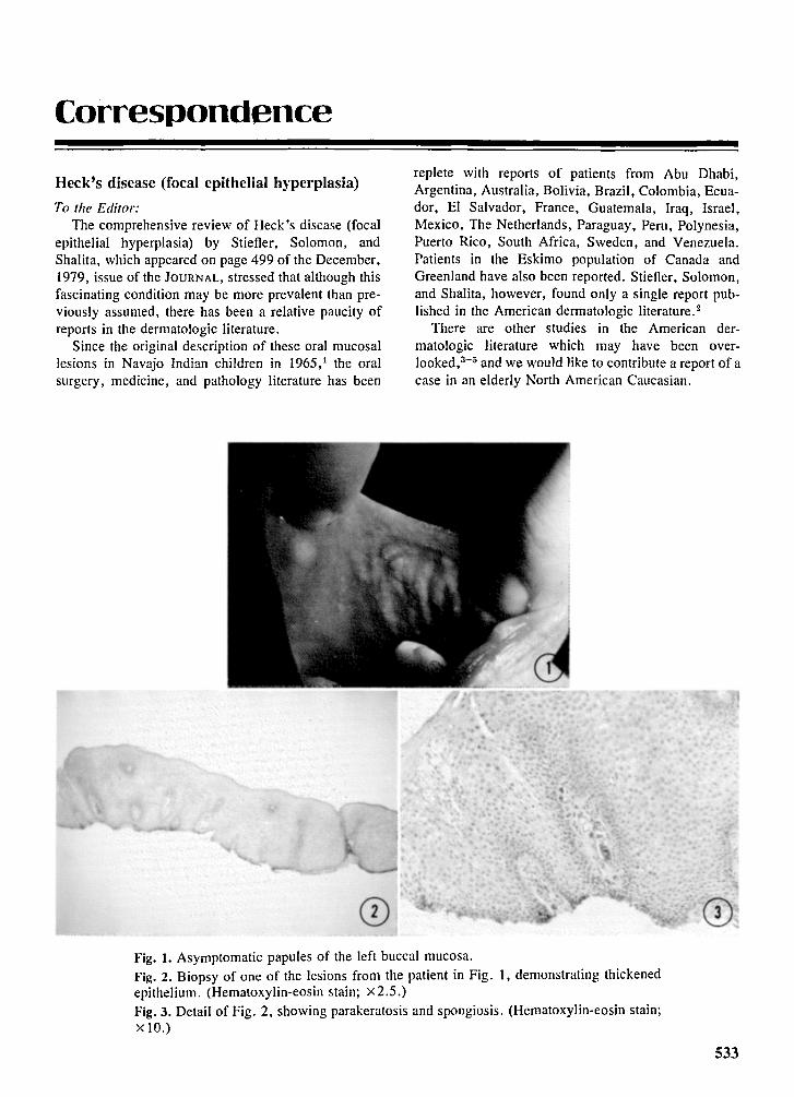
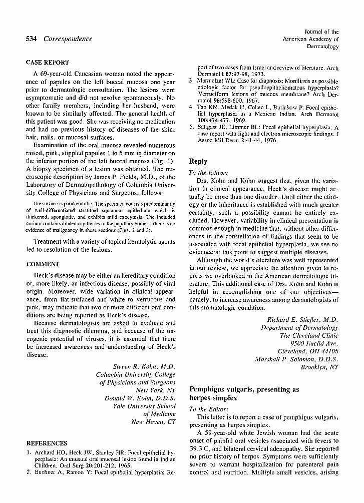
o CDFig. 1. Asymptomatic papules of the left buccal mucosa.Fig. 2. Biopsy of one of the lesions from the patient in Fig. I, demonstrating thickenedepithelium. (Hematoxylin-eosin stain; x2.5.)Fig. 3. Detail of Fig. 2, showing parakeratosis and spongiosis. (Hematoxylin-eosin stain;x 10.)
533
534 Correspondence
CASE REPORT
A 69-year-old Caucasian woman noted the appearance of papules on the left buccal mucosa one yearprior to dermatologic consultation. The lesions wereasymptomatic arid did not resolve spontaneously. Noother family members, including her husband, wereknown to be similarly affected. The general health ofthis patient was good. She was receiving no medicationand had no previous history of diseases of the skin,hair, nails, or mucosal surfaces.
Examination of the oral mucosa revealed numerousraised, pink, stippled papuies 1 to 5 mm in diameter onthe inferior portion of the left buccal mucosa (Fig. 1).A biopsy specimen of a lesion was obtained. '[he microscopic description by James P. Fields, M.D., of theLaboratory of Dermatopathology of Columbia University College of Physicians and Surgeons, follows:
The surface is parakeratotic. The specimen consists predominantlyof well-d ifferentiated stratified squamous epithelium which isthickened, spongiotic. and exhibits mild exocytosi s. The includedcorium contains dilated capillaries in the papillary bodies . There is noevidence of malignancy in these sect ions (Figs . 2 and 3).
Treatment with a variety of topical keratolytic agentsled to resolution of the lesions.
COMMENT
Heck's disease may be either an hereditary conditionor, more likely, an infectious disease, possibly of viralorigin . Moreover, wide variation in clinical appearance, from flat-surfaced and white to verrucous andpink, may indicate that two or more different oral conditions are being reported as Heck's disease.
Because dermatologists are asked to evaluate andtreat this diagnostic dilemma, and because of the oncogenic potential of viruses, it is essential that therebe increased awareness and understanding of Heck'sdisease.
Steven R. KO/Ill. M.D.Columbia University Collegeof Physicians and Surgeons
Nell' York, NYDonald W. Kohli. D.D.S.
Yale University Schoolof Medicine
Nell' Haven, CT
REFERENCES
I. Archard HO, Heck JW, Stanley HR: Focal epithelial hyperplasia: An unusual oral mucosal lesion found in IndianChildren. Oral Surg 20:201·212, 1965.
2. Buchner A, Ramon Y: Focal epithelial hyperplasia: Re-
Journal of theAmerican Academy of
Dermatology
port of two cases from Israel and review of literature. ArchDermatol 1 07:97-98, 1973.
3. Marmelzat WL: Case for diagnosis: Moniliasis as possibleetiologic factor for pseudoepitheliomatous hyperplasia?Verruciform lesions of mucous membrane? Arch Derrnatol 96:598-600, 1967.
4 . Tan KN, Medak H, Cohen L, Burlakow P: Focal epithelial hyperplasia in a Mexican Indian. Arch Dermatol100:474-477, 1969.
5 . Sattgast JE, Limmer BL: Focal epithelial hyperplasia: Acase report with light and electron microscopic findings. JAssoc Mil Derm 2:41-44, 1976,
Reply
To the Editor:Drs. Kohn and Kohn suggest that, given the varia
tion in clinical appearance, Heck's disease might actually be more than one disorder. Until either the etiology or the inheritance is established with much greatercertainty, such a possibility cannot be entirely excluded, However, variability in clinical presentation iscommon enough in medicine that , without other differences in the constellation of findings that seem to beassociated with focal epithelial hyperplasia, we see noevidence-at this point to suggest multiple diseases.
Although the world's literature was well representedin our review, we appreciate the attention given to reports we overlooked in the American dermatologic literature . This additional case of Drs. Kohn and Kohn ishelpful in accomplishing one of our objectivesnamely, to increase awareness among dermatologists ofthis stomatologic condition.
Richard E. Stiefier, M.D.Department of Dermatology
The Cleveland cun«9500 Euclid At'e.
Cleveland, OH 44106Marshall P. So1011/011 , D.D.S.
BruoH)'II, NY
Pemphigus vulgaris, presenting asherpes simplex
To the Editor:This letter is to report a case of pemphigus vulgaris,
presenting as herpes simplex.A 59-year-old white Jewish woman had the acute
onset of painful oral vesicles associated with fevers to39.3 C, and bilateral cervical adenopathy. She reportedno prior history of herpes . Symptoms were sufficientlysevere to warrant hospitalization for parenteral paincontrol and nutrition. Multiple small vesicles, arising