Embed Size (px)
Citation preview
of May 7, 2018.This information is current as
Involving Lysosomal EndosomesTumor Cells by a Nonclassical Pathway Heat Shock Protein 70 Is Secreted from
Salamatu S. Mambula and Stuart K. Calderwood
http://www.jimmunol.org/content/177/11/7849doi: 10.4049/jimmunol.177.11.7849
2006; 177:7849-7857; ;J Immunol
Referenceshttp://www.jimmunol.org/content/177/11/7849.full#ref-list-1
, 19 of which you can access for free at: cites 50 articlesThis article
average*
4 weeks from acceptance to publicationFast Publication! •
Every submission reviewed by practicing scientistsNo Triage! •
from submission to initial decisionRapid Reviews! 30 days* •
Submit online. ?The JIWhy
Subscriptionhttp://jimmunol.org/subscription
is online at: The Journal of ImmunologyInformation about subscribing to
Permissionshttp://www.aai.org/About/Publications/JI/copyright.htmlSubmit copyright permission requests at:
Email Alertshttp://jimmunol.org/alertsReceive free email-alerts when new articles cite this article. Sign up at:
Print ISSN: 0022-1767 Online ISSN: 1550-6606. Immunologists All rights reserved.Copyright © 2006 by The American Association of1451 Rockville Pike, Suite 650, Rockville, MD 20852The American Association of Immunologists, Inc.,
is published twice each month byThe Journal of Immunology
by guest on May 7, 2018
http://ww
w.jim
munol.org/
Dow
nloaded from
by guest on May 7, 2018
http://ww
w.jim
munol.org/
Dow
nloaded from

Heat Shock Protein 70 Is Secreted from Tumor Cells by aNonclassical Pathway Involving Lysosomal Endosomes1
Salamatu S. Mambula and Stuart K. Calderwood2
Heat shock protein (HSP)70 can be released from tumor cells and stimulate a potent antitumor immune response. However,HSP70 does not contain a consensus secretory signal and thus cannot traverse the plasma membrane by conventional mechanisms.We have observed HSP70 release from intact human prostate carcinoma cell lines (PC-3 and LNCaP) by a mechanism indepen-dent of de novo HSP70 synthesis or cell death. This pathway is similar to one used by the leaderless protein IL-1�. Our studiesshow that HSP70 release involves transit though an endolysosomal compartment and is inhibited by lysosomotropic compounds.In addition, the rate of HSP70 secretion correlates well with the appearance of the lysosomal marker LAMP1 on the cell surface,further suggesting the role for endolysosomes. The entry of HSP70 into this secretory compartment appears to involve the ABCfamily transporter proteins and ABC transporter inhibitor glibenclamide antagonizes secretion. Although the cell signals involvedin triggering stress induced HSP70 release though this lysosomal pathway are largely unknown, our experiments suggest aregulatory role for extracellular ATP. These mechanisms appear to be shared by IL-1� secretion. Following release, we observedthe binding of extracellular HSP70 to the cell surface of the prostate carcinoma cells. These findings suggest that secreted HSP70can take part in paracrine or autocrine interactions with adjacent cell surfaces. Our experiments therefore suggest a mechanismfor HSP70 secretion and binding to the surface of other cells that may be involved in recognition of the tumor cells by the immunesystem. The Journal of Immunology, 2006, 177: 7849–7857.
H eat shock proteins (HSP)3 are essential intracellular mo-lecular chaperones (1). However, recent studies showthat a fraction of these proteins, normally localized to
the cytoplasm or nucleus, can be released from cells and functionas intercellular-signaling ligands (2). Indeed, extracellular HSP70interacts with immune effector cells though high-affinity receptorsand can thus orchestrate the immune response (3–5). Such inter-actions include binding of free extracellular HSP70 to LOX-1 re-ceptors on dendritic cells (DC) or association of cell surfaceHSP70 with CD94 on NK cells (4, 6). This property of HSP70 isof significance in tumor immunology as HSP70 complexed to tu-mor Ags is an effective component of antitumor vaccines (7–13).Intriguingly, HSP70 peptide complexes activate both the innateand adaptive immune responses (5, 8, 9). It has been suggested thatHSP70 may be sequentially induced in tumor cells in vivo bytherapy and then released from cells undergoing subsequent ne-crosis in association with tumor Ags and thus mediate antitumorimmunity (7, 10, 11). We have examined the mechanisms ofHSP70 release from tumor cells. The absence of a consensus se-cretory signal sequence in HSP70 prompted us to examine whethernonclassical secretion mechanisms characterized in other leader-
less proteins such as IL-1� and IL-18 were used by HSP70 (12,13). It has been shown that exposing cells to heat shock causes therelease of a number of proteins that lack secretion leaders, includ-ing basic fibroblast growth factor 1 (FGF-1) and IL-1� release andthis release appears to be unaffected by inhibitors of common se-cretory pathways such as colchicine and brefeldin A (14–16)(S. K. Calderwood, unpublished data). Although the pathways ofsecretion of leaderless proteins are not fully defined, a number ofcontrasting mechanisms have been elucidated. Most notableamong these are the pathway of lysosomal exocytosis used byIL-1� release and a plasma membrane translocation mechanismfor FGF-1 release after heat shock (12, 15, 17). IL-1� releasefollows a complex pathway that includes transport of IL-1� to thecell surface in lysosomal endosomes and passage across theplasma membrane by a mechanism involving ABC family trans-membrane transporters (12). Secretory lysosomes are acidic or-ganelles sharing similar traits with conventional lysosomes, in-cluding a requirement for the maintenance of low pH, that areinvolved in the secretion of many leaderless proteins (18). ABCfamily transmembrane-transport proteins are ubiquitous poly-peptides found in all cellular organisms, and in unicellular or-ganisms mediate secretion of most leaderless secretory proteins(17, 19). ABC transporter molecules include proteins of fairlylarge Mr such as toxins secreted by Gram-negative bacteria(20). In mammalian cells, secretion of IL-1� by macrophagesalso involves an ABC family transporter and is stimulatedthough the binding of extracellular ATP to cell surface puri-nergic receptors (21, 22).
Our current experiments show that HSP70 is released from pros-tate carcinoma cells though an active mechanism independently ofeither the rate of HSP70 synthesis or cell death. Active HSP70secretion involves translocation though a lysosomal compartmentand can be inhibited by lysomotropic agents. HSP70 release cor-relates well with the appearance of the lysosomal-associated mem-brane protein 1 (LAMP1) on the cell surface, suggesting thatHSP70-containing lysosomal vesicles may lyse on exit from cells
Division of Molecular and Cellular Biology, Department of Radiation Oncology, BethIsrael Deaconess Medical Center, Harvard Medical School, Boston, MA 02215
Received for publication January 1, 2006. Accepted for publication September11, 2006.
The costs of publication of this article were defrayed in part by the payment of pagecharges. This article must therefore be hereby marked advertisement in accordancewith 18 U.S.C. Section 1734 solely to indicate this fact.1 This work was supported by National Institutes of Health Grants CA047407,CA094397, and CA094397-05S1 (to S.S.M.).2 Address correspondence and reprint requests to Dr. Stuart K. Calderwood, BethIsrael Deaconess Medical Center, 21-27 Burlington Avenue, Room 553, Boston,MA 02215. E-mail address: [email protected] Abbreviations used in this paper: HSP, heat shock protein; DC, dendritic cells,FGF-1, fibroblast growth factor 1; LAMP1, lysosomal-associated membrane protein1; DIDS, 4,4�-diisothiocyanatostilbene-2,2�-disulfonic acid; HMGB1, high-mobilityprotein b1.
The Journal of Immunology
Copyright © 2006 by The American Association of Immunologists, Inc. 0022-1767/06/$02.00
by guest on May 7, 2018
http://ww
w.jim
munol.org/
Dow
nloaded from
and deposit LAMP1 on the cell surface, whereas HSP70 is releasedinto the external milieu. The factors required for HSP70 releasefurther resemble those involved in IL-1� secretion and glibenclamideand 4,4�-diisothiocyanatostilbene-2,2�-disulfonic acid (DIDS), potentinhibitors of ABC family transporter activity, inhibit HSP70 secretion.
We also examined signals that may be involved in HSP70 re-lease and our experiments suggest that HSP70 release requiresextracellular ATP. HSP70 secretion therefore appears to sharemechanisms with other leaderless cell proteins and may play apart in cell regulation by interaction with receptors on the cellsurface.
Materials and MethodsCell culture
The prostate carcinoma cell lines PC-3 and LNCaP were cultured respec-tively in complete Ham’s F-12 and RPMI 1640 medium supplemented with10% FCS (Mediatech), glutamine (Invitrogen Life Technologies), penicil-lin, and streptomycin (Invitrogen Life Technologies).
Hyperthermia treatments and HSP70 release
Five million cells at a concentration of 1 � 106 cells/ml (PC-3 or LNCaP)were cultured at 37°C overnight and fed with fresh medium before treat-ment at 37°C and 43°C for 30 min or 37°C and 40°C for 6 h representingthe untreated control group and the heat shock group, respectively. Heatshock was conducted in a circulating water bath and temperature monitoredto a resolution of 0.1°C using a high-resolution mercury-in-glass thermom-eter. Aliquots of medium were harvested at the indicated times in experi-ments and cleared by spinning at 900 � g for 15 min. These aliquots ofmedium were replaced by replicate volumes of fresh medium to preservea constant extracellular volume. HSP70 release was corrected for the re-sultant dilution.
Detection of extracellular HSP70 by ELISA
Sandwich ELISA was used for detection of HSP70 in supernatants de-scribed previously (23). Briefly, ultra-high-binding 96-well microtiterELISA plates (ThermoLabsystems) were coated overnight with HSP70mAb (StressGen Biotechnologies) in carbonate buffer followed by threewashes in PBS plus 0.1% Tween 20 (PBST; Sigma-Aldrich). The plateswere blocked with 1% BSA (Sigma-Aldrich) in PBST followed by threewashes in PBST. Samples and recombinant human HSP70 protein(StressGen Biotechnologies) were added to the wells and incubated for 1 hfollowed by three washes in PBST. Rabbit polyclonal anti-HSP70 (StressGenBiotechnologies) was added to each well and incubated for 1 h followed bythree washes with PBST. Anti-rabbit IgG alkaline phosphatase-conjugatedmAb (�) was added to the 96-well plates followed by three washes with PBST.Finally, the substrate p-nitrophenyl phosphate (Sigma-Aldrich) was added, and
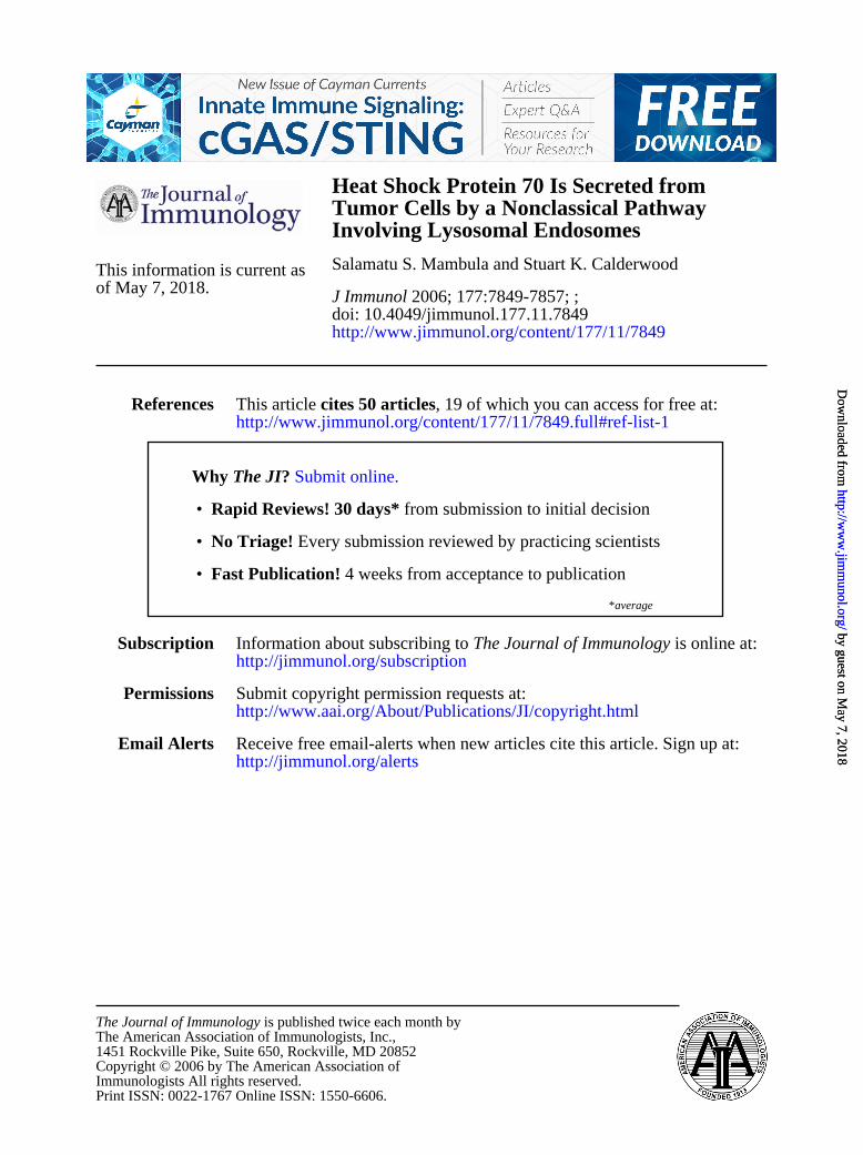
FIGURE 1. A, Effects of heat shock on intracellular levels of HSP70 atvarious temperatures. A total of 1 � 106 cells/ml PC-3 and LNCaP cellswere heat treated at 40°C for 6 h (lanes 2 and 5) and at 43°C for 30 min(lanes 3 and 6) followed by recovery at 37°C overnight. Control samples(lanes 1 and 4) were incubated at 37°C overnight. Following cell lysis,proteins were analyzed by 10% SDS-PAGE and probed for HSP70 byWestern blot. Experiments were performed reproducibly in duplicate. B,Kinetics of intracellular levels of HSP70 following hyperthermia treatment.Cells were incubated at 40°C for 6 h and at 43°C for 30 min in tissueculture dishes containing 1 � 106 cells. At the times indicated, cell lysateswere prepared and probed for the presence of HSP70 by Western blot. Eachexperiment was performed twice with similar results.
FIGURE 2. Kinetics of HSP70 release followingheat shock treatment. A total of 1 � 106 cells/ml PC-3(A and C) and LNCaP (B and D) cells were incubated at40°C for 6 h (Œ), 43°C for 30 min (f), and 37°C controlgroup (�) or nonheat shocked cells in tissue culturedishes containing 5 � 106 cells. Following heat treat-ments, the dishes were incubated at 37°C for recovery.Supernatants were taken at 60-min intervals (A and B)and 10-min intervals (C and D) and analyzed for thepresence of HSP70 by ELISA. Data represent themean � SD of three independent experiments. Statisti-cal analyses compared 0 time point and 60 in A and Band 0 min vs 30 min in C and D. ��, p � 0.01; com-paring 0 vs 360 min in A and B and 0 vs 360 in C andD (���, p � 0.001).
7850 HSP70 SECRETION, NONCLASSICAL PATHWAY
by guest on May 7, 2018
http://ww
w.jim
munol.org/
Dow
nloaded from
absorbance was read by spectrophotometer (Bio-Rad) at absorbances of 405and 650 nm (reference wavelength).
Western analysis of intracellular HSP70 expression
Aliquots of 1 � 106 cells were washed three times in ice-cold PBS, sol-ubilized in reducing sample buffer, and resolved by 10% SDS-PAGE underreducing conditions. Proteins were then electrophoretically transferred tonitrocellulose membranes (Amersham Biosciences) and blocked for 1 hwith 10% nonfat dry milk (Bio-Rad) in PBST buffer (Boston BioProducts).Filters were hybridized sequentially with the following: HSP70 mAb(StressGen Biotechnologies), anti-mouse IgG hp-conjugate (AmershamBiosciences) and Ab-Ag complexes developed by ECL-plus luminescence-based detection agent (Amersham Biosciences) according to the manufac-turer’s instructions.
Western analysis of ABCA-1 expression
A total of 1 � 106 cells was grown until growth was confluent, and cellswere harvested and washed three times in PBS. Total cell lysates wereprepared by solubilizing cells in reducing sample buffer, and samples wererun on 6% SDS-PAGE under reducing conditions. Transfer of proteins wasconducted as described above, and detection was confirm by usingABCA-1 Abs (Novus Biologicals). Actin controls were run on eachlysate.
Isolation of cell lysosomal fraction
PC3 and LNCaP cells were grown until there was packed cell volume of 3ml, which was resuspended in an equal volume of packed cells with hy-potonic buffer (10 mM HEPES, 1.5 mM MgCl2, 10 mM KCl, 1 mM DTT)and allowed to swell for 15 min on ice. Cells were lysed by rapidly pushingcells through a 1 ml syringe with a 25-gauge needle five times while keep-ing the cells on ice. Crude nuclear pellet was pelleted by centrifugation for30 s at 12,000 � g. Supernatants were stored and lysosomal fraction iso-lated as described by Andrei et al. (12). Briefly, supernatants were treatedwith proteinase K for 30 min on ice and inhibited with protease inhib-itors. The supernatant was diluted 10-fold in homogenizing buffer andcentrifuged at 50,000 � g for 20 min. The pellet was washed once inhomogenizing buffer and solubilized in reducing sample buffer, andHSP70 was detected by Western blots as described previously. Cathep-sin D was detected as the control for the lysosomal compartment byWestern blot.
Immunofluorescence microscopy
Cells were plated onto fibronectin-coated 8-well chamber glass slides withcover (Nalge Nunc International) at 105 cells per well. Twenty-four hourslater, the culture medium was removed and replaced with fresh mediumbefore the cells were hyperthermia treated as described above. The cellswere immediately fixed with 3.7% paraformaldehyde. After fixation for 15min, the cells were washed once with PBS and blocked with blockingbuffer (PBS containing 3% BSA) for 30 min. For intracellular staining, thecells were permeabilized with 0.1% Triton X-100 (Sigma-Aldrich) for 15min at room temperature after the fixing step. The cells were next washedonce with PBS, incubated for 1 h anti-HSP70 (StressGen Biotechnologies)or LAMP1 (CD107a; BD Pharmingen) mAbs followed by five washes inwash buffer (PBS containing 0.05% Tween 20). The cells were incu-bated with fluorescein-conjugated anti-mouse/rabbit IgG Ab (Alexa 594and Alexa 488; Invitrogen Life Technologies), respectively. Slides werethen washed and mounted in aqueous mounting solution using coverglass (Fisher Scientific) before fluorescent microscopy. Images wereacquired using a Nikon Eclipse E600 microscope fitted with a RT SPOTdigital camera and processed using SPOT software (DiagnosticInstruments).
Statistical analysis
Data are shown as mean � SD. Statistical significance of differencesbetween experimental groups was analyzed by using a Student’s t test.
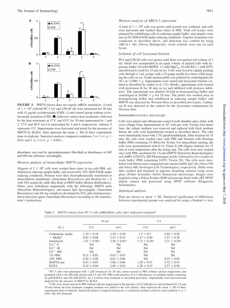
FIGURE 3. HSP70 release does not require mRNA translation. A totalof 1 � 106 cells/ml PC-3 (A) and LNCaP (B) were pretreated for 30 minwith 15 �g/ml cyclohexamide (CHX; ▫) and control group without cyclo-hexamide treatment (CHX; f) followed various heat treatments, followedby the heat treatments at 37°C and 43°C for 30 min represented by 1 and2, 37°C and 40°C for 6 h represented by 3 and 4, respectively, whereas 5represents 4°C. Supernatants were harvested and tested for the presence ofHSP70 by ELISA. Data represent the mean � SD of three experimentsdone in triplicate. Statistical analyses compared conditions 2 vs 1 (��, p �0.01) and 4 vs 3 (���, p � 0.001).
Table I. HSP70 release from PC-3 cells (nM/million cells) after indicated treatmenta
30 min 6 h
PC-3 37°C 43°C 37°C 40°C
Continuous media 1.19 � 0.18 3.29 � 0.30 1.5 � 0.5 2.86 � 0.28� Mediab 0.83 � 0.08 3.43 � 0.15 1.47 � 0.40 3.57 � 0.30Ionomycin 1.41 � 0.50� 3.56 � 0.82� 1.73 � 0.20� 3.21 � 0.29�Ca2� 0 Nd Nd Nd NdCa2� 40 Nd Nd Nd NdCa2� 400 Nd Nd Nd Nd1% FBS 0.21 � 0.02 0.63 � 0.03 Nd Nd10% FBS 0.20 � 0.06 0.61 � 0.06 Nd 0.25 � 0.05BAPTA-am 0.43 � 0.03 3.86 � 0.86 1.29 � 0.51 4.72 � 0.43EGTA 0.14 � 0.04 2.86 � 0.94 1.28 � 0.47 2.57 � 0.56
a PC-3 cells were pretreated with 1 �M ionomycin for 30 min, serum starved in PBS without calcium magnesium, andincubated with 0, 40, 400 �M calcium and 1% and 10% FBS in the presence of 0.2 mM glucose, in complete media containing25 �M BAPTA and 250 mM EGTA, for 2 h before heat treatment as described previously. Supernatants were harvested andanalyzed for the presence of HSP70 by ELISA.
b Cells were serum starved in PBS without calcium magnesium in the presence of 0.2 mM glucose and incubated for 2 h and10 min before the heat treatment complete medium was added to the cell cultures. Data represent the mean � SD of threeexperiments done in triplicate. Statistical analyses compared ionomycin vs continuous medium control at each condition (�, p �0.05). Nd, Not Detected.
7851The Journal of Immunology
by guest on May 7, 2018
http://ww
w.jim
munol.org/
Dow
nloaded from
Statistical significance was defined as follows: �, p � 0.05; ��, p �0.01; ���, p � 0.001.
ResultsRate of HSP70 release from prostate carcinoma cells after heatshock
We first investigated whether the rate of HSP70 release is relatedto the overall intracellular levels of the protein using PC-3 andLNCaP cells. These cell lines were chosen for their contrastinglevels of intracellular HSP70. We have shown previously that al-though LNCaP express minimal levels of intracellular HSP70,PC-3 express HSP70 abundantly even at nonstress temperatures(24). As a stimulus for HSP70 release, we used heat shock whichhas previously been shown to cause release of a number of lead-erless proteins including FGF-1 and IL-1� (25). We used two heatshock conditions for the experiments, including 40°C, which is inthe fever range and minimally toxic in tissue culture and stimulatestumor immunity in mice (26, 27) and 43°C, which strongly inducestranscriptional activation of hsp70 genes (28). As mentioned, PC-3
cells have high endogenous HSP70 levels which increase signifi-cantly after heat shock at 43°C although with comparatively minorincreases at 40°C. Similar trends were observed in LNCaP cellsthat have almost undetectable levels of HSP70 in unstressed cellsbut undergo significant HSP70 induction at 40°C and 43°C (Fig. 1,A and B). These heat shock treatments resulted in minimal (�1%)levels of necrosis and apoptosis at the 40°C or 43°C condition inboth cell lines at the period investigated (data not shown). This isin line with many previous studies of the kinetics of heat-inducedcell death that show minimal effects on cell survival of 40°Cheating or heating at 43°C up to 30 min reviewed by Hahn and Li(28). Low, but significant levels of necrosis and apoptosis (5–10%)did develop in the prostate cancer cells at later times (24–48 h)(29, 30). Our recent studies indicate that increasing temperaturesabove 43°C when necrosis increases actually decreases HSP70 se-cretion, presumably by denaturing components in the secretionpathway (30). HSP70 release at the time period examined in thisstudy is apparently due to active secretion (30).
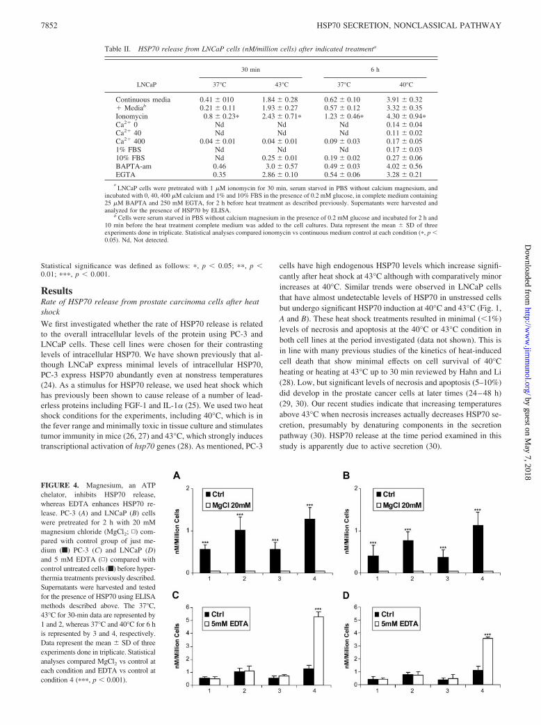
FIGURE 4. Magnesium, an ATPchelator, inhibits HSP70 release,whereas EDTA enhances HSP70 re-lease. PC-3 (A) and LNCaP (B) cellswere pretreated for 2 h with 20 mMmagnesium chloride (MgCl2; ▫) com-pared with control group of just me-dium (f) PC-3 (C) and LNCaP (D)and 5 mM EDTA (▫) compared withcontrol untreated cells (f) before hyper-thermia treatments previously described.Supernatants were harvested and testedfor the presence of HSP70 using ELISAmethods described above. The 37°C,43°C for 30-min data are represented by1 and 2, whereas 37°C and 40°C for 6 his represented by 3 and 4, respectively.Data represent the mean � SD of threeexperiments done in triplicate. Statisticalanalyses compared MgCl2 vs control ateach condition and EDTA vs control atcondition 4 (���, p � 0.001).
Table II. HSP70 release from LNCaP cells (nM/million cells) after indicated treatmenta
30 min 6 h
LNCaP 37°C 43°C 37°C 40°C
Continuous media 0.41 � 010 1.84 � 0.28 0.62 � 0.10 3.91 � 0.32� Mediab 0.21 � 0.11 1.93 � 0.27 0.57 � 0.12 3.32 � 0.35Ionomycin 0.8 � 0.23� 2.43 � 0.71� 1.23 � 0.46� 4.30 � 0.94�Ca2� 0 Nd Nd Nd 0.14 � 0.04Ca2� 40 Nd Nd Nd 0.11 � 0.02Ca2� 400 0.04 � 0.01 0.04 � 0.01 0.09 � 0.03 0.17 � 0.051% FBS Nd Nd Nd 0.17 � 0.0310% FBS Nd 0.25 � 0.01 0.19 � 0.02 0.27 � 0.06BAPTA-am 0.46 3.0 � 0.57 0.49 � 0.03 4.02 � 0.56EGTA 0.35 2.86 � 0.10 0.54 � 0.06 3.28 � 0.21
aLNCaP cells were pretreated with 1 �M ionomycin for 30 min, serum starved in PBS without calcium magnesium, and
incubated with 0, 40, 400 �M calcium and 1% and 10% FBS in the presence of 0.2 mM glucose, in complete medium containing25 �M BAPTA and 250 mM EGTA, for 2 h before heat treatment as described previously. Supernatants were harvested andanalyzed for the presence of HSP70 by ELISA.
b Cells were serum starved in PBS without calcium magnesium in the presence of 0.2 mM glucose and incubated for 2 h and10 min before the heat treatment complete medium was added to the cell cultures. Data represent the mean � SD of threeexperiments done in triplicate. Statistical analyses compared ionomycin vs continuous medium control at each condition (�, p �0.05). Nd, Not detected.
7852 HSP70 SECRETION, NONCLASSICAL PATHWAY
by guest on May 7, 2018
http://ww
w.jim
munol.org/
Dow
nloaded from

We next examined the rate of extracellular HSP70 release inboth cell lines after heat shock at 40°C and 43°C (Fig. 2). HSP70release was assayed in samples of medium by ELISA. Becausesome reports in the literature indicate that HSP70 family membersmay be released from cells in membrane-bounded vesicles, weinvestigated whether treating samples with detergents such asNonidet P-40 or Lubrol affects recovery of extracellular HSP70 bycausing release of HSP70 from vesicles. However, we did notobserve significant changes when medium from PC-3 or LNCaPcells was treated with detergents, suggesting that most HSP70 isreleased from these cells is in free solution (S. S. Mambula andS. K. Calderwood, unpublished data). The rate of HSP70 releasewas therefore assayed as free extracellular HSP70 released intocell medium. The rate of accumulation of extracellular HSP70 inPC-3 and LNCaP cells measured at hourly intervals is shown inFig. 2, A and B. Fig. 2, C and D, show more detailed kineticanalyses of the initial heat shock period with extracellular HSP70measured at 10-min intervals. In each cell line, we observed basalHSP70 release that was significantly increased after heat shock(Fig. 2). The rate of HSP70 accumulation was at least as great at40°C as at the higher temperature (43°C). In addition, HSP70 re-lease occurred during exposure to heat shock and was not mark-edly increased in the recovery period at 37°C (Fig. 2, A–D). A rolefor de novo HSP70 synthesis in heat-induced release seems un-likely because the bulk of the HSP70 release occurs before weobserve increased intracellular levels of the protein (compare Fig.1B with 2, C and D); HSP70 expression was not increased by 30min at 43°C or by 6 h at 40°C in the PC-3 cell line, whereasLNCaP only then shows HSP70 expression (Fig. 1B) when HSP70release is maximal at either temperature (Fig. 2, A–C). The effectsof heat shock thus appear to be largely due to a direct stimulationof secretion of endogenous HSP70, and increased HSP70 expres-sion due to heat shock appears to be an independent aspect of theheat shock response.
Heat shock-induced HSP70 release does not require HSP70mRNA translation
The experiments in Fig. 2 suggested that HSP70 release is inde-pendent of de novo synthesis. We have directly examined the re-
quirement for translation using protein synthesis-inhibitor cyclo-heximide (Fig. 3, A and B). Cycloheximide did not inhibit the rateof HSP70 release in either cell line further indicating that HSP70translation is not required for release (Fig. 3, A and B). In addition,we found that the rate of HSP70 release was temperature depen-dent and minimal release was seen at 4°C, suggesting a require-ment for metabolic activity in HSP70 secretion (Fig. 3, A and B).There were significant differences observed in HSP70 release atboth 43°C and 40°C compared with 37°C controls. These exper-iments thus suggest an active process of HSP70 release that can bestimulated by heat shock. Interestingly, we observed that HSP70release was stimulated only in full medium and was not observedin cells incubated in balanced salt solutions (Tables I and II). Thiseffect did not appear to involve energy depletion or the absence ofthe stimulatory effects of serum, as HSP70 release was not ob-served in PBS supplemented with either 5 mM glucose or 10%FCS (Tables I and II). As the active secretion of some intracellularproteins may require Ca2�, we therefore tested the potential role ofextracellular Ca2� in HSP70 release (Tables I and II). In cellsincubated at various concentrations of extracellular Ca2� (40–400mM) in PBS (Tables I and II), we did not see restoration of stress-induced HSP70 release. In addition, we did not observe differencesin HSP70 release when cells in full medium were pretreated withthe intracellular Ca2� chelator BAPTA-AM or the extracellularCa2� chelator EGTA before stress (Table I and II). We did, how-ever, observe a significantly higher rate of HSP70 release in cellstreated with the calcium ionophore ionomycin alone comparedwith nontreated cells, whereas there was a significant additive in-crease when the cells were treated with ionomycin and heat com-pared with heat treatment alone (Table I and II).
We next examined the effects of modulating extracellular Mg2�
on HSP70 secretion because the concentration of extracellularMg2� can mediate the release of other leaderless proteins thoughits effects on the state of extracellular ATP (12). Modulation ofextracellular Mg2� has a profound effect on the release of IL-1�;addition of Mg2� decreases secretion due ATP sequestration,whereas addition of Mg2� chelator EDTA can increase secretionthough its chelation of Mg2� and release of ATP in unbound form(12). We incubated the prostate carcinoma cells in growth medium
FIGURE 5. The effects of lyso-motrophic agents that lead to increasedendoluminal pH inhibits vesicular ac-cumulation and secretion of HSP70. Atotal of 1 � 106 cells/ml PC-3 (A) andLNCaP (B) were pretreated for 2 hwith 30 �M methylamine (Meth; ▫)and 50 mM ammonium chloride(NH4Cl; ▫) compared with control (f)untreated cells PC-3 (C) and LNCaP(D) followed by the heat treatments at37°C and 43°C for 30 min representedby 1 and 2 and 37°C and 40°C for 6 hrepresented by 3 and 4, respectively.Supernatants were harvested andtested for the presence of HSP70 byELISA. Data represent the mean � SDof three experiments done in triplicate.Statistical analyses compared Meth vscontrol conditions 2 and 4 (�, p �0.05) and NH4Cl vs control at condi-tions 2 and 4 (���, p � 0.001).
7853The Journal of Immunology
by guest on May 7, 2018
http://ww
w.jim
munol.org/
Dow
nloaded from

supplemented with 20 mM MgCl2 before heat shock. Indeed, theaddition of MgCl2 had a profound significant effect on the rate ofHSP70 release and almost completely inhibited both the basal re-lease and heat-induced HSP70 release in both cell lines (Fig. 4, Aand B). This suggests that extracellular ATP is present in a freeform and that addition of MgCl2 sequesters free ATP and blocksHSP70 release. EDTA significantly enhanced HSP70 release butonly under the 6 h at 40°C condition. The rationale behind thesefindings is not clear but may be related to the longer incubationtime at 40°C (Fig. 4, C and D) (21). However, addition of ATPalone does not cause release of HSP70 in the absence of heatshock, suggesting that it is necessary but not sufficient for HSP70release (data not shown). Similarly, findings are observed withIL-1� in which extracellular ATP is required for secretion, but notsufficient in itself (21).
Potential role of endosomal lysosomes in stress-inducedsecretion of HSP70
The ability of HSP70 to exit cells under nontoxic conditionssuggests a pathway for traversing cell membranes and activesecretion. Therefore, we next examined the pathway of HSP70release, exploring the hypothesis that HSP70 release may fol-low mechanisms comparable to IL-1� secretion. We first ex-plored a role for secretory lysosomes in HSP70 release andinvestigated whether increasing pH in these compartments us-ing lysosomotropic agents inhibits HSP70 secretion. Bothmethylamine and ammonium chloride, which increase intraly-sosomal pH, decreased the rate of HSP70 release in PC-3 andLNCaP cells (Fig. 5). Incubation with methylamine signifi-cantly caused a �50% reduction in HSP70 release in both celllines exposed to either heat shock condition (Fig. 5, A and B).The effects of NH4Cl were significantly more marked, with ab-olition of both basal and heat shock-induced HSP70 release(Fig. 5, C and D). Thus, lysomotropic drugs inhibit HSP70 se-cretion, and these experiments indicate a key role for the en-dolysosme pathway in HSP70 release.
We next examined whether the heat shock conditions thatcause HSP70 release lead to the transport of secretory lyso-somes to the cell surface using the intralysosmal proteinLAMP1 as a marker (Fig. 6). LAMP1 was detected by indirectimmunofluorescence in small patches on the cell surface of bothcell types at 37°C. However, conditions that lead to HSP70secretion including 30 min at 43°C and 6 h at 40°C lead to amajor increase in cell surface LAMP1, which becomes concen-trated in large caps on the surface of PC-3 and LNCaP cells(Fig. 6, A and B, respectively). These data are consistent with ahypothesis involving stress-induced transport of secretory ly-sosomes, containing HSP70 to the cell surface, fusion with theplasma membrane, and display of LAMP1 on the extracellularface of the plasma membrane accompanied by HSP70 secretion.We further confirmed this hypothesis by isolating the lysosomalfraction from LNCaP and PC3 cells and detecting the presenceof HSP70 in this compartment as shown in Fig. 6C. HSP70levels and cathepsin D controls were minimal in the lysosomalfraction before heating but increased after both the heat shockconditions (Fig. 6C).
Role of ABC-binding cassette membrane transporter in HSP70secretion
Because previous studies have implicated a role for ABC familytransporters in the secretion of IL-1�, we examined whether thismechanism might also relate to HSP70 secretion (12). Cells werepretreated with the ABC inhibitor glibenclamide before examina-tion of basal and heat shock-induced HSP70 secretion. Gliben-
clamide significantly inhibited heat-induced HSP70 release inLNCaP and PC-3 cells although it had a less-pronounced effect onbasal release, suggesting a role for ABC family transporters inHSP70 secretion (Fig. 7, A and B). Previous studies have impli-cated the ABC family member ABCA-1 in IL-1� secretion (17).The role for ABCA-1 can be examined using another chemical,inhibitor DIDS. DIDS inhibits ABCA-1 by blocking the chloridecurrent necessary for its function (21). DIDS proved to signifi-cantly inhibit HSP70 secretion at both basal and heat shock-stim-ulated PC-3 and LNCaP cells (Fig. 7, C and D). Fig. 7E indicatesthat ABCA-1 is expressed in both cell lines both under nonheatshock and heat shock conditions. ABCA-1 may be involved in themultidrug resistance phenotype exhibited by both of the tumorcells. These experiments therefore indicate that HSP70 secretionrequires the ABCA-1 isoform ABC transporters.
FIGURE 6. Immunostaining for surface LAMP1. Following no heattreatment at 37°C and heat shock at 43°C for 30 min and 40°C for 6 h,PC-3 (A) and LNCaP (B) were fixed and stained with mAbs to humanLAMP-1 (Isotype Labeling, mouse IgG1�) and mouse IgG1� T-2 myco-toxin (Isotype Labeling, matched controls) followed by Alexa-488-conju-gated secondary Ab (green). Perm represents intracellular staining forLAMP1. Images were acquired using fluorescent microscopy at a magni-fication of �400. This experiment was repeated in triplicate with repro-ducible results. Presence of HSP70 was determined using Western blots ofthe lysosomal fraction (C) in both PC3 and LNCaP cells.
7854 HSP70 SECRETION, NONCLASSICAL PATHWAY
by guest on May 7, 2018
http://ww
w.jim
munol.org/
Dow
nloaded from

HSP70 secreted for prostate carcinoma cells binds to the cellsurface prostate cancer cells
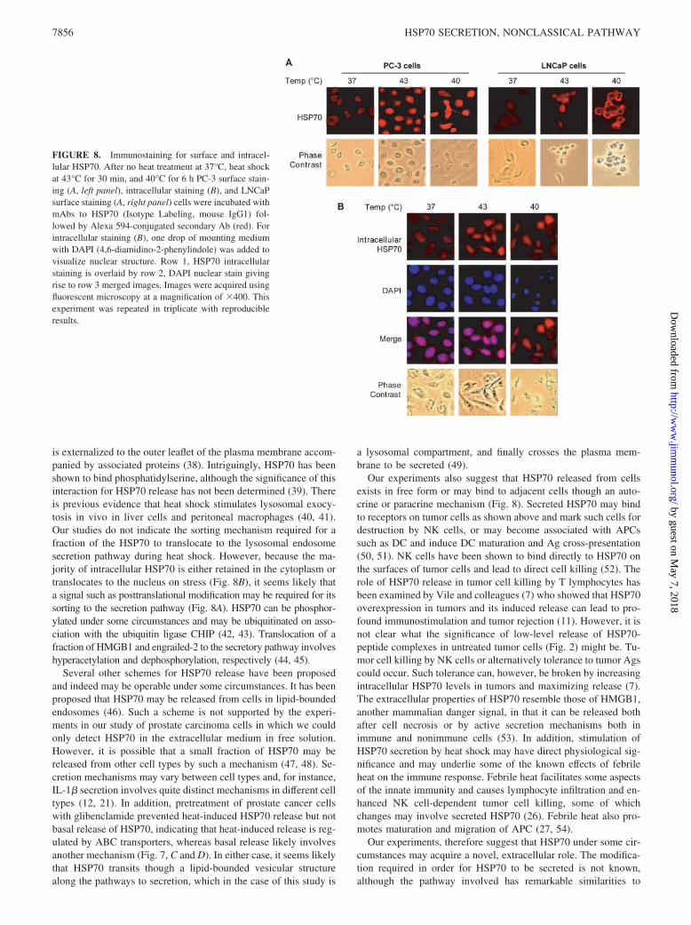
We next examine whether a fraction of the HSP70 secreted fromtumor cells can bind to the cell surface (Fig. 8). Such a mechanismwas suggested by kinetic studies of extracellular HSP70 accumulationas in Fig. 2. In each case, we observed that after initial accumulation,extracellular HSP70 levels declined. This decrease in extracellularHSP70 may be due to binding and internalization of secreted HSP70.Indeed, cell surface HSP70 was minimal in unstressed cells but wasreadily detectable after 43°C for 30 min or 6 h at 40°C in both celllines (Fig. 8, A and C). This increase in surface expression of HSP70correlates with the increases observed in LAMP1 surface expression(Fig. 6). We also examined the behavior of intracellular HSP70 afterheat shock (Fig. 8B). HSP70 appears to be evenly distributed withinthe cell at 37°C but was translocated to the nucleus at 43°C, where itwas found in granular structures (31). However, 40°C heating did notappear to markedly alter the intracellular HSP70 distribution (Fig.8B). This experiment therefore indicates a profound and complex re-distribution of HSP70 in cells after heat shock. For instance, at 43°C,HSP70 may be translocated to the nucleus (Fig. 8B) or associated withthe cell surface (Fig. 8A). We observed similar findings with the in-tracellular staining of LNCaP cells (data not shown).
DiscussionOur experiments were conducted with the aim of determiningmechanisms of HSP70 release from tumor cells that may be im-portant in understanding its immunological functions. Previous re-ports suggested that release of HSP70 family proteins may involvephysiological mechanisms such as cosecretion in exosomes ofHSC70 with transferrin (contains a secretion leader) or dischargefrom cells undergoing necrosis (32). However, our current studiesindicate that HSP70 release from prostate carcinoma cells is not
inhibited by inhibitors of endoplasmic reticulum/Golgi function(data not shown) and takes place under conditions in which min-imal necrosis is observed (Fig. 1B and 2, A–D). Thus, HSP70 canbe secreted from tumor cells by an active secretion pathway aswell as from cells dying from the stress of therapy (10, 32).
The mechanisms of secretion of leaderless proteins such asHSP70 are complex and incompletely understood. However, twomain pathways have been deduced. The first (i) used by proteinssuch as FGF-1 and IL-1�, appears to translocate across the plasmamembrane on partial denaturation to a molten globule form (15). Ithas been conjectured that heat shock stimulates this pathway inpart by permitting the proteins to partially unfold, a requirementfor membrane translocation of many proteins (33, 34). More com-mon (ii) appears to be the pathway used by IL-1�, which involvesentry of the secreted protein into endolysosomes and release fromthe cell by a vesiculation mechanism (35). Our largely pharmaco-logical dissection indicates that HSP70 release belongs to this sec-ond class of leaderless secretion, and its mechanisms of releasefurther resemble IL-1� in requiring the activity of ABC familytransmembrane transporters and the likely participation of puriner-gic receptors (21). Other secreted proteins that are largely of cy-toplasmic/nuclear distribution in addition to HSP70, such as high-mobility protein b1 (HMGB1) and engrailed-2, appear to besecreted along this pathway under specialized conditions that pro-mote secretion (36, 37). Curiously, heat shock has been shown tocause the secretion of two proteins, FGF-1 and IL-1�, through thealternative pathway (i) involving heat-induced membrane translo-cation (15). HSP70 secretion by heat shock may therefore use ahybrid pathway involving mechanisms found in pathways (i) and (ii).For instance, binding of FGF-1 and IL-1� to phosphatidylserine onthe inner leaflet of the plasma membrane before translocation maybe involved in a flipping mechanism in which phosphatidylserine
FIGURE 7. HSP70 secretion re-quires ABC family transporters. PC-3(A) and LNCaP (B) cells were pre-treated for 2 h with 1 mM gliben-clamide (Glib) (▫) and 250 �M DIDS(▫) compared with controls (f), PC-3(C), and LNCaP (D) before hyperther-mia treatments previously described.Supernatants were harvested andtested for the presence of HSP70 usingELISA methods described above. The37°C and 43°C for 30-min data arerepresented by 1 and 2 and 37°C and40°C for 6 h is represented by 3 and 4,respectively. ABCA-1 levels in PC3 andLNCaP was determined using Westernblots from total lysate (E). Data repre-sent the mean � SD of the experimentsdone in triplicate. Statistical analysescompared Glib vs control at conditions 2(��, p � 0.01) and 4 (���, p � 0.001).DIDS was compared statistically to con-trol at conditions 1 and 3 (�, p � 0.05);(C), 2 and 4 (���, p � 0.001); (D) 2 and4 (��, p � 0.01).
7855The Journal of Immunology
by guest on May 7, 2018
http://ww
w.jim
munol.org/
Dow
nloaded from
is externalized to the outer leaflet of the plasma membrane accom-panied by associated proteins (38). Intriguingly, HSP70 has beenshown to bind phosphatidylserine, although the significance of thisinteraction for HSP70 release has not been determined (39). Thereis previous evidence that heat shock stimulates lysosomal exocy-tosis in vivo in liver cells and peritoneal macrophages (40, 41).Our studies do not indicate the sorting mechanism required for afraction of the HSP70 to translocate to the lysosomal endosomesecretion pathway during heat shock. However, because the ma-jority of intracellular HSP70 is either retained in the cytoplasm ortranslocates to the nucleus on stress (Fig. 8B), it seems likely thata signal such as posttranslational modification may be required for itssorting to the secretion pathway (Fig. 8A). HSP70 can be phosphor-ylated under some circumstances and may be ubiquitinated on asso-ciation with the ubiquitin ligase CHIP (42, 43). Translocation of afraction of HMGB1 and engrailed-2 to the secretory pathway involveshyperacetylation and dephosphorylation, respectively (44, 45).
Several other schemes for HSP70 release have been proposedand indeed may be operable under some circumstances. It has beenproposed that HSP70 may be released from cells in lipid-boundedendosomes (46). Such a scheme is not supported by the experi-ments in our study of prostate carcinoma cells in which we couldonly detect HSP70 in the extracellular medium in free solution.However, it is possible that a small fraction of HSP70 may bereleased from other cell types by such a mechanism (47, 48). Se-cretion mechanisms may vary between cell types and, for instance,IL-1� secretion involves quite distinct mechanisms in different celltypes (12, 21). In addition, pretreatment of prostate cancer cellswith glibenclamide prevented heat-induced HSP70 release but notbasal release of HSP70, indicating that heat-induced release is reg-ulated by ABC transporters, whereas basal release likely involvesanother mechanism (Fig. 7, C and D). In either case, it seems likelythat HSP70 transits though a lipid-bounded vesicular structurealong the pathways to secretion, which in the case of this study is
a lysosomal compartment, and finally crosses the plasma mem-brane to be secreted (49).
Our experiments also suggest that HSP70 released from cellsexists in free form or may bind to adjacent cells though an auto-crine or paracrine mechanism (Fig. 8). Secreted HSP70 may bindto receptors on tumor cells as shown above and mark such cells fordestruction by NK cells, or may become associated with APCssuch as DC and induce DC maturation and Ag cross-presentation(50, 51). NK cells have been shown to bind directly to HSP70 onthe surfaces of tumor cells and lead to direct cell killing (52). Therole of HSP70 release in tumor cell killing by T lymphocytes hasbeen examined by Vile and colleagues (7) who showed that HSP70overexpression in tumors and its induced release can lead to pro-found immunostimulation and tumor rejection (11). However, it isnot clear what the significance of low-level release of HSP70-peptide complexes in untreated tumor cells (Fig. 2) might be. Tu-mor cell killing by NK cells or alternatively tolerance to tumor Agscould occur. Such tolerance can, however, be broken by increasingintracellular HSP70 levels in tumors and maximizing release (7).The extracellular properties of HSP70 resemble those of HMGB1,another mammalian danger signal, in that it can be released bothafter cell necrosis or by active secretion mechanisms both inimmune and nonimmune cells (53). In addition, stimulation ofHSP70 secretion by heat shock may have direct physiological sig-nificance and may underlie some of the known effects of febrileheat on the immune response. Febrile heat facilitates some aspectsof the innate immunity and causes lymphocyte infiltration and en-hanced NK cell-dependent tumor cell killing, some of whichchanges may involve secreted HSP70 (26). Febrile heat also pro-motes maturation and migration of APC (27, 54).
Our experiments, therefore suggest that HSP70 under some cir-cumstances may acquire a novel, extracellular role. The modifica-tion required in order for HSP70 to be secreted is not known,although the pathway involved has remarkable similarities to
FIGURE 8. Immunostaining for surface and intracel-lular HSP70. After no heat treatment at 37°C, heat shockat 43°C for 30 min, and 40°C for 6 h PC-3 surface stain-ing (A, left panel), intracellular staining (B), and LNCaPsurface staining (A, right panel) cells were incubated withmAbs to HSP70 (Isotype Labeling, mouse IgG1) fol-lowed by Alexa 594-conjugated secondary Ab (red). Forintracellular staining (B), one drop of mounting mediumwith DAPI (4,6-diamidino-2-phenylindole) was added tovisualize nuclear structure. Row 1, HSP70 intracellularstaining is overlaid by row 2, DAPI nuclear stain givingrise to row 3 merged images. Images were acquired usingfluorescent microscopy at a magnification of �400. Thisexperiment was repeated in triplicate with reproducibleresults.
7856 HSP70 SECRETION, NONCLASSICAL PATHWAY
by guest on May 7, 2018
http://ww
w.jim
munol.org/
Dow
nloaded from
IL-1� secretion in macrophages and glial cells (12, 21). As HSP70has potent immunological properties, this pathway may play a rolein tumor immune response.
DisclosuresThe authors have no financial conflict of interest.
References1. Lindquist, S., and E. A. Craig. 1988. The heat-shock proteins. Annu. Rev. Genet.
22: 631–677.2. Asea, A., S. K. Kraeft, E. A. Kurt-Jones, M. A. Stevenson, L. B. Chen, R. W.
Finberg, G. C. Koo, and S. K. Calderwood. 2000. HSP70 stimulates cytokine pro-duction through a CD14-dependant pathway, demonstrating its dual role as a chap-erone and cytokine. Nat. Med. 6: 435–442.
3. Srivastava, P. 2002. Interaction of heat shock proteins with peptides and antigenpresenting cells: chaperoning of the innate and adaptive immune responses. Annu.Rev. Immunol. 20: 395–425.
4. Theriault, J. R., S. S. Mambula, T. Sawamura, M. A. Stevenson, and S. K.Calderwood. 2005. Extracellular HSP70 binding to surface receptors present on an-tigen presenting cells and endothelial/epithelial cells. FEBS Lett. 579: 1951–1960.
5. Calderwood, S. K., J. R. Theriault, and J. Gong. 2005. How is the immuneresponse affected by hyperthermia and heat shock proteins? Int. J. Hyperthermia21: 713–716.
6. Chen, X., Q. Tao, H. Yu, L. Zhang, and X. Cao. 2002. Tumor cell membrane-bound heat shock protein 70 elicits antitumor immunity. Immunol. Lett. 84:81–87.
7. Daniels, G. A., L. Sanchez-Perez, R. M. Diaz, T. Kottke, J. Thompson, M. Lai,M. Gough, M. Karim, A. Bushell, H. Chong, et al. 2004. A simple method to cureestablished tumors by inflammatory killing of normal cells. Nat. Biotechnol. 22:1125–1132.
8. Noessner, E., R. Gastpar, V. Milani, A. Brandl, P. J. Hutzler, M. C. Kuppner,M. Roos, E. Kremmer, A. Asea, S. K. Calderwood, and R. D. Issels. 2002.Tumor-derived heat shock protein 70 peptide complexes are cross-presented byhuman dendritic cells. J. Immunol. 169: 5424–5432.
9. Baker-LePain, J. C., R. C. Reed, and C. V. Nicchitta. 2003. ISO: a critical eval-uation of the role of peptides in heat shock/chaperone protein-mediated tumorrejection. Curr. Opin. Immunol. 15: 89–94.
10. Basu, S., R. J. Binder, R. Suto, K. M. Anderson, and P. K. Srivastava. 2000.Necrotic but not apoptotic cell death releases heat shock proteins, which delivera partial maturation signal to dendritic cells and activate the NF-�B pathway. Int.Immunol. 12: 1539–1546.
11. Calderwood, S. K. 2005. Chaperones and slow death: a recipe for tumor immu-notherapy. Trends Biotechnol. 23: 57–59.
12. Andrei, C., C. Dazzi, L. Lotti, M. R. Torrisi, G. Chimini, and A. Rubartelli. 1999.The secretory route of the leaderless protein interleukin 1� involves exocytosis ofendolysosome-related vesicles. Mol. Biol. Cell 10: 1463–1475.
13. Powers, C. J., S. W. McLeskey, and A. Wellstein. 2000. Fibroblast growth fac-tors, their receptors and signaling. Endocr. Relat. Cancer 7: 165–197.
14. Tarantini, F., I. Micucci, S. Bellum, M. Landriscina, S. Garfinkel, I. Prudovsky,and T. Maciag. 2001. The precursor but not the mature form of IL1� blocks therelease of FGF1 in response to heat shock. J. Biol. Chem. 276: 5147–5151.
15. Prudovsky, I., A. Mandinova, R. Soldi, C. Bagala, I. Graziani, M. Landriscina,F. Tarantini, M. Duarte, S. Bellum, H. Doherty, and T. Maciag. 2003. The non-classical export routes: FGF1 and IL-1� point the way. J. Cell Sci. 116:4871–4881.
16. Hightower, L. E., and P. T. Guidon, Jr. 1989. Selective release from culturedmammalian cells of heat-shock (stress) proteins that resemble glia-axon transferproteins. J. Cell. Physiol. 138: 257–266.
17. Hamon, Y., M. F. Luciani, F. Becq, B. Verrier, A. Rubartelli, and G. Chimini.1997. Interleukin-1� secretion is impaired by inhibitors of the Atp binding cas-sette transporter, ABC1. Blood 90: 2911–2915.
18. Blott, E. J., and G. M. Griffiths. 2002. Secretory lysosomes. Nat. Rev. Mol. CellBiol. 3: 122–131.
19. Kuchler, K., A. Rubartelli, and B. Holland. 1997. Unusual Secretory Pathways:From Bacteria to Man. R. G. Landes Company, Georgetown.
20. Higgins, C. F. 1992. ABC transporters: from microorganisms to man. Annu. Rev.Cell Biol. 8: 67–113.
21. Marty, V., C. Medina, C. Combe, P. Parnet, and T. Amedee. 2005. ATP bindingcassette transporter ABC1 is required for the release of interleukin-1� by P2X7-stimulated and lipopolysaccharide-primed mouse Schwann cells. Glia 49:511–519.
22. Ferrari, D., P. Chiozzi, S. Falzoni, M. Dal Susino, L. Melchiorri, O. R. Baricordi,and F. Di Virgilio. 1997. Extracellular ATP triggers IL-1� release by activa-ting the purinergic P2Z receptor of human macrophages. J. Immunol. 159:1451–1458.
23. Mambula, S. S., K. Sau, P. Henneke, D. T. Golenbock, and S. M. Levitz. 2002.Toll-like receptor (TLR) signaling in response to Aspergillus fumigatus. J. Biol.Chem. 277: 39320–39326.
24. Tang, D., M. A. Khaleque, E. L. Jones, J. R. Theriault, C. Li, W. H. Wong,M. A. Stevenson, and S. K. Calderwood. 2005. Expression of heat shock proteinsand heat shock protein messenger ribonucleic acid in human prostate carcinomain vitro and in tumors in vivo. Cell Stress Chaperones 10: 46–58.
25. Jackson, A., S. Friedman, X. Zhan, K. A. Engleka, R. Forough, and T. Maciag.1992. Heat shock induces the release of fibroblast growth factor 1 from NIH 3T3cells. Proc. Natl. Acad. Sci. USA 89: 10691–10695.
26. Burd, R., T. S. Dziedzic, Y. Xu, M. A. Caligiuri, J. R. Subjeck, and E. A. Repasky.1998. Tumor cell apoptosis, lymphocyte recruitment and tumor vascular changes areinduced by low temperature, long duration (fever-like) whole body hyperthermia.J. Cell. Physiol. 177: 137–147.
27. Ostberg, J. R., R. Patel, and E. A. Repasky. 2000. Regulation of immune activityby mild (fever-range) whole body hyperthermia: effects on epidermal Langerhanscells. Cell Stress Chaperones 5: 458–461.
28. Hahn, G. M., and G. C. Li. 1982. Thermotolerance and heat shock proteins inmammalian cells. Radiat. Res. 92: 452–457.
29. Jones, E. L., M. J. Zhao, M. A. Stevenson, and S. K. Calderwood. 2004. The 70kilodalton heat shock protein is an inhibitor of apoptosis in prostate cancer. Int.J. Hyperthermia 20: 835–849.
30. Mambula, S. S. a. S. K. C. 2006. Heat induced HSP70 release from prostatecarcinoma cells involves both active secretion and passive release from necroticcells. Int. J. Hyperthermia. In press.
31. Mariethoz, E., M. R. Jacquier-Sarlin, G. Multhoff, A. M. Healy, F. Tacchini-Cottier,and B. S. Polla. 1997. Heat shock and proinflammatory stressors induce differentiallocalization of heat shock proteins in human monocytes. Inflammation 21: 629–642.
32. Asea, A. A., and S. K. Calderwood. 2005. Regulation of Signal Transduction byIntracellular and Extracellular HSP70. Cambridge University Press,Cambridge, U.K.
33. Arai, M., and K. Kuwajima. 2000. Role of the molten globule state in proteinfolding. Adv. Protein Chem. 53: 209–282.
34. Ptitsyn, O. B. 1995. Molten globule and protein folding. Adv. Protein Chem. 47:83–229.
35. Andrei, C., P. Margiocco, A. Poggi, L. V. Lotti, M. R. Torrisi, and A. Rubartelli.2004. Phospholipases C and A2 control lysosome-mediated IL-1� secretion:implications for inflammatory processes. Proc. Natl. Acad. Sci. USA 101:9745–9750.
36. Gardella, S., C. Andrei, D. Ferrera, L. V. Lotti, M. R. Torrisi, M. E. Bianchi, andA. Rubartelli. 2002. The nuclear protein HMGB1 is secreted by monocytes via anon-classical, vesicle-mediated secretory pathway. EMBO Rep. 3: 995–1001.
37. Maizel, A., O. Bensaude, A. Prochiantz, and A. Joliot. 1999. A short region of itshomeodomain is necessary for engrailed nuclear export and secretion. Develop-ment 126: 3183–3190.
38. Peterson, E. A., M. R. Sutherland, M. E. Nesheim, and E. L. Pryzdial. 2003.Thrombin induces endothelial cell-surface exposure of the plasminogen receptorannexin 2. J. Cell Sci. 116: 2399–2408.
39. Arispe, N., M. Doh, and A. De Maio. 2002. Lipid interaction differentiates theconstitutive and stress-induced heat shock proteins Hsc70 and Hsp70. Cell StressChaperones 7: 330–338.
40. Barni, S., V. Bertone, M. G. Silvotti, I. Freitas, G. Mathe, and P. Pontiggia. 1996.Lysosomal exocytosis induced by hyperthermia: a new model of cancer death III:effect on liver metastasis. Biomed. Pharmacother. 50: 79–84.
41. Pontiggia, P., S. Barni, G. Mathe, V. Bertone, and E. Pontiggia. 1995. Lysosomalexocytosis induced by hyperthermia: a new model of cancer cell death II: effecton peritoneal macrophages. Biomed. Pharmacother. 49: 429–430.
42. Evdonin, A. L., I. V. Guzhova, B. A. Margulis, and N. D. Medvedeva. 2004.Phospholipse c inhibitor, u73122, stimulates release of hsp-70 stress protein fromA431 human carcinoma cells. Cancer Cell Int. 4: 2.
43. Lim, Y. P., C. Y. Wong, L. L. Ooi, B. J. Druker, and R. J. Epstein. 2004. Selectivetyrosine hyperphosphorylation of cytoskeletal and stress proteins in primary hu-man breast cancers: implications for adjuvant use of kinase-inhibitory drugs.Clin. Cancer Res. 10: 3980–3987.
44. Bonaldi, T., F. Talamo, P. Scaffidi, D. Ferrera, A. Porto, A. Bachi, A. Rubartelli,A. Agresti, and M. E. Bianchi. 2003. Monocytic cells hyperacetylate chromatinprotein HMGB1 to redirect it towards secretion. EMBO J. 22: 5551–5560.
45. Wisniewski, J. R., Z. Szewczuk, I. Petry, R. Schwanbeck, and U. Renner. 1999.Constitutive phosphorylation of the acidic tails of the high mobility group 1proteins by casein kinase II alters their conformation, stability, and DNA bindingspecificity. J. Biol. Chem. 274: 20116–20122.
46. Tytell, M. 2005. Release of heat shock proteins (Hsps) and the effects of extra-cellular Hsps on neural cells and tissues. Int. J. Hyperthermia 21: 445–455.
47. Lancaster, G. I., and M. A. Febbraio. 2005. Exosome-dependent trafficking ofHSP70: a novel secretory pathway for cellular stress proteins. J. Biol. Chem. 280:23349–23355.
48. Gastpar, R., M. Gehrmann, M. A. Bausero, A. Asea, C. Gross, J. A. Schroeder,and G. Multhoff. 2005. Heat shock protein 70 surface-positive tumor exosomesstimulate migratory and cytolytic activity of natural killer cells. Cancer Res. 65:5238–5247.
49. Rodriguez, A., P. Webster, J. Ortego, and N. W. Andrews. 1997. Lysosomesbehave as Ca2�-regulated exocytic vesicles in fibroblasts and epithelial cells.J. Cell Biol. 137: 93–104.
50. Multhoff, G., L. Mizzen, C. C. Winchester, C. M. Milner, S. Wenk, G. Eissner,H. H. Kampinga, B. Laumbacher, and J. Johnson. 1999. Heat shock protein 70(Hsp70) stimulates proliferation and cytolytic activity of natural killer cells. Exp.Hematol. 27: 1627–1636.
51. Calderwood, S. K., J. R. Theriault, and J. Gong. 2005. Message in a bottle: roleof the 70-kDa heat shock protein family in anti-tumor immunity. Eur. J. Immunol.35: 2518–2527.
52. Multhoff, G. 2002. Activation of natural killer cells by heat shock protein 70. Int.J. Hyperthermia 18: 576–585.
53. Dumitriu, I. E., P. Baruah, A. A. Manfredi, M. E. Bianchi, and P. Rovere-Querini.2005. HMGB1: guiding immunity from within. Trends Immunol. 26: 381–387.
54. Ostberg, J. R., and E. A. Repasky. 2000. Comparison of the effects of two dif-ferent whole body hyperthermia protocols on the distribution of murine leukocytepopulations. Int. J. Hyperthermia 16: 29–43.
7857The Journal of Immunology
by guest on May 7, 2018
http://ww
w.jim
munol.org/
Dow
nloaded from


![A novel endogenous inhibitor of the secreted streptococcal ... · itis, impetigo) to life threatening (toxic shock syndrome, necrotizing fasciitis) [4]. The contribution that any](https://img.dokumen.tips/doc/110x75/5c9bc45d09d3f206138bc209/a-novel-endogenous-inhibitor-of-the-secreted-streptococcal-itis-impetigo.jpg)


![Research Paper Paired box 2 promotes progression of ...[19], Wilms’ tumor suppressor gene [20, 21] and secreted frizzled related protein 2 gene [22] were reported. However, there](https://img.dokumen.tips/doc/110x75/60943d2f5dfdce3c6d63ac20/research-paper-paired-box-2-promotes-progression-of-19-wilmsa-tumor-suppressor.jpg)