Embed Size (px)
Citation preview
Health Professions Regulatory Advisory
Council (HPRAC)
Diagnostic Sonographers
A Literature Review
A Rapid Literature Review on Sonographers
A Rapid Literature Review on Sonographers
Prepared by the Planning Unit Planning Research and Analysis Branch Health System Strategy amp Policy Division Ministry of Health and Long-Term Care April 2013
Please note that this Rapid Literature Review is a summary of information from other sources not a representation of the policy position or goals of the Ministry of Health and Long-Term Care If material in the review is to be referenced
please cite the original primary source rather than the review itself
A Rapid Literature Review on Sonographers
OBJECTIVES The requestorrsquos stated objective was to examine information related to the sonography profession including
bull Regulation licensure or certification in various jurisdictions (in particular California NY UK and Australia)
bull Patient safety and potential risk of harm associated with ultrasound technology and the sonography profession
bull Interprofessional collaboration bull Emerging trends
SEARCH METHODS FOR IDENTIFICATION OF STUDIES Individual peer-reviewed articles and review articles were identified through the Ontario Ministry of Health and Long-Term Carersquos computerized library database PubMed and Google Scholar Grey literature was identified through Google and relevant government websites The search was limited to English sources and therefore may not capture the full extent of initiatives in non-English speaking countries The Medical Subject Heading (MeSH) terms ldquoUltrasonographytrends[Mesh]rdquo ldquoUltrasonographyadverse effects[Mesh] OR Ultrasonographymortalityrdquo and ldquoPatient Safety[Mesh]rdquo ldquoInterprofessional Relationsrdquo ldquoInterdisciplinary Communicationrdquo ldquoAccreditationrdquo ldquoLicensurerdquo ldquoCertificationrdquo were used in combination with the following keywords to identify relevant articles and documents for this review ldquoCaliforniardquo ldquoNew Yorkrdquo ldquoAustraliardquo and ldquoEnglandrdquo ldquoNew Mexicordquo ldquoOregonrdquo ldquointerprofessionalrdquo ldquorisksrdquo ldquosafetyrdquo collaboration A total of 47 references were identified and cited in this review nine review articles 25 original research papers from peer-reviewed journals and 13 documents from the grey literature Table 3 in the Appendix consists of a summary table with details for each of the sources cited in the review In total the searching for relevant material and the writing of this review took approximately seven working days to complete by one person SUMMARY OF MAIN FINDINGS
bull In Canada and the US sonographers are professionals who perform ultrasound scans and report their findings to a doctor who issues a final diagnosis and report In England sonographers independently conduct and report on ultrasound findings without supervision of a doctor Although sonographers specialize in ultrasound other professions may use the technology such as emergency physicians and radiologists
Regulation bull In Australia New Zealand and two US states (New Mexico and Oregon) sonographers
must register with (or be licensed or accredited by) an official body in order to practice and must complete required education andor work experience to do so
bull In contrast in England and all other US states sonography is not regulated bull In most US states anyone can practice sonography but employers prefer to hire
sonographers with credentials issued by certification bodies such as The American Registry for Diagnostic Medical Sonography
Patient safety and potential risk of harm from ultrasound
A Rapid Literature Review on Sonographers
bull A body of literature has found no evidence of harm from use of ultrasound in humans though various authors caution that most data are based on older studies using ultrasound devices with lower intensities Advisory groups continue to urge caution with the use of ultrasound and recommend limiting acoustic power and exposure duration
bull The potential health risks of ultrasound include (1) thermal effects (ultrasound can cause an increase in tissue temperature) and (2) mechanical effects (ultrasound can create shear forces pressure changes and other non-thermal effects)
bull The Output Display Standard (ODS) on every ultrasound machine is an on-screen indicator that provides information about the thermal and mechanical exposures however several studies have shown sonographers (and other health professionals) have poor knowledge of how to find and interpret this safety information on their own machines
bull A potential risk is that a sonographer might not observe and communicate to a physician the key ultrasound images necessary to make a diagnosis However one source noted that if dynamic (ie video-like) clips are used several observers can re-evaluate them and increase patient safety
bull Three studies from England (where sonographers report on ultrasounds independently) found that sonographers had high accuracy and strong agreement with the findings of radiologists As well extending sonographersrsquo scope of practice was associated with positive outcomes such as improved productivity and a freeing of radiologists time in two studies
bull One study however found sonographers were less likely to provide a clear diagnosis and more likely to include a disclaimer about the quality of the images
Interprofessional collaboration bull Little information was found on sonographersrsquo interprofessional collaboration in general or
their working relationship with cardiology technologists or physicians in particular bull Three articles described instances of cardiac sonographers working with other health
professionals (eg catheterization lab technologists and various specialized cardiologists) in cardiac catheterization or electrophysiology labs
Emerging trends bull Several sources commented on the recent growth in ultrasound scans performed and
interpreted by clinicians (ie not sonographer) at the bedside This is not meant to replace the use of more comprehensive imaging studies
bull Three sources discuss how cardiac sonographers increasingly need cross-training in other subfields (eg vascular sonography) to meet current demands
bull Several sources highlighted the increasing use of prenatal ultrasound including the Doppler Mode1
bull Several sources discussed how sonographers in different jurisdictions are increasing their scope of practice and performing more procedures (eg amniocentesis
3D and 4D ultrasound and the use of ultrasound for prenatal sex selection
2
administering IVs)
1 Doppler ultrasound is used to measure blood flow and blood pressure by bouncing ultrasound waves off circulating red blood cells A regular ultrasound uses sound waves to produce images but cant show blood flow 2 Amniocentesis removes a small amount of fluid from the sac that surrounds the baby in the womb (uterus)
A Rapid Literature Review on Sonographers
DESCRIPTION OF THE FINDINGS 1 Scopes of Practice In Canada Australia and the US the role of the sonographer is to perform ultrasound scans and to document observations which are communicated to a physician (eg a radiologist3
or obstetrician) who determines a diagnosis and issues a final ultrasound report (CSDMS nd SDMS 2013c McGregor 2009) In contrast sonographers in England commonly report on ultrasound examinations independently of radiologists or other doctors (Hart amp Dixon 2008Price et al 2007 as cited in McGregor et al 2009) and some are also authorized by the health care institutions they work for to make recommendations for further management (Stoyles amp Harrison 2006 as cited in Hart amp Dixon 2008)
Sonographers may be generalists or may specialize in cardiac sonography vascular4phlebology5
sonography or other subfields (ARDMS 2012a) Although sonographers specialize in ultrasound a variety of other professions also operate ultrasound machines such as doctors and paramedics (see section 41 for more details)
2 Regulation of Sonography In those jurisdictions that regulate the profession of sonography sonographers are required to be officially registered (in the US the term ldquolicensedrdquo is used in Australia ldquoaccreditedrdquo) with a specific registry board or government department in order to practice In Australia sonographers must register with the Australian Sonographer Accreditation Registry (ASAR) (ASA 2013) and in New Zealand with the Medical Radiation Technologists Board (MRTB nd A) In Oregon sonographers must be licensed by the Oregon Board of Medical Imaging (SDMS 2013a) and in New Mexico the Environment Department is responsible for administering the sonographer licensure program (SDMS 2013b) In all these cases the official government body requires sonographers to complete required education andor work experience as well as satisfy continuing professional development requirements in order to keep practicing (see Table 1 in the Appendix for details) In Australia it was noted that ASAR does not have the authority to remove a sonographer from the registry due to professional misconduct nor the ability to prevent a sonographer from practicing6
(ASAR nd B)The Australian Sonography Association (ASA) states it would like to change this regulatory framework and make sonography a self-regulating health profession which can withdraw the ability to practice from its members (ASA 2013)
In all US states except New Mexico and Oregon sonographers are not regulated and the Society for Diagnostic Medical Sonography (2013b) notes that anyone can take a weekend course buy ultrasound equipment and perform scans However the Bureau of Labor Statistics (2012) notes that most US employers prefer to hire sonographers have been professional certified ie have 3 A radiologist is a physician who has specialized training in obtaining and interpreting medical images including x-ray CT MRI and ultrasound 4 Vascular refers to the tubes or system of tubes for the conveyance of a body fluid (eg ducts or blood vessels) 5 Phlebology refers to the veins 6 In Australia there is a distinction between a ldquoregistryrdquo (such as ASAR) which cannot remove sonographers due to professional misconduct or prevent sonographers from practicing and a ldquoregistration boardrdquo which can (ASAR ndb)
A Rapid Literature Review on Sonographers
acquired credentials7
issued by organizations such as The American Registry for Diagnostic Medical Sonography (ARDMS) The American Registry of Radiologic Technologists (ARRT) and Cardiovascular Credentialing International (CCI) (ARDMS 2012a ARRT 2013a CCI 2013) These organizations require sonographers to obtain various qualifications or meet educational requirements complete exams re-register regularly and complete continuing education requirements (ARDMS 2012BC ARRT 2013ab CCI 2013)
In England most sonographers first qualify as radiographers (professionals who use a range of imaging technologies such as x-ray CT fluoroscopy) and then obtain further training in sonography (NHS nd) Although radiographers must be registered with the Health and Care Professions Council (HCPC) there is no mechanism whereby an individual can register with or be regulated by the HCPC as a sonographer (BMUS nd) The College of Radiographers is currently working with the HCPC to get sonography recognized as a separate profession in the UK (BMUS nd) However Gibbs (2013) comments that the current UK government is more in favour of voluntary registers than statutory registration and that full regulation of sonographers is unlikely to be imminent 2 Patient Safety and Risk of Harm 21 Potential Risk of Harm from the Use of Ultrasound Technology The potential health risks of ultrasound are of two types
bull Thermal effects Ultrasound increases temperature in the focal area of the beam and therefore has the potential to cause thermal changes in tissue Heat can produce a wide variety of tissue injury including necrosis and apoptosis abnormal cell migration altered gene expression and membrane dysfunction adverse changes in myelination and cell damage in neuronal tissue (Shankar amp Pagel 2011)
bull Mechanical Effects Also known as non-thermal effects Ultrasound energy creates mechanical forces thereby causing biological effects that are not related to temperature rise alone such as shear forces pressure changes and release of various reactive molecules (Shankar amp Pagel 2011)
In order to alert the user of the potential of an ultrasound examination to produce tissue damage international standards require ultrasound machines to have an Output Display Standard (ODS) (Marsal 2005) The ODS supplies on screen in real time numerical displays that provide information about the potential for temperature increases (the thermal exposure index or ldquoTIrdquo) and mechanical damage (the mechanical exposure index or ldquoMIrdquo)(Sheiner et al 2007 Aiken amp Lees 2012 Shankar amp Pagel 2011)8
The use of ODS on ultrasound machines puts responsibility for patient safety on the end user (Sheiner et al 2007 Marsal 2005) who is expected to use the lowest possible acoustic output setting to obtain the necessary diagnostic information ndash known as the ALARA principle (As Low As Reasonably Achievable) (Aiken amp Lees 2012 Sheiner et al 2007 Nelson et al 2009)
7 Each organization offers several sonography credentials For example ARDMS certifies people as ldquoRegistered Diagnostic Medical Sonographersrdquo and ldquoRegistered Diagnostic Cardiac Sonographersrdquo among others Those who have obtained these credentials may use the letters ldquoRDMSrdquo and ldquoRDCSrdquo (respectively) after their names (ARDMS 2012a) 8 Two authors did note however that TI and MI are more rough estimates rather than perfect indicators of the actual thermal and non-thermal risks (Sheiner et al 2007 Marsal 2005)
A Rapid Literature Review on Sonographers
However several studies have demonstrated the ultrasound users (including sonographers) who are supposed to be responsible for controlling ultrasound exposure have poor knowledge of relevant safety issues including the meaning of MI and TI and the ODS display For example
bull A European survey of 199 clinicians sonographers and midwives revealed that only about one-third of the ultrasound users were able to define the abbreviations TI and MI only 28 of the respondents correctly indicated where on their own machine the information on safety indices is displayed and only 22 knew how to adjust the energy output of their own machine There were no significant differences in the results between the three categories of respondents (ie physicians sonographers and midwives) (Marsal 2005)
bull A US study of 130 physicians sonographers and nursenurse practitioners found that only 208 of ultrasound users were able to identify the display location of TI and MI during an ultrasound examination Only 322 and 223 of respondents said they were familiar with the terms TI and MI respectively No significant differences were noted between physicians and other end users (including sonographers) regarding knowledge of safety issues (Sheiner et al 2007)
bull A US study of 212 sonographers found that 53 said they never monitored the MITI 40 said they monitored one to two times per procedure and 7 said they monitored three to five times per procedure Moreover only an average of 267 of questions about bioeffects and MITI were answered correctly Whether sonographers were credentialed or not did not appear to improve the probability of answering a higher percentage of questions neither did greater years in the profession (Bagley et al 2011)
22 Evidence on Patient Safety Ultrasound scanning has been routinely used for many years Although there are some limitations to the available literature several studies and reviews state that a body of literature has so far found no evidence of harm from ultrasound in humans (Torloni et al 2009 Aiken amp Lees 2012 Shankar amp Pagel 2011 Marsal 2005 Nelson et al 2009 Moore et al 2011) For example
bull The incidence of fetal malformations has remained constant despite the widespread use of obstetrical ultrasound (Shankar amp Pagel 2011)
bull The American Institute of Ultrasound in Medicine concluded that there are no significant effects of ultrasound unless exposure duration is prolonged (Shankar amp Pagel 2011)
bull Abramowicz (2012) also recently concluded that despite recent suppositions to the contrary there is no independently confirmed peer-reviewed published evidence that a cause-effect relationship exists between in utero exposure to clinical ultrasound and development of Autism Spectrum Disorders in childhood
bull Two meta-analyses have found ultrasound use is weakly associated with non-right handedness in boys (Salvesen amp Eik-Nes 1999 Torloni et al 2009) however the authors of the relevant studies have presented explanations andor potential methodological flaws that explain this result (Torloni et al 2009)
Several sources note caveats to these conclusions about ultrasound safety Including that
bull Most data indicating a lack of adverse effects on human fetuses are based on older studies using lower intensities (Sheiner et al Marsal 2005 Shankar amp Pagel 2011 Houston 2009) Devices have increased in power output and intensity over the years (Sheiner et al 2007 Nelson et al 2009) as much as eightfold since the early 1990s (Bagley et al 2011) and this newer generation ultrasound equipment has the potential to
A Rapid Literature Review on Sonographers
generate adverse effects (Shankar amp Pagel 2011) Additional well-designed research is needed using ultrasound machines representative of modern output potential and assessing outcomes from Doppler exams (Houston 2009)
bull Most of the studies or published data to date on ultrasound safety pertain to fetal exposure or therapeutic ultrasound (Shankar amp Pagel 2011)
bull Ultrasound has been shown to have detrimental effects on animals such as producing increases in apoptosis in rat livers (Aiken amp Lees 2012) and lung hemorrhage in animals (Shankar amp Pagel 2011 Houston 2009)
bull National and international advisory groups continue to urge caution with the use of ultrasound especially the Doppler mode Several international ultrasound organizations recommend limiting acoustic power and exposure duration during two-dimensions andor Doppler model sonography (Shankar amp Pagel 2011) and many authors noted the importance of the ALARA principle (Nelson et al 2009 Aiken amp Lees 2012 CSDMS nd)
23 Potential Risk of Harm from the Practice of Sonography A potential risk from the practice of sonography is that the sonographer might not observe and communicate to a physician the key ultrasound images necessary to make a diagnosis or appropriately manage patient care Stenman et al (2010) noted that the experience and education of the ultrasound examiner are crucial in the traditional method of documenting ultrasound exams which relies on static images saved from regions of particular interest or where pathology is seen Although the patient may have been examined in a systematic way only the ultrasound examiner knows what was seen before and after the static images which could have implications for patient safety They note however that if dynamic (ie video-like) clips are used several observers can re-evaluate them and increase patient safety In England where the sonography profession has a wider scope of practice (see section 1) an additional risk is that sonographers will not accurately interpret ultrasound images and report relevant findings or diagnoses However several published research reports from England have found that sonographers provide a standard of service similar to that formerly provided by radiologists (Hart amp Dixon 2008 Riley et al 2010) For example
bull One study took a random sample of 250 examinations done by one experienced musculoskeletal sonographer and had them reviewed by two radiologists This audit showed a high level of agreement between the sonographer and radiologists with both physicians agreeing completely with the sonographer in 948 of the cases (Riley et al 2010)
bull Another study retrospectively reviewed upper abdominal examinations performed and reported by three sonographers and found their accuracy in reporting was 90 (Dongola et al 2003)
bull A study had 100 patients examined separately by a sonographer9
and radiologist who wrote separate reports In 93 of the cases there was complete agreement between the two professionals In the seven cases with disagreement the radiologist was later found to be correct in four cases and the sonographer in three (Leslie et al 2000)
9 In this study the practitioners are referred to as lsquoradiographersrsquo however each had a diploma in medical ultrasound or a postgraduate diploma in medical imaging (Leslie et al 2000)
A Rapid Literature Review on Sonographers
However there was one study from England that found radiologists performed significantly better than sonographers in providing a clear negative or positive diagnosis in their ultrasound reports (885 vs 654) The authors also found that sonographers were more likely to include a disclaimer regarding the quality of the ultrasound images (571) compared with radiologists (99) Although it is possible that radiologists were under-reporting poor image quality the authors noted that sonographersrsquo disclaimers coupled with their lack of firm diagnosis meant there was more uncertainty in their ultrasound findings (Garcea et al 2010) 3 Interprofessional collaboration Little information was found on sonographersrsquo interprofessional collaboration in general or their working relationship with cardiology technologists or physicians in particular The Canadian Society of Diagnostic Medical Sonographer [CSDMS] (nd) notes that sonographersrsquo written technical impressions from an ultrasound scan are intended as a form of communication between the sonographer and the reporting physician who reviews and subsequently reports these observations to the patient andor other physicians Only three instances of interprofessional collaboration were directly described in the literature
bull Orenstein (2010) noted that a trend in pediatric and adult echocardiography10 is for sonographers to spend more time in the cardiac catheterization11 lab In this environment cardiac sonographers and catheterization lab technologists might work to produce an echocardiogram that guides certain procedures by a cardiologist (eg when making repairs to a hole between the two chambers of the heart percutaneously12
bull Moukabary et al (2011) discussed how three-dimensional echocardiography (3DE) is being used during electrophysiology
)
13 procedures The authors noted that live (real-time) 3D transesophageal echocardiography (TEE)14 in particular is a promising technology in the electrophysiology laboratory that can provide critical anatomic information15 and requires two operators (an electrophysiologist16
bull McCulloch (2011) commented that in the future sonographers will be spending much more time in the interventional and electrophysiology labs increasing their interconnection with interventional cardiologists as volumes increase due to more devices being implanted more percutaneous valves being placed and more robotic surgery being performed
and a cardiac sonographer)
10 Echocardiography is the use of ultrasound to examine the heart 11 Cardiac catheterization is when a long thin flexible catheter is put into a blood vessel in our arm groin or neck and threaded to your heart for various diagnostic tests (eg x-ray ultrasound) 12 Percutaneous or catheter-based procedures are done without any incisions in the chest or stopping the heart Instead a thin flexible tube called a catheter is inserted into a blood vessel in the groin or the arm and then threaded up into the interior of the heart 13 Electrophysiology is a branch of cardiology that deals with the diagnosis and treatment of heart rhythm disorders 14 TEE is an ultrasound test in which a flexible tube (probe) with a transducer at its tip is guide through the throat into the esophagus to acquire pictures of the heart and its blood vessels 15 This technology provides radiation-free real time 3D anatomic data and assists in catheter positioning which can allows for a significant reduction in procedure and fluoroscopy times in complex ablation procedures and other situations (Moukabary et al 2011) 16 Electrophysiologists are cardiologists who have additional education and training in the diagnosis and treatment of abnormal heart rhythms
A Rapid Literature Review on Sonographers
4 Emerging trends 41 Increasing use of Point-of-Care Ultrasound Several sources commented on the recent growth in point-of-care ultrasonography mdash that is ultrasonography performed and interpreted by the clinician at the bedside (Moore et al 2011 Gibbs 2013 Bennett 2009 Hile et al 2012 CAEP Ultrasound Position Statement Working Group 2012) Over the past two decades ultrasound equipment has become more compact portable higher quality less expensive and more intuitive (Moore et al 2011 Bennett 2009) Instead of bringing patients to stationary machines operated by sonographers ultrasound can now be brought to the patient and the clinician can use real-time dynamic images (rather than images recorded by a sonographer and interpreted later by a physician) allowing findings to be directly correlated with the patients presenting signs and symptoms (Moore et al 2011) Two sources noted that ultrasound will become similar to a stethoscope and used routinely to assess patients (Bennett 2009 Moore et al 2011) Table 1 in the Appendix provides examples of point-of-care ultrasound use However a Canadian Association of Emergency Physicians (CAEP) working group notes that this type of sonography is different from the sonographic imaging performed in the radiology department by technologists and radiologists and is not meant to alter the established indications for or replace the use of comprehensive diagnostic imaging studies (CAEP Ultrasound Position Statement Working Group 2012) McCulloch (2011) argues that at least in echocardiography small systems will ultimately be used as a lsquolsquofirst lookrsquorsquo screen and become part of the patientrsquos history only patients for whom more testing is necessary will be referred for a comprehensive diagnostic imaging study Two sources also noted roles for cardiac sonographers with this new portable equipment
bull Badano et al (2009) assessed the cost-effectiveness of using certified sonographers and a miniaturized echocardiography system to perform echocardiograms on hospital inpatients directly at bedside in comparison to moving them to the echocardiography laboratory They found this new model improved the cost-effectiveness of echocardiography services provided in the hospital and avoided patientsrsquo discomfort derived from prolonged waiting time before and after the exam
bull Orenstein (2010) noted that in the future cardiac sonographers may find themselves working with portable ultrasound equipment in satellite offices where there is no hospital or cardiologist
42 Extended Roles for Sonographers Several sources discussed how sonographers in different jurisdictions17
are increasing their scope of practice These extended roles have been associated with positive outcomes such as improved productivity (Tang et al 2013) and a freeing of radiologists time (Stenman et al 2010) as discussed below
17 McGregor et al (2009) noted that this so far has not been the case in Australia
A Rapid Literature Review on Sonographers
England In England where sonographersrsquo roles are already extended compared to US and Canada (see discussion above) many hospital departments are offering sonographer-led interventional practice which encompasses a spectrum of both diagnostic and therapeutic procedures across a wide field of practice (Gibbs 2013) Specific examples noted by Hart and Dixon (2008) include sonographers performing hystero-contrast sonography18 ultrasound-guided breast biopsy paracentesis19 and amniocentesis20 and musculoskeletal ultrasound diagnosis and therapy As well one study in England examined a follow up clinic for heart disease where sonographers performed standardized ultrasound exams and asked patients preset questions about symptoms The study found that over two years the clinic adherence to guidelines increased to 92 (previously with usual care it was 41) the median yearly rate of echocardiogram21
decreased from 2 to 1 and there was a massive reduction in outpatient visits (from 998 to 31) (Taggu et al 2009)
Canada An Alberta study of 145 patients examined the feasibility of having a specially-trained sonographer perform intravenous (IV) access and inject echocontrast into patients undergoing a contrast echocardiogram (instead of having a physician or nurse do this which is the current standard of care) The study found that the use of a sonographer in this way reduced the time to complete the contrast echocardiograms from approximately one hour down to 43 minutes with no contrast-related adverse reaction or IV access complications ndash resulting in a net improvement in productivity of 53 annually (Tang et al 2013) Sweden A Swedish study examined whether sonographers22
could take on extended roles in abdominal ultrasounds which are typically done at bedside by a radiologist In a study of 64 adults the authors found good agreement between the standard radiologist-performed exam and sonographer-performed exams of the same patients Moreover it was noted that a radiologist could evaluate 10 sonographer examinations per hour but performing the exam directly took approximately 30 minutes ndash thus the new method allowed the radiologists to devote their time to more complex tasks (Stenman et al 2010)
United States In 2009 an American echocardiography taskforce recommended that a new role Advanced Cardiovascular Sonographer (ACS) be created An ACS would
bull teach staff sonographers who are less experienced with current technology how to assess cases that require the use of specific echocardiographic methods
bull review studies that have been performed by staff sonographers bull provide in-service education for staff sonographers concerning new methods that are to be
incorporated into the echocardiographic examination and bull ensure that the necessary echocardiographic data are obtained for the patient on the basis
of the clinical history and presentation (Mitchell et al 2009)
18 Hystero Contrast Sonography is an ultrasound procedure that tests for blocked Fallopian tubes 19 Paracentesis is a procedure during which fluid from the abdomen is removed through a needle 20 Amniocentesis removes a small amount of fluid from the sac that surrounds the baby in the womb (uterus) 21 An echocardiogram is an ultrasound of the heart 22 Referred to as lsquoradiographersrsquo in the study but the individual in question had worked with ultrasound for two years
A Rapid Literature Review on Sonographers
In 2013 however the US Society of Diagnostic Medical Sonography (SDMS) commented that despite years of discussion there has been little movement towards establishing advanced level sonographers 43 Trends in Prenatal Ultrasound 431 Increasing Use You et al (2010) documented substantial increases in the use of prenatal ultrasonography in Ontario from 1995 to 2006 Moreover this trend was more pronounced in low-risk pregnancies than those with high risk You et al (2010) commented that the unintended harmful consequences of this practice outweigh any potential benefits as for example incidental benign findings can cause anxiety and can lead to additional investigations some of which may be invasive such as amniocentesis Other literature also highlighted the increasing use of new ultrasound technologies such as
bull Doppler Mode (Houston 2009 Aiken amp Lees 2012) The Doppler mode of an ultrasound machine which has an output potential that is significantly higher than the standard mode (Houston 2009 Sheiner et al 2007) but the full safety implications of exposure to Doppler during early development are not clear (Aiken amp Lees 2012 Houston 2009) Two sources state that first-trimester Doppler exams should be discouraged (Nelson et al 2009 Houston 2009)
bull 3-D23 and 4-D24 technology These technologies are often used for fetal lsquokeepsakersquo imaging (Houston 2009 Nelson et al 2009) One concern with this practice is that the length of the exam may be unnecessarily long because of inexperience or desire to obtain a lsquoperfect picturersquo (Houston 2009 Nelson et al 2009) As well such scans may provide a patient with medically inappropriate reassurance regarding the fetus (Houston 2009 CSDMS nd) In Canada the CSDMS the Society of Obstetrics and Gynaecologists of Canada and the Canadian Association or Radiologists do not support the use of ultrasound for such entertainment activities (CSDMS nd)25
432 Increasing use of Prenatal Sex Selection Thiele and Leier (2010) note that it has been documented for many years that as a result of use of ultrasound to determine the sex of the fetus the male to female ratio in countries such as China and India have become increasingly skewed (due to subsequent abortions and infanticide of females) Almond et al (2013) found evidence that this trend is also occurring in Canada In particular for Chinese Korean and Vietnamese immigrants who already have two daughters the male female sex ratio for third birth is 139 (ie 139 boys are born for every girl) For third births to Indian immigrants the ratio is even higher at 190
23 Three-dimensional sonography can provide clearer images of the developing fetus that are recognizable to the family and physician (Nelson et al 2009) 24 Four-dimensional sonographic equipment further facilitates observing movements similar to real-time two-dimensional imaging (Nelson et al 2009) 25 However Orenstein (2010) notes that such technology does have appropriate medical uses as 3D and 4D ultrasound have the potential to significantly improve the evaluation of the fetal heart
A Rapid Literature Review on Sonographers
Similarly Ray et al (2012) found evidence that among women in Ontario with more than one child Indian-born women were significantly more likely to have a son than Canada-born women Thus Indian-born couples living in Canada who already have children may be using prenatal sex determination and terminating pregnancies if the fetus is female The Society of Obstetricians and Gynaecologists of Canada (SOGC) does not recommend fetal ultrasound assessments for non-medical purposes such as determining fetal sex and notes an ultrasound examination should not be prolonged or repeated solely to determine fetal sex (Theile amp Leier 2010) Theile and Leier (2010) argue that health care professionals (including diagnostic imaging specialists such as sonographers) can act to discourage the practice of sex selection in Canada by not disclosing fetal sex at the parentrsquos request (unless indicated for medical reasons) until the pregnancy reaches a gestational age at which termination for non-medical reasons is no longer an option 44 Increasing Training for Cardiac Sonographers Three sources discuss how cardiac sonographers increasingly need cross-training in other subfields to meet current demands such as vascular imaging (McCulloch 2011 Zemanek 2012) and magnetic resonance imaging (Orenstein 2010) Orenstein (2010) noted that the growing trend to increase the knowledge and educational opportunities for sonographers is reflected in the growing recognition of fetal echocardiography as a sub-specialty The ARDMS offers a specialty exam in fetal echocardiography (ARDMS 2012d) and multiple interactive tutorials dedicated conferences and educational products have also emerged to support this growing specialty (Orenstein 2010) 45 Trends in Cardiac Ultrasound (Echocardiography) Finally one article by Orenstein (2010) was solely devoted to discussing current trends in the US in echocardiography the sub-field of sonography dedicated to ultrasound of the heart It notes the following recent advances
bull There has been a move from ldquobasicrdquo obstetric ultrasound examination of the fetal heart involving a four-chamber view to a more thorough ldquoextended basicrdquo exam which includes the cardiac outflow tracts26
bull Pediatric echocardiography is moving towards a digital format allowing for instantaneous comparison of previous studies
bull The advent of smaller probes (which can fit down a baby or childrsquos esophagus) has made pediatric transesophageal echocardiography exams possible
bull Cardiac sonography is playing a new a role in cardiac resynchronization therapy27
bull The field of echocardiology has started to use contrast agents again (their use dropped for a while due to safety concerns which have been resolved)
The cardiac sonographer can provide data from imaging that first tells the cardiologist whether the patient is a candidate for resynchronization therapy and second if the patient is how the cardiologist can best optimize the pacing of the heart
26 ie the main pulmonary artery exiting the right ventricle and the aorta exiting the left ventricle (Orenstein 2010) 27 Cardiac resynchronization therapy is when a stopwatch-sized device is implanted into the chest and connected by leads to the heartrsquos left and right ventricles Through electrical impulses the device resynchronizes heartbeats allowing blood to be pumped more effectively through the body (Orenstein 2010)
A Rapid Literature Review on Sonographers
bull 2D and 3D speckle tracking are being introduced These are relatively new techniques for the assessment of heart function and strain imaging
A Rapid Literature Review on Sonographers
APPENDIX A Table 1 Regulation of Sonographers in Various Jurisdictions Regulation of
Sonographers Organizations that Accredit License Register or Certify Sonographers
Requirements for Accreditation Licensure Registration or Certification
Requirements for Renewal Organizations that Certify Sonography Training Programs
Most US states (including New York and California)
No Although most employers prefer to hire those with professional certification (Bureau of Labor Statistics 2012)
Several bodies offer voluntary certification of sonographers bull The American Registry for
Diagnostic Medical Sonography (ARDMS) issues credentials for Registered Diagnostic Medical Sonographer (RDMS) Registered Diagnostic Cardiac Sonographer (RDCS) Registered Vascular Technologist (RVT) and Registered in Musculoskeletal Sonography (ARDMS 2012a)
bull The American Registry of Radiologic Technologists (ARRT) offers certification as an ARRT Registered Technologist (RT) (ARRT 2013a)
bull Cardiovascular Credentialing International (CCI) credentials Registered Cardiac Sonographers (RCS) Registered Congenital Cardiac Sonographer (RCCS) and Registered Phlebology Sonographers (RPhS) (CCI 2013)
bull ARDMS Requires examinations along with various combinations of work experience and education (ARDMS 2012c)
bull ARRT requires applicants to meet education ethics and exam requirements (ARRT 2013a)
bull CCI requires exams and relevant education or employment experience (CCI 2013)
bull ARDMS Annual registration is required after paying an annual fee attesting to compliance with ARDMS rule and policies and completing continuing medical education requirements (ADMS 2012b)
bull ARRT Annual registration is required registrants must agree to comply with the rules and regulations comply with the Standards of Ethics and meet continuing education requirements (ARRT 2013a)
bull CCI Maintenance of an ldquoactive statusrdquo for Registry- and Certificate-Level credentials requires the submission of dues compliance to the Code of Ethics and the completion of Continuing Education Units every three years (CCI 2013)
bull ARDMS and CCI recognize various degrees as well as programs accredited by an agency recognized by the Council for Higher Education Accreditation (CHEA) United States Department of Education (USDE) or Canadian Medical Association (CMA) that specifically conducts programmatic accreditation for diagnostic medical sonographydiagnostic cardiac sonography vascular technology (AARDMS 2012c CCI 2013)
bull ARRT recognizes sonography education programs that are accredited by agencies recognized b the Committee on Education in Radiologic Technology or the USDE (ARRT 2013b)
A Rapid Literature Review on Sonographers
Regulation of Sonographers
Organizations that Accredit License Register or Certify Sonographers
Requirements for Accreditation Licensure Registration or Certification
Requirements for Renewal Organizations that Certify Sonography Training Programs
Oregon Yes bull The sonographer licensure law requires Oregon sonographers to become licensed by Oregon Board of Medical Imaging (OBMI) effective July 1 2010 (SDMS 2013a)
bull Beginning on January 1 2014 all sonography licensees will be required to hold a national sonography certification credential (or be currently enrolled as a student) (SDMS 2013a)
Unknown NA
New Mexico
bull Yes On April 6 2009 the governor signed a bill that adds sonographers to the list of medical imaging professionals licensed by the state (SDMS 2013b)
bull The New Mexico Environment Department is responsible for administering the sonographer licensure program (this is because they were already managing the radiologic technology licensure program) (SDMS 2013b)
bull Under the bill sonographers are required to meet national certification standards (ie be certified by ARDMS ARRT etc) with no additional state exams beyond the national certification exams) (SDMS 2013b)
bull Two-year licenses will be issued to qualified sonographers for a fee of $100 (SDMS 2013b)
bull The bill requires the state to recognize the continuing medical education completed for national certification renewal (SDMS 2013b)
bull NA
A Rapid Literature Review on Sonographers
Regulation of Sonographers
Organizations that Accredit License Register or Certify Sonographers
Requirements for Accreditation Licensure Registration or Certification
Requirements for Renewal Organizations that Certify Sonography Training Programs
England bull No (BMUS nd)
bull Although radiographers must be registered with the Health and Care Professions Council (HCPC) (NHS nd B) and most sonographers are qualified radiographers there is no mechanism whereby an individual can register with the HCPC as a sonographer or be regulated by them as a sonographer (BMUS nd)
bull None Most sonographers train as a radiographer then undertake an approved post-registration course offered by higher education institutions (NHS nd A)
bull The courses are a minimum of one academic year Normally a pre-requisite for acceptance is access to a clinical department with supervised practice for students (NHS nd A)
bull NA bull The Consortium for the Accreditation of Sonographic Education (CASE) accredits and monitors UK sonography training programs which are delivered at the postgraduate level (ie Masters Postgraduate DiplomaCertificate in medical or clinical ultrasound) (CASE nd)
Australia bull Yes Sonographers and student sonographers are required to be accredited in order to practice (ASA 2013)
bull The Australian Sonographer Accreditation Registry [ASAR] is the regulating body (ASA 2013)
bull Accreditation of sonographers is linked to achieving (or in the case of students studying towards) an accredited entry-level qualification completion of continuing professional development and payment of an annual fee to ASAR (ASA 2013)
bull In order to remain on the Register a sonographer must satisfy continuing professional development requirements and pay an annual fee (ASAR nd A)
bull ASAR accredits sonography education courses (ASAR nd B)
A Rapid Literature Review on Sonographers
Regulation of Sonographers
Organizations that Accredit License Register or Certify Sonographers
Requirements for Accreditation Licensure Registration or Certification
Requirements for Renewal Organizations that Certify Sonography Training Programs
New Zealand
bull Yes bull The Medical Radiation Technologists Board is a health registration authority created under the Health Practitioners Competence Assurance Act 2003 that maintains the register of medical radiation technology (including sonographers) (MRTB nd A)
bull To be eligible for registration a sonographer must hold a postgraduate diploma in health science (Ultrasound) issued by the Unitec Institute of Technology (or an education approved by the Board as being substantially equivalent) (MRTB nd B)
bull Sonographers may also be required to demonstrate adequate experience in the relevant scope of practice ie a minimum of 3360 hours of clinical experience (MRTB nd B)
bull Each year sonographers must apply for and be issued with an annual practising certificate (APC)
bull Sonographer must provide evidence of enrolment and satisfactory progress in one of seven Board-approved recertification programmes (MRTB ndc)
bull The Medical Radiation Technologists Board accredits and monitors educational institutions and degrees courses of studies or programmes (MRTB nd A)
A Rapid Literature Review on Sonographers
Table 2 Examples of Point-of-Care Ultrasound Use Use Examples
Diagnostic Assessment Clinicians from diverse specialties can become very adept at using ultrasonography to examine a particular organ disease or procedure that is directly relevant to their area of expertise whereas imaging specialists typically perform more comprehensive examinations (Moore et al 2011)
bull Emergency physicians have integrated ultrasound into their repertoire for rapid assessment of emergent conditions (Hile et al 2012)
bull A 2012 review on the use of pre-hospital ultrasound concluded that medics (non-physicians trained in the delivery of medical care and the point of injury) can perform ultrasound with a high degree of accuracy (Hile et al 2012)
bull In prehospital settings ultrasound has used been by non-physicians to do fracture detection evaluation of the aorta and Focused Assessment with Sonography for Trauma (FAST) examinations (Hile et al 2012) o The aim of FAST is to assist in the trauma or bleeding patient The areas mentioned
above (perihepatic perisplenic pelvis and pericardium scanning and moreover thoracic ultrasound primarily for haemothorax) are scanned for the detection of fluid as an indication of bleeding rather than organ function (Bennett 2009)
bull Ultrasonography has been used at the Mount Everest base camp to diagnose high-altitude pulmonary edema (Moore et al 2011)
Screening Ultrasonography has been described as a screening test for cardiovascular and gynecologic disease and compact ultrasonography has been incorporated into ldquomobile screening labsrdquo (Moore et al 2011)
bull Ultrasound can be used to screen for abdominal aortic aneurysm (Moore et al 2011)
Guiding Procedures Ultrasound guidance may improve success and decrease complications in procedures performed by multiple specialties (Moore et al 2011)
bull Ultrasound can be used to guide central and peripheral vascular access thoracentesis paracentesis arthrocentesis regional anesthesia incision and drainage of abscesses localization and removal of foreign bodies lumbar puncture biopsies and other procedures (Moore et al 2011)
A Rapid Literature Review on Sonographers
Table 3 Description of the content in the articles being summarized28
No
Description Reference Review Articles
1 Autism spectrum disorders (ASDs) affect an estimated 1 of children in the United States The etiology is probably multifactorial including genetic components and exposure to infections toxins and other environmental factors particularly unfavorable perinatal and neonatal conditions There has been an increase in the frequency of diagnosis of ASDs over the last 20 years with a parallel increase in the use of obstetric diagnostic ultrasound with prenatal ultrasound exposure mentioned as the possible main etiology for autism ldquoepidemicsrdquo Central nervous system alterations have been described in ASDs and certain similar changes have been described in animals after exposure to ultrasound However analysis of in utero exposure in humans has failed to show harmful effects in neonates or children particularly in school performance attention disorders and behavioral changes There is no independently confirmed peer-reviewed published evidence that a cause-effect relationship exists between in utero exposure to clinical ultrasound and development of ASDs in childhood Ultrasound is a form of energy which effects in the tissues it traverses and its use should be restricted to medical indications by trained professionals for as short a period and as low an intensity as compatible with accurate diagnosis
Abramowicz J (2012) Ultrasound and Autism Association Link or Coincidence J Ultrasound Med 311261ndash1269
2 Over the past decade point-of-care ultrasound (US) use by nonphysician providers has grown substantially The purpose of this article is to (1) summarize the literature evaluating military medics facility at US (2) more clearly define the potential utility of military prehospital US technology and (3) lay a pathway for future research of military prehospital US The authors performed a keyword search using multiple search engines Each author independently reviewed the search results and evaluated the literature for inclusion Of 30 studies identified five studies met inclusion criteria The applications included evaluation of cardiac activity pneumothorax evaluation and fracture evaluation Additionally a descriptive study demonstrated distribution of US exam types during practical use by Army Special Forces Medical Sergeants No studies evaluated retention of skills over prolonged periods Multiple studies demonstrate the feasibility of training military medics in US Even under austere conditions the majority of studies conclude that medic can perform US with a high degree of accuracy Lessons learned from these studies tend to support continued use of US in out-of-hospital settings and exploration of the optimal curriculum to introduce this skill
Hile D C Morgan A R Laselle B T amp Bothwell J D (2012) Is Point-of-Care Ultrasound Accurate and Useful in the Hands of Military Medical Technicians A Review of the Literature Military Medicine 177(8) 983-7
28 Please note the studies programs and findings presented in this table may originate from jurisdictions with health systems that are significantly different from Ontarios If there is intent to draw heavily from one or more sources presented in this table we recommend that you contact the lead author of this review for assistance with evaluating the local applicability
A Rapid Literature Review on Sonographers
No Description Reference 3 Ultrasonography is a safe and effective form of imaging that has been used by physicians for more than half a century to aid in
diagnosis and guide procedures Over the past two decades ultrasound equipment has become more compact higher quality and less expensive which has facilitated the growth of point-of-care ultrasonography mdash that is ultrasonography performed and interpreted by the clinician at the bedside In 2004 a conference on compact ultrasonography hosted by the American Institute of Ultrasound in Medicine (AIUM) concluded that ldquothe concept of an lsquoultrasound stethoscopersquo is rapidly moving from the theoretical to realityrdquo This conference included representatives from 19 medical organizations in November 2010 the AIUM hosted a similar forum attended by 45 organizations Some medical schools are now beginning to provide their students with hand-carried ultrasound equipment for use during clinical rotations Although ionizing radiation from computed tomographic (CT) scanning is increasingly recognized as a potentially major cause of cancer ultrasonography has been used in obstetrics for decades with no epidemiologic evidence of harmful effects at normal diagnostic levels However ultrasonography is a user-dependent technology and as usage spreads there is a need to ensure competence define the benefits of appropriate use and limit unnecessary imaging and its consequences This article provides an overview of the history and current status of compact point-of-care ultrasonography with examples and discussion of its use
Moore C L amp Copel J A (2011) Point-of-Care Ultrasonography N Engl J Med 364 749-57
4 Sound travels through objects that block light Only very recently has technology advanced enough to decipher ultrasound for medical use Machines have become smaller cheaper more versatile and more advanced than ever before The medical use of ultrasound spreads across many fields so that traditional areas no longer have a monopoly With this comes the question of training doctors This has been done by various colleges and societies worldwide Some have been quicker to act than others There also needs to be an understanding of when broad experience and advanced technical skills are required or when limited skills will do In addition some procedures can be performed more safely with ultrasound where the knowledge of ultrasound is not paramount This article covers current provision of training in echo-cardiography and ultrasound in areas relevant to anaesthetists who are working in critical care (including accident and emergency) and complex surgery (mainly cardiac)
Bennett S (2009) Training guidelines for ultrasound worldwide trends Best Practice amp Research Clinical Anaesthesiology 23 363ndash373
5 Ultrasound is a commonly employed imaging modality in obstetrics and is generally regarded as safe to the fetus Current ultrasound technology however has significantly higher output potential than older machines used in most clinical studies and the safety profile for the increasing use of Doppler 3-dimensional (D) and 4-D ultrasound with modern machines is unknown This article reviews the current status of ultrasound safety within obstetrics including proposed mechanisms of harm existing scientific and clinical evidence regarding those mechanisms and considerations of safety for the clinical user
Houston L E Odibo A O amp Macones G A (2009) The safety of obstetrical ultrasound a review Prenat Diagn 29 1204ndash1212
A Rapid Literature Review on Sonographers
No Description Reference 6 The purpose of this article is to present the practicing sonographer and sonologist with an overview of the biohazards of ultrasound
and guidelines for safe use Overview Ultrasound is an imaging modality that has important diagnostic value Although useful in a variety of applications diagnostic ultrasound is particularly useful in prenatal diagnosis To date there is no evidence that diagnostic ultrasound produces harm in humans or the developing fetus when used properly There are however an increasing range of ultrasound studies being performed Newer technologies can have higher acoustic output levels than earlier equipment Also subtle or transient effects of diagnostic ultrasound such as changes in membrane permeability or neuronal migration are not completely understood Nondiagnostic Imaging Applications Nondiagnostic uses of ultrasound equipment such as repeated scans of healthy subjects for equipment demonstrations generally should be avoided First-trimester fetal scans should not use color and power Doppler modes and should not be performed for the sole purpose of producing souvenir videos or photographs Production of fetal souvenir photographs or videos during diagnostic clinical studies should not increase exposure levels or extend scan times beyond those needed for clinical purposes Operators should follow safe scanning guidelines and ALARA (as low as reasonably achievable) principles The FDA considers the promotion selling or leasing of ultrasound equipment for making ldquokeepsake fetal videosrdquo that are not part of a medical diagnostic procedure to be an unapproved use of a medical device
NelsonT R FowlkesJ B Abramowicz J S Church C C (2009) Ultrasound Biosafety Considerations for the Practicing Sonographer and Sonologist J Ultrasound Med 28139ndash150
7 Objective A possible association between ultrasound during pregnancy and subsequent non-right handedness among children has been suggested The association has been reported to be stronger among boys The aim of the present study was to explore this further through a meta-analysis of two follow-up studies of three randomized controlled trials of routine ultrasonography during pregnancy Design Handedness was assessed through a questionnaire to the parents and 4715 children at the age of 8ndash9 years were included in the meta-analysis Results There was no statistically significant difference in the prevalence of non-right handedness between the ultrasound-screened children and the controls but there was a statistically significant difference in a subgroup analysis among the boys Exploratory analyses according to exposure or non-exposure indicated an even stronger association between ultrasound and non-right handedness Conclusions A conservative analytical approach indicates no association between ultrasound in utero and subsequent non-right handedness The results from the exploratory analyses must be interpreted with caution There is still a need for further research
Salvesen K A amp Eik-Nes (1999) Ultrasound during pregnancy and subsequent childhood non-right handedness a meta-analysis Ultrasound Obstet Gynecol13 241ndash246
8 Ultrasound energy exerts important cellular genetic thermal and mechanical effects Concern about the safety of ultrasound prompted several agencies to devise regulatory limits on the machine output intensities The visual display of thermal and mechanical indices during ultrasound imaging provides an aid to limit the output of the machine Despite many animal studies no human investigations conducted to date have documented major physiologic consequences of ultrasound exposed during imaging To date ultrasound imaging appears to be safe for use in regional anesthesia and pain medicine interventions and adherence to limiting the output of ultrasound machines as outlined by the Food and Drug Administration may avoid complications in the future This article reviews ultrasound-related biologic effects the role of the regulatory agencies in ensuring safety with the use of ultrasound and the limitations and implications of ultrasound use in humans
Shankar H amp Pagel P S (2011) Potential Adverse Ultrasound-related Biological Effects A Critical Review Anesthesiology 115(5) 1109-1124
A Rapid Literature Review on Sonographers
No Description Reference 9 Objective In the context of the planned International Society of Ultrasound in Obstetrics and GynecologyndashWorld Health Organization
multicenter study for the development of fetal growth standards for international application we conducted a systematic review and meta-analysis to evaluate the safety of human exposure to ultrasonography in pregnancy Methods A systematic search of electronic databases reference lists and unpublished literature was conducted for trials and observational studies that assessed short- and long-term effects of exposure to ultrasonography involving women and their fetuses exposed to ultrasonography using B-mode or Doppler sonography during any period of pregnancy for any number of times The outcome measures were (1) adverse maternal outcome (2) adverse perinatal outcome (3) abnormal childhood growth and neurological development (4) non-right handedness (5) childhood malignancy and (6) intellectual performance and mental disease Results The electronic search identified 6716 citations and 19 were identified from secondary sources A total of 61 publications reporting data from 41 different studies were included 16 controlled trials 13 cohort and 12 casendashcontrol studies Ultrasonography in pregnancy was not associated with adverse maternal or perinatal outcome impaired physical or neurological development increased risk for malignancy in childhood subnormal intellectual performance or mental diseases According to the available clinical trials there was a weak association between exposure to ultrasonography and non-right handedness in boys (odds ratio 126 95 CI 103ndash154) Conclusion According to the available evidence exposure to diagnostic ultrasonography during pregnancy appears to be safe
Torloni M R Vedmedovska Merialdi M Betran A P Allen T et al (2009) Safety of ultrasonography in pregnancy WHO systematic review of the literature and meta-analysis Ultrasound Obstet Gynecol 33 599ndash608
Articles in Peer-Reviewed Journals 10 It has been documented for many years that as a result of use of ultrasound to determine the sex of the fetus the male to female
ratio in countries such as China and India have become increasingly skewed (due to subsequent abortions and infanticide of females) Preference for sons over daughters evident in Chinas and South Asias male sex ratios is commonly rationalized by poverty and the need for old-age support In this article the authors study South and East Asian immigrants to Canada a group for whom the economic imperative to select sons is largely absent Analyzing the 2001 and 2006 censuses 20 percent samples the authors find clear evidence of extensive sex selection in favor of boys at higher parities among South and East Asian immigrants unless they are Christian or Muslim The latter finding accords with the explicit prohibition against (female) infanticidemdashtraditionally the main sex-selection methodmdashin these religions The findings point to a strong cultural component to both the preference for sons and the willingness to resort to induced abortion based on sex
Almond D Edlund L Milligan K (2013) Son Preference and the Persistence of Culture Evidence from South and East Asian Immigrants to Canada Population and Development Review 39(1) 75ndash95
11 The rapid growth in the use of ultrasound as a diagnostic imaging technology over the past forty years has led to a demand for a workforce with the appropriate skills to perform and interpret the scans In the UK ultrasound investigations now comprise the largest group of all diagnostic imaging examinations However there remains no statutory regulation of the practice of sonography in the UK and little recognition of the considerable training that many practitioners have undertaken to obtain the skills to become safe and competent sonographers Many in the field consider that this should change and are working to obtain professional status for the practice of sonography Although the Health Care Professions Council (HCPC) has recommended regulation of sonography practice this is unlikely to happen in the near future This paper discusses the evolution of sonography practice and explores some of the complex issues associated with the professionalisation of sonography
Gibbs V (2013) The long and winding road to achieving professional registration for sonographers Radiography19 164-167
A Rapid Literature Review on Sonographers
No Description Reference 12 Background Contrast echocardiography (which uses contrast agents injected into the blood flow in order to visualize the blood flow
in very small blood vessels) has been shown to improve diagnostic quality especially in technically difficult patients However the learning curve and increased time for preparation and image acquisition have led to low use Methods The authors sought to determine whether the contrast echocardiography procedure performed independently by a specialized trained sonographer could improve efficiency In one centre routine echocardiograms were scheduled for one hour and any study exceeding one hour would result in patient booking cancellations The authors compared the standard of care in which a physician or nurse administers echocontrast with a sonographer-administered program (SAP) Results The time to complete contrast echocardiograms was significantly reduced by the SAP strategy (43 min 17 s plusmn 23 min 42 s vs 1 h 1 min 6 s plusmn 31 min 0 s P lt 0001) Subgroup analysis of the inpatients and outpatients demonstrated similar results Only 10 of studies (6 of 61) in the SAP exceeded 60 minutes compared with 45 (34 of 76) in the standard-of-care group (P lt 0001) Based on study volumes in the centre the net improvement in productivity with the SAP could be up to 53 annually Conclusion Sonographer-administered echocontrast is feasible and potentially removes a barrier to implementation of contrast echocardiography
Tang A Chiew S K Rashkovetsky R Becher H Choy J B (2013) Feasibility of Sonographer-Administered Echocontrast in a Large-Volume Tertiary-Care Echocardiography Laboratory Canadian Journal of Cardiology 29 391ndash395
13 Ultrasound scanning has been used as a diagnostic and screening tool in obstetric practice for over 50 years There is no evidence of immediate or long-term harm to the developing fetus from exposure to B mode ultrasound However exposure to high levels of Doppler ultrasound during early development is increasingly common and the full safety implications of this exposure are not clear Doppler ultrasound exposure in utero gives rise to increased apoptosis in animal models and there is evidence of the effects of exposure to Doppler ultrasound persisting throughout life with increased non-right-handedness observed in human epidemiological studies The authors consider the idea that there may be long-term developmental implications for fetuses exposed to Doppler ultrasound early in gestation These effects may be mediated via thermal or mechanical disruption to the developing conceptus giving rise to free radical damage Excess free radical exposure early in gestation is a strong candidate for the final common pathway underlying developmental programming effects and gives rise to concern that fetuses exposed to high levels of ultrasound are at risk of a developmental programming effect It is suggested that there is a need for animal studies of developmental programming using exposure to Doppler ultrasound scanning as the exposure of interest and for more observational data to be collected in the clinical setting While these data are collected it seems prudent to continue to adhere to the principle of lsquoas low as reasonably achievablersquo (ALARA) when exposing first-trimester fetuses to Doppler ultrasound
Aiken CE Lees CC (2012) Long-term effects of in utero Doppler ultrasound scanning ndash A developmental programming perspective Medical Hypotheses 78 539ndash541
14 Point of care sonography by emergency physicians in the emergency department can be an effective aid in the diagnosis and management of patients presenting with a variety of medical and traumatic conditions Its use can improve patient outcomes enhance patient safety speed patient disposition and save lives CAEP supports the use of point of care sonography
Canadian Assocation of Emergency (CAEP) Physicians Ultrasound Position Statement Working Group (2012) Use of point of care sonography by emergency physicians CJEM 14(2)106-112
A Rapid Literature Review on Sonographers
No Description Reference 15 The use of three-dimensional echocardiography (3DE ie a 3D ultrasound of the heart) during electrophysiology (EP - a branch of
cardiology that deals with the diagnosis and treatment of heart rhythm disorders) procedures is the end product of years of growth in two diverse cardiology subspecialties namely advanced cardiac imaging and the EP During the past decade progress in both fields has resulted in many important advances that have culminated in their union for a new area of growth and development Imaging advances have provided the cardiovascular specialist with enhanced cardiac volume and function data and more recently 3DE capabilities with improved spatial and temporal resolution providing unprecedented spatial relationships This latter development is valued by EP specialists in need of hitherto never required anatomic knowledge as they press forward with extraordinary expansion in their capabilities It makes sense that by combining these two rapidly growing subspecialties future capabilities in patient care may be achieved that would otherwise not be possible This paper discusses the value of 3DE during EP procedures and offers the readers insight into this novel multispecialty hybrid arena Using this model as a template it is likely that the readers may identify other areas within their practices where periprocedural advanced imaging may afford significant dividends in patient outcomes
Moukabary T Faletra F F Kronzon I Thomas W amp Sorrell V L (2012) Three-Dimensional Echocardiography in the Electrophysiology Laboratory Echocardiography 29 117-122
16 Background There has been much discussion about whether female feticide occurs in certain immigrant groups in Canada The authors examined data on live births in Ontario and compared sex ratios in different groups according to the motherrsquos country or region of birth and parity Methods The authors completed a population-based study of 766 688 singleton live births between 2002 and 2007 They used birth records provided by Ontario Vital Statistics for live births in the province between 23 and 41 weeksrsquo gestation They categorized each newborn according to the motherrsquos country or region of birth namely Canada (n = 486 599) Europe (n = 58 505) South Korea (n = 3663) China (n = 23 818) Philippines (n = 15 367) rest of East Asia (n = 18 971) Pakistan (n = 18 018) India (n = 31 978) rest of South Asia (n = 20 695) and other countries (n = 89 074) They calculated malefemale ratios and 95 confidence intervals (CIs) for all live births by these regions and stratified them by maternal parity at the time of delivery (0 1 2 or ge 3) Results Among infants of nulliparous women (women with no previous children) the malefemale ratio was about 105 overall As parity (the number of previous children) increased the ratio remained unchanged among infants of Canadian-born women In contrast the malefemale ratio was significantly higher among infants of primiparous women (women with one previous child) born in South Korea (120 95 CI 109ndash134) and India (111 95 CI 107ndash115) than among infants of Canadian-born primiparous women Among multiparous women (women with 2+ previous children) those born in India were significantly more likely than Canadian-born women to have a male infant (parity 2 ratio 136 95 CI 127ndash 146 parity ge 3 ratio 125 95 CI 109ndash143) Interpretation The study of malefemale ratios in Ontario showed that multiparous women born in India were significantly more likely than multiparous women born in Canada to have a male infant
Ray J G Henry D A Urquia M L (2012) Sex ratios among Canadian liveborn infants of mothers from different countries CMAJ184(9) E92-96
A Rapid Literature Review on Sonographers
No Description Reference 17 This letter consists of a case study that illustrates how a cross-disciplined sonographer (educated in cardiac and vascular ultrasound)
can quickly and efficiently and with great Cost-effectiveness deliver timely care to a patient in certain clinical settings The literature is comparatively sparse in referencing clinical diagnostics in cross-discipline sonographers Echocardiographers must be clinicians fully comfortable with evaluating the circulatory dynamics of the body from the left and right heart to the peripheral arteries and veins including the extracranial circulation (carotid arteries) Included in this skill set is substantive understanding of the ECG patientrsquos history and integration of cardiac and vascular findings Limitations of each diagnostic tool and how these limitations can be overcome by the strengths of the other examination are important For the dual-skilled sonographer only experience continuous education and constant clinical feedback will lead to the independent decision making that is counted on by the physician The American Registry for Diagnostic Medical Sonography offers credentials in cardiac and vascular imaging (RDCS and RVT) Cardiovascular Credentialing International offers credentials in cardiac and vascular imaging as well (registered cardiac sonographer [RCS] and registered vascular specialist [RVS]) All imaging facilities should consider merging cardiac and vascular noninvasive imaging with dual-registered clinical sonographers credentialed by one of these two agencies
Zemanek M (2012) What It Means to Be a Cardiovascular Sonographer Journal of Ultrasound in Medicine 311859-1862
18 Diagnostic medical sonography is viewed as a safe imaging modality However bioeffects research has yet to define the effects of exposure to sonography in humans Therefore sonographers must be aware of potential risks and practice ALARA (ldquoas low as reasonably achievablerdquo) routinely This research explores sonographersrsquo ability to answer basic questions about the bioeffects of sonography as well as their knowledge and use of the safety indices called the mechanical index (MI) and thermal index (TI) Members of the Society of Diagnostic Medical Sonographers in the US a southwestern sonographersrsquo society and a sonography community on Facebook 2010 were surveyed (n = 212) There is a low level of bioeffects knowledge and minimal safety practices among sonographers in this survey No significant relationship was found between sonographer knowledge of the bioeffects of sonography years in the profession or whether the sonographers monitor the MITI
Bagley J Thomas K DiGiacinto D (2011) Safety Practices of Sonographers and Their Knowledge of the Biologic Effects of Sonography Journal of Diagnostic Medical Sonography 27(6) 252ndash261
19 This article highlights trends in echocardiography Point of care ultrasound (hand-carried ultrasound) Initially point of care ultrasound will increase the use of echocardiography in the same manner that cardiac CT increased catheterizations However once the basics are learned it has the potential to be a very powerful tool in that less unnecessary studies will be ordered In the clinic small systems will be used as a lsquolsquofirst lookrsquorsquo screen and become part of the patientrsquos history only patients for whom more testing is necessary will be referred for a comprehensive study This is likely to reduce the number of Doppler studies requested The miniaturization of echo equipment will continue with improved qualityfunctionality of the lsquolsquohand-heldrsquorsquo echo machines to the extent that cordless probes will become a reality sooner rather than later Role in interventional echocardiography Sonographers will be spending much more time in the interventional and electrophysiology (EP) labs There will be more interconnection with interventional cardiologists and the volume will increase as more devices are implanted more percutaneous valves placed and more robotic surgery is performed Crosstraining and multitasking Sonographers entering the profession would be prudent to learn cardiac and vascular imaging Individuals who multitask andor cross-train will be ideal For example a sonographer who starts an IV monitors ECG stress can perform cardiac MRI and can interpret a preliminary report or a nurse who can do an echo monitor sedation and start an IV will be invaluable The sonographer should continue to grow professionally by cross training as institutions try to minimize costs and maximize throughput
McCulloch M L (2011) Thoughts regarding the future of sonography Journal of the American Society of Echocardiography 24(5) 29A-30A
A Rapid Literature Review on Sonographers
No Description Reference 20 Background Radiology reports provide specialist interpretation of images and relate these findings to the patientrsquos symptoms and
signs This study compared ultrasound (USS) reports generated by radiologists and sonographers to determine if any significant or clinically relevant differences existed Methods A retrospective analysis of 624 consecutive USS reports was carried out The reports were assessed for the presence of a lsquodisclaimerrsquo or lsquocaveatrsquo pertaining to the quality of the images and were analysed with respect to the clarity of their wording and ability to answer the clinical request Results The majority of sonographer USS reports contained a disclaimer regarding the quality of the USS images (571) compared with a smaller proportion of radiologist scans (99) (P lt 0001) Overall radiologists performed significantly better in providing a clear negative or positive diagnosis to the clinical question on the request form when compared with sonographers (885 vs 654 P lt 0001) Conclusion lsquoDisclaimerrsquo comments and lsquohedgingrsquo must be avoided in radiology reporting While feedback as to the accuracy of the radiology images is important overuse of such terms undermines the validity of the radiology report
Garcea G Mahmoud A Ong S L Rees Y Berry Det al (2010) Caveat reporting in ultrasound interpretation of surgical pathology a comparison of sonographer versus radiologist Journal of Evaluation in Clinical Practice 16 97ndash99
21 A nine-point questionnaire was constructed based on previously published work by Marsal and Sheiner et al The survey was distributed to clinical users of ultrasound practicing sonography in Hamilton (New Zealand) during July 2010 totalling 35 users consisting of sonographers sonography trainees one obstetric sonologist and one fetal medicine registrar Results Twenty sonographers returned the questionnaire which represents survey response of 61 The level of clinical experience was equally split between junior sonographers of lt 5 yearsrsquo experience (30) sonographers with 5-10 yearsrsquo experience (35) and senior sonographers of gt10 yearsrsquo experience (35) While 100 of the sonographers surveyed reported being familiar with the term Thermal Index (TI) and 90 claimed they adhered to the Action Learning Action Response Association (ALARA) principle all the time or most of the time only 15 of sonographers correctly described or defined what the term TI means Of the sonographers 80 knew where to find the TI during real-time examinations but only 35 knew which TI index to use for nuchal translucency (NT) scan and only 10 knew the expected range of TI encountered during routine obstetric examinations Of the eight sonographers who reported adhering to ALARA all the time the majority (64) could not describe or define TI In the same subgroup the majority (64) did not know which TI to monitor during a NT scan Only three of 20 (15) sonographers knew what the term TI means Of these two were junior sonographers with lt 5 yearsrsquo experience and one had 5ndash10 yearsrsquo experience Despite understanding the term TI one of the three sonographers still selected the incorrect index to monitor during a NT scan Not one of the seven senior sonographers with gt 10 years of experience could define or describe the TI In total only 10 of sonographers knew what the TI means and which TI to monitor during NT scanning
Necas M (2010) New Zealand sonographers do not outperform their European or American colleagues in the knowledge of ultrasound safety Australasian Journal of Ultrasound in Medicine13(4) 28-32
A Rapid Literature Review on Sonographers
No Description Reference 22 Musculoskeletal (MSK) ultrasound performed by non-medical practitioners is a developing practice The aim of this paper is
(1) to audit an experienced sonographerrsquos (SJ Riley one of the authors) performance after one yearrsquos independent reporting against two experienced MSK radiologists (CJ Grove amp M Chandramohan the other two authors) and (2) to establish an audit standard against which such role development might be contrived Images and reports from 250 MSK ultrasound examinations performed by a sonographer were reviewed independently by two consultant MSK radiologists The examinations were graded for discrepancy eg grade 1 ndash agree with the sonographer report grade 2 ndash minor discrepancy unlikely to alter patient care grade 3 ndash potentially significant discrepancy grade 4 ndash definite significant discrepancy likely to have adverse consequences for patient care Two of 250 (08) cases were excluded Both radiologists agreed completely with the sonographer (grade 1) in 235 of the 248 cases (948) In 13 cases there was discrepancy between the reports of SJR and the radiologists The discrepancy was grade 2 in six of the 248 cases (24) grade 3 in six of the 248 cases (24) and grade 4 in one of the 248 cases (04) In conclusion this audit shows a high level of agreement between the sonographer and the consultant MSK radiologist reporting of MSK ultrasound This level of agreement may set the standard for future quality assurance audit of sonographer MSK reporting
Riley S J Groves C J Chandramohan M (2010) Musculoskeletal ultrasound audit of sonographer reporting Ultrasound 18 36ndash40
23 Background Growing demand for ultrasound examinations and higher quality requirements motivate searching for routines combining the diagnostic accuracy of radiologist-performed examinations with the economical advantages of sonographer-performed examinations One possible approach is to use strictly standardized acquisition and documentation schemes that give the radiologist access to all relevant information after the examination Purpose To compare a recently introduced routine combining acquisition by a radiographer (more specifically a sonographer with two years experience) documentation as standardized cine-loops and review by a radiologist (lsquostandardized methodrsquo) with the formerly used routine where the diagnosis is made bedside by the radiologist (lsquotraditional methodrsquo) Material and Methods In 64 policlinic patients the kidneys (n = 27) or the gallbladder (n = 37) were examined with both the standardized and the traditional method The radiologistsrsquo findings of hydronephrosis tumors cysts echogenicity changes and cortical thickness (in the kidneys) and wall thickness concrements and polyps (in the gallbladder) were compared between the methods with respect to agreement (proportion of agreement and kappa coefficient) as well as systematic differences (McNemarrsquos test) Results The findings at the gallbladder examination showed a median agreement of 97 (86ndash100 kappa = 064ndash100) and those at the kidney examination an agreement of 90 (78ndash100 kappa = 069ndash 100) There were no significant systematic differences between the methods Conclusion The satisfactory agreement in this preliminary study indicates that the new workflow with ultrasound examinations performed by a radiographer (sonographer) and analyzed off-line by a radiologist is promising and motivates further studies
Stenman C Thorelius L Knutsson A amp Smedby Ouml (2010) Radiographer-acquired and radiologist-reviewed ultrasound examination ndash agreement with radiologistrsquos bedside evaluation Acta Radiologica 52 70ndash74
A Rapid Literature Review on Sonographers
No Description Reference 24 The troubling practice of fetal sex selection has historically been considered an Asian phenomenon However recent evidence shows
that a similar situation is emerging in North America albeit on a smaller scale The Society of Obstetricians and Gynaecologists of Canada (SOGC) has firmly stated its opposition to sex selection for non-medical reasons as well as to the use of any technology used solely for the purpose of determining fetal sex However because fetal sex may be disclosed to the parents at the time of ultrasound examination if they request this information guidance for health care professionals to assist in discouraging fetal sex selection would be useful Because no declaration of motives or reasons is required when a woman seeks a termination of pregnancy the authors suggest that health care professionals need not disclose the sex of a fetus until it has reached a gestational age at which abortion for non-medical purposes would not be possible This proposal would facilitate consistency between clinical practice and the values of Canadian citizens the SOGC the Canadian Medical Association and other professional organizations while still respecting current laws pertaining to disclosure of patient information and patientsrsquo rights to autonomous decision-making
Thiele A T amp Leier B (2010) Towards an Ethical Policy for the Prevention of Fetal Sex Selection in Canada J Obstet Gynaecol Can 32(1) 54ndash57
25 Background The extent to which increases in the use of prenatal ultrasonography over time reflect changes in maternal risk (ie an increase in high-risk pregnancies over time) is unknown In this population-based study the authors examined the use of prenatal ultrasonography from 1996 to 2006 in Ontario Methods With fiscal year 199697 as the baseline the authors evaluated the relative risk (RR) and 95 confidence interval (CI) for the change in rates of ultrasonography for each subsequent year The RR was adjusted for maternal age income rural residence maternal comorbidities receipt of genetics consultation or amniocentesis mdash all in the index pregnancy mdash and history of complications in a prior pregnancy Results The study sample consisted of 1 399 389 singleton deliveries The rate of prenatal ultrasonography increased from 2055 per 1000 pregnancies in 1996 to 3264 per 1000 in 2006 (adjusted RR 155 95 CI 154ndash155) The rate increased among both women with low-risk pregnancies (adjusted RR 154 95 CI 153ndash155) and those with high-risk pregnancies (adjusted RR 155 95 CI 154ndash157) The proportion of pregnancies with at least four ultrasound examinations in the second or third trimesters rose from 64 in 1996 to 187 in 2006 (adjusted RR 268 95 CI 261ndash274) Paradoxically this increase was more pronounced among low-risk pregnancies (adjusted RR 292 95 CI 283ndash301) than among high-risk pregnancies (adjusted RR 225 95 CI 216ndash235) Interpretation Substantial increases in the use of prenatal ultrasonography over the past decade do not appear to reflect changes in maternal risk Nearly one in five women now undergo four or more ultrasound examinations during the second and third trimesters Efforts to promote more appropriate use of prenatal ultrasonography for singleton pregnancies appear warranted
You J J Alter D A Stukel T A McDonald S D Laupacis A et al (2010) Proliferation of prenatal ultrasonography CMAJ 182(2) 143-151
A Rapid Literature Review on Sonographers
No Description Reference 26 Aims The aim of this study was to assess the cost-effectiveness of using certified sonographers and miniaturized echocardiography
systems to perform echocardiograms at bedside in comparison to moving inpatients from the admission department to the echocardiography laboratory (echo-lab) Methods and Results From 26 September 2005 to 27 October 2005 112 patients admitted in six hospital wards in Italy connected through a 100 Mbit LAN to the echo-lab were scanned within the admission ward by sonographers using a miniaturized echo system Logistical data were collected and results were compared with those obtained from 194 consecutive patients coming from the same wards and studied in the echo-lab with high-end machines between 8 March 2005 and 15 April 2005 Performing echocardiograms in the admission department avoided long waiting time of the inpatients in the echo-lab before and after the study increased the percentage of patients studied within 3 and 5 days from request (88 vs 77 and 100 vs 95 respectively P = 003) increased both sonographer (by 339 P lt 0001) and echo-lab productivity (by 41 P lt 0001) and reduced costs of echocardiograms by 29 Conclusion Implementation of digital echocardiography certified sonographers and a miniaturized echo system allowed improvement of the cost-effectiveness of the service provided by the echo-lab for inpatients and avoided patientsrsquo discomfort derived from prolonged waiting time before and after the exam
Badano L Gaetano N Stacul S Gianfagna P Pericoli M et al (2009) Improved workflow sonographer productivity and cost-effectiveness of echocardiographic service for inpatients by using miniaturized systems European Journal of Echocardiography 10 537ndash542
27 The present report is a proposal from the American Society of Echocardiography Advanced Practice Task Force that identifies the potential of cardiac sonographers to achieve the Advanced Cardiovasculat Sonographer (ACS) level According to this report echocardiographic examinations require a well-trained and competent sonographer to obtain proper anatomic and physiologic data to establish an accurate diagnosis for clinical decision-making and patient management Although the formal education and training of cardiovascular sonographers are evolving many entry-level and staff sonographers may not have sufficient practical or clinical knowledge of the necessary components of the echocardiographic study for the individual patientrsquos clinical presentation In many clinical settings echocardiograms are read after the patient has left the laboratory Thus there is a role for a sonographer who can practice at an advanced level in a cardiovascular ultrasound laboratory to ensure a proper echocardiographic examination is performed on every patient In this setting an Advanced Cardiovascular Sonographer (ACS) would be able to review the indication for and quality of the examination If additional images were needed the ACS would assist the sonographer in obtaining these images which would lead to the performance of a complete and fully diagnostic examination before the patient had left the echocardiography laboratory In clinical practice the quality of the examinations performed would improve advancements in echocardiographic methods could be taught and incorporated into daily practice and patients would be better served
Mitchell C Miller F A Bierig S M Bremer M L Ehler D et al (2009) Advanced Cardiovascular Sonographer A Proposal of the American Society of Echocardiography Advanced Practice Sonographer Task Force J Am Soc Echocardiogr 22 1409-13
A Rapid Literature Review on Sonographers
No Description Reference 28 Sonographer practitioner development involves the expansion and extension of the sonographer role to include reporting on
ultrasound examinations Australian sonographers have not seen the same degree of role extension and expansion as their counterparts in the United Kingdom despite increasing levels of discussion regarding sonographer practitioner development The aim of this study was to determine if Australian sonographers want to extend their professional role and what they consider are the important issues associated with role extension This paper reports on qualitative data derived from a survey of Australian sonographers and investigates if Australian sonographers are interested in extending and expanding their professional role and responsibilities and if they do what might be necessary or desirable from a professional point of view for this change to occur A survey was mailed to all members of the Australian Sonographers Association (ASA) in October 2006 The 31-item survey included 28 closed-ended and three opened-ended items to provide both quantitative and qualitative data The quantitative data will be reported separately Qualitative data was derived from responses to the opened-ended questions which asked respondents to elaborate on their attitudes and feelings about role extension and development Analysis used Nvivo7 software to aid in uncovering common themes from the qualitative data The analysis focused on the reported incentives or motivations for becoming a sonographer practitioner as well as disincentives or perceived hurdles that would discourage respondents from becoming sonographer practitioners The three most reported incentives or motivations for becoming a sonographer practitioner were professional recognition remuneration and increased knowledge The three most commonly reported disincentives or perceived hurdles that would discourage respondents from becoming sonographer practitioners were legal issues insurance and further study
McGregor R OrsquoLoughlin K Cox J Clarke J Snowden A (2009) Sonographer practitioner development in Australia Qualitative analysis of an Australian sonographersrsquo survey Radiography15 313-319
29 Introduction This study describes the effect of introducing a cardiac sonographer led follow up clinic within a hospital cardiology department for patients with stable heart valve disease The two years before and after the instigation of the valve clinic were audited Materials and methods The clinic was conducted in a single centre and undertaken in the cardiology department of a district general hospital A total of 382 patients with 397 clinically significant valve lesions but for whom surgery was not yet indicated but follow up required were seen in a cardiac sonographer run clinics These patients no longer attended a medical follow up clinic unless there was clinical or echocardiographic deterioration Effectiveness was judged by the percentage treated according to current best practice guidelines the number of echocardiograms performed and the number of hospital outpatient visits attended In addition mortality data for the subjects in the clinic was collected Results The proportion followed up according to best practice guidelines rose from 157 (41) to 354 (92) (p lt 001) The total number of echocardiograms performed fell from 807 to 550 Total number of outpatient visits fell from 998 to 31 A total of 11 patients died in the two year study period none from progressive valve disease Discussion This study demonstrates that a protocol driven sonographer led heart valve disease follow up clinic significantly improved the quality of follow up while bringing about a major reduction in outpatients visits without compromising patient safety
Taggu W Topham A Hart L Carr-White G Sulke N et al (2009) A cardiac sonographer led follow up clinic for heart valve disease International Journal of Cardiology 132 240 ndash243
30 Role development for imaging allied health professionals has progressed significantly over the last three decades with many major developments pioneered in the field of ultrasound This paper reviews the developing role of the radiographer in ultrasound (in some countries such as the UK sonographers are considered a type of radiographer) and some of the clinical professional and educational implications of such development As the published evidence base underpinning the latest developments is scarce several contemporary case study examples which have been presented verbally at study days attended by the authors have been included In conclusion sonographer role development is considered within the professional contexts of extended role advanced and consultant practice
Hart A amp Dixon A (2008) Sonographer Role Extension and Career Development a Review of the Evidence Ultrasound 16(1) 31-35
A Rapid Literature Review on Sonographers
No Description Reference 31 Objective The main goal of this study was to determine end usersrsquo knowledge regarding safety aspects of diagnostic ultrasound
during pregnancy End usersrsquo attitudes toward the use of ultrasound in low-risk pregnancies were also assessed Methods A questionnaire was distributed to ultrasound end users attending review courses and hospital grand rounds between April and June 2006 in Chicago Results One hundred thirty end users completed the questionnaires (63 response rate) Sixty-three percent were physicians (n = 84) most of them obstetricians (817) About 18 of participants routinely performed Doppler ultrasound examinations during the first trimester Fifty percent of end users thought that the number of ultrasound examinations in low-risk pregnancy should be limited to 1 to 3 (mean plusmn SD 26 plusmn 09) Almost 70 disapproved of ldquokeepsakeentertainmentrdquo ultrasound Although 322 of the participants were familiar with the term thermal index only 177 actually gave the correct answer to the question on the nature of the thermal index About 22 were familiar with the term mechanical index but only 38 described it properly Almost 80 of end users did not know where to find the acoustic indices Only 208 were aware that they are displayed on the sonographic monitor during the examinations End users with higher knowledge of safety issues thought that there should be limitations on the number of ultrasound examinations in low-risk pregnancies (odds ratio 33 95 confidence interval 11ndash100 P = 028) Likewise these end users were more likely to respond that ultrasound might have adverse effects during pregnancy (odds ratio 32 95 confidence interval 11ndash125 P = 045) Conclusions Ultrasound end users are poorly informed regarding safety issues during pregnancy Further efforts in the realm of education and training are needed to improve end user knowledge about the acoustic output of the machines and safety issues
Sheiner E Shoham-Vardi I Abramowicz J S (2007) What Do Clinical Users Know Regarding Safety of Ultrasound During Pregnancy J Ultrasound Med 26 319ndash325
A Rapid Literature Review on Sonographers
No Description Reference 32 In an attempt to survey the knowledge among ultrasound users of some safety aspects of diagnostic ultrasound a questionnaire was
distributed to professionals using ultrasound for fetal examinations The questionnaire was answered by the participants and faculty members of six postgraduate courses in obstetric ultrasound Doppler ultrasound and fetal echocardiography given in Sweden Norway and Austria in 2003 Furthermore in Sweden the questionnaire was distributed to the staff meeting participants of two major obstetric ultrasound units and of one unit for fetal echocardiography and at a meeting of the Working Group for Ultrasound of the Swedish Society of Obstetrics and Gynecology In total 199 questionnaires were anonymously answered by doctors (n = 145) sonographers (n = 22) and midwives trained in ultrasound (n = 32) All of them were using diagnostic ultrasound on a daily or weekly basis The respondents were from nine European countries In none of the countries is formal certification for the use of diagnostic ultrasound in pregnancy or otherwise required The doctors who answered the questionnaire were specialists in obstetrics and gynecology (including some of the internationally recognized experts in fetal ultrasound) pediatric cardiology radiology or clinical physiology The respondents were not informed about the questionnaire in advance and they had a limited time to answer the 16 questions typically 10ndash15 min The questions concerned explanation of the safety indices and their relation to various ultrasound modes ie real-time B-mode M-mode three-dimensional (3D) spectral Doppler color and power Doppler ultrasound The respondents were also asked which ultrasound system they used most often and where on the system the information on thermal index (TI) and mechanical (MI) was displayed (eg in the top right corner of the screen in a separate window etc) Furthermore the respondents had to indicate how they adjust the output energy level on their own machine For all questions there was the optional answer lsquoDonrsquot knowrsquo Results About one-third of the ultrasound experts were able to define the abbreviations TI and MI For the subclasses of the TI the correct answer was found in only 3-8 of cases Of those who knew what the TI and MI meant only two-thirds and one-third of respondents respectively were able to give a simple explanation of the indices In 49 of questionnaires the Doppler modes were correctly ranked as generally producing higher ultrasound exposure than the imaging modes (B-mode 3D) Some 28 of the respondents correctly indicated where on their own machine the information on safety indices is displayed However not all of them knew how to control the output energy level For all items in the questionnaire there were no significant differences in the results between the three categories of respondents (ie physicians sonographers and midwives) The results of the questionnaire were anything but encouraging and they indicated that the users who are supposed to be responsible for controlling exposure of the fetus to ultrasound had a very poor knowledge of the basic safety aspects of ultrasound
Maršaacutel K (2005) The output display standard has it missed its target Ultrasound Obstet Gynecol 25 211ndash214
33 Purpose This study was performed to evaluate the accuracy of upper abdominal ultrasound (US) scanning performed by sonographers in a district general hospital to identify potential areas of weakness and to make recommendations to improve the service Materials and Methods Upper abdominal US examinations performed and reported by sonographers over a 4-week period were retrospectively reviewed The accuracy of the imaging findings and reports were assessed against other imaging surgical histological or laboratory findings and against clinical outcome Results A heterogenous group of 104 patients were included in the study 62 of whom had an US abnormality Errors of scanning or interpretation were identified in 10 patients (96) of whom five (48) were felt to be potentially significant Conclusions The sonographersrsquo accuracy in reporting upper abdominal US scans was 90 However on the basis of this study the authors have implemented specific recommendations to improve the quality of the service
Dongola N Guy R Ward S amp Giles J A (2003) Can sonographers offer an accurate upper abdominal ultrasound service in a district general hospital Radiography 9 29ndash33
A Rapid Literature Review on Sonographers
No Description Reference 34 Aim To compare the accuracy of radiographers and radiologists in routine abdominal ultrasound Materials and Methods One
hundred consecutive patients attending for routine abdominal ultrasound were included Each patient was examined by both a radiographer and radiologist Both operators noted their fndings and wrote a concluding report without conferring Reports were compared Where there was disagreement the patient was either re-examined by another radiologist or had further investigation Results Of 100 patients 52 were men and 48 were women The age range was 19plusmn88 years (median 52 years) Thirty-seven patients had renal tract ultrasound one had an aortic ultrasound and 62 had general upper abdominal ultrasound In 44 cases both operators reported the examination as normal In 49 cases both operators reported the examinations as abnormal and there was complete agreement between the operators In seven cases there was not complete agreement between operators Three of these disagreements were considered minor and four major In three of the seven cases the radiographer was correct and in four the radiologist was correct Conclusion Experienced radiographers and radiologists are highly accurate in performing and interpreting routine abdominal sonography Both operators missed a small minority of abnormalities There was no statistically significant difference in the accuracy of radiographers and radiologist
Leslie A Lockyer H amp Virjee J P (2000) Who Should be Performing Routine Abdominal Ultrasound A Prospective Double-Blind Study Comparing the Accuracy of Radiologist and Radiographer Clinical Radiology 55 606-609
Grey Literature 35 The ARRT The American Registry of Radiologic Technologists tests certifies and annually registers more than a quarter-of-a-million
radiologic technologists to promote high standards of patient care They award the Registered Technologist or RT designation to individuals who have completed the prescribed classroom and clinical education passed the appropriate exam and met the ethics requirements And they ensure their continuing education and ongoing compliance with their high standards by requiring annual registration of their certificate Certification by ARRT is voluntary not mandatory But employers state licensing agencies and federal regulators view ARRTrsquos credential as an indicator that a person has met recognized national standards for medical imaging and radiation therapy professionals How Are Radiologic Technologists Deemed Qualified First comes initial certification Certification is the process of initially recognizing individuals who have satisfied certain standards within a profession A person is certified by ARRT after completing an approved educational program complying with the ethics standards and passing a certification exam Then comes annual registration Registration is the yearly procedure required to maintain certification ARRT registrants are those who having already fulfilled the requirements for initial certification continue to meet the requirements for annual registration The ARRT annually registers the certificates of individuals who meet the following three criteria agreeing to comply with the ARRT Rules and Regulations continuing to comply with the ARRT Standards of Ethics and meeting our continuing education requirements Only technologists who are currently registered (which means that they have renewed within the past year) may designate themselves as ARRT Registered Technologists and use the initials RT after their names Federal laws regulate personnel qualifications in some modalities (mammography for example) but they accredit the health care facility rather than licensing the individual radiologic technologist who is providing patient care And while many states have laws regulating who can perform radiologic procedures they are typically based on ARRT certification Some employers and state agencies may require ARRT certification but not necessarily current registration Unless they require current registration by ARRT employers cannot assume that the person has complied with the ethics requirements or the continuing education requirements
American Registry of Radiologic Technologists (ARRT) (2013a) Patient Page Accessed April 2013
A Rapid Literature Review on Sonographers
No Description Reference 36 This document outlines certification requirements for sonography There are two pathways (primary and post-primary) For the
primary pathway it notes that sonography educational programs must be accredited by a mechanism acceptable to the ARRT ARRT generally recognizes only accreditation agencies that are recognized by the Council for Higher Education Accreditation andor United States Department of Education
American Registry of Radiologic Technologists (ARRT) (2013b) Sonography Certification Accessed April 2013
A Rapid Literature Review on Sonographers
No Description Reference 37 In Australia sonographers and student sonographers are required to be accredited by Australian Sonographer Accreditation Registry
(ASAR) in order to practise This requirement is contained within the Australian Governmentrsquos Diagnostic Imaging Accreditation Scheme standards and Medicare legislation Accreditation of sonographers is linked only to achieving - or in the case of students studying towards - an accredited entry-level qualification completion of continuing professional development and payment of an annual fee to ASAR The ASA contends that the lack of a requirement to ensure each sonographer is lsquofit to practisersquo is an unacceptable gap in current regulation undermining efforts to ensure the quality of sonographic services and failing to protect patients from harm The ASA supports and advocates for nationally-enforceable regulation of sonographers to meet our patientrsquos expectations National registration The National Registration and Accreditation Scheme (NRAS) for health workers commenced on 1 July 2010 The ten professions which comprised the initial intake into this scheme were those that had registration requirements in all Australian states or territories as well as those in which legislated regulation is deemed to be in the public interest An additional four professions - including medical radiation practitioners which are defined as radiographers radiation therapists and nuclear medicine technologists - will be included in the NRAS commencing on 1 July 2012 As sonography did not have registration in any states or territories prior to the commencement of NRAS the profession has not been in a position to negotiate entry into NRAS In early August 2011 the ASA lodged a submission with the Health Workforce Principal Committee requesting a profession-specific Sonography Board of Australia be formed under NRAS This submission was prepared based on information that consideration of professions which were unregistered would commence More recently the Australian Health Workforce Ministerial Council (AHWMC) issued a communiqueacute stating in part lsquoMinisters have commissioned work on unregistered professions and future directions for national registration Given this Ministers agreed to defer consideration of the inclusion of additional professions into the NRAS Ministers agreed that it would be premature to consider further applications for the scheme until this work has been completed other than paramedicsrsquo Options for unregistered health practitioners including sonographers Policy work is continuing at a federal level to establish an alternative regulatory framework The National Alliance for Self-Regulating Health Professions (NASRHP) consisting of the peak professional associations of eight self-regulating health professions including the ASA representing sonography released a proposal lsquoHarnessing self-regulation to support safety and quality in healthcare deliveryrsquo in March 2012 This proposal concluded that the framework that regulates all health practitioners should be expanded beyond NRAS to include
bull authorised self-regulating professions via a health professions panel bull negative licensing to withdraw the ability to practise from registered and self-regulated practitioners who do not meet the
regulatory requirements or unregulated practitioners due to poor practice As with all advocacy initiatives consideration by the federal government of the introduction of the NASRHPrsquos proposal may be prolonged and the outcomes may not match the expectations of NASRHPrsquos member professional associations including the ASA The ASA will continue both independently and collaboratively to lobby on behalf of our members and the public for the introduction of a timely and equitable solution to health practitioner regulation In the interim as there is no regulatory model with the rigour the ASA believes is required for regulation of sonographers and other health practitioners other than NRAS the ASA continues to support its Position Statement published in early 2010
Australian Sonographer Association (ASA) (2013) Sonographer Regulation Accessed April 2013
A Rapid Literature Review on Sonographers
No Description Reference 38 This publication contains information about Cardiovascular Credentialing International (CCI) It also provides the necessary
information and references concerning the process by which candidates may earn certificate or registry-level credentials CCI is a not-for-profit corporation established for the purpose of administering credentialing examinations as an independent credentialing agency CCI began credentialing cardiovascular professionals in 1968 CCI offers eight credentials which when earned demonstrate that the registrant holds fundamental knowledge in the particular cardiovascular specialty These include RCCS ndash Registered Congenital Cardiac Sonographer RCS ndash Registered Cardiac Sonographer and RPhS ndash Registered Phlebology Sonographer
Cardiovascular Credentialing International (2013) 2013 Examination Application amp Overview Accessed April 2013
39 Oregon Governor Signs Sonographer Licensure Legislation On July 28 2009 Oregon Governor Ted Kulongoski signed a sonographer licensure bill (HB 2245) into law The new law makes Oregon only the second state in the United States to require licensure of sonographers The Society of Diagnostic Medical Sonographers (SDMS) has worked closely with other sonography-related organizations and Oregon sonographers to help craft the language in new law The law renames and restructures the Oregon Board of Radiologic Technologists to the Oregon Board of Medical Imaging (OBMI) and ensures representation of sonographers on the Board The sonographer licensure law requires Oregon sonographers to become licensed by OBMI effective July 1 2010 Beginning on January 1 2014 all sonography licensees would be required to hold a national sonography certificationcredential (or be currently enrolled as a student) While the Oregon bill is far more detailed than the sonographer licensure law that passed in New Mexico earlier this year additional administrative rulesregulations will be developed over the next year to fully implement the new law SDMS will be continue to monitor developments regarding sonographer licensure in other states
Society of Diagnostic Medical Sonographers (SDMS) (2013a) News Accessed April 2013
A Rapid Literature Review on Sonographers
No Description Reference 40 On April 6 2009 New Mexico (NM) Governor Bill Richardson signed the historic bill that for the first time in the United States
requires licensure of sonographers The bill adds sonographers (and MRI technologists) to the list of medical imaging professionals licensed by the State of New Mexico Until now anyone could perform sonograms in New Mexico Prior to the passage of the New Mexico licensure law virtually all other health care providers and professionals had to be licensed As a result the public is often surprised to learn that the person performing a medical sonogram does not have any state licensure or national certification requirements The responsibility for administering the NM sonographer licensure program would rest with the New Mexico Environment Department Although this was not the first choice in most sonographersrsquo minds the Environment Department currently manages the radiologic technology licensure program so the administrative infrastructure is already in place By ensuring the creation of an advisory council that includes representation by each medical imaging and radiation therapy modality regulated the bill helps ensure that the Departmentrsquos other areas of emphasis do not detract from sonographer licensure Because sonographers have never had to be licensed before they frequently do not see or understand the distinction between state licensure and national certification National certifications or credentials issued by voluntary organizations such as the American Registry for Diagnostic Medical Sonography (ARDMS) Cardiovascular Credentialing International (CCI) and the American Registry of Radiologic Technologists (ARRT) have little legal weight The certification process and resulting credential are valuable tools in ensuring sonographers have met minimum education and knowledge requirements but do not really regulate anyonersquos ability to perform sonography In an ideal world no additional regulation of sonography would be required However as everyone knows more and more people are taking weekend courses and buying ultrasound equipment without understanding the ramifications of using ultrasound technology (eg ALARA) Under the bill passed by the New Mexico legislature national credentialscertifications should provide evidence of meeting the statersquos standards All sonographers who are performing these critical medical imaging services in New Mexico will be required to meet national certification standards (ie have certifications or credentials issued by ARDMS CCI or ARRT) As a result there would be no additional exams in New Mexico beyond the national certification exams required by ARDMS CCI or ARRT The bill also requires the state to recognize the continuing medical education completed for national certification renewal The most visible impact on New Mexico sonographers will be a license fee However the legislation caps the license fee at $100 per two-year license In addition only the New Mexico Legislature can change this cap
SDMS (2013b) New Mexico Sonographer Licensure Law Signed Accessed April 2013
A Rapid Literature Review on Sonographers
No Description Reference 41 Preamble The purpose of this document is to define the Scope of Practice for Diagnostic Ultrasound Professionals in the US and to
specify their roles as members of the health care team acting in the best interest of the patient This scope of practice is a living document that will evolve as the technology expands Definition of the Profession The Diagnostic Ultrasound Profession is a multi-specialty field comprised of Diagnostic Medical Sonography (with subspecialties in abdominal neurologic obstetricalgynecologic and ophthalmic ultrasound) Diagnostic Cardiac Sonography (with subspecialties in adult and pediatric echocardiography) Vascular Technology and other emerging fields These diverse specialties are distinguished by their use of diagnostic medical ultrasound as a primary technology in their daily work Certification is considered the standard of practice in ultrasound Individuals who are not yet certified should reference the Scope as a professional model and strive to become certified Scope of Practice of the Profession The Diagnostic Ultrasound Professional is an individual qualified by professional credentialing and academic and clinical experience to provide diagnostic patient care services using ultrasound and related diagnostic procedures The scope of practice of the Diagnostic Ultrasound Professional includes those procedures acts and processes permitted by law for which the individual has received education and clinical experience and in which heshe has demonstrated competency Diagnostic Ultrasound Professionals
bull Perform patient assessments bull Acquire and analyze data obtained using ultrasound and related diagnostic technologies bull Provide a summary of findings to the physician to aid in patient diagnosis and management bull Use independent judgment and systematic problem solving methods to produce high quality diagnostic information and
optimize patient care
Society of Diagnostic Medical Sonography [SDMS] (2013c) Scope of Practice for the Diagnostic Ultrasound Professional Accessed April 2013
42 This website notes that in the US most employers in the US prefer to hire sonographers who have professional certification A sonographer can get certification and passing an exam Most exams relate to the specialty that the sonographer is most interested inmdashfor example an exam to become certified in abdominal sonography A few states require diagnostic medical sonographers to be licensed Typically professional certifications required for licensure other requirements vary by state Sonographers must take continuing education to keep their certification current
Bureau of Labor Statistics US Department of Labor (2012) How to become a Diagnostic Medical Sonographer Occupational Outlook Handbook 2012-13 Edition Accessed April 2013
A Rapid Literature Review on Sonographers
No Description Reference 43 The American Registry for Diagnostic Medical Sonography (ARDMS) is an independent not-for-profit organization founded in 1975
that administers examinations and awards the following credentials bull RDMSreg ndash Registered Diagnostic Medical Sonographerreg bull RDCSreg ndash Registered Diagnostic Cardiac Sonographerreg bull RVTreg ndash Registered Vascular Technologistreg bull RPVIreg ndash Registered Physician in Vascular Interpretationreg bull RMSKtrade - Registered in Musculoskeletaltrade sonography
ARDMS is the globally recognized standard of excellence in sonography and has certified more than 75000 individuals ARDMS is composed of three key groups
bull Board of Directors ndash Sonographers vascular technologists physicians research scientists and a member of the public are volunteer members of the board that determines the direction and policies of our organization
bull Examination Development Task Forces ndash A group of sonographers vascular technologists physicians and scientists volunteer their time and expertise to produce our examinations
bull Professional Staff ndash A paid highly-skilled staff administers the programs and assists applicants and registrants Credentials awarded by ARDMS document personal achievement of recognized professional standards and are widely accepted in the medical community by sonography and vascular professional organizations However ARDMS does not validate the day-to-day job performance of any sonography professional ARDMS has earned the prestigious ANSI-ISO 17024 accreditation for certifying bodies from the International Organization for Standardization (ISO) Accreditation is granted through the American National Standards Institute (ANSI) ARDMS has also earned accreditation with the National Commission for Certifying Agencies (NCCA) The NCCA is the accrediting arm of the National Organization for Competency Assurance (NOCA) Established in 1977 as a nonprofit organization NOCA is a leader in establishing quality standards for certifying organizations
American Registry for Diagnostic Medical Sonography (ARDMS) (2012a) Overview of ARDMS Accessed April 2013
A Rapid Literature Review on Sonographers
No Description Reference 44 ARDMS is developing a program to assure that Registrants continue to meet the basic competency requirements of core clinical skills
in present day practice bull Each Registrant who holds an active ARDMS certification as of December 31 2011 will be assigned the 2012 ndash 2021 10-
year recertification assessment period bull The 10-year period for new Registrants will begin the year following the attainment of the credential bull If a Registrant earns a new credential or specialty after January 1 2012 then that credentialspecialty will have a separate
10-year recertification assessment period that will begin the year following the attainment of the credentialspecialty ARDMS will develop tools and resources to assist Registrants in tracking and meeting the recertification assessment program requirements
bull Registrants who do not wish to take a recertification assessment in a specific credentialspecialty may voluntarily relinquish the credentialspecialty
As an ARDMS Registrant individuals are committing to demonstrate the highest level of competency in the modality of sonography by meeting and maintaining the standards set by the ARDMS The pathway of maintenance your certification is comprised of Paying the ARDMS annual renewal fee each year Each year ARDMS assesses Registrants an annual renewal fee By paying your annual renewal fee you are contributing to the ARDMS mission of promoting quality care and patient safety through the certification and continuing competency of ultrasound professionals In order to maintain your credentials you must pay the annual renewal fee of $75 USD or $100 USD (for RPVI and MSK Registrants with MDDODPM) designation on or before December 31 of each year in order to stay active for the following year Attesting to ARDMS rules and policies Each year when you pay your annual renewal fee you are attesting that you are in compliance with all ARDMS rules and discipline policies and are in good standing with the ARDMS Earning a minimum of 30 ARDMS-accepted CMEs during your three-year CME period ARDMS verifies its Registrants compliance with continuing medical education (CME) requirements using an audit process Earning your CMEs is an integral component of maintaining your ARDMS credential(s) and demonstrates your ongoing commitment to lifelong learning RecertificationContinued Commitment to Excellence Following the attainment of the ARDMS credential Registrants are assigned a 10-year recertification period the year after the credential is earned ARDMS is committed to developing a recertification program that is easy for Registrants to comply with
American Registry for Diagnostic Medical Sonography (ARDMS) (2012b) Recertification General Information
A Rapid Literature Review on Sonographers
No Description Reference 45 This document reviews Sonography Principles and Instrumentation (SPI) Examination Requirements offered through ARDMS in
order to achieve sonography certification and the various prerequisites needed before taking the exam For example one prerequisite entails (1) Education Graduate of a program accredited by an agency recognized by the Council for Higher Education Accreditation (CHEA) United States Department of Education (USDOE) or Canadian Medical Association (CMA) that specifically conducts programmatic accreditation for diagnostic medical sonographydiagnostic cardiac sonographyvascular technology Currently the only organizations that offer programmatic accreditation under the aforementioned associations are the Commission on Accreditation of Allied Health Education Programs (CAAHEP) and the Canadian Medical Association (CMA) (2) Required Clinical UltrasoundVascular Experience No additional experience is required (3) Documentation Required with Application 1) Copy of diploma from ultrasoundvascular program or an official transcript indicating the date the degree was conferred 2) Original letter signed by program director andor medical director indicating date of graduation or successful completion of the program4 Program directors must use the mandatory formatted sample letter available on ARDMSorg sampleletters 3) The CV form is not required if the application is submitted and received in the ARDMS office within one year after successful completion of the program Otherwise an original signed and completed CV form for each appropriate specialty area(s) must be submitted 4) Photocopy of a non-expired government issued photo identification with signature the name on the identification must exactly match the name under which you are applying for ARDMS examination
American Registry for Diagnostic Medical Sonography (ARDMS) (2012c) SPI Requirement and General Prerequisites Accessed April 2013
46 This page notes the five credentials offered by the ARDMS and the corresponding exams and specialty exams required bull RDMS requires the SPI exam (sonography principles and instrumentation) and a specialty exam in abdomen breast fetal
echocardiography neurosonolgoy or obstetricsgynecology bull RDCS requires the SPI exam and a speciality exam in adult echocardiography fetal echocardiography pediatric
echocardiography bull RVT requires the SPI and a speciality exam in vascular technology bull RMSK requires the musculoskeletal sonography exam bull RPVI requires the physiciansrsquo vascular interpretation exam
American Registry for Diagnostic Medical Sonography (ARDMS) (2012d) Credentials and Examinations Accessed April 2013
A Rapid Literature Review on Sonographers
No Description Reference 47 This article summarizes some of the many trends in fetal pediatric and adult echocardiography that SDMS members are excited
about Fetal Echocardiography Sonography has been used since the mid-1980s to help identify congenital heart defects during the second trimester The ldquobasicrdquo examination of the fetal heart involves a four-chamber view ndash evaluation of the right and left atrial and ventricular chambers and their respective valves In the past several years there has been a movement in the obstetric ultrasound community to encourage obstetric sonographers to perform the ldquoextended basicrdquo exam which includes the cardiac outflow tracts The outflow tracts consist of the main pulmonary artery exiting the right ventricle and the aorta exiting the left ventricle The fetal echocardiography community also is abuzz over three-dimensional (3D) and four-dimensional (4D) ultrasound and the potential of this technology to significantly improve the evaluation of the fetal heart Spatiotemporal Image Correlation (STIC) a 4D application allows for evaluation and manipulation of the heart in real-time Allen says Todayrsquos ultrasound equipment allows sonographers to gather a volume of data from which they can create detailed 3D images of the tiny fetal heart which is constantly in motion With computer manipulation of the data sets the images also can be viewed in 4D allowing physicians to see the entire fetal heart cycle from start to finish Pediatric Echocardiography One of the most significant advances in pediatric echo over the last decade is moving to a digital format The advent of pediatric transesophageal (TEE) probes also has been a big advance for pediatric echo Transesophageal probes provide an unimpeded sonographic visualization of the intracardiac structures Theyrsquore useful in visualizing very fine valvular abnormalities the thoracic aorta vegetations and thrombus in the atrial appendage It is also useful in detecting subtle anatomic variations seen in congenital heart disease Pediatric-sized TEE probes make the exams possible In the past it wasnrsquot possible because the adult probe was just too big to put down the babyrsquos or childrsquos esophagus Now the probe is small enough to successfully manipulate it in children and get superb images Because the new sonographic imaging equipment and better surgical techniques allow clinicians to detect and repair more cardiac abnormalities earlier patients are living longer Adult Echocardiography The adult echo world is abuzz about 2D and 3D speckle tracking relatively new techniques for the assessment of heart function and strain imaging Speckle tracking evaluates both regional and global function by measuring the deformation of the heart muscle
Orenstein B W (2010) Current Trends in Echocardiography SDMS New Wave February 1-5 Accessed April 2013
48 This document describes the requirements for admission to the Register of Accredited Medical Sonographer in Australia Requirements include Continuation of Accreditation In order to remain on the Register an accredited Sonographer must satisfy the ASAR continuing professional development (CPD) requirements and pay the prescribed annual fee Student Sonographers apart from paying the prescribed annual fee should provide evidence of their student status every year when they renew their ASAR accreditation
Australian Sonographer Accreditation Registry (ASAR) (no date A) Sonographer Accreditation Accessed April 2013
A Rapid Literature Review on Sonographers
No Description Reference 49 ASARrsquos functions
1 Accredit sonography education programs within Australia 2 Maintain a register of Accredited Sonographers and Accredited Student Sonographers 3 Establish minimum standards of Continuing Professional Development (CPD) and to monitor the CPD requirements for
Sonographers to remain on the register 4 To support activities that advance the profession of sonography 5 To provide registration information about Sonographers to Medicare Australia 6 To provide public access to the name registration number State or Territory current type of accreditation and area of
specialty for active Sonographers ASARrsquos Role ASAR is not a registration board ASAR does not have the authority to remove a Sonographer from the Registry due to professional misconduct nor the ability to prevent a Sonographer from practicing ASAR is only able to remove a Sonographer from the Registry if they
bull fail to meet CPD requirements and or bull fail to pay annual fees
ASAR (no date B) About ASAR Accessed April 2013
50 Regulatory bodies A Regulatory Body maintains a register of individuals who have achieved the required qualification of that profession to practice and can evidence continuing competence at the required level For a doctor to practice in the UK he or she must be registered with the General Medical Council (GMC) This is the regulatory body for medical practitioners The majority of sonographers who are employed in the UK come from a background of radiography or midwifery To practice radiography in the UK a qualified radiographer must be registered with the Health Professions Council (HPC) Similarly a midwife must be registered with the National Midwifery Council to practice as a midwife Sonographer registration in the UK There are a number of professions allied to medicine that are regulated by the HPC for example radiography physiotherapy However sonography is not recognised as a profession by the HPC and therefore there is no mechanism whereby an individual can register with the HPC as a sonographer or be regulated by them as a sonographer It is a common misunderstanding that sonographers have to be registered with the HPC to practice This comes from the fact that most sonographers are also qualified radiographers and maintain their registration with the HPC because they wish to remain on the HPC register as a radiographer Professional bodies An individual can be a member of a Professional Body such as the Royal College of Obstetricians amp Gynecologists (RCOG) Royal College of Radiologists (RCR) Society amp College of Radiographers (SCoR) or the Royal College of Midwives (RCM) but this is not the same as being accepted onto the register of one of the regulatory bodies The professional bodies are responsible for setting and maintaining the standards of their members providing a code of conduct etc They may also act in the capacity of a trades union and can also provide professional indemnity insurance The British Medical Ultrasound Society BMUS is not a professional body but is a scientific society so will offer a different range of benefits Whether or not one wishes to join the society has no impact on their ability to work as a sonographer in the UK although from a continuing professional development point of view it can be beneficial The College of Radiographers are currently working with the HPC to get sonography recognized as a separate profession
British Medical Ultrasound Society (BMUS) (no date) Information on Sonographer Regulation in the UK Accessed April 2013
A Rapid Literature Review on Sonographers
No Description Reference 51 This document included sections on the role of the sonographer and the use of sonography for entertainment Role of the
Sonographer and Technical Impressions The role of the sonographer is to perform the ultrasound scan and to document observations The sonographerrsquos written technical impressions are intended as a form of communication between the sonographer and the reporting physician These observations must be reviewed and subsequently reported by the reporting physician Issuing of a final reportdiagnosis represents the practice of medicine and is therefore the domain of the reporting physician Use of Ultrasound for Entertainment or Non-Diagnostic Purposes In contrast to this justified form of assessment ultrasound for entertainment is simply an elaborate version of a portrait For a variable fee the surface of the fetus is skimmed over and clients are provided with keepsake images or a DVD of their fetus There are no measurements taken during this exam no morphological assessment performed and no dictated diagnostic report of findings Essentially there are no diagnostic benefits to the exam and uninformed parents may be falsely reassured that the beautiful portrait indicates a normal fetus Entertainment ultrasound facilities may operate outside of medical guidelines and without any controls which may result in a lack of technical safeguards operator expertise or governance of technical competency These facilities do not comply with the ALARA principle which could result in increased fetal exposures The Canadian Society of Diagnostic Medical Sonograph (CSDMS) does not support persons or facilities that participate in ultrasound for entertainment activities Liability insurance purchased through the CSDMS group insurance plan does not cover any activities carried out for entertainment ultrasound purposes Other leaders in Canadian medicine support this statement In 2005 the SOGC (Society of Obstetrics and Gynaecologists of Canada) published an article entitled ldquoObstetrical Ultrasound Biological Effects and Safetyrdquo and their conclusion was that ldquothe theoretical risk of adverse bio-effects even from standard 2D obstetrical ultrasound makes it hard to justify its use for non-medical reasons such as sex-determination making non-medical photos or video or for commercial purposerdquo The SOGC recommends that ultrasound be used prudently and that energy exposure be limited to the minimum that is medically necessary The SOGC further recommends a complete ban on the non-medical use of fetal ultrasound and encourages government to join with society to find appropriate means to deal with this public health issue The CAR (Canadian Association of Radiologists) makes a similar statement ldquoThe CAR strongly opposes the use of diagnostic ultrasound equipment for non-medical purposes and considers the use of medical ultrasound for entertainment to be a misuse of the technology especially if fetal subjects are involvedrdquo
Canadian Society of Diagnostic Medical Sonography (no date) Professional Practice Guidelines and Policy Statements For Canadian Sonography Accessed April 2013
A Rapid Literature Review on Sonographers
No Description Reference 52 In 1993 the Consortium for the Accreditation of Sonographic Education (CASE) was formed through the common desire to ensure
that the education and training of sonographers in the United Kingdom was delivered at the highest level All members share common concerns that standards of service provision and education are developed in parallel with increasing demand and improving technology All members strive to create an environment in which education and training is maximised This will make validation and accreditation to be as robust and straightforward as possible CASEs philosophy is to promote best ultrasound practise through the accreditation of those training programmes that develop safe and competent ultrasound practitioners This should be delivered at postgraduate level The majority of its activities relate to supporting those Institutions that offer or wish to offer courses leading to the award of a Masters Postgraduate DiplomaCertificate in Medical or clinical Ultrasound In addition to offering accreditation of new and re-accreditation of established ultrasound education programmes CASE undertakes annual monitoring of the courses it has accredited The information obtained is fed back to the Institutions both on an individual basis and via the annual CASE report Each member organisation is unique and has its own obligations to its membership and its own agenda while CASE brings them together to provide a forum for the interchange of ideas in which shared views may be synthesised and thus inform education and training programmes
Consortium for the Accreditation of Sonographic Education (no date) Home Accessed April 2013
A Rapid Literature Review on Sonographers
No Description Reference 53 The Medical Radiation Technologists Board (the Board) is one of sixteen New Zealand health registration authorities appointed by
the Minister of Health under the Health Practitioners Competence Assurance Act 2003 (the Act) The Board is responsible for the administration of the Act in regard to the profession of medical radiation technology The primary responsibility of the Board is to protect the health and safety of the New Zealand public by ensuring practitioners registered in the profession of medical radiation technology are competent and fit to practice In accordance with Section 118 of the Act the Board is responsible for fulfilling a number of functions
bull 01 To prescribe the qualifications required for scopes of practice within the profession of medical radiation technology and for that purpose to accredit and monitor educational institutions and degrees courses of studies or programmes
bull 02 To authorise the registration of medical radiation technologists under the Act and to maintain registers bull 03 To consider applications for annual practising certificates bull 04 To review and promote the competence of medical radiation technologists bull 05 To recognise accredit and set programmes to ensure the ongoing competence of medical radiation technologists bull 06 To receive and act on information from health practitioners employers and the Health and Disability Commissioner
about the competence of medical radiation technologists bull 07 To notify employers the Accident Compensation Corporation the Director-General of Health and the Health and
Disability Commissioner that the practice of a medical radiation technologist may pose a risk of harm to the public bull 08 To consider the cases of medical radiation technologists who may be unable to perform the functions required for the
practice of the medical radiation technology profession bull 09 To set standards of clinical competence cultural competence and ethical conduct to be observed by medical radiation
technologists bull 10 To liaise with other authorities appointed under the Act about matters of common interest bull 11 To promote education and training in the profession of medical radiation technology bull 12 To promote public awareness of the responsibilities of the Board bull 13 To exercise and perform any other functions powers and duties that are conferred or imposed on it by or under the Act
or any other enactment
Medical Radiation Technologists Board (MRTB) (no date A) What we do Accessed April 2013
54 Qualifications To be considered for registration as a medical radiation technologist in any of the specified scopes of practice an individual must hold a qualification for that particular scope as prescribed by the Board or approved by the Board as being substantially equivalent to the prescribed qualifications In addition to having an approved qualification individuals may also be required to demonstrate that they have adequate experience in the relevant scope of practice Sonographer The Board has prescribed one New Zealand postgraduate qualification for a person to be eligible for registration in the scope of practice of Sonographer Postgraduate Diploma in Health Science (Ultrasound) issued by the Unitec Institute of Technology Required Experience If an individual is applying for registration in Nuclear Medicine Ultrasound or Magnetic Resonance Imaging in addition to holding a relevant postgraduate qualification they must also be able to provide evidence that you have a minimum of 3360 hours of clinical experience in the relevant scope of practice you are applying for registration in
Medical Radiation Technologists Board (MRTB) (no date B) Qualifications Accessed April 2013
A Rapid Literature Review on Sonographers
No Description Reference 55 Recertification is a mechanism used by the Medical Radiation Technologists Board in New Zealand to ensure that registered medical
radiation technologists continue to be competent to practise within their scopes of practice Annual Practising Certificates Each year if an individual wishes to continue practising in your scope(s) of practice in New Zealand they must apply for and be issued with an annual practising certificate (APC) A condition of being issued with an APC is that one must provide evidence of enrolment and satisfactory progress in a Board-approved recertification programme Alternatively the Board may issue them with an interim practising certificate (IPC) until it is satisfied the individual demonstrates that they are competent to practice in the scope(s) of practice they applied for Working with an IPC will require an individual to work under the supervision of a registered medical radiation technologist or other relevant registered health practitioner Recertification Programmes The Boards recertification programme framework is based on a continuing professional development (CPD) model Individuals have the option of enrolling in one of seven recertification programmes for medical radiation technologists that are currently approved by the Board They must meet the requirements of the particular recertification programme in which they are enrolled Recertification Audits An essential component of the CPD-based recertification programme adopted by the Board is the audit of CPD recordsThe Board undertakes an annual audit of approximately 10 of its registrant database to assess individual medical radiation technologistsrsquo compliance with the Boarrsquods recertification requirements
Medical Radiation Technologists Board (MRTB) (no date C) Recertification Accessed April 2013
56 There are two types of radiography - diagnostic and therapeutic Both need considerable knowledge of technology anatomy physiology and pathology to carry out their work Many also undertake further training to become a sonographer Some of the imaging technology that a diagnostic radiographer may use are
bull X-Ray - looks through tissues to examine bones cavities and foreign objects bull Fluoroscopy - images the digestive system providing a real-time image bull CT (Computed Tomography) - which provides cross-sectional views (slices) of the body bull MRI (Magnetic Resonance Imaging) - builds a 2-D or 3-D map of the different tissue types within the body bull Ultrasound - well known for its use in obstetrics and gynaecology Also used to check circulation and examine the heart bull Angiography - used to investigate blood vessels
Sonographyultrasonography Ultrasound is used in various settings in a hospital including abdominal scanning and breast ultrasound Ultrasound imaging is the use of high frequency sound in excess of human hearing to produce images of structures of the human body that may be observed on a screen These images may subsequently be transferred to photographic film or paper or onto video or CD forming part of the patients record of their examination There are no direct entry routes into ultrasound Most sonographers train as a radiographer then undertake an approved post-registration course Offered by higher education institutions The courses are a minimum of one academic year and prepare sonographers clinically and academically for practice Normally a pre-requisite for acceptance is access to a clinical department with supervised practice for students
National Health Service (NHS) (no date A) Radiographer Accessed April 2013
57 To practice as a radiographer in the UK an individual must be registered with the Health and Care Professions Council (HCPC) To register individuals must have successfully completed an approved training programme in diagnostic or therapeutic radiography These are offered at undergraduate degree (full-time and part-time) and at postgraduate degreediploma level at a number of universities across the UK
NHS (no date B) Entry requirements and training in radiography Accessed April 2013
Health Professions Regulatory Advisory Council 56 Wellesley Street West 12th Floor
Toronto ON M5S 2S3
Telephone 416-326-1550 Toll-Free 1-888-377-7746
Fax 416-326-1549
Website wwwhpracorg
Email hpracwebmasterontarioca
A Rapid Literature Review on Sonographers
A Rapid Literature Review on Sonographers
Prepared by the Planning Unit Planning Research and Analysis Branch Health System Strategy amp Policy Division Ministry of Health and Long-Term Care April 2013
Please note that this Rapid Literature Review is a summary of information from other sources not a representation of the policy position or goals of the Ministry of Health and Long-Term Care If material in the review is to be referenced
please cite the original primary source rather than the review itself
A Rapid Literature Review on Sonographers
OBJECTIVES The requestorrsquos stated objective was to examine information related to the sonography profession including
bull Regulation licensure or certification in various jurisdictions (in particular California NY UK and Australia)
bull Patient safety and potential risk of harm associated with ultrasound technology and the sonography profession
bull Interprofessional collaboration bull Emerging trends
SEARCH METHODS FOR IDENTIFICATION OF STUDIES Individual peer-reviewed articles and review articles were identified through the Ontario Ministry of Health and Long-Term Carersquos computerized library database PubMed and Google Scholar Grey literature was identified through Google and relevant government websites The search was limited to English sources and therefore may not capture the full extent of initiatives in non-English speaking countries The Medical Subject Heading (MeSH) terms ldquoUltrasonographytrends[Mesh]rdquo ldquoUltrasonographyadverse effects[Mesh] OR Ultrasonographymortalityrdquo and ldquoPatient Safety[Mesh]rdquo ldquoInterprofessional Relationsrdquo ldquoInterdisciplinary Communicationrdquo ldquoAccreditationrdquo ldquoLicensurerdquo ldquoCertificationrdquo were used in combination with the following keywords to identify relevant articles and documents for this review ldquoCaliforniardquo ldquoNew Yorkrdquo ldquoAustraliardquo and ldquoEnglandrdquo ldquoNew Mexicordquo ldquoOregonrdquo ldquointerprofessionalrdquo ldquorisksrdquo ldquosafetyrdquo collaboration A total of 47 references were identified and cited in this review nine review articles 25 original research papers from peer-reviewed journals and 13 documents from the grey literature Table 3 in the Appendix consists of a summary table with details for each of the sources cited in the review In total the searching for relevant material and the writing of this review took approximately seven working days to complete by one person SUMMARY OF MAIN FINDINGS
bull In Canada and the US sonographers are professionals who perform ultrasound scans and report their findings to a doctor who issues a final diagnosis and report In England sonographers independently conduct and report on ultrasound findings without supervision of a doctor Although sonographers specialize in ultrasound other professions may use the technology such as emergency physicians and radiologists
Regulation bull In Australia New Zealand and two US states (New Mexico and Oregon) sonographers
must register with (or be licensed or accredited by) an official body in order to practice and must complete required education andor work experience to do so
bull In contrast in England and all other US states sonography is not regulated bull In most US states anyone can practice sonography but employers prefer to hire
sonographers with credentials issued by certification bodies such as The American Registry for Diagnostic Medical Sonography
Patient safety and potential risk of harm from ultrasound
A Rapid Literature Review on Sonographers
bull A body of literature has found no evidence of harm from use of ultrasound in humans though various authors caution that most data are based on older studies using ultrasound devices with lower intensities Advisory groups continue to urge caution with the use of ultrasound and recommend limiting acoustic power and exposure duration
bull The potential health risks of ultrasound include (1) thermal effects (ultrasound can cause an increase in tissue temperature) and (2) mechanical effects (ultrasound can create shear forces pressure changes and other non-thermal effects)
bull The Output Display Standard (ODS) on every ultrasound machine is an on-screen indicator that provides information about the thermal and mechanical exposures however several studies have shown sonographers (and other health professionals) have poor knowledge of how to find and interpret this safety information on their own machines
bull A potential risk is that a sonographer might not observe and communicate to a physician the key ultrasound images necessary to make a diagnosis However one source noted that if dynamic (ie video-like) clips are used several observers can re-evaluate them and increase patient safety
bull Three studies from England (where sonographers report on ultrasounds independently) found that sonographers had high accuracy and strong agreement with the findings of radiologists As well extending sonographersrsquo scope of practice was associated with positive outcomes such as improved productivity and a freeing of radiologists time in two studies
bull One study however found sonographers were less likely to provide a clear diagnosis and more likely to include a disclaimer about the quality of the images
Interprofessional collaboration bull Little information was found on sonographersrsquo interprofessional collaboration in general or
their working relationship with cardiology technologists or physicians in particular bull Three articles described instances of cardiac sonographers working with other health
professionals (eg catheterization lab technologists and various specialized cardiologists) in cardiac catheterization or electrophysiology labs
Emerging trends bull Several sources commented on the recent growth in ultrasound scans performed and
interpreted by clinicians (ie not sonographer) at the bedside This is not meant to replace the use of more comprehensive imaging studies
bull Three sources discuss how cardiac sonographers increasingly need cross-training in other subfields (eg vascular sonography) to meet current demands
bull Several sources highlighted the increasing use of prenatal ultrasound including the Doppler Mode1
bull Several sources discussed how sonographers in different jurisdictions are increasing their scope of practice and performing more procedures (eg amniocentesis
3D and 4D ultrasound and the use of ultrasound for prenatal sex selection
2
administering IVs)
1 Doppler ultrasound is used to measure blood flow and blood pressure by bouncing ultrasound waves off circulating red blood cells A regular ultrasound uses sound waves to produce images but cant show blood flow 2 Amniocentesis removes a small amount of fluid from the sac that surrounds the baby in the womb (uterus)
A Rapid Literature Review on Sonographers
DESCRIPTION OF THE FINDINGS 1 Scopes of Practice In Canada Australia and the US the role of the sonographer is to perform ultrasound scans and to document observations which are communicated to a physician (eg a radiologist3
or obstetrician) who determines a diagnosis and issues a final ultrasound report (CSDMS nd SDMS 2013c McGregor 2009) In contrast sonographers in England commonly report on ultrasound examinations independently of radiologists or other doctors (Hart amp Dixon 2008Price et al 2007 as cited in McGregor et al 2009) and some are also authorized by the health care institutions they work for to make recommendations for further management (Stoyles amp Harrison 2006 as cited in Hart amp Dixon 2008)
Sonographers may be generalists or may specialize in cardiac sonography vascular4phlebology5
sonography or other subfields (ARDMS 2012a) Although sonographers specialize in ultrasound a variety of other professions also operate ultrasound machines such as doctors and paramedics (see section 41 for more details)
2 Regulation of Sonography In those jurisdictions that regulate the profession of sonography sonographers are required to be officially registered (in the US the term ldquolicensedrdquo is used in Australia ldquoaccreditedrdquo) with a specific registry board or government department in order to practice In Australia sonographers must register with the Australian Sonographer Accreditation Registry (ASAR) (ASA 2013) and in New Zealand with the Medical Radiation Technologists Board (MRTB nd A) In Oregon sonographers must be licensed by the Oregon Board of Medical Imaging (SDMS 2013a) and in New Mexico the Environment Department is responsible for administering the sonographer licensure program (SDMS 2013b) In all these cases the official government body requires sonographers to complete required education andor work experience as well as satisfy continuing professional development requirements in order to keep practicing (see Table 1 in the Appendix for details) In Australia it was noted that ASAR does not have the authority to remove a sonographer from the registry due to professional misconduct nor the ability to prevent a sonographer from practicing6
(ASAR nd B)The Australian Sonography Association (ASA) states it would like to change this regulatory framework and make sonography a self-regulating health profession which can withdraw the ability to practice from its members (ASA 2013)
In all US states except New Mexico and Oregon sonographers are not regulated and the Society for Diagnostic Medical Sonography (2013b) notes that anyone can take a weekend course buy ultrasound equipment and perform scans However the Bureau of Labor Statistics (2012) notes that most US employers prefer to hire sonographers have been professional certified ie have 3 A radiologist is a physician who has specialized training in obtaining and interpreting medical images including x-ray CT MRI and ultrasound 4 Vascular refers to the tubes or system of tubes for the conveyance of a body fluid (eg ducts or blood vessels) 5 Phlebology refers to the veins 6 In Australia there is a distinction between a ldquoregistryrdquo (such as ASAR) which cannot remove sonographers due to professional misconduct or prevent sonographers from practicing and a ldquoregistration boardrdquo which can (ASAR ndb)
A Rapid Literature Review on Sonographers
acquired credentials7
issued by organizations such as The American Registry for Diagnostic Medical Sonography (ARDMS) The American Registry of Radiologic Technologists (ARRT) and Cardiovascular Credentialing International (CCI) (ARDMS 2012a ARRT 2013a CCI 2013) These organizations require sonographers to obtain various qualifications or meet educational requirements complete exams re-register regularly and complete continuing education requirements (ARDMS 2012BC ARRT 2013ab CCI 2013)
In England most sonographers first qualify as radiographers (professionals who use a range of imaging technologies such as x-ray CT fluoroscopy) and then obtain further training in sonography (NHS nd) Although radiographers must be registered with the Health and Care Professions Council (HCPC) there is no mechanism whereby an individual can register with or be regulated by the HCPC as a sonographer (BMUS nd) The College of Radiographers is currently working with the HCPC to get sonography recognized as a separate profession in the UK (BMUS nd) However Gibbs (2013) comments that the current UK government is more in favour of voluntary registers than statutory registration and that full regulation of sonographers is unlikely to be imminent 2 Patient Safety and Risk of Harm 21 Potential Risk of Harm from the Use of Ultrasound Technology The potential health risks of ultrasound are of two types
bull Thermal effects Ultrasound increases temperature in the focal area of the beam and therefore has the potential to cause thermal changes in tissue Heat can produce a wide variety of tissue injury including necrosis and apoptosis abnormal cell migration altered gene expression and membrane dysfunction adverse changes in myelination and cell damage in neuronal tissue (Shankar amp Pagel 2011)
bull Mechanical Effects Also known as non-thermal effects Ultrasound energy creates mechanical forces thereby causing biological effects that are not related to temperature rise alone such as shear forces pressure changes and release of various reactive molecules (Shankar amp Pagel 2011)
In order to alert the user of the potential of an ultrasound examination to produce tissue damage international standards require ultrasound machines to have an Output Display Standard (ODS) (Marsal 2005) The ODS supplies on screen in real time numerical displays that provide information about the potential for temperature increases (the thermal exposure index or ldquoTIrdquo) and mechanical damage (the mechanical exposure index or ldquoMIrdquo)(Sheiner et al 2007 Aiken amp Lees 2012 Shankar amp Pagel 2011)8
The use of ODS on ultrasound machines puts responsibility for patient safety on the end user (Sheiner et al 2007 Marsal 2005) who is expected to use the lowest possible acoustic output setting to obtain the necessary diagnostic information ndash known as the ALARA principle (As Low As Reasonably Achievable) (Aiken amp Lees 2012 Sheiner et al 2007 Nelson et al 2009)
7 Each organization offers several sonography credentials For example ARDMS certifies people as ldquoRegistered Diagnostic Medical Sonographersrdquo and ldquoRegistered Diagnostic Cardiac Sonographersrdquo among others Those who have obtained these credentials may use the letters ldquoRDMSrdquo and ldquoRDCSrdquo (respectively) after their names (ARDMS 2012a) 8 Two authors did note however that TI and MI are more rough estimates rather than perfect indicators of the actual thermal and non-thermal risks (Sheiner et al 2007 Marsal 2005)
A Rapid Literature Review on Sonographers
However several studies have demonstrated the ultrasound users (including sonographers) who are supposed to be responsible for controlling ultrasound exposure have poor knowledge of relevant safety issues including the meaning of MI and TI and the ODS display For example
bull A European survey of 199 clinicians sonographers and midwives revealed that only about one-third of the ultrasound users were able to define the abbreviations TI and MI only 28 of the respondents correctly indicated where on their own machine the information on safety indices is displayed and only 22 knew how to adjust the energy output of their own machine There were no significant differences in the results between the three categories of respondents (ie physicians sonographers and midwives) (Marsal 2005)
bull A US study of 130 physicians sonographers and nursenurse practitioners found that only 208 of ultrasound users were able to identify the display location of TI and MI during an ultrasound examination Only 322 and 223 of respondents said they were familiar with the terms TI and MI respectively No significant differences were noted between physicians and other end users (including sonographers) regarding knowledge of safety issues (Sheiner et al 2007)
bull A US study of 212 sonographers found that 53 said they never monitored the MITI 40 said they monitored one to two times per procedure and 7 said they monitored three to five times per procedure Moreover only an average of 267 of questions about bioeffects and MITI were answered correctly Whether sonographers were credentialed or not did not appear to improve the probability of answering a higher percentage of questions neither did greater years in the profession (Bagley et al 2011)
22 Evidence on Patient Safety Ultrasound scanning has been routinely used for many years Although there are some limitations to the available literature several studies and reviews state that a body of literature has so far found no evidence of harm from ultrasound in humans (Torloni et al 2009 Aiken amp Lees 2012 Shankar amp Pagel 2011 Marsal 2005 Nelson et al 2009 Moore et al 2011) For example
bull The incidence of fetal malformations has remained constant despite the widespread use of obstetrical ultrasound (Shankar amp Pagel 2011)
bull The American Institute of Ultrasound in Medicine concluded that there are no significant effects of ultrasound unless exposure duration is prolonged (Shankar amp Pagel 2011)
bull Abramowicz (2012) also recently concluded that despite recent suppositions to the contrary there is no independently confirmed peer-reviewed published evidence that a cause-effect relationship exists between in utero exposure to clinical ultrasound and development of Autism Spectrum Disorders in childhood
bull Two meta-analyses have found ultrasound use is weakly associated with non-right handedness in boys (Salvesen amp Eik-Nes 1999 Torloni et al 2009) however the authors of the relevant studies have presented explanations andor potential methodological flaws that explain this result (Torloni et al 2009)
Several sources note caveats to these conclusions about ultrasound safety Including that
bull Most data indicating a lack of adverse effects on human fetuses are based on older studies using lower intensities (Sheiner et al Marsal 2005 Shankar amp Pagel 2011 Houston 2009) Devices have increased in power output and intensity over the years (Sheiner et al 2007 Nelson et al 2009) as much as eightfold since the early 1990s (Bagley et al 2011) and this newer generation ultrasound equipment has the potential to
A Rapid Literature Review on Sonographers
generate adverse effects (Shankar amp Pagel 2011) Additional well-designed research is needed using ultrasound machines representative of modern output potential and assessing outcomes from Doppler exams (Houston 2009)
bull Most of the studies or published data to date on ultrasound safety pertain to fetal exposure or therapeutic ultrasound (Shankar amp Pagel 2011)
bull Ultrasound has been shown to have detrimental effects on animals such as producing increases in apoptosis in rat livers (Aiken amp Lees 2012) and lung hemorrhage in animals (Shankar amp Pagel 2011 Houston 2009)
bull National and international advisory groups continue to urge caution with the use of ultrasound especially the Doppler mode Several international ultrasound organizations recommend limiting acoustic power and exposure duration during two-dimensions andor Doppler model sonography (Shankar amp Pagel 2011) and many authors noted the importance of the ALARA principle (Nelson et al 2009 Aiken amp Lees 2012 CSDMS nd)
23 Potential Risk of Harm from the Practice of Sonography A potential risk from the practice of sonography is that the sonographer might not observe and communicate to a physician the key ultrasound images necessary to make a diagnosis or appropriately manage patient care Stenman et al (2010) noted that the experience and education of the ultrasound examiner are crucial in the traditional method of documenting ultrasound exams which relies on static images saved from regions of particular interest or where pathology is seen Although the patient may have been examined in a systematic way only the ultrasound examiner knows what was seen before and after the static images which could have implications for patient safety They note however that if dynamic (ie video-like) clips are used several observers can re-evaluate them and increase patient safety In England where the sonography profession has a wider scope of practice (see section 1) an additional risk is that sonographers will not accurately interpret ultrasound images and report relevant findings or diagnoses However several published research reports from England have found that sonographers provide a standard of service similar to that formerly provided by radiologists (Hart amp Dixon 2008 Riley et al 2010) For example
bull One study took a random sample of 250 examinations done by one experienced musculoskeletal sonographer and had them reviewed by two radiologists This audit showed a high level of agreement between the sonographer and radiologists with both physicians agreeing completely with the sonographer in 948 of the cases (Riley et al 2010)
bull Another study retrospectively reviewed upper abdominal examinations performed and reported by three sonographers and found their accuracy in reporting was 90 (Dongola et al 2003)
bull A study had 100 patients examined separately by a sonographer9
and radiologist who wrote separate reports In 93 of the cases there was complete agreement between the two professionals In the seven cases with disagreement the radiologist was later found to be correct in four cases and the sonographer in three (Leslie et al 2000)
9 In this study the practitioners are referred to as lsquoradiographersrsquo however each had a diploma in medical ultrasound or a postgraduate diploma in medical imaging (Leslie et al 2000)
A Rapid Literature Review on Sonographers
However there was one study from England that found radiologists performed significantly better than sonographers in providing a clear negative or positive diagnosis in their ultrasound reports (885 vs 654) The authors also found that sonographers were more likely to include a disclaimer regarding the quality of the ultrasound images (571) compared with radiologists (99) Although it is possible that radiologists were under-reporting poor image quality the authors noted that sonographersrsquo disclaimers coupled with their lack of firm diagnosis meant there was more uncertainty in their ultrasound findings (Garcea et al 2010) 3 Interprofessional collaboration Little information was found on sonographersrsquo interprofessional collaboration in general or their working relationship with cardiology technologists or physicians in particular The Canadian Society of Diagnostic Medical Sonographer [CSDMS] (nd) notes that sonographersrsquo written technical impressions from an ultrasound scan are intended as a form of communication between the sonographer and the reporting physician who reviews and subsequently reports these observations to the patient andor other physicians Only three instances of interprofessional collaboration were directly described in the literature
bull Orenstein (2010) noted that a trend in pediatric and adult echocardiography10 is for sonographers to spend more time in the cardiac catheterization11 lab In this environment cardiac sonographers and catheterization lab technologists might work to produce an echocardiogram that guides certain procedures by a cardiologist (eg when making repairs to a hole between the two chambers of the heart percutaneously12
bull Moukabary et al (2011) discussed how three-dimensional echocardiography (3DE) is being used during electrophysiology
)
13 procedures The authors noted that live (real-time) 3D transesophageal echocardiography (TEE)14 in particular is a promising technology in the electrophysiology laboratory that can provide critical anatomic information15 and requires two operators (an electrophysiologist16
bull McCulloch (2011) commented that in the future sonographers will be spending much more time in the interventional and electrophysiology labs increasing their interconnection with interventional cardiologists as volumes increase due to more devices being implanted more percutaneous valves being placed and more robotic surgery being performed
and a cardiac sonographer)
10 Echocardiography is the use of ultrasound to examine the heart 11 Cardiac catheterization is when a long thin flexible catheter is put into a blood vessel in our arm groin or neck and threaded to your heart for various diagnostic tests (eg x-ray ultrasound) 12 Percutaneous or catheter-based procedures are done without any incisions in the chest or stopping the heart Instead a thin flexible tube called a catheter is inserted into a blood vessel in the groin or the arm and then threaded up into the interior of the heart 13 Electrophysiology is a branch of cardiology that deals with the diagnosis and treatment of heart rhythm disorders 14 TEE is an ultrasound test in which a flexible tube (probe) with a transducer at its tip is guide through the throat into the esophagus to acquire pictures of the heart and its blood vessels 15 This technology provides radiation-free real time 3D anatomic data and assists in catheter positioning which can allows for a significant reduction in procedure and fluoroscopy times in complex ablation procedures and other situations (Moukabary et al 2011) 16 Electrophysiologists are cardiologists who have additional education and training in the diagnosis and treatment of abnormal heart rhythms
A Rapid Literature Review on Sonographers
4 Emerging trends 41 Increasing use of Point-of-Care Ultrasound Several sources commented on the recent growth in point-of-care ultrasonography mdash that is ultrasonography performed and interpreted by the clinician at the bedside (Moore et al 2011 Gibbs 2013 Bennett 2009 Hile et al 2012 CAEP Ultrasound Position Statement Working Group 2012) Over the past two decades ultrasound equipment has become more compact portable higher quality less expensive and more intuitive (Moore et al 2011 Bennett 2009) Instead of bringing patients to stationary machines operated by sonographers ultrasound can now be brought to the patient and the clinician can use real-time dynamic images (rather than images recorded by a sonographer and interpreted later by a physician) allowing findings to be directly correlated with the patients presenting signs and symptoms (Moore et al 2011) Two sources noted that ultrasound will become similar to a stethoscope and used routinely to assess patients (Bennett 2009 Moore et al 2011) Table 1 in the Appendix provides examples of point-of-care ultrasound use However a Canadian Association of Emergency Physicians (CAEP) working group notes that this type of sonography is different from the sonographic imaging performed in the radiology department by technologists and radiologists and is not meant to alter the established indications for or replace the use of comprehensive diagnostic imaging studies (CAEP Ultrasound Position Statement Working Group 2012) McCulloch (2011) argues that at least in echocardiography small systems will ultimately be used as a lsquolsquofirst lookrsquorsquo screen and become part of the patientrsquos history only patients for whom more testing is necessary will be referred for a comprehensive diagnostic imaging study Two sources also noted roles for cardiac sonographers with this new portable equipment
bull Badano et al (2009) assessed the cost-effectiveness of using certified sonographers and a miniaturized echocardiography system to perform echocardiograms on hospital inpatients directly at bedside in comparison to moving them to the echocardiography laboratory They found this new model improved the cost-effectiveness of echocardiography services provided in the hospital and avoided patientsrsquo discomfort derived from prolonged waiting time before and after the exam
bull Orenstein (2010) noted that in the future cardiac sonographers may find themselves working with portable ultrasound equipment in satellite offices where there is no hospital or cardiologist
42 Extended Roles for Sonographers Several sources discussed how sonographers in different jurisdictions17
are increasing their scope of practice These extended roles have been associated with positive outcomes such as improved productivity (Tang et al 2013) and a freeing of radiologists time (Stenman et al 2010) as discussed below
17 McGregor et al (2009) noted that this so far has not been the case in Australia
A Rapid Literature Review on Sonographers
England In England where sonographersrsquo roles are already extended compared to US and Canada (see discussion above) many hospital departments are offering sonographer-led interventional practice which encompasses a spectrum of both diagnostic and therapeutic procedures across a wide field of practice (Gibbs 2013) Specific examples noted by Hart and Dixon (2008) include sonographers performing hystero-contrast sonography18 ultrasound-guided breast biopsy paracentesis19 and amniocentesis20 and musculoskeletal ultrasound diagnosis and therapy As well one study in England examined a follow up clinic for heart disease where sonographers performed standardized ultrasound exams and asked patients preset questions about symptoms The study found that over two years the clinic adherence to guidelines increased to 92 (previously with usual care it was 41) the median yearly rate of echocardiogram21
decreased from 2 to 1 and there was a massive reduction in outpatient visits (from 998 to 31) (Taggu et al 2009)
Canada An Alberta study of 145 patients examined the feasibility of having a specially-trained sonographer perform intravenous (IV) access and inject echocontrast into patients undergoing a contrast echocardiogram (instead of having a physician or nurse do this which is the current standard of care) The study found that the use of a sonographer in this way reduced the time to complete the contrast echocardiograms from approximately one hour down to 43 minutes with no contrast-related adverse reaction or IV access complications ndash resulting in a net improvement in productivity of 53 annually (Tang et al 2013) Sweden A Swedish study examined whether sonographers22
could take on extended roles in abdominal ultrasounds which are typically done at bedside by a radiologist In a study of 64 adults the authors found good agreement between the standard radiologist-performed exam and sonographer-performed exams of the same patients Moreover it was noted that a radiologist could evaluate 10 sonographer examinations per hour but performing the exam directly took approximately 30 minutes ndash thus the new method allowed the radiologists to devote their time to more complex tasks (Stenman et al 2010)
United States In 2009 an American echocardiography taskforce recommended that a new role Advanced Cardiovascular Sonographer (ACS) be created An ACS would
bull teach staff sonographers who are less experienced with current technology how to assess cases that require the use of specific echocardiographic methods
bull review studies that have been performed by staff sonographers bull provide in-service education for staff sonographers concerning new methods that are to be
incorporated into the echocardiographic examination and bull ensure that the necessary echocardiographic data are obtained for the patient on the basis
of the clinical history and presentation (Mitchell et al 2009)
18 Hystero Contrast Sonography is an ultrasound procedure that tests for blocked Fallopian tubes 19 Paracentesis is a procedure during which fluid from the abdomen is removed through a needle 20 Amniocentesis removes a small amount of fluid from the sac that surrounds the baby in the womb (uterus) 21 An echocardiogram is an ultrasound of the heart 22 Referred to as lsquoradiographersrsquo in the study but the individual in question had worked with ultrasound for two years
A Rapid Literature Review on Sonographers
In 2013 however the US Society of Diagnostic Medical Sonography (SDMS) commented that despite years of discussion there has been little movement towards establishing advanced level sonographers 43 Trends in Prenatal Ultrasound 431 Increasing Use You et al (2010) documented substantial increases in the use of prenatal ultrasonography in Ontario from 1995 to 2006 Moreover this trend was more pronounced in low-risk pregnancies than those with high risk You et al (2010) commented that the unintended harmful consequences of this practice outweigh any potential benefits as for example incidental benign findings can cause anxiety and can lead to additional investigations some of which may be invasive such as amniocentesis Other literature also highlighted the increasing use of new ultrasound technologies such as
bull Doppler Mode (Houston 2009 Aiken amp Lees 2012) The Doppler mode of an ultrasound machine which has an output potential that is significantly higher than the standard mode (Houston 2009 Sheiner et al 2007) but the full safety implications of exposure to Doppler during early development are not clear (Aiken amp Lees 2012 Houston 2009) Two sources state that first-trimester Doppler exams should be discouraged (Nelson et al 2009 Houston 2009)
bull 3-D23 and 4-D24 technology These technologies are often used for fetal lsquokeepsakersquo imaging (Houston 2009 Nelson et al 2009) One concern with this practice is that the length of the exam may be unnecessarily long because of inexperience or desire to obtain a lsquoperfect picturersquo (Houston 2009 Nelson et al 2009) As well such scans may provide a patient with medically inappropriate reassurance regarding the fetus (Houston 2009 CSDMS nd) In Canada the CSDMS the Society of Obstetrics and Gynaecologists of Canada and the Canadian Association or Radiologists do not support the use of ultrasound for such entertainment activities (CSDMS nd)25
432 Increasing use of Prenatal Sex Selection Thiele and Leier (2010) note that it has been documented for many years that as a result of use of ultrasound to determine the sex of the fetus the male to female ratio in countries such as China and India have become increasingly skewed (due to subsequent abortions and infanticide of females) Almond et al (2013) found evidence that this trend is also occurring in Canada In particular for Chinese Korean and Vietnamese immigrants who already have two daughters the male female sex ratio for third birth is 139 (ie 139 boys are born for every girl) For third births to Indian immigrants the ratio is even higher at 190
23 Three-dimensional sonography can provide clearer images of the developing fetus that are recognizable to the family and physician (Nelson et al 2009) 24 Four-dimensional sonographic equipment further facilitates observing movements similar to real-time two-dimensional imaging (Nelson et al 2009) 25 However Orenstein (2010) notes that such technology does have appropriate medical uses as 3D and 4D ultrasound have the potential to significantly improve the evaluation of the fetal heart
A Rapid Literature Review on Sonographers
Similarly Ray et al (2012) found evidence that among women in Ontario with more than one child Indian-born women were significantly more likely to have a son than Canada-born women Thus Indian-born couples living in Canada who already have children may be using prenatal sex determination and terminating pregnancies if the fetus is female The Society of Obstetricians and Gynaecologists of Canada (SOGC) does not recommend fetal ultrasound assessments for non-medical purposes such as determining fetal sex and notes an ultrasound examination should not be prolonged or repeated solely to determine fetal sex (Theile amp Leier 2010) Theile and Leier (2010) argue that health care professionals (including diagnostic imaging specialists such as sonographers) can act to discourage the practice of sex selection in Canada by not disclosing fetal sex at the parentrsquos request (unless indicated for medical reasons) until the pregnancy reaches a gestational age at which termination for non-medical reasons is no longer an option 44 Increasing Training for Cardiac Sonographers Three sources discuss how cardiac sonographers increasingly need cross-training in other subfields to meet current demands such as vascular imaging (McCulloch 2011 Zemanek 2012) and magnetic resonance imaging (Orenstein 2010) Orenstein (2010) noted that the growing trend to increase the knowledge and educational opportunities for sonographers is reflected in the growing recognition of fetal echocardiography as a sub-specialty The ARDMS offers a specialty exam in fetal echocardiography (ARDMS 2012d) and multiple interactive tutorials dedicated conferences and educational products have also emerged to support this growing specialty (Orenstein 2010) 45 Trends in Cardiac Ultrasound (Echocardiography) Finally one article by Orenstein (2010) was solely devoted to discussing current trends in the US in echocardiography the sub-field of sonography dedicated to ultrasound of the heart It notes the following recent advances
bull There has been a move from ldquobasicrdquo obstetric ultrasound examination of the fetal heart involving a four-chamber view to a more thorough ldquoextended basicrdquo exam which includes the cardiac outflow tracts26
bull Pediatric echocardiography is moving towards a digital format allowing for instantaneous comparison of previous studies
bull The advent of smaller probes (which can fit down a baby or childrsquos esophagus) has made pediatric transesophageal echocardiography exams possible
bull Cardiac sonography is playing a new a role in cardiac resynchronization therapy27
bull The field of echocardiology has started to use contrast agents again (their use dropped for a while due to safety concerns which have been resolved)
The cardiac sonographer can provide data from imaging that first tells the cardiologist whether the patient is a candidate for resynchronization therapy and second if the patient is how the cardiologist can best optimize the pacing of the heart
26 ie the main pulmonary artery exiting the right ventricle and the aorta exiting the left ventricle (Orenstein 2010) 27 Cardiac resynchronization therapy is when a stopwatch-sized device is implanted into the chest and connected by leads to the heartrsquos left and right ventricles Through electrical impulses the device resynchronizes heartbeats allowing blood to be pumped more effectively through the body (Orenstein 2010)
A Rapid Literature Review on Sonographers
bull 2D and 3D speckle tracking are being introduced These are relatively new techniques for the assessment of heart function and strain imaging
A Rapid Literature Review on Sonographers
APPENDIX A Table 1 Regulation of Sonographers in Various Jurisdictions Regulation of
Sonographers Organizations that Accredit License Register or Certify Sonographers
Requirements for Accreditation Licensure Registration or Certification
Requirements for Renewal Organizations that Certify Sonography Training Programs
Most US states (including New York and California)
No Although most employers prefer to hire those with professional certification (Bureau of Labor Statistics 2012)
Several bodies offer voluntary certification of sonographers bull The American Registry for
Diagnostic Medical Sonography (ARDMS) issues credentials for Registered Diagnostic Medical Sonographer (RDMS) Registered Diagnostic Cardiac Sonographer (RDCS) Registered Vascular Technologist (RVT) and Registered in Musculoskeletal Sonography (ARDMS 2012a)
bull The American Registry of Radiologic Technologists (ARRT) offers certification as an ARRT Registered Technologist (RT) (ARRT 2013a)
bull Cardiovascular Credentialing International (CCI) credentials Registered Cardiac Sonographers (RCS) Registered Congenital Cardiac Sonographer (RCCS) and Registered Phlebology Sonographers (RPhS) (CCI 2013)
bull ARDMS Requires examinations along with various combinations of work experience and education (ARDMS 2012c)
bull ARRT requires applicants to meet education ethics and exam requirements (ARRT 2013a)
bull CCI requires exams and relevant education or employment experience (CCI 2013)
bull ARDMS Annual registration is required after paying an annual fee attesting to compliance with ARDMS rule and policies and completing continuing medical education requirements (ADMS 2012b)
bull ARRT Annual registration is required registrants must agree to comply with the rules and regulations comply with the Standards of Ethics and meet continuing education requirements (ARRT 2013a)
bull CCI Maintenance of an ldquoactive statusrdquo for Registry- and Certificate-Level credentials requires the submission of dues compliance to the Code of Ethics and the completion of Continuing Education Units every three years (CCI 2013)
bull ARDMS and CCI recognize various degrees as well as programs accredited by an agency recognized by the Council for Higher Education Accreditation (CHEA) United States Department of Education (USDE) or Canadian Medical Association (CMA) that specifically conducts programmatic accreditation for diagnostic medical sonographydiagnostic cardiac sonography vascular technology (AARDMS 2012c CCI 2013)
bull ARRT recognizes sonography education programs that are accredited by agencies recognized b the Committee on Education in Radiologic Technology or the USDE (ARRT 2013b)
A Rapid Literature Review on Sonographers
Regulation of Sonographers
Organizations that Accredit License Register or Certify Sonographers
Requirements for Accreditation Licensure Registration or Certification
Requirements for Renewal Organizations that Certify Sonography Training Programs
Oregon Yes bull The sonographer licensure law requires Oregon sonographers to become licensed by Oregon Board of Medical Imaging (OBMI) effective July 1 2010 (SDMS 2013a)
bull Beginning on January 1 2014 all sonography licensees will be required to hold a national sonography certification credential (or be currently enrolled as a student) (SDMS 2013a)
Unknown NA
New Mexico
bull Yes On April 6 2009 the governor signed a bill that adds sonographers to the list of medical imaging professionals licensed by the state (SDMS 2013b)
bull The New Mexico Environment Department is responsible for administering the sonographer licensure program (this is because they were already managing the radiologic technology licensure program) (SDMS 2013b)
bull Under the bill sonographers are required to meet national certification standards (ie be certified by ARDMS ARRT etc) with no additional state exams beyond the national certification exams) (SDMS 2013b)
bull Two-year licenses will be issued to qualified sonographers for a fee of $100 (SDMS 2013b)
bull The bill requires the state to recognize the continuing medical education completed for national certification renewal (SDMS 2013b)
bull NA
A Rapid Literature Review on Sonographers
Regulation of Sonographers
Organizations that Accredit License Register or Certify Sonographers
Requirements for Accreditation Licensure Registration or Certification
Requirements for Renewal Organizations that Certify Sonography Training Programs
England bull No (BMUS nd)
bull Although radiographers must be registered with the Health and Care Professions Council (HCPC) (NHS nd B) and most sonographers are qualified radiographers there is no mechanism whereby an individual can register with the HCPC as a sonographer or be regulated by them as a sonographer (BMUS nd)
bull None Most sonographers train as a radiographer then undertake an approved post-registration course offered by higher education institutions (NHS nd A)
bull The courses are a minimum of one academic year Normally a pre-requisite for acceptance is access to a clinical department with supervised practice for students (NHS nd A)
bull NA bull The Consortium for the Accreditation of Sonographic Education (CASE) accredits and monitors UK sonography training programs which are delivered at the postgraduate level (ie Masters Postgraduate DiplomaCertificate in medical or clinical ultrasound) (CASE nd)
Australia bull Yes Sonographers and student sonographers are required to be accredited in order to practice (ASA 2013)
bull The Australian Sonographer Accreditation Registry [ASAR] is the regulating body (ASA 2013)
bull Accreditation of sonographers is linked to achieving (or in the case of students studying towards) an accredited entry-level qualification completion of continuing professional development and payment of an annual fee to ASAR (ASA 2013)
bull In order to remain on the Register a sonographer must satisfy continuing professional development requirements and pay an annual fee (ASAR nd A)
bull ASAR accredits sonography education courses (ASAR nd B)
A Rapid Literature Review on Sonographers
Regulation of Sonographers
Organizations that Accredit License Register or Certify Sonographers
Requirements for Accreditation Licensure Registration or Certification
Requirements for Renewal Organizations that Certify Sonography Training Programs
New Zealand
bull Yes bull The Medical Radiation Technologists Board is a health registration authority created under the Health Practitioners Competence Assurance Act 2003 that maintains the register of medical radiation technology (including sonographers) (MRTB nd A)
bull To be eligible for registration a sonographer must hold a postgraduate diploma in health science (Ultrasound) issued by the Unitec Institute of Technology (or an education approved by the Board as being substantially equivalent) (MRTB nd B)
bull Sonographers may also be required to demonstrate adequate experience in the relevant scope of practice ie a minimum of 3360 hours of clinical experience (MRTB nd B)
bull Each year sonographers must apply for and be issued with an annual practising certificate (APC)
bull Sonographer must provide evidence of enrolment and satisfactory progress in one of seven Board-approved recertification programmes (MRTB ndc)
bull The Medical Radiation Technologists Board accredits and monitors educational institutions and degrees courses of studies or programmes (MRTB nd A)
A Rapid Literature Review on Sonographers
Table 2 Examples of Point-of-Care Ultrasound Use Use Examples
Diagnostic Assessment Clinicians from diverse specialties can become very adept at using ultrasonography to examine a particular organ disease or procedure that is directly relevant to their area of expertise whereas imaging specialists typically perform more comprehensive examinations (Moore et al 2011)
bull Emergency physicians have integrated ultrasound into their repertoire for rapid assessment of emergent conditions (Hile et al 2012)
bull A 2012 review on the use of pre-hospital ultrasound concluded that medics (non-physicians trained in the delivery of medical care and the point of injury) can perform ultrasound with a high degree of accuracy (Hile et al 2012)
bull In prehospital settings ultrasound has used been by non-physicians to do fracture detection evaluation of the aorta and Focused Assessment with Sonography for Trauma (FAST) examinations (Hile et al 2012) o The aim of FAST is to assist in the trauma or bleeding patient The areas mentioned
above (perihepatic perisplenic pelvis and pericardium scanning and moreover thoracic ultrasound primarily for haemothorax) are scanned for the detection of fluid as an indication of bleeding rather than organ function (Bennett 2009)
bull Ultrasonography has been used at the Mount Everest base camp to diagnose high-altitude pulmonary edema (Moore et al 2011)
Screening Ultrasonography has been described as a screening test for cardiovascular and gynecologic disease and compact ultrasonography has been incorporated into ldquomobile screening labsrdquo (Moore et al 2011)
bull Ultrasound can be used to screen for abdominal aortic aneurysm (Moore et al 2011)
Guiding Procedures Ultrasound guidance may improve success and decrease complications in procedures performed by multiple specialties (Moore et al 2011)
bull Ultrasound can be used to guide central and peripheral vascular access thoracentesis paracentesis arthrocentesis regional anesthesia incision and drainage of abscesses localization and removal of foreign bodies lumbar puncture biopsies and other procedures (Moore et al 2011)
A Rapid Literature Review on Sonographers
Table 3 Description of the content in the articles being summarized28
No
Description Reference Review Articles
1 Autism spectrum disorders (ASDs) affect an estimated 1 of children in the United States The etiology is probably multifactorial including genetic components and exposure to infections toxins and other environmental factors particularly unfavorable perinatal and neonatal conditions There has been an increase in the frequency of diagnosis of ASDs over the last 20 years with a parallel increase in the use of obstetric diagnostic ultrasound with prenatal ultrasound exposure mentioned as the possible main etiology for autism ldquoepidemicsrdquo Central nervous system alterations have been described in ASDs and certain similar changes have been described in animals after exposure to ultrasound However analysis of in utero exposure in humans has failed to show harmful effects in neonates or children particularly in school performance attention disorders and behavioral changes There is no independently confirmed peer-reviewed published evidence that a cause-effect relationship exists between in utero exposure to clinical ultrasound and development of ASDs in childhood Ultrasound is a form of energy which effects in the tissues it traverses and its use should be restricted to medical indications by trained professionals for as short a period and as low an intensity as compatible with accurate diagnosis
Abramowicz J (2012) Ultrasound and Autism Association Link or Coincidence J Ultrasound Med 311261ndash1269
2 Over the past decade point-of-care ultrasound (US) use by nonphysician providers has grown substantially The purpose of this article is to (1) summarize the literature evaluating military medics facility at US (2) more clearly define the potential utility of military prehospital US technology and (3) lay a pathway for future research of military prehospital US The authors performed a keyword search using multiple search engines Each author independently reviewed the search results and evaluated the literature for inclusion Of 30 studies identified five studies met inclusion criteria The applications included evaluation of cardiac activity pneumothorax evaluation and fracture evaluation Additionally a descriptive study demonstrated distribution of US exam types during practical use by Army Special Forces Medical Sergeants No studies evaluated retention of skills over prolonged periods Multiple studies demonstrate the feasibility of training military medics in US Even under austere conditions the majority of studies conclude that medic can perform US with a high degree of accuracy Lessons learned from these studies tend to support continued use of US in out-of-hospital settings and exploration of the optimal curriculum to introduce this skill
Hile D C Morgan A R Laselle B T amp Bothwell J D (2012) Is Point-of-Care Ultrasound Accurate and Useful in the Hands of Military Medical Technicians A Review of the Literature Military Medicine 177(8) 983-7
28 Please note the studies programs and findings presented in this table may originate from jurisdictions with health systems that are significantly different from Ontarios If there is intent to draw heavily from one or more sources presented in this table we recommend that you contact the lead author of this review for assistance with evaluating the local applicability
A Rapid Literature Review on Sonographers
No Description Reference 3 Ultrasonography is a safe and effective form of imaging that has been used by physicians for more than half a century to aid in
diagnosis and guide procedures Over the past two decades ultrasound equipment has become more compact higher quality and less expensive which has facilitated the growth of point-of-care ultrasonography mdash that is ultrasonography performed and interpreted by the clinician at the bedside In 2004 a conference on compact ultrasonography hosted by the American Institute of Ultrasound in Medicine (AIUM) concluded that ldquothe concept of an lsquoultrasound stethoscopersquo is rapidly moving from the theoretical to realityrdquo This conference included representatives from 19 medical organizations in November 2010 the AIUM hosted a similar forum attended by 45 organizations Some medical schools are now beginning to provide their students with hand-carried ultrasound equipment for use during clinical rotations Although ionizing radiation from computed tomographic (CT) scanning is increasingly recognized as a potentially major cause of cancer ultrasonography has been used in obstetrics for decades with no epidemiologic evidence of harmful effects at normal diagnostic levels However ultrasonography is a user-dependent technology and as usage spreads there is a need to ensure competence define the benefits of appropriate use and limit unnecessary imaging and its consequences This article provides an overview of the history and current status of compact point-of-care ultrasonography with examples and discussion of its use
Moore C L amp Copel J A (2011) Point-of-Care Ultrasonography N Engl J Med 364 749-57
4 Sound travels through objects that block light Only very recently has technology advanced enough to decipher ultrasound for medical use Machines have become smaller cheaper more versatile and more advanced than ever before The medical use of ultrasound spreads across many fields so that traditional areas no longer have a monopoly With this comes the question of training doctors This has been done by various colleges and societies worldwide Some have been quicker to act than others There also needs to be an understanding of when broad experience and advanced technical skills are required or when limited skills will do In addition some procedures can be performed more safely with ultrasound where the knowledge of ultrasound is not paramount This article covers current provision of training in echo-cardiography and ultrasound in areas relevant to anaesthetists who are working in critical care (including accident and emergency) and complex surgery (mainly cardiac)
Bennett S (2009) Training guidelines for ultrasound worldwide trends Best Practice amp Research Clinical Anaesthesiology 23 363ndash373
5 Ultrasound is a commonly employed imaging modality in obstetrics and is generally regarded as safe to the fetus Current ultrasound technology however has significantly higher output potential than older machines used in most clinical studies and the safety profile for the increasing use of Doppler 3-dimensional (D) and 4-D ultrasound with modern machines is unknown This article reviews the current status of ultrasound safety within obstetrics including proposed mechanisms of harm existing scientific and clinical evidence regarding those mechanisms and considerations of safety for the clinical user
Houston L E Odibo A O amp Macones G A (2009) The safety of obstetrical ultrasound a review Prenat Diagn 29 1204ndash1212
A Rapid Literature Review on Sonographers
No Description Reference 6 The purpose of this article is to present the practicing sonographer and sonologist with an overview of the biohazards of ultrasound
and guidelines for safe use Overview Ultrasound is an imaging modality that has important diagnostic value Although useful in a variety of applications diagnostic ultrasound is particularly useful in prenatal diagnosis To date there is no evidence that diagnostic ultrasound produces harm in humans or the developing fetus when used properly There are however an increasing range of ultrasound studies being performed Newer technologies can have higher acoustic output levels than earlier equipment Also subtle or transient effects of diagnostic ultrasound such as changes in membrane permeability or neuronal migration are not completely understood Nondiagnostic Imaging Applications Nondiagnostic uses of ultrasound equipment such as repeated scans of healthy subjects for equipment demonstrations generally should be avoided First-trimester fetal scans should not use color and power Doppler modes and should not be performed for the sole purpose of producing souvenir videos or photographs Production of fetal souvenir photographs or videos during diagnostic clinical studies should not increase exposure levels or extend scan times beyond those needed for clinical purposes Operators should follow safe scanning guidelines and ALARA (as low as reasonably achievable) principles The FDA considers the promotion selling or leasing of ultrasound equipment for making ldquokeepsake fetal videosrdquo that are not part of a medical diagnostic procedure to be an unapproved use of a medical device
NelsonT R FowlkesJ B Abramowicz J S Church C C (2009) Ultrasound Biosafety Considerations for the Practicing Sonographer and Sonologist J Ultrasound Med 28139ndash150
7 Objective A possible association between ultrasound during pregnancy and subsequent non-right handedness among children has been suggested The association has been reported to be stronger among boys The aim of the present study was to explore this further through a meta-analysis of two follow-up studies of three randomized controlled trials of routine ultrasonography during pregnancy Design Handedness was assessed through a questionnaire to the parents and 4715 children at the age of 8ndash9 years were included in the meta-analysis Results There was no statistically significant difference in the prevalence of non-right handedness between the ultrasound-screened children and the controls but there was a statistically significant difference in a subgroup analysis among the boys Exploratory analyses according to exposure or non-exposure indicated an even stronger association between ultrasound and non-right handedness Conclusions A conservative analytical approach indicates no association between ultrasound in utero and subsequent non-right handedness The results from the exploratory analyses must be interpreted with caution There is still a need for further research
Salvesen K A amp Eik-Nes (1999) Ultrasound during pregnancy and subsequent childhood non-right handedness a meta-analysis Ultrasound Obstet Gynecol13 241ndash246
8 Ultrasound energy exerts important cellular genetic thermal and mechanical effects Concern about the safety of ultrasound prompted several agencies to devise regulatory limits on the machine output intensities The visual display of thermal and mechanical indices during ultrasound imaging provides an aid to limit the output of the machine Despite many animal studies no human investigations conducted to date have documented major physiologic consequences of ultrasound exposed during imaging To date ultrasound imaging appears to be safe for use in regional anesthesia and pain medicine interventions and adherence to limiting the output of ultrasound machines as outlined by the Food and Drug Administration may avoid complications in the future This article reviews ultrasound-related biologic effects the role of the regulatory agencies in ensuring safety with the use of ultrasound and the limitations and implications of ultrasound use in humans
Shankar H amp Pagel P S (2011) Potential Adverse Ultrasound-related Biological Effects A Critical Review Anesthesiology 115(5) 1109-1124
A Rapid Literature Review on Sonographers
No Description Reference 9 Objective In the context of the planned International Society of Ultrasound in Obstetrics and GynecologyndashWorld Health Organization
multicenter study for the development of fetal growth standards for international application we conducted a systematic review and meta-analysis to evaluate the safety of human exposure to ultrasonography in pregnancy Methods A systematic search of electronic databases reference lists and unpublished literature was conducted for trials and observational studies that assessed short- and long-term effects of exposure to ultrasonography involving women and their fetuses exposed to ultrasonography using B-mode or Doppler sonography during any period of pregnancy for any number of times The outcome measures were (1) adverse maternal outcome (2) adverse perinatal outcome (3) abnormal childhood growth and neurological development (4) non-right handedness (5) childhood malignancy and (6) intellectual performance and mental disease Results The electronic search identified 6716 citations and 19 were identified from secondary sources A total of 61 publications reporting data from 41 different studies were included 16 controlled trials 13 cohort and 12 casendashcontrol studies Ultrasonography in pregnancy was not associated with adverse maternal or perinatal outcome impaired physical or neurological development increased risk for malignancy in childhood subnormal intellectual performance or mental diseases According to the available clinical trials there was a weak association between exposure to ultrasonography and non-right handedness in boys (odds ratio 126 95 CI 103ndash154) Conclusion According to the available evidence exposure to diagnostic ultrasonography during pregnancy appears to be safe
Torloni M R Vedmedovska Merialdi M Betran A P Allen T et al (2009) Safety of ultrasonography in pregnancy WHO systematic review of the literature and meta-analysis Ultrasound Obstet Gynecol 33 599ndash608
Articles in Peer-Reviewed Journals 10 It has been documented for many years that as a result of use of ultrasound to determine the sex of the fetus the male to female
ratio in countries such as China and India have become increasingly skewed (due to subsequent abortions and infanticide of females) Preference for sons over daughters evident in Chinas and South Asias male sex ratios is commonly rationalized by poverty and the need for old-age support In this article the authors study South and East Asian immigrants to Canada a group for whom the economic imperative to select sons is largely absent Analyzing the 2001 and 2006 censuses 20 percent samples the authors find clear evidence of extensive sex selection in favor of boys at higher parities among South and East Asian immigrants unless they are Christian or Muslim The latter finding accords with the explicit prohibition against (female) infanticidemdashtraditionally the main sex-selection methodmdashin these religions The findings point to a strong cultural component to both the preference for sons and the willingness to resort to induced abortion based on sex
Almond D Edlund L Milligan K (2013) Son Preference and the Persistence of Culture Evidence from South and East Asian Immigrants to Canada Population and Development Review 39(1) 75ndash95
11 The rapid growth in the use of ultrasound as a diagnostic imaging technology over the past forty years has led to a demand for a workforce with the appropriate skills to perform and interpret the scans In the UK ultrasound investigations now comprise the largest group of all diagnostic imaging examinations However there remains no statutory regulation of the practice of sonography in the UK and little recognition of the considerable training that many practitioners have undertaken to obtain the skills to become safe and competent sonographers Many in the field consider that this should change and are working to obtain professional status for the practice of sonography Although the Health Care Professions Council (HCPC) has recommended regulation of sonography practice this is unlikely to happen in the near future This paper discusses the evolution of sonography practice and explores some of the complex issues associated with the professionalisation of sonography
Gibbs V (2013) The long and winding road to achieving professional registration for sonographers Radiography19 164-167
A Rapid Literature Review on Sonographers
No Description Reference 12 Background Contrast echocardiography (which uses contrast agents injected into the blood flow in order to visualize the blood flow
in very small blood vessels) has been shown to improve diagnostic quality especially in technically difficult patients However the learning curve and increased time for preparation and image acquisition have led to low use Methods The authors sought to determine whether the contrast echocardiography procedure performed independently by a specialized trained sonographer could improve efficiency In one centre routine echocardiograms were scheduled for one hour and any study exceeding one hour would result in patient booking cancellations The authors compared the standard of care in which a physician or nurse administers echocontrast with a sonographer-administered program (SAP) Results The time to complete contrast echocardiograms was significantly reduced by the SAP strategy (43 min 17 s plusmn 23 min 42 s vs 1 h 1 min 6 s plusmn 31 min 0 s P lt 0001) Subgroup analysis of the inpatients and outpatients demonstrated similar results Only 10 of studies (6 of 61) in the SAP exceeded 60 minutes compared with 45 (34 of 76) in the standard-of-care group (P lt 0001) Based on study volumes in the centre the net improvement in productivity with the SAP could be up to 53 annually Conclusion Sonographer-administered echocontrast is feasible and potentially removes a barrier to implementation of contrast echocardiography
Tang A Chiew S K Rashkovetsky R Becher H Choy J B (2013) Feasibility of Sonographer-Administered Echocontrast in a Large-Volume Tertiary-Care Echocardiography Laboratory Canadian Journal of Cardiology 29 391ndash395
13 Ultrasound scanning has been used as a diagnostic and screening tool in obstetric practice for over 50 years There is no evidence of immediate or long-term harm to the developing fetus from exposure to B mode ultrasound However exposure to high levels of Doppler ultrasound during early development is increasingly common and the full safety implications of this exposure are not clear Doppler ultrasound exposure in utero gives rise to increased apoptosis in animal models and there is evidence of the effects of exposure to Doppler ultrasound persisting throughout life with increased non-right-handedness observed in human epidemiological studies The authors consider the idea that there may be long-term developmental implications for fetuses exposed to Doppler ultrasound early in gestation These effects may be mediated via thermal or mechanical disruption to the developing conceptus giving rise to free radical damage Excess free radical exposure early in gestation is a strong candidate for the final common pathway underlying developmental programming effects and gives rise to concern that fetuses exposed to high levels of ultrasound are at risk of a developmental programming effect It is suggested that there is a need for animal studies of developmental programming using exposure to Doppler ultrasound scanning as the exposure of interest and for more observational data to be collected in the clinical setting While these data are collected it seems prudent to continue to adhere to the principle of lsquoas low as reasonably achievablersquo (ALARA) when exposing first-trimester fetuses to Doppler ultrasound
Aiken CE Lees CC (2012) Long-term effects of in utero Doppler ultrasound scanning ndash A developmental programming perspective Medical Hypotheses 78 539ndash541
14 Point of care sonography by emergency physicians in the emergency department can be an effective aid in the diagnosis and management of patients presenting with a variety of medical and traumatic conditions Its use can improve patient outcomes enhance patient safety speed patient disposition and save lives CAEP supports the use of point of care sonography
Canadian Assocation of Emergency (CAEP) Physicians Ultrasound Position Statement Working Group (2012) Use of point of care sonography by emergency physicians CJEM 14(2)106-112
A Rapid Literature Review on Sonographers
No Description Reference 15 The use of three-dimensional echocardiography (3DE ie a 3D ultrasound of the heart) during electrophysiology (EP - a branch of
cardiology that deals with the diagnosis and treatment of heart rhythm disorders) procedures is the end product of years of growth in two diverse cardiology subspecialties namely advanced cardiac imaging and the EP During the past decade progress in both fields has resulted in many important advances that have culminated in their union for a new area of growth and development Imaging advances have provided the cardiovascular specialist with enhanced cardiac volume and function data and more recently 3DE capabilities with improved spatial and temporal resolution providing unprecedented spatial relationships This latter development is valued by EP specialists in need of hitherto never required anatomic knowledge as they press forward with extraordinary expansion in their capabilities It makes sense that by combining these two rapidly growing subspecialties future capabilities in patient care may be achieved that would otherwise not be possible This paper discusses the value of 3DE during EP procedures and offers the readers insight into this novel multispecialty hybrid arena Using this model as a template it is likely that the readers may identify other areas within their practices where periprocedural advanced imaging may afford significant dividends in patient outcomes
Moukabary T Faletra F F Kronzon I Thomas W amp Sorrell V L (2012) Three-Dimensional Echocardiography in the Electrophysiology Laboratory Echocardiography 29 117-122
16 Background There has been much discussion about whether female feticide occurs in certain immigrant groups in Canada The authors examined data on live births in Ontario and compared sex ratios in different groups according to the motherrsquos country or region of birth and parity Methods The authors completed a population-based study of 766 688 singleton live births between 2002 and 2007 They used birth records provided by Ontario Vital Statistics for live births in the province between 23 and 41 weeksrsquo gestation They categorized each newborn according to the motherrsquos country or region of birth namely Canada (n = 486 599) Europe (n = 58 505) South Korea (n = 3663) China (n = 23 818) Philippines (n = 15 367) rest of East Asia (n = 18 971) Pakistan (n = 18 018) India (n = 31 978) rest of South Asia (n = 20 695) and other countries (n = 89 074) They calculated malefemale ratios and 95 confidence intervals (CIs) for all live births by these regions and stratified them by maternal parity at the time of delivery (0 1 2 or ge 3) Results Among infants of nulliparous women (women with no previous children) the malefemale ratio was about 105 overall As parity (the number of previous children) increased the ratio remained unchanged among infants of Canadian-born women In contrast the malefemale ratio was significantly higher among infants of primiparous women (women with one previous child) born in South Korea (120 95 CI 109ndash134) and India (111 95 CI 107ndash115) than among infants of Canadian-born primiparous women Among multiparous women (women with 2+ previous children) those born in India were significantly more likely than Canadian-born women to have a male infant (parity 2 ratio 136 95 CI 127ndash 146 parity ge 3 ratio 125 95 CI 109ndash143) Interpretation The study of malefemale ratios in Ontario showed that multiparous women born in India were significantly more likely than multiparous women born in Canada to have a male infant
Ray J G Henry D A Urquia M L (2012) Sex ratios among Canadian liveborn infants of mothers from different countries CMAJ184(9) E92-96
A Rapid Literature Review on Sonographers
No Description Reference 17 This letter consists of a case study that illustrates how a cross-disciplined sonographer (educated in cardiac and vascular ultrasound)
can quickly and efficiently and with great Cost-effectiveness deliver timely care to a patient in certain clinical settings The literature is comparatively sparse in referencing clinical diagnostics in cross-discipline sonographers Echocardiographers must be clinicians fully comfortable with evaluating the circulatory dynamics of the body from the left and right heart to the peripheral arteries and veins including the extracranial circulation (carotid arteries) Included in this skill set is substantive understanding of the ECG patientrsquos history and integration of cardiac and vascular findings Limitations of each diagnostic tool and how these limitations can be overcome by the strengths of the other examination are important For the dual-skilled sonographer only experience continuous education and constant clinical feedback will lead to the independent decision making that is counted on by the physician The American Registry for Diagnostic Medical Sonography offers credentials in cardiac and vascular imaging (RDCS and RVT) Cardiovascular Credentialing International offers credentials in cardiac and vascular imaging as well (registered cardiac sonographer [RCS] and registered vascular specialist [RVS]) All imaging facilities should consider merging cardiac and vascular noninvasive imaging with dual-registered clinical sonographers credentialed by one of these two agencies
Zemanek M (2012) What It Means to Be a Cardiovascular Sonographer Journal of Ultrasound in Medicine 311859-1862
18 Diagnostic medical sonography is viewed as a safe imaging modality However bioeffects research has yet to define the effects of exposure to sonography in humans Therefore sonographers must be aware of potential risks and practice ALARA (ldquoas low as reasonably achievablerdquo) routinely This research explores sonographersrsquo ability to answer basic questions about the bioeffects of sonography as well as their knowledge and use of the safety indices called the mechanical index (MI) and thermal index (TI) Members of the Society of Diagnostic Medical Sonographers in the US a southwestern sonographersrsquo society and a sonography community on Facebook 2010 were surveyed (n = 212) There is a low level of bioeffects knowledge and minimal safety practices among sonographers in this survey No significant relationship was found between sonographer knowledge of the bioeffects of sonography years in the profession or whether the sonographers monitor the MITI
Bagley J Thomas K DiGiacinto D (2011) Safety Practices of Sonographers and Their Knowledge of the Biologic Effects of Sonography Journal of Diagnostic Medical Sonography 27(6) 252ndash261
19 This article highlights trends in echocardiography Point of care ultrasound (hand-carried ultrasound) Initially point of care ultrasound will increase the use of echocardiography in the same manner that cardiac CT increased catheterizations However once the basics are learned it has the potential to be a very powerful tool in that less unnecessary studies will be ordered In the clinic small systems will be used as a lsquolsquofirst lookrsquorsquo screen and become part of the patientrsquos history only patients for whom more testing is necessary will be referred for a comprehensive study This is likely to reduce the number of Doppler studies requested The miniaturization of echo equipment will continue with improved qualityfunctionality of the lsquolsquohand-heldrsquorsquo echo machines to the extent that cordless probes will become a reality sooner rather than later Role in interventional echocardiography Sonographers will be spending much more time in the interventional and electrophysiology (EP) labs There will be more interconnection with interventional cardiologists and the volume will increase as more devices are implanted more percutaneous valves placed and more robotic surgery is performed Crosstraining and multitasking Sonographers entering the profession would be prudent to learn cardiac and vascular imaging Individuals who multitask andor cross-train will be ideal For example a sonographer who starts an IV monitors ECG stress can perform cardiac MRI and can interpret a preliminary report or a nurse who can do an echo monitor sedation and start an IV will be invaluable The sonographer should continue to grow professionally by cross training as institutions try to minimize costs and maximize throughput
McCulloch M L (2011) Thoughts regarding the future of sonography Journal of the American Society of Echocardiography 24(5) 29A-30A
A Rapid Literature Review on Sonographers
No Description Reference 20 Background Radiology reports provide specialist interpretation of images and relate these findings to the patientrsquos symptoms and
signs This study compared ultrasound (USS) reports generated by radiologists and sonographers to determine if any significant or clinically relevant differences existed Methods A retrospective analysis of 624 consecutive USS reports was carried out The reports were assessed for the presence of a lsquodisclaimerrsquo or lsquocaveatrsquo pertaining to the quality of the images and were analysed with respect to the clarity of their wording and ability to answer the clinical request Results The majority of sonographer USS reports contained a disclaimer regarding the quality of the USS images (571) compared with a smaller proportion of radiologist scans (99) (P lt 0001) Overall radiologists performed significantly better in providing a clear negative or positive diagnosis to the clinical question on the request form when compared with sonographers (885 vs 654 P lt 0001) Conclusion lsquoDisclaimerrsquo comments and lsquohedgingrsquo must be avoided in radiology reporting While feedback as to the accuracy of the radiology images is important overuse of such terms undermines the validity of the radiology report
Garcea G Mahmoud A Ong S L Rees Y Berry Det al (2010) Caveat reporting in ultrasound interpretation of surgical pathology a comparison of sonographer versus radiologist Journal of Evaluation in Clinical Practice 16 97ndash99
21 A nine-point questionnaire was constructed based on previously published work by Marsal and Sheiner et al The survey was distributed to clinical users of ultrasound practicing sonography in Hamilton (New Zealand) during July 2010 totalling 35 users consisting of sonographers sonography trainees one obstetric sonologist and one fetal medicine registrar Results Twenty sonographers returned the questionnaire which represents survey response of 61 The level of clinical experience was equally split between junior sonographers of lt 5 yearsrsquo experience (30) sonographers with 5-10 yearsrsquo experience (35) and senior sonographers of gt10 yearsrsquo experience (35) While 100 of the sonographers surveyed reported being familiar with the term Thermal Index (TI) and 90 claimed they adhered to the Action Learning Action Response Association (ALARA) principle all the time or most of the time only 15 of sonographers correctly described or defined what the term TI means Of the sonographers 80 knew where to find the TI during real-time examinations but only 35 knew which TI index to use for nuchal translucency (NT) scan and only 10 knew the expected range of TI encountered during routine obstetric examinations Of the eight sonographers who reported adhering to ALARA all the time the majority (64) could not describe or define TI In the same subgroup the majority (64) did not know which TI to monitor during a NT scan Only three of 20 (15) sonographers knew what the term TI means Of these two were junior sonographers with lt 5 yearsrsquo experience and one had 5ndash10 yearsrsquo experience Despite understanding the term TI one of the three sonographers still selected the incorrect index to monitor during a NT scan Not one of the seven senior sonographers with gt 10 years of experience could define or describe the TI In total only 10 of sonographers knew what the TI means and which TI to monitor during NT scanning
Necas M (2010) New Zealand sonographers do not outperform their European or American colleagues in the knowledge of ultrasound safety Australasian Journal of Ultrasound in Medicine13(4) 28-32
A Rapid Literature Review on Sonographers
No Description Reference 22 Musculoskeletal (MSK) ultrasound performed by non-medical practitioners is a developing practice The aim of this paper is
(1) to audit an experienced sonographerrsquos (SJ Riley one of the authors) performance after one yearrsquos independent reporting against two experienced MSK radiologists (CJ Grove amp M Chandramohan the other two authors) and (2) to establish an audit standard against which such role development might be contrived Images and reports from 250 MSK ultrasound examinations performed by a sonographer were reviewed independently by two consultant MSK radiologists The examinations were graded for discrepancy eg grade 1 ndash agree with the sonographer report grade 2 ndash minor discrepancy unlikely to alter patient care grade 3 ndash potentially significant discrepancy grade 4 ndash definite significant discrepancy likely to have adverse consequences for patient care Two of 250 (08) cases were excluded Both radiologists agreed completely with the sonographer (grade 1) in 235 of the 248 cases (948) In 13 cases there was discrepancy between the reports of SJR and the radiologists The discrepancy was grade 2 in six of the 248 cases (24) grade 3 in six of the 248 cases (24) and grade 4 in one of the 248 cases (04) In conclusion this audit shows a high level of agreement between the sonographer and the consultant MSK radiologist reporting of MSK ultrasound This level of agreement may set the standard for future quality assurance audit of sonographer MSK reporting
Riley S J Groves C J Chandramohan M (2010) Musculoskeletal ultrasound audit of sonographer reporting Ultrasound 18 36ndash40
23 Background Growing demand for ultrasound examinations and higher quality requirements motivate searching for routines combining the diagnostic accuracy of radiologist-performed examinations with the economical advantages of sonographer-performed examinations One possible approach is to use strictly standardized acquisition and documentation schemes that give the radiologist access to all relevant information after the examination Purpose To compare a recently introduced routine combining acquisition by a radiographer (more specifically a sonographer with two years experience) documentation as standardized cine-loops and review by a radiologist (lsquostandardized methodrsquo) with the formerly used routine where the diagnosis is made bedside by the radiologist (lsquotraditional methodrsquo) Material and Methods In 64 policlinic patients the kidneys (n = 27) or the gallbladder (n = 37) were examined with both the standardized and the traditional method The radiologistsrsquo findings of hydronephrosis tumors cysts echogenicity changes and cortical thickness (in the kidneys) and wall thickness concrements and polyps (in the gallbladder) were compared between the methods with respect to agreement (proportion of agreement and kappa coefficient) as well as systematic differences (McNemarrsquos test) Results The findings at the gallbladder examination showed a median agreement of 97 (86ndash100 kappa = 064ndash100) and those at the kidney examination an agreement of 90 (78ndash100 kappa = 069ndash 100) There were no significant systematic differences between the methods Conclusion The satisfactory agreement in this preliminary study indicates that the new workflow with ultrasound examinations performed by a radiographer (sonographer) and analyzed off-line by a radiologist is promising and motivates further studies
Stenman C Thorelius L Knutsson A amp Smedby Ouml (2010) Radiographer-acquired and radiologist-reviewed ultrasound examination ndash agreement with radiologistrsquos bedside evaluation Acta Radiologica 52 70ndash74
A Rapid Literature Review on Sonographers
No Description Reference 24 The troubling practice of fetal sex selection has historically been considered an Asian phenomenon However recent evidence shows
that a similar situation is emerging in North America albeit on a smaller scale The Society of Obstetricians and Gynaecologists of Canada (SOGC) has firmly stated its opposition to sex selection for non-medical reasons as well as to the use of any technology used solely for the purpose of determining fetal sex However because fetal sex may be disclosed to the parents at the time of ultrasound examination if they request this information guidance for health care professionals to assist in discouraging fetal sex selection would be useful Because no declaration of motives or reasons is required when a woman seeks a termination of pregnancy the authors suggest that health care professionals need not disclose the sex of a fetus until it has reached a gestational age at which abortion for non-medical purposes would not be possible This proposal would facilitate consistency between clinical practice and the values of Canadian citizens the SOGC the Canadian Medical Association and other professional organizations while still respecting current laws pertaining to disclosure of patient information and patientsrsquo rights to autonomous decision-making
Thiele A T amp Leier B (2010) Towards an Ethical Policy for the Prevention of Fetal Sex Selection in Canada J Obstet Gynaecol Can 32(1) 54ndash57
25 Background The extent to which increases in the use of prenatal ultrasonography over time reflect changes in maternal risk (ie an increase in high-risk pregnancies over time) is unknown In this population-based study the authors examined the use of prenatal ultrasonography from 1996 to 2006 in Ontario Methods With fiscal year 199697 as the baseline the authors evaluated the relative risk (RR) and 95 confidence interval (CI) for the change in rates of ultrasonography for each subsequent year The RR was adjusted for maternal age income rural residence maternal comorbidities receipt of genetics consultation or amniocentesis mdash all in the index pregnancy mdash and history of complications in a prior pregnancy Results The study sample consisted of 1 399 389 singleton deliveries The rate of prenatal ultrasonography increased from 2055 per 1000 pregnancies in 1996 to 3264 per 1000 in 2006 (adjusted RR 155 95 CI 154ndash155) The rate increased among both women with low-risk pregnancies (adjusted RR 154 95 CI 153ndash155) and those with high-risk pregnancies (adjusted RR 155 95 CI 154ndash157) The proportion of pregnancies with at least four ultrasound examinations in the second or third trimesters rose from 64 in 1996 to 187 in 2006 (adjusted RR 268 95 CI 261ndash274) Paradoxically this increase was more pronounced among low-risk pregnancies (adjusted RR 292 95 CI 283ndash301) than among high-risk pregnancies (adjusted RR 225 95 CI 216ndash235) Interpretation Substantial increases in the use of prenatal ultrasonography over the past decade do not appear to reflect changes in maternal risk Nearly one in five women now undergo four or more ultrasound examinations during the second and third trimesters Efforts to promote more appropriate use of prenatal ultrasonography for singleton pregnancies appear warranted
You J J Alter D A Stukel T A McDonald S D Laupacis A et al (2010) Proliferation of prenatal ultrasonography CMAJ 182(2) 143-151
A Rapid Literature Review on Sonographers
No Description Reference 26 Aims The aim of this study was to assess the cost-effectiveness of using certified sonographers and miniaturized echocardiography
systems to perform echocardiograms at bedside in comparison to moving inpatients from the admission department to the echocardiography laboratory (echo-lab) Methods and Results From 26 September 2005 to 27 October 2005 112 patients admitted in six hospital wards in Italy connected through a 100 Mbit LAN to the echo-lab were scanned within the admission ward by sonographers using a miniaturized echo system Logistical data were collected and results were compared with those obtained from 194 consecutive patients coming from the same wards and studied in the echo-lab with high-end machines between 8 March 2005 and 15 April 2005 Performing echocardiograms in the admission department avoided long waiting time of the inpatients in the echo-lab before and after the study increased the percentage of patients studied within 3 and 5 days from request (88 vs 77 and 100 vs 95 respectively P = 003) increased both sonographer (by 339 P lt 0001) and echo-lab productivity (by 41 P lt 0001) and reduced costs of echocardiograms by 29 Conclusion Implementation of digital echocardiography certified sonographers and a miniaturized echo system allowed improvement of the cost-effectiveness of the service provided by the echo-lab for inpatients and avoided patientsrsquo discomfort derived from prolonged waiting time before and after the exam
Badano L Gaetano N Stacul S Gianfagna P Pericoli M et al (2009) Improved workflow sonographer productivity and cost-effectiveness of echocardiographic service for inpatients by using miniaturized systems European Journal of Echocardiography 10 537ndash542
27 The present report is a proposal from the American Society of Echocardiography Advanced Practice Task Force that identifies the potential of cardiac sonographers to achieve the Advanced Cardiovasculat Sonographer (ACS) level According to this report echocardiographic examinations require a well-trained and competent sonographer to obtain proper anatomic and physiologic data to establish an accurate diagnosis for clinical decision-making and patient management Although the formal education and training of cardiovascular sonographers are evolving many entry-level and staff sonographers may not have sufficient practical or clinical knowledge of the necessary components of the echocardiographic study for the individual patientrsquos clinical presentation In many clinical settings echocardiograms are read after the patient has left the laboratory Thus there is a role for a sonographer who can practice at an advanced level in a cardiovascular ultrasound laboratory to ensure a proper echocardiographic examination is performed on every patient In this setting an Advanced Cardiovascular Sonographer (ACS) would be able to review the indication for and quality of the examination If additional images were needed the ACS would assist the sonographer in obtaining these images which would lead to the performance of a complete and fully diagnostic examination before the patient had left the echocardiography laboratory In clinical practice the quality of the examinations performed would improve advancements in echocardiographic methods could be taught and incorporated into daily practice and patients would be better served
Mitchell C Miller F A Bierig S M Bremer M L Ehler D et al (2009) Advanced Cardiovascular Sonographer A Proposal of the American Society of Echocardiography Advanced Practice Sonographer Task Force J Am Soc Echocardiogr 22 1409-13
A Rapid Literature Review on Sonographers
No Description Reference 28 Sonographer practitioner development involves the expansion and extension of the sonographer role to include reporting on
ultrasound examinations Australian sonographers have not seen the same degree of role extension and expansion as their counterparts in the United Kingdom despite increasing levels of discussion regarding sonographer practitioner development The aim of this study was to determine if Australian sonographers want to extend their professional role and what they consider are the important issues associated with role extension This paper reports on qualitative data derived from a survey of Australian sonographers and investigates if Australian sonographers are interested in extending and expanding their professional role and responsibilities and if they do what might be necessary or desirable from a professional point of view for this change to occur A survey was mailed to all members of the Australian Sonographers Association (ASA) in October 2006 The 31-item survey included 28 closed-ended and three opened-ended items to provide both quantitative and qualitative data The quantitative data will be reported separately Qualitative data was derived from responses to the opened-ended questions which asked respondents to elaborate on their attitudes and feelings about role extension and development Analysis used Nvivo7 software to aid in uncovering common themes from the qualitative data The analysis focused on the reported incentives or motivations for becoming a sonographer practitioner as well as disincentives or perceived hurdles that would discourage respondents from becoming sonographer practitioners The three most reported incentives or motivations for becoming a sonographer practitioner were professional recognition remuneration and increased knowledge The three most commonly reported disincentives or perceived hurdles that would discourage respondents from becoming sonographer practitioners were legal issues insurance and further study
McGregor R OrsquoLoughlin K Cox J Clarke J Snowden A (2009) Sonographer practitioner development in Australia Qualitative analysis of an Australian sonographersrsquo survey Radiography15 313-319
29 Introduction This study describes the effect of introducing a cardiac sonographer led follow up clinic within a hospital cardiology department for patients with stable heart valve disease The two years before and after the instigation of the valve clinic were audited Materials and methods The clinic was conducted in a single centre and undertaken in the cardiology department of a district general hospital A total of 382 patients with 397 clinically significant valve lesions but for whom surgery was not yet indicated but follow up required were seen in a cardiac sonographer run clinics These patients no longer attended a medical follow up clinic unless there was clinical or echocardiographic deterioration Effectiveness was judged by the percentage treated according to current best practice guidelines the number of echocardiograms performed and the number of hospital outpatient visits attended In addition mortality data for the subjects in the clinic was collected Results The proportion followed up according to best practice guidelines rose from 157 (41) to 354 (92) (p lt 001) The total number of echocardiograms performed fell from 807 to 550 Total number of outpatient visits fell from 998 to 31 A total of 11 patients died in the two year study period none from progressive valve disease Discussion This study demonstrates that a protocol driven sonographer led heart valve disease follow up clinic significantly improved the quality of follow up while bringing about a major reduction in outpatients visits without compromising patient safety
Taggu W Topham A Hart L Carr-White G Sulke N et al (2009) A cardiac sonographer led follow up clinic for heart valve disease International Journal of Cardiology 132 240 ndash243
30 Role development for imaging allied health professionals has progressed significantly over the last three decades with many major developments pioneered in the field of ultrasound This paper reviews the developing role of the radiographer in ultrasound (in some countries such as the UK sonographers are considered a type of radiographer) and some of the clinical professional and educational implications of such development As the published evidence base underpinning the latest developments is scarce several contemporary case study examples which have been presented verbally at study days attended by the authors have been included In conclusion sonographer role development is considered within the professional contexts of extended role advanced and consultant practice
Hart A amp Dixon A (2008) Sonographer Role Extension and Career Development a Review of the Evidence Ultrasound 16(1) 31-35
A Rapid Literature Review on Sonographers
No Description Reference 31 Objective The main goal of this study was to determine end usersrsquo knowledge regarding safety aspects of diagnostic ultrasound
during pregnancy End usersrsquo attitudes toward the use of ultrasound in low-risk pregnancies were also assessed Methods A questionnaire was distributed to ultrasound end users attending review courses and hospital grand rounds between April and June 2006 in Chicago Results One hundred thirty end users completed the questionnaires (63 response rate) Sixty-three percent were physicians (n = 84) most of them obstetricians (817) About 18 of participants routinely performed Doppler ultrasound examinations during the first trimester Fifty percent of end users thought that the number of ultrasound examinations in low-risk pregnancy should be limited to 1 to 3 (mean plusmn SD 26 plusmn 09) Almost 70 disapproved of ldquokeepsakeentertainmentrdquo ultrasound Although 322 of the participants were familiar with the term thermal index only 177 actually gave the correct answer to the question on the nature of the thermal index About 22 were familiar with the term mechanical index but only 38 described it properly Almost 80 of end users did not know where to find the acoustic indices Only 208 were aware that they are displayed on the sonographic monitor during the examinations End users with higher knowledge of safety issues thought that there should be limitations on the number of ultrasound examinations in low-risk pregnancies (odds ratio 33 95 confidence interval 11ndash100 P = 028) Likewise these end users were more likely to respond that ultrasound might have adverse effects during pregnancy (odds ratio 32 95 confidence interval 11ndash125 P = 045) Conclusions Ultrasound end users are poorly informed regarding safety issues during pregnancy Further efforts in the realm of education and training are needed to improve end user knowledge about the acoustic output of the machines and safety issues
Sheiner E Shoham-Vardi I Abramowicz J S (2007) What Do Clinical Users Know Regarding Safety of Ultrasound During Pregnancy J Ultrasound Med 26 319ndash325
A Rapid Literature Review on Sonographers
No Description Reference 32 In an attempt to survey the knowledge among ultrasound users of some safety aspects of diagnostic ultrasound a questionnaire was
distributed to professionals using ultrasound for fetal examinations The questionnaire was answered by the participants and faculty members of six postgraduate courses in obstetric ultrasound Doppler ultrasound and fetal echocardiography given in Sweden Norway and Austria in 2003 Furthermore in Sweden the questionnaire was distributed to the staff meeting participants of two major obstetric ultrasound units and of one unit for fetal echocardiography and at a meeting of the Working Group for Ultrasound of the Swedish Society of Obstetrics and Gynecology In total 199 questionnaires were anonymously answered by doctors (n = 145) sonographers (n = 22) and midwives trained in ultrasound (n = 32) All of them were using diagnostic ultrasound on a daily or weekly basis The respondents were from nine European countries In none of the countries is formal certification for the use of diagnostic ultrasound in pregnancy or otherwise required The doctors who answered the questionnaire were specialists in obstetrics and gynecology (including some of the internationally recognized experts in fetal ultrasound) pediatric cardiology radiology or clinical physiology The respondents were not informed about the questionnaire in advance and they had a limited time to answer the 16 questions typically 10ndash15 min The questions concerned explanation of the safety indices and their relation to various ultrasound modes ie real-time B-mode M-mode three-dimensional (3D) spectral Doppler color and power Doppler ultrasound The respondents were also asked which ultrasound system they used most often and where on the system the information on thermal index (TI) and mechanical (MI) was displayed (eg in the top right corner of the screen in a separate window etc) Furthermore the respondents had to indicate how they adjust the output energy level on their own machine For all questions there was the optional answer lsquoDonrsquot knowrsquo Results About one-third of the ultrasound experts were able to define the abbreviations TI and MI For the subclasses of the TI the correct answer was found in only 3-8 of cases Of those who knew what the TI and MI meant only two-thirds and one-third of respondents respectively were able to give a simple explanation of the indices In 49 of questionnaires the Doppler modes were correctly ranked as generally producing higher ultrasound exposure than the imaging modes (B-mode 3D) Some 28 of the respondents correctly indicated where on their own machine the information on safety indices is displayed However not all of them knew how to control the output energy level For all items in the questionnaire there were no significant differences in the results between the three categories of respondents (ie physicians sonographers and midwives) The results of the questionnaire were anything but encouraging and they indicated that the users who are supposed to be responsible for controlling exposure of the fetus to ultrasound had a very poor knowledge of the basic safety aspects of ultrasound
Maršaacutel K (2005) The output display standard has it missed its target Ultrasound Obstet Gynecol 25 211ndash214
33 Purpose This study was performed to evaluate the accuracy of upper abdominal ultrasound (US) scanning performed by sonographers in a district general hospital to identify potential areas of weakness and to make recommendations to improve the service Materials and Methods Upper abdominal US examinations performed and reported by sonographers over a 4-week period were retrospectively reviewed The accuracy of the imaging findings and reports were assessed against other imaging surgical histological or laboratory findings and against clinical outcome Results A heterogenous group of 104 patients were included in the study 62 of whom had an US abnormality Errors of scanning or interpretation were identified in 10 patients (96) of whom five (48) were felt to be potentially significant Conclusions The sonographersrsquo accuracy in reporting upper abdominal US scans was 90 However on the basis of this study the authors have implemented specific recommendations to improve the quality of the service
Dongola N Guy R Ward S amp Giles J A (2003) Can sonographers offer an accurate upper abdominal ultrasound service in a district general hospital Radiography 9 29ndash33
A Rapid Literature Review on Sonographers
No Description Reference 34 Aim To compare the accuracy of radiographers and radiologists in routine abdominal ultrasound Materials and Methods One
hundred consecutive patients attending for routine abdominal ultrasound were included Each patient was examined by both a radiographer and radiologist Both operators noted their fndings and wrote a concluding report without conferring Reports were compared Where there was disagreement the patient was either re-examined by another radiologist or had further investigation Results Of 100 patients 52 were men and 48 were women The age range was 19plusmn88 years (median 52 years) Thirty-seven patients had renal tract ultrasound one had an aortic ultrasound and 62 had general upper abdominal ultrasound In 44 cases both operators reported the examination as normal In 49 cases both operators reported the examinations as abnormal and there was complete agreement between the operators In seven cases there was not complete agreement between operators Three of these disagreements were considered minor and four major In three of the seven cases the radiographer was correct and in four the radiologist was correct Conclusion Experienced radiographers and radiologists are highly accurate in performing and interpreting routine abdominal sonography Both operators missed a small minority of abnormalities There was no statistically significant difference in the accuracy of radiographers and radiologist
Leslie A Lockyer H amp Virjee J P (2000) Who Should be Performing Routine Abdominal Ultrasound A Prospective Double-Blind Study Comparing the Accuracy of Radiologist and Radiographer Clinical Radiology 55 606-609
Grey Literature 35 The ARRT The American Registry of Radiologic Technologists tests certifies and annually registers more than a quarter-of-a-million
radiologic technologists to promote high standards of patient care They award the Registered Technologist or RT designation to individuals who have completed the prescribed classroom and clinical education passed the appropriate exam and met the ethics requirements And they ensure their continuing education and ongoing compliance with their high standards by requiring annual registration of their certificate Certification by ARRT is voluntary not mandatory But employers state licensing agencies and federal regulators view ARRTrsquos credential as an indicator that a person has met recognized national standards for medical imaging and radiation therapy professionals How Are Radiologic Technologists Deemed Qualified First comes initial certification Certification is the process of initially recognizing individuals who have satisfied certain standards within a profession A person is certified by ARRT after completing an approved educational program complying with the ethics standards and passing a certification exam Then comes annual registration Registration is the yearly procedure required to maintain certification ARRT registrants are those who having already fulfilled the requirements for initial certification continue to meet the requirements for annual registration The ARRT annually registers the certificates of individuals who meet the following three criteria agreeing to comply with the ARRT Rules and Regulations continuing to comply with the ARRT Standards of Ethics and meeting our continuing education requirements Only technologists who are currently registered (which means that they have renewed within the past year) may designate themselves as ARRT Registered Technologists and use the initials RT after their names Federal laws regulate personnel qualifications in some modalities (mammography for example) but they accredit the health care facility rather than licensing the individual radiologic technologist who is providing patient care And while many states have laws regulating who can perform radiologic procedures they are typically based on ARRT certification Some employers and state agencies may require ARRT certification but not necessarily current registration Unless they require current registration by ARRT employers cannot assume that the person has complied with the ethics requirements or the continuing education requirements
American Registry of Radiologic Technologists (ARRT) (2013a) Patient Page Accessed April 2013
A Rapid Literature Review on Sonographers
No Description Reference 36 This document outlines certification requirements for sonography There are two pathways (primary and post-primary) For the
primary pathway it notes that sonography educational programs must be accredited by a mechanism acceptable to the ARRT ARRT generally recognizes only accreditation agencies that are recognized by the Council for Higher Education Accreditation andor United States Department of Education
American Registry of Radiologic Technologists (ARRT) (2013b) Sonography Certification Accessed April 2013
A Rapid Literature Review on Sonographers
No Description Reference 37 In Australia sonographers and student sonographers are required to be accredited by Australian Sonographer Accreditation Registry
(ASAR) in order to practise This requirement is contained within the Australian Governmentrsquos Diagnostic Imaging Accreditation Scheme standards and Medicare legislation Accreditation of sonographers is linked only to achieving - or in the case of students studying towards - an accredited entry-level qualification completion of continuing professional development and payment of an annual fee to ASAR The ASA contends that the lack of a requirement to ensure each sonographer is lsquofit to practisersquo is an unacceptable gap in current regulation undermining efforts to ensure the quality of sonographic services and failing to protect patients from harm The ASA supports and advocates for nationally-enforceable regulation of sonographers to meet our patientrsquos expectations National registration The National Registration and Accreditation Scheme (NRAS) for health workers commenced on 1 July 2010 The ten professions which comprised the initial intake into this scheme were those that had registration requirements in all Australian states or territories as well as those in which legislated regulation is deemed to be in the public interest An additional four professions - including medical radiation practitioners which are defined as radiographers radiation therapists and nuclear medicine technologists - will be included in the NRAS commencing on 1 July 2012 As sonography did not have registration in any states or territories prior to the commencement of NRAS the profession has not been in a position to negotiate entry into NRAS In early August 2011 the ASA lodged a submission with the Health Workforce Principal Committee requesting a profession-specific Sonography Board of Australia be formed under NRAS This submission was prepared based on information that consideration of professions which were unregistered would commence More recently the Australian Health Workforce Ministerial Council (AHWMC) issued a communiqueacute stating in part lsquoMinisters have commissioned work on unregistered professions and future directions for national registration Given this Ministers agreed to defer consideration of the inclusion of additional professions into the NRAS Ministers agreed that it would be premature to consider further applications for the scheme until this work has been completed other than paramedicsrsquo Options for unregistered health practitioners including sonographers Policy work is continuing at a federal level to establish an alternative regulatory framework The National Alliance for Self-Regulating Health Professions (NASRHP) consisting of the peak professional associations of eight self-regulating health professions including the ASA representing sonography released a proposal lsquoHarnessing self-regulation to support safety and quality in healthcare deliveryrsquo in March 2012 This proposal concluded that the framework that regulates all health practitioners should be expanded beyond NRAS to include
bull authorised self-regulating professions via a health professions panel bull negative licensing to withdraw the ability to practise from registered and self-regulated practitioners who do not meet the
regulatory requirements or unregulated practitioners due to poor practice As with all advocacy initiatives consideration by the federal government of the introduction of the NASRHPrsquos proposal may be prolonged and the outcomes may not match the expectations of NASRHPrsquos member professional associations including the ASA The ASA will continue both independently and collaboratively to lobby on behalf of our members and the public for the introduction of a timely and equitable solution to health practitioner regulation In the interim as there is no regulatory model with the rigour the ASA believes is required for regulation of sonographers and other health practitioners other than NRAS the ASA continues to support its Position Statement published in early 2010
Australian Sonographer Association (ASA) (2013) Sonographer Regulation Accessed April 2013
A Rapid Literature Review on Sonographers
No Description Reference 38 This publication contains information about Cardiovascular Credentialing International (CCI) It also provides the necessary
information and references concerning the process by which candidates may earn certificate or registry-level credentials CCI is a not-for-profit corporation established for the purpose of administering credentialing examinations as an independent credentialing agency CCI began credentialing cardiovascular professionals in 1968 CCI offers eight credentials which when earned demonstrate that the registrant holds fundamental knowledge in the particular cardiovascular specialty These include RCCS ndash Registered Congenital Cardiac Sonographer RCS ndash Registered Cardiac Sonographer and RPhS ndash Registered Phlebology Sonographer
Cardiovascular Credentialing International (2013) 2013 Examination Application amp Overview Accessed April 2013
39 Oregon Governor Signs Sonographer Licensure Legislation On July 28 2009 Oregon Governor Ted Kulongoski signed a sonographer licensure bill (HB 2245) into law The new law makes Oregon only the second state in the United States to require licensure of sonographers The Society of Diagnostic Medical Sonographers (SDMS) has worked closely with other sonography-related organizations and Oregon sonographers to help craft the language in new law The law renames and restructures the Oregon Board of Radiologic Technologists to the Oregon Board of Medical Imaging (OBMI) and ensures representation of sonographers on the Board The sonographer licensure law requires Oregon sonographers to become licensed by OBMI effective July 1 2010 Beginning on January 1 2014 all sonography licensees would be required to hold a national sonography certificationcredential (or be currently enrolled as a student) While the Oregon bill is far more detailed than the sonographer licensure law that passed in New Mexico earlier this year additional administrative rulesregulations will be developed over the next year to fully implement the new law SDMS will be continue to monitor developments regarding sonographer licensure in other states
Society of Diagnostic Medical Sonographers (SDMS) (2013a) News Accessed April 2013
A Rapid Literature Review on Sonographers
No Description Reference 40 On April 6 2009 New Mexico (NM) Governor Bill Richardson signed the historic bill that for the first time in the United States
requires licensure of sonographers The bill adds sonographers (and MRI technologists) to the list of medical imaging professionals licensed by the State of New Mexico Until now anyone could perform sonograms in New Mexico Prior to the passage of the New Mexico licensure law virtually all other health care providers and professionals had to be licensed As a result the public is often surprised to learn that the person performing a medical sonogram does not have any state licensure or national certification requirements The responsibility for administering the NM sonographer licensure program would rest with the New Mexico Environment Department Although this was not the first choice in most sonographersrsquo minds the Environment Department currently manages the radiologic technology licensure program so the administrative infrastructure is already in place By ensuring the creation of an advisory council that includes representation by each medical imaging and radiation therapy modality regulated the bill helps ensure that the Departmentrsquos other areas of emphasis do not detract from sonographer licensure Because sonographers have never had to be licensed before they frequently do not see or understand the distinction between state licensure and national certification National certifications or credentials issued by voluntary organizations such as the American Registry for Diagnostic Medical Sonography (ARDMS) Cardiovascular Credentialing International (CCI) and the American Registry of Radiologic Technologists (ARRT) have little legal weight The certification process and resulting credential are valuable tools in ensuring sonographers have met minimum education and knowledge requirements but do not really regulate anyonersquos ability to perform sonography In an ideal world no additional regulation of sonography would be required However as everyone knows more and more people are taking weekend courses and buying ultrasound equipment without understanding the ramifications of using ultrasound technology (eg ALARA) Under the bill passed by the New Mexico legislature national credentialscertifications should provide evidence of meeting the statersquos standards All sonographers who are performing these critical medical imaging services in New Mexico will be required to meet national certification standards (ie have certifications or credentials issued by ARDMS CCI or ARRT) As a result there would be no additional exams in New Mexico beyond the national certification exams required by ARDMS CCI or ARRT The bill also requires the state to recognize the continuing medical education completed for national certification renewal The most visible impact on New Mexico sonographers will be a license fee However the legislation caps the license fee at $100 per two-year license In addition only the New Mexico Legislature can change this cap
SDMS (2013b) New Mexico Sonographer Licensure Law Signed Accessed April 2013
A Rapid Literature Review on Sonographers
No Description Reference 41 Preamble The purpose of this document is to define the Scope of Practice for Diagnostic Ultrasound Professionals in the US and to
specify their roles as members of the health care team acting in the best interest of the patient This scope of practice is a living document that will evolve as the technology expands Definition of the Profession The Diagnostic Ultrasound Profession is a multi-specialty field comprised of Diagnostic Medical Sonography (with subspecialties in abdominal neurologic obstetricalgynecologic and ophthalmic ultrasound) Diagnostic Cardiac Sonography (with subspecialties in adult and pediatric echocardiography) Vascular Technology and other emerging fields These diverse specialties are distinguished by their use of diagnostic medical ultrasound as a primary technology in their daily work Certification is considered the standard of practice in ultrasound Individuals who are not yet certified should reference the Scope as a professional model and strive to become certified Scope of Practice of the Profession The Diagnostic Ultrasound Professional is an individual qualified by professional credentialing and academic and clinical experience to provide diagnostic patient care services using ultrasound and related diagnostic procedures The scope of practice of the Diagnostic Ultrasound Professional includes those procedures acts and processes permitted by law for which the individual has received education and clinical experience and in which heshe has demonstrated competency Diagnostic Ultrasound Professionals
bull Perform patient assessments bull Acquire and analyze data obtained using ultrasound and related diagnostic technologies bull Provide a summary of findings to the physician to aid in patient diagnosis and management bull Use independent judgment and systematic problem solving methods to produce high quality diagnostic information and
optimize patient care
Society of Diagnostic Medical Sonography [SDMS] (2013c) Scope of Practice for the Diagnostic Ultrasound Professional Accessed April 2013
42 This website notes that in the US most employers in the US prefer to hire sonographers who have professional certification A sonographer can get certification and passing an exam Most exams relate to the specialty that the sonographer is most interested inmdashfor example an exam to become certified in abdominal sonography A few states require diagnostic medical sonographers to be licensed Typically professional certifications required for licensure other requirements vary by state Sonographers must take continuing education to keep their certification current
Bureau of Labor Statistics US Department of Labor (2012) How to become a Diagnostic Medical Sonographer Occupational Outlook Handbook 2012-13 Edition Accessed April 2013
A Rapid Literature Review on Sonographers
No Description Reference 43 The American Registry for Diagnostic Medical Sonography (ARDMS) is an independent not-for-profit organization founded in 1975
that administers examinations and awards the following credentials bull RDMSreg ndash Registered Diagnostic Medical Sonographerreg bull RDCSreg ndash Registered Diagnostic Cardiac Sonographerreg bull RVTreg ndash Registered Vascular Technologistreg bull RPVIreg ndash Registered Physician in Vascular Interpretationreg bull RMSKtrade - Registered in Musculoskeletaltrade sonography
ARDMS is the globally recognized standard of excellence in sonography and has certified more than 75000 individuals ARDMS is composed of three key groups
bull Board of Directors ndash Sonographers vascular technologists physicians research scientists and a member of the public are volunteer members of the board that determines the direction and policies of our organization
bull Examination Development Task Forces ndash A group of sonographers vascular technologists physicians and scientists volunteer their time and expertise to produce our examinations
bull Professional Staff ndash A paid highly-skilled staff administers the programs and assists applicants and registrants Credentials awarded by ARDMS document personal achievement of recognized professional standards and are widely accepted in the medical community by sonography and vascular professional organizations However ARDMS does not validate the day-to-day job performance of any sonography professional ARDMS has earned the prestigious ANSI-ISO 17024 accreditation for certifying bodies from the International Organization for Standardization (ISO) Accreditation is granted through the American National Standards Institute (ANSI) ARDMS has also earned accreditation with the National Commission for Certifying Agencies (NCCA) The NCCA is the accrediting arm of the National Organization for Competency Assurance (NOCA) Established in 1977 as a nonprofit organization NOCA is a leader in establishing quality standards for certifying organizations
American Registry for Diagnostic Medical Sonography (ARDMS) (2012a) Overview of ARDMS Accessed April 2013
A Rapid Literature Review on Sonographers
No Description Reference 44 ARDMS is developing a program to assure that Registrants continue to meet the basic competency requirements of core clinical skills
in present day practice bull Each Registrant who holds an active ARDMS certification as of December 31 2011 will be assigned the 2012 ndash 2021 10-
year recertification assessment period bull The 10-year period for new Registrants will begin the year following the attainment of the credential bull If a Registrant earns a new credential or specialty after January 1 2012 then that credentialspecialty will have a separate
10-year recertification assessment period that will begin the year following the attainment of the credentialspecialty ARDMS will develop tools and resources to assist Registrants in tracking and meeting the recertification assessment program requirements
bull Registrants who do not wish to take a recertification assessment in a specific credentialspecialty may voluntarily relinquish the credentialspecialty
As an ARDMS Registrant individuals are committing to demonstrate the highest level of competency in the modality of sonography by meeting and maintaining the standards set by the ARDMS The pathway of maintenance your certification is comprised of Paying the ARDMS annual renewal fee each year Each year ARDMS assesses Registrants an annual renewal fee By paying your annual renewal fee you are contributing to the ARDMS mission of promoting quality care and patient safety through the certification and continuing competency of ultrasound professionals In order to maintain your credentials you must pay the annual renewal fee of $75 USD or $100 USD (for RPVI and MSK Registrants with MDDODPM) designation on or before December 31 of each year in order to stay active for the following year Attesting to ARDMS rules and policies Each year when you pay your annual renewal fee you are attesting that you are in compliance with all ARDMS rules and discipline policies and are in good standing with the ARDMS Earning a minimum of 30 ARDMS-accepted CMEs during your three-year CME period ARDMS verifies its Registrants compliance with continuing medical education (CME) requirements using an audit process Earning your CMEs is an integral component of maintaining your ARDMS credential(s) and demonstrates your ongoing commitment to lifelong learning RecertificationContinued Commitment to Excellence Following the attainment of the ARDMS credential Registrants are assigned a 10-year recertification period the year after the credential is earned ARDMS is committed to developing a recertification program that is easy for Registrants to comply with
American Registry for Diagnostic Medical Sonography (ARDMS) (2012b) Recertification General Information
A Rapid Literature Review on Sonographers
No Description Reference 45 This document reviews Sonography Principles and Instrumentation (SPI) Examination Requirements offered through ARDMS in
order to achieve sonography certification and the various prerequisites needed before taking the exam For example one prerequisite entails (1) Education Graduate of a program accredited by an agency recognized by the Council for Higher Education Accreditation (CHEA) United States Department of Education (USDOE) or Canadian Medical Association (CMA) that specifically conducts programmatic accreditation for diagnostic medical sonographydiagnostic cardiac sonographyvascular technology Currently the only organizations that offer programmatic accreditation under the aforementioned associations are the Commission on Accreditation of Allied Health Education Programs (CAAHEP) and the Canadian Medical Association (CMA) (2) Required Clinical UltrasoundVascular Experience No additional experience is required (3) Documentation Required with Application 1) Copy of diploma from ultrasoundvascular program or an official transcript indicating the date the degree was conferred 2) Original letter signed by program director andor medical director indicating date of graduation or successful completion of the program4 Program directors must use the mandatory formatted sample letter available on ARDMSorg sampleletters 3) The CV form is not required if the application is submitted and received in the ARDMS office within one year after successful completion of the program Otherwise an original signed and completed CV form for each appropriate specialty area(s) must be submitted 4) Photocopy of a non-expired government issued photo identification with signature the name on the identification must exactly match the name under which you are applying for ARDMS examination
American Registry for Diagnostic Medical Sonography (ARDMS) (2012c) SPI Requirement and General Prerequisites Accessed April 2013
46 This page notes the five credentials offered by the ARDMS and the corresponding exams and specialty exams required bull RDMS requires the SPI exam (sonography principles and instrumentation) and a specialty exam in abdomen breast fetal
echocardiography neurosonolgoy or obstetricsgynecology bull RDCS requires the SPI exam and a speciality exam in adult echocardiography fetal echocardiography pediatric
echocardiography bull RVT requires the SPI and a speciality exam in vascular technology bull RMSK requires the musculoskeletal sonography exam bull RPVI requires the physiciansrsquo vascular interpretation exam
American Registry for Diagnostic Medical Sonography (ARDMS) (2012d) Credentials and Examinations Accessed April 2013
A Rapid Literature Review on Sonographers
No Description Reference 47 This article summarizes some of the many trends in fetal pediatric and adult echocardiography that SDMS members are excited
about Fetal Echocardiography Sonography has been used since the mid-1980s to help identify congenital heart defects during the second trimester The ldquobasicrdquo examination of the fetal heart involves a four-chamber view ndash evaluation of the right and left atrial and ventricular chambers and their respective valves In the past several years there has been a movement in the obstetric ultrasound community to encourage obstetric sonographers to perform the ldquoextended basicrdquo exam which includes the cardiac outflow tracts The outflow tracts consist of the main pulmonary artery exiting the right ventricle and the aorta exiting the left ventricle The fetal echocardiography community also is abuzz over three-dimensional (3D) and four-dimensional (4D) ultrasound and the potential of this technology to significantly improve the evaluation of the fetal heart Spatiotemporal Image Correlation (STIC) a 4D application allows for evaluation and manipulation of the heart in real-time Allen says Todayrsquos ultrasound equipment allows sonographers to gather a volume of data from which they can create detailed 3D images of the tiny fetal heart which is constantly in motion With computer manipulation of the data sets the images also can be viewed in 4D allowing physicians to see the entire fetal heart cycle from start to finish Pediatric Echocardiography One of the most significant advances in pediatric echo over the last decade is moving to a digital format The advent of pediatric transesophageal (TEE) probes also has been a big advance for pediatric echo Transesophageal probes provide an unimpeded sonographic visualization of the intracardiac structures Theyrsquore useful in visualizing very fine valvular abnormalities the thoracic aorta vegetations and thrombus in the atrial appendage It is also useful in detecting subtle anatomic variations seen in congenital heart disease Pediatric-sized TEE probes make the exams possible In the past it wasnrsquot possible because the adult probe was just too big to put down the babyrsquos or childrsquos esophagus Now the probe is small enough to successfully manipulate it in children and get superb images Because the new sonographic imaging equipment and better surgical techniques allow clinicians to detect and repair more cardiac abnormalities earlier patients are living longer Adult Echocardiography The adult echo world is abuzz about 2D and 3D speckle tracking relatively new techniques for the assessment of heart function and strain imaging Speckle tracking evaluates both regional and global function by measuring the deformation of the heart muscle
Orenstein B W (2010) Current Trends in Echocardiography SDMS New Wave February 1-5 Accessed April 2013
48 This document describes the requirements for admission to the Register of Accredited Medical Sonographer in Australia Requirements include Continuation of Accreditation In order to remain on the Register an accredited Sonographer must satisfy the ASAR continuing professional development (CPD) requirements and pay the prescribed annual fee Student Sonographers apart from paying the prescribed annual fee should provide evidence of their student status every year when they renew their ASAR accreditation
Australian Sonographer Accreditation Registry (ASAR) (no date A) Sonographer Accreditation Accessed April 2013
A Rapid Literature Review on Sonographers
No Description Reference 49 ASARrsquos functions
1 Accredit sonography education programs within Australia 2 Maintain a register of Accredited Sonographers and Accredited Student Sonographers 3 Establish minimum standards of Continuing Professional Development (CPD) and to monitor the CPD requirements for
Sonographers to remain on the register 4 To support activities that advance the profession of sonography 5 To provide registration information about Sonographers to Medicare Australia 6 To provide public access to the name registration number State or Territory current type of accreditation and area of
specialty for active Sonographers ASARrsquos Role ASAR is not a registration board ASAR does not have the authority to remove a Sonographer from the Registry due to professional misconduct nor the ability to prevent a Sonographer from practicing ASAR is only able to remove a Sonographer from the Registry if they
bull fail to meet CPD requirements and or bull fail to pay annual fees
ASAR (no date B) About ASAR Accessed April 2013
50 Regulatory bodies A Regulatory Body maintains a register of individuals who have achieved the required qualification of that profession to practice and can evidence continuing competence at the required level For a doctor to practice in the UK he or she must be registered with the General Medical Council (GMC) This is the regulatory body for medical practitioners The majority of sonographers who are employed in the UK come from a background of radiography or midwifery To practice radiography in the UK a qualified radiographer must be registered with the Health Professions Council (HPC) Similarly a midwife must be registered with the National Midwifery Council to practice as a midwife Sonographer registration in the UK There are a number of professions allied to medicine that are regulated by the HPC for example radiography physiotherapy However sonography is not recognised as a profession by the HPC and therefore there is no mechanism whereby an individual can register with the HPC as a sonographer or be regulated by them as a sonographer It is a common misunderstanding that sonographers have to be registered with the HPC to practice This comes from the fact that most sonographers are also qualified radiographers and maintain their registration with the HPC because they wish to remain on the HPC register as a radiographer Professional bodies An individual can be a member of a Professional Body such as the Royal College of Obstetricians amp Gynecologists (RCOG) Royal College of Radiologists (RCR) Society amp College of Radiographers (SCoR) or the Royal College of Midwives (RCM) but this is not the same as being accepted onto the register of one of the regulatory bodies The professional bodies are responsible for setting and maintaining the standards of their members providing a code of conduct etc They may also act in the capacity of a trades union and can also provide professional indemnity insurance The British Medical Ultrasound Society BMUS is not a professional body but is a scientific society so will offer a different range of benefits Whether or not one wishes to join the society has no impact on their ability to work as a sonographer in the UK although from a continuing professional development point of view it can be beneficial The College of Radiographers are currently working with the HPC to get sonography recognized as a separate profession
British Medical Ultrasound Society (BMUS) (no date) Information on Sonographer Regulation in the UK Accessed April 2013
A Rapid Literature Review on Sonographers
No Description Reference 51 This document included sections on the role of the sonographer and the use of sonography for entertainment Role of the
Sonographer and Technical Impressions The role of the sonographer is to perform the ultrasound scan and to document observations The sonographerrsquos written technical impressions are intended as a form of communication between the sonographer and the reporting physician These observations must be reviewed and subsequently reported by the reporting physician Issuing of a final reportdiagnosis represents the practice of medicine and is therefore the domain of the reporting physician Use of Ultrasound for Entertainment or Non-Diagnostic Purposes In contrast to this justified form of assessment ultrasound for entertainment is simply an elaborate version of a portrait For a variable fee the surface of the fetus is skimmed over and clients are provided with keepsake images or a DVD of their fetus There are no measurements taken during this exam no morphological assessment performed and no dictated diagnostic report of findings Essentially there are no diagnostic benefits to the exam and uninformed parents may be falsely reassured that the beautiful portrait indicates a normal fetus Entertainment ultrasound facilities may operate outside of medical guidelines and without any controls which may result in a lack of technical safeguards operator expertise or governance of technical competency These facilities do not comply with the ALARA principle which could result in increased fetal exposures The Canadian Society of Diagnostic Medical Sonograph (CSDMS) does not support persons or facilities that participate in ultrasound for entertainment activities Liability insurance purchased through the CSDMS group insurance plan does not cover any activities carried out for entertainment ultrasound purposes Other leaders in Canadian medicine support this statement In 2005 the SOGC (Society of Obstetrics and Gynaecologists of Canada) published an article entitled ldquoObstetrical Ultrasound Biological Effects and Safetyrdquo and their conclusion was that ldquothe theoretical risk of adverse bio-effects even from standard 2D obstetrical ultrasound makes it hard to justify its use for non-medical reasons such as sex-determination making non-medical photos or video or for commercial purposerdquo The SOGC recommends that ultrasound be used prudently and that energy exposure be limited to the minimum that is medically necessary The SOGC further recommends a complete ban on the non-medical use of fetal ultrasound and encourages government to join with society to find appropriate means to deal with this public health issue The CAR (Canadian Association of Radiologists) makes a similar statement ldquoThe CAR strongly opposes the use of diagnostic ultrasound equipment for non-medical purposes and considers the use of medical ultrasound for entertainment to be a misuse of the technology especially if fetal subjects are involvedrdquo
Canadian Society of Diagnostic Medical Sonography (no date) Professional Practice Guidelines and Policy Statements For Canadian Sonography Accessed April 2013
A Rapid Literature Review on Sonographers
No Description Reference 52 In 1993 the Consortium for the Accreditation of Sonographic Education (CASE) was formed through the common desire to ensure
that the education and training of sonographers in the United Kingdom was delivered at the highest level All members share common concerns that standards of service provision and education are developed in parallel with increasing demand and improving technology All members strive to create an environment in which education and training is maximised This will make validation and accreditation to be as robust and straightforward as possible CASEs philosophy is to promote best ultrasound practise through the accreditation of those training programmes that develop safe and competent ultrasound practitioners This should be delivered at postgraduate level The majority of its activities relate to supporting those Institutions that offer or wish to offer courses leading to the award of a Masters Postgraduate DiplomaCertificate in Medical or clinical Ultrasound In addition to offering accreditation of new and re-accreditation of established ultrasound education programmes CASE undertakes annual monitoring of the courses it has accredited The information obtained is fed back to the Institutions both on an individual basis and via the annual CASE report Each member organisation is unique and has its own obligations to its membership and its own agenda while CASE brings them together to provide a forum for the interchange of ideas in which shared views may be synthesised and thus inform education and training programmes
Consortium for the Accreditation of Sonographic Education (no date) Home Accessed April 2013
A Rapid Literature Review on Sonographers
No Description Reference 53 The Medical Radiation Technologists Board (the Board) is one of sixteen New Zealand health registration authorities appointed by
the Minister of Health under the Health Practitioners Competence Assurance Act 2003 (the Act) The Board is responsible for the administration of the Act in regard to the profession of medical radiation technology The primary responsibility of the Board is to protect the health and safety of the New Zealand public by ensuring practitioners registered in the profession of medical radiation technology are competent and fit to practice In accordance with Section 118 of the Act the Board is responsible for fulfilling a number of functions
bull 01 To prescribe the qualifications required for scopes of practice within the profession of medical radiation technology and for that purpose to accredit and monitor educational institutions and degrees courses of studies or programmes
bull 02 To authorise the registration of medical radiation technologists under the Act and to maintain registers bull 03 To consider applications for annual practising certificates bull 04 To review and promote the competence of medical radiation technologists bull 05 To recognise accredit and set programmes to ensure the ongoing competence of medical radiation technologists bull 06 To receive and act on information from health practitioners employers and the Health and Disability Commissioner
about the competence of medical radiation technologists bull 07 To notify employers the Accident Compensation Corporation the Director-General of Health and the Health and
Disability Commissioner that the practice of a medical radiation technologist may pose a risk of harm to the public bull 08 To consider the cases of medical radiation technologists who may be unable to perform the functions required for the
practice of the medical radiation technology profession bull 09 To set standards of clinical competence cultural competence and ethical conduct to be observed by medical radiation
technologists bull 10 To liaise with other authorities appointed under the Act about matters of common interest bull 11 To promote education and training in the profession of medical radiation technology bull 12 To promote public awareness of the responsibilities of the Board bull 13 To exercise and perform any other functions powers and duties that are conferred or imposed on it by or under the Act
or any other enactment
Medical Radiation Technologists Board (MRTB) (no date A) What we do Accessed April 2013
54 Qualifications To be considered for registration as a medical radiation technologist in any of the specified scopes of practice an individual must hold a qualification for that particular scope as prescribed by the Board or approved by the Board as being substantially equivalent to the prescribed qualifications In addition to having an approved qualification individuals may also be required to demonstrate that they have adequate experience in the relevant scope of practice Sonographer The Board has prescribed one New Zealand postgraduate qualification for a person to be eligible for registration in the scope of practice of Sonographer Postgraduate Diploma in Health Science (Ultrasound) issued by the Unitec Institute of Technology Required Experience If an individual is applying for registration in Nuclear Medicine Ultrasound or Magnetic Resonance Imaging in addition to holding a relevant postgraduate qualification they must also be able to provide evidence that you have a minimum of 3360 hours of clinical experience in the relevant scope of practice you are applying for registration in
Medical Radiation Technologists Board (MRTB) (no date B) Qualifications Accessed April 2013
A Rapid Literature Review on Sonographers
No Description Reference 55 Recertification is a mechanism used by the Medical Radiation Technologists Board in New Zealand to ensure that registered medical
radiation technologists continue to be competent to practise within their scopes of practice Annual Practising Certificates Each year if an individual wishes to continue practising in your scope(s) of practice in New Zealand they must apply for and be issued with an annual practising certificate (APC) A condition of being issued with an APC is that one must provide evidence of enrolment and satisfactory progress in a Board-approved recertification programme Alternatively the Board may issue them with an interim practising certificate (IPC) until it is satisfied the individual demonstrates that they are competent to practice in the scope(s) of practice they applied for Working with an IPC will require an individual to work under the supervision of a registered medical radiation technologist or other relevant registered health practitioner Recertification Programmes The Boards recertification programme framework is based on a continuing professional development (CPD) model Individuals have the option of enrolling in one of seven recertification programmes for medical radiation technologists that are currently approved by the Board They must meet the requirements of the particular recertification programme in which they are enrolled Recertification Audits An essential component of the CPD-based recertification programme adopted by the Board is the audit of CPD recordsThe Board undertakes an annual audit of approximately 10 of its registrant database to assess individual medical radiation technologistsrsquo compliance with the Boarrsquods recertification requirements
Medical Radiation Technologists Board (MRTB) (no date C) Recertification Accessed April 2013
56 There are two types of radiography - diagnostic and therapeutic Both need considerable knowledge of technology anatomy physiology and pathology to carry out their work Many also undertake further training to become a sonographer Some of the imaging technology that a diagnostic radiographer may use are
bull X-Ray - looks through tissues to examine bones cavities and foreign objects bull Fluoroscopy - images the digestive system providing a real-time image bull CT (Computed Tomography) - which provides cross-sectional views (slices) of the body bull MRI (Magnetic Resonance Imaging) - builds a 2-D or 3-D map of the different tissue types within the body bull Ultrasound - well known for its use in obstetrics and gynaecology Also used to check circulation and examine the heart bull Angiography - used to investigate blood vessels
Sonographyultrasonography Ultrasound is used in various settings in a hospital including abdominal scanning and breast ultrasound Ultrasound imaging is the use of high frequency sound in excess of human hearing to produce images of structures of the human body that may be observed on a screen These images may subsequently be transferred to photographic film or paper or onto video or CD forming part of the patients record of their examination There are no direct entry routes into ultrasound Most sonographers train as a radiographer then undertake an approved post-registration course Offered by higher education institutions The courses are a minimum of one academic year and prepare sonographers clinically and academically for practice Normally a pre-requisite for acceptance is access to a clinical department with supervised practice for students
National Health Service (NHS) (no date A) Radiographer Accessed April 2013
57 To practice as a radiographer in the UK an individual must be registered with the Health and Care Professions Council (HCPC) To register individuals must have successfully completed an approved training programme in diagnostic or therapeutic radiography These are offered at undergraduate degree (full-time and part-time) and at postgraduate degreediploma level at a number of universities across the UK
NHS (no date B) Entry requirements and training in radiography Accessed April 2013
Health Professions Regulatory Advisory Council 56 Wellesley Street West 12th Floor
Toronto ON M5S 2S3
Telephone 416-326-1550 Toll-Free 1-888-377-7746
Fax 416-326-1549
Website wwwhpracorg
Email hpracwebmasterontarioca
A Rapid Literature Review on Sonographers
Similarly Ray et al (2012) found evidence that among women in Ontario with more than one child Indian-born women were significantly more likely to have a son than Canada-born women Thus Indian-born couples living in Canada who already have children may be using prenatal sex determination and terminating pregnancies if the fetus is female The Society of Obstetricians and Gynaecologists of Canada (SOGC) does not recommend fetal ultrasound assessments for non-medical purposes such as determining fetal sex and notes an ultrasound examination should not be prolonged or repeated solely to determine fetal sex (Theile amp Leier 2010) Theile and Leier (2010) argue that health care professionals (including diagnostic imaging specialists such as sonographers) can act to discourage the practice of sex selection in Canada by not disclosing fetal sex at the parentrsquos request (unless indicated for medical reasons) until the pregnancy reaches a gestational age at which termination for non-medical reasons is no longer an option 44 Increasing Training for Cardiac Sonographers Three sources discuss how cardiac sonographers increasingly need cross-training in other subfields to meet current demands such as vascular imaging (McCulloch 2011 Zemanek 2012) and magnetic resonance imaging (Orenstein 2010) Orenstein (2010) noted that the growing trend to increase the knowledge and educational opportunities for sonographers is reflected in the growing recognition of fetal echocardiography as a sub-specialty The ARDMS offers a specialty exam in fetal echocardiography (ARDMS 2012d) and multiple interactive tutorials dedicated conferences and educational products have also emerged to support this growing specialty (Orenstein 2010) 45 Trends in Cardiac Ultrasound (Echocardiography) Finally one article by Orenstein (2010) was solely devoted to discussing current trends in the US in echocardiography the sub-field of sonography dedicated to ultrasound of the heart It notes the following recent advances
bull There has been a move from ldquobasicrdquo obstetric ultrasound examination of the fetal heart involving a four-chamber view to a more thorough ldquoextended basicrdquo exam which includes the cardiac outflow tracts26
bull Pediatric echocardiography is moving towards a digital format allowing for instantaneous comparison of previous studies
bull The advent of smaller probes (which can fit down a baby or childrsquos esophagus) has made pediatric transesophageal echocardiography exams possible
bull Cardiac sonography is playing a new a role in cardiac resynchronization therapy27
bull The field of echocardiology has started to use contrast agents again (their use dropped for a while due to safety concerns which have been resolved)
The cardiac sonographer can provide data from imaging that first tells the cardiologist whether the patient is a candidate for resynchronization therapy and second if the patient is how the cardiologist can best optimize the pacing of the heart
26 ie the main pulmonary artery exiting the right ventricle and the aorta exiting the left ventricle (Orenstein 2010) 27 Cardiac resynchronization therapy is when a stopwatch-sized device is implanted into the chest and connected by leads to the heartrsquos left and right ventricles Through electrical impulses the device resynchronizes heartbeats allowing blood to be pumped more effectively through the body (Orenstein 2010)
A Rapid Literature Review on Sonographers
bull 2D and 3D speckle tracking are being introduced These are relatively new techniques for the assessment of heart function and strain imaging
A Rapid Literature Review on Sonographers
APPENDIX A Table 1 Regulation of Sonographers in Various Jurisdictions Regulation of
Sonographers Organizations that Accredit License Register or Certify Sonographers
Requirements for Accreditation Licensure Registration or Certification
Requirements for Renewal Organizations that Certify Sonography Training Programs
Most US states (including New York and California)
No Although most employers prefer to hire those with professional certification (Bureau of Labor Statistics 2012)
Several bodies offer voluntary certification of sonographers bull The American Registry for
Diagnostic Medical Sonography (ARDMS) issues credentials for Registered Diagnostic Medical Sonographer (RDMS) Registered Diagnostic Cardiac Sonographer (RDCS) Registered Vascular Technologist (RVT) and Registered in Musculoskeletal Sonography (ARDMS 2012a)
bull The American Registry of Radiologic Technologists (ARRT) offers certification as an ARRT Registered Technologist (RT) (ARRT 2013a)
bull Cardiovascular Credentialing International (CCI) credentials Registered Cardiac Sonographers (RCS) Registered Congenital Cardiac Sonographer (RCCS) and Registered Phlebology Sonographers (RPhS) (CCI 2013)
bull ARDMS Requires examinations along with various combinations of work experience and education (ARDMS 2012c)
bull ARRT requires applicants to meet education ethics and exam requirements (ARRT 2013a)
bull CCI requires exams and relevant education or employment experience (CCI 2013)
bull ARDMS Annual registration is required after paying an annual fee attesting to compliance with ARDMS rule and policies and completing continuing medical education requirements (ADMS 2012b)
bull ARRT Annual registration is required registrants must agree to comply with the rules and regulations comply with the Standards of Ethics and meet continuing education requirements (ARRT 2013a)
bull CCI Maintenance of an ldquoactive statusrdquo for Registry- and Certificate-Level credentials requires the submission of dues compliance to the Code of Ethics and the completion of Continuing Education Units every three years (CCI 2013)
bull ARDMS and CCI recognize various degrees as well as programs accredited by an agency recognized by the Council for Higher Education Accreditation (CHEA) United States Department of Education (USDE) or Canadian Medical Association (CMA) that specifically conducts programmatic accreditation for diagnostic medical sonographydiagnostic cardiac sonography vascular technology (AARDMS 2012c CCI 2013)
bull ARRT recognizes sonography education programs that are accredited by agencies recognized b the Committee on Education in Radiologic Technology or the USDE (ARRT 2013b)
A Rapid Literature Review on Sonographers
Regulation of Sonographers
Organizations that Accredit License Register or Certify Sonographers
Requirements for Accreditation Licensure Registration or Certification
Requirements for Renewal Organizations that Certify Sonography Training Programs
Oregon Yes bull The sonographer licensure law requires Oregon sonographers to become licensed by Oregon Board of Medical Imaging (OBMI) effective July 1 2010 (SDMS 2013a)
bull Beginning on January 1 2014 all sonography licensees will be required to hold a national sonography certification credential (or be currently enrolled as a student) (SDMS 2013a)
Unknown NA
New Mexico
bull Yes On April 6 2009 the governor signed a bill that adds sonographers to the list of medical imaging professionals licensed by the state (SDMS 2013b)
bull The New Mexico Environment Department is responsible for administering the sonographer licensure program (this is because they were already managing the radiologic technology licensure program) (SDMS 2013b)
bull Under the bill sonographers are required to meet national certification standards (ie be certified by ARDMS ARRT etc) with no additional state exams beyond the national certification exams) (SDMS 2013b)
bull Two-year licenses will be issued to qualified sonographers for a fee of $100 (SDMS 2013b)
bull The bill requires the state to recognize the continuing medical education completed for national certification renewal (SDMS 2013b)
bull NA
A Rapid Literature Review on Sonographers
Regulation of Sonographers
Organizations that Accredit License Register or Certify Sonographers
Requirements for Accreditation Licensure Registration or Certification
Requirements for Renewal Organizations that Certify Sonography Training Programs
England bull No (BMUS nd)
bull Although radiographers must be registered with the Health and Care Professions Council (HCPC) (NHS nd B) and most sonographers are qualified radiographers there is no mechanism whereby an individual can register with the HCPC as a sonographer or be regulated by them as a sonographer (BMUS nd)
bull None Most sonographers train as a radiographer then undertake an approved post-registration course offered by higher education institutions (NHS nd A)
bull The courses are a minimum of one academic year Normally a pre-requisite for acceptance is access to a clinical department with supervised practice for students (NHS nd A)
bull NA bull The Consortium for the Accreditation of Sonographic Education (CASE) accredits and monitors UK sonography training programs which are delivered at the postgraduate level (ie Masters Postgraduate DiplomaCertificate in medical or clinical ultrasound) (CASE nd)
Australia bull Yes Sonographers and student sonographers are required to be accredited in order to practice (ASA 2013)
bull The Australian Sonographer Accreditation Registry [ASAR] is the regulating body (ASA 2013)
bull Accreditation of sonographers is linked to achieving (or in the case of students studying towards) an accredited entry-level qualification completion of continuing professional development and payment of an annual fee to ASAR (ASA 2013)
bull In order to remain on the Register a sonographer must satisfy continuing professional development requirements and pay an annual fee (ASAR nd A)
bull ASAR accredits sonography education courses (ASAR nd B)
A Rapid Literature Review on Sonographers
Regulation of Sonographers
Organizations that Accredit License Register or Certify Sonographers
Requirements for Accreditation Licensure Registration or Certification
Requirements for Renewal Organizations that Certify Sonography Training Programs
New Zealand
bull Yes bull The Medical Radiation Technologists Board is a health registration authority created under the Health Practitioners Competence Assurance Act 2003 that maintains the register of medical radiation technology (including sonographers) (MRTB nd A)
bull To be eligible for registration a sonographer must hold a postgraduate diploma in health science (Ultrasound) issued by the Unitec Institute of Technology (or an education approved by the Board as being substantially equivalent) (MRTB nd B)
bull Sonographers may also be required to demonstrate adequate experience in the relevant scope of practice ie a minimum of 3360 hours of clinical experience (MRTB nd B)
bull Each year sonographers must apply for and be issued with an annual practising certificate (APC)
bull Sonographer must provide evidence of enrolment and satisfactory progress in one of seven Board-approved recertification programmes (MRTB ndc)
bull The Medical Radiation Technologists Board accredits and monitors educational institutions and degrees courses of studies or programmes (MRTB nd A)
A Rapid Literature Review on Sonographers
Table 2 Examples of Point-of-Care Ultrasound Use Use Examples
Diagnostic Assessment Clinicians from diverse specialties can become very adept at using ultrasonography to examine a particular organ disease or procedure that is directly relevant to their area of expertise whereas imaging specialists typically perform more comprehensive examinations (Moore et al 2011)
bull Emergency physicians have integrated ultrasound into their repertoire for rapid assessment of emergent conditions (Hile et al 2012)
bull A 2012 review on the use of pre-hospital ultrasound concluded that medics (non-physicians trained in the delivery of medical care and the point of injury) can perform ultrasound with a high degree of accuracy (Hile et al 2012)
bull In prehospital settings ultrasound has used been by non-physicians to do fracture detection evaluation of the aorta and Focused Assessment with Sonography for Trauma (FAST) examinations (Hile et al 2012) o The aim of FAST is to assist in the trauma or bleeding patient The areas mentioned
above (perihepatic perisplenic pelvis and pericardium scanning and moreover thoracic ultrasound primarily for haemothorax) are scanned for the detection of fluid as an indication of bleeding rather than organ function (Bennett 2009)
bull Ultrasonography has been used at the Mount Everest base camp to diagnose high-altitude pulmonary edema (Moore et al 2011)
Screening Ultrasonography has been described as a screening test for cardiovascular and gynecologic disease and compact ultrasonography has been incorporated into ldquomobile screening labsrdquo (Moore et al 2011)
bull Ultrasound can be used to screen for abdominal aortic aneurysm (Moore et al 2011)
Guiding Procedures Ultrasound guidance may improve success and decrease complications in procedures performed by multiple specialties (Moore et al 2011)
bull Ultrasound can be used to guide central and peripheral vascular access thoracentesis paracentesis arthrocentesis regional anesthesia incision and drainage of abscesses localization and removal of foreign bodies lumbar puncture biopsies and other procedures (Moore et al 2011)
A Rapid Literature Review on Sonographers
Table 3 Description of the content in the articles being summarized28
No
Description Reference Review Articles
1 Autism spectrum disorders (ASDs) affect an estimated 1 of children in the United States The etiology is probably multifactorial including genetic components and exposure to infections toxins and other environmental factors particularly unfavorable perinatal and neonatal conditions There has been an increase in the frequency of diagnosis of ASDs over the last 20 years with a parallel increase in the use of obstetric diagnostic ultrasound with prenatal ultrasound exposure mentioned as the possible main etiology for autism ldquoepidemicsrdquo Central nervous system alterations have been described in ASDs and certain similar changes have been described in animals after exposure to ultrasound However analysis of in utero exposure in humans has failed to show harmful effects in neonates or children particularly in school performance attention disorders and behavioral changes There is no independently confirmed peer-reviewed published evidence that a cause-effect relationship exists between in utero exposure to clinical ultrasound and development of ASDs in childhood Ultrasound is a form of energy which effects in the tissues it traverses and its use should be restricted to medical indications by trained professionals for as short a period and as low an intensity as compatible with accurate diagnosis
Abramowicz J (2012) Ultrasound and Autism Association Link or Coincidence J Ultrasound Med 311261ndash1269
2 Over the past decade point-of-care ultrasound (US) use by nonphysician providers has grown substantially The purpose of this article is to (1) summarize the literature evaluating military medics facility at US (2) more clearly define the potential utility of military prehospital US technology and (3) lay a pathway for future research of military prehospital US The authors performed a keyword search using multiple search engines Each author independently reviewed the search results and evaluated the literature for inclusion Of 30 studies identified five studies met inclusion criteria The applications included evaluation of cardiac activity pneumothorax evaluation and fracture evaluation Additionally a descriptive study demonstrated distribution of US exam types during practical use by Army Special Forces Medical Sergeants No studies evaluated retention of skills over prolonged periods Multiple studies demonstrate the feasibility of training military medics in US Even under austere conditions the majority of studies conclude that medic can perform US with a high degree of accuracy Lessons learned from these studies tend to support continued use of US in out-of-hospital settings and exploration of the optimal curriculum to introduce this skill
Hile D C Morgan A R Laselle B T amp Bothwell J D (2012) Is Point-of-Care Ultrasound Accurate and Useful in the Hands of Military Medical Technicians A Review of the Literature Military Medicine 177(8) 983-7
28 Please note the studies programs and findings presented in this table may originate from jurisdictions with health systems that are significantly different from Ontarios If there is intent to draw heavily from one or more sources presented in this table we recommend that you contact the lead author of this review for assistance with evaluating the local applicability
A Rapid Literature Review on Sonographers
No Description Reference 3 Ultrasonography is a safe and effective form of imaging that has been used by physicians for more than half a century to aid in
diagnosis and guide procedures Over the past two decades ultrasound equipment has become more compact higher quality and less expensive which has facilitated the growth of point-of-care ultrasonography mdash that is ultrasonography performed and interpreted by the clinician at the bedside In 2004 a conference on compact ultrasonography hosted by the American Institute of Ultrasound in Medicine (AIUM) concluded that ldquothe concept of an lsquoultrasound stethoscopersquo is rapidly moving from the theoretical to realityrdquo This conference included representatives from 19 medical organizations in November 2010 the AIUM hosted a similar forum attended by 45 organizations Some medical schools are now beginning to provide their students with hand-carried ultrasound equipment for use during clinical rotations Although ionizing radiation from computed tomographic (CT) scanning is increasingly recognized as a potentially major cause of cancer ultrasonography has been used in obstetrics for decades with no epidemiologic evidence of harmful effects at normal diagnostic levels However ultrasonography is a user-dependent technology and as usage spreads there is a need to ensure competence define the benefits of appropriate use and limit unnecessary imaging and its consequences This article provides an overview of the history and current status of compact point-of-care ultrasonography with examples and discussion of its use
Moore C L amp Copel J A (2011) Point-of-Care Ultrasonography N Engl J Med 364 749-57
4 Sound travels through objects that block light Only very recently has technology advanced enough to decipher ultrasound for medical use Machines have become smaller cheaper more versatile and more advanced than ever before The medical use of ultrasound spreads across many fields so that traditional areas no longer have a monopoly With this comes the question of training doctors This has been done by various colleges and societies worldwide Some have been quicker to act than others There also needs to be an understanding of when broad experience and advanced technical skills are required or when limited skills will do In addition some procedures can be performed more safely with ultrasound where the knowledge of ultrasound is not paramount This article covers current provision of training in echo-cardiography and ultrasound in areas relevant to anaesthetists who are working in critical care (including accident and emergency) and complex surgery (mainly cardiac)
Bennett S (2009) Training guidelines for ultrasound worldwide trends Best Practice amp Research Clinical Anaesthesiology 23 363ndash373
5 Ultrasound is a commonly employed imaging modality in obstetrics and is generally regarded as safe to the fetus Current ultrasound technology however has significantly higher output potential than older machines used in most clinical studies and the safety profile for the increasing use of Doppler 3-dimensional (D) and 4-D ultrasound with modern machines is unknown This article reviews the current status of ultrasound safety within obstetrics including proposed mechanisms of harm existing scientific and clinical evidence regarding those mechanisms and considerations of safety for the clinical user
Houston L E Odibo A O amp Macones G A (2009) The safety of obstetrical ultrasound a review Prenat Diagn 29 1204ndash1212
A Rapid Literature Review on Sonographers
No Description Reference 6 The purpose of this article is to present the practicing sonographer and sonologist with an overview of the biohazards of ultrasound
and guidelines for safe use Overview Ultrasound is an imaging modality that has important diagnostic value Although useful in a variety of applications diagnostic ultrasound is particularly useful in prenatal diagnosis To date there is no evidence that diagnostic ultrasound produces harm in humans or the developing fetus when used properly There are however an increasing range of ultrasound studies being performed Newer technologies can have higher acoustic output levels than earlier equipment Also subtle or transient effects of diagnostic ultrasound such as changes in membrane permeability or neuronal migration are not completely understood Nondiagnostic Imaging Applications Nondiagnostic uses of ultrasound equipment such as repeated scans of healthy subjects for equipment demonstrations generally should be avoided First-trimester fetal scans should not use color and power Doppler modes and should not be performed for the sole purpose of producing souvenir videos or photographs Production of fetal souvenir photographs or videos during diagnostic clinical studies should not increase exposure levels or extend scan times beyond those needed for clinical purposes Operators should follow safe scanning guidelines and ALARA (as low as reasonably achievable) principles The FDA considers the promotion selling or leasing of ultrasound equipment for making ldquokeepsake fetal videosrdquo that are not part of a medical diagnostic procedure to be an unapproved use of a medical device
NelsonT R FowlkesJ B Abramowicz J S Church C C (2009) Ultrasound Biosafety Considerations for the Practicing Sonographer and Sonologist J Ultrasound Med 28139ndash150
7 Objective A possible association between ultrasound during pregnancy and subsequent non-right handedness among children has been suggested The association has been reported to be stronger among boys The aim of the present study was to explore this further through a meta-analysis of two follow-up studies of three randomized controlled trials of routine ultrasonography during pregnancy Design Handedness was assessed through a questionnaire to the parents and 4715 children at the age of 8ndash9 years were included in the meta-analysis Results There was no statistically significant difference in the prevalence of non-right handedness between the ultrasound-screened children and the controls but there was a statistically significant difference in a subgroup analysis among the boys Exploratory analyses according to exposure or non-exposure indicated an even stronger association between ultrasound and non-right handedness Conclusions A conservative analytical approach indicates no association between ultrasound in utero and subsequent non-right handedness The results from the exploratory analyses must be interpreted with caution There is still a need for further research
Salvesen K A amp Eik-Nes (1999) Ultrasound during pregnancy and subsequent childhood non-right handedness a meta-analysis Ultrasound Obstet Gynecol13 241ndash246
8 Ultrasound energy exerts important cellular genetic thermal and mechanical effects Concern about the safety of ultrasound prompted several agencies to devise regulatory limits on the machine output intensities The visual display of thermal and mechanical indices during ultrasound imaging provides an aid to limit the output of the machine Despite many animal studies no human investigations conducted to date have documented major physiologic consequences of ultrasound exposed during imaging To date ultrasound imaging appears to be safe for use in regional anesthesia and pain medicine interventions and adherence to limiting the output of ultrasound machines as outlined by the Food and Drug Administration may avoid complications in the future This article reviews ultrasound-related biologic effects the role of the regulatory agencies in ensuring safety with the use of ultrasound and the limitations and implications of ultrasound use in humans
Shankar H amp Pagel P S (2011) Potential Adverse Ultrasound-related Biological Effects A Critical Review Anesthesiology 115(5) 1109-1124
A Rapid Literature Review on Sonographers
No Description Reference 9 Objective In the context of the planned International Society of Ultrasound in Obstetrics and GynecologyndashWorld Health Organization
multicenter study for the development of fetal growth standards for international application we conducted a systematic review and meta-analysis to evaluate the safety of human exposure to ultrasonography in pregnancy Methods A systematic search of electronic databases reference lists and unpublished literature was conducted for trials and observational studies that assessed short- and long-term effects of exposure to ultrasonography involving women and their fetuses exposed to ultrasonography using B-mode or Doppler sonography during any period of pregnancy for any number of times The outcome measures were (1) adverse maternal outcome (2) adverse perinatal outcome (3) abnormal childhood growth and neurological development (4) non-right handedness (5) childhood malignancy and (6) intellectual performance and mental disease Results The electronic search identified 6716 citations and 19 were identified from secondary sources A total of 61 publications reporting data from 41 different studies were included 16 controlled trials 13 cohort and 12 casendashcontrol studies Ultrasonography in pregnancy was not associated with adverse maternal or perinatal outcome impaired physical or neurological development increased risk for malignancy in childhood subnormal intellectual performance or mental diseases According to the available clinical trials there was a weak association between exposure to ultrasonography and non-right handedness in boys (odds ratio 126 95 CI 103ndash154) Conclusion According to the available evidence exposure to diagnostic ultrasonography during pregnancy appears to be safe
Torloni M R Vedmedovska Merialdi M Betran A P Allen T et al (2009) Safety of ultrasonography in pregnancy WHO systematic review of the literature and meta-analysis Ultrasound Obstet Gynecol 33 599ndash608
Articles in Peer-Reviewed Journals 10 It has been documented for many years that as a result of use of ultrasound to determine the sex of the fetus the male to female
ratio in countries such as China and India have become increasingly skewed (due to subsequent abortions and infanticide of females) Preference for sons over daughters evident in Chinas and South Asias male sex ratios is commonly rationalized by poverty and the need for old-age support In this article the authors study South and East Asian immigrants to Canada a group for whom the economic imperative to select sons is largely absent Analyzing the 2001 and 2006 censuses 20 percent samples the authors find clear evidence of extensive sex selection in favor of boys at higher parities among South and East Asian immigrants unless they are Christian or Muslim The latter finding accords with the explicit prohibition against (female) infanticidemdashtraditionally the main sex-selection methodmdashin these religions The findings point to a strong cultural component to both the preference for sons and the willingness to resort to induced abortion based on sex
Almond D Edlund L Milligan K (2013) Son Preference and the Persistence of Culture Evidence from South and East Asian Immigrants to Canada Population and Development Review 39(1) 75ndash95
11 The rapid growth in the use of ultrasound as a diagnostic imaging technology over the past forty years has led to a demand for a workforce with the appropriate skills to perform and interpret the scans In the UK ultrasound investigations now comprise the largest group of all diagnostic imaging examinations However there remains no statutory regulation of the practice of sonography in the UK and little recognition of the considerable training that many practitioners have undertaken to obtain the skills to become safe and competent sonographers Many in the field consider that this should change and are working to obtain professional status for the practice of sonography Although the Health Care Professions Council (HCPC) has recommended regulation of sonography practice this is unlikely to happen in the near future This paper discusses the evolution of sonography practice and explores some of the complex issues associated with the professionalisation of sonography
Gibbs V (2013) The long and winding road to achieving professional registration for sonographers Radiography19 164-167
A Rapid Literature Review on Sonographers
No Description Reference 12 Background Contrast echocardiography (which uses contrast agents injected into the blood flow in order to visualize the blood flow
in very small blood vessels) has been shown to improve diagnostic quality especially in technically difficult patients However the learning curve and increased time for preparation and image acquisition have led to low use Methods The authors sought to determine whether the contrast echocardiography procedure performed independently by a specialized trained sonographer could improve efficiency In one centre routine echocardiograms were scheduled for one hour and any study exceeding one hour would result in patient booking cancellations The authors compared the standard of care in which a physician or nurse administers echocontrast with a sonographer-administered program (SAP) Results The time to complete contrast echocardiograms was significantly reduced by the SAP strategy (43 min 17 s plusmn 23 min 42 s vs 1 h 1 min 6 s plusmn 31 min 0 s P lt 0001) Subgroup analysis of the inpatients and outpatients demonstrated similar results Only 10 of studies (6 of 61) in the SAP exceeded 60 minutes compared with 45 (34 of 76) in the standard-of-care group (P lt 0001) Based on study volumes in the centre the net improvement in productivity with the SAP could be up to 53 annually Conclusion Sonographer-administered echocontrast is feasible and potentially removes a barrier to implementation of contrast echocardiography
Tang A Chiew S K Rashkovetsky R Becher H Choy J B (2013) Feasibility of Sonographer-Administered Echocontrast in a Large-Volume Tertiary-Care Echocardiography Laboratory Canadian Journal of Cardiology 29 391ndash395
13 Ultrasound scanning has been used as a diagnostic and screening tool in obstetric practice for over 50 years There is no evidence of immediate or long-term harm to the developing fetus from exposure to B mode ultrasound However exposure to high levels of Doppler ultrasound during early development is increasingly common and the full safety implications of this exposure are not clear Doppler ultrasound exposure in utero gives rise to increased apoptosis in animal models and there is evidence of the effects of exposure to Doppler ultrasound persisting throughout life with increased non-right-handedness observed in human epidemiological studies The authors consider the idea that there may be long-term developmental implications for fetuses exposed to Doppler ultrasound early in gestation These effects may be mediated via thermal or mechanical disruption to the developing conceptus giving rise to free radical damage Excess free radical exposure early in gestation is a strong candidate for the final common pathway underlying developmental programming effects and gives rise to concern that fetuses exposed to high levels of ultrasound are at risk of a developmental programming effect It is suggested that there is a need for animal studies of developmental programming using exposure to Doppler ultrasound scanning as the exposure of interest and for more observational data to be collected in the clinical setting While these data are collected it seems prudent to continue to adhere to the principle of lsquoas low as reasonably achievablersquo (ALARA) when exposing first-trimester fetuses to Doppler ultrasound
Aiken CE Lees CC (2012) Long-term effects of in utero Doppler ultrasound scanning ndash A developmental programming perspective Medical Hypotheses 78 539ndash541
14 Point of care sonography by emergency physicians in the emergency department can be an effective aid in the diagnosis and management of patients presenting with a variety of medical and traumatic conditions Its use can improve patient outcomes enhance patient safety speed patient disposition and save lives CAEP supports the use of point of care sonography
Canadian Assocation of Emergency (CAEP) Physicians Ultrasound Position Statement Working Group (2012) Use of point of care sonography by emergency physicians CJEM 14(2)106-112
A Rapid Literature Review on Sonographers
No Description Reference 15 The use of three-dimensional echocardiography (3DE ie a 3D ultrasound of the heart) during electrophysiology (EP - a branch of
cardiology that deals with the diagnosis and treatment of heart rhythm disorders) procedures is the end product of years of growth in two diverse cardiology subspecialties namely advanced cardiac imaging and the EP During the past decade progress in both fields has resulted in many important advances that have culminated in their union for a new area of growth and development Imaging advances have provided the cardiovascular specialist with enhanced cardiac volume and function data and more recently 3DE capabilities with improved spatial and temporal resolution providing unprecedented spatial relationships This latter development is valued by EP specialists in need of hitherto never required anatomic knowledge as they press forward with extraordinary expansion in their capabilities It makes sense that by combining these two rapidly growing subspecialties future capabilities in patient care may be achieved that would otherwise not be possible This paper discusses the value of 3DE during EP procedures and offers the readers insight into this novel multispecialty hybrid arena Using this model as a template it is likely that the readers may identify other areas within their practices where periprocedural advanced imaging may afford significant dividends in patient outcomes
Moukabary T Faletra F F Kronzon I Thomas W amp Sorrell V L (2012) Three-Dimensional Echocardiography in the Electrophysiology Laboratory Echocardiography 29 117-122
16 Background There has been much discussion about whether female feticide occurs in certain immigrant groups in Canada The authors examined data on live births in Ontario and compared sex ratios in different groups according to the motherrsquos country or region of birth and parity Methods The authors completed a population-based study of 766 688 singleton live births between 2002 and 2007 They used birth records provided by Ontario Vital Statistics for live births in the province between 23 and 41 weeksrsquo gestation They categorized each newborn according to the motherrsquos country or region of birth namely Canada (n = 486 599) Europe (n = 58 505) South Korea (n = 3663) China (n = 23 818) Philippines (n = 15 367) rest of East Asia (n = 18 971) Pakistan (n = 18 018) India (n = 31 978) rest of South Asia (n = 20 695) and other countries (n = 89 074) They calculated malefemale ratios and 95 confidence intervals (CIs) for all live births by these regions and stratified them by maternal parity at the time of delivery (0 1 2 or ge 3) Results Among infants of nulliparous women (women with no previous children) the malefemale ratio was about 105 overall As parity (the number of previous children) increased the ratio remained unchanged among infants of Canadian-born women In contrast the malefemale ratio was significantly higher among infants of primiparous women (women with one previous child) born in South Korea (120 95 CI 109ndash134) and India (111 95 CI 107ndash115) than among infants of Canadian-born primiparous women Among multiparous women (women with 2+ previous children) those born in India were significantly more likely than Canadian-born women to have a male infant (parity 2 ratio 136 95 CI 127ndash 146 parity ge 3 ratio 125 95 CI 109ndash143) Interpretation The study of malefemale ratios in Ontario showed that multiparous women born in India were significantly more likely than multiparous women born in Canada to have a male infant
Ray J G Henry D A Urquia M L (2012) Sex ratios among Canadian liveborn infants of mothers from different countries CMAJ184(9) E92-96
A Rapid Literature Review on Sonographers
No Description Reference 17 This letter consists of a case study that illustrates how a cross-disciplined sonographer (educated in cardiac and vascular ultrasound)
can quickly and efficiently and with great Cost-effectiveness deliver timely care to a patient in certain clinical settings The literature is comparatively sparse in referencing clinical diagnostics in cross-discipline sonographers Echocardiographers must be clinicians fully comfortable with evaluating the circulatory dynamics of the body from the left and right heart to the peripheral arteries and veins including the extracranial circulation (carotid arteries) Included in this skill set is substantive understanding of the ECG patientrsquos history and integration of cardiac and vascular findings Limitations of each diagnostic tool and how these limitations can be overcome by the strengths of the other examination are important For the dual-skilled sonographer only experience continuous education and constant clinical feedback will lead to the independent decision making that is counted on by the physician The American Registry for Diagnostic Medical Sonography offers credentials in cardiac and vascular imaging (RDCS and RVT) Cardiovascular Credentialing International offers credentials in cardiac and vascular imaging as well (registered cardiac sonographer [RCS] and registered vascular specialist [RVS]) All imaging facilities should consider merging cardiac and vascular noninvasive imaging with dual-registered clinical sonographers credentialed by one of these two agencies
Zemanek M (2012) What It Means to Be a Cardiovascular Sonographer Journal of Ultrasound in Medicine 311859-1862
18 Diagnostic medical sonography is viewed as a safe imaging modality However bioeffects research has yet to define the effects of exposure to sonography in humans Therefore sonographers must be aware of potential risks and practice ALARA (ldquoas low as reasonably achievablerdquo) routinely This research explores sonographersrsquo ability to answer basic questions about the bioeffects of sonography as well as their knowledge and use of the safety indices called the mechanical index (MI) and thermal index (TI) Members of the Society of Diagnostic Medical Sonographers in the US a southwestern sonographersrsquo society and a sonography community on Facebook 2010 were surveyed (n = 212) There is a low level of bioeffects knowledge and minimal safety practices among sonographers in this survey No significant relationship was found between sonographer knowledge of the bioeffects of sonography years in the profession or whether the sonographers monitor the MITI
Bagley J Thomas K DiGiacinto D (2011) Safety Practices of Sonographers and Their Knowledge of the Biologic Effects of Sonography Journal of Diagnostic Medical Sonography 27(6) 252ndash261
19 This article highlights trends in echocardiography Point of care ultrasound (hand-carried ultrasound) Initially point of care ultrasound will increase the use of echocardiography in the same manner that cardiac CT increased catheterizations However once the basics are learned it has the potential to be a very powerful tool in that less unnecessary studies will be ordered In the clinic small systems will be used as a lsquolsquofirst lookrsquorsquo screen and become part of the patientrsquos history only patients for whom more testing is necessary will be referred for a comprehensive study This is likely to reduce the number of Doppler studies requested The miniaturization of echo equipment will continue with improved qualityfunctionality of the lsquolsquohand-heldrsquorsquo echo machines to the extent that cordless probes will become a reality sooner rather than later Role in interventional echocardiography Sonographers will be spending much more time in the interventional and electrophysiology (EP) labs There will be more interconnection with interventional cardiologists and the volume will increase as more devices are implanted more percutaneous valves placed and more robotic surgery is performed Crosstraining and multitasking Sonographers entering the profession would be prudent to learn cardiac and vascular imaging Individuals who multitask andor cross-train will be ideal For example a sonographer who starts an IV monitors ECG stress can perform cardiac MRI and can interpret a preliminary report or a nurse who can do an echo monitor sedation and start an IV will be invaluable The sonographer should continue to grow professionally by cross training as institutions try to minimize costs and maximize throughput
McCulloch M L (2011) Thoughts regarding the future of sonography Journal of the American Society of Echocardiography 24(5) 29A-30A
A Rapid Literature Review on Sonographers
No Description Reference 20 Background Radiology reports provide specialist interpretation of images and relate these findings to the patientrsquos symptoms and
signs This study compared ultrasound (USS) reports generated by radiologists and sonographers to determine if any significant or clinically relevant differences existed Methods A retrospective analysis of 624 consecutive USS reports was carried out The reports were assessed for the presence of a lsquodisclaimerrsquo or lsquocaveatrsquo pertaining to the quality of the images and were analysed with respect to the clarity of their wording and ability to answer the clinical request Results The majority of sonographer USS reports contained a disclaimer regarding the quality of the USS images (571) compared with a smaller proportion of radiologist scans (99) (P lt 0001) Overall radiologists performed significantly better in providing a clear negative or positive diagnosis to the clinical question on the request form when compared with sonographers (885 vs 654 P lt 0001) Conclusion lsquoDisclaimerrsquo comments and lsquohedgingrsquo must be avoided in radiology reporting While feedback as to the accuracy of the radiology images is important overuse of such terms undermines the validity of the radiology report
Garcea G Mahmoud A Ong S L Rees Y Berry Det al (2010) Caveat reporting in ultrasound interpretation of surgical pathology a comparison of sonographer versus radiologist Journal of Evaluation in Clinical Practice 16 97ndash99
21 A nine-point questionnaire was constructed based on previously published work by Marsal and Sheiner et al The survey was distributed to clinical users of ultrasound practicing sonography in Hamilton (New Zealand) during July 2010 totalling 35 users consisting of sonographers sonography trainees one obstetric sonologist and one fetal medicine registrar Results Twenty sonographers returned the questionnaire which represents survey response of 61 The level of clinical experience was equally split between junior sonographers of lt 5 yearsrsquo experience (30) sonographers with 5-10 yearsrsquo experience (35) and senior sonographers of gt10 yearsrsquo experience (35) While 100 of the sonographers surveyed reported being familiar with the term Thermal Index (TI) and 90 claimed they adhered to the Action Learning Action Response Association (ALARA) principle all the time or most of the time only 15 of sonographers correctly described or defined what the term TI means Of the sonographers 80 knew where to find the TI during real-time examinations but only 35 knew which TI index to use for nuchal translucency (NT) scan and only 10 knew the expected range of TI encountered during routine obstetric examinations Of the eight sonographers who reported adhering to ALARA all the time the majority (64) could not describe or define TI In the same subgroup the majority (64) did not know which TI to monitor during a NT scan Only three of 20 (15) sonographers knew what the term TI means Of these two were junior sonographers with lt 5 yearsrsquo experience and one had 5ndash10 yearsrsquo experience Despite understanding the term TI one of the three sonographers still selected the incorrect index to monitor during a NT scan Not one of the seven senior sonographers with gt 10 years of experience could define or describe the TI In total only 10 of sonographers knew what the TI means and which TI to monitor during NT scanning
Necas M (2010) New Zealand sonographers do not outperform their European or American colleagues in the knowledge of ultrasound safety Australasian Journal of Ultrasound in Medicine13(4) 28-32
A Rapid Literature Review on Sonographers
No Description Reference 22 Musculoskeletal (MSK) ultrasound performed by non-medical practitioners is a developing practice The aim of this paper is
(1) to audit an experienced sonographerrsquos (SJ Riley one of the authors) performance after one yearrsquos independent reporting against two experienced MSK radiologists (CJ Grove amp M Chandramohan the other two authors) and (2) to establish an audit standard against which such role development might be contrived Images and reports from 250 MSK ultrasound examinations performed by a sonographer were reviewed independently by two consultant MSK radiologists The examinations were graded for discrepancy eg grade 1 ndash agree with the sonographer report grade 2 ndash minor discrepancy unlikely to alter patient care grade 3 ndash potentially significant discrepancy grade 4 ndash definite significant discrepancy likely to have adverse consequences for patient care Two of 250 (08) cases were excluded Both radiologists agreed completely with the sonographer (grade 1) in 235 of the 248 cases (948) In 13 cases there was discrepancy between the reports of SJR and the radiologists The discrepancy was grade 2 in six of the 248 cases (24) grade 3 in six of the 248 cases (24) and grade 4 in one of the 248 cases (04) In conclusion this audit shows a high level of agreement between the sonographer and the consultant MSK radiologist reporting of MSK ultrasound This level of agreement may set the standard for future quality assurance audit of sonographer MSK reporting
Riley S J Groves C J Chandramohan M (2010) Musculoskeletal ultrasound audit of sonographer reporting Ultrasound 18 36ndash40
23 Background Growing demand for ultrasound examinations and higher quality requirements motivate searching for routines combining the diagnostic accuracy of radiologist-performed examinations with the economical advantages of sonographer-performed examinations One possible approach is to use strictly standardized acquisition and documentation schemes that give the radiologist access to all relevant information after the examination Purpose To compare a recently introduced routine combining acquisition by a radiographer (more specifically a sonographer with two years experience) documentation as standardized cine-loops and review by a radiologist (lsquostandardized methodrsquo) with the formerly used routine where the diagnosis is made bedside by the radiologist (lsquotraditional methodrsquo) Material and Methods In 64 policlinic patients the kidneys (n = 27) or the gallbladder (n = 37) were examined with both the standardized and the traditional method The radiologistsrsquo findings of hydronephrosis tumors cysts echogenicity changes and cortical thickness (in the kidneys) and wall thickness concrements and polyps (in the gallbladder) were compared between the methods with respect to agreement (proportion of agreement and kappa coefficient) as well as systematic differences (McNemarrsquos test) Results The findings at the gallbladder examination showed a median agreement of 97 (86ndash100 kappa = 064ndash100) and those at the kidney examination an agreement of 90 (78ndash100 kappa = 069ndash 100) There were no significant systematic differences between the methods Conclusion The satisfactory agreement in this preliminary study indicates that the new workflow with ultrasound examinations performed by a radiographer (sonographer) and analyzed off-line by a radiologist is promising and motivates further studies
Stenman C Thorelius L Knutsson A amp Smedby Ouml (2010) Radiographer-acquired and radiologist-reviewed ultrasound examination ndash agreement with radiologistrsquos bedside evaluation Acta Radiologica 52 70ndash74
A Rapid Literature Review on Sonographers
No Description Reference 24 The troubling practice of fetal sex selection has historically been considered an Asian phenomenon However recent evidence shows
that a similar situation is emerging in North America albeit on a smaller scale The Society of Obstetricians and Gynaecologists of Canada (SOGC) has firmly stated its opposition to sex selection for non-medical reasons as well as to the use of any technology used solely for the purpose of determining fetal sex However because fetal sex may be disclosed to the parents at the time of ultrasound examination if they request this information guidance for health care professionals to assist in discouraging fetal sex selection would be useful Because no declaration of motives or reasons is required when a woman seeks a termination of pregnancy the authors suggest that health care professionals need not disclose the sex of a fetus until it has reached a gestational age at which abortion for non-medical purposes would not be possible This proposal would facilitate consistency between clinical practice and the values of Canadian citizens the SOGC the Canadian Medical Association and other professional organizations while still respecting current laws pertaining to disclosure of patient information and patientsrsquo rights to autonomous decision-making
Thiele A T amp Leier B (2010) Towards an Ethical Policy for the Prevention of Fetal Sex Selection in Canada J Obstet Gynaecol Can 32(1) 54ndash57
25 Background The extent to which increases in the use of prenatal ultrasonography over time reflect changes in maternal risk (ie an increase in high-risk pregnancies over time) is unknown In this population-based study the authors examined the use of prenatal ultrasonography from 1996 to 2006 in Ontario Methods With fiscal year 199697 as the baseline the authors evaluated the relative risk (RR) and 95 confidence interval (CI) for the change in rates of ultrasonography for each subsequent year The RR was adjusted for maternal age income rural residence maternal comorbidities receipt of genetics consultation or amniocentesis mdash all in the index pregnancy mdash and history of complications in a prior pregnancy Results The study sample consisted of 1 399 389 singleton deliveries The rate of prenatal ultrasonography increased from 2055 per 1000 pregnancies in 1996 to 3264 per 1000 in 2006 (adjusted RR 155 95 CI 154ndash155) The rate increased among both women with low-risk pregnancies (adjusted RR 154 95 CI 153ndash155) and those with high-risk pregnancies (adjusted RR 155 95 CI 154ndash157) The proportion of pregnancies with at least four ultrasound examinations in the second or third trimesters rose from 64 in 1996 to 187 in 2006 (adjusted RR 268 95 CI 261ndash274) Paradoxically this increase was more pronounced among low-risk pregnancies (adjusted RR 292 95 CI 283ndash301) than among high-risk pregnancies (adjusted RR 225 95 CI 216ndash235) Interpretation Substantial increases in the use of prenatal ultrasonography over the past decade do not appear to reflect changes in maternal risk Nearly one in five women now undergo four or more ultrasound examinations during the second and third trimesters Efforts to promote more appropriate use of prenatal ultrasonography for singleton pregnancies appear warranted
You J J Alter D A Stukel T A McDonald S D Laupacis A et al (2010) Proliferation of prenatal ultrasonography CMAJ 182(2) 143-151
A Rapid Literature Review on Sonographers
No Description Reference 26 Aims The aim of this study was to assess the cost-effectiveness of using certified sonographers and miniaturized echocardiography
systems to perform echocardiograms at bedside in comparison to moving inpatients from the admission department to the echocardiography laboratory (echo-lab) Methods and Results From 26 September 2005 to 27 October 2005 112 patients admitted in six hospital wards in Italy connected through a 100 Mbit LAN to the echo-lab were scanned within the admission ward by sonographers using a miniaturized echo system Logistical data were collected and results were compared with those obtained from 194 consecutive patients coming from the same wards and studied in the echo-lab with high-end machines between 8 March 2005 and 15 April 2005 Performing echocardiograms in the admission department avoided long waiting time of the inpatients in the echo-lab before and after the study increased the percentage of patients studied within 3 and 5 days from request (88 vs 77 and 100 vs 95 respectively P = 003) increased both sonographer (by 339 P lt 0001) and echo-lab productivity (by 41 P lt 0001) and reduced costs of echocardiograms by 29 Conclusion Implementation of digital echocardiography certified sonographers and a miniaturized echo system allowed improvement of the cost-effectiveness of the service provided by the echo-lab for inpatients and avoided patientsrsquo discomfort derived from prolonged waiting time before and after the exam
Badano L Gaetano N Stacul S Gianfagna P Pericoli M et al (2009) Improved workflow sonographer productivity and cost-effectiveness of echocardiographic service for inpatients by using miniaturized systems European Journal of Echocardiography 10 537ndash542
27 The present report is a proposal from the American Society of Echocardiography Advanced Practice Task Force that identifies the potential of cardiac sonographers to achieve the Advanced Cardiovasculat Sonographer (ACS) level According to this report echocardiographic examinations require a well-trained and competent sonographer to obtain proper anatomic and physiologic data to establish an accurate diagnosis for clinical decision-making and patient management Although the formal education and training of cardiovascular sonographers are evolving many entry-level and staff sonographers may not have sufficient practical or clinical knowledge of the necessary components of the echocardiographic study for the individual patientrsquos clinical presentation In many clinical settings echocardiograms are read after the patient has left the laboratory Thus there is a role for a sonographer who can practice at an advanced level in a cardiovascular ultrasound laboratory to ensure a proper echocardiographic examination is performed on every patient In this setting an Advanced Cardiovascular Sonographer (ACS) would be able to review the indication for and quality of the examination If additional images were needed the ACS would assist the sonographer in obtaining these images which would lead to the performance of a complete and fully diagnostic examination before the patient had left the echocardiography laboratory In clinical practice the quality of the examinations performed would improve advancements in echocardiographic methods could be taught and incorporated into daily practice and patients would be better served
Mitchell C Miller F A Bierig S M Bremer M L Ehler D et al (2009) Advanced Cardiovascular Sonographer A Proposal of the American Society of Echocardiography Advanced Practice Sonographer Task Force J Am Soc Echocardiogr 22 1409-13
A Rapid Literature Review on Sonographers
No Description Reference 28 Sonographer practitioner development involves the expansion and extension of the sonographer role to include reporting on
ultrasound examinations Australian sonographers have not seen the same degree of role extension and expansion as their counterparts in the United Kingdom despite increasing levels of discussion regarding sonographer practitioner development The aim of this study was to determine if Australian sonographers want to extend their professional role and what they consider are the important issues associated with role extension This paper reports on qualitative data derived from a survey of Australian sonographers and investigates if Australian sonographers are interested in extending and expanding their professional role and responsibilities and if they do what might be necessary or desirable from a professional point of view for this change to occur A survey was mailed to all members of the Australian Sonographers Association (ASA) in October 2006 The 31-item survey included 28 closed-ended and three opened-ended items to provide both quantitative and qualitative data The quantitative data will be reported separately Qualitative data was derived from responses to the opened-ended questions which asked respondents to elaborate on their attitudes and feelings about role extension and development Analysis used Nvivo7 software to aid in uncovering common themes from the qualitative data The analysis focused on the reported incentives or motivations for becoming a sonographer practitioner as well as disincentives or perceived hurdles that would discourage respondents from becoming sonographer practitioners The three most reported incentives or motivations for becoming a sonographer practitioner were professional recognition remuneration and increased knowledge The three most commonly reported disincentives or perceived hurdles that would discourage respondents from becoming sonographer practitioners were legal issues insurance and further study
McGregor R OrsquoLoughlin K Cox J Clarke J Snowden A (2009) Sonographer practitioner development in Australia Qualitative analysis of an Australian sonographersrsquo survey Radiography15 313-319
29 Introduction This study describes the effect of introducing a cardiac sonographer led follow up clinic within a hospital cardiology department for patients with stable heart valve disease The two years before and after the instigation of the valve clinic were audited Materials and methods The clinic was conducted in a single centre and undertaken in the cardiology department of a district general hospital A total of 382 patients with 397 clinically significant valve lesions but for whom surgery was not yet indicated but follow up required were seen in a cardiac sonographer run clinics These patients no longer attended a medical follow up clinic unless there was clinical or echocardiographic deterioration Effectiveness was judged by the percentage treated according to current best practice guidelines the number of echocardiograms performed and the number of hospital outpatient visits attended In addition mortality data for the subjects in the clinic was collected Results The proportion followed up according to best practice guidelines rose from 157 (41) to 354 (92) (p lt 001) The total number of echocardiograms performed fell from 807 to 550 Total number of outpatient visits fell from 998 to 31 A total of 11 patients died in the two year study period none from progressive valve disease Discussion This study demonstrates that a protocol driven sonographer led heart valve disease follow up clinic significantly improved the quality of follow up while bringing about a major reduction in outpatients visits without compromising patient safety
Taggu W Topham A Hart L Carr-White G Sulke N et al (2009) A cardiac sonographer led follow up clinic for heart valve disease International Journal of Cardiology 132 240 ndash243
30 Role development for imaging allied health professionals has progressed significantly over the last three decades with many major developments pioneered in the field of ultrasound This paper reviews the developing role of the radiographer in ultrasound (in some countries such as the UK sonographers are considered a type of radiographer) and some of the clinical professional and educational implications of such development As the published evidence base underpinning the latest developments is scarce several contemporary case study examples which have been presented verbally at study days attended by the authors have been included In conclusion sonographer role development is considered within the professional contexts of extended role advanced and consultant practice
Hart A amp Dixon A (2008) Sonographer Role Extension and Career Development a Review of the Evidence Ultrasound 16(1) 31-35
A Rapid Literature Review on Sonographers
No Description Reference 31 Objective The main goal of this study was to determine end usersrsquo knowledge regarding safety aspects of diagnostic ultrasound
during pregnancy End usersrsquo attitudes toward the use of ultrasound in low-risk pregnancies were also assessed Methods A questionnaire was distributed to ultrasound end users attending review courses and hospital grand rounds between April and June 2006 in Chicago Results One hundred thirty end users completed the questionnaires (63 response rate) Sixty-three percent were physicians (n = 84) most of them obstetricians (817) About 18 of participants routinely performed Doppler ultrasound examinations during the first trimester Fifty percent of end users thought that the number of ultrasound examinations in low-risk pregnancy should be limited to 1 to 3 (mean plusmn SD 26 plusmn 09) Almost 70 disapproved of ldquokeepsakeentertainmentrdquo ultrasound Although 322 of the participants were familiar with the term thermal index only 177 actually gave the correct answer to the question on the nature of the thermal index About 22 were familiar with the term mechanical index but only 38 described it properly Almost 80 of end users did not know where to find the acoustic indices Only 208 were aware that they are displayed on the sonographic monitor during the examinations End users with higher knowledge of safety issues thought that there should be limitations on the number of ultrasound examinations in low-risk pregnancies (odds ratio 33 95 confidence interval 11ndash100 P = 028) Likewise these end users were more likely to respond that ultrasound might have adverse effects during pregnancy (odds ratio 32 95 confidence interval 11ndash125 P = 045) Conclusions Ultrasound end users are poorly informed regarding safety issues during pregnancy Further efforts in the realm of education and training are needed to improve end user knowledge about the acoustic output of the machines and safety issues
Sheiner E Shoham-Vardi I Abramowicz J S (2007) What Do Clinical Users Know Regarding Safety of Ultrasound During Pregnancy J Ultrasound Med 26 319ndash325
A Rapid Literature Review on Sonographers
No Description Reference 32 In an attempt to survey the knowledge among ultrasound users of some safety aspects of diagnostic ultrasound a questionnaire was
distributed to professionals using ultrasound for fetal examinations The questionnaire was answered by the participants and faculty members of six postgraduate courses in obstetric ultrasound Doppler ultrasound and fetal echocardiography given in Sweden Norway and Austria in 2003 Furthermore in Sweden the questionnaire was distributed to the staff meeting participants of two major obstetric ultrasound units and of one unit for fetal echocardiography and at a meeting of the Working Group for Ultrasound of the Swedish Society of Obstetrics and Gynecology In total 199 questionnaires were anonymously answered by doctors (n = 145) sonographers (n = 22) and midwives trained in ultrasound (n = 32) All of them were using diagnostic ultrasound on a daily or weekly basis The respondents were from nine European countries In none of the countries is formal certification for the use of diagnostic ultrasound in pregnancy or otherwise required The doctors who answered the questionnaire were specialists in obstetrics and gynecology (including some of the internationally recognized experts in fetal ultrasound) pediatric cardiology radiology or clinical physiology The respondents were not informed about the questionnaire in advance and they had a limited time to answer the 16 questions typically 10ndash15 min The questions concerned explanation of the safety indices and their relation to various ultrasound modes ie real-time B-mode M-mode three-dimensional (3D) spectral Doppler color and power Doppler ultrasound The respondents were also asked which ultrasound system they used most often and where on the system the information on thermal index (TI) and mechanical (MI) was displayed (eg in the top right corner of the screen in a separate window etc) Furthermore the respondents had to indicate how they adjust the output energy level on their own machine For all questions there was the optional answer lsquoDonrsquot knowrsquo Results About one-third of the ultrasound experts were able to define the abbreviations TI and MI For the subclasses of the TI the correct answer was found in only 3-8 of cases Of those who knew what the TI and MI meant only two-thirds and one-third of respondents respectively were able to give a simple explanation of the indices In 49 of questionnaires the Doppler modes were correctly ranked as generally producing higher ultrasound exposure than the imaging modes (B-mode 3D) Some 28 of the respondents correctly indicated where on their own machine the information on safety indices is displayed However not all of them knew how to control the output energy level For all items in the questionnaire there were no significant differences in the results between the three categories of respondents (ie physicians sonographers and midwives) The results of the questionnaire were anything but encouraging and they indicated that the users who are supposed to be responsible for controlling exposure of the fetus to ultrasound had a very poor knowledge of the basic safety aspects of ultrasound
Maršaacutel K (2005) The output display standard has it missed its target Ultrasound Obstet Gynecol 25 211ndash214
33 Purpose This study was performed to evaluate the accuracy of upper abdominal ultrasound (US) scanning performed by sonographers in a district general hospital to identify potential areas of weakness and to make recommendations to improve the service Materials and Methods Upper abdominal US examinations performed and reported by sonographers over a 4-week period were retrospectively reviewed The accuracy of the imaging findings and reports were assessed against other imaging surgical histological or laboratory findings and against clinical outcome Results A heterogenous group of 104 patients were included in the study 62 of whom had an US abnormality Errors of scanning or interpretation were identified in 10 patients (96) of whom five (48) were felt to be potentially significant Conclusions The sonographersrsquo accuracy in reporting upper abdominal US scans was 90 However on the basis of this study the authors have implemented specific recommendations to improve the quality of the service
Dongola N Guy R Ward S amp Giles J A (2003) Can sonographers offer an accurate upper abdominal ultrasound service in a district general hospital Radiography 9 29ndash33
A Rapid Literature Review on Sonographers
No Description Reference 34 Aim To compare the accuracy of radiographers and radiologists in routine abdominal ultrasound Materials and Methods One
hundred consecutive patients attending for routine abdominal ultrasound were included Each patient was examined by both a radiographer and radiologist Both operators noted their fndings and wrote a concluding report without conferring Reports were compared Where there was disagreement the patient was either re-examined by another radiologist or had further investigation Results Of 100 patients 52 were men and 48 were women The age range was 19plusmn88 years (median 52 years) Thirty-seven patients had renal tract ultrasound one had an aortic ultrasound and 62 had general upper abdominal ultrasound In 44 cases both operators reported the examination as normal In 49 cases both operators reported the examinations as abnormal and there was complete agreement between the operators In seven cases there was not complete agreement between operators Three of these disagreements were considered minor and four major In three of the seven cases the radiographer was correct and in four the radiologist was correct Conclusion Experienced radiographers and radiologists are highly accurate in performing and interpreting routine abdominal sonography Both operators missed a small minority of abnormalities There was no statistically significant difference in the accuracy of radiographers and radiologist
Leslie A Lockyer H amp Virjee J P (2000) Who Should be Performing Routine Abdominal Ultrasound A Prospective Double-Blind Study Comparing the Accuracy of Radiologist and Radiographer Clinical Radiology 55 606-609
Grey Literature 35 The ARRT The American Registry of Radiologic Technologists tests certifies and annually registers more than a quarter-of-a-million
radiologic technologists to promote high standards of patient care They award the Registered Technologist or RT designation to individuals who have completed the prescribed classroom and clinical education passed the appropriate exam and met the ethics requirements And they ensure their continuing education and ongoing compliance with their high standards by requiring annual registration of their certificate Certification by ARRT is voluntary not mandatory But employers state licensing agencies and federal regulators view ARRTrsquos credential as an indicator that a person has met recognized national standards for medical imaging and radiation therapy professionals How Are Radiologic Technologists Deemed Qualified First comes initial certification Certification is the process of initially recognizing individuals who have satisfied certain standards within a profession A person is certified by ARRT after completing an approved educational program complying with the ethics standards and passing a certification exam Then comes annual registration Registration is the yearly procedure required to maintain certification ARRT registrants are those who having already fulfilled the requirements for initial certification continue to meet the requirements for annual registration The ARRT annually registers the certificates of individuals who meet the following three criteria agreeing to comply with the ARRT Rules and Regulations continuing to comply with the ARRT Standards of Ethics and meeting our continuing education requirements Only technologists who are currently registered (which means that they have renewed within the past year) may designate themselves as ARRT Registered Technologists and use the initials RT after their names Federal laws regulate personnel qualifications in some modalities (mammography for example) but they accredit the health care facility rather than licensing the individual radiologic technologist who is providing patient care And while many states have laws regulating who can perform radiologic procedures they are typically based on ARRT certification Some employers and state agencies may require ARRT certification but not necessarily current registration Unless they require current registration by ARRT employers cannot assume that the person has complied with the ethics requirements or the continuing education requirements
American Registry of Radiologic Technologists (ARRT) (2013a) Patient Page Accessed April 2013
A Rapid Literature Review on Sonographers
No Description Reference 36 This document outlines certification requirements for sonography There are two pathways (primary and post-primary) For the
primary pathway it notes that sonography educational programs must be accredited by a mechanism acceptable to the ARRT ARRT generally recognizes only accreditation agencies that are recognized by the Council for Higher Education Accreditation andor United States Department of Education
American Registry of Radiologic Technologists (ARRT) (2013b) Sonography Certification Accessed April 2013
A Rapid Literature Review on Sonographers
No Description Reference 37 In Australia sonographers and student sonographers are required to be accredited by Australian Sonographer Accreditation Registry
(ASAR) in order to practise This requirement is contained within the Australian Governmentrsquos Diagnostic Imaging Accreditation Scheme standards and Medicare legislation Accreditation of sonographers is linked only to achieving - or in the case of students studying towards - an accredited entry-level qualification completion of continuing professional development and payment of an annual fee to ASAR The ASA contends that the lack of a requirement to ensure each sonographer is lsquofit to practisersquo is an unacceptable gap in current regulation undermining efforts to ensure the quality of sonographic services and failing to protect patients from harm The ASA supports and advocates for nationally-enforceable regulation of sonographers to meet our patientrsquos expectations National registration The National Registration and Accreditation Scheme (NRAS) for health workers commenced on 1 July 2010 The ten professions which comprised the initial intake into this scheme were those that had registration requirements in all Australian states or territories as well as those in which legislated regulation is deemed to be in the public interest An additional four professions - including medical radiation practitioners which are defined as radiographers radiation therapists and nuclear medicine technologists - will be included in the NRAS commencing on 1 July 2012 As sonography did not have registration in any states or territories prior to the commencement of NRAS the profession has not been in a position to negotiate entry into NRAS In early August 2011 the ASA lodged a submission with the Health Workforce Principal Committee requesting a profession-specific Sonography Board of Australia be formed under NRAS This submission was prepared based on information that consideration of professions which were unregistered would commence More recently the Australian Health Workforce Ministerial Council (AHWMC) issued a communiqueacute stating in part lsquoMinisters have commissioned work on unregistered professions and future directions for national registration Given this Ministers agreed to defer consideration of the inclusion of additional professions into the NRAS Ministers agreed that it would be premature to consider further applications for the scheme until this work has been completed other than paramedicsrsquo Options for unregistered health practitioners including sonographers Policy work is continuing at a federal level to establish an alternative regulatory framework The National Alliance for Self-Regulating Health Professions (NASRHP) consisting of the peak professional associations of eight self-regulating health professions including the ASA representing sonography released a proposal lsquoHarnessing self-regulation to support safety and quality in healthcare deliveryrsquo in March 2012 This proposal concluded that the framework that regulates all health practitioners should be expanded beyond NRAS to include
bull authorised self-regulating professions via a health professions panel bull negative licensing to withdraw the ability to practise from registered and self-regulated practitioners who do not meet the
regulatory requirements or unregulated practitioners due to poor practice As with all advocacy initiatives consideration by the federal government of the introduction of the NASRHPrsquos proposal may be prolonged and the outcomes may not match the expectations of NASRHPrsquos member professional associations including the ASA The ASA will continue both independently and collaboratively to lobby on behalf of our members and the public for the introduction of a timely and equitable solution to health practitioner regulation In the interim as there is no regulatory model with the rigour the ASA believes is required for regulation of sonographers and other health practitioners other than NRAS the ASA continues to support its Position Statement published in early 2010
Australian Sonographer Association (ASA) (2013) Sonographer Regulation Accessed April 2013
A Rapid Literature Review on Sonographers
No Description Reference 38 This publication contains information about Cardiovascular Credentialing International (CCI) It also provides the necessary
information and references concerning the process by which candidates may earn certificate or registry-level credentials CCI is a not-for-profit corporation established for the purpose of administering credentialing examinations as an independent credentialing agency CCI began credentialing cardiovascular professionals in 1968 CCI offers eight credentials which when earned demonstrate that the registrant holds fundamental knowledge in the particular cardiovascular specialty These include RCCS ndash Registered Congenital Cardiac Sonographer RCS ndash Registered Cardiac Sonographer and RPhS ndash Registered Phlebology Sonographer
Cardiovascular Credentialing International (2013) 2013 Examination Application amp Overview Accessed April 2013
39 Oregon Governor Signs Sonographer Licensure Legislation On July 28 2009 Oregon Governor Ted Kulongoski signed a sonographer licensure bill (HB 2245) into law The new law makes Oregon only the second state in the United States to require licensure of sonographers The Society of Diagnostic Medical Sonographers (SDMS) has worked closely with other sonography-related organizations and Oregon sonographers to help craft the language in new law The law renames and restructures the Oregon Board of Radiologic Technologists to the Oregon Board of Medical Imaging (OBMI) and ensures representation of sonographers on the Board The sonographer licensure law requires Oregon sonographers to become licensed by OBMI effective July 1 2010 Beginning on January 1 2014 all sonography licensees would be required to hold a national sonography certificationcredential (or be currently enrolled as a student) While the Oregon bill is far more detailed than the sonographer licensure law that passed in New Mexico earlier this year additional administrative rulesregulations will be developed over the next year to fully implement the new law SDMS will be continue to monitor developments regarding sonographer licensure in other states
Society of Diagnostic Medical Sonographers (SDMS) (2013a) News Accessed April 2013
A Rapid Literature Review on Sonographers
No Description Reference 40 On April 6 2009 New Mexico (NM) Governor Bill Richardson signed the historic bill that for the first time in the United States
requires licensure of sonographers The bill adds sonographers (and MRI technologists) to the list of medical imaging professionals licensed by the State of New Mexico Until now anyone could perform sonograms in New Mexico Prior to the passage of the New Mexico licensure law virtually all other health care providers and professionals had to be licensed As a result the public is often surprised to learn that the person performing a medical sonogram does not have any state licensure or national certification requirements The responsibility for administering the NM sonographer licensure program would rest with the New Mexico Environment Department Although this was not the first choice in most sonographersrsquo minds the Environment Department currently manages the radiologic technology licensure program so the administrative infrastructure is already in place By ensuring the creation of an advisory council that includes representation by each medical imaging and radiation therapy modality regulated the bill helps ensure that the Departmentrsquos other areas of emphasis do not detract from sonographer licensure Because sonographers have never had to be licensed before they frequently do not see or understand the distinction between state licensure and national certification National certifications or credentials issued by voluntary organizations such as the American Registry for Diagnostic Medical Sonography (ARDMS) Cardiovascular Credentialing International (CCI) and the American Registry of Radiologic Technologists (ARRT) have little legal weight The certification process and resulting credential are valuable tools in ensuring sonographers have met minimum education and knowledge requirements but do not really regulate anyonersquos ability to perform sonography In an ideal world no additional regulation of sonography would be required However as everyone knows more and more people are taking weekend courses and buying ultrasound equipment without understanding the ramifications of using ultrasound technology (eg ALARA) Under the bill passed by the New Mexico legislature national credentialscertifications should provide evidence of meeting the statersquos standards All sonographers who are performing these critical medical imaging services in New Mexico will be required to meet national certification standards (ie have certifications or credentials issued by ARDMS CCI or ARRT) As a result there would be no additional exams in New Mexico beyond the national certification exams required by ARDMS CCI or ARRT The bill also requires the state to recognize the continuing medical education completed for national certification renewal The most visible impact on New Mexico sonographers will be a license fee However the legislation caps the license fee at $100 per two-year license In addition only the New Mexico Legislature can change this cap
SDMS (2013b) New Mexico Sonographer Licensure Law Signed Accessed April 2013
A Rapid Literature Review on Sonographers
No Description Reference 41 Preamble The purpose of this document is to define the Scope of Practice for Diagnostic Ultrasound Professionals in the US and to
specify their roles as members of the health care team acting in the best interest of the patient This scope of practice is a living document that will evolve as the technology expands Definition of the Profession The Diagnostic Ultrasound Profession is a multi-specialty field comprised of Diagnostic Medical Sonography (with subspecialties in abdominal neurologic obstetricalgynecologic and ophthalmic ultrasound) Diagnostic Cardiac Sonography (with subspecialties in adult and pediatric echocardiography) Vascular Technology and other emerging fields These diverse specialties are distinguished by their use of diagnostic medical ultrasound as a primary technology in their daily work Certification is considered the standard of practice in ultrasound Individuals who are not yet certified should reference the Scope as a professional model and strive to become certified Scope of Practice of the Profession The Diagnostic Ultrasound Professional is an individual qualified by professional credentialing and academic and clinical experience to provide diagnostic patient care services using ultrasound and related diagnostic procedures The scope of practice of the Diagnostic Ultrasound Professional includes those procedures acts and processes permitted by law for which the individual has received education and clinical experience and in which heshe has demonstrated competency Diagnostic Ultrasound Professionals
bull Perform patient assessments bull Acquire and analyze data obtained using ultrasound and related diagnostic technologies bull Provide a summary of findings to the physician to aid in patient diagnosis and management bull Use independent judgment and systematic problem solving methods to produce high quality diagnostic information and
optimize patient care
Society of Diagnostic Medical Sonography [SDMS] (2013c) Scope of Practice for the Diagnostic Ultrasound Professional Accessed April 2013
42 This website notes that in the US most employers in the US prefer to hire sonographers who have professional certification A sonographer can get certification and passing an exam Most exams relate to the specialty that the sonographer is most interested inmdashfor example an exam to become certified in abdominal sonography A few states require diagnostic medical sonographers to be licensed Typically professional certifications required for licensure other requirements vary by state Sonographers must take continuing education to keep their certification current
Bureau of Labor Statistics US Department of Labor (2012) How to become a Diagnostic Medical Sonographer Occupational Outlook Handbook 2012-13 Edition Accessed April 2013
A Rapid Literature Review on Sonographers
No Description Reference 43 The American Registry for Diagnostic Medical Sonography (ARDMS) is an independent not-for-profit organization founded in 1975
that administers examinations and awards the following credentials bull RDMSreg ndash Registered Diagnostic Medical Sonographerreg bull RDCSreg ndash Registered Diagnostic Cardiac Sonographerreg bull RVTreg ndash Registered Vascular Technologistreg bull RPVIreg ndash Registered Physician in Vascular Interpretationreg bull RMSKtrade - Registered in Musculoskeletaltrade sonography
ARDMS is the globally recognized standard of excellence in sonography and has certified more than 75000 individuals ARDMS is composed of three key groups
bull Board of Directors ndash Sonographers vascular technologists physicians research scientists and a member of the public are volunteer members of the board that determines the direction and policies of our organization
bull Examination Development Task Forces ndash A group of sonographers vascular technologists physicians and scientists volunteer their time and expertise to produce our examinations
bull Professional Staff ndash A paid highly-skilled staff administers the programs and assists applicants and registrants Credentials awarded by ARDMS document personal achievement of recognized professional standards and are widely accepted in the medical community by sonography and vascular professional organizations However ARDMS does not validate the day-to-day job performance of any sonography professional ARDMS has earned the prestigious ANSI-ISO 17024 accreditation for certifying bodies from the International Organization for Standardization (ISO) Accreditation is granted through the American National Standards Institute (ANSI) ARDMS has also earned accreditation with the National Commission for Certifying Agencies (NCCA) The NCCA is the accrediting arm of the National Organization for Competency Assurance (NOCA) Established in 1977 as a nonprofit organization NOCA is a leader in establishing quality standards for certifying organizations
American Registry for Diagnostic Medical Sonography (ARDMS) (2012a) Overview of ARDMS Accessed April 2013
A Rapid Literature Review on Sonographers
No Description Reference 44 ARDMS is developing a program to assure that Registrants continue to meet the basic competency requirements of core clinical skills
in present day practice bull Each Registrant who holds an active ARDMS certification as of December 31 2011 will be assigned the 2012 ndash 2021 10-
year recertification assessment period bull The 10-year period for new Registrants will begin the year following the attainment of the credential bull If a Registrant earns a new credential or specialty after January 1 2012 then that credentialspecialty will have a separate
10-year recertification assessment period that will begin the year following the attainment of the credentialspecialty ARDMS will develop tools and resources to assist Registrants in tracking and meeting the recertification assessment program requirements
bull Registrants who do not wish to take a recertification assessment in a specific credentialspecialty may voluntarily relinquish the credentialspecialty
As an ARDMS Registrant individuals are committing to demonstrate the highest level of competency in the modality of sonography by meeting and maintaining the standards set by the ARDMS The pathway of maintenance your certification is comprised of Paying the ARDMS annual renewal fee each year Each year ARDMS assesses Registrants an annual renewal fee By paying your annual renewal fee you are contributing to the ARDMS mission of promoting quality care and patient safety through the certification and continuing competency of ultrasound professionals In order to maintain your credentials you must pay the annual renewal fee of $75 USD or $100 USD (for RPVI and MSK Registrants with MDDODPM) designation on or before December 31 of each year in order to stay active for the following year Attesting to ARDMS rules and policies Each year when you pay your annual renewal fee you are attesting that you are in compliance with all ARDMS rules and discipline policies and are in good standing with the ARDMS Earning a minimum of 30 ARDMS-accepted CMEs during your three-year CME period ARDMS verifies its Registrants compliance with continuing medical education (CME) requirements using an audit process Earning your CMEs is an integral component of maintaining your ARDMS credential(s) and demonstrates your ongoing commitment to lifelong learning RecertificationContinued Commitment to Excellence Following the attainment of the ARDMS credential Registrants are assigned a 10-year recertification period the year after the credential is earned ARDMS is committed to developing a recertification program that is easy for Registrants to comply with
American Registry for Diagnostic Medical Sonography (ARDMS) (2012b) Recertification General Information
A Rapid Literature Review on Sonographers
No Description Reference 45 This document reviews Sonography Principles and Instrumentation (SPI) Examination Requirements offered through ARDMS in
order to achieve sonography certification and the various prerequisites needed before taking the exam For example one prerequisite entails (1) Education Graduate of a program accredited by an agency recognized by the Council for Higher Education Accreditation (CHEA) United States Department of Education (USDOE) or Canadian Medical Association (CMA) that specifically conducts programmatic accreditation for diagnostic medical sonographydiagnostic cardiac sonographyvascular technology Currently the only organizations that offer programmatic accreditation under the aforementioned associations are the Commission on Accreditation of Allied Health Education Programs (CAAHEP) and the Canadian Medical Association (CMA) (2) Required Clinical UltrasoundVascular Experience No additional experience is required (3) Documentation Required with Application 1) Copy of diploma from ultrasoundvascular program or an official transcript indicating the date the degree was conferred 2) Original letter signed by program director andor medical director indicating date of graduation or successful completion of the program4 Program directors must use the mandatory formatted sample letter available on ARDMSorg sampleletters 3) The CV form is not required if the application is submitted and received in the ARDMS office within one year after successful completion of the program Otherwise an original signed and completed CV form for each appropriate specialty area(s) must be submitted 4) Photocopy of a non-expired government issued photo identification with signature the name on the identification must exactly match the name under which you are applying for ARDMS examination
American Registry for Diagnostic Medical Sonography (ARDMS) (2012c) SPI Requirement and General Prerequisites Accessed April 2013
46 This page notes the five credentials offered by the ARDMS and the corresponding exams and specialty exams required bull RDMS requires the SPI exam (sonography principles and instrumentation) and a specialty exam in abdomen breast fetal
echocardiography neurosonolgoy or obstetricsgynecology bull RDCS requires the SPI exam and a speciality exam in adult echocardiography fetal echocardiography pediatric
echocardiography bull RVT requires the SPI and a speciality exam in vascular technology bull RMSK requires the musculoskeletal sonography exam bull RPVI requires the physiciansrsquo vascular interpretation exam
American Registry for Diagnostic Medical Sonography (ARDMS) (2012d) Credentials and Examinations Accessed April 2013
A Rapid Literature Review on Sonographers
No Description Reference 47 This article summarizes some of the many trends in fetal pediatric and adult echocardiography that SDMS members are excited
about Fetal Echocardiography Sonography has been used since the mid-1980s to help identify congenital heart defects during the second trimester The ldquobasicrdquo examination of the fetal heart involves a four-chamber view ndash evaluation of the right and left atrial and ventricular chambers and their respective valves In the past several years there has been a movement in the obstetric ultrasound community to encourage obstetric sonographers to perform the ldquoextended basicrdquo exam which includes the cardiac outflow tracts The outflow tracts consist of the main pulmonary artery exiting the right ventricle and the aorta exiting the left ventricle The fetal echocardiography community also is abuzz over three-dimensional (3D) and four-dimensional (4D) ultrasound and the potential of this technology to significantly improve the evaluation of the fetal heart Spatiotemporal Image Correlation (STIC) a 4D application allows for evaluation and manipulation of the heart in real-time Allen says Todayrsquos ultrasound equipment allows sonographers to gather a volume of data from which they can create detailed 3D images of the tiny fetal heart which is constantly in motion With computer manipulation of the data sets the images also can be viewed in 4D allowing physicians to see the entire fetal heart cycle from start to finish Pediatric Echocardiography One of the most significant advances in pediatric echo over the last decade is moving to a digital format The advent of pediatric transesophageal (TEE) probes also has been a big advance for pediatric echo Transesophageal probes provide an unimpeded sonographic visualization of the intracardiac structures Theyrsquore useful in visualizing very fine valvular abnormalities the thoracic aorta vegetations and thrombus in the atrial appendage It is also useful in detecting subtle anatomic variations seen in congenital heart disease Pediatric-sized TEE probes make the exams possible In the past it wasnrsquot possible because the adult probe was just too big to put down the babyrsquos or childrsquos esophagus Now the probe is small enough to successfully manipulate it in children and get superb images Because the new sonographic imaging equipment and better surgical techniques allow clinicians to detect and repair more cardiac abnormalities earlier patients are living longer Adult Echocardiography The adult echo world is abuzz about 2D and 3D speckle tracking relatively new techniques for the assessment of heart function and strain imaging Speckle tracking evaluates both regional and global function by measuring the deformation of the heart muscle
Orenstein B W (2010) Current Trends in Echocardiography SDMS New Wave February 1-5 Accessed April 2013
48 This document describes the requirements for admission to the Register of Accredited Medical Sonographer in Australia Requirements include Continuation of Accreditation In order to remain on the Register an accredited Sonographer must satisfy the ASAR continuing professional development (CPD) requirements and pay the prescribed annual fee Student Sonographers apart from paying the prescribed annual fee should provide evidence of their student status every year when they renew their ASAR accreditation
Australian Sonographer Accreditation Registry (ASAR) (no date A) Sonographer Accreditation Accessed April 2013
A Rapid Literature Review on Sonographers
No Description Reference 49 ASARrsquos functions
1 Accredit sonography education programs within Australia 2 Maintain a register of Accredited Sonographers and Accredited Student Sonographers 3 Establish minimum standards of Continuing Professional Development (CPD) and to monitor the CPD requirements for
Sonographers to remain on the register 4 To support activities that advance the profession of sonography 5 To provide registration information about Sonographers to Medicare Australia 6 To provide public access to the name registration number State or Territory current type of accreditation and area of
specialty for active Sonographers ASARrsquos Role ASAR is not a registration board ASAR does not have the authority to remove a Sonographer from the Registry due to professional misconduct nor the ability to prevent a Sonographer from practicing ASAR is only able to remove a Sonographer from the Registry if they
bull fail to meet CPD requirements and or bull fail to pay annual fees
ASAR (no date B) About ASAR Accessed April 2013
50 Regulatory bodies A Regulatory Body maintains a register of individuals who have achieved the required qualification of that profession to practice and can evidence continuing competence at the required level For a doctor to practice in the UK he or she must be registered with the General Medical Council (GMC) This is the regulatory body for medical practitioners The majority of sonographers who are employed in the UK come from a background of radiography or midwifery To practice radiography in the UK a qualified radiographer must be registered with the Health Professions Council (HPC) Similarly a midwife must be registered with the National Midwifery Council to practice as a midwife Sonographer registration in the UK There are a number of professions allied to medicine that are regulated by the HPC for example radiography physiotherapy However sonography is not recognised as a profession by the HPC and therefore there is no mechanism whereby an individual can register with the HPC as a sonographer or be regulated by them as a sonographer It is a common misunderstanding that sonographers have to be registered with the HPC to practice This comes from the fact that most sonographers are also qualified radiographers and maintain their registration with the HPC because they wish to remain on the HPC register as a radiographer Professional bodies An individual can be a member of a Professional Body such as the Royal College of Obstetricians amp Gynecologists (RCOG) Royal College of Radiologists (RCR) Society amp College of Radiographers (SCoR) or the Royal College of Midwives (RCM) but this is not the same as being accepted onto the register of one of the regulatory bodies The professional bodies are responsible for setting and maintaining the standards of their members providing a code of conduct etc They may also act in the capacity of a trades union and can also provide professional indemnity insurance The British Medical Ultrasound Society BMUS is not a professional body but is a scientific society so will offer a different range of benefits Whether or not one wishes to join the society has no impact on their ability to work as a sonographer in the UK although from a continuing professional development point of view it can be beneficial The College of Radiographers are currently working with the HPC to get sonography recognized as a separate profession
British Medical Ultrasound Society (BMUS) (no date) Information on Sonographer Regulation in the UK Accessed April 2013
A Rapid Literature Review on Sonographers
No Description Reference 51 This document included sections on the role of the sonographer and the use of sonography for entertainment Role of the
Sonographer and Technical Impressions The role of the sonographer is to perform the ultrasound scan and to document observations The sonographerrsquos written technical impressions are intended as a form of communication between the sonographer and the reporting physician These observations must be reviewed and subsequently reported by the reporting physician Issuing of a final reportdiagnosis represents the practice of medicine and is therefore the domain of the reporting physician Use of Ultrasound for Entertainment or Non-Diagnostic Purposes In contrast to this justified form of assessment ultrasound for entertainment is simply an elaborate version of a portrait For a variable fee the surface of the fetus is skimmed over and clients are provided with keepsake images or a DVD of their fetus There are no measurements taken during this exam no morphological assessment performed and no dictated diagnostic report of findings Essentially there are no diagnostic benefits to the exam and uninformed parents may be falsely reassured that the beautiful portrait indicates a normal fetus Entertainment ultrasound facilities may operate outside of medical guidelines and without any controls which may result in a lack of technical safeguards operator expertise or governance of technical competency These facilities do not comply with the ALARA principle which could result in increased fetal exposures The Canadian Society of Diagnostic Medical Sonograph (CSDMS) does not support persons or facilities that participate in ultrasound for entertainment activities Liability insurance purchased through the CSDMS group insurance plan does not cover any activities carried out for entertainment ultrasound purposes Other leaders in Canadian medicine support this statement In 2005 the SOGC (Society of Obstetrics and Gynaecologists of Canada) published an article entitled ldquoObstetrical Ultrasound Biological Effects and Safetyrdquo and their conclusion was that ldquothe theoretical risk of adverse bio-effects even from standard 2D obstetrical ultrasound makes it hard to justify its use for non-medical reasons such as sex-determination making non-medical photos or video or for commercial purposerdquo The SOGC recommends that ultrasound be used prudently and that energy exposure be limited to the minimum that is medically necessary The SOGC further recommends a complete ban on the non-medical use of fetal ultrasound and encourages government to join with society to find appropriate means to deal with this public health issue The CAR (Canadian Association of Radiologists) makes a similar statement ldquoThe CAR strongly opposes the use of diagnostic ultrasound equipment for non-medical purposes and considers the use of medical ultrasound for entertainment to be a misuse of the technology especially if fetal subjects are involvedrdquo
Canadian Society of Diagnostic Medical Sonography (no date) Professional Practice Guidelines and Policy Statements For Canadian Sonography Accessed April 2013
A Rapid Literature Review on Sonographers
No Description Reference 52 In 1993 the Consortium for the Accreditation of Sonographic Education (CASE) was formed through the common desire to ensure
that the education and training of sonographers in the United Kingdom was delivered at the highest level All members share common concerns that standards of service provision and education are developed in parallel with increasing demand and improving technology All members strive to create an environment in which education and training is maximised This will make validation and accreditation to be as robust and straightforward as possible CASEs philosophy is to promote best ultrasound practise through the accreditation of those training programmes that develop safe and competent ultrasound practitioners This should be delivered at postgraduate level The majority of its activities relate to supporting those Institutions that offer or wish to offer courses leading to the award of a Masters Postgraduate DiplomaCertificate in Medical or clinical Ultrasound In addition to offering accreditation of new and re-accreditation of established ultrasound education programmes CASE undertakes annual monitoring of the courses it has accredited The information obtained is fed back to the Institutions both on an individual basis and via the annual CASE report Each member organisation is unique and has its own obligations to its membership and its own agenda while CASE brings them together to provide a forum for the interchange of ideas in which shared views may be synthesised and thus inform education and training programmes
Consortium for the Accreditation of Sonographic Education (no date) Home Accessed April 2013
A Rapid Literature Review on Sonographers
No Description Reference 53 The Medical Radiation Technologists Board (the Board) is one of sixteen New Zealand health registration authorities appointed by
the Minister of Health under the Health Practitioners Competence Assurance Act 2003 (the Act) The Board is responsible for the administration of the Act in regard to the profession of medical radiation technology The primary responsibility of the Board is to protect the health and safety of the New Zealand public by ensuring practitioners registered in the profession of medical radiation technology are competent and fit to practice In accordance with Section 118 of the Act the Board is responsible for fulfilling a number of functions
bull 01 To prescribe the qualifications required for scopes of practice within the profession of medical radiation technology and for that purpose to accredit and monitor educational institutions and degrees courses of studies or programmes
bull 02 To authorise the registration of medical radiation technologists under the Act and to maintain registers bull 03 To consider applications for annual practising certificates bull 04 To review and promote the competence of medical radiation technologists bull 05 To recognise accredit and set programmes to ensure the ongoing competence of medical radiation technologists bull 06 To receive and act on information from health practitioners employers and the Health and Disability Commissioner
about the competence of medical radiation technologists bull 07 To notify employers the Accident Compensation Corporation the Director-General of Health and the Health and
Disability Commissioner that the practice of a medical radiation technologist may pose a risk of harm to the public bull 08 To consider the cases of medical radiation technologists who may be unable to perform the functions required for the
practice of the medical radiation technology profession bull 09 To set standards of clinical competence cultural competence and ethical conduct to be observed by medical radiation
technologists bull 10 To liaise with other authorities appointed under the Act about matters of common interest bull 11 To promote education and training in the profession of medical radiation technology bull 12 To promote public awareness of the responsibilities of the Board bull 13 To exercise and perform any other functions powers and duties that are conferred or imposed on it by or under the Act
or any other enactment
Medical Radiation Technologists Board (MRTB) (no date A) What we do Accessed April 2013
54 Qualifications To be considered for registration as a medical radiation technologist in any of the specified scopes of practice an individual must hold a qualification for that particular scope as prescribed by the Board or approved by the Board as being substantially equivalent to the prescribed qualifications In addition to having an approved qualification individuals may also be required to demonstrate that they have adequate experience in the relevant scope of practice Sonographer The Board has prescribed one New Zealand postgraduate qualification for a person to be eligible for registration in the scope of practice of Sonographer Postgraduate Diploma in Health Science (Ultrasound) issued by the Unitec Institute of Technology Required Experience If an individual is applying for registration in Nuclear Medicine Ultrasound or Magnetic Resonance Imaging in addition to holding a relevant postgraduate qualification they must also be able to provide evidence that you have a minimum of 3360 hours of clinical experience in the relevant scope of practice you are applying for registration in
Medical Radiation Technologists Board (MRTB) (no date B) Qualifications Accessed April 2013
A Rapid Literature Review on Sonographers
No Description Reference 55 Recertification is a mechanism used by the Medical Radiation Technologists Board in New Zealand to ensure that registered medical
radiation technologists continue to be competent to practise within their scopes of practice Annual Practising Certificates Each year if an individual wishes to continue practising in your scope(s) of practice in New Zealand they must apply for and be issued with an annual practising certificate (APC) A condition of being issued with an APC is that one must provide evidence of enrolment and satisfactory progress in a Board-approved recertification programme Alternatively the Board may issue them with an interim practising certificate (IPC) until it is satisfied the individual demonstrates that they are competent to practice in the scope(s) of practice they applied for Working with an IPC will require an individual to work under the supervision of a registered medical radiation technologist or other relevant registered health practitioner Recertification Programmes The Boards recertification programme framework is based on a continuing professional development (CPD) model Individuals have the option of enrolling in one of seven recertification programmes for medical radiation technologists that are currently approved by the Board They must meet the requirements of the particular recertification programme in which they are enrolled Recertification Audits An essential component of the CPD-based recertification programme adopted by the Board is the audit of CPD recordsThe Board undertakes an annual audit of approximately 10 of its registrant database to assess individual medical radiation technologistsrsquo compliance with the Boarrsquods recertification requirements
Medical Radiation Technologists Board (MRTB) (no date C) Recertification Accessed April 2013
56 There are two types of radiography - diagnostic and therapeutic Both need considerable knowledge of technology anatomy physiology and pathology to carry out their work Many also undertake further training to become a sonographer Some of the imaging technology that a diagnostic radiographer may use are
bull X-Ray - looks through tissues to examine bones cavities and foreign objects bull Fluoroscopy - images the digestive system providing a real-time image bull CT (Computed Tomography) - which provides cross-sectional views (slices) of the body bull MRI (Magnetic Resonance Imaging) - builds a 2-D or 3-D map of the different tissue types within the body bull Ultrasound - well known for its use in obstetrics and gynaecology Also used to check circulation and examine the heart bull Angiography - used to investigate blood vessels
Sonographyultrasonography Ultrasound is used in various settings in a hospital including abdominal scanning and breast ultrasound Ultrasound imaging is the use of high frequency sound in excess of human hearing to produce images of structures of the human body that may be observed on a screen These images may subsequently be transferred to photographic film or paper or onto video or CD forming part of the patients record of their examination There are no direct entry routes into ultrasound Most sonographers train as a radiographer then undertake an approved post-registration course Offered by higher education institutions The courses are a minimum of one academic year and prepare sonographers clinically and academically for practice Normally a pre-requisite for acceptance is access to a clinical department with supervised practice for students
National Health Service (NHS) (no date A) Radiographer Accessed April 2013
57 To practice as a radiographer in the UK an individual must be registered with the Health and Care Professions Council (HCPC) To register individuals must have successfully completed an approved training programme in diagnostic or therapeutic radiography These are offered at undergraduate degree (full-time and part-time) and at postgraduate degreediploma level at a number of universities across the UK
NHS (no date B) Entry requirements and training in radiography Accessed April 2013
Health Professions Regulatory Advisory Council 56 Wellesley Street West 12th Floor
Toronto ON M5S 2S3
Telephone 416-326-1550 Toll-Free 1-888-377-7746
Fax 416-326-1549
Website wwwhpracorg
Email hpracwebmasterontarioca

A Rapid Literature Review on Sonographers
bull 2D and 3D speckle tracking are being introduced These are relatively new techniques for the assessment of heart function and strain imaging
A Rapid Literature Review on Sonographers
APPENDIX A Table 1 Regulation of Sonographers in Various Jurisdictions Regulation of
Sonographers Organizations that Accredit License Register or Certify Sonographers
Requirements for Accreditation Licensure Registration or Certification
Requirements for Renewal Organizations that Certify Sonography Training Programs
Most US states (including New York and California)
No Although most employers prefer to hire those with professional certification (Bureau of Labor Statistics 2012)
Several bodies offer voluntary certification of sonographers bull The American Registry for
Diagnostic Medical Sonography (ARDMS) issues credentials for Registered Diagnostic Medical Sonographer (RDMS) Registered Diagnostic Cardiac Sonographer (RDCS) Registered Vascular Technologist (RVT) and Registered in Musculoskeletal Sonography (ARDMS 2012a)
bull The American Registry of Radiologic Technologists (ARRT) offers certification as an ARRT Registered Technologist (RT) (ARRT 2013a)
bull Cardiovascular Credentialing International (CCI) credentials Registered Cardiac Sonographers (RCS) Registered Congenital Cardiac Sonographer (RCCS) and Registered Phlebology Sonographers (RPhS) (CCI 2013)
bull ARDMS Requires examinations along with various combinations of work experience and education (ARDMS 2012c)
bull ARRT requires applicants to meet education ethics and exam requirements (ARRT 2013a)
bull CCI requires exams and relevant education or employment experience (CCI 2013)
bull ARDMS Annual registration is required after paying an annual fee attesting to compliance with ARDMS rule and policies and completing continuing medical education requirements (ADMS 2012b)
bull ARRT Annual registration is required registrants must agree to comply with the rules and regulations comply with the Standards of Ethics and meet continuing education requirements (ARRT 2013a)
bull CCI Maintenance of an ldquoactive statusrdquo for Registry- and Certificate-Level credentials requires the submission of dues compliance to the Code of Ethics and the completion of Continuing Education Units every three years (CCI 2013)
bull ARDMS and CCI recognize various degrees as well as programs accredited by an agency recognized by the Council for Higher Education Accreditation (CHEA) United States Department of Education (USDE) or Canadian Medical Association (CMA) that specifically conducts programmatic accreditation for diagnostic medical sonographydiagnostic cardiac sonography vascular technology (AARDMS 2012c CCI 2013)
bull ARRT recognizes sonography education programs that are accredited by agencies recognized b the Committee on Education in Radiologic Technology or the USDE (ARRT 2013b)
A Rapid Literature Review on Sonographers
Regulation of Sonographers
Organizations that Accredit License Register or Certify Sonographers
Requirements for Accreditation Licensure Registration or Certification
Requirements for Renewal Organizations that Certify Sonography Training Programs
Oregon Yes bull The sonographer licensure law requires Oregon sonographers to become licensed by Oregon Board of Medical Imaging (OBMI) effective July 1 2010 (SDMS 2013a)
bull Beginning on January 1 2014 all sonography licensees will be required to hold a national sonography certification credential (or be currently enrolled as a student) (SDMS 2013a)
Unknown NA
New Mexico
bull Yes On April 6 2009 the governor signed a bill that adds sonographers to the list of medical imaging professionals licensed by the state (SDMS 2013b)
bull The New Mexico Environment Department is responsible for administering the sonographer licensure program (this is because they were already managing the radiologic technology licensure program) (SDMS 2013b)
bull Under the bill sonographers are required to meet national certification standards (ie be certified by ARDMS ARRT etc) with no additional state exams beyond the national certification exams) (SDMS 2013b)
bull Two-year licenses will be issued to qualified sonographers for a fee of $100 (SDMS 2013b)
bull The bill requires the state to recognize the continuing medical education completed for national certification renewal (SDMS 2013b)
bull NA
A Rapid Literature Review on Sonographers
Regulation of Sonographers
Organizations that Accredit License Register or Certify Sonographers
Requirements for Accreditation Licensure Registration or Certification
Requirements for Renewal Organizations that Certify Sonography Training Programs
England bull No (BMUS nd)
bull Although radiographers must be registered with the Health and Care Professions Council (HCPC) (NHS nd B) and most sonographers are qualified radiographers there is no mechanism whereby an individual can register with the HCPC as a sonographer or be regulated by them as a sonographer (BMUS nd)
bull None Most sonographers train as a radiographer then undertake an approved post-registration course offered by higher education institutions (NHS nd A)
bull The courses are a minimum of one academic year Normally a pre-requisite for acceptance is access to a clinical department with supervised practice for students (NHS nd A)
bull NA bull The Consortium for the Accreditation of Sonographic Education (CASE) accredits and monitors UK sonography training programs which are delivered at the postgraduate level (ie Masters Postgraduate DiplomaCertificate in medical or clinical ultrasound) (CASE nd)
Australia bull Yes Sonographers and student sonographers are required to be accredited in order to practice (ASA 2013)
bull The Australian Sonographer Accreditation Registry [ASAR] is the regulating body (ASA 2013)
bull Accreditation of sonographers is linked to achieving (or in the case of students studying towards) an accredited entry-level qualification completion of continuing professional development and payment of an annual fee to ASAR (ASA 2013)
bull In order to remain on the Register a sonographer must satisfy continuing professional development requirements and pay an annual fee (ASAR nd A)
bull ASAR accredits sonography education courses (ASAR nd B)
A Rapid Literature Review on Sonographers
Regulation of Sonographers
Organizations that Accredit License Register or Certify Sonographers
Requirements for Accreditation Licensure Registration or Certification
Requirements for Renewal Organizations that Certify Sonography Training Programs
New Zealand
bull Yes bull The Medical Radiation Technologists Board is a health registration authority created under the Health Practitioners Competence Assurance Act 2003 that maintains the register of medical radiation technology (including sonographers) (MRTB nd A)
bull To be eligible for registration a sonographer must hold a postgraduate diploma in health science (Ultrasound) issued by the Unitec Institute of Technology (or an education approved by the Board as being substantially equivalent) (MRTB nd B)
bull Sonographers may also be required to demonstrate adequate experience in the relevant scope of practice ie a minimum of 3360 hours of clinical experience (MRTB nd B)
bull Each year sonographers must apply for and be issued with an annual practising certificate (APC)
bull Sonographer must provide evidence of enrolment and satisfactory progress in one of seven Board-approved recertification programmes (MRTB ndc)
bull The Medical Radiation Technologists Board accredits and monitors educational institutions and degrees courses of studies or programmes (MRTB nd A)
A Rapid Literature Review on Sonographers
Table 2 Examples of Point-of-Care Ultrasound Use Use Examples
Diagnostic Assessment Clinicians from diverse specialties can become very adept at using ultrasonography to examine a particular organ disease or procedure that is directly relevant to their area of expertise whereas imaging specialists typically perform more comprehensive examinations (Moore et al 2011)
bull Emergency physicians have integrated ultrasound into their repertoire for rapid assessment of emergent conditions (Hile et al 2012)
bull A 2012 review on the use of pre-hospital ultrasound concluded that medics (non-physicians trained in the delivery of medical care and the point of injury) can perform ultrasound with a high degree of accuracy (Hile et al 2012)
bull In prehospital settings ultrasound has used been by non-physicians to do fracture detection evaluation of the aorta and Focused Assessment with Sonography for Trauma (FAST) examinations (Hile et al 2012) o The aim of FAST is to assist in the trauma or bleeding patient The areas mentioned
above (perihepatic perisplenic pelvis and pericardium scanning and moreover thoracic ultrasound primarily for haemothorax) are scanned for the detection of fluid as an indication of bleeding rather than organ function (Bennett 2009)
bull Ultrasonography has been used at the Mount Everest base camp to diagnose high-altitude pulmonary edema (Moore et al 2011)
Screening Ultrasonography has been described as a screening test for cardiovascular and gynecologic disease and compact ultrasonography has been incorporated into ldquomobile screening labsrdquo (Moore et al 2011)
bull Ultrasound can be used to screen for abdominal aortic aneurysm (Moore et al 2011)
Guiding Procedures Ultrasound guidance may improve success and decrease complications in procedures performed by multiple specialties (Moore et al 2011)
bull Ultrasound can be used to guide central and peripheral vascular access thoracentesis paracentesis arthrocentesis regional anesthesia incision and drainage of abscesses localization and removal of foreign bodies lumbar puncture biopsies and other procedures (Moore et al 2011)
A Rapid Literature Review on Sonographers
Table 3 Description of the content in the articles being summarized28
No
Description Reference Review Articles
1 Autism spectrum disorders (ASDs) affect an estimated 1 of children in the United States The etiology is probably multifactorial including genetic components and exposure to infections toxins and other environmental factors particularly unfavorable perinatal and neonatal conditions There has been an increase in the frequency of diagnosis of ASDs over the last 20 years with a parallel increase in the use of obstetric diagnostic ultrasound with prenatal ultrasound exposure mentioned as the possible main etiology for autism ldquoepidemicsrdquo Central nervous system alterations have been described in ASDs and certain similar changes have been described in animals after exposure to ultrasound However analysis of in utero exposure in humans has failed to show harmful effects in neonates or children particularly in school performance attention disorders and behavioral changes There is no independently confirmed peer-reviewed published evidence that a cause-effect relationship exists between in utero exposure to clinical ultrasound and development of ASDs in childhood Ultrasound is a form of energy which effects in the tissues it traverses and its use should be restricted to medical indications by trained professionals for as short a period and as low an intensity as compatible with accurate diagnosis
Abramowicz J (2012) Ultrasound and Autism Association Link or Coincidence J Ultrasound Med 311261ndash1269
2 Over the past decade point-of-care ultrasound (US) use by nonphysician providers has grown substantially The purpose of this article is to (1) summarize the literature evaluating military medics facility at US (2) more clearly define the potential utility of military prehospital US technology and (3) lay a pathway for future research of military prehospital US The authors performed a keyword search using multiple search engines Each author independently reviewed the search results and evaluated the literature for inclusion Of 30 studies identified five studies met inclusion criteria The applications included evaluation of cardiac activity pneumothorax evaluation and fracture evaluation Additionally a descriptive study demonstrated distribution of US exam types during practical use by Army Special Forces Medical Sergeants No studies evaluated retention of skills over prolonged periods Multiple studies demonstrate the feasibility of training military medics in US Even under austere conditions the majority of studies conclude that medic can perform US with a high degree of accuracy Lessons learned from these studies tend to support continued use of US in out-of-hospital settings and exploration of the optimal curriculum to introduce this skill
Hile D C Morgan A R Laselle B T amp Bothwell J D (2012) Is Point-of-Care Ultrasound Accurate and Useful in the Hands of Military Medical Technicians A Review of the Literature Military Medicine 177(8) 983-7
28 Please note the studies programs and findings presented in this table may originate from jurisdictions with health systems that are significantly different from Ontarios If there is intent to draw heavily from one or more sources presented in this table we recommend that you contact the lead author of this review for assistance with evaluating the local applicability
A Rapid Literature Review on Sonographers
No Description Reference 3 Ultrasonography is a safe and effective form of imaging that has been used by physicians for more than half a century to aid in
diagnosis and guide procedures Over the past two decades ultrasound equipment has become more compact higher quality and less expensive which has facilitated the growth of point-of-care ultrasonography mdash that is ultrasonography performed and interpreted by the clinician at the bedside In 2004 a conference on compact ultrasonography hosted by the American Institute of Ultrasound in Medicine (AIUM) concluded that ldquothe concept of an lsquoultrasound stethoscopersquo is rapidly moving from the theoretical to realityrdquo This conference included representatives from 19 medical organizations in November 2010 the AIUM hosted a similar forum attended by 45 organizations Some medical schools are now beginning to provide their students with hand-carried ultrasound equipment for use during clinical rotations Although ionizing radiation from computed tomographic (CT) scanning is increasingly recognized as a potentially major cause of cancer ultrasonography has been used in obstetrics for decades with no epidemiologic evidence of harmful effects at normal diagnostic levels However ultrasonography is a user-dependent technology and as usage spreads there is a need to ensure competence define the benefits of appropriate use and limit unnecessary imaging and its consequences This article provides an overview of the history and current status of compact point-of-care ultrasonography with examples and discussion of its use
Moore C L amp Copel J A (2011) Point-of-Care Ultrasonography N Engl J Med 364 749-57
4 Sound travels through objects that block light Only very recently has technology advanced enough to decipher ultrasound for medical use Machines have become smaller cheaper more versatile and more advanced than ever before The medical use of ultrasound spreads across many fields so that traditional areas no longer have a monopoly With this comes the question of training doctors This has been done by various colleges and societies worldwide Some have been quicker to act than others There also needs to be an understanding of when broad experience and advanced technical skills are required or when limited skills will do In addition some procedures can be performed more safely with ultrasound where the knowledge of ultrasound is not paramount This article covers current provision of training in echo-cardiography and ultrasound in areas relevant to anaesthetists who are working in critical care (including accident and emergency) and complex surgery (mainly cardiac)
Bennett S (2009) Training guidelines for ultrasound worldwide trends Best Practice amp Research Clinical Anaesthesiology 23 363ndash373
5 Ultrasound is a commonly employed imaging modality in obstetrics and is generally regarded as safe to the fetus Current ultrasound technology however has significantly higher output potential than older machines used in most clinical studies and the safety profile for the increasing use of Doppler 3-dimensional (D) and 4-D ultrasound with modern machines is unknown This article reviews the current status of ultrasound safety within obstetrics including proposed mechanisms of harm existing scientific and clinical evidence regarding those mechanisms and considerations of safety for the clinical user
Houston L E Odibo A O amp Macones G A (2009) The safety of obstetrical ultrasound a review Prenat Diagn 29 1204ndash1212
A Rapid Literature Review on Sonographers
No Description Reference 6 The purpose of this article is to present the practicing sonographer and sonologist with an overview of the biohazards of ultrasound
and guidelines for safe use Overview Ultrasound is an imaging modality that has important diagnostic value Although useful in a variety of applications diagnostic ultrasound is particularly useful in prenatal diagnosis To date there is no evidence that diagnostic ultrasound produces harm in humans or the developing fetus when used properly There are however an increasing range of ultrasound studies being performed Newer technologies can have higher acoustic output levels than earlier equipment Also subtle or transient effects of diagnostic ultrasound such as changes in membrane permeability or neuronal migration are not completely understood Nondiagnostic Imaging Applications Nondiagnostic uses of ultrasound equipment such as repeated scans of healthy subjects for equipment demonstrations generally should be avoided First-trimester fetal scans should not use color and power Doppler modes and should not be performed for the sole purpose of producing souvenir videos or photographs Production of fetal souvenir photographs or videos during diagnostic clinical studies should not increase exposure levels or extend scan times beyond those needed for clinical purposes Operators should follow safe scanning guidelines and ALARA (as low as reasonably achievable) principles The FDA considers the promotion selling or leasing of ultrasound equipment for making ldquokeepsake fetal videosrdquo that are not part of a medical diagnostic procedure to be an unapproved use of a medical device
NelsonT R FowlkesJ B Abramowicz J S Church C C (2009) Ultrasound Biosafety Considerations for the Practicing Sonographer and Sonologist J Ultrasound Med 28139ndash150
7 Objective A possible association between ultrasound during pregnancy and subsequent non-right handedness among children has been suggested The association has been reported to be stronger among boys The aim of the present study was to explore this further through a meta-analysis of two follow-up studies of three randomized controlled trials of routine ultrasonography during pregnancy Design Handedness was assessed through a questionnaire to the parents and 4715 children at the age of 8ndash9 years were included in the meta-analysis Results There was no statistically significant difference in the prevalence of non-right handedness between the ultrasound-screened children and the controls but there was a statistically significant difference in a subgroup analysis among the boys Exploratory analyses according to exposure or non-exposure indicated an even stronger association between ultrasound and non-right handedness Conclusions A conservative analytical approach indicates no association between ultrasound in utero and subsequent non-right handedness The results from the exploratory analyses must be interpreted with caution There is still a need for further research
Salvesen K A amp Eik-Nes (1999) Ultrasound during pregnancy and subsequent childhood non-right handedness a meta-analysis Ultrasound Obstet Gynecol13 241ndash246
8 Ultrasound energy exerts important cellular genetic thermal and mechanical effects Concern about the safety of ultrasound prompted several agencies to devise regulatory limits on the machine output intensities The visual display of thermal and mechanical indices during ultrasound imaging provides an aid to limit the output of the machine Despite many animal studies no human investigations conducted to date have documented major physiologic consequences of ultrasound exposed during imaging To date ultrasound imaging appears to be safe for use in regional anesthesia and pain medicine interventions and adherence to limiting the output of ultrasound machines as outlined by the Food and Drug Administration may avoid complications in the future This article reviews ultrasound-related biologic effects the role of the regulatory agencies in ensuring safety with the use of ultrasound and the limitations and implications of ultrasound use in humans
Shankar H amp Pagel P S (2011) Potential Adverse Ultrasound-related Biological Effects A Critical Review Anesthesiology 115(5) 1109-1124
A Rapid Literature Review on Sonographers
No Description Reference 9 Objective In the context of the planned International Society of Ultrasound in Obstetrics and GynecologyndashWorld Health Organization
multicenter study for the development of fetal growth standards for international application we conducted a systematic review and meta-analysis to evaluate the safety of human exposure to ultrasonography in pregnancy Methods A systematic search of electronic databases reference lists and unpublished literature was conducted for trials and observational studies that assessed short- and long-term effects of exposure to ultrasonography involving women and their fetuses exposed to ultrasonography using B-mode or Doppler sonography during any period of pregnancy for any number of times The outcome measures were (1) adverse maternal outcome (2) adverse perinatal outcome (3) abnormal childhood growth and neurological development (4) non-right handedness (5) childhood malignancy and (6) intellectual performance and mental disease Results The electronic search identified 6716 citations and 19 were identified from secondary sources A total of 61 publications reporting data from 41 different studies were included 16 controlled trials 13 cohort and 12 casendashcontrol studies Ultrasonography in pregnancy was not associated with adverse maternal or perinatal outcome impaired physical or neurological development increased risk for malignancy in childhood subnormal intellectual performance or mental diseases According to the available clinical trials there was a weak association between exposure to ultrasonography and non-right handedness in boys (odds ratio 126 95 CI 103ndash154) Conclusion According to the available evidence exposure to diagnostic ultrasonography during pregnancy appears to be safe
Torloni M R Vedmedovska Merialdi M Betran A P Allen T et al (2009) Safety of ultrasonography in pregnancy WHO systematic review of the literature and meta-analysis Ultrasound Obstet Gynecol 33 599ndash608
Articles in Peer-Reviewed Journals 10 It has been documented for many years that as a result of use of ultrasound to determine the sex of the fetus the male to female
ratio in countries such as China and India have become increasingly skewed (due to subsequent abortions and infanticide of females) Preference for sons over daughters evident in Chinas and South Asias male sex ratios is commonly rationalized by poverty and the need for old-age support In this article the authors study South and East Asian immigrants to Canada a group for whom the economic imperative to select sons is largely absent Analyzing the 2001 and 2006 censuses 20 percent samples the authors find clear evidence of extensive sex selection in favor of boys at higher parities among South and East Asian immigrants unless they are Christian or Muslim The latter finding accords with the explicit prohibition against (female) infanticidemdashtraditionally the main sex-selection methodmdashin these religions The findings point to a strong cultural component to both the preference for sons and the willingness to resort to induced abortion based on sex
Almond D Edlund L Milligan K (2013) Son Preference and the Persistence of Culture Evidence from South and East Asian Immigrants to Canada Population and Development Review 39(1) 75ndash95
11 The rapid growth in the use of ultrasound as a diagnostic imaging technology over the past forty years has led to a demand for a workforce with the appropriate skills to perform and interpret the scans In the UK ultrasound investigations now comprise the largest group of all diagnostic imaging examinations However there remains no statutory regulation of the practice of sonography in the UK and little recognition of the considerable training that many practitioners have undertaken to obtain the skills to become safe and competent sonographers Many in the field consider that this should change and are working to obtain professional status for the practice of sonography Although the Health Care Professions Council (HCPC) has recommended regulation of sonography practice this is unlikely to happen in the near future This paper discusses the evolution of sonography practice and explores some of the complex issues associated with the professionalisation of sonography
Gibbs V (2013) The long and winding road to achieving professional registration for sonographers Radiography19 164-167
A Rapid Literature Review on Sonographers
No Description Reference 12 Background Contrast echocardiography (which uses contrast agents injected into the blood flow in order to visualize the blood flow
in very small blood vessels) has been shown to improve diagnostic quality especially in technically difficult patients However the learning curve and increased time for preparation and image acquisition have led to low use Methods The authors sought to determine whether the contrast echocardiography procedure performed independently by a specialized trained sonographer could improve efficiency In one centre routine echocardiograms were scheduled for one hour and any study exceeding one hour would result in patient booking cancellations The authors compared the standard of care in which a physician or nurse administers echocontrast with a sonographer-administered program (SAP) Results The time to complete contrast echocardiograms was significantly reduced by the SAP strategy (43 min 17 s plusmn 23 min 42 s vs 1 h 1 min 6 s plusmn 31 min 0 s P lt 0001) Subgroup analysis of the inpatients and outpatients demonstrated similar results Only 10 of studies (6 of 61) in the SAP exceeded 60 minutes compared with 45 (34 of 76) in the standard-of-care group (P lt 0001) Based on study volumes in the centre the net improvement in productivity with the SAP could be up to 53 annually Conclusion Sonographer-administered echocontrast is feasible and potentially removes a barrier to implementation of contrast echocardiography
Tang A Chiew S K Rashkovetsky R Becher H Choy J B (2013) Feasibility of Sonographer-Administered Echocontrast in a Large-Volume Tertiary-Care Echocardiography Laboratory Canadian Journal of Cardiology 29 391ndash395
13 Ultrasound scanning has been used as a diagnostic and screening tool in obstetric practice for over 50 years There is no evidence of immediate or long-term harm to the developing fetus from exposure to B mode ultrasound However exposure to high levels of Doppler ultrasound during early development is increasingly common and the full safety implications of this exposure are not clear Doppler ultrasound exposure in utero gives rise to increased apoptosis in animal models and there is evidence of the effects of exposure to Doppler ultrasound persisting throughout life with increased non-right-handedness observed in human epidemiological studies The authors consider the idea that there may be long-term developmental implications for fetuses exposed to Doppler ultrasound early in gestation These effects may be mediated via thermal or mechanical disruption to the developing conceptus giving rise to free radical damage Excess free radical exposure early in gestation is a strong candidate for the final common pathway underlying developmental programming effects and gives rise to concern that fetuses exposed to high levels of ultrasound are at risk of a developmental programming effect It is suggested that there is a need for animal studies of developmental programming using exposure to Doppler ultrasound scanning as the exposure of interest and for more observational data to be collected in the clinical setting While these data are collected it seems prudent to continue to adhere to the principle of lsquoas low as reasonably achievablersquo (ALARA) when exposing first-trimester fetuses to Doppler ultrasound
Aiken CE Lees CC (2012) Long-term effects of in utero Doppler ultrasound scanning ndash A developmental programming perspective Medical Hypotheses 78 539ndash541
14 Point of care sonography by emergency physicians in the emergency department can be an effective aid in the diagnosis and management of patients presenting with a variety of medical and traumatic conditions Its use can improve patient outcomes enhance patient safety speed patient disposition and save lives CAEP supports the use of point of care sonography
Canadian Assocation of Emergency (CAEP) Physicians Ultrasound Position Statement Working Group (2012) Use of point of care sonography by emergency physicians CJEM 14(2)106-112
A Rapid Literature Review on Sonographers
No Description Reference 15 The use of three-dimensional echocardiography (3DE ie a 3D ultrasound of the heart) during electrophysiology (EP - a branch of
cardiology that deals with the diagnosis and treatment of heart rhythm disorders) procedures is the end product of years of growth in two diverse cardiology subspecialties namely advanced cardiac imaging and the EP During the past decade progress in both fields has resulted in many important advances that have culminated in their union for a new area of growth and development Imaging advances have provided the cardiovascular specialist with enhanced cardiac volume and function data and more recently 3DE capabilities with improved spatial and temporal resolution providing unprecedented spatial relationships This latter development is valued by EP specialists in need of hitherto never required anatomic knowledge as they press forward with extraordinary expansion in their capabilities It makes sense that by combining these two rapidly growing subspecialties future capabilities in patient care may be achieved that would otherwise not be possible This paper discusses the value of 3DE during EP procedures and offers the readers insight into this novel multispecialty hybrid arena Using this model as a template it is likely that the readers may identify other areas within their practices where periprocedural advanced imaging may afford significant dividends in patient outcomes
Moukabary T Faletra F F Kronzon I Thomas W amp Sorrell V L (2012) Three-Dimensional Echocardiography in the Electrophysiology Laboratory Echocardiography 29 117-122
16 Background There has been much discussion about whether female feticide occurs in certain immigrant groups in Canada The authors examined data on live births in Ontario and compared sex ratios in different groups according to the motherrsquos country or region of birth and parity Methods The authors completed a population-based study of 766 688 singleton live births between 2002 and 2007 They used birth records provided by Ontario Vital Statistics for live births in the province between 23 and 41 weeksrsquo gestation They categorized each newborn according to the motherrsquos country or region of birth namely Canada (n = 486 599) Europe (n = 58 505) South Korea (n = 3663) China (n = 23 818) Philippines (n = 15 367) rest of East Asia (n = 18 971) Pakistan (n = 18 018) India (n = 31 978) rest of South Asia (n = 20 695) and other countries (n = 89 074) They calculated malefemale ratios and 95 confidence intervals (CIs) for all live births by these regions and stratified them by maternal parity at the time of delivery (0 1 2 or ge 3) Results Among infants of nulliparous women (women with no previous children) the malefemale ratio was about 105 overall As parity (the number of previous children) increased the ratio remained unchanged among infants of Canadian-born women In contrast the malefemale ratio was significantly higher among infants of primiparous women (women with one previous child) born in South Korea (120 95 CI 109ndash134) and India (111 95 CI 107ndash115) than among infants of Canadian-born primiparous women Among multiparous women (women with 2+ previous children) those born in India were significantly more likely than Canadian-born women to have a male infant (parity 2 ratio 136 95 CI 127ndash 146 parity ge 3 ratio 125 95 CI 109ndash143) Interpretation The study of malefemale ratios in Ontario showed that multiparous women born in India were significantly more likely than multiparous women born in Canada to have a male infant
Ray J G Henry D A Urquia M L (2012) Sex ratios among Canadian liveborn infants of mothers from different countries CMAJ184(9) E92-96
A Rapid Literature Review on Sonographers
No Description Reference 17 This letter consists of a case study that illustrates how a cross-disciplined sonographer (educated in cardiac and vascular ultrasound)
can quickly and efficiently and with great Cost-effectiveness deliver timely care to a patient in certain clinical settings The literature is comparatively sparse in referencing clinical diagnostics in cross-discipline sonographers Echocardiographers must be clinicians fully comfortable with evaluating the circulatory dynamics of the body from the left and right heart to the peripheral arteries and veins including the extracranial circulation (carotid arteries) Included in this skill set is substantive understanding of the ECG patientrsquos history and integration of cardiac and vascular findings Limitations of each diagnostic tool and how these limitations can be overcome by the strengths of the other examination are important For the dual-skilled sonographer only experience continuous education and constant clinical feedback will lead to the independent decision making that is counted on by the physician The American Registry for Diagnostic Medical Sonography offers credentials in cardiac and vascular imaging (RDCS and RVT) Cardiovascular Credentialing International offers credentials in cardiac and vascular imaging as well (registered cardiac sonographer [RCS] and registered vascular specialist [RVS]) All imaging facilities should consider merging cardiac and vascular noninvasive imaging with dual-registered clinical sonographers credentialed by one of these two agencies
Zemanek M (2012) What It Means to Be a Cardiovascular Sonographer Journal of Ultrasound in Medicine 311859-1862
18 Diagnostic medical sonography is viewed as a safe imaging modality However bioeffects research has yet to define the effects of exposure to sonography in humans Therefore sonographers must be aware of potential risks and practice ALARA (ldquoas low as reasonably achievablerdquo) routinely This research explores sonographersrsquo ability to answer basic questions about the bioeffects of sonography as well as their knowledge and use of the safety indices called the mechanical index (MI) and thermal index (TI) Members of the Society of Diagnostic Medical Sonographers in the US a southwestern sonographersrsquo society and a sonography community on Facebook 2010 were surveyed (n = 212) There is a low level of bioeffects knowledge and minimal safety practices among sonographers in this survey No significant relationship was found between sonographer knowledge of the bioeffects of sonography years in the profession or whether the sonographers monitor the MITI
Bagley J Thomas K DiGiacinto D (2011) Safety Practices of Sonographers and Their Knowledge of the Biologic Effects of Sonography Journal of Diagnostic Medical Sonography 27(6) 252ndash261
19 This article highlights trends in echocardiography Point of care ultrasound (hand-carried ultrasound) Initially point of care ultrasound will increase the use of echocardiography in the same manner that cardiac CT increased catheterizations However once the basics are learned it has the potential to be a very powerful tool in that less unnecessary studies will be ordered In the clinic small systems will be used as a lsquolsquofirst lookrsquorsquo screen and become part of the patientrsquos history only patients for whom more testing is necessary will be referred for a comprehensive study This is likely to reduce the number of Doppler studies requested The miniaturization of echo equipment will continue with improved qualityfunctionality of the lsquolsquohand-heldrsquorsquo echo machines to the extent that cordless probes will become a reality sooner rather than later Role in interventional echocardiography Sonographers will be spending much more time in the interventional and electrophysiology (EP) labs There will be more interconnection with interventional cardiologists and the volume will increase as more devices are implanted more percutaneous valves placed and more robotic surgery is performed Crosstraining and multitasking Sonographers entering the profession would be prudent to learn cardiac and vascular imaging Individuals who multitask andor cross-train will be ideal For example a sonographer who starts an IV monitors ECG stress can perform cardiac MRI and can interpret a preliminary report or a nurse who can do an echo monitor sedation and start an IV will be invaluable The sonographer should continue to grow professionally by cross training as institutions try to minimize costs and maximize throughput
McCulloch M L (2011) Thoughts regarding the future of sonography Journal of the American Society of Echocardiography 24(5) 29A-30A
A Rapid Literature Review on Sonographers
No Description Reference 20 Background Radiology reports provide specialist interpretation of images and relate these findings to the patientrsquos symptoms and
signs This study compared ultrasound (USS) reports generated by radiologists and sonographers to determine if any significant or clinically relevant differences existed Methods A retrospective analysis of 624 consecutive USS reports was carried out The reports were assessed for the presence of a lsquodisclaimerrsquo or lsquocaveatrsquo pertaining to the quality of the images and were analysed with respect to the clarity of their wording and ability to answer the clinical request Results The majority of sonographer USS reports contained a disclaimer regarding the quality of the USS images (571) compared with a smaller proportion of radiologist scans (99) (P lt 0001) Overall radiologists performed significantly better in providing a clear negative or positive diagnosis to the clinical question on the request form when compared with sonographers (885 vs 654 P lt 0001) Conclusion lsquoDisclaimerrsquo comments and lsquohedgingrsquo must be avoided in radiology reporting While feedback as to the accuracy of the radiology images is important overuse of such terms undermines the validity of the radiology report
Garcea G Mahmoud A Ong S L Rees Y Berry Det al (2010) Caveat reporting in ultrasound interpretation of surgical pathology a comparison of sonographer versus radiologist Journal of Evaluation in Clinical Practice 16 97ndash99
21 A nine-point questionnaire was constructed based on previously published work by Marsal and Sheiner et al The survey was distributed to clinical users of ultrasound practicing sonography in Hamilton (New Zealand) during July 2010 totalling 35 users consisting of sonographers sonography trainees one obstetric sonologist and one fetal medicine registrar Results Twenty sonographers returned the questionnaire which represents survey response of 61 The level of clinical experience was equally split between junior sonographers of lt 5 yearsrsquo experience (30) sonographers with 5-10 yearsrsquo experience (35) and senior sonographers of gt10 yearsrsquo experience (35) While 100 of the sonographers surveyed reported being familiar with the term Thermal Index (TI) and 90 claimed they adhered to the Action Learning Action Response Association (ALARA) principle all the time or most of the time only 15 of sonographers correctly described or defined what the term TI means Of the sonographers 80 knew where to find the TI during real-time examinations but only 35 knew which TI index to use for nuchal translucency (NT) scan and only 10 knew the expected range of TI encountered during routine obstetric examinations Of the eight sonographers who reported adhering to ALARA all the time the majority (64) could not describe or define TI In the same subgroup the majority (64) did not know which TI to monitor during a NT scan Only three of 20 (15) sonographers knew what the term TI means Of these two were junior sonographers with lt 5 yearsrsquo experience and one had 5ndash10 yearsrsquo experience Despite understanding the term TI one of the three sonographers still selected the incorrect index to monitor during a NT scan Not one of the seven senior sonographers with gt 10 years of experience could define or describe the TI In total only 10 of sonographers knew what the TI means and which TI to monitor during NT scanning
Necas M (2010) New Zealand sonographers do not outperform their European or American colleagues in the knowledge of ultrasound safety Australasian Journal of Ultrasound in Medicine13(4) 28-32
A Rapid Literature Review on Sonographers
No Description Reference 22 Musculoskeletal (MSK) ultrasound performed by non-medical practitioners is a developing practice The aim of this paper is
(1) to audit an experienced sonographerrsquos (SJ Riley one of the authors) performance after one yearrsquos independent reporting against two experienced MSK radiologists (CJ Grove amp M Chandramohan the other two authors) and (2) to establish an audit standard against which such role development might be contrived Images and reports from 250 MSK ultrasound examinations performed by a sonographer were reviewed independently by two consultant MSK radiologists The examinations were graded for discrepancy eg grade 1 ndash agree with the sonographer report grade 2 ndash minor discrepancy unlikely to alter patient care grade 3 ndash potentially significant discrepancy grade 4 ndash definite significant discrepancy likely to have adverse consequences for patient care Two of 250 (08) cases were excluded Both radiologists agreed completely with the sonographer (grade 1) in 235 of the 248 cases (948) In 13 cases there was discrepancy between the reports of SJR and the radiologists The discrepancy was grade 2 in six of the 248 cases (24) grade 3 in six of the 248 cases (24) and grade 4 in one of the 248 cases (04) In conclusion this audit shows a high level of agreement between the sonographer and the consultant MSK radiologist reporting of MSK ultrasound This level of agreement may set the standard for future quality assurance audit of sonographer MSK reporting
Riley S J Groves C J Chandramohan M (2010) Musculoskeletal ultrasound audit of sonographer reporting Ultrasound 18 36ndash40
23 Background Growing demand for ultrasound examinations and higher quality requirements motivate searching for routines combining the diagnostic accuracy of radiologist-performed examinations with the economical advantages of sonographer-performed examinations One possible approach is to use strictly standardized acquisition and documentation schemes that give the radiologist access to all relevant information after the examination Purpose To compare a recently introduced routine combining acquisition by a radiographer (more specifically a sonographer with two years experience) documentation as standardized cine-loops and review by a radiologist (lsquostandardized methodrsquo) with the formerly used routine where the diagnosis is made bedside by the radiologist (lsquotraditional methodrsquo) Material and Methods In 64 policlinic patients the kidneys (n = 27) or the gallbladder (n = 37) were examined with both the standardized and the traditional method The radiologistsrsquo findings of hydronephrosis tumors cysts echogenicity changes and cortical thickness (in the kidneys) and wall thickness concrements and polyps (in the gallbladder) were compared between the methods with respect to agreement (proportion of agreement and kappa coefficient) as well as systematic differences (McNemarrsquos test) Results The findings at the gallbladder examination showed a median agreement of 97 (86ndash100 kappa = 064ndash100) and those at the kidney examination an agreement of 90 (78ndash100 kappa = 069ndash 100) There were no significant systematic differences between the methods Conclusion The satisfactory agreement in this preliminary study indicates that the new workflow with ultrasound examinations performed by a radiographer (sonographer) and analyzed off-line by a radiologist is promising and motivates further studies
Stenman C Thorelius L Knutsson A amp Smedby Ouml (2010) Radiographer-acquired and radiologist-reviewed ultrasound examination ndash agreement with radiologistrsquos bedside evaluation Acta Radiologica 52 70ndash74
A Rapid Literature Review on Sonographers
No Description Reference 24 The troubling practice of fetal sex selection has historically been considered an Asian phenomenon However recent evidence shows
that a similar situation is emerging in North America albeit on a smaller scale The Society of Obstetricians and Gynaecologists of Canada (SOGC) has firmly stated its opposition to sex selection for non-medical reasons as well as to the use of any technology used solely for the purpose of determining fetal sex However because fetal sex may be disclosed to the parents at the time of ultrasound examination if they request this information guidance for health care professionals to assist in discouraging fetal sex selection would be useful Because no declaration of motives or reasons is required when a woman seeks a termination of pregnancy the authors suggest that health care professionals need not disclose the sex of a fetus until it has reached a gestational age at which abortion for non-medical purposes would not be possible This proposal would facilitate consistency between clinical practice and the values of Canadian citizens the SOGC the Canadian Medical Association and other professional organizations while still respecting current laws pertaining to disclosure of patient information and patientsrsquo rights to autonomous decision-making
Thiele A T amp Leier B (2010) Towards an Ethical Policy for the Prevention of Fetal Sex Selection in Canada J Obstet Gynaecol Can 32(1) 54ndash57
25 Background The extent to which increases in the use of prenatal ultrasonography over time reflect changes in maternal risk (ie an increase in high-risk pregnancies over time) is unknown In this population-based study the authors examined the use of prenatal ultrasonography from 1996 to 2006 in Ontario Methods With fiscal year 199697 as the baseline the authors evaluated the relative risk (RR) and 95 confidence interval (CI) for the change in rates of ultrasonography for each subsequent year The RR was adjusted for maternal age income rural residence maternal comorbidities receipt of genetics consultation or amniocentesis mdash all in the index pregnancy mdash and history of complications in a prior pregnancy Results The study sample consisted of 1 399 389 singleton deliveries The rate of prenatal ultrasonography increased from 2055 per 1000 pregnancies in 1996 to 3264 per 1000 in 2006 (adjusted RR 155 95 CI 154ndash155) The rate increased among both women with low-risk pregnancies (adjusted RR 154 95 CI 153ndash155) and those with high-risk pregnancies (adjusted RR 155 95 CI 154ndash157) The proportion of pregnancies with at least four ultrasound examinations in the second or third trimesters rose from 64 in 1996 to 187 in 2006 (adjusted RR 268 95 CI 261ndash274) Paradoxically this increase was more pronounced among low-risk pregnancies (adjusted RR 292 95 CI 283ndash301) than among high-risk pregnancies (adjusted RR 225 95 CI 216ndash235) Interpretation Substantial increases in the use of prenatal ultrasonography over the past decade do not appear to reflect changes in maternal risk Nearly one in five women now undergo four or more ultrasound examinations during the second and third trimesters Efforts to promote more appropriate use of prenatal ultrasonography for singleton pregnancies appear warranted
You J J Alter D A Stukel T A McDonald S D Laupacis A et al (2010) Proliferation of prenatal ultrasonography CMAJ 182(2) 143-151
A Rapid Literature Review on Sonographers
No Description Reference 26 Aims The aim of this study was to assess the cost-effectiveness of using certified sonographers and miniaturized echocardiography
systems to perform echocardiograms at bedside in comparison to moving inpatients from the admission department to the echocardiography laboratory (echo-lab) Methods and Results From 26 September 2005 to 27 October 2005 112 patients admitted in six hospital wards in Italy connected through a 100 Mbit LAN to the echo-lab were scanned within the admission ward by sonographers using a miniaturized echo system Logistical data were collected and results were compared with those obtained from 194 consecutive patients coming from the same wards and studied in the echo-lab with high-end machines between 8 March 2005 and 15 April 2005 Performing echocardiograms in the admission department avoided long waiting time of the inpatients in the echo-lab before and after the study increased the percentage of patients studied within 3 and 5 days from request (88 vs 77 and 100 vs 95 respectively P = 003) increased both sonographer (by 339 P lt 0001) and echo-lab productivity (by 41 P lt 0001) and reduced costs of echocardiograms by 29 Conclusion Implementation of digital echocardiography certified sonographers and a miniaturized echo system allowed improvement of the cost-effectiveness of the service provided by the echo-lab for inpatients and avoided patientsrsquo discomfort derived from prolonged waiting time before and after the exam
Badano L Gaetano N Stacul S Gianfagna P Pericoli M et al (2009) Improved workflow sonographer productivity and cost-effectiveness of echocardiographic service for inpatients by using miniaturized systems European Journal of Echocardiography 10 537ndash542
27 The present report is a proposal from the American Society of Echocardiography Advanced Practice Task Force that identifies the potential of cardiac sonographers to achieve the Advanced Cardiovasculat Sonographer (ACS) level According to this report echocardiographic examinations require a well-trained and competent sonographer to obtain proper anatomic and physiologic data to establish an accurate diagnosis for clinical decision-making and patient management Although the formal education and training of cardiovascular sonographers are evolving many entry-level and staff sonographers may not have sufficient practical or clinical knowledge of the necessary components of the echocardiographic study for the individual patientrsquos clinical presentation In many clinical settings echocardiograms are read after the patient has left the laboratory Thus there is a role for a sonographer who can practice at an advanced level in a cardiovascular ultrasound laboratory to ensure a proper echocardiographic examination is performed on every patient In this setting an Advanced Cardiovascular Sonographer (ACS) would be able to review the indication for and quality of the examination If additional images were needed the ACS would assist the sonographer in obtaining these images which would lead to the performance of a complete and fully diagnostic examination before the patient had left the echocardiography laboratory In clinical practice the quality of the examinations performed would improve advancements in echocardiographic methods could be taught and incorporated into daily practice and patients would be better served
Mitchell C Miller F A Bierig S M Bremer M L Ehler D et al (2009) Advanced Cardiovascular Sonographer A Proposal of the American Society of Echocardiography Advanced Practice Sonographer Task Force J Am Soc Echocardiogr 22 1409-13
A Rapid Literature Review on Sonographers
No Description Reference 28 Sonographer practitioner development involves the expansion and extension of the sonographer role to include reporting on
ultrasound examinations Australian sonographers have not seen the same degree of role extension and expansion as their counterparts in the United Kingdom despite increasing levels of discussion regarding sonographer practitioner development The aim of this study was to determine if Australian sonographers want to extend their professional role and what they consider are the important issues associated with role extension This paper reports on qualitative data derived from a survey of Australian sonographers and investigates if Australian sonographers are interested in extending and expanding their professional role and responsibilities and if they do what might be necessary or desirable from a professional point of view for this change to occur A survey was mailed to all members of the Australian Sonographers Association (ASA) in October 2006 The 31-item survey included 28 closed-ended and three opened-ended items to provide both quantitative and qualitative data The quantitative data will be reported separately Qualitative data was derived from responses to the opened-ended questions which asked respondents to elaborate on their attitudes and feelings about role extension and development Analysis used Nvivo7 software to aid in uncovering common themes from the qualitative data The analysis focused on the reported incentives or motivations for becoming a sonographer practitioner as well as disincentives or perceived hurdles that would discourage respondents from becoming sonographer practitioners The three most reported incentives or motivations for becoming a sonographer practitioner were professional recognition remuneration and increased knowledge The three most commonly reported disincentives or perceived hurdles that would discourage respondents from becoming sonographer practitioners were legal issues insurance and further study
McGregor R OrsquoLoughlin K Cox J Clarke J Snowden A (2009) Sonographer practitioner development in Australia Qualitative analysis of an Australian sonographersrsquo survey Radiography15 313-319
29 Introduction This study describes the effect of introducing a cardiac sonographer led follow up clinic within a hospital cardiology department for patients with stable heart valve disease The two years before and after the instigation of the valve clinic were audited Materials and methods The clinic was conducted in a single centre and undertaken in the cardiology department of a district general hospital A total of 382 patients with 397 clinically significant valve lesions but for whom surgery was not yet indicated but follow up required were seen in a cardiac sonographer run clinics These patients no longer attended a medical follow up clinic unless there was clinical or echocardiographic deterioration Effectiveness was judged by the percentage treated according to current best practice guidelines the number of echocardiograms performed and the number of hospital outpatient visits attended In addition mortality data for the subjects in the clinic was collected Results The proportion followed up according to best practice guidelines rose from 157 (41) to 354 (92) (p lt 001) The total number of echocardiograms performed fell from 807 to 550 Total number of outpatient visits fell from 998 to 31 A total of 11 patients died in the two year study period none from progressive valve disease Discussion This study demonstrates that a protocol driven sonographer led heart valve disease follow up clinic significantly improved the quality of follow up while bringing about a major reduction in outpatients visits without compromising patient safety
Taggu W Topham A Hart L Carr-White G Sulke N et al (2009) A cardiac sonographer led follow up clinic for heart valve disease International Journal of Cardiology 132 240 ndash243
30 Role development for imaging allied health professionals has progressed significantly over the last three decades with many major developments pioneered in the field of ultrasound This paper reviews the developing role of the radiographer in ultrasound (in some countries such as the UK sonographers are considered a type of radiographer) and some of the clinical professional and educational implications of such development As the published evidence base underpinning the latest developments is scarce several contemporary case study examples which have been presented verbally at study days attended by the authors have been included In conclusion sonographer role development is considered within the professional contexts of extended role advanced and consultant practice
Hart A amp Dixon A (2008) Sonographer Role Extension and Career Development a Review of the Evidence Ultrasound 16(1) 31-35
A Rapid Literature Review on Sonographers
No Description Reference 31 Objective The main goal of this study was to determine end usersrsquo knowledge regarding safety aspects of diagnostic ultrasound
during pregnancy End usersrsquo attitudes toward the use of ultrasound in low-risk pregnancies were also assessed Methods A questionnaire was distributed to ultrasound end users attending review courses and hospital grand rounds between April and June 2006 in Chicago Results One hundred thirty end users completed the questionnaires (63 response rate) Sixty-three percent were physicians (n = 84) most of them obstetricians (817) About 18 of participants routinely performed Doppler ultrasound examinations during the first trimester Fifty percent of end users thought that the number of ultrasound examinations in low-risk pregnancy should be limited to 1 to 3 (mean plusmn SD 26 plusmn 09) Almost 70 disapproved of ldquokeepsakeentertainmentrdquo ultrasound Although 322 of the participants were familiar with the term thermal index only 177 actually gave the correct answer to the question on the nature of the thermal index About 22 were familiar with the term mechanical index but only 38 described it properly Almost 80 of end users did not know where to find the acoustic indices Only 208 were aware that they are displayed on the sonographic monitor during the examinations End users with higher knowledge of safety issues thought that there should be limitations on the number of ultrasound examinations in low-risk pregnancies (odds ratio 33 95 confidence interval 11ndash100 P = 028) Likewise these end users were more likely to respond that ultrasound might have adverse effects during pregnancy (odds ratio 32 95 confidence interval 11ndash125 P = 045) Conclusions Ultrasound end users are poorly informed regarding safety issues during pregnancy Further efforts in the realm of education and training are needed to improve end user knowledge about the acoustic output of the machines and safety issues
Sheiner E Shoham-Vardi I Abramowicz J S (2007) What Do Clinical Users Know Regarding Safety of Ultrasound During Pregnancy J Ultrasound Med 26 319ndash325
A Rapid Literature Review on Sonographers
No Description Reference 32 In an attempt to survey the knowledge among ultrasound users of some safety aspects of diagnostic ultrasound a questionnaire was
distributed to professionals using ultrasound for fetal examinations The questionnaire was answered by the participants and faculty members of six postgraduate courses in obstetric ultrasound Doppler ultrasound and fetal echocardiography given in Sweden Norway and Austria in 2003 Furthermore in Sweden the questionnaire was distributed to the staff meeting participants of two major obstetric ultrasound units and of one unit for fetal echocardiography and at a meeting of the Working Group for Ultrasound of the Swedish Society of Obstetrics and Gynecology In total 199 questionnaires were anonymously answered by doctors (n = 145) sonographers (n = 22) and midwives trained in ultrasound (n = 32) All of them were using diagnostic ultrasound on a daily or weekly basis The respondents were from nine European countries In none of the countries is formal certification for the use of diagnostic ultrasound in pregnancy or otherwise required The doctors who answered the questionnaire were specialists in obstetrics and gynecology (including some of the internationally recognized experts in fetal ultrasound) pediatric cardiology radiology or clinical physiology The respondents were not informed about the questionnaire in advance and they had a limited time to answer the 16 questions typically 10ndash15 min The questions concerned explanation of the safety indices and their relation to various ultrasound modes ie real-time B-mode M-mode three-dimensional (3D) spectral Doppler color and power Doppler ultrasound The respondents were also asked which ultrasound system they used most often and where on the system the information on thermal index (TI) and mechanical (MI) was displayed (eg in the top right corner of the screen in a separate window etc) Furthermore the respondents had to indicate how they adjust the output energy level on their own machine For all questions there was the optional answer lsquoDonrsquot knowrsquo Results About one-third of the ultrasound experts were able to define the abbreviations TI and MI For the subclasses of the TI the correct answer was found in only 3-8 of cases Of those who knew what the TI and MI meant only two-thirds and one-third of respondents respectively were able to give a simple explanation of the indices In 49 of questionnaires the Doppler modes were correctly ranked as generally producing higher ultrasound exposure than the imaging modes (B-mode 3D) Some 28 of the respondents correctly indicated where on their own machine the information on safety indices is displayed However not all of them knew how to control the output energy level For all items in the questionnaire there were no significant differences in the results between the three categories of respondents (ie physicians sonographers and midwives) The results of the questionnaire were anything but encouraging and they indicated that the users who are supposed to be responsible for controlling exposure of the fetus to ultrasound had a very poor knowledge of the basic safety aspects of ultrasound
Maršaacutel K (2005) The output display standard has it missed its target Ultrasound Obstet Gynecol 25 211ndash214
33 Purpose This study was performed to evaluate the accuracy of upper abdominal ultrasound (US) scanning performed by sonographers in a district general hospital to identify potential areas of weakness and to make recommendations to improve the service Materials and Methods Upper abdominal US examinations performed and reported by sonographers over a 4-week period were retrospectively reviewed The accuracy of the imaging findings and reports were assessed against other imaging surgical histological or laboratory findings and against clinical outcome Results A heterogenous group of 104 patients were included in the study 62 of whom had an US abnormality Errors of scanning or interpretation were identified in 10 patients (96) of whom five (48) were felt to be potentially significant Conclusions The sonographersrsquo accuracy in reporting upper abdominal US scans was 90 However on the basis of this study the authors have implemented specific recommendations to improve the quality of the service
Dongola N Guy R Ward S amp Giles J A (2003) Can sonographers offer an accurate upper abdominal ultrasound service in a district general hospital Radiography 9 29ndash33
A Rapid Literature Review on Sonographers
No Description Reference 34 Aim To compare the accuracy of radiographers and radiologists in routine abdominal ultrasound Materials and Methods One
hundred consecutive patients attending for routine abdominal ultrasound were included Each patient was examined by both a radiographer and radiologist Both operators noted their fndings and wrote a concluding report without conferring Reports were compared Where there was disagreement the patient was either re-examined by another radiologist or had further investigation Results Of 100 patients 52 were men and 48 were women The age range was 19plusmn88 years (median 52 years) Thirty-seven patients had renal tract ultrasound one had an aortic ultrasound and 62 had general upper abdominal ultrasound In 44 cases both operators reported the examination as normal In 49 cases both operators reported the examinations as abnormal and there was complete agreement between the operators In seven cases there was not complete agreement between operators Three of these disagreements were considered minor and four major In three of the seven cases the radiographer was correct and in four the radiologist was correct Conclusion Experienced radiographers and radiologists are highly accurate in performing and interpreting routine abdominal sonography Both operators missed a small minority of abnormalities There was no statistically significant difference in the accuracy of radiographers and radiologist
Leslie A Lockyer H amp Virjee J P (2000) Who Should be Performing Routine Abdominal Ultrasound A Prospective Double-Blind Study Comparing the Accuracy of Radiologist and Radiographer Clinical Radiology 55 606-609
Grey Literature 35 The ARRT The American Registry of Radiologic Technologists tests certifies and annually registers more than a quarter-of-a-million
radiologic technologists to promote high standards of patient care They award the Registered Technologist or RT designation to individuals who have completed the prescribed classroom and clinical education passed the appropriate exam and met the ethics requirements And they ensure their continuing education and ongoing compliance with their high standards by requiring annual registration of their certificate Certification by ARRT is voluntary not mandatory But employers state licensing agencies and federal regulators view ARRTrsquos credential as an indicator that a person has met recognized national standards for medical imaging and radiation therapy professionals How Are Radiologic Technologists Deemed Qualified First comes initial certification Certification is the process of initially recognizing individuals who have satisfied certain standards within a profession A person is certified by ARRT after completing an approved educational program complying with the ethics standards and passing a certification exam Then comes annual registration Registration is the yearly procedure required to maintain certification ARRT registrants are those who having already fulfilled the requirements for initial certification continue to meet the requirements for annual registration The ARRT annually registers the certificates of individuals who meet the following three criteria agreeing to comply with the ARRT Rules and Regulations continuing to comply with the ARRT Standards of Ethics and meeting our continuing education requirements Only technologists who are currently registered (which means that they have renewed within the past year) may designate themselves as ARRT Registered Technologists and use the initials RT after their names Federal laws regulate personnel qualifications in some modalities (mammography for example) but they accredit the health care facility rather than licensing the individual radiologic technologist who is providing patient care And while many states have laws regulating who can perform radiologic procedures they are typically based on ARRT certification Some employers and state agencies may require ARRT certification but not necessarily current registration Unless they require current registration by ARRT employers cannot assume that the person has complied with the ethics requirements or the continuing education requirements
American Registry of Radiologic Technologists (ARRT) (2013a) Patient Page Accessed April 2013
A Rapid Literature Review on Sonographers
No Description Reference 36 This document outlines certification requirements for sonography There are two pathways (primary and post-primary) For the
primary pathway it notes that sonography educational programs must be accredited by a mechanism acceptable to the ARRT ARRT generally recognizes only accreditation agencies that are recognized by the Council for Higher Education Accreditation andor United States Department of Education
American Registry of Radiologic Technologists (ARRT) (2013b) Sonography Certification Accessed April 2013
A Rapid Literature Review on Sonographers
No Description Reference 37 In Australia sonographers and student sonographers are required to be accredited by Australian Sonographer Accreditation Registry
(ASAR) in order to practise This requirement is contained within the Australian Governmentrsquos Diagnostic Imaging Accreditation Scheme standards and Medicare legislation Accreditation of sonographers is linked only to achieving - or in the case of students studying towards - an accredited entry-level qualification completion of continuing professional development and payment of an annual fee to ASAR The ASA contends that the lack of a requirement to ensure each sonographer is lsquofit to practisersquo is an unacceptable gap in current regulation undermining efforts to ensure the quality of sonographic services and failing to protect patients from harm The ASA supports and advocates for nationally-enforceable regulation of sonographers to meet our patientrsquos expectations National registration The National Registration and Accreditation Scheme (NRAS) for health workers commenced on 1 July 2010 The ten professions which comprised the initial intake into this scheme were those that had registration requirements in all Australian states or territories as well as those in which legislated regulation is deemed to be in the public interest An additional four professions - including medical radiation practitioners which are defined as radiographers radiation therapists and nuclear medicine technologists - will be included in the NRAS commencing on 1 July 2012 As sonography did not have registration in any states or territories prior to the commencement of NRAS the profession has not been in a position to negotiate entry into NRAS In early August 2011 the ASA lodged a submission with the Health Workforce Principal Committee requesting a profession-specific Sonography Board of Australia be formed under NRAS This submission was prepared based on information that consideration of professions which were unregistered would commence More recently the Australian Health Workforce Ministerial Council (AHWMC) issued a communiqueacute stating in part lsquoMinisters have commissioned work on unregistered professions and future directions for national registration Given this Ministers agreed to defer consideration of the inclusion of additional professions into the NRAS Ministers agreed that it would be premature to consider further applications for the scheme until this work has been completed other than paramedicsrsquo Options for unregistered health practitioners including sonographers Policy work is continuing at a federal level to establish an alternative regulatory framework The National Alliance for Self-Regulating Health Professions (NASRHP) consisting of the peak professional associations of eight self-regulating health professions including the ASA representing sonography released a proposal lsquoHarnessing self-regulation to support safety and quality in healthcare deliveryrsquo in March 2012 This proposal concluded that the framework that regulates all health practitioners should be expanded beyond NRAS to include
bull authorised self-regulating professions via a health professions panel bull negative licensing to withdraw the ability to practise from registered and self-regulated practitioners who do not meet the
regulatory requirements or unregulated practitioners due to poor practice As with all advocacy initiatives consideration by the federal government of the introduction of the NASRHPrsquos proposal may be prolonged and the outcomes may not match the expectations of NASRHPrsquos member professional associations including the ASA The ASA will continue both independently and collaboratively to lobby on behalf of our members and the public for the introduction of a timely and equitable solution to health practitioner regulation In the interim as there is no regulatory model with the rigour the ASA believes is required for regulation of sonographers and other health practitioners other than NRAS the ASA continues to support its Position Statement published in early 2010
Australian Sonographer Association (ASA) (2013) Sonographer Regulation Accessed April 2013
A Rapid Literature Review on Sonographers
No Description Reference 38 This publication contains information about Cardiovascular Credentialing International (CCI) It also provides the necessary
information and references concerning the process by which candidates may earn certificate or registry-level credentials CCI is a not-for-profit corporation established for the purpose of administering credentialing examinations as an independent credentialing agency CCI began credentialing cardiovascular professionals in 1968 CCI offers eight credentials which when earned demonstrate that the registrant holds fundamental knowledge in the particular cardiovascular specialty These include RCCS ndash Registered Congenital Cardiac Sonographer RCS ndash Registered Cardiac Sonographer and RPhS ndash Registered Phlebology Sonographer
Cardiovascular Credentialing International (2013) 2013 Examination Application amp Overview Accessed April 2013
39 Oregon Governor Signs Sonographer Licensure Legislation On July 28 2009 Oregon Governor Ted Kulongoski signed a sonographer licensure bill (HB 2245) into law The new law makes Oregon only the second state in the United States to require licensure of sonographers The Society of Diagnostic Medical Sonographers (SDMS) has worked closely with other sonography-related organizations and Oregon sonographers to help craft the language in new law The law renames and restructures the Oregon Board of Radiologic Technologists to the Oregon Board of Medical Imaging (OBMI) and ensures representation of sonographers on the Board The sonographer licensure law requires Oregon sonographers to become licensed by OBMI effective July 1 2010 Beginning on January 1 2014 all sonography licensees would be required to hold a national sonography certificationcredential (or be currently enrolled as a student) While the Oregon bill is far more detailed than the sonographer licensure law that passed in New Mexico earlier this year additional administrative rulesregulations will be developed over the next year to fully implement the new law SDMS will be continue to monitor developments regarding sonographer licensure in other states
Society of Diagnostic Medical Sonographers (SDMS) (2013a) News Accessed April 2013
A Rapid Literature Review on Sonographers
No Description Reference 40 On April 6 2009 New Mexico (NM) Governor Bill Richardson signed the historic bill that for the first time in the United States
requires licensure of sonographers The bill adds sonographers (and MRI technologists) to the list of medical imaging professionals licensed by the State of New Mexico Until now anyone could perform sonograms in New Mexico Prior to the passage of the New Mexico licensure law virtually all other health care providers and professionals had to be licensed As a result the public is often surprised to learn that the person performing a medical sonogram does not have any state licensure or national certification requirements The responsibility for administering the NM sonographer licensure program would rest with the New Mexico Environment Department Although this was not the first choice in most sonographersrsquo minds the Environment Department currently manages the radiologic technology licensure program so the administrative infrastructure is already in place By ensuring the creation of an advisory council that includes representation by each medical imaging and radiation therapy modality regulated the bill helps ensure that the Departmentrsquos other areas of emphasis do not detract from sonographer licensure Because sonographers have never had to be licensed before they frequently do not see or understand the distinction between state licensure and national certification National certifications or credentials issued by voluntary organizations such as the American Registry for Diagnostic Medical Sonography (ARDMS) Cardiovascular Credentialing International (CCI) and the American Registry of Radiologic Technologists (ARRT) have little legal weight The certification process and resulting credential are valuable tools in ensuring sonographers have met minimum education and knowledge requirements but do not really regulate anyonersquos ability to perform sonography In an ideal world no additional regulation of sonography would be required However as everyone knows more and more people are taking weekend courses and buying ultrasound equipment without understanding the ramifications of using ultrasound technology (eg ALARA) Under the bill passed by the New Mexico legislature national credentialscertifications should provide evidence of meeting the statersquos standards All sonographers who are performing these critical medical imaging services in New Mexico will be required to meet national certification standards (ie have certifications or credentials issued by ARDMS CCI or ARRT) As a result there would be no additional exams in New Mexico beyond the national certification exams required by ARDMS CCI or ARRT The bill also requires the state to recognize the continuing medical education completed for national certification renewal The most visible impact on New Mexico sonographers will be a license fee However the legislation caps the license fee at $100 per two-year license In addition only the New Mexico Legislature can change this cap
SDMS (2013b) New Mexico Sonographer Licensure Law Signed Accessed April 2013
A Rapid Literature Review on Sonographers
No Description Reference 41 Preamble The purpose of this document is to define the Scope of Practice for Diagnostic Ultrasound Professionals in the US and to
specify their roles as members of the health care team acting in the best interest of the patient This scope of practice is a living document that will evolve as the technology expands Definition of the Profession The Diagnostic Ultrasound Profession is a multi-specialty field comprised of Diagnostic Medical Sonography (with subspecialties in abdominal neurologic obstetricalgynecologic and ophthalmic ultrasound) Diagnostic Cardiac Sonography (with subspecialties in adult and pediatric echocardiography) Vascular Technology and other emerging fields These diverse specialties are distinguished by their use of diagnostic medical ultrasound as a primary technology in their daily work Certification is considered the standard of practice in ultrasound Individuals who are not yet certified should reference the Scope as a professional model and strive to become certified Scope of Practice of the Profession The Diagnostic Ultrasound Professional is an individual qualified by professional credentialing and academic and clinical experience to provide diagnostic patient care services using ultrasound and related diagnostic procedures The scope of practice of the Diagnostic Ultrasound Professional includes those procedures acts and processes permitted by law for which the individual has received education and clinical experience and in which heshe has demonstrated competency Diagnostic Ultrasound Professionals
bull Perform patient assessments bull Acquire and analyze data obtained using ultrasound and related diagnostic technologies bull Provide a summary of findings to the physician to aid in patient diagnosis and management bull Use independent judgment and systematic problem solving methods to produce high quality diagnostic information and
optimize patient care
Society of Diagnostic Medical Sonography [SDMS] (2013c) Scope of Practice for the Diagnostic Ultrasound Professional Accessed April 2013
42 This website notes that in the US most employers in the US prefer to hire sonographers who have professional certification A sonographer can get certification and passing an exam Most exams relate to the specialty that the sonographer is most interested inmdashfor example an exam to become certified in abdominal sonography A few states require diagnostic medical sonographers to be licensed Typically professional certifications required for licensure other requirements vary by state Sonographers must take continuing education to keep their certification current
Bureau of Labor Statistics US Department of Labor (2012) How to become a Diagnostic Medical Sonographer Occupational Outlook Handbook 2012-13 Edition Accessed April 2013
A Rapid Literature Review on Sonographers
No Description Reference 43 The American Registry for Diagnostic Medical Sonography (ARDMS) is an independent not-for-profit organization founded in 1975
that administers examinations and awards the following credentials bull RDMSreg ndash Registered Diagnostic Medical Sonographerreg bull RDCSreg ndash Registered Diagnostic Cardiac Sonographerreg bull RVTreg ndash Registered Vascular Technologistreg bull RPVIreg ndash Registered Physician in Vascular Interpretationreg bull RMSKtrade - Registered in Musculoskeletaltrade sonography
ARDMS is the globally recognized standard of excellence in sonography and has certified more than 75000 individuals ARDMS is composed of three key groups
bull Board of Directors ndash Sonographers vascular technologists physicians research scientists and a member of the public are volunteer members of the board that determines the direction and policies of our organization
bull Examination Development Task Forces ndash A group of sonographers vascular technologists physicians and scientists volunteer their time and expertise to produce our examinations
bull Professional Staff ndash A paid highly-skilled staff administers the programs and assists applicants and registrants Credentials awarded by ARDMS document personal achievement of recognized professional standards and are widely accepted in the medical community by sonography and vascular professional organizations However ARDMS does not validate the day-to-day job performance of any sonography professional ARDMS has earned the prestigious ANSI-ISO 17024 accreditation for certifying bodies from the International Organization for Standardization (ISO) Accreditation is granted through the American National Standards Institute (ANSI) ARDMS has also earned accreditation with the National Commission for Certifying Agencies (NCCA) The NCCA is the accrediting arm of the National Organization for Competency Assurance (NOCA) Established in 1977 as a nonprofit organization NOCA is a leader in establishing quality standards for certifying organizations
American Registry for Diagnostic Medical Sonography (ARDMS) (2012a) Overview of ARDMS Accessed April 2013
A Rapid Literature Review on Sonographers
No Description Reference 44 ARDMS is developing a program to assure that Registrants continue to meet the basic competency requirements of core clinical skills
in present day practice bull Each Registrant who holds an active ARDMS certification as of December 31 2011 will be assigned the 2012 ndash 2021 10-
year recertification assessment period bull The 10-year period for new Registrants will begin the year following the attainment of the credential bull If a Registrant earns a new credential or specialty after January 1 2012 then that credentialspecialty will have a separate
10-year recertification assessment period that will begin the year following the attainment of the credentialspecialty ARDMS will develop tools and resources to assist Registrants in tracking and meeting the recertification assessment program requirements
bull Registrants who do not wish to take a recertification assessment in a specific credentialspecialty may voluntarily relinquish the credentialspecialty
As an ARDMS Registrant individuals are committing to demonstrate the highest level of competency in the modality of sonography by meeting and maintaining the standards set by the ARDMS The pathway of maintenance your certification is comprised of Paying the ARDMS annual renewal fee each year Each year ARDMS assesses Registrants an annual renewal fee By paying your annual renewal fee you are contributing to the ARDMS mission of promoting quality care and patient safety through the certification and continuing competency of ultrasound professionals In order to maintain your credentials you must pay the annual renewal fee of $75 USD or $100 USD (for RPVI and MSK Registrants with MDDODPM) designation on or before December 31 of each year in order to stay active for the following year Attesting to ARDMS rules and policies Each year when you pay your annual renewal fee you are attesting that you are in compliance with all ARDMS rules and discipline policies and are in good standing with the ARDMS Earning a minimum of 30 ARDMS-accepted CMEs during your three-year CME period ARDMS verifies its Registrants compliance with continuing medical education (CME) requirements using an audit process Earning your CMEs is an integral component of maintaining your ARDMS credential(s) and demonstrates your ongoing commitment to lifelong learning RecertificationContinued Commitment to Excellence Following the attainment of the ARDMS credential Registrants are assigned a 10-year recertification period the year after the credential is earned ARDMS is committed to developing a recertification program that is easy for Registrants to comply with
American Registry for Diagnostic Medical Sonography (ARDMS) (2012b) Recertification General Information
A Rapid Literature Review on Sonographers
No Description Reference 45 This document reviews Sonography Principles and Instrumentation (SPI) Examination Requirements offered through ARDMS in
order to achieve sonography certification and the various prerequisites needed before taking the exam For example one prerequisite entails (1) Education Graduate of a program accredited by an agency recognized by the Council for Higher Education Accreditation (CHEA) United States Department of Education (USDOE) or Canadian Medical Association (CMA) that specifically conducts programmatic accreditation for diagnostic medical sonographydiagnostic cardiac sonographyvascular technology Currently the only organizations that offer programmatic accreditation under the aforementioned associations are the Commission on Accreditation of Allied Health Education Programs (CAAHEP) and the Canadian Medical Association (CMA) (2) Required Clinical UltrasoundVascular Experience No additional experience is required (3) Documentation Required with Application 1) Copy of diploma from ultrasoundvascular program or an official transcript indicating the date the degree was conferred 2) Original letter signed by program director andor medical director indicating date of graduation or successful completion of the program4 Program directors must use the mandatory formatted sample letter available on ARDMSorg sampleletters 3) The CV form is not required if the application is submitted and received in the ARDMS office within one year after successful completion of the program Otherwise an original signed and completed CV form for each appropriate specialty area(s) must be submitted 4) Photocopy of a non-expired government issued photo identification with signature the name on the identification must exactly match the name under which you are applying for ARDMS examination
American Registry for Diagnostic Medical Sonography (ARDMS) (2012c) SPI Requirement and General Prerequisites Accessed April 2013
46 This page notes the five credentials offered by the ARDMS and the corresponding exams and specialty exams required bull RDMS requires the SPI exam (sonography principles and instrumentation) and a specialty exam in abdomen breast fetal
echocardiography neurosonolgoy or obstetricsgynecology bull RDCS requires the SPI exam and a speciality exam in adult echocardiography fetal echocardiography pediatric
echocardiography bull RVT requires the SPI and a speciality exam in vascular technology bull RMSK requires the musculoskeletal sonography exam bull RPVI requires the physiciansrsquo vascular interpretation exam
American Registry for Diagnostic Medical Sonography (ARDMS) (2012d) Credentials and Examinations Accessed April 2013
A Rapid Literature Review on Sonographers
No Description Reference 47 This article summarizes some of the many trends in fetal pediatric and adult echocardiography that SDMS members are excited
about Fetal Echocardiography Sonography has been used since the mid-1980s to help identify congenital heart defects during the second trimester The ldquobasicrdquo examination of the fetal heart involves a four-chamber view ndash evaluation of the right and left atrial and ventricular chambers and their respective valves In the past several years there has been a movement in the obstetric ultrasound community to encourage obstetric sonographers to perform the ldquoextended basicrdquo exam which includes the cardiac outflow tracts The outflow tracts consist of the main pulmonary artery exiting the right ventricle and the aorta exiting the left ventricle The fetal echocardiography community also is abuzz over three-dimensional (3D) and four-dimensional (4D) ultrasound and the potential of this technology to significantly improve the evaluation of the fetal heart Spatiotemporal Image Correlation (STIC) a 4D application allows for evaluation and manipulation of the heart in real-time Allen says Todayrsquos ultrasound equipment allows sonographers to gather a volume of data from which they can create detailed 3D images of the tiny fetal heart which is constantly in motion With computer manipulation of the data sets the images also can be viewed in 4D allowing physicians to see the entire fetal heart cycle from start to finish Pediatric Echocardiography One of the most significant advances in pediatric echo over the last decade is moving to a digital format The advent of pediatric transesophageal (TEE) probes also has been a big advance for pediatric echo Transesophageal probes provide an unimpeded sonographic visualization of the intracardiac structures Theyrsquore useful in visualizing very fine valvular abnormalities the thoracic aorta vegetations and thrombus in the atrial appendage It is also useful in detecting subtle anatomic variations seen in congenital heart disease Pediatric-sized TEE probes make the exams possible In the past it wasnrsquot possible because the adult probe was just too big to put down the babyrsquos or childrsquos esophagus Now the probe is small enough to successfully manipulate it in children and get superb images Because the new sonographic imaging equipment and better surgical techniques allow clinicians to detect and repair more cardiac abnormalities earlier patients are living longer Adult Echocardiography The adult echo world is abuzz about 2D and 3D speckle tracking relatively new techniques for the assessment of heart function and strain imaging Speckle tracking evaluates both regional and global function by measuring the deformation of the heart muscle
Orenstein B W (2010) Current Trends in Echocardiography SDMS New Wave February 1-5 Accessed April 2013
48 This document describes the requirements for admission to the Register of Accredited Medical Sonographer in Australia Requirements include Continuation of Accreditation In order to remain on the Register an accredited Sonographer must satisfy the ASAR continuing professional development (CPD) requirements and pay the prescribed annual fee Student Sonographers apart from paying the prescribed annual fee should provide evidence of their student status every year when they renew their ASAR accreditation
Australian Sonographer Accreditation Registry (ASAR) (no date A) Sonographer Accreditation Accessed April 2013
A Rapid Literature Review on Sonographers
No Description Reference 49 ASARrsquos functions
1 Accredit sonography education programs within Australia 2 Maintain a register of Accredited Sonographers and Accredited Student Sonographers 3 Establish minimum standards of Continuing Professional Development (CPD) and to monitor the CPD requirements for
Sonographers to remain on the register 4 To support activities that advance the profession of sonography 5 To provide registration information about Sonographers to Medicare Australia 6 To provide public access to the name registration number State or Territory current type of accreditation and area of
specialty for active Sonographers ASARrsquos Role ASAR is not a registration board ASAR does not have the authority to remove a Sonographer from the Registry due to professional misconduct nor the ability to prevent a Sonographer from practicing ASAR is only able to remove a Sonographer from the Registry if they
bull fail to meet CPD requirements and or bull fail to pay annual fees
ASAR (no date B) About ASAR Accessed April 2013
50 Regulatory bodies A Regulatory Body maintains a register of individuals who have achieved the required qualification of that profession to practice and can evidence continuing competence at the required level For a doctor to practice in the UK he or she must be registered with the General Medical Council (GMC) This is the regulatory body for medical practitioners The majority of sonographers who are employed in the UK come from a background of radiography or midwifery To practice radiography in the UK a qualified radiographer must be registered with the Health Professions Council (HPC) Similarly a midwife must be registered with the National Midwifery Council to practice as a midwife Sonographer registration in the UK There are a number of professions allied to medicine that are regulated by the HPC for example radiography physiotherapy However sonography is not recognised as a profession by the HPC and therefore there is no mechanism whereby an individual can register with the HPC as a sonographer or be regulated by them as a sonographer It is a common misunderstanding that sonographers have to be registered with the HPC to practice This comes from the fact that most sonographers are also qualified radiographers and maintain their registration with the HPC because they wish to remain on the HPC register as a radiographer Professional bodies An individual can be a member of a Professional Body such as the Royal College of Obstetricians amp Gynecologists (RCOG) Royal College of Radiologists (RCR) Society amp College of Radiographers (SCoR) or the Royal College of Midwives (RCM) but this is not the same as being accepted onto the register of one of the regulatory bodies The professional bodies are responsible for setting and maintaining the standards of their members providing a code of conduct etc They may also act in the capacity of a trades union and can also provide professional indemnity insurance The British Medical Ultrasound Society BMUS is not a professional body but is a scientific society so will offer a different range of benefits Whether or not one wishes to join the society has no impact on their ability to work as a sonographer in the UK although from a continuing professional development point of view it can be beneficial The College of Radiographers are currently working with the HPC to get sonography recognized as a separate profession
British Medical Ultrasound Society (BMUS) (no date) Information on Sonographer Regulation in the UK Accessed April 2013
A Rapid Literature Review on Sonographers
No Description Reference 51 This document included sections on the role of the sonographer and the use of sonography for entertainment Role of the
Sonographer and Technical Impressions The role of the sonographer is to perform the ultrasound scan and to document observations The sonographerrsquos written technical impressions are intended as a form of communication between the sonographer and the reporting physician These observations must be reviewed and subsequently reported by the reporting physician Issuing of a final reportdiagnosis represents the practice of medicine and is therefore the domain of the reporting physician Use of Ultrasound for Entertainment or Non-Diagnostic Purposes In contrast to this justified form of assessment ultrasound for entertainment is simply an elaborate version of a portrait For a variable fee the surface of the fetus is skimmed over and clients are provided with keepsake images or a DVD of their fetus There are no measurements taken during this exam no morphological assessment performed and no dictated diagnostic report of findings Essentially there are no diagnostic benefits to the exam and uninformed parents may be falsely reassured that the beautiful portrait indicates a normal fetus Entertainment ultrasound facilities may operate outside of medical guidelines and without any controls which may result in a lack of technical safeguards operator expertise or governance of technical competency These facilities do not comply with the ALARA principle which could result in increased fetal exposures The Canadian Society of Diagnostic Medical Sonograph (CSDMS) does not support persons or facilities that participate in ultrasound for entertainment activities Liability insurance purchased through the CSDMS group insurance plan does not cover any activities carried out for entertainment ultrasound purposes Other leaders in Canadian medicine support this statement In 2005 the SOGC (Society of Obstetrics and Gynaecologists of Canada) published an article entitled ldquoObstetrical Ultrasound Biological Effects and Safetyrdquo and their conclusion was that ldquothe theoretical risk of adverse bio-effects even from standard 2D obstetrical ultrasound makes it hard to justify its use for non-medical reasons such as sex-determination making non-medical photos or video or for commercial purposerdquo The SOGC recommends that ultrasound be used prudently and that energy exposure be limited to the minimum that is medically necessary The SOGC further recommends a complete ban on the non-medical use of fetal ultrasound and encourages government to join with society to find appropriate means to deal with this public health issue The CAR (Canadian Association of Radiologists) makes a similar statement ldquoThe CAR strongly opposes the use of diagnostic ultrasound equipment for non-medical purposes and considers the use of medical ultrasound for entertainment to be a misuse of the technology especially if fetal subjects are involvedrdquo
Canadian Society of Diagnostic Medical Sonography (no date) Professional Practice Guidelines and Policy Statements For Canadian Sonography Accessed April 2013
A Rapid Literature Review on Sonographers
No Description Reference 52 In 1993 the Consortium for the Accreditation of Sonographic Education (CASE) was formed through the common desire to ensure
that the education and training of sonographers in the United Kingdom was delivered at the highest level All members share common concerns that standards of service provision and education are developed in parallel with increasing demand and improving technology All members strive to create an environment in which education and training is maximised This will make validation and accreditation to be as robust and straightforward as possible CASEs philosophy is to promote best ultrasound practise through the accreditation of those training programmes that develop safe and competent ultrasound practitioners This should be delivered at postgraduate level The majority of its activities relate to supporting those Institutions that offer or wish to offer courses leading to the award of a Masters Postgraduate DiplomaCertificate in Medical or clinical Ultrasound In addition to offering accreditation of new and re-accreditation of established ultrasound education programmes CASE undertakes annual monitoring of the courses it has accredited The information obtained is fed back to the Institutions both on an individual basis and via the annual CASE report Each member organisation is unique and has its own obligations to its membership and its own agenda while CASE brings them together to provide a forum for the interchange of ideas in which shared views may be synthesised and thus inform education and training programmes
Consortium for the Accreditation of Sonographic Education (no date) Home Accessed April 2013
A Rapid Literature Review on Sonographers
No Description Reference 53 The Medical Radiation Technologists Board (the Board) is one of sixteen New Zealand health registration authorities appointed by
the Minister of Health under the Health Practitioners Competence Assurance Act 2003 (the Act) The Board is responsible for the administration of the Act in regard to the profession of medical radiation technology The primary responsibility of the Board is to protect the health and safety of the New Zealand public by ensuring practitioners registered in the profession of medical radiation technology are competent and fit to practice In accordance with Section 118 of the Act the Board is responsible for fulfilling a number of functions
bull 01 To prescribe the qualifications required for scopes of practice within the profession of medical radiation technology and for that purpose to accredit and monitor educational institutions and degrees courses of studies or programmes
bull 02 To authorise the registration of medical radiation technologists under the Act and to maintain registers bull 03 To consider applications for annual practising certificates bull 04 To review and promote the competence of medical radiation technologists bull 05 To recognise accredit and set programmes to ensure the ongoing competence of medical radiation technologists bull 06 To receive and act on information from health practitioners employers and the Health and Disability Commissioner
about the competence of medical radiation technologists bull 07 To notify employers the Accident Compensation Corporation the Director-General of Health and the Health and
Disability Commissioner that the practice of a medical radiation technologist may pose a risk of harm to the public bull 08 To consider the cases of medical radiation technologists who may be unable to perform the functions required for the
practice of the medical radiation technology profession bull 09 To set standards of clinical competence cultural competence and ethical conduct to be observed by medical radiation
technologists bull 10 To liaise with other authorities appointed under the Act about matters of common interest bull 11 To promote education and training in the profession of medical radiation technology bull 12 To promote public awareness of the responsibilities of the Board bull 13 To exercise and perform any other functions powers and duties that are conferred or imposed on it by or under the Act
or any other enactment
Medical Radiation Technologists Board (MRTB) (no date A) What we do Accessed April 2013
54 Qualifications To be considered for registration as a medical radiation technologist in any of the specified scopes of practice an individual must hold a qualification for that particular scope as prescribed by the Board or approved by the Board as being substantially equivalent to the prescribed qualifications In addition to having an approved qualification individuals may also be required to demonstrate that they have adequate experience in the relevant scope of practice Sonographer The Board has prescribed one New Zealand postgraduate qualification for a person to be eligible for registration in the scope of practice of Sonographer Postgraduate Diploma in Health Science (Ultrasound) issued by the Unitec Institute of Technology Required Experience If an individual is applying for registration in Nuclear Medicine Ultrasound or Magnetic Resonance Imaging in addition to holding a relevant postgraduate qualification they must also be able to provide evidence that you have a minimum of 3360 hours of clinical experience in the relevant scope of practice you are applying for registration in
Medical Radiation Technologists Board (MRTB) (no date B) Qualifications Accessed April 2013
A Rapid Literature Review on Sonographers
No Description Reference 55 Recertification is a mechanism used by the Medical Radiation Technologists Board in New Zealand to ensure that registered medical
radiation technologists continue to be competent to practise within their scopes of practice Annual Practising Certificates Each year if an individual wishes to continue practising in your scope(s) of practice in New Zealand they must apply for and be issued with an annual practising certificate (APC) A condition of being issued with an APC is that one must provide evidence of enrolment and satisfactory progress in a Board-approved recertification programme Alternatively the Board may issue them with an interim practising certificate (IPC) until it is satisfied the individual demonstrates that they are competent to practice in the scope(s) of practice they applied for Working with an IPC will require an individual to work under the supervision of a registered medical radiation technologist or other relevant registered health practitioner Recertification Programmes The Boards recertification programme framework is based on a continuing professional development (CPD) model Individuals have the option of enrolling in one of seven recertification programmes for medical radiation technologists that are currently approved by the Board They must meet the requirements of the particular recertification programme in which they are enrolled Recertification Audits An essential component of the CPD-based recertification programme adopted by the Board is the audit of CPD recordsThe Board undertakes an annual audit of approximately 10 of its registrant database to assess individual medical radiation technologistsrsquo compliance with the Boarrsquods recertification requirements
Medical Radiation Technologists Board (MRTB) (no date C) Recertification Accessed April 2013
56 There are two types of radiography - diagnostic and therapeutic Both need considerable knowledge of technology anatomy physiology and pathology to carry out their work Many also undertake further training to become a sonographer Some of the imaging technology that a diagnostic radiographer may use are
bull X-Ray - looks through tissues to examine bones cavities and foreign objects bull Fluoroscopy - images the digestive system providing a real-time image bull CT (Computed Tomography) - which provides cross-sectional views (slices) of the body bull MRI (Magnetic Resonance Imaging) - builds a 2-D or 3-D map of the different tissue types within the body bull Ultrasound - well known for its use in obstetrics and gynaecology Also used to check circulation and examine the heart bull Angiography - used to investigate blood vessels
Sonographyultrasonography Ultrasound is used in various settings in a hospital including abdominal scanning and breast ultrasound Ultrasound imaging is the use of high frequency sound in excess of human hearing to produce images of structures of the human body that may be observed on a screen These images may subsequently be transferred to photographic film or paper or onto video or CD forming part of the patients record of their examination There are no direct entry routes into ultrasound Most sonographers train as a radiographer then undertake an approved post-registration course Offered by higher education institutions The courses are a minimum of one academic year and prepare sonographers clinically and academically for practice Normally a pre-requisite for acceptance is access to a clinical department with supervised practice for students
National Health Service (NHS) (no date A) Radiographer Accessed April 2013
57 To practice as a radiographer in the UK an individual must be registered with the Health and Care Professions Council (HCPC) To register individuals must have successfully completed an approved training programme in diagnostic or therapeutic radiography These are offered at undergraduate degree (full-time and part-time) and at postgraduate degreediploma level at a number of universities across the UK
NHS (no date B) Entry requirements and training in radiography Accessed April 2013
Health Professions Regulatory Advisory Council 56 Wellesley Street West 12th Floor
Toronto ON M5S 2S3
Telephone 416-326-1550 Toll-Free 1-888-377-7746
Fax 416-326-1549
Website wwwhpracorg
Email hpracwebmasterontarioca
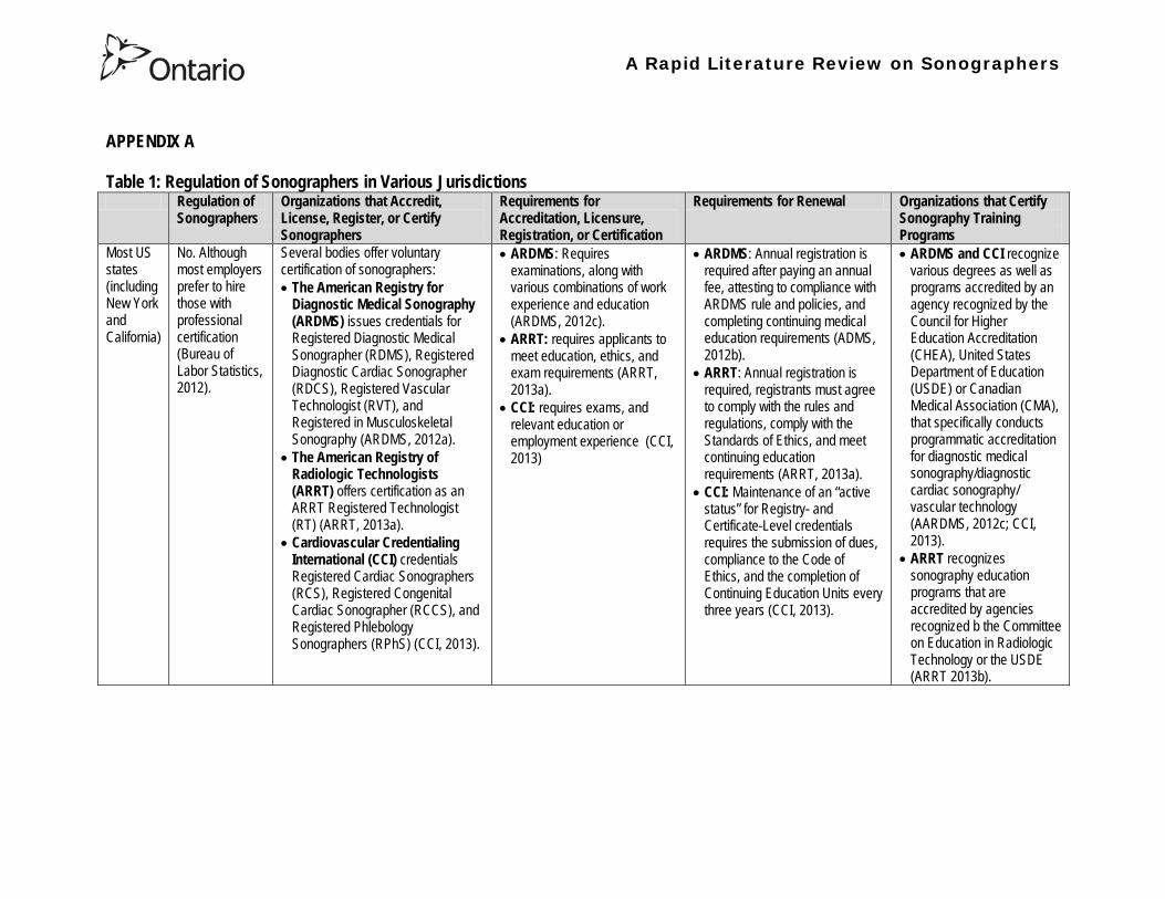
A Rapid Literature Review on Sonographers
Regulation of Sonographers
Organizations that Accredit License Register or Certify Sonographers
Requirements for Accreditation Licensure Registration or Certification
Requirements for Renewal Organizations that Certify Sonography Training Programs
Oregon Yes bull The sonographer licensure law requires Oregon sonographers to become licensed by Oregon Board of Medical Imaging (OBMI) effective July 1 2010 (SDMS 2013a)
bull Beginning on January 1 2014 all sonography licensees will be required to hold a national sonography certification credential (or be currently enrolled as a student) (SDMS 2013a)
Unknown NA
New Mexico
bull Yes On April 6 2009 the governor signed a bill that adds sonographers to the list of medical imaging professionals licensed by the state (SDMS 2013b)
bull The New Mexico Environment Department is responsible for administering the sonographer licensure program (this is because they were already managing the radiologic technology licensure program) (SDMS 2013b)
bull Under the bill sonographers are required to meet national certification standards (ie be certified by ARDMS ARRT etc) with no additional state exams beyond the national certification exams) (SDMS 2013b)
bull Two-year licenses will be issued to qualified sonographers for a fee of $100 (SDMS 2013b)
bull The bill requires the state to recognize the continuing medical education completed for national certification renewal (SDMS 2013b)
bull NA
A Rapid Literature Review on Sonographers
Regulation of Sonographers
Organizations that Accredit License Register or Certify Sonographers
Requirements for Accreditation Licensure Registration or Certification
Requirements for Renewal Organizations that Certify Sonography Training Programs
England bull No (BMUS nd)
bull Although radiographers must be registered with the Health and Care Professions Council (HCPC) (NHS nd B) and most sonographers are qualified radiographers there is no mechanism whereby an individual can register with the HCPC as a sonographer or be regulated by them as a sonographer (BMUS nd)
bull None Most sonographers train as a radiographer then undertake an approved post-registration course offered by higher education institutions (NHS nd A)
bull The courses are a minimum of one academic year Normally a pre-requisite for acceptance is access to a clinical department with supervised practice for students (NHS nd A)
bull NA bull The Consortium for the Accreditation of Sonographic Education (CASE) accredits and monitors UK sonography training programs which are delivered at the postgraduate level (ie Masters Postgraduate DiplomaCertificate in medical or clinical ultrasound) (CASE nd)
Australia bull Yes Sonographers and student sonographers are required to be accredited in order to practice (ASA 2013)
bull The Australian Sonographer Accreditation Registry [ASAR] is the regulating body (ASA 2013)
bull Accreditation of sonographers is linked to achieving (or in the case of students studying towards) an accredited entry-level qualification completion of continuing professional development and payment of an annual fee to ASAR (ASA 2013)
bull In order to remain on the Register a sonographer must satisfy continuing professional development requirements and pay an annual fee (ASAR nd A)
bull ASAR accredits sonography education courses (ASAR nd B)
A Rapid Literature Review on Sonographers
Regulation of Sonographers
Organizations that Accredit License Register or Certify Sonographers
Requirements for Accreditation Licensure Registration or Certification
Requirements for Renewal Organizations that Certify Sonography Training Programs
New Zealand
bull Yes bull The Medical Radiation Technologists Board is a health registration authority created under the Health Practitioners Competence Assurance Act 2003 that maintains the register of medical radiation technology (including sonographers) (MRTB nd A)
bull To be eligible for registration a sonographer must hold a postgraduate diploma in health science (Ultrasound) issued by the Unitec Institute of Technology (or an education approved by the Board as being substantially equivalent) (MRTB nd B)
bull Sonographers may also be required to demonstrate adequate experience in the relevant scope of practice ie a minimum of 3360 hours of clinical experience (MRTB nd B)
bull Each year sonographers must apply for and be issued with an annual practising certificate (APC)
bull Sonographer must provide evidence of enrolment and satisfactory progress in one of seven Board-approved recertification programmes (MRTB ndc)
bull The Medical Radiation Technologists Board accredits and monitors educational institutions and degrees courses of studies or programmes (MRTB nd A)
A Rapid Literature Review on Sonographers
Table 2 Examples of Point-of-Care Ultrasound Use Use Examples
Diagnostic Assessment Clinicians from diverse specialties can become very adept at using ultrasonography to examine a particular organ disease or procedure that is directly relevant to their area of expertise whereas imaging specialists typically perform more comprehensive examinations (Moore et al 2011)
bull Emergency physicians have integrated ultrasound into their repertoire for rapid assessment of emergent conditions (Hile et al 2012)
bull A 2012 review on the use of pre-hospital ultrasound concluded that medics (non-physicians trained in the delivery of medical care and the point of injury) can perform ultrasound with a high degree of accuracy (Hile et al 2012)
bull In prehospital settings ultrasound has used been by non-physicians to do fracture detection evaluation of the aorta and Focused Assessment with Sonography for Trauma (FAST) examinations (Hile et al 2012) o The aim of FAST is to assist in the trauma or bleeding patient The areas mentioned
above (perihepatic perisplenic pelvis and pericardium scanning and moreover thoracic ultrasound primarily for haemothorax) are scanned for the detection of fluid as an indication of bleeding rather than organ function (Bennett 2009)
bull Ultrasonography has been used at the Mount Everest base camp to diagnose high-altitude pulmonary edema (Moore et al 2011)
Screening Ultrasonography has been described as a screening test for cardiovascular and gynecologic disease and compact ultrasonography has been incorporated into ldquomobile screening labsrdquo (Moore et al 2011)
bull Ultrasound can be used to screen for abdominal aortic aneurysm (Moore et al 2011)
Guiding Procedures Ultrasound guidance may improve success and decrease complications in procedures performed by multiple specialties (Moore et al 2011)
bull Ultrasound can be used to guide central and peripheral vascular access thoracentesis paracentesis arthrocentesis regional anesthesia incision and drainage of abscesses localization and removal of foreign bodies lumbar puncture biopsies and other procedures (Moore et al 2011)
A Rapid Literature Review on Sonographers
Table 3 Description of the content in the articles being summarized28
No
Description Reference Review Articles
1 Autism spectrum disorders (ASDs) affect an estimated 1 of children in the United States The etiology is probably multifactorial including genetic components and exposure to infections toxins and other environmental factors particularly unfavorable perinatal and neonatal conditions There has been an increase in the frequency of diagnosis of ASDs over the last 20 years with a parallel increase in the use of obstetric diagnostic ultrasound with prenatal ultrasound exposure mentioned as the possible main etiology for autism ldquoepidemicsrdquo Central nervous system alterations have been described in ASDs and certain similar changes have been described in animals after exposure to ultrasound However analysis of in utero exposure in humans has failed to show harmful effects in neonates or children particularly in school performance attention disorders and behavioral changes There is no independently confirmed peer-reviewed published evidence that a cause-effect relationship exists between in utero exposure to clinical ultrasound and development of ASDs in childhood Ultrasound is a form of energy which effects in the tissues it traverses and its use should be restricted to medical indications by trained professionals for as short a period and as low an intensity as compatible with accurate diagnosis
Abramowicz J (2012) Ultrasound and Autism Association Link or Coincidence J Ultrasound Med 311261ndash1269
2 Over the past decade point-of-care ultrasound (US) use by nonphysician providers has grown substantially The purpose of this article is to (1) summarize the literature evaluating military medics facility at US (2) more clearly define the potential utility of military prehospital US technology and (3) lay a pathway for future research of military prehospital US The authors performed a keyword search using multiple search engines Each author independently reviewed the search results and evaluated the literature for inclusion Of 30 studies identified five studies met inclusion criteria The applications included evaluation of cardiac activity pneumothorax evaluation and fracture evaluation Additionally a descriptive study demonstrated distribution of US exam types during practical use by Army Special Forces Medical Sergeants No studies evaluated retention of skills over prolonged periods Multiple studies demonstrate the feasibility of training military medics in US Even under austere conditions the majority of studies conclude that medic can perform US with a high degree of accuracy Lessons learned from these studies tend to support continued use of US in out-of-hospital settings and exploration of the optimal curriculum to introduce this skill
Hile D C Morgan A R Laselle B T amp Bothwell J D (2012) Is Point-of-Care Ultrasound Accurate and Useful in the Hands of Military Medical Technicians A Review of the Literature Military Medicine 177(8) 983-7
28 Please note the studies programs and findings presented in this table may originate from jurisdictions with health systems that are significantly different from Ontarios If there is intent to draw heavily from one or more sources presented in this table we recommend that you contact the lead author of this review for assistance with evaluating the local applicability
A Rapid Literature Review on Sonographers
No Description Reference 3 Ultrasonography is a safe and effective form of imaging that has been used by physicians for more than half a century to aid in
diagnosis and guide procedures Over the past two decades ultrasound equipment has become more compact higher quality and less expensive which has facilitated the growth of point-of-care ultrasonography mdash that is ultrasonography performed and interpreted by the clinician at the bedside In 2004 a conference on compact ultrasonography hosted by the American Institute of Ultrasound in Medicine (AIUM) concluded that ldquothe concept of an lsquoultrasound stethoscopersquo is rapidly moving from the theoretical to realityrdquo This conference included representatives from 19 medical organizations in November 2010 the AIUM hosted a similar forum attended by 45 organizations Some medical schools are now beginning to provide their students with hand-carried ultrasound equipment for use during clinical rotations Although ionizing radiation from computed tomographic (CT) scanning is increasingly recognized as a potentially major cause of cancer ultrasonography has been used in obstetrics for decades with no epidemiologic evidence of harmful effects at normal diagnostic levels However ultrasonography is a user-dependent technology and as usage spreads there is a need to ensure competence define the benefits of appropriate use and limit unnecessary imaging and its consequences This article provides an overview of the history and current status of compact point-of-care ultrasonography with examples and discussion of its use
Moore C L amp Copel J A (2011) Point-of-Care Ultrasonography N Engl J Med 364 749-57
4 Sound travels through objects that block light Only very recently has technology advanced enough to decipher ultrasound for medical use Machines have become smaller cheaper more versatile and more advanced than ever before The medical use of ultrasound spreads across many fields so that traditional areas no longer have a monopoly With this comes the question of training doctors This has been done by various colleges and societies worldwide Some have been quicker to act than others There also needs to be an understanding of when broad experience and advanced technical skills are required or when limited skills will do In addition some procedures can be performed more safely with ultrasound where the knowledge of ultrasound is not paramount This article covers current provision of training in echo-cardiography and ultrasound in areas relevant to anaesthetists who are working in critical care (including accident and emergency) and complex surgery (mainly cardiac)
Bennett S (2009) Training guidelines for ultrasound worldwide trends Best Practice amp Research Clinical Anaesthesiology 23 363ndash373
5 Ultrasound is a commonly employed imaging modality in obstetrics and is generally regarded as safe to the fetus Current ultrasound technology however has significantly higher output potential than older machines used in most clinical studies and the safety profile for the increasing use of Doppler 3-dimensional (D) and 4-D ultrasound with modern machines is unknown This article reviews the current status of ultrasound safety within obstetrics including proposed mechanisms of harm existing scientific and clinical evidence regarding those mechanisms and considerations of safety for the clinical user
Houston L E Odibo A O amp Macones G A (2009) The safety of obstetrical ultrasound a review Prenat Diagn 29 1204ndash1212
A Rapid Literature Review on Sonographers
No Description Reference 6 The purpose of this article is to present the practicing sonographer and sonologist with an overview of the biohazards of ultrasound
and guidelines for safe use Overview Ultrasound is an imaging modality that has important diagnostic value Although useful in a variety of applications diagnostic ultrasound is particularly useful in prenatal diagnosis To date there is no evidence that diagnostic ultrasound produces harm in humans or the developing fetus when used properly There are however an increasing range of ultrasound studies being performed Newer technologies can have higher acoustic output levels than earlier equipment Also subtle or transient effects of diagnostic ultrasound such as changes in membrane permeability or neuronal migration are not completely understood Nondiagnostic Imaging Applications Nondiagnostic uses of ultrasound equipment such as repeated scans of healthy subjects for equipment demonstrations generally should be avoided First-trimester fetal scans should not use color and power Doppler modes and should not be performed for the sole purpose of producing souvenir videos or photographs Production of fetal souvenir photographs or videos during diagnostic clinical studies should not increase exposure levels or extend scan times beyond those needed for clinical purposes Operators should follow safe scanning guidelines and ALARA (as low as reasonably achievable) principles The FDA considers the promotion selling or leasing of ultrasound equipment for making ldquokeepsake fetal videosrdquo that are not part of a medical diagnostic procedure to be an unapproved use of a medical device
NelsonT R FowlkesJ B Abramowicz J S Church C C (2009) Ultrasound Biosafety Considerations for the Practicing Sonographer and Sonologist J Ultrasound Med 28139ndash150
7 Objective A possible association between ultrasound during pregnancy and subsequent non-right handedness among children has been suggested The association has been reported to be stronger among boys The aim of the present study was to explore this further through a meta-analysis of two follow-up studies of three randomized controlled trials of routine ultrasonography during pregnancy Design Handedness was assessed through a questionnaire to the parents and 4715 children at the age of 8ndash9 years were included in the meta-analysis Results There was no statistically significant difference in the prevalence of non-right handedness between the ultrasound-screened children and the controls but there was a statistically significant difference in a subgroup analysis among the boys Exploratory analyses according to exposure or non-exposure indicated an even stronger association between ultrasound and non-right handedness Conclusions A conservative analytical approach indicates no association between ultrasound in utero and subsequent non-right handedness The results from the exploratory analyses must be interpreted with caution There is still a need for further research
Salvesen K A amp Eik-Nes (1999) Ultrasound during pregnancy and subsequent childhood non-right handedness a meta-analysis Ultrasound Obstet Gynecol13 241ndash246
8 Ultrasound energy exerts important cellular genetic thermal and mechanical effects Concern about the safety of ultrasound prompted several agencies to devise regulatory limits on the machine output intensities The visual display of thermal and mechanical indices during ultrasound imaging provides an aid to limit the output of the machine Despite many animal studies no human investigations conducted to date have documented major physiologic consequences of ultrasound exposed during imaging To date ultrasound imaging appears to be safe for use in regional anesthesia and pain medicine interventions and adherence to limiting the output of ultrasound machines as outlined by the Food and Drug Administration may avoid complications in the future This article reviews ultrasound-related biologic effects the role of the regulatory agencies in ensuring safety with the use of ultrasound and the limitations and implications of ultrasound use in humans
Shankar H amp Pagel P S (2011) Potential Adverse Ultrasound-related Biological Effects A Critical Review Anesthesiology 115(5) 1109-1124
A Rapid Literature Review on Sonographers
No Description Reference 9 Objective In the context of the planned International Society of Ultrasound in Obstetrics and GynecologyndashWorld Health Organization
multicenter study for the development of fetal growth standards for international application we conducted a systematic review and meta-analysis to evaluate the safety of human exposure to ultrasonography in pregnancy Methods A systematic search of electronic databases reference lists and unpublished literature was conducted for trials and observational studies that assessed short- and long-term effects of exposure to ultrasonography involving women and their fetuses exposed to ultrasonography using B-mode or Doppler sonography during any period of pregnancy for any number of times The outcome measures were (1) adverse maternal outcome (2) adverse perinatal outcome (3) abnormal childhood growth and neurological development (4) non-right handedness (5) childhood malignancy and (6) intellectual performance and mental disease Results The electronic search identified 6716 citations and 19 were identified from secondary sources A total of 61 publications reporting data from 41 different studies were included 16 controlled trials 13 cohort and 12 casendashcontrol studies Ultrasonography in pregnancy was not associated with adverse maternal or perinatal outcome impaired physical or neurological development increased risk for malignancy in childhood subnormal intellectual performance or mental diseases According to the available clinical trials there was a weak association between exposure to ultrasonography and non-right handedness in boys (odds ratio 126 95 CI 103ndash154) Conclusion According to the available evidence exposure to diagnostic ultrasonography during pregnancy appears to be safe
Torloni M R Vedmedovska Merialdi M Betran A P Allen T et al (2009) Safety of ultrasonography in pregnancy WHO systematic review of the literature and meta-analysis Ultrasound Obstet Gynecol 33 599ndash608
Articles in Peer-Reviewed Journals 10 It has been documented for many years that as a result of use of ultrasound to determine the sex of the fetus the male to female
ratio in countries such as China and India have become increasingly skewed (due to subsequent abortions and infanticide of females) Preference for sons over daughters evident in Chinas and South Asias male sex ratios is commonly rationalized by poverty and the need for old-age support In this article the authors study South and East Asian immigrants to Canada a group for whom the economic imperative to select sons is largely absent Analyzing the 2001 and 2006 censuses 20 percent samples the authors find clear evidence of extensive sex selection in favor of boys at higher parities among South and East Asian immigrants unless they are Christian or Muslim The latter finding accords with the explicit prohibition against (female) infanticidemdashtraditionally the main sex-selection methodmdashin these religions The findings point to a strong cultural component to both the preference for sons and the willingness to resort to induced abortion based on sex
Almond D Edlund L Milligan K (2013) Son Preference and the Persistence of Culture Evidence from South and East Asian Immigrants to Canada Population and Development Review 39(1) 75ndash95
11 The rapid growth in the use of ultrasound as a diagnostic imaging technology over the past forty years has led to a demand for a workforce with the appropriate skills to perform and interpret the scans In the UK ultrasound investigations now comprise the largest group of all diagnostic imaging examinations However there remains no statutory regulation of the practice of sonography in the UK and little recognition of the considerable training that many practitioners have undertaken to obtain the skills to become safe and competent sonographers Many in the field consider that this should change and are working to obtain professional status for the practice of sonography Although the Health Care Professions Council (HCPC) has recommended regulation of sonography practice this is unlikely to happen in the near future This paper discusses the evolution of sonography practice and explores some of the complex issues associated with the professionalisation of sonography
Gibbs V (2013) The long and winding road to achieving professional registration for sonographers Radiography19 164-167
A Rapid Literature Review on Sonographers
No Description Reference 12 Background Contrast echocardiography (which uses contrast agents injected into the blood flow in order to visualize the blood flow
in very small blood vessels) has been shown to improve diagnostic quality especially in technically difficult patients However the learning curve and increased time for preparation and image acquisition have led to low use Methods The authors sought to determine whether the contrast echocardiography procedure performed independently by a specialized trained sonographer could improve efficiency In one centre routine echocardiograms were scheduled for one hour and any study exceeding one hour would result in patient booking cancellations The authors compared the standard of care in which a physician or nurse administers echocontrast with a sonographer-administered program (SAP) Results The time to complete contrast echocardiograms was significantly reduced by the SAP strategy (43 min 17 s plusmn 23 min 42 s vs 1 h 1 min 6 s plusmn 31 min 0 s P lt 0001) Subgroup analysis of the inpatients and outpatients demonstrated similar results Only 10 of studies (6 of 61) in the SAP exceeded 60 minutes compared with 45 (34 of 76) in the standard-of-care group (P lt 0001) Based on study volumes in the centre the net improvement in productivity with the SAP could be up to 53 annually Conclusion Sonographer-administered echocontrast is feasible and potentially removes a barrier to implementation of contrast echocardiography
Tang A Chiew S K Rashkovetsky R Becher H Choy J B (2013) Feasibility of Sonographer-Administered Echocontrast in a Large-Volume Tertiary-Care Echocardiography Laboratory Canadian Journal of Cardiology 29 391ndash395
13 Ultrasound scanning has been used as a diagnostic and screening tool in obstetric practice for over 50 years There is no evidence of immediate or long-term harm to the developing fetus from exposure to B mode ultrasound However exposure to high levels of Doppler ultrasound during early development is increasingly common and the full safety implications of this exposure are not clear Doppler ultrasound exposure in utero gives rise to increased apoptosis in animal models and there is evidence of the effects of exposure to Doppler ultrasound persisting throughout life with increased non-right-handedness observed in human epidemiological studies The authors consider the idea that there may be long-term developmental implications for fetuses exposed to Doppler ultrasound early in gestation These effects may be mediated via thermal or mechanical disruption to the developing conceptus giving rise to free radical damage Excess free radical exposure early in gestation is a strong candidate for the final common pathway underlying developmental programming effects and gives rise to concern that fetuses exposed to high levels of ultrasound are at risk of a developmental programming effect It is suggested that there is a need for animal studies of developmental programming using exposure to Doppler ultrasound scanning as the exposure of interest and for more observational data to be collected in the clinical setting While these data are collected it seems prudent to continue to adhere to the principle of lsquoas low as reasonably achievablersquo (ALARA) when exposing first-trimester fetuses to Doppler ultrasound
Aiken CE Lees CC (2012) Long-term effects of in utero Doppler ultrasound scanning ndash A developmental programming perspective Medical Hypotheses 78 539ndash541
14 Point of care sonography by emergency physicians in the emergency department can be an effective aid in the diagnosis and management of patients presenting with a variety of medical and traumatic conditions Its use can improve patient outcomes enhance patient safety speed patient disposition and save lives CAEP supports the use of point of care sonography
Canadian Assocation of Emergency (CAEP) Physicians Ultrasound Position Statement Working Group (2012) Use of point of care sonography by emergency physicians CJEM 14(2)106-112
A Rapid Literature Review on Sonographers
No Description Reference 15 The use of three-dimensional echocardiography (3DE ie a 3D ultrasound of the heart) during electrophysiology (EP - a branch of
cardiology that deals with the diagnosis and treatment of heart rhythm disorders) procedures is the end product of years of growth in two diverse cardiology subspecialties namely advanced cardiac imaging and the EP During the past decade progress in both fields has resulted in many important advances that have culminated in their union for a new area of growth and development Imaging advances have provided the cardiovascular specialist with enhanced cardiac volume and function data and more recently 3DE capabilities with improved spatial and temporal resolution providing unprecedented spatial relationships This latter development is valued by EP specialists in need of hitherto never required anatomic knowledge as they press forward with extraordinary expansion in their capabilities It makes sense that by combining these two rapidly growing subspecialties future capabilities in patient care may be achieved that would otherwise not be possible This paper discusses the value of 3DE during EP procedures and offers the readers insight into this novel multispecialty hybrid arena Using this model as a template it is likely that the readers may identify other areas within their practices where periprocedural advanced imaging may afford significant dividends in patient outcomes
Moukabary T Faletra F F Kronzon I Thomas W amp Sorrell V L (2012) Three-Dimensional Echocardiography in the Electrophysiology Laboratory Echocardiography 29 117-122
16 Background There has been much discussion about whether female feticide occurs in certain immigrant groups in Canada The authors examined data on live births in Ontario and compared sex ratios in different groups according to the motherrsquos country or region of birth and parity Methods The authors completed a population-based study of 766 688 singleton live births between 2002 and 2007 They used birth records provided by Ontario Vital Statistics for live births in the province between 23 and 41 weeksrsquo gestation They categorized each newborn according to the motherrsquos country or region of birth namely Canada (n = 486 599) Europe (n = 58 505) South Korea (n = 3663) China (n = 23 818) Philippines (n = 15 367) rest of East Asia (n = 18 971) Pakistan (n = 18 018) India (n = 31 978) rest of South Asia (n = 20 695) and other countries (n = 89 074) They calculated malefemale ratios and 95 confidence intervals (CIs) for all live births by these regions and stratified them by maternal parity at the time of delivery (0 1 2 or ge 3) Results Among infants of nulliparous women (women with no previous children) the malefemale ratio was about 105 overall As parity (the number of previous children) increased the ratio remained unchanged among infants of Canadian-born women In contrast the malefemale ratio was significantly higher among infants of primiparous women (women with one previous child) born in South Korea (120 95 CI 109ndash134) and India (111 95 CI 107ndash115) than among infants of Canadian-born primiparous women Among multiparous women (women with 2+ previous children) those born in India were significantly more likely than Canadian-born women to have a male infant (parity 2 ratio 136 95 CI 127ndash 146 parity ge 3 ratio 125 95 CI 109ndash143) Interpretation The study of malefemale ratios in Ontario showed that multiparous women born in India were significantly more likely than multiparous women born in Canada to have a male infant
Ray J G Henry D A Urquia M L (2012) Sex ratios among Canadian liveborn infants of mothers from different countries CMAJ184(9) E92-96
A Rapid Literature Review on Sonographers
No Description Reference 17 This letter consists of a case study that illustrates how a cross-disciplined sonographer (educated in cardiac and vascular ultrasound)
can quickly and efficiently and with great Cost-effectiveness deliver timely care to a patient in certain clinical settings The literature is comparatively sparse in referencing clinical diagnostics in cross-discipline sonographers Echocardiographers must be clinicians fully comfortable with evaluating the circulatory dynamics of the body from the left and right heart to the peripheral arteries and veins including the extracranial circulation (carotid arteries) Included in this skill set is substantive understanding of the ECG patientrsquos history and integration of cardiac and vascular findings Limitations of each diagnostic tool and how these limitations can be overcome by the strengths of the other examination are important For the dual-skilled sonographer only experience continuous education and constant clinical feedback will lead to the independent decision making that is counted on by the physician The American Registry for Diagnostic Medical Sonography offers credentials in cardiac and vascular imaging (RDCS and RVT) Cardiovascular Credentialing International offers credentials in cardiac and vascular imaging as well (registered cardiac sonographer [RCS] and registered vascular specialist [RVS]) All imaging facilities should consider merging cardiac and vascular noninvasive imaging with dual-registered clinical sonographers credentialed by one of these two agencies
Zemanek M (2012) What It Means to Be a Cardiovascular Sonographer Journal of Ultrasound in Medicine 311859-1862
18 Diagnostic medical sonography is viewed as a safe imaging modality However bioeffects research has yet to define the effects of exposure to sonography in humans Therefore sonographers must be aware of potential risks and practice ALARA (ldquoas low as reasonably achievablerdquo) routinely This research explores sonographersrsquo ability to answer basic questions about the bioeffects of sonography as well as their knowledge and use of the safety indices called the mechanical index (MI) and thermal index (TI) Members of the Society of Diagnostic Medical Sonographers in the US a southwestern sonographersrsquo society and a sonography community on Facebook 2010 were surveyed (n = 212) There is a low level of bioeffects knowledge and minimal safety practices among sonographers in this survey No significant relationship was found between sonographer knowledge of the bioeffects of sonography years in the profession or whether the sonographers monitor the MITI
Bagley J Thomas K DiGiacinto D (2011) Safety Practices of Sonographers and Their Knowledge of the Biologic Effects of Sonography Journal of Diagnostic Medical Sonography 27(6) 252ndash261
19 This article highlights trends in echocardiography Point of care ultrasound (hand-carried ultrasound) Initially point of care ultrasound will increase the use of echocardiography in the same manner that cardiac CT increased catheterizations However once the basics are learned it has the potential to be a very powerful tool in that less unnecessary studies will be ordered In the clinic small systems will be used as a lsquolsquofirst lookrsquorsquo screen and become part of the patientrsquos history only patients for whom more testing is necessary will be referred for a comprehensive study This is likely to reduce the number of Doppler studies requested The miniaturization of echo equipment will continue with improved qualityfunctionality of the lsquolsquohand-heldrsquorsquo echo machines to the extent that cordless probes will become a reality sooner rather than later Role in interventional echocardiography Sonographers will be spending much more time in the interventional and electrophysiology (EP) labs There will be more interconnection with interventional cardiologists and the volume will increase as more devices are implanted more percutaneous valves placed and more robotic surgery is performed Crosstraining and multitasking Sonographers entering the profession would be prudent to learn cardiac and vascular imaging Individuals who multitask andor cross-train will be ideal For example a sonographer who starts an IV monitors ECG stress can perform cardiac MRI and can interpret a preliminary report or a nurse who can do an echo monitor sedation and start an IV will be invaluable The sonographer should continue to grow professionally by cross training as institutions try to minimize costs and maximize throughput
McCulloch M L (2011) Thoughts regarding the future of sonography Journal of the American Society of Echocardiography 24(5) 29A-30A
A Rapid Literature Review on Sonographers
No Description Reference 20 Background Radiology reports provide specialist interpretation of images and relate these findings to the patientrsquos symptoms and
signs This study compared ultrasound (USS) reports generated by radiologists and sonographers to determine if any significant or clinically relevant differences existed Methods A retrospective analysis of 624 consecutive USS reports was carried out The reports were assessed for the presence of a lsquodisclaimerrsquo or lsquocaveatrsquo pertaining to the quality of the images and were analysed with respect to the clarity of their wording and ability to answer the clinical request Results The majority of sonographer USS reports contained a disclaimer regarding the quality of the USS images (571) compared with a smaller proportion of radiologist scans (99) (P lt 0001) Overall radiologists performed significantly better in providing a clear negative or positive diagnosis to the clinical question on the request form when compared with sonographers (885 vs 654 P lt 0001) Conclusion lsquoDisclaimerrsquo comments and lsquohedgingrsquo must be avoided in radiology reporting While feedback as to the accuracy of the radiology images is important overuse of such terms undermines the validity of the radiology report
Garcea G Mahmoud A Ong S L Rees Y Berry Det al (2010) Caveat reporting in ultrasound interpretation of surgical pathology a comparison of sonographer versus radiologist Journal of Evaluation in Clinical Practice 16 97ndash99
21 A nine-point questionnaire was constructed based on previously published work by Marsal and Sheiner et al The survey was distributed to clinical users of ultrasound practicing sonography in Hamilton (New Zealand) during July 2010 totalling 35 users consisting of sonographers sonography trainees one obstetric sonologist and one fetal medicine registrar Results Twenty sonographers returned the questionnaire which represents survey response of 61 The level of clinical experience was equally split between junior sonographers of lt 5 yearsrsquo experience (30) sonographers with 5-10 yearsrsquo experience (35) and senior sonographers of gt10 yearsrsquo experience (35) While 100 of the sonographers surveyed reported being familiar with the term Thermal Index (TI) and 90 claimed they adhered to the Action Learning Action Response Association (ALARA) principle all the time or most of the time only 15 of sonographers correctly described or defined what the term TI means Of the sonographers 80 knew where to find the TI during real-time examinations but only 35 knew which TI index to use for nuchal translucency (NT) scan and only 10 knew the expected range of TI encountered during routine obstetric examinations Of the eight sonographers who reported adhering to ALARA all the time the majority (64) could not describe or define TI In the same subgroup the majority (64) did not know which TI to monitor during a NT scan Only three of 20 (15) sonographers knew what the term TI means Of these two were junior sonographers with lt 5 yearsrsquo experience and one had 5ndash10 yearsrsquo experience Despite understanding the term TI one of the three sonographers still selected the incorrect index to monitor during a NT scan Not one of the seven senior sonographers with gt 10 years of experience could define or describe the TI In total only 10 of sonographers knew what the TI means and which TI to monitor during NT scanning
Necas M (2010) New Zealand sonographers do not outperform their European or American colleagues in the knowledge of ultrasound safety Australasian Journal of Ultrasound in Medicine13(4) 28-32
A Rapid Literature Review on Sonographers
No Description Reference 22 Musculoskeletal (MSK) ultrasound performed by non-medical practitioners is a developing practice The aim of this paper is
(1) to audit an experienced sonographerrsquos (SJ Riley one of the authors) performance after one yearrsquos independent reporting against two experienced MSK radiologists (CJ Grove amp M Chandramohan the other two authors) and (2) to establish an audit standard against which such role development might be contrived Images and reports from 250 MSK ultrasound examinations performed by a sonographer were reviewed independently by two consultant MSK radiologists The examinations were graded for discrepancy eg grade 1 ndash agree with the sonographer report grade 2 ndash minor discrepancy unlikely to alter patient care grade 3 ndash potentially significant discrepancy grade 4 ndash definite significant discrepancy likely to have adverse consequences for patient care Two of 250 (08) cases were excluded Both radiologists agreed completely with the sonographer (grade 1) in 235 of the 248 cases (948) In 13 cases there was discrepancy between the reports of SJR and the radiologists The discrepancy was grade 2 in six of the 248 cases (24) grade 3 in six of the 248 cases (24) and grade 4 in one of the 248 cases (04) In conclusion this audit shows a high level of agreement between the sonographer and the consultant MSK radiologist reporting of MSK ultrasound This level of agreement may set the standard for future quality assurance audit of sonographer MSK reporting
Riley S J Groves C J Chandramohan M (2010) Musculoskeletal ultrasound audit of sonographer reporting Ultrasound 18 36ndash40
23 Background Growing demand for ultrasound examinations and higher quality requirements motivate searching for routines combining the diagnostic accuracy of radiologist-performed examinations with the economical advantages of sonographer-performed examinations One possible approach is to use strictly standardized acquisition and documentation schemes that give the radiologist access to all relevant information after the examination Purpose To compare a recently introduced routine combining acquisition by a radiographer (more specifically a sonographer with two years experience) documentation as standardized cine-loops and review by a radiologist (lsquostandardized methodrsquo) with the formerly used routine where the diagnosis is made bedside by the radiologist (lsquotraditional methodrsquo) Material and Methods In 64 policlinic patients the kidneys (n = 27) or the gallbladder (n = 37) were examined with both the standardized and the traditional method The radiologistsrsquo findings of hydronephrosis tumors cysts echogenicity changes and cortical thickness (in the kidneys) and wall thickness concrements and polyps (in the gallbladder) were compared between the methods with respect to agreement (proportion of agreement and kappa coefficient) as well as systematic differences (McNemarrsquos test) Results The findings at the gallbladder examination showed a median agreement of 97 (86ndash100 kappa = 064ndash100) and those at the kidney examination an agreement of 90 (78ndash100 kappa = 069ndash 100) There were no significant systematic differences between the methods Conclusion The satisfactory agreement in this preliminary study indicates that the new workflow with ultrasound examinations performed by a radiographer (sonographer) and analyzed off-line by a radiologist is promising and motivates further studies
Stenman C Thorelius L Knutsson A amp Smedby Ouml (2010) Radiographer-acquired and radiologist-reviewed ultrasound examination ndash agreement with radiologistrsquos bedside evaluation Acta Radiologica 52 70ndash74
A Rapid Literature Review on Sonographers
No Description Reference 24 The troubling practice of fetal sex selection has historically been considered an Asian phenomenon However recent evidence shows
that a similar situation is emerging in North America albeit on a smaller scale The Society of Obstetricians and Gynaecologists of Canada (SOGC) has firmly stated its opposition to sex selection for non-medical reasons as well as to the use of any technology used solely for the purpose of determining fetal sex However because fetal sex may be disclosed to the parents at the time of ultrasound examination if they request this information guidance for health care professionals to assist in discouraging fetal sex selection would be useful Because no declaration of motives or reasons is required when a woman seeks a termination of pregnancy the authors suggest that health care professionals need not disclose the sex of a fetus until it has reached a gestational age at which abortion for non-medical purposes would not be possible This proposal would facilitate consistency between clinical practice and the values of Canadian citizens the SOGC the Canadian Medical Association and other professional organizations while still respecting current laws pertaining to disclosure of patient information and patientsrsquo rights to autonomous decision-making
Thiele A T amp Leier B (2010) Towards an Ethical Policy for the Prevention of Fetal Sex Selection in Canada J Obstet Gynaecol Can 32(1) 54ndash57
25 Background The extent to which increases in the use of prenatal ultrasonography over time reflect changes in maternal risk (ie an increase in high-risk pregnancies over time) is unknown In this population-based study the authors examined the use of prenatal ultrasonography from 1996 to 2006 in Ontario Methods With fiscal year 199697 as the baseline the authors evaluated the relative risk (RR) and 95 confidence interval (CI) for the change in rates of ultrasonography for each subsequent year The RR was adjusted for maternal age income rural residence maternal comorbidities receipt of genetics consultation or amniocentesis mdash all in the index pregnancy mdash and history of complications in a prior pregnancy Results The study sample consisted of 1 399 389 singleton deliveries The rate of prenatal ultrasonography increased from 2055 per 1000 pregnancies in 1996 to 3264 per 1000 in 2006 (adjusted RR 155 95 CI 154ndash155) The rate increased among both women with low-risk pregnancies (adjusted RR 154 95 CI 153ndash155) and those with high-risk pregnancies (adjusted RR 155 95 CI 154ndash157) The proportion of pregnancies with at least four ultrasound examinations in the second or third trimesters rose from 64 in 1996 to 187 in 2006 (adjusted RR 268 95 CI 261ndash274) Paradoxically this increase was more pronounced among low-risk pregnancies (adjusted RR 292 95 CI 283ndash301) than among high-risk pregnancies (adjusted RR 225 95 CI 216ndash235) Interpretation Substantial increases in the use of prenatal ultrasonography over the past decade do not appear to reflect changes in maternal risk Nearly one in five women now undergo four or more ultrasound examinations during the second and third trimesters Efforts to promote more appropriate use of prenatal ultrasonography for singleton pregnancies appear warranted
You J J Alter D A Stukel T A McDonald S D Laupacis A et al (2010) Proliferation of prenatal ultrasonography CMAJ 182(2) 143-151
A Rapid Literature Review on Sonographers
No Description Reference 26 Aims The aim of this study was to assess the cost-effectiveness of using certified sonographers and miniaturized echocardiography
systems to perform echocardiograms at bedside in comparison to moving inpatients from the admission department to the echocardiography laboratory (echo-lab) Methods and Results From 26 September 2005 to 27 October 2005 112 patients admitted in six hospital wards in Italy connected through a 100 Mbit LAN to the echo-lab were scanned within the admission ward by sonographers using a miniaturized echo system Logistical data were collected and results were compared with those obtained from 194 consecutive patients coming from the same wards and studied in the echo-lab with high-end machines between 8 March 2005 and 15 April 2005 Performing echocardiograms in the admission department avoided long waiting time of the inpatients in the echo-lab before and after the study increased the percentage of patients studied within 3 and 5 days from request (88 vs 77 and 100 vs 95 respectively P = 003) increased both sonographer (by 339 P lt 0001) and echo-lab productivity (by 41 P lt 0001) and reduced costs of echocardiograms by 29 Conclusion Implementation of digital echocardiography certified sonographers and a miniaturized echo system allowed improvement of the cost-effectiveness of the service provided by the echo-lab for inpatients and avoided patientsrsquo discomfort derived from prolonged waiting time before and after the exam
Badano L Gaetano N Stacul S Gianfagna P Pericoli M et al (2009) Improved workflow sonographer productivity and cost-effectiveness of echocardiographic service for inpatients by using miniaturized systems European Journal of Echocardiography 10 537ndash542
27 The present report is a proposal from the American Society of Echocardiography Advanced Practice Task Force that identifies the potential of cardiac sonographers to achieve the Advanced Cardiovasculat Sonographer (ACS) level According to this report echocardiographic examinations require a well-trained and competent sonographer to obtain proper anatomic and physiologic data to establish an accurate diagnosis for clinical decision-making and patient management Although the formal education and training of cardiovascular sonographers are evolving many entry-level and staff sonographers may not have sufficient practical or clinical knowledge of the necessary components of the echocardiographic study for the individual patientrsquos clinical presentation In many clinical settings echocardiograms are read after the patient has left the laboratory Thus there is a role for a sonographer who can practice at an advanced level in a cardiovascular ultrasound laboratory to ensure a proper echocardiographic examination is performed on every patient In this setting an Advanced Cardiovascular Sonographer (ACS) would be able to review the indication for and quality of the examination If additional images were needed the ACS would assist the sonographer in obtaining these images which would lead to the performance of a complete and fully diagnostic examination before the patient had left the echocardiography laboratory In clinical practice the quality of the examinations performed would improve advancements in echocardiographic methods could be taught and incorporated into daily practice and patients would be better served
Mitchell C Miller F A Bierig S M Bremer M L Ehler D et al (2009) Advanced Cardiovascular Sonographer A Proposal of the American Society of Echocardiography Advanced Practice Sonographer Task Force J Am Soc Echocardiogr 22 1409-13
A Rapid Literature Review on Sonographers
No Description Reference 28 Sonographer practitioner development involves the expansion and extension of the sonographer role to include reporting on
ultrasound examinations Australian sonographers have not seen the same degree of role extension and expansion as their counterparts in the United Kingdom despite increasing levels of discussion regarding sonographer practitioner development The aim of this study was to determine if Australian sonographers want to extend their professional role and what they consider are the important issues associated with role extension This paper reports on qualitative data derived from a survey of Australian sonographers and investigates if Australian sonographers are interested in extending and expanding their professional role and responsibilities and if they do what might be necessary or desirable from a professional point of view for this change to occur A survey was mailed to all members of the Australian Sonographers Association (ASA) in October 2006 The 31-item survey included 28 closed-ended and three opened-ended items to provide both quantitative and qualitative data The quantitative data will be reported separately Qualitative data was derived from responses to the opened-ended questions which asked respondents to elaborate on their attitudes and feelings about role extension and development Analysis used Nvivo7 software to aid in uncovering common themes from the qualitative data The analysis focused on the reported incentives or motivations for becoming a sonographer practitioner as well as disincentives or perceived hurdles that would discourage respondents from becoming sonographer practitioners The three most reported incentives or motivations for becoming a sonographer practitioner were professional recognition remuneration and increased knowledge The three most commonly reported disincentives or perceived hurdles that would discourage respondents from becoming sonographer practitioners were legal issues insurance and further study
McGregor R OrsquoLoughlin K Cox J Clarke J Snowden A (2009) Sonographer practitioner development in Australia Qualitative analysis of an Australian sonographersrsquo survey Radiography15 313-319
29 Introduction This study describes the effect of introducing a cardiac sonographer led follow up clinic within a hospital cardiology department for patients with stable heart valve disease The two years before and after the instigation of the valve clinic were audited Materials and methods The clinic was conducted in a single centre and undertaken in the cardiology department of a district general hospital A total of 382 patients with 397 clinically significant valve lesions but for whom surgery was not yet indicated but follow up required were seen in a cardiac sonographer run clinics These patients no longer attended a medical follow up clinic unless there was clinical or echocardiographic deterioration Effectiveness was judged by the percentage treated according to current best practice guidelines the number of echocardiograms performed and the number of hospital outpatient visits attended In addition mortality data for the subjects in the clinic was collected Results The proportion followed up according to best practice guidelines rose from 157 (41) to 354 (92) (p lt 001) The total number of echocardiograms performed fell from 807 to 550 Total number of outpatient visits fell from 998 to 31 A total of 11 patients died in the two year study period none from progressive valve disease Discussion This study demonstrates that a protocol driven sonographer led heart valve disease follow up clinic significantly improved the quality of follow up while bringing about a major reduction in outpatients visits without compromising patient safety
Taggu W Topham A Hart L Carr-White G Sulke N et al (2009) A cardiac sonographer led follow up clinic for heart valve disease International Journal of Cardiology 132 240 ndash243
30 Role development for imaging allied health professionals has progressed significantly over the last three decades with many major developments pioneered in the field of ultrasound This paper reviews the developing role of the radiographer in ultrasound (in some countries such as the UK sonographers are considered a type of radiographer) and some of the clinical professional and educational implications of such development As the published evidence base underpinning the latest developments is scarce several contemporary case study examples which have been presented verbally at study days attended by the authors have been included In conclusion sonographer role development is considered within the professional contexts of extended role advanced and consultant practice
Hart A amp Dixon A (2008) Sonographer Role Extension and Career Development a Review of the Evidence Ultrasound 16(1) 31-35
A Rapid Literature Review on Sonographers
No Description Reference 31 Objective The main goal of this study was to determine end usersrsquo knowledge regarding safety aspects of diagnostic ultrasound
during pregnancy End usersrsquo attitudes toward the use of ultrasound in low-risk pregnancies were also assessed Methods A questionnaire was distributed to ultrasound end users attending review courses and hospital grand rounds between April and June 2006 in Chicago Results One hundred thirty end users completed the questionnaires (63 response rate) Sixty-three percent were physicians (n = 84) most of them obstetricians (817) About 18 of participants routinely performed Doppler ultrasound examinations during the first trimester Fifty percent of end users thought that the number of ultrasound examinations in low-risk pregnancy should be limited to 1 to 3 (mean plusmn SD 26 plusmn 09) Almost 70 disapproved of ldquokeepsakeentertainmentrdquo ultrasound Although 322 of the participants were familiar with the term thermal index only 177 actually gave the correct answer to the question on the nature of the thermal index About 22 were familiar with the term mechanical index but only 38 described it properly Almost 80 of end users did not know where to find the acoustic indices Only 208 were aware that they are displayed on the sonographic monitor during the examinations End users with higher knowledge of safety issues thought that there should be limitations on the number of ultrasound examinations in low-risk pregnancies (odds ratio 33 95 confidence interval 11ndash100 P = 028) Likewise these end users were more likely to respond that ultrasound might have adverse effects during pregnancy (odds ratio 32 95 confidence interval 11ndash125 P = 045) Conclusions Ultrasound end users are poorly informed regarding safety issues during pregnancy Further efforts in the realm of education and training are needed to improve end user knowledge about the acoustic output of the machines and safety issues
Sheiner E Shoham-Vardi I Abramowicz J S (2007) What Do Clinical Users Know Regarding Safety of Ultrasound During Pregnancy J Ultrasound Med 26 319ndash325
A Rapid Literature Review on Sonographers
No Description Reference 32 In an attempt to survey the knowledge among ultrasound users of some safety aspects of diagnostic ultrasound a questionnaire was
distributed to professionals using ultrasound for fetal examinations The questionnaire was answered by the participants and faculty members of six postgraduate courses in obstetric ultrasound Doppler ultrasound and fetal echocardiography given in Sweden Norway and Austria in 2003 Furthermore in Sweden the questionnaire was distributed to the staff meeting participants of two major obstetric ultrasound units and of one unit for fetal echocardiography and at a meeting of the Working Group for Ultrasound of the Swedish Society of Obstetrics and Gynecology In total 199 questionnaires were anonymously answered by doctors (n = 145) sonographers (n = 22) and midwives trained in ultrasound (n = 32) All of them were using diagnostic ultrasound on a daily or weekly basis The respondents were from nine European countries In none of the countries is formal certification for the use of diagnostic ultrasound in pregnancy or otherwise required The doctors who answered the questionnaire were specialists in obstetrics and gynecology (including some of the internationally recognized experts in fetal ultrasound) pediatric cardiology radiology or clinical physiology The respondents were not informed about the questionnaire in advance and they had a limited time to answer the 16 questions typically 10ndash15 min The questions concerned explanation of the safety indices and their relation to various ultrasound modes ie real-time B-mode M-mode three-dimensional (3D) spectral Doppler color and power Doppler ultrasound The respondents were also asked which ultrasound system they used most often and where on the system the information on thermal index (TI) and mechanical (MI) was displayed (eg in the top right corner of the screen in a separate window etc) Furthermore the respondents had to indicate how they adjust the output energy level on their own machine For all questions there was the optional answer lsquoDonrsquot knowrsquo Results About one-third of the ultrasound experts were able to define the abbreviations TI and MI For the subclasses of the TI the correct answer was found in only 3-8 of cases Of those who knew what the TI and MI meant only two-thirds and one-third of respondents respectively were able to give a simple explanation of the indices In 49 of questionnaires the Doppler modes were correctly ranked as generally producing higher ultrasound exposure than the imaging modes (B-mode 3D) Some 28 of the respondents correctly indicated where on their own machine the information on safety indices is displayed However not all of them knew how to control the output energy level For all items in the questionnaire there were no significant differences in the results between the three categories of respondents (ie physicians sonographers and midwives) The results of the questionnaire were anything but encouraging and they indicated that the users who are supposed to be responsible for controlling exposure of the fetus to ultrasound had a very poor knowledge of the basic safety aspects of ultrasound
Maršaacutel K (2005) The output display standard has it missed its target Ultrasound Obstet Gynecol 25 211ndash214
33 Purpose This study was performed to evaluate the accuracy of upper abdominal ultrasound (US) scanning performed by sonographers in a district general hospital to identify potential areas of weakness and to make recommendations to improve the service Materials and Methods Upper abdominal US examinations performed and reported by sonographers over a 4-week period were retrospectively reviewed The accuracy of the imaging findings and reports were assessed against other imaging surgical histological or laboratory findings and against clinical outcome Results A heterogenous group of 104 patients were included in the study 62 of whom had an US abnormality Errors of scanning or interpretation were identified in 10 patients (96) of whom five (48) were felt to be potentially significant Conclusions The sonographersrsquo accuracy in reporting upper abdominal US scans was 90 However on the basis of this study the authors have implemented specific recommendations to improve the quality of the service
Dongola N Guy R Ward S amp Giles J A (2003) Can sonographers offer an accurate upper abdominal ultrasound service in a district general hospital Radiography 9 29ndash33
A Rapid Literature Review on Sonographers
No Description Reference 34 Aim To compare the accuracy of radiographers and radiologists in routine abdominal ultrasound Materials and Methods One
hundred consecutive patients attending for routine abdominal ultrasound were included Each patient was examined by both a radiographer and radiologist Both operators noted their fndings and wrote a concluding report without conferring Reports were compared Where there was disagreement the patient was either re-examined by another radiologist or had further investigation Results Of 100 patients 52 were men and 48 were women The age range was 19plusmn88 years (median 52 years) Thirty-seven patients had renal tract ultrasound one had an aortic ultrasound and 62 had general upper abdominal ultrasound In 44 cases both operators reported the examination as normal In 49 cases both operators reported the examinations as abnormal and there was complete agreement between the operators In seven cases there was not complete agreement between operators Three of these disagreements were considered minor and four major In three of the seven cases the radiographer was correct and in four the radiologist was correct Conclusion Experienced radiographers and radiologists are highly accurate in performing and interpreting routine abdominal sonography Both operators missed a small minority of abnormalities There was no statistically significant difference in the accuracy of radiographers and radiologist
Leslie A Lockyer H amp Virjee J P (2000) Who Should be Performing Routine Abdominal Ultrasound A Prospective Double-Blind Study Comparing the Accuracy of Radiologist and Radiographer Clinical Radiology 55 606-609
Grey Literature 35 The ARRT The American Registry of Radiologic Technologists tests certifies and annually registers more than a quarter-of-a-million
radiologic technologists to promote high standards of patient care They award the Registered Technologist or RT designation to individuals who have completed the prescribed classroom and clinical education passed the appropriate exam and met the ethics requirements And they ensure their continuing education and ongoing compliance with their high standards by requiring annual registration of their certificate Certification by ARRT is voluntary not mandatory But employers state licensing agencies and federal regulators view ARRTrsquos credential as an indicator that a person has met recognized national standards for medical imaging and radiation therapy professionals How Are Radiologic Technologists Deemed Qualified First comes initial certification Certification is the process of initially recognizing individuals who have satisfied certain standards within a profession A person is certified by ARRT after completing an approved educational program complying with the ethics standards and passing a certification exam Then comes annual registration Registration is the yearly procedure required to maintain certification ARRT registrants are those who having already fulfilled the requirements for initial certification continue to meet the requirements for annual registration The ARRT annually registers the certificates of individuals who meet the following three criteria agreeing to comply with the ARRT Rules and Regulations continuing to comply with the ARRT Standards of Ethics and meeting our continuing education requirements Only technologists who are currently registered (which means that they have renewed within the past year) may designate themselves as ARRT Registered Technologists and use the initials RT after their names Federal laws regulate personnel qualifications in some modalities (mammography for example) but they accredit the health care facility rather than licensing the individual radiologic technologist who is providing patient care And while many states have laws regulating who can perform radiologic procedures they are typically based on ARRT certification Some employers and state agencies may require ARRT certification but not necessarily current registration Unless they require current registration by ARRT employers cannot assume that the person has complied with the ethics requirements or the continuing education requirements
American Registry of Radiologic Technologists (ARRT) (2013a) Patient Page Accessed April 2013
A Rapid Literature Review on Sonographers
No Description Reference 36 This document outlines certification requirements for sonography There are two pathways (primary and post-primary) For the
primary pathway it notes that sonography educational programs must be accredited by a mechanism acceptable to the ARRT ARRT generally recognizes only accreditation agencies that are recognized by the Council for Higher Education Accreditation andor United States Department of Education
American Registry of Radiologic Technologists (ARRT) (2013b) Sonography Certification Accessed April 2013
A Rapid Literature Review on Sonographers
No Description Reference 37 In Australia sonographers and student sonographers are required to be accredited by Australian Sonographer Accreditation Registry
(ASAR) in order to practise This requirement is contained within the Australian Governmentrsquos Diagnostic Imaging Accreditation Scheme standards and Medicare legislation Accreditation of sonographers is linked only to achieving - or in the case of students studying towards - an accredited entry-level qualification completion of continuing professional development and payment of an annual fee to ASAR The ASA contends that the lack of a requirement to ensure each sonographer is lsquofit to practisersquo is an unacceptable gap in current regulation undermining efforts to ensure the quality of sonographic services and failing to protect patients from harm The ASA supports and advocates for nationally-enforceable regulation of sonographers to meet our patientrsquos expectations National registration The National Registration and Accreditation Scheme (NRAS) for health workers commenced on 1 July 2010 The ten professions which comprised the initial intake into this scheme were those that had registration requirements in all Australian states or territories as well as those in which legislated regulation is deemed to be in the public interest An additional four professions - including medical radiation practitioners which are defined as radiographers radiation therapists and nuclear medicine technologists - will be included in the NRAS commencing on 1 July 2012 As sonography did not have registration in any states or territories prior to the commencement of NRAS the profession has not been in a position to negotiate entry into NRAS In early August 2011 the ASA lodged a submission with the Health Workforce Principal Committee requesting a profession-specific Sonography Board of Australia be formed under NRAS This submission was prepared based on information that consideration of professions which were unregistered would commence More recently the Australian Health Workforce Ministerial Council (AHWMC) issued a communiqueacute stating in part lsquoMinisters have commissioned work on unregistered professions and future directions for national registration Given this Ministers agreed to defer consideration of the inclusion of additional professions into the NRAS Ministers agreed that it would be premature to consider further applications for the scheme until this work has been completed other than paramedicsrsquo Options for unregistered health practitioners including sonographers Policy work is continuing at a federal level to establish an alternative regulatory framework The National Alliance for Self-Regulating Health Professions (NASRHP) consisting of the peak professional associations of eight self-regulating health professions including the ASA representing sonography released a proposal lsquoHarnessing self-regulation to support safety and quality in healthcare deliveryrsquo in March 2012 This proposal concluded that the framework that regulates all health practitioners should be expanded beyond NRAS to include
bull authorised self-regulating professions via a health professions panel bull negative licensing to withdraw the ability to practise from registered and self-regulated practitioners who do not meet the
regulatory requirements or unregulated practitioners due to poor practice As with all advocacy initiatives consideration by the federal government of the introduction of the NASRHPrsquos proposal may be prolonged and the outcomes may not match the expectations of NASRHPrsquos member professional associations including the ASA The ASA will continue both independently and collaboratively to lobby on behalf of our members and the public for the introduction of a timely and equitable solution to health practitioner regulation In the interim as there is no regulatory model with the rigour the ASA believes is required for regulation of sonographers and other health practitioners other than NRAS the ASA continues to support its Position Statement published in early 2010
Australian Sonographer Association (ASA) (2013) Sonographer Regulation Accessed April 2013
A Rapid Literature Review on Sonographers
No Description Reference 38 This publication contains information about Cardiovascular Credentialing International (CCI) It also provides the necessary
information and references concerning the process by which candidates may earn certificate or registry-level credentials CCI is a not-for-profit corporation established for the purpose of administering credentialing examinations as an independent credentialing agency CCI began credentialing cardiovascular professionals in 1968 CCI offers eight credentials which when earned demonstrate that the registrant holds fundamental knowledge in the particular cardiovascular specialty These include RCCS ndash Registered Congenital Cardiac Sonographer RCS ndash Registered Cardiac Sonographer and RPhS ndash Registered Phlebology Sonographer
Cardiovascular Credentialing International (2013) 2013 Examination Application amp Overview Accessed April 2013
39 Oregon Governor Signs Sonographer Licensure Legislation On July 28 2009 Oregon Governor Ted Kulongoski signed a sonographer licensure bill (HB 2245) into law The new law makes Oregon only the second state in the United States to require licensure of sonographers The Society of Diagnostic Medical Sonographers (SDMS) has worked closely with other sonography-related organizations and Oregon sonographers to help craft the language in new law The law renames and restructures the Oregon Board of Radiologic Technologists to the Oregon Board of Medical Imaging (OBMI) and ensures representation of sonographers on the Board The sonographer licensure law requires Oregon sonographers to become licensed by OBMI effective July 1 2010 Beginning on January 1 2014 all sonography licensees would be required to hold a national sonography certificationcredential (or be currently enrolled as a student) While the Oregon bill is far more detailed than the sonographer licensure law that passed in New Mexico earlier this year additional administrative rulesregulations will be developed over the next year to fully implement the new law SDMS will be continue to monitor developments regarding sonographer licensure in other states
Society of Diagnostic Medical Sonographers (SDMS) (2013a) News Accessed April 2013
A Rapid Literature Review on Sonographers
No Description Reference 40 On April 6 2009 New Mexico (NM) Governor Bill Richardson signed the historic bill that for the first time in the United States
requires licensure of sonographers The bill adds sonographers (and MRI technologists) to the list of medical imaging professionals licensed by the State of New Mexico Until now anyone could perform sonograms in New Mexico Prior to the passage of the New Mexico licensure law virtually all other health care providers and professionals had to be licensed As a result the public is often surprised to learn that the person performing a medical sonogram does not have any state licensure or national certification requirements The responsibility for administering the NM sonographer licensure program would rest with the New Mexico Environment Department Although this was not the first choice in most sonographersrsquo minds the Environment Department currently manages the radiologic technology licensure program so the administrative infrastructure is already in place By ensuring the creation of an advisory council that includes representation by each medical imaging and radiation therapy modality regulated the bill helps ensure that the Departmentrsquos other areas of emphasis do not detract from sonographer licensure Because sonographers have never had to be licensed before they frequently do not see or understand the distinction between state licensure and national certification National certifications or credentials issued by voluntary organizations such as the American Registry for Diagnostic Medical Sonography (ARDMS) Cardiovascular Credentialing International (CCI) and the American Registry of Radiologic Technologists (ARRT) have little legal weight The certification process and resulting credential are valuable tools in ensuring sonographers have met minimum education and knowledge requirements but do not really regulate anyonersquos ability to perform sonography In an ideal world no additional regulation of sonography would be required However as everyone knows more and more people are taking weekend courses and buying ultrasound equipment without understanding the ramifications of using ultrasound technology (eg ALARA) Under the bill passed by the New Mexico legislature national credentialscertifications should provide evidence of meeting the statersquos standards All sonographers who are performing these critical medical imaging services in New Mexico will be required to meet national certification standards (ie have certifications or credentials issued by ARDMS CCI or ARRT) As a result there would be no additional exams in New Mexico beyond the national certification exams required by ARDMS CCI or ARRT The bill also requires the state to recognize the continuing medical education completed for national certification renewal The most visible impact on New Mexico sonographers will be a license fee However the legislation caps the license fee at $100 per two-year license In addition only the New Mexico Legislature can change this cap
SDMS (2013b) New Mexico Sonographer Licensure Law Signed Accessed April 2013
A Rapid Literature Review on Sonographers
No Description Reference 41 Preamble The purpose of this document is to define the Scope of Practice for Diagnostic Ultrasound Professionals in the US and to
specify their roles as members of the health care team acting in the best interest of the patient This scope of practice is a living document that will evolve as the technology expands Definition of the Profession The Diagnostic Ultrasound Profession is a multi-specialty field comprised of Diagnostic Medical Sonography (with subspecialties in abdominal neurologic obstetricalgynecologic and ophthalmic ultrasound) Diagnostic Cardiac Sonography (with subspecialties in adult and pediatric echocardiography) Vascular Technology and other emerging fields These diverse specialties are distinguished by their use of diagnostic medical ultrasound as a primary technology in their daily work Certification is considered the standard of practice in ultrasound Individuals who are not yet certified should reference the Scope as a professional model and strive to become certified Scope of Practice of the Profession The Diagnostic Ultrasound Professional is an individual qualified by professional credentialing and academic and clinical experience to provide diagnostic patient care services using ultrasound and related diagnostic procedures The scope of practice of the Diagnostic Ultrasound Professional includes those procedures acts and processes permitted by law for which the individual has received education and clinical experience and in which heshe has demonstrated competency Diagnostic Ultrasound Professionals
bull Perform patient assessments bull Acquire and analyze data obtained using ultrasound and related diagnostic technologies bull Provide a summary of findings to the physician to aid in patient diagnosis and management bull Use independent judgment and systematic problem solving methods to produce high quality diagnostic information and
optimize patient care
Society of Diagnostic Medical Sonography [SDMS] (2013c) Scope of Practice for the Diagnostic Ultrasound Professional Accessed April 2013
42 This website notes that in the US most employers in the US prefer to hire sonographers who have professional certification A sonographer can get certification and passing an exam Most exams relate to the specialty that the sonographer is most interested inmdashfor example an exam to become certified in abdominal sonography A few states require diagnostic medical sonographers to be licensed Typically professional certifications required for licensure other requirements vary by state Sonographers must take continuing education to keep their certification current
Bureau of Labor Statistics US Department of Labor (2012) How to become a Diagnostic Medical Sonographer Occupational Outlook Handbook 2012-13 Edition Accessed April 2013
A Rapid Literature Review on Sonographers
No Description Reference 43 The American Registry for Diagnostic Medical Sonography (ARDMS) is an independent not-for-profit organization founded in 1975
that administers examinations and awards the following credentials bull RDMSreg ndash Registered Diagnostic Medical Sonographerreg bull RDCSreg ndash Registered Diagnostic Cardiac Sonographerreg bull RVTreg ndash Registered Vascular Technologistreg bull RPVIreg ndash Registered Physician in Vascular Interpretationreg bull RMSKtrade - Registered in Musculoskeletaltrade sonography
ARDMS is the globally recognized standard of excellence in sonography and has certified more than 75000 individuals ARDMS is composed of three key groups
bull Board of Directors ndash Sonographers vascular technologists physicians research scientists and a member of the public are volunteer members of the board that determines the direction and policies of our organization
bull Examination Development Task Forces ndash A group of sonographers vascular technologists physicians and scientists volunteer their time and expertise to produce our examinations
bull Professional Staff ndash A paid highly-skilled staff administers the programs and assists applicants and registrants Credentials awarded by ARDMS document personal achievement of recognized professional standards and are widely accepted in the medical community by sonography and vascular professional organizations However ARDMS does not validate the day-to-day job performance of any sonography professional ARDMS has earned the prestigious ANSI-ISO 17024 accreditation for certifying bodies from the International Organization for Standardization (ISO) Accreditation is granted through the American National Standards Institute (ANSI) ARDMS has also earned accreditation with the National Commission for Certifying Agencies (NCCA) The NCCA is the accrediting arm of the National Organization for Competency Assurance (NOCA) Established in 1977 as a nonprofit organization NOCA is a leader in establishing quality standards for certifying organizations
American Registry for Diagnostic Medical Sonography (ARDMS) (2012a) Overview of ARDMS Accessed April 2013
A Rapid Literature Review on Sonographers
No Description Reference 44 ARDMS is developing a program to assure that Registrants continue to meet the basic competency requirements of core clinical skills
in present day practice bull Each Registrant who holds an active ARDMS certification as of December 31 2011 will be assigned the 2012 ndash 2021 10-
year recertification assessment period bull The 10-year period for new Registrants will begin the year following the attainment of the credential bull If a Registrant earns a new credential or specialty after January 1 2012 then that credentialspecialty will have a separate
10-year recertification assessment period that will begin the year following the attainment of the credentialspecialty ARDMS will develop tools and resources to assist Registrants in tracking and meeting the recertification assessment program requirements
bull Registrants who do not wish to take a recertification assessment in a specific credentialspecialty may voluntarily relinquish the credentialspecialty
As an ARDMS Registrant individuals are committing to demonstrate the highest level of competency in the modality of sonography by meeting and maintaining the standards set by the ARDMS The pathway of maintenance your certification is comprised of Paying the ARDMS annual renewal fee each year Each year ARDMS assesses Registrants an annual renewal fee By paying your annual renewal fee you are contributing to the ARDMS mission of promoting quality care and patient safety through the certification and continuing competency of ultrasound professionals In order to maintain your credentials you must pay the annual renewal fee of $75 USD or $100 USD (for RPVI and MSK Registrants with MDDODPM) designation on or before December 31 of each year in order to stay active for the following year Attesting to ARDMS rules and policies Each year when you pay your annual renewal fee you are attesting that you are in compliance with all ARDMS rules and discipline policies and are in good standing with the ARDMS Earning a minimum of 30 ARDMS-accepted CMEs during your three-year CME period ARDMS verifies its Registrants compliance with continuing medical education (CME) requirements using an audit process Earning your CMEs is an integral component of maintaining your ARDMS credential(s) and demonstrates your ongoing commitment to lifelong learning RecertificationContinued Commitment to Excellence Following the attainment of the ARDMS credential Registrants are assigned a 10-year recertification period the year after the credential is earned ARDMS is committed to developing a recertification program that is easy for Registrants to comply with
American Registry for Diagnostic Medical Sonography (ARDMS) (2012b) Recertification General Information
A Rapid Literature Review on Sonographers
No Description Reference 45 This document reviews Sonography Principles and Instrumentation (SPI) Examination Requirements offered through ARDMS in
order to achieve sonography certification and the various prerequisites needed before taking the exam For example one prerequisite entails (1) Education Graduate of a program accredited by an agency recognized by the Council for Higher Education Accreditation (CHEA) United States Department of Education (USDOE) or Canadian Medical Association (CMA) that specifically conducts programmatic accreditation for diagnostic medical sonographydiagnostic cardiac sonographyvascular technology Currently the only organizations that offer programmatic accreditation under the aforementioned associations are the Commission on Accreditation of Allied Health Education Programs (CAAHEP) and the Canadian Medical Association (CMA) (2) Required Clinical UltrasoundVascular Experience No additional experience is required (3) Documentation Required with Application 1) Copy of diploma from ultrasoundvascular program or an official transcript indicating the date the degree was conferred 2) Original letter signed by program director andor medical director indicating date of graduation or successful completion of the program4 Program directors must use the mandatory formatted sample letter available on ARDMSorg sampleletters 3) The CV form is not required if the application is submitted and received in the ARDMS office within one year after successful completion of the program Otherwise an original signed and completed CV form for each appropriate specialty area(s) must be submitted 4) Photocopy of a non-expired government issued photo identification with signature the name on the identification must exactly match the name under which you are applying for ARDMS examination
American Registry for Diagnostic Medical Sonography (ARDMS) (2012c) SPI Requirement and General Prerequisites Accessed April 2013
46 This page notes the five credentials offered by the ARDMS and the corresponding exams and specialty exams required bull RDMS requires the SPI exam (sonography principles and instrumentation) and a specialty exam in abdomen breast fetal
echocardiography neurosonolgoy or obstetricsgynecology bull RDCS requires the SPI exam and a speciality exam in adult echocardiography fetal echocardiography pediatric
echocardiography bull RVT requires the SPI and a speciality exam in vascular technology bull RMSK requires the musculoskeletal sonography exam bull RPVI requires the physiciansrsquo vascular interpretation exam
American Registry for Diagnostic Medical Sonography (ARDMS) (2012d) Credentials and Examinations Accessed April 2013
A Rapid Literature Review on Sonographers
No Description Reference 47 This article summarizes some of the many trends in fetal pediatric and adult echocardiography that SDMS members are excited
about Fetal Echocardiography Sonography has been used since the mid-1980s to help identify congenital heart defects during the second trimester The ldquobasicrdquo examination of the fetal heart involves a four-chamber view ndash evaluation of the right and left atrial and ventricular chambers and their respective valves In the past several years there has been a movement in the obstetric ultrasound community to encourage obstetric sonographers to perform the ldquoextended basicrdquo exam which includes the cardiac outflow tracts The outflow tracts consist of the main pulmonary artery exiting the right ventricle and the aorta exiting the left ventricle The fetal echocardiography community also is abuzz over three-dimensional (3D) and four-dimensional (4D) ultrasound and the potential of this technology to significantly improve the evaluation of the fetal heart Spatiotemporal Image Correlation (STIC) a 4D application allows for evaluation and manipulation of the heart in real-time Allen says Todayrsquos ultrasound equipment allows sonographers to gather a volume of data from which they can create detailed 3D images of the tiny fetal heart which is constantly in motion With computer manipulation of the data sets the images also can be viewed in 4D allowing physicians to see the entire fetal heart cycle from start to finish Pediatric Echocardiography One of the most significant advances in pediatric echo over the last decade is moving to a digital format The advent of pediatric transesophageal (TEE) probes also has been a big advance for pediatric echo Transesophageal probes provide an unimpeded sonographic visualization of the intracardiac structures Theyrsquore useful in visualizing very fine valvular abnormalities the thoracic aorta vegetations and thrombus in the atrial appendage It is also useful in detecting subtle anatomic variations seen in congenital heart disease Pediatric-sized TEE probes make the exams possible In the past it wasnrsquot possible because the adult probe was just too big to put down the babyrsquos or childrsquos esophagus Now the probe is small enough to successfully manipulate it in children and get superb images Because the new sonographic imaging equipment and better surgical techniques allow clinicians to detect and repair more cardiac abnormalities earlier patients are living longer Adult Echocardiography The adult echo world is abuzz about 2D and 3D speckle tracking relatively new techniques for the assessment of heart function and strain imaging Speckle tracking evaluates both regional and global function by measuring the deformation of the heart muscle
Orenstein B W (2010) Current Trends in Echocardiography SDMS New Wave February 1-5 Accessed April 2013
48 This document describes the requirements for admission to the Register of Accredited Medical Sonographer in Australia Requirements include Continuation of Accreditation In order to remain on the Register an accredited Sonographer must satisfy the ASAR continuing professional development (CPD) requirements and pay the prescribed annual fee Student Sonographers apart from paying the prescribed annual fee should provide evidence of their student status every year when they renew their ASAR accreditation
Australian Sonographer Accreditation Registry (ASAR) (no date A) Sonographer Accreditation Accessed April 2013
A Rapid Literature Review on Sonographers
No Description Reference 49 ASARrsquos functions
1 Accredit sonography education programs within Australia 2 Maintain a register of Accredited Sonographers and Accredited Student Sonographers 3 Establish minimum standards of Continuing Professional Development (CPD) and to monitor the CPD requirements for
Sonographers to remain on the register 4 To support activities that advance the profession of sonography 5 To provide registration information about Sonographers to Medicare Australia 6 To provide public access to the name registration number State or Territory current type of accreditation and area of
specialty for active Sonographers ASARrsquos Role ASAR is not a registration board ASAR does not have the authority to remove a Sonographer from the Registry due to professional misconduct nor the ability to prevent a Sonographer from practicing ASAR is only able to remove a Sonographer from the Registry if they
bull fail to meet CPD requirements and or bull fail to pay annual fees
ASAR (no date B) About ASAR Accessed April 2013
50 Regulatory bodies A Regulatory Body maintains a register of individuals who have achieved the required qualification of that profession to practice and can evidence continuing competence at the required level For a doctor to practice in the UK he or she must be registered with the General Medical Council (GMC) This is the regulatory body for medical practitioners The majority of sonographers who are employed in the UK come from a background of radiography or midwifery To practice radiography in the UK a qualified radiographer must be registered with the Health Professions Council (HPC) Similarly a midwife must be registered with the National Midwifery Council to practice as a midwife Sonographer registration in the UK There are a number of professions allied to medicine that are regulated by the HPC for example radiography physiotherapy However sonography is not recognised as a profession by the HPC and therefore there is no mechanism whereby an individual can register with the HPC as a sonographer or be regulated by them as a sonographer It is a common misunderstanding that sonographers have to be registered with the HPC to practice This comes from the fact that most sonographers are also qualified radiographers and maintain their registration with the HPC because they wish to remain on the HPC register as a radiographer Professional bodies An individual can be a member of a Professional Body such as the Royal College of Obstetricians amp Gynecologists (RCOG) Royal College of Radiologists (RCR) Society amp College of Radiographers (SCoR) or the Royal College of Midwives (RCM) but this is not the same as being accepted onto the register of one of the regulatory bodies The professional bodies are responsible for setting and maintaining the standards of their members providing a code of conduct etc They may also act in the capacity of a trades union and can also provide professional indemnity insurance The British Medical Ultrasound Society BMUS is not a professional body but is a scientific society so will offer a different range of benefits Whether or not one wishes to join the society has no impact on their ability to work as a sonographer in the UK although from a continuing professional development point of view it can be beneficial The College of Radiographers are currently working with the HPC to get sonography recognized as a separate profession
British Medical Ultrasound Society (BMUS) (no date) Information on Sonographer Regulation in the UK Accessed April 2013
A Rapid Literature Review on Sonographers
No Description Reference 51 This document included sections on the role of the sonographer and the use of sonography for entertainment Role of the
Sonographer and Technical Impressions The role of the sonographer is to perform the ultrasound scan and to document observations The sonographerrsquos written technical impressions are intended as a form of communication between the sonographer and the reporting physician These observations must be reviewed and subsequently reported by the reporting physician Issuing of a final reportdiagnosis represents the practice of medicine and is therefore the domain of the reporting physician Use of Ultrasound for Entertainment or Non-Diagnostic Purposes In contrast to this justified form of assessment ultrasound for entertainment is simply an elaborate version of a portrait For a variable fee the surface of the fetus is skimmed over and clients are provided with keepsake images or a DVD of their fetus There are no measurements taken during this exam no morphological assessment performed and no dictated diagnostic report of findings Essentially there are no diagnostic benefits to the exam and uninformed parents may be falsely reassured that the beautiful portrait indicates a normal fetus Entertainment ultrasound facilities may operate outside of medical guidelines and without any controls which may result in a lack of technical safeguards operator expertise or governance of technical competency These facilities do not comply with the ALARA principle which could result in increased fetal exposures The Canadian Society of Diagnostic Medical Sonograph (CSDMS) does not support persons or facilities that participate in ultrasound for entertainment activities Liability insurance purchased through the CSDMS group insurance plan does not cover any activities carried out for entertainment ultrasound purposes Other leaders in Canadian medicine support this statement In 2005 the SOGC (Society of Obstetrics and Gynaecologists of Canada) published an article entitled ldquoObstetrical Ultrasound Biological Effects and Safetyrdquo and their conclusion was that ldquothe theoretical risk of adverse bio-effects even from standard 2D obstetrical ultrasound makes it hard to justify its use for non-medical reasons such as sex-determination making non-medical photos or video or for commercial purposerdquo The SOGC recommends that ultrasound be used prudently and that energy exposure be limited to the minimum that is medically necessary The SOGC further recommends a complete ban on the non-medical use of fetal ultrasound and encourages government to join with society to find appropriate means to deal with this public health issue The CAR (Canadian Association of Radiologists) makes a similar statement ldquoThe CAR strongly opposes the use of diagnostic ultrasound equipment for non-medical purposes and considers the use of medical ultrasound for entertainment to be a misuse of the technology especially if fetal subjects are involvedrdquo
Canadian Society of Diagnostic Medical Sonography (no date) Professional Practice Guidelines and Policy Statements For Canadian Sonography Accessed April 2013
A Rapid Literature Review on Sonographers
No Description Reference 52 In 1993 the Consortium for the Accreditation of Sonographic Education (CASE) was formed through the common desire to ensure
that the education and training of sonographers in the United Kingdom was delivered at the highest level All members share common concerns that standards of service provision and education are developed in parallel with increasing demand and improving technology All members strive to create an environment in which education and training is maximised This will make validation and accreditation to be as robust and straightforward as possible CASEs philosophy is to promote best ultrasound practise through the accreditation of those training programmes that develop safe and competent ultrasound practitioners This should be delivered at postgraduate level The majority of its activities relate to supporting those Institutions that offer or wish to offer courses leading to the award of a Masters Postgraduate DiplomaCertificate in Medical or clinical Ultrasound In addition to offering accreditation of new and re-accreditation of established ultrasound education programmes CASE undertakes annual monitoring of the courses it has accredited The information obtained is fed back to the Institutions both on an individual basis and via the annual CASE report Each member organisation is unique and has its own obligations to its membership and its own agenda while CASE brings them together to provide a forum for the interchange of ideas in which shared views may be synthesised and thus inform education and training programmes
Consortium for the Accreditation of Sonographic Education (no date) Home Accessed April 2013
A Rapid Literature Review on Sonographers
No Description Reference 53 The Medical Radiation Technologists Board (the Board) is one of sixteen New Zealand health registration authorities appointed by
the Minister of Health under the Health Practitioners Competence Assurance Act 2003 (the Act) The Board is responsible for the administration of the Act in regard to the profession of medical radiation technology The primary responsibility of the Board is to protect the health and safety of the New Zealand public by ensuring practitioners registered in the profession of medical radiation technology are competent and fit to practice In accordance with Section 118 of the Act the Board is responsible for fulfilling a number of functions
bull 01 To prescribe the qualifications required for scopes of practice within the profession of medical radiation technology and for that purpose to accredit and monitor educational institutions and degrees courses of studies or programmes
bull 02 To authorise the registration of medical radiation technologists under the Act and to maintain registers bull 03 To consider applications for annual practising certificates bull 04 To review and promote the competence of medical radiation technologists bull 05 To recognise accredit and set programmes to ensure the ongoing competence of medical radiation technologists bull 06 To receive and act on information from health practitioners employers and the Health and Disability Commissioner
about the competence of medical radiation technologists bull 07 To notify employers the Accident Compensation Corporation the Director-General of Health and the Health and
Disability Commissioner that the practice of a medical radiation technologist may pose a risk of harm to the public bull 08 To consider the cases of medical radiation technologists who may be unable to perform the functions required for the
practice of the medical radiation technology profession bull 09 To set standards of clinical competence cultural competence and ethical conduct to be observed by medical radiation
technologists bull 10 To liaise with other authorities appointed under the Act about matters of common interest bull 11 To promote education and training in the profession of medical radiation technology bull 12 To promote public awareness of the responsibilities of the Board bull 13 To exercise and perform any other functions powers and duties that are conferred or imposed on it by or under the Act
or any other enactment
Medical Radiation Technologists Board (MRTB) (no date A) What we do Accessed April 2013
54 Qualifications To be considered for registration as a medical radiation technologist in any of the specified scopes of practice an individual must hold a qualification for that particular scope as prescribed by the Board or approved by the Board as being substantially equivalent to the prescribed qualifications In addition to having an approved qualification individuals may also be required to demonstrate that they have adequate experience in the relevant scope of practice Sonographer The Board has prescribed one New Zealand postgraduate qualification for a person to be eligible for registration in the scope of practice of Sonographer Postgraduate Diploma in Health Science (Ultrasound) issued by the Unitec Institute of Technology Required Experience If an individual is applying for registration in Nuclear Medicine Ultrasound or Magnetic Resonance Imaging in addition to holding a relevant postgraduate qualification they must also be able to provide evidence that you have a minimum of 3360 hours of clinical experience in the relevant scope of practice you are applying for registration in
Medical Radiation Technologists Board (MRTB) (no date B) Qualifications Accessed April 2013
A Rapid Literature Review on Sonographers
No Description Reference 55 Recertification is a mechanism used by the Medical Radiation Technologists Board in New Zealand to ensure that registered medical
radiation technologists continue to be competent to practise within their scopes of practice Annual Practising Certificates Each year if an individual wishes to continue practising in your scope(s) of practice in New Zealand they must apply for and be issued with an annual practising certificate (APC) A condition of being issued with an APC is that one must provide evidence of enrolment and satisfactory progress in a Board-approved recertification programme Alternatively the Board may issue them with an interim practising certificate (IPC) until it is satisfied the individual demonstrates that they are competent to practice in the scope(s) of practice they applied for Working with an IPC will require an individual to work under the supervision of a registered medical radiation technologist or other relevant registered health practitioner Recertification Programmes The Boards recertification programme framework is based on a continuing professional development (CPD) model Individuals have the option of enrolling in one of seven recertification programmes for medical radiation technologists that are currently approved by the Board They must meet the requirements of the particular recertification programme in which they are enrolled Recertification Audits An essential component of the CPD-based recertification programme adopted by the Board is the audit of CPD recordsThe Board undertakes an annual audit of approximately 10 of its registrant database to assess individual medical radiation technologistsrsquo compliance with the Boarrsquods recertification requirements
Medical Radiation Technologists Board (MRTB) (no date C) Recertification Accessed April 2013
56 There are two types of radiography - diagnostic and therapeutic Both need considerable knowledge of technology anatomy physiology and pathology to carry out their work Many also undertake further training to become a sonographer Some of the imaging technology that a diagnostic radiographer may use are
bull X-Ray - looks through tissues to examine bones cavities and foreign objects bull Fluoroscopy - images the digestive system providing a real-time image bull CT (Computed Tomography) - which provides cross-sectional views (slices) of the body bull MRI (Magnetic Resonance Imaging) - builds a 2-D or 3-D map of the different tissue types within the body bull Ultrasound - well known for its use in obstetrics and gynaecology Also used to check circulation and examine the heart bull Angiography - used to investigate blood vessels
Sonographyultrasonography Ultrasound is used in various settings in a hospital including abdominal scanning and breast ultrasound Ultrasound imaging is the use of high frequency sound in excess of human hearing to produce images of structures of the human body that may be observed on a screen These images may subsequently be transferred to photographic film or paper or onto video or CD forming part of the patients record of their examination There are no direct entry routes into ultrasound Most sonographers train as a radiographer then undertake an approved post-registration course Offered by higher education institutions The courses are a minimum of one academic year and prepare sonographers clinically and academically for practice Normally a pre-requisite for acceptance is access to a clinical department with supervised practice for students
National Health Service (NHS) (no date A) Radiographer Accessed April 2013
57 To practice as a radiographer in the UK an individual must be registered with the Health and Care Professions Council (HCPC) To register individuals must have successfully completed an approved training programme in diagnostic or therapeutic radiography These are offered at undergraduate degree (full-time and part-time) and at postgraduate degreediploma level at a number of universities across the UK
NHS (no date B) Entry requirements and training in radiography Accessed April 2013
Health Professions Regulatory Advisory Council 56 Wellesley Street West 12th Floor
Toronto ON M5S 2S3
Telephone 416-326-1550 Toll-Free 1-888-377-7746
Fax 416-326-1549
Website wwwhpracorg
Email hpracwebmasterontarioca
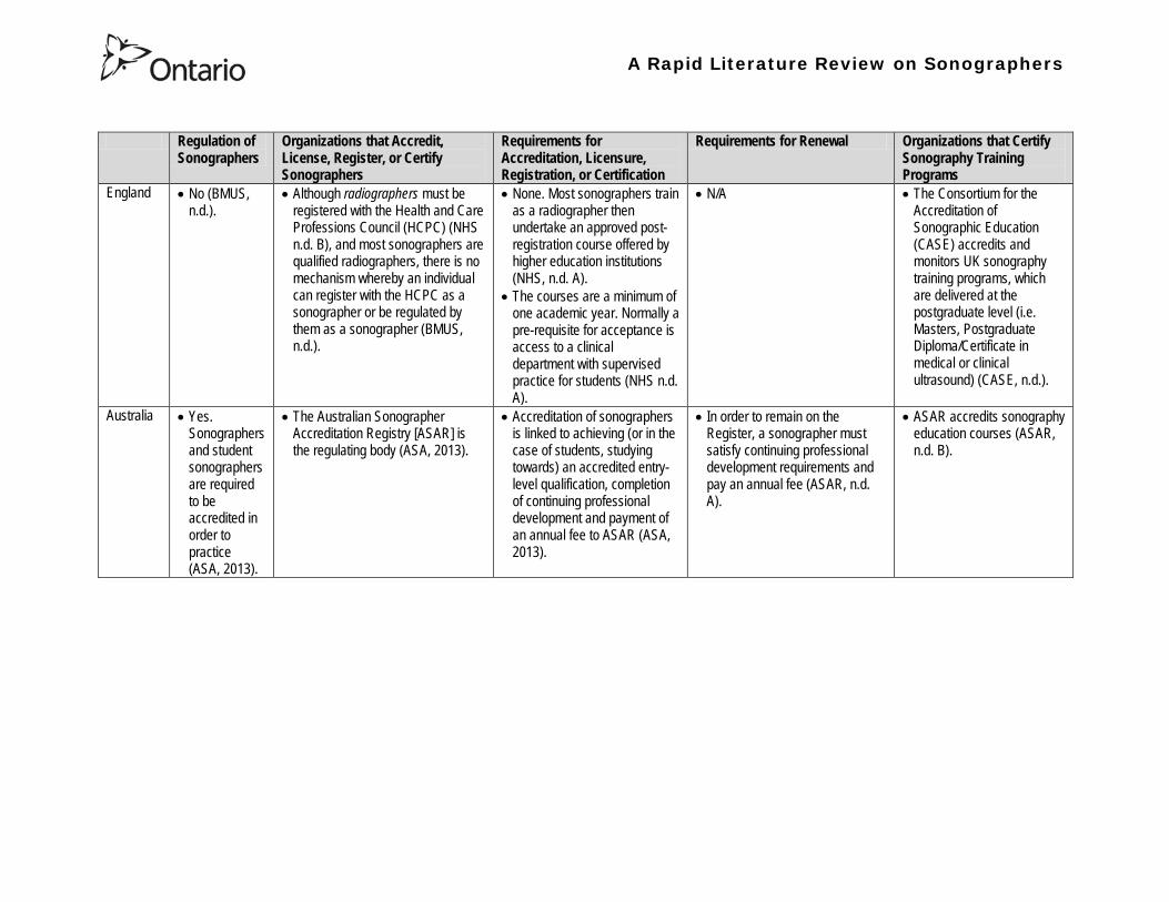
A Rapid Literature Review on Sonographers
Regulation of Sonographers
Organizations that Accredit License Register or Certify Sonographers
Requirements for Accreditation Licensure Registration or Certification
Requirements for Renewal Organizations that Certify Sonography Training Programs
England bull No (BMUS nd)
bull Although radiographers must be registered with the Health and Care Professions Council (HCPC) (NHS nd B) and most sonographers are qualified radiographers there is no mechanism whereby an individual can register with the HCPC as a sonographer or be regulated by them as a sonographer (BMUS nd)
bull None Most sonographers train as a radiographer then undertake an approved post-registration course offered by higher education institutions (NHS nd A)
bull The courses are a minimum of one academic year Normally a pre-requisite for acceptance is access to a clinical department with supervised practice for students (NHS nd A)
bull NA bull The Consortium for the Accreditation of Sonographic Education (CASE) accredits and monitors UK sonography training programs which are delivered at the postgraduate level (ie Masters Postgraduate DiplomaCertificate in medical or clinical ultrasound) (CASE nd)
Australia bull Yes Sonographers and student sonographers are required to be accredited in order to practice (ASA 2013)
bull The Australian Sonographer Accreditation Registry [ASAR] is the regulating body (ASA 2013)
bull Accreditation of sonographers is linked to achieving (or in the case of students studying towards) an accredited entry-level qualification completion of continuing professional development and payment of an annual fee to ASAR (ASA 2013)
bull In order to remain on the Register a sonographer must satisfy continuing professional development requirements and pay an annual fee (ASAR nd A)
bull ASAR accredits sonography education courses (ASAR nd B)
A Rapid Literature Review on Sonographers
Regulation of Sonographers
Organizations that Accredit License Register or Certify Sonographers
Requirements for Accreditation Licensure Registration or Certification
Requirements for Renewal Organizations that Certify Sonography Training Programs
New Zealand
bull Yes bull The Medical Radiation Technologists Board is a health registration authority created under the Health Practitioners Competence Assurance Act 2003 that maintains the register of medical radiation technology (including sonographers) (MRTB nd A)
bull To be eligible for registration a sonographer must hold a postgraduate diploma in health science (Ultrasound) issued by the Unitec Institute of Technology (or an education approved by the Board as being substantially equivalent) (MRTB nd B)
bull Sonographers may also be required to demonstrate adequate experience in the relevant scope of practice ie a minimum of 3360 hours of clinical experience (MRTB nd B)
bull Each year sonographers must apply for and be issued with an annual practising certificate (APC)
bull Sonographer must provide evidence of enrolment and satisfactory progress in one of seven Board-approved recertification programmes (MRTB ndc)
bull The Medical Radiation Technologists Board accredits and monitors educational institutions and degrees courses of studies or programmes (MRTB nd A)
A Rapid Literature Review on Sonographers
Table 2 Examples of Point-of-Care Ultrasound Use Use Examples
Diagnostic Assessment Clinicians from diverse specialties can become very adept at using ultrasonography to examine a particular organ disease or procedure that is directly relevant to their area of expertise whereas imaging specialists typically perform more comprehensive examinations (Moore et al 2011)
bull Emergency physicians have integrated ultrasound into their repertoire for rapid assessment of emergent conditions (Hile et al 2012)
bull A 2012 review on the use of pre-hospital ultrasound concluded that medics (non-physicians trained in the delivery of medical care and the point of injury) can perform ultrasound with a high degree of accuracy (Hile et al 2012)
bull In prehospital settings ultrasound has used been by non-physicians to do fracture detection evaluation of the aorta and Focused Assessment with Sonography for Trauma (FAST) examinations (Hile et al 2012) o The aim of FAST is to assist in the trauma or bleeding patient The areas mentioned
above (perihepatic perisplenic pelvis and pericardium scanning and moreover thoracic ultrasound primarily for haemothorax) are scanned for the detection of fluid as an indication of bleeding rather than organ function (Bennett 2009)
bull Ultrasonography has been used at the Mount Everest base camp to diagnose high-altitude pulmonary edema (Moore et al 2011)
Screening Ultrasonography has been described as a screening test for cardiovascular and gynecologic disease and compact ultrasonography has been incorporated into ldquomobile screening labsrdquo (Moore et al 2011)
bull Ultrasound can be used to screen for abdominal aortic aneurysm (Moore et al 2011)
Guiding Procedures Ultrasound guidance may improve success and decrease complications in procedures performed by multiple specialties (Moore et al 2011)
bull Ultrasound can be used to guide central and peripheral vascular access thoracentesis paracentesis arthrocentesis regional anesthesia incision and drainage of abscesses localization and removal of foreign bodies lumbar puncture biopsies and other procedures (Moore et al 2011)
A Rapid Literature Review on Sonographers
Table 3 Description of the content in the articles being summarized28
No
Description Reference Review Articles
1 Autism spectrum disorders (ASDs) affect an estimated 1 of children in the United States The etiology is probably multifactorial including genetic components and exposure to infections toxins and other environmental factors particularly unfavorable perinatal and neonatal conditions There has been an increase in the frequency of diagnosis of ASDs over the last 20 years with a parallel increase in the use of obstetric diagnostic ultrasound with prenatal ultrasound exposure mentioned as the possible main etiology for autism ldquoepidemicsrdquo Central nervous system alterations have been described in ASDs and certain similar changes have been described in animals after exposure to ultrasound However analysis of in utero exposure in humans has failed to show harmful effects in neonates or children particularly in school performance attention disorders and behavioral changes There is no independently confirmed peer-reviewed published evidence that a cause-effect relationship exists between in utero exposure to clinical ultrasound and development of ASDs in childhood Ultrasound is a form of energy which effects in the tissues it traverses and its use should be restricted to medical indications by trained professionals for as short a period and as low an intensity as compatible with accurate diagnosis
Abramowicz J (2012) Ultrasound and Autism Association Link or Coincidence J Ultrasound Med 311261ndash1269
2 Over the past decade point-of-care ultrasound (US) use by nonphysician providers has grown substantially The purpose of this article is to (1) summarize the literature evaluating military medics facility at US (2) more clearly define the potential utility of military prehospital US technology and (3) lay a pathway for future research of military prehospital US The authors performed a keyword search using multiple search engines Each author independently reviewed the search results and evaluated the literature for inclusion Of 30 studies identified five studies met inclusion criteria The applications included evaluation of cardiac activity pneumothorax evaluation and fracture evaluation Additionally a descriptive study demonstrated distribution of US exam types during practical use by Army Special Forces Medical Sergeants No studies evaluated retention of skills over prolonged periods Multiple studies demonstrate the feasibility of training military medics in US Even under austere conditions the majority of studies conclude that medic can perform US with a high degree of accuracy Lessons learned from these studies tend to support continued use of US in out-of-hospital settings and exploration of the optimal curriculum to introduce this skill
Hile D C Morgan A R Laselle B T amp Bothwell J D (2012) Is Point-of-Care Ultrasound Accurate and Useful in the Hands of Military Medical Technicians A Review of the Literature Military Medicine 177(8) 983-7
28 Please note the studies programs and findings presented in this table may originate from jurisdictions with health systems that are significantly different from Ontarios If there is intent to draw heavily from one or more sources presented in this table we recommend that you contact the lead author of this review for assistance with evaluating the local applicability
A Rapid Literature Review on Sonographers
No Description Reference 3 Ultrasonography is a safe and effective form of imaging that has been used by physicians for more than half a century to aid in
diagnosis and guide procedures Over the past two decades ultrasound equipment has become more compact higher quality and less expensive which has facilitated the growth of point-of-care ultrasonography mdash that is ultrasonography performed and interpreted by the clinician at the bedside In 2004 a conference on compact ultrasonography hosted by the American Institute of Ultrasound in Medicine (AIUM) concluded that ldquothe concept of an lsquoultrasound stethoscopersquo is rapidly moving from the theoretical to realityrdquo This conference included representatives from 19 medical organizations in November 2010 the AIUM hosted a similar forum attended by 45 organizations Some medical schools are now beginning to provide their students with hand-carried ultrasound equipment for use during clinical rotations Although ionizing radiation from computed tomographic (CT) scanning is increasingly recognized as a potentially major cause of cancer ultrasonography has been used in obstetrics for decades with no epidemiologic evidence of harmful effects at normal diagnostic levels However ultrasonography is a user-dependent technology and as usage spreads there is a need to ensure competence define the benefits of appropriate use and limit unnecessary imaging and its consequences This article provides an overview of the history and current status of compact point-of-care ultrasonography with examples and discussion of its use
Moore C L amp Copel J A (2011) Point-of-Care Ultrasonography N Engl J Med 364 749-57
4 Sound travels through objects that block light Only very recently has technology advanced enough to decipher ultrasound for medical use Machines have become smaller cheaper more versatile and more advanced than ever before The medical use of ultrasound spreads across many fields so that traditional areas no longer have a monopoly With this comes the question of training doctors This has been done by various colleges and societies worldwide Some have been quicker to act than others There also needs to be an understanding of when broad experience and advanced technical skills are required or when limited skills will do In addition some procedures can be performed more safely with ultrasound where the knowledge of ultrasound is not paramount This article covers current provision of training in echo-cardiography and ultrasound in areas relevant to anaesthetists who are working in critical care (including accident and emergency) and complex surgery (mainly cardiac)
Bennett S (2009) Training guidelines for ultrasound worldwide trends Best Practice amp Research Clinical Anaesthesiology 23 363ndash373
5 Ultrasound is a commonly employed imaging modality in obstetrics and is generally regarded as safe to the fetus Current ultrasound technology however has significantly higher output potential than older machines used in most clinical studies and the safety profile for the increasing use of Doppler 3-dimensional (D) and 4-D ultrasound with modern machines is unknown This article reviews the current status of ultrasound safety within obstetrics including proposed mechanisms of harm existing scientific and clinical evidence regarding those mechanisms and considerations of safety for the clinical user
Houston L E Odibo A O amp Macones G A (2009) The safety of obstetrical ultrasound a review Prenat Diagn 29 1204ndash1212
A Rapid Literature Review on Sonographers
No Description Reference 6 The purpose of this article is to present the practicing sonographer and sonologist with an overview of the biohazards of ultrasound
and guidelines for safe use Overview Ultrasound is an imaging modality that has important diagnostic value Although useful in a variety of applications diagnostic ultrasound is particularly useful in prenatal diagnosis To date there is no evidence that diagnostic ultrasound produces harm in humans or the developing fetus when used properly There are however an increasing range of ultrasound studies being performed Newer technologies can have higher acoustic output levels than earlier equipment Also subtle or transient effects of diagnostic ultrasound such as changes in membrane permeability or neuronal migration are not completely understood Nondiagnostic Imaging Applications Nondiagnostic uses of ultrasound equipment such as repeated scans of healthy subjects for equipment demonstrations generally should be avoided First-trimester fetal scans should not use color and power Doppler modes and should not be performed for the sole purpose of producing souvenir videos or photographs Production of fetal souvenir photographs or videos during diagnostic clinical studies should not increase exposure levels or extend scan times beyond those needed for clinical purposes Operators should follow safe scanning guidelines and ALARA (as low as reasonably achievable) principles The FDA considers the promotion selling or leasing of ultrasound equipment for making ldquokeepsake fetal videosrdquo that are not part of a medical diagnostic procedure to be an unapproved use of a medical device
NelsonT R FowlkesJ B Abramowicz J S Church C C (2009) Ultrasound Biosafety Considerations for the Practicing Sonographer and Sonologist J Ultrasound Med 28139ndash150
7 Objective A possible association between ultrasound during pregnancy and subsequent non-right handedness among children has been suggested The association has been reported to be stronger among boys The aim of the present study was to explore this further through a meta-analysis of two follow-up studies of three randomized controlled trials of routine ultrasonography during pregnancy Design Handedness was assessed through a questionnaire to the parents and 4715 children at the age of 8ndash9 years were included in the meta-analysis Results There was no statistically significant difference in the prevalence of non-right handedness between the ultrasound-screened children and the controls but there was a statistically significant difference in a subgroup analysis among the boys Exploratory analyses according to exposure or non-exposure indicated an even stronger association between ultrasound and non-right handedness Conclusions A conservative analytical approach indicates no association between ultrasound in utero and subsequent non-right handedness The results from the exploratory analyses must be interpreted with caution There is still a need for further research
Salvesen K A amp Eik-Nes (1999) Ultrasound during pregnancy and subsequent childhood non-right handedness a meta-analysis Ultrasound Obstet Gynecol13 241ndash246
8 Ultrasound energy exerts important cellular genetic thermal and mechanical effects Concern about the safety of ultrasound prompted several agencies to devise regulatory limits on the machine output intensities The visual display of thermal and mechanical indices during ultrasound imaging provides an aid to limit the output of the machine Despite many animal studies no human investigations conducted to date have documented major physiologic consequences of ultrasound exposed during imaging To date ultrasound imaging appears to be safe for use in regional anesthesia and pain medicine interventions and adherence to limiting the output of ultrasound machines as outlined by the Food and Drug Administration may avoid complications in the future This article reviews ultrasound-related biologic effects the role of the regulatory agencies in ensuring safety with the use of ultrasound and the limitations and implications of ultrasound use in humans
Shankar H amp Pagel P S (2011) Potential Adverse Ultrasound-related Biological Effects A Critical Review Anesthesiology 115(5) 1109-1124
A Rapid Literature Review on Sonographers
No Description Reference 9 Objective In the context of the planned International Society of Ultrasound in Obstetrics and GynecologyndashWorld Health Organization
multicenter study for the development of fetal growth standards for international application we conducted a systematic review and meta-analysis to evaluate the safety of human exposure to ultrasonography in pregnancy Methods A systematic search of electronic databases reference lists and unpublished literature was conducted for trials and observational studies that assessed short- and long-term effects of exposure to ultrasonography involving women and their fetuses exposed to ultrasonography using B-mode or Doppler sonography during any period of pregnancy for any number of times The outcome measures were (1) adverse maternal outcome (2) adverse perinatal outcome (3) abnormal childhood growth and neurological development (4) non-right handedness (5) childhood malignancy and (6) intellectual performance and mental disease Results The electronic search identified 6716 citations and 19 were identified from secondary sources A total of 61 publications reporting data from 41 different studies were included 16 controlled trials 13 cohort and 12 casendashcontrol studies Ultrasonography in pregnancy was not associated with adverse maternal or perinatal outcome impaired physical or neurological development increased risk for malignancy in childhood subnormal intellectual performance or mental diseases According to the available clinical trials there was a weak association between exposure to ultrasonography and non-right handedness in boys (odds ratio 126 95 CI 103ndash154) Conclusion According to the available evidence exposure to diagnostic ultrasonography during pregnancy appears to be safe
Torloni M R Vedmedovska Merialdi M Betran A P Allen T et al (2009) Safety of ultrasonography in pregnancy WHO systematic review of the literature and meta-analysis Ultrasound Obstet Gynecol 33 599ndash608
Articles in Peer-Reviewed Journals 10 It has been documented for many years that as a result of use of ultrasound to determine the sex of the fetus the male to female
ratio in countries such as China and India have become increasingly skewed (due to subsequent abortions and infanticide of females) Preference for sons over daughters evident in Chinas and South Asias male sex ratios is commonly rationalized by poverty and the need for old-age support In this article the authors study South and East Asian immigrants to Canada a group for whom the economic imperative to select sons is largely absent Analyzing the 2001 and 2006 censuses 20 percent samples the authors find clear evidence of extensive sex selection in favor of boys at higher parities among South and East Asian immigrants unless they are Christian or Muslim The latter finding accords with the explicit prohibition against (female) infanticidemdashtraditionally the main sex-selection methodmdashin these religions The findings point to a strong cultural component to both the preference for sons and the willingness to resort to induced abortion based on sex
Almond D Edlund L Milligan K (2013) Son Preference and the Persistence of Culture Evidence from South and East Asian Immigrants to Canada Population and Development Review 39(1) 75ndash95
11 The rapid growth in the use of ultrasound as a diagnostic imaging technology over the past forty years has led to a demand for a workforce with the appropriate skills to perform and interpret the scans In the UK ultrasound investigations now comprise the largest group of all diagnostic imaging examinations However there remains no statutory regulation of the practice of sonography in the UK and little recognition of the considerable training that many practitioners have undertaken to obtain the skills to become safe and competent sonographers Many in the field consider that this should change and are working to obtain professional status for the practice of sonography Although the Health Care Professions Council (HCPC) has recommended regulation of sonography practice this is unlikely to happen in the near future This paper discusses the evolution of sonography practice and explores some of the complex issues associated with the professionalisation of sonography
Gibbs V (2013) The long and winding road to achieving professional registration for sonographers Radiography19 164-167
A Rapid Literature Review on Sonographers
No Description Reference 12 Background Contrast echocardiography (which uses contrast agents injected into the blood flow in order to visualize the blood flow
in very small blood vessels) has been shown to improve diagnostic quality especially in technically difficult patients However the learning curve and increased time for preparation and image acquisition have led to low use Methods The authors sought to determine whether the contrast echocardiography procedure performed independently by a specialized trained sonographer could improve efficiency In one centre routine echocardiograms were scheduled for one hour and any study exceeding one hour would result in patient booking cancellations The authors compared the standard of care in which a physician or nurse administers echocontrast with a sonographer-administered program (SAP) Results The time to complete contrast echocardiograms was significantly reduced by the SAP strategy (43 min 17 s plusmn 23 min 42 s vs 1 h 1 min 6 s plusmn 31 min 0 s P lt 0001) Subgroup analysis of the inpatients and outpatients demonstrated similar results Only 10 of studies (6 of 61) in the SAP exceeded 60 minutes compared with 45 (34 of 76) in the standard-of-care group (P lt 0001) Based on study volumes in the centre the net improvement in productivity with the SAP could be up to 53 annually Conclusion Sonographer-administered echocontrast is feasible and potentially removes a barrier to implementation of contrast echocardiography
Tang A Chiew S K Rashkovetsky R Becher H Choy J B (2013) Feasibility of Sonographer-Administered Echocontrast in a Large-Volume Tertiary-Care Echocardiography Laboratory Canadian Journal of Cardiology 29 391ndash395
13 Ultrasound scanning has been used as a diagnostic and screening tool in obstetric practice for over 50 years There is no evidence of immediate or long-term harm to the developing fetus from exposure to B mode ultrasound However exposure to high levels of Doppler ultrasound during early development is increasingly common and the full safety implications of this exposure are not clear Doppler ultrasound exposure in utero gives rise to increased apoptosis in animal models and there is evidence of the effects of exposure to Doppler ultrasound persisting throughout life with increased non-right-handedness observed in human epidemiological studies The authors consider the idea that there may be long-term developmental implications for fetuses exposed to Doppler ultrasound early in gestation These effects may be mediated via thermal or mechanical disruption to the developing conceptus giving rise to free radical damage Excess free radical exposure early in gestation is a strong candidate for the final common pathway underlying developmental programming effects and gives rise to concern that fetuses exposed to high levels of ultrasound are at risk of a developmental programming effect It is suggested that there is a need for animal studies of developmental programming using exposure to Doppler ultrasound scanning as the exposure of interest and for more observational data to be collected in the clinical setting While these data are collected it seems prudent to continue to adhere to the principle of lsquoas low as reasonably achievablersquo (ALARA) when exposing first-trimester fetuses to Doppler ultrasound
Aiken CE Lees CC (2012) Long-term effects of in utero Doppler ultrasound scanning ndash A developmental programming perspective Medical Hypotheses 78 539ndash541
14 Point of care sonography by emergency physicians in the emergency department can be an effective aid in the diagnosis and management of patients presenting with a variety of medical and traumatic conditions Its use can improve patient outcomes enhance patient safety speed patient disposition and save lives CAEP supports the use of point of care sonography
Canadian Assocation of Emergency (CAEP) Physicians Ultrasound Position Statement Working Group (2012) Use of point of care sonography by emergency physicians CJEM 14(2)106-112
A Rapid Literature Review on Sonographers
No Description Reference 15 The use of three-dimensional echocardiography (3DE ie a 3D ultrasound of the heart) during electrophysiology (EP - a branch of
cardiology that deals with the diagnosis and treatment of heart rhythm disorders) procedures is the end product of years of growth in two diverse cardiology subspecialties namely advanced cardiac imaging and the EP During the past decade progress in both fields has resulted in many important advances that have culminated in their union for a new area of growth and development Imaging advances have provided the cardiovascular specialist with enhanced cardiac volume and function data and more recently 3DE capabilities with improved spatial and temporal resolution providing unprecedented spatial relationships This latter development is valued by EP specialists in need of hitherto never required anatomic knowledge as they press forward with extraordinary expansion in their capabilities It makes sense that by combining these two rapidly growing subspecialties future capabilities in patient care may be achieved that would otherwise not be possible This paper discusses the value of 3DE during EP procedures and offers the readers insight into this novel multispecialty hybrid arena Using this model as a template it is likely that the readers may identify other areas within their practices where periprocedural advanced imaging may afford significant dividends in patient outcomes
Moukabary T Faletra F F Kronzon I Thomas W amp Sorrell V L (2012) Three-Dimensional Echocardiography in the Electrophysiology Laboratory Echocardiography 29 117-122
16 Background There has been much discussion about whether female feticide occurs in certain immigrant groups in Canada The authors examined data on live births in Ontario and compared sex ratios in different groups according to the motherrsquos country or region of birth and parity Methods The authors completed a population-based study of 766 688 singleton live births between 2002 and 2007 They used birth records provided by Ontario Vital Statistics for live births in the province between 23 and 41 weeksrsquo gestation They categorized each newborn according to the motherrsquos country or region of birth namely Canada (n = 486 599) Europe (n = 58 505) South Korea (n = 3663) China (n = 23 818) Philippines (n = 15 367) rest of East Asia (n = 18 971) Pakistan (n = 18 018) India (n = 31 978) rest of South Asia (n = 20 695) and other countries (n = 89 074) They calculated malefemale ratios and 95 confidence intervals (CIs) for all live births by these regions and stratified them by maternal parity at the time of delivery (0 1 2 or ge 3) Results Among infants of nulliparous women (women with no previous children) the malefemale ratio was about 105 overall As parity (the number of previous children) increased the ratio remained unchanged among infants of Canadian-born women In contrast the malefemale ratio was significantly higher among infants of primiparous women (women with one previous child) born in South Korea (120 95 CI 109ndash134) and India (111 95 CI 107ndash115) than among infants of Canadian-born primiparous women Among multiparous women (women with 2+ previous children) those born in India were significantly more likely than Canadian-born women to have a male infant (parity 2 ratio 136 95 CI 127ndash 146 parity ge 3 ratio 125 95 CI 109ndash143) Interpretation The study of malefemale ratios in Ontario showed that multiparous women born in India were significantly more likely than multiparous women born in Canada to have a male infant
Ray J G Henry D A Urquia M L (2012) Sex ratios among Canadian liveborn infants of mothers from different countries CMAJ184(9) E92-96
A Rapid Literature Review on Sonographers
No Description Reference 17 This letter consists of a case study that illustrates how a cross-disciplined sonographer (educated in cardiac and vascular ultrasound)
can quickly and efficiently and with great Cost-effectiveness deliver timely care to a patient in certain clinical settings The literature is comparatively sparse in referencing clinical diagnostics in cross-discipline sonographers Echocardiographers must be clinicians fully comfortable with evaluating the circulatory dynamics of the body from the left and right heart to the peripheral arteries and veins including the extracranial circulation (carotid arteries) Included in this skill set is substantive understanding of the ECG patientrsquos history and integration of cardiac and vascular findings Limitations of each diagnostic tool and how these limitations can be overcome by the strengths of the other examination are important For the dual-skilled sonographer only experience continuous education and constant clinical feedback will lead to the independent decision making that is counted on by the physician The American Registry for Diagnostic Medical Sonography offers credentials in cardiac and vascular imaging (RDCS and RVT) Cardiovascular Credentialing International offers credentials in cardiac and vascular imaging as well (registered cardiac sonographer [RCS] and registered vascular specialist [RVS]) All imaging facilities should consider merging cardiac and vascular noninvasive imaging with dual-registered clinical sonographers credentialed by one of these two agencies
Zemanek M (2012) What It Means to Be a Cardiovascular Sonographer Journal of Ultrasound in Medicine 311859-1862
18 Diagnostic medical sonography is viewed as a safe imaging modality However bioeffects research has yet to define the effects of exposure to sonography in humans Therefore sonographers must be aware of potential risks and practice ALARA (ldquoas low as reasonably achievablerdquo) routinely This research explores sonographersrsquo ability to answer basic questions about the bioeffects of sonography as well as their knowledge and use of the safety indices called the mechanical index (MI) and thermal index (TI) Members of the Society of Diagnostic Medical Sonographers in the US a southwestern sonographersrsquo society and a sonography community on Facebook 2010 were surveyed (n = 212) There is a low level of bioeffects knowledge and minimal safety practices among sonographers in this survey No significant relationship was found between sonographer knowledge of the bioeffects of sonography years in the profession or whether the sonographers monitor the MITI
Bagley J Thomas K DiGiacinto D (2011) Safety Practices of Sonographers and Their Knowledge of the Biologic Effects of Sonography Journal of Diagnostic Medical Sonography 27(6) 252ndash261
19 This article highlights trends in echocardiography Point of care ultrasound (hand-carried ultrasound) Initially point of care ultrasound will increase the use of echocardiography in the same manner that cardiac CT increased catheterizations However once the basics are learned it has the potential to be a very powerful tool in that less unnecessary studies will be ordered In the clinic small systems will be used as a lsquolsquofirst lookrsquorsquo screen and become part of the patientrsquos history only patients for whom more testing is necessary will be referred for a comprehensive study This is likely to reduce the number of Doppler studies requested The miniaturization of echo equipment will continue with improved qualityfunctionality of the lsquolsquohand-heldrsquorsquo echo machines to the extent that cordless probes will become a reality sooner rather than later Role in interventional echocardiography Sonographers will be spending much more time in the interventional and electrophysiology (EP) labs There will be more interconnection with interventional cardiologists and the volume will increase as more devices are implanted more percutaneous valves placed and more robotic surgery is performed Crosstraining and multitasking Sonographers entering the profession would be prudent to learn cardiac and vascular imaging Individuals who multitask andor cross-train will be ideal For example a sonographer who starts an IV monitors ECG stress can perform cardiac MRI and can interpret a preliminary report or a nurse who can do an echo monitor sedation and start an IV will be invaluable The sonographer should continue to grow professionally by cross training as institutions try to minimize costs and maximize throughput
McCulloch M L (2011) Thoughts regarding the future of sonography Journal of the American Society of Echocardiography 24(5) 29A-30A
A Rapid Literature Review on Sonographers
No Description Reference 20 Background Radiology reports provide specialist interpretation of images and relate these findings to the patientrsquos symptoms and
signs This study compared ultrasound (USS) reports generated by radiologists and sonographers to determine if any significant or clinically relevant differences existed Methods A retrospective analysis of 624 consecutive USS reports was carried out The reports were assessed for the presence of a lsquodisclaimerrsquo or lsquocaveatrsquo pertaining to the quality of the images and were analysed with respect to the clarity of their wording and ability to answer the clinical request Results The majority of sonographer USS reports contained a disclaimer regarding the quality of the USS images (571) compared with a smaller proportion of radiologist scans (99) (P lt 0001) Overall radiologists performed significantly better in providing a clear negative or positive diagnosis to the clinical question on the request form when compared with sonographers (885 vs 654 P lt 0001) Conclusion lsquoDisclaimerrsquo comments and lsquohedgingrsquo must be avoided in radiology reporting While feedback as to the accuracy of the radiology images is important overuse of such terms undermines the validity of the radiology report
Garcea G Mahmoud A Ong S L Rees Y Berry Det al (2010) Caveat reporting in ultrasound interpretation of surgical pathology a comparison of sonographer versus radiologist Journal of Evaluation in Clinical Practice 16 97ndash99
21 A nine-point questionnaire was constructed based on previously published work by Marsal and Sheiner et al The survey was distributed to clinical users of ultrasound practicing sonography in Hamilton (New Zealand) during July 2010 totalling 35 users consisting of sonographers sonography trainees one obstetric sonologist and one fetal medicine registrar Results Twenty sonographers returned the questionnaire which represents survey response of 61 The level of clinical experience was equally split between junior sonographers of lt 5 yearsrsquo experience (30) sonographers with 5-10 yearsrsquo experience (35) and senior sonographers of gt10 yearsrsquo experience (35) While 100 of the sonographers surveyed reported being familiar with the term Thermal Index (TI) and 90 claimed they adhered to the Action Learning Action Response Association (ALARA) principle all the time or most of the time only 15 of sonographers correctly described or defined what the term TI means Of the sonographers 80 knew where to find the TI during real-time examinations but only 35 knew which TI index to use for nuchal translucency (NT) scan and only 10 knew the expected range of TI encountered during routine obstetric examinations Of the eight sonographers who reported adhering to ALARA all the time the majority (64) could not describe or define TI In the same subgroup the majority (64) did not know which TI to monitor during a NT scan Only three of 20 (15) sonographers knew what the term TI means Of these two were junior sonographers with lt 5 yearsrsquo experience and one had 5ndash10 yearsrsquo experience Despite understanding the term TI one of the three sonographers still selected the incorrect index to monitor during a NT scan Not one of the seven senior sonographers with gt 10 years of experience could define or describe the TI In total only 10 of sonographers knew what the TI means and which TI to monitor during NT scanning
Necas M (2010) New Zealand sonographers do not outperform their European or American colleagues in the knowledge of ultrasound safety Australasian Journal of Ultrasound in Medicine13(4) 28-32
A Rapid Literature Review on Sonographers
No Description Reference 22 Musculoskeletal (MSK) ultrasound performed by non-medical practitioners is a developing practice The aim of this paper is
(1) to audit an experienced sonographerrsquos (SJ Riley one of the authors) performance after one yearrsquos independent reporting against two experienced MSK radiologists (CJ Grove amp M Chandramohan the other two authors) and (2) to establish an audit standard against which such role development might be contrived Images and reports from 250 MSK ultrasound examinations performed by a sonographer were reviewed independently by two consultant MSK radiologists The examinations were graded for discrepancy eg grade 1 ndash agree with the sonographer report grade 2 ndash minor discrepancy unlikely to alter patient care grade 3 ndash potentially significant discrepancy grade 4 ndash definite significant discrepancy likely to have adverse consequences for patient care Two of 250 (08) cases were excluded Both radiologists agreed completely with the sonographer (grade 1) in 235 of the 248 cases (948) In 13 cases there was discrepancy between the reports of SJR and the radiologists The discrepancy was grade 2 in six of the 248 cases (24) grade 3 in six of the 248 cases (24) and grade 4 in one of the 248 cases (04) In conclusion this audit shows a high level of agreement between the sonographer and the consultant MSK radiologist reporting of MSK ultrasound This level of agreement may set the standard for future quality assurance audit of sonographer MSK reporting
Riley S J Groves C J Chandramohan M (2010) Musculoskeletal ultrasound audit of sonographer reporting Ultrasound 18 36ndash40
23 Background Growing demand for ultrasound examinations and higher quality requirements motivate searching for routines combining the diagnostic accuracy of radiologist-performed examinations with the economical advantages of sonographer-performed examinations One possible approach is to use strictly standardized acquisition and documentation schemes that give the radiologist access to all relevant information after the examination Purpose To compare a recently introduced routine combining acquisition by a radiographer (more specifically a sonographer with two years experience) documentation as standardized cine-loops and review by a radiologist (lsquostandardized methodrsquo) with the formerly used routine where the diagnosis is made bedside by the radiologist (lsquotraditional methodrsquo) Material and Methods In 64 policlinic patients the kidneys (n = 27) or the gallbladder (n = 37) were examined with both the standardized and the traditional method The radiologistsrsquo findings of hydronephrosis tumors cysts echogenicity changes and cortical thickness (in the kidneys) and wall thickness concrements and polyps (in the gallbladder) were compared between the methods with respect to agreement (proportion of agreement and kappa coefficient) as well as systematic differences (McNemarrsquos test) Results The findings at the gallbladder examination showed a median agreement of 97 (86ndash100 kappa = 064ndash100) and those at the kidney examination an agreement of 90 (78ndash100 kappa = 069ndash 100) There were no significant systematic differences between the methods Conclusion The satisfactory agreement in this preliminary study indicates that the new workflow with ultrasound examinations performed by a radiographer (sonographer) and analyzed off-line by a radiologist is promising and motivates further studies
Stenman C Thorelius L Knutsson A amp Smedby Ouml (2010) Radiographer-acquired and radiologist-reviewed ultrasound examination ndash agreement with radiologistrsquos bedside evaluation Acta Radiologica 52 70ndash74
A Rapid Literature Review on Sonographers
No Description Reference 24 The troubling practice of fetal sex selection has historically been considered an Asian phenomenon However recent evidence shows
that a similar situation is emerging in North America albeit on a smaller scale The Society of Obstetricians and Gynaecologists of Canada (SOGC) has firmly stated its opposition to sex selection for non-medical reasons as well as to the use of any technology used solely for the purpose of determining fetal sex However because fetal sex may be disclosed to the parents at the time of ultrasound examination if they request this information guidance for health care professionals to assist in discouraging fetal sex selection would be useful Because no declaration of motives or reasons is required when a woman seeks a termination of pregnancy the authors suggest that health care professionals need not disclose the sex of a fetus until it has reached a gestational age at which abortion for non-medical purposes would not be possible This proposal would facilitate consistency between clinical practice and the values of Canadian citizens the SOGC the Canadian Medical Association and other professional organizations while still respecting current laws pertaining to disclosure of patient information and patientsrsquo rights to autonomous decision-making
Thiele A T amp Leier B (2010) Towards an Ethical Policy for the Prevention of Fetal Sex Selection in Canada J Obstet Gynaecol Can 32(1) 54ndash57
25 Background The extent to which increases in the use of prenatal ultrasonography over time reflect changes in maternal risk (ie an increase in high-risk pregnancies over time) is unknown In this population-based study the authors examined the use of prenatal ultrasonography from 1996 to 2006 in Ontario Methods With fiscal year 199697 as the baseline the authors evaluated the relative risk (RR) and 95 confidence interval (CI) for the change in rates of ultrasonography for each subsequent year The RR was adjusted for maternal age income rural residence maternal comorbidities receipt of genetics consultation or amniocentesis mdash all in the index pregnancy mdash and history of complications in a prior pregnancy Results The study sample consisted of 1 399 389 singleton deliveries The rate of prenatal ultrasonography increased from 2055 per 1000 pregnancies in 1996 to 3264 per 1000 in 2006 (adjusted RR 155 95 CI 154ndash155) The rate increased among both women with low-risk pregnancies (adjusted RR 154 95 CI 153ndash155) and those with high-risk pregnancies (adjusted RR 155 95 CI 154ndash157) The proportion of pregnancies with at least four ultrasound examinations in the second or third trimesters rose from 64 in 1996 to 187 in 2006 (adjusted RR 268 95 CI 261ndash274) Paradoxically this increase was more pronounced among low-risk pregnancies (adjusted RR 292 95 CI 283ndash301) than among high-risk pregnancies (adjusted RR 225 95 CI 216ndash235) Interpretation Substantial increases in the use of prenatal ultrasonography over the past decade do not appear to reflect changes in maternal risk Nearly one in five women now undergo four or more ultrasound examinations during the second and third trimesters Efforts to promote more appropriate use of prenatal ultrasonography for singleton pregnancies appear warranted
You J J Alter D A Stukel T A McDonald S D Laupacis A et al (2010) Proliferation of prenatal ultrasonography CMAJ 182(2) 143-151
A Rapid Literature Review on Sonographers
No Description Reference 26 Aims The aim of this study was to assess the cost-effectiveness of using certified sonographers and miniaturized echocardiography
systems to perform echocardiograms at bedside in comparison to moving inpatients from the admission department to the echocardiography laboratory (echo-lab) Methods and Results From 26 September 2005 to 27 October 2005 112 patients admitted in six hospital wards in Italy connected through a 100 Mbit LAN to the echo-lab were scanned within the admission ward by sonographers using a miniaturized echo system Logistical data were collected and results were compared with those obtained from 194 consecutive patients coming from the same wards and studied in the echo-lab with high-end machines between 8 March 2005 and 15 April 2005 Performing echocardiograms in the admission department avoided long waiting time of the inpatients in the echo-lab before and after the study increased the percentage of patients studied within 3 and 5 days from request (88 vs 77 and 100 vs 95 respectively P = 003) increased both sonographer (by 339 P lt 0001) and echo-lab productivity (by 41 P lt 0001) and reduced costs of echocardiograms by 29 Conclusion Implementation of digital echocardiography certified sonographers and a miniaturized echo system allowed improvement of the cost-effectiveness of the service provided by the echo-lab for inpatients and avoided patientsrsquo discomfort derived from prolonged waiting time before and after the exam
Badano L Gaetano N Stacul S Gianfagna P Pericoli M et al (2009) Improved workflow sonographer productivity and cost-effectiveness of echocardiographic service for inpatients by using miniaturized systems European Journal of Echocardiography 10 537ndash542
27 The present report is a proposal from the American Society of Echocardiography Advanced Practice Task Force that identifies the potential of cardiac sonographers to achieve the Advanced Cardiovasculat Sonographer (ACS) level According to this report echocardiographic examinations require a well-trained and competent sonographer to obtain proper anatomic and physiologic data to establish an accurate diagnosis for clinical decision-making and patient management Although the formal education and training of cardiovascular sonographers are evolving many entry-level and staff sonographers may not have sufficient practical or clinical knowledge of the necessary components of the echocardiographic study for the individual patientrsquos clinical presentation In many clinical settings echocardiograms are read after the patient has left the laboratory Thus there is a role for a sonographer who can practice at an advanced level in a cardiovascular ultrasound laboratory to ensure a proper echocardiographic examination is performed on every patient In this setting an Advanced Cardiovascular Sonographer (ACS) would be able to review the indication for and quality of the examination If additional images were needed the ACS would assist the sonographer in obtaining these images which would lead to the performance of a complete and fully diagnostic examination before the patient had left the echocardiography laboratory In clinical practice the quality of the examinations performed would improve advancements in echocardiographic methods could be taught and incorporated into daily practice and patients would be better served
Mitchell C Miller F A Bierig S M Bremer M L Ehler D et al (2009) Advanced Cardiovascular Sonographer A Proposal of the American Society of Echocardiography Advanced Practice Sonographer Task Force J Am Soc Echocardiogr 22 1409-13
A Rapid Literature Review on Sonographers
No Description Reference 28 Sonographer practitioner development involves the expansion and extension of the sonographer role to include reporting on
ultrasound examinations Australian sonographers have not seen the same degree of role extension and expansion as their counterparts in the United Kingdom despite increasing levels of discussion regarding sonographer practitioner development The aim of this study was to determine if Australian sonographers want to extend their professional role and what they consider are the important issues associated with role extension This paper reports on qualitative data derived from a survey of Australian sonographers and investigates if Australian sonographers are interested in extending and expanding their professional role and responsibilities and if they do what might be necessary or desirable from a professional point of view for this change to occur A survey was mailed to all members of the Australian Sonographers Association (ASA) in October 2006 The 31-item survey included 28 closed-ended and three opened-ended items to provide both quantitative and qualitative data The quantitative data will be reported separately Qualitative data was derived from responses to the opened-ended questions which asked respondents to elaborate on their attitudes and feelings about role extension and development Analysis used Nvivo7 software to aid in uncovering common themes from the qualitative data The analysis focused on the reported incentives or motivations for becoming a sonographer practitioner as well as disincentives or perceived hurdles that would discourage respondents from becoming sonographer practitioners The three most reported incentives or motivations for becoming a sonographer practitioner were professional recognition remuneration and increased knowledge The three most commonly reported disincentives or perceived hurdles that would discourage respondents from becoming sonographer practitioners were legal issues insurance and further study
McGregor R OrsquoLoughlin K Cox J Clarke J Snowden A (2009) Sonographer practitioner development in Australia Qualitative analysis of an Australian sonographersrsquo survey Radiography15 313-319
29 Introduction This study describes the effect of introducing a cardiac sonographer led follow up clinic within a hospital cardiology department for patients with stable heart valve disease The two years before and after the instigation of the valve clinic were audited Materials and methods The clinic was conducted in a single centre and undertaken in the cardiology department of a district general hospital A total of 382 patients with 397 clinically significant valve lesions but for whom surgery was not yet indicated but follow up required were seen in a cardiac sonographer run clinics These patients no longer attended a medical follow up clinic unless there was clinical or echocardiographic deterioration Effectiveness was judged by the percentage treated according to current best practice guidelines the number of echocardiograms performed and the number of hospital outpatient visits attended In addition mortality data for the subjects in the clinic was collected Results The proportion followed up according to best practice guidelines rose from 157 (41) to 354 (92) (p lt 001) The total number of echocardiograms performed fell from 807 to 550 Total number of outpatient visits fell from 998 to 31 A total of 11 patients died in the two year study period none from progressive valve disease Discussion This study demonstrates that a protocol driven sonographer led heart valve disease follow up clinic significantly improved the quality of follow up while bringing about a major reduction in outpatients visits without compromising patient safety
Taggu W Topham A Hart L Carr-White G Sulke N et al (2009) A cardiac sonographer led follow up clinic for heart valve disease International Journal of Cardiology 132 240 ndash243
30 Role development for imaging allied health professionals has progressed significantly over the last three decades with many major developments pioneered in the field of ultrasound This paper reviews the developing role of the radiographer in ultrasound (in some countries such as the UK sonographers are considered a type of radiographer) and some of the clinical professional and educational implications of such development As the published evidence base underpinning the latest developments is scarce several contemporary case study examples which have been presented verbally at study days attended by the authors have been included In conclusion sonographer role development is considered within the professional contexts of extended role advanced and consultant practice
Hart A amp Dixon A (2008) Sonographer Role Extension and Career Development a Review of the Evidence Ultrasound 16(1) 31-35
A Rapid Literature Review on Sonographers
No Description Reference 31 Objective The main goal of this study was to determine end usersrsquo knowledge regarding safety aspects of diagnostic ultrasound
during pregnancy End usersrsquo attitudes toward the use of ultrasound in low-risk pregnancies were also assessed Methods A questionnaire was distributed to ultrasound end users attending review courses and hospital grand rounds between April and June 2006 in Chicago Results One hundred thirty end users completed the questionnaires (63 response rate) Sixty-three percent were physicians (n = 84) most of them obstetricians (817) About 18 of participants routinely performed Doppler ultrasound examinations during the first trimester Fifty percent of end users thought that the number of ultrasound examinations in low-risk pregnancy should be limited to 1 to 3 (mean plusmn SD 26 plusmn 09) Almost 70 disapproved of ldquokeepsakeentertainmentrdquo ultrasound Although 322 of the participants were familiar with the term thermal index only 177 actually gave the correct answer to the question on the nature of the thermal index About 22 were familiar with the term mechanical index but only 38 described it properly Almost 80 of end users did not know where to find the acoustic indices Only 208 were aware that they are displayed on the sonographic monitor during the examinations End users with higher knowledge of safety issues thought that there should be limitations on the number of ultrasound examinations in low-risk pregnancies (odds ratio 33 95 confidence interval 11ndash100 P = 028) Likewise these end users were more likely to respond that ultrasound might have adverse effects during pregnancy (odds ratio 32 95 confidence interval 11ndash125 P = 045) Conclusions Ultrasound end users are poorly informed regarding safety issues during pregnancy Further efforts in the realm of education and training are needed to improve end user knowledge about the acoustic output of the machines and safety issues
Sheiner E Shoham-Vardi I Abramowicz J S (2007) What Do Clinical Users Know Regarding Safety of Ultrasound During Pregnancy J Ultrasound Med 26 319ndash325
A Rapid Literature Review on Sonographers
No Description Reference 32 In an attempt to survey the knowledge among ultrasound users of some safety aspects of diagnostic ultrasound a questionnaire was
distributed to professionals using ultrasound for fetal examinations The questionnaire was answered by the participants and faculty members of six postgraduate courses in obstetric ultrasound Doppler ultrasound and fetal echocardiography given in Sweden Norway and Austria in 2003 Furthermore in Sweden the questionnaire was distributed to the staff meeting participants of two major obstetric ultrasound units and of one unit for fetal echocardiography and at a meeting of the Working Group for Ultrasound of the Swedish Society of Obstetrics and Gynecology In total 199 questionnaires were anonymously answered by doctors (n = 145) sonographers (n = 22) and midwives trained in ultrasound (n = 32) All of them were using diagnostic ultrasound on a daily or weekly basis The respondents were from nine European countries In none of the countries is formal certification for the use of diagnostic ultrasound in pregnancy or otherwise required The doctors who answered the questionnaire were specialists in obstetrics and gynecology (including some of the internationally recognized experts in fetal ultrasound) pediatric cardiology radiology or clinical physiology The respondents were not informed about the questionnaire in advance and they had a limited time to answer the 16 questions typically 10ndash15 min The questions concerned explanation of the safety indices and their relation to various ultrasound modes ie real-time B-mode M-mode three-dimensional (3D) spectral Doppler color and power Doppler ultrasound The respondents were also asked which ultrasound system they used most often and where on the system the information on thermal index (TI) and mechanical (MI) was displayed (eg in the top right corner of the screen in a separate window etc) Furthermore the respondents had to indicate how they adjust the output energy level on their own machine For all questions there was the optional answer lsquoDonrsquot knowrsquo Results About one-third of the ultrasound experts were able to define the abbreviations TI and MI For the subclasses of the TI the correct answer was found in only 3-8 of cases Of those who knew what the TI and MI meant only two-thirds and one-third of respondents respectively were able to give a simple explanation of the indices In 49 of questionnaires the Doppler modes were correctly ranked as generally producing higher ultrasound exposure than the imaging modes (B-mode 3D) Some 28 of the respondents correctly indicated where on their own machine the information on safety indices is displayed However not all of them knew how to control the output energy level For all items in the questionnaire there were no significant differences in the results between the three categories of respondents (ie physicians sonographers and midwives) The results of the questionnaire were anything but encouraging and they indicated that the users who are supposed to be responsible for controlling exposure of the fetus to ultrasound had a very poor knowledge of the basic safety aspects of ultrasound
Maršaacutel K (2005) The output display standard has it missed its target Ultrasound Obstet Gynecol 25 211ndash214
33 Purpose This study was performed to evaluate the accuracy of upper abdominal ultrasound (US) scanning performed by sonographers in a district general hospital to identify potential areas of weakness and to make recommendations to improve the service Materials and Methods Upper abdominal US examinations performed and reported by sonographers over a 4-week period were retrospectively reviewed The accuracy of the imaging findings and reports were assessed against other imaging surgical histological or laboratory findings and against clinical outcome Results A heterogenous group of 104 patients were included in the study 62 of whom had an US abnormality Errors of scanning or interpretation were identified in 10 patients (96) of whom five (48) were felt to be potentially significant Conclusions The sonographersrsquo accuracy in reporting upper abdominal US scans was 90 However on the basis of this study the authors have implemented specific recommendations to improve the quality of the service
Dongola N Guy R Ward S amp Giles J A (2003) Can sonographers offer an accurate upper abdominal ultrasound service in a district general hospital Radiography 9 29ndash33
A Rapid Literature Review on Sonographers
No Description Reference 34 Aim To compare the accuracy of radiographers and radiologists in routine abdominal ultrasound Materials and Methods One
hundred consecutive patients attending for routine abdominal ultrasound were included Each patient was examined by both a radiographer and radiologist Both operators noted their fndings and wrote a concluding report without conferring Reports were compared Where there was disagreement the patient was either re-examined by another radiologist or had further investigation Results Of 100 patients 52 were men and 48 were women The age range was 19plusmn88 years (median 52 years) Thirty-seven patients had renal tract ultrasound one had an aortic ultrasound and 62 had general upper abdominal ultrasound In 44 cases both operators reported the examination as normal In 49 cases both operators reported the examinations as abnormal and there was complete agreement between the operators In seven cases there was not complete agreement between operators Three of these disagreements were considered minor and four major In three of the seven cases the radiographer was correct and in four the radiologist was correct Conclusion Experienced radiographers and radiologists are highly accurate in performing and interpreting routine abdominal sonography Both operators missed a small minority of abnormalities There was no statistically significant difference in the accuracy of radiographers and radiologist
Leslie A Lockyer H amp Virjee J P (2000) Who Should be Performing Routine Abdominal Ultrasound A Prospective Double-Blind Study Comparing the Accuracy of Radiologist and Radiographer Clinical Radiology 55 606-609
Grey Literature 35 The ARRT The American Registry of Radiologic Technologists tests certifies and annually registers more than a quarter-of-a-million
radiologic technologists to promote high standards of patient care They award the Registered Technologist or RT designation to individuals who have completed the prescribed classroom and clinical education passed the appropriate exam and met the ethics requirements And they ensure their continuing education and ongoing compliance with their high standards by requiring annual registration of their certificate Certification by ARRT is voluntary not mandatory But employers state licensing agencies and federal regulators view ARRTrsquos credential as an indicator that a person has met recognized national standards for medical imaging and radiation therapy professionals How Are Radiologic Technologists Deemed Qualified First comes initial certification Certification is the process of initially recognizing individuals who have satisfied certain standards within a profession A person is certified by ARRT after completing an approved educational program complying with the ethics standards and passing a certification exam Then comes annual registration Registration is the yearly procedure required to maintain certification ARRT registrants are those who having already fulfilled the requirements for initial certification continue to meet the requirements for annual registration The ARRT annually registers the certificates of individuals who meet the following three criteria agreeing to comply with the ARRT Rules and Regulations continuing to comply with the ARRT Standards of Ethics and meeting our continuing education requirements Only technologists who are currently registered (which means that they have renewed within the past year) may designate themselves as ARRT Registered Technologists and use the initials RT after their names Federal laws regulate personnel qualifications in some modalities (mammography for example) but they accredit the health care facility rather than licensing the individual radiologic technologist who is providing patient care And while many states have laws regulating who can perform radiologic procedures they are typically based on ARRT certification Some employers and state agencies may require ARRT certification but not necessarily current registration Unless they require current registration by ARRT employers cannot assume that the person has complied with the ethics requirements or the continuing education requirements
American Registry of Radiologic Technologists (ARRT) (2013a) Patient Page Accessed April 2013
A Rapid Literature Review on Sonographers
No Description Reference 36 This document outlines certification requirements for sonography There are two pathways (primary and post-primary) For the
primary pathway it notes that sonography educational programs must be accredited by a mechanism acceptable to the ARRT ARRT generally recognizes only accreditation agencies that are recognized by the Council for Higher Education Accreditation andor United States Department of Education
American Registry of Radiologic Technologists (ARRT) (2013b) Sonography Certification Accessed April 2013
A Rapid Literature Review on Sonographers
No Description Reference 37 In Australia sonographers and student sonographers are required to be accredited by Australian Sonographer Accreditation Registry
(ASAR) in order to practise This requirement is contained within the Australian Governmentrsquos Diagnostic Imaging Accreditation Scheme standards and Medicare legislation Accreditation of sonographers is linked only to achieving - or in the case of students studying towards - an accredited entry-level qualification completion of continuing professional development and payment of an annual fee to ASAR The ASA contends that the lack of a requirement to ensure each sonographer is lsquofit to practisersquo is an unacceptable gap in current regulation undermining efforts to ensure the quality of sonographic services and failing to protect patients from harm The ASA supports and advocates for nationally-enforceable regulation of sonographers to meet our patientrsquos expectations National registration The National Registration and Accreditation Scheme (NRAS) for health workers commenced on 1 July 2010 The ten professions which comprised the initial intake into this scheme were those that had registration requirements in all Australian states or territories as well as those in which legislated regulation is deemed to be in the public interest An additional four professions - including medical radiation practitioners which are defined as radiographers radiation therapists and nuclear medicine technologists - will be included in the NRAS commencing on 1 July 2012 As sonography did not have registration in any states or territories prior to the commencement of NRAS the profession has not been in a position to negotiate entry into NRAS In early August 2011 the ASA lodged a submission with the Health Workforce Principal Committee requesting a profession-specific Sonography Board of Australia be formed under NRAS This submission was prepared based on information that consideration of professions which were unregistered would commence More recently the Australian Health Workforce Ministerial Council (AHWMC) issued a communiqueacute stating in part lsquoMinisters have commissioned work on unregistered professions and future directions for national registration Given this Ministers agreed to defer consideration of the inclusion of additional professions into the NRAS Ministers agreed that it would be premature to consider further applications for the scheme until this work has been completed other than paramedicsrsquo Options for unregistered health practitioners including sonographers Policy work is continuing at a federal level to establish an alternative regulatory framework The National Alliance for Self-Regulating Health Professions (NASRHP) consisting of the peak professional associations of eight self-regulating health professions including the ASA representing sonography released a proposal lsquoHarnessing self-regulation to support safety and quality in healthcare deliveryrsquo in March 2012 This proposal concluded that the framework that regulates all health practitioners should be expanded beyond NRAS to include
bull authorised self-regulating professions via a health professions panel bull negative licensing to withdraw the ability to practise from registered and self-regulated practitioners who do not meet the
regulatory requirements or unregulated practitioners due to poor practice As with all advocacy initiatives consideration by the federal government of the introduction of the NASRHPrsquos proposal may be prolonged and the outcomes may not match the expectations of NASRHPrsquos member professional associations including the ASA The ASA will continue both independently and collaboratively to lobby on behalf of our members and the public for the introduction of a timely and equitable solution to health practitioner regulation In the interim as there is no regulatory model with the rigour the ASA believes is required for regulation of sonographers and other health practitioners other than NRAS the ASA continues to support its Position Statement published in early 2010
Australian Sonographer Association (ASA) (2013) Sonographer Regulation Accessed April 2013
A Rapid Literature Review on Sonographers
No Description Reference 38 This publication contains information about Cardiovascular Credentialing International (CCI) It also provides the necessary
information and references concerning the process by which candidates may earn certificate or registry-level credentials CCI is a not-for-profit corporation established for the purpose of administering credentialing examinations as an independent credentialing agency CCI began credentialing cardiovascular professionals in 1968 CCI offers eight credentials which when earned demonstrate that the registrant holds fundamental knowledge in the particular cardiovascular specialty These include RCCS ndash Registered Congenital Cardiac Sonographer RCS ndash Registered Cardiac Sonographer and RPhS ndash Registered Phlebology Sonographer
Cardiovascular Credentialing International (2013) 2013 Examination Application amp Overview Accessed April 2013
39 Oregon Governor Signs Sonographer Licensure Legislation On July 28 2009 Oregon Governor Ted Kulongoski signed a sonographer licensure bill (HB 2245) into law The new law makes Oregon only the second state in the United States to require licensure of sonographers The Society of Diagnostic Medical Sonographers (SDMS) has worked closely with other sonography-related organizations and Oregon sonographers to help craft the language in new law The law renames and restructures the Oregon Board of Radiologic Technologists to the Oregon Board of Medical Imaging (OBMI) and ensures representation of sonographers on the Board The sonographer licensure law requires Oregon sonographers to become licensed by OBMI effective July 1 2010 Beginning on January 1 2014 all sonography licensees would be required to hold a national sonography certificationcredential (or be currently enrolled as a student) While the Oregon bill is far more detailed than the sonographer licensure law that passed in New Mexico earlier this year additional administrative rulesregulations will be developed over the next year to fully implement the new law SDMS will be continue to monitor developments regarding sonographer licensure in other states
Society of Diagnostic Medical Sonographers (SDMS) (2013a) News Accessed April 2013
A Rapid Literature Review on Sonographers
No Description Reference 40 On April 6 2009 New Mexico (NM) Governor Bill Richardson signed the historic bill that for the first time in the United States
requires licensure of sonographers The bill adds sonographers (and MRI technologists) to the list of medical imaging professionals licensed by the State of New Mexico Until now anyone could perform sonograms in New Mexico Prior to the passage of the New Mexico licensure law virtually all other health care providers and professionals had to be licensed As a result the public is often surprised to learn that the person performing a medical sonogram does not have any state licensure or national certification requirements The responsibility for administering the NM sonographer licensure program would rest with the New Mexico Environment Department Although this was not the first choice in most sonographersrsquo minds the Environment Department currently manages the radiologic technology licensure program so the administrative infrastructure is already in place By ensuring the creation of an advisory council that includes representation by each medical imaging and radiation therapy modality regulated the bill helps ensure that the Departmentrsquos other areas of emphasis do not detract from sonographer licensure Because sonographers have never had to be licensed before they frequently do not see or understand the distinction between state licensure and national certification National certifications or credentials issued by voluntary organizations such as the American Registry for Diagnostic Medical Sonography (ARDMS) Cardiovascular Credentialing International (CCI) and the American Registry of Radiologic Technologists (ARRT) have little legal weight The certification process and resulting credential are valuable tools in ensuring sonographers have met minimum education and knowledge requirements but do not really regulate anyonersquos ability to perform sonography In an ideal world no additional regulation of sonography would be required However as everyone knows more and more people are taking weekend courses and buying ultrasound equipment without understanding the ramifications of using ultrasound technology (eg ALARA) Under the bill passed by the New Mexico legislature national credentialscertifications should provide evidence of meeting the statersquos standards All sonographers who are performing these critical medical imaging services in New Mexico will be required to meet national certification standards (ie have certifications or credentials issued by ARDMS CCI or ARRT) As a result there would be no additional exams in New Mexico beyond the national certification exams required by ARDMS CCI or ARRT The bill also requires the state to recognize the continuing medical education completed for national certification renewal The most visible impact on New Mexico sonographers will be a license fee However the legislation caps the license fee at $100 per two-year license In addition only the New Mexico Legislature can change this cap
SDMS (2013b) New Mexico Sonographer Licensure Law Signed Accessed April 2013
A Rapid Literature Review on Sonographers
No Description Reference 41 Preamble The purpose of this document is to define the Scope of Practice for Diagnostic Ultrasound Professionals in the US and to
specify their roles as members of the health care team acting in the best interest of the patient This scope of practice is a living document that will evolve as the technology expands Definition of the Profession The Diagnostic Ultrasound Profession is a multi-specialty field comprised of Diagnostic Medical Sonography (with subspecialties in abdominal neurologic obstetricalgynecologic and ophthalmic ultrasound) Diagnostic Cardiac Sonography (with subspecialties in adult and pediatric echocardiography) Vascular Technology and other emerging fields These diverse specialties are distinguished by their use of diagnostic medical ultrasound as a primary technology in their daily work Certification is considered the standard of practice in ultrasound Individuals who are not yet certified should reference the Scope as a professional model and strive to become certified Scope of Practice of the Profession The Diagnostic Ultrasound Professional is an individual qualified by professional credentialing and academic and clinical experience to provide diagnostic patient care services using ultrasound and related diagnostic procedures The scope of practice of the Diagnostic Ultrasound Professional includes those procedures acts and processes permitted by law for which the individual has received education and clinical experience and in which heshe has demonstrated competency Diagnostic Ultrasound Professionals
bull Perform patient assessments bull Acquire and analyze data obtained using ultrasound and related diagnostic technologies bull Provide a summary of findings to the physician to aid in patient diagnosis and management bull Use independent judgment and systematic problem solving methods to produce high quality diagnostic information and
optimize patient care
Society of Diagnostic Medical Sonography [SDMS] (2013c) Scope of Practice for the Diagnostic Ultrasound Professional Accessed April 2013
42 This website notes that in the US most employers in the US prefer to hire sonographers who have professional certification A sonographer can get certification and passing an exam Most exams relate to the specialty that the sonographer is most interested inmdashfor example an exam to become certified in abdominal sonography A few states require diagnostic medical sonographers to be licensed Typically professional certifications required for licensure other requirements vary by state Sonographers must take continuing education to keep their certification current
Bureau of Labor Statistics US Department of Labor (2012) How to become a Diagnostic Medical Sonographer Occupational Outlook Handbook 2012-13 Edition Accessed April 2013
A Rapid Literature Review on Sonographers
No Description Reference 43 The American Registry for Diagnostic Medical Sonography (ARDMS) is an independent not-for-profit organization founded in 1975
that administers examinations and awards the following credentials bull RDMSreg ndash Registered Diagnostic Medical Sonographerreg bull RDCSreg ndash Registered Diagnostic Cardiac Sonographerreg bull RVTreg ndash Registered Vascular Technologistreg bull RPVIreg ndash Registered Physician in Vascular Interpretationreg bull RMSKtrade - Registered in Musculoskeletaltrade sonography
ARDMS is the globally recognized standard of excellence in sonography and has certified more than 75000 individuals ARDMS is composed of three key groups
bull Board of Directors ndash Sonographers vascular technologists physicians research scientists and a member of the public are volunteer members of the board that determines the direction and policies of our organization
bull Examination Development Task Forces ndash A group of sonographers vascular technologists physicians and scientists volunteer their time and expertise to produce our examinations
bull Professional Staff ndash A paid highly-skilled staff administers the programs and assists applicants and registrants Credentials awarded by ARDMS document personal achievement of recognized professional standards and are widely accepted in the medical community by sonography and vascular professional organizations However ARDMS does not validate the day-to-day job performance of any sonography professional ARDMS has earned the prestigious ANSI-ISO 17024 accreditation for certifying bodies from the International Organization for Standardization (ISO) Accreditation is granted through the American National Standards Institute (ANSI) ARDMS has also earned accreditation with the National Commission for Certifying Agencies (NCCA) The NCCA is the accrediting arm of the National Organization for Competency Assurance (NOCA) Established in 1977 as a nonprofit organization NOCA is a leader in establishing quality standards for certifying organizations
American Registry for Diagnostic Medical Sonography (ARDMS) (2012a) Overview of ARDMS Accessed April 2013
A Rapid Literature Review on Sonographers
No Description Reference 44 ARDMS is developing a program to assure that Registrants continue to meet the basic competency requirements of core clinical skills
in present day practice bull Each Registrant who holds an active ARDMS certification as of December 31 2011 will be assigned the 2012 ndash 2021 10-
year recertification assessment period bull The 10-year period for new Registrants will begin the year following the attainment of the credential bull If a Registrant earns a new credential or specialty after January 1 2012 then that credentialspecialty will have a separate
10-year recertification assessment period that will begin the year following the attainment of the credentialspecialty ARDMS will develop tools and resources to assist Registrants in tracking and meeting the recertification assessment program requirements
bull Registrants who do not wish to take a recertification assessment in a specific credentialspecialty may voluntarily relinquish the credentialspecialty
As an ARDMS Registrant individuals are committing to demonstrate the highest level of competency in the modality of sonography by meeting and maintaining the standards set by the ARDMS The pathway of maintenance your certification is comprised of Paying the ARDMS annual renewal fee each year Each year ARDMS assesses Registrants an annual renewal fee By paying your annual renewal fee you are contributing to the ARDMS mission of promoting quality care and patient safety through the certification and continuing competency of ultrasound professionals In order to maintain your credentials you must pay the annual renewal fee of $75 USD or $100 USD (for RPVI and MSK Registrants with MDDODPM) designation on or before December 31 of each year in order to stay active for the following year Attesting to ARDMS rules and policies Each year when you pay your annual renewal fee you are attesting that you are in compliance with all ARDMS rules and discipline policies and are in good standing with the ARDMS Earning a minimum of 30 ARDMS-accepted CMEs during your three-year CME period ARDMS verifies its Registrants compliance with continuing medical education (CME) requirements using an audit process Earning your CMEs is an integral component of maintaining your ARDMS credential(s) and demonstrates your ongoing commitment to lifelong learning RecertificationContinued Commitment to Excellence Following the attainment of the ARDMS credential Registrants are assigned a 10-year recertification period the year after the credential is earned ARDMS is committed to developing a recertification program that is easy for Registrants to comply with
American Registry for Diagnostic Medical Sonography (ARDMS) (2012b) Recertification General Information
A Rapid Literature Review on Sonographers
No Description Reference 45 This document reviews Sonography Principles and Instrumentation (SPI) Examination Requirements offered through ARDMS in
order to achieve sonography certification and the various prerequisites needed before taking the exam For example one prerequisite entails (1) Education Graduate of a program accredited by an agency recognized by the Council for Higher Education Accreditation (CHEA) United States Department of Education (USDOE) or Canadian Medical Association (CMA) that specifically conducts programmatic accreditation for diagnostic medical sonographydiagnostic cardiac sonographyvascular technology Currently the only organizations that offer programmatic accreditation under the aforementioned associations are the Commission on Accreditation of Allied Health Education Programs (CAAHEP) and the Canadian Medical Association (CMA) (2) Required Clinical UltrasoundVascular Experience No additional experience is required (3) Documentation Required with Application 1) Copy of diploma from ultrasoundvascular program or an official transcript indicating the date the degree was conferred 2) Original letter signed by program director andor medical director indicating date of graduation or successful completion of the program4 Program directors must use the mandatory formatted sample letter available on ARDMSorg sampleletters 3) The CV form is not required if the application is submitted and received in the ARDMS office within one year after successful completion of the program Otherwise an original signed and completed CV form for each appropriate specialty area(s) must be submitted 4) Photocopy of a non-expired government issued photo identification with signature the name on the identification must exactly match the name under which you are applying for ARDMS examination
American Registry for Diagnostic Medical Sonography (ARDMS) (2012c) SPI Requirement and General Prerequisites Accessed April 2013
46 This page notes the five credentials offered by the ARDMS and the corresponding exams and specialty exams required bull RDMS requires the SPI exam (sonography principles and instrumentation) and a specialty exam in abdomen breast fetal
echocardiography neurosonolgoy or obstetricsgynecology bull RDCS requires the SPI exam and a speciality exam in adult echocardiography fetal echocardiography pediatric
echocardiography bull RVT requires the SPI and a speciality exam in vascular technology bull RMSK requires the musculoskeletal sonography exam bull RPVI requires the physiciansrsquo vascular interpretation exam
American Registry for Diagnostic Medical Sonography (ARDMS) (2012d) Credentials and Examinations Accessed April 2013
A Rapid Literature Review on Sonographers
No Description Reference 47 This article summarizes some of the many trends in fetal pediatric and adult echocardiography that SDMS members are excited
about Fetal Echocardiography Sonography has been used since the mid-1980s to help identify congenital heart defects during the second trimester The ldquobasicrdquo examination of the fetal heart involves a four-chamber view ndash evaluation of the right and left atrial and ventricular chambers and their respective valves In the past several years there has been a movement in the obstetric ultrasound community to encourage obstetric sonographers to perform the ldquoextended basicrdquo exam which includes the cardiac outflow tracts The outflow tracts consist of the main pulmonary artery exiting the right ventricle and the aorta exiting the left ventricle The fetal echocardiography community also is abuzz over three-dimensional (3D) and four-dimensional (4D) ultrasound and the potential of this technology to significantly improve the evaluation of the fetal heart Spatiotemporal Image Correlation (STIC) a 4D application allows for evaluation and manipulation of the heart in real-time Allen says Todayrsquos ultrasound equipment allows sonographers to gather a volume of data from which they can create detailed 3D images of the tiny fetal heart which is constantly in motion With computer manipulation of the data sets the images also can be viewed in 4D allowing physicians to see the entire fetal heart cycle from start to finish Pediatric Echocardiography One of the most significant advances in pediatric echo over the last decade is moving to a digital format The advent of pediatric transesophageal (TEE) probes also has been a big advance for pediatric echo Transesophageal probes provide an unimpeded sonographic visualization of the intracardiac structures Theyrsquore useful in visualizing very fine valvular abnormalities the thoracic aorta vegetations and thrombus in the atrial appendage It is also useful in detecting subtle anatomic variations seen in congenital heart disease Pediatric-sized TEE probes make the exams possible In the past it wasnrsquot possible because the adult probe was just too big to put down the babyrsquos or childrsquos esophagus Now the probe is small enough to successfully manipulate it in children and get superb images Because the new sonographic imaging equipment and better surgical techniques allow clinicians to detect and repair more cardiac abnormalities earlier patients are living longer Adult Echocardiography The adult echo world is abuzz about 2D and 3D speckle tracking relatively new techniques for the assessment of heart function and strain imaging Speckle tracking evaluates both regional and global function by measuring the deformation of the heart muscle
Orenstein B W (2010) Current Trends in Echocardiography SDMS New Wave February 1-5 Accessed April 2013
48 This document describes the requirements for admission to the Register of Accredited Medical Sonographer in Australia Requirements include Continuation of Accreditation In order to remain on the Register an accredited Sonographer must satisfy the ASAR continuing professional development (CPD) requirements and pay the prescribed annual fee Student Sonographers apart from paying the prescribed annual fee should provide evidence of their student status every year when they renew their ASAR accreditation
Australian Sonographer Accreditation Registry (ASAR) (no date A) Sonographer Accreditation Accessed April 2013
A Rapid Literature Review on Sonographers
No Description Reference 49 ASARrsquos functions
1 Accredit sonography education programs within Australia 2 Maintain a register of Accredited Sonographers and Accredited Student Sonographers 3 Establish minimum standards of Continuing Professional Development (CPD) and to monitor the CPD requirements for
Sonographers to remain on the register 4 To support activities that advance the profession of sonography 5 To provide registration information about Sonographers to Medicare Australia 6 To provide public access to the name registration number State or Territory current type of accreditation and area of
specialty for active Sonographers ASARrsquos Role ASAR is not a registration board ASAR does not have the authority to remove a Sonographer from the Registry due to professional misconduct nor the ability to prevent a Sonographer from practicing ASAR is only able to remove a Sonographer from the Registry if they
bull fail to meet CPD requirements and or bull fail to pay annual fees
ASAR (no date B) About ASAR Accessed April 2013
50 Regulatory bodies A Regulatory Body maintains a register of individuals who have achieved the required qualification of that profession to practice and can evidence continuing competence at the required level For a doctor to practice in the UK he or she must be registered with the General Medical Council (GMC) This is the regulatory body for medical practitioners The majority of sonographers who are employed in the UK come from a background of radiography or midwifery To practice radiography in the UK a qualified radiographer must be registered with the Health Professions Council (HPC) Similarly a midwife must be registered with the National Midwifery Council to practice as a midwife Sonographer registration in the UK There are a number of professions allied to medicine that are regulated by the HPC for example radiography physiotherapy However sonography is not recognised as a profession by the HPC and therefore there is no mechanism whereby an individual can register with the HPC as a sonographer or be regulated by them as a sonographer It is a common misunderstanding that sonographers have to be registered with the HPC to practice This comes from the fact that most sonographers are also qualified radiographers and maintain their registration with the HPC because they wish to remain on the HPC register as a radiographer Professional bodies An individual can be a member of a Professional Body such as the Royal College of Obstetricians amp Gynecologists (RCOG) Royal College of Radiologists (RCR) Society amp College of Radiographers (SCoR) or the Royal College of Midwives (RCM) but this is not the same as being accepted onto the register of one of the regulatory bodies The professional bodies are responsible for setting and maintaining the standards of their members providing a code of conduct etc They may also act in the capacity of a trades union and can also provide professional indemnity insurance The British Medical Ultrasound Society BMUS is not a professional body but is a scientific society so will offer a different range of benefits Whether or not one wishes to join the society has no impact on their ability to work as a sonographer in the UK although from a continuing professional development point of view it can be beneficial The College of Radiographers are currently working with the HPC to get sonography recognized as a separate profession
British Medical Ultrasound Society (BMUS) (no date) Information on Sonographer Regulation in the UK Accessed April 2013
A Rapid Literature Review on Sonographers
No Description Reference 51 This document included sections on the role of the sonographer and the use of sonography for entertainment Role of the
Sonographer and Technical Impressions The role of the sonographer is to perform the ultrasound scan and to document observations The sonographerrsquos written technical impressions are intended as a form of communication between the sonographer and the reporting physician These observations must be reviewed and subsequently reported by the reporting physician Issuing of a final reportdiagnosis represents the practice of medicine and is therefore the domain of the reporting physician Use of Ultrasound for Entertainment or Non-Diagnostic Purposes In contrast to this justified form of assessment ultrasound for entertainment is simply an elaborate version of a portrait For a variable fee the surface of the fetus is skimmed over and clients are provided with keepsake images or a DVD of their fetus There are no measurements taken during this exam no morphological assessment performed and no dictated diagnostic report of findings Essentially there are no diagnostic benefits to the exam and uninformed parents may be falsely reassured that the beautiful portrait indicates a normal fetus Entertainment ultrasound facilities may operate outside of medical guidelines and without any controls which may result in a lack of technical safeguards operator expertise or governance of technical competency These facilities do not comply with the ALARA principle which could result in increased fetal exposures The Canadian Society of Diagnostic Medical Sonograph (CSDMS) does not support persons or facilities that participate in ultrasound for entertainment activities Liability insurance purchased through the CSDMS group insurance plan does not cover any activities carried out for entertainment ultrasound purposes Other leaders in Canadian medicine support this statement In 2005 the SOGC (Society of Obstetrics and Gynaecologists of Canada) published an article entitled ldquoObstetrical Ultrasound Biological Effects and Safetyrdquo and their conclusion was that ldquothe theoretical risk of adverse bio-effects even from standard 2D obstetrical ultrasound makes it hard to justify its use for non-medical reasons such as sex-determination making non-medical photos or video or for commercial purposerdquo The SOGC recommends that ultrasound be used prudently and that energy exposure be limited to the minimum that is medically necessary The SOGC further recommends a complete ban on the non-medical use of fetal ultrasound and encourages government to join with society to find appropriate means to deal with this public health issue The CAR (Canadian Association of Radiologists) makes a similar statement ldquoThe CAR strongly opposes the use of diagnostic ultrasound equipment for non-medical purposes and considers the use of medical ultrasound for entertainment to be a misuse of the technology especially if fetal subjects are involvedrdquo
Canadian Society of Diagnostic Medical Sonography (no date) Professional Practice Guidelines and Policy Statements For Canadian Sonography Accessed April 2013
A Rapid Literature Review on Sonographers
No Description Reference 52 In 1993 the Consortium for the Accreditation of Sonographic Education (CASE) was formed through the common desire to ensure
that the education and training of sonographers in the United Kingdom was delivered at the highest level All members share common concerns that standards of service provision and education are developed in parallel with increasing demand and improving technology All members strive to create an environment in which education and training is maximised This will make validation and accreditation to be as robust and straightforward as possible CASEs philosophy is to promote best ultrasound practise through the accreditation of those training programmes that develop safe and competent ultrasound practitioners This should be delivered at postgraduate level The majority of its activities relate to supporting those Institutions that offer or wish to offer courses leading to the award of a Masters Postgraduate DiplomaCertificate in Medical or clinical Ultrasound In addition to offering accreditation of new and re-accreditation of established ultrasound education programmes CASE undertakes annual monitoring of the courses it has accredited The information obtained is fed back to the Institutions both on an individual basis and via the annual CASE report Each member organisation is unique and has its own obligations to its membership and its own agenda while CASE brings them together to provide a forum for the interchange of ideas in which shared views may be synthesised and thus inform education and training programmes
Consortium for the Accreditation of Sonographic Education (no date) Home Accessed April 2013
A Rapid Literature Review on Sonographers
No Description Reference 53 The Medical Radiation Technologists Board (the Board) is one of sixteen New Zealand health registration authorities appointed by
the Minister of Health under the Health Practitioners Competence Assurance Act 2003 (the Act) The Board is responsible for the administration of the Act in regard to the profession of medical radiation technology The primary responsibility of the Board is to protect the health and safety of the New Zealand public by ensuring practitioners registered in the profession of medical radiation technology are competent and fit to practice In accordance with Section 118 of the Act the Board is responsible for fulfilling a number of functions
bull 01 To prescribe the qualifications required for scopes of practice within the profession of medical radiation technology and for that purpose to accredit and monitor educational institutions and degrees courses of studies or programmes
bull 02 To authorise the registration of medical radiation technologists under the Act and to maintain registers bull 03 To consider applications for annual practising certificates bull 04 To review and promote the competence of medical radiation technologists bull 05 To recognise accredit and set programmes to ensure the ongoing competence of medical radiation technologists bull 06 To receive and act on information from health practitioners employers and the Health and Disability Commissioner
about the competence of medical radiation technologists bull 07 To notify employers the Accident Compensation Corporation the Director-General of Health and the Health and
Disability Commissioner that the practice of a medical radiation technologist may pose a risk of harm to the public bull 08 To consider the cases of medical radiation technologists who may be unable to perform the functions required for the
practice of the medical radiation technology profession bull 09 To set standards of clinical competence cultural competence and ethical conduct to be observed by medical radiation
technologists bull 10 To liaise with other authorities appointed under the Act about matters of common interest bull 11 To promote education and training in the profession of medical radiation technology bull 12 To promote public awareness of the responsibilities of the Board bull 13 To exercise and perform any other functions powers and duties that are conferred or imposed on it by or under the Act
or any other enactment
Medical Radiation Technologists Board (MRTB) (no date A) What we do Accessed April 2013
54 Qualifications To be considered for registration as a medical radiation technologist in any of the specified scopes of practice an individual must hold a qualification for that particular scope as prescribed by the Board or approved by the Board as being substantially equivalent to the prescribed qualifications In addition to having an approved qualification individuals may also be required to demonstrate that they have adequate experience in the relevant scope of practice Sonographer The Board has prescribed one New Zealand postgraduate qualification for a person to be eligible for registration in the scope of practice of Sonographer Postgraduate Diploma in Health Science (Ultrasound) issued by the Unitec Institute of Technology Required Experience If an individual is applying for registration in Nuclear Medicine Ultrasound or Magnetic Resonance Imaging in addition to holding a relevant postgraduate qualification they must also be able to provide evidence that you have a minimum of 3360 hours of clinical experience in the relevant scope of practice you are applying for registration in
Medical Radiation Technologists Board (MRTB) (no date B) Qualifications Accessed April 2013
A Rapid Literature Review on Sonographers
No Description Reference 55 Recertification is a mechanism used by the Medical Radiation Technologists Board in New Zealand to ensure that registered medical
radiation technologists continue to be competent to practise within their scopes of practice Annual Practising Certificates Each year if an individual wishes to continue practising in your scope(s) of practice in New Zealand they must apply for and be issued with an annual practising certificate (APC) A condition of being issued with an APC is that one must provide evidence of enrolment and satisfactory progress in a Board-approved recertification programme Alternatively the Board may issue them with an interim practising certificate (IPC) until it is satisfied the individual demonstrates that they are competent to practice in the scope(s) of practice they applied for Working with an IPC will require an individual to work under the supervision of a registered medical radiation technologist or other relevant registered health practitioner Recertification Programmes The Boards recertification programme framework is based on a continuing professional development (CPD) model Individuals have the option of enrolling in one of seven recertification programmes for medical radiation technologists that are currently approved by the Board They must meet the requirements of the particular recertification programme in which they are enrolled Recertification Audits An essential component of the CPD-based recertification programme adopted by the Board is the audit of CPD recordsThe Board undertakes an annual audit of approximately 10 of its registrant database to assess individual medical radiation technologistsrsquo compliance with the Boarrsquods recertification requirements
Medical Radiation Technologists Board (MRTB) (no date C) Recertification Accessed April 2013
56 There are two types of radiography - diagnostic and therapeutic Both need considerable knowledge of technology anatomy physiology and pathology to carry out their work Many also undertake further training to become a sonographer Some of the imaging technology that a diagnostic radiographer may use are
bull X-Ray - looks through tissues to examine bones cavities and foreign objects bull Fluoroscopy - images the digestive system providing a real-time image bull CT (Computed Tomography) - which provides cross-sectional views (slices) of the body bull MRI (Magnetic Resonance Imaging) - builds a 2-D or 3-D map of the different tissue types within the body bull Ultrasound - well known for its use in obstetrics and gynaecology Also used to check circulation and examine the heart bull Angiography - used to investigate blood vessels
Sonographyultrasonography Ultrasound is used in various settings in a hospital including abdominal scanning and breast ultrasound Ultrasound imaging is the use of high frequency sound in excess of human hearing to produce images of structures of the human body that may be observed on a screen These images may subsequently be transferred to photographic film or paper or onto video or CD forming part of the patients record of their examination There are no direct entry routes into ultrasound Most sonographers train as a radiographer then undertake an approved post-registration course Offered by higher education institutions The courses are a minimum of one academic year and prepare sonographers clinically and academically for practice Normally a pre-requisite for acceptance is access to a clinical department with supervised practice for students
National Health Service (NHS) (no date A) Radiographer Accessed April 2013
57 To practice as a radiographer in the UK an individual must be registered with the Health and Care Professions Council (HCPC) To register individuals must have successfully completed an approved training programme in diagnostic or therapeutic radiography These are offered at undergraduate degree (full-time and part-time) and at postgraduate degreediploma level at a number of universities across the UK
NHS (no date B) Entry requirements and training in radiography Accessed April 2013
Health Professions Regulatory Advisory Council 56 Wellesley Street West 12th Floor
Toronto ON M5S 2S3
Telephone 416-326-1550 Toll-Free 1-888-377-7746
Fax 416-326-1549
Website wwwhpracorg
Email hpracwebmasterontarioca
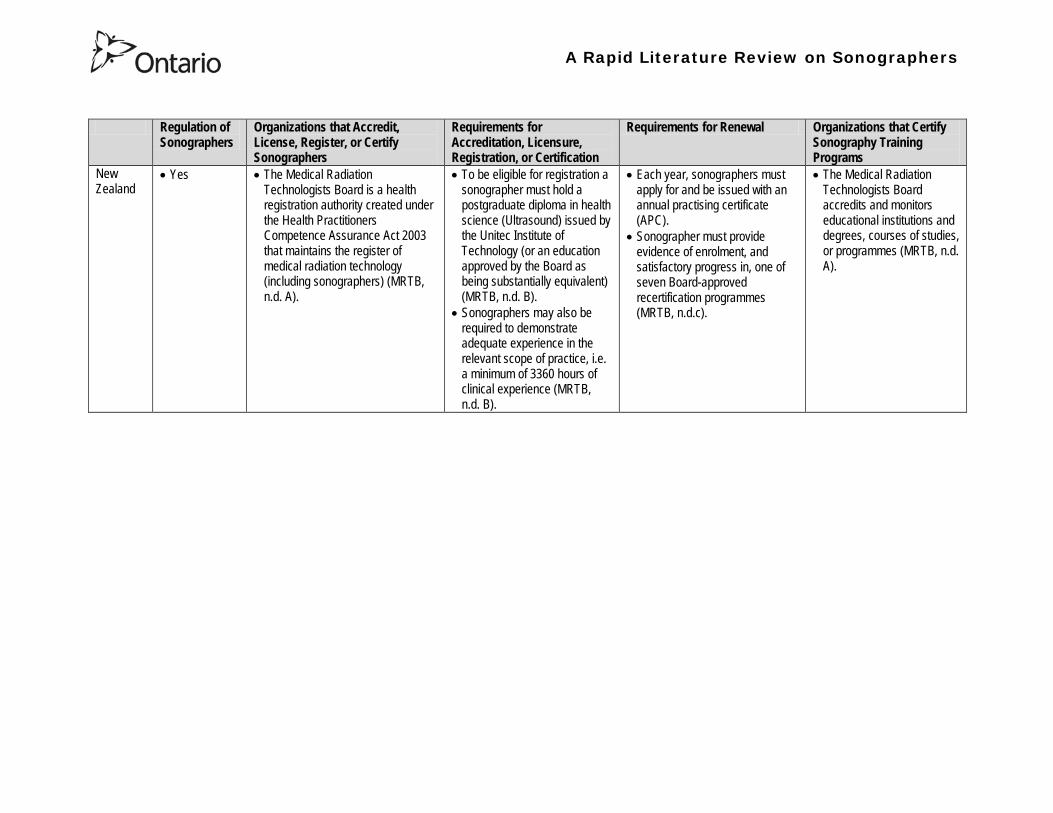
A Rapid Literature Review on Sonographers
Regulation of Sonographers
Organizations that Accredit License Register or Certify Sonographers
Requirements for Accreditation Licensure Registration or Certification
Requirements for Renewal Organizations that Certify Sonography Training Programs
New Zealand
bull Yes bull The Medical Radiation Technologists Board is a health registration authority created under the Health Practitioners Competence Assurance Act 2003 that maintains the register of medical radiation technology (including sonographers) (MRTB nd A)
bull To be eligible for registration a sonographer must hold a postgraduate diploma in health science (Ultrasound) issued by the Unitec Institute of Technology (or an education approved by the Board as being substantially equivalent) (MRTB nd B)
bull Sonographers may also be required to demonstrate adequate experience in the relevant scope of practice ie a minimum of 3360 hours of clinical experience (MRTB nd B)
bull Each year sonographers must apply for and be issued with an annual practising certificate (APC)
bull Sonographer must provide evidence of enrolment and satisfactory progress in one of seven Board-approved recertification programmes (MRTB ndc)
bull The Medical Radiation Technologists Board accredits and monitors educational institutions and degrees courses of studies or programmes (MRTB nd A)
A Rapid Literature Review on Sonographers
Table 2 Examples of Point-of-Care Ultrasound Use Use Examples
Diagnostic Assessment Clinicians from diverse specialties can become very adept at using ultrasonography to examine a particular organ disease or procedure that is directly relevant to their area of expertise whereas imaging specialists typically perform more comprehensive examinations (Moore et al 2011)
bull Emergency physicians have integrated ultrasound into their repertoire for rapid assessment of emergent conditions (Hile et al 2012)
bull A 2012 review on the use of pre-hospital ultrasound concluded that medics (non-physicians trained in the delivery of medical care and the point of injury) can perform ultrasound with a high degree of accuracy (Hile et al 2012)
bull In prehospital settings ultrasound has used been by non-physicians to do fracture detection evaluation of the aorta and Focused Assessment with Sonography for Trauma (FAST) examinations (Hile et al 2012) o The aim of FAST is to assist in the trauma or bleeding patient The areas mentioned
above (perihepatic perisplenic pelvis and pericardium scanning and moreover thoracic ultrasound primarily for haemothorax) are scanned for the detection of fluid as an indication of bleeding rather than organ function (Bennett 2009)
bull Ultrasonography has been used at the Mount Everest base camp to diagnose high-altitude pulmonary edema (Moore et al 2011)
Screening Ultrasonography has been described as a screening test for cardiovascular and gynecologic disease and compact ultrasonography has been incorporated into ldquomobile screening labsrdquo (Moore et al 2011)
bull Ultrasound can be used to screen for abdominal aortic aneurysm (Moore et al 2011)
Guiding Procedures Ultrasound guidance may improve success and decrease complications in procedures performed by multiple specialties (Moore et al 2011)
bull Ultrasound can be used to guide central and peripheral vascular access thoracentesis paracentesis arthrocentesis regional anesthesia incision and drainage of abscesses localization and removal of foreign bodies lumbar puncture biopsies and other procedures (Moore et al 2011)
A Rapid Literature Review on Sonographers
Table 3 Description of the content in the articles being summarized28
No
Description Reference Review Articles
1 Autism spectrum disorders (ASDs) affect an estimated 1 of children in the United States The etiology is probably multifactorial including genetic components and exposure to infections toxins and other environmental factors particularly unfavorable perinatal and neonatal conditions There has been an increase in the frequency of diagnosis of ASDs over the last 20 years with a parallel increase in the use of obstetric diagnostic ultrasound with prenatal ultrasound exposure mentioned as the possible main etiology for autism ldquoepidemicsrdquo Central nervous system alterations have been described in ASDs and certain similar changes have been described in animals after exposure to ultrasound However analysis of in utero exposure in humans has failed to show harmful effects in neonates or children particularly in school performance attention disorders and behavioral changes There is no independently confirmed peer-reviewed published evidence that a cause-effect relationship exists between in utero exposure to clinical ultrasound and development of ASDs in childhood Ultrasound is a form of energy which effects in the tissues it traverses and its use should be restricted to medical indications by trained professionals for as short a period and as low an intensity as compatible with accurate diagnosis
Abramowicz J (2012) Ultrasound and Autism Association Link or Coincidence J Ultrasound Med 311261ndash1269
2 Over the past decade point-of-care ultrasound (US) use by nonphysician providers has grown substantially The purpose of this article is to (1) summarize the literature evaluating military medics facility at US (2) more clearly define the potential utility of military prehospital US technology and (3) lay a pathway for future research of military prehospital US The authors performed a keyword search using multiple search engines Each author independently reviewed the search results and evaluated the literature for inclusion Of 30 studies identified five studies met inclusion criteria The applications included evaluation of cardiac activity pneumothorax evaluation and fracture evaluation Additionally a descriptive study demonstrated distribution of US exam types during practical use by Army Special Forces Medical Sergeants No studies evaluated retention of skills over prolonged periods Multiple studies demonstrate the feasibility of training military medics in US Even under austere conditions the majority of studies conclude that medic can perform US with a high degree of accuracy Lessons learned from these studies tend to support continued use of US in out-of-hospital settings and exploration of the optimal curriculum to introduce this skill
Hile D C Morgan A R Laselle B T amp Bothwell J D (2012) Is Point-of-Care Ultrasound Accurate and Useful in the Hands of Military Medical Technicians A Review of the Literature Military Medicine 177(8) 983-7
28 Please note the studies programs and findings presented in this table may originate from jurisdictions with health systems that are significantly different from Ontarios If there is intent to draw heavily from one or more sources presented in this table we recommend that you contact the lead author of this review for assistance with evaluating the local applicability
A Rapid Literature Review on Sonographers
No Description Reference 3 Ultrasonography is a safe and effective form of imaging that has been used by physicians for more than half a century to aid in
diagnosis and guide procedures Over the past two decades ultrasound equipment has become more compact higher quality and less expensive which has facilitated the growth of point-of-care ultrasonography mdash that is ultrasonography performed and interpreted by the clinician at the bedside In 2004 a conference on compact ultrasonography hosted by the American Institute of Ultrasound in Medicine (AIUM) concluded that ldquothe concept of an lsquoultrasound stethoscopersquo is rapidly moving from the theoretical to realityrdquo This conference included representatives from 19 medical organizations in November 2010 the AIUM hosted a similar forum attended by 45 organizations Some medical schools are now beginning to provide their students with hand-carried ultrasound equipment for use during clinical rotations Although ionizing radiation from computed tomographic (CT) scanning is increasingly recognized as a potentially major cause of cancer ultrasonography has been used in obstetrics for decades with no epidemiologic evidence of harmful effects at normal diagnostic levels However ultrasonography is a user-dependent technology and as usage spreads there is a need to ensure competence define the benefits of appropriate use and limit unnecessary imaging and its consequences This article provides an overview of the history and current status of compact point-of-care ultrasonography with examples and discussion of its use
Moore C L amp Copel J A (2011) Point-of-Care Ultrasonography N Engl J Med 364 749-57
4 Sound travels through objects that block light Only very recently has technology advanced enough to decipher ultrasound for medical use Machines have become smaller cheaper more versatile and more advanced than ever before The medical use of ultrasound spreads across many fields so that traditional areas no longer have a monopoly With this comes the question of training doctors This has been done by various colleges and societies worldwide Some have been quicker to act than others There also needs to be an understanding of when broad experience and advanced technical skills are required or when limited skills will do In addition some procedures can be performed more safely with ultrasound where the knowledge of ultrasound is not paramount This article covers current provision of training in echo-cardiography and ultrasound in areas relevant to anaesthetists who are working in critical care (including accident and emergency) and complex surgery (mainly cardiac)
Bennett S (2009) Training guidelines for ultrasound worldwide trends Best Practice amp Research Clinical Anaesthesiology 23 363ndash373
5 Ultrasound is a commonly employed imaging modality in obstetrics and is generally regarded as safe to the fetus Current ultrasound technology however has significantly higher output potential than older machines used in most clinical studies and the safety profile for the increasing use of Doppler 3-dimensional (D) and 4-D ultrasound with modern machines is unknown This article reviews the current status of ultrasound safety within obstetrics including proposed mechanisms of harm existing scientific and clinical evidence regarding those mechanisms and considerations of safety for the clinical user
Houston L E Odibo A O amp Macones G A (2009) The safety of obstetrical ultrasound a review Prenat Diagn 29 1204ndash1212
A Rapid Literature Review on Sonographers
No Description Reference 6 The purpose of this article is to present the practicing sonographer and sonologist with an overview of the biohazards of ultrasound
and guidelines for safe use Overview Ultrasound is an imaging modality that has important diagnostic value Although useful in a variety of applications diagnostic ultrasound is particularly useful in prenatal diagnosis To date there is no evidence that diagnostic ultrasound produces harm in humans or the developing fetus when used properly There are however an increasing range of ultrasound studies being performed Newer technologies can have higher acoustic output levels than earlier equipment Also subtle or transient effects of diagnostic ultrasound such as changes in membrane permeability or neuronal migration are not completely understood Nondiagnostic Imaging Applications Nondiagnostic uses of ultrasound equipment such as repeated scans of healthy subjects for equipment demonstrations generally should be avoided First-trimester fetal scans should not use color and power Doppler modes and should not be performed for the sole purpose of producing souvenir videos or photographs Production of fetal souvenir photographs or videos during diagnostic clinical studies should not increase exposure levels or extend scan times beyond those needed for clinical purposes Operators should follow safe scanning guidelines and ALARA (as low as reasonably achievable) principles The FDA considers the promotion selling or leasing of ultrasound equipment for making ldquokeepsake fetal videosrdquo that are not part of a medical diagnostic procedure to be an unapproved use of a medical device
NelsonT R FowlkesJ B Abramowicz J S Church C C (2009) Ultrasound Biosafety Considerations for the Practicing Sonographer and Sonologist J Ultrasound Med 28139ndash150
7 Objective A possible association between ultrasound during pregnancy and subsequent non-right handedness among children has been suggested The association has been reported to be stronger among boys The aim of the present study was to explore this further through a meta-analysis of two follow-up studies of three randomized controlled trials of routine ultrasonography during pregnancy Design Handedness was assessed through a questionnaire to the parents and 4715 children at the age of 8ndash9 years were included in the meta-analysis Results There was no statistically significant difference in the prevalence of non-right handedness between the ultrasound-screened children and the controls but there was a statistically significant difference in a subgroup analysis among the boys Exploratory analyses according to exposure or non-exposure indicated an even stronger association between ultrasound and non-right handedness Conclusions A conservative analytical approach indicates no association between ultrasound in utero and subsequent non-right handedness The results from the exploratory analyses must be interpreted with caution There is still a need for further research
Salvesen K A amp Eik-Nes (1999) Ultrasound during pregnancy and subsequent childhood non-right handedness a meta-analysis Ultrasound Obstet Gynecol13 241ndash246
8 Ultrasound energy exerts important cellular genetic thermal and mechanical effects Concern about the safety of ultrasound prompted several agencies to devise regulatory limits on the machine output intensities The visual display of thermal and mechanical indices during ultrasound imaging provides an aid to limit the output of the machine Despite many animal studies no human investigations conducted to date have documented major physiologic consequences of ultrasound exposed during imaging To date ultrasound imaging appears to be safe for use in regional anesthesia and pain medicine interventions and adherence to limiting the output of ultrasound machines as outlined by the Food and Drug Administration may avoid complications in the future This article reviews ultrasound-related biologic effects the role of the regulatory agencies in ensuring safety with the use of ultrasound and the limitations and implications of ultrasound use in humans
Shankar H amp Pagel P S (2011) Potential Adverse Ultrasound-related Biological Effects A Critical Review Anesthesiology 115(5) 1109-1124
A Rapid Literature Review on Sonographers
No Description Reference 9 Objective In the context of the planned International Society of Ultrasound in Obstetrics and GynecologyndashWorld Health Organization
multicenter study for the development of fetal growth standards for international application we conducted a systematic review and meta-analysis to evaluate the safety of human exposure to ultrasonography in pregnancy Methods A systematic search of electronic databases reference lists and unpublished literature was conducted for trials and observational studies that assessed short- and long-term effects of exposure to ultrasonography involving women and their fetuses exposed to ultrasonography using B-mode or Doppler sonography during any period of pregnancy for any number of times The outcome measures were (1) adverse maternal outcome (2) adverse perinatal outcome (3) abnormal childhood growth and neurological development (4) non-right handedness (5) childhood malignancy and (6) intellectual performance and mental disease Results The electronic search identified 6716 citations and 19 were identified from secondary sources A total of 61 publications reporting data from 41 different studies were included 16 controlled trials 13 cohort and 12 casendashcontrol studies Ultrasonography in pregnancy was not associated with adverse maternal or perinatal outcome impaired physical or neurological development increased risk for malignancy in childhood subnormal intellectual performance or mental diseases According to the available clinical trials there was a weak association between exposure to ultrasonography and non-right handedness in boys (odds ratio 126 95 CI 103ndash154) Conclusion According to the available evidence exposure to diagnostic ultrasonography during pregnancy appears to be safe
Torloni M R Vedmedovska Merialdi M Betran A P Allen T et al (2009) Safety of ultrasonography in pregnancy WHO systematic review of the literature and meta-analysis Ultrasound Obstet Gynecol 33 599ndash608
Articles in Peer-Reviewed Journals 10 It has been documented for many years that as a result of use of ultrasound to determine the sex of the fetus the male to female
ratio in countries such as China and India have become increasingly skewed (due to subsequent abortions and infanticide of females) Preference for sons over daughters evident in Chinas and South Asias male sex ratios is commonly rationalized by poverty and the need for old-age support In this article the authors study South and East Asian immigrants to Canada a group for whom the economic imperative to select sons is largely absent Analyzing the 2001 and 2006 censuses 20 percent samples the authors find clear evidence of extensive sex selection in favor of boys at higher parities among South and East Asian immigrants unless they are Christian or Muslim The latter finding accords with the explicit prohibition against (female) infanticidemdashtraditionally the main sex-selection methodmdashin these religions The findings point to a strong cultural component to both the preference for sons and the willingness to resort to induced abortion based on sex
Almond D Edlund L Milligan K (2013) Son Preference and the Persistence of Culture Evidence from South and East Asian Immigrants to Canada Population and Development Review 39(1) 75ndash95
11 The rapid growth in the use of ultrasound as a diagnostic imaging technology over the past forty years has led to a demand for a workforce with the appropriate skills to perform and interpret the scans In the UK ultrasound investigations now comprise the largest group of all diagnostic imaging examinations However there remains no statutory regulation of the practice of sonography in the UK and little recognition of the considerable training that many practitioners have undertaken to obtain the skills to become safe and competent sonographers Many in the field consider that this should change and are working to obtain professional status for the practice of sonography Although the Health Care Professions Council (HCPC) has recommended regulation of sonography practice this is unlikely to happen in the near future This paper discusses the evolution of sonography practice and explores some of the complex issues associated with the professionalisation of sonography
Gibbs V (2013) The long and winding road to achieving professional registration for sonographers Radiography19 164-167
A Rapid Literature Review on Sonographers
No Description Reference 12 Background Contrast echocardiography (which uses contrast agents injected into the blood flow in order to visualize the blood flow
in very small blood vessels) has been shown to improve diagnostic quality especially in technically difficult patients However the learning curve and increased time for preparation and image acquisition have led to low use Methods The authors sought to determine whether the contrast echocardiography procedure performed independently by a specialized trained sonographer could improve efficiency In one centre routine echocardiograms were scheduled for one hour and any study exceeding one hour would result in patient booking cancellations The authors compared the standard of care in which a physician or nurse administers echocontrast with a sonographer-administered program (SAP) Results The time to complete contrast echocardiograms was significantly reduced by the SAP strategy (43 min 17 s plusmn 23 min 42 s vs 1 h 1 min 6 s plusmn 31 min 0 s P lt 0001) Subgroup analysis of the inpatients and outpatients demonstrated similar results Only 10 of studies (6 of 61) in the SAP exceeded 60 minutes compared with 45 (34 of 76) in the standard-of-care group (P lt 0001) Based on study volumes in the centre the net improvement in productivity with the SAP could be up to 53 annually Conclusion Sonographer-administered echocontrast is feasible and potentially removes a barrier to implementation of contrast echocardiography
Tang A Chiew S K Rashkovetsky R Becher H Choy J B (2013) Feasibility of Sonographer-Administered Echocontrast in a Large-Volume Tertiary-Care Echocardiography Laboratory Canadian Journal of Cardiology 29 391ndash395
13 Ultrasound scanning has been used as a diagnostic and screening tool in obstetric practice for over 50 years There is no evidence of immediate or long-term harm to the developing fetus from exposure to B mode ultrasound However exposure to high levels of Doppler ultrasound during early development is increasingly common and the full safety implications of this exposure are not clear Doppler ultrasound exposure in utero gives rise to increased apoptosis in animal models and there is evidence of the effects of exposure to Doppler ultrasound persisting throughout life with increased non-right-handedness observed in human epidemiological studies The authors consider the idea that there may be long-term developmental implications for fetuses exposed to Doppler ultrasound early in gestation These effects may be mediated via thermal or mechanical disruption to the developing conceptus giving rise to free radical damage Excess free radical exposure early in gestation is a strong candidate for the final common pathway underlying developmental programming effects and gives rise to concern that fetuses exposed to high levels of ultrasound are at risk of a developmental programming effect It is suggested that there is a need for animal studies of developmental programming using exposure to Doppler ultrasound scanning as the exposure of interest and for more observational data to be collected in the clinical setting While these data are collected it seems prudent to continue to adhere to the principle of lsquoas low as reasonably achievablersquo (ALARA) when exposing first-trimester fetuses to Doppler ultrasound
Aiken CE Lees CC (2012) Long-term effects of in utero Doppler ultrasound scanning ndash A developmental programming perspective Medical Hypotheses 78 539ndash541
14 Point of care sonography by emergency physicians in the emergency department can be an effective aid in the diagnosis and management of patients presenting with a variety of medical and traumatic conditions Its use can improve patient outcomes enhance patient safety speed patient disposition and save lives CAEP supports the use of point of care sonography
Canadian Assocation of Emergency (CAEP) Physicians Ultrasound Position Statement Working Group (2012) Use of point of care sonography by emergency physicians CJEM 14(2)106-112
A Rapid Literature Review on Sonographers
No Description Reference 15 The use of three-dimensional echocardiography (3DE ie a 3D ultrasound of the heart) during electrophysiology (EP - a branch of
cardiology that deals with the diagnosis and treatment of heart rhythm disorders) procedures is the end product of years of growth in two diverse cardiology subspecialties namely advanced cardiac imaging and the EP During the past decade progress in both fields has resulted in many important advances that have culminated in their union for a new area of growth and development Imaging advances have provided the cardiovascular specialist with enhanced cardiac volume and function data and more recently 3DE capabilities with improved spatial and temporal resolution providing unprecedented spatial relationships This latter development is valued by EP specialists in need of hitherto never required anatomic knowledge as they press forward with extraordinary expansion in their capabilities It makes sense that by combining these two rapidly growing subspecialties future capabilities in patient care may be achieved that would otherwise not be possible This paper discusses the value of 3DE during EP procedures and offers the readers insight into this novel multispecialty hybrid arena Using this model as a template it is likely that the readers may identify other areas within their practices where periprocedural advanced imaging may afford significant dividends in patient outcomes
Moukabary T Faletra F F Kronzon I Thomas W amp Sorrell V L (2012) Three-Dimensional Echocardiography in the Electrophysiology Laboratory Echocardiography 29 117-122
16 Background There has been much discussion about whether female feticide occurs in certain immigrant groups in Canada The authors examined data on live births in Ontario and compared sex ratios in different groups according to the motherrsquos country or region of birth and parity Methods The authors completed a population-based study of 766 688 singleton live births between 2002 and 2007 They used birth records provided by Ontario Vital Statistics for live births in the province between 23 and 41 weeksrsquo gestation They categorized each newborn according to the motherrsquos country or region of birth namely Canada (n = 486 599) Europe (n = 58 505) South Korea (n = 3663) China (n = 23 818) Philippines (n = 15 367) rest of East Asia (n = 18 971) Pakistan (n = 18 018) India (n = 31 978) rest of South Asia (n = 20 695) and other countries (n = 89 074) They calculated malefemale ratios and 95 confidence intervals (CIs) for all live births by these regions and stratified them by maternal parity at the time of delivery (0 1 2 or ge 3) Results Among infants of nulliparous women (women with no previous children) the malefemale ratio was about 105 overall As parity (the number of previous children) increased the ratio remained unchanged among infants of Canadian-born women In contrast the malefemale ratio was significantly higher among infants of primiparous women (women with one previous child) born in South Korea (120 95 CI 109ndash134) and India (111 95 CI 107ndash115) than among infants of Canadian-born primiparous women Among multiparous women (women with 2+ previous children) those born in India were significantly more likely than Canadian-born women to have a male infant (parity 2 ratio 136 95 CI 127ndash 146 parity ge 3 ratio 125 95 CI 109ndash143) Interpretation The study of malefemale ratios in Ontario showed that multiparous women born in India were significantly more likely than multiparous women born in Canada to have a male infant
Ray J G Henry D A Urquia M L (2012) Sex ratios among Canadian liveborn infants of mothers from different countries CMAJ184(9) E92-96
A Rapid Literature Review on Sonographers
No Description Reference 17 This letter consists of a case study that illustrates how a cross-disciplined sonographer (educated in cardiac and vascular ultrasound)
can quickly and efficiently and with great Cost-effectiveness deliver timely care to a patient in certain clinical settings The literature is comparatively sparse in referencing clinical diagnostics in cross-discipline sonographers Echocardiographers must be clinicians fully comfortable with evaluating the circulatory dynamics of the body from the left and right heart to the peripheral arteries and veins including the extracranial circulation (carotid arteries) Included in this skill set is substantive understanding of the ECG patientrsquos history and integration of cardiac and vascular findings Limitations of each diagnostic tool and how these limitations can be overcome by the strengths of the other examination are important For the dual-skilled sonographer only experience continuous education and constant clinical feedback will lead to the independent decision making that is counted on by the physician The American Registry for Diagnostic Medical Sonography offers credentials in cardiac and vascular imaging (RDCS and RVT) Cardiovascular Credentialing International offers credentials in cardiac and vascular imaging as well (registered cardiac sonographer [RCS] and registered vascular specialist [RVS]) All imaging facilities should consider merging cardiac and vascular noninvasive imaging with dual-registered clinical sonographers credentialed by one of these two agencies
Zemanek M (2012) What It Means to Be a Cardiovascular Sonographer Journal of Ultrasound in Medicine 311859-1862
18 Diagnostic medical sonography is viewed as a safe imaging modality However bioeffects research has yet to define the effects of exposure to sonography in humans Therefore sonographers must be aware of potential risks and practice ALARA (ldquoas low as reasonably achievablerdquo) routinely This research explores sonographersrsquo ability to answer basic questions about the bioeffects of sonography as well as their knowledge and use of the safety indices called the mechanical index (MI) and thermal index (TI) Members of the Society of Diagnostic Medical Sonographers in the US a southwestern sonographersrsquo society and a sonography community on Facebook 2010 were surveyed (n = 212) There is a low level of bioeffects knowledge and minimal safety practices among sonographers in this survey No significant relationship was found between sonographer knowledge of the bioeffects of sonography years in the profession or whether the sonographers monitor the MITI
Bagley J Thomas K DiGiacinto D (2011) Safety Practices of Sonographers and Their Knowledge of the Biologic Effects of Sonography Journal of Diagnostic Medical Sonography 27(6) 252ndash261
19 This article highlights trends in echocardiography Point of care ultrasound (hand-carried ultrasound) Initially point of care ultrasound will increase the use of echocardiography in the same manner that cardiac CT increased catheterizations However once the basics are learned it has the potential to be a very powerful tool in that less unnecessary studies will be ordered In the clinic small systems will be used as a lsquolsquofirst lookrsquorsquo screen and become part of the patientrsquos history only patients for whom more testing is necessary will be referred for a comprehensive study This is likely to reduce the number of Doppler studies requested The miniaturization of echo equipment will continue with improved qualityfunctionality of the lsquolsquohand-heldrsquorsquo echo machines to the extent that cordless probes will become a reality sooner rather than later Role in interventional echocardiography Sonographers will be spending much more time in the interventional and electrophysiology (EP) labs There will be more interconnection with interventional cardiologists and the volume will increase as more devices are implanted more percutaneous valves placed and more robotic surgery is performed Crosstraining and multitasking Sonographers entering the profession would be prudent to learn cardiac and vascular imaging Individuals who multitask andor cross-train will be ideal For example a sonographer who starts an IV monitors ECG stress can perform cardiac MRI and can interpret a preliminary report or a nurse who can do an echo monitor sedation and start an IV will be invaluable The sonographer should continue to grow professionally by cross training as institutions try to minimize costs and maximize throughput
McCulloch M L (2011) Thoughts regarding the future of sonography Journal of the American Society of Echocardiography 24(5) 29A-30A
A Rapid Literature Review on Sonographers
No Description Reference 20 Background Radiology reports provide specialist interpretation of images and relate these findings to the patientrsquos symptoms and
signs This study compared ultrasound (USS) reports generated by radiologists and sonographers to determine if any significant or clinically relevant differences existed Methods A retrospective analysis of 624 consecutive USS reports was carried out The reports were assessed for the presence of a lsquodisclaimerrsquo or lsquocaveatrsquo pertaining to the quality of the images and were analysed with respect to the clarity of their wording and ability to answer the clinical request Results The majority of sonographer USS reports contained a disclaimer regarding the quality of the USS images (571) compared with a smaller proportion of radiologist scans (99) (P lt 0001) Overall radiologists performed significantly better in providing a clear negative or positive diagnosis to the clinical question on the request form when compared with sonographers (885 vs 654 P lt 0001) Conclusion lsquoDisclaimerrsquo comments and lsquohedgingrsquo must be avoided in radiology reporting While feedback as to the accuracy of the radiology images is important overuse of such terms undermines the validity of the radiology report
Garcea G Mahmoud A Ong S L Rees Y Berry Det al (2010) Caveat reporting in ultrasound interpretation of surgical pathology a comparison of sonographer versus radiologist Journal of Evaluation in Clinical Practice 16 97ndash99
21 A nine-point questionnaire was constructed based on previously published work by Marsal and Sheiner et al The survey was distributed to clinical users of ultrasound practicing sonography in Hamilton (New Zealand) during July 2010 totalling 35 users consisting of sonographers sonography trainees one obstetric sonologist and one fetal medicine registrar Results Twenty sonographers returned the questionnaire which represents survey response of 61 The level of clinical experience was equally split between junior sonographers of lt 5 yearsrsquo experience (30) sonographers with 5-10 yearsrsquo experience (35) and senior sonographers of gt10 yearsrsquo experience (35) While 100 of the sonographers surveyed reported being familiar with the term Thermal Index (TI) and 90 claimed they adhered to the Action Learning Action Response Association (ALARA) principle all the time or most of the time only 15 of sonographers correctly described or defined what the term TI means Of the sonographers 80 knew where to find the TI during real-time examinations but only 35 knew which TI index to use for nuchal translucency (NT) scan and only 10 knew the expected range of TI encountered during routine obstetric examinations Of the eight sonographers who reported adhering to ALARA all the time the majority (64) could not describe or define TI In the same subgroup the majority (64) did not know which TI to monitor during a NT scan Only three of 20 (15) sonographers knew what the term TI means Of these two were junior sonographers with lt 5 yearsrsquo experience and one had 5ndash10 yearsrsquo experience Despite understanding the term TI one of the three sonographers still selected the incorrect index to monitor during a NT scan Not one of the seven senior sonographers with gt 10 years of experience could define or describe the TI In total only 10 of sonographers knew what the TI means and which TI to monitor during NT scanning
Necas M (2010) New Zealand sonographers do not outperform their European or American colleagues in the knowledge of ultrasound safety Australasian Journal of Ultrasound in Medicine13(4) 28-32
A Rapid Literature Review on Sonographers
No Description Reference 22 Musculoskeletal (MSK) ultrasound performed by non-medical practitioners is a developing practice The aim of this paper is
(1) to audit an experienced sonographerrsquos (SJ Riley one of the authors) performance after one yearrsquos independent reporting against two experienced MSK radiologists (CJ Grove amp M Chandramohan the other two authors) and (2) to establish an audit standard against which such role development might be contrived Images and reports from 250 MSK ultrasound examinations performed by a sonographer were reviewed independently by two consultant MSK radiologists The examinations were graded for discrepancy eg grade 1 ndash agree with the sonographer report grade 2 ndash minor discrepancy unlikely to alter patient care grade 3 ndash potentially significant discrepancy grade 4 ndash definite significant discrepancy likely to have adverse consequences for patient care Two of 250 (08) cases were excluded Both radiologists agreed completely with the sonographer (grade 1) in 235 of the 248 cases (948) In 13 cases there was discrepancy between the reports of SJR and the radiologists The discrepancy was grade 2 in six of the 248 cases (24) grade 3 in six of the 248 cases (24) and grade 4 in one of the 248 cases (04) In conclusion this audit shows a high level of agreement between the sonographer and the consultant MSK radiologist reporting of MSK ultrasound This level of agreement may set the standard for future quality assurance audit of sonographer MSK reporting
Riley S J Groves C J Chandramohan M (2010) Musculoskeletal ultrasound audit of sonographer reporting Ultrasound 18 36ndash40
23 Background Growing demand for ultrasound examinations and higher quality requirements motivate searching for routines combining the diagnostic accuracy of radiologist-performed examinations with the economical advantages of sonographer-performed examinations One possible approach is to use strictly standardized acquisition and documentation schemes that give the radiologist access to all relevant information after the examination Purpose To compare a recently introduced routine combining acquisition by a radiographer (more specifically a sonographer with two years experience) documentation as standardized cine-loops and review by a radiologist (lsquostandardized methodrsquo) with the formerly used routine where the diagnosis is made bedside by the radiologist (lsquotraditional methodrsquo) Material and Methods In 64 policlinic patients the kidneys (n = 27) or the gallbladder (n = 37) were examined with both the standardized and the traditional method The radiologistsrsquo findings of hydronephrosis tumors cysts echogenicity changes and cortical thickness (in the kidneys) and wall thickness concrements and polyps (in the gallbladder) were compared between the methods with respect to agreement (proportion of agreement and kappa coefficient) as well as systematic differences (McNemarrsquos test) Results The findings at the gallbladder examination showed a median agreement of 97 (86ndash100 kappa = 064ndash100) and those at the kidney examination an agreement of 90 (78ndash100 kappa = 069ndash 100) There were no significant systematic differences between the methods Conclusion The satisfactory agreement in this preliminary study indicates that the new workflow with ultrasound examinations performed by a radiographer (sonographer) and analyzed off-line by a radiologist is promising and motivates further studies
Stenman C Thorelius L Knutsson A amp Smedby Ouml (2010) Radiographer-acquired and radiologist-reviewed ultrasound examination ndash agreement with radiologistrsquos bedside evaluation Acta Radiologica 52 70ndash74
A Rapid Literature Review on Sonographers
No Description Reference 24 The troubling practice of fetal sex selection has historically been considered an Asian phenomenon However recent evidence shows
that a similar situation is emerging in North America albeit on a smaller scale The Society of Obstetricians and Gynaecologists of Canada (SOGC) has firmly stated its opposition to sex selection for non-medical reasons as well as to the use of any technology used solely for the purpose of determining fetal sex However because fetal sex may be disclosed to the parents at the time of ultrasound examination if they request this information guidance for health care professionals to assist in discouraging fetal sex selection would be useful Because no declaration of motives or reasons is required when a woman seeks a termination of pregnancy the authors suggest that health care professionals need not disclose the sex of a fetus until it has reached a gestational age at which abortion for non-medical purposes would not be possible This proposal would facilitate consistency between clinical practice and the values of Canadian citizens the SOGC the Canadian Medical Association and other professional organizations while still respecting current laws pertaining to disclosure of patient information and patientsrsquo rights to autonomous decision-making
Thiele A T amp Leier B (2010) Towards an Ethical Policy for the Prevention of Fetal Sex Selection in Canada J Obstet Gynaecol Can 32(1) 54ndash57
25 Background The extent to which increases in the use of prenatal ultrasonography over time reflect changes in maternal risk (ie an increase in high-risk pregnancies over time) is unknown In this population-based study the authors examined the use of prenatal ultrasonography from 1996 to 2006 in Ontario Methods With fiscal year 199697 as the baseline the authors evaluated the relative risk (RR) and 95 confidence interval (CI) for the change in rates of ultrasonography for each subsequent year The RR was adjusted for maternal age income rural residence maternal comorbidities receipt of genetics consultation or amniocentesis mdash all in the index pregnancy mdash and history of complications in a prior pregnancy Results The study sample consisted of 1 399 389 singleton deliveries The rate of prenatal ultrasonography increased from 2055 per 1000 pregnancies in 1996 to 3264 per 1000 in 2006 (adjusted RR 155 95 CI 154ndash155) The rate increased among both women with low-risk pregnancies (adjusted RR 154 95 CI 153ndash155) and those with high-risk pregnancies (adjusted RR 155 95 CI 154ndash157) The proportion of pregnancies with at least four ultrasound examinations in the second or third trimesters rose from 64 in 1996 to 187 in 2006 (adjusted RR 268 95 CI 261ndash274) Paradoxically this increase was more pronounced among low-risk pregnancies (adjusted RR 292 95 CI 283ndash301) than among high-risk pregnancies (adjusted RR 225 95 CI 216ndash235) Interpretation Substantial increases in the use of prenatal ultrasonography over the past decade do not appear to reflect changes in maternal risk Nearly one in five women now undergo four or more ultrasound examinations during the second and third trimesters Efforts to promote more appropriate use of prenatal ultrasonography for singleton pregnancies appear warranted
You J J Alter D A Stukel T A McDonald S D Laupacis A et al (2010) Proliferation of prenatal ultrasonography CMAJ 182(2) 143-151
A Rapid Literature Review on Sonographers
No Description Reference 26 Aims The aim of this study was to assess the cost-effectiveness of using certified sonographers and miniaturized echocardiography
systems to perform echocardiograms at bedside in comparison to moving inpatients from the admission department to the echocardiography laboratory (echo-lab) Methods and Results From 26 September 2005 to 27 October 2005 112 patients admitted in six hospital wards in Italy connected through a 100 Mbit LAN to the echo-lab were scanned within the admission ward by sonographers using a miniaturized echo system Logistical data were collected and results were compared with those obtained from 194 consecutive patients coming from the same wards and studied in the echo-lab with high-end machines between 8 March 2005 and 15 April 2005 Performing echocardiograms in the admission department avoided long waiting time of the inpatients in the echo-lab before and after the study increased the percentage of patients studied within 3 and 5 days from request (88 vs 77 and 100 vs 95 respectively P = 003) increased both sonographer (by 339 P lt 0001) and echo-lab productivity (by 41 P lt 0001) and reduced costs of echocardiograms by 29 Conclusion Implementation of digital echocardiography certified sonographers and a miniaturized echo system allowed improvement of the cost-effectiveness of the service provided by the echo-lab for inpatients and avoided patientsrsquo discomfort derived from prolonged waiting time before and after the exam
Badano L Gaetano N Stacul S Gianfagna P Pericoli M et al (2009) Improved workflow sonographer productivity and cost-effectiveness of echocardiographic service for inpatients by using miniaturized systems European Journal of Echocardiography 10 537ndash542
27 The present report is a proposal from the American Society of Echocardiography Advanced Practice Task Force that identifies the potential of cardiac sonographers to achieve the Advanced Cardiovasculat Sonographer (ACS) level According to this report echocardiographic examinations require a well-trained and competent sonographer to obtain proper anatomic and physiologic data to establish an accurate diagnosis for clinical decision-making and patient management Although the formal education and training of cardiovascular sonographers are evolving many entry-level and staff sonographers may not have sufficient practical or clinical knowledge of the necessary components of the echocardiographic study for the individual patientrsquos clinical presentation In many clinical settings echocardiograms are read after the patient has left the laboratory Thus there is a role for a sonographer who can practice at an advanced level in a cardiovascular ultrasound laboratory to ensure a proper echocardiographic examination is performed on every patient In this setting an Advanced Cardiovascular Sonographer (ACS) would be able to review the indication for and quality of the examination If additional images were needed the ACS would assist the sonographer in obtaining these images which would lead to the performance of a complete and fully diagnostic examination before the patient had left the echocardiography laboratory In clinical practice the quality of the examinations performed would improve advancements in echocardiographic methods could be taught and incorporated into daily practice and patients would be better served
Mitchell C Miller F A Bierig S M Bremer M L Ehler D et al (2009) Advanced Cardiovascular Sonographer A Proposal of the American Society of Echocardiography Advanced Practice Sonographer Task Force J Am Soc Echocardiogr 22 1409-13
A Rapid Literature Review on Sonographers
No Description Reference 28 Sonographer practitioner development involves the expansion and extension of the sonographer role to include reporting on
ultrasound examinations Australian sonographers have not seen the same degree of role extension and expansion as their counterparts in the United Kingdom despite increasing levels of discussion regarding sonographer practitioner development The aim of this study was to determine if Australian sonographers want to extend their professional role and what they consider are the important issues associated with role extension This paper reports on qualitative data derived from a survey of Australian sonographers and investigates if Australian sonographers are interested in extending and expanding their professional role and responsibilities and if they do what might be necessary or desirable from a professional point of view for this change to occur A survey was mailed to all members of the Australian Sonographers Association (ASA) in October 2006 The 31-item survey included 28 closed-ended and three opened-ended items to provide both quantitative and qualitative data The quantitative data will be reported separately Qualitative data was derived from responses to the opened-ended questions which asked respondents to elaborate on their attitudes and feelings about role extension and development Analysis used Nvivo7 software to aid in uncovering common themes from the qualitative data The analysis focused on the reported incentives or motivations for becoming a sonographer practitioner as well as disincentives or perceived hurdles that would discourage respondents from becoming sonographer practitioners The three most reported incentives or motivations for becoming a sonographer practitioner were professional recognition remuneration and increased knowledge The three most commonly reported disincentives or perceived hurdles that would discourage respondents from becoming sonographer practitioners were legal issues insurance and further study
McGregor R OrsquoLoughlin K Cox J Clarke J Snowden A (2009) Sonographer practitioner development in Australia Qualitative analysis of an Australian sonographersrsquo survey Radiography15 313-319
29 Introduction This study describes the effect of introducing a cardiac sonographer led follow up clinic within a hospital cardiology department for patients with stable heart valve disease The two years before and after the instigation of the valve clinic were audited Materials and methods The clinic was conducted in a single centre and undertaken in the cardiology department of a district general hospital A total of 382 patients with 397 clinically significant valve lesions but for whom surgery was not yet indicated but follow up required were seen in a cardiac sonographer run clinics These patients no longer attended a medical follow up clinic unless there was clinical or echocardiographic deterioration Effectiveness was judged by the percentage treated according to current best practice guidelines the number of echocardiograms performed and the number of hospital outpatient visits attended In addition mortality data for the subjects in the clinic was collected Results The proportion followed up according to best practice guidelines rose from 157 (41) to 354 (92) (p lt 001) The total number of echocardiograms performed fell from 807 to 550 Total number of outpatient visits fell from 998 to 31 A total of 11 patients died in the two year study period none from progressive valve disease Discussion This study demonstrates that a protocol driven sonographer led heart valve disease follow up clinic significantly improved the quality of follow up while bringing about a major reduction in outpatients visits without compromising patient safety
Taggu W Topham A Hart L Carr-White G Sulke N et al (2009) A cardiac sonographer led follow up clinic for heart valve disease International Journal of Cardiology 132 240 ndash243
30 Role development for imaging allied health professionals has progressed significantly over the last three decades with many major developments pioneered in the field of ultrasound This paper reviews the developing role of the radiographer in ultrasound (in some countries such as the UK sonographers are considered a type of radiographer) and some of the clinical professional and educational implications of such development As the published evidence base underpinning the latest developments is scarce several contemporary case study examples which have been presented verbally at study days attended by the authors have been included In conclusion sonographer role development is considered within the professional contexts of extended role advanced and consultant practice
Hart A amp Dixon A (2008) Sonographer Role Extension and Career Development a Review of the Evidence Ultrasound 16(1) 31-35
A Rapid Literature Review on Sonographers
No Description Reference 31 Objective The main goal of this study was to determine end usersrsquo knowledge regarding safety aspects of diagnostic ultrasound
during pregnancy End usersrsquo attitudes toward the use of ultrasound in low-risk pregnancies were also assessed Methods A questionnaire was distributed to ultrasound end users attending review courses and hospital grand rounds between April and June 2006 in Chicago Results One hundred thirty end users completed the questionnaires (63 response rate) Sixty-three percent were physicians (n = 84) most of them obstetricians (817) About 18 of participants routinely performed Doppler ultrasound examinations during the first trimester Fifty percent of end users thought that the number of ultrasound examinations in low-risk pregnancy should be limited to 1 to 3 (mean plusmn SD 26 plusmn 09) Almost 70 disapproved of ldquokeepsakeentertainmentrdquo ultrasound Although 322 of the participants were familiar with the term thermal index only 177 actually gave the correct answer to the question on the nature of the thermal index About 22 were familiar with the term mechanical index but only 38 described it properly Almost 80 of end users did not know where to find the acoustic indices Only 208 were aware that they are displayed on the sonographic monitor during the examinations End users with higher knowledge of safety issues thought that there should be limitations on the number of ultrasound examinations in low-risk pregnancies (odds ratio 33 95 confidence interval 11ndash100 P = 028) Likewise these end users were more likely to respond that ultrasound might have adverse effects during pregnancy (odds ratio 32 95 confidence interval 11ndash125 P = 045) Conclusions Ultrasound end users are poorly informed regarding safety issues during pregnancy Further efforts in the realm of education and training are needed to improve end user knowledge about the acoustic output of the machines and safety issues
Sheiner E Shoham-Vardi I Abramowicz J S (2007) What Do Clinical Users Know Regarding Safety of Ultrasound During Pregnancy J Ultrasound Med 26 319ndash325
A Rapid Literature Review on Sonographers
No Description Reference 32 In an attempt to survey the knowledge among ultrasound users of some safety aspects of diagnostic ultrasound a questionnaire was
distributed to professionals using ultrasound for fetal examinations The questionnaire was answered by the participants and faculty members of six postgraduate courses in obstetric ultrasound Doppler ultrasound and fetal echocardiography given in Sweden Norway and Austria in 2003 Furthermore in Sweden the questionnaire was distributed to the staff meeting participants of two major obstetric ultrasound units and of one unit for fetal echocardiography and at a meeting of the Working Group for Ultrasound of the Swedish Society of Obstetrics and Gynecology In total 199 questionnaires were anonymously answered by doctors (n = 145) sonographers (n = 22) and midwives trained in ultrasound (n = 32) All of them were using diagnostic ultrasound on a daily or weekly basis The respondents were from nine European countries In none of the countries is formal certification for the use of diagnostic ultrasound in pregnancy or otherwise required The doctors who answered the questionnaire were specialists in obstetrics and gynecology (including some of the internationally recognized experts in fetal ultrasound) pediatric cardiology radiology or clinical physiology The respondents were not informed about the questionnaire in advance and they had a limited time to answer the 16 questions typically 10ndash15 min The questions concerned explanation of the safety indices and their relation to various ultrasound modes ie real-time B-mode M-mode three-dimensional (3D) spectral Doppler color and power Doppler ultrasound The respondents were also asked which ultrasound system they used most often and where on the system the information on thermal index (TI) and mechanical (MI) was displayed (eg in the top right corner of the screen in a separate window etc) Furthermore the respondents had to indicate how they adjust the output energy level on their own machine For all questions there was the optional answer lsquoDonrsquot knowrsquo Results About one-third of the ultrasound experts were able to define the abbreviations TI and MI For the subclasses of the TI the correct answer was found in only 3-8 of cases Of those who knew what the TI and MI meant only two-thirds and one-third of respondents respectively were able to give a simple explanation of the indices In 49 of questionnaires the Doppler modes were correctly ranked as generally producing higher ultrasound exposure than the imaging modes (B-mode 3D) Some 28 of the respondents correctly indicated where on their own machine the information on safety indices is displayed However not all of them knew how to control the output energy level For all items in the questionnaire there were no significant differences in the results between the three categories of respondents (ie physicians sonographers and midwives) The results of the questionnaire were anything but encouraging and they indicated that the users who are supposed to be responsible for controlling exposure of the fetus to ultrasound had a very poor knowledge of the basic safety aspects of ultrasound
Maršaacutel K (2005) The output display standard has it missed its target Ultrasound Obstet Gynecol 25 211ndash214
33 Purpose This study was performed to evaluate the accuracy of upper abdominal ultrasound (US) scanning performed by sonographers in a district general hospital to identify potential areas of weakness and to make recommendations to improve the service Materials and Methods Upper abdominal US examinations performed and reported by sonographers over a 4-week period were retrospectively reviewed The accuracy of the imaging findings and reports were assessed against other imaging surgical histological or laboratory findings and against clinical outcome Results A heterogenous group of 104 patients were included in the study 62 of whom had an US abnormality Errors of scanning or interpretation were identified in 10 patients (96) of whom five (48) were felt to be potentially significant Conclusions The sonographersrsquo accuracy in reporting upper abdominal US scans was 90 However on the basis of this study the authors have implemented specific recommendations to improve the quality of the service
Dongola N Guy R Ward S amp Giles J A (2003) Can sonographers offer an accurate upper abdominal ultrasound service in a district general hospital Radiography 9 29ndash33
A Rapid Literature Review on Sonographers
No Description Reference 34 Aim To compare the accuracy of radiographers and radiologists in routine abdominal ultrasound Materials and Methods One
hundred consecutive patients attending for routine abdominal ultrasound were included Each patient was examined by both a radiographer and radiologist Both operators noted their fndings and wrote a concluding report without conferring Reports were compared Where there was disagreement the patient was either re-examined by another radiologist or had further investigation Results Of 100 patients 52 were men and 48 were women The age range was 19plusmn88 years (median 52 years) Thirty-seven patients had renal tract ultrasound one had an aortic ultrasound and 62 had general upper abdominal ultrasound In 44 cases both operators reported the examination as normal In 49 cases both operators reported the examinations as abnormal and there was complete agreement between the operators In seven cases there was not complete agreement between operators Three of these disagreements were considered minor and four major In three of the seven cases the radiographer was correct and in four the radiologist was correct Conclusion Experienced radiographers and radiologists are highly accurate in performing and interpreting routine abdominal sonography Both operators missed a small minority of abnormalities There was no statistically significant difference in the accuracy of radiographers and radiologist
Leslie A Lockyer H amp Virjee J P (2000) Who Should be Performing Routine Abdominal Ultrasound A Prospective Double-Blind Study Comparing the Accuracy of Radiologist and Radiographer Clinical Radiology 55 606-609
Grey Literature 35 The ARRT The American Registry of Radiologic Technologists tests certifies and annually registers more than a quarter-of-a-million
radiologic technologists to promote high standards of patient care They award the Registered Technologist or RT designation to individuals who have completed the prescribed classroom and clinical education passed the appropriate exam and met the ethics requirements And they ensure their continuing education and ongoing compliance with their high standards by requiring annual registration of their certificate Certification by ARRT is voluntary not mandatory But employers state licensing agencies and federal regulators view ARRTrsquos credential as an indicator that a person has met recognized national standards for medical imaging and radiation therapy professionals How Are Radiologic Technologists Deemed Qualified First comes initial certification Certification is the process of initially recognizing individuals who have satisfied certain standards within a profession A person is certified by ARRT after completing an approved educational program complying with the ethics standards and passing a certification exam Then comes annual registration Registration is the yearly procedure required to maintain certification ARRT registrants are those who having already fulfilled the requirements for initial certification continue to meet the requirements for annual registration The ARRT annually registers the certificates of individuals who meet the following three criteria agreeing to comply with the ARRT Rules and Regulations continuing to comply with the ARRT Standards of Ethics and meeting our continuing education requirements Only technologists who are currently registered (which means that they have renewed within the past year) may designate themselves as ARRT Registered Technologists and use the initials RT after their names Federal laws regulate personnel qualifications in some modalities (mammography for example) but they accredit the health care facility rather than licensing the individual radiologic technologist who is providing patient care And while many states have laws regulating who can perform radiologic procedures they are typically based on ARRT certification Some employers and state agencies may require ARRT certification but not necessarily current registration Unless they require current registration by ARRT employers cannot assume that the person has complied with the ethics requirements or the continuing education requirements
American Registry of Radiologic Technologists (ARRT) (2013a) Patient Page Accessed April 2013
A Rapid Literature Review on Sonographers
No Description Reference 36 This document outlines certification requirements for sonography There are two pathways (primary and post-primary) For the
primary pathway it notes that sonography educational programs must be accredited by a mechanism acceptable to the ARRT ARRT generally recognizes only accreditation agencies that are recognized by the Council for Higher Education Accreditation andor United States Department of Education
American Registry of Radiologic Technologists (ARRT) (2013b) Sonography Certification Accessed April 2013
A Rapid Literature Review on Sonographers
No Description Reference 37 In Australia sonographers and student sonographers are required to be accredited by Australian Sonographer Accreditation Registry
(ASAR) in order to practise This requirement is contained within the Australian Governmentrsquos Diagnostic Imaging Accreditation Scheme standards and Medicare legislation Accreditation of sonographers is linked only to achieving - or in the case of students studying towards - an accredited entry-level qualification completion of continuing professional development and payment of an annual fee to ASAR The ASA contends that the lack of a requirement to ensure each sonographer is lsquofit to practisersquo is an unacceptable gap in current regulation undermining efforts to ensure the quality of sonographic services and failing to protect patients from harm The ASA supports and advocates for nationally-enforceable regulation of sonographers to meet our patientrsquos expectations National registration The National Registration and Accreditation Scheme (NRAS) for health workers commenced on 1 July 2010 The ten professions which comprised the initial intake into this scheme were those that had registration requirements in all Australian states or territories as well as those in which legislated regulation is deemed to be in the public interest An additional four professions - including medical radiation practitioners which are defined as radiographers radiation therapists and nuclear medicine technologists - will be included in the NRAS commencing on 1 July 2012 As sonography did not have registration in any states or territories prior to the commencement of NRAS the profession has not been in a position to negotiate entry into NRAS In early August 2011 the ASA lodged a submission with the Health Workforce Principal Committee requesting a profession-specific Sonography Board of Australia be formed under NRAS This submission was prepared based on information that consideration of professions which were unregistered would commence More recently the Australian Health Workforce Ministerial Council (AHWMC) issued a communiqueacute stating in part lsquoMinisters have commissioned work on unregistered professions and future directions for national registration Given this Ministers agreed to defer consideration of the inclusion of additional professions into the NRAS Ministers agreed that it would be premature to consider further applications for the scheme until this work has been completed other than paramedicsrsquo Options for unregistered health practitioners including sonographers Policy work is continuing at a federal level to establish an alternative regulatory framework The National Alliance for Self-Regulating Health Professions (NASRHP) consisting of the peak professional associations of eight self-regulating health professions including the ASA representing sonography released a proposal lsquoHarnessing self-regulation to support safety and quality in healthcare deliveryrsquo in March 2012 This proposal concluded that the framework that regulates all health practitioners should be expanded beyond NRAS to include
bull authorised self-regulating professions via a health professions panel bull negative licensing to withdraw the ability to practise from registered and self-regulated practitioners who do not meet the
regulatory requirements or unregulated practitioners due to poor practice As with all advocacy initiatives consideration by the federal government of the introduction of the NASRHPrsquos proposal may be prolonged and the outcomes may not match the expectations of NASRHPrsquos member professional associations including the ASA The ASA will continue both independently and collaboratively to lobby on behalf of our members and the public for the introduction of a timely and equitable solution to health practitioner regulation In the interim as there is no regulatory model with the rigour the ASA believes is required for regulation of sonographers and other health practitioners other than NRAS the ASA continues to support its Position Statement published in early 2010
Australian Sonographer Association (ASA) (2013) Sonographer Regulation Accessed April 2013
A Rapid Literature Review on Sonographers
No Description Reference 38 This publication contains information about Cardiovascular Credentialing International (CCI) It also provides the necessary
information and references concerning the process by which candidates may earn certificate or registry-level credentials CCI is a not-for-profit corporation established for the purpose of administering credentialing examinations as an independent credentialing agency CCI began credentialing cardiovascular professionals in 1968 CCI offers eight credentials which when earned demonstrate that the registrant holds fundamental knowledge in the particular cardiovascular specialty These include RCCS ndash Registered Congenital Cardiac Sonographer RCS ndash Registered Cardiac Sonographer and RPhS ndash Registered Phlebology Sonographer
Cardiovascular Credentialing International (2013) 2013 Examination Application amp Overview Accessed April 2013
39 Oregon Governor Signs Sonographer Licensure Legislation On July 28 2009 Oregon Governor Ted Kulongoski signed a sonographer licensure bill (HB 2245) into law The new law makes Oregon only the second state in the United States to require licensure of sonographers The Society of Diagnostic Medical Sonographers (SDMS) has worked closely with other sonography-related organizations and Oregon sonographers to help craft the language in new law The law renames and restructures the Oregon Board of Radiologic Technologists to the Oregon Board of Medical Imaging (OBMI) and ensures representation of sonographers on the Board The sonographer licensure law requires Oregon sonographers to become licensed by OBMI effective July 1 2010 Beginning on January 1 2014 all sonography licensees would be required to hold a national sonography certificationcredential (or be currently enrolled as a student) While the Oregon bill is far more detailed than the sonographer licensure law that passed in New Mexico earlier this year additional administrative rulesregulations will be developed over the next year to fully implement the new law SDMS will be continue to monitor developments regarding sonographer licensure in other states
Society of Diagnostic Medical Sonographers (SDMS) (2013a) News Accessed April 2013
A Rapid Literature Review on Sonographers
No Description Reference 40 On April 6 2009 New Mexico (NM) Governor Bill Richardson signed the historic bill that for the first time in the United States
requires licensure of sonographers The bill adds sonographers (and MRI technologists) to the list of medical imaging professionals licensed by the State of New Mexico Until now anyone could perform sonograms in New Mexico Prior to the passage of the New Mexico licensure law virtually all other health care providers and professionals had to be licensed As a result the public is often surprised to learn that the person performing a medical sonogram does not have any state licensure or national certification requirements The responsibility for administering the NM sonographer licensure program would rest with the New Mexico Environment Department Although this was not the first choice in most sonographersrsquo minds the Environment Department currently manages the radiologic technology licensure program so the administrative infrastructure is already in place By ensuring the creation of an advisory council that includes representation by each medical imaging and radiation therapy modality regulated the bill helps ensure that the Departmentrsquos other areas of emphasis do not detract from sonographer licensure Because sonographers have never had to be licensed before they frequently do not see or understand the distinction between state licensure and national certification National certifications or credentials issued by voluntary organizations such as the American Registry for Diagnostic Medical Sonography (ARDMS) Cardiovascular Credentialing International (CCI) and the American Registry of Radiologic Technologists (ARRT) have little legal weight The certification process and resulting credential are valuable tools in ensuring sonographers have met minimum education and knowledge requirements but do not really regulate anyonersquos ability to perform sonography In an ideal world no additional regulation of sonography would be required However as everyone knows more and more people are taking weekend courses and buying ultrasound equipment without understanding the ramifications of using ultrasound technology (eg ALARA) Under the bill passed by the New Mexico legislature national credentialscertifications should provide evidence of meeting the statersquos standards All sonographers who are performing these critical medical imaging services in New Mexico will be required to meet national certification standards (ie have certifications or credentials issued by ARDMS CCI or ARRT) As a result there would be no additional exams in New Mexico beyond the national certification exams required by ARDMS CCI or ARRT The bill also requires the state to recognize the continuing medical education completed for national certification renewal The most visible impact on New Mexico sonographers will be a license fee However the legislation caps the license fee at $100 per two-year license In addition only the New Mexico Legislature can change this cap
SDMS (2013b) New Mexico Sonographer Licensure Law Signed Accessed April 2013
A Rapid Literature Review on Sonographers
No Description Reference 41 Preamble The purpose of this document is to define the Scope of Practice for Diagnostic Ultrasound Professionals in the US and to
specify their roles as members of the health care team acting in the best interest of the patient This scope of practice is a living document that will evolve as the technology expands Definition of the Profession The Diagnostic Ultrasound Profession is a multi-specialty field comprised of Diagnostic Medical Sonography (with subspecialties in abdominal neurologic obstetricalgynecologic and ophthalmic ultrasound) Diagnostic Cardiac Sonography (with subspecialties in adult and pediatric echocardiography) Vascular Technology and other emerging fields These diverse specialties are distinguished by their use of diagnostic medical ultrasound as a primary technology in their daily work Certification is considered the standard of practice in ultrasound Individuals who are not yet certified should reference the Scope as a professional model and strive to become certified Scope of Practice of the Profession The Diagnostic Ultrasound Professional is an individual qualified by professional credentialing and academic and clinical experience to provide diagnostic patient care services using ultrasound and related diagnostic procedures The scope of practice of the Diagnostic Ultrasound Professional includes those procedures acts and processes permitted by law for which the individual has received education and clinical experience and in which heshe has demonstrated competency Diagnostic Ultrasound Professionals
bull Perform patient assessments bull Acquire and analyze data obtained using ultrasound and related diagnostic technologies bull Provide a summary of findings to the physician to aid in patient diagnosis and management bull Use independent judgment and systematic problem solving methods to produce high quality diagnostic information and
optimize patient care
Society of Diagnostic Medical Sonography [SDMS] (2013c) Scope of Practice for the Diagnostic Ultrasound Professional Accessed April 2013
42 This website notes that in the US most employers in the US prefer to hire sonographers who have professional certification A sonographer can get certification and passing an exam Most exams relate to the specialty that the sonographer is most interested inmdashfor example an exam to become certified in abdominal sonography A few states require diagnostic medical sonographers to be licensed Typically professional certifications required for licensure other requirements vary by state Sonographers must take continuing education to keep their certification current
Bureau of Labor Statistics US Department of Labor (2012) How to become a Diagnostic Medical Sonographer Occupational Outlook Handbook 2012-13 Edition Accessed April 2013
A Rapid Literature Review on Sonographers
No Description Reference 43 The American Registry for Diagnostic Medical Sonography (ARDMS) is an independent not-for-profit organization founded in 1975
that administers examinations and awards the following credentials bull RDMSreg ndash Registered Diagnostic Medical Sonographerreg bull RDCSreg ndash Registered Diagnostic Cardiac Sonographerreg bull RVTreg ndash Registered Vascular Technologistreg bull RPVIreg ndash Registered Physician in Vascular Interpretationreg bull RMSKtrade - Registered in Musculoskeletaltrade sonography
ARDMS is the globally recognized standard of excellence in sonography and has certified more than 75000 individuals ARDMS is composed of three key groups
bull Board of Directors ndash Sonographers vascular technologists physicians research scientists and a member of the public are volunteer members of the board that determines the direction and policies of our organization
bull Examination Development Task Forces ndash A group of sonographers vascular technologists physicians and scientists volunteer their time and expertise to produce our examinations
bull Professional Staff ndash A paid highly-skilled staff administers the programs and assists applicants and registrants Credentials awarded by ARDMS document personal achievement of recognized professional standards and are widely accepted in the medical community by sonography and vascular professional organizations However ARDMS does not validate the day-to-day job performance of any sonography professional ARDMS has earned the prestigious ANSI-ISO 17024 accreditation for certifying bodies from the International Organization for Standardization (ISO) Accreditation is granted through the American National Standards Institute (ANSI) ARDMS has also earned accreditation with the National Commission for Certifying Agencies (NCCA) The NCCA is the accrediting arm of the National Organization for Competency Assurance (NOCA) Established in 1977 as a nonprofit organization NOCA is a leader in establishing quality standards for certifying organizations
American Registry for Diagnostic Medical Sonography (ARDMS) (2012a) Overview of ARDMS Accessed April 2013
A Rapid Literature Review on Sonographers
No Description Reference 44 ARDMS is developing a program to assure that Registrants continue to meet the basic competency requirements of core clinical skills
in present day practice bull Each Registrant who holds an active ARDMS certification as of December 31 2011 will be assigned the 2012 ndash 2021 10-
year recertification assessment period bull The 10-year period for new Registrants will begin the year following the attainment of the credential bull If a Registrant earns a new credential or specialty after January 1 2012 then that credentialspecialty will have a separate
10-year recertification assessment period that will begin the year following the attainment of the credentialspecialty ARDMS will develop tools and resources to assist Registrants in tracking and meeting the recertification assessment program requirements
bull Registrants who do not wish to take a recertification assessment in a specific credentialspecialty may voluntarily relinquish the credentialspecialty
As an ARDMS Registrant individuals are committing to demonstrate the highest level of competency in the modality of sonography by meeting and maintaining the standards set by the ARDMS The pathway of maintenance your certification is comprised of Paying the ARDMS annual renewal fee each year Each year ARDMS assesses Registrants an annual renewal fee By paying your annual renewal fee you are contributing to the ARDMS mission of promoting quality care and patient safety through the certification and continuing competency of ultrasound professionals In order to maintain your credentials you must pay the annual renewal fee of $75 USD or $100 USD (for RPVI and MSK Registrants with MDDODPM) designation on or before December 31 of each year in order to stay active for the following year Attesting to ARDMS rules and policies Each year when you pay your annual renewal fee you are attesting that you are in compliance with all ARDMS rules and discipline policies and are in good standing with the ARDMS Earning a minimum of 30 ARDMS-accepted CMEs during your three-year CME period ARDMS verifies its Registrants compliance with continuing medical education (CME) requirements using an audit process Earning your CMEs is an integral component of maintaining your ARDMS credential(s) and demonstrates your ongoing commitment to lifelong learning RecertificationContinued Commitment to Excellence Following the attainment of the ARDMS credential Registrants are assigned a 10-year recertification period the year after the credential is earned ARDMS is committed to developing a recertification program that is easy for Registrants to comply with
American Registry for Diagnostic Medical Sonography (ARDMS) (2012b) Recertification General Information
A Rapid Literature Review on Sonographers
No Description Reference 45 This document reviews Sonography Principles and Instrumentation (SPI) Examination Requirements offered through ARDMS in
order to achieve sonography certification and the various prerequisites needed before taking the exam For example one prerequisite entails (1) Education Graduate of a program accredited by an agency recognized by the Council for Higher Education Accreditation (CHEA) United States Department of Education (USDOE) or Canadian Medical Association (CMA) that specifically conducts programmatic accreditation for diagnostic medical sonographydiagnostic cardiac sonographyvascular technology Currently the only organizations that offer programmatic accreditation under the aforementioned associations are the Commission on Accreditation of Allied Health Education Programs (CAAHEP) and the Canadian Medical Association (CMA) (2) Required Clinical UltrasoundVascular Experience No additional experience is required (3) Documentation Required with Application 1) Copy of diploma from ultrasoundvascular program or an official transcript indicating the date the degree was conferred 2) Original letter signed by program director andor medical director indicating date of graduation or successful completion of the program4 Program directors must use the mandatory formatted sample letter available on ARDMSorg sampleletters 3) The CV form is not required if the application is submitted and received in the ARDMS office within one year after successful completion of the program Otherwise an original signed and completed CV form for each appropriate specialty area(s) must be submitted 4) Photocopy of a non-expired government issued photo identification with signature the name on the identification must exactly match the name under which you are applying for ARDMS examination
American Registry for Diagnostic Medical Sonography (ARDMS) (2012c) SPI Requirement and General Prerequisites Accessed April 2013
46 This page notes the five credentials offered by the ARDMS and the corresponding exams and specialty exams required bull RDMS requires the SPI exam (sonography principles and instrumentation) and a specialty exam in abdomen breast fetal
echocardiography neurosonolgoy or obstetricsgynecology bull RDCS requires the SPI exam and a speciality exam in adult echocardiography fetal echocardiography pediatric
echocardiography bull RVT requires the SPI and a speciality exam in vascular technology bull RMSK requires the musculoskeletal sonography exam bull RPVI requires the physiciansrsquo vascular interpretation exam
American Registry for Diagnostic Medical Sonography (ARDMS) (2012d) Credentials and Examinations Accessed April 2013
A Rapid Literature Review on Sonographers
No Description Reference 47 This article summarizes some of the many trends in fetal pediatric and adult echocardiography that SDMS members are excited
about Fetal Echocardiography Sonography has been used since the mid-1980s to help identify congenital heart defects during the second trimester The ldquobasicrdquo examination of the fetal heart involves a four-chamber view ndash evaluation of the right and left atrial and ventricular chambers and their respective valves In the past several years there has been a movement in the obstetric ultrasound community to encourage obstetric sonographers to perform the ldquoextended basicrdquo exam which includes the cardiac outflow tracts The outflow tracts consist of the main pulmonary artery exiting the right ventricle and the aorta exiting the left ventricle The fetal echocardiography community also is abuzz over three-dimensional (3D) and four-dimensional (4D) ultrasound and the potential of this technology to significantly improve the evaluation of the fetal heart Spatiotemporal Image Correlation (STIC) a 4D application allows for evaluation and manipulation of the heart in real-time Allen says Todayrsquos ultrasound equipment allows sonographers to gather a volume of data from which they can create detailed 3D images of the tiny fetal heart which is constantly in motion With computer manipulation of the data sets the images also can be viewed in 4D allowing physicians to see the entire fetal heart cycle from start to finish Pediatric Echocardiography One of the most significant advances in pediatric echo over the last decade is moving to a digital format The advent of pediatric transesophageal (TEE) probes also has been a big advance for pediatric echo Transesophageal probes provide an unimpeded sonographic visualization of the intracardiac structures Theyrsquore useful in visualizing very fine valvular abnormalities the thoracic aorta vegetations and thrombus in the atrial appendage It is also useful in detecting subtle anatomic variations seen in congenital heart disease Pediatric-sized TEE probes make the exams possible In the past it wasnrsquot possible because the adult probe was just too big to put down the babyrsquos or childrsquos esophagus Now the probe is small enough to successfully manipulate it in children and get superb images Because the new sonographic imaging equipment and better surgical techniques allow clinicians to detect and repair more cardiac abnormalities earlier patients are living longer Adult Echocardiography The adult echo world is abuzz about 2D and 3D speckle tracking relatively new techniques for the assessment of heart function and strain imaging Speckle tracking evaluates both regional and global function by measuring the deformation of the heart muscle
Orenstein B W (2010) Current Trends in Echocardiography SDMS New Wave February 1-5 Accessed April 2013
48 This document describes the requirements for admission to the Register of Accredited Medical Sonographer in Australia Requirements include Continuation of Accreditation In order to remain on the Register an accredited Sonographer must satisfy the ASAR continuing professional development (CPD) requirements and pay the prescribed annual fee Student Sonographers apart from paying the prescribed annual fee should provide evidence of their student status every year when they renew their ASAR accreditation
Australian Sonographer Accreditation Registry (ASAR) (no date A) Sonographer Accreditation Accessed April 2013
A Rapid Literature Review on Sonographers
No Description Reference 49 ASARrsquos functions
1 Accredit sonography education programs within Australia 2 Maintain a register of Accredited Sonographers and Accredited Student Sonographers 3 Establish minimum standards of Continuing Professional Development (CPD) and to monitor the CPD requirements for
Sonographers to remain on the register 4 To support activities that advance the profession of sonography 5 To provide registration information about Sonographers to Medicare Australia 6 To provide public access to the name registration number State or Territory current type of accreditation and area of
specialty for active Sonographers ASARrsquos Role ASAR is not a registration board ASAR does not have the authority to remove a Sonographer from the Registry due to professional misconduct nor the ability to prevent a Sonographer from practicing ASAR is only able to remove a Sonographer from the Registry if they
bull fail to meet CPD requirements and or bull fail to pay annual fees
ASAR (no date B) About ASAR Accessed April 2013
50 Regulatory bodies A Regulatory Body maintains a register of individuals who have achieved the required qualification of that profession to practice and can evidence continuing competence at the required level For a doctor to practice in the UK he or she must be registered with the General Medical Council (GMC) This is the regulatory body for medical practitioners The majority of sonographers who are employed in the UK come from a background of radiography or midwifery To practice radiography in the UK a qualified radiographer must be registered with the Health Professions Council (HPC) Similarly a midwife must be registered with the National Midwifery Council to practice as a midwife Sonographer registration in the UK There are a number of professions allied to medicine that are regulated by the HPC for example radiography physiotherapy However sonography is not recognised as a profession by the HPC and therefore there is no mechanism whereby an individual can register with the HPC as a sonographer or be regulated by them as a sonographer It is a common misunderstanding that sonographers have to be registered with the HPC to practice This comes from the fact that most sonographers are also qualified radiographers and maintain their registration with the HPC because they wish to remain on the HPC register as a radiographer Professional bodies An individual can be a member of a Professional Body such as the Royal College of Obstetricians amp Gynecologists (RCOG) Royal College of Radiologists (RCR) Society amp College of Radiographers (SCoR) or the Royal College of Midwives (RCM) but this is not the same as being accepted onto the register of one of the regulatory bodies The professional bodies are responsible for setting and maintaining the standards of their members providing a code of conduct etc They may also act in the capacity of a trades union and can also provide professional indemnity insurance The British Medical Ultrasound Society BMUS is not a professional body but is a scientific society so will offer a different range of benefits Whether or not one wishes to join the society has no impact on their ability to work as a sonographer in the UK although from a continuing professional development point of view it can be beneficial The College of Radiographers are currently working with the HPC to get sonography recognized as a separate profession
British Medical Ultrasound Society (BMUS) (no date) Information on Sonographer Regulation in the UK Accessed April 2013
A Rapid Literature Review on Sonographers
No Description Reference 51 This document included sections on the role of the sonographer and the use of sonography for entertainment Role of the
Sonographer and Technical Impressions The role of the sonographer is to perform the ultrasound scan and to document observations The sonographerrsquos written technical impressions are intended as a form of communication between the sonographer and the reporting physician These observations must be reviewed and subsequently reported by the reporting physician Issuing of a final reportdiagnosis represents the practice of medicine and is therefore the domain of the reporting physician Use of Ultrasound for Entertainment or Non-Diagnostic Purposes In contrast to this justified form of assessment ultrasound for entertainment is simply an elaborate version of a portrait For a variable fee the surface of the fetus is skimmed over and clients are provided with keepsake images or a DVD of their fetus There are no measurements taken during this exam no morphological assessment performed and no dictated diagnostic report of findings Essentially there are no diagnostic benefits to the exam and uninformed parents may be falsely reassured that the beautiful portrait indicates a normal fetus Entertainment ultrasound facilities may operate outside of medical guidelines and without any controls which may result in a lack of technical safeguards operator expertise or governance of technical competency These facilities do not comply with the ALARA principle which could result in increased fetal exposures The Canadian Society of Diagnostic Medical Sonograph (CSDMS) does not support persons or facilities that participate in ultrasound for entertainment activities Liability insurance purchased through the CSDMS group insurance plan does not cover any activities carried out for entertainment ultrasound purposes Other leaders in Canadian medicine support this statement In 2005 the SOGC (Society of Obstetrics and Gynaecologists of Canada) published an article entitled ldquoObstetrical Ultrasound Biological Effects and Safetyrdquo and their conclusion was that ldquothe theoretical risk of adverse bio-effects even from standard 2D obstetrical ultrasound makes it hard to justify its use for non-medical reasons such as sex-determination making non-medical photos or video or for commercial purposerdquo The SOGC recommends that ultrasound be used prudently and that energy exposure be limited to the minimum that is medically necessary The SOGC further recommends a complete ban on the non-medical use of fetal ultrasound and encourages government to join with society to find appropriate means to deal with this public health issue The CAR (Canadian Association of Radiologists) makes a similar statement ldquoThe CAR strongly opposes the use of diagnostic ultrasound equipment for non-medical purposes and considers the use of medical ultrasound for entertainment to be a misuse of the technology especially if fetal subjects are involvedrdquo
Canadian Society of Diagnostic Medical Sonography (no date) Professional Practice Guidelines and Policy Statements For Canadian Sonography Accessed April 2013
A Rapid Literature Review on Sonographers
No Description Reference 52 In 1993 the Consortium for the Accreditation of Sonographic Education (CASE) was formed through the common desire to ensure
that the education and training of sonographers in the United Kingdom was delivered at the highest level All members share common concerns that standards of service provision and education are developed in parallel with increasing demand and improving technology All members strive to create an environment in which education and training is maximised This will make validation and accreditation to be as robust and straightforward as possible CASEs philosophy is to promote best ultrasound practise through the accreditation of those training programmes that develop safe and competent ultrasound practitioners This should be delivered at postgraduate level The majority of its activities relate to supporting those Institutions that offer or wish to offer courses leading to the award of a Masters Postgraduate DiplomaCertificate in Medical or clinical Ultrasound In addition to offering accreditation of new and re-accreditation of established ultrasound education programmes CASE undertakes annual monitoring of the courses it has accredited The information obtained is fed back to the Institutions both on an individual basis and via the annual CASE report Each member organisation is unique and has its own obligations to its membership and its own agenda while CASE brings them together to provide a forum for the interchange of ideas in which shared views may be synthesised and thus inform education and training programmes
Consortium for the Accreditation of Sonographic Education (no date) Home Accessed April 2013
A Rapid Literature Review on Sonographers
No Description Reference 53 The Medical Radiation Technologists Board (the Board) is one of sixteen New Zealand health registration authorities appointed by
the Minister of Health under the Health Practitioners Competence Assurance Act 2003 (the Act) The Board is responsible for the administration of the Act in regard to the profession of medical radiation technology The primary responsibility of the Board is to protect the health and safety of the New Zealand public by ensuring practitioners registered in the profession of medical radiation technology are competent and fit to practice In accordance with Section 118 of the Act the Board is responsible for fulfilling a number of functions
bull 01 To prescribe the qualifications required for scopes of practice within the profession of medical radiation technology and for that purpose to accredit and monitor educational institutions and degrees courses of studies or programmes
bull 02 To authorise the registration of medical radiation technologists under the Act and to maintain registers bull 03 To consider applications for annual practising certificates bull 04 To review and promote the competence of medical radiation technologists bull 05 To recognise accredit and set programmes to ensure the ongoing competence of medical radiation technologists bull 06 To receive and act on information from health practitioners employers and the Health and Disability Commissioner
about the competence of medical radiation technologists bull 07 To notify employers the Accident Compensation Corporation the Director-General of Health and the Health and
Disability Commissioner that the practice of a medical radiation technologist may pose a risk of harm to the public bull 08 To consider the cases of medical radiation technologists who may be unable to perform the functions required for the
practice of the medical radiation technology profession bull 09 To set standards of clinical competence cultural competence and ethical conduct to be observed by medical radiation
technologists bull 10 To liaise with other authorities appointed under the Act about matters of common interest bull 11 To promote education and training in the profession of medical radiation technology bull 12 To promote public awareness of the responsibilities of the Board bull 13 To exercise and perform any other functions powers and duties that are conferred or imposed on it by or under the Act
or any other enactment
Medical Radiation Technologists Board (MRTB) (no date A) What we do Accessed April 2013
54 Qualifications To be considered for registration as a medical radiation technologist in any of the specified scopes of practice an individual must hold a qualification for that particular scope as prescribed by the Board or approved by the Board as being substantially equivalent to the prescribed qualifications In addition to having an approved qualification individuals may also be required to demonstrate that they have adequate experience in the relevant scope of practice Sonographer The Board has prescribed one New Zealand postgraduate qualification for a person to be eligible for registration in the scope of practice of Sonographer Postgraduate Diploma in Health Science (Ultrasound) issued by the Unitec Institute of Technology Required Experience If an individual is applying for registration in Nuclear Medicine Ultrasound or Magnetic Resonance Imaging in addition to holding a relevant postgraduate qualification they must also be able to provide evidence that you have a minimum of 3360 hours of clinical experience in the relevant scope of practice you are applying for registration in
Medical Radiation Technologists Board (MRTB) (no date B) Qualifications Accessed April 2013
A Rapid Literature Review on Sonographers
No Description Reference 55 Recertification is a mechanism used by the Medical Radiation Technologists Board in New Zealand to ensure that registered medical
radiation technologists continue to be competent to practise within their scopes of practice Annual Practising Certificates Each year if an individual wishes to continue practising in your scope(s) of practice in New Zealand they must apply for and be issued with an annual practising certificate (APC) A condition of being issued with an APC is that one must provide evidence of enrolment and satisfactory progress in a Board-approved recertification programme Alternatively the Board may issue them with an interim practising certificate (IPC) until it is satisfied the individual demonstrates that they are competent to practice in the scope(s) of practice they applied for Working with an IPC will require an individual to work under the supervision of a registered medical radiation technologist or other relevant registered health practitioner Recertification Programmes The Boards recertification programme framework is based on a continuing professional development (CPD) model Individuals have the option of enrolling in one of seven recertification programmes for medical radiation technologists that are currently approved by the Board They must meet the requirements of the particular recertification programme in which they are enrolled Recertification Audits An essential component of the CPD-based recertification programme adopted by the Board is the audit of CPD recordsThe Board undertakes an annual audit of approximately 10 of its registrant database to assess individual medical radiation technologistsrsquo compliance with the Boarrsquods recertification requirements
Medical Radiation Technologists Board (MRTB) (no date C) Recertification Accessed April 2013
56 There are two types of radiography - diagnostic and therapeutic Both need considerable knowledge of technology anatomy physiology and pathology to carry out their work Many also undertake further training to become a sonographer Some of the imaging technology that a diagnostic radiographer may use are
bull X-Ray - looks through tissues to examine bones cavities and foreign objects bull Fluoroscopy - images the digestive system providing a real-time image bull CT (Computed Tomography) - which provides cross-sectional views (slices) of the body bull MRI (Magnetic Resonance Imaging) - builds a 2-D or 3-D map of the different tissue types within the body bull Ultrasound - well known for its use in obstetrics and gynaecology Also used to check circulation and examine the heart bull Angiography - used to investigate blood vessels
Sonographyultrasonography Ultrasound is used in various settings in a hospital including abdominal scanning and breast ultrasound Ultrasound imaging is the use of high frequency sound in excess of human hearing to produce images of structures of the human body that may be observed on a screen These images may subsequently be transferred to photographic film or paper or onto video or CD forming part of the patients record of their examination There are no direct entry routes into ultrasound Most sonographers train as a radiographer then undertake an approved post-registration course Offered by higher education institutions The courses are a minimum of one academic year and prepare sonographers clinically and academically for practice Normally a pre-requisite for acceptance is access to a clinical department with supervised practice for students
National Health Service (NHS) (no date A) Radiographer Accessed April 2013
57 To practice as a radiographer in the UK an individual must be registered with the Health and Care Professions Council (HCPC) To register individuals must have successfully completed an approved training programme in diagnostic or therapeutic radiography These are offered at undergraduate degree (full-time and part-time) and at postgraduate degreediploma level at a number of universities across the UK
NHS (no date B) Entry requirements and training in radiography Accessed April 2013
Health Professions Regulatory Advisory Council 56 Wellesley Street West 12th Floor
Toronto ON M5S 2S3
Telephone 416-326-1550 Toll-Free 1-888-377-7746
Fax 416-326-1549
Website wwwhpracorg
Email hpracwebmasterontarioca
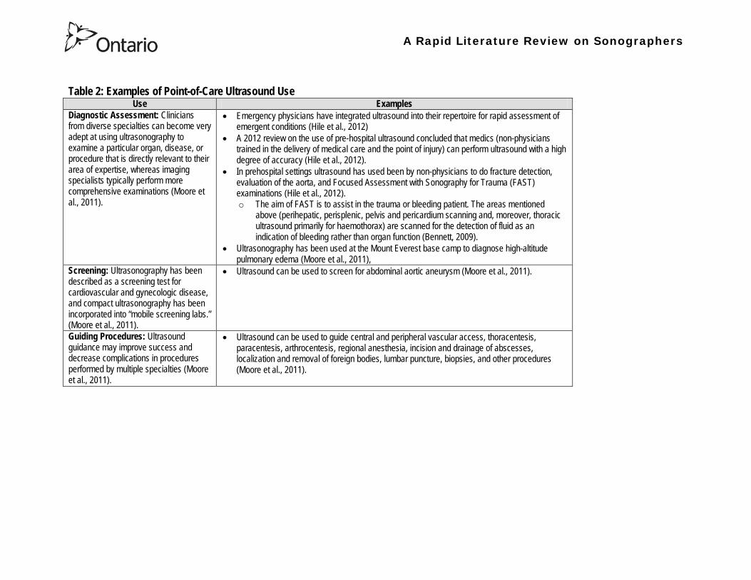
A Rapid Literature Review on Sonographers
Table 2 Examples of Point-of-Care Ultrasound Use Use Examples
Diagnostic Assessment Clinicians from diverse specialties can become very adept at using ultrasonography to examine a particular organ disease or procedure that is directly relevant to their area of expertise whereas imaging specialists typically perform more comprehensive examinations (Moore et al 2011)
bull Emergency physicians have integrated ultrasound into their repertoire for rapid assessment of emergent conditions (Hile et al 2012)
bull A 2012 review on the use of pre-hospital ultrasound concluded that medics (non-physicians trained in the delivery of medical care and the point of injury) can perform ultrasound with a high degree of accuracy (Hile et al 2012)
bull In prehospital settings ultrasound has used been by non-physicians to do fracture detection evaluation of the aorta and Focused Assessment with Sonography for Trauma (FAST) examinations (Hile et al 2012) o The aim of FAST is to assist in the trauma or bleeding patient The areas mentioned
above (perihepatic perisplenic pelvis and pericardium scanning and moreover thoracic ultrasound primarily for haemothorax) are scanned for the detection of fluid as an indication of bleeding rather than organ function (Bennett 2009)
bull Ultrasonography has been used at the Mount Everest base camp to diagnose high-altitude pulmonary edema (Moore et al 2011)
Screening Ultrasonography has been described as a screening test for cardiovascular and gynecologic disease and compact ultrasonography has been incorporated into ldquomobile screening labsrdquo (Moore et al 2011)
bull Ultrasound can be used to screen for abdominal aortic aneurysm (Moore et al 2011)
Guiding Procedures Ultrasound guidance may improve success and decrease complications in procedures performed by multiple specialties (Moore et al 2011)
bull Ultrasound can be used to guide central and peripheral vascular access thoracentesis paracentesis arthrocentesis regional anesthesia incision and drainage of abscesses localization and removal of foreign bodies lumbar puncture biopsies and other procedures (Moore et al 2011)
A Rapid Literature Review on Sonographers
Table 3 Description of the content in the articles being summarized28
No
Description Reference Review Articles
1 Autism spectrum disorders (ASDs) affect an estimated 1 of children in the United States The etiology is probably multifactorial including genetic components and exposure to infections toxins and other environmental factors particularly unfavorable perinatal and neonatal conditions There has been an increase in the frequency of diagnosis of ASDs over the last 20 years with a parallel increase in the use of obstetric diagnostic ultrasound with prenatal ultrasound exposure mentioned as the possible main etiology for autism ldquoepidemicsrdquo Central nervous system alterations have been described in ASDs and certain similar changes have been described in animals after exposure to ultrasound However analysis of in utero exposure in humans has failed to show harmful effects in neonates or children particularly in school performance attention disorders and behavioral changes There is no independently confirmed peer-reviewed published evidence that a cause-effect relationship exists between in utero exposure to clinical ultrasound and development of ASDs in childhood Ultrasound is a form of energy which effects in the tissues it traverses and its use should be restricted to medical indications by trained professionals for as short a period and as low an intensity as compatible with accurate diagnosis
Abramowicz J (2012) Ultrasound and Autism Association Link or Coincidence J Ultrasound Med 311261ndash1269
2 Over the past decade point-of-care ultrasound (US) use by nonphysician providers has grown substantially The purpose of this article is to (1) summarize the literature evaluating military medics facility at US (2) more clearly define the potential utility of military prehospital US technology and (3) lay a pathway for future research of military prehospital US The authors performed a keyword search using multiple search engines Each author independently reviewed the search results and evaluated the literature for inclusion Of 30 studies identified five studies met inclusion criteria The applications included evaluation of cardiac activity pneumothorax evaluation and fracture evaluation Additionally a descriptive study demonstrated distribution of US exam types during practical use by Army Special Forces Medical Sergeants No studies evaluated retention of skills over prolonged periods Multiple studies demonstrate the feasibility of training military medics in US Even under austere conditions the majority of studies conclude that medic can perform US with a high degree of accuracy Lessons learned from these studies tend to support continued use of US in out-of-hospital settings and exploration of the optimal curriculum to introduce this skill
Hile D C Morgan A R Laselle B T amp Bothwell J D (2012) Is Point-of-Care Ultrasound Accurate and Useful in the Hands of Military Medical Technicians A Review of the Literature Military Medicine 177(8) 983-7
28 Please note the studies programs and findings presented in this table may originate from jurisdictions with health systems that are significantly different from Ontarios If there is intent to draw heavily from one or more sources presented in this table we recommend that you contact the lead author of this review for assistance with evaluating the local applicability
A Rapid Literature Review on Sonographers
No Description Reference 3 Ultrasonography is a safe and effective form of imaging that has been used by physicians for more than half a century to aid in
diagnosis and guide procedures Over the past two decades ultrasound equipment has become more compact higher quality and less expensive which has facilitated the growth of point-of-care ultrasonography mdash that is ultrasonography performed and interpreted by the clinician at the bedside In 2004 a conference on compact ultrasonography hosted by the American Institute of Ultrasound in Medicine (AIUM) concluded that ldquothe concept of an lsquoultrasound stethoscopersquo is rapidly moving from the theoretical to realityrdquo This conference included representatives from 19 medical organizations in November 2010 the AIUM hosted a similar forum attended by 45 organizations Some medical schools are now beginning to provide their students with hand-carried ultrasound equipment for use during clinical rotations Although ionizing radiation from computed tomographic (CT) scanning is increasingly recognized as a potentially major cause of cancer ultrasonography has been used in obstetrics for decades with no epidemiologic evidence of harmful effects at normal diagnostic levels However ultrasonography is a user-dependent technology and as usage spreads there is a need to ensure competence define the benefits of appropriate use and limit unnecessary imaging and its consequences This article provides an overview of the history and current status of compact point-of-care ultrasonography with examples and discussion of its use
Moore C L amp Copel J A (2011) Point-of-Care Ultrasonography N Engl J Med 364 749-57
4 Sound travels through objects that block light Only very recently has technology advanced enough to decipher ultrasound for medical use Machines have become smaller cheaper more versatile and more advanced than ever before The medical use of ultrasound spreads across many fields so that traditional areas no longer have a monopoly With this comes the question of training doctors This has been done by various colleges and societies worldwide Some have been quicker to act than others There also needs to be an understanding of when broad experience and advanced technical skills are required or when limited skills will do In addition some procedures can be performed more safely with ultrasound where the knowledge of ultrasound is not paramount This article covers current provision of training in echo-cardiography and ultrasound in areas relevant to anaesthetists who are working in critical care (including accident and emergency) and complex surgery (mainly cardiac)
Bennett S (2009) Training guidelines for ultrasound worldwide trends Best Practice amp Research Clinical Anaesthesiology 23 363ndash373
5 Ultrasound is a commonly employed imaging modality in obstetrics and is generally regarded as safe to the fetus Current ultrasound technology however has significantly higher output potential than older machines used in most clinical studies and the safety profile for the increasing use of Doppler 3-dimensional (D) and 4-D ultrasound with modern machines is unknown This article reviews the current status of ultrasound safety within obstetrics including proposed mechanisms of harm existing scientific and clinical evidence regarding those mechanisms and considerations of safety for the clinical user
Houston L E Odibo A O amp Macones G A (2009) The safety of obstetrical ultrasound a review Prenat Diagn 29 1204ndash1212
A Rapid Literature Review on Sonographers
No Description Reference 6 The purpose of this article is to present the practicing sonographer and sonologist with an overview of the biohazards of ultrasound
and guidelines for safe use Overview Ultrasound is an imaging modality that has important diagnostic value Although useful in a variety of applications diagnostic ultrasound is particularly useful in prenatal diagnosis To date there is no evidence that diagnostic ultrasound produces harm in humans or the developing fetus when used properly There are however an increasing range of ultrasound studies being performed Newer technologies can have higher acoustic output levels than earlier equipment Also subtle or transient effects of diagnostic ultrasound such as changes in membrane permeability or neuronal migration are not completely understood Nondiagnostic Imaging Applications Nondiagnostic uses of ultrasound equipment such as repeated scans of healthy subjects for equipment demonstrations generally should be avoided First-trimester fetal scans should not use color and power Doppler modes and should not be performed for the sole purpose of producing souvenir videos or photographs Production of fetal souvenir photographs or videos during diagnostic clinical studies should not increase exposure levels or extend scan times beyond those needed for clinical purposes Operators should follow safe scanning guidelines and ALARA (as low as reasonably achievable) principles The FDA considers the promotion selling or leasing of ultrasound equipment for making ldquokeepsake fetal videosrdquo that are not part of a medical diagnostic procedure to be an unapproved use of a medical device
NelsonT R FowlkesJ B Abramowicz J S Church C C (2009) Ultrasound Biosafety Considerations for the Practicing Sonographer and Sonologist J Ultrasound Med 28139ndash150
7 Objective A possible association between ultrasound during pregnancy and subsequent non-right handedness among children has been suggested The association has been reported to be stronger among boys The aim of the present study was to explore this further through a meta-analysis of two follow-up studies of three randomized controlled trials of routine ultrasonography during pregnancy Design Handedness was assessed through a questionnaire to the parents and 4715 children at the age of 8ndash9 years were included in the meta-analysis Results There was no statistically significant difference in the prevalence of non-right handedness between the ultrasound-screened children and the controls but there was a statistically significant difference in a subgroup analysis among the boys Exploratory analyses according to exposure or non-exposure indicated an even stronger association between ultrasound and non-right handedness Conclusions A conservative analytical approach indicates no association between ultrasound in utero and subsequent non-right handedness The results from the exploratory analyses must be interpreted with caution There is still a need for further research
Salvesen K A amp Eik-Nes (1999) Ultrasound during pregnancy and subsequent childhood non-right handedness a meta-analysis Ultrasound Obstet Gynecol13 241ndash246
8 Ultrasound energy exerts important cellular genetic thermal and mechanical effects Concern about the safety of ultrasound prompted several agencies to devise regulatory limits on the machine output intensities The visual display of thermal and mechanical indices during ultrasound imaging provides an aid to limit the output of the machine Despite many animal studies no human investigations conducted to date have documented major physiologic consequences of ultrasound exposed during imaging To date ultrasound imaging appears to be safe for use in regional anesthesia and pain medicine interventions and adherence to limiting the output of ultrasound machines as outlined by the Food and Drug Administration may avoid complications in the future This article reviews ultrasound-related biologic effects the role of the regulatory agencies in ensuring safety with the use of ultrasound and the limitations and implications of ultrasound use in humans
Shankar H amp Pagel P S (2011) Potential Adverse Ultrasound-related Biological Effects A Critical Review Anesthesiology 115(5) 1109-1124
A Rapid Literature Review on Sonographers
No Description Reference 9 Objective In the context of the planned International Society of Ultrasound in Obstetrics and GynecologyndashWorld Health Organization
multicenter study for the development of fetal growth standards for international application we conducted a systematic review and meta-analysis to evaluate the safety of human exposure to ultrasonography in pregnancy Methods A systematic search of electronic databases reference lists and unpublished literature was conducted for trials and observational studies that assessed short- and long-term effects of exposure to ultrasonography involving women and their fetuses exposed to ultrasonography using B-mode or Doppler sonography during any period of pregnancy for any number of times The outcome measures were (1) adverse maternal outcome (2) adverse perinatal outcome (3) abnormal childhood growth and neurological development (4) non-right handedness (5) childhood malignancy and (6) intellectual performance and mental disease Results The electronic search identified 6716 citations and 19 were identified from secondary sources A total of 61 publications reporting data from 41 different studies were included 16 controlled trials 13 cohort and 12 casendashcontrol studies Ultrasonography in pregnancy was not associated with adverse maternal or perinatal outcome impaired physical or neurological development increased risk for malignancy in childhood subnormal intellectual performance or mental diseases According to the available clinical trials there was a weak association between exposure to ultrasonography and non-right handedness in boys (odds ratio 126 95 CI 103ndash154) Conclusion According to the available evidence exposure to diagnostic ultrasonography during pregnancy appears to be safe
Torloni M R Vedmedovska Merialdi M Betran A P Allen T et al (2009) Safety of ultrasonography in pregnancy WHO systematic review of the literature and meta-analysis Ultrasound Obstet Gynecol 33 599ndash608
Articles in Peer-Reviewed Journals 10 It has been documented for many years that as a result of use of ultrasound to determine the sex of the fetus the male to female
ratio in countries such as China and India have become increasingly skewed (due to subsequent abortions and infanticide of females) Preference for sons over daughters evident in Chinas and South Asias male sex ratios is commonly rationalized by poverty and the need for old-age support In this article the authors study South and East Asian immigrants to Canada a group for whom the economic imperative to select sons is largely absent Analyzing the 2001 and 2006 censuses 20 percent samples the authors find clear evidence of extensive sex selection in favor of boys at higher parities among South and East Asian immigrants unless they are Christian or Muslim The latter finding accords with the explicit prohibition against (female) infanticidemdashtraditionally the main sex-selection methodmdashin these religions The findings point to a strong cultural component to both the preference for sons and the willingness to resort to induced abortion based on sex
Almond D Edlund L Milligan K (2013) Son Preference and the Persistence of Culture Evidence from South and East Asian Immigrants to Canada Population and Development Review 39(1) 75ndash95
11 The rapid growth in the use of ultrasound as a diagnostic imaging technology over the past forty years has led to a demand for a workforce with the appropriate skills to perform and interpret the scans In the UK ultrasound investigations now comprise the largest group of all diagnostic imaging examinations However there remains no statutory regulation of the practice of sonography in the UK and little recognition of the considerable training that many practitioners have undertaken to obtain the skills to become safe and competent sonographers Many in the field consider that this should change and are working to obtain professional status for the practice of sonography Although the Health Care Professions Council (HCPC) has recommended regulation of sonography practice this is unlikely to happen in the near future This paper discusses the evolution of sonography practice and explores some of the complex issues associated with the professionalisation of sonography
Gibbs V (2013) The long and winding road to achieving professional registration for sonographers Radiography19 164-167
A Rapid Literature Review on Sonographers
No Description Reference 12 Background Contrast echocardiography (which uses contrast agents injected into the blood flow in order to visualize the blood flow
in very small blood vessels) has been shown to improve diagnostic quality especially in technically difficult patients However the learning curve and increased time for preparation and image acquisition have led to low use Methods The authors sought to determine whether the contrast echocardiography procedure performed independently by a specialized trained sonographer could improve efficiency In one centre routine echocardiograms were scheduled for one hour and any study exceeding one hour would result in patient booking cancellations The authors compared the standard of care in which a physician or nurse administers echocontrast with a sonographer-administered program (SAP) Results The time to complete contrast echocardiograms was significantly reduced by the SAP strategy (43 min 17 s plusmn 23 min 42 s vs 1 h 1 min 6 s plusmn 31 min 0 s P lt 0001) Subgroup analysis of the inpatients and outpatients demonstrated similar results Only 10 of studies (6 of 61) in the SAP exceeded 60 minutes compared with 45 (34 of 76) in the standard-of-care group (P lt 0001) Based on study volumes in the centre the net improvement in productivity with the SAP could be up to 53 annually Conclusion Sonographer-administered echocontrast is feasible and potentially removes a barrier to implementation of contrast echocardiography
Tang A Chiew S K Rashkovetsky R Becher H Choy J B (2013) Feasibility of Sonographer-Administered Echocontrast in a Large-Volume Tertiary-Care Echocardiography Laboratory Canadian Journal of Cardiology 29 391ndash395
13 Ultrasound scanning has been used as a diagnostic and screening tool in obstetric practice for over 50 years There is no evidence of immediate or long-term harm to the developing fetus from exposure to B mode ultrasound However exposure to high levels of Doppler ultrasound during early development is increasingly common and the full safety implications of this exposure are not clear Doppler ultrasound exposure in utero gives rise to increased apoptosis in animal models and there is evidence of the effects of exposure to Doppler ultrasound persisting throughout life with increased non-right-handedness observed in human epidemiological studies The authors consider the idea that there may be long-term developmental implications for fetuses exposed to Doppler ultrasound early in gestation These effects may be mediated via thermal or mechanical disruption to the developing conceptus giving rise to free radical damage Excess free radical exposure early in gestation is a strong candidate for the final common pathway underlying developmental programming effects and gives rise to concern that fetuses exposed to high levels of ultrasound are at risk of a developmental programming effect It is suggested that there is a need for animal studies of developmental programming using exposure to Doppler ultrasound scanning as the exposure of interest and for more observational data to be collected in the clinical setting While these data are collected it seems prudent to continue to adhere to the principle of lsquoas low as reasonably achievablersquo (ALARA) when exposing first-trimester fetuses to Doppler ultrasound
Aiken CE Lees CC (2012) Long-term effects of in utero Doppler ultrasound scanning ndash A developmental programming perspective Medical Hypotheses 78 539ndash541
14 Point of care sonography by emergency physicians in the emergency department can be an effective aid in the diagnosis and management of patients presenting with a variety of medical and traumatic conditions Its use can improve patient outcomes enhance patient safety speed patient disposition and save lives CAEP supports the use of point of care sonography
Canadian Assocation of Emergency (CAEP) Physicians Ultrasound Position Statement Working Group (2012) Use of point of care sonography by emergency physicians CJEM 14(2)106-112
A Rapid Literature Review on Sonographers
No Description Reference 15 The use of three-dimensional echocardiography (3DE ie a 3D ultrasound of the heart) during electrophysiology (EP - a branch of
cardiology that deals with the diagnosis and treatment of heart rhythm disorders) procedures is the end product of years of growth in two diverse cardiology subspecialties namely advanced cardiac imaging and the EP During the past decade progress in both fields has resulted in many important advances that have culminated in their union for a new area of growth and development Imaging advances have provided the cardiovascular specialist with enhanced cardiac volume and function data and more recently 3DE capabilities with improved spatial and temporal resolution providing unprecedented spatial relationships This latter development is valued by EP specialists in need of hitherto never required anatomic knowledge as they press forward with extraordinary expansion in their capabilities It makes sense that by combining these two rapidly growing subspecialties future capabilities in patient care may be achieved that would otherwise not be possible This paper discusses the value of 3DE during EP procedures and offers the readers insight into this novel multispecialty hybrid arena Using this model as a template it is likely that the readers may identify other areas within their practices where periprocedural advanced imaging may afford significant dividends in patient outcomes
Moukabary T Faletra F F Kronzon I Thomas W amp Sorrell V L (2012) Three-Dimensional Echocardiography in the Electrophysiology Laboratory Echocardiography 29 117-122
16 Background There has been much discussion about whether female feticide occurs in certain immigrant groups in Canada The authors examined data on live births in Ontario and compared sex ratios in different groups according to the motherrsquos country or region of birth and parity Methods The authors completed a population-based study of 766 688 singleton live births between 2002 and 2007 They used birth records provided by Ontario Vital Statistics for live births in the province between 23 and 41 weeksrsquo gestation They categorized each newborn according to the motherrsquos country or region of birth namely Canada (n = 486 599) Europe (n = 58 505) South Korea (n = 3663) China (n = 23 818) Philippines (n = 15 367) rest of East Asia (n = 18 971) Pakistan (n = 18 018) India (n = 31 978) rest of South Asia (n = 20 695) and other countries (n = 89 074) They calculated malefemale ratios and 95 confidence intervals (CIs) for all live births by these regions and stratified them by maternal parity at the time of delivery (0 1 2 or ge 3) Results Among infants of nulliparous women (women with no previous children) the malefemale ratio was about 105 overall As parity (the number of previous children) increased the ratio remained unchanged among infants of Canadian-born women In contrast the malefemale ratio was significantly higher among infants of primiparous women (women with one previous child) born in South Korea (120 95 CI 109ndash134) and India (111 95 CI 107ndash115) than among infants of Canadian-born primiparous women Among multiparous women (women with 2+ previous children) those born in India were significantly more likely than Canadian-born women to have a male infant (parity 2 ratio 136 95 CI 127ndash 146 parity ge 3 ratio 125 95 CI 109ndash143) Interpretation The study of malefemale ratios in Ontario showed that multiparous women born in India were significantly more likely than multiparous women born in Canada to have a male infant
Ray J G Henry D A Urquia M L (2012) Sex ratios among Canadian liveborn infants of mothers from different countries CMAJ184(9) E92-96
A Rapid Literature Review on Sonographers
No Description Reference 17 This letter consists of a case study that illustrates how a cross-disciplined sonographer (educated in cardiac and vascular ultrasound)
can quickly and efficiently and with great Cost-effectiveness deliver timely care to a patient in certain clinical settings The literature is comparatively sparse in referencing clinical diagnostics in cross-discipline sonographers Echocardiographers must be clinicians fully comfortable with evaluating the circulatory dynamics of the body from the left and right heart to the peripheral arteries and veins including the extracranial circulation (carotid arteries) Included in this skill set is substantive understanding of the ECG patientrsquos history and integration of cardiac and vascular findings Limitations of each diagnostic tool and how these limitations can be overcome by the strengths of the other examination are important For the dual-skilled sonographer only experience continuous education and constant clinical feedback will lead to the independent decision making that is counted on by the physician The American Registry for Diagnostic Medical Sonography offers credentials in cardiac and vascular imaging (RDCS and RVT) Cardiovascular Credentialing International offers credentials in cardiac and vascular imaging as well (registered cardiac sonographer [RCS] and registered vascular specialist [RVS]) All imaging facilities should consider merging cardiac and vascular noninvasive imaging with dual-registered clinical sonographers credentialed by one of these two agencies
Zemanek M (2012) What It Means to Be a Cardiovascular Sonographer Journal of Ultrasound in Medicine 311859-1862
18 Diagnostic medical sonography is viewed as a safe imaging modality However bioeffects research has yet to define the effects of exposure to sonography in humans Therefore sonographers must be aware of potential risks and practice ALARA (ldquoas low as reasonably achievablerdquo) routinely This research explores sonographersrsquo ability to answer basic questions about the bioeffects of sonography as well as their knowledge and use of the safety indices called the mechanical index (MI) and thermal index (TI) Members of the Society of Diagnostic Medical Sonographers in the US a southwestern sonographersrsquo society and a sonography community on Facebook 2010 were surveyed (n = 212) There is a low level of bioeffects knowledge and minimal safety practices among sonographers in this survey No significant relationship was found between sonographer knowledge of the bioeffects of sonography years in the profession or whether the sonographers monitor the MITI
Bagley J Thomas K DiGiacinto D (2011) Safety Practices of Sonographers and Their Knowledge of the Biologic Effects of Sonography Journal of Diagnostic Medical Sonography 27(6) 252ndash261
19 This article highlights trends in echocardiography Point of care ultrasound (hand-carried ultrasound) Initially point of care ultrasound will increase the use of echocardiography in the same manner that cardiac CT increased catheterizations However once the basics are learned it has the potential to be a very powerful tool in that less unnecessary studies will be ordered In the clinic small systems will be used as a lsquolsquofirst lookrsquorsquo screen and become part of the patientrsquos history only patients for whom more testing is necessary will be referred for a comprehensive study This is likely to reduce the number of Doppler studies requested The miniaturization of echo equipment will continue with improved qualityfunctionality of the lsquolsquohand-heldrsquorsquo echo machines to the extent that cordless probes will become a reality sooner rather than later Role in interventional echocardiography Sonographers will be spending much more time in the interventional and electrophysiology (EP) labs There will be more interconnection with interventional cardiologists and the volume will increase as more devices are implanted more percutaneous valves placed and more robotic surgery is performed Crosstraining and multitasking Sonographers entering the profession would be prudent to learn cardiac and vascular imaging Individuals who multitask andor cross-train will be ideal For example a sonographer who starts an IV monitors ECG stress can perform cardiac MRI and can interpret a preliminary report or a nurse who can do an echo monitor sedation and start an IV will be invaluable The sonographer should continue to grow professionally by cross training as institutions try to minimize costs and maximize throughput
McCulloch M L (2011) Thoughts regarding the future of sonography Journal of the American Society of Echocardiography 24(5) 29A-30A
A Rapid Literature Review on Sonographers
No Description Reference 20 Background Radiology reports provide specialist interpretation of images and relate these findings to the patientrsquos symptoms and
signs This study compared ultrasound (USS) reports generated by radiologists and sonographers to determine if any significant or clinically relevant differences existed Methods A retrospective analysis of 624 consecutive USS reports was carried out The reports were assessed for the presence of a lsquodisclaimerrsquo or lsquocaveatrsquo pertaining to the quality of the images and were analysed with respect to the clarity of their wording and ability to answer the clinical request Results The majority of sonographer USS reports contained a disclaimer regarding the quality of the USS images (571) compared with a smaller proportion of radiologist scans (99) (P lt 0001) Overall radiologists performed significantly better in providing a clear negative or positive diagnosis to the clinical question on the request form when compared with sonographers (885 vs 654 P lt 0001) Conclusion lsquoDisclaimerrsquo comments and lsquohedgingrsquo must be avoided in radiology reporting While feedback as to the accuracy of the radiology images is important overuse of such terms undermines the validity of the radiology report
Garcea G Mahmoud A Ong S L Rees Y Berry Det al (2010) Caveat reporting in ultrasound interpretation of surgical pathology a comparison of sonographer versus radiologist Journal of Evaluation in Clinical Practice 16 97ndash99
21 A nine-point questionnaire was constructed based on previously published work by Marsal and Sheiner et al The survey was distributed to clinical users of ultrasound practicing sonography in Hamilton (New Zealand) during July 2010 totalling 35 users consisting of sonographers sonography trainees one obstetric sonologist and one fetal medicine registrar Results Twenty sonographers returned the questionnaire which represents survey response of 61 The level of clinical experience was equally split between junior sonographers of lt 5 yearsrsquo experience (30) sonographers with 5-10 yearsrsquo experience (35) and senior sonographers of gt10 yearsrsquo experience (35) While 100 of the sonographers surveyed reported being familiar with the term Thermal Index (TI) and 90 claimed they adhered to the Action Learning Action Response Association (ALARA) principle all the time or most of the time only 15 of sonographers correctly described or defined what the term TI means Of the sonographers 80 knew where to find the TI during real-time examinations but only 35 knew which TI index to use for nuchal translucency (NT) scan and only 10 knew the expected range of TI encountered during routine obstetric examinations Of the eight sonographers who reported adhering to ALARA all the time the majority (64) could not describe or define TI In the same subgroup the majority (64) did not know which TI to monitor during a NT scan Only three of 20 (15) sonographers knew what the term TI means Of these two were junior sonographers with lt 5 yearsrsquo experience and one had 5ndash10 yearsrsquo experience Despite understanding the term TI one of the three sonographers still selected the incorrect index to monitor during a NT scan Not one of the seven senior sonographers with gt 10 years of experience could define or describe the TI In total only 10 of sonographers knew what the TI means and which TI to monitor during NT scanning
Necas M (2010) New Zealand sonographers do not outperform their European or American colleagues in the knowledge of ultrasound safety Australasian Journal of Ultrasound in Medicine13(4) 28-32
A Rapid Literature Review on Sonographers
No Description Reference 22 Musculoskeletal (MSK) ultrasound performed by non-medical practitioners is a developing practice The aim of this paper is
(1) to audit an experienced sonographerrsquos (SJ Riley one of the authors) performance after one yearrsquos independent reporting against two experienced MSK radiologists (CJ Grove amp M Chandramohan the other two authors) and (2) to establish an audit standard against which such role development might be contrived Images and reports from 250 MSK ultrasound examinations performed by a sonographer were reviewed independently by two consultant MSK radiologists The examinations were graded for discrepancy eg grade 1 ndash agree with the sonographer report grade 2 ndash minor discrepancy unlikely to alter patient care grade 3 ndash potentially significant discrepancy grade 4 ndash definite significant discrepancy likely to have adverse consequences for patient care Two of 250 (08) cases were excluded Both radiologists agreed completely with the sonographer (grade 1) in 235 of the 248 cases (948) In 13 cases there was discrepancy between the reports of SJR and the radiologists The discrepancy was grade 2 in six of the 248 cases (24) grade 3 in six of the 248 cases (24) and grade 4 in one of the 248 cases (04) In conclusion this audit shows a high level of agreement between the sonographer and the consultant MSK radiologist reporting of MSK ultrasound This level of agreement may set the standard for future quality assurance audit of sonographer MSK reporting
Riley S J Groves C J Chandramohan M (2010) Musculoskeletal ultrasound audit of sonographer reporting Ultrasound 18 36ndash40
23 Background Growing demand for ultrasound examinations and higher quality requirements motivate searching for routines combining the diagnostic accuracy of radiologist-performed examinations with the economical advantages of sonographer-performed examinations One possible approach is to use strictly standardized acquisition and documentation schemes that give the radiologist access to all relevant information after the examination Purpose To compare a recently introduced routine combining acquisition by a radiographer (more specifically a sonographer with two years experience) documentation as standardized cine-loops and review by a radiologist (lsquostandardized methodrsquo) with the formerly used routine where the diagnosis is made bedside by the radiologist (lsquotraditional methodrsquo) Material and Methods In 64 policlinic patients the kidneys (n = 27) or the gallbladder (n = 37) were examined with both the standardized and the traditional method The radiologistsrsquo findings of hydronephrosis tumors cysts echogenicity changes and cortical thickness (in the kidneys) and wall thickness concrements and polyps (in the gallbladder) were compared between the methods with respect to agreement (proportion of agreement and kappa coefficient) as well as systematic differences (McNemarrsquos test) Results The findings at the gallbladder examination showed a median agreement of 97 (86ndash100 kappa = 064ndash100) and those at the kidney examination an agreement of 90 (78ndash100 kappa = 069ndash 100) There were no significant systematic differences between the methods Conclusion The satisfactory agreement in this preliminary study indicates that the new workflow with ultrasound examinations performed by a radiographer (sonographer) and analyzed off-line by a radiologist is promising and motivates further studies
Stenman C Thorelius L Knutsson A amp Smedby Ouml (2010) Radiographer-acquired and radiologist-reviewed ultrasound examination ndash agreement with radiologistrsquos bedside evaluation Acta Radiologica 52 70ndash74
A Rapid Literature Review on Sonographers
No Description Reference 24 The troubling practice of fetal sex selection has historically been considered an Asian phenomenon However recent evidence shows
that a similar situation is emerging in North America albeit on a smaller scale The Society of Obstetricians and Gynaecologists of Canada (SOGC) has firmly stated its opposition to sex selection for non-medical reasons as well as to the use of any technology used solely for the purpose of determining fetal sex However because fetal sex may be disclosed to the parents at the time of ultrasound examination if they request this information guidance for health care professionals to assist in discouraging fetal sex selection would be useful Because no declaration of motives or reasons is required when a woman seeks a termination of pregnancy the authors suggest that health care professionals need not disclose the sex of a fetus until it has reached a gestational age at which abortion for non-medical purposes would not be possible This proposal would facilitate consistency between clinical practice and the values of Canadian citizens the SOGC the Canadian Medical Association and other professional organizations while still respecting current laws pertaining to disclosure of patient information and patientsrsquo rights to autonomous decision-making
Thiele A T amp Leier B (2010) Towards an Ethical Policy for the Prevention of Fetal Sex Selection in Canada J Obstet Gynaecol Can 32(1) 54ndash57
25 Background The extent to which increases in the use of prenatal ultrasonography over time reflect changes in maternal risk (ie an increase in high-risk pregnancies over time) is unknown In this population-based study the authors examined the use of prenatal ultrasonography from 1996 to 2006 in Ontario Methods With fiscal year 199697 as the baseline the authors evaluated the relative risk (RR) and 95 confidence interval (CI) for the change in rates of ultrasonography for each subsequent year The RR was adjusted for maternal age income rural residence maternal comorbidities receipt of genetics consultation or amniocentesis mdash all in the index pregnancy mdash and history of complications in a prior pregnancy Results The study sample consisted of 1 399 389 singleton deliveries The rate of prenatal ultrasonography increased from 2055 per 1000 pregnancies in 1996 to 3264 per 1000 in 2006 (adjusted RR 155 95 CI 154ndash155) The rate increased among both women with low-risk pregnancies (adjusted RR 154 95 CI 153ndash155) and those with high-risk pregnancies (adjusted RR 155 95 CI 154ndash157) The proportion of pregnancies with at least four ultrasound examinations in the second or third trimesters rose from 64 in 1996 to 187 in 2006 (adjusted RR 268 95 CI 261ndash274) Paradoxically this increase was more pronounced among low-risk pregnancies (adjusted RR 292 95 CI 283ndash301) than among high-risk pregnancies (adjusted RR 225 95 CI 216ndash235) Interpretation Substantial increases in the use of prenatal ultrasonography over the past decade do not appear to reflect changes in maternal risk Nearly one in five women now undergo four or more ultrasound examinations during the second and third trimesters Efforts to promote more appropriate use of prenatal ultrasonography for singleton pregnancies appear warranted
You J J Alter D A Stukel T A McDonald S D Laupacis A et al (2010) Proliferation of prenatal ultrasonography CMAJ 182(2) 143-151
A Rapid Literature Review on Sonographers
No Description Reference 26 Aims The aim of this study was to assess the cost-effectiveness of using certified sonographers and miniaturized echocardiography
systems to perform echocardiograms at bedside in comparison to moving inpatients from the admission department to the echocardiography laboratory (echo-lab) Methods and Results From 26 September 2005 to 27 October 2005 112 patients admitted in six hospital wards in Italy connected through a 100 Mbit LAN to the echo-lab were scanned within the admission ward by sonographers using a miniaturized echo system Logistical data were collected and results were compared with those obtained from 194 consecutive patients coming from the same wards and studied in the echo-lab with high-end machines between 8 March 2005 and 15 April 2005 Performing echocardiograms in the admission department avoided long waiting time of the inpatients in the echo-lab before and after the study increased the percentage of patients studied within 3 and 5 days from request (88 vs 77 and 100 vs 95 respectively P = 003) increased both sonographer (by 339 P lt 0001) and echo-lab productivity (by 41 P lt 0001) and reduced costs of echocardiograms by 29 Conclusion Implementation of digital echocardiography certified sonographers and a miniaturized echo system allowed improvement of the cost-effectiveness of the service provided by the echo-lab for inpatients and avoided patientsrsquo discomfort derived from prolonged waiting time before and after the exam
Badano L Gaetano N Stacul S Gianfagna P Pericoli M et al (2009) Improved workflow sonographer productivity and cost-effectiveness of echocardiographic service for inpatients by using miniaturized systems European Journal of Echocardiography 10 537ndash542
27 The present report is a proposal from the American Society of Echocardiography Advanced Practice Task Force that identifies the potential of cardiac sonographers to achieve the Advanced Cardiovasculat Sonographer (ACS) level According to this report echocardiographic examinations require a well-trained and competent sonographer to obtain proper anatomic and physiologic data to establish an accurate diagnosis for clinical decision-making and patient management Although the formal education and training of cardiovascular sonographers are evolving many entry-level and staff sonographers may not have sufficient practical or clinical knowledge of the necessary components of the echocardiographic study for the individual patientrsquos clinical presentation In many clinical settings echocardiograms are read after the patient has left the laboratory Thus there is a role for a sonographer who can practice at an advanced level in a cardiovascular ultrasound laboratory to ensure a proper echocardiographic examination is performed on every patient In this setting an Advanced Cardiovascular Sonographer (ACS) would be able to review the indication for and quality of the examination If additional images were needed the ACS would assist the sonographer in obtaining these images which would lead to the performance of a complete and fully diagnostic examination before the patient had left the echocardiography laboratory In clinical practice the quality of the examinations performed would improve advancements in echocardiographic methods could be taught and incorporated into daily practice and patients would be better served
Mitchell C Miller F A Bierig S M Bremer M L Ehler D et al (2009) Advanced Cardiovascular Sonographer A Proposal of the American Society of Echocardiography Advanced Practice Sonographer Task Force J Am Soc Echocardiogr 22 1409-13
A Rapid Literature Review on Sonographers
No Description Reference 28 Sonographer practitioner development involves the expansion and extension of the sonographer role to include reporting on
ultrasound examinations Australian sonographers have not seen the same degree of role extension and expansion as their counterparts in the United Kingdom despite increasing levels of discussion regarding sonographer practitioner development The aim of this study was to determine if Australian sonographers want to extend their professional role and what they consider are the important issues associated with role extension This paper reports on qualitative data derived from a survey of Australian sonographers and investigates if Australian sonographers are interested in extending and expanding their professional role and responsibilities and if they do what might be necessary or desirable from a professional point of view for this change to occur A survey was mailed to all members of the Australian Sonographers Association (ASA) in October 2006 The 31-item survey included 28 closed-ended and three opened-ended items to provide both quantitative and qualitative data The quantitative data will be reported separately Qualitative data was derived from responses to the opened-ended questions which asked respondents to elaborate on their attitudes and feelings about role extension and development Analysis used Nvivo7 software to aid in uncovering common themes from the qualitative data The analysis focused on the reported incentives or motivations for becoming a sonographer practitioner as well as disincentives or perceived hurdles that would discourage respondents from becoming sonographer practitioners The three most reported incentives or motivations for becoming a sonographer practitioner were professional recognition remuneration and increased knowledge The three most commonly reported disincentives or perceived hurdles that would discourage respondents from becoming sonographer practitioners were legal issues insurance and further study
McGregor R OrsquoLoughlin K Cox J Clarke J Snowden A (2009) Sonographer practitioner development in Australia Qualitative analysis of an Australian sonographersrsquo survey Radiography15 313-319
29 Introduction This study describes the effect of introducing a cardiac sonographer led follow up clinic within a hospital cardiology department for patients with stable heart valve disease The two years before and after the instigation of the valve clinic were audited Materials and methods The clinic was conducted in a single centre and undertaken in the cardiology department of a district general hospital A total of 382 patients with 397 clinically significant valve lesions but for whom surgery was not yet indicated but follow up required were seen in a cardiac sonographer run clinics These patients no longer attended a medical follow up clinic unless there was clinical or echocardiographic deterioration Effectiveness was judged by the percentage treated according to current best practice guidelines the number of echocardiograms performed and the number of hospital outpatient visits attended In addition mortality data for the subjects in the clinic was collected Results The proportion followed up according to best practice guidelines rose from 157 (41) to 354 (92) (p lt 001) The total number of echocardiograms performed fell from 807 to 550 Total number of outpatient visits fell from 998 to 31 A total of 11 patients died in the two year study period none from progressive valve disease Discussion This study demonstrates that a protocol driven sonographer led heart valve disease follow up clinic significantly improved the quality of follow up while bringing about a major reduction in outpatients visits without compromising patient safety
Taggu W Topham A Hart L Carr-White G Sulke N et al (2009) A cardiac sonographer led follow up clinic for heart valve disease International Journal of Cardiology 132 240 ndash243
30 Role development for imaging allied health professionals has progressed significantly over the last three decades with many major developments pioneered in the field of ultrasound This paper reviews the developing role of the radiographer in ultrasound (in some countries such as the UK sonographers are considered a type of radiographer) and some of the clinical professional and educational implications of such development As the published evidence base underpinning the latest developments is scarce several contemporary case study examples which have been presented verbally at study days attended by the authors have been included In conclusion sonographer role development is considered within the professional contexts of extended role advanced and consultant practice
Hart A amp Dixon A (2008) Sonographer Role Extension and Career Development a Review of the Evidence Ultrasound 16(1) 31-35
A Rapid Literature Review on Sonographers
No Description Reference 31 Objective The main goal of this study was to determine end usersrsquo knowledge regarding safety aspects of diagnostic ultrasound
during pregnancy End usersrsquo attitudes toward the use of ultrasound in low-risk pregnancies were also assessed Methods A questionnaire was distributed to ultrasound end users attending review courses and hospital grand rounds between April and June 2006 in Chicago Results One hundred thirty end users completed the questionnaires (63 response rate) Sixty-three percent were physicians (n = 84) most of them obstetricians (817) About 18 of participants routinely performed Doppler ultrasound examinations during the first trimester Fifty percent of end users thought that the number of ultrasound examinations in low-risk pregnancy should be limited to 1 to 3 (mean plusmn SD 26 plusmn 09) Almost 70 disapproved of ldquokeepsakeentertainmentrdquo ultrasound Although 322 of the participants were familiar with the term thermal index only 177 actually gave the correct answer to the question on the nature of the thermal index About 22 were familiar with the term mechanical index but only 38 described it properly Almost 80 of end users did not know where to find the acoustic indices Only 208 were aware that they are displayed on the sonographic monitor during the examinations End users with higher knowledge of safety issues thought that there should be limitations on the number of ultrasound examinations in low-risk pregnancies (odds ratio 33 95 confidence interval 11ndash100 P = 028) Likewise these end users were more likely to respond that ultrasound might have adverse effects during pregnancy (odds ratio 32 95 confidence interval 11ndash125 P = 045) Conclusions Ultrasound end users are poorly informed regarding safety issues during pregnancy Further efforts in the realm of education and training are needed to improve end user knowledge about the acoustic output of the machines and safety issues
Sheiner E Shoham-Vardi I Abramowicz J S (2007) What Do Clinical Users Know Regarding Safety of Ultrasound During Pregnancy J Ultrasound Med 26 319ndash325
A Rapid Literature Review on Sonographers
No Description Reference 32 In an attempt to survey the knowledge among ultrasound users of some safety aspects of diagnostic ultrasound a questionnaire was
distributed to professionals using ultrasound for fetal examinations The questionnaire was answered by the participants and faculty members of six postgraduate courses in obstetric ultrasound Doppler ultrasound and fetal echocardiography given in Sweden Norway and Austria in 2003 Furthermore in Sweden the questionnaire was distributed to the staff meeting participants of two major obstetric ultrasound units and of one unit for fetal echocardiography and at a meeting of the Working Group for Ultrasound of the Swedish Society of Obstetrics and Gynecology In total 199 questionnaires were anonymously answered by doctors (n = 145) sonographers (n = 22) and midwives trained in ultrasound (n = 32) All of them were using diagnostic ultrasound on a daily or weekly basis The respondents were from nine European countries In none of the countries is formal certification for the use of diagnostic ultrasound in pregnancy or otherwise required The doctors who answered the questionnaire were specialists in obstetrics and gynecology (including some of the internationally recognized experts in fetal ultrasound) pediatric cardiology radiology or clinical physiology The respondents were not informed about the questionnaire in advance and they had a limited time to answer the 16 questions typically 10ndash15 min The questions concerned explanation of the safety indices and their relation to various ultrasound modes ie real-time B-mode M-mode three-dimensional (3D) spectral Doppler color and power Doppler ultrasound The respondents were also asked which ultrasound system they used most often and where on the system the information on thermal index (TI) and mechanical (MI) was displayed (eg in the top right corner of the screen in a separate window etc) Furthermore the respondents had to indicate how they adjust the output energy level on their own machine For all questions there was the optional answer lsquoDonrsquot knowrsquo Results About one-third of the ultrasound experts were able to define the abbreviations TI and MI For the subclasses of the TI the correct answer was found in only 3-8 of cases Of those who knew what the TI and MI meant only two-thirds and one-third of respondents respectively were able to give a simple explanation of the indices In 49 of questionnaires the Doppler modes were correctly ranked as generally producing higher ultrasound exposure than the imaging modes (B-mode 3D) Some 28 of the respondents correctly indicated where on their own machine the information on safety indices is displayed However not all of them knew how to control the output energy level For all items in the questionnaire there were no significant differences in the results between the three categories of respondents (ie physicians sonographers and midwives) The results of the questionnaire were anything but encouraging and they indicated that the users who are supposed to be responsible for controlling exposure of the fetus to ultrasound had a very poor knowledge of the basic safety aspects of ultrasound
Maršaacutel K (2005) The output display standard has it missed its target Ultrasound Obstet Gynecol 25 211ndash214
33 Purpose This study was performed to evaluate the accuracy of upper abdominal ultrasound (US) scanning performed by sonographers in a district general hospital to identify potential areas of weakness and to make recommendations to improve the service Materials and Methods Upper abdominal US examinations performed and reported by sonographers over a 4-week period were retrospectively reviewed The accuracy of the imaging findings and reports were assessed against other imaging surgical histological or laboratory findings and against clinical outcome Results A heterogenous group of 104 patients were included in the study 62 of whom had an US abnormality Errors of scanning or interpretation were identified in 10 patients (96) of whom five (48) were felt to be potentially significant Conclusions The sonographersrsquo accuracy in reporting upper abdominal US scans was 90 However on the basis of this study the authors have implemented specific recommendations to improve the quality of the service
Dongola N Guy R Ward S amp Giles J A (2003) Can sonographers offer an accurate upper abdominal ultrasound service in a district general hospital Radiography 9 29ndash33
A Rapid Literature Review on Sonographers
No Description Reference 34 Aim To compare the accuracy of radiographers and radiologists in routine abdominal ultrasound Materials and Methods One
hundred consecutive patients attending for routine abdominal ultrasound were included Each patient was examined by both a radiographer and radiologist Both operators noted their fndings and wrote a concluding report without conferring Reports were compared Where there was disagreement the patient was either re-examined by another radiologist or had further investigation Results Of 100 patients 52 were men and 48 were women The age range was 19plusmn88 years (median 52 years) Thirty-seven patients had renal tract ultrasound one had an aortic ultrasound and 62 had general upper abdominal ultrasound In 44 cases both operators reported the examination as normal In 49 cases both operators reported the examinations as abnormal and there was complete agreement between the operators In seven cases there was not complete agreement between operators Three of these disagreements were considered minor and four major In three of the seven cases the radiographer was correct and in four the radiologist was correct Conclusion Experienced radiographers and radiologists are highly accurate in performing and interpreting routine abdominal sonography Both operators missed a small minority of abnormalities There was no statistically significant difference in the accuracy of radiographers and radiologist
Leslie A Lockyer H amp Virjee J P (2000) Who Should be Performing Routine Abdominal Ultrasound A Prospective Double-Blind Study Comparing the Accuracy of Radiologist and Radiographer Clinical Radiology 55 606-609
Grey Literature 35 The ARRT The American Registry of Radiologic Technologists tests certifies and annually registers more than a quarter-of-a-million
radiologic technologists to promote high standards of patient care They award the Registered Technologist or RT designation to individuals who have completed the prescribed classroom and clinical education passed the appropriate exam and met the ethics requirements And they ensure their continuing education and ongoing compliance with their high standards by requiring annual registration of their certificate Certification by ARRT is voluntary not mandatory But employers state licensing agencies and federal regulators view ARRTrsquos credential as an indicator that a person has met recognized national standards for medical imaging and radiation therapy professionals How Are Radiologic Technologists Deemed Qualified First comes initial certification Certification is the process of initially recognizing individuals who have satisfied certain standards within a profession A person is certified by ARRT after completing an approved educational program complying with the ethics standards and passing a certification exam Then comes annual registration Registration is the yearly procedure required to maintain certification ARRT registrants are those who having already fulfilled the requirements for initial certification continue to meet the requirements for annual registration The ARRT annually registers the certificates of individuals who meet the following three criteria agreeing to comply with the ARRT Rules and Regulations continuing to comply with the ARRT Standards of Ethics and meeting our continuing education requirements Only technologists who are currently registered (which means that they have renewed within the past year) may designate themselves as ARRT Registered Technologists and use the initials RT after their names Federal laws regulate personnel qualifications in some modalities (mammography for example) but they accredit the health care facility rather than licensing the individual radiologic technologist who is providing patient care And while many states have laws regulating who can perform radiologic procedures they are typically based on ARRT certification Some employers and state agencies may require ARRT certification but not necessarily current registration Unless they require current registration by ARRT employers cannot assume that the person has complied with the ethics requirements or the continuing education requirements
American Registry of Radiologic Technologists (ARRT) (2013a) Patient Page Accessed April 2013
A Rapid Literature Review on Sonographers
No Description Reference 36 This document outlines certification requirements for sonography There are two pathways (primary and post-primary) For the
primary pathway it notes that sonography educational programs must be accredited by a mechanism acceptable to the ARRT ARRT generally recognizes only accreditation agencies that are recognized by the Council for Higher Education Accreditation andor United States Department of Education
American Registry of Radiologic Technologists (ARRT) (2013b) Sonography Certification Accessed April 2013
A Rapid Literature Review on Sonographers
No Description Reference 37 In Australia sonographers and student sonographers are required to be accredited by Australian Sonographer Accreditation Registry
(ASAR) in order to practise This requirement is contained within the Australian Governmentrsquos Diagnostic Imaging Accreditation Scheme standards and Medicare legislation Accreditation of sonographers is linked only to achieving - or in the case of students studying towards - an accredited entry-level qualification completion of continuing professional development and payment of an annual fee to ASAR The ASA contends that the lack of a requirement to ensure each sonographer is lsquofit to practisersquo is an unacceptable gap in current regulation undermining efforts to ensure the quality of sonographic services and failing to protect patients from harm The ASA supports and advocates for nationally-enforceable regulation of sonographers to meet our patientrsquos expectations National registration The National Registration and Accreditation Scheme (NRAS) for health workers commenced on 1 July 2010 The ten professions which comprised the initial intake into this scheme were those that had registration requirements in all Australian states or territories as well as those in which legislated regulation is deemed to be in the public interest An additional four professions - including medical radiation practitioners which are defined as radiographers radiation therapists and nuclear medicine technologists - will be included in the NRAS commencing on 1 July 2012 As sonography did not have registration in any states or territories prior to the commencement of NRAS the profession has not been in a position to negotiate entry into NRAS In early August 2011 the ASA lodged a submission with the Health Workforce Principal Committee requesting a profession-specific Sonography Board of Australia be formed under NRAS This submission was prepared based on information that consideration of professions which were unregistered would commence More recently the Australian Health Workforce Ministerial Council (AHWMC) issued a communiqueacute stating in part lsquoMinisters have commissioned work on unregistered professions and future directions for national registration Given this Ministers agreed to defer consideration of the inclusion of additional professions into the NRAS Ministers agreed that it would be premature to consider further applications for the scheme until this work has been completed other than paramedicsrsquo Options for unregistered health practitioners including sonographers Policy work is continuing at a federal level to establish an alternative regulatory framework The National Alliance for Self-Regulating Health Professions (NASRHP) consisting of the peak professional associations of eight self-regulating health professions including the ASA representing sonography released a proposal lsquoHarnessing self-regulation to support safety and quality in healthcare deliveryrsquo in March 2012 This proposal concluded that the framework that regulates all health practitioners should be expanded beyond NRAS to include
bull authorised self-regulating professions via a health professions panel bull negative licensing to withdraw the ability to practise from registered and self-regulated practitioners who do not meet the
regulatory requirements or unregulated practitioners due to poor practice As with all advocacy initiatives consideration by the federal government of the introduction of the NASRHPrsquos proposal may be prolonged and the outcomes may not match the expectations of NASRHPrsquos member professional associations including the ASA The ASA will continue both independently and collaboratively to lobby on behalf of our members and the public for the introduction of a timely and equitable solution to health practitioner regulation In the interim as there is no regulatory model with the rigour the ASA believes is required for regulation of sonographers and other health practitioners other than NRAS the ASA continues to support its Position Statement published in early 2010
Australian Sonographer Association (ASA) (2013) Sonographer Regulation Accessed April 2013
A Rapid Literature Review on Sonographers
No Description Reference 38 This publication contains information about Cardiovascular Credentialing International (CCI) It also provides the necessary
information and references concerning the process by which candidates may earn certificate or registry-level credentials CCI is a not-for-profit corporation established for the purpose of administering credentialing examinations as an independent credentialing agency CCI began credentialing cardiovascular professionals in 1968 CCI offers eight credentials which when earned demonstrate that the registrant holds fundamental knowledge in the particular cardiovascular specialty These include RCCS ndash Registered Congenital Cardiac Sonographer RCS ndash Registered Cardiac Sonographer and RPhS ndash Registered Phlebology Sonographer
Cardiovascular Credentialing International (2013) 2013 Examination Application amp Overview Accessed April 2013
39 Oregon Governor Signs Sonographer Licensure Legislation On July 28 2009 Oregon Governor Ted Kulongoski signed a sonographer licensure bill (HB 2245) into law The new law makes Oregon only the second state in the United States to require licensure of sonographers The Society of Diagnostic Medical Sonographers (SDMS) has worked closely with other sonography-related organizations and Oregon sonographers to help craft the language in new law The law renames and restructures the Oregon Board of Radiologic Technologists to the Oregon Board of Medical Imaging (OBMI) and ensures representation of sonographers on the Board The sonographer licensure law requires Oregon sonographers to become licensed by OBMI effective July 1 2010 Beginning on January 1 2014 all sonography licensees would be required to hold a national sonography certificationcredential (or be currently enrolled as a student) While the Oregon bill is far more detailed than the sonographer licensure law that passed in New Mexico earlier this year additional administrative rulesregulations will be developed over the next year to fully implement the new law SDMS will be continue to monitor developments regarding sonographer licensure in other states
Society of Diagnostic Medical Sonographers (SDMS) (2013a) News Accessed April 2013
A Rapid Literature Review on Sonographers
No Description Reference 40 On April 6 2009 New Mexico (NM) Governor Bill Richardson signed the historic bill that for the first time in the United States
requires licensure of sonographers The bill adds sonographers (and MRI technologists) to the list of medical imaging professionals licensed by the State of New Mexico Until now anyone could perform sonograms in New Mexico Prior to the passage of the New Mexico licensure law virtually all other health care providers and professionals had to be licensed As a result the public is often surprised to learn that the person performing a medical sonogram does not have any state licensure or national certification requirements The responsibility for administering the NM sonographer licensure program would rest with the New Mexico Environment Department Although this was not the first choice in most sonographersrsquo minds the Environment Department currently manages the radiologic technology licensure program so the administrative infrastructure is already in place By ensuring the creation of an advisory council that includes representation by each medical imaging and radiation therapy modality regulated the bill helps ensure that the Departmentrsquos other areas of emphasis do not detract from sonographer licensure Because sonographers have never had to be licensed before they frequently do not see or understand the distinction between state licensure and national certification National certifications or credentials issued by voluntary organizations such as the American Registry for Diagnostic Medical Sonography (ARDMS) Cardiovascular Credentialing International (CCI) and the American Registry of Radiologic Technologists (ARRT) have little legal weight The certification process and resulting credential are valuable tools in ensuring sonographers have met minimum education and knowledge requirements but do not really regulate anyonersquos ability to perform sonography In an ideal world no additional regulation of sonography would be required However as everyone knows more and more people are taking weekend courses and buying ultrasound equipment without understanding the ramifications of using ultrasound technology (eg ALARA) Under the bill passed by the New Mexico legislature national credentialscertifications should provide evidence of meeting the statersquos standards All sonographers who are performing these critical medical imaging services in New Mexico will be required to meet national certification standards (ie have certifications or credentials issued by ARDMS CCI or ARRT) As a result there would be no additional exams in New Mexico beyond the national certification exams required by ARDMS CCI or ARRT The bill also requires the state to recognize the continuing medical education completed for national certification renewal The most visible impact on New Mexico sonographers will be a license fee However the legislation caps the license fee at $100 per two-year license In addition only the New Mexico Legislature can change this cap
SDMS (2013b) New Mexico Sonographer Licensure Law Signed Accessed April 2013
A Rapid Literature Review on Sonographers
No Description Reference 41 Preamble The purpose of this document is to define the Scope of Practice for Diagnostic Ultrasound Professionals in the US and to
specify their roles as members of the health care team acting in the best interest of the patient This scope of practice is a living document that will evolve as the technology expands Definition of the Profession The Diagnostic Ultrasound Profession is a multi-specialty field comprised of Diagnostic Medical Sonography (with subspecialties in abdominal neurologic obstetricalgynecologic and ophthalmic ultrasound) Diagnostic Cardiac Sonography (with subspecialties in adult and pediatric echocardiography) Vascular Technology and other emerging fields These diverse specialties are distinguished by their use of diagnostic medical ultrasound as a primary technology in their daily work Certification is considered the standard of practice in ultrasound Individuals who are not yet certified should reference the Scope as a professional model and strive to become certified Scope of Practice of the Profession The Diagnostic Ultrasound Professional is an individual qualified by professional credentialing and academic and clinical experience to provide diagnostic patient care services using ultrasound and related diagnostic procedures The scope of practice of the Diagnostic Ultrasound Professional includes those procedures acts and processes permitted by law for which the individual has received education and clinical experience and in which heshe has demonstrated competency Diagnostic Ultrasound Professionals
bull Perform patient assessments bull Acquire and analyze data obtained using ultrasound and related diagnostic technologies bull Provide a summary of findings to the physician to aid in patient diagnosis and management bull Use independent judgment and systematic problem solving methods to produce high quality diagnostic information and
optimize patient care
Society of Diagnostic Medical Sonography [SDMS] (2013c) Scope of Practice for the Diagnostic Ultrasound Professional Accessed April 2013
42 This website notes that in the US most employers in the US prefer to hire sonographers who have professional certification A sonographer can get certification and passing an exam Most exams relate to the specialty that the sonographer is most interested inmdashfor example an exam to become certified in abdominal sonography A few states require diagnostic medical sonographers to be licensed Typically professional certifications required for licensure other requirements vary by state Sonographers must take continuing education to keep their certification current
Bureau of Labor Statistics US Department of Labor (2012) How to become a Diagnostic Medical Sonographer Occupational Outlook Handbook 2012-13 Edition Accessed April 2013
A Rapid Literature Review on Sonographers
No Description Reference 43 The American Registry for Diagnostic Medical Sonography (ARDMS) is an independent not-for-profit organization founded in 1975
that administers examinations and awards the following credentials bull RDMSreg ndash Registered Diagnostic Medical Sonographerreg bull RDCSreg ndash Registered Diagnostic Cardiac Sonographerreg bull RVTreg ndash Registered Vascular Technologistreg bull RPVIreg ndash Registered Physician in Vascular Interpretationreg bull RMSKtrade - Registered in Musculoskeletaltrade sonography
ARDMS is the globally recognized standard of excellence in sonography and has certified more than 75000 individuals ARDMS is composed of three key groups
bull Board of Directors ndash Sonographers vascular technologists physicians research scientists and a member of the public are volunteer members of the board that determines the direction and policies of our organization
bull Examination Development Task Forces ndash A group of sonographers vascular technologists physicians and scientists volunteer their time and expertise to produce our examinations
bull Professional Staff ndash A paid highly-skilled staff administers the programs and assists applicants and registrants Credentials awarded by ARDMS document personal achievement of recognized professional standards and are widely accepted in the medical community by sonography and vascular professional organizations However ARDMS does not validate the day-to-day job performance of any sonography professional ARDMS has earned the prestigious ANSI-ISO 17024 accreditation for certifying bodies from the International Organization for Standardization (ISO) Accreditation is granted through the American National Standards Institute (ANSI) ARDMS has also earned accreditation with the National Commission for Certifying Agencies (NCCA) The NCCA is the accrediting arm of the National Organization for Competency Assurance (NOCA) Established in 1977 as a nonprofit organization NOCA is a leader in establishing quality standards for certifying organizations
American Registry for Diagnostic Medical Sonography (ARDMS) (2012a) Overview of ARDMS Accessed April 2013
A Rapid Literature Review on Sonographers
No Description Reference 44 ARDMS is developing a program to assure that Registrants continue to meet the basic competency requirements of core clinical skills
in present day practice bull Each Registrant who holds an active ARDMS certification as of December 31 2011 will be assigned the 2012 ndash 2021 10-
year recertification assessment period bull The 10-year period for new Registrants will begin the year following the attainment of the credential bull If a Registrant earns a new credential or specialty after January 1 2012 then that credentialspecialty will have a separate
10-year recertification assessment period that will begin the year following the attainment of the credentialspecialty ARDMS will develop tools and resources to assist Registrants in tracking and meeting the recertification assessment program requirements
bull Registrants who do not wish to take a recertification assessment in a specific credentialspecialty may voluntarily relinquish the credentialspecialty
As an ARDMS Registrant individuals are committing to demonstrate the highest level of competency in the modality of sonography by meeting and maintaining the standards set by the ARDMS The pathway of maintenance your certification is comprised of Paying the ARDMS annual renewal fee each year Each year ARDMS assesses Registrants an annual renewal fee By paying your annual renewal fee you are contributing to the ARDMS mission of promoting quality care and patient safety through the certification and continuing competency of ultrasound professionals In order to maintain your credentials you must pay the annual renewal fee of $75 USD or $100 USD (for RPVI and MSK Registrants with MDDODPM) designation on or before December 31 of each year in order to stay active for the following year Attesting to ARDMS rules and policies Each year when you pay your annual renewal fee you are attesting that you are in compliance with all ARDMS rules and discipline policies and are in good standing with the ARDMS Earning a minimum of 30 ARDMS-accepted CMEs during your three-year CME period ARDMS verifies its Registrants compliance with continuing medical education (CME) requirements using an audit process Earning your CMEs is an integral component of maintaining your ARDMS credential(s) and demonstrates your ongoing commitment to lifelong learning RecertificationContinued Commitment to Excellence Following the attainment of the ARDMS credential Registrants are assigned a 10-year recertification period the year after the credential is earned ARDMS is committed to developing a recertification program that is easy for Registrants to comply with
American Registry for Diagnostic Medical Sonography (ARDMS) (2012b) Recertification General Information
A Rapid Literature Review on Sonographers
No Description Reference 45 This document reviews Sonography Principles and Instrumentation (SPI) Examination Requirements offered through ARDMS in
order to achieve sonography certification and the various prerequisites needed before taking the exam For example one prerequisite entails (1) Education Graduate of a program accredited by an agency recognized by the Council for Higher Education Accreditation (CHEA) United States Department of Education (USDOE) or Canadian Medical Association (CMA) that specifically conducts programmatic accreditation for diagnostic medical sonographydiagnostic cardiac sonographyvascular technology Currently the only organizations that offer programmatic accreditation under the aforementioned associations are the Commission on Accreditation of Allied Health Education Programs (CAAHEP) and the Canadian Medical Association (CMA) (2) Required Clinical UltrasoundVascular Experience No additional experience is required (3) Documentation Required with Application 1) Copy of diploma from ultrasoundvascular program or an official transcript indicating the date the degree was conferred 2) Original letter signed by program director andor medical director indicating date of graduation or successful completion of the program4 Program directors must use the mandatory formatted sample letter available on ARDMSorg sampleletters 3) The CV form is not required if the application is submitted and received in the ARDMS office within one year after successful completion of the program Otherwise an original signed and completed CV form for each appropriate specialty area(s) must be submitted 4) Photocopy of a non-expired government issued photo identification with signature the name on the identification must exactly match the name under which you are applying for ARDMS examination
American Registry for Diagnostic Medical Sonography (ARDMS) (2012c) SPI Requirement and General Prerequisites Accessed April 2013
46 This page notes the five credentials offered by the ARDMS and the corresponding exams and specialty exams required bull RDMS requires the SPI exam (sonography principles and instrumentation) and a specialty exam in abdomen breast fetal
echocardiography neurosonolgoy or obstetricsgynecology bull RDCS requires the SPI exam and a speciality exam in adult echocardiography fetal echocardiography pediatric
echocardiography bull RVT requires the SPI and a speciality exam in vascular technology bull RMSK requires the musculoskeletal sonography exam bull RPVI requires the physiciansrsquo vascular interpretation exam
American Registry for Diagnostic Medical Sonography (ARDMS) (2012d) Credentials and Examinations Accessed April 2013
A Rapid Literature Review on Sonographers
No Description Reference 47 This article summarizes some of the many trends in fetal pediatric and adult echocardiography that SDMS members are excited
about Fetal Echocardiography Sonography has been used since the mid-1980s to help identify congenital heart defects during the second trimester The ldquobasicrdquo examination of the fetal heart involves a four-chamber view ndash evaluation of the right and left atrial and ventricular chambers and their respective valves In the past several years there has been a movement in the obstetric ultrasound community to encourage obstetric sonographers to perform the ldquoextended basicrdquo exam which includes the cardiac outflow tracts The outflow tracts consist of the main pulmonary artery exiting the right ventricle and the aorta exiting the left ventricle The fetal echocardiography community also is abuzz over three-dimensional (3D) and four-dimensional (4D) ultrasound and the potential of this technology to significantly improve the evaluation of the fetal heart Spatiotemporal Image Correlation (STIC) a 4D application allows for evaluation and manipulation of the heart in real-time Allen says Todayrsquos ultrasound equipment allows sonographers to gather a volume of data from which they can create detailed 3D images of the tiny fetal heart which is constantly in motion With computer manipulation of the data sets the images also can be viewed in 4D allowing physicians to see the entire fetal heart cycle from start to finish Pediatric Echocardiography One of the most significant advances in pediatric echo over the last decade is moving to a digital format The advent of pediatric transesophageal (TEE) probes also has been a big advance for pediatric echo Transesophageal probes provide an unimpeded sonographic visualization of the intracardiac structures Theyrsquore useful in visualizing very fine valvular abnormalities the thoracic aorta vegetations and thrombus in the atrial appendage It is also useful in detecting subtle anatomic variations seen in congenital heart disease Pediatric-sized TEE probes make the exams possible In the past it wasnrsquot possible because the adult probe was just too big to put down the babyrsquos or childrsquos esophagus Now the probe is small enough to successfully manipulate it in children and get superb images Because the new sonographic imaging equipment and better surgical techniques allow clinicians to detect and repair more cardiac abnormalities earlier patients are living longer Adult Echocardiography The adult echo world is abuzz about 2D and 3D speckle tracking relatively new techniques for the assessment of heart function and strain imaging Speckle tracking evaluates both regional and global function by measuring the deformation of the heart muscle
Orenstein B W (2010) Current Trends in Echocardiography SDMS New Wave February 1-5 Accessed April 2013
48 This document describes the requirements for admission to the Register of Accredited Medical Sonographer in Australia Requirements include Continuation of Accreditation In order to remain on the Register an accredited Sonographer must satisfy the ASAR continuing professional development (CPD) requirements and pay the prescribed annual fee Student Sonographers apart from paying the prescribed annual fee should provide evidence of their student status every year when they renew their ASAR accreditation
Australian Sonographer Accreditation Registry (ASAR) (no date A) Sonographer Accreditation Accessed April 2013
A Rapid Literature Review on Sonographers
No Description Reference 49 ASARrsquos functions
1 Accredit sonography education programs within Australia 2 Maintain a register of Accredited Sonographers and Accredited Student Sonographers 3 Establish minimum standards of Continuing Professional Development (CPD) and to monitor the CPD requirements for
Sonographers to remain on the register 4 To support activities that advance the profession of sonography 5 To provide registration information about Sonographers to Medicare Australia 6 To provide public access to the name registration number State or Territory current type of accreditation and area of
specialty for active Sonographers ASARrsquos Role ASAR is not a registration board ASAR does not have the authority to remove a Sonographer from the Registry due to professional misconduct nor the ability to prevent a Sonographer from practicing ASAR is only able to remove a Sonographer from the Registry if they
bull fail to meet CPD requirements and or bull fail to pay annual fees
ASAR (no date B) About ASAR Accessed April 2013
50 Regulatory bodies A Regulatory Body maintains a register of individuals who have achieved the required qualification of that profession to practice and can evidence continuing competence at the required level For a doctor to practice in the UK he or she must be registered with the General Medical Council (GMC) This is the regulatory body for medical practitioners The majority of sonographers who are employed in the UK come from a background of radiography or midwifery To practice radiography in the UK a qualified radiographer must be registered with the Health Professions Council (HPC) Similarly a midwife must be registered with the National Midwifery Council to practice as a midwife Sonographer registration in the UK There are a number of professions allied to medicine that are regulated by the HPC for example radiography physiotherapy However sonography is not recognised as a profession by the HPC and therefore there is no mechanism whereby an individual can register with the HPC as a sonographer or be regulated by them as a sonographer It is a common misunderstanding that sonographers have to be registered with the HPC to practice This comes from the fact that most sonographers are also qualified radiographers and maintain their registration with the HPC because they wish to remain on the HPC register as a radiographer Professional bodies An individual can be a member of a Professional Body such as the Royal College of Obstetricians amp Gynecologists (RCOG) Royal College of Radiologists (RCR) Society amp College of Radiographers (SCoR) or the Royal College of Midwives (RCM) but this is not the same as being accepted onto the register of one of the regulatory bodies The professional bodies are responsible for setting and maintaining the standards of their members providing a code of conduct etc They may also act in the capacity of a trades union and can also provide professional indemnity insurance The British Medical Ultrasound Society BMUS is not a professional body but is a scientific society so will offer a different range of benefits Whether or not one wishes to join the society has no impact on their ability to work as a sonographer in the UK although from a continuing professional development point of view it can be beneficial The College of Radiographers are currently working with the HPC to get sonography recognized as a separate profession
British Medical Ultrasound Society (BMUS) (no date) Information on Sonographer Regulation in the UK Accessed April 2013
A Rapid Literature Review on Sonographers
No Description Reference 51 This document included sections on the role of the sonographer and the use of sonography for entertainment Role of the
Sonographer and Technical Impressions The role of the sonographer is to perform the ultrasound scan and to document observations The sonographerrsquos written technical impressions are intended as a form of communication between the sonographer and the reporting physician These observations must be reviewed and subsequently reported by the reporting physician Issuing of a final reportdiagnosis represents the practice of medicine and is therefore the domain of the reporting physician Use of Ultrasound for Entertainment or Non-Diagnostic Purposes In contrast to this justified form of assessment ultrasound for entertainment is simply an elaborate version of a portrait For a variable fee the surface of the fetus is skimmed over and clients are provided with keepsake images or a DVD of their fetus There are no measurements taken during this exam no morphological assessment performed and no dictated diagnostic report of findings Essentially there are no diagnostic benefits to the exam and uninformed parents may be falsely reassured that the beautiful portrait indicates a normal fetus Entertainment ultrasound facilities may operate outside of medical guidelines and without any controls which may result in a lack of technical safeguards operator expertise or governance of technical competency These facilities do not comply with the ALARA principle which could result in increased fetal exposures The Canadian Society of Diagnostic Medical Sonograph (CSDMS) does not support persons or facilities that participate in ultrasound for entertainment activities Liability insurance purchased through the CSDMS group insurance plan does not cover any activities carried out for entertainment ultrasound purposes Other leaders in Canadian medicine support this statement In 2005 the SOGC (Society of Obstetrics and Gynaecologists of Canada) published an article entitled ldquoObstetrical Ultrasound Biological Effects and Safetyrdquo and their conclusion was that ldquothe theoretical risk of adverse bio-effects even from standard 2D obstetrical ultrasound makes it hard to justify its use for non-medical reasons such as sex-determination making non-medical photos or video or for commercial purposerdquo The SOGC recommends that ultrasound be used prudently and that energy exposure be limited to the minimum that is medically necessary The SOGC further recommends a complete ban on the non-medical use of fetal ultrasound and encourages government to join with society to find appropriate means to deal with this public health issue The CAR (Canadian Association of Radiologists) makes a similar statement ldquoThe CAR strongly opposes the use of diagnostic ultrasound equipment for non-medical purposes and considers the use of medical ultrasound for entertainment to be a misuse of the technology especially if fetal subjects are involvedrdquo
Canadian Society of Diagnostic Medical Sonography (no date) Professional Practice Guidelines and Policy Statements For Canadian Sonography Accessed April 2013
A Rapid Literature Review on Sonographers
No Description Reference 52 In 1993 the Consortium for the Accreditation of Sonographic Education (CASE) was formed through the common desire to ensure
that the education and training of sonographers in the United Kingdom was delivered at the highest level All members share common concerns that standards of service provision and education are developed in parallel with increasing demand and improving technology All members strive to create an environment in which education and training is maximised This will make validation and accreditation to be as robust and straightforward as possible CASEs philosophy is to promote best ultrasound practise through the accreditation of those training programmes that develop safe and competent ultrasound practitioners This should be delivered at postgraduate level The majority of its activities relate to supporting those Institutions that offer or wish to offer courses leading to the award of a Masters Postgraduate DiplomaCertificate in Medical or clinical Ultrasound In addition to offering accreditation of new and re-accreditation of established ultrasound education programmes CASE undertakes annual monitoring of the courses it has accredited The information obtained is fed back to the Institutions both on an individual basis and via the annual CASE report Each member organisation is unique and has its own obligations to its membership and its own agenda while CASE brings them together to provide a forum for the interchange of ideas in which shared views may be synthesised and thus inform education and training programmes
Consortium for the Accreditation of Sonographic Education (no date) Home Accessed April 2013
A Rapid Literature Review on Sonographers
No Description Reference 53 The Medical Radiation Technologists Board (the Board) is one of sixteen New Zealand health registration authorities appointed by
the Minister of Health under the Health Practitioners Competence Assurance Act 2003 (the Act) The Board is responsible for the administration of the Act in regard to the profession of medical radiation technology The primary responsibility of the Board is to protect the health and safety of the New Zealand public by ensuring practitioners registered in the profession of medical radiation technology are competent and fit to practice In accordance with Section 118 of the Act the Board is responsible for fulfilling a number of functions
bull 01 To prescribe the qualifications required for scopes of practice within the profession of medical radiation technology and for that purpose to accredit and monitor educational institutions and degrees courses of studies or programmes
bull 02 To authorise the registration of medical radiation technologists under the Act and to maintain registers bull 03 To consider applications for annual practising certificates bull 04 To review and promote the competence of medical radiation technologists bull 05 To recognise accredit and set programmes to ensure the ongoing competence of medical radiation technologists bull 06 To receive and act on information from health practitioners employers and the Health and Disability Commissioner
about the competence of medical radiation technologists bull 07 To notify employers the Accident Compensation Corporation the Director-General of Health and the Health and
Disability Commissioner that the practice of a medical radiation technologist may pose a risk of harm to the public bull 08 To consider the cases of medical radiation technologists who may be unable to perform the functions required for the
practice of the medical radiation technology profession bull 09 To set standards of clinical competence cultural competence and ethical conduct to be observed by medical radiation
technologists bull 10 To liaise with other authorities appointed under the Act about matters of common interest bull 11 To promote education and training in the profession of medical radiation technology bull 12 To promote public awareness of the responsibilities of the Board bull 13 To exercise and perform any other functions powers and duties that are conferred or imposed on it by or under the Act
or any other enactment
Medical Radiation Technologists Board (MRTB) (no date A) What we do Accessed April 2013
54 Qualifications To be considered for registration as a medical radiation technologist in any of the specified scopes of practice an individual must hold a qualification for that particular scope as prescribed by the Board or approved by the Board as being substantially equivalent to the prescribed qualifications In addition to having an approved qualification individuals may also be required to demonstrate that they have adequate experience in the relevant scope of practice Sonographer The Board has prescribed one New Zealand postgraduate qualification for a person to be eligible for registration in the scope of practice of Sonographer Postgraduate Diploma in Health Science (Ultrasound) issued by the Unitec Institute of Technology Required Experience If an individual is applying for registration in Nuclear Medicine Ultrasound or Magnetic Resonance Imaging in addition to holding a relevant postgraduate qualification they must also be able to provide evidence that you have a minimum of 3360 hours of clinical experience in the relevant scope of practice you are applying for registration in
Medical Radiation Technologists Board (MRTB) (no date B) Qualifications Accessed April 2013
A Rapid Literature Review on Sonographers
No Description Reference 55 Recertification is a mechanism used by the Medical Radiation Technologists Board in New Zealand to ensure that registered medical
radiation technologists continue to be competent to practise within their scopes of practice Annual Practising Certificates Each year if an individual wishes to continue practising in your scope(s) of practice in New Zealand they must apply for and be issued with an annual practising certificate (APC) A condition of being issued with an APC is that one must provide evidence of enrolment and satisfactory progress in a Board-approved recertification programme Alternatively the Board may issue them with an interim practising certificate (IPC) until it is satisfied the individual demonstrates that they are competent to practice in the scope(s) of practice they applied for Working with an IPC will require an individual to work under the supervision of a registered medical radiation technologist or other relevant registered health practitioner Recertification Programmes The Boards recertification programme framework is based on a continuing professional development (CPD) model Individuals have the option of enrolling in one of seven recertification programmes for medical radiation technologists that are currently approved by the Board They must meet the requirements of the particular recertification programme in which they are enrolled Recertification Audits An essential component of the CPD-based recertification programme adopted by the Board is the audit of CPD recordsThe Board undertakes an annual audit of approximately 10 of its registrant database to assess individual medical radiation technologistsrsquo compliance with the Boarrsquods recertification requirements
Medical Radiation Technologists Board (MRTB) (no date C) Recertification Accessed April 2013
56 There are two types of radiography - diagnostic and therapeutic Both need considerable knowledge of technology anatomy physiology and pathology to carry out their work Many also undertake further training to become a sonographer Some of the imaging technology that a diagnostic radiographer may use are
bull X-Ray - looks through tissues to examine bones cavities and foreign objects bull Fluoroscopy - images the digestive system providing a real-time image bull CT (Computed Tomography) - which provides cross-sectional views (slices) of the body bull MRI (Magnetic Resonance Imaging) - builds a 2-D or 3-D map of the different tissue types within the body bull Ultrasound - well known for its use in obstetrics and gynaecology Also used to check circulation and examine the heart bull Angiography - used to investigate blood vessels
Sonographyultrasonography Ultrasound is used in various settings in a hospital including abdominal scanning and breast ultrasound Ultrasound imaging is the use of high frequency sound in excess of human hearing to produce images of structures of the human body that may be observed on a screen These images may subsequently be transferred to photographic film or paper or onto video or CD forming part of the patients record of their examination There are no direct entry routes into ultrasound Most sonographers train as a radiographer then undertake an approved post-registration course Offered by higher education institutions The courses are a minimum of one academic year and prepare sonographers clinically and academically for practice Normally a pre-requisite for acceptance is access to a clinical department with supervised practice for students
National Health Service (NHS) (no date A) Radiographer Accessed April 2013
57 To practice as a radiographer in the UK an individual must be registered with the Health and Care Professions Council (HCPC) To register individuals must have successfully completed an approved training programme in diagnostic or therapeutic radiography These are offered at undergraduate degree (full-time and part-time) and at postgraduate degreediploma level at a number of universities across the UK
NHS (no date B) Entry requirements and training in radiography Accessed April 2013
Health Professions Regulatory Advisory Council 56 Wellesley Street West 12th Floor
Toronto ON M5S 2S3
Telephone 416-326-1550 Toll-Free 1-888-377-7746
Fax 416-326-1549
Website wwwhpracorg
Email hpracwebmasterontarioca
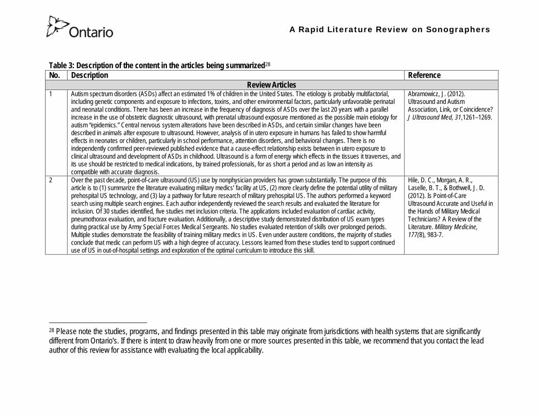
A Rapid Literature Review on Sonographers
Table 3 Description of the content in the articles being summarized28
No
Description Reference Review Articles
1 Autism spectrum disorders (ASDs) affect an estimated 1 of children in the United States The etiology is probably multifactorial including genetic components and exposure to infections toxins and other environmental factors particularly unfavorable perinatal and neonatal conditions There has been an increase in the frequency of diagnosis of ASDs over the last 20 years with a parallel increase in the use of obstetric diagnostic ultrasound with prenatal ultrasound exposure mentioned as the possible main etiology for autism ldquoepidemicsrdquo Central nervous system alterations have been described in ASDs and certain similar changes have been described in animals after exposure to ultrasound However analysis of in utero exposure in humans has failed to show harmful effects in neonates or children particularly in school performance attention disorders and behavioral changes There is no independently confirmed peer-reviewed published evidence that a cause-effect relationship exists between in utero exposure to clinical ultrasound and development of ASDs in childhood Ultrasound is a form of energy which effects in the tissues it traverses and its use should be restricted to medical indications by trained professionals for as short a period and as low an intensity as compatible with accurate diagnosis
Abramowicz J (2012) Ultrasound and Autism Association Link or Coincidence J Ultrasound Med 311261ndash1269
2 Over the past decade point-of-care ultrasound (US) use by nonphysician providers has grown substantially The purpose of this article is to (1) summarize the literature evaluating military medics facility at US (2) more clearly define the potential utility of military prehospital US technology and (3) lay a pathway for future research of military prehospital US The authors performed a keyword search using multiple search engines Each author independently reviewed the search results and evaluated the literature for inclusion Of 30 studies identified five studies met inclusion criteria The applications included evaluation of cardiac activity pneumothorax evaluation and fracture evaluation Additionally a descriptive study demonstrated distribution of US exam types during practical use by Army Special Forces Medical Sergeants No studies evaluated retention of skills over prolonged periods Multiple studies demonstrate the feasibility of training military medics in US Even under austere conditions the majority of studies conclude that medic can perform US with a high degree of accuracy Lessons learned from these studies tend to support continued use of US in out-of-hospital settings and exploration of the optimal curriculum to introduce this skill
Hile D C Morgan A R Laselle B T amp Bothwell J D (2012) Is Point-of-Care Ultrasound Accurate and Useful in the Hands of Military Medical Technicians A Review of the Literature Military Medicine 177(8) 983-7
28 Please note the studies programs and findings presented in this table may originate from jurisdictions with health systems that are significantly different from Ontarios If there is intent to draw heavily from one or more sources presented in this table we recommend that you contact the lead author of this review for assistance with evaluating the local applicability
A Rapid Literature Review on Sonographers
No Description Reference 3 Ultrasonography is a safe and effective form of imaging that has been used by physicians for more than half a century to aid in
diagnosis and guide procedures Over the past two decades ultrasound equipment has become more compact higher quality and less expensive which has facilitated the growth of point-of-care ultrasonography mdash that is ultrasonography performed and interpreted by the clinician at the bedside In 2004 a conference on compact ultrasonography hosted by the American Institute of Ultrasound in Medicine (AIUM) concluded that ldquothe concept of an lsquoultrasound stethoscopersquo is rapidly moving from the theoretical to realityrdquo This conference included representatives from 19 medical organizations in November 2010 the AIUM hosted a similar forum attended by 45 organizations Some medical schools are now beginning to provide their students with hand-carried ultrasound equipment for use during clinical rotations Although ionizing radiation from computed tomographic (CT) scanning is increasingly recognized as a potentially major cause of cancer ultrasonography has been used in obstetrics for decades with no epidemiologic evidence of harmful effects at normal diagnostic levels However ultrasonography is a user-dependent technology and as usage spreads there is a need to ensure competence define the benefits of appropriate use and limit unnecessary imaging and its consequences This article provides an overview of the history and current status of compact point-of-care ultrasonography with examples and discussion of its use
Moore C L amp Copel J A (2011) Point-of-Care Ultrasonography N Engl J Med 364 749-57
4 Sound travels through objects that block light Only very recently has technology advanced enough to decipher ultrasound for medical use Machines have become smaller cheaper more versatile and more advanced than ever before The medical use of ultrasound spreads across many fields so that traditional areas no longer have a monopoly With this comes the question of training doctors This has been done by various colleges and societies worldwide Some have been quicker to act than others There also needs to be an understanding of when broad experience and advanced technical skills are required or when limited skills will do In addition some procedures can be performed more safely with ultrasound where the knowledge of ultrasound is not paramount This article covers current provision of training in echo-cardiography and ultrasound in areas relevant to anaesthetists who are working in critical care (including accident and emergency) and complex surgery (mainly cardiac)
Bennett S (2009) Training guidelines for ultrasound worldwide trends Best Practice amp Research Clinical Anaesthesiology 23 363ndash373
5 Ultrasound is a commonly employed imaging modality in obstetrics and is generally regarded as safe to the fetus Current ultrasound technology however has significantly higher output potential than older machines used in most clinical studies and the safety profile for the increasing use of Doppler 3-dimensional (D) and 4-D ultrasound with modern machines is unknown This article reviews the current status of ultrasound safety within obstetrics including proposed mechanisms of harm existing scientific and clinical evidence regarding those mechanisms and considerations of safety for the clinical user
Houston L E Odibo A O amp Macones G A (2009) The safety of obstetrical ultrasound a review Prenat Diagn 29 1204ndash1212
A Rapid Literature Review on Sonographers
No Description Reference 6 The purpose of this article is to present the practicing sonographer and sonologist with an overview of the biohazards of ultrasound
and guidelines for safe use Overview Ultrasound is an imaging modality that has important diagnostic value Although useful in a variety of applications diagnostic ultrasound is particularly useful in prenatal diagnosis To date there is no evidence that diagnostic ultrasound produces harm in humans or the developing fetus when used properly There are however an increasing range of ultrasound studies being performed Newer technologies can have higher acoustic output levels than earlier equipment Also subtle or transient effects of diagnostic ultrasound such as changes in membrane permeability or neuronal migration are not completely understood Nondiagnostic Imaging Applications Nondiagnostic uses of ultrasound equipment such as repeated scans of healthy subjects for equipment demonstrations generally should be avoided First-trimester fetal scans should not use color and power Doppler modes and should not be performed for the sole purpose of producing souvenir videos or photographs Production of fetal souvenir photographs or videos during diagnostic clinical studies should not increase exposure levels or extend scan times beyond those needed for clinical purposes Operators should follow safe scanning guidelines and ALARA (as low as reasonably achievable) principles The FDA considers the promotion selling or leasing of ultrasound equipment for making ldquokeepsake fetal videosrdquo that are not part of a medical diagnostic procedure to be an unapproved use of a medical device
NelsonT R FowlkesJ B Abramowicz J S Church C C (2009) Ultrasound Biosafety Considerations for the Practicing Sonographer and Sonologist J Ultrasound Med 28139ndash150
7 Objective A possible association between ultrasound during pregnancy and subsequent non-right handedness among children has been suggested The association has been reported to be stronger among boys The aim of the present study was to explore this further through a meta-analysis of two follow-up studies of three randomized controlled trials of routine ultrasonography during pregnancy Design Handedness was assessed through a questionnaire to the parents and 4715 children at the age of 8ndash9 years were included in the meta-analysis Results There was no statistically significant difference in the prevalence of non-right handedness between the ultrasound-screened children and the controls but there was a statistically significant difference in a subgroup analysis among the boys Exploratory analyses according to exposure or non-exposure indicated an even stronger association between ultrasound and non-right handedness Conclusions A conservative analytical approach indicates no association between ultrasound in utero and subsequent non-right handedness The results from the exploratory analyses must be interpreted with caution There is still a need for further research
Salvesen K A amp Eik-Nes (1999) Ultrasound during pregnancy and subsequent childhood non-right handedness a meta-analysis Ultrasound Obstet Gynecol13 241ndash246
8 Ultrasound energy exerts important cellular genetic thermal and mechanical effects Concern about the safety of ultrasound prompted several agencies to devise regulatory limits on the machine output intensities The visual display of thermal and mechanical indices during ultrasound imaging provides an aid to limit the output of the machine Despite many animal studies no human investigations conducted to date have documented major physiologic consequences of ultrasound exposed during imaging To date ultrasound imaging appears to be safe for use in regional anesthesia and pain medicine interventions and adherence to limiting the output of ultrasound machines as outlined by the Food and Drug Administration may avoid complications in the future This article reviews ultrasound-related biologic effects the role of the regulatory agencies in ensuring safety with the use of ultrasound and the limitations and implications of ultrasound use in humans
Shankar H amp Pagel P S (2011) Potential Adverse Ultrasound-related Biological Effects A Critical Review Anesthesiology 115(5) 1109-1124
A Rapid Literature Review on Sonographers
No Description Reference 9 Objective In the context of the planned International Society of Ultrasound in Obstetrics and GynecologyndashWorld Health Organization
multicenter study for the development of fetal growth standards for international application we conducted a systematic review and meta-analysis to evaluate the safety of human exposure to ultrasonography in pregnancy Methods A systematic search of electronic databases reference lists and unpublished literature was conducted for trials and observational studies that assessed short- and long-term effects of exposure to ultrasonography involving women and their fetuses exposed to ultrasonography using B-mode or Doppler sonography during any period of pregnancy for any number of times The outcome measures were (1) adverse maternal outcome (2) adverse perinatal outcome (3) abnormal childhood growth and neurological development (4) non-right handedness (5) childhood malignancy and (6) intellectual performance and mental disease Results The electronic search identified 6716 citations and 19 were identified from secondary sources A total of 61 publications reporting data from 41 different studies were included 16 controlled trials 13 cohort and 12 casendashcontrol studies Ultrasonography in pregnancy was not associated with adverse maternal or perinatal outcome impaired physical or neurological development increased risk for malignancy in childhood subnormal intellectual performance or mental diseases According to the available clinical trials there was a weak association between exposure to ultrasonography and non-right handedness in boys (odds ratio 126 95 CI 103ndash154) Conclusion According to the available evidence exposure to diagnostic ultrasonography during pregnancy appears to be safe
Torloni M R Vedmedovska Merialdi M Betran A P Allen T et al (2009) Safety of ultrasonography in pregnancy WHO systematic review of the literature and meta-analysis Ultrasound Obstet Gynecol 33 599ndash608
Articles in Peer-Reviewed Journals 10 It has been documented for many years that as a result of use of ultrasound to determine the sex of the fetus the male to female
ratio in countries such as China and India have become increasingly skewed (due to subsequent abortions and infanticide of females) Preference for sons over daughters evident in Chinas and South Asias male sex ratios is commonly rationalized by poverty and the need for old-age support In this article the authors study South and East Asian immigrants to Canada a group for whom the economic imperative to select sons is largely absent Analyzing the 2001 and 2006 censuses 20 percent samples the authors find clear evidence of extensive sex selection in favor of boys at higher parities among South and East Asian immigrants unless they are Christian or Muslim The latter finding accords with the explicit prohibition against (female) infanticidemdashtraditionally the main sex-selection methodmdashin these religions The findings point to a strong cultural component to both the preference for sons and the willingness to resort to induced abortion based on sex
Almond D Edlund L Milligan K (2013) Son Preference and the Persistence of Culture Evidence from South and East Asian Immigrants to Canada Population and Development Review 39(1) 75ndash95
11 The rapid growth in the use of ultrasound as a diagnostic imaging technology over the past forty years has led to a demand for a workforce with the appropriate skills to perform and interpret the scans In the UK ultrasound investigations now comprise the largest group of all diagnostic imaging examinations However there remains no statutory regulation of the practice of sonography in the UK and little recognition of the considerable training that many practitioners have undertaken to obtain the skills to become safe and competent sonographers Many in the field consider that this should change and are working to obtain professional status for the practice of sonography Although the Health Care Professions Council (HCPC) has recommended regulation of sonography practice this is unlikely to happen in the near future This paper discusses the evolution of sonography practice and explores some of the complex issues associated with the professionalisation of sonography
Gibbs V (2013) The long and winding road to achieving professional registration for sonographers Radiography19 164-167
A Rapid Literature Review on Sonographers
No Description Reference 12 Background Contrast echocardiography (which uses contrast agents injected into the blood flow in order to visualize the blood flow
in very small blood vessels) has been shown to improve diagnostic quality especially in technically difficult patients However the learning curve and increased time for preparation and image acquisition have led to low use Methods The authors sought to determine whether the contrast echocardiography procedure performed independently by a specialized trained sonographer could improve efficiency In one centre routine echocardiograms were scheduled for one hour and any study exceeding one hour would result in patient booking cancellations The authors compared the standard of care in which a physician or nurse administers echocontrast with a sonographer-administered program (SAP) Results The time to complete contrast echocardiograms was significantly reduced by the SAP strategy (43 min 17 s plusmn 23 min 42 s vs 1 h 1 min 6 s plusmn 31 min 0 s P lt 0001) Subgroup analysis of the inpatients and outpatients demonstrated similar results Only 10 of studies (6 of 61) in the SAP exceeded 60 minutes compared with 45 (34 of 76) in the standard-of-care group (P lt 0001) Based on study volumes in the centre the net improvement in productivity with the SAP could be up to 53 annually Conclusion Sonographer-administered echocontrast is feasible and potentially removes a barrier to implementation of contrast echocardiography
Tang A Chiew S K Rashkovetsky R Becher H Choy J B (2013) Feasibility of Sonographer-Administered Echocontrast in a Large-Volume Tertiary-Care Echocardiography Laboratory Canadian Journal of Cardiology 29 391ndash395
13 Ultrasound scanning has been used as a diagnostic and screening tool in obstetric practice for over 50 years There is no evidence of immediate or long-term harm to the developing fetus from exposure to B mode ultrasound However exposure to high levels of Doppler ultrasound during early development is increasingly common and the full safety implications of this exposure are not clear Doppler ultrasound exposure in utero gives rise to increased apoptosis in animal models and there is evidence of the effects of exposure to Doppler ultrasound persisting throughout life with increased non-right-handedness observed in human epidemiological studies The authors consider the idea that there may be long-term developmental implications for fetuses exposed to Doppler ultrasound early in gestation These effects may be mediated via thermal or mechanical disruption to the developing conceptus giving rise to free radical damage Excess free radical exposure early in gestation is a strong candidate for the final common pathway underlying developmental programming effects and gives rise to concern that fetuses exposed to high levels of ultrasound are at risk of a developmental programming effect It is suggested that there is a need for animal studies of developmental programming using exposure to Doppler ultrasound scanning as the exposure of interest and for more observational data to be collected in the clinical setting While these data are collected it seems prudent to continue to adhere to the principle of lsquoas low as reasonably achievablersquo (ALARA) when exposing first-trimester fetuses to Doppler ultrasound
Aiken CE Lees CC (2012) Long-term effects of in utero Doppler ultrasound scanning ndash A developmental programming perspective Medical Hypotheses 78 539ndash541
14 Point of care sonography by emergency physicians in the emergency department can be an effective aid in the diagnosis and management of patients presenting with a variety of medical and traumatic conditions Its use can improve patient outcomes enhance patient safety speed patient disposition and save lives CAEP supports the use of point of care sonography
Canadian Assocation of Emergency (CAEP) Physicians Ultrasound Position Statement Working Group (2012) Use of point of care sonography by emergency physicians CJEM 14(2)106-112
A Rapid Literature Review on Sonographers
No Description Reference 15 The use of three-dimensional echocardiography (3DE ie a 3D ultrasound of the heart) during electrophysiology (EP - a branch of
cardiology that deals with the diagnosis and treatment of heart rhythm disorders) procedures is the end product of years of growth in two diverse cardiology subspecialties namely advanced cardiac imaging and the EP During the past decade progress in both fields has resulted in many important advances that have culminated in their union for a new area of growth and development Imaging advances have provided the cardiovascular specialist with enhanced cardiac volume and function data and more recently 3DE capabilities with improved spatial and temporal resolution providing unprecedented spatial relationships This latter development is valued by EP specialists in need of hitherto never required anatomic knowledge as they press forward with extraordinary expansion in their capabilities It makes sense that by combining these two rapidly growing subspecialties future capabilities in patient care may be achieved that would otherwise not be possible This paper discusses the value of 3DE during EP procedures and offers the readers insight into this novel multispecialty hybrid arena Using this model as a template it is likely that the readers may identify other areas within their practices where periprocedural advanced imaging may afford significant dividends in patient outcomes
Moukabary T Faletra F F Kronzon I Thomas W amp Sorrell V L (2012) Three-Dimensional Echocardiography in the Electrophysiology Laboratory Echocardiography 29 117-122
16 Background There has been much discussion about whether female feticide occurs in certain immigrant groups in Canada The authors examined data on live births in Ontario and compared sex ratios in different groups according to the motherrsquos country or region of birth and parity Methods The authors completed a population-based study of 766 688 singleton live births between 2002 and 2007 They used birth records provided by Ontario Vital Statistics for live births in the province between 23 and 41 weeksrsquo gestation They categorized each newborn according to the motherrsquos country or region of birth namely Canada (n = 486 599) Europe (n = 58 505) South Korea (n = 3663) China (n = 23 818) Philippines (n = 15 367) rest of East Asia (n = 18 971) Pakistan (n = 18 018) India (n = 31 978) rest of South Asia (n = 20 695) and other countries (n = 89 074) They calculated malefemale ratios and 95 confidence intervals (CIs) for all live births by these regions and stratified them by maternal parity at the time of delivery (0 1 2 or ge 3) Results Among infants of nulliparous women (women with no previous children) the malefemale ratio was about 105 overall As parity (the number of previous children) increased the ratio remained unchanged among infants of Canadian-born women In contrast the malefemale ratio was significantly higher among infants of primiparous women (women with one previous child) born in South Korea (120 95 CI 109ndash134) and India (111 95 CI 107ndash115) than among infants of Canadian-born primiparous women Among multiparous women (women with 2+ previous children) those born in India were significantly more likely than Canadian-born women to have a male infant (parity 2 ratio 136 95 CI 127ndash 146 parity ge 3 ratio 125 95 CI 109ndash143) Interpretation The study of malefemale ratios in Ontario showed that multiparous women born in India were significantly more likely than multiparous women born in Canada to have a male infant
Ray J G Henry D A Urquia M L (2012) Sex ratios among Canadian liveborn infants of mothers from different countries CMAJ184(9) E92-96
A Rapid Literature Review on Sonographers
No Description Reference 17 This letter consists of a case study that illustrates how a cross-disciplined sonographer (educated in cardiac and vascular ultrasound)
can quickly and efficiently and with great Cost-effectiveness deliver timely care to a patient in certain clinical settings The literature is comparatively sparse in referencing clinical diagnostics in cross-discipline sonographers Echocardiographers must be clinicians fully comfortable with evaluating the circulatory dynamics of the body from the left and right heart to the peripheral arteries and veins including the extracranial circulation (carotid arteries) Included in this skill set is substantive understanding of the ECG patientrsquos history and integration of cardiac and vascular findings Limitations of each diagnostic tool and how these limitations can be overcome by the strengths of the other examination are important For the dual-skilled sonographer only experience continuous education and constant clinical feedback will lead to the independent decision making that is counted on by the physician The American Registry for Diagnostic Medical Sonography offers credentials in cardiac and vascular imaging (RDCS and RVT) Cardiovascular Credentialing International offers credentials in cardiac and vascular imaging as well (registered cardiac sonographer [RCS] and registered vascular specialist [RVS]) All imaging facilities should consider merging cardiac and vascular noninvasive imaging with dual-registered clinical sonographers credentialed by one of these two agencies
Zemanek M (2012) What It Means to Be a Cardiovascular Sonographer Journal of Ultrasound in Medicine 311859-1862
18 Diagnostic medical sonography is viewed as a safe imaging modality However bioeffects research has yet to define the effects of exposure to sonography in humans Therefore sonographers must be aware of potential risks and practice ALARA (ldquoas low as reasonably achievablerdquo) routinely This research explores sonographersrsquo ability to answer basic questions about the bioeffects of sonography as well as their knowledge and use of the safety indices called the mechanical index (MI) and thermal index (TI) Members of the Society of Diagnostic Medical Sonographers in the US a southwestern sonographersrsquo society and a sonography community on Facebook 2010 were surveyed (n = 212) There is a low level of bioeffects knowledge and minimal safety practices among sonographers in this survey No significant relationship was found between sonographer knowledge of the bioeffects of sonography years in the profession or whether the sonographers monitor the MITI
Bagley J Thomas K DiGiacinto D (2011) Safety Practices of Sonographers and Their Knowledge of the Biologic Effects of Sonography Journal of Diagnostic Medical Sonography 27(6) 252ndash261
19 This article highlights trends in echocardiography Point of care ultrasound (hand-carried ultrasound) Initially point of care ultrasound will increase the use of echocardiography in the same manner that cardiac CT increased catheterizations However once the basics are learned it has the potential to be a very powerful tool in that less unnecessary studies will be ordered In the clinic small systems will be used as a lsquolsquofirst lookrsquorsquo screen and become part of the patientrsquos history only patients for whom more testing is necessary will be referred for a comprehensive study This is likely to reduce the number of Doppler studies requested The miniaturization of echo equipment will continue with improved qualityfunctionality of the lsquolsquohand-heldrsquorsquo echo machines to the extent that cordless probes will become a reality sooner rather than later Role in interventional echocardiography Sonographers will be spending much more time in the interventional and electrophysiology (EP) labs There will be more interconnection with interventional cardiologists and the volume will increase as more devices are implanted more percutaneous valves placed and more robotic surgery is performed Crosstraining and multitasking Sonographers entering the profession would be prudent to learn cardiac and vascular imaging Individuals who multitask andor cross-train will be ideal For example a sonographer who starts an IV monitors ECG stress can perform cardiac MRI and can interpret a preliminary report or a nurse who can do an echo monitor sedation and start an IV will be invaluable The sonographer should continue to grow professionally by cross training as institutions try to minimize costs and maximize throughput
McCulloch M L (2011) Thoughts regarding the future of sonography Journal of the American Society of Echocardiography 24(5) 29A-30A
A Rapid Literature Review on Sonographers
No Description Reference 20 Background Radiology reports provide specialist interpretation of images and relate these findings to the patientrsquos symptoms and
signs This study compared ultrasound (USS) reports generated by radiologists and sonographers to determine if any significant or clinically relevant differences existed Methods A retrospective analysis of 624 consecutive USS reports was carried out The reports were assessed for the presence of a lsquodisclaimerrsquo or lsquocaveatrsquo pertaining to the quality of the images and were analysed with respect to the clarity of their wording and ability to answer the clinical request Results The majority of sonographer USS reports contained a disclaimer regarding the quality of the USS images (571) compared with a smaller proportion of radiologist scans (99) (P lt 0001) Overall radiologists performed significantly better in providing a clear negative or positive diagnosis to the clinical question on the request form when compared with sonographers (885 vs 654 P lt 0001) Conclusion lsquoDisclaimerrsquo comments and lsquohedgingrsquo must be avoided in radiology reporting While feedback as to the accuracy of the radiology images is important overuse of such terms undermines the validity of the radiology report
Garcea G Mahmoud A Ong S L Rees Y Berry Det al (2010) Caveat reporting in ultrasound interpretation of surgical pathology a comparison of sonographer versus radiologist Journal of Evaluation in Clinical Practice 16 97ndash99
21 A nine-point questionnaire was constructed based on previously published work by Marsal and Sheiner et al The survey was distributed to clinical users of ultrasound practicing sonography in Hamilton (New Zealand) during July 2010 totalling 35 users consisting of sonographers sonography trainees one obstetric sonologist and one fetal medicine registrar Results Twenty sonographers returned the questionnaire which represents survey response of 61 The level of clinical experience was equally split between junior sonographers of lt 5 yearsrsquo experience (30) sonographers with 5-10 yearsrsquo experience (35) and senior sonographers of gt10 yearsrsquo experience (35) While 100 of the sonographers surveyed reported being familiar with the term Thermal Index (TI) and 90 claimed they adhered to the Action Learning Action Response Association (ALARA) principle all the time or most of the time only 15 of sonographers correctly described or defined what the term TI means Of the sonographers 80 knew where to find the TI during real-time examinations but only 35 knew which TI index to use for nuchal translucency (NT) scan and only 10 knew the expected range of TI encountered during routine obstetric examinations Of the eight sonographers who reported adhering to ALARA all the time the majority (64) could not describe or define TI In the same subgroup the majority (64) did not know which TI to monitor during a NT scan Only three of 20 (15) sonographers knew what the term TI means Of these two were junior sonographers with lt 5 yearsrsquo experience and one had 5ndash10 yearsrsquo experience Despite understanding the term TI one of the three sonographers still selected the incorrect index to monitor during a NT scan Not one of the seven senior sonographers with gt 10 years of experience could define or describe the TI In total only 10 of sonographers knew what the TI means and which TI to monitor during NT scanning
Necas M (2010) New Zealand sonographers do not outperform their European or American colleagues in the knowledge of ultrasound safety Australasian Journal of Ultrasound in Medicine13(4) 28-32
A Rapid Literature Review on Sonographers
No Description Reference 22 Musculoskeletal (MSK) ultrasound performed by non-medical practitioners is a developing practice The aim of this paper is
(1) to audit an experienced sonographerrsquos (SJ Riley one of the authors) performance after one yearrsquos independent reporting against two experienced MSK radiologists (CJ Grove amp M Chandramohan the other two authors) and (2) to establish an audit standard against which such role development might be contrived Images and reports from 250 MSK ultrasound examinations performed by a sonographer were reviewed independently by two consultant MSK radiologists The examinations were graded for discrepancy eg grade 1 ndash agree with the sonographer report grade 2 ndash minor discrepancy unlikely to alter patient care grade 3 ndash potentially significant discrepancy grade 4 ndash definite significant discrepancy likely to have adverse consequences for patient care Two of 250 (08) cases were excluded Both radiologists agreed completely with the sonographer (grade 1) in 235 of the 248 cases (948) In 13 cases there was discrepancy between the reports of SJR and the radiologists The discrepancy was grade 2 in six of the 248 cases (24) grade 3 in six of the 248 cases (24) and grade 4 in one of the 248 cases (04) In conclusion this audit shows a high level of agreement between the sonographer and the consultant MSK radiologist reporting of MSK ultrasound This level of agreement may set the standard for future quality assurance audit of sonographer MSK reporting
Riley S J Groves C J Chandramohan M (2010) Musculoskeletal ultrasound audit of sonographer reporting Ultrasound 18 36ndash40
23 Background Growing demand for ultrasound examinations and higher quality requirements motivate searching for routines combining the diagnostic accuracy of radiologist-performed examinations with the economical advantages of sonographer-performed examinations One possible approach is to use strictly standardized acquisition and documentation schemes that give the radiologist access to all relevant information after the examination Purpose To compare a recently introduced routine combining acquisition by a radiographer (more specifically a sonographer with two years experience) documentation as standardized cine-loops and review by a radiologist (lsquostandardized methodrsquo) with the formerly used routine where the diagnosis is made bedside by the radiologist (lsquotraditional methodrsquo) Material and Methods In 64 policlinic patients the kidneys (n = 27) or the gallbladder (n = 37) were examined with both the standardized and the traditional method The radiologistsrsquo findings of hydronephrosis tumors cysts echogenicity changes and cortical thickness (in the kidneys) and wall thickness concrements and polyps (in the gallbladder) were compared between the methods with respect to agreement (proportion of agreement and kappa coefficient) as well as systematic differences (McNemarrsquos test) Results The findings at the gallbladder examination showed a median agreement of 97 (86ndash100 kappa = 064ndash100) and those at the kidney examination an agreement of 90 (78ndash100 kappa = 069ndash 100) There were no significant systematic differences between the methods Conclusion The satisfactory agreement in this preliminary study indicates that the new workflow with ultrasound examinations performed by a radiographer (sonographer) and analyzed off-line by a radiologist is promising and motivates further studies
Stenman C Thorelius L Knutsson A amp Smedby Ouml (2010) Radiographer-acquired and radiologist-reviewed ultrasound examination ndash agreement with radiologistrsquos bedside evaluation Acta Radiologica 52 70ndash74
A Rapid Literature Review on Sonographers
No Description Reference 24 The troubling practice of fetal sex selection has historically been considered an Asian phenomenon However recent evidence shows
that a similar situation is emerging in North America albeit on a smaller scale The Society of Obstetricians and Gynaecologists of Canada (SOGC) has firmly stated its opposition to sex selection for non-medical reasons as well as to the use of any technology used solely for the purpose of determining fetal sex However because fetal sex may be disclosed to the parents at the time of ultrasound examination if they request this information guidance for health care professionals to assist in discouraging fetal sex selection would be useful Because no declaration of motives or reasons is required when a woman seeks a termination of pregnancy the authors suggest that health care professionals need not disclose the sex of a fetus until it has reached a gestational age at which abortion for non-medical purposes would not be possible This proposal would facilitate consistency between clinical practice and the values of Canadian citizens the SOGC the Canadian Medical Association and other professional organizations while still respecting current laws pertaining to disclosure of patient information and patientsrsquo rights to autonomous decision-making
Thiele A T amp Leier B (2010) Towards an Ethical Policy for the Prevention of Fetal Sex Selection in Canada J Obstet Gynaecol Can 32(1) 54ndash57
25 Background The extent to which increases in the use of prenatal ultrasonography over time reflect changes in maternal risk (ie an increase in high-risk pregnancies over time) is unknown In this population-based study the authors examined the use of prenatal ultrasonography from 1996 to 2006 in Ontario Methods With fiscal year 199697 as the baseline the authors evaluated the relative risk (RR) and 95 confidence interval (CI) for the change in rates of ultrasonography for each subsequent year The RR was adjusted for maternal age income rural residence maternal comorbidities receipt of genetics consultation or amniocentesis mdash all in the index pregnancy mdash and history of complications in a prior pregnancy Results The study sample consisted of 1 399 389 singleton deliveries The rate of prenatal ultrasonography increased from 2055 per 1000 pregnancies in 1996 to 3264 per 1000 in 2006 (adjusted RR 155 95 CI 154ndash155) The rate increased among both women with low-risk pregnancies (adjusted RR 154 95 CI 153ndash155) and those with high-risk pregnancies (adjusted RR 155 95 CI 154ndash157) The proportion of pregnancies with at least four ultrasound examinations in the second or third trimesters rose from 64 in 1996 to 187 in 2006 (adjusted RR 268 95 CI 261ndash274) Paradoxically this increase was more pronounced among low-risk pregnancies (adjusted RR 292 95 CI 283ndash301) than among high-risk pregnancies (adjusted RR 225 95 CI 216ndash235) Interpretation Substantial increases in the use of prenatal ultrasonography over the past decade do not appear to reflect changes in maternal risk Nearly one in five women now undergo four or more ultrasound examinations during the second and third trimesters Efforts to promote more appropriate use of prenatal ultrasonography for singleton pregnancies appear warranted
You J J Alter D A Stukel T A McDonald S D Laupacis A et al (2010) Proliferation of prenatal ultrasonography CMAJ 182(2) 143-151
A Rapid Literature Review on Sonographers
No Description Reference 26 Aims The aim of this study was to assess the cost-effectiveness of using certified sonographers and miniaturized echocardiography
systems to perform echocardiograms at bedside in comparison to moving inpatients from the admission department to the echocardiography laboratory (echo-lab) Methods and Results From 26 September 2005 to 27 October 2005 112 patients admitted in six hospital wards in Italy connected through a 100 Mbit LAN to the echo-lab were scanned within the admission ward by sonographers using a miniaturized echo system Logistical data were collected and results were compared with those obtained from 194 consecutive patients coming from the same wards and studied in the echo-lab with high-end machines between 8 March 2005 and 15 April 2005 Performing echocardiograms in the admission department avoided long waiting time of the inpatients in the echo-lab before and after the study increased the percentage of patients studied within 3 and 5 days from request (88 vs 77 and 100 vs 95 respectively P = 003) increased both sonographer (by 339 P lt 0001) and echo-lab productivity (by 41 P lt 0001) and reduced costs of echocardiograms by 29 Conclusion Implementation of digital echocardiography certified sonographers and a miniaturized echo system allowed improvement of the cost-effectiveness of the service provided by the echo-lab for inpatients and avoided patientsrsquo discomfort derived from prolonged waiting time before and after the exam
Badano L Gaetano N Stacul S Gianfagna P Pericoli M et al (2009) Improved workflow sonographer productivity and cost-effectiveness of echocardiographic service for inpatients by using miniaturized systems European Journal of Echocardiography 10 537ndash542
27 The present report is a proposal from the American Society of Echocardiography Advanced Practice Task Force that identifies the potential of cardiac sonographers to achieve the Advanced Cardiovasculat Sonographer (ACS) level According to this report echocardiographic examinations require a well-trained and competent sonographer to obtain proper anatomic and physiologic data to establish an accurate diagnosis for clinical decision-making and patient management Although the formal education and training of cardiovascular sonographers are evolving many entry-level and staff sonographers may not have sufficient practical or clinical knowledge of the necessary components of the echocardiographic study for the individual patientrsquos clinical presentation In many clinical settings echocardiograms are read after the patient has left the laboratory Thus there is a role for a sonographer who can practice at an advanced level in a cardiovascular ultrasound laboratory to ensure a proper echocardiographic examination is performed on every patient In this setting an Advanced Cardiovascular Sonographer (ACS) would be able to review the indication for and quality of the examination If additional images were needed the ACS would assist the sonographer in obtaining these images which would lead to the performance of a complete and fully diagnostic examination before the patient had left the echocardiography laboratory In clinical practice the quality of the examinations performed would improve advancements in echocardiographic methods could be taught and incorporated into daily practice and patients would be better served
Mitchell C Miller F A Bierig S M Bremer M L Ehler D et al (2009) Advanced Cardiovascular Sonographer A Proposal of the American Society of Echocardiography Advanced Practice Sonographer Task Force J Am Soc Echocardiogr 22 1409-13
A Rapid Literature Review on Sonographers
No Description Reference 28 Sonographer practitioner development involves the expansion and extension of the sonographer role to include reporting on
ultrasound examinations Australian sonographers have not seen the same degree of role extension and expansion as their counterparts in the United Kingdom despite increasing levels of discussion regarding sonographer practitioner development The aim of this study was to determine if Australian sonographers want to extend their professional role and what they consider are the important issues associated with role extension This paper reports on qualitative data derived from a survey of Australian sonographers and investigates if Australian sonographers are interested in extending and expanding their professional role and responsibilities and if they do what might be necessary or desirable from a professional point of view for this change to occur A survey was mailed to all members of the Australian Sonographers Association (ASA) in October 2006 The 31-item survey included 28 closed-ended and three opened-ended items to provide both quantitative and qualitative data The quantitative data will be reported separately Qualitative data was derived from responses to the opened-ended questions which asked respondents to elaborate on their attitudes and feelings about role extension and development Analysis used Nvivo7 software to aid in uncovering common themes from the qualitative data The analysis focused on the reported incentives or motivations for becoming a sonographer practitioner as well as disincentives or perceived hurdles that would discourage respondents from becoming sonographer practitioners The three most reported incentives or motivations for becoming a sonographer practitioner were professional recognition remuneration and increased knowledge The three most commonly reported disincentives or perceived hurdles that would discourage respondents from becoming sonographer practitioners were legal issues insurance and further study
McGregor R OrsquoLoughlin K Cox J Clarke J Snowden A (2009) Sonographer practitioner development in Australia Qualitative analysis of an Australian sonographersrsquo survey Radiography15 313-319
29 Introduction This study describes the effect of introducing a cardiac sonographer led follow up clinic within a hospital cardiology department for patients with stable heart valve disease The two years before and after the instigation of the valve clinic were audited Materials and methods The clinic was conducted in a single centre and undertaken in the cardiology department of a district general hospital A total of 382 patients with 397 clinically significant valve lesions but for whom surgery was not yet indicated but follow up required were seen in a cardiac sonographer run clinics These patients no longer attended a medical follow up clinic unless there was clinical or echocardiographic deterioration Effectiveness was judged by the percentage treated according to current best practice guidelines the number of echocardiograms performed and the number of hospital outpatient visits attended In addition mortality data for the subjects in the clinic was collected Results The proportion followed up according to best practice guidelines rose from 157 (41) to 354 (92) (p lt 001) The total number of echocardiograms performed fell from 807 to 550 Total number of outpatient visits fell from 998 to 31 A total of 11 patients died in the two year study period none from progressive valve disease Discussion This study demonstrates that a protocol driven sonographer led heart valve disease follow up clinic significantly improved the quality of follow up while bringing about a major reduction in outpatients visits without compromising patient safety
Taggu W Topham A Hart L Carr-White G Sulke N et al (2009) A cardiac sonographer led follow up clinic for heart valve disease International Journal of Cardiology 132 240 ndash243
30 Role development for imaging allied health professionals has progressed significantly over the last three decades with many major developments pioneered in the field of ultrasound This paper reviews the developing role of the radiographer in ultrasound (in some countries such as the UK sonographers are considered a type of radiographer) and some of the clinical professional and educational implications of such development As the published evidence base underpinning the latest developments is scarce several contemporary case study examples which have been presented verbally at study days attended by the authors have been included In conclusion sonographer role development is considered within the professional contexts of extended role advanced and consultant practice
Hart A amp Dixon A (2008) Sonographer Role Extension and Career Development a Review of the Evidence Ultrasound 16(1) 31-35
A Rapid Literature Review on Sonographers
No Description Reference 31 Objective The main goal of this study was to determine end usersrsquo knowledge regarding safety aspects of diagnostic ultrasound
during pregnancy End usersrsquo attitudes toward the use of ultrasound in low-risk pregnancies were also assessed Methods A questionnaire was distributed to ultrasound end users attending review courses and hospital grand rounds between April and June 2006 in Chicago Results One hundred thirty end users completed the questionnaires (63 response rate) Sixty-three percent were physicians (n = 84) most of them obstetricians (817) About 18 of participants routinely performed Doppler ultrasound examinations during the first trimester Fifty percent of end users thought that the number of ultrasound examinations in low-risk pregnancy should be limited to 1 to 3 (mean plusmn SD 26 plusmn 09) Almost 70 disapproved of ldquokeepsakeentertainmentrdquo ultrasound Although 322 of the participants were familiar with the term thermal index only 177 actually gave the correct answer to the question on the nature of the thermal index About 22 were familiar with the term mechanical index but only 38 described it properly Almost 80 of end users did not know where to find the acoustic indices Only 208 were aware that they are displayed on the sonographic monitor during the examinations End users with higher knowledge of safety issues thought that there should be limitations on the number of ultrasound examinations in low-risk pregnancies (odds ratio 33 95 confidence interval 11ndash100 P = 028) Likewise these end users were more likely to respond that ultrasound might have adverse effects during pregnancy (odds ratio 32 95 confidence interval 11ndash125 P = 045) Conclusions Ultrasound end users are poorly informed regarding safety issues during pregnancy Further efforts in the realm of education and training are needed to improve end user knowledge about the acoustic output of the machines and safety issues
Sheiner E Shoham-Vardi I Abramowicz J S (2007) What Do Clinical Users Know Regarding Safety of Ultrasound During Pregnancy J Ultrasound Med 26 319ndash325
A Rapid Literature Review on Sonographers
No Description Reference 32 In an attempt to survey the knowledge among ultrasound users of some safety aspects of diagnostic ultrasound a questionnaire was
distributed to professionals using ultrasound for fetal examinations The questionnaire was answered by the participants and faculty members of six postgraduate courses in obstetric ultrasound Doppler ultrasound and fetal echocardiography given in Sweden Norway and Austria in 2003 Furthermore in Sweden the questionnaire was distributed to the staff meeting participants of two major obstetric ultrasound units and of one unit for fetal echocardiography and at a meeting of the Working Group for Ultrasound of the Swedish Society of Obstetrics and Gynecology In total 199 questionnaires were anonymously answered by doctors (n = 145) sonographers (n = 22) and midwives trained in ultrasound (n = 32) All of them were using diagnostic ultrasound on a daily or weekly basis The respondents were from nine European countries In none of the countries is formal certification for the use of diagnostic ultrasound in pregnancy or otherwise required The doctors who answered the questionnaire were specialists in obstetrics and gynecology (including some of the internationally recognized experts in fetal ultrasound) pediatric cardiology radiology or clinical physiology The respondents were not informed about the questionnaire in advance and they had a limited time to answer the 16 questions typically 10ndash15 min The questions concerned explanation of the safety indices and their relation to various ultrasound modes ie real-time B-mode M-mode three-dimensional (3D) spectral Doppler color and power Doppler ultrasound The respondents were also asked which ultrasound system they used most often and where on the system the information on thermal index (TI) and mechanical (MI) was displayed (eg in the top right corner of the screen in a separate window etc) Furthermore the respondents had to indicate how they adjust the output energy level on their own machine For all questions there was the optional answer lsquoDonrsquot knowrsquo Results About one-third of the ultrasound experts were able to define the abbreviations TI and MI For the subclasses of the TI the correct answer was found in only 3-8 of cases Of those who knew what the TI and MI meant only two-thirds and one-third of respondents respectively were able to give a simple explanation of the indices In 49 of questionnaires the Doppler modes were correctly ranked as generally producing higher ultrasound exposure than the imaging modes (B-mode 3D) Some 28 of the respondents correctly indicated where on their own machine the information on safety indices is displayed However not all of them knew how to control the output energy level For all items in the questionnaire there were no significant differences in the results between the three categories of respondents (ie physicians sonographers and midwives) The results of the questionnaire were anything but encouraging and they indicated that the users who are supposed to be responsible for controlling exposure of the fetus to ultrasound had a very poor knowledge of the basic safety aspects of ultrasound
Maršaacutel K (2005) The output display standard has it missed its target Ultrasound Obstet Gynecol 25 211ndash214
33 Purpose This study was performed to evaluate the accuracy of upper abdominal ultrasound (US) scanning performed by sonographers in a district general hospital to identify potential areas of weakness and to make recommendations to improve the service Materials and Methods Upper abdominal US examinations performed and reported by sonographers over a 4-week period were retrospectively reviewed The accuracy of the imaging findings and reports were assessed against other imaging surgical histological or laboratory findings and against clinical outcome Results A heterogenous group of 104 patients were included in the study 62 of whom had an US abnormality Errors of scanning or interpretation were identified in 10 patients (96) of whom five (48) were felt to be potentially significant Conclusions The sonographersrsquo accuracy in reporting upper abdominal US scans was 90 However on the basis of this study the authors have implemented specific recommendations to improve the quality of the service
Dongola N Guy R Ward S amp Giles J A (2003) Can sonographers offer an accurate upper abdominal ultrasound service in a district general hospital Radiography 9 29ndash33
A Rapid Literature Review on Sonographers
No Description Reference 34 Aim To compare the accuracy of radiographers and radiologists in routine abdominal ultrasound Materials and Methods One
hundred consecutive patients attending for routine abdominal ultrasound were included Each patient was examined by both a radiographer and radiologist Both operators noted their fndings and wrote a concluding report without conferring Reports were compared Where there was disagreement the patient was either re-examined by another radiologist or had further investigation Results Of 100 patients 52 were men and 48 were women The age range was 19plusmn88 years (median 52 years) Thirty-seven patients had renal tract ultrasound one had an aortic ultrasound and 62 had general upper abdominal ultrasound In 44 cases both operators reported the examination as normal In 49 cases both operators reported the examinations as abnormal and there was complete agreement between the operators In seven cases there was not complete agreement between operators Three of these disagreements were considered minor and four major In three of the seven cases the radiographer was correct and in four the radiologist was correct Conclusion Experienced radiographers and radiologists are highly accurate in performing and interpreting routine abdominal sonography Both operators missed a small minority of abnormalities There was no statistically significant difference in the accuracy of radiographers and radiologist
Leslie A Lockyer H amp Virjee J P (2000) Who Should be Performing Routine Abdominal Ultrasound A Prospective Double-Blind Study Comparing the Accuracy of Radiologist and Radiographer Clinical Radiology 55 606-609
Grey Literature 35 The ARRT The American Registry of Radiologic Technologists tests certifies and annually registers more than a quarter-of-a-million
radiologic technologists to promote high standards of patient care They award the Registered Technologist or RT designation to individuals who have completed the prescribed classroom and clinical education passed the appropriate exam and met the ethics requirements And they ensure their continuing education and ongoing compliance with their high standards by requiring annual registration of their certificate Certification by ARRT is voluntary not mandatory But employers state licensing agencies and federal regulators view ARRTrsquos credential as an indicator that a person has met recognized national standards for medical imaging and radiation therapy professionals How Are Radiologic Technologists Deemed Qualified First comes initial certification Certification is the process of initially recognizing individuals who have satisfied certain standards within a profession A person is certified by ARRT after completing an approved educational program complying with the ethics standards and passing a certification exam Then comes annual registration Registration is the yearly procedure required to maintain certification ARRT registrants are those who having already fulfilled the requirements for initial certification continue to meet the requirements for annual registration The ARRT annually registers the certificates of individuals who meet the following three criteria agreeing to comply with the ARRT Rules and Regulations continuing to comply with the ARRT Standards of Ethics and meeting our continuing education requirements Only technologists who are currently registered (which means that they have renewed within the past year) may designate themselves as ARRT Registered Technologists and use the initials RT after their names Federal laws regulate personnel qualifications in some modalities (mammography for example) but they accredit the health care facility rather than licensing the individual radiologic technologist who is providing patient care And while many states have laws regulating who can perform radiologic procedures they are typically based on ARRT certification Some employers and state agencies may require ARRT certification but not necessarily current registration Unless they require current registration by ARRT employers cannot assume that the person has complied with the ethics requirements or the continuing education requirements
American Registry of Radiologic Technologists (ARRT) (2013a) Patient Page Accessed April 2013
A Rapid Literature Review on Sonographers
No Description Reference 36 This document outlines certification requirements for sonography There are two pathways (primary and post-primary) For the
primary pathway it notes that sonography educational programs must be accredited by a mechanism acceptable to the ARRT ARRT generally recognizes only accreditation agencies that are recognized by the Council for Higher Education Accreditation andor United States Department of Education
American Registry of Radiologic Technologists (ARRT) (2013b) Sonography Certification Accessed April 2013
A Rapid Literature Review on Sonographers
No Description Reference 37 In Australia sonographers and student sonographers are required to be accredited by Australian Sonographer Accreditation Registry
(ASAR) in order to practise This requirement is contained within the Australian Governmentrsquos Diagnostic Imaging Accreditation Scheme standards and Medicare legislation Accreditation of sonographers is linked only to achieving - or in the case of students studying towards - an accredited entry-level qualification completion of continuing professional development and payment of an annual fee to ASAR The ASA contends that the lack of a requirement to ensure each sonographer is lsquofit to practisersquo is an unacceptable gap in current regulation undermining efforts to ensure the quality of sonographic services and failing to protect patients from harm The ASA supports and advocates for nationally-enforceable regulation of sonographers to meet our patientrsquos expectations National registration The National Registration and Accreditation Scheme (NRAS) for health workers commenced on 1 July 2010 The ten professions which comprised the initial intake into this scheme were those that had registration requirements in all Australian states or territories as well as those in which legislated regulation is deemed to be in the public interest An additional four professions - including medical radiation practitioners which are defined as radiographers radiation therapists and nuclear medicine technologists - will be included in the NRAS commencing on 1 July 2012 As sonography did not have registration in any states or territories prior to the commencement of NRAS the profession has not been in a position to negotiate entry into NRAS In early August 2011 the ASA lodged a submission with the Health Workforce Principal Committee requesting a profession-specific Sonography Board of Australia be formed under NRAS This submission was prepared based on information that consideration of professions which were unregistered would commence More recently the Australian Health Workforce Ministerial Council (AHWMC) issued a communiqueacute stating in part lsquoMinisters have commissioned work on unregistered professions and future directions for national registration Given this Ministers agreed to defer consideration of the inclusion of additional professions into the NRAS Ministers agreed that it would be premature to consider further applications for the scheme until this work has been completed other than paramedicsrsquo Options for unregistered health practitioners including sonographers Policy work is continuing at a federal level to establish an alternative regulatory framework The National Alliance for Self-Regulating Health Professions (NASRHP) consisting of the peak professional associations of eight self-regulating health professions including the ASA representing sonography released a proposal lsquoHarnessing self-regulation to support safety and quality in healthcare deliveryrsquo in March 2012 This proposal concluded that the framework that regulates all health practitioners should be expanded beyond NRAS to include
bull authorised self-regulating professions via a health professions panel bull negative licensing to withdraw the ability to practise from registered and self-regulated practitioners who do not meet the
regulatory requirements or unregulated practitioners due to poor practice As with all advocacy initiatives consideration by the federal government of the introduction of the NASRHPrsquos proposal may be prolonged and the outcomes may not match the expectations of NASRHPrsquos member professional associations including the ASA The ASA will continue both independently and collaboratively to lobby on behalf of our members and the public for the introduction of a timely and equitable solution to health practitioner regulation In the interim as there is no regulatory model with the rigour the ASA believes is required for regulation of sonographers and other health practitioners other than NRAS the ASA continues to support its Position Statement published in early 2010
Australian Sonographer Association (ASA) (2013) Sonographer Regulation Accessed April 2013
A Rapid Literature Review on Sonographers
No Description Reference 38 This publication contains information about Cardiovascular Credentialing International (CCI) It also provides the necessary
information and references concerning the process by which candidates may earn certificate or registry-level credentials CCI is a not-for-profit corporation established for the purpose of administering credentialing examinations as an independent credentialing agency CCI began credentialing cardiovascular professionals in 1968 CCI offers eight credentials which when earned demonstrate that the registrant holds fundamental knowledge in the particular cardiovascular specialty These include RCCS ndash Registered Congenital Cardiac Sonographer RCS ndash Registered Cardiac Sonographer and RPhS ndash Registered Phlebology Sonographer
Cardiovascular Credentialing International (2013) 2013 Examination Application amp Overview Accessed April 2013
39 Oregon Governor Signs Sonographer Licensure Legislation On July 28 2009 Oregon Governor Ted Kulongoski signed a sonographer licensure bill (HB 2245) into law The new law makes Oregon only the second state in the United States to require licensure of sonographers The Society of Diagnostic Medical Sonographers (SDMS) has worked closely with other sonography-related organizations and Oregon sonographers to help craft the language in new law The law renames and restructures the Oregon Board of Radiologic Technologists to the Oregon Board of Medical Imaging (OBMI) and ensures representation of sonographers on the Board The sonographer licensure law requires Oregon sonographers to become licensed by OBMI effective July 1 2010 Beginning on January 1 2014 all sonography licensees would be required to hold a national sonography certificationcredential (or be currently enrolled as a student) While the Oregon bill is far more detailed than the sonographer licensure law that passed in New Mexico earlier this year additional administrative rulesregulations will be developed over the next year to fully implement the new law SDMS will be continue to monitor developments regarding sonographer licensure in other states
Society of Diagnostic Medical Sonographers (SDMS) (2013a) News Accessed April 2013
A Rapid Literature Review on Sonographers
No Description Reference 40 On April 6 2009 New Mexico (NM) Governor Bill Richardson signed the historic bill that for the first time in the United States
requires licensure of sonographers The bill adds sonographers (and MRI technologists) to the list of medical imaging professionals licensed by the State of New Mexico Until now anyone could perform sonograms in New Mexico Prior to the passage of the New Mexico licensure law virtually all other health care providers and professionals had to be licensed As a result the public is often surprised to learn that the person performing a medical sonogram does not have any state licensure or national certification requirements The responsibility for administering the NM sonographer licensure program would rest with the New Mexico Environment Department Although this was not the first choice in most sonographersrsquo minds the Environment Department currently manages the radiologic technology licensure program so the administrative infrastructure is already in place By ensuring the creation of an advisory council that includes representation by each medical imaging and radiation therapy modality regulated the bill helps ensure that the Departmentrsquos other areas of emphasis do not detract from sonographer licensure Because sonographers have never had to be licensed before they frequently do not see or understand the distinction between state licensure and national certification National certifications or credentials issued by voluntary organizations such as the American Registry for Diagnostic Medical Sonography (ARDMS) Cardiovascular Credentialing International (CCI) and the American Registry of Radiologic Technologists (ARRT) have little legal weight The certification process and resulting credential are valuable tools in ensuring sonographers have met minimum education and knowledge requirements but do not really regulate anyonersquos ability to perform sonography In an ideal world no additional regulation of sonography would be required However as everyone knows more and more people are taking weekend courses and buying ultrasound equipment without understanding the ramifications of using ultrasound technology (eg ALARA) Under the bill passed by the New Mexico legislature national credentialscertifications should provide evidence of meeting the statersquos standards All sonographers who are performing these critical medical imaging services in New Mexico will be required to meet national certification standards (ie have certifications or credentials issued by ARDMS CCI or ARRT) As a result there would be no additional exams in New Mexico beyond the national certification exams required by ARDMS CCI or ARRT The bill also requires the state to recognize the continuing medical education completed for national certification renewal The most visible impact on New Mexico sonographers will be a license fee However the legislation caps the license fee at $100 per two-year license In addition only the New Mexico Legislature can change this cap
SDMS (2013b) New Mexico Sonographer Licensure Law Signed Accessed April 2013
A Rapid Literature Review on Sonographers
No Description Reference 41 Preamble The purpose of this document is to define the Scope of Practice for Diagnostic Ultrasound Professionals in the US and to
specify their roles as members of the health care team acting in the best interest of the patient This scope of practice is a living document that will evolve as the technology expands Definition of the Profession The Diagnostic Ultrasound Profession is a multi-specialty field comprised of Diagnostic Medical Sonography (with subspecialties in abdominal neurologic obstetricalgynecologic and ophthalmic ultrasound) Diagnostic Cardiac Sonography (with subspecialties in adult and pediatric echocardiography) Vascular Technology and other emerging fields These diverse specialties are distinguished by their use of diagnostic medical ultrasound as a primary technology in their daily work Certification is considered the standard of practice in ultrasound Individuals who are not yet certified should reference the Scope as a professional model and strive to become certified Scope of Practice of the Profession The Diagnostic Ultrasound Professional is an individual qualified by professional credentialing and academic and clinical experience to provide diagnostic patient care services using ultrasound and related diagnostic procedures The scope of practice of the Diagnostic Ultrasound Professional includes those procedures acts and processes permitted by law for which the individual has received education and clinical experience and in which heshe has demonstrated competency Diagnostic Ultrasound Professionals
bull Perform patient assessments bull Acquire and analyze data obtained using ultrasound and related diagnostic technologies bull Provide a summary of findings to the physician to aid in patient diagnosis and management bull Use independent judgment and systematic problem solving methods to produce high quality diagnostic information and
optimize patient care
Society of Diagnostic Medical Sonography [SDMS] (2013c) Scope of Practice for the Diagnostic Ultrasound Professional Accessed April 2013
42 This website notes that in the US most employers in the US prefer to hire sonographers who have professional certification A sonographer can get certification and passing an exam Most exams relate to the specialty that the sonographer is most interested inmdashfor example an exam to become certified in abdominal sonography A few states require diagnostic medical sonographers to be licensed Typically professional certifications required for licensure other requirements vary by state Sonographers must take continuing education to keep their certification current
Bureau of Labor Statistics US Department of Labor (2012) How to become a Diagnostic Medical Sonographer Occupational Outlook Handbook 2012-13 Edition Accessed April 2013
A Rapid Literature Review on Sonographers
No Description Reference 43 The American Registry for Diagnostic Medical Sonography (ARDMS) is an independent not-for-profit organization founded in 1975
that administers examinations and awards the following credentials bull RDMSreg ndash Registered Diagnostic Medical Sonographerreg bull RDCSreg ndash Registered Diagnostic Cardiac Sonographerreg bull RVTreg ndash Registered Vascular Technologistreg bull RPVIreg ndash Registered Physician in Vascular Interpretationreg bull RMSKtrade - Registered in Musculoskeletaltrade sonography
ARDMS is the globally recognized standard of excellence in sonography and has certified more than 75000 individuals ARDMS is composed of three key groups
bull Board of Directors ndash Sonographers vascular technologists physicians research scientists and a member of the public are volunteer members of the board that determines the direction and policies of our organization
bull Examination Development Task Forces ndash A group of sonographers vascular technologists physicians and scientists volunteer their time and expertise to produce our examinations
bull Professional Staff ndash A paid highly-skilled staff administers the programs and assists applicants and registrants Credentials awarded by ARDMS document personal achievement of recognized professional standards and are widely accepted in the medical community by sonography and vascular professional organizations However ARDMS does not validate the day-to-day job performance of any sonography professional ARDMS has earned the prestigious ANSI-ISO 17024 accreditation for certifying bodies from the International Organization for Standardization (ISO) Accreditation is granted through the American National Standards Institute (ANSI) ARDMS has also earned accreditation with the National Commission for Certifying Agencies (NCCA) The NCCA is the accrediting arm of the National Organization for Competency Assurance (NOCA) Established in 1977 as a nonprofit organization NOCA is a leader in establishing quality standards for certifying organizations
American Registry for Diagnostic Medical Sonography (ARDMS) (2012a) Overview of ARDMS Accessed April 2013
A Rapid Literature Review on Sonographers
No Description Reference 44 ARDMS is developing a program to assure that Registrants continue to meet the basic competency requirements of core clinical skills
in present day practice bull Each Registrant who holds an active ARDMS certification as of December 31 2011 will be assigned the 2012 ndash 2021 10-
year recertification assessment period bull The 10-year period for new Registrants will begin the year following the attainment of the credential bull If a Registrant earns a new credential or specialty after January 1 2012 then that credentialspecialty will have a separate
10-year recertification assessment period that will begin the year following the attainment of the credentialspecialty ARDMS will develop tools and resources to assist Registrants in tracking and meeting the recertification assessment program requirements
bull Registrants who do not wish to take a recertification assessment in a specific credentialspecialty may voluntarily relinquish the credentialspecialty
As an ARDMS Registrant individuals are committing to demonstrate the highest level of competency in the modality of sonography by meeting and maintaining the standards set by the ARDMS The pathway of maintenance your certification is comprised of Paying the ARDMS annual renewal fee each year Each year ARDMS assesses Registrants an annual renewal fee By paying your annual renewal fee you are contributing to the ARDMS mission of promoting quality care and patient safety through the certification and continuing competency of ultrasound professionals In order to maintain your credentials you must pay the annual renewal fee of $75 USD or $100 USD (for RPVI and MSK Registrants with MDDODPM) designation on or before December 31 of each year in order to stay active for the following year Attesting to ARDMS rules and policies Each year when you pay your annual renewal fee you are attesting that you are in compliance with all ARDMS rules and discipline policies and are in good standing with the ARDMS Earning a minimum of 30 ARDMS-accepted CMEs during your three-year CME period ARDMS verifies its Registrants compliance with continuing medical education (CME) requirements using an audit process Earning your CMEs is an integral component of maintaining your ARDMS credential(s) and demonstrates your ongoing commitment to lifelong learning RecertificationContinued Commitment to Excellence Following the attainment of the ARDMS credential Registrants are assigned a 10-year recertification period the year after the credential is earned ARDMS is committed to developing a recertification program that is easy for Registrants to comply with
American Registry for Diagnostic Medical Sonography (ARDMS) (2012b) Recertification General Information
A Rapid Literature Review on Sonographers
No Description Reference 45 This document reviews Sonography Principles and Instrumentation (SPI) Examination Requirements offered through ARDMS in
order to achieve sonography certification and the various prerequisites needed before taking the exam For example one prerequisite entails (1) Education Graduate of a program accredited by an agency recognized by the Council for Higher Education Accreditation (CHEA) United States Department of Education (USDOE) or Canadian Medical Association (CMA) that specifically conducts programmatic accreditation for diagnostic medical sonographydiagnostic cardiac sonographyvascular technology Currently the only organizations that offer programmatic accreditation under the aforementioned associations are the Commission on Accreditation of Allied Health Education Programs (CAAHEP) and the Canadian Medical Association (CMA) (2) Required Clinical UltrasoundVascular Experience No additional experience is required (3) Documentation Required with Application 1) Copy of diploma from ultrasoundvascular program or an official transcript indicating the date the degree was conferred 2) Original letter signed by program director andor medical director indicating date of graduation or successful completion of the program4 Program directors must use the mandatory formatted sample letter available on ARDMSorg sampleletters 3) The CV form is not required if the application is submitted and received in the ARDMS office within one year after successful completion of the program Otherwise an original signed and completed CV form for each appropriate specialty area(s) must be submitted 4) Photocopy of a non-expired government issued photo identification with signature the name on the identification must exactly match the name under which you are applying for ARDMS examination
American Registry for Diagnostic Medical Sonography (ARDMS) (2012c) SPI Requirement and General Prerequisites Accessed April 2013
46 This page notes the five credentials offered by the ARDMS and the corresponding exams and specialty exams required bull RDMS requires the SPI exam (sonography principles and instrumentation) and a specialty exam in abdomen breast fetal
echocardiography neurosonolgoy or obstetricsgynecology bull RDCS requires the SPI exam and a speciality exam in adult echocardiography fetal echocardiography pediatric
echocardiography bull RVT requires the SPI and a speciality exam in vascular technology bull RMSK requires the musculoskeletal sonography exam bull RPVI requires the physiciansrsquo vascular interpretation exam
American Registry for Diagnostic Medical Sonography (ARDMS) (2012d) Credentials and Examinations Accessed April 2013
A Rapid Literature Review on Sonographers
No Description Reference 47 This article summarizes some of the many trends in fetal pediatric and adult echocardiography that SDMS members are excited
about Fetal Echocardiography Sonography has been used since the mid-1980s to help identify congenital heart defects during the second trimester The ldquobasicrdquo examination of the fetal heart involves a four-chamber view ndash evaluation of the right and left atrial and ventricular chambers and their respective valves In the past several years there has been a movement in the obstetric ultrasound community to encourage obstetric sonographers to perform the ldquoextended basicrdquo exam which includes the cardiac outflow tracts The outflow tracts consist of the main pulmonary artery exiting the right ventricle and the aorta exiting the left ventricle The fetal echocardiography community also is abuzz over three-dimensional (3D) and four-dimensional (4D) ultrasound and the potential of this technology to significantly improve the evaluation of the fetal heart Spatiotemporal Image Correlation (STIC) a 4D application allows for evaluation and manipulation of the heart in real-time Allen says Todayrsquos ultrasound equipment allows sonographers to gather a volume of data from which they can create detailed 3D images of the tiny fetal heart which is constantly in motion With computer manipulation of the data sets the images also can be viewed in 4D allowing physicians to see the entire fetal heart cycle from start to finish Pediatric Echocardiography One of the most significant advances in pediatric echo over the last decade is moving to a digital format The advent of pediatric transesophageal (TEE) probes also has been a big advance for pediatric echo Transesophageal probes provide an unimpeded sonographic visualization of the intracardiac structures Theyrsquore useful in visualizing very fine valvular abnormalities the thoracic aorta vegetations and thrombus in the atrial appendage It is also useful in detecting subtle anatomic variations seen in congenital heart disease Pediatric-sized TEE probes make the exams possible In the past it wasnrsquot possible because the adult probe was just too big to put down the babyrsquos or childrsquos esophagus Now the probe is small enough to successfully manipulate it in children and get superb images Because the new sonographic imaging equipment and better surgical techniques allow clinicians to detect and repair more cardiac abnormalities earlier patients are living longer Adult Echocardiography The adult echo world is abuzz about 2D and 3D speckle tracking relatively new techniques for the assessment of heart function and strain imaging Speckle tracking evaluates both regional and global function by measuring the deformation of the heart muscle
Orenstein B W (2010) Current Trends in Echocardiography SDMS New Wave February 1-5 Accessed April 2013
48 This document describes the requirements for admission to the Register of Accredited Medical Sonographer in Australia Requirements include Continuation of Accreditation In order to remain on the Register an accredited Sonographer must satisfy the ASAR continuing professional development (CPD) requirements and pay the prescribed annual fee Student Sonographers apart from paying the prescribed annual fee should provide evidence of their student status every year when they renew their ASAR accreditation
Australian Sonographer Accreditation Registry (ASAR) (no date A) Sonographer Accreditation Accessed April 2013
A Rapid Literature Review on Sonographers
No Description Reference 49 ASARrsquos functions
1 Accredit sonography education programs within Australia 2 Maintain a register of Accredited Sonographers and Accredited Student Sonographers 3 Establish minimum standards of Continuing Professional Development (CPD) and to monitor the CPD requirements for
Sonographers to remain on the register 4 To support activities that advance the profession of sonography 5 To provide registration information about Sonographers to Medicare Australia 6 To provide public access to the name registration number State or Territory current type of accreditation and area of
specialty for active Sonographers ASARrsquos Role ASAR is not a registration board ASAR does not have the authority to remove a Sonographer from the Registry due to professional misconduct nor the ability to prevent a Sonographer from practicing ASAR is only able to remove a Sonographer from the Registry if they
bull fail to meet CPD requirements and or bull fail to pay annual fees
ASAR (no date B) About ASAR Accessed April 2013
50 Regulatory bodies A Regulatory Body maintains a register of individuals who have achieved the required qualification of that profession to practice and can evidence continuing competence at the required level For a doctor to practice in the UK he or she must be registered with the General Medical Council (GMC) This is the regulatory body for medical practitioners The majority of sonographers who are employed in the UK come from a background of radiography or midwifery To practice radiography in the UK a qualified radiographer must be registered with the Health Professions Council (HPC) Similarly a midwife must be registered with the National Midwifery Council to practice as a midwife Sonographer registration in the UK There are a number of professions allied to medicine that are regulated by the HPC for example radiography physiotherapy However sonography is not recognised as a profession by the HPC and therefore there is no mechanism whereby an individual can register with the HPC as a sonographer or be regulated by them as a sonographer It is a common misunderstanding that sonographers have to be registered with the HPC to practice This comes from the fact that most sonographers are also qualified radiographers and maintain their registration with the HPC because they wish to remain on the HPC register as a radiographer Professional bodies An individual can be a member of a Professional Body such as the Royal College of Obstetricians amp Gynecologists (RCOG) Royal College of Radiologists (RCR) Society amp College of Radiographers (SCoR) or the Royal College of Midwives (RCM) but this is not the same as being accepted onto the register of one of the regulatory bodies The professional bodies are responsible for setting and maintaining the standards of their members providing a code of conduct etc They may also act in the capacity of a trades union and can also provide professional indemnity insurance The British Medical Ultrasound Society BMUS is not a professional body but is a scientific society so will offer a different range of benefits Whether or not one wishes to join the society has no impact on their ability to work as a sonographer in the UK although from a continuing professional development point of view it can be beneficial The College of Radiographers are currently working with the HPC to get sonography recognized as a separate profession
British Medical Ultrasound Society (BMUS) (no date) Information on Sonographer Regulation in the UK Accessed April 2013
A Rapid Literature Review on Sonographers
No Description Reference 51 This document included sections on the role of the sonographer and the use of sonography for entertainment Role of the
Sonographer and Technical Impressions The role of the sonographer is to perform the ultrasound scan and to document observations The sonographerrsquos written technical impressions are intended as a form of communication between the sonographer and the reporting physician These observations must be reviewed and subsequently reported by the reporting physician Issuing of a final reportdiagnosis represents the practice of medicine and is therefore the domain of the reporting physician Use of Ultrasound for Entertainment or Non-Diagnostic Purposes In contrast to this justified form of assessment ultrasound for entertainment is simply an elaborate version of a portrait For a variable fee the surface of the fetus is skimmed over and clients are provided with keepsake images or a DVD of their fetus There are no measurements taken during this exam no morphological assessment performed and no dictated diagnostic report of findings Essentially there are no diagnostic benefits to the exam and uninformed parents may be falsely reassured that the beautiful portrait indicates a normal fetus Entertainment ultrasound facilities may operate outside of medical guidelines and without any controls which may result in a lack of technical safeguards operator expertise or governance of technical competency These facilities do not comply with the ALARA principle which could result in increased fetal exposures The Canadian Society of Diagnostic Medical Sonograph (CSDMS) does not support persons or facilities that participate in ultrasound for entertainment activities Liability insurance purchased through the CSDMS group insurance plan does not cover any activities carried out for entertainment ultrasound purposes Other leaders in Canadian medicine support this statement In 2005 the SOGC (Society of Obstetrics and Gynaecologists of Canada) published an article entitled ldquoObstetrical Ultrasound Biological Effects and Safetyrdquo and their conclusion was that ldquothe theoretical risk of adverse bio-effects even from standard 2D obstetrical ultrasound makes it hard to justify its use for non-medical reasons such as sex-determination making non-medical photos or video or for commercial purposerdquo The SOGC recommends that ultrasound be used prudently and that energy exposure be limited to the minimum that is medically necessary The SOGC further recommends a complete ban on the non-medical use of fetal ultrasound and encourages government to join with society to find appropriate means to deal with this public health issue The CAR (Canadian Association of Radiologists) makes a similar statement ldquoThe CAR strongly opposes the use of diagnostic ultrasound equipment for non-medical purposes and considers the use of medical ultrasound for entertainment to be a misuse of the technology especially if fetal subjects are involvedrdquo
Canadian Society of Diagnostic Medical Sonography (no date) Professional Practice Guidelines and Policy Statements For Canadian Sonography Accessed April 2013
A Rapid Literature Review on Sonographers
No Description Reference 52 In 1993 the Consortium for the Accreditation of Sonographic Education (CASE) was formed through the common desire to ensure
that the education and training of sonographers in the United Kingdom was delivered at the highest level All members share common concerns that standards of service provision and education are developed in parallel with increasing demand and improving technology All members strive to create an environment in which education and training is maximised This will make validation and accreditation to be as robust and straightforward as possible CASEs philosophy is to promote best ultrasound practise through the accreditation of those training programmes that develop safe and competent ultrasound practitioners This should be delivered at postgraduate level The majority of its activities relate to supporting those Institutions that offer or wish to offer courses leading to the award of a Masters Postgraduate DiplomaCertificate in Medical or clinical Ultrasound In addition to offering accreditation of new and re-accreditation of established ultrasound education programmes CASE undertakes annual monitoring of the courses it has accredited The information obtained is fed back to the Institutions both on an individual basis and via the annual CASE report Each member organisation is unique and has its own obligations to its membership and its own agenda while CASE brings them together to provide a forum for the interchange of ideas in which shared views may be synthesised and thus inform education and training programmes
Consortium for the Accreditation of Sonographic Education (no date) Home Accessed April 2013
A Rapid Literature Review on Sonographers
No Description Reference 53 The Medical Radiation Technologists Board (the Board) is one of sixteen New Zealand health registration authorities appointed by
the Minister of Health under the Health Practitioners Competence Assurance Act 2003 (the Act) The Board is responsible for the administration of the Act in regard to the profession of medical radiation technology The primary responsibility of the Board is to protect the health and safety of the New Zealand public by ensuring practitioners registered in the profession of medical radiation technology are competent and fit to practice In accordance with Section 118 of the Act the Board is responsible for fulfilling a number of functions
bull 01 To prescribe the qualifications required for scopes of practice within the profession of medical radiation technology and for that purpose to accredit and monitor educational institutions and degrees courses of studies or programmes
bull 02 To authorise the registration of medical radiation technologists under the Act and to maintain registers bull 03 To consider applications for annual practising certificates bull 04 To review and promote the competence of medical radiation technologists bull 05 To recognise accredit and set programmes to ensure the ongoing competence of medical radiation technologists bull 06 To receive and act on information from health practitioners employers and the Health and Disability Commissioner
about the competence of medical radiation technologists bull 07 To notify employers the Accident Compensation Corporation the Director-General of Health and the Health and
Disability Commissioner that the practice of a medical radiation technologist may pose a risk of harm to the public bull 08 To consider the cases of medical radiation technologists who may be unable to perform the functions required for the
practice of the medical radiation technology profession bull 09 To set standards of clinical competence cultural competence and ethical conduct to be observed by medical radiation
technologists bull 10 To liaise with other authorities appointed under the Act about matters of common interest bull 11 To promote education and training in the profession of medical radiation technology bull 12 To promote public awareness of the responsibilities of the Board bull 13 To exercise and perform any other functions powers and duties that are conferred or imposed on it by or under the Act
or any other enactment
Medical Radiation Technologists Board (MRTB) (no date A) What we do Accessed April 2013
54 Qualifications To be considered for registration as a medical radiation technologist in any of the specified scopes of practice an individual must hold a qualification for that particular scope as prescribed by the Board or approved by the Board as being substantially equivalent to the prescribed qualifications In addition to having an approved qualification individuals may also be required to demonstrate that they have adequate experience in the relevant scope of practice Sonographer The Board has prescribed one New Zealand postgraduate qualification for a person to be eligible for registration in the scope of practice of Sonographer Postgraduate Diploma in Health Science (Ultrasound) issued by the Unitec Institute of Technology Required Experience If an individual is applying for registration in Nuclear Medicine Ultrasound or Magnetic Resonance Imaging in addition to holding a relevant postgraduate qualification they must also be able to provide evidence that you have a minimum of 3360 hours of clinical experience in the relevant scope of practice you are applying for registration in
Medical Radiation Technologists Board (MRTB) (no date B) Qualifications Accessed April 2013
A Rapid Literature Review on Sonographers
No Description Reference 55 Recertification is a mechanism used by the Medical Radiation Technologists Board in New Zealand to ensure that registered medical
radiation technologists continue to be competent to practise within their scopes of practice Annual Practising Certificates Each year if an individual wishes to continue practising in your scope(s) of practice in New Zealand they must apply for and be issued with an annual practising certificate (APC) A condition of being issued with an APC is that one must provide evidence of enrolment and satisfactory progress in a Board-approved recertification programme Alternatively the Board may issue them with an interim practising certificate (IPC) until it is satisfied the individual demonstrates that they are competent to practice in the scope(s) of practice they applied for Working with an IPC will require an individual to work under the supervision of a registered medical radiation technologist or other relevant registered health practitioner Recertification Programmes The Boards recertification programme framework is based on a continuing professional development (CPD) model Individuals have the option of enrolling in one of seven recertification programmes for medical radiation technologists that are currently approved by the Board They must meet the requirements of the particular recertification programme in which they are enrolled Recertification Audits An essential component of the CPD-based recertification programme adopted by the Board is the audit of CPD recordsThe Board undertakes an annual audit of approximately 10 of its registrant database to assess individual medical radiation technologistsrsquo compliance with the Boarrsquods recertification requirements
Medical Radiation Technologists Board (MRTB) (no date C) Recertification Accessed April 2013
56 There are two types of radiography - diagnostic and therapeutic Both need considerable knowledge of technology anatomy physiology and pathology to carry out their work Many also undertake further training to become a sonographer Some of the imaging technology that a diagnostic radiographer may use are
bull X-Ray - looks through tissues to examine bones cavities and foreign objects bull Fluoroscopy - images the digestive system providing a real-time image bull CT (Computed Tomography) - which provides cross-sectional views (slices) of the body bull MRI (Magnetic Resonance Imaging) - builds a 2-D or 3-D map of the different tissue types within the body bull Ultrasound - well known for its use in obstetrics and gynaecology Also used to check circulation and examine the heart bull Angiography - used to investigate blood vessels
Sonographyultrasonography Ultrasound is used in various settings in a hospital including abdominal scanning and breast ultrasound Ultrasound imaging is the use of high frequency sound in excess of human hearing to produce images of structures of the human body that may be observed on a screen These images may subsequently be transferred to photographic film or paper or onto video or CD forming part of the patients record of their examination There are no direct entry routes into ultrasound Most sonographers train as a radiographer then undertake an approved post-registration course Offered by higher education institutions The courses are a minimum of one academic year and prepare sonographers clinically and academically for practice Normally a pre-requisite for acceptance is access to a clinical department with supervised practice for students
National Health Service (NHS) (no date A) Radiographer Accessed April 2013
57 To practice as a radiographer in the UK an individual must be registered with the Health and Care Professions Council (HCPC) To register individuals must have successfully completed an approved training programme in diagnostic or therapeutic radiography These are offered at undergraduate degree (full-time and part-time) and at postgraduate degreediploma level at a number of universities across the UK
NHS (no date B) Entry requirements and training in radiography Accessed April 2013
Health Professions Regulatory Advisory Council 56 Wellesley Street West 12th Floor
Toronto ON M5S 2S3
Telephone 416-326-1550 Toll-Free 1-888-377-7746
Fax 416-326-1549
Website wwwhpracorg
Email hpracwebmasterontarioca
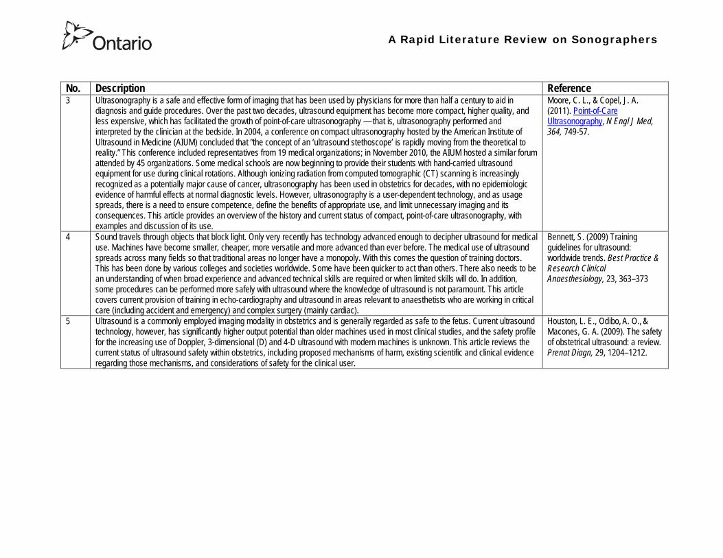
A Rapid Literature Review on Sonographers
No Description Reference 3 Ultrasonography is a safe and effective form of imaging that has been used by physicians for more than half a century to aid in
diagnosis and guide procedures Over the past two decades ultrasound equipment has become more compact higher quality and less expensive which has facilitated the growth of point-of-care ultrasonography mdash that is ultrasonography performed and interpreted by the clinician at the bedside In 2004 a conference on compact ultrasonography hosted by the American Institute of Ultrasound in Medicine (AIUM) concluded that ldquothe concept of an lsquoultrasound stethoscopersquo is rapidly moving from the theoretical to realityrdquo This conference included representatives from 19 medical organizations in November 2010 the AIUM hosted a similar forum attended by 45 organizations Some medical schools are now beginning to provide their students with hand-carried ultrasound equipment for use during clinical rotations Although ionizing radiation from computed tomographic (CT) scanning is increasingly recognized as a potentially major cause of cancer ultrasonography has been used in obstetrics for decades with no epidemiologic evidence of harmful effects at normal diagnostic levels However ultrasonography is a user-dependent technology and as usage spreads there is a need to ensure competence define the benefits of appropriate use and limit unnecessary imaging and its consequences This article provides an overview of the history and current status of compact point-of-care ultrasonography with examples and discussion of its use
Moore C L amp Copel J A (2011) Point-of-Care Ultrasonography N Engl J Med 364 749-57
4 Sound travels through objects that block light Only very recently has technology advanced enough to decipher ultrasound for medical use Machines have become smaller cheaper more versatile and more advanced than ever before The medical use of ultrasound spreads across many fields so that traditional areas no longer have a monopoly With this comes the question of training doctors This has been done by various colleges and societies worldwide Some have been quicker to act than others There also needs to be an understanding of when broad experience and advanced technical skills are required or when limited skills will do In addition some procedures can be performed more safely with ultrasound where the knowledge of ultrasound is not paramount This article covers current provision of training in echo-cardiography and ultrasound in areas relevant to anaesthetists who are working in critical care (including accident and emergency) and complex surgery (mainly cardiac)
Bennett S (2009) Training guidelines for ultrasound worldwide trends Best Practice amp Research Clinical Anaesthesiology 23 363ndash373
5 Ultrasound is a commonly employed imaging modality in obstetrics and is generally regarded as safe to the fetus Current ultrasound technology however has significantly higher output potential than older machines used in most clinical studies and the safety profile for the increasing use of Doppler 3-dimensional (D) and 4-D ultrasound with modern machines is unknown This article reviews the current status of ultrasound safety within obstetrics including proposed mechanisms of harm existing scientific and clinical evidence regarding those mechanisms and considerations of safety for the clinical user
Houston L E Odibo A O amp Macones G A (2009) The safety of obstetrical ultrasound a review Prenat Diagn 29 1204ndash1212
A Rapid Literature Review on Sonographers
No Description Reference 6 The purpose of this article is to present the practicing sonographer and sonologist with an overview of the biohazards of ultrasound
and guidelines for safe use Overview Ultrasound is an imaging modality that has important diagnostic value Although useful in a variety of applications diagnostic ultrasound is particularly useful in prenatal diagnosis To date there is no evidence that diagnostic ultrasound produces harm in humans or the developing fetus when used properly There are however an increasing range of ultrasound studies being performed Newer technologies can have higher acoustic output levels than earlier equipment Also subtle or transient effects of diagnostic ultrasound such as changes in membrane permeability or neuronal migration are not completely understood Nondiagnostic Imaging Applications Nondiagnostic uses of ultrasound equipment such as repeated scans of healthy subjects for equipment demonstrations generally should be avoided First-trimester fetal scans should not use color and power Doppler modes and should not be performed for the sole purpose of producing souvenir videos or photographs Production of fetal souvenir photographs or videos during diagnostic clinical studies should not increase exposure levels or extend scan times beyond those needed for clinical purposes Operators should follow safe scanning guidelines and ALARA (as low as reasonably achievable) principles The FDA considers the promotion selling or leasing of ultrasound equipment for making ldquokeepsake fetal videosrdquo that are not part of a medical diagnostic procedure to be an unapproved use of a medical device
NelsonT R FowlkesJ B Abramowicz J S Church C C (2009) Ultrasound Biosafety Considerations for the Practicing Sonographer and Sonologist J Ultrasound Med 28139ndash150
7 Objective A possible association between ultrasound during pregnancy and subsequent non-right handedness among children has been suggested The association has been reported to be stronger among boys The aim of the present study was to explore this further through a meta-analysis of two follow-up studies of three randomized controlled trials of routine ultrasonography during pregnancy Design Handedness was assessed through a questionnaire to the parents and 4715 children at the age of 8ndash9 years were included in the meta-analysis Results There was no statistically significant difference in the prevalence of non-right handedness between the ultrasound-screened children and the controls but there was a statistically significant difference in a subgroup analysis among the boys Exploratory analyses according to exposure or non-exposure indicated an even stronger association between ultrasound and non-right handedness Conclusions A conservative analytical approach indicates no association between ultrasound in utero and subsequent non-right handedness The results from the exploratory analyses must be interpreted with caution There is still a need for further research
Salvesen K A amp Eik-Nes (1999) Ultrasound during pregnancy and subsequent childhood non-right handedness a meta-analysis Ultrasound Obstet Gynecol13 241ndash246
8 Ultrasound energy exerts important cellular genetic thermal and mechanical effects Concern about the safety of ultrasound prompted several agencies to devise regulatory limits on the machine output intensities The visual display of thermal and mechanical indices during ultrasound imaging provides an aid to limit the output of the machine Despite many animal studies no human investigations conducted to date have documented major physiologic consequences of ultrasound exposed during imaging To date ultrasound imaging appears to be safe for use in regional anesthesia and pain medicine interventions and adherence to limiting the output of ultrasound machines as outlined by the Food and Drug Administration may avoid complications in the future This article reviews ultrasound-related biologic effects the role of the regulatory agencies in ensuring safety with the use of ultrasound and the limitations and implications of ultrasound use in humans
Shankar H amp Pagel P S (2011) Potential Adverse Ultrasound-related Biological Effects A Critical Review Anesthesiology 115(5) 1109-1124
A Rapid Literature Review on Sonographers
No Description Reference 9 Objective In the context of the planned International Society of Ultrasound in Obstetrics and GynecologyndashWorld Health Organization
multicenter study for the development of fetal growth standards for international application we conducted a systematic review and meta-analysis to evaluate the safety of human exposure to ultrasonography in pregnancy Methods A systematic search of electronic databases reference lists and unpublished literature was conducted for trials and observational studies that assessed short- and long-term effects of exposure to ultrasonography involving women and their fetuses exposed to ultrasonography using B-mode or Doppler sonography during any period of pregnancy for any number of times The outcome measures were (1) adverse maternal outcome (2) adverse perinatal outcome (3) abnormal childhood growth and neurological development (4) non-right handedness (5) childhood malignancy and (6) intellectual performance and mental disease Results The electronic search identified 6716 citations and 19 were identified from secondary sources A total of 61 publications reporting data from 41 different studies were included 16 controlled trials 13 cohort and 12 casendashcontrol studies Ultrasonography in pregnancy was not associated with adverse maternal or perinatal outcome impaired physical or neurological development increased risk for malignancy in childhood subnormal intellectual performance or mental diseases According to the available clinical trials there was a weak association between exposure to ultrasonography and non-right handedness in boys (odds ratio 126 95 CI 103ndash154) Conclusion According to the available evidence exposure to diagnostic ultrasonography during pregnancy appears to be safe
Torloni M R Vedmedovska Merialdi M Betran A P Allen T et al (2009) Safety of ultrasonography in pregnancy WHO systematic review of the literature and meta-analysis Ultrasound Obstet Gynecol 33 599ndash608
Articles in Peer-Reviewed Journals 10 It has been documented for many years that as a result of use of ultrasound to determine the sex of the fetus the male to female
ratio in countries such as China and India have become increasingly skewed (due to subsequent abortions and infanticide of females) Preference for sons over daughters evident in Chinas and South Asias male sex ratios is commonly rationalized by poverty and the need for old-age support In this article the authors study South and East Asian immigrants to Canada a group for whom the economic imperative to select sons is largely absent Analyzing the 2001 and 2006 censuses 20 percent samples the authors find clear evidence of extensive sex selection in favor of boys at higher parities among South and East Asian immigrants unless they are Christian or Muslim The latter finding accords with the explicit prohibition against (female) infanticidemdashtraditionally the main sex-selection methodmdashin these religions The findings point to a strong cultural component to both the preference for sons and the willingness to resort to induced abortion based on sex
Almond D Edlund L Milligan K (2013) Son Preference and the Persistence of Culture Evidence from South and East Asian Immigrants to Canada Population and Development Review 39(1) 75ndash95
11 The rapid growth in the use of ultrasound as a diagnostic imaging technology over the past forty years has led to a demand for a workforce with the appropriate skills to perform and interpret the scans In the UK ultrasound investigations now comprise the largest group of all diagnostic imaging examinations However there remains no statutory regulation of the practice of sonography in the UK and little recognition of the considerable training that many practitioners have undertaken to obtain the skills to become safe and competent sonographers Many in the field consider that this should change and are working to obtain professional status for the practice of sonography Although the Health Care Professions Council (HCPC) has recommended regulation of sonography practice this is unlikely to happen in the near future This paper discusses the evolution of sonography practice and explores some of the complex issues associated with the professionalisation of sonography
Gibbs V (2013) The long and winding road to achieving professional registration for sonographers Radiography19 164-167
A Rapid Literature Review on Sonographers
No Description Reference 12 Background Contrast echocardiography (which uses contrast agents injected into the blood flow in order to visualize the blood flow
in very small blood vessels) has been shown to improve diagnostic quality especially in technically difficult patients However the learning curve and increased time for preparation and image acquisition have led to low use Methods The authors sought to determine whether the contrast echocardiography procedure performed independently by a specialized trained sonographer could improve efficiency In one centre routine echocardiograms were scheduled for one hour and any study exceeding one hour would result in patient booking cancellations The authors compared the standard of care in which a physician or nurse administers echocontrast with a sonographer-administered program (SAP) Results The time to complete contrast echocardiograms was significantly reduced by the SAP strategy (43 min 17 s plusmn 23 min 42 s vs 1 h 1 min 6 s plusmn 31 min 0 s P lt 0001) Subgroup analysis of the inpatients and outpatients demonstrated similar results Only 10 of studies (6 of 61) in the SAP exceeded 60 minutes compared with 45 (34 of 76) in the standard-of-care group (P lt 0001) Based on study volumes in the centre the net improvement in productivity with the SAP could be up to 53 annually Conclusion Sonographer-administered echocontrast is feasible and potentially removes a barrier to implementation of contrast echocardiography
Tang A Chiew S K Rashkovetsky R Becher H Choy J B (2013) Feasibility of Sonographer-Administered Echocontrast in a Large-Volume Tertiary-Care Echocardiography Laboratory Canadian Journal of Cardiology 29 391ndash395
13 Ultrasound scanning has been used as a diagnostic and screening tool in obstetric practice for over 50 years There is no evidence of immediate or long-term harm to the developing fetus from exposure to B mode ultrasound However exposure to high levels of Doppler ultrasound during early development is increasingly common and the full safety implications of this exposure are not clear Doppler ultrasound exposure in utero gives rise to increased apoptosis in animal models and there is evidence of the effects of exposure to Doppler ultrasound persisting throughout life with increased non-right-handedness observed in human epidemiological studies The authors consider the idea that there may be long-term developmental implications for fetuses exposed to Doppler ultrasound early in gestation These effects may be mediated via thermal or mechanical disruption to the developing conceptus giving rise to free radical damage Excess free radical exposure early in gestation is a strong candidate for the final common pathway underlying developmental programming effects and gives rise to concern that fetuses exposed to high levels of ultrasound are at risk of a developmental programming effect It is suggested that there is a need for animal studies of developmental programming using exposure to Doppler ultrasound scanning as the exposure of interest and for more observational data to be collected in the clinical setting While these data are collected it seems prudent to continue to adhere to the principle of lsquoas low as reasonably achievablersquo (ALARA) when exposing first-trimester fetuses to Doppler ultrasound
Aiken CE Lees CC (2012) Long-term effects of in utero Doppler ultrasound scanning ndash A developmental programming perspective Medical Hypotheses 78 539ndash541
14 Point of care sonography by emergency physicians in the emergency department can be an effective aid in the diagnosis and management of patients presenting with a variety of medical and traumatic conditions Its use can improve patient outcomes enhance patient safety speed patient disposition and save lives CAEP supports the use of point of care sonography
Canadian Assocation of Emergency (CAEP) Physicians Ultrasound Position Statement Working Group (2012) Use of point of care sonography by emergency physicians CJEM 14(2)106-112
A Rapid Literature Review on Sonographers
No Description Reference 15 The use of three-dimensional echocardiography (3DE ie a 3D ultrasound of the heart) during electrophysiology (EP - a branch of
cardiology that deals with the diagnosis and treatment of heart rhythm disorders) procedures is the end product of years of growth in two diverse cardiology subspecialties namely advanced cardiac imaging and the EP During the past decade progress in both fields has resulted in many important advances that have culminated in their union for a new area of growth and development Imaging advances have provided the cardiovascular specialist with enhanced cardiac volume and function data and more recently 3DE capabilities with improved spatial and temporal resolution providing unprecedented spatial relationships This latter development is valued by EP specialists in need of hitherto never required anatomic knowledge as they press forward with extraordinary expansion in their capabilities It makes sense that by combining these two rapidly growing subspecialties future capabilities in patient care may be achieved that would otherwise not be possible This paper discusses the value of 3DE during EP procedures and offers the readers insight into this novel multispecialty hybrid arena Using this model as a template it is likely that the readers may identify other areas within their practices where periprocedural advanced imaging may afford significant dividends in patient outcomes
Moukabary T Faletra F F Kronzon I Thomas W amp Sorrell V L (2012) Three-Dimensional Echocardiography in the Electrophysiology Laboratory Echocardiography 29 117-122
16 Background There has been much discussion about whether female feticide occurs in certain immigrant groups in Canada The authors examined data on live births in Ontario and compared sex ratios in different groups according to the motherrsquos country or region of birth and parity Methods The authors completed a population-based study of 766 688 singleton live births between 2002 and 2007 They used birth records provided by Ontario Vital Statistics for live births in the province between 23 and 41 weeksrsquo gestation They categorized each newborn according to the motherrsquos country or region of birth namely Canada (n = 486 599) Europe (n = 58 505) South Korea (n = 3663) China (n = 23 818) Philippines (n = 15 367) rest of East Asia (n = 18 971) Pakistan (n = 18 018) India (n = 31 978) rest of South Asia (n = 20 695) and other countries (n = 89 074) They calculated malefemale ratios and 95 confidence intervals (CIs) for all live births by these regions and stratified them by maternal parity at the time of delivery (0 1 2 or ge 3) Results Among infants of nulliparous women (women with no previous children) the malefemale ratio was about 105 overall As parity (the number of previous children) increased the ratio remained unchanged among infants of Canadian-born women In contrast the malefemale ratio was significantly higher among infants of primiparous women (women with one previous child) born in South Korea (120 95 CI 109ndash134) and India (111 95 CI 107ndash115) than among infants of Canadian-born primiparous women Among multiparous women (women with 2+ previous children) those born in India were significantly more likely than Canadian-born women to have a male infant (parity 2 ratio 136 95 CI 127ndash 146 parity ge 3 ratio 125 95 CI 109ndash143) Interpretation The study of malefemale ratios in Ontario showed that multiparous women born in India were significantly more likely than multiparous women born in Canada to have a male infant
Ray J G Henry D A Urquia M L (2012) Sex ratios among Canadian liveborn infants of mothers from different countries CMAJ184(9) E92-96
A Rapid Literature Review on Sonographers
No Description Reference 17 This letter consists of a case study that illustrates how a cross-disciplined sonographer (educated in cardiac and vascular ultrasound)
can quickly and efficiently and with great Cost-effectiveness deliver timely care to a patient in certain clinical settings The literature is comparatively sparse in referencing clinical diagnostics in cross-discipline sonographers Echocardiographers must be clinicians fully comfortable with evaluating the circulatory dynamics of the body from the left and right heart to the peripheral arteries and veins including the extracranial circulation (carotid arteries) Included in this skill set is substantive understanding of the ECG patientrsquos history and integration of cardiac and vascular findings Limitations of each diagnostic tool and how these limitations can be overcome by the strengths of the other examination are important For the dual-skilled sonographer only experience continuous education and constant clinical feedback will lead to the independent decision making that is counted on by the physician The American Registry for Diagnostic Medical Sonography offers credentials in cardiac and vascular imaging (RDCS and RVT) Cardiovascular Credentialing International offers credentials in cardiac and vascular imaging as well (registered cardiac sonographer [RCS] and registered vascular specialist [RVS]) All imaging facilities should consider merging cardiac and vascular noninvasive imaging with dual-registered clinical sonographers credentialed by one of these two agencies
Zemanek M (2012) What It Means to Be a Cardiovascular Sonographer Journal of Ultrasound in Medicine 311859-1862
18 Diagnostic medical sonography is viewed as a safe imaging modality However bioeffects research has yet to define the effects of exposure to sonography in humans Therefore sonographers must be aware of potential risks and practice ALARA (ldquoas low as reasonably achievablerdquo) routinely This research explores sonographersrsquo ability to answer basic questions about the bioeffects of sonography as well as their knowledge and use of the safety indices called the mechanical index (MI) and thermal index (TI) Members of the Society of Diagnostic Medical Sonographers in the US a southwestern sonographersrsquo society and a sonography community on Facebook 2010 were surveyed (n = 212) There is a low level of bioeffects knowledge and minimal safety practices among sonographers in this survey No significant relationship was found between sonographer knowledge of the bioeffects of sonography years in the profession or whether the sonographers monitor the MITI
Bagley J Thomas K DiGiacinto D (2011) Safety Practices of Sonographers and Their Knowledge of the Biologic Effects of Sonography Journal of Diagnostic Medical Sonography 27(6) 252ndash261
19 This article highlights trends in echocardiography Point of care ultrasound (hand-carried ultrasound) Initially point of care ultrasound will increase the use of echocardiography in the same manner that cardiac CT increased catheterizations However once the basics are learned it has the potential to be a very powerful tool in that less unnecessary studies will be ordered In the clinic small systems will be used as a lsquolsquofirst lookrsquorsquo screen and become part of the patientrsquos history only patients for whom more testing is necessary will be referred for a comprehensive study This is likely to reduce the number of Doppler studies requested The miniaturization of echo equipment will continue with improved qualityfunctionality of the lsquolsquohand-heldrsquorsquo echo machines to the extent that cordless probes will become a reality sooner rather than later Role in interventional echocardiography Sonographers will be spending much more time in the interventional and electrophysiology (EP) labs There will be more interconnection with interventional cardiologists and the volume will increase as more devices are implanted more percutaneous valves placed and more robotic surgery is performed Crosstraining and multitasking Sonographers entering the profession would be prudent to learn cardiac and vascular imaging Individuals who multitask andor cross-train will be ideal For example a sonographer who starts an IV monitors ECG stress can perform cardiac MRI and can interpret a preliminary report or a nurse who can do an echo monitor sedation and start an IV will be invaluable The sonographer should continue to grow professionally by cross training as institutions try to minimize costs and maximize throughput
McCulloch M L (2011) Thoughts regarding the future of sonography Journal of the American Society of Echocardiography 24(5) 29A-30A
A Rapid Literature Review on Sonographers
No Description Reference 20 Background Radiology reports provide specialist interpretation of images and relate these findings to the patientrsquos symptoms and
signs This study compared ultrasound (USS) reports generated by radiologists and sonographers to determine if any significant or clinically relevant differences existed Methods A retrospective analysis of 624 consecutive USS reports was carried out The reports were assessed for the presence of a lsquodisclaimerrsquo or lsquocaveatrsquo pertaining to the quality of the images and were analysed with respect to the clarity of their wording and ability to answer the clinical request Results The majority of sonographer USS reports contained a disclaimer regarding the quality of the USS images (571) compared with a smaller proportion of radiologist scans (99) (P lt 0001) Overall radiologists performed significantly better in providing a clear negative or positive diagnosis to the clinical question on the request form when compared with sonographers (885 vs 654 P lt 0001) Conclusion lsquoDisclaimerrsquo comments and lsquohedgingrsquo must be avoided in radiology reporting While feedback as to the accuracy of the radiology images is important overuse of such terms undermines the validity of the radiology report
Garcea G Mahmoud A Ong S L Rees Y Berry Det al (2010) Caveat reporting in ultrasound interpretation of surgical pathology a comparison of sonographer versus radiologist Journal of Evaluation in Clinical Practice 16 97ndash99
21 A nine-point questionnaire was constructed based on previously published work by Marsal and Sheiner et al The survey was distributed to clinical users of ultrasound practicing sonography in Hamilton (New Zealand) during July 2010 totalling 35 users consisting of sonographers sonography trainees one obstetric sonologist and one fetal medicine registrar Results Twenty sonographers returned the questionnaire which represents survey response of 61 The level of clinical experience was equally split between junior sonographers of lt 5 yearsrsquo experience (30) sonographers with 5-10 yearsrsquo experience (35) and senior sonographers of gt10 yearsrsquo experience (35) While 100 of the sonographers surveyed reported being familiar with the term Thermal Index (TI) and 90 claimed they adhered to the Action Learning Action Response Association (ALARA) principle all the time or most of the time only 15 of sonographers correctly described or defined what the term TI means Of the sonographers 80 knew where to find the TI during real-time examinations but only 35 knew which TI index to use for nuchal translucency (NT) scan and only 10 knew the expected range of TI encountered during routine obstetric examinations Of the eight sonographers who reported adhering to ALARA all the time the majority (64) could not describe or define TI In the same subgroup the majority (64) did not know which TI to monitor during a NT scan Only three of 20 (15) sonographers knew what the term TI means Of these two were junior sonographers with lt 5 yearsrsquo experience and one had 5ndash10 yearsrsquo experience Despite understanding the term TI one of the three sonographers still selected the incorrect index to monitor during a NT scan Not one of the seven senior sonographers with gt 10 years of experience could define or describe the TI In total only 10 of sonographers knew what the TI means and which TI to monitor during NT scanning
Necas M (2010) New Zealand sonographers do not outperform their European or American colleagues in the knowledge of ultrasound safety Australasian Journal of Ultrasound in Medicine13(4) 28-32
A Rapid Literature Review on Sonographers
No Description Reference 22 Musculoskeletal (MSK) ultrasound performed by non-medical practitioners is a developing practice The aim of this paper is
(1) to audit an experienced sonographerrsquos (SJ Riley one of the authors) performance after one yearrsquos independent reporting against two experienced MSK radiologists (CJ Grove amp M Chandramohan the other two authors) and (2) to establish an audit standard against which such role development might be contrived Images and reports from 250 MSK ultrasound examinations performed by a sonographer were reviewed independently by two consultant MSK radiologists The examinations were graded for discrepancy eg grade 1 ndash agree with the sonographer report grade 2 ndash minor discrepancy unlikely to alter patient care grade 3 ndash potentially significant discrepancy grade 4 ndash definite significant discrepancy likely to have adverse consequences for patient care Two of 250 (08) cases were excluded Both radiologists agreed completely with the sonographer (grade 1) in 235 of the 248 cases (948) In 13 cases there was discrepancy between the reports of SJR and the radiologists The discrepancy was grade 2 in six of the 248 cases (24) grade 3 in six of the 248 cases (24) and grade 4 in one of the 248 cases (04) In conclusion this audit shows a high level of agreement between the sonographer and the consultant MSK radiologist reporting of MSK ultrasound This level of agreement may set the standard for future quality assurance audit of sonographer MSK reporting
Riley S J Groves C J Chandramohan M (2010) Musculoskeletal ultrasound audit of sonographer reporting Ultrasound 18 36ndash40
23 Background Growing demand for ultrasound examinations and higher quality requirements motivate searching for routines combining the diagnostic accuracy of radiologist-performed examinations with the economical advantages of sonographer-performed examinations One possible approach is to use strictly standardized acquisition and documentation schemes that give the radiologist access to all relevant information after the examination Purpose To compare a recently introduced routine combining acquisition by a radiographer (more specifically a sonographer with two years experience) documentation as standardized cine-loops and review by a radiologist (lsquostandardized methodrsquo) with the formerly used routine where the diagnosis is made bedside by the radiologist (lsquotraditional methodrsquo) Material and Methods In 64 policlinic patients the kidneys (n = 27) or the gallbladder (n = 37) were examined with both the standardized and the traditional method The radiologistsrsquo findings of hydronephrosis tumors cysts echogenicity changes and cortical thickness (in the kidneys) and wall thickness concrements and polyps (in the gallbladder) were compared between the methods with respect to agreement (proportion of agreement and kappa coefficient) as well as systematic differences (McNemarrsquos test) Results The findings at the gallbladder examination showed a median agreement of 97 (86ndash100 kappa = 064ndash100) and those at the kidney examination an agreement of 90 (78ndash100 kappa = 069ndash 100) There were no significant systematic differences between the methods Conclusion The satisfactory agreement in this preliminary study indicates that the new workflow with ultrasound examinations performed by a radiographer (sonographer) and analyzed off-line by a radiologist is promising and motivates further studies
Stenman C Thorelius L Knutsson A amp Smedby Ouml (2010) Radiographer-acquired and radiologist-reviewed ultrasound examination ndash agreement with radiologistrsquos bedside evaluation Acta Radiologica 52 70ndash74
A Rapid Literature Review on Sonographers
No Description Reference 24 The troubling practice of fetal sex selection has historically been considered an Asian phenomenon However recent evidence shows
that a similar situation is emerging in North America albeit on a smaller scale The Society of Obstetricians and Gynaecologists of Canada (SOGC) has firmly stated its opposition to sex selection for non-medical reasons as well as to the use of any technology used solely for the purpose of determining fetal sex However because fetal sex may be disclosed to the parents at the time of ultrasound examination if they request this information guidance for health care professionals to assist in discouraging fetal sex selection would be useful Because no declaration of motives or reasons is required when a woman seeks a termination of pregnancy the authors suggest that health care professionals need not disclose the sex of a fetus until it has reached a gestational age at which abortion for non-medical purposes would not be possible This proposal would facilitate consistency between clinical practice and the values of Canadian citizens the SOGC the Canadian Medical Association and other professional organizations while still respecting current laws pertaining to disclosure of patient information and patientsrsquo rights to autonomous decision-making
Thiele A T amp Leier B (2010) Towards an Ethical Policy for the Prevention of Fetal Sex Selection in Canada J Obstet Gynaecol Can 32(1) 54ndash57
25 Background The extent to which increases in the use of prenatal ultrasonography over time reflect changes in maternal risk (ie an increase in high-risk pregnancies over time) is unknown In this population-based study the authors examined the use of prenatal ultrasonography from 1996 to 2006 in Ontario Methods With fiscal year 199697 as the baseline the authors evaluated the relative risk (RR) and 95 confidence interval (CI) for the change in rates of ultrasonography for each subsequent year The RR was adjusted for maternal age income rural residence maternal comorbidities receipt of genetics consultation or amniocentesis mdash all in the index pregnancy mdash and history of complications in a prior pregnancy Results The study sample consisted of 1 399 389 singleton deliveries The rate of prenatal ultrasonography increased from 2055 per 1000 pregnancies in 1996 to 3264 per 1000 in 2006 (adjusted RR 155 95 CI 154ndash155) The rate increased among both women with low-risk pregnancies (adjusted RR 154 95 CI 153ndash155) and those with high-risk pregnancies (adjusted RR 155 95 CI 154ndash157) The proportion of pregnancies with at least four ultrasound examinations in the second or third trimesters rose from 64 in 1996 to 187 in 2006 (adjusted RR 268 95 CI 261ndash274) Paradoxically this increase was more pronounced among low-risk pregnancies (adjusted RR 292 95 CI 283ndash301) than among high-risk pregnancies (adjusted RR 225 95 CI 216ndash235) Interpretation Substantial increases in the use of prenatal ultrasonography over the past decade do not appear to reflect changes in maternal risk Nearly one in five women now undergo four or more ultrasound examinations during the second and third trimesters Efforts to promote more appropriate use of prenatal ultrasonography for singleton pregnancies appear warranted
You J J Alter D A Stukel T A McDonald S D Laupacis A et al (2010) Proliferation of prenatal ultrasonography CMAJ 182(2) 143-151
A Rapid Literature Review on Sonographers
No Description Reference 26 Aims The aim of this study was to assess the cost-effectiveness of using certified sonographers and miniaturized echocardiography
systems to perform echocardiograms at bedside in comparison to moving inpatients from the admission department to the echocardiography laboratory (echo-lab) Methods and Results From 26 September 2005 to 27 October 2005 112 patients admitted in six hospital wards in Italy connected through a 100 Mbit LAN to the echo-lab were scanned within the admission ward by sonographers using a miniaturized echo system Logistical data were collected and results were compared with those obtained from 194 consecutive patients coming from the same wards and studied in the echo-lab with high-end machines between 8 March 2005 and 15 April 2005 Performing echocardiograms in the admission department avoided long waiting time of the inpatients in the echo-lab before and after the study increased the percentage of patients studied within 3 and 5 days from request (88 vs 77 and 100 vs 95 respectively P = 003) increased both sonographer (by 339 P lt 0001) and echo-lab productivity (by 41 P lt 0001) and reduced costs of echocardiograms by 29 Conclusion Implementation of digital echocardiography certified sonographers and a miniaturized echo system allowed improvement of the cost-effectiveness of the service provided by the echo-lab for inpatients and avoided patientsrsquo discomfort derived from prolonged waiting time before and after the exam
Badano L Gaetano N Stacul S Gianfagna P Pericoli M et al (2009) Improved workflow sonographer productivity and cost-effectiveness of echocardiographic service for inpatients by using miniaturized systems European Journal of Echocardiography 10 537ndash542
27 The present report is a proposal from the American Society of Echocardiography Advanced Practice Task Force that identifies the potential of cardiac sonographers to achieve the Advanced Cardiovasculat Sonographer (ACS) level According to this report echocardiographic examinations require a well-trained and competent sonographer to obtain proper anatomic and physiologic data to establish an accurate diagnosis for clinical decision-making and patient management Although the formal education and training of cardiovascular sonographers are evolving many entry-level and staff sonographers may not have sufficient practical or clinical knowledge of the necessary components of the echocardiographic study for the individual patientrsquos clinical presentation In many clinical settings echocardiograms are read after the patient has left the laboratory Thus there is a role for a sonographer who can practice at an advanced level in a cardiovascular ultrasound laboratory to ensure a proper echocardiographic examination is performed on every patient In this setting an Advanced Cardiovascular Sonographer (ACS) would be able to review the indication for and quality of the examination If additional images were needed the ACS would assist the sonographer in obtaining these images which would lead to the performance of a complete and fully diagnostic examination before the patient had left the echocardiography laboratory In clinical practice the quality of the examinations performed would improve advancements in echocardiographic methods could be taught and incorporated into daily practice and patients would be better served
Mitchell C Miller F A Bierig S M Bremer M L Ehler D et al (2009) Advanced Cardiovascular Sonographer A Proposal of the American Society of Echocardiography Advanced Practice Sonographer Task Force J Am Soc Echocardiogr 22 1409-13
A Rapid Literature Review on Sonographers
No Description Reference 28 Sonographer practitioner development involves the expansion and extension of the sonographer role to include reporting on
ultrasound examinations Australian sonographers have not seen the same degree of role extension and expansion as their counterparts in the United Kingdom despite increasing levels of discussion regarding sonographer practitioner development The aim of this study was to determine if Australian sonographers want to extend their professional role and what they consider are the important issues associated with role extension This paper reports on qualitative data derived from a survey of Australian sonographers and investigates if Australian sonographers are interested in extending and expanding their professional role and responsibilities and if they do what might be necessary or desirable from a professional point of view for this change to occur A survey was mailed to all members of the Australian Sonographers Association (ASA) in October 2006 The 31-item survey included 28 closed-ended and three opened-ended items to provide both quantitative and qualitative data The quantitative data will be reported separately Qualitative data was derived from responses to the opened-ended questions which asked respondents to elaborate on their attitudes and feelings about role extension and development Analysis used Nvivo7 software to aid in uncovering common themes from the qualitative data The analysis focused on the reported incentives or motivations for becoming a sonographer practitioner as well as disincentives or perceived hurdles that would discourage respondents from becoming sonographer practitioners The three most reported incentives or motivations for becoming a sonographer practitioner were professional recognition remuneration and increased knowledge The three most commonly reported disincentives or perceived hurdles that would discourage respondents from becoming sonographer practitioners were legal issues insurance and further study
McGregor R OrsquoLoughlin K Cox J Clarke J Snowden A (2009) Sonographer practitioner development in Australia Qualitative analysis of an Australian sonographersrsquo survey Radiography15 313-319
29 Introduction This study describes the effect of introducing a cardiac sonographer led follow up clinic within a hospital cardiology department for patients with stable heart valve disease The two years before and after the instigation of the valve clinic were audited Materials and methods The clinic was conducted in a single centre and undertaken in the cardiology department of a district general hospital A total of 382 patients with 397 clinically significant valve lesions but for whom surgery was not yet indicated but follow up required were seen in a cardiac sonographer run clinics These patients no longer attended a medical follow up clinic unless there was clinical or echocardiographic deterioration Effectiveness was judged by the percentage treated according to current best practice guidelines the number of echocardiograms performed and the number of hospital outpatient visits attended In addition mortality data for the subjects in the clinic was collected Results The proportion followed up according to best practice guidelines rose from 157 (41) to 354 (92) (p lt 001) The total number of echocardiograms performed fell from 807 to 550 Total number of outpatient visits fell from 998 to 31 A total of 11 patients died in the two year study period none from progressive valve disease Discussion This study demonstrates that a protocol driven sonographer led heart valve disease follow up clinic significantly improved the quality of follow up while bringing about a major reduction in outpatients visits without compromising patient safety
Taggu W Topham A Hart L Carr-White G Sulke N et al (2009) A cardiac sonographer led follow up clinic for heart valve disease International Journal of Cardiology 132 240 ndash243
30 Role development for imaging allied health professionals has progressed significantly over the last three decades with many major developments pioneered in the field of ultrasound This paper reviews the developing role of the radiographer in ultrasound (in some countries such as the UK sonographers are considered a type of radiographer) and some of the clinical professional and educational implications of such development As the published evidence base underpinning the latest developments is scarce several contemporary case study examples which have been presented verbally at study days attended by the authors have been included In conclusion sonographer role development is considered within the professional contexts of extended role advanced and consultant practice
Hart A amp Dixon A (2008) Sonographer Role Extension and Career Development a Review of the Evidence Ultrasound 16(1) 31-35
A Rapid Literature Review on Sonographers
No Description Reference 31 Objective The main goal of this study was to determine end usersrsquo knowledge regarding safety aspects of diagnostic ultrasound
during pregnancy End usersrsquo attitudes toward the use of ultrasound in low-risk pregnancies were also assessed Methods A questionnaire was distributed to ultrasound end users attending review courses and hospital grand rounds between April and June 2006 in Chicago Results One hundred thirty end users completed the questionnaires (63 response rate) Sixty-three percent were physicians (n = 84) most of them obstetricians (817) About 18 of participants routinely performed Doppler ultrasound examinations during the first trimester Fifty percent of end users thought that the number of ultrasound examinations in low-risk pregnancy should be limited to 1 to 3 (mean plusmn SD 26 plusmn 09) Almost 70 disapproved of ldquokeepsakeentertainmentrdquo ultrasound Although 322 of the participants were familiar with the term thermal index only 177 actually gave the correct answer to the question on the nature of the thermal index About 22 were familiar with the term mechanical index but only 38 described it properly Almost 80 of end users did not know where to find the acoustic indices Only 208 were aware that they are displayed on the sonographic monitor during the examinations End users with higher knowledge of safety issues thought that there should be limitations on the number of ultrasound examinations in low-risk pregnancies (odds ratio 33 95 confidence interval 11ndash100 P = 028) Likewise these end users were more likely to respond that ultrasound might have adverse effects during pregnancy (odds ratio 32 95 confidence interval 11ndash125 P = 045) Conclusions Ultrasound end users are poorly informed regarding safety issues during pregnancy Further efforts in the realm of education and training are needed to improve end user knowledge about the acoustic output of the machines and safety issues
Sheiner E Shoham-Vardi I Abramowicz J S (2007) What Do Clinical Users Know Regarding Safety of Ultrasound During Pregnancy J Ultrasound Med 26 319ndash325
A Rapid Literature Review on Sonographers
No Description Reference 32 In an attempt to survey the knowledge among ultrasound users of some safety aspects of diagnostic ultrasound a questionnaire was
distributed to professionals using ultrasound for fetal examinations The questionnaire was answered by the participants and faculty members of six postgraduate courses in obstetric ultrasound Doppler ultrasound and fetal echocardiography given in Sweden Norway and Austria in 2003 Furthermore in Sweden the questionnaire was distributed to the staff meeting participants of two major obstetric ultrasound units and of one unit for fetal echocardiography and at a meeting of the Working Group for Ultrasound of the Swedish Society of Obstetrics and Gynecology In total 199 questionnaires were anonymously answered by doctors (n = 145) sonographers (n = 22) and midwives trained in ultrasound (n = 32) All of them were using diagnostic ultrasound on a daily or weekly basis The respondents were from nine European countries In none of the countries is formal certification for the use of diagnostic ultrasound in pregnancy or otherwise required The doctors who answered the questionnaire were specialists in obstetrics and gynecology (including some of the internationally recognized experts in fetal ultrasound) pediatric cardiology radiology or clinical physiology The respondents were not informed about the questionnaire in advance and they had a limited time to answer the 16 questions typically 10ndash15 min The questions concerned explanation of the safety indices and their relation to various ultrasound modes ie real-time B-mode M-mode three-dimensional (3D) spectral Doppler color and power Doppler ultrasound The respondents were also asked which ultrasound system they used most often and where on the system the information on thermal index (TI) and mechanical (MI) was displayed (eg in the top right corner of the screen in a separate window etc) Furthermore the respondents had to indicate how they adjust the output energy level on their own machine For all questions there was the optional answer lsquoDonrsquot knowrsquo Results About one-third of the ultrasound experts were able to define the abbreviations TI and MI For the subclasses of the TI the correct answer was found in only 3-8 of cases Of those who knew what the TI and MI meant only two-thirds and one-third of respondents respectively were able to give a simple explanation of the indices In 49 of questionnaires the Doppler modes were correctly ranked as generally producing higher ultrasound exposure than the imaging modes (B-mode 3D) Some 28 of the respondents correctly indicated where on their own machine the information on safety indices is displayed However not all of them knew how to control the output energy level For all items in the questionnaire there were no significant differences in the results between the three categories of respondents (ie physicians sonographers and midwives) The results of the questionnaire were anything but encouraging and they indicated that the users who are supposed to be responsible for controlling exposure of the fetus to ultrasound had a very poor knowledge of the basic safety aspects of ultrasound
Maršaacutel K (2005) The output display standard has it missed its target Ultrasound Obstet Gynecol 25 211ndash214
33 Purpose This study was performed to evaluate the accuracy of upper abdominal ultrasound (US) scanning performed by sonographers in a district general hospital to identify potential areas of weakness and to make recommendations to improve the service Materials and Methods Upper abdominal US examinations performed and reported by sonographers over a 4-week period were retrospectively reviewed The accuracy of the imaging findings and reports were assessed against other imaging surgical histological or laboratory findings and against clinical outcome Results A heterogenous group of 104 patients were included in the study 62 of whom had an US abnormality Errors of scanning or interpretation were identified in 10 patients (96) of whom five (48) were felt to be potentially significant Conclusions The sonographersrsquo accuracy in reporting upper abdominal US scans was 90 However on the basis of this study the authors have implemented specific recommendations to improve the quality of the service
Dongola N Guy R Ward S amp Giles J A (2003) Can sonographers offer an accurate upper abdominal ultrasound service in a district general hospital Radiography 9 29ndash33
A Rapid Literature Review on Sonographers
No Description Reference 34 Aim To compare the accuracy of radiographers and radiologists in routine abdominal ultrasound Materials and Methods One
hundred consecutive patients attending for routine abdominal ultrasound were included Each patient was examined by both a radiographer and radiologist Both operators noted their fndings and wrote a concluding report without conferring Reports were compared Where there was disagreement the patient was either re-examined by another radiologist or had further investigation Results Of 100 patients 52 were men and 48 were women The age range was 19plusmn88 years (median 52 years) Thirty-seven patients had renal tract ultrasound one had an aortic ultrasound and 62 had general upper abdominal ultrasound In 44 cases both operators reported the examination as normal In 49 cases both operators reported the examinations as abnormal and there was complete agreement between the operators In seven cases there was not complete agreement between operators Three of these disagreements were considered minor and four major In three of the seven cases the radiographer was correct and in four the radiologist was correct Conclusion Experienced radiographers and radiologists are highly accurate in performing and interpreting routine abdominal sonography Both operators missed a small minority of abnormalities There was no statistically significant difference in the accuracy of radiographers and radiologist
Leslie A Lockyer H amp Virjee J P (2000) Who Should be Performing Routine Abdominal Ultrasound A Prospective Double-Blind Study Comparing the Accuracy of Radiologist and Radiographer Clinical Radiology 55 606-609
Grey Literature 35 The ARRT The American Registry of Radiologic Technologists tests certifies and annually registers more than a quarter-of-a-million
radiologic technologists to promote high standards of patient care They award the Registered Technologist or RT designation to individuals who have completed the prescribed classroom and clinical education passed the appropriate exam and met the ethics requirements And they ensure their continuing education and ongoing compliance with their high standards by requiring annual registration of their certificate Certification by ARRT is voluntary not mandatory But employers state licensing agencies and federal regulators view ARRTrsquos credential as an indicator that a person has met recognized national standards for medical imaging and radiation therapy professionals How Are Radiologic Technologists Deemed Qualified First comes initial certification Certification is the process of initially recognizing individuals who have satisfied certain standards within a profession A person is certified by ARRT after completing an approved educational program complying with the ethics standards and passing a certification exam Then comes annual registration Registration is the yearly procedure required to maintain certification ARRT registrants are those who having already fulfilled the requirements for initial certification continue to meet the requirements for annual registration The ARRT annually registers the certificates of individuals who meet the following three criteria agreeing to comply with the ARRT Rules and Regulations continuing to comply with the ARRT Standards of Ethics and meeting our continuing education requirements Only technologists who are currently registered (which means that they have renewed within the past year) may designate themselves as ARRT Registered Technologists and use the initials RT after their names Federal laws regulate personnel qualifications in some modalities (mammography for example) but they accredit the health care facility rather than licensing the individual radiologic technologist who is providing patient care And while many states have laws regulating who can perform radiologic procedures they are typically based on ARRT certification Some employers and state agencies may require ARRT certification but not necessarily current registration Unless they require current registration by ARRT employers cannot assume that the person has complied with the ethics requirements or the continuing education requirements
American Registry of Radiologic Technologists (ARRT) (2013a) Patient Page Accessed April 2013
A Rapid Literature Review on Sonographers
No Description Reference 36 This document outlines certification requirements for sonography There are two pathways (primary and post-primary) For the
primary pathway it notes that sonography educational programs must be accredited by a mechanism acceptable to the ARRT ARRT generally recognizes only accreditation agencies that are recognized by the Council for Higher Education Accreditation andor United States Department of Education
American Registry of Radiologic Technologists (ARRT) (2013b) Sonography Certification Accessed April 2013
A Rapid Literature Review on Sonographers
No Description Reference 37 In Australia sonographers and student sonographers are required to be accredited by Australian Sonographer Accreditation Registry
(ASAR) in order to practise This requirement is contained within the Australian Governmentrsquos Diagnostic Imaging Accreditation Scheme standards and Medicare legislation Accreditation of sonographers is linked only to achieving - or in the case of students studying towards - an accredited entry-level qualification completion of continuing professional development and payment of an annual fee to ASAR The ASA contends that the lack of a requirement to ensure each sonographer is lsquofit to practisersquo is an unacceptable gap in current regulation undermining efforts to ensure the quality of sonographic services and failing to protect patients from harm The ASA supports and advocates for nationally-enforceable regulation of sonographers to meet our patientrsquos expectations National registration The National Registration and Accreditation Scheme (NRAS) for health workers commenced on 1 July 2010 The ten professions which comprised the initial intake into this scheme were those that had registration requirements in all Australian states or territories as well as those in which legislated regulation is deemed to be in the public interest An additional four professions - including medical radiation practitioners which are defined as radiographers radiation therapists and nuclear medicine technologists - will be included in the NRAS commencing on 1 July 2012 As sonography did not have registration in any states or territories prior to the commencement of NRAS the profession has not been in a position to negotiate entry into NRAS In early August 2011 the ASA lodged a submission with the Health Workforce Principal Committee requesting a profession-specific Sonography Board of Australia be formed under NRAS This submission was prepared based on information that consideration of professions which were unregistered would commence More recently the Australian Health Workforce Ministerial Council (AHWMC) issued a communiqueacute stating in part lsquoMinisters have commissioned work on unregistered professions and future directions for national registration Given this Ministers agreed to defer consideration of the inclusion of additional professions into the NRAS Ministers agreed that it would be premature to consider further applications for the scheme until this work has been completed other than paramedicsrsquo Options for unregistered health practitioners including sonographers Policy work is continuing at a federal level to establish an alternative regulatory framework The National Alliance for Self-Regulating Health Professions (NASRHP) consisting of the peak professional associations of eight self-regulating health professions including the ASA representing sonography released a proposal lsquoHarnessing self-regulation to support safety and quality in healthcare deliveryrsquo in March 2012 This proposal concluded that the framework that regulates all health practitioners should be expanded beyond NRAS to include
bull authorised self-regulating professions via a health professions panel bull negative licensing to withdraw the ability to practise from registered and self-regulated practitioners who do not meet the
regulatory requirements or unregulated practitioners due to poor practice As with all advocacy initiatives consideration by the federal government of the introduction of the NASRHPrsquos proposal may be prolonged and the outcomes may not match the expectations of NASRHPrsquos member professional associations including the ASA The ASA will continue both independently and collaboratively to lobby on behalf of our members and the public for the introduction of a timely and equitable solution to health practitioner regulation In the interim as there is no regulatory model with the rigour the ASA believes is required for regulation of sonographers and other health practitioners other than NRAS the ASA continues to support its Position Statement published in early 2010
Australian Sonographer Association (ASA) (2013) Sonographer Regulation Accessed April 2013
A Rapid Literature Review on Sonographers
No Description Reference 38 This publication contains information about Cardiovascular Credentialing International (CCI) It also provides the necessary
information and references concerning the process by which candidates may earn certificate or registry-level credentials CCI is a not-for-profit corporation established for the purpose of administering credentialing examinations as an independent credentialing agency CCI began credentialing cardiovascular professionals in 1968 CCI offers eight credentials which when earned demonstrate that the registrant holds fundamental knowledge in the particular cardiovascular specialty These include RCCS ndash Registered Congenital Cardiac Sonographer RCS ndash Registered Cardiac Sonographer and RPhS ndash Registered Phlebology Sonographer
Cardiovascular Credentialing International (2013) 2013 Examination Application amp Overview Accessed April 2013
39 Oregon Governor Signs Sonographer Licensure Legislation On July 28 2009 Oregon Governor Ted Kulongoski signed a sonographer licensure bill (HB 2245) into law The new law makes Oregon only the second state in the United States to require licensure of sonographers The Society of Diagnostic Medical Sonographers (SDMS) has worked closely with other sonography-related organizations and Oregon sonographers to help craft the language in new law The law renames and restructures the Oregon Board of Radiologic Technologists to the Oregon Board of Medical Imaging (OBMI) and ensures representation of sonographers on the Board The sonographer licensure law requires Oregon sonographers to become licensed by OBMI effective July 1 2010 Beginning on January 1 2014 all sonography licensees would be required to hold a national sonography certificationcredential (or be currently enrolled as a student) While the Oregon bill is far more detailed than the sonographer licensure law that passed in New Mexico earlier this year additional administrative rulesregulations will be developed over the next year to fully implement the new law SDMS will be continue to monitor developments regarding sonographer licensure in other states
Society of Diagnostic Medical Sonographers (SDMS) (2013a) News Accessed April 2013
A Rapid Literature Review on Sonographers
No Description Reference 40 On April 6 2009 New Mexico (NM) Governor Bill Richardson signed the historic bill that for the first time in the United States
requires licensure of sonographers The bill adds sonographers (and MRI technologists) to the list of medical imaging professionals licensed by the State of New Mexico Until now anyone could perform sonograms in New Mexico Prior to the passage of the New Mexico licensure law virtually all other health care providers and professionals had to be licensed As a result the public is often surprised to learn that the person performing a medical sonogram does not have any state licensure or national certification requirements The responsibility for administering the NM sonographer licensure program would rest with the New Mexico Environment Department Although this was not the first choice in most sonographersrsquo minds the Environment Department currently manages the radiologic technology licensure program so the administrative infrastructure is already in place By ensuring the creation of an advisory council that includes representation by each medical imaging and radiation therapy modality regulated the bill helps ensure that the Departmentrsquos other areas of emphasis do not detract from sonographer licensure Because sonographers have never had to be licensed before they frequently do not see or understand the distinction between state licensure and national certification National certifications or credentials issued by voluntary organizations such as the American Registry for Diagnostic Medical Sonography (ARDMS) Cardiovascular Credentialing International (CCI) and the American Registry of Radiologic Technologists (ARRT) have little legal weight The certification process and resulting credential are valuable tools in ensuring sonographers have met minimum education and knowledge requirements but do not really regulate anyonersquos ability to perform sonography In an ideal world no additional regulation of sonography would be required However as everyone knows more and more people are taking weekend courses and buying ultrasound equipment without understanding the ramifications of using ultrasound technology (eg ALARA) Under the bill passed by the New Mexico legislature national credentialscertifications should provide evidence of meeting the statersquos standards All sonographers who are performing these critical medical imaging services in New Mexico will be required to meet national certification standards (ie have certifications or credentials issued by ARDMS CCI or ARRT) As a result there would be no additional exams in New Mexico beyond the national certification exams required by ARDMS CCI or ARRT The bill also requires the state to recognize the continuing medical education completed for national certification renewal The most visible impact on New Mexico sonographers will be a license fee However the legislation caps the license fee at $100 per two-year license In addition only the New Mexico Legislature can change this cap
SDMS (2013b) New Mexico Sonographer Licensure Law Signed Accessed April 2013
A Rapid Literature Review on Sonographers
No Description Reference 41 Preamble The purpose of this document is to define the Scope of Practice for Diagnostic Ultrasound Professionals in the US and to
specify their roles as members of the health care team acting in the best interest of the patient This scope of practice is a living document that will evolve as the technology expands Definition of the Profession The Diagnostic Ultrasound Profession is a multi-specialty field comprised of Diagnostic Medical Sonography (with subspecialties in abdominal neurologic obstetricalgynecologic and ophthalmic ultrasound) Diagnostic Cardiac Sonography (with subspecialties in adult and pediatric echocardiography) Vascular Technology and other emerging fields These diverse specialties are distinguished by their use of diagnostic medical ultrasound as a primary technology in their daily work Certification is considered the standard of practice in ultrasound Individuals who are not yet certified should reference the Scope as a professional model and strive to become certified Scope of Practice of the Profession The Diagnostic Ultrasound Professional is an individual qualified by professional credentialing and academic and clinical experience to provide diagnostic patient care services using ultrasound and related diagnostic procedures The scope of practice of the Diagnostic Ultrasound Professional includes those procedures acts and processes permitted by law for which the individual has received education and clinical experience and in which heshe has demonstrated competency Diagnostic Ultrasound Professionals
bull Perform patient assessments bull Acquire and analyze data obtained using ultrasound and related diagnostic technologies bull Provide a summary of findings to the physician to aid in patient diagnosis and management bull Use independent judgment and systematic problem solving methods to produce high quality diagnostic information and
optimize patient care
Society of Diagnostic Medical Sonography [SDMS] (2013c) Scope of Practice for the Diagnostic Ultrasound Professional Accessed April 2013
42 This website notes that in the US most employers in the US prefer to hire sonographers who have professional certification A sonographer can get certification and passing an exam Most exams relate to the specialty that the sonographer is most interested inmdashfor example an exam to become certified in abdominal sonography A few states require diagnostic medical sonographers to be licensed Typically professional certifications required for licensure other requirements vary by state Sonographers must take continuing education to keep their certification current
Bureau of Labor Statistics US Department of Labor (2012) How to become a Diagnostic Medical Sonographer Occupational Outlook Handbook 2012-13 Edition Accessed April 2013
A Rapid Literature Review on Sonographers
No Description Reference 43 The American Registry for Diagnostic Medical Sonography (ARDMS) is an independent not-for-profit organization founded in 1975
that administers examinations and awards the following credentials bull RDMSreg ndash Registered Diagnostic Medical Sonographerreg bull RDCSreg ndash Registered Diagnostic Cardiac Sonographerreg bull RVTreg ndash Registered Vascular Technologistreg bull RPVIreg ndash Registered Physician in Vascular Interpretationreg bull RMSKtrade - Registered in Musculoskeletaltrade sonography
ARDMS is the globally recognized standard of excellence in sonography and has certified more than 75000 individuals ARDMS is composed of three key groups
bull Board of Directors ndash Sonographers vascular technologists physicians research scientists and a member of the public are volunteer members of the board that determines the direction and policies of our organization
bull Examination Development Task Forces ndash A group of sonographers vascular technologists physicians and scientists volunteer their time and expertise to produce our examinations
bull Professional Staff ndash A paid highly-skilled staff administers the programs and assists applicants and registrants Credentials awarded by ARDMS document personal achievement of recognized professional standards and are widely accepted in the medical community by sonography and vascular professional organizations However ARDMS does not validate the day-to-day job performance of any sonography professional ARDMS has earned the prestigious ANSI-ISO 17024 accreditation for certifying bodies from the International Organization for Standardization (ISO) Accreditation is granted through the American National Standards Institute (ANSI) ARDMS has also earned accreditation with the National Commission for Certifying Agencies (NCCA) The NCCA is the accrediting arm of the National Organization for Competency Assurance (NOCA) Established in 1977 as a nonprofit organization NOCA is a leader in establishing quality standards for certifying organizations
American Registry for Diagnostic Medical Sonography (ARDMS) (2012a) Overview of ARDMS Accessed April 2013
A Rapid Literature Review on Sonographers
No Description Reference 44 ARDMS is developing a program to assure that Registrants continue to meet the basic competency requirements of core clinical skills
in present day practice bull Each Registrant who holds an active ARDMS certification as of December 31 2011 will be assigned the 2012 ndash 2021 10-
year recertification assessment period bull The 10-year period for new Registrants will begin the year following the attainment of the credential bull If a Registrant earns a new credential or specialty after January 1 2012 then that credentialspecialty will have a separate
10-year recertification assessment period that will begin the year following the attainment of the credentialspecialty ARDMS will develop tools and resources to assist Registrants in tracking and meeting the recertification assessment program requirements
bull Registrants who do not wish to take a recertification assessment in a specific credentialspecialty may voluntarily relinquish the credentialspecialty
As an ARDMS Registrant individuals are committing to demonstrate the highest level of competency in the modality of sonography by meeting and maintaining the standards set by the ARDMS The pathway of maintenance your certification is comprised of Paying the ARDMS annual renewal fee each year Each year ARDMS assesses Registrants an annual renewal fee By paying your annual renewal fee you are contributing to the ARDMS mission of promoting quality care and patient safety through the certification and continuing competency of ultrasound professionals In order to maintain your credentials you must pay the annual renewal fee of $75 USD or $100 USD (for RPVI and MSK Registrants with MDDODPM) designation on or before December 31 of each year in order to stay active for the following year Attesting to ARDMS rules and policies Each year when you pay your annual renewal fee you are attesting that you are in compliance with all ARDMS rules and discipline policies and are in good standing with the ARDMS Earning a minimum of 30 ARDMS-accepted CMEs during your three-year CME period ARDMS verifies its Registrants compliance with continuing medical education (CME) requirements using an audit process Earning your CMEs is an integral component of maintaining your ARDMS credential(s) and demonstrates your ongoing commitment to lifelong learning RecertificationContinued Commitment to Excellence Following the attainment of the ARDMS credential Registrants are assigned a 10-year recertification period the year after the credential is earned ARDMS is committed to developing a recertification program that is easy for Registrants to comply with
American Registry for Diagnostic Medical Sonography (ARDMS) (2012b) Recertification General Information
A Rapid Literature Review on Sonographers
No Description Reference 45 This document reviews Sonography Principles and Instrumentation (SPI) Examination Requirements offered through ARDMS in
order to achieve sonography certification and the various prerequisites needed before taking the exam For example one prerequisite entails (1) Education Graduate of a program accredited by an agency recognized by the Council for Higher Education Accreditation (CHEA) United States Department of Education (USDOE) or Canadian Medical Association (CMA) that specifically conducts programmatic accreditation for diagnostic medical sonographydiagnostic cardiac sonographyvascular technology Currently the only organizations that offer programmatic accreditation under the aforementioned associations are the Commission on Accreditation of Allied Health Education Programs (CAAHEP) and the Canadian Medical Association (CMA) (2) Required Clinical UltrasoundVascular Experience No additional experience is required (3) Documentation Required with Application 1) Copy of diploma from ultrasoundvascular program or an official transcript indicating the date the degree was conferred 2) Original letter signed by program director andor medical director indicating date of graduation or successful completion of the program4 Program directors must use the mandatory formatted sample letter available on ARDMSorg sampleletters 3) The CV form is not required if the application is submitted and received in the ARDMS office within one year after successful completion of the program Otherwise an original signed and completed CV form for each appropriate specialty area(s) must be submitted 4) Photocopy of a non-expired government issued photo identification with signature the name on the identification must exactly match the name under which you are applying for ARDMS examination
American Registry for Diagnostic Medical Sonography (ARDMS) (2012c) SPI Requirement and General Prerequisites Accessed April 2013
46 This page notes the five credentials offered by the ARDMS and the corresponding exams and specialty exams required bull RDMS requires the SPI exam (sonography principles and instrumentation) and a specialty exam in abdomen breast fetal
echocardiography neurosonolgoy or obstetricsgynecology bull RDCS requires the SPI exam and a speciality exam in adult echocardiography fetal echocardiography pediatric
echocardiography bull RVT requires the SPI and a speciality exam in vascular technology bull RMSK requires the musculoskeletal sonography exam bull RPVI requires the physiciansrsquo vascular interpretation exam
American Registry for Diagnostic Medical Sonography (ARDMS) (2012d) Credentials and Examinations Accessed April 2013
A Rapid Literature Review on Sonographers
No Description Reference 47 This article summarizes some of the many trends in fetal pediatric and adult echocardiography that SDMS members are excited
about Fetal Echocardiography Sonography has been used since the mid-1980s to help identify congenital heart defects during the second trimester The ldquobasicrdquo examination of the fetal heart involves a four-chamber view ndash evaluation of the right and left atrial and ventricular chambers and their respective valves In the past several years there has been a movement in the obstetric ultrasound community to encourage obstetric sonographers to perform the ldquoextended basicrdquo exam which includes the cardiac outflow tracts The outflow tracts consist of the main pulmonary artery exiting the right ventricle and the aorta exiting the left ventricle The fetal echocardiography community also is abuzz over three-dimensional (3D) and four-dimensional (4D) ultrasound and the potential of this technology to significantly improve the evaluation of the fetal heart Spatiotemporal Image Correlation (STIC) a 4D application allows for evaluation and manipulation of the heart in real-time Allen says Todayrsquos ultrasound equipment allows sonographers to gather a volume of data from which they can create detailed 3D images of the tiny fetal heart which is constantly in motion With computer manipulation of the data sets the images also can be viewed in 4D allowing physicians to see the entire fetal heart cycle from start to finish Pediatric Echocardiography One of the most significant advances in pediatric echo over the last decade is moving to a digital format The advent of pediatric transesophageal (TEE) probes also has been a big advance for pediatric echo Transesophageal probes provide an unimpeded sonographic visualization of the intracardiac structures Theyrsquore useful in visualizing very fine valvular abnormalities the thoracic aorta vegetations and thrombus in the atrial appendage It is also useful in detecting subtle anatomic variations seen in congenital heart disease Pediatric-sized TEE probes make the exams possible In the past it wasnrsquot possible because the adult probe was just too big to put down the babyrsquos or childrsquos esophagus Now the probe is small enough to successfully manipulate it in children and get superb images Because the new sonographic imaging equipment and better surgical techniques allow clinicians to detect and repair more cardiac abnormalities earlier patients are living longer Adult Echocardiography The adult echo world is abuzz about 2D and 3D speckle tracking relatively new techniques for the assessment of heart function and strain imaging Speckle tracking evaluates both regional and global function by measuring the deformation of the heart muscle
Orenstein B W (2010) Current Trends in Echocardiography SDMS New Wave February 1-5 Accessed April 2013
48 This document describes the requirements for admission to the Register of Accredited Medical Sonographer in Australia Requirements include Continuation of Accreditation In order to remain on the Register an accredited Sonographer must satisfy the ASAR continuing professional development (CPD) requirements and pay the prescribed annual fee Student Sonographers apart from paying the prescribed annual fee should provide evidence of their student status every year when they renew their ASAR accreditation
Australian Sonographer Accreditation Registry (ASAR) (no date A) Sonographer Accreditation Accessed April 2013
A Rapid Literature Review on Sonographers
No Description Reference 49 ASARrsquos functions
1 Accredit sonography education programs within Australia 2 Maintain a register of Accredited Sonographers and Accredited Student Sonographers 3 Establish minimum standards of Continuing Professional Development (CPD) and to monitor the CPD requirements for
Sonographers to remain on the register 4 To support activities that advance the profession of sonography 5 To provide registration information about Sonographers to Medicare Australia 6 To provide public access to the name registration number State or Territory current type of accreditation and area of
specialty for active Sonographers ASARrsquos Role ASAR is not a registration board ASAR does not have the authority to remove a Sonographer from the Registry due to professional misconduct nor the ability to prevent a Sonographer from practicing ASAR is only able to remove a Sonographer from the Registry if they
bull fail to meet CPD requirements and or bull fail to pay annual fees
ASAR (no date B) About ASAR Accessed April 2013
50 Regulatory bodies A Regulatory Body maintains a register of individuals who have achieved the required qualification of that profession to practice and can evidence continuing competence at the required level For a doctor to practice in the UK he or she must be registered with the General Medical Council (GMC) This is the regulatory body for medical practitioners The majority of sonographers who are employed in the UK come from a background of radiography or midwifery To practice radiography in the UK a qualified radiographer must be registered with the Health Professions Council (HPC) Similarly a midwife must be registered with the National Midwifery Council to practice as a midwife Sonographer registration in the UK There are a number of professions allied to medicine that are regulated by the HPC for example radiography physiotherapy However sonography is not recognised as a profession by the HPC and therefore there is no mechanism whereby an individual can register with the HPC as a sonographer or be regulated by them as a sonographer It is a common misunderstanding that sonographers have to be registered with the HPC to practice This comes from the fact that most sonographers are also qualified radiographers and maintain their registration with the HPC because they wish to remain on the HPC register as a radiographer Professional bodies An individual can be a member of a Professional Body such as the Royal College of Obstetricians amp Gynecologists (RCOG) Royal College of Radiologists (RCR) Society amp College of Radiographers (SCoR) or the Royal College of Midwives (RCM) but this is not the same as being accepted onto the register of one of the regulatory bodies The professional bodies are responsible for setting and maintaining the standards of their members providing a code of conduct etc They may also act in the capacity of a trades union and can also provide professional indemnity insurance The British Medical Ultrasound Society BMUS is not a professional body but is a scientific society so will offer a different range of benefits Whether or not one wishes to join the society has no impact on their ability to work as a sonographer in the UK although from a continuing professional development point of view it can be beneficial The College of Radiographers are currently working with the HPC to get sonography recognized as a separate profession
British Medical Ultrasound Society (BMUS) (no date) Information on Sonographer Regulation in the UK Accessed April 2013
A Rapid Literature Review on Sonographers
No Description Reference 51 This document included sections on the role of the sonographer and the use of sonography for entertainment Role of the
Sonographer and Technical Impressions The role of the sonographer is to perform the ultrasound scan and to document observations The sonographerrsquos written technical impressions are intended as a form of communication between the sonographer and the reporting physician These observations must be reviewed and subsequently reported by the reporting physician Issuing of a final reportdiagnosis represents the practice of medicine and is therefore the domain of the reporting physician Use of Ultrasound for Entertainment or Non-Diagnostic Purposes In contrast to this justified form of assessment ultrasound for entertainment is simply an elaborate version of a portrait For a variable fee the surface of the fetus is skimmed over and clients are provided with keepsake images or a DVD of their fetus There are no measurements taken during this exam no morphological assessment performed and no dictated diagnostic report of findings Essentially there are no diagnostic benefits to the exam and uninformed parents may be falsely reassured that the beautiful portrait indicates a normal fetus Entertainment ultrasound facilities may operate outside of medical guidelines and without any controls which may result in a lack of technical safeguards operator expertise or governance of technical competency These facilities do not comply with the ALARA principle which could result in increased fetal exposures The Canadian Society of Diagnostic Medical Sonograph (CSDMS) does not support persons or facilities that participate in ultrasound for entertainment activities Liability insurance purchased through the CSDMS group insurance plan does not cover any activities carried out for entertainment ultrasound purposes Other leaders in Canadian medicine support this statement In 2005 the SOGC (Society of Obstetrics and Gynaecologists of Canada) published an article entitled ldquoObstetrical Ultrasound Biological Effects and Safetyrdquo and their conclusion was that ldquothe theoretical risk of adverse bio-effects even from standard 2D obstetrical ultrasound makes it hard to justify its use for non-medical reasons such as sex-determination making non-medical photos or video or for commercial purposerdquo The SOGC recommends that ultrasound be used prudently and that energy exposure be limited to the minimum that is medically necessary The SOGC further recommends a complete ban on the non-medical use of fetal ultrasound and encourages government to join with society to find appropriate means to deal with this public health issue The CAR (Canadian Association of Radiologists) makes a similar statement ldquoThe CAR strongly opposes the use of diagnostic ultrasound equipment for non-medical purposes and considers the use of medical ultrasound for entertainment to be a misuse of the technology especially if fetal subjects are involvedrdquo
Canadian Society of Diagnostic Medical Sonography (no date) Professional Practice Guidelines and Policy Statements For Canadian Sonography Accessed April 2013
A Rapid Literature Review on Sonographers
No Description Reference 52 In 1993 the Consortium for the Accreditation of Sonographic Education (CASE) was formed through the common desire to ensure
that the education and training of sonographers in the United Kingdom was delivered at the highest level All members share common concerns that standards of service provision and education are developed in parallel with increasing demand and improving technology All members strive to create an environment in which education and training is maximised This will make validation and accreditation to be as robust and straightforward as possible CASEs philosophy is to promote best ultrasound practise through the accreditation of those training programmes that develop safe and competent ultrasound practitioners This should be delivered at postgraduate level The majority of its activities relate to supporting those Institutions that offer or wish to offer courses leading to the award of a Masters Postgraduate DiplomaCertificate in Medical or clinical Ultrasound In addition to offering accreditation of new and re-accreditation of established ultrasound education programmes CASE undertakes annual monitoring of the courses it has accredited The information obtained is fed back to the Institutions both on an individual basis and via the annual CASE report Each member organisation is unique and has its own obligations to its membership and its own agenda while CASE brings them together to provide a forum for the interchange of ideas in which shared views may be synthesised and thus inform education and training programmes
Consortium for the Accreditation of Sonographic Education (no date) Home Accessed April 2013
A Rapid Literature Review on Sonographers
No Description Reference 53 The Medical Radiation Technologists Board (the Board) is one of sixteen New Zealand health registration authorities appointed by
the Minister of Health under the Health Practitioners Competence Assurance Act 2003 (the Act) The Board is responsible for the administration of the Act in regard to the profession of medical radiation technology The primary responsibility of the Board is to protect the health and safety of the New Zealand public by ensuring practitioners registered in the profession of medical radiation technology are competent and fit to practice In accordance with Section 118 of the Act the Board is responsible for fulfilling a number of functions
bull 01 To prescribe the qualifications required for scopes of practice within the profession of medical radiation technology and for that purpose to accredit and monitor educational institutions and degrees courses of studies or programmes
bull 02 To authorise the registration of medical radiation technologists under the Act and to maintain registers bull 03 To consider applications for annual practising certificates bull 04 To review and promote the competence of medical radiation technologists bull 05 To recognise accredit and set programmes to ensure the ongoing competence of medical radiation technologists bull 06 To receive and act on information from health practitioners employers and the Health and Disability Commissioner
about the competence of medical radiation technologists bull 07 To notify employers the Accident Compensation Corporation the Director-General of Health and the Health and
Disability Commissioner that the practice of a medical radiation technologist may pose a risk of harm to the public bull 08 To consider the cases of medical radiation technologists who may be unable to perform the functions required for the
practice of the medical radiation technology profession bull 09 To set standards of clinical competence cultural competence and ethical conduct to be observed by medical radiation
technologists bull 10 To liaise with other authorities appointed under the Act about matters of common interest bull 11 To promote education and training in the profession of medical radiation technology bull 12 To promote public awareness of the responsibilities of the Board bull 13 To exercise and perform any other functions powers and duties that are conferred or imposed on it by or under the Act
or any other enactment
Medical Radiation Technologists Board (MRTB) (no date A) What we do Accessed April 2013
54 Qualifications To be considered for registration as a medical radiation technologist in any of the specified scopes of practice an individual must hold a qualification for that particular scope as prescribed by the Board or approved by the Board as being substantially equivalent to the prescribed qualifications In addition to having an approved qualification individuals may also be required to demonstrate that they have adequate experience in the relevant scope of practice Sonographer The Board has prescribed one New Zealand postgraduate qualification for a person to be eligible for registration in the scope of practice of Sonographer Postgraduate Diploma in Health Science (Ultrasound) issued by the Unitec Institute of Technology Required Experience If an individual is applying for registration in Nuclear Medicine Ultrasound or Magnetic Resonance Imaging in addition to holding a relevant postgraduate qualification they must also be able to provide evidence that you have a minimum of 3360 hours of clinical experience in the relevant scope of practice you are applying for registration in
Medical Radiation Technologists Board (MRTB) (no date B) Qualifications Accessed April 2013
A Rapid Literature Review on Sonographers
No Description Reference 55 Recertification is a mechanism used by the Medical Radiation Technologists Board in New Zealand to ensure that registered medical
radiation technologists continue to be competent to practise within their scopes of practice Annual Practising Certificates Each year if an individual wishes to continue practising in your scope(s) of practice in New Zealand they must apply for and be issued with an annual practising certificate (APC) A condition of being issued with an APC is that one must provide evidence of enrolment and satisfactory progress in a Board-approved recertification programme Alternatively the Board may issue them with an interim practising certificate (IPC) until it is satisfied the individual demonstrates that they are competent to practice in the scope(s) of practice they applied for Working with an IPC will require an individual to work under the supervision of a registered medical radiation technologist or other relevant registered health practitioner Recertification Programmes The Boards recertification programme framework is based on a continuing professional development (CPD) model Individuals have the option of enrolling in one of seven recertification programmes for medical radiation technologists that are currently approved by the Board They must meet the requirements of the particular recertification programme in which they are enrolled Recertification Audits An essential component of the CPD-based recertification programme adopted by the Board is the audit of CPD recordsThe Board undertakes an annual audit of approximately 10 of its registrant database to assess individual medical radiation technologistsrsquo compliance with the Boarrsquods recertification requirements
Medical Radiation Technologists Board (MRTB) (no date C) Recertification Accessed April 2013
56 There are two types of radiography - diagnostic and therapeutic Both need considerable knowledge of technology anatomy physiology and pathology to carry out their work Many also undertake further training to become a sonographer Some of the imaging technology that a diagnostic radiographer may use are
bull X-Ray - looks through tissues to examine bones cavities and foreign objects bull Fluoroscopy - images the digestive system providing a real-time image bull CT (Computed Tomography) - which provides cross-sectional views (slices) of the body bull MRI (Magnetic Resonance Imaging) - builds a 2-D or 3-D map of the different tissue types within the body bull Ultrasound - well known for its use in obstetrics and gynaecology Also used to check circulation and examine the heart bull Angiography - used to investigate blood vessels
Sonographyultrasonography Ultrasound is used in various settings in a hospital including abdominal scanning and breast ultrasound Ultrasound imaging is the use of high frequency sound in excess of human hearing to produce images of structures of the human body that may be observed on a screen These images may subsequently be transferred to photographic film or paper or onto video or CD forming part of the patients record of their examination There are no direct entry routes into ultrasound Most sonographers train as a radiographer then undertake an approved post-registration course Offered by higher education institutions The courses are a minimum of one academic year and prepare sonographers clinically and academically for practice Normally a pre-requisite for acceptance is access to a clinical department with supervised practice for students
National Health Service (NHS) (no date A) Radiographer Accessed April 2013
57 To practice as a radiographer in the UK an individual must be registered with the Health and Care Professions Council (HCPC) To register individuals must have successfully completed an approved training programme in diagnostic or therapeutic radiography These are offered at undergraduate degree (full-time and part-time) and at postgraduate degreediploma level at a number of universities across the UK
NHS (no date B) Entry requirements and training in radiography Accessed April 2013
Health Professions Regulatory Advisory Council 56 Wellesley Street West 12th Floor
Toronto ON M5S 2S3
Telephone 416-326-1550 Toll-Free 1-888-377-7746
Fax 416-326-1549
Website wwwhpracorg
Email hpracwebmasterontarioca
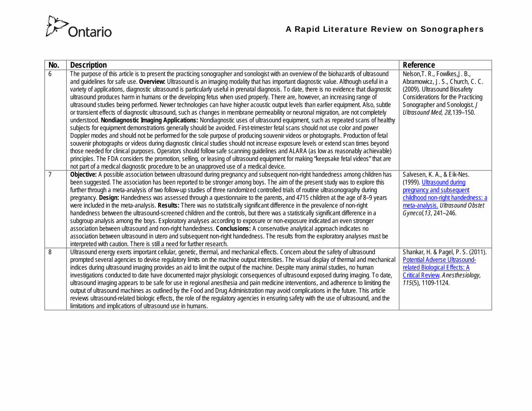
A Rapid Literature Review on Sonographers
No Description Reference 6 The purpose of this article is to present the practicing sonographer and sonologist with an overview of the biohazards of ultrasound
and guidelines for safe use Overview Ultrasound is an imaging modality that has important diagnostic value Although useful in a variety of applications diagnostic ultrasound is particularly useful in prenatal diagnosis To date there is no evidence that diagnostic ultrasound produces harm in humans or the developing fetus when used properly There are however an increasing range of ultrasound studies being performed Newer technologies can have higher acoustic output levels than earlier equipment Also subtle or transient effects of diagnostic ultrasound such as changes in membrane permeability or neuronal migration are not completely understood Nondiagnostic Imaging Applications Nondiagnostic uses of ultrasound equipment such as repeated scans of healthy subjects for equipment demonstrations generally should be avoided First-trimester fetal scans should not use color and power Doppler modes and should not be performed for the sole purpose of producing souvenir videos or photographs Production of fetal souvenir photographs or videos during diagnostic clinical studies should not increase exposure levels or extend scan times beyond those needed for clinical purposes Operators should follow safe scanning guidelines and ALARA (as low as reasonably achievable) principles The FDA considers the promotion selling or leasing of ultrasound equipment for making ldquokeepsake fetal videosrdquo that are not part of a medical diagnostic procedure to be an unapproved use of a medical device
NelsonT R FowlkesJ B Abramowicz J S Church C C (2009) Ultrasound Biosafety Considerations for the Practicing Sonographer and Sonologist J Ultrasound Med 28139ndash150
7 Objective A possible association between ultrasound during pregnancy and subsequent non-right handedness among children has been suggested The association has been reported to be stronger among boys The aim of the present study was to explore this further through a meta-analysis of two follow-up studies of three randomized controlled trials of routine ultrasonography during pregnancy Design Handedness was assessed through a questionnaire to the parents and 4715 children at the age of 8ndash9 years were included in the meta-analysis Results There was no statistically significant difference in the prevalence of non-right handedness between the ultrasound-screened children and the controls but there was a statistically significant difference in a subgroup analysis among the boys Exploratory analyses according to exposure or non-exposure indicated an even stronger association between ultrasound and non-right handedness Conclusions A conservative analytical approach indicates no association between ultrasound in utero and subsequent non-right handedness The results from the exploratory analyses must be interpreted with caution There is still a need for further research
Salvesen K A amp Eik-Nes (1999) Ultrasound during pregnancy and subsequent childhood non-right handedness a meta-analysis Ultrasound Obstet Gynecol13 241ndash246
8 Ultrasound energy exerts important cellular genetic thermal and mechanical effects Concern about the safety of ultrasound prompted several agencies to devise regulatory limits on the machine output intensities The visual display of thermal and mechanical indices during ultrasound imaging provides an aid to limit the output of the machine Despite many animal studies no human investigations conducted to date have documented major physiologic consequences of ultrasound exposed during imaging To date ultrasound imaging appears to be safe for use in regional anesthesia and pain medicine interventions and adherence to limiting the output of ultrasound machines as outlined by the Food and Drug Administration may avoid complications in the future This article reviews ultrasound-related biologic effects the role of the regulatory agencies in ensuring safety with the use of ultrasound and the limitations and implications of ultrasound use in humans
Shankar H amp Pagel P S (2011) Potential Adverse Ultrasound-related Biological Effects A Critical Review Anesthesiology 115(5) 1109-1124
A Rapid Literature Review on Sonographers
No Description Reference 9 Objective In the context of the planned International Society of Ultrasound in Obstetrics and GynecologyndashWorld Health Organization
multicenter study for the development of fetal growth standards for international application we conducted a systematic review and meta-analysis to evaluate the safety of human exposure to ultrasonography in pregnancy Methods A systematic search of electronic databases reference lists and unpublished literature was conducted for trials and observational studies that assessed short- and long-term effects of exposure to ultrasonography involving women and their fetuses exposed to ultrasonography using B-mode or Doppler sonography during any period of pregnancy for any number of times The outcome measures were (1) adverse maternal outcome (2) adverse perinatal outcome (3) abnormal childhood growth and neurological development (4) non-right handedness (5) childhood malignancy and (6) intellectual performance and mental disease Results The electronic search identified 6716 citations and 19 were identified from secondary sources A total of 61 publications reporting data from 41 different studies were included 16 controlled trials 13 cohort and 12 casendashcontrol studies Ultrasonography in pregnancy was not associated with adverse maternal or perinatal outcome impaired physical or neurological development increased risk for malignancy in childhood subnormal intellectual performance or mental diseases According to the available clinical trials there was a weak association between exposure to ultrasonography and non-right handedness in boys (odds ratio 126 95 CI 103ndash154) Conclusion According to the available evidence exposure to diagnostic ultrasonography during pregnancy appears to be safe
Torloni M R Vedmedovska Merialdi M Betran A P Allen T et al (2009) Safety of ultrasonography in pregnancy WHO systematic review of the literature and meta-analysis Ultrasound Obstet Gynecol 33 599ndash608
Articles in Peer-Reviewed Journals 10 It has been documented for many years that as a result of use of ultrasound to determine the sex of the fetus the male to female
ratio in countries such as China and India have become increasingly skewed (due to subsequent abortions and infanticide of females) Preference for sons over daughters evident in Chinas and South Asias male sex ratios is commonly rationalized by poverty and the need for old-age support In this article the authors study South and East Asian immigrants to Canada a group for whom the economic imperative to select sons is largely absent Analyzing the 2001 and 2006 censuses 20 percent samples the authors find clear evidence of extensive sex selection in favor of boys at higher parities among South and East Asian immigrants unless they are Christian or Muslim The latter finding accords with the explicit prohibition against (female) infanticidemdashtraditionally the main sex-selection methodmdashin these religions The findings point to a strong cultural component to both the preference for sons and the willingness to resort to induced abortion based on sex
Almond D Edlund L Milligan K (2013) Son Preference and the Persistence of Culture Evidence from South and East Asian Immigrants to Canada Population and Development Review 39(1) 75ndash95
11 The rapid growth in the use of ultrasound as a diagnostic imaging technology over the past forty years has led to a demand for a workforce with the appropriate skills to perform and interpret the scans In the UK ultrasound investigations now comprise the largest group of all diagnostic imaging examinations However there remains no statutory regulation of the practice of sonography in the UK and little recognition of the considerable training that many practitioners have undertaken to obtain the skills to become safe and competent sonographers Many in the field consider that this should change and are working to obtain professional status for the practice of sonography Although the Health Care Professions Council (HCPC) has recommended regulation of sonography practice this is unlikely to happen in the near future This paper discusses the evolution of sonography practice and explores some of the complex issues associated with the professionalisation of sonography
Gibbs V (2013) The long and winding road to achieving professional registration for sonographers Radiography19 164-167
A Rapid Literature Review on Sonographers
No Description Reference 12 Background Contrast echocardiography (which uses contrast agents injected into the blood flow in order to visualize the blood flow
in very small blood vessels) has been shown to improve diagnostic quality especially in technically difficult patients However the learning curve and increased time for preparation and image acquisition have led to low use Methods The authors sought to determine whether the contrast echocardiography procedure performed independently by a specialized trained sonographer could improve efficiency In one centre routine echocardiograms were scheduled for one hour and any study exceeding one hour would result in patient booking cancellations The authors compared the standard of care in which a physician or nurse administers echocontrast with a sonographer-administered program (SAP) Results The time to complete contrast echocardiograms was significantly reduced by the SAP strategy (43 min 17 s plusmn 23 min 42 s vs 1 h 1 min 6 s plusmn 31 min 0 s P lt 0001) Subgroup analysis of the inpatients and outpatients demonstrated similar results Only 10 of studies (6 of 61) in the SAP exceeded 60 minutes compared with 45 (34 of 76) in the standard-of-care group (P lt 0001) Based on study volumes in the centre the net improvement in productivity with the SAP could be up to 53 annually Conclusion Sonographer-administered echocontrast is feasible and potentially removes a barrier to implementation of contrast echocardiography
Tang A Chiew S K Rashkovetsky R Becher H Choy J B (2013) Feasibility of Sonographer-Administered Echocontrast in a Large-Volume Tertiary-Care Echocardiography Laboratory Canadian Journal of Cardiology 29 391ndash395
13 Ultrasound scanning has been used as a diagnostic and screening tool in obstetric practice for over 50 years There is no evidence of immediate or long-term harm to the developing fetus from exposure to B mode ultrasound However exposure to high levels of Doppler ultrasound during early development is increasingly common and the full safety implications of this exposure are not clear Doppler ultrasound exposure in utero gives rise to increased apoptosis in animal models and there is evidence of the effects of exposure to Doppler ultrasound persisting throughout life with increased non-right-handedness observed in human epidemiological studies The authors consider the idea that there may be long-term developmental implications for fetuses exposed to Doppler ultrasound early in gestation These effects may be mediated via thermal or mechanical disruption to the developing conceptus giving rise to free radical damage Excess free radical exposure early in gestation is a strong candidate for the final common pathway underlying developmental programming effects and gives rise to concern that fetuses exposed to high levels of ultrasound are at risk of a developmental programming effect It is suggested that there is a need for animal studies of developmental programming using exposure to Doppler ultrasound scanning as the exposure of interest and for more observational data to be collected in the clinical setting While these data are collected it seems prudent to continue to adhere to the principle of lsquoas low as reasonably achievablersquo (ALARA) when exposing first-trimester fetuses to Doppler ultrasound
Aiken CE Lees CC (2012) Long-term effects of in utero Doppler ultrasound scanning ndash A developmental programming perspective Medical Hypotheses 78 539ndash541
14 Point of care sonography by emergency physicians in the emergency department can be an effective aid in the diagnosis and management of patients presenting with a variety of medical and traumatic conditions Its use can improve patient outcomes enhance patient safety speed patient disposition and save lives CAEP supports the use of point of care sonography
Canadian Assocation of Emergency (CAEP) Physicians Ultrasound Position Statement Working Group (2012) Use of point of care sonography by emergency physicians CJEM 14(2)106-112
A Rapid Literature Review on Sonographers
No Description Reference 15 The use of three-dimensional echocardiography (3DE ie a 3D ultrasound of the heart) during electrophysiology (EP - a branch of
cardiology that deals with the diagnosis and treatment of heart rhythm disorders) procedures is the end product of years of growth in two diverse cardiology subspecialties namely advanced cardiac imaging and the EP During the past decade progress in both fields has resulted in many important advances that have culminated in their union for a new area of growth and development Imaging advances have provided the cardiovascular specialist with enhanced cardiac volume and function data and more recently 3DE capabilities with improved spatial and temporal resolution providing unprecedented spatial relationships This latter development is valued by EP specialists in need of hitherto never required anatomic knowledge as they press forward with extraordinary expansion in their capabilities It makes sense that by combining these two rapidly growing subspecialties future capabilities in patient care may be achieved that would otherwise not be possible This paper discusses the value of 3DE during EP procedures and offers the readers insight into this novel multispecialty hybrid arena Using this model as a template it is likely that the readers may identify other areas within their practices where periprocedural advanced imaging may afford significant dividends in patient outcomes
Moukabary T Faletra F F Kronzon I Thomas W amp Sorrell V L (2012) Three-Dimensional Echocardiography in the Electrophysiology Laboratory Echocardiography 29 117-122
16 Background There has been much discussion about whether female feticide occurs in certain immigrant groups in Canada The authors examined data on live births in Ontario and compared sex ratios in different groups according to the motherrsquos country or region of birth and parity Methods The authors completed a population-based study of 766 688 singleton live births between 2002 and 2007 They used birth records provided by Ontario Vital Statistics for live births in the province between 23 and 41 weeksrsquo gestation They categorized each newborn according to the motherrsquos country or region of birth namely Canada (n = 486 599) Europe (n = 58 505) South Korea (n = 3663) China (n = 23 818) Philippines (n = 15 367) rest of East Asia (n = 18 971) Pakistan (n = 18 018) India (n = 31 978) rest of South Asia (n = 20 695) and other countries (n = 89 074) They calculated malefemale ratios and 95 confidence intervals (CIs) for all live births by these regions and stratified them by maternal parity at the time of delivery (0 1 2 or ge 3) Results Among infants of nulliparous women (women with no previous children) the malefemale ratio was about 105 overall As parity (the number of previous children) increased the ratio remained unchanged among infants of Canadian-born women In contrast the malefemale ratio was significantly higher among infants of primiparous women (women with one previous child) born in South Korea (120 95 CI 109ndash134) and India (111 95 CI 107ndash115) than among infants of Canadian-born primiparous women Among multiparous women (women with 2+ previous children) those born in India were significantly more likely than Canadian-born women to have a male infant (parity 2 ratio 136 95 CI 127ndash 146 parity ge 3 ratio 125 95 CI 109ndash143) Interpretation The study of malefemale ratios in Ontario showed that multiparous women born in India were significantly more likely than multiparous women born in Canada to have a male infant
Ray J G Henry D A Urquia M L (2012) Sex ratios among Canadian liveborn infants of mothers from different countries CMAJ184(9) E92-96
A Rapid Literature Review on Sonographers
No Description Reference 17 This letter consists of a case study that illustrates how a cross-disciplined sonographer (educated in cardiac and vascular ultrasound)
can quickly and efficiently and with great Cost-effectiveness deliver timely care to a patient in certain clinical settings The literature is comparatively sparse in referencing clinical diagnostics in cross-discipline sonographers Echocardiographers must be clinicians fully comfortable with evaluating the circulatory dynamics of the body from the left and right heart to the peripheral arteries and veins including the extracranial circulation (carotid arteries) Included in this skill set is substantive understanding of the ECG patientrsquos history and integration of cardiac and vascular findings Limitations of each diagnostic tool and how these limitations can be overcome by the strengths of the other examination are important For the dual-skilled sonographer only experience continuous education and constant clinical feedback will lead to the independent decision making that is counted on by the physician The American Registry for Diagnostic Medical Sonography offers credentials in cardiac and vascular imaging (RDCS and RVT) Cardiovascular Credentialing International offers credentials in cardiac and vascular imaging as well (registered cardiac sonographer [RCS] and registered vascular specialist [RVS]) All imaging facilities should consider merging cardiac and vascular noninvasive imaging with dual-registered clinical sonographers credentialed by one of these two agencies
Zemanek M (2012) What It Means to Be a Cardiovascular Sonographer Journal of Ultrasound in Medicine 311859-1862
18 Diagnostic medical sonography is viewed as a safe imaging modality However bioeffects research has yet to define the effects of exposure to sonography in humans Therefore sonographers must be aware of potential risks and practice ALARA (ldquoas low as reasonably achievablerdquo) routinely This research explores sonographersrsquo ability to answer basic questions about the bioeffects of sonography as well as their knowledge and use of the safety indices called the mechanical index (MI) and thermal index (TI) Members of the Society of Diagnostic Medical Sonographers in the US a southwestern sonographersrsquo society and a sonography community on Facebook 2010 were surveyed (n = 212) There is a low level of bioeffects knowledge and minimal safety practices among sonographers in this survey No significant relationship was found between sonographer knowledge of the bioeffects of sonography years in the profession or whether the sonographers monitor the MITI
Bagley J Thomas K DiGiacinto D (2011) Safety Practices of Sonographers and Their Knowledge of the Biologic Effects of Sonography Journal of Diagnostic Medical Sonography 27(6) 252ndash261
19 This article highlights trends in echocardiography Point of care ultrasound (hand-carried ultrasound) Initially point of care ultrasound will increase the use of echocardiography in the same manner that cardiac CT increased catheterizations However once the basics are learned it has the potential to be a very powerful tool in that less unnecessary studies will be ordered In the clinic small systems will be used as a lsquolsquofirst lookrsquorsquo screen and become part of the patientrsquos history only patients for whom more testing is necessary will be referred for a comprehensive study This is likely to reduce the number of Doppler studies requested The miniaturization of echo equipment will continue with improved qualityfunctionality of the lsquolsquohand-heldrsquorsquo echo machines to the extent that cordless probes will become a reality sooner rather than later Role in interventional echocardiography Sonographers will be spending much more time in the interventional and electrophysiology (EP) labs There will be more interconnection with interventional cardiologists and the volume will increase as more devices are implanted more percutaneous valves placed and more robotic surgery is performed Crosstraining and multitasking Sonographers entering the profession would be prudent to learn cardiac and vascular imaging Individuals who multitask andor cross-train will be ideal For example a sonographer who starts an IV monitors ECG stress can perform cardiac MRI and can interpret a preliminary report or a nurse who can do an echo monitor sedation and start an IV will be invaluable The sonographer should continue to grow professionally by cross training as institutions try to minimize costs and maximize throughput
McCulloch M L (2011) Thoughts regarding the future of sonography Journal of the American Society of Echocardiography 24(5) 29A-30A
A Rapid Literature Review on Sonographers
No Description Reference 20 Background Radiology reports provide specialist interpretation of images and relate these findings to the patientrsquos symptoms and
signs This study compared ultrasound (USS) reports generated by radiologists and sonographers to determine if any significant or clinically relevant differences existed Methods A retrospective analysis of 624 consecutive USS reports was carried out The reports were assessed for the presence of a lsquodisclaimerrsquo or lsquocaveatrsquo pertaining to the quality of the images and were analysed with respect to the clarity of their wording and ability to answer the clinical request Results The majority of sonographer USS reports contained a disclaimer regarding the quality of the USS images (571) compared with a smaller proportion of radiologist scans (99) (P lt 0001) Overall radiologists performed significantly better in providing a clear negative or positive diagnosis to the clinical question on the request form when compared with sonographers (885 vs 654 P lt 0001) Conclusion lsquoDisclaimerrsquo comments and lsquohedgingrsquo must be avoided in radiology reporting While feedback as to the accuracy of the radiology images is important overuse of such terms undermines the validity of the radiology report
Garcea G Mahmoud A Ong S L Rees Y Berry Det al (2010) Caveat reporting in ultrasound interpretation of surgical pathology a comparison of sonographer versus radiologist Journal of Evaluation in Clinical Practice 16 97ndash99
21 A nine-point questionnaire was constructed based on previously published work by Marsal and Sheiner et al The survey was distributed to clinical users of ultrasound practicing sonography in Hamilton (New Zealand) during July 2010 totalling 35 users consisting of sonographers sonography trainees one obstetric sonologist and one fetal medicine registrar Results Twenty sonographers returned the questionnaire which represents survey response of 61 The level of clinical experience was equally split between junior sonographers of lt 5 yearsrsquo experience (30) sonographers with 5-10 yearsrsquo experience (35) and senior sonographers of gt10 yearsrsquo experience (35) While 100 of the sonographers surveyed reported being familiar with the term Thermal Index (TI) and 90 claimed they adhered to the Action Learning Action Response Association (ALARA) principle all the time or most of the time only 15 of sonographers correctly described or defined what the term TI means Of the sonographers 80 knew where to find the TI during real-time examinations but only 35 knew which TI index to use for nuchal translucency (NT) scan and only 10 knew the expected range of TI encountered during routine obstetric examinations Of the eight sonographers who reported adhering to ALARA all the time the majority (64) could not describe or define TI In the same subgroup the majority (64) did not know which TI to monitor during a NT scan Only three of 20 (15) sonographers knew what the term TI means Of these two were junior sonographers with lt 5 yearsrsquo experience and one had 5ndash10 yearsrsquo experience Despite understanding the term TI one of the three sonographers still selected the incorrect index to monitor during a NT scan Not one of the seven senior sonographers with gt 10 years of experience could define or describe the TI In total only 10 of sonographers knew what the TI means and which TI to monitor during NT scanning
Necas M (2010) New Zealand sonographers do not outperform their European or American colleagues in the knowledge of ultrasound safety Australasian Journal of Ultrasound in Medicine13(4) 28-32
A Rapid Literature Review on Sonographers
No Description Reference 22 Musculoskeletal (MSK) ultrasound performed by non-medical practitioners is a developing practice The aim of this paper is
(1) to audit an experienced sonographerrsquos (SJ Riley one of the authors) performance after one yearrsquos independent reporting against two experienced MSK radiologists (CJ Grove amp M Chandramohan the other two authors) and (2) to establish an audit standard against which such role development might be contrived Images and reports from 250 MSK ultrasound examinations performed by a sonographer were reviewed independently by two consultant MSK radiologists The examinations were graded for discrepancy eg grade 1 ndash agree with the sonographer report grade 2 ndash minor discrepancy unlikely to alter patient care grade 3 ndash potentially significant discrepancy grade 4 ndash definite significant discrepancy likely to have adverse consequences for patient care Two of 250 (08) cases were excluded Both radiologists agreed completely with the sonographer (grade 1) in 235 of the 248 cases (948) In 13 cases there was discrepancy between the reports of SJR and the radiologists The discrepancy was grade 2 in six of the 248 cases (24) grade 3 in six of the 248 cases (24) and grade 4 in one of the 248 cases (04) In conclusion this audit shows a high level of agreement between the sonographer and the consultant MSK radiologist reporting of MSK ultrasound This level of agreement may set the standard for future quality assurance audit of sonographer MSK reporting
Riley S J Groves C J Chandramohan M (2010) Musculoskeletal ultrasound audit of sonographer reporting Ultrasound 18 36ndash40
23 Background Growing demand for ultrasound examinations and higher quality requirements motivate searching for routines combining the diagnostic accuracy of radiologist-performed examinations with the economical advantages of sonographer-performed examinations One possible approach is to use strictly standardized acquisition and documentation schemes that give the radiologist access to all relevant information after the examination Purpose To compare a recently introduced routine combining acquisition by a radiographer (more specifically a sonographer with two years experience) documentation as standardized cine-loops and review by a radiologist (lsquostandardized methodrsquo) with the formerly used routine where the diagnosis is made bedside by the radiologist (lsquotraditional methodrsquo) Material and Methods In 64 policlinic patients the kidneys (n = 27) or the gallbladder (n = 37) were examined with both the standardized and the traditional method The radiologistsrsquo findings of hydronephrosis tumors cysts echogenicity changes and cortical thickness (in the kidneys) and wall thickness concrements and polyps (in the gallbladder) were compared between the methods with respect to agreement (proportion of agreement and kappa coefficient) as well as systematic differences (McNemarrsquos test) Results The findings at the gallbladder examination showed a median agreement of 97 (86ndash100 kappa = 064ndash100) and those at the kidney examination an agreement of 90 (78ndash100 kappa = 069ndash 100) There were no significant systematic differences between the methods Conclusion The satisfactory agreement in this preliminary study indicates that the new workflow with ultrasound examinations performed by a radiographer (sonographer) and analyzed off-line by a radiologist is promising and motivates further studies
Stenman C Thorelius L Knutsson A amp Smedby Ouml (2010) Radiographer-acquired and radiologist-reviewed ultrasound examination ndash agreement with radiologistrsquos bedside evaluation Acta Radiologica 52 70ndash74
A Rapid Literature Review on Sonographers
No Description Reference 24 The troubling practice of fetal sex selection has historically been considered an Asian phenomenon However recent evidence shows
that a similar situation is emerging in North America albeit on a smaller scale The Society of Obstetricians and Gynaecologists of Canada (SOGC) has firmly stated its opposition to sex selection for non-medical reasons as well as to the use of any technology used solely for the purpose of determining fetal sex However because fetal sex may be disclosed to the parents at the time of ultrasound examination if they request this information guidance for health care professionals to assist in discouraging fetal sex selection would be useful Because no declaration of motives or reasons is required when a woman seeks a termination of pregnancy the authors suggest that health care professionals need not disclose the sex of a fetus until it has reached a gestational age at which abortion for non-medical purposes would not be possible This proposal would facilitate consistency between clinical practice and the values of Canadian citizens the SOGC the Canadian Medical Association and other professional organizations while still respecting current laws pertaining to disclosure of patient information and patientsrsquo rights to autonomous decision-making
Thiele A T amp Leier B (2010) Towards an Ethical Policy for the Prevention of Fetal Sex Selection in Canada J Obstet Gynaecol Can 32(1) 54ndash57
25 Background The extent to which increases in the use of prenatal ultrasonography over time reflect changes in maternal risk (ie an increase in high-risk pregnancies over time) is unknown In this population-based study the authors examined the use of prenatal ultrasonography from 1996 to 2006 in Ontario Methods With fiscal year 199697 as the baseline the authors evaluated the relative risk (RR) and 95 confidence interval (CI) for the change in rates of ultrasonography for each subsequent year The RR was adjusted for maternal age income rural residence maternal comorbidities receipt of genetics consultation or amniocentesis mdash all in the index pregnancy mdash and history of complications in a prior pregnancy Results The study sample consisted of 1 399 389 singleton deliveries The rate of prenatal ultrasonography increased from 2055 per 1000 pregnancies in 1996 to 3264 per 1000 in 2006 (adjusted RR 155 95 CI 154ndash155) The rate increased among both women with low-risk pregnancies (adjusted RR 154 95 CI 153ndash155) and those with high-risk pregnancies (adjusted RR 155 95 CI 154ndash157) The proportion of pregnancies with at least four ultrasound examinations in the second or third trimesters rose from 64 in 1996 to 187 in 2006 (adjusted RR 268 95 CI 261ndash274) Paradoxically this increase was more pronounced among low-risk pregnancies (adjusted RR 292 95 CI 283ndash301) than among high-risk pregnancies (adjusted RR 225 95 CI 216ndash235) Interpretation Substantial increases in the use of prenatal ultrasonography over the past decade do not appear to reflect changes in maternal risk Nearly one in five women now undergo four or more ultrasound examinations during the second and third trimesters Efforts to promote more appropriate use of prenatal ultrasonography for singleton pregnancies appear warranted
You J J Alter D A Stukel T A McDonald S D Laupacis A et al (2010) Proliferation of prenatal ultrasonography CMAJ 182(2) 143-151
A Rapid Literature Review on Sonographers
No Description Reference 26 Aims The aim of this study was to assess the cost-effectiveness of using certified sonographers and miniaturized echocardiography
systems to perform echocardiograms at bedside in comparison to moving inpatients from the admission department to the echocardiography laboratory (echo-lab) Methods and Results From 26 September 2005 to 27 October 2005 112 patients admitted in six hospital wards in Italy connected through a 100 Mbit LAN to the echo-lab were scanned within the admission ward by sonographers using a miniaturized echo system Logistical data were collected and results were compared with those obtained from 194 consecutive patients coming from the same wards and studied in the echo-lab with high-end machines between 8 March 2005 and 15 April 2005 Performing echocardiograms in the admission department avoided long waiting time of the inpatients in the echo-lab before and after the study increased the percentage of patients studied within 3 and 5 days from request (88 vs 77 and 100 vs 95 respectively P = 003) increased both sonographer (by 339 P lt 0001) and echo-lab productivity (by 41 P lt 0001) and reduced costs of echocardiograms by 29 Conclusion Implementation of digital echocardiography certified sonographers and a miniaturized echo system allowed improvement of the cost-effectiveness of the service provided by the echo-lab for inpatients and avoided patientsrsquo discomfort derived from prolonged waiting time before and after the exam
Badano L Gaetano N Stacul S Gianfagna P Pericoli M et al (2009) Improved workflow sonographer productivity and cost-effectiveness of echocardiographic service for inpatients by using miniaturized systems European Journal of Echocardiography 10 537ndash542
27 The present report is a proposal from the American Society of Echocardiography Advanced Practice Task Force that identifies the potential of cardiac sonographers to achieve the Advanced Cardiovasculat Sonographer (ACS) level According to this report echocardiographic examinations require a well-trained and competent sonographer to obtain proper anatomic and physiologic data to establish an accurate diagnosis for clinical decision-making and patient management Although the formal education and training of cardiovascular sonographers are evolving many entry-level and staff sonographers may not have sufficient practical or clinical knowledge of the necessary components of the echocardiographic study for the individual patientrsquos clinical presentation In many clinical settings echocardiograms are read after the patient has left the laboratory Thus there is a role for a sonographer who can practice at an advanced level in a cardiovascular ultrasound laboratory to ensure a proper echocardiographic examination is performed on every patient In this setting an Advanced Cardiovascular Sonographer (ACS) would be able to review the indication for and quality of the examination If additional images were needed the ACS would assist the sonographer in obtaining these images which would lead to the performance of a complete and fully diagnostic examination before the patient had left the echocardiography laboratory In clinical practice the quality of the examinations performed would improve advancements in echocardiographic methods could be taught and incorporated into daily practice and patients would be better served
Mitchell C Miller F A Bierig S M Bremer M L Ehler D et al (2009) Advanced Cardiovascular Sonographer A Proposal of the American Society of Echocardiography Advanced Practice Sonographer Task Force J Am Soc Echocardiogr 22 1409-13
A Rapid Literature Review on Sonographers
No Description Reference 28 Sonographer practitioner development involves the expansion and extension of the sonographer role to include reporting on
ultrasound examinations Australian sonographers have not seen the same degree of role extension and expansion as their counterparts in the United Kingdom despite increasing levels of discussion regarding sonographer practitioner development The aim of this study was to determine if Australian sonographers want to extend their professional role and what they consider are the important issues associated with role extension This paper reports on qualitative data derived from a survey of Australian sonographers and investigates if Australian sonographers are interested in extending and expanding their professional role and responsibilities and if they do what might be necessary or desirable from a professional point of view for this change to occur A survey was mailed to all members of the Australian Sonographers Association (ASA) in October 2006 The 31-item survey included 28 closed-ended and three opened-ended items to provide both quantitative and qualitative data The quantitative data will be reported separately Qualitative data was derived from responses to the opened-ended questions which asked respondents to elaborate on their attitudes and feelings about role extension and development Analysis used Nvivo7 software to aid in uncovering common themes from the qualitative data The analysis focused on the reported incentives or motivations for becoming a sonographer practitioner as well as disincentives or perceived hurdles that would discourage respondents from becoming sonographer practitioners The three most reported incentives or motivations for becoming a sonographer practitioner were professional recognition remuneration and increased knowledge The three most commonly reported disincentives or perceived hurdles that would discourage respondents from becoming sonographer practitioners were legal issues insurance and further study
McGregor R OrsquoLoughlin K Cox J Clarke J Snowden A (2009) Sonographer practitioner development in Australia Qualitative analysis of an Australian sonographersrsquo survey Radiography15 313-319
29 Introduction This study describes the effect of introducing a cardiac sonographer led follow up clinic within a hospital cardiology department for patients with stable heart valve disease The two years before and after the instigation of the valve clinic were audited Materials and methods The clinic was conducted in a single centre and undertaken in the cardiology department of a district general hospital A total of 382 patients with 397 clinically significant valve lesions but for whom surgery was not yet indicated but follow up required were seen in a cardiac sonographer run clinics These patients no longer attended a medical follow up clinic unless there was clinical or echocardiographic deterioration Effectiveness was judged by the percentage treated according to current best practice guidelines the number of echocardiograms performed and the number of hospital outpatient visits attended In addition mortality data for the subjects in the clinic was collected Results The proportion followed up according to best practice guidelines rose from 157 (41) to 354 (92) (p lt 001) The total number of echocardiograms performed fell from 807 to 550 Total number of outpatient visits fell from 998 to 31 A total of 11 patients died in the two year study period none from progressive valve disease Discussion This study demonstrates that a protocol driven sonographer led heart valve disease follow up clinic significantly improved the quality of follow up while bringing about a major reduction in outpatients visits without compromising patient safety
Taggu W Topham A Hart L Carr-White G Sulke N et al (2009) A cardiac sonographer led follow up clinic for heart valve disease International Journal of Cardiology 132 240 ndash243
30 Role development for imaging allied health professionals has progressed significantly over the last three decades with many major developments pioneered in the field of ultrasound This paper reviews the developing role of the radiographer in ultrasound (in some countries such as the UK sonographers are considered a type of radiographer) and some of the clinical professional and educational implications of such development As the published evidence base underpinning the latest developments is scarce several contemporary case study examples which have been presented verbally at study days attended by the authors have been included In conclusion sonographer role development is considered within the professional contexts of extended role advanced and consultant practice
Hart A amp Dixon A (2008) Sonographer Role Extension and Career Development a Review of the Evidence Ultrasound 16(1) 31-35
A Rapid Literature Review on Sonographers
No Description Reference 31 Objective The main goal of this study was to determine end usersrsquo knowledge regarding safety aspects of diagnostic ultrasound
during pregnancy End usersrsquo attitudes toward the use of ultrasound in low-risk pregnancies were also assessed Methods A questionnaire was distributed to ultrasound end users attending review courses and hospital grand rounds between April and June 2006 in Chicago Results One hundred thirty end users completed the questionnaires (63 response rate) Sixty-three percent were physicians (n = 84) most of them obstetricians (817) About 18 of participants routinely performed Doppler ultrasound examinations during the first trimester Fifty percent of end users thought that the number of ultrasound examinations in low-risk pregnancy should be limited to 1 to 3 (mean plusmn SD 26 plusmn 09) Almost 70 disapproved of ldquokeepsakeentertainmentrdquo ultrasound Although 322 of the participants were familiar with the term thermal index only 177 actually gave the correct answer to the question on the nature of the thermal index About 22 were familiar with the term mechanical index but only 38 described it properly Almost 80 of end users did not know where to find the acoustic indices Only 208 were aware that they are displayed on the sonographic monitor during the examinations End users with higher knowledge of safety issues thought that there should be limitations on the number of ultrasound examinations in low-risk pregnancies (odds ratio 33 95 confidence interval 11ndash100 P = 028) Likewise these end users were more likely to respond that ultrasound might have adverse effects during pregnancy (odds ratio 32 95 confidence interval 11ndash125 P = 045) Conclusions Ultrasound end users are poorly informed regarding safety issues during pregnancy Further efforts in the realm of education and training are needed to improve end user knowledge about the acoustic output of the machines and safety issues
Sheiner E Shoham-Vardi I Abramowicz J S (2007) What Do Clinical Users Know Regarding Safety of Ultrasound During Pregnancy J Ultrasound Med 26 319ndash325
A Rapid Literature Review on Sonographers
No Description Reference 32 In an attempt to survey the knowledge among ultrasound users of some safety aspects of diagnostic ultrasound a questionnaire was
distributed to professionals using ultrasound for fetal examinations The questionnaire was answered by the participants and faculty members of six postgraduate courses in obstetric ultrasound Doppler ultrasound and fetal echocardiography given in Sweden Norway and Austria in 2003 Furthermore in Sweden the questionnaire was distributed to the staff meeting participants of two major obstetric ultrasound units and of one unit for fetal echocardiography and at a meeting of the Working Group for Ultrasound of the Swedish Society of Obstetrics and Gynecology In total 199 questionnaires were anonymously answered by doctors (n = 145) sonographers (n = 22) and midwives trained in ultrasound (n = 32) All of them were using diagnostic ultrasound on a daily or weekly basis The respondents were from nine European countries In none of the countries is formal certification for the use of diagnostic ultrasound in pregnancy or otherwise required The doctors who answered the questionnaire were specialists in obstetrics and gynecology (including some of the internationally recognized experts in fetal ultrasound) pediatric cardiology radiology or clinical physiology The respondents were not informed about the questionnaire in advance and they had a limited time to answer the 16 questions typically 10ndash15 min The questions concerned explanation of the safety indices and their relation to various ultrasound modes ie real-time B-mode M-mode three-dimensional (3D) spectral Doppler color and power Doppler ultrasound The respondents were also asked which ultrasound system they used most often and where on the system the information on thermal index (TI) and mechanical (MI) was displayed (eg in the top right corner of the screen in a separate window etc) Furthermore the respondents had to indicate how they adjust the output energy level on their own machine For all questions there was the optional answer lsquoDonrsquot knowrsquo Results About one-third of the ultrasound experts were able to define the abbreviations TI and MI For the subclasses of the TI the correct answer was found in only 3-8 of cases Of those who knew what the TI and MI meant only two-thirds and one-third of respondents respectively were able to give a simple explanation of the indices In 49 of questionnaires the Doppler modes were correctly ranked as generally producing higher ultrasound exposure than the imaging modes (B-mode 3D) Some 28 of the respondents correctly indicated where on their own machine the information on safety indices is displayed However not all of them knew how to control the output energy level For all items in the questionnaire there were no significant differences in the results between the three categories of respondents (ie physicians sonographers and midwives) The results of the questionnaire were anything but encouraging and they indicated that the users who are supposed to be responsible for controlling exposure of the fetus to ultrasound had a very poor knowledge of the basic safety aspects of ultrasound
Maršaacutel K (2005) The output display standard has it missed its target Ultrasound Obstet Gynecol 25 211ndash214
33 Purpose This study was performed to evaluate the accuracy of upper abdominal ultrasound (US) scanning performed by sonographers in a district general hospital to identify potential areas of weakness and to make recommendations to improve the service Materials and Methods Upper abdominal US examinations performed and reported by sonographers over a 4-week period were retrospectively reviewed The accuracy of the imaging findings and reports were assessed against other imaging surgical histological or laboratory findings and against clinical outcome Results A heterogenous group of 104 patients were included in the study 62 of whom had an US abnormality Errors of scanning or interpretation were identified in 10 patients (96) of whom five (48) were felt to be potentially significant Conclusions The sonographersrsquo accuracy in reporting upper abdominal US scans was 90 However on the basis of this study the authors have implemented specific recommendations to improve the quality of the service
Dongola N Guy R Ward S amp Giles J A (2003) Can sonographers offer an accurate upper abdominal ultrasound service in a district general hospital Radiography 9 29ndash33
A Rapid Literature Review on Sonographers
No Description Reference 34 Aim To compare the accuracy of radiographers and radiologists in routine abdominal ultrasound Materials and Methods One
hundred consecutive patients attending for routine abdominal ultrasound were included Each patient was examined by both a radiographer and radiologist Both operators noted their fndings and wrote a concluding report without conferring Reports were compared Where there was disagreement the patient was either re-examined by another radiologist or had further investigation Results Of 100 patients 52 were men and 48 were women The age range was 19plusmn88 years (median 52 years) Thirty-seven patients had renal tract ultrasound one had an aortic ultrasound and 62 had general upper abdominal ultrasound In 44 cases both operators reported the examination as normal In 49 cases both operators reported the examinations as abnormal and there was complete agreement between the operators In seven cases there was not complete agreement between operators Three of these disagreements were considered minor and four major In three of the seven cases the radiographer was correct and in four the radiologist was correct Conclusion Experienced radiographers and radiologists are highly accurate in performing and interpreting routine abdominal sonography Both operators missed a small minority of abnormalities There was no statistically significant difference in the accuracy of radiographers and radiologist
Leslie A Lockyer H amp Virjee J P (2000) Who Should be Performing Routine Abdominal Ultrasound A Prospective Double-Blind Study Comparing the Accuracy of Radiologist and Radiographer Clinical Radiology 55 606-609
Grey Literature 35 The ARRT The American Registry of Radiologic Technologists tests certifies and annually registers more than a quarter-of-a-million
radiologic technologists to promote high standards of patient care They award the Registered Technologist or RT designation to individuals who have completed the prescribed classroom and clinical education passed the appropriate exam and met the ethics requirements And they ensure their continuing education and ongoing compliance with their high standards by requiring annual registration of their certificate Certification by ARRT is voluntary not mandatory But employers state licensing agencies and federal regulators view ARRTrsquos credential as an indicator that a person has met recognized national standards for medical imaging and radiation therapy professionals How Are Radiologic Technologists Deemed Qualified First comes initial certification Certification is the process of initially recognizing individuals who have satisfied certain standards within a profession A person is certified by ARRT after completing an approved educational program complying with the ethics standards and passing a certification exam Then comes annual registration Registration is the yearly procedure required to maintain certification ARRT registrants are those who having already fulfilled the requirements for initial certification continue to meet the requirements for annual registration The ARRT annually registers the certificates of individuals who meet the following three criteria agreeing to comply with the ARRT Rules and Regulations continuing to comply with the ARRT Standards of Ethics and meeting our continuing education requirements Only technologists who are currently registered (which means that they have renewed within the past year) may designate themselves as ARRT Registered Technologists and use the initials RT after their names Federal laws regulate personnel qualifications in some modalities (mammography for example) but they accredit the health care facility rather than licensing the individual radiologic technologist who is providing patient care And while many states have laws regulating who can perform radiologic procedures they are typically based on ARRT certification Some employers and state agencies may require ARRT certification but not necessarily current registration Unless they require current registration by ARRT employers cannot assume that the person has complied with the ethics requirements or the continuing education requirements
American Registry of Radiologic Technologists (ARRT) (2013a) Patient Page Accessed April 2013
A Rapid Literature Review on Sonographers
No Description Reference 36 This document outlines certification requirements for sonography There are two pathways (primary and post-primary) For the
primary pathway it notes that sonography educational programs must be accredited by a mechanism acceptable to the ARRT ARRT generally recognizes only accreditation agencies that are recognized by the Council for Higher Education Accreditation andor United States Department of Education
American Registry of Radiologic Technologists (ARRT) (2013b) Sonography Certification Accessed April 2013
A Rapid Literature Review on Sonographers
No Description Reference 37 In Australia sonographers and student sonographers are required to be accredited by Australian Sonographer Accreditation Registry
(ASAR) in order to practise This requirement is contained within the Australian Governmentrsquos Diagnostic Imaging Accreditation Scheme standards and Medicare legislation Accreditation of sonographers is linked only to achieving - or in the case of students studying towards - an accredited entry-level qualification completion of continuing professional development and payment of an annual fee to ASAR The ASA contends that the lack of a requirement to ensure each sonographer is lsquofit to practisersquo is an unacceptable gap in current regulation undermining efforts to ensure the quality of sonographic services and failing to protect patients from harm The ASA supports and advocates for nationally-enforceable regulation of sonographers to meet our patientrsquos expectations National registration The National Registration and Accreditation Scheme (NRAS) for health workers commenced on 1 July 2010 The ten professions which comprised the initial intake into this scheme were those that had registration requirements in all Australian states or territories as well as those in which legislated regulation is deemed to be in the public interest An additional four professions - including medical radiation practitioners which are defined as radiographers radiation therapists and nuclear medicine technologists - will be included in the NRAS commencing on 1 July 2012 As sonography did not have registration in any states or territories prior to the commencement of NRAS the profession has not been in a position to negotiate entry into NRAS In early August 2011 the ASA lodged a submission with the Health Workforce Principal Committee requesting a profession-specific Sonography Board of Australia be formed under NRAS This submission was prepared based on information that consideration of professions which were unregistered would commence More recently the Australian Health Workforce Ministerial Council (AHWMC) issued a communiqueacute stating in part lsquoMinisters have commissioned work on unregistered professions and future directions for national registration Given this Ministers agreed to defer consideration of the inclusion of additional professions into the NRAS Ministers agreed that it would be premature to consider further applications for the scheme until this work has been completed other than paramedicsrsquo Options for unregistered health practitioners including sonographers Policy work is continuing at a federal level to establish an alternative regulatory framework The National Alliance for Self-Regulating Health Professions (NASRHP) consisting of the peak professional associations of eight self-regulating health professions including the ASA representing sonography released a proposal lsquoHarnessing self-regulation to support safety and quality in healthcare deliveryrsquo in March 2012 This proposal concluded that the framework that regulates all health practitioners should be expanded beyond NRAS to include
bull authorised self-regulating professions via a health professions panel bull negative licensing to withdraw the ability to practise from registered and self-regulated practitioners who do not meet the
regulatory requirements or unregulated practitioners due to poor practice As with all advocacy initiatives consideration by the federal government of the introduction of the NASRHPrsquos proposal may be prolonged and the outcomes may not match the expectations of NASRHPrsquos member professional associations including the ASA The ASA will continue both independently and collaboratively to lobby on behalf of our members and the public for the introduction of a timely and equitable solution to health practitioner regulation In the interim as there is no regulatory model with the rigour the ASA believes is required for regulation of sonographers and other health practitioners other than NRAS the ASA continues to support its Position Statement published in early 2010
Australian Sonographer Association (ASA) (2013) Sonographer Regulation Accessed April 2013
A Rapid Literature Review on Sonographers
No Description Reference 38 This publication contains information about Cardiovascular Credentialing International (CCI) It also provides the necessary
information and references concerning the process by which candidates may earn certificate or registry-level credentials CCI is a not-for-profit corporation established for the purpose of administering credentialing examinations as an independent credentialing agency CCI began credentialing cardiovascular professionals in 1968 CCI offers eight credentials which when earned demonstrate that the registrant holds fundamental knowledge in the particular cardiovascular specialty These include RCCS ndash Registered Congenital Cardiac Sonographer RCS ndash Registered Cardiac Sonographer and RPhS ndash Registered Phlebology Sonographer
Cardiovascular Credentialing International (2013) 2013 Examination Application amp Overview Accessed April 2013
39 Oregon Governor Signs Sonographer Licensure Legislation On July 28 2009 Oregon Governor Ted Kulongoski signed a sonographer licensure bill (HB 2245) into law The new law makes Oregon only the second state in the United States to require licensure of sonographers The Society of Diagnostic Medical Sonographers (SDMS) has worked closely with other sonography-related organizations and Oregon sonographers to help craft the language in new law The law renames and restructures the Oregon Board of Radiologic Technologists to the Oregon Board of Medical Imaging (OBMI) and ensures representation of sonographers on the Board The sonographer licensure law requires Oregon sonographers to become licensed by OBMI effective July 1 2010 Beginning on January 1 2014 all sonography licensees would be required to hold a national sonography certificationcredential (or be currently enrolled as a student) While the Oregon bill is far more detailed than the sonographer licensure law that passed in New Mexico earlier this year additional administrative rulesregulations will be developed over the next year to fully implement the new law SDMS will be continue to monitor developments regarding sonographer licensure in other states
Society of Diagnostic Medical Sonographers (SDMS) (2013a) News Accessed April 2013
A Rapid Literature Review on Sonographers
No Description Reference 40 On April 6 2009 New Mexico (NM) Governor Bill Richardson signed the historic bill that for the first time in the United States
requires licensure of sonographers The bill adds sonographers (and MRI technologists) to the list of medical imaging professionals licensed by the State of New Mexico Until now anyone could perform sonograms in New Mexico Prior to the passage of the New Mexico licensure law virtually all other health care providers and professionals had to be licensed As a result the public is often surprised to learn that the person performing a medical sonogram does not have any state licensure or national certification requirements The responsibility for administering the NM sonographer licensure program would rest with the New Mexico Environment Department Although this was not the first choice in most sonographersrsquo minds the Environment Department currently manages the radiologic technology licensure program so the administrative infrastructure is already in place By ensuring the creation of an advisory council that includes representation by each medical imaging and radiation therapy modality regulated the bill helps ensure that the Departmentrsquos other areas of emphasis do not detract from sonographer licensure Because sonographers have never had to be licensed before they frequently do not see or understand the distinction between state licensure and national certification National certifications or credentials issued by voluntary organizations such as the American Registry for Diagnostic Medical Sonography (ARDMS) Cardiovascular Credentialing International (CCI) and the American Registry of Radiologic Technologists (ARRT) have little legal weight The certification process and resulting credential are valuable tools in ensuring sonographers have met minimum education and knowledge requirements but do not really regulate anyonersquos ability to perform sonography In an ideal world no additional regulation of sonography would be required However as everyone knows more and more people are taking weekend courses and buying ultrasound equipment without understanding the ramifications of using ultrasound technology (eg ALARA) Under the bill passed by the New Mexico legislature national credentialscertifications should provide evidence of meeting the statersquos standards All sonographers who are performing these critical medical imaging services in New Mexico will be required to meet national certification standards (ie have certifications or credentials issued by ARDMS CCI or ARRT) As a result there would be no additional exams in New Mexico beyond the national certification exams required by ARDMS CCI or ARRT The bill also requires the state to recognize the continuing medical education completed for national certification renewal The most visible impact on New Mexico sonographers will be a license fee However the legislation caps the license fee at $100 per two-year license In addition only the New Mexico Legislature can change this cap
SDMS (2013b) New Mexico Sonographer Licensure Law Signed Accessed April 2013
A Rapid Literature Review on Sonographers
No Description Reference 41 Preamble The purpose of this document is to define the Scope of Practice for Diagnostic Ultrasound Professionals in the US and to
specify their roles as members of the health care team acting in the best interest of the patient This scope of practice is a living document that will evolve as the technology expands Definition of the Profession The Diagnostic Ultrasound Profession is a multi-specialty field comprised of Diagnostic Medical Sonography (with subspecialties in abdominal neurologic obstetricalgynecologic and ophthalmic ultrasound) Diagnostic Cardiac Sonography (with subspecialties in adult and pediatric echocardiography) Vascular Technology and other emerging fields These diverse specialties are distinguished by their use of diagnostic medical ultrasound as a primary technology in their daily work Certification is considered the standard of practice in ultrasound Individuals who are not yet certified should reference the Scope as a professional model and strive to become certified Scope of Practice of the Profession The Diagnostic Ultrasound Professional is an individual qualified by professional credentialing and academic and clinical experience to provide diagnostic patient care services using ultrasound and related diagnostic procedures The scope of practice of the Diagnostic Ultrasound Professional includes those procedures acts and processes permitted by law for which the individual has received education and clinical experience and in which heshe has demonstrated competency Diagnostic Ultrasound Professionals
bull Perform patient assessments bull Acquire and analyze data obtained using ultrasound and related diagnostic technologies bull Provide a summary of findings to the physician to aid in patient diagnosis and management bull Use independent judgment and systematic problem solving methods to produce high quality diagnostic information and
optimize patient care
Society of Diagnostic Medical Sonography [SDMS] (2013c) Scope of Practice for the Diagnostic Ultrasound Professional Accessed April 2013
42 This website notes that in the US most employers in the US prefer to hire sonographers who have professional certification A sonographer can get certification and passing an exam Most exams relate to the specialty that the sonographer is most interested inmdashfor example an exam to become certified in abdominal sonography A few states require diagnostic medical sonographers to be licensed Typically professional certifications required for licensure other requirements vary by state Sonographers must take continuing education to keep their certification current
Bureau of Labor Statistics US Department of Labor (2012) How to become a Diagnostic Medical Sonographer Occupational Outlook Handbook 2012-13 Edition Accessed April 2013
A Rapid Literature Review on Sonographers
No Description Reference 43 The American Registry for Diagnostic Medical Sonography (ARDMS) is an independent not-for-profit organization founded in 1975
that administers examinations and awards the following credentials bull RDMSreg ndash Registered Diagnostic Medical Sonographerreg bull RDCSreg ndash Registered Diagnostic Cardiac Sonographerreg bull RVTreg ndash Registered Vascular Technologistreg bull RPVIreg ndash Registered Physician in Vascular Interpretationreg bull RMSKtrade - Registered in Musculoskeletaltrade sonography
ARDMS is the globally recognized standard of excellence in sonography and has certified more than 75000 individuals ARDMS is composed of three key groups
bull Board of Directors ndash Sonographers vascular technologists physicians research scientists and a member of the public are volunteer members of the board that determines the direction and policies of our organization
bull Examination Development Task Forces ndash A group of sonographers vascular technologists physicians and scientists volunteer their time and expertise to produce our examinations
bull Professional Staff ndash A paid highly-skilled staff administers the programs and assists applicants and registrants Credentials awarded by ARDMS document personal achievement of recognized professional standards and are widely accepted in the medical community by sonography and vascular professional organizations However ARDMS does not validate the day-to-day job performance of any sonography professional ARDMS has earned the prestigious ANSI-ISO 17024 accreditation for certifying bodies from the International Organization for Standardization (ISO) Accreditation is granted through the American National Standards Institute (ANSI) ARDMS has also earned accreditation with the National Commission for Certifying Agencies (NCCA) The NCCA is the accrediting arm of the National Organization for Competency Assurance (NOCA) Established in 1977 as a nonprofit organization NOCA is a leader in establishing quality standards for certifying organizations
American Registry for Diagnostic Medical Sonography (ARDMS) (2012a) Overview of ARDMS Accessed April 2013
A Rapid Literature Review on Sonographers
No Description Reference 44 ARDMS is developing a program to assure that Registrants continue to meet the basic competency requirements of core clinical skills
in present day practice bull Each Registrant who holds an active ARDMS certification as of December 31 2011 will be assigned the 2012 ndash 2021 10-
year recertification assessment period bull The 10-year period for new Registrants will begin the year following the attainment of the credential bull If a Registrant earns a new credential or specialty after January 1 2012 then that credentialspecialty will have a separate
10-year recertification assessment period that will begin the year following the attainment of the credentialspecialty ARDMS will develop tools and resources to assist Registrants in tracking and meeting the recertification assessment program requirements
bull Registrants who do not wish to take a recertification assessment in a specific credentialspecialty may voluntarily relinquish the credentialspecialty
As an ARDMS Registrant individuals are committing to demonstrate the highest level of competency in the modality of sonography by meeting and maintaining the standards set by the ARDMS The pathway of maintenance your certification is comprised of Paying the ARDMS annual renewal fee each year Each year ARDMS assesses Registrants an annual renewal fee By paying your annual renewal fee you are contributing to the ARDMS mission of promoting quality care and patient safety through the certification and continuing competency of ultrasound professionals In order to maintain your credentials you must pay the annual renewal fee of $75 USD or $100 USD (for RPVI and MSK Registrants with MDDODPM) designation on or before December 31 of each year in order to stay active for the following year Attesting to ARDMS rules and policies Each year when you pay your annual renewal fee you are attesting that you are in compliance with all ARDMS rules and discipline policies and are in good standing with the ARDMS Earning a minimum of 30 ARDMS-accepted CMEs during your three-year CME period ARDMS verifies its Registrants compliance with continuing medical education (CME) requirements using an audit process Earning your CMEs is an integral component of maintaining your ARDMS credential(s) and demonstrates your ongoing commitment to lifelong learning RecertificationContinued Commitment to Excellence Following the attainment of the ARDMS credential Registrants are assigned a 10-year recertification period the year after the credential is earned ARDMS is committed to developing a recertification program that is easy for Registrants to comply with
American Registry for Diagnostic Medical Sonography (ARDMS) (2012b) Recertification General Information
A Rapid Literature Review on Sonographers
No Description Reference 45 This document reviews Sonography Principles and Instrumentation (SPI) Examination Requirements offered through ARDMS in
order to achieve sonography certification and the various prerequisites needed before taking the exam For example one prerequisite entails (1) Education Graduate of a program accredited by an agency recognized by the Council for Higher Education Accreditation (CHEA) United States Department of Education (USDOE) or Canadian Medical Association (CMA) that specifically conducts programmatic accreditation for diagnostic medical sonographydiagnostic cardiac sonographyvascular technology Currently the only organizations that offer programmatic accreditation under the aforementioned associations are the Commission on Accreditation of Allied Health Education Programs (CAAHEP) and the Canadian Medical Association (CMA) (2) Required Clinical UltrasoundVascular Experience No additional experience is required (3) Documentation Required with Application 1) Copy of diploma from ultrasoundvascular program or an official transcript indicating the date the degree was conferred 2) Original letter signed by program director andor medical director indicating date of graduation or successful completion of the program4 Program directors must use the mandatory formatted sample letter available on ARDMSorg sampleletters 3) The CV form is not required if the application is submitted and received in the ARDMS office within one year after successful completion of the program Otherwise an original signed and completed CV form for each appropriate specialty area(s) must be submitted 4) Photocopy of a non-expired government issued photo identification with signature the name on the identification must exactly match the name under which you are applying for ARDMS examination
American Registry for Diagnostic Medical Sonography (ARDMS) (2012c) SPI Requirement and General Prerequisites Accessed April 2013
46 This page notes the five credentials offered by the ARDMS and the corresponding exams and specialty exams required bull RDMS requires the SPI exam (sonography principles and instrumentation) and a specialty exam in abdomen breast fetal
echocardiography neurosonolgoy or obstetricsgynecology bull RDCS requires the SPI exam and a speciality exam in adult echocardiography fetal echocardiography pediatric
echocardiography bull RVT requires the SPI and a speciality exam in vascular technology bull RMSK requires the musculoskeletal sonography exam bull RPVI requires the physiciansrsquo vascular interpretation exam
American Registry for Diagnostic Medical Sonography (ARDMS) (2012d) Credentials and Examinations Accessed April 2013
A Rapid Literature Review on Sonographers
No Description Reference 47 This article summarizes some of the many trends in fetal pediatric and adult echocardiography that SDMS members are excited
about Fetal Echocardiography Sonography has been used since the mid-1980s to help identify congenital heart defects during the second trimester The ldquobasicrdquo examination of the fetal heart involves a four-chamber view ndash evaluation of the right and left atrial and ventricular chambers and their respective valves In the past several years there has been a movement in the obstetric ultrasound community to encourage obstetric sonographers to perform the ldquoextended basicrdquo exam which includes the cardiac outflow tracts The outflow tracts consist of the main pulmonary artery exiting the right ventricle and the aorta exiting the left ventricle The fetal echocardiography community also is abuzz over three-dimensional (3D) and four-dimensional (4D) ultrasound and the potential of this technology to significantly improve the evaluation of the fetal heart Spatiotemporal Image Correlation (STIC) a 4D application allows for evaluation and manipulation of the heart in real-time Allen says Todayrsquos ultrasound equipment allows sonographers to gather a volume of data from which they can create detailed 3D images of the tiny fetal heart which is constantly in motion With computer manipulation of the data sets the images also can be viewed in 4D allowing physicians to see the entire fetal heart cycle from start to finish Pediatric Echocardiography One of the most significant advances in pediatric echo over the last decade is moving to a digital format The advent of pediatric transesophageal (TEE) probes also has been a big advance for pediatric echo Transesophageal probes provide an unimpeded sonographic visualization of the intracardiac structures Theyrsquore useful in visualizing very fine valvular abnormalities the thoracic aorta vegetations and thrombus in the atrial appendage It is also useful in detecting subtle anatomic variations seen in congenital heart disease Pediatric-sized TEE probes make the exams possible In the past it wasnrsquot possible because the adult probe was just too big to put down the babyrsquos or childrsquos esophagus Now the probe is small enough to successfully manipulate it in children and get superb images Because the new sonographic imaging equipment and better surgical techniques allow clinicians to detect and repair more cardiac abnormalities earlier patients are living longer Adult Echocardiography The adult echo world is abuzz about 2D and 3D speckle tracking relatively new techniques for the assessment of heart function and strain imaging Speckle tracking evaluates both regional and global function by measuring the deformation of the heart muscle
Orenstein B W (2010) Current Trends in Echocardiography SDMS New Wave February 1-5 Accessed April 2013
48 This document describes the requirements for admission to the Register of Accredited Medical Sonographer in Australia Requirements include Continuation of Accreditation In order to remain on the Register an accredited Sonographer must satisfy the ASAR continuing professional development (CPD) requirements and pay the prescribed annual fee Student Sonographers apart from paying the prescribed annual fee should provide evidence of their student status every year when they renew their ASAR accreditation
Australian Sonographer Accreditation Registry (ASAR) (no date A) Sonographer Accreditation Accessed April 2013
A Rapid Literature Review on Sonographers
No Description Reference 49 ASARrsquos functions
1 Accredit sonography education programs within Australia 2 Maintain a register of Accredited Sonographers and Accredited Student Sonographers 3 Establish minimum standards of Continuing Professional Development (CPD) and to monitor the CPD requirements for
Sonographers to remain on the register 4 To support activities that advance the profession of sonography 5 To provide registration information about Sonographers to Medicare Australia 6 To provide public access to the name registration number State or Territory current type of accreditation and area of
specialty for active Sonographers ASARrsquos Role ASAR is not a registration board ASAR does not have the authority to remove a Sonographer from the Registry due to professional misconduct nor the ability to prevent a Sonographer from practicing ASAR is only able to remove a Sonographer from the Registry if they
bull fail to meet CPD requirements and or bull fail to pay annual fees
ASAR (no date B) About ASAR Accessed April 2013
50 Regulatory bodies A Regulatory Body maintains a register of individuals who have achieved the required qualification of that profession to practice and can evidence continuing competence at the required level For a doctor to practice in the UK he or she must be registered with the General Medical Council (GMC) This is the regulatory body for medical practitioners The majority of sonographers who are employed in the UK come from a background of radiography or midwifery To practice radiography in the UK a qualified radiographer must be registered with the Health Professions Council (HPC) Similarly a midwife must be registered with the National Midwifery Council to practice as a midwife Sonographer registration in the UK There are a number of professions allied to medicine that are regulated by the HPC for example radiography physiotherapy However sonography is not recognised as a profession by the HPC and therefore there is no mechanism whereby an individual can register with the HPC as a sonographer or be regulated by them as a sonographer It is a common misunderstanding that sonographers have to be registered with the HPC to practice This comes from the fact that most sonographers are also qualified radiographers and maintain their registration with the HPC because they wish to remain on the HPC register as a radiographer Professional bodies An individual can be a member of a Professional Body such as the Royal College of Obstetricians amp Gynecologists (RCOG) Royal College of Radiologists (RCR) Society amp College of Radiographers (SCoR) or the Royal College of Midwives (RCM) but this is not the same as being accepted onto the register of one of the regulatory bodies The professional bodies are responsible for setting and maintaining the standards of their members providing a code of conduct etc They may also act in the capacity of a trades union and can also provide professional indemnity insurance The British Medical Ultrasound Society BMUS is not a professional body but is a scientific society so will offer a different range of benefits Whether or not one wishes to join the society has no impact on their ability to work as a sonographer in the UK although from a continuing professional development point of view it can be beneficial The College of Radiographers are currently working with the HPC to get sonography recognized as a separate profession
British Medical Ultrasound Society (BMUS) (no date) Information on Sonographer Regulation in the UK Accessed April 2013
A Rapid Literature Review on Sonographers
No Description Reference 51 This document included sections on the role of the sonographer and the use of sonography for entertainment Role of the
Sonographer and Technical Impressions The role of the sonographer is to perform the ultrasound scan and to document observations The sonographerrsquos written technical impressions are intended as a form of communication between the sonographer and the reporting physician These observations must be reviewed and subsequently reported by the reporting physician Issuing of a final reportdiagnosis represents the practice of medicine and is therefore the domain of the reporting physician Use of Ultrasound for Entertainment or Non-Diagnostic Purposes In contrast to this justified form of assessment ultrasound for entertainment is simply an elaborate version of a portrait For a variable fee the surface of the fetus is skimmed over and clients are provided with keepsake images or a DVD of their fetus There are no measurements taken during this exam no morphological assessment performed and no dictated diagnostic report of findings Essentially there are no diagnostic benefits to the exam and uninformed parents may be falsely reassured that the beautiful portrait indicates a normal fetus Entertainment ultrasound facilities may operate outside of medical guidelines and without any controls which may result in a lack of technical safeguards operator expertise or governance of technical competency These facilities do not comply with the ALARA principle which could result in increased fetal exposures The Canadian Society of Diagnostic Medical Sonograph (CSDMS) does not support persons or facilities that participate in ultrasound for entertainment activities Liability insurance purchased through the CSDMS group insurance plan does not cover any activities carried out for entertainment ultrasound purposes Other leaders in Canadian medicine support this statement In 2005 the SOGC (Society of Obstetrics and Gynaecologists of Canada) published an article entitled ldquoObstetrical Ultrasound Biological Effects and Safetyrdquo and their conclusion was that ldquothe theoretical risk of adverse bio-effects even from standard 2D obstetrical ultrasound makes it hard to justify its use for non-medical reasons such as sex-determination making non-medical photos or video or for commercial purposerdquo The SOGC recommends that ultrasound be used prudently and that energy exposure be limited to the minimum that is medically necessary The SOGC further recommends a complete ban on the non-medical use of fetal ultrasound and encourages government to join with society to find appropriate means to deal with this public health issue The CAR (Canadian Association of Radiologists) makes a similar statement ldquoThe CAR strongly opposes the use of diagnostic ultrasound equipment for non-medical purposes and considers the use of medical ultrasound for entertainment to be a misuse of the technology especially if fetal subjects are involvedrdquo
Canadian Society of Diagnostic Medical Sonography (no date) Professional Practice Guidelines and Policy Statements For Canadian Sonography Accessed April 2013
A Rapid Literature Review on Sonographers
No Description Reference 52 In 1993 the Consortium for the Accreditation of Sonographic Education (CASE) was formed through the common desire to ensure
that the education and training of sonographers in the United Kingdom was delivered at the highest level All members share common concerns that standards of service provision and education are developed in parallel with increasing demand and improving technology All members strive to create an environment in which education and training is maximised This will make validation and accreditation to be as robust and straightforward as possible CASEs philosophy is to promote best ultrasound practise through the accreditation of those training programmes that develop safe and competent ultrasound practitioners This should be delivered at postgraduate level The majority of its activities relate to supporting those Institutions that offer or wish to offer courses leading to the award of a Masters Postgraduate DiplomaCertificate in Medical or clinical Ultrasound In addition to offering accreditation of new and re-accreditation of established ultrasound education programmes CASE undertakes annual monitoring of the courses it has accredited The information obtained is fed back to the Institutions both on an individual basis and via the annual CASE report Each member organisation is unique and has its own obligations to its membership and its own agenda while CASE brings them together to provide a forum for the interchange of ideas in which shared views may be synthesised and thus inform education and training programmes
Consortium for the Accreditation of Sonographic Education (no date) Home Accessed April 2013
A Rapid Literature Review on Sonographers
No Description Reference 53 The Medical Radiation Technologists Board (the Board) is one of sixteen New Zealand health registration authorities appointed by
the Minister of Health under the Health Practitioners Competence Assurance Act 2003 (the Act) The Board is responsible for the administration of the Act in regard to the profession of medical radiation technology The primary responsibility of the Board is to protect the health and safety of the New Zealand public by ensuring practitioners registered in the profession of medical radiation technology are competent and fit to practice In accordance with Section 118 of the Act the Board is responsible for fulfilling a number of functions
bull 01 To prescribe the qualifications required for scopes of practice within the profession of medical radiation technology and for that purpose to accredit and monitor educational institutions and degrees courses of studies or programmes
bull 02 To authorise the registration of medical radiation technologists under the Act and to maintain registers bull 03 To consider applications for annual practising certificates bull 04 To review and promote the competence of medical radiation technologists bull 05 To recognise accredit and set programmes to ensure the ongoing competence of medical radiation technologists bull 06 To receive and act on information from health practitioners employers and the Health and Disability Commissioner
about the competence of medical radiation technologists bull 07 To notify employers the Accident Compensation Corporation the Director-General of Health and the Health and
Disability Commissioner that the practice of a medical radiation technologist may pose a risk of harm to the public bull 08 To consider the cases of medical radiation technologists who may be unable to perform the functions required for the
practice of the medical radiation technology profession bull 09 To set standards of clinical competence cultural competence and ethical conduct to be observed by medical radiation
technologists bull 10 To liaise with other authorities appointed under the Act about matters of common interest bull 11 To promote education and training in the profession of medical radiation technology bull 12 To promote public awareness of the responsibilities of the Board bull 13 To exercise and perform any other functions powers and duties that are conferred or imposed on it by or under the Act
or any other enactment
Medical Radiation Technologists Board (MRTB) (no date A) What we do Accessed April 2013
54 Qualifications To be considered for registration as a medical radiation technologist in any of the specified scopes of practice an individual must hold a qualification for that particular scope as prescribed by the Board or approved by the Board as being substantially equivalent to the prescribed qualifications In addition to having an approved qualification individuals may also be required to demonstrate that they have adequate experience in the relevant scope of practice Sonographer The Board has prescribed one New Zealand postgraduate qualification for a person to be eligible for registration in the scope of practice of Sonographer Postgraduate Diploma in Health Science (Ultrasound) issued by the Unitec Institute of Technology Required Experience If an individual is applying for registration in Nuclear Medicine Ultrasound or Magnetic Resonance Imaging in addition to holding a relevant postgraduate qualification they must also be able to provide evidence that you have a minimum of 3360 hours of clinical experience in the relevant scope of practice you are applying for registration in
Medical Radiation Technologists Board (MRTB) (no date B) Qualifications Accessed April 2013
A Rapid Literature Review on Sonographers
No Description Reference 55 Recertification is a mechanism used by the Medical Radiation Technologists Board in New Zealand to ensure that registered medical
radiation technologists continue to be competent to practise within their scopes of practice Annual Practising Certificates Each year if an individual wishes to continue practising in your scope(s) of practice in New Zealand they must apply for and be issued with an annual practising certificate (APC) A condition of being issued with an APC is that one must provide evidence of enrolment and satisfactory progress in a Board-approved recertification programme Alternatively the Board may issue them with an interim practising certificate (IPC) until it is satisfied the individual demonstrates that they are competent to practice in the scope(s) of practice they applied for Working with an IPC will require an individual to work under the supervision of a registered medical radiation technologist or other relevant registered health practitioner Recertification Programmes The Boards recertification programme framework is based on a continuing professional development (CPD) model Individuals have the option of enrolling in one of seven recertification programmes for medical radiation technologists that are currently approved by the Board They must meet the requirements of the particular recertification programme in which they are enrolled Recertification Audits An essential component of the CPD-based recertification programme adopted by the Board is the audit of CPD recordsThe Board undertakes an annual audit of approximately 10 of its registrant database to assess individual medical radiation technologistsrsquo compliance with the Boarrsquods recertification requirements
Medical Radiation Technologists Board (MRTB) (no date C) Recertification Accessed April 2013
56 There are two types of radiography - diagnostic and therapeutic Both need considerable knowledge of technology anatomy physiology and pathology to carry out their work Many also undertake further training to become a sonographer Some of the imaging technology that a diagnostic radiographer may use are
bull X-Ray - looks through tissues to examine bones cavities and foreign objects bull Fluoroscopy - images the digestive system providing a real-time image bull CT (Computed Tomography) - which provides cross-sectional views (slices) of the body bull MRI (Magnetic Resonance Imaging) - builds a 2-D or 3-D map of the different tissue types within the body bull Ultrasound - well known for its use in obstetrics and gynaecology Also used to check circulation and examine the heart bull Angiography - used to investigate blood vessels
Sonographyultrasonography Ultrasound is used in various settings in a hospital including abdominal scanning and breast ultrasound Ultrasound imaging is the use of high frequency sound in excess of human hearing to produce images of structures of the human body that may be observed on a screen These images may subsequently be transferred to photographic film or paper or onto video or CD forming part of the patients record of their examination There are no direct entry routes into ultrasound Most sonographers train as a radiographer then undertake an approved post-registration course Offered by higher education institutions The courses are a minimum of one academic year and prepare sonographers clinically and academically for practice Normally a pre-requisite for acceptance is access to a clinical department with supervised practice for students
National Health Service (NHS) (no date A) Radiographer Accessed April 2013
57 To practice as a radiographer in the UK an individual must be registered with the Health and Care Professions Council (HCPC) To register individuals must have successfully completed an approved training programme in diagnostic or therapeutic radiography These are offered at undergraduate degree (full-time and part-time) and at postgraduate degreediploma level at a number of universities across the UK
NHS (no date B) Entry requirements and training in radiography Accessed April 2013
Health Professions Regulatory Advisory Council 56 Wellesley Street West 12th Floor
Toronto ON M5S 2S3
Telephone 416-326-1550 Toll-Free 1-888-377-7746
Fax 416-326-1549
Website wwwhpracorg
Email hpracwebmasterontarioca
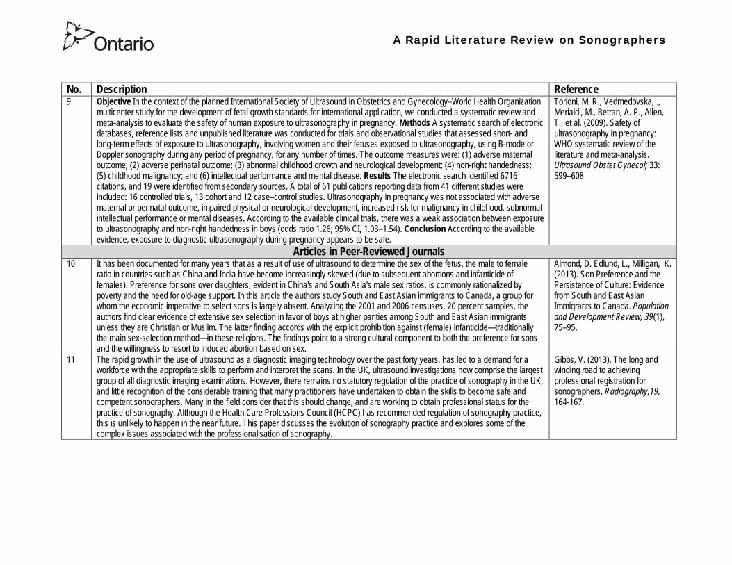
A Rapid Literature Review on Sonographers
No Description Reference 9 Objective In the context of the planned International Society of Ultrasound in Obstetrics and GynecologyndashWorld Health Organization
multicenter study for the development of fetal growth standards for international application we conducted a systematic review and meta-analysis to evaluate the safety of human exposure to ultrasonography in pregnancy Methods A systematic search of electronic databases reference lists and unpublished literature was conducted for trials and observational studies that assessed short- and long-term effects of exposure to ultrasonography involving women and their fetuses exposed to ultrasonography using B-mode or Doppler sonography during any period of pregnancy for any number of times The outcome measures were (1) adverse maternal outcome (2) adverse perinatal outcome (3) abnormal childhood growth and neurological development (4) non-right handedness (5) childhood malignancy and (6) intellectual performance and mental disease Results The electronic search identified 6716 citations and 19 were identified from secondary sources A total of 61 publications reporting data from 41 different studies were included 16 controlled trials 13 cohort and 12 casendashcontrol studies Ultrasonography in pregnancy was not associated with adverse maternal or perinatal outcome impaired physical or neurological development increased risk for malignancy in childhood subnormal intellectual performance or mental diseases According to the available clinical trials there was a weak association between exposure to ultrasonography and non-right handedness in boys (odds ratio 126 95 CI 103ndash154) Conclusion According to the available evidence exposure to diagnostic ultrasonography during pregnancy appears to be safe
Torloni M R Vedmedovska Merialdi M Betran A P Allen T et al (2009) Safety of ultrasonography in pregnancy WHO systematic review of the literature and meta-analysis Ultrasound Obstet Gynecol 33 599ndash608
Articles in Peer-Reviewed Journals 10 It has been documented for many years that as a result of use of ultrasound to determine the sex of the fetus the male to female
ratio in countries such as China and India have become increasingly skewed (due to subsequent abortions and infanticide of females) Preference for sons over daughters evident in Chinas and South Asias male sex ratios is commonly rationalized by poverty and the need for old-age support In this article the authors study South and East Asian immigrants to Canada a group for whom the economic imperative to select sons is largely absent Analyzing the 2001 and 2006 censuses 20 percent samples the authors find clear evidence of extensive sex selection in favor of boys at higher parities among South and East Asian immigrants unless they are Christian or Muslim The latter finding accords with the explicit prohibition against (female) infanticidemdashtraditionally the main sex-selection methodmdashin these religions The findings point to a strong cultural component to both the preference for sons and the willingness to resort to induced abortion based on sex
Almond D Edlund L Milligan K (2013) Son Preference and the Persistence of Culture Evidence from South and East Asian Immigrants to Canada Population and Development Review 39(1) 75ndash95
11 The rapid growth in the use of ultrasound as a diagnostic imaging technology over the past forty years has led to a demand for a workforce with the appropriate skills to perform and interpret the scans In the UK ultrasound investigations now comprise the largest group of all diagnostic imaging examinations However there remains no statutory regulation of the practice of sonography in the UK and little recognition of the considerable training that many practitioners have undertaken to obtain the skills to become safe and competent sonographers Many in the field consider that this should change and are working to obtain professional status for the practice of sonography Although the Health Care Professions Council (HCPC) has recommended regulation of sonography practice this is unlikely to happen in the near future This paper discusses the evolution of sonography practice and explores some of the complex issues associated with the professionalisation of sonography
Gibbs V (2013) The long and winding road to achieving professional registration for sonographers Radiography19 164-167
A Rapid Literature Review on Sonographers
No Description Reference 12 Background Contrast echocardiography (which uses contrast agents injected into the blood flow in order to visualize the blood flow
in very small blood vessels) has been shown to improve diagnostic quality especially in technically difficult patients However the learning curve and increased time for preparation and image acquisition have led to low use Methods The authors sought to determine whether the contrast echocardiography procedure performed independently by a specialized trained sonographer could improve efficiency In one centre routine echocardiograms were scheduled for one hour and any study exceeding one hour would result in patient booking cancellations The authors compared the standard of care in which a physician or nurse administers echocontrast with a sonographer-administered program (SAP) Results The time to complete contrast echocardiograms was significantly reduced by the SAP strategy (43 min 17 s plusmn 23 min 42 s vs 1 h 1 min 6 s plusmn 31 min 0 s P lt 0001) Subgroup analysis of the inpatients and outpatients demonstrated similar results Only 10 of studies (6 of 61) in the SAP exceeded 60 minutes compared with 45 (34 of 76) in the standard-of-care group (P lt 0001) Based on study volumes in the centre the net improvement in productivity with the SAP could be up to 53 annually Conclusion Sonographer-administered echocontrast is feasible and potentially removes a barrier to implementation of contrast echocardiography
Tang A Chiew S K Rashkovetsky R Becher H Choy J B (2013) Feasibility of Sonographer-Administered Echocontrast in a Large-Volume Tertiary-Care Echocardiography Laboratory Canadian Journal of Cardiology 29 391ndash395
13 Ultrasound scanning has been used as a diagnostic and screening tool in obstetric practice for over 50 years There is no evidence of immediate or long-term harm to the developing fetus from exposure to B mode ultrasound However exposure to high levels of Doppler ultrasound during early development is increasingly common and the full safety implications of this exposure are not clear Doppler ultrasound exposure in utero gives rise to increased apoptosis in animal models and there is evidence of the effects of exposure to Doppler ultrasound persisting throughout life with increased non-right-handedness observed in human epidemiological studies The authors consider the idea that there may be long-term developmental implications for fetuses exposed to Doppler ultrasound early in gestation These effects may be mediated via thermal or mechanical disruption to the developing conceptus giving rise to free radical damage Excess free radical exposure early in gestation is a strong candidate for the final common pathway underlying developmental programming effects and gives rise to concern that fetuses exposed to high levels of ultrasound are at risk of a developmental programming effect It is suggested that there is a need for animal studies of developmental programming using exposure to Doppler ultrasound scanning as the exposure of interest and for more observational data to be collected in the clinical setting While these data are collected it seems prudent to continue to adhere to the principle of lsquoas low as reasonably achievablersquo (ALARA) when exposing first-trimester fetuses to Doppler ultrasound
Aiken CE Lees CC (2012) Long-term effects of in utero Doppler ultrasound scanning ndash A developmental programming perspective Medical Hypotheses 78 539ndash541
14 Point of care sonography by emergency physicians in the emergency department can be an effective aid in the diagnosis and management of patients presenting with a variety of medical and traumatic conditions Its use can improve patient outcomes enhance patient safety speed patient disposition and save lives CAEP supports the use of point of care sonography
Canadian Assocation of Emergency (CAEP) Physicians Ultrasound Position Statement Working Group (2012) Use of point of care sonography by emergency physicians CJEM 14(2)106-112
A Rapid Literature Review on Sonographers
No Description Reference 15 The use of three-dimensional echocardiography (3DE ie a 3D ultrasound of the heart) during electrophysiology (EP - a branch of
cardiology that deals with the diagnosis and treatment of heart rhythm disorders) procedures is the end product of years of growth in two diverse cardiology subspecialties namely advanced cardiac imaging and the EP During the past decade progress in both fields has resulted in many important advances that have culminated in their union for a new area of growth and development Imaging advances have provided the cardiovascular specialist with enhanced cardiac volume and function data and more recently 3DE capabilities with improved spatial and temporal resolution providing unprecedented spatial relationships This latter development is valued by EP specialists in need of hitherto never required anatomic knowledge as they press forward with extraordinary expansion in their capabilities It makes sense that by combining these two rapidly growing subspecialties future capabilities in patient care may be achieved that would otherwise not be possible This paper discusses the value of 3DE during EP procedures and offers the readers insight into this novel multispecialty hybrid arena Using this model as a template it is likely that the readers may identify other areas within their practices where periprocedural advanced imaging may afford significant dividends in patient outcomes
Moukabary T Faletra F F Kronzon I Thomas W amp Sorrell V L (2012) Three-Dimensional Echocardiography in the Electrophysiology Laboratory Echocardiography 29 117-122
16 Background There has been much discussion about whether female feticide occurs in certain immigrant groups in Canada The authors examined data on live births in Ontario and compared sex ratios in different groups according to the motherrsquos country or region of birth and parity Methods The authors completed a population-based study of 766 688 singleton live births between 2002 and 2007 They used birth records provided by Ontario Vital Statistics for live births in the province between 23 and 41 weeksrsquo gestation They categorized each newborn according to the motherrsquos country or region of birth namely Canada (n = 486 599) Europe (n = 58 505) South Korea (n = 3663) China (n = 23 818) Philippines (n = 15 367) rest of East Asia (n = 18 971) Pakistan (n = 18 018) India (n = 31 978) rest of South Asia (n = 20 695) and other countries (n = 89 074) They calculated malefemale ratios and 95 confidence intervals (CIs) for all live births by these regions and stratified them by maternal parity at the time of delivery (0 1 2 or ge 3) Results Among infants of nulliparous women (women with no previous children) the malefemale ratio was about 105 overall As parity (the number of previous children) increased the ratio remained unchanged among infants of Canadian-born women In contrast the malefemale ratio was significantly higher among infants of primiparous women (women with one previous child) born in South Korea (120 95 CI 109ndash134) and India (111 95 CI 107ndash115) than among infants of Canadian-born primiparous women Among multiparous women (women with 2+ previous children) those born in India were significantly more likely than Canadian-born women to have a male infant (parity 2 ratio 136 95 CI 127ndash 146 parity ge 3 ratio 125 95 CI 109ndash143) Interpretation The study of malefemale ratios in Ontario showed that multiparous women born in India were significantly more likely than multiparous women born in Canada to have a male infant
Ray J G Henry D A Urquia M L (2012) Sex ratios among Canadian liveborn infants of mothers from different countries CMAJ184(9) E92-96
A Rapid Literature Review on Sonographers
No Description Reference 17 This letter consists of a case study that illustrates how a cross-disciplined sonographer (educated in cardiac and vascular ultrasound)
can quickly and efficiently and with great Cost-effectiveness deliver timely care to a patient in certain clinical settings The literature is comparatively sparse in referencing clinical diagnostics in cross-discipline sonographers Echocardiographers must be clinicians fully comfortable with evaluating the circulatory dynamics of the body from the left and right heart to the peripheral arteries and veins including the extracranial circulation (carotid arteries) Included in this skill set is substantive understanding of the ECG patientrsquos history and integration of cardiac and vascular findings Limitations of each diagnostic tool and how these limitations can be overcome by the strengths of the other examination are important For the dual-skilled sonographer only experience continuous education and constant clinical feedback will lead to the independent decision making that is counted on by the physician The American Registry for Diagnostic Medical Sonography offers credentials in cardiac and vascular imaging (RDCS and RVT) Cardiovascular Credentialing International offers credentials in cardiac and vascular imaging as well (registered cardiac sonographer [RCS] and registered vascular specialist [RVS]) All imaging facilities should consider merging cardiac and vascular noninvasive imaging with dual-registered clinical sonographers credentialed by one of these two agencies
Zemanek M (2012) What It Means to Be a Cardiovascular Sonographer Journal of Ultrasound in Medicine 311859-1862
18 Diagnostic medical sonography is viewed as a safe imaging modality However bioeffects research has yet to define the effects of exposure to sonography in humans Therefore sonographers must be aware of potential risks and practice ALARA (ldquoas low as reasonably achievablerdquo) routinely This research explores sonographersrsquo ability to answer basic questions about the bioeffects of sonography as well as their knowledge and use of the safety indices called the mechanical index (MI) and thermal index (TI) Members of the Society of Diagnostic Medical Sonographers in the US a southwestern sonographersrsquo society and a sonography community on Facebook 2010 were surveyed (n = 212) There is a low level of bioeffects knowledge and minimal safety practices among sonographers in this survey No significant relationship was found between sonographer knowledge of the bioeffects of sonography years in the profession or whether the sonographers monitor the MITI
Bagley J Thomas K DiGiacinto D (2011) Safety Practices of Sonographers and Their Knowledge of the Biologic Effects of Sonography Journal of Diagnostic Medical Sonography 27(6) 252ndash261
19 This article highlights trends in echocardiography Point of care ultrasound (hand-carried ultrasound) Initially point of care ultrasound will increase the use of echocardiography in the same manner that cardiac CT increased catheterizations However once the basics are learned it has the potential to be a very powerful tool in that less unnecessary studies will be ordered In the clinic small systems will be used as a lsquolsquofirst lookrsquorsquo screen and become part of the patientrsquos history only patients for whom more testing is necessary will be referred for a comprehensive study This is likely to reduce the number of Doppler studies requested The miniaturization of echo equipment will continue with improved qualityfunctionality of the lsquolsquohand-heldrsquorsquo echo machines to the extent that cordless probes will become a reality sooner rather than later Role in interventional echocardiography Sonographers will be spending much more time in the interventional and electrophysiology (EP) labs There will be more interconnection with interventional cardiologists and the volume will increase as more devices are implanted more percutaneous valves placed and more robotic surgery is performed Crosstraining and multitasking Sonographers entering the profession would be prudent to learn cardiac and vascular imaging Individuals who multitask andor cross-train will be ideal For example a sonographer who starts an IV monitors ECG stress can perform cardiac MRI and can interpret a preliminary report or a nurse who can do an echo monitor sedation and start an IV will be invaluable The sonographer should continue to grow professionally by cross training as institutions try to minimize costs and maximize throughput
McCulloch M L (2011) Thoughts regarding the future of sonography Journal of the American Society of Echocardiography 24(5) 29A-30A
A Rapid Literature Review on Sonographers
No Description Reference 20 Background Radiology reports provide specialist interpretation of images and relate these findings to the patientrsquos symptoms and
signs This study compared ultrasound (USS) reports generated by radiologists and sonographers to determine if any significant or clinically relevant differences existed Methods A retrospective analysis of 624 consecutive USS reports was carried out The reports were assessed for the presence of a lsquodisclaimerrsquo or lsquocaveatrsquo pertaining to the quality of the images and were analysed with respect to the clarity of their wording and ability to answer the clinical request Results The majority of sonographer USS reports contained a disclaimer regarding the quality of the USS images (571) compared with a smaller proportion of radiologist scans (99) (P lt 0001) Overall radiologists performed significantly better in providing a clear negative or positive diagnosis to the clinical question on the request form when compared with sonographers (885 vs 654 P lt 0001) Conclusion lsquoDisclaimerrsquo comments and lsquohedgingrsquo must be avoided in radiology reporting While feedback as to the accuracy of the radiology images is important overuse of such terms undermines the validity of the radiology report
Garcea G Mahmoud A Ong S L Rees Y Berry Det al (2010) Caveat reporting in ultrasound interpretation of surgical pathology a comparison of sonographer versus radiologist Journal of Evaluation in Clinical Practice 16 97ndash99
21 A nine-point questionnaire was constructed based on previously published work by Marsal and Sheiner et al The survey was distributed to clinical users of ultrasound practicing sonography in Hamilton (New Zealand) during July 2010 totalling 35 users consisting of sonographers sonography trainees one obstetric sonologist and one fetal medicine registrar Results Twenty sonographers returned the questionnaire which represents survey response of 61 The level of clinical experience was equally split between junior sonographers of lt 5 yearsrsquo experience (30) sonographers with 5-10 yearsrsquo experience (35) and senior sonographers of gt10 yearsrsquo experience (35) While 100 of the sonographers surveyed reported being familiar with the term Thermal Index (TI) and 90 claimed they adhered to the Action Learning Action Response Association (ALARA) principle all the time or most of the time only 15 of sonographers correctly described or defined what the term TI means Of the sonographers 80 knew where to find the TI during real-time examinations but only 35 knew which TI index to use for nuchal translucency (NT) scan and only 10 knew the expected range of TI encountered during routine obstetric examinations Of the eight sonographers who reported adhering to ALARA all the time the majority (64) could not describe or define TI In the same subgroup the majority (64) did not know which TI to monitor during a NT scan Only three of 20 (15) sonographers knew what the term TI means Of these two were junior sonographers with lt 5 yearsrsquo experience and one had 5ndash10 yearsrsquo experience Despite understanding the term TI one of the three sonographers still selected the incorrect index to monitor during a NT scan Not one of the seven senior sonographers with gt 10 years of experience could define or describe the TI In total only 10 of sonographers knew what the TI means and which TI to monitor during NT scanning
Necas M (2010) New Zealand sonographers do not outperform their European or American colleagues in the knowledge of ultrasound safety Australasian Journal of Ultrasound in Medicine13(4) 28-32
A Rapid Literature Review on Sonographers
No Description Reference 22 Musculoskeletal (MSK) ultrasound performed by non-medical practitioners is a developing practice The aim of this paper is
(1) to audit an experienced sonographerrsquos (SJ Riley one of the authors) performance after one yearrsquos independent reporting against two experienced MSK radiologists (CJ Grove amp M Chandramohan the other two authors) and (2) to establish an audit standard against which such role development might be contrived Images and reports from 250 MSK ultrasound examinations performed by a sonographer were reviewed independently by two consultant MSK radiologists The examinations were graded for discrepancy eg grade 1 ndash agree with the sonographer report grade 2 ndash minor discrepancy unlikely to alter patient care grade 3 ndash potentially significant discrepancy grade 4 ndash definite significant discrepancy likely to have adverse consequences for patient care Two of 250 (08) cases were excluded Both radiologists agreed completely with the sonographer (grade 1) in 235 of the 248 cases (948) In 13 cases there was discrepancy between the reports of SJR and the radiologists The discrepancy was grade 2 in six of the 248 cases (24) grade 3 in six of the 248 cases (24) and grade 4 in one of the 248 cases (04) In conclusion this audit shows a high level of agreement between the sonographer and the consultant MSK radiologist reporting of MSK ultrasound This level of agreement may set the standard for future quality assurance audit of sonographer MSK reporting
Riley S J Groves C J Chandramohan M (2010) Musculoskeletal ultrasound audit of sonographer reporting Ultrasound 18 36ndash40
23 Background Growing demand for ultrasound examinations and higher quality requirements motivate searching for routines combining the diagnostic accuracy of radiologist-performed examinations with the economical advantages of sonographer-performed examinations One possible approach is to use strictly standardized acquisition and documentation schemes that give the radiologist access to all relevant information after the examination Purpose To compare a recently introduced routine combining acquisition by a radiographer (more specifically a sonographer with two years experience) documentation as standardized cine-loops and review by a radiologist (lsquostandardized methodrsquo) with the formerly used routine where the diagnosis is made bedside by the radiologist (lsquotraditional methodrsquo) Material and Methods In 64 policlinic patients the kidneys (n = 27) or the gallbladder (n = 37) were examined with both the standardized and the traditional method The radiologistsrsquo findings of hydronephrosis tumors cysts echogenicity changes and cortical thickness (in the kidneys) and wall thickness concrements and polyps (in the gallbladder) were compared between the methods with respect to agreement (proportion of agreement and kappa coefficient) as well as systematic differences (McNemarrsquos test) Results The findings at the gallbladder examination showed a median agreement of 97 (86ndash100 kappa = 064ndash100) and those at the kidney examination an agreement of 90 (78ndash100 kappa = 069ndash 100) There were no significant systematic differences between the methods Conclusion The satisfactory agreement in this preliminary study indicates that the new workflow with ultrasound examinations performed by a radiographer (sonographer) and analyzed off-line by a radiologist is promising and motivates further studies
Stenman C Thorelius L Knutsson A amp Smedby Ouml (2010) Radiographer-acquired and radiologist-reviewed ultrasound examination ndash agreement with radiologistrsquos bedside evaluation Acta Radiologica 52 70ndash74
A Rapid Literature Review on Sonographers
No Description Reference 24 The troubling practice of fetal sex selection has historically been considered an Asian phenomenon However recent evidence shows
that a similar situation is emerging in North America albeit on a smaller scale The Society of Obstetricians and Gynaecologists of Canada (SOGC) has firmly stated its opposition to sex selection for non-medical reasons as well as to the use of any technology used solely for the purpose of determining fetal sex However because fetal sex may be disclosed to the parents at the time of ultrasound examination if they request this information guidance for health care professionals to assist in discouraging fetal sex selection would be useful Because no declaration of motives or reasons is required when a woman seeks a termination of pregnancy the authors suggest that health care professionals need not disclose the sex of a fetus until it has reached a gestational age at which abortion for non-medical purposes would not be possible This proposal would facilitate consistency between clinical practice and the values of Canadian citizens the SOGC the Canadian Medical Association and other professional organizations while still respecting current laws pertaining to disclosure of patient information and patientsrsquo rights to autonomous decision-making
Thiele A T amp Leier B (2010) Towards an Ethical Policy for the Prevention of Fetal Sex Selection in Canada J Obstet Gynaecol Can 32(1) 54ndash57
25 Background The extent to which increases in the use of prenatal ultrasonography over time reflect changes in maternal risk (ie an increase in high-risk pregnancies over time) is unknown In this population-based study the authors examined the use of prenatal ultrasonography from 1996 to 2006 in Ontario Methods With fiscal year 199697 as the baseline the authors evaluated the relative risk (RR) and 95 confidence interval (CI) for the change in rates of ultrasonography for each subsequent year The RR was adjusted for maternal age income rural residence maternal comorbidities receipt of genetics consultation or amniocentesis mdash all in the index pregnancy mdash and history of complications in a prior pregnancy Results The study sample consisted of 1 399 389 singleton deliveries The rate of prenatal ultrasonography increased from 2055 per 1000 pregnancies in 1996 to 3264 per 1000 in 2006 (adjusted RR 155 95 CI 154ndash155) The rate increased among both women with low-risk pregnancies (adjusted RR 154 95 CI 153ndash155) and those with high-risk pregnancies (adjusted RR 155 95 CI 154ndash157) The proportion of pregnancies with at least four ultrasound examinations in the second or third trimesters rose from 64 in 1996 to 187 in 2006 (adjusted RR 268 95 CI 261ndash274) Paradoxically this increase was more pronounced among low-risk pregnancies (adjusted RR 292 95 CI 283ndash301) than among high-risk pregnancies (adjusted RR 225 95 CI 216ndash235) Interpretation Substantial increases in the use of prenatal ultrasonography over the past decade do not appear to reflect changes in maternal risk Nearly one in five women now undergo four or more ultrasound examinations during the second and third trimesters Efforts to promote more appropriate use of prenatal ultrasonography for singleton pregnancies appear warranted
You J J Alter D A Stukel T A McDonald S D Laupacis A et al (2010) Proliferation of prenatal ultrasonography CMAJ 182(2) 143-151
A Rapid Literature Review on Sonographers
No Description Reference 26 Aims The aim of this study was to assess the cost-effectiveness of using certified sonographers and miniaturized echocardiography
systems to perform echocardiograms at bedside in comparison to moving inpatients from the admission department to the echocardiography laboratory (echo-lab) Methods and Results From 26 September 2005 to 27 October 2005 112 patients admitted in six hospital wards in Italy connected through a 100 Mbit LAN to the echo-lab were scanned within the admission ward by sonographers using a miniaturized echo system Logistical data were collected and results were compared with those obtained from 194 consecutive patients coming from the same wards and studied in the echo-lab with high-end machines between 8 March 2005 and 15 April 2005 Performing echocardiograms in the admission department avoided long waiting time of the inpatients in the echo-lab before and after the study increased the percentage of patients studied within 3 and 5 days from request (88 vs 77 and 100 vs 95 respectively P = 003) increased both sonographer (by 339 P lt 0001) and echo-lab productivity (by 41 P lt 0001) and reduced costs of echocardiograms by 29 Conclusion Implementation of digital echocardiography certified sonographers and a miniaturized echo system allowed improvement of the cost-effectiveness of the service provided by the echo-lab for inpatients and avoided patientsrsquo discomfort derived from prolonged waiting time before and after the exam
Badano L Gaetano N Stacul S Gianfagna P Pericoli M et al (2009) Improved workflow sonographer productivity and cost-effectiveness of echocardiographic service for inpatients by using miniaturized systems European Journal of Echocardiography 10 537ndash542
27 The present report is a proposal from the American Society of Echocardiography Advanced Practice Task Force that identifies the potential of cardiac sonographers to achieve the Advanced Cardiovasculat Sonographer (ACS) level According to this report echocardiographic examinations require a well-trained and competent sonographer to obtain proper anatomic and physiologic data to establish an accurate diagnosis for clinical decision-making and patient management Although the formal education and training of cardiovascular sonographers are evolving many entry-level and staff sonographers may not have sufficient practical or clinical knowledge of the necessary components of the echocardiographic study for the individual patientrsquos clinical presentation In many clinical settings echocardiograms are read after the patient has left the laboratory Thus there is a role for a sonographer who can practice at an advanced level in a cardiovascular ultrasound laboratory to ensure a proper echocardiographic examination is performed on every patient In this setting an Advanced Cardiovascular Sonographer (ACS) would be able to review the indication for and quality of the examination If additional images were needed the ACS would assist the sonographer in obtaining these images which would lead to the performance of a complete and fully diagnostic examination before the patient had left the echocardiography laboratory In clinical practice the quality of the examinations performed would improve advancements in echocardiographic methods could be taught and incorporated into daily practice and patients would be better served
Mitchell C Miller F A Bierig S M Bremer M L Ehler D et al (2009) Advanced Cardiovascular Sonographer A Proposal of the American Society of Echocardiography Advanced Practice Sonographer Task Force J Am Soc Echocardiogr 22 1409-13
A Rapid Literature Review on Sonographers
No Description Reference 28 Sonographer practitioner development involves the expansion and extension of the sonographer role to include reporting on
ultrasound examinations Australian sonographers have not seen the same degree of role extension and expansion as their counterparts in the United Kingdom despite increasing levels of discussion regarding sonographer practitioner development The aim of this study was to determine if Australian sonographers want to extend their professional role and what they consider are the important issues associated with role extension This paper reports on qualitative data derived from a survey of Australian sonographers and investigates if Australian sonographers are interested in extending and expanding their professional role and responsibilities and if they do what might be necessary or desirable from a professional point of view for this change to occur A survey was mailed to all members of the Australian Sonographers Association (ASA) in October 2006 The 31-item survey included 28 closed-ended and three opened-ended items to provide both quantitative and qualitative data The quantitative data will be reported separately Qualitative data was derived from responses to the opened-ended questions which asked respondents to elaborate on their attitudes and feelings about role extension and development Analysis used Nvivo7 software to aid in uncovering common themes from the qualitative data The analysis focused on the reported incentives or motivations for becoming a sonographer practitioner as well as disincentives or perceived hurdles that would discourage respondents from becoming sonographer practitioners The three most reported incentives or motivations for becoming a sonographer practitioner were professional recognition remuneration and increased knowledge The three most commonly reported disincentives or perceived hurdles that would discourage respondents from becoming sonographer practitioners were legal issues insurance and further study
McGregor R OrsquoLoughlin K Cox J Clarke J Snowden A (2009) Sonographer practitioner development in Australia Qualitative analysis of an Australian sonographersrsquo survey Radiography15 313-319
29 Introduction This study describes the effect of introducing a cardiac sonographer led follow up clinic within a hospital cardiology department for patients with stable heart valve disease The two years before and after the instigation of the valve clinic were audited Materials and methods The clinic was conducted in a single centre and undertaken in the cardiology department of a district general hospital A total of 382 patients with 397 clinically significant valve lesions but for whom surgery was not yet indicated but follow up required were seen in a cardiac sonographer run clinics These patients no longer attended a medical follow up clinic unless there was clinical or echocardiographic deterioration Effectiveness was judged by the percentage treated according to current best practice guidelines the number of echocardiograms performed and the number of hospital outpatient visits attended In addition mortality data for the subjects in the clinic was collected Results The proportion followed up according to best practice guidelines rose from 157 (41) to 354 (92) (p lt 001) The total number of echocardiograms performed fell from 807 to 550 Total number of outpatient visits fell from 998 to 31 A total of 11 patients died in the two year study period none from progressive valve disease Discussion This study demonstrates that a protocol driven sonographer led heart valve disease follow up clinic significantly improved the quality of follow up while bringing about a major reduction in outpatients visits without compromising patient safety
Taggu W Topham A Hart L Carr-White G Sulke N et al (2009) A cardiac sonographer led follow up clinic for heart valve disease International Journal of Cardiology 132 240 ndash243
30 Role development for imaging allied health professionals has progressed significantly over the last three decades with many major developments pioneered in the field of ultrasound This paper reviews the developing role of the radiographer in ultrasound (in some countries such as the UK sonographers are considered a type of radiographer) and some of the clinical professional and educational implications of such development As the published evidence base underpinning the latest developments is scarce several contemporary case study examples which have been presented verbally at study days attended by the authors have been included In conclusion sonographer role development is considered within the professional contexts of extended role advanced and consultant practice
Hart A amp Dixon A (2008) Sonographer Role Extension and Career Development a Review of the Evidence Ultrasound 16(1) 31-35
A Rapid Literature Review on Sonographers
No Description Reference 31 Objective The main goal of this study was to determine end usersrsquo knowledge regarding safety aspects of diagnostic ultrasound
during pregnancy End usersrsquo attitudes toward the use of ultrasound in low-risk pregnancies were also assessed Methods A questionnaire was distributed to ultrasound end users attending review courses and hospital grand rounds between April and June 2006 in Chicago Results One hundred thirty end users completed the questionnaires (63 response rate) Sixty-three percent were physicians (n = 84) most of them obstetricians (817) About 18 of participants routinely performed Doppler ultrasound examinations during the first trimester Fifty percent of end users thought that the number of ultrasound examinations in low-risk pregnancy should be limited to 1 to 3 (mean plusmn SD 26 plusmn 09) Almost 70 disapproved of ldquokeepsakeentertainmentrdquo ultrasound Although 322 of the participants were familiar with the term thermal index only 177 actually gave the correct answer to the question on the nature of the thermal index About 22 were familiar with the term mechanical index but only 38 described it properly Almost 80 of end users did not know where to find the acoustic indices Only 208 were aware that they are displayed on the sonographic monitor during the examinations End users with higher knowledge of safety issues thought that there should be limitations on the number of ultrasound examinations in low-risk pregnancies (odds ratio 33 95 confidence interval 11ndash100 P = 028) Likewise these end users were more likely to respond that ultrasound might have adverse effects during pregnancy (odds ratio 32 95 confidence interval 11ndash125 P = 045) Conclusions Ultrasound end users are poorly informed regarding safety issues during pregnancy Further efforts in the realm of education and training are needed to improve end user knowledge about the acoustic output of the machines and safety issues
Sheiner E Shoham-Vardi I Abramowicz J S (2007) What Do Clinical Users Know Regarding Safety of Ultrasound During Pregnancy J Ultrasound Med 26 319ndash325
A Rapid Literature Review on Sonographers
No Description Reference 32 In an attempt to survey the knowledge among ultrasound users of some safety aspects of diagnostic ultrasound a questionnaire was
distributed to professionals using ultrasound for fetal examinations The questionnaire was answered by the participants and faculty members of six postgraduate courses in obstetric ultrasound Doppler ultrasound and fetal echocardiography given in Sweden Norway and Austria in 2003 Furthermore in Sweden the questionnaire was distributed to the staff meeting participants of two major obstetric ultrasound units and of one unit for fetal echocardiography and at a meeting of the Working Group for Ultrasound of the Swedish Society of Obstetrics and Gynecology In total 199 questionnaires were anonymously answered by doctors (n = 145) sonographers (n = 22) and midwives trained in ultrasound (n = 32) All of them were using diagnostic ultrasound on a daily or weekly basis The respondents were from nine European countries In none of the countries is formal certification for the use of diagnostic ultrasound in pregnancy or otherwise required The doctors who answered the questionnaire were specialists in obstetrics and gynecology (including some of the internationally recognized experts in fetal ultrasound) pediatric cardiology radiology or clinical physiology The respondents were not informed about the questionnaire in advance and they had a limited time to answer the 16 questions typically 10ndash15 min The questions concerned explanation of the safety indices and their relation to various ultrasound modes ie real-time B-mode M-mode three-dimensional (3D) spectral Doppler color and power Doppler ultrasound The respondents were also asked which ultrasound system they used most often and where on the system the information on thermal index (TI) and mechanical (MI) was displayed (eg in the top right corner of the screen in a separate window etc) Furthermore the respondents had to indicate how they adjust the output energy level on their own machine For all questions there was the optional answer lsquoDonrsquot knowrsquo Results About one-third of the ultrasound experts were able to define the abbreviations TI and MI For the subclasses of the TI the correct answer was found in only 3-8 of cases Of those who knew what the TI and MI meant only two-thirds and one-third of respondents respectively were able to give a simple explanation of the indices In 49 of questionnaires the Doppler modes were correctly ranked as generally producing higher ultrasound exposure than the imaging modes (B-mode 3D) Some 28 of the respondents correctly indicated where on their own machine the information on safety indices is displayed However not all of them knew how to control the output energy level For all items in the questionnaire there were no significant differences in the results between the three categories of respondents (ie physicians sonographers and midwives) The results of the questionnaire were anything but encouraging and they indicated that the users who are supposed to be responsible for controlling exposure of the fetus to ultrasound had a very poor knowledge of the basic safety aspects of ultrasound
Maršaacutel K (2005) The output display standard has it missed its target Ultrasound Obstet Gynecol 25 211ndash214
33 Purpose This study was performed to evaluate the accuracy of upper abdominal ultrasound (US) scanning performed by sonographers in a district general hospital to identify potential areas of weakness and to make recommendations to improve the service Materials and Methods Upper abdominal US examinations performed and reported by sonographers over a 4-week period were retrospectively reviewed The accuracy of the imaging findings and reports were assessed against other imaging surgical histological or laboratory findings and against clinical outcome Results A heterogenous group of 104 patients were included in the study 62 of whom had an US abnormality Errors of scanning or interpretation were identified in 10 patients (96) of whom five (48) were felt to be potentially significant Conclusions The sonographersrsquo accuracy in reporting upper abdominal US scans was 90 However on the basis of this study the authors have implemented specific recommendations to improve the quality of the service
Dongola N Guy R Ward S amp Giles J A (2003) Can sonographers offer an accurate upper abdominal ultrasound service in a district general hospital Radiography 9 29ndash33
A Rapid Literature Review on Sonographers
No Description Reference 34 Aim To compare the accuracy of radiographers and radiologists in routine abdominal ultrasound Materials and Methods One
hundred consecutive patients attending for routine abdominal ultrasound were included Each patient was examined by both a radiographer and radiologist Both operators noted their fndings and wrote a concluding report without conferring Reports were compared Where there was disagreement the patient was either re-examined by another radiologist or had further investigation Results Of 100 patients 52 were men and 48 were women The age range was 19plusmn88 years (median 52 years) Thirty-seven patients had renal tract ultrasound one had an aortic ultrasound and 62 had general upper abdominal ultrasound In 44 cases both operators reported the examination as normal In 49 cases both operators reported the examinations as abnormal and there was complete agreement between the operators In seven cases there was not complete agreement between operators Three of these disagreements were considered minor and four major In three of the seven cases the radiographer was correct and in four the radiologist was correct Conclusion Experienced radiographers and radiologists are highly accurate in performing and interpreting routine abdominal sonography Both operators missed a small minority of abnormalities There was no statistically significant difference in the accuracy of radiographers and radiologist
Leslie A Lockyer H amp Virjee J P (2000) Who Should be Performing Routine Abdominal Ultrasound A Prospective Double-Blind Study Comparing the Accuracy of Radiologist and Radiographer Clinical Radiology 55 606-609
Grey Literature 35 The ARRT The American Registry of Radiologic Technologists tests certifies and annually registers more than a quarter-of-a-million
radiologic technologists to promote high standards of patient care They award the Registered Technologist or RT designation to individuals who have completed the prescribed classroom and clinical education passed the appropriate exam and met the ethics requirements And they ensure their continuing education and ongoing compliance with their high standards by requiring annual registration of their certificate Certification by ARRT is voluntary not mandatory But employers state licensing agencies and federal regulators view ARRTrsquos credential as an indicator that a person has met recognized national standards for medical imaging and radiation therapy professionals How Are Radiologic Technologists Deemed Qualified First comes initial certification Certification is the process of initially recognizing individuals who have satisfied certain standards within a profession A person is certified by ARRT after completing an approved educational program complying with the ethics standards and passing a certification exam Then comes annual registration Registration is the yearly procedure required to maintain certification ARRT registrants are those who having already fulfilled the requirements for initial certification continue to meet the requirements for annual registration The ARRT annually registers the certificates of individuals who meet the following three criteria agreeing to comply with the ARRT Rules and Regulations continuing to comply with the ARRT Standards of Ethics and meeting our continuing education requirements Only technologists who are currently registered (which means that they have renewed within the past year) may designate themselves as ARRT Registered Technologists and use the initials RT after their names Federal laws regulate personnel qualifications in some modalities (mammography for example) but they accredit the health care facility rather than licensing the individual radiologic technologist who is providing patient care And while many states have laws regulating who can perform radiologic procedures they are typically based on ARRT certification Some employers and state agencies may require ARRT certification but not necessarily current registration Unless they require current registration by ARRT employers cannot assume that the person has complied with the ethics requirements or the continuing education requirements
American Registry of Radiologic Technologists (ARRT) (2013a) Patient Page Accessed April 2013
A Rapid Literature Review on Sonographers
No Description Reference 36 This document outlines certification requirements for sonography There are two pathways (primary and post-primary) For the
primary pathway it notes that sonography educational programs must be accredited by a mechanism acceptable to the ARRT ARRT generally recognizes only accreditation agencies that are recognized by the Council for Higher Education Accreditation andor United States Department of Education
American Registry of Radiologic Technologists (ARRT) (2013b) Sonography Certification Accessed April 2013
A Rapid Literature Review on Sonographers
No Description Reference 37 In Australia sonographers and student sonographers are required to be accredited by Australian Sonographer Accreditation Registry
(ASAR) in order to practise This requirement is contained within the Australian Governmentrsquos Diagnostic Imaging Accreditation Scheme standards and Medicare legislation Accreditation of sonographers is linked only to achieving - or in the case of students studying towards - an accredited entry-level qualification completion of continuing professional development and payment of an annual fee to ASAR The ASA contends that the lack of a requirement to ensure each sonographer is lsquofit to practisersquo is an unacceptable gap in current regulation undermining efforts to ensure the quality of sonographic services and failing to protect patients from harm The ASA supports and advocates for nationally-enforceable regulation of sonographers to meet our patientrsquos expectations National registration The National Registration and Accreditation Scheme (NRAS) for health workers commenced on 1 July 2010 The ten professions which comprised the initial intake into this scheme were those that had registration requirements in all Australian states or territories as well as those in which legislated regulation is deemed to be in the public interest An additional four professions - including medical radiation practitioners which are defined as radiographers radiation therapists and nuclear medicine technologists - will be included in the NRAS commencing on 1 July 2012 As sonography did not have registration in any states or territories prior to the commencement of NRAS the profession has not been in a position to negotiate entry into NRAS In early August 2011 the ASA lodged a submission with the Health Workforce Principal Committee requesting a profession-specific Sonography Board of Australia be formed under NRAS This submission was prepared based on information that consideration of professions which were unregistered would commence More recently the Australian Health Workforce Ministerial Council (AHWMC) issued a communiqueacute stating in part lsquoMinisters have commissioned work on unregistered professions and future directions for national registration Given this Ministers agreed to defer consideration of the inclusion of additional professions into the NRAS Ministers agreed that it would be premature to consider further applications for the scheme until this work has been completed other than paramedicsrsquo Options for unregistered health practitioners including sonographers Policy work is continuing at a federal level to establish an alternative regulatory framework The National Alliance for Self-Regulating Health Professions (NASRHP) consisting of the peak professional associations of eight self-regulating health professions including the ASA representing sonography released a proposal lsquoHarnessing self-regulation to support safety and quality in healthcare deliveryrsquo in March 2012 This proposal concluded that the framework that regulates all health practitioners should be expanded beyond NRAS to include
bull authorised self-regulating professions via a health professions panel bull negative licensing to withdraw the ability to practise from registered and self-regulated practitioners who do not meet the
regulatory requirements or unregulated practitioners due to poor practice As with all advocacy initiatives consideration by the federal government of the introduction of the NASRHPrsquos proposal may be prolonged and the outcomes may not match the expectations of NASRHPrsquos member professional associations including the ASA The ASA will continue both independently and collaboratively to lobby on behalf of our members and the public for the introduction of a timely and equitable solution to health practitioner regulation In the interim as there is no regulatory model with the rigour the ASA believes is required for regulation of sonographers and other health practitioners other than NRAS the ASA continues to support its Position Statement published in early 2010
Australian Sonographer Association (ASA) (2013) Sonographer Regulation Accessed April 2013
A Rapid Literature Review on Sonographers
No Description Reference 38 This publication contains information about Cardiovascular Credentialing International (CCI) It also provides the necessary
information and references concerning the process by which candidates may earn certificate or registry-level credentials CCI is a not-for-profit corporation established for the purpose of administering credentialing examinations as an independent credentialing agency CCI began credentialing cardiovascular professionals in 1968 CCI offers eight credentials which when earned demonstrate that the registrant holds fundamental knowledge in the particular cardiovascular specialty These include RCCS ndash Registered Congenital Cardiac Sonographer RCS ndash Registered Cardiac Sonographer and RPhS ndash Registered Phlebology Sonographer
Cardiovascular Credentialing International (2013) 2013 Examination Application amp Overview Accessed April 2013
39 Oregon Governor Signs Sonographer Licensure Legislation On July 28 2009 Oregon Governor Ted Kulongoski signed a sonographer licensure bill (HB 2245) into law The new law makes Oregon only the second state in the United States to require licensure of sonographers The Society of Diagnostic Medical Sonographers (SDMS) has worked closely with other sonography-related organizations and Oregon sonographers to help craft the language in new law The law renames and restructures the Oregon Board of Radiologic Technologists to the Oregon Board of Medical Imaging (OBMI) and ensures representation of sonographers on the Board The sonographer licensure law requires Oregon sonographers to become licensed by OBMI effective July 1 2010 Beginning on January 1 2014 all sonography licensees would be required to hold a national sonography certificationcredential (or be currently enrolled as a student) While the Oregon bill is far more detailed than the sonographer licensure law that passed in New Mexico earlier this year additional administrative rulesregulations will be developed over the next year to fully implement the new law SDMS will be continue to monitor developments regarding sonographer licensure in other states
Society of Diagnostic Medical Sonographers (SDMS) (2013a) News Accessed April 2013
A Rapid Literature Review on Sonographers
No Description Reference 40 On April 6 2009 New Mexico (NM) Governor Bill Richardson signed the historic bill that for the first time in the United States
requires licensure of sonographers The bill adds sonographers (and MRI technologists) to the list of medical imaging professionals licensed by the State of New Mexico Until now anyone could perform sonograms in New Mexico Prior to the passage of the New Mexico licensure law virtually all other health care providers and professionals had to be licensed As a result the public is often surprised to learn that the person performing a medical sonogram does not have any state licensure or national certification requirements The responsibility for administering the NM sonographer licensure program would rest with the New Mexico Environment Department Although this was not the first choice in most sonographersrsquo minds the Environment Department currently manages the radiologic technology licensure program so the administrative infrastructure is already in place By ensuring the creation of an advisory council that includes representation by each medical imaging and radiation therapy modality regulated the bill helps ensure that the Departmentrsquos other areas of emphasis do not detract from sonographer licensure Because sonographers have never had to be licensed before they frequently do not see or understand the distinction between state licensure and national certification National certifications or credentials issued by voluntary organizations such as the American Registry for Diagnostic Medical Sonography (ARDMS) Cardiovascular Credentialing International (CCI) and the American Registry of Radiologic Technologists (ARRT) have little legal weight The certification process and resulting credential are valuable tools in ensuring sonographers have met minimum education and knowledge requirements but do not really regulate anyonersquos ability to perform sonography In an ideal world no additional regulation of sonography would be required However as everyone knows more and more people are taking weekend courses and buying ultrasound equipment without understanding the ramifications of using ultrasound technology (eg ALARA) Under the bill passed by the New Mexico legislature national credentialscertifications should provide evidence of meeting the statersquos standards All sonographers who are performing these critical medical imaging services in New Mexico will be required to meet national certification standards (ie have certifications or credentials issued by ARDMS CCI or ARRT) As a result there would be no additional exams in New Mexico beyond the national certification exams required by ARDMS CCI or ARRT The bill also requires the state to recognize the continuing medical education completed for national certification renewal The most visible impact on New Mexico sonographers will be a license fee However the legislation caps the license fee at $100 per two-year license In addition only the New Mexico Legislature can change this cap
SDMS (2013b) New Mexico Sonographer Licensure Law Signed Accessed April 2013
A Rapid Literature Review on Sonographers
No Description Reference 41 Preamble The purpose of this document is to define the Scope of Practice for Diagnostic Ultrasound Professionals in the US and to
specify their roles as members of the health care team acting in the best interest of the patient This scope of practice is a living document that will evolve as the technology expands Definition of the Profession The Diagnostic Ultrasound Profession is a multi-specialty field comprised of Diagnostic Medical Sonography (with subspecialties in abdominal neurologic obstetricalgynecologic and ophthalmic ultrasound) Diagnostic Cardiac Sonography (with subspecialties in adult and pediatric echocardiography) Vascular Technology and other emerging fields These diverse specialties are distinguished by their use of diagnostic medical ultrasound as a primary technology in their daily work Certification is considered the standard of practice in ultrasound Individuals who are not yet certified should reference the Scope as a professional model and strive to become certified Scope of Practice of the Profession The Diagnostic Ultrasound Professional is an individual qualified by professional credentialing and academic and clinical experience to provide diagnostic patient care services using ultrasound and related diagnostic procedures The scope of practice of the Diagnostic Ultrasound Professional includes those procedures acts and processes permitted by law for which the individual has received education and clinical experience and in which heshe has demonstrated competency Diagnostic Ultrasound Professionals
bull Perform patient assessments bull Acquire and analyze data obtained using ultrasound and related diagnostic technologies bull Provide a summary of findings to the physician to aid in patient diagnosis and management bull Use independent judgment and systematic problem solving methods to produce high quality diagnostic information and
optimize patient care
Society of Diagnostic Medical Sonography [SDMS] (2013c) Scope of Practice for the Diagnostic Ultrasound Professional Accessed April 2013
42 This website notes that in the US most employers in the US prefer to hire sonographers who have professional certification A sonographer can get certification and passing an exam Most exams relate to the specialty that the sonographer is most interested inmdashfor example an exam to become certified in abdominal sonography A few states require diagnostic medical sonographers to be licensed Typically professional certifications required for licensure other requirements vary by state Sonographers must take continuing education to keep their certification current
Bureau of Labor Statistics US Department of Labor (2012) How to become a Diagnostic Medical Sonographer Occupational Outlook Handbook 2012-13 Edition Accessed April 2013
A Rapid Literature Review on Sonographers
No Description Reference 43 The American Registry for Diagnostic Medical Sonography (ARDMS) is an independent not-for-profit organization founded in 1975
that administers examinations and awards the following credentials bull RDMSreg ndash Registered Diagnostic Medical Sonographerreg bull RDCSreg ndash Registered Diagnostic Cardiac Sonographerreg bull RVTreg ndash Registered Vascular Technologistreg bull RPVIreg ndash Registered Physician in Vascular Interpretationreg bull RMSKtrade - Registered in Musculoskeletaltrade sonography
ARDMS is the globally recognized standard of excellence in sonography and has certified more than 75000 individuals ARDMS is composed of three key groups
bull Board of Directors ndash Sonographers vascular technologists physicians research scientists and a member of the public are volunteer members of the board that determines the direction and policies of our organization
bull Examination Development Task Forces ndash A group of sonographers vascular technologists physicians and scientists volunteer their time and expertise to produce our examinations
bull Professional Staff ndash A paid highly-skilled staff administers the programs and assists applicants and registrants Credentials awarded by ARDMS document personal achievement of recognized professional standards and are widely accepted in the medical community by sonography and vascular professional organizations However ARDMS does not validate the day-to-day job performance of any sonography professional ARDMS has earned the prestigious ANSI-ISO 17024 accreditation for certifying bodies from the International Organization for Standardization (ISO) Accreditation is granted through the American National Standards Institute (ANSI) ARDMS has also earned accreditation with the National Commission for Certifying Agencies (NCCA) The NCCA is the accrediting arm of the National Organization for Competency Assurance (NOCA) Established in 1977 as a nonprofit organization NOCA is a leader in establishing quality standards for certifying organizations
American Registry for Diagnostic Medical Sonography (ARDMS) (2012a) Overview of ARDMS Accessed April 2013
A Rapid Literature Review on Sonographers
No Description Reference 44 ARDMS is developing a program to assure that Registrants continue to meet the basic competency requirements of core clinical skills
in present day practice bull Each Registrant who holds an active ARDMS certification as of December 31 2011 will be assigned the 2012 ndash 2021 10-
year recertification assessment period bull The 10-year period for new Registrants will begin the year following the attainment of the credential bull If a Registrant earns a new credential or specialty after January 1 2012 then that credentialspecialty will have a separate
10-year recertification assessment period that will begin the year following the attainment of the credentialspecialty ARDMS will develop tools and resources to assist Registrants in tracking and meeting the recertification assessment program requirements
bull Registrants who do not wish to take a recertification assessment in a specific credentialspecialty may voluntarily relinquish the credentialspecialty
As an ARDMS Registrant individuals are committing to demonstrate the highest level of competency in the modality of sonography by meeting and maintaining the standards set by the ARDMS The pathway of maintenance your certification is comprised of Paying the ARDMS annual renewal fee each year Each year ARDMS assesses Registrants an annual renewal fee By paying your annual renewal fee you are contributing to the ARDMS mission of promoting quality care and patient safety through the certification and continuing competency of ultrasound professionals In order to maintain your credentials you must pay the annual renewal fee of $75 USD or $100 USD (for RPVI and MSK Registrants with MDDODPM) designation on or before December 31 of each year in order to stay active for the following year Attesting to ARDMS rules and policies Each year when you pay your annual renewal fee you are attesting that you are in compliance with all ARDMS rules and discipline policies and are in good standing with the ARDMS Earning a minimum of 30 ARDMS-accepted CMEs during your three-year CME period ARDMS verifies its Registrants compliance with continuing medical education (CME) requirements using an audit process Earning your CMEs is an integral component of maintaining your ARDMS credential(s) and demonstrates your ongoing commitment to lifelong learning RecertificationContinued Commitment to Excellence Following the attainment of the ARDMS credential Registrants are assigned a 10-year recertification period the year after the credential is earned ARDMS is committed to developing a recertification program that is easy for Registrants to comply with
American Registry for Diagnostic Medical Sonography (ARDMS) (2012b) Recertification General Information
A Rapid Literature Review on Sonographers
No Description Reference 45 This document reviews Sonography Principles and Instrumentation (SPI) Examination Requirements offered through ARDMS in
order to achieve sonography certification and the various prerequisites needed before taking the exam For example one prerequisite entails (1) Education Graduate of a program accredited by an agency recognized by the Council for Higher Education Accreditation (CHEA) United States Department of Education (USDOE) or Canadian Medical Association (CMA) that specifically conducts programmatic accreditation for diagnostic medical sonographydiagnostic cardiac sonographyvascular technology Currently the only organizations that offer programmatic accreditation under the aforementioned associations are the Commission on Accreditation of Allied Health Education Programs (CAAHEP) and the Canadian Medical Association (CMA) (2) Required Clinical UltrasoundVascular Experience No additional experience is required (3) Documentation Required with Application 1) Copy of diploma from ultrasoundvascular program or an official transcript indicating the date the degree was conferred 2) Original letter signed by program director andor medical director indicating date of graduation or successful completion of the program4 Program directors must use the mandatory formatted sample letter available on ARDMSorg sampleletters 3) The CV form is not required if the application is submitted and received in the ARDMS office within one year after successful completion of the program Otherwise an original signed and completed CV form for each appropriate specialty area(s) must be submitted 4) Photocopy of a non-expired government issued photo identification with signature the name on the identification must exactly match the name under which you are applying for ARDMS examination
American Registry for Diagnostic Medical Sonography (ARDMS) (2012c) SPI Requirement and General Prerequisites Accessed April 2013
46 This page notes the five credentials offered by the ARDMS and the corresponding exams and specialty exams required bull RDMS requires the SPI exam (sonography principles and instrumentation) and a specialty exam in abdomen breast fetal
echocardiography neurosonolgoy or obstetricsgynecology bull RDCS requires the SPI exam and a speciality exam in adult echocardiography fetal echocardiography pediatric
echocardiography bull RVT requires the SPI and a speciality exam in vascular technology bull RMSK requires the musculoskeletal sonography exam bull RPVI requires the physiciansrsquo vascular interpretation exam
American Registry for Diagnostic Medical Sonography (ARDMS) (2012d) Credentials and Examinations Accessed April 2013
A Rapid Literature Review on Sonographers
No Description Reference 47 This article summarizes some of the many trends in fetal pediatric and adult echocardiography that SDMS members are excited
about Fetal Echocardiography Sonography has been used since the mid-1980s to help identify congenital heart defects during the second trimester The ldquobasicrdquo examination of the fetal heart involves a four-chamber view ndash evaluation of the right and left atrial and ventricular chambers and their respective valves In the past several years there has been a movement in the obstetric ultrasound community to encourage obstetric sonographers to perform the ldquoextended basicrdquo exam which includes the cardiac outflow tracts The outflow tracts consist of the main pulmonary artery exiting the right ventricle and the aorta exiting the left ventricle The fetal echocardiography community also is abuzz over three-dimensional (3D) and four-dimensional (4D) ultrasound and the potential of this technology to significantly improve the evaluation of the fetal heart Spatiotemporal Image Correlation (STIC) a 4D application allows for evaluation and manipulation of the heart in real-time Allen says Todayrsquos ultrasound equipment allows sonographers to gather a volume of data from which they can create detailed 3D images of the tiny fetal heart which is constantly in motion With computer manipulation of the data sets the images also can be viewed in 4D allowing physicians to see the entire fetal heart cycle from start to finish Pediatric Echocardiography One of the most significant advances in pediatric echo over the last decade is moving to a digital format The advent of pediatric transesophageal (TEE) probes also has been a big advance for pediatric echo Transesophageal probes provide an unimpeded sonographic visualization of the intracardiac structures Theyrsquore useful in visualizing very fine valvular abnormalities the thoracic aorta vegetations and thrombus in the atrial appendage It is also useful in detecting subtle anatomic variations seen in congenital heart disease Pediatric-sized TEE probes make the exams possible In the past it wasnrsquot possible because the adult probe was just too big to put down the babyrsquos or childrsquos esophagus Now the probe is small enough to successfully manipulate it in children and get superb images Because the new sonographic imaging equipment and better surgical techniques allow clinicians to detect and repair more cardiac abnormalities earlier patients are living longer Adult Echocardiography The adult echo world is abuzz about 2D and 3D speckle tracking relatively new techniques for the assessment of heart function and strain imaging Speckle tracking evaluates both regional and global function by measuring the deformation of the heart muscle
Orenstein B W (2010) Current Trends in Echocardiography SDMS New Wave February 1-5 Accessed April 2013
48 This document describes the requirements for admission to the Register of Accredited Medical Sonographer in Australia Requirements include Continuation of Accreditation In order to remain on the Register an accredited Sonographer must satisfy the ASAR continuing professional development (CPD) requirements and pay the prescribed annual fee Student Sonographers apart from paying the prescribed annual fee should provide evidence of their student status every year when they renew their ASAR accreditation
Australian Sonographer Accreditation Registry (ASAR) (no date A) Sonographer Accreditation Accessed April 2013
A Rapid Literature Review on Sonographers
No Description Reference 49 ASARrsquos functions
1 Accredit sonography education programs within Australia 2 Maintain a register of Accredited Sonographers and Accredited Student Sonographers 3 Establish minimum standards of Continuing Professional Development (CPD) and to monitor the CPD requirements for
Sonographers to remain on the register 4 To support activities that advance the profession of sonography 5 To provide registration information about Sonographers to Medicare Australia 6 To provide public access to the name registration number State or Territory current type of accreditation and area of
specialty for active Sonographers ASARrsquos Role ASAR is not a registration board ASAR does not have the authority to remove a Sonographer from the Registry due to professional misconduct nor the ability to prevent a Sonographer from practicing ASAR is only able to remove a Sonographer from the Registry if they
bull fail to meet CPD requirements and or bull fail to pay annual fees
ASAR (no date B) About ASAR Accessed April 2013
50 Regulatory bodies A Regulatory Body maintains a register of individuals who have achieved the required qualification of that profession to practice and can evidence continuing competence at the required level For a doctor to practice in the UK he or she must be registered with the General Medical Council (GMC) This is the regulatory body for medical practitioners The majority of sonographers who are employed in the UK come from a background of radiography or midwifery To practice radiography in the UK a qualified radiographer must be registered with the Health Professions Council (HPC) Similarly a midwife must be registered with the National Midwifery Council to practice as a midwife Sonographer registration in the UK There are a number of professions allied to medicine that are regulated by the HPC for example radiography physiotherapy However sonography is not recognised as a profession by the HPC and therefore there is no mechanism whereby an individual can register with the HPC as a sonographer or be regulated by them as a sonographer It is a common misunderstanding that sonographers have to be registered with the HPC to practice This comes from the fact that most sonographers are also qualified radiographers and maintain their registration with the HPC because they wish to remain on the HPC register as a radiographer Professional bodies An individual can be a member of a Professional Body such as the Royal College of Obstetricians amp Gynecologists (RCOG) Royal College of Radiologists (RCR) Society amp College of Radiographers (SCoR) or the Royal College of Midwives (RCM) but this is not the same as being accepted onto the register of one of the regulatory bodies The professional bodies are responsible for setting and maintaining the standards of their members providing a code of conduct etc They may also act in the capacity of a trades union and can also provide professional indemnity insurance The British Medical Ultrasound Society BMUS is not a professional body but is a scientific society so will offer a different range of benefits Whether or not one wishes to join the society has no impact on their ability to work as a sonographer in the UK although from a continuing professional development point of view it can be beneficial The College of Radiographers are currently working with the HPC to get sonography recognized as a separate profession
British Medical Ultrasound Society (BMUS) (no date) Information on Sonographer Regulation in the UK Accessed April 2013
A Rapid Literature Review on Sonographers
No Description Reference 51 This document included sections on the role of the sonographer and the use of sonography for entertainment Role of the
Sonographer and Technical Impressions The role of the sonographer is to perform the ultrasound scan and to document observations The sonographerrsquos written technical impressions are intended as a form of communication between the sonographer and the reporting physician These observations must be reviewed and subsequently reported by the reporting physician Issuing of a final reportdiagnosis represents the practice of medicine and is therefore the domain of the reporting physician Use of Ultrasound for Entertainment or Non-Diagnostic Purposes In contrast to this justified form of assessment ultrasound for entertainment is simply an elaborate version of a portrait For a variable fee the surface of the fetus is skimmed over and clients are provided with keepsake images or a DVD of their fetus There are no measurements taken during this exam no morphological assessment performed and no dictated diagnostic report of findings Essentially there are no diagnostic benefits to the exam and uninformed parents may be falsely reassured that the beautiful portrait indicates a normal fetus Entertainment ultrasound facilities may operate outside of medical guidelines and without any controls which may result in a lack of technical safeguards operator expertise or governance of technical competency These facilities do not comply with the ALARA principle which could result in increased fetal exposures The Canadian Society of Diagnostic Medical Sonograph (CSDMS) does not support persons or facilities that participate in ultrasound for entertainment activities Liability insurance purchased through the CSDMS group insurance plan does not cover any activities carried out for entertainment ultrasound purposes Other leaders in Canadian medicine support this statement In 2005 the SOGC (Society of Obstetrics and Gynaecologists of Canada) published an article entitled ldquoObstetrical Ultrasound Biological Effects and Safetyrdquo and their conclusion was that ldquothe theoretical risk of adverse bio-effects even from standard 2D obstetrical ultrasound makes it hard to justify its use for non-medical reasons such as sex-determination making non-medical photos or video or for commercial purposerdquo The SOGC recommends that ultrasound be used prudently and that energy exposure be limited to the minimum that is medically necessary The SOGC further recommends a complete ban on the non-medical use of fetal ultrasound and encourages government to join with society to find appropriate means to deal with this public health issue The CAR (Canadian Association of Radiologists) makes a similar statement ldquoThe CAR strongly opposes the use of diagnostic ultrasound equipment for non-medical purposes and considers the use of medical ultrasound for entertainment to be a misuse of the technology especially if fetal subjects are involvedrdquo
Canadian Society of Diagnostic Medical Sonography (no date) Professional Practice Guidelines and Policy Statements For Canadian Sonography Accessed April 2013
A Rapid Literature Review on Sonographers
No Description Reference 52 In 1993 the Consortium for the Accreditation of Sonographic Education (CASE) was formed through the common desire to ensure
that the education and training of sonographers in the United Kingdom was delivered at the highest level All members share common concerns that standards of service provision and education are developed in parallel with increasing demand and improving technology All members strive to create an environment in which education and training is maximised This will make validation and accreditation to be as robust and straightforward as possible CASEs philosophy is to promote best ultrasound practise through the accreditation of those training programmes that develop safe and competent ultrasound practitioners This should be delivered at postgraduate level The majority of its activities relate to supporting those Institutions that offer or wish to offer courses leading to the award of a Masters Postgraduate DiplomaCertificate in Medical or clinical Ultrasound In addition to offering accreditation of new and re-accreditation of established ultrasound education programmes CASE undertakes annual monitoring of the courses it has accredited The information obtained is fed back to the Institutions both on an individual basis and via the annual CASE report Each member organisation is unique and has its own obligations to its membership and its own agenda while CASE brings them together to provide a forum for the interchange of ideas in which shared views may be synthesised and thus inform education and training programmes
Consortium for the Accreditation of Sonographic Education (no date) Home Accessed April 2013
A Rapid Literature Review on Sonographers
No Description Reference 53 The Medical Radiation Technologists Board (the Board) is one of sixteen New Zealand health registration authorities appointed by
the Minister of Health under the Health Practitioners Competence Assurance Act 2003 (the Act) The Board is responsible for the administration of the Act in regard to the profession of medical radiation technology The primary responsibility of the Board is to protect the health and safety of the New Zealand public by ensuring practitioners registered in the profession of medical radiation technology are competent and fit to practice In accordance with Section 118 of the Act the Board is responsible for fulfilling a number of functions
bull 01 To prescribe the qualifications required for scopes of practice within the profession of medical radiation technology and for that purpose to accredit and monitor educational institutions and degrees courses of studies or programmes
bull 02 To authorise the registration of medical radiation technologists under the Act and to maintain registers bull 03 To consider applications for annual practising certificates bull 04 To review and promote the competence of medical radiation technologists bull 05 To recognise accredit and set programmes to ensure the ongoing competence of medical radiation technologists bull 06 To receive and act on information from health practitioners employers and the Health and Disability Commissioner
about the competence of medical radiation technologists bull 07 To notify employers the Accident Compensation Corporation the Director-General of Health and the Health and
Disability Commissioner that the practice of a medical radiation technologist may pose a risk of harm to the public bull 08 To consider the cases of medical radiation technologists who may be unable to perform the functions required for the
practice of the medical radiation technology profession bull 09 To set standards of clinical competence cultural competence and ethical conduct to be observed by medical radiation
technologists bull 10 To liaise with other authorities appointed under the Act about matters of common interest bull 11 To promote education and training in the profession of medical radiation technology bull 12 To promote public awareness of the responsibilities of the Board bull 13 To exercise and perform any other functions powers and duties that are conferred or imposed on it by or under the Act
or any other enactment
Medical Radiation Technologists Board (MRTB) (no date A) What we do Accessed April 2013
54 Qualifications To be considered for registration as a medical radiation technologist in any of the specified scopes of practice an individual must hold a qualification for that particular scope as prescribed by the Board or approved by the Board as being substantially equivalent to the prescribed qualifications In addition to having an approved qualification individuals may also be required to demonstrate that they have adequate experience in the relevant scope of practice Sonographer The Board has prescribed one New Zealand postgraduate qualification for a person to be eligible for registration in the scope of practice of Sonographer Postgraduate Diploma in Health Science (Ultrasound) issued by the Unitec Institute of Technology Required Experience If an individual is applying for registration in Nuclear Medicine Ultrasound or Magnetic Resonance Imaging in addition to holding a relevant postgraduate qualification they must also be able to provide evidence that you have a minimum of 3360 hours of clinical experience in the relevant scope of practice you are applying for registration in
Medical Radiation Technologists Board (MRTB) (no date B) Qualifications Accessed April 2013
A Rapid Literature Review on Sonographers
No Description Reference 55 Recertification is a mechanism used by the Medical Radiation Technologists Board in New Zealand to ensure that registered medical
radiation technologists continue to be competent to practise within their scopes of practice Annual Practising Certificates Each year if an individual wishes to continue practising in your scope(s) of practice in New Zealand they must apply for and be issued with an annual practising certificate (APC) A condition of being issued with an APC is that one must provide evidence of enrolment and satisfactory progress in a Board-approved recertification programme Alternatively the Board may issue them with an interim practising certificate (IPC) until it is satisfied the individual demonstrates that they are competent to practice in the scope(s) of practice they applied for Working with an IPC will require an individual to work under the supervision of a registered medical radiation technologist or other relevant registered health practitioner Recertification Programmes The Boards recertification programme framework is based on a continuing professional development (CPD) model Individuals have the option of enrolling in one of seven recertification programmes for medical radiation technologists that are currently approved by the Board They must meet the requirements of the particular recertification programme in which they are enrolled Recertification Audits An essential component of the CPD-based recertification programme adopted by the Board is the audit of CPD recordsThe Board undertakes an annual audit of approximately 10 of its registrant database to assess individual medical radiation technologistsrsquo compliance with the Boarrsquods recertification requirements
Medical Radiation Technologists Board (MRTB) (no date C) Recertification Accessed April 2013
56 There are two types of radiography - diagnostic and therapeutic Both need considerable knowledge of technology anatomy physiology and pathology to carry out their work Many also undertake further training to become a sonographer Some of the imaging technology that a diagnostic radiographer may use are
bull X-Ray - looks through tissues to examine bones cavities and foreign objects bull Fluoroscopy - images the digestive system providing a real-time image bull CT (Computed Tomography) - which provides cross-sectional views (slices) of the body bull MRI (Magnetic Resonance Imaging) - builds a 2-D or 3-D map of the different tissue types within the body bull Ultrasound - well known for its use in obstetrics and gynaecology Also used to check circulation and examine the heart bull Angiography - used to investigate blood vessels
Sonographyultrasonography Ultrasound is used in various settings in a hospital including abdominal scanning and breast ultrasound Ultrasound imaging is the use of high frequency sound in excess of human hearing to produce images of structures of the human body that may be observed on a screen These images may subsequently be transferred to photographic film or paper or onto video or CD forming part of the patients record of their examination There are no direct entry routes into ultrasound Most sonographers train as a radiographer then undertake an approved post-registration course Offered by higher education institutions The courses are a minimum of one academic year and prepare sonographers clinically and academically for practice Normally a pre-requisite for acceptance is access to a clinical department with supervised practice for students
National Health Service (NHS) (no date A) Radiographer Accessed April 2013
57 To practice as a radiographer in the UK an individual must be registered with the Health and Care Professions Council (HCPC) To register individuals must have successfully completed an approved training programme in diagnostic or therapeutic radiography These are offered at undergraduate degree (full-time and part-time) and at postgraduate degreediploma level at a number of universities across the UK
NHS (no date B) Entry requirements and training in radiography Accessed April 2013
Health Professions Regulatory Advisory Council 56 Wellesley Street West 12th Floor
Toronto ON M5S 2S3
Telephone 416-326-1550 Toll-Free 1-888-377-7746
Fax 416-326-1549
Website wwwhpracorg
Email hpracwebmasterontarioca
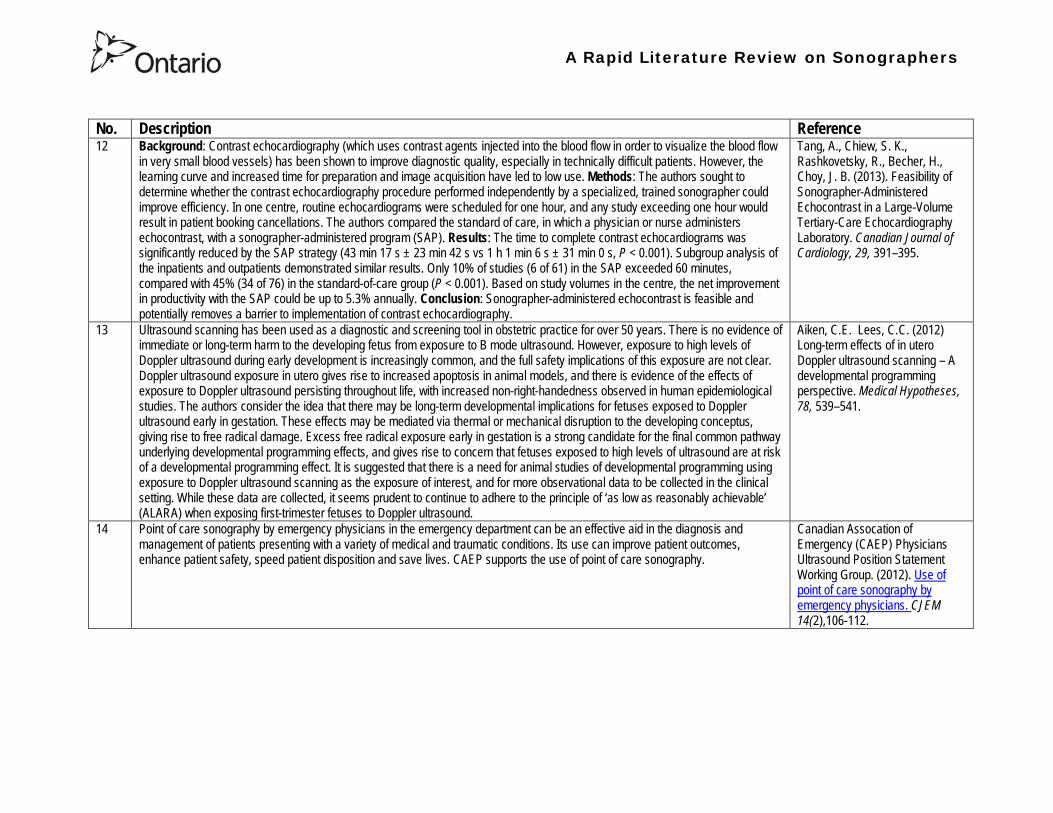
A Rapid Literature Review on Sonographers
No Description Reference 12 Background Contrast echocardiography (which uses contrast agents injected into the blood flow in order to visualize the blood flow
in very small blood vessels) has been shown to improve diagnostic quality especially in technically difficult patients However the learning curve and increased time for preparation and image acquisition have led to low use Methods The authors sought to determine whether the contrast echocardiography procedure performed independently by a specialized trained sonographer could improve efficiency In one centre routine echocardiograms were scheduled for one hour and any study exceeding one hour would result in patient booking cancellations The authors compared the standard of care in which a physician or nurse administers echocontrast with a sonographer-administered program (SAP) Results The time to complete contrast echocardiograms was significantly reduced by the SAP strategy (43 min 17 s plusmn 23 min 42 s vs 1 h 1 min 6 s plusmn 31 min 0 s P lt 0001) Subgroup analysis of the inpatients and outpatients demonstrated similar results Only 10 of studies (6 of 61) in the SAP exceeded 60 minutes compared with 45 (34 of 76) in the standard-of-care group (P lt 0001) Based on study volumes in the centre the net improvement in productivity with the SAP could be up to 53 annually Conclusion Sonographer-administered echocontrast is feasible and potentially removes a barrier to implementation of contrast echocardiography
Tang A Chiew S K Rashkovetsky R Becher H Choy J B (2013) Feasibility of Sonographer-Administered Echocontrast in a Large-Volume Tertiary-Care Echocardiography Laboratory Canadian Journal of Cardiology 29 391ndash395
13 Ultrasound scanning has been used as a diagnostic and screening tool in obstetric practice for over 50 years There is no evidence of immediate or long-term harm to the developing fetus from exposure to B mode ultrasound However exposure to high levels of Doppler ultrasound during early development is increasingly common and the full safety implications of this exposure are not clear Doppler ultrasound exposure in utero gives rise to increased apoptosis in animal models and there is evidence of the effects of exposure to Doppler ultrasound persisting throughout life with increased non-right-handedness observed in human epidemiological studies The authors consider the idea that there may be long-term developmental implications for fetuses exposed to Doppler ultrasound early in gestation These effects may be mediated via thermal or mechanical disruption to the developing conceptus giving rise to free radical damage Excess free radical exposure early in gestation is a strong candidate for the final common pathway underlying developmental programming effects and gives rise to concern that fetuses exposed to high levels of ultrasound are at risk of a developmental programming effect It is suggested that there is a need for animal studies of developmental programming using exposure to Doppler ultrasound scanning as the exposure of interest and for more observational data to be collected in the clinical setting While these data are collected it seems prudent to continue to adhere to the principle of lsquoas low as reasonably achievablersquo (ALARA) when exposing first-trimester fetuses to Doppler ultrasound
Aiken CE Lees CC (2012) Long-term effects of in utero Doppler ultrasound scanning ndash A developmental programming perspective Medical Hypotheses 78 539ndash541
14 Point of care sonography by emergency physicians in the emergency department can be an effective aid in the diagnosis and management of patients presenting with a variety of medical and traumatic conditions Its use can improve patient outcomes enhance patient safety speed patient disposition and save lives CAEP supports the use of point of care sonography
Canadian Assocation of Emergency (CAEP) Physicians Ultrasound Position Statement Working Group (2012) Use of point of care sonography by emergency physicians CJEM 14(2)106-112
A Rapid Literature Review on Sonographers
No Description Reference 15 The use of three-dimensional echocardiography (3DE ie a 3D ultrasound of the heart) during electrophysiology (EP - a branch of
cardiology that deals with the diagnosis and treatment of heart rhythm disorders) procedures is the end product of years of growth in two diverse cardiology subspecialties namely advanced cardiac imaging and the EP During the past decade progress in both fields has resulted in many important advances that have culminated in their union for a new area of growth and development Imaging advances have provided the cardiovascular specialist with enhanced cardiac volume and function data and more recently 3DE capabilities with improved spatial and temporal resolution providing unprecedented spatial relationships This latter development is valued by EP specialists in need of hitherto never required anatomic knowledge as they press forward with extraordinary expansion in their capabilities It makes sense that by combining these two rapidly growing subspecialties future capabilities in patient care may be achieved that would otherwise not be possible This paper discusses the value of 3DE during EP procedures and offers the readers insight into this novel multispecialty hybrid arena Using this model as a template it is likely that the readers may identify other areas within their practices where periprocedural advanced imaging may afford significant dividends in patient outcomes
Moukabary T Faletra F F Kronzon I Thomas W amp Sorrell V L (2012) Three-Dimensional Echocardiography in the Electrophysiology Laboratory Echocardiography 29 117-122
16 Background There has been much discussion about whether female feticide occurs in certain immigrant groups in Canada The authors examined data on live births in Ontario and compared sex ratios in different groups according to the motherrsquos country or region of birth and parity Methods The authors completed a population-based study of 766 688 singleton live births between 2002 and 2007 They used birth records provided by Ontario Vital Statistics for live births in the province between 23 and 41 weeksrsquo gestation They categorized each newborn according to the motherrsquos country or region of birth namely Canada (n = 486 599) Europe (n = 58 505) South Korea (n = 3663) China (n = 23 818) Philippines (n = 15 367) rest of East Asia (n = 18 971) Pakistan (n = 18 018) India (n = 31 978) rest of South Asia (n = 20 695) and other countries (n = 89 074) They calculated malefemale ratios and 95 confidence intervals (CIs) for all live births by these regions and stratified them by maternal parity at the time of delivery (0 1 2 or ge 3) Results Among infants of nulliparous women (women with no previous children) the malefemale ratio was about 105 overall As parity (the number of previous children) increased the ratio remained unchanged among infants of Canadian-born women In contrast the malefemale ratio was significantly higher among infants of primiparous women (women with one previous child) born in South Korea (120 95 CI 109ndash134) and India (111 95 CI 107ndash115) than among infants of Canadian-born primiparous women Among multiparous women (women with 2+ previous children) those born in India were significantly more likely than Canadian-born women to have a male infant (parity 2 ratio 136 95 CI 127ndash 146 parity ge 3 ratio 125 95 CI 109ndash143) Interpretation The study of malefemale ratios in Ontario showed that multiparous women born in India were significantly more likely than multiparous women born in Canada to have a male infant
Ray J G Henry D A Urquia M L (2012) Sex ratios among Canadian liveborn infants of mothers from different countries CMAJ184(9) E92-96
A Rapid Literature Review on Sonographers
No Description Reference 17 This letter consists of a case study that illustrates how a cross-disciplined sonographer (educated in cardiac and vascular ultrasound)
can quickly and efficiently and with great Cost-effectiveness deliver timely care to a patient in certain clinical settings The literature is comparatively sparse in referencing clinical diagnostics in cross-discipline sonographers Echocardiographers must be clinicians fully comfortable with evaluating the circulatory dynamics of the body from the left and right heart to the peripheral arteries and veins including the extracranial circulation (carotid arteries) Included in this skill set is substantive understanding of the ECG patientrsquos history and integration of cardiac and vascular findings Limitations of each diagnostic tool and how these limitations can be overcome by the strengths of the other examination are important For the dual-skilled sonographer only experience continuous education and constant clinical feedback will lead to the independent decision making that is counted on by the physician The American Registry for Diagnostic Medical Sonography offers credentials in cardiac and vascular imaging (RDCS and RVT) Cardiovascular Credentialing International offers credentials in cardiac and vascular imaging as well (registered cardiac sonographer [RCS] and registered vascular specialist [RVS]) All imaging facilities should consider merging cardiac and vascular noninvasive imaging with dual-registered clinical sonographers credentialed by one of these two agencies
Zemanek M (2012) What It Means to Be a Cardiovascular Sonographer Journal of Ultrasound in Medicine 311859-1862
18 Diagnostic medical sonography is viewed as a safe imaging modality However bioeffects research has yet to define the effects of exposure to sonography in humans Therefore sonographers must be aware of potential risks and practice ALARA (ldquoas low as reasonably achievablerdquo) routinely This research explores sonographersrsquo ability to answer basic questions about the bioeffects of sonography as well as their knowledge and use of the safety indices called the mechanical index (MI) and thermal index (TI) Members of the Society of Diagnostic Medical Sonographers in the US a southwestern sonographersrsquo society and a sonography community on Facebook 2010 were surveyed (n = 212) There is a low level of bioeffects knowledge and minimal safety practices among sonographers in this survey No significant relationship was found between sonographer knowledge of the bioeffects of sonography years in the profession or whether the sonographers monitor the MITI
Bagley J Thomas K DiGiacinto D (2011) Safety Practices of Sonographers and Their Knowledge of the Biologic Effects of Sonography Journal of Diagnostic Medical Sonography 27(6) 252ndash261
19 This article highlights trends in echocardiography Point of care ultrasound (hand-carried ultrasound) Initially point of care ultrasound will increase the use of echocardiography in the same manner that cardiac CT increased catheterizations However once the basics are learned it has the potential to be a very powerful tool in that less unnecessary studies will be ordered In the clinic small systems will be used as a lsquolsquofirst lookrsquorsquo screen and become part of the patientrsquos history only patients for whom more testing is necessary will be referred for a comprehensive study This is likely to reduce the number of Doppler studies requested The miniaturization of echo equipment will continue with improved qualityfunctionality of the lsquolsquohand-heldrsquorsquo echo machines to the extent that cordless probes will become a reality sooner rather than later Role in interventional echocardiography Sonographers will be spending much more time in the interventional and electrophysiology (EP) labs There will be more interconnection with interventional cardiologists and the volume will increase as more devices are implanted more percutaneous valves placed and more robotic surgery is performed Crosstraining and multitasking Sonographers entering the profession would be prudent to learn cardiac and vascular imaging Individuals who multitask andor cross-train will be ideal For example a sonographer who starts an IV monitors ECG stress can perform cardiac MRI and can interpret a preliminary report or a nurse who can do an echo monitor sedation and start an IV will be invaluable The sonographer should continue to grow professionally by cross training as institutions try to minimize costs and maximize throughput
McCulloch M L (2011) Thoughts regarding the future of sonography Journal of the American Society of Echocardiography 24(5) 29A-30A
A Rapid Literature Review on Sonographers
No Description Reference 20 Background Radiology reports provide specialist interpretation of images and relate these findings to the patientrsquos symptoms and
signs This study compared ultrasound (USS) reports generated by radiologists and sonographers to determine if any significant or clinically relevant differences existed Methods A retrospective analysis of 624 consecutive USS reports was carried out The reports were assessed for the presence of a lsquodisclaimerrsquo or lsquocaveatrsquo pertaining to the quality of the images and were analysed with respect to the clarity of their wording and ability to answer the clinical request Results The majority of sonographer USS reports contained a disclaimer regarding the quality of the USS images (571) compared with a smaller proportion of radiologist scans (99) (P lt 0001) Overall radiologists performed significantly better in providing a clear negative or positive diagnosis to the clinical question on the request form when compared with sonographers (885 vs 654 P lt 0001) Conclusion lsquoDisclaimerrsquo comments and lsquohedgingrsquo must be avoided in radiology reporting While feedback as to the accuracy of the radiology images is important overuse of such terms undermines the validity of the radiology report
Garcea G Mahmoud A Ong S L Rees Y Berry Det al (2010) Caveat reporting in ultrasound interpretation of surgical pathology a comparison of sonographer versus radiologist Journal of Evaluation in Clinical Practice 16 97ndash99
21 A nine-point questionnaire was constructed based on previously published work by Marsal and Sheiner et al The survey was distributed to clinical users of ultrasound practicing sonography in Hamilton (New Zealand) during July 2010 totalling 35 users consisting of sonographers sonography trainees one obstetric sonologist and one fetal medicine registrar Results Twenty sonographers returned the questionnaire which represents survey response of 61 The level of clinical experience was equally split between junior sonographers of lt 5 yearsrsquo experience (30) sonographers with 5-10 yearsrsquo experience (35) and senior sonographers of gt10 yearsrsquo experience (35) While 100 of the sonographers surveyed reported being familiar with the term Thermal Index (TI) and 90 claimed they adhered to the Action Learning Action Response Association (ALARA) principle all the time or most of the time only 15 of sonographers correctly described or defined what the term TI means Of the sonographers 80 knew where to find the TI during real-time examinations but only 35 knew which TI index to use for nuchal translucency (NT) scan and only 10 knew the expected range of TI encountered during routine obstetric examinations Of the eight sonographers who reported adhering to ALARA all the time the majority (64) could not describe or define TI In the same subgroup the majority (64) did not know which TI to monitor during a NT scan Only three of 20 (15) sonographers knew what the term TI means Of these two were junior sonographers with lt 5 yearsrsquo experience and one had 5ndash10 yearsrsquo experience Despite understanding the term TI one of the three sonographers still selected the incorrect index to monitor during a NT scan Not one of the seven senior sonographers with gt 10 years of experience could define or describe the TI In total only 10 of sonographers knew what the TI means and which TI to monitor during NT scanning
Necas M (2010) New Zealand sonographers do not outperform their European or American colleagues in the knowledge of ultrasound safety Australasian Journal of Ultrasound in Medicine13(4) 28-32
A Rapid Literature Review on Sonographers
No Description Reference 22 Musculoskeletal (MSK) ultrasound performed by non-medical practitioners is a developing practice The aim of this paper is
(1) to audit an experienced sonographerrsquos (SJ Riley one of the authors) performance after one yearrsquos independent reporting against two experienced MSK radiologists (CJ Grove amp M Chandramohan the other two authors) and (2) to establish an audit standard against which such role development might be contrived Images and reports from 250 MSK ultrasound examinations performed by a sonographer were reviewed independently by two consultant MSK radiologists The examinations were graded for discrepancy eg grade 1 ndash agree with the sonographer report grade 2 ndash minor discrepancy unlikely to alter patient care grade 3 ndash potentially significant discrepancy grade 4 ndash definite significant discrepancy likely to have adverse consequences for patient care Two of 250 (08) cases were excluded Both radiologists agreed completely with the sonographer (grade 1) in 235 of the 248 cases (948) In 13 cases there was discrepancy between the reports of SJR and the radiologists The discrepancy was grade 2 in six of the 248 cases (24) grade 3 in six of the 248 cases (24) and grade 4 in one of the 248 cases (04) In conclusion this audit shows a high level of agreement between the sonographer and the consultant MSK radiologist reporting of MSK ultrasound This level of agreement may set the standard for future quality assurance audit of sonographer MSK reporting
Riley S J Groves C J Chandramohan M (2010) Musculoskeletal ultrasound audit of sonographer reporting Ultrasound 18 36ndash40
23 Background Growing demand for ultrasound examinations and higher quality requirements motivate searching for routines combining the diagnostic accuracy of radiologist-performed examinations with the economical advantages of sonographer-performed examinations One possible approach is to use strictly standardized acquisition and documentation schemes that give the radiologist access to all relevant information after the examination Purpose To compare a recently introduced routine combining acquisition by a radiographer (more specifically a sonographer with two years experience) documentation as standardized cine-loops and review by a radiologist (lsquostandardized methodrsquo) with the formerly used routine where the diagnosis is made bedside by the radiologist (lsquotraditional methodrsquo) Material and Methods In 64 policlinic patients the kidneys (n = 27) or the gallbladder (n = 37) were examined with both the standardized and the traditional method The radiologistsrsquo findings of hydronephrosis tumors cysts echogenicity changes and cortical thickness (in the kidneys) and wall thickness concrements and polyps (in the gallbladder) were compared between the methods with respect to agreement (proportion of agreement and kappa coefficient) as well as systematic differences (McNemarrsquos test) Results The findings at the gallbladder examination showed a median agreement of 97 (86ndash100 kappa = 064ndash100) and those at the kidney examination an agreement of 90 (78ndash100 kappa = 069ndash 100) There were no significant systematic differences between the methods Conclusion The satisfactory agreement in this preliminary study indicates that the new workflow with ultrasound examinations performed by a radiographer (sonographer) and analyzed off-line by a radiologist is promising and motivates further studies
Stenman C Thorelius L Knutsson A amp Smedby Ouml (2010) Radiographer-acquired and radiologist-reviewed ultrasound examination ndash agreement with radiologistrsquos bedside evaluation Acta Radiologica 52 70ndash74
A Rapid Literature Review on Sonographers
No Description Reference 24 The troubling practice of fetal sex selection has historically been considered an Asian phenomenon However recent evidence shows
that a similar situation is emerging in North America albeit on a smaller scale The Society of Obstetricians and Gynaecologists of Canada (SOGC) has firmly stated its opposition to sex selection for non-medical reasons as well as to the use of any technology used solely for the purpose of determining fetal sex However because fetal sex may be disclosed to the parents at the time of ultrasound examination if they request this information guidance for health care professionals to assist in discouraging fetal sex selection would be useful Because no declaration of motives or reasons is required when a woman seeks a termination of pregnancy the authors suggest that health care professionals need not disclose the sex of a fetus until it has reached a gestational age at which abortion for non-medical purposes would not be possible This proposal would facilitate consistency between clinical practice and the values of Canadian citizens the SOGC the Canadian Medical Association and other professional organizations while still respecting current laws pertaining to disclosure of patient information and patientsrsquo rights to autonomous decision-making
Thiele A T amp Leier B (2010) Towards an Ethical Policy for the Prevention of Fetal Sex Selection in Canada J Obstet Gynaecol Can 32(1) 54ndash57
25 Background The extent to which increases in the use of prenatal ultrasonography over time reflect changes in maternal risk (ie an increase in high-risk pregnancies over time) is unknown In this population-based study the authors examined the use of prenatal ultrasonography from 1996 to 2006 in Ontario Methods With fiscal year 199697 as the baseline the authors evaluated the relative risk (RR) and 95 confidence interval (CI) for the change in rates of ultrasonography for each subsequent year The RR was adjusted for maternal age income rural residence maternal comorbidities receipt of genetics consultation or amniocentesis mdash all in the index pregnancy mdash and history of complications in a prior pregnancy Results The study sample consisted of 1 399 389 singleton deliveries The rate of prenatal ultrasonography increased from 2055 per 1000 pregnancies in 1996 to 3264 per 1000 in 2006 (adjusted RR 155 95 CI 154ndash155) The rate increased among both women with low-risk pregnancies (adjusted RR 154 95 CI 153ndash155) and those with high-risk pregnancies (adjusted RR 155 95 CI 154ndash157) The proportion of pregnancies with at least four ultrasound examinations in the second or third trimesters rose from 64 in 1996 to 187 in 2006 (adjusted RR 268 95 CI 261ndash274) Paradoxically this increase was more pronounced among low-risk pregnancies (adjusted RR 292 95 CI 283ndash301) than among high-risk pregnancies (adjusted RR 225 95 CI 216ndash235) Interpretation Substantial increases in the use of prenatal ultrasonography over the past decade do not appear to reflect changes in maternal risk Nearly one in five women now undergo four or more ultrasound examinations during the second and third trimesters Efforts to promote more appropriate use of prenatal ultrasonography for singleton pregnancies appear warranted
You J J Alter D A Stukel T A McDonald S D Laupacis A et al (2010) Proliferation of prenatal ultrasonography CMAJ 182(2) 143-151
A Rapid Literature Review on Sonographers
No Description Reference 26 Aims The aim of this study was to assess the cost-effectiveness of using certified sonographers and miniaturized echocardiography
systems to perform echocardiograms at bedside in comparison to moving inpatients from the admission department to the echocardiography laboratory (echo-lab) Methods and Results From 26 September 2005 to 27 October 2005 112 patients admitted in six hospital wards in Italy connected through a 100 Mbit LAN to the echo-lab were scanned within the admission ward by sonographers using a miniaturized echo system Logistical data were collected and results were compared with those obtained from 194 consecutive patients coming from the same wards and studied in the echo-lab with high-end machines between 8 March 2005 and 15 April 2005 Performing echocardiograms in the admission department avoided long waiting time of the inpatients in the echo-lab before and after the study increased the percentage of patients studied within 3 and 5 days from request (88 vs 77 and 100 vs 95 respectively P = 003) increased both sonographer (by 339 P lt 0001) and echo-lab productivity (by 41 P lt 0001) and reduced costs of echocardiograms by 29 Conclusion Implementation of digital echocardiography certified sonographers and a miniaturized echo system allowed improvement of the cost-effectiveness of the service provided by the echo-lab for inpatients and avoided patientsrsquo discomfort derived from prolonged waiting time before and after the exam
Badano L Gaetano N Stacul S Gianfagna P Pericoli M et al (2009) Improved workflow sonographer productivity and cost-effectiveness of echocardiographic service for inpatients by using miniaturized systems European Journal of Echocardiography 10 537ndash542
27 The present report is a proposal from the American Society of Echocardiography Advanced Practice Task Force that identifies the potential of cardiac sonographers to achieve the Advanced Cardiovasculat Sonographer (ACS) level According to this report echocardiographic examinations require a well-trained and competent sonographer to obtain proper anatomic and physiologic data to establish an accurate diagnosis for clinical decision-making and patient management Although the formal education and training of cardiovascular sonographers are evolving many entry-level and staff sonographers may not have sufficient practical or clinical knowledge of the necessary components of the echocardiographic study for the individual patientrsquos clinical presentation In many clinical settings echocardiograms are read after the patient has left the laboratory Thus there is a role for a sonographer who can practice at an advanced level in a cardiovascular ultrasound laboratory to ensure a proper echocardiographic examination is performed on every patient In this setting an Advanced Cardiovascular Sonographer (ACS) would be able to review the indication for and quality of the examination If additional images were needed the ACS would assist the sonographer in obtaining these images which would lead to the performance of a complete and fully diagnostic examination before the patient had left the echocardiography laboratory In clinical practice the quality of the examinations performed would improve advancements in echocardiographic methods could be taught and incorporated into daily practice and patients would be better served
Mitchell C Miller F A Bierig S M Bremer M L Ehler D et al (2009) Advanced Cardiovascular Sonographer A Proposal of the American Society of Echocardiography Advanced Practice Sonographer Task Force J Am Soc Echocardiogr 22 1409-13
A Rapid Literature Review on Sonographers
No Description Reference 28 Sonographer practitioner development involves the expansion and extension of the sonographer role to include reporting on
ultrasound examinations Australian sonographers have not seen the same degree of role extension and expansion as their counterparts in the United Kingdom despite increasing levels of discussion regarding sonographer practitioner development The aim of this study was to determine if Australian sonographers want to extend their professional role and what they consider are the important issues associated with role extension This paper reports on qualitative data derived from a survey of Australian sonographers and investigates if Australian sonographers are interested in extending and expanding their professional role and responsibilities and if they do what might be necessary or desirable from a professional point of view for this change to occur A survey was mailed to all members of the Australian Sonographers Association (ASA) in October 2006 The 31-item survey included 28 closed-ended and three opened-ended items to provide both quantitative and qualitative data The quantitative data will be reported separately Qualitative data was derived from responses to the opened-ended questions which asked respondents to elaborate on their attitudes and feelings about role extension and development Analysis used Nvivo7 software to aid in uncovering common themes from the qualitative data The analysis focused on the reported incentives or motivations for becoming a sonographer practitioner as well as disincentives or perceived hurdles that would discourage respondents from becoming sonographer practitioners The three most reported incentives or motivations for becoming a sonographer practitioner were professional recognition remuneration and increased knowledge The three most commonly reported disincentives or perceived hurdles that would discourage respondents from becoming sonographer practitioners were legal issues insurance and further study
McGregor R OrsquoLoughlin K Cox J Clarke J Snowden A (2009) Sonographer practitioner development in Australia Qualitative analysis of an Australian sonographersrsquo survey Radiography15 313-319
29 Introduction This study describes the effect of introducing a cardiac sonographer led follow up clinic within a hospital cardiology department for patients with stable heart valve disease The two years before and after the instigation of the valve clinic were audited Materials and methods The clinic was conducted in a single centre and undertaken in the cardiology department of a district general hospital A total of 382 patients with 397 clinically significant valve lesions but for whom surgery was not yet indicated but follow up required were seen in a cardiac sonographer run clinics These patients no longer attended a medical follow up clinic unless there was clinical or echocardiographic deterioration Effectiveness was judged by the percentage treated according to current best practice guidelines the number of echocardiograms performed and the number of hospital outpatient visits attended In addition mortality data for the subjects in the clinic was collected Results The proportion followed up according to best practice guidelines rose from 157 (41) to 354 (92) (p lt 001) The total number of echocardiograms performed fell from 807 to 550 Total number of outpatient visits fell from 998 to 31 A total of 11 patients died in the two year study period none from progressive valve disease Discussion This study demonstrates that a protocol driven sonographer led heart valve disease follow up clinic significantly improved the quality of follow up while bringing about a major reduction in outpatients visits without compromising patient safety
Taggu W Topham A Hart L Carr-White G Sulke N et al (2009) A cardiac sonographer led follow up clinic for heart valve disease International Journal of Cardiology 132 240 ndash243
30 Role development for imaging allied health professionals has progressed significantly over the last three decades with many major developments pioneered in the field of ultrasound This paper reviews the developing role of the radiographer in ultrasound (in some countries such as the UK sonographers are considered a type of radiographer) and some of the clinical professional and educational implications of such development As the published evidence base underpinning the latest developments is scarce several contemporary case study examples which have been presented verbally at study days attended by the authors have been included In conclusion sonographer role development is considered within the professional contexts of extended role advanced and consultant practice
Hart A amp Dixon A (2008) Sonographer Role Extension and Career Development a Review of the Evidence Ultrasound 16(1) 31-35
A Rapid Literature Review on Sonographers
No Description Reference 31 Objective The main goal of this study was to determine end usersrsquo knowledge regarding safety aspects of diagnostic ultrasound
during pregnancy End usersrsquo attitudes toward the use of ultrasound in low-risk pregnancies were also assessed Methods A questionnaire was distributed to ultrasound end users attending review courses and hospital grand rounds between April and June 2006 in Chicago Results One hundred thirty end users completed the questionnaires (63 response rate) Sixty-three percent were physicians (n = 84) most of them obstetricians (817) About 18 of participants routinely performed Doppler ultrasound examinations during the first trimester Fifty percent of end users thought that the number of ultrasound examinations in low-risk pregnancy should be limited to 1 to 3 (mean plusmn SD 26 plusmn 09) Almost 70 disapproved of ldquokeepsakeentertainmentrdquo ultrasound Although 322 of the participants were familiar with the term thermal index only 177 actually gave the correct answer to the question on the nature of the thermal index About 22 were familiar with the term mechanical index but only 38 described it properly Almost 80 of end users did not know where to find the acoustic indices Only 208 were aware that they are displayed on the sonographic monitor during the examinations End users with higher knowledge of safety issues thought that there should be limitations on the number of ultrasound examinations in low-risk pregnancies (odds ratio 33 95 confidence interval 11ndash100 P = 028) Likewise these end users were more likely to respond that ultrasound might have adverse effects during pregnancy (odds ratio 32 95 confidence interval 11ndash125 P = 045) Conclusions Ultrasound end users are poorly informed regarding safety issues during pregnancy Further efforts in the realm of education and training are needed to improve end user knowledge about the acoustic output of the machines and safety issues
Sheiner E Shoham-Vardi I Abramowicz J S (2007) What Do Clinical Users Know Regarding Safety of Ultrasound During Pregnancy J Ultrasound Med 26 319ndash325
A Rapid Literature Review on Sonographers
No Description Reference 32 In an attempt to survey the knowledge among ultrasound users of some safety aspects of diagnostic ultrasound a questionnaire was
distributed to professionals using ultrasound for fetal examinations The questionnaire was answered by the participants and faculty members of six postgraduate courses in obstetric ultrasound Doppler ultrasound and fetal echocardiography given in Sweden Norway and Austria in 2003 Furthermore in Sweden the questionnaire was distributed to the staff meeting participants of two major obstetric ultrasound units and of one unit for fetal echocardiography and at a meeting of the Working Group for Ultrasound of the Swedish Society of Obstetrics and Gynecology In total 199 questionnaires were anonymously answered by doctors (n = 145) sonographers (n = 22) and midwives trained in ultrasound (n = 32) All of them were using diagnostic ultrasound on a daily or weekly basis The respondents were from nine European countries In none of the countries is formal certification for the use of diagnostic ultrasound in pregnancy or otherwise required The doctors who answered the questionnaire were specialists in obstetrics and gynecology (including some of the internationally recognized experts in fetal ultrasound) pediatric cardiology radiology or clinical physiology The respondents were not informed about the questionnaire in advance and they had a limited time to answer the 16 questions typically 10ndash15 min The questions concerned explanation of the safety indices and their relation to various ultrasound modes ie real-time B-mode M-mode three-dimensional (3D) spectral Doppler color and power Doppler ultrasound The respondents were also asked which ultrasound system they used most often and where on the system the information on thermal index (TI) and mechanical (MI) was displayed (eg in the top right corner of the screen in a separate window etc) Furthermore the respondents had to indicate how they adjust the output energy level on their own machine For all questions there was the optional answer lsquoDonrsquot knowrsquo Results About one-third of the ultrasound experts were able to define the abbreviations TI and MI For the subclasses of the TI the correct answer was found in only 3-8 of cases Of those who knew what the TI and MI meant only two-thirds and one-third of respondents respectively were able to give a simple explanation of the indices In 49 of questionnaires the Doppler modes were correctly ranked as generally producing higher ultrasound exposure than the imaging modes (B-mode 3D) Some 28 of the respondents correctly indicated where on their own machine the information on safety indices is displayed However not all of them knew how to control the output energy level For all items in the questionnaire there were no significant differences in the results between the three categories of respondents (ie physicians sonographers and midwives) The results of the questionnaire were anything but encouraging and they indicated that the users who are supposed to be responsible for controlling exposure of the fetus to ultrasound had a very poor knowledge of the basic safety aspects of ultrasound
Maršaacutel K (2005) The output display standard has it missed its target Ultrasound Obstet Gynecol 25 211ndash214
33 Purpose This study was performed to evaluate the accuracy of upper abdominal ultrasound (US) scanning performed by sonographers in a district general hospital to identify potential areas of weakness and to make recommendations to improve the service Materials and Methods Upper abdominal US examinations performed and reported by sonographers over a 4-week period were retrospectively reviewed The accuracy of the imaging findings and reports were assessed against other imaging surgical histological or laboratory findings and against clinical outcome Results A heterogenous group of 104 patients were included in the study 62 of whom had an US abnormality Errors of scanning or interpretation were identified in 10 patients (96) of whom five (48) were felt to be potentially significant Conclusions The sonographersrsquo accuracy in reporting upper abdominal US scans was 90 However on the basis of this study the authors have implemented specific recommendations to improve the quality of the service
Dongola N Guy R Ward S amp Giles J A (2003) Can sonographers offer an accurate upper abdominal ultrasound service in a district general hospital Radiography 9 29ndash33
A Rapid Literature Review on Sonographers
No Description Reference 34 Aim To compare the accuracy of radiographers and radiologists in routine abdominal ultrasound Materials and Methods One
hundred consecutive patients attending for routine abdominal ultrasound were included Each patient was examined by both a radiographer and radiologist Both operators noted their fndings and wrote a concluding report without conferring Reports were compared Where there was disagreement the patient was either re-examined by another radiologist or had further investigation Results Of 100 patients 52 were men and 48 were women The age range was 19plusmn88 years (median 52 years) Thirty-seven patients had renal tract ultrasound one had an aortic ultrasound and 62 had general upper abdominal ultrasound In 44 cases both operators reported the examination as normal In 49 cases both operators reported the examinations as abnormal and there was complete agreement between the operators In seven cases there was not complete agreement between operators Three of these disagreements were considered minor and four major In three of the seven cases the radiographer was correct and in four the radiologist was correct Conclusion Experienced radiographers and radiologists are highly accurate in performing and interpreting routine abdominal sonography Both operators missed a small minority of abnormalities There was no statistically significant difference in the accuracy of radiographers and radiologist
Leslie A Lockyer H amp Virjee J P (2000) Who Should be Performing Routine Abdominal Ultrasound A Prospective Double-Blind Study Comparing the Accuracy of Radiologist and Radiographer Clinical Radiology 55 606-609
Grey Literature 35 The ARRT The American Registry of Radiologic Technologists tests certifies and annually registers more than a quarter-of-a-million
radiologic technologists to promote high standards of patient care They award the Registered Technologist or RT designation to individuals who have completed the prescribed classroom and clinical education passed the appropriate exam and met the ethics requirements And they ensure their continuing education and ongoing compliance with their high standards by requiring annual registration of their certificate Certification by ARRT is voluntary not mandatory But employers state licensing agencies and federal regulators view ARRTrsquos credential as an indicator that a person has met recognized national standards for medical imaging and radiation therapy professionals How Are Radiologic Technologists Deemed Qualified First comes initial certification Certification is the process of initially recognizing individuals who have satisfied certain standards within a profession A person is certified by ARRT after completing an approved educational program complying with the ethics standards and passing a certification exam Then comes annual registration Registration is the yearly procedure required to maintain certification ARRT registrants are those who having already fulfilled the requirements for initial certification continue to meet the requirements for annual registration The ARRT annually registers the certificates of individuals who meet the following three criteria agreeing to comply with the ARRT Rules and Regulations continuing to comply with the ARRT Standards of Ethics and meeting our continuing education requirements Only technologists who are currently registered (which means that they have renewed within the past year) may designate themselves as ARRT Registered Technologists and use the initials RT after their names Federal laws regulate personnel qualifications in some modalities (mammography for example) but they accredit the health care facility rather than licensing the individual radiologic technologist who is providing patient care And while many states have laws regulating who can perform radiologic procedures they are typically based on ARRT certification Some employers and state agencies may require ARRT certification but not necessarily current registration Unless they require current registration by ARRT employers cannot assume that the person has complied with the ethics requirements or the continuing education requirements
American Registry of Radiologic Technologists (ARRT) (2013a) Patient Page Accessed April 2013
A Rapid Literature Review on Sonographers
No Description Reference 36 This document outlines certification requirements for sonography There are two pathways (primary and post-primary) For the
primary pathway it notes that sonography educational programs must be accredited by a mechanism acceptable to the ARRT ARRT generally recognizes only accreditation agencies that are recognized by the Council for Higher Education Accreditation andor United States Department of Education
American Registry of Radiologic Technologists (ARRT) (2013b) Sonography Certification Accessed April 2013
A Rapid Literature Review on Sonographers
No Description Reference 37 In Australia sonographers and student sonographers are required to be accredited by Australian Sonographer Accreditation Registry
(ASAR) in order to practise This requirement is contained within the Australian Governmentrsquos Diagnostic Imaging Accreditation Scheme standards and Medicare legislation Accreditation of sonographers is linked only to achieving - or in the case of students studying towards - an accredited entry-level qualification completion of continuing professional development and payment of an annual fee to ASAR The ASA contends that the lack of a requirement to ensure each sonographer is lsquofit to practisersquo is an unacceptable gap in current regulation undermining efforts to ensure the quality of sonographic services and failing to protect patients from harm The ASA supports and advocates for nationally-enforceable regulation of sonographers to meet our patientrsquos expectations National registration The National Registration and Accreditation Scheme (NRAS) for health workers commenced on 1 July 2010 The ten professions which comprised the initial intake into this scheme were those that had registration requirements in all Australian states or territories as well as those in which legislated regulation is deemed to be in the public interest An additional four professions - including medical radiation practitioners which are defined as radiographers radiation therapists and nuclear medicine technologists - will be included in the NRAS commencing on 1 July 2012 As sonography did not have registration in any states or territories prior to the commencement of NRAS the profession has not been in a position to negotiate entry into NRAS In early August 2011 the ASA lodged a submission with the Health Workforce Principal Committee requesting a profession-specific Sonography Board of Australia be formed under NRAS This submission was prepared based on information that consideration of professions which were unregistered would commence More recently the Australian Health Workforce Ministerial Council (AHWMC) issued a communiqueacute stating in part lsquoMinisters have commissioned work on unregistered professions and future directions for national registration Given this Ministers agreed to defer consideration of the inclusion of additional professions into the NRAS Ministers agreed that it would be premature to consider further applications for the scheme until this work has been completed other than paramedicsrsquo Options for unregistered health practitioners including sonographers Policy work is continuing at a federal level to establish an alternative regulatory framework The National Alliance for Self-Regulating Health Professions (NASRHP) consisting of the peak professional associations of eight self-regulating health professions including the ASA representing sonography released a proposal lsquoHarnessing self-regulation to support safety and quality in healthcare deliveryrsquo in March 2012 This proposal concluded that the framework that regulates all health practitioners should be expanded beyond NRAS to include
bull authorised self-regulating professions via a health professions panel bull negative licensing to withdraw the ability to practise from registered and self-regulated practitioners who do not meet the
regulatory requirements or unregulated practitioners due to poor practice As with all advocacy initiatives consideration by the federal government of the introduction of the NASRHPrsquos proposal may be prolonged and the outcomes may not match the expectations of NASRHPrsquos member professional associations including the ASA The ASA will continue both independently and collaboratively to lobby on behalf of our members and the public for the introduction of a timely and equitable solution to health practitioner regulation In the interim as there is no regulatory model with the rigour the ASA believes is required for regulation of sonographers and other health practitioners other than NRAS the ASA continues to support its Position Statement published in early 2010
Australian Sonographer Association (ASA) (2013) Sonographer Regulation Accessed April 2013
A Rapid Literature Review on Sonographers
No Description Reference 38 This publication contains information about Cardiovascular Credentialing International (CCI) It also provides the necessary
information and references concerning the process by which candidates may earn certificate or registry-level credentials CCI is a not-for-profit corporation established for the purpose of administering credentialing examinations as an independent credentialing agency CCI began credentialing cardiovascular professionals in 1968 CCI offers eight credentials which when earned demonstrate that the registrant holds fundamental knowledge in the particular cardiovascular specialty These include RCCS ndash Registered Congenital Cardiac Sonographer RCS ndash Registered Cardiac Sonographer and RPhS ndash Registered Phlebology Sonographer
Cardiovascular Credentialing International (2013) 2013 Examination Application amp Overview Accessed April 2013
39 Oregon Governor Signs Sonographer Licensure Legislation On July 28 2009 Oregon Governor Ted Kulongoski signed a sonographer licensure bill (HB 2245) into law The new law makes Oregon only the second state in the United States to require licensure of sonographers The Society of Diagnostic Medical Sonographers (SDMS) has worked closely with other sonography-related organizations and Oregon sonographers to help craft the language in new law The law renames and restructures the Oregon Board of Radiologic Technologists to the Oregon Board of Medical Imaging (OBMI) and ensures representation of sonographers on the Board The sonographer licensure law requires Oregon sonographers to become licensed by OBMI effective July 1 2010 Beginning on January 1 2014 all sonography licensees would be required to hold a national sonography certificationcredential (or be currently enrolled as a student) While the Oregon bill is far more detailed than the sonographer licensure law that passed in New Mexico earlier this year additional administrative rulesregulations will be developed over the next year to fully implement the new law SDMS will be continue to monitor developments regarding sonographer licensure in other states
Society of Diagnostic Medical Sonographers (SDMS) (2013a) News Accessed April 2013
A Rapid Literature Review on Sonographers
No Description Reference 40 On April 6 2009 New Mexico (NM) Governor Bill Richardson signed the historic bill that for the first time in the United States
requires licensure of sonographers The bill adds sonographers (and MRI technologists) to the list of medical imaging professionals licensed by the State of New Mexico Until now anyone could perform sonograms in New Mexico Prior to the passage of the New Mexico licensure law virtually all other health care providers and professionals had to be licensed As a result the public is often surprised to learn that the person performing a medical sonogram does not have any state licensure or national certification requirements The responsibility for administering the NM sonographer licensure program would rest with the New Mexico Environment Department Although this was not the first choice in most sonographersrsquo minds the Environment Department currently manages the radiologic technology licensure program so the administrative infrastructure is already in place By ensuring the creation of an advisory council that includes representation by each medical imaging and radiation therapy modality regulated the bill helps ensure that the Departmentrsquos other areas of emphasis do not detract from sonographer licensure Because sonographers have never had to be licensed before they frequently do not see or understand the distinction between state licensure and national certification National certifications or credentials issued by voluntary organizations such as the American Registry for Diagnostic Medical Sonography (ARDMS) Cardiovascular Credentialing International (CCI) and the American Registry of Radiologic Technologists (ARRT) have little legal weight The certification process and resulting credential are valuable tools in ensuring sonographers have met minimum education and knowledge requirements but do not really regulate anyonersquos ability to perform sonography In an ideal world no additional regulation of sonography would be required However as everyone knows more and more people are taking weekend courses and buying ultrasound equipment without understanding the ramifications of using ultrasound technology (eg ALARA) Under the bill passed by the New Mexico legislature national credentialscertifications should provide evidence of meeting the statersquos standards All sonographers who are performing these critical medical imaging services in New Mexico will be required to meet national certification standards (ie have certifications or credentials issued by ARDMS CCI or ARRT) As a result there would be no additional exams in New Mexico beyond the national certification exams required by ARDMS CCI or ARRT The bill also requires the state to recognize the continuing medical education completed for national certification renewal The most visible impact on New Mexico sonographers will be a license fee However the legislation caps the license fee at $100 per two-year license In addition only the New Mexico Legislature can change this cap
SDMS (2013b) New Mexico Sonographer Licensure Law Signed Accessed April 2013
A Rapid Literature Review on Sonographers
No Description Reference 41 Preamble The purpose of this document is to define the Scope of Practice for Diagnostic Ultrasound Professionals in the US and to
specify their roles as members of the health care team acting in the best interest of the patient This scope of practice is a living document that will evolve as the technology expands Definition of the Profession The Diagnostic Ultrasound Profession is a multi-specialty field comprised of Diagnostic Medical Sonography (with subspecialties in abdominal neurologic obstetricalgynecologic and ophthalmic ultrasound) Diagnostic Cardiac Sonography (with subspecialties in adult and pediatric echocardiography) Vascular Technology and other emerging fields These diverse specialties are distinguished by their use of diagnostic medical ultrasound as a primary technology in their daily work Certification is considered the standard of practice in ultrasound Individuals who are not yet certified should reference the Scope as a professional model and strive to become certified Scope of Practice of the Profession The Diagnostic Ultrasound Professional is an individual qualified by professional credentialing and academic and clinical experience to provide diagnostic patient care services using ultrasound and related diagnostic procedures The scope of practice of the Diagnostic Ultrasound Professional includes those procedures acts and processes permitted by law for which the individual has received education and clinical experience and in which heshe has demonstrated competency Diagnostic Ultrasound Professionals
bull Perform patient assessments bull Acquire and analyze data obtained using ultrasound and related diagnostic technologies bull Provide a summary of findings to the physician to aid in patient diagnosis and management bull Use independent judgment and systematic problem solving methods to produce high quality diagnostic information and
optimize patient care
Society of Diagnostic Medical Sonography [SDMS] (2013c) Scope of Practice for the Diagnostic Ultrasound Professional Accessed April 2013
42 This website notes that in the US most employers in the US prefer to hire sonographers who have professional certification A sonographer can get certification and passing an exam Most exams relate to the specialty that the sonographer is most interested inmdashfor example an exam to become certified in abdominal sonography A few states require diagnostic medical sonographers to be licensed Typically professional certifications required for licensure other requirements vary by state Sonographers must take continuing education to keep their certification current
Bureau of Labor Statistics US Department of Labor (2012) How to become a Diagnostic Medical Sonographer Occupational Outlook Handbook 2012-13 Edition Accessed April 2013
A Rapid Literature Review on Sonographers
No Description Reference 43 The American Registry for Diagnostic Medical Sonography (ARDMS) is an independent not-for-profit organization founded in 1975
that administers examinations and awards the following credentials bull RDMSreg ndash Registered Diagnostic Medical Sonographerreg bull RDCSreg ndash Registered Diagnostic Cardiac Sonographerreg bull RVTreg ndash Registered Vascular Technologistreg bull RPVIreg ndash Registered Physician in Vascular Interpretationreg bull RMSKtrade - Registered in Musculoskeletaltrade sonography
ARDMS is the globally recognized standard of excellence in sonography and has certified more than 75000 individuals ARDMS is composed of three key groups
bull Board of Directors ndash Sonographers vascular technologists physicians research scientists and a member of the public are volunteer members of the board that determines the direction and policies of our organization
bull Examination Development Task Forces ndash A group of sonographers vascular technologists physicians and scientists volunteer their time and expertise to produce our examinations
bull Professional Staff ndash A paid highly-skilled staff administers the programs and assists applicants and registrants Credentials awarded by ARDMS document personal achievement of recognized professional standards and are widely accepted in the medical community by sonography and vascular professional organizations However ARDMS does not validate the day-to-day job performance of any sonography professional ARDMS has earned the prestigious ANSI-ISO 17024 accreditation for certifying bodies from the International Organization for Standardization (ISO) Accreditation is granted through the American National Standards Institute (ANSI) ARDMS has also earned accreditation with the National Commission for Certifying Agencies (NCCA) The NCCA is the accrediting arm of the National Organization for Competency Assurance (NOCA) Established in 1977 as a nonprofit organization NOCA is a leader in establishing quality standards for certifying organizations
American Registry for Diagnostic Medical Sonography (ARDMS) (2012a) Overview of ARDMS Accessed April 2013
A Rapid Literature Review on Sonographers
No Description Reference 44 ARDMS is developing a program to assure that Registrants continue to meet the basic competency requirements of core clinical skills
in present day practice bull Each Registrant who holds an active ARDMS certification as of December 31 2011 will be assigned the 2012 ndash 2021 10-
year recertification assessment period bull The 10-year period for new Registrants will begin the year following the attainment of the credential bull If a Registrant earns a new credential or specialty after January 1 2012 then that credentialspecialty will have a separate
10-year recertification assessment period that will begin the year following the attainment of the credentialspecialty ARDMS will develop tools and resources to assist Registrants in tracking and meeting the recertification assessment program requirements
bull Registrants who do not wish to take a recertification assessment in a specific credentialspecialty may voluntarily relinquish the credentialspecialty
As an ARDMS Registrant individuals are committing to demonstrate the highest level of competency in the modality of sonography by meeting and maintaining the standards set by the ARDMS The pathway of maintenance your certification is comprised of Paying the ARDMS annual renewal fee each year Each year ARDMS assesses Registrants an annual renewal fee By paying your annual renewal fee you are contributing to the ARDMS mission of promoting quality care and patient safety through the certification and continuing competency of ultrasound professionals In order to maintain your credentials you must pay the annual renewal fee of $75 USD or $100 USD (for RPVI and MSK Registrants with MDDODPM) designation on or before December 31 of each year in order to stay active for the following year Attesting to ARDMS rules and policies Each year when you pay your annual renewal fee you are attesting that you are in compliance with all ARDMS rules and discipline policies and are in good standing with the ARDMS Earning a minimum of 30 ARDMS-accepted CMEs during your three-year CME period ARDMS verifies its Registrants compliance with continuing medical education (CME) requirements using an audit process Earning your CMEs is an integral component of maintaining your ARDMS credential(s) and demonstrates your ongoing commitment to lifelong learning RecertificationContinued Commitment to Excellence Following the attainment of the ARDMS credential Registrants are assigned a 10-year recertification period the year after the credential is earned ARDMS is committed to developing a recertification program that is easy for Registrants to comply with
American Registry for Diagnostic Medical Sonography (ARDMS) (2012b) Recertification General Information
A Rapid Literature Review on Sonographers
No Description Reference 45 This document reviews Sonography Principles and Instrumentation (SPI) Examination Requirements offered through ARDMS in
order to achieve sonography certification and the various prerequisites needed before taking the exam For example one prerequisite entails (1) Education Graduate of a program accredited by an agency recognized by the Council for Higher Education Accreditation (CHEA) United States Department of Education (USDOE) or Canadian Medical Association (CMA) that specifically conducts programmatic accreditation for diagnostic medical sonographydiagnostic cardiac sonographyvascular technology Currently the only organizations that offer programmatic accreditation under the aforementioned associations are the Commission on Accreditation of Allied Health Education Programs (CAAHEP) and the Canadian Medical Association (CMA) (2) Required Clinical UltrasoundVascular Experience No additional experience is required (3) Documentation Required with Application 1) Copy of diploma from ultrasoundvascular program or an official transcript indicating the date the degree was conferred 2) Original letter signed by program director andor medical director indicating date of graduation or successful completion of the program4 Program directors must use the mandatory formatted sample letter available on ARDMSorg sampleletters 3) The CV form is not required if the application is submitted and received in the ARDMS office within one year after successful completion of the program Otherwise an original signed and completed CV form for each appropriate specialty area(s) must be submitted 4) Photocopy of a non-expired government issued photo identification with signature the name on the identification must exactly match the name under which you are applying for ARDMS examination
American Registry for Diagnostic Medical Sonography (ARDMS) (2012c) SPI Requirement and General Prerequisites Accessed April 2013
46 This page notes the five credentials offered by the ARDMS and the corresponding exams and specialty exams required bull RDMS requires the SPI exam (sonography principles and instrumentation) and a specialty exam in abdomen breast fetal
echocardiography neurosonolgoy or obstetricsgynecology bull RDCS requires the SPI exam and a speciality exam in adult echocardiography fetal echocardiography pediatric
echocardiography bull RVT requires the SPI and a speciality exam in vascular technology bull RMSK requires the musculoskeletal sonography exam bull RPVI requires the physiciansrsquo vascular interpretation exam
American Registry for Diagnostic Medical Sonography (ARDMS) (2012d) Credentials and Examinations Accessed April 2013
A Rapid Literature Review on Sonographers
No Description Reference 47 This article summarizes some of the many trends in fetal pediatric and adult echocardiography that SDMS members are excited
about Fetal Echocardiography Sonography has been used since the mid-1980s to help identify congenital heart defects during the second trimester The ldquobasicrdquo examination of the fetal heart involves a four-chamber view ndash evaluation of the right and left atrial and ventricular chambers and their respective valves In the past several years there has been a movement in the obstetric ultrasound community to encourage obstetric sonographers to perform the ldquoextended basicrdquo exam which includes the cardiac outflow tracts The outflow tracts consist of the main pulmonary artery exiting the right ventricle and the aorta exiting the left ventricle The fetal echocardiography community also is abuzz over three-dimensional (3D) and four-dimensional (4D) ultrasound and the potential of this technology to significantly improve the evaluation of the fetal heart Spatiotemporal Image Correlation (STIC) a 4D application allows for evaluation and manipulation of the heart in real-time Allen says Todayrsquos ultrasound equipment allows sonographers to gather a volume of data from which they can create detailed 3D images of the tiny fetal heart which is constantly in motion With computer manipulation of the data sets the images also can be viewed in 4D allowing physicians to see the entire fetal heart cycle from start to finish Pediatric Echocardiography One of the most significant advances in pediatric echo over the last decade is moving to a digital format The advent of pediatric transesophageal (TEE) probes also has been a big advance for pediatric echo Transesophageal probes provide an unimpeded sonographic visualization of the intracardiac structures Theyrsquore useful in visualizing very fine valvular abnormalities the thoracic aorta vegetations and thrombus in the atrial appendage It is also useful in detecting subtle anatomic variations seen in congenital heart disease Pediatric-sized TEE probes make the exams possible In the past it wasnrsquot possible because the adult probe was just too big to put down the babyrsquos or childrsquos esophagus Now the probe is small enough to successfully manipulate it in children and get superb images Because the new sonographic imaging equipment and better surgical techniques allow clinicians to detect and repair more cardiac abnormalities earlier patients are living longer Adult Echocardiography The adult echo world is abuzz about 2D and 3D speckle tracking relatively new techniques for the assessment of heart function and strain imaging Speckle tracking evaluates both regional and global function by measuring the deformation of the heart muscle
Orenstein B W (2010) Current Trends in Echocardiography SDMS New Wave February 1-5 Accessed April 2013
48 This document describes the requirements for admission to the Register of Accredited Medical Sonographer in Australia Requirements include Continuation of Accreditation In order to remain on the Register an accredited Sonographer must satisfy the ASAR continuing professional development (CPD) requirements and pay the prescribed annual fee Student Sonographers apart from paying the prescribed annual fee should provide evidence of their student status every year when they renew their ASAR accreditation
Australian Sonographer Accreditation Registry (ASAR) (no date A) Sonographer Accreditation Accessed April 2013
A Rapid Literature Review on Sonographers
No Description Reference 49 ASARrsquos functions
1 Accredit sonography education programs within Australia 2 Maintain a register of Accredited Sonographers and Accredited Student Sonographers 3 Establish minimum standards of Continuing Professional Development (CPD) and to monitor the CPD requirements for
Sonographers to remain on the register 4 To support activities that advance the profession of sonography 5 To provide registration information about Sonographers to Medicare Australia 6 To provide public access to the name registration number State or Territory current type of accreditation and area of
specialty for active Sonographers ASARrsquos Role ASAR is not a registration board ASAR does not have the authority to remove a Sonographer from the Registry due to professional misconduct nor the ability to prevent a Sonographer from practicing ASAR is only able to remove a Sonographer from the Registry if they
bull fail to meet CPD requirements and or bull fail to pay annual fees
ASAR (no date B) About ASAR Accessed April 2013
50 Regulatory bodies A Regulatory Body maintains a register of individuals who have achieved the required qualification of that profession to practice and can evidence continuing competence at the required level For a doctor to practice in the UK he or she must be registered with the General Medical Council (GMC) This is the regulatory body for medical practitioners The majority of sonographers who are employed in the UK come from a background of radiography or midwifery To practice radiography in the UK a qualified radiographer must be registered with the Health Professions Council (HPC) Similarly a midwife must be registered with the National Midwifery Council to practice as a midwife Sonographer registration in the UK There are a number of professions allied to medicine that are regulated by the HPC for example radiography physiotherapy However sonography is not recognised as a profession by the HPC and therefore there is no mechanism whereby an individual can register with the HPC as a sonographer or be regulated by them as a sonographer It is a common misunderstanding that sonographers have to be registered with the HPC to practice This comes from the fact that most sonographers are also qualified radiographers and maintain their registration with the HPC because they wish to remain on the HPC register as a radiographer Professional bodies An individual can be a member of a Professional Body such as the Royal College of Obstetricians amp Gynecologists (RCOG) Royal College of Radiologists (RCR) Society amp College of Radiographers (SCoR) or the Royal College of Midwives (RCM) but this is not the same as being accepted onto the register of one of the regulatory bodies The professional bodies are responsible for setting and maintaining the standards of their members providing a code of conduct etc They may also act in the capacity of a trades union and can also provide professional indemnity insurance The British Medical Ultrasound Society BMUS is not a professional body but is a scientific society so will offer a different range of benefits Whether or not one wishes to join the society has no impact on their ability to work as a sonographer in the UK although from a continuing professional development point of view it can be beneficial The College of Radiographers are currently working with the HPC to get sonography recognized as a separate profession
British Medical Ultrasound Society (BMUS) (no date) Information on Sonographer Regulation in the UK Accessed April 2013
A Rapid Literature Review on Sonographers
No Description Reference 51 This document included sections on the role of the sonographer and the use of sonography for entertainment Role of the
Sonographer and Technical Impressions The role of the sonographer is to perform the ultrasound scan and to document observations The sonographerrsquos written technical impressions are intended as a form of communication between the sonographer and the reporting physician These observations must be reviewed and subsequently reported by the reporting physician Issuing of a final reportdiagnosis represents the practice of medicine and is therefore the domain of the reporting physician Use of Ultrasound for Entertainment or Non-Diagnostic Purposes In contrast to this justified form of assessment ultrasound for entertainment is simply an elaborate version of a portrait For a variable fee the surface of the fetus is skimmed over and clients are provided with keepsake images or a DVD of their fetus There are no measurements taken during this exam no morphological assessment performed and no dictated diagnostic report of findings Essentially there are no diagnostic benefits to the exam and uninformed parents may be falsely reassured that the beautiful portrait indicates a normal fetus Entertainment ultrasound facilities may operate outside of medical guidelines and without any controls which may result in a lack of technical safeguards operator expertise or governance of technical competency These facilities do not comply with the ALARA principle which could result in increased fetal exposures The Canadian Society of Diagnostic Medical Sonograph (CSDMS) does not support persons or facilities that participate in ultrasound for entertainment activities Liability insurance purchased through the CSDMS group insurance plan does not cover any activities carried out for entertainment ultrasound purposes Other leaders in Canadian medicine support this statement In 2005 the SOGC (Society of Obstetrics and Gynaecologists of Canada) published an article entitled ldquoObstetrical Ultrasound Biological Effects and Safetyrdquo and their conclusion was that ldquothe theoretical risk of adverse bio-effects even from standard 2D obstetrical ultrasound makes it hard to justify its use for non-medical reasons such as sex-determination making non-medical photos or video or for commercial purposerdquo The SOGC recommends that ultrasound be used prudently and that energy exposure be limited to the minimum that is medically necessary The SOGC further recommends a complete ban on the non-medical use of fetal ultrasound and encourages government to join with society to find appropriate means to deal with this public health issue The CAR (Canadian Association of Radiologists) makes a similar statement ldquoThe CAR strongly opposes the use of diagnostic ultrasound equipment for non-medical purposes and considers the use of medical ultrasound for entertainment to be a misuse of the technology especially if fetal subjects are involvedrdquo
Canadian Society of Diagnostic Medical Sonography (no date) Professional Practice Guidelines and Policy Statements For Canadian Sonography Accessed April 2013
A Rapid Literature Review on Sonographers
No Description Reference 52 In 1993 the Consortium for the Accreditation of Sonographic Education (CASE) was formed through the common desire to ensure
that the education and training of sonographers in the United Kingdom was delivered at the highest level All members share common concerns that standards of service provision and education are developed in parallel with increasing demand and improving technology All members strive to create an environment in which education and training is maximised This will make validation and accreditation to be as robust and straightforward as possible CASEs philosophy is to promote best ultrasound practise through the accreditation of those training programmes that develop safe and competent ultrasound practitioners This should be delivered at postgraduate level The majority of its activities relate to supporting those Institutions that offer or wish to offer courses leading to the award of a Masters Postgraduate DiplomaCertificate in Medical or clinical Ultrasound In addition to offering accreditation of new and re-accreditation of established ultrasound education programmes CASE undertakes annual monitoring of the courses it has accredited The information obtained is fed back to the Institutions both on an individual basis and via the annual CASE report Each member organisation is unique and has its own obligations to its membership and its own agenda while CASE brings them together to provide a forum for the interchange of ideas in which shared views may be synthesised and thus inform education and training programmes
Consortium for the Accreditation of Sonographic Education (no date) Home Accessed April 2013
A Rapid Literature Review on Sonographers
No Description Reference 53 The Medical Radiation Technologists Board (the Board) is one of sixteen New Zealand health registration authorities appointed by
the Minister of Health under the Health Practitioners Competence Assurance Act 2003 (the Act) The Board is responsible for the administration of the Act in regard to the profession of medical radiation technology The primary responsibility of the Board is to protect the health and safety of the New Zealand public by ensuring practitioners registered in the profession of medical radiation technology are competent and fit to practice In accordance with Section 118 of the Act the Board is responsible for fulfilling a number of functions
bull 01 To prescribe the qualifications required for scopes of practice within the profession of medical radiation technology and for that purpose to accredit and monitor educational institutions and degrees courses of studies or programmes
bull 02 To authorise the registration of medical radiation technologists under the Act and to maintain registers bull 03 To consider applications for annual practising certificates bull 04 To review and promote the competence of medical radiation technologists bull 05 To recognise accredit and set programmes to ensure the ongoing competence of medical radiation technologists bull 06 To receive and act on information from health practitioners employers and the Health and Disability Commissioner
about the competence of medical radiation technologists bull 07 To notify employers the Accident Compensation Corporation the Director-General of Health and the Health and
Disability Commissioner that the practice of a medical radiation technologist may pose a risk of harm to the public bull 08 To consider the cases of medical radiation technologists who may be unable to perform the functions required for the
practice of the medical radiation technology profession bull 09 To set standards of clinical competence cultural competence and ethical conduct to be observed by medical radiation
technologists bull 10 To liaise with other authorities appointed under the Act about matters of common interest bull 11 To promote education and training in the profession of medical radiation technology bull 12 To promote public awareness of the responsibilities of the Board bull 13 To exercise and perform any other functions powers and duties that are conferred or imposed on it by or under the Act
or any other enactment
Medical Radiation Technologists Board (MRTB) (no date A) What we do Accessed April 2013
54 Qualifications To be considered for registration as a medical radiation technologist in any of the specified scopes of practice an individual must hold a qualification for that particular scope as prescribed by the Board or approved by the Board as being substantially equivalent to the prescribed qualifications In addition to having an approved qualification individuals may also be required to demonstrate that they have adequate experience in the relevant scope of practice Sonographer The Board has prescribed one New Zealand postgraduate qualification for a person to be eligible for registration in the scope of practice of Sonographer Postgraduate Diploma in Health Science (Ultrasound) issued by the Unitec Institute of Technology Required Experience If an individual is applying for registration in Nuclear Medicine Ultrasound or Magnetic Resonance Imaging in addition to holding a relevant postgraduate qualification they must also be able to provide evidence that you have a minimum of 3360 hours of clinical experience in the relevant scope of practice you are applying for registration in
Medical Radiation Technologists Board (MRTB) (no date B) Qualifications Accessed April 2013
A Rapid Literature Review on Sonographers
No Description Reference 55 Recertification is a mechanism used by the Medical Radiation Technologists Board in New Zealand to ensure that registered medical
radiation technologists continue to be competent to practise within their scopes of practice Annual Practising Certificates Each year if an individual wishes to continue practising in your scope(s) of practice in New Zealand they must apply for and be issued with an annual practising certificate (APC) A condition of being issued with an APC is that one must provide evidence of enrolment and satisfactory progress in a Board-approved recertification programme Alternatively the Board may issue them with an interim practising certificate (IPC) until it is satisfied the individual demonstrates that they are competent to practice in the scope(s) of practice they applied for Working with an IPC will require an individual to work under the supervision of a registered medical radiation technologist or other relevant registered health practitioner Recertification Programmes The Boards recertification programme framework is based on a continuing professional development (CPD) model Individuals have the option of enrolling in one of seven recertification programmes for medical radiation technologists that are currently approved by the Board They must meet the requirements of the particular recertification programme in which they are enrolled Recertification Audits An essential component of the CPD-based recertification programme adopted by the Board is the audit of CPD recordsThe Board undertakes an annual audit of approximately 10 of its registrant database to assess individual medical radiation technologistsrsquo compliance with the Boarrsquods recertification requirements
Medical Radiation Technologists Board (MRTB) (no date C) Recertification Accessed April 2013
56 There are two types of radiography - diagnostic and therapeutic Both need considerable knowledge of technology anatomy physiology and pathology to carry out their work Many also undertake further training to become a sonographer Some of the imaging technology that a diagnostic radiographer may use are
bull X-Ray - looks through tissues to examine bones cavities and foreign objects bull Fluoroscopy - images the digestive system providing a real-time image bull CT (Computed Tomography) - which provides cross-sectional views (slices) of the body bull MRI (Magnetic Resonance Imaging) - builds a 2-D or 3-D map of the different tissue types within the body bull Ultrasound - well known for its use in obstetrics and gynaecology Also used to check circulation and examine the heart bull Angiography - used to investigate blood vessels
Sonographyultrasonography Ultrasound is used in various settings in a hospital including abdominal scanning and breast ultrasound Ultrasound imaging is the use of high frequency sound in excess of human hearing to produce images of structures of the human body that may be observed on a screen These images may subsequently be transferred to photographic film or paper or onto video or CD forming part of the patients record of their examination There are no direct entry routes into ultrasound Most sonographers train as a radiographer then undertake an approved post-registration course Offered by higher education institutions The courses are a minimum of one academic year and prepare sonographers clinically and academically for practice Normally a pre-requisite for acceptance is access to a clinical department with supervised practice for students
National Health Service (NHS) (no date A) Radiographer Accessed April 2013
57 To practice as a radiographer in the UK an individual must be registered with the Health and Care Professions Council (HCPC) To register individuals must have successfully completed an approved training programme in diagnostic or therapeutic radiography These are offered at undergraduate degree (full-time and part-time) and at postgraduate degreediploma level at a number of universities across the UK
NHS (no date B) Entry requirements and training in radiography Accessed April 2013
Health Professions Regulatory Advisory Council 56 Wellesley Street West 12th Floor
Toronto ON M5S 2S3
Telephone 416-326-1550 Toll-Free 1-888-377-7746
Fax 416-326-1549
Website wwwhpracorg
Email hpracwebmasterontarioca
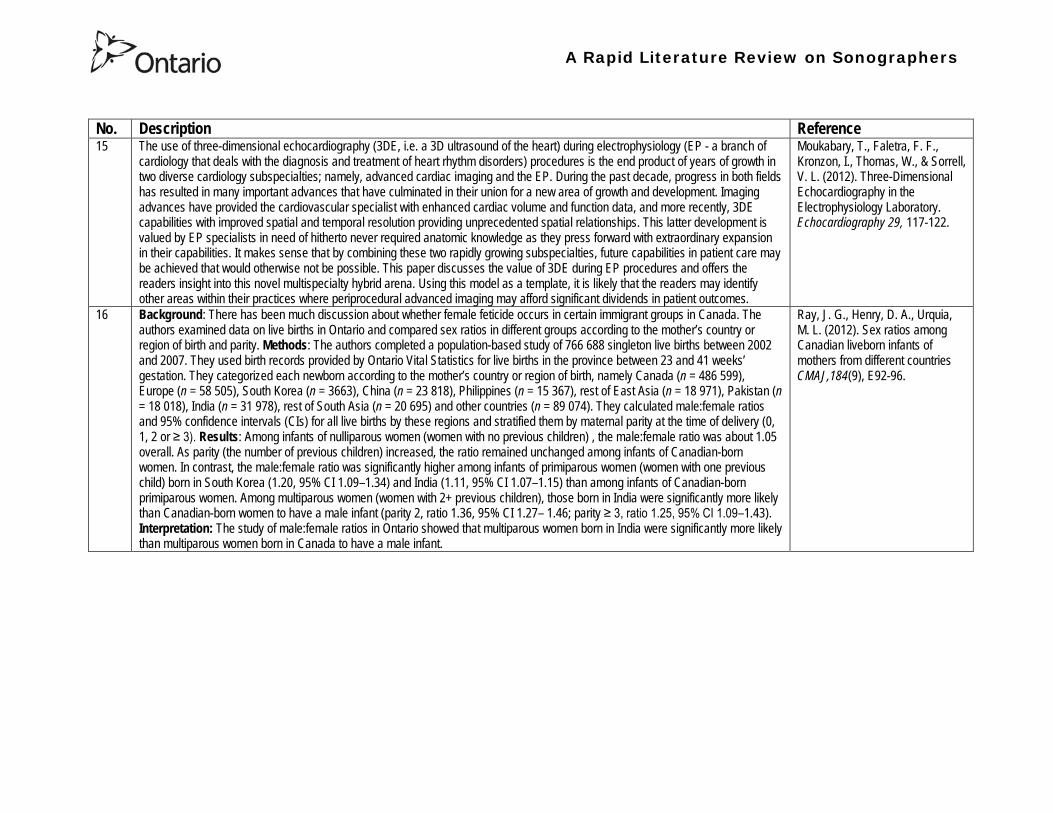
A Rapid Literature Review on Sonographers
No Description Reference 15 The use of three-dimensional echocardiography (3DE ie a 3D ultrasound of the heart) during electrophysiology (EP - a branch of
cardiology that deals with the diagnosis and treatment of heart rhythm disorders) procedures is the end product of years of growth in two diverse cardiology subspecialties namely advanced cardiac imaging and the EP During the past decade progress in both fields has resulted in many important advances that have culminated in their union for a new area of growth and development Imaging advances have provided the cardiovascular specialist with enhanced cardiac volume and function data and more recently 3DE capabilities with improved spatial and temporal resolution providing unprecedented spatial relationships This latter development is valued by EP specialists in need of hitherto never required anatomic knowledge as they press forward with extraordinary expansion in their capabilities It makes sense that by combining these two rapidly growing subspecialties future capabilities in patient care may be achieved that would otherwise not be possible This paper discusses the value of 3DE during EP procedures and offers the readers insight into this novel multispecialty hybrid arena Using this model as a template it is likely that the readers may identify other areas within their practices where periprocedural advanced imaging may afford significant dividends in patient outcomes
Moukabary T Faletra F F Kronzon I Thomas W amp Sorrell V L (2012) Three-Dimensional Echocardiography in the Electrophysiology Laboratory Echocardiography 29 117-122
16 Background There has been much discussion about whether female feticide occurs in certain immigrant groups in Canada The authors examined data on live births in Ontario and compared sex ratios in different groups according to the motherrsquos country or region of birth and parity Methods The authors completed a population-based study of 766 688 singleton live births between 2002 and 2007 They used birth records provided by Ontario Vital Statistics for live births in the province between 23 and 41 weeksrsquo gestation They categorized each newborn according to the motherrsquos country or region of birth namely Canada (n = 486 599) Europe (n = 58 505) South Korea (n = 3663) China (n = 23 818) Philippines (n = 15 367) rest of East Asia (n = 18 971) Pakistan (n = 18 018) India (n = 31 978) rest of South Asia (n = 20 695) and other countries (n = 89 074) They calculated malefemale ratios and 95 confidence intervals (CIs) for all live births by these regions and stratified them by maternal parity at the time of delivery (0 1 2 or ge 3) Results Among infants of nulliparous women (women with no previous children) the malefemale ratio was about 105 overall As parity (the number of previous children) increased the ratio remained unchanged among infants of Canadian-born women In contrast the malefemale ratio was significantly higher among infants of primiparous women (women with one previous child) born in South Korea (120 95 CI 109ndash134) and India (111 95 CI 107ndash115) than among infants of Canadian-born primiparous women Among multiparous women (women with 2+ previous children) those born in India were significantly more likely than Canadian-born women to have a male infant (parity 2 ratio 136 95 CI 127ndash 146 parity ge 3 ratio 125 95 CI 109ndash143) Interpretation The study of malefemale ratios in Ontario showed that multiparous women born in India were significantly more likely than multiparous women born in Canada to have a male infant
Ray J G Henry D A Urquia M L (2012) Sex ratios among Canadian liveborn infants of mothers from different countries CMAJ184(9) E92-96
A Rapid Literature Review on Sonographers
No Description Reference 17 This letter consists of a case study that illustrates how a cross-disciplined sonographer (educated in cardiac and vascular ultrasound)
can quickly and efficiently and with great Cost-effectiveness deliver timely care to a patient in certain clinical settings The literature is comparatively sparse in referencing clinical diagnostics in cross-discipline sonographers Echocardiographers must be clinicians fully comfortable with evaluating the circulatory dynamics of the body from the left and right heart to the peripheral arteries and veins including the extracranial circulation (carotid arteries) Included in this skill set is substantive understanding of the ECG patientrsquos history and integration of cardiac and vascular findings Limitations of each diagnostic tool and how these limitations can be overcome by the strengths of the other examination are important For the dual-skilled sonographer only experience continuous education and constant clinical feedback will lead to the independent decision making that is counted on by the physician The American Registry for Diagnostic Medical Sonography offers credentials in cardiac and vascular imaging (RDCS and RVT) Cardiovascular Credentialing International offers credentials in cardiac and vascular imaging as well (registered cardiac sonographer [RCS] and registered vascular specialist [RVS]) All imaging facilities should consider merging cardiac and vascular noninvasive imaging with dual-registered clinical sonographers credentialed by one of these two agencies
Zemanek M (2012) What It Means to Be a Cardiovascular Sonographer Journal of Ultrasound in Medicine 311859-1862
18 Diagnostic medical sonography is viewed as a safe imaging modality However bioeffects research has yet to define the effects of exposure to sonography in humans Therefore sonographers must be aware of potential risks and practice ALARA (ldquoas low as reasonably achievablerdquo) routinely This research explores sonographersrsquo ability to answer basic questions about the bioeffects of sonography as well as their knowledge and use of the safety indices called the mechanical index (MI) and thermal index (TI) Members of the Society of Diagnostic Medical Sonographers in the US a southwestern sonographersrsquo society and a sonography community on Facebook 2010 were surveyed (n = 212) There is a low level of bioeffects knowledge and minimal safety practices among sonographers in this survey No significant relationship was found between sonographer knowledge of the bioeffects of sonography years in the profession or whether the sonographers monitor the MITI
Bagley J Thomas K DiGiacinto D (2011) Safety Practices of Sonographers and Their Knowledge of the Biologic Effects of Sonography Journal of Diagnostic Medical Sonography 27(6) 252ndash261
19 This article highlights trends in echocardiography Point of care ultrasound (hand-carried ultrasound) Initially point of care ultrasound will increase the use of echocardiography in the same manner that cardiac CT increased catheterizations However once the basics are learned it has the potential to be a very powerful tool in that less unnecessary studies will be ordered In the clinic small systems will be used as a lsquolsquofirst lookrsquorsquo screen and become part of the patientrsquos history only patients for whom more testing is necessary will be referred for a comprehensive study This is likely to reduce the number of Doppler studies requested The miniaturization of echo equipment will continue with improved qualityfunctionality of the lsquolsquohand-heldrsquorsquo echo machines to the extent that cordless probes will become a reality sooner rather than later Role in interventional echocardiography Sonographers will be spending much more time in the interventional and electrophysiology (EP) labs There will be more interconnection with interventional cardiologists and the volume will increase as more devices are implanted more percutaneous valves placed and more robotic surgery is performed Crosstraining and multitasking Sonographers entering the profession would be prudent to learn cardiac and vascular imaging Individuals who multitask andor cross-train will be ideal For example a sonographer who starts an IV monitors ECG stress can perform cardiac MRI and can interpret a preliminary report or a nurse who can do an echo monitor sedation and start an IV will be invaluable The sonographer should continue to grow professionally by cross training as institutions try to minimize costs and maximize throughput
McCulloch M L (2011) Thoughts regarding the future of sonography Journal of the American Society of Echocardiography 24(5) 29A-30A
A Rapid Literature Review on Sonographers
No Description Reference 20 Background Radiology reports provide specialist interpretation of images and relate these findings to the patientrsquos symptoms and
signs This study compared ultrasound (USS) reports generated by radiologists and sonographers to determine if any significant or clinically relevant differences existed Methods A retrospective analysis of 624 consecutive USS reports was carried out The reports were assessed for the presence of a lsquodisclaimerrsquo or lsquocaveatrsquo pertaining to the quality of the images and were analysed with respect to the clarity of their wording and ability to answer the clinical request Results The majority of sonographer USS reports contained a disclaimer regarding the quality of the USS images (571) compared with a smaller proportion of radiologist scans (99) (P lt 0001) Overall radiologists performed significantly better in providing a clear negative or positive diagnosis to the clinical question on the request form when compared with sonographers (885 vs 654 P lt 0001) Conclusion lsquoDisclaimerrsquo comments and lsquohedgingrsquo must be avoided in radiology reporting While feedback as to the accuracy of the radiology images is important overuse of such terms undermines the validity of the radiology report
Garcea G Mahmoud A Ong S L Rees Y Berry Det al (2010) Caveat reporting in ultrasound interpretation of surgical pathology a comparison of sonographer versus radiologist Journal of Evaluation in Clinical Practice 16 97ndash99
21 A nine-point questionnaire was constructed based on previously published work by Marsal and Sheiner et al The survey was distributed to clinical users of ultrasound practicing sonography in Hamilton (New Zealand) during July 2010 totalling 35 users consisting of sonographers sonography trainees one obstetric sonologist and one fetal medicine registrar Results Twenty sonographers returned the questionnaire which represents survey response of 61 The level of clinical experience was equally split between junior sonographers of lt 5 yearsrsquo experience (30) sonographers with 5-10 yearsrsquo experience (35) and senior sonographers of gt10 yearsrsquo experience (35) While 100 of the sonographers surveyed reported being familiar with the term Thermal Index (TI) and 90 claimed they adhered to the Action Learning Action Response Association (ALARA) principle all the time or most of the time only 15 of sonographers correctly described or defined what the term TI means Of the sonographers 80 knew where to find the TI during real-time examinations but only 35 knew which TI index to use for nuchal translucency (NT) scan and only 10 knew the expected range of TI encountered during routine obstetric examinations Of the eight sonographers who reported adhering to ALARA all the time the majority (64) could not describe or define TI In the same subgroup the majority (64) did not know which TI to monitor during a NT scan Only three of 20 (15) sonographers knew what the term TI means Of these two were junior sonographers with lt 5 yearsrsquo experience and one had 5ndash10 yearsrsquo experience Despite understanding the term TI one of the three sonographers still selected the incorrect index to monitor during a NT scan Not one of the seven senior sonographers with gt 10 years of experience could define or describe the TI In total only 10 of sonographers knew what the TI means and which TI to monitor during NT scanning
Necas M (2010) New Zealand sonographers do not outperform their European or American colleagues in the knowledge of ultrasound safety Australasian Journal of Ultrasound in Medicine13(4) 28-32
A Rapid Literature Review on Sonographers
No Description Reference 22 Musculoskeletal (MSK) ultrasound performed by non-medical practitioners is a developing practice The aim of this paper is
(1) to audit an experienced sonographerrsquos (SJ Riley one of the authors) performance after one yearrsquos independent reporting against two experienced MSK radiologists (CJ Grove amp M Chandramohan the other two authors) and (2) to establish an audit standard against which such role development might be contrived Images and reports from 250 MSK ultrasound examinations performed by a sonographer were reviewed independently by two consultant MSK radiologists The examinations were graded for discrepancy eg grade 1 ndash agree with the sonographer report grade 2 ndash minor discrepancy unlikely to alter patient care grade 3 ndash potentially significant discrepancy grade 4 ndash definite significant discrepancy likely to have adverse consequences for patient care Two of 250 (08) cases were excluded Both radiologists agreed completely with the sonographer (grade 1) in 235 of the 248 cases (948) In 13 cases there was discrepancy between the reports of SJR and the radiologists The discrepancy was grade 2 in six of the 248 cases (24) grade 3 in six of the 248 cases (24) and grade 4 in one of the 248 cases (04) In conclusion this audit shows a high level of agreement between the sonographer and the consultant MSK radiologist reporting of MSK ultrasound This level of agreement may set the standard for future quality assurance audit of sonographer MSK reporting
Riley S J Groves C J Chandramohan M (2010) Musculoskeletal ultrasound audit of sonographer reporting Ultrasound 18 36ndash40
23 Background Growing demand for ultrasound examinations and higher quality requirements motivate searching for routines combining the diagnostic accuracy of radiologist-performed examinations with the economical advantages of sonographer-performed examinations One possible approach is to use strictly standardized acquisition and documentation schemes that give the radiologist access to all relevant information after the examination Purpose To compare a recently introduced routine combining acquisition by a radiographer (more specifically a sonographer with two years experience) documentation as standardized cine-loops and review by a radiologist (lsquostandardized methodrsquo) with the formerly used routine where the diagnosis is made bedside by the radiologist (lsquotraditional methodrsquo) Material and Methods In 64 policlinic patients the kidneys (n = 27) or the gallbladder (n = 37) were examined with both the standardized and the traditional method The radiologistsrsquo findings of hydronephrosis tumors cysts echogenicity changes and cortical thickness (in the kidneys) and wall thickness concrements and polyps (in the gallbladder) were compared between the methods with respect to agreement (proportion of agreement and kappa coefficient) as well as systematic differences (McNemarrsquos test) Results The findings at the gallbladder examination showed a median agreement of 97 (86ndash100 kappa = 064ndash100) and those at the kidney examination an agreement of 90 (78ndash100 kappa = 069ndash 100) There were no significant systematic differences between the methods Conclusion The satisfactory agreement in this preliminary study indicates that the new workflow with ultrasound examinations performed by a radiographer (sonographer) and analyzed off-line by a radiologist is promising and motivates further studies
Stenman C Thorelius L Knutsson A amp Smedby Ouml (2010) Radiographer-acquired and radiologist-reviewed ultrasound examination ndash agreement with radiologistrsquos bedside evaluation Acta Radiologica 52 70ndash74
A Rapid Literature Review on Sonographers
No Description Reference 24 The troubling practice of fetal sex selection has historically been considered an Asian phenomenon However recent evidence shows
that a similar situation is emerging in North America albeit on a smaller scale The Society of Obstetricians and Gynaecologists of Canada (SOGC) has firmly stated its opposition to sex selection for non-medical reasons as well as to the use of any technology used solely for the purpose of determining fetal sex However because fetal sex may be disclosed to the parents at the time of ultrasound examination if they request this information guidance for health care professionals to assist in discouraging fetal sex selection would be useful Because no declaration of motives or reasons is required when a woman seeks a termination of pregnancy the authors suggest that health care professionals need not disclose the sex of a fetus until it has reached a gestational age at which abortion for non-medical purposes would not be possible This proposal would facilitate consistency between clinical practice and the values of Canadian citizens the SOGC the Canadian Medical Association and other professional organizations while still respecting current laws pertaining to disclosure of patient information and patientsrsquo rights to autonomous decision-making
Thiele A T amp Leier B (2010) Towards an Ethical Policy for the Prevention of Fetal Sex Selection in Canada J Obstet Gynaecol Can 32(1) 54ndash57
25 Background The extent to which increases in the use of prenatal ultrasonography over time reflect changes in maternal risk (ie an increase in high-risk pregnancies over time) is unknown In this population-based study the authors examined the use of prenatal ultrasonography from 1996 to 2006 in Ontario Methods With fiscal year 199697 as the baseline the authors evaluated the relative risk (RR) and 95 confidence interval (CI) for the change in rates of ultrasonography for each subsequent year The RR was adjusted for maternal age income rural residence maternal comorbidities receipt of genetics consultation or amniocentesis mdash all in the index pregnancy mdash and history of complications in a prior pregnancy Results The study sample consisted of 1 399 389 singleton deliveries The rate of prenatal ultrasonography increased from 2055 per 1000 pregnancies in 1996 to 3264 per 1000 in 2006 (adjusted RR 155 95 CI 154ndash155) The rate increased among both women with low-risk pregnancies (adjusted RR 154 95 CI 153ndash155) and those with high-risk pregnancies (adjusted RR 155 95 CI 154ndash157) The proportion of pregnancies with at least four ultrasound examinations in the second or third trimesters rose from 64 in 1996 to 187 in 2006 (adjusted RR 268 95 CI 261ndash274) Paradoxically this increase was more pronounced among low-risk pregnancies (adjusted RR 292 95 CI 283ndash301) than among high-risk pregnancies (adjusted RR 225 95 CI 216ndash235) Interpretation Substantial increases in the use of prenatal ultrasonography over the past decade do not appear to reflect changes in maternal risk Nearly one in five women now undergo four or more ultrasound examinations during the second and third trimesters Efforts to promote more appropriate use of prenatal ultrasonography for singleton pregnancies appear warranted
You J J Alter D A Stukel T A McDonald S D Laupacis A et al (2010) Proliferation of prenatal ultrasonography CMAJ 182(2) 143-151
A Rapid Literature Review on Sonographers
No Description Reference 26 Aims The aim of this study was to assess the cost-effectiveness of using certified sonographers and miniaturized echocardiography
systems to perform echocardiograms at bedside in comparison to moving inpatients from the admission department to the echocardiography laboratory (echo-lab) Methods and Results From 26 September 2005 to 27 October 2005 112 patients admitted in six hospital wards in Italy connected through a 100 Mbit LAN to the echo-lab were scanned within the admission ward by sonographers using a miniaturized echo system Logistical data were collected and results were compared with those obtained from 194 consecutive patients coming from the same wards and studied in the echo-lab with high-end machines between 8 March 2005 and 15 April 2005 Performing echocardiograms in the admission department avoided long waiting time of the inpatients in the echo-lab before and after the study increased the percentage of patients studied within 3 and 5 days from request (88 vs 77 and 100 vs 95 respectively P = 003) increased both sonographer (by 339 P lt 0001) and echo-lab productivity (by 41 P lt 0001) and reduced costs of echocardiograms by 29 Conclusion Implementation of digital echocardiography certified sonographers and a miniaturized echo system allowed improvement of the cost-effectiveness of the service provided by the echo-lab for inpatients and avoided patientsrsquo discomfort derived from prolonged waiting time before and after the exam
Badano L Gaetano N Stacul S Gianfagna P Pericoli M et al (2009) Improved workflow sonographer productivity and cost-effectiveness of echocardiographic service for inpatients by using miniaturized systems European Journal of Echocardiography 10 537ndash542
27 The present report is a proposal from the American Society of Echocardiography Advanced Practice Task Force that identifies the potential of cardiac sonographers to achieve the Advanced Cardiovasculat Sonographer (ACS) level According to this report echocardiographic examinations require a well-trained and competent sonographer to obtain proper anatomic and physiologic data to establish an accurate diagnosis for clinical decision-making and patient management Although the formal education and training of cardiovascular sonographers are evolving many entry-level and staff sonographers may not have sufficient practical or clinical knowledge of the necessary components of the echocardiographic study for the individual patientrsquos clinical presentation In many clinical settings echocardiograms are read after the patient has left the laboratory Thus there is a role for a sonographer who can practice at an advanced level in a cardiovascular ultrasound laboratory to ensure a proper echocardiographic examination is performed on every patient In this setting an Advanced Cardiovascular Sonographer (ACS) would be able to review the indication for and quality of the examination If additional images were needed the ACS would assist the sonographer in obtaining these images which would lead to the performance of a complete and fully diagnostic examination before the patient had left the echocardiography laboratory In clinical practice the quality of the examinations performed would improve advancements in echocardiographic methods could be taught and incorporated into daily practice and patients would be better served
Mitchell C Miller F A Bierig S M Bremer M L Ehler D et al (2009) Advanced Cardiovascular Sonographer A Proposal of the American Society of Echocardiography Advanced Practice Sonographer Task Force J Am Soc Echocardiogr 22 1409-13
A Rapid Literature Review on Sonographers
No Description Reference 28 Sonographer practitioner development involves the expansion and extension of the sonographer role to include reporting on
ultrasound examinations Australian sonographers have not seen the same degree of role extension and expansion as their counterparts in the United Kingdom despite increasing levels of discussion regarding sonographer practitioner development The aim of this study was to determine if Australian sonographers want to extend their professional role and what they consider are the important issues associated with role extension This paper reports on qualitative data derived from a survey of Australian sonographers and investigates if Australian sonographers are interested in extending and expanding their professional role and responsibilities and if they do what might be necessary or desirable from a professional point of view for this change to occur A survey was mailed to all members of the Australian Sonographers Association (ASA) in October 2006 The 31-item survey included 28 closed-ended and three opened-ended items to provide both quantitative and qualitative data The quantitative data will be reported separately Qualitative data was derived from responses to the opened-ended questions which asked respondents to elaborate on their attitudes and feelings about role extension and development Analysis used Nvivo7 software to aid in uncovering common themes from the qualitative data The analysis focused on the reported incentives or motivations for becoming a sonographer practitioner as well as disincentives or perceived hurdles that would discourage respondents from becoming sonographer practitioners The three most reported incentives or motivations for becoming a sonographer practitioner were professional recognition remuneration and increased knowledge The three most commonly reported disincentives or perceived hurdles that would discourage respondents from becoming sonographer practitioners were legal issues insurance and further study
McGregor R OrsquoLoughlin K Cox J Clarke J Snowden A (2009) Sonographer practitioner development in Australia Qualitative analysis of an Australian sonographersrsquo survey Radiography15 313-319
29 Introduction This study describes the effect of introducing a cardiac sonographer led follow up clinic within a hospital cardiology department for patients with stable heart valve disease The two years before and after the instigation of the valve clinic were audited Materials and methods The clinic was conducted in a single centre and undertaken in the cardiology department of a district general hospital A total of 382 patients with 397 clinically significant valve lesions but for whom surgery was not yet indicated but follow up required were seen in a cardiac sonographer run clinics These patients no longer attended a medical follow up clinic unless there was clinical or echocardiographic deterioration Effectiveness was judged by the percentage treated according to current best practice guidelines the number of echocardiograms performed and the number of hospital outpatient visits attended In addition mortality data for the subjects in the clinic was collected Results The proportion followed up according to best practice guidelines rose from 157 (41) to 354 (92) (p lt 001) The total number of echocardiograms performed fell from 807 to 550 Total number of outpatient visits fell from 998 to 31 A total of 11 patients died in the two year study period none from progressive valve disease Discussion This study demonstrates that a protocol driven sonographer led heart valve disease follow up clinic significantly improved the quality of follow up while bringing about a major reduction in outpatients visits without compromising patient safety
Taggu W Topham A Hart L Carr-White G Sulke N et al (2009) A cardiac sonographer led follow up clinic for heart valve disease International Journal of Cardiology 132 240 ndash243
30 Role development for imaging allied health professionals has progressed significantly over the last three decades with many major developments pioneered in the field of ultrasound This paper reviews the developing role of the radiographer in ultrasound (in some countries such as the UK sonographers are considered a type of radiographer) and some of the clinical professional and educational implications of such development As the published evidence base underpinning the latest developments is scarce several contemporary case study examples which have been presented verbally at study days attended by the authors have been included In conclusion sonographer role development is considered within the professional contexts of extended role advanced and consultant practice
Hart A amp Dixon A (2008) Sonographer Role Extension and Career Development a Review of the Evidence Ultrasound 16(1) 31-35
A Rapid Literature Review on Sonographers
No Description Reference 31 Objective The main goal of this study was to determine end usersrsquo knowledge regarding safety aspects of diagnostic ultrasound
during pregnancy End usersrsquo attitudes toward the use of ultrasound in low-risk pregnancies were also assessed Methods A questionnaire was distributed to ultrasound end users attending review courses and hospital grand rounds between April and June 2006 in Chicago Results One hundred thirty end users completed the questionnaires (63 response rate) Sixty-three percent were physicians (n = 84) most of them obstetricians (817) About 18 of participants routinely performed Doppler ultrasound examinations during the first trimester Fifty percent of end users thought that the number of ultrasound examinations in low-risk pregnancy should be limited to 1 to 3 (mean plusmn SD 26 plusmn 09) Almost 70 disapproved of ldquokeepsakeentertainmentrdquo ultrasound Although 322 of the participants were familiar with the term thermal index only 177 actually gave the correct answer to the question on the nature of the thermal index About 22 were familiar with the term mechanical index but only 38 described it properly Almost 80 of end users did not know where to find the acoustic indices Only 208 were aware that they are displayed on the sonographic monitor during the examinations End users with higher knowledge of safety issues thought that there should be limitations on the number of ultrasound examinations in low-risk pregnancies (odds ratio 33 95 confidence interval 11ndash100 P = 028) Likewise these end users were more likely to respond that ultrasound might have adverse effects during pregnancy (odds ratio 32 95 confidence interval 11ndash125 P = 045) Conclusions Ultrasound end users are poorly informed regarding safety issues during pregnancy Further efforts in the realm of education and training are needed to improve end user knowledge about the acoustic output of the machines and safety issues
Sheiner E Shoham-Vardi I Abramowicz J S (2007) What Do Clinical Users Know Regarding Safety of Ultrasound During Pregnancy J Ultrasound Med 26 319ndash325
A Rapid Literature Review on Sonographers
No Description Reference 32 In an attempt to survey the knowledge among ultrasound users of some safety aspects of diagnostic ultrasound a questionnaire was
distributed to professionals using ultrasound for fetal examinations The questionnaire was answered by the participants and faculty members of six postgraduate courses in obstetric ultrasound Doppler ultrasound and fetal echocardiography given in Sweden Norway and Austria in 2003 Furthermore in Sweden the questionnaire was distributed to the staff meeting participants of two major obstetric ultrasound units and of one unit for fetal echocardiography and at a meeting of the Working Group for Ultrasound of the Swedish Society of Obstetrics and Gynecology In total 199 questionnaires were anonymously answered by doctors (n = 145) sonographers (n = 22) and midwives trained in ultrasound (n = 32) All of them were using diagnostic ultrasound on a daily or weekly basis The respondents were from nine European countries In none of the countries is formal certification for the use of diagnostic ultrasound in pregnancy or otherwise required The doctors who answered the questionnaire were specialists in obstetrics and gynecology (including some of the internationally recognized experts in fetal ultrasound) pediatric cardiology radiology or clinical physiology The respondents were not informed about the questionnaire in advance and they had a limited time to answer the 16 questions typically 10ndash15 min The questions concerned explanation of the safety indices and their relation to various ultrasound modes ie real-time B-mode M-mode three-dimensional (3D) spectral Doppler color and power Doppler ultrasound The respondents were also asked which ultrasound system they used most often and where on the system the information on thermal index (TI) and mechanical (MI) was displayed (eg in the top right corner of the screen in a separate window etc) Furthermore the respondents had to indicate how they adjust the output energy level on their own machine For all questions there was the optional answer lsquoDonrsquot knowrsquo Results About one-third of the ultrasound experts were able to define the abbreviations TI and MI For the subclasses of the TI the correct answer was found in only 3-8 of cases Of those who knew what the TI and MI meant only two-thirds and one-third of respondents respectively were able to give a simple explanation of the indices In 49 of questionnaires the Doppler modes were correctly ranked as generally producing higher ultrasound exposure than the imaging modes (B-mode 3D) Some 28 of the respondents correctly indicated where on their own machine the information on safety indices is displayed However not all of them knew how to control the output energy level For all items in the questionnaire there were no significant differences in the results between the three categories of respondents (ie physicians sonographers and midwives) The results of the questionnaire were anything but encouraging and they indicated that the users who are supposed to be responsible for controlling exposure of the fetus to ultrasound had a very poor knowledge of the basic safety aspects of ultrasound
Maršaacutel K (2005) The output display standard has it missed its target Ultrasound Obstet Gynecol 25 211ndash214
33 Purpose This study was performed to evaluate the accuracy of upper abdominal ultrasound (US) scanning performed by sonographers in a district general hospital to identify potential areas of weakness and to make recommendations to improve the service Materials and Methods Upper abdominal US examinations performed and reported by sonographers over a 4-week period were retrospectively reviewed The accuracy of the imaging findings and reports were assessed against other imaging surgical histological or laboratory findings and against clinical outcome Results A heterogenous group of 104 patients were included in the study 62 of whom had an US abnormality Errors of scanning or interpretation were identified in 10 patients (96) of whom five (48) were felt to be potentially significant Conclusions The sonographersrsquo accuracy in reporting upper abdominal US scans was 90 However on the basis of this study the authors have implemented specific recommendations to improve the quality of the service
Dongola N Guy R Ward S amp Giles J A (2003) Can sonographers offer an accurate upper abdominal ultrasound service in a district general hospital Radiography 9 29ndash33
A Rapid Literature Review on Sonographers
No Description Reference 34 Aim To compare the accuracy of radiographers and radiologists in routine abdominal ultrasound Materials and Methods One
hundred consecutive patients attending for routine abdominal ultrasound were included Each patient was examined by both a radiographer and radiologist Both operators noted their fndings and wrote a concluding report without conferring Reports were compared Where there was disagreement the patient was either re-examined by another radiologist or had further investigation Results Of 100 patients 52 were men and 48 were women The age range was 19plusmn88 years (median 52 years) Thirty-seven patients had renal tract ultrasound one had an aortic ultrasound and 62 had general upper abdominal ultrasound In 44 cases both operators reported the examination as normal In 49 cases both operators reported the examinations as abnormal and there was complete agreement between the operators In seven cases there was not complete agreement between operators Three of these disagreements were considered minor and four major In three of the seven cases the radiographer was correct and in four the radiologist was correct Conclusion Experienced radiographers and radiologists are highly accurate in performing and interpreting routine abdominal sonography Both operators missed a small minority of abnormalities There was no statistically significant difference in the accuracy of radiographers and radiologist
Leslie A Lockyer H amp Virjee J P (2000) Who Should be Performing Routine Abdominal Ultrasound A Prospective Double-Blind Study Comparing the Accuracy of Radiologist and Radiographer Clinical Radiology 55 606-609
Grey Literature 35 The ARRT The American Registry of Radiologic Technologists tests certifies and annually registers more than a quarter-of-a-million
radiologic technologists to promote high standards of patient care They award the Registered Technologist or RT designation to individuals who have completed the prescribed classroom and clinical education passed the appropriate exam and met the ethics requirements And they ensure their continuing education and ongoing compliance with their high standards by requiring annual registration of their certificate Certification by ARRT is voluntary not mandatory But employers state licensing agencies and federal regulators view ARRTrsquos credential as an indicator that a person has met recognized national standards for medical imaging and radiation therapy professionals How Are Radiologic Technologists Deemed Qualified First comes initial certification Certification is the process of initially recognizing individuals who have satisfied certain standards within a profession A person is certified by ARRT after completing an approved educational program complying with the ethics standards and passing a certification exam Then comes annual registration Registration is the yearly procedure required to maintain certification ARRT registrants are those who having already fulfilled the requirements for initial certification continue to meet the requirements for annual registration The ARRT annually registers the certificates of individuals who meet the following three criteria agreeing to comply with the ARRT Rules and Regulations continuing to comply with the ARRT Standards of Ethics and meeting our continuing education requirements Only technologists who are currently registered (which means that they have renewed within the past year) may designate themselves as ARRT Registered Technologists and use the initials RT after their names Federal laws regulate personnel qualifications in some modalities (mammography for example) but they accredit the health care facility rather than licensing the individual radiologic technologist who is providing patient care And while many states have laws regulating who can perform radiologic procedures they are typically based on ARRT certification Some employers and state agencies may require ARRT certification but not necessarily current registration Unless they require current registration by ARRT employers cannot assume that the person has complied with the ethics requirements or the continuing education requirements
American Registry of Radiologic Technologists (ARRT) (2013a) Patient Page Accessed April 2013
A Rapid Literature Review on Sonographers
No Description Reference 36 This document outlines certification requirements for sonography There are two pathways (primary and post-primary) For the
primary pathway it notes that sonography educational programs must be accredited by a mechanism acceptable to the ARRT ARRT generally recognizes only accreditation agencies that are recognized by the Council for Higher Education Accreditation andor United States Department of Education
American Registry of Radiologic Technologists (ARRT) (2013b) Sonography Certification Accessed April 2013
A Rapid Literature Review on Sonographers
No Description Reference 37 In Australia sonographers and student sonographers are required to be accredited by Australian Sonographer Accreditation Registry
(ASAR) in order to practise This requirement is contained within the Australian Governmentrsquos Diagnostic Imaging Accreditation Scheme standards and Medicare legislation Accreditation of sonographers is linked only to achieving - or in the case of students studying towards - an accredited entry-level qualification completion of continuing professional development and payment of an annual fee to ASAR The ASA contends that the lack of a requirement to ensure each sonographer is lsquofit to practisersquo is an unacceptable gap in current regulation undermining efforts to ensure the quality of sonographic services and failing to protect patients from harm The ASA supports and advocates for nationally-enforceable regulation of sonographers to meet our patientrsquos expectations National registration The National Registration and Accreditation Scheme (NRAS) for health workers commenced on 1 July 2010 The ten professions which comprised the initial intake into this scheme were those that had registration requirements in all Australian states or territories as well as those in which legislated regulation is deemed to be in the public interest An additional four professions - including medical radiation practitioners which are defined as radiographers radiation therapists and nuclear medicine technologists - will be included in the NRAS commencing on 1 July 2012 As sonography did not have registration in any states or territories prior to the commencement of NRAS the profession has not been in a position to negotiate entry into NRAS In early August 2011 the ASA lodged a submission with the Health Workforce Principal Committee requesting a profession-specific Sonography Board of Australia be formed under NRAS This submission was prepared based on information that consideration of professions which were unregistered would commence More recently the Australian Health Workforce Ministerial Council (AHWMC) issued a communiqueacute stating in part lsquoMinisters have commissioned work on unregistered professions and future directions for national registration Given this Ministers agreed to defer consideration of the inclusion of additional professions into the NRAS Ministers agreed that it would be premature to consider further applications for the scheme until this work has been completed other than paramedicsrsquo Options for unregistered health practitioners including sonographers Policy work is continuing at a federal level to establish an alternative regulatory framework The National Alliance for Self-Regulating Health Professions (NASRHP) consisting of the peak professional associations of eight self-regulating health professions including the ASA representing sonography released a proposal lsquoHarnessing self-regulation to support safety and quality in healthcare deliveryrsquo in March 2012 This proposal concluded that the framework that regulates all health practitioners should be expanded beyond NRAS to include
bull authorised self-regulating professions via a health professions panel bull negative licensing to withdraw the ability to practise from registered and self-regulated practitioners who do not meet the
regulatory requirements or unregulated practitioners due to poor practice As with all advocacy initiatives consideration by the federal government of the introduction of the NASRHPrsquos proposal may be prolonged and the outcomes may not match the expectations of NASRHPrsquos member professional associations including the ASA The ASA will continue both independently and collaboratively to lobby on behalf of our members and the public for the introduction of a timely and equitable solution to health practitioner regulation In the interim as there is no regulatory model with the rigour the ASA believes is required for regulation of sonographers and other health practitioners other than NRAS the ASA continues to support its Position Statement published in early 2010
Australian Sonographer Association (ASA) (2013) Sonographer Regulation Accessed April 2013
A Rapid Literature Review on Sonographers
No Description Reference 38 This publication contains information about Cardiovascular Credentialing International (CCI) It also provides the necessary
information and references concerning the process by which candidates may earn certificate or registry-level credentials CCI is a not-for-profit corporation established for the purpose of administering credentialing examinations as an independent credentialing agency CCI began credentialing cardiovascular professionals in 1968 CCI offers eight credentials which when earned demonstrate that the registrant holds fundamental knowledge in the particular cardiovascular specialty These include RCCS ndash Registered Congenital Cardiac Sonographer RCS ndash Registered Cardiac Sonographer and RPhS ndash Registered Phlebology Sonographer
Cardiovascular Credentialing International (2013) 2013 Examination Application amp Overview Accessed April 2013
39 Oregon Governor Signs Sonographer Licensure Legislation On July 28 2009 Oregon Governor Ted Kulongoski signed a sonographer licensure bill (HB 2245) into law The new law makes Oregon only the second state in the United States to require licensure of sonographers The Society of Diagnostic Medical Sonographers (SDMS) has worked closely with other sonography-related organizations and Oregon sonographers to help craft the language in new law The law renames and restructures the Oregon Board of Radiologic Technologists to the Oregon Board of Medical Imaging (OBMI) and ensures representation of sonographers on the Board The sonographer licensure law requires Oregon sonographers to become licensed by OBMI effective July 1 2010 Beginning on January 1 2014 all sonography licensees would be required to hold a national sonography certificationcredential (or be currently enrolled as a student) While the Oregon bill is far more detailed than the sonographer licensure law that passed in New Mexico earlier this year additional administrative rulesregulations will be developed over the next year to fully implement the new law SDMS will be continue to monitor developments regarding sonographer licensure in other states
Society of Diagnostic Medical Sonographers (SDMS) (2013a) News Accessed April 2013
A Rapid Literature Review on Sonographers
No Description Reference 40 On April 6 2009 New Mexico (NM) Governor Bill Richardson signed the historic bill that for the first time in the United States
requires licensure of sonographers The bill adds sonographers (and MRI technologists) to the list of medical imaging professionals licensed by the State of New Mexico Until now anyone could perform sonograms in New Mexico Prior to the passage of the New Mexico licensure law virtually all other health care providers and professionals had to be licensed As a result the public is often surprised to learn that the person performing a medical sonogram does not have any state licensure or national certification requirements The responsibility for administering the NM sonographer licensure program would rest with the New Mexico Environment Department Although this was not the first choice in most sonographersrsquo minds the Environment Department currently manages the radiologic technology licensure program so the administrative infrastructure is already in place By ensuring the creation of an advisory council that includes representation by each medical imaging and radiation therapy modality regulated the bill helps ensure that the Departmentrsquos other areas of emphasis do not detract from sonographer licensure Because sonographers have never had to be licensed before they frequently do not see or understand the distinction between state licensure and national certification National certifications or credentials issued by voluntary organizations such as the American Registry for Diagnostic Medical Sonography (ARDMS) Cardiovascular Credentialing International (CCI) and the American Registry of Radiologic Technologists (ARRT) have little legal weight The certification process and resulting credential are valuable tools in ensuring sonographers have met minimum education and knowledge requirements but do not really regulate anyonersquos ability to perform sonography In an ideal world no additional regulation of sonography would be required However as everyone knows more and more people are taking weekend courses and buying ultrasound equipment without understanding the ramifications of using ultrasound technology (eg ALARA) Under the bill passed by the New Mexico legislature national credentialscertifications should provide evidence of meeting the statersquos standards All sonographers who are performing these critical medical imaging services in New Mexico will be required to meet national certification standards (ie have certifications or credentials issued by ARDMS CCI or ARRT) As a result there would be no additional exams in New Mexico beyond the national certification exams required by ARDMS CCI or ARRT The bill also requires the state to recognize the continuing medical education completed for national certification renewal The most visible impact on New Mexico sonographers will be a license fee However the legislation caps the license fee at $100 per two-year license In addition only the New Mexico Legislature can change this cap
SDMS (2013b) New Mexico Sonographer Licensure Law Signed Accessed April 2013
A Rapid Literature Review on Sonographers
No Description Reference 41 Preamble The purpose of this document is to define the Scope of Practice for Diagnostic Ultrasound Professionals in the US and to
specify their roles as members of the health care team acting in the best interest of the patient This scope of practice is a living document that will evolve as the technology expands Definition of the Profession The Diagnostic Ultrasound Profession is a multi-specialty field comprised of Diagnostic Medical Sonography (with subspecialties in abdominal neurologic obstetricalgynecologic and ophthalmic ultrasound) Diagnostic Cardiac Sonography (with subspecialties in adult and pediatric echocardiography) Vascular Technology and other emerging fields These diverse specialties are distinguished by their use of diagnostic medical ultrasound as a primary technology in their daily work Certification is considered the standard of practice in ultrasound Individuals who are not yet certified should reference the Scope as a professional model and strive to become certified Scope of Practice of the Profession The Diagnostic Ultrasound Professional is an individual qualified by professional credentialing and academic and clinical experience to provide diagnostic patient care services using ultrasound and related diagnostic procedures The scope of practice of the Diagnostic Ultrasound Professional includes those procedures acts and processes permitted by law for which the individual has received education and clinical experience and in which heshe has demonstrated competency Diagnostic Ultrasound Professionals
bull Perform patient assessments bull Acquire and analyze data obtained using ultrasound and related diagnostic technologies bull Provide a summary of findings to the physician to aid in patient diagnosis and management bull Use independent judgment and systematic problem solving methods to produce high quality diagnostic information and
optimize patient care
Society of Diagnostic Medical Sonography [SDMS] (2013c) Scope of Practice for the Diagnostic Ultrasound Professional Accessed April 2013
42 This website notes that in the US most employers in the US prefer to hire sonographers who have professional certification A sonographer can get certification and passing an exam Most exams relate to the specialty that the sonographer is most interested inmdashfor example an exam to become certified in abdominal sonography A few states require diagnostic medical sonographers to be licensed Typically professional certifications required for licensure other requirements vary by state Sonographers must take continuing education to keep their certification current
Bureau of Labor Statistics US Department of Labor (2012) How to become a Diagnostic Medical Sonographer Occupational Outlook Handbook 2012-13 Edition Accessed April 2013
A Rapid Literature Review on Sonographers
No Description Reference 43 The American Registry for Diagnostic Medical Sonography (ARDMS) is an independent not-for-profit organization founded in 1975
that administers examinations and awards the following credentials bull RDMSreg ndash Registered Diagnostic Medical Sonographerreg bull RDCSreg ndash Registered Diagnostic Cardiac Sonographerreg bull RVTreg ndash Registered Vascular Technologistreg bull RPVIreg ndash Registered Physician in Vascular Interpretationreg bull RMSKtrade - Registered in Musculoskeletaltrade sonography
ARDMS is the globally recognized standard of excellence in sonography and has certified more than 75000 individuals ARDMS is composed of three key groups
bull Board of Directors ndash Sonographers vascular technologists physicians research scientists and a member of the public are volunteer members of the board that determines the direction and policies of our organization
bull Examination Development Task Forces ndash A group of sonographers vascular technologists physicians and scientists volunteer their time and expertise to produce our examinations
bull Professional Staff ndash A paid highly-skilled staff administers the programs and assists applicants and registrants Credentials awarded by ARDMS document personal achievement of recognized professional standards and are widely accepted in the medical community by sonography and vascular professional organizations However ARDMS does not validate the day-to-day job performance of any sonography professional ARDMS has earned the prestigious ANSI-ISO 17024 accreditation for certifying bodies from the International Organization for Standardization (ISO) Accreditation is granted through the American National Standards Institute (ANSI) ARDMS has also earned accreditation with the National Commission for Certifying Agencies (NCCA) The NCCA is the accrediting arm of the National Organization for Competency Assurance (NOCA) Established in 1977 as a nonprofit organization NOCA is a leader in establishing quality standards for certifying organizations
American Registry for Diagnostic Medical Sonography (ARDMS) (2012a) Overview of ARDMS Accessed April 2013
A Rapid Literature Review on Sonographers
No Description Reference 44 ARDMS is developing a program to assure that Registrants continue to meet the basic competency requirements of core clinical skills
in present day practice bull Each Registrant who holds an active ARDMS certification as of December 31 2011 will be assigned the 2012 ndash 2021 10-
year recertification assessment period bull The 10-year period for new Registrants will begin the year following the attainment of the credential bull If a Registrant earns a new credential or specialty after January 1 2012 then that credentialspecialty will have a separate
10-year recertification assessment period that will begin the year following the attainment of the credentialspecialty ARDMS will develop tools and resources to assist Registrants in tracking and meeting the recertification assessment program requirements
bull Registrants who do not wish to take a recertification assessment in a specific credentialspecialty may voluntarily relinquish the credentialspecialty
As an ARDMS Registrant individuals are committing to demonstrate the highest level of competency in the modality of sonography by meeting and maintaining the standards set by the ARDMS The pathway of maintenance your certification is comprised of Paying the ARDMS annual renewal fee each year Each year ARDMS assesses Registrants an annual renewal fee By paying your annual renewal fee you are contributing to the ARDMS mission of promoting quality care and patient safety through the certification and continuing competency of ultrasound professionals In order to maintain your credentials you must pay the annual renewal fee of $75 USD or $100 USD (for RPVI and MSK Registrants with MDDODPM) designation on or before December 31 of each year in order to stay active for the following year Attesting to ARDMS rules and policies Each year when you pay your annual renewal fee you are attesting that you are in compliance with all ARDMS rules and discipline policies and are in good standing with the ARDMS Earning a minimum of 30 ARDMS-accepted CMEs during your three-year CME period ARDMS verifies its Registrants compliance with continuing medical education (CME) requirements using an audit process Earning your CMEs is an integral component of maintaining your ARDMS credential(s) and demonstrates your ongoing commitment to lifelong learning RecertificationContinued Commitment to Excellence Following the attainment of the ARDMS credential Registrants are assigned a 10-year recertification period the year after the credential is earned ARDMS is committed to developing a recertification program that is easy for Registrants to comply with
American Registry for Diagnostic Medical Sonography (ARDMS) (2012b) Recertification General Information
A Rapid Literature Review on Sonographers
No Description Reference 45 This document reviews Sonography Principles and Instrumentation (SPI) Examination Requirements offered through ARDMS in
order to achieve sonography certification and the various prerequisites needed before taking the exam For example one prerequisite entails (1) Education Graduate of a program accredited by an agency recognized by the Council for Higher Education Accreditation (CHEA) United States Department of Education (USDOE) or Canadian Medical Association (CMA) that specifically conducts programmatic accreditation for diagnostic medical sonographydiagnostic cardiac sonographyvascular technology Currently the only organizations that offer programmatic accreditation under the aforementioned associations are the Commission on Accreditation of Allied Health Education Programs (CAAHEP) and the Canadian Medical Association (CMA) (2) Required Clinical UltrasoundVascular Experience No additional experience is required (3) Documentation Required with Application 1) Copy of diploma from ultrasoundvascular program or an official transcript indicating the date the degree was conferred 2) Original letter signed by program director andor medical director indicating date of graduation or successful completion of the program4 Program directors must use the mandatory formatted sample letter available on ARDMSorg sampleletters 3) The CV form is not required if the application is submitted and received in the ARDMS office within one year after successful completion of the program Otherwise an original signed and completed CV form for each appropriate specialty area(s) must be submitted 4) Photocopy of a non-expired government issued photo identification with signature the name on the identification must exactly match the name under which you are applying for ARDMS examination
American Registry for Diagnostic Medical Sonography (ARDMS) (2012c) SPI Requirement and General Prerequisites Accessed April 2013
46 This page notes the five credentials offered by the ARDMS and the corresponding exams and specialty exams required bull RDMS requires the SPI exam (sonography principles and instrumentation) and a specialty exam in abdomen breast fetal
echocardiography neurosonolgoy or obstetricsgynecology bull RDCS requires the SPI exam and a speciality exam in adult echocardiography fetal echocardiography pediatric
echocardiography bull RVT requires the SPI and a speciality exam in vascular technology bull RMSK requires the musculoskeletal sonography exam bull RPVI requires the physiciansrsquo vascular interpretation exam
American Registry for Diagnostic Medical Sonography (ARDMS) (2012d) Credentials and Examinations Accessed April 2013
A Rapid Literature Review on Sonographers
No Description Reference 47 This article summarizes some of the many trends in fetal pediatric and adult echocardiography that SDMS members are excited
about Fetal Echocardiography Sonography has been used since the mid-1980s to help identify congenital heart defects during the second trimester The ldquobasicrdquo examination of the fetal heart involves a four-chamber view ndash evaluation of the right and left atrial and ventricular chambers and their respective valves In the past several years there has been a movement in the obstetric ultrasound community to encourage obstetric sonographers to perform the ldquoextended basicrdquo exam which includes the cardiac outflow tracts The outflow tracts consist of the main pulmonary artery exiting the right ventricle and the aorta exiting the left ventricle The fetal echocardiography community also is abuzz over three-dimensional (3D) and four-dimensional (4D) ultrasound and the potential of this technology to significantly improve the evaluation of the fetal heart Spatiotemporal Image Correlation (STIC) a 4D application allows for evaluation and manipulation of the heart in real-time Allen says Todayrsquos ultrasound equipment allows sonographers to gather a volume of data from which they can create detailed 3D images of the tiny fetal heart which is constantly in motion With computer manipulation of the data sets the images also can be viewed in 4D allowing physicians to see the entire fetal heart cycle from start to finish Pediatric Echocardiography One of the most significant advances in pediatric echo over the last decade is moving to a digital format The advent of pediatric transesophageal (TEE) probes also has been a big advance for pediatric echo Transesophageal probes provide an unimpeded sonographic visualization of the intracardiac structures Theyrsquore useful in visualizing very fine valvular abnormalities the thoracic aorta vegetations and thrombus in the atrial appendage It is also useful in detecting subtle anatomic variations seen in congenital heart disease Pediatric-sized TEE probes make the exams possible In the past it wasnrsquot possible because the adult probe was just too big to put down the babyrsquos or childrsquos esophagus Now the probe is small enough to successfully manipulate it in children and get superb images Because the new sonographic imaging equipment and better surgical techniques allow clinicians to detect and repair more cardiac abnormalities earlier patients are living longer Adult Echocardiography The adult echo world is abuzz about 2D and 3D speckle tracking relatively new techniques for the assessment of heart function and strain imaging Speckle tracking evaluates both regional and global function by measuring the deformation of the heart muscle
Orenstein B W (2010) Current Trends in Echocardiography SDMS New Wave February 1-5 Accessed April 2013
48 This document describes the requirements for admission to the Register of Accredited Medical Sonographer in Australia Requirements include Continuation of Accreditation In order to remain on the Register an accredited Sonographer must satisfy the ASAR continuing professional development (CPD) requirements and pay the prescribed annual fee Student Sonographers apart from paying the prescribed annual fee should provide evidence of their student status every year when they renew their ASAR accreditation
Australian Sonographer Accreditation Registry (ASAR) (no date A) Sonographer Accreditation Accessed April 2013
A Rapid Literature Review on Sonographers
No Description Reference 49 ASARrsquos functions
1 Accredit sonography education programs within Australia 2 Maintain a register of Accredited Sonographers and Accredited Student Sonographers 3 Establish minimum standards of Continuing Professional Development (CPD) and to monitor the CPD requirements for
Sonographers to remain on the register 4 To support activities that advance the profession of sonography 5 To provide registration information about Sonographers to Medicare Australia 6 To provide public access to the name registration number State or Territory current type of accreditation and area of
specialty for active Sonographers ASARrsquos Role ASAR is not a registration board ASAR does not have the authority to remove a Sonographer from the Registry due to professional misconduct nor the ability to prevent a Sonographer from practicing ASAR is only able to remove a Sonographer from the Registry if they
bull fail to meet CPD requirements and or bull fail to pay annual fees
ASAR (no date B) About ASAR Accessed April 2013
50 Regulatory bodies A Regulatory Body maintains a register of individuals who have achieved the required qualification of that profession to practice and can evidence continuing competence at the required level For a doctor to practice in the UK he or she must be registered with the General Medical Council (GMC) This is the regulatory body for medical practitioners The majority of sonographers who are employed in the UK come from a background of radiography or midwifery To practice radiography in the UK a qualified radiographer must be registered with the Health Professions Council (HPC) Similarly a midwife must be registered with the National Midwifery Council to practice as a midwife Sonographer registration in the UK There are a number of professions allied to medicine that are regulated by the HPC for example radiography physiotherapy However sonography is not recognised as a profession by the HPC and therefore there is no mechanism whereby an individual can register with the HPC as a sonographer or be regulated by them as a sonographer It is a common misunderstanding that sonographers have to be registered with the HPC to practice This comes from the fact that most sonographers are also qualified radiographers and maintain their registration with the HPC because they wish to remain on the HPC register as a radiographer Professional bodies An individual can be a member of a Professional Body such as the Royal College of Obstetricians amp Gynecologists (RCOG) Royal College of Radiologists (RCR) Society amp College of Radiographers (SCoR) or the Royal College of Midwives (RCM) but this is not the same as being accepted onto the register of one of the regulatory bodies The professional bodies are responsible for setting and maintaining the standards of their members providing a code of conduct etc They may also act in the capacity of a trades union and can also provide professional indemnity insurance The British Medical Ultrasound Society BMUS is not a professional body but is a scientific society so will offer a different range of benefits Whether or not one wishes to join the society has no impact on their ability to work as a sonographer in the UK although from a continuing professional development point of view it can be beneficial The College of Radiographers are currently working with the HPC to get sonography recognized as a separate profession
British Medical Ultrasound Society (BMUS) (no date) Information on Sonographer Regulation in the UK Accessed April 2013
A Rapid Literature Review on Sonographers
No Description Reference 51 This document included sections on the role of the sonographer and the use of sonography for entertainment Role of the
Sonographer and Technical Impressions The role of the sonographer is to perform the ultrasound scan and to document observations The sonographerrsquos written technical impressions are intended as a form of communication between the sonographer and the reporting physician These observations must be reviewed and subsequently reported by the reporting physician Issuing of a final reportdiagnosis represents the practice of medicine and is therefore the domain of the reporting physician Use of Ultrasound for Entertainment or Non-Diagnostic Purposes In contrast to this justified form of assessment ultrasound for entertainment is simply an elaborate version of a portrait For a variable fee the surface of the fetus is skimmed over and clients are provided with keepsake images or a DVD of their fetus There are no measurements taken during this exam no morphological assessment performed and no dictated diagnostic report of findings Essentially there are no diagnostic benefits to the exam and uninformed parents may be falsely reassured that the beautiful portrait indicates a normal fetus Entertainment ultrasound facilities may operate outside of medical guidelines and without any controls which may result in a lack of technical safeguards operator expertise or governance of technical competency These facilities do not comply with the ALARA principle which could result in increased fetal exposures The Canadian Society of Diagnostic Medical Sonograph (CSDMS) does not support persons or facilities that participate in ultrasound for entertainment activities Liability insurance purchased through the CSDMS group insurance plan does not cover any activities carried out for entertainment ultrasound purposes Other leaders in Canadian medicine support this statement In 2005 the SOGC (Society of Obstetrics and Gynaecologists of Canada) published an article entitled ldquoObstetrical Ultrasound Biological Effects and Safetyrdquo and their conclusion was that ldquothe theoretical risk of adverse bio-effects even from standard 2D obstetrical ultrasound makes it hard to justify its use for non-medical reasons such as sex-determination making non-medical photos or video or for commercial purposerdquo The SOGC recommends that ultrasound be used prudently and that energy exposure be limited to the minimum that is medically necessary The SOGC further recommends a complete ban on the non-medical use of fetal ultrasound and encourages government to join with society to find appropriate means to deal with this public health issue The CAR (Canadian Association of Radiologists) makes a similar statement ldquoThe CAR strongly opposes the use of diagnostic ultrasound equipment for non-medical purposes and considers the use of medical ultrasound for entertainment to be a misuse of the technology especially if fetal subjects are involvedrdquo
Canadian Society of Diagnostic Medical Sonography (no date) Professional Practice Guidelines and Policy Statements For Canadian Sonography Accessed April 2013
A Rapid Literature Review on Sonographers
No Description Reference 52 In 1993 the Consortium for the Accreditation of Sonographic Education (CASE) was formed through the common desire to ensure
that the education and training of sonographers in the United Kingdom was delivered at the highest level All members share common concerns that standards of service provision and education are developed in parallel with increasing demand and improving technology All members strive to create an environment in which education and training is maximised This will make validation and accreditation to be as robust and straightforward as possible CASEs philosophy is to promote best ultrasound practise through the accreditation of those training programmes that develop safe and competent ultrasound practitioners This should be delivered at postgraduate level The majority of its activities relate to supporting those Institutions that offer or wish to offer courses leading to the award of a Masters Postgraduate DiplomaCertificate in Medical or clinical Ultrasound In addition to offering accreditation of new and re-accreditation of established ultrasound education programmes CASE undertakes annual monitoring of the courses it has accredited The information obtained is fed back to the Institutions both on an individual basis and via the annual CASE report Each member organisation is unique and has its own obligations to its membership and its own agenda while CASE brings them together to provide a forum for the interchange of ideas in which shared views may be synthesised and thus inform education and training programmes
Consortium for the Accreditation of Sonographic Education (no date) Home Accessed April 2013
A Rapid Literature Review on Sonographers
No Description Reference 53 The Medical Radiation Technologists Board (the Board) is one of sixteen New Zealand health registration authorities appointed by
the Minister of Health under the Health Practitioners Competence Assurance Act 2003 (the Act) The Board is responsible for the administration of the Act in regard to the profession of medical radiation technology The primary responsibility of the Board is to protect the health and safety of the New Zealand public by ensuring practitioners registered in the profession of medical radiation technology are competent and fit to practice In accordance with Section 118 of the Act the Board is responsible for fulfilling a number of functions
bull 01 To prescribe the qualifications required for scopes of practice within the profession of medical radiation technology and for that purpose to accredit and monitor educational institutions and degrees courses of studies or programmes
bull 02 To authorise the registration of medical radiation technologists under the Act and to maintain registers bull 03 To consider applications for annual practising certificates bull 04 To review and promote the competence of medical radiation technologists bull 05 To recognise accredit and set programmes to ensure the ongoing competence of medical radiation technologists bull 06 To receive and act on information from health practitioners employers and the Health and Disability Commissioner
about the competence of medical radiation technologists bull 07 To notify employers the Accident Compensation Corporation the Director-General of Health and the Health and
Disability Commissioner that the practice of a medical radiation technologist may pose a risk of harm to the public bull 08 To consider the cases of medical radiation technologists who may be unable to perform the functions required for the
practice of the medical radiation technology profession bull 09 To set standards of clinical competence cultural competence and ethical conduct to be observed by medical radiation
technologists bull 10 To liaise with other authorities appointed under the Act about matters of common interest bull 11 To promote education and training in the profession of medical radiation technology bull 12 To promote public awareness of the responsibilities of the Board bull 13 To exercise and perform any other functions powers and duties that are conferred or imposed on it by or under the Act
or any other enactment
Medical Radiation Technologists Board (MRTB) (no date A) What we do Accessed April 2013
54 Qualifications To be considered for registration as a medical radiation technologist in any of the specified scopes of practice an individual must hold a qualification for that particular scope as prescribed by the Board or approved by the Board as being substantially equivalent to the prescribed qualifications In addition to having an approved qualification individuals may also be required to demonstrate that they have adequate experience in the relevant scope of practice Sonographer The Board has prescribed one New Zealand postgraduate qualification for a person to be eligible for registration in the scope of practice of Sonographer Postgraduate Diploma in Health Science (Ultrasound) issued by the Unitec Institute of Technology Required Experience If an individual is applying for registration in Nuclear Medicine Ultrasound or Magnetic Resonance Imaging in addition to holding a relevant postgraduate qualification they must also be able to provide evidence that you have a minimum of 3360 hours of clinical experience in the relevant scope of practice you are applying for registration in
Medical Radiation Technologists Board (MRTB) (no date B) Qualifications Accessed April 2013
A Rapid Literature Review on Sonographers
No Description Reference 55 Recertification is a mechanism used by the Medical Radiation Technologists Board in New Zealand to ensure that registered medical
radiation technologists continue to be competent to practise within their scopes of practice Annual Practising Certificates Each year if an individual wishes to continue practising in your scope(s) of practice in New Zealand they must apply for and be issued with an annual practising certificate (APC) A condition of being issued with an APC is that one must provide evidence of enrolment and satisfactory progress in a Board-approved recertification programme Alternatively the Board may issue them with an interim practising certificate (IPC) until it is satisfied the individual demonstrates that they are competent to practice in the scope(s) of practice they applied for Working with an IPC will require an individual to work under the supervision of a registered medical radiation technologist or other relevant registered health practitioner Recertification Programmes The Boards recertification programme framework is based on a continuing professional development (CPD) model Individuals have the option of enrolling in one of seven recertification programmes for medical radiation technologists that are currently approved by the Board They must meet the requirements of the particular recertification programme in which they are enrolled Recertification Audits An essential component of the CPD-based recertification programme adopted by the Board is the audit of CPD recordsThe Board undertakes an annual audit of approximately 10 of its registrant database to assess individual medical radiation technologistsrsquo compliance with the Boarrsquods recertification requirements
Medical Radiation Technologists Board (MRTB) (no date C) Recertification Accessed April 2013
56 There are two types of radiography - diagnostic and therapeutic Both need considerable knowledge of technology anatomy physiology and pathology to carry out their work Many also undertake further training to become a sonographer Some of the imaging technology that a diagnostic radiographer may use are
bull X-Ray - looks through tissues to examine bones cavities and foreign objects bull Fluoroscopy - images the digestive system providing a real-time image bull CT (Computed Tomography) - which provides cross-sectional views (slices) of the body bull MRI (Magnetic Resonance Imaging) - builds a 2-D or 3-D map of the different tissue types within the body bull Ultrasound - well known for its use in obstetrics and gynaecology Also used to check circulation and examine the heart bull Angiography - used to investigate blood vessels
Sonographyultrasonography Ultrasound is used in various settings in a hospital including abdominal scanning and breast ultrasound Ultrasound imaging is the use of high frequency sound in excess of human hearing to produce images of structures of the human body that may be observed on a screen These images may subsequently be transferred to photographic film or paper or onto video or CD forming part of the patients record of their examination There are no direct entry routes into ultrasound Most sonographers train as a radiographer then undertake an approved post-registration course Offered by higher education institutions The courses are a minimum of one academic year and prepare sonographers clinically and academically for practice Normally a pre-requisite for acceptance is access to a clinical department with supervised practice for students
National Health Service (NHS) (no date A) Radiographer Accessed April 2013
57 To practice as a radiographer in the UK an individual must be registered with the Health and Care Professions Council (HCPC) To register individuals must have successfully completed an approved training programme in diagnostic or therapeutic radiography These are offered at undergraduate degree (full-time and part-time) and at postgraduate degreediploma level at a number of universities across the UK
NHS (no date B) Entry requirements and training in radiography Accessed April 2013
Health Professions Regulatory Advisory Council 56 Wellesley Street West 12th Floor
Toronto ON M5S 2S3
Telephone 416-326-1550 Toll-Free 1-888-377-7746
Fax 416-326-1549
Website wwwhpracorg
Email hpracwebmasterontarioca
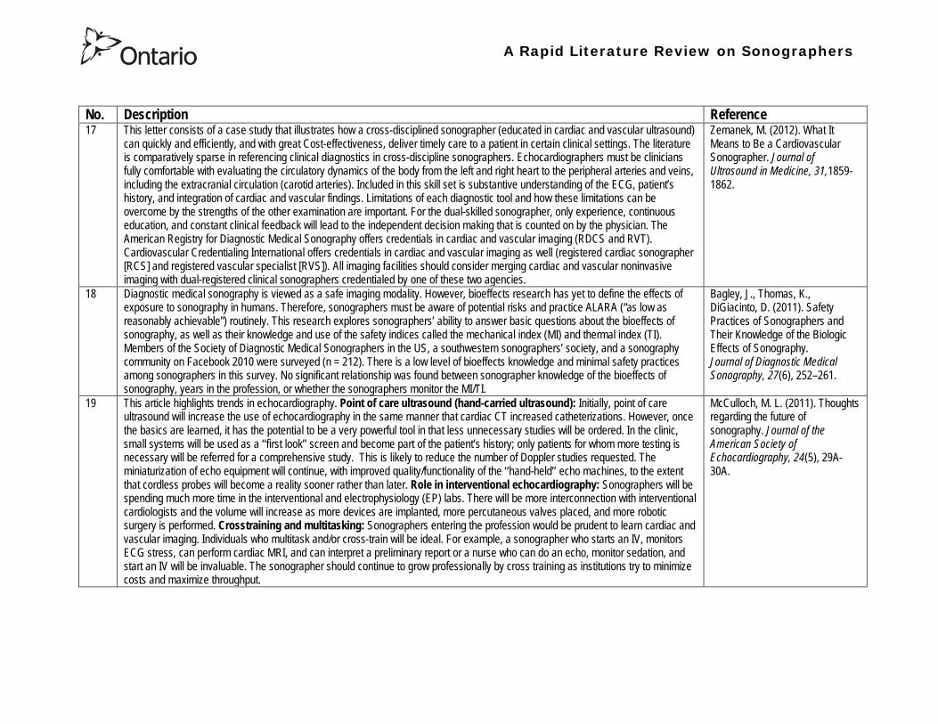
A Rapid Literature Review on Sonographers
No Description Reference 17 This letter consists of a case study that illustrates how a cross-disciplined sonographer (educated in cardiac and vascular ultrasound)
can quickly and efficiently and with great Cost-effectiveness deliver timely care to a patient in certain clinical settings The literature is comparatively sparse in referencing clinical diagnostics in cross-discipline sonographers Echocardiographers must be clinicians fully comfortable with evaluating the circulatory dynamics of the body from the left and right heart to the peripheral arteries and veins including the extracranial circulation (carotid arteries) Included in this skill set is substantive understanding of the ECG patientrsquos history and integration of cardiac and vascular findings Limitations of each diagnostic tool and how these limitations can be overcome by the strengths of the other examination are important For the dual-skilled sonographer only experience continuous education and constant clinical feedback will lead to the independent decision making that is counted on by the physician The American Registry for Diagnostic Medical Sonography offers credentials in cardiac and vascular imaging (RDCS and RVT) Cardiovascular Credentialing International offers credentials in cardiac and vascular imaging as well (registered cardiac sonographer [RCS] and registered vascular specialist [RVS]) All imaging facilities should consider merging cardiac and vascular noninvasive imaging with dual-registered clinical sonographers credentialed by one of these two agencies
Zemanek M (2012) What It Means to Be a Cardiovascular Sonographer Journal of Ultrasound in Medicine 311859-1862
18 Diagnostic medical sonography is viewed as a safe imaging modality However bioeffects research has yet to define the effects of exposure to sonography in humans Therefore sonographers must be aware of potential risks and practice ALARA (ldquoas low as reasonably achievablerdquo) routinely This research explores sonographersrsquo ability to answer basic questions about the bioeffects of sonography as well as their knowledge and use of the safety indices called the mechanical index (MI) and thermal index (TI) Members of the Society of Diagnostic Medical Sonographers in the US a southwestern sonographersrsquo society and a sonography community on Facebook 2010 were surveyed (n = 212) There is a low level of bioeffects knowledge and minimal safety practices among sonographers in this survey No significant relationship was found between sonographer knowledge of the bioeffects of sonography years in the profession or whether the sonographers monitor the MITI
Bagley J Thomas K DiGiacinto D (2011) Safety Practices of Sonographers and Their Knowledge of the Biologic Effects of Sonography Journal of Diagnostic Medical Sonography 27(6) 252ndash261
19 This article highlights trends in echocardiography Point of care ultrasound (hand-carried ultrasound) Initially point of care ultrasound will increase the use of echocardiography in the same manner that cardiac CT increased catheterizations However once the basics are learned it has the potential to be a very powerful tool in that less unnecessary studies will be ordered In the clinic small systems will be used as a lsquolsquofirst lookrsquorsquo screen and become part of the patientrsquos history only patients for whom more testing is necessary will be referred for a comprehensive study This is likely to reduce the number of Doppler studies requested The miniaturization of echo equipment will continue with improved qualityfunctionality of the lsquolsquohand-heldrsquorsquo echo machines to the extent that cordless probes will become a reality sooner rather than later Role in interventional echocardiography Sonographers will be spending much more time in the interventional and electrophysiology (EP) labs There will be more interconnection with interventional cardiologists and the volume will increase as more devices are implanted more percutaneous valves placed and more robotic surgery is performed Crosstraining and multitasking Sonographers entering the profession would be prudent to learn cardiac and vascular imaging Individuals who multitask andor cross-train will be ideal For example a sonographer who starts an IV monitors ECG stress can perform cardiac MRI and can interpret a preliminary report or a nurse who can do an echo monitor sedation and start an IV will be invaluable The sonographer should continue to grow professionally by cross training as institutions try to minimize costs and maximize throughput
McCulloch M L (2011) Thoughts regarding the future of sonography Journal of the American Society of Echocardiography 24(5) 29A-30A
A Rapid Literature Review on Sonographers
No Description Reference 20 Background Radiology reports provide specialist interpretation of images and relate these findings to the patientrsquos symptoms and
signs This study compared ultrasound (USS) reports generated by radiologists and sonographers to determine if any significant or clinically relevant differences existed Methods A retrospective analysis of 624 consecutive USS reports was carried out The reports were assessed for the presence of a lsquodisclaimerrsquo or lsquocaveatrsquo pertaining to the quality of the images and were analysed with respect to the clarity of their wording and ability to answer the clinical request Results The majority of sonographer USS reports contained a disclaimer regarding the quality of the USS images (571) compared with a smaller proportion of radiologist scans (99) (P lt 0001) Overall radiologists performed significantly better in providing a clear negative or positive diagnosis to the clinical question on the request form when compared with sonographers (885 vs 654 P lt 0001) Conclusion lsquoDisclaimerrsquo comments and lsquohedgingrsquo must be avoided in radiology reporting While feedback as to the accuracy of the radiology images is important overuse of such terms undermines the validity of the radiology report
Garcea G Mahmoud A Ong S L Rees Y Berry Det al (2010) Caveat reporting in ultrasound interpretation of surgical pathology a comparison of sonographer versus radiologist Journal of Evaluation in Clinical Practice 16 97ndash99
21 A nine-point questionnaire was constructed based on previously published work by Marsal and Sheiner et al The survey was distributed to clinical users of ultrasound practicing sonography in Hamilton (New Zealand) during July 2010 totalling 35 users consisting of sonographers sonography trainees one obstetric sonologist and one fetal medicine registrar Results Twenty sonographers returned the questionnaire which represents survey response of 61 The level of clinical experience was equally split between junior sonographers of lt 5 yearsrsquo experience (30) sonographers with 5-10 yearsrsquo experience (35) and senior sonographers of gt10 yearsrsquo experience (35) While 100 of the sonographers surveyed reported being familiar with the term Thermal Index (TI) and 90 claimed they adhered to the Action Learning Action Response Association (ALARA) principle all the time or most of the time only 15 of sonographers correctly described or defined what the term TI means Of the sonographers 80 knew where to find the TI during real-time examinations but only 35 knew which TI index to use for nuchal translucency (NT) scan and only 10 knew the expected range of TI encountered during routine obstetric examinations Of the eight sonographers who reported adhering to ALARA all the time the majority (64) could not describe or define TI In the same subgroup the majority (64) did not know which TI to monitor during a NT scan Only three of 20 (15) sonographers knew what the term TI means Of these two were junior sonographers with lt 5 yearsrsquo experience and one had 5ndash10 yearsrsquo experience Despite understanding the term TI one of the three sonographers still selected the incorrect index to monitor during a NT scan Not one of the seven senior sonographers with gt 10 years of experience could define or describe the TI In total only 10 of sonographers knew what the TI means and which TI to monitor during NT scanning
Necas M (2010) New Zealand sonographers do not outperform their European or American colleagues in the knowledge of ultrasound safety Australasian Journal of Ultrasound in Medicine13(4) 28-32
A Rapid Literature Review on Sonographers
No Description Reference 22 Musculoskeletal (MSK) ultrasound performed by non-medical practitioners is a developing practice The aim of this paper is
(1) to audit an experienced sonographerrsquos (SJ Riley one of the authors) performance after one yearrsquos independent reporting against two experienced MSK radiologists (CJ Grove amp M Chandramohan the other two authors) and (2) to establish an audit standard against which such role development might be contrived Images and reports from 250 MSK ultrasound examinations performed by a sonographer were reviewed independently by two consultant MSK radiologists The examinations were graded for discrepancy eg grade 1 ndash agree with the sonographer report grade 2 ndash minor discrepancy unlikely to alter patient care grade 3 ndash potentially significant discrepancy grade 4 ndash definite significant discrepancy likely to have adverse consequences for patient care Two of 250 (08) cases were excluded Both radiologists agreed completely with the sonographer (grade 1) in 235 of the 248 cases (948) In 13 cases there was discrepancy between the reports of SJR and the radiologists The discrepancy was grade 2 in six of the 248 cases (24) grade 3 in six of the 248 cases (24) and grade 4 in one of the 248 cases (04) In conclusion this audit shows a high level of agreement between the sonographer and the consultant MSK radiologist reporting of MSK ultrasound This level of agreement may set the standard for future quality assurance audit of sonographer MSK reporting
Riley S J Groves C J Chandramohan M (2010) Musculoskeletal ultrasound audit of sonographer reporting Ultrasound 18 36ndash40
23 Background Growing demand for ultrasound examinations and higher quality requirements motivate searching for routines combining the diagnostic accuracy of radiologist-performed examinations with the economical advantages of sonographer-performed examinations One possible approach is to use strictly standardized acquisition and documentation schemes that give the radiologist access to all relevant information after the examination Purpose To compare a recently introduced routine combining acquisition by a radiographer (more specifically a sonographer with two years experience) documentation as standardized cine-loops and review by a radiologist (lsquostandardized methodrsquo) with the formerly used routine where the diagnosis is made bedside by the radiologist (lsquotraditional methodrsquo) Material and Methods In 64 policlinic patients the kidneys (n = 27) or the gallbladder (n = 37) were examined with both the standardized and the traditional method The radiologistsrsquo findings of hydronephrosis tumors cysts echogenicity changes and cortical thickness (in the kidneys) and wall thickness concrements and polyps (in the gallbladder) were compared between the methods with respect to agreement (proportion of agreement and kappa coefficient) as well as systematic differences (McNemarrsquos test) Results The findings at the gallbladder examination showed a median agreement of 97 (86ndash100 kappa = 064ndash100) and those at the kidney examination an agreement of 90 (78ndash100 kappa = 069ndash 100) There were no significant systematic differences between the methods Conclusion The satisfactory agreement in this preliminary study indicates that the new workflow with ultrasound examinations performed by a radiographer (sonographer) and analyzed off-line by a radiologist is promising and motivates further studies
Stenman C Thorelius L Knutsson A amp Smedby Ouml (2010) Radiographer-acquired and radiologist-reviewed ultrasound examination ndash agreement with radiologistrsquos bedside evaluation Acta Radiologica 52 70ndash74
A Rapid Literature Review on Sonographers
No Description Reference 24 The troubling practice of fetal sex selection has historically been considered an Asian phenomenon However recent evidence shows
that a similar situation is emerging in North America albeit on a smaller scale The Society of Obstetricians and Gynaecologists of Canada (SOGC) has firmly stated its opposition to sex selection for non-medical reasons as well as to the use of any technology used solely for the purpose of determining fetal sex However because fetal sex may be disclosed to the parents at the time of ultrasound examination if they request this information guidance for health care professionals to assist in discouraging fetal sex selection would be useful Because no declaration of motives or reasons is required when a woman seeks a termination of pregnancy the authors suggest that health care professionals need not disclose the sex of a fetus until it has reached a gestational age at which abortion for non-medical purposes would not be possible This proposal would facilitate consistency between clinical practice and the values of Canadian citizens the SOGC the Canadian Medical Association and other professional organizations while still respecting current laws pertaining to disclosure of patient information and patientsrsquo rights to autonomous decision-making
Thiele A T amp Leier B (2010) Towards an Ethical Policy for the Prevention of Fetal Sex Selection in Canada J Obstet Gynaecol Can 32(1) 54ndash57
25 Background The extent to which increases in the use of prenatal ultrasonography over time reflect changes in maternal risk (ie an increase in high-risk pregnancies over time) is unknown In this population-based study the authors examined the use of prenatal ultrasonography from 1996 to 2006 in Ontario Methods With fiscal year 199697 as the baseline the authors evaluated the relative risk (RR) and 95 confidence interval (CI) for the change in rates of ultrasonography for each subsequent year The RR was adjusted for maternal age income rural residence maternal comorbidities receipt of genetics consultation or amniocentesis mdash all in the index pregnancy mdash and history of complications in a prior pregnancy Results The study sample consisted of 1 399 389 singleton deliveries The rate of prenatal ultrasonography increased from 2055 per 1000 pregnancies in 1996 to 3264 per 1000 in 2006 (adjusted RR 155 95 CI 154ndash155) The rate increased among both women with low-risk pregnancies (adjusted RR 154 95 CI 153ndash155) and those with high-risk pregnancies (adjusted RR 155 95 CI 154ndash157) The proportion of pregnancies with at least four ultrasound examinations in the second or third trimesters rose from 64 in 1996 to 187 in 2006 (adjusted RR 268 95 CI 261ndash274) Paradoxically this increase was more pronounced among low-risk pregnancies (adjusted RR 292 95 CI 283ndash301) than among high-risk pregnancies (adjusted RR 225 95 CI 216ndash235) Interpretation Substantial increases in the use of prenatal ultrasonography over the past decade do not appear to reflect changes in maternal risk Nearly one in five women now undergo four or more ultrasound examinations during the second and third trimesters Efforts to promote more appropriate use of prenatal ultrasonography for singleton pregnancies appear warranted
You J J Alter D A Stukel T A McDonald S D Laupacis A et al (2010) Proliferation of prenatal ultrasonography CMAJ 182(2) 143-151
A Rapid Literature Review on Sonographers
No Description Reference 26 Aims The aim of this study was to assess the cost-effectiveness of using certified sonographers and miniaturized echocardiography
systems to perform echocardiograms at bedside in comparison to moving inpatients from the admission department to the echocardiography laboratory (echo-lab) Methods and Results From 26 September 2005 to 27 October 2005 112 patients admitted in six hospital wards in Italy connected through a 100 Mbit LAN to the echo-lab were scanned within the admission ward by sonographers using a miniaturized echo system Logistical data were collected and results were compared with those obtained from 194 consecutive patients coming from the same wards and studied in the echo-lab with high-end machines between 8 March 2005 and 15 April 2005 Performing echocardiograms in the admission department avoided long waiting time of the inpatients in the echo-lab before and after the study increased the percentage of patients studied within 3 and 5 days from request (88 vs 77 and 100 vs 95 respectively P = 003) increased both sonographer (by 339 P lt 0001) and echo-lab productivity (by 41 P lt 0001) and reduced costs of echocardiograms by 29 Conclusion Implementation of digital echocardiography certified sonographers and a miniaturized echo system allowed improvement of the cost-effectiveness of the service provided by the echo-lab for inpatients and avoided patientsrsquo discomfort derived from prolonged waiting time before and after the exam
Badano L Gaetano N Stacul S Gianfagna P Pericoli M et al (2009) Improved workflow sonographer productivity and cost-effectiveness of echocardiographic service for inpatients by using miniaturized systems European Journal of Echocardiography 10 537ndash542
27 The present report is a proposal from the American Society of Echocardiography Advanced Practice Task Force that identifies the potential of cardiac sonographers to achieve the Advanced Cardiovasculat Sonographer (ACS) level According to this report echocardiographic examinations require a well-trained and competent sonographer to obtain proper anatomic and physiologic data to establish an accurate diagnosis for clinical decision-making and patient management Although the formal education and training of cardiovascular sonographers are evolving many entry-level and staff sonographers may not have sufficient practical or clinical knowledge of the necessary components of the echocardiographic study for the individual patientrsquos clinical presentation In many clinical settings echocardiograms are read after the patient has left the laboratory Thus there is a role for a sonographer who can practice at an advanced level in a cardiovascular ultrasound laboratory to ensure a proper echocardiographic examination is performed on every patient In this setting an Advanced Cardiovascular Sonographer (ACS) would be able to review the indication for and quality of the examination If additional images were needed the ACS would assist the sonographer in obtaining these images which would lead to the performance of a complete and fully diagnostic examination before the patient had left the echocardiography laboratory In clinical practice the quality of the examinations performed would improve advancements in echocardiographic methods could be taught and incorporated into daily practice and patients would be better served
Mitchell C Miller F A Bierig S M Bremer M L Ehler D et al (2009) Advanced Cardiovascular Sonographer A Proposal of the American Society of Echocardiography Advanced Practice Sonographer Task Force J Am Soc Echocardiogr 22 1409-13
A Rapid Literature Review on Sonographers
No Description Reference 28 Sonographer practitioner development involves the expansion and extension of the sonographer role to include reporting on
ultrasound examinations Australian sonographers have not seen the same degree of role extension and expansion as their counterparts in the United Kingdom despite increasing levels of discussion regarding sonographer practitioner development The aim of this study was to determine if Australian sonographers want to extend their professional role and what they consider are the important issues associated with role extension This paper reports on qualitative data derived from a survey of Australian sonographers and investigates if Australian sonographers are interested in extending and expanding their professional role and responsibilities and if they do what might be necessary or desirable from a professional point of view for this change to occur A survey was mailed to all members of the Australian Sonographers Association (ASA) in October 2006 The 31-item survey included 28 closed-ended and three opened-ended items to provide both quantitative and qualitative data The quantitative data will be reported separately Qualitative data was derived from responses to the opened-ended questions which asked respondents to elaborate on their attitudes and feelings about role extension and development Analysis used Nvivo7 software to aid in uncovering common themes from the qualitative data The analysis focused on the reported incentives or motivations for becoming a sonographer practitioner as well as disincentives or perceived hurdles that would discourage respondents from becoming sonographer practitioners The three most reported incentives or motivations for becoming a sonographer practitioner were professional recognition remuneration and increased knowledge The three most commonly reported disincentives or perceived hurdles that would discourage respondents from becoming sonographer practitioners were legal issues insurance and further study
McGregor R OrsquoLoughlin K Cox J Clarke J Snowden A (2009) Sonographer practitioner development in Australia Qualitative analysis of an Australian sonographersrsquo survey Radiography15 313-319
29 Introduction This study describes the effect of introducing a cardiac sonographer led follow up clinic within a hospital cardiology department for patients with stable heart valve disease The two years before and after the instigation of the valve clinic were audited Materials and methods The clinic was conducted in a single centre and undertaken in the cardiology department of a district general hospital A total of 382 patients with 397 clinically significant valve lesions but for whom surgery was not yet indicated but follow up required were seen in a cardiac sonographer run clinics These patients no longer attended a medical follow up clinic unless there was clinical or echocardiographic deterioration Effectiveness was judged by the percentage treated according to current best practice guidelines the number of echocardiograms performed and the number of hospital outpatient visits attended In addition mortality data for the subjects in the clinic was collected Results The proportion followed up according to best practice guidelines rose from 157 (41) to 354 (92) (p lt 001) The total number of echocardiograms performed fell from 807 to 550 Total number of outpatient visits fell from 998 to 31 A total of 11 patients died in the two year study period none from progressive valve disease Discussion This study demonstrates that a protocol driven sonographer led heart valve disease follow up clinic significantly improved the quality of follow up while bringing about a major reduction in outpatients visits without compromising patient safety
Taggu W Topham A Hart L Carr-White G Sulke N et al (2009) A cardiac sonographer led follow up clinic for heart valve disease International Journal of Cardiology 132 240 ndash243
30 Role development for imaging allied health professionals has progressed significantly over the last three decades with many major developments pioneered in the field of ultrasound This paper reviews the developing role of the radiographer in ultrasound (in some countries such as the UK sonographers are considered a type of radiographer) and some of the clinical professional and educational implications of such development As the published evidence base underpinning the latest developments is scarce several contemporary case study examples which have been presented verbally at study days attended by the authors have been included In conclusion sonographer role development is considered within the professional contexts of extended role advanced and consultant practice
Hart A amp Dixon A (2008) Sonographer Role Extension and Career Development a Review of the Evidence Ultrasound 16(1) 31-35
A Rapid Literature Review on Sonographers
No Description Reference 31 Objective The main goal of this study was to determine end usersrsquo knowledge regarding safety aspects of diagnostic ultrasound
during pregnancy End usersrsquo attitudes toward the use of ultrasound in low-risk pregnancies were also assessed Methods A questionnaire was distributed to ultrasound end users attending review courses and hospital grand rounds between April and June 2006 in Chicago Results One hundred thirty end users completed the questionnaires (63 response rate) Sixty-three percent were physicians (n = 84) most of them obstetricians (817) About 18 of participants routinely performed Doppler ultrasound examinations during the first trimester Fifty percent of end users thought that the number of ultrasound examinations in low-risk pregnancy should be limited to 1 to 3 (mean plusmn SD 26 plusmn 09) Almost 70 disapproved of ldquokeepsakeentertainmentrdquo ultrasound Although 322 of the participants were familiar with the term thermal index only 177 actually gave the correct answer to the question on the nature of the thermal index About 22 were familiar with the term mechanical index but only 38 described it properly Almost 80 of end users did not know where to find the acoustic indices Only 208 were aware that they are displayed on the sonographic monitor during the examinations End users with higher knowledge of safety issues thought that there should be limitations on the number of ultrasound examinations in low-risk pregnancies (odds ratio 33 95 confidence interval 11ndash100 P = 028) Likewise these end users were more likely to respond that ultrasound might have adverse effects during pregnancy (odds ratio 32 95 confidence interval 11ndash125 P = 045) Conclusions Ultrasound end users are poorly informed regarding safety issues during pregnancy Further efforts in the realm of education and training are needed to improve end user knowledge about the acoustic output of the machines and safety issues
Sheiner E Shoham-Vardi I Abramowicz J S (2007) What Do Clinical Users Know Regarding Safety of Ultrasound During Pregnancy J Ultrasound Med 26 319ndash325
A Rapid Literature Review on Sonographers
No Description Reference 32 In an attempt to survey the knowledge among ultrasound users of some safety aspects of diagnostic ultrasound a questionnaire was
distributed to professionals using ultrasound for fetal examinations The questionnaire was answered by the participants and faculty members of six postgraduate courses in obstetric ultrasound Doppler ultrasound and fetal echocardiography given in Sweden Norway and Austria in 2003 Furthermore in Sweden the questionnaire was distributed to the staff meeting participants of two major obstetric ultrasound units and of one unit for fetal echocardiography and at a meeting of the Working Group for Ultrasound of the Swedish Society of Obstetrics and Gynecology In total 199 questionnaires were anonymously answered by doctors (n = 145) sonographers (n = 22) and midwives trained in ultrasound (n = 32) All of them were using diagnostic ultrasound on a daily or weekly basis The respondents were from nine European countries In none of the countries is formal certification for the use of diagnostic ultrasound in pregnancy or otherwise required The doctors who answered the questionnaire were specialists in obstetrics and gynecology (including some of the internationally recognized experts in fetal ultrasound) pediatric cardiology radiology or clinical physiology The respondents were not informed about the questionnaire in advance and they had a limited time to answer the 16 questions typically 10ndash15 min The questions concerned explanation of the safety indices and their relation to various ultrasound modes ie real-time B-mode M-mode three-dimensional (3D) spectral Doppler color and power Doppler ultrasound The respondents were also asked which ultrasound system they used most often and where on the system the information on thermal index (TI) and mechanical (MI) was displayed (eg in the top right corner of the screen in a separate window etc) Furthermore the respondents had to indicate how they adjust the output energy level on their own machine For all questions there was the optional answer lsquoDonrsquot knowrsquo Results About one-third of the ultrasound experts were able to define the abbreviations TI and MI For the subclasses of the TI the correct answer was found in only 3-8 of cases Of those who knew what the TI and MI meant only two-thirds and one-third of respondents respectively were able to give a simple explanation of the indices In 49 of questionnaires the Doppler modes were correctly ranked as generally producing higher ultrasound exposure than the imaging modes (B-mode 3D) Some 28 of the respondents correctly indicated where on their own machine the information on safety indices is displayed However not all of them knew how to control the output energy level For all items in the questionnaire there were no significant differences in the results between the three categories of respondents (ie physicians sonographers and midwives) The results of the questionnaire were anything but encouraging and they indicated that the users who are supposed to be responsible for controlling exposure of the fetus to ultrasound had a very poor knowledge of the basic safety aspects of ultrasound
Maršaacutel K (2005) The output display standard has it missed its target Ultrasound Obstet Gynecol 25 211ndash214
33 Purpose This study was performed to evaluate the accuracy of upper abdominal ultrasound (US) scanning performed by sonographers in a district general hospital to identify potential areas of weakness and to make recommendations to improve the service Materials and Methods Upper abdominal US examinations performed and reported by sonographers over a 4-week period were retrospectively reviewed The accuracy of the imaging findings and reports were assessed against other imaging surgical histological or laboratory findings and against clinical outcome Results A heterogenous group of 104 patients were included in the study 62 of whom had an US abnormality Errors of scanning or interpretation were identified in 10 patients (96) of whom five (48) were felt to be potentially significant Conclusions The sonographersrsquo accuracy in reporting upper abdominal US scans was 90 However on the basis of this study the authors have implemented specific recommendations to improve the quality of the service
Dongola N Guy R Ward S amp Giles J A (2003) Can sonographers offer an accurate upper abdominal ultrasound service in a district general hospital Radiography 9 29ndash33
A Rapid Literature Review on Sonographers
No Description Reference 34 Aim To compare the accuracy of radiographers and radiologists in routine abdominal ultrasound Materials and Methods One
hundred consecutive patients attending for routine abdominal ultrasound were included Each patient was examined by both a radiographer and radiologist Both operators noted their fndings and wrote a concluding report without conferring Reports were compared Where there was disagreement the patient was either re-examined by another radiologist or had further investigation Results Of 100 patients 52 were men and 48 were women The age range was 19plusmn88 years (median 52 years) Thirty-seven patients had renal tract ultrasound one had an aortic ultrasound and 62 had general upper abdominal ultrasound In 44 cases both operators reported the examination as normal In 49 cases both operators reported the examinations as abnormal and there was complete agreement between the operators In seven cases there was not complete agreement between operators Three of these disagreements were considered minor and four major In three of the seven cases the radiographer was correct and in four the radiologist was correct Conclusion Experienced radiographers and radiologists are highly accurate in performing and interpreting routine abdominal sonography Both operators missed a small minority of abnormalities There was no statistically significant difference in the accuracy of radiographers and radiologist
Leslie A Lockyer H amp Virjee J P (2000) Who Should be Performing Routine Abdominal Ultrasound A Prospective Double-Blind Study Comparing the Accuracy of Radiologist and Radiographer Clinical Radiology 55 606-609
Grey Literature 35 The ARRT The American Registry of Radiologic Technologists tests certifies and annually registers more than a quarter-of-a-million
radiologic technologists to promote high standards of patient care They award the Registered Technologist or RT designation to individuals who have completed the prescribed classroom and clinical education passed the appropriate exam and met the ethics requirements And they ensure their continuing education and ongoing compliance with their high standards by requiring annual registration of their certificate Certification by ARRT is voluntary not mandatory But employers state licensing agencies and federal regulators view ARRTrsquos credential as an indicator that a person has met recognized national standards for medical imaging and radiation therapy professionals How Are Radiologic Technologists Deemed Qualified First comes initial certification Certification is the process of initially recognizing individuals who have satisfied certain standards within a profession A person is certified by ARRT after completing an approved educational program complying with the ethics standards and passing a certification exam Then comes annual registration Registration is the yearly procedure required to maintain certification ARRT registrants are those who having already fulfilled the requirements for initial certification continue to meet the requirements for annual registration The ARRT annually registers the certificates of individuals who meet the following three criteria agreeing to comply with the ARRT Rules and Regulations continuing to comply with the ARRT Standards of Ethics and meeting our continuing education requirements Only technologists who are currently registered (which means that they have renewed within the past year) may designate themselves as ARRT Registered Technologists and use the initials RT after their names Federal laws regulate personnel qualifications in some modalities (mammography for example) but they accredit the health care facility rather than licensing the individual radiologic technologist who is providing patient care And while many states have laws regulating who can perform radiologic procedures they are typically based on ARRT certification Some employers and state agencies may require ARRT certification but not necessarily current registration Unless they require current registration by ARRT employers cannot assume that the person has complied with the ethics requirements or the continuing education requirements
American Registry of Radiologic Technologists (ARRT) (2013a) Patient Page Accessed April 2013
A Rapid Literature Review on Sonographers
No Description Reference 36 This document outlines certification requirements for sonography There are two pathways (primary and post-primary) For the
primary pathway it notes that sonography educational programs must be accredited by a mechanism acceptable to the ARRT ARRT generally recognizes only accreditation agencies that are recognized by the Council for Higher Education Accreditation andor United States Department of Education
American Registry of Radiologic Technologists (ARRT) (2013b) Sonography Certification Accessed April 2013
A Rapid Literature Review on Sonographers
No Description Reference 37 In Australia sonographers and student sonographers are required to be accredited by Australian Sonographer Accreditation Registry
(ASAR) in order to practise This requirement is contained within the Australian Governmentrsquos Diagnostic Imaging Accreditation Scheme standards and Medicare legislation Accreditation of sonographers is linked only to achieving - or in the case of students studying towards - an accredited entry-level qualification completion of continuing professional development and payment of an annual fee to ASAR The ASA contends that the lack of a requirement to ensure each sonographer is lsquofit to practisersquo is an unacceptable gap in current regulation undermining efforts to ensure the quality of sonographic services and failing to protect patients from harm The ASA supports and advocates for nationally-enforceable regulation of sonographers to meet our patientrsquos expectations National registration The National Registration and Accreditation Scheme (NRAS) for health workers commenced on 1 July 2010 The ten professions which comprised the initial intake into this scheme were those that had registration requirements in all Australian states or territories as well as those in which legislated regulation is deemed to be in the public interest An additional four professions - including medical radiation practitioners which are defined as radiographers radiation therapists and nuclear medicine technologists - will be included in the NRAS commencing on 1 July 2012 As sonography did not have registration in any states or territories prior to the commencement of NRAS the profession has not been in a position to negotiate entry into NRAS In early August 2011 the ASA lodged a submission with the Health Workforce Principal Committee requesting a profession-specific Sonography Board of Australia be formed under NRAS This submission was prepared based on information that consideration of professions which were unregistered would commence More recently the Australian Health Workforce Ministerial Council (AHWMC) issued a communiqueacute stating in part lsquoMinisters have commissioned work on unregistered professions and future directions for national registration Given this Ministers agreed to defer consideration of the inclusion of additional professions into the NRAS Ministers agreed that it would be premature to consider further applications for the scheme until this work has been completed other than paramedicsrsquo Options for unregistered health practitioners including sonographers Policy work is continuing at a federal level to establish an alternative regulatory framework The National Alliance for Self-Regulating Health Professions (NASRHP) consisting of the peak professional associations of eight self-regulating health professions including the ASA representing sonography released a proposal lsquoHarnessing self-regulation to support safety and quality in healthcare deliveryrsquo in March 2012 This proposal concluded that the framework that regulates all health practitioners should be expanded beyond NRAS to include
bull authorised self-regulating professions via a health professions panel bull negative licensing to withdraw the ability to practise from registered and self-regulated practitioners who do not meet the
regulatory requirements or unregulated practitioners due to poor practice As with all advocacy initiatives consideration by the federal government of the introduction of the NASRHPrsquos proposal may be prolonged and the outcomes may not match the expectations of NASRHPrsquos member professional associations including the ASA The ASA will continue both independently and collaboratively to lobby on behalf of our members and the public for the introduction of a timely and equitable solution to health practitioner regulation In the interim as there is no regulatory model with the rigour the ASA believes is required for regulation of sonographers and other health practitioners other than NRAS the ASA continues to support its Position Statement published in early 2010
Australian Sonographer Association (ASA) (2013) Sonographer Regulation Accessed April 2013
A Rapid Literature Review on Sonographers
No Description Reference 38 This publication contains information about Cardiovascular Credentialing International (CCI) It also provides the necessary
information and references concerning the process by which candidates may earn certificate or registry-level credentials CCI is a not-for-profit corporation established for the purpose of administering credentialing examinations as an independent credentialing agency CCI began credentialing cardiovascular professionals in 1968 CCI offers eight credentials which when earned demonstrate that the registrant holds fundamental knowledge in the particular cardiovascular specialty These include RCCS ndash Registered Congenital Cardiac Sonographer RCS ndash Registered Cardiac Sonographer and RPhS ndash Registered Phlebology Sonographer
Cardiovascular Credentialing International (2013) 2013 Examination Application amp Overview Accessed April 2013
39 Oregon Governor Signs Sonographer Licensure Legislation On July 28 2009 Oregon Governor Ted Kulongoski signed a sonographer licensure bill (HB 2245) into law The new law makes Oregon only the second state in the United States to require licensure of sonographers The Society of Diagnostic Medical Sonographers (SDMS) has worked closely with other sonography-related organizations and Oregon sonographers to help craft the language in new law The law renames and restructures the Oregon Board of Radiologic Technologists to the Oregon Board of Medical Imaging (OBMI) and ensures representation of sonographers on the Board The sonographer licensure law requires Oregon sonographers to become licensed by OBMI effective July 1 2010 Beginning on January 1 2014 all sonography licensees would be required to hold a national sonography certificationcredential (or be currently enrolled as a student) While the Oregon bill is far more detailed than the sonographer licensure law that passed in New Mexico earlier this year additional administrative rulesregulations will be developed over the next year to fully implement the new law SDMS will be continue to monitor developments regarding sonographer licensure in other states
Society of Diagnostic Medical Sonographers (SDMS) (2013a) News Accessed April 2013
A Rapid Literature Review on Sonographers
No Description Reference 40 On April 6 2009 New Mexico (NM) Governor Bill Richardson signed the historic bill that for the first time in the United States
requires licensure of sonographers The bill adds sonographers (and MRI technologists) to the list of medical imaging professionals licensed by the State of New Mexico Until now anyone could perform sonograms in New Mexico Prior to the passage of the New Mexico licensure law virtually all other health care providers and professionals had to be licensed As a result the public is often surprised to learn that the person performing a medical sonogram does not have any state licensure or national certification requirements The responsibility for administering the NM sonographer licensure program would rest with the New Mexico Environment Department Although this was not the first choice in most sonographersrsquo minds the Environment Department currently manages the radiologic technology licensure program so the administrative infrastructure is already in place By ensuring the creation of an advisory council that includes representation by each medical imaging and radiation therapy modality regulated the bill helps ensure that the Departmentrsquos other areas of emphasis do not detract from sonographer licensure Because sonographers have never had to be licensed before they frequently do not see or understand the distinction between state licensure and national certification National certifications or credentials issued by voluntary organizations such as the American Registry for Diagnostic Medical Sonography (ARDMS) Cardiovascular Credentialing International (CCI) and the American Registry of Radiologic Technologists (ARRT) have little legal weight The certification process and resulting credential are valuable tools in ensuring sonographers have met minimum education and knowledge requirements but do not really regulate anyonersquos ability to perform sonography In an ideal world no additional regulation of sonography would be required However as everyone knows more and more people are taking weekend courses and buying ultrasound equipment without understanding the ramifications of using ultrasound technology (eg ALARA) Under the bill passed by the New Mexico legislature national credentialscertifications should provide evidence of meeting the statersquos standards All sonographers who are performing these critical medical imaging services in New Mexico will be required to meet national certification standards (ie have certifications or credentials issued by ARDMS CCI or ARRT) As a result there would be no additional exams in New Mexico beyond the national certification exams required by ARDMS CCI or ARRT The bill also requires the state to recognize the continuing medical education completed for national certification renewal The most visible impact on New Mexico sonographers will be a license fee However the legislation caps the license fee at $100 per two-year license In addition only the New Mexico Legislature can change this cap
SDMS (2013b) New Mexico Sonographer Licensure Law Signed Accessed April 2013
A Rapid Literature Review on Sonographers
No Description Reference 41 Preamble The purpose of this document is to define the Scope of Practice for Diagnostic Ultrasound Professionals in the US and to
specify their roles as members of the health care team acting in the best interest of the patient This scope of practice is a living document that will evolve as the technology expands Definition of the Profession The Diagnostic Ultrasound Profession is a multi-specialty field comprised of Diagnostic Medical Sonography (with subspecialties in abdominal neurologic obstetricalgynecologic and ophthalmic ultrasound) Diagnostic Cardiac Sonography (with subspecialties in adult and pediatric echocardiography) Vascular Technology and other emerging fields These diverse specialties are distinguished by their use of diagnostic medical ultrasound as a primary technology in their daily work Certification is considered the standard of practice in ultrasound Individuals who are not yet certified should reference the Scope as a professional model and strive to become certified Scope of Practice of the Profession The Diagnostic Ultrasound Professional is an individual qualified by professional credentialing and academic and clinical experience to provide diagnostic patient care services using ultrasound and related diagnostic procedures The scope of practice of the Diagnostic Ultrasound Professional includes those procedures acts and processes permitted by law for which the individual has received education and clinical experience and in which heshe has demonstrated competency Diagnostic Ultrasound Professionals
bull Perform patient assessments bull Acquire and analyze data obtained using ultrasound and related diagnostic technologies bull Provide a summary of findings to the physician to aid in patient diagnosis and management bull Use independent judgment and systematic problem solving methods to produce high quality diagnostic information and
optimize patient care
Society of Diagnostic Medical Sonography [SDMS] (2013c) Scope of Practice for the Diagnostic Ultrasound Professional Accessed April 2013
42 This website notes that in the US most employers in the US prefer to hire sonographers who have professional certification A sonographer can get certification and passing an exam Most exams relate to the specialty that the sonographer is most interested inmdashfor example an exam to become certified in abdominal sonography A few states require diagnostic medical sonographers to be licensed Typically professional certifications required for licensure other requirements vary by state Sonographers must take continuing education to keep their certification current
Bureau of Labor Statistics US Department of Labor (2012) How to become a Diagnostic Medical Sonographer Occupational Outlook Handbook 2012-13 Edition Accessed April 2013
A Rapid Literature Review on Sonographers
No Description Reference 43 The American Registry for Diagnostic Medical Sonography (ARDMS) is an independent not-for-profit organization founded in 1975
that administers examinations and awards the following credentials bull RDMSreg ndash Registered Diagnostic Medical Sonographerreg bull RDCSreg ndash Registered Diagnostic Cardiac Sonographerreg bull RVTreg ndash Registered Vascular Technologistreg bull RPVIreg ndash Registered Physician in Vascular Interpretationreg bull RMSKtrade - Registered in Musculoskeletaltrade sonography
ARDMS is the globally recognized standard of excellence in sonography and has certified more than 75000 individuals ARDMS is composed of three key groups
bull Board of Directors ndash Sonographers vascular technologists physicians research scientists and a member of the public are volunteer members of the board that determines the direction and policies of our organization
bull Examination Development Task Forces ndash A group of sonographers vascular technologists physicians and scientists volunteer their time and expertise to produce our examinations
bull Professional Staff ndash A paid highly-skilled staff administers the programs and assists applicants and registrants Credentials awarded by ARDMS document personal achievement of recognized professional standards and are widely accepted in the medical community by sonography and vascular professional organizations However ARDMS does not validate the day-to-day job performance of any sonography professional ARDMS has earned the prestigious ANSI-ISO 17024 accreditation for certifying bodies from the International Organization for Standardization (ISO) Accreditation is granted through the American National Standards Institute (ANSI) ARDMS has also earned accreditation with the National Commission for Certifying Agencies (NCCA) The NCCA is the accrediting arm of the National Organization for Competency Assurance (NOCA) Established in 1977 as a nonprofit organization NOCA is a leader in establishing quality standards for certifying organizations
American Registry for Diagnostic Medical Sonography (ARDMS) (2012a) Overview of ARDMS Accessed April 2013
A Rapid Literature Review on Sonographers
No Description Reference 44 ARDMS is developing a program to assure that Registrants continue to meet the basic competency requirements of core clinical skills
in present day practice bull Each Registrant who holds an active ARDMS certification as of December 31 2011 will be assigned the 2012 ndash 2021 10-
year recertification assessment period bull The 10-year period for new Registrants will begin the year following the attainment of the credential bull If a Registrant earns a new credential or specialty after January 1 2012 then that credentialspecialty will have a separate
10-year recertification assessment period that will begin the year following the attainment of the credentialspecialty ARDMS will develop tools and resources to assist Registrants in tracking and meeting the recertification assessment program requirements
bull Registrants who do not wish to take a recertification assessment in a specific credentialspecialty may voluntarily relinquish the credentialspecialty
As an ARDMS Registrant individuals are committing to demonstrate the highest level of competency in the modality of sonography by meeting and maintaining the standards set by the ARDMS The pathway of maintenance your certification is comprised of Paying the ARDMS annual renewal fee each year Each year ARDMS assesses Registrants an annual renewal fee By paying your annual renewal fee you are contributing to the ARDMS mission of promoting quality care and patient safety through the certification and continuing competency of ultrasound professionals In order to maintain your credentials you must pay the annual renewal fee of $75 USD or $100 USD (for RPVI and MSK Registrants with MDDODPM) designation on or before December 31 of each year in order to stay active for the following year Attesting to ARDMS rules and policies Each year when you pay your annual renewal fee you are attesting that you are in compliance with all ARDMS rules and discipline policies and are in good standing with the ARDMS Earning a minimum of 30 ARDMS-accepted CMEs during your three-year CME period ARDMS verifies its Registrants compliance with continuing medical education (CME) requirements using an audit process Earning your CMEs is an integral component of maintaining your ARDMS credential(s) and demonstrates your ongoing commitment to lifelong learning RecertificationContinued Commitment to Excellence Following the attainment of the ARDMS credential Registrants are assigned a 10-year recertification period the year after the credential is earned ARDMS is committed to developing a recertification program that is easy for Registrants to comply with
American Registry for Diagnostic Medical Sonography (ARDMS) (2012b) Recertification General Information
A Rapid Literature Review on Sonographers
No Description Reference 45 This document reviews Sonography Principles and Instrumentation (SPI) Examination Requirements offered through ARDMS in
order to achieve sonography certification and the various prerequisites needed before taking the exam For example one prerequisite entails (1) Education Graduate of a program accredited by an agency recognized by the Council for Higher Education Accreditation (CHEA) United States Department of Education (USDOE) or Canadian Medical Association (CMA) that specifically conducts programmatic accreditation for diagnostic medical sonographydiagnostic cardiac sonographyvascular technology Currently the only organizations that offer programmatic accreditation under the aforementioned associations are the Commission on Accreditation of Allied Health Education Programs (CAAHEP) and the Canadian Medical Association (CMA) (2) Required Clinical UltrasoundVascular Experience No additional experience is required (3) Documentation Required with Application 1) Copy of diploma from ultrasoundvascular program or an official transcript indicating the date the degree was conferred 2) Original letter signed by program director andor medical director indicating date of graduation or successful completion of the program4 Program directors must use the mandatory formatted sample letter available on ARDMSorg sampleletters 3) The CV form is not required if the application is submitted and received in the ARDMS office within one year after successful completion of the program Otherwise an original signed and completed CV form for each appropriate specialty area(s) must be submitted 4) Photocopy of a non-expired government issued photo identification with signature the name on the identification must exactly match the name under which you are applying for ARDMS examination
American Registry for Diagnostic Medical Sonography (ARDMS) (2012c) SPI Requirement and General Prerequisites Accessed April 2013
46 This page notes the five credentials offered by the ARDMS and the corresponding exams and specialty exams required bull RDMS requires the SPI exam (sonography principles and instrumentation) and a specialty exam in abdomen breast fetal
echocardiography neurosonolgoy or obstetricsgynecology bull RDCS requires the SPI exam and a speciality exam in adult echocardiography fetal echocardiography pediatric
echocardiography bull RVT requires the SPI and a speciality exam in vascular technology bull RMSK requires the musculoskeletal sonography exam bull RPVI requires the physiciansrsquo vascular interpretation exam
American Registry for Diagnostic Medical Sonography (ARDMS) (2012d) Credentials and Examinations Accessed April 2013
A Rapid Literature Review on Sonographers
No Description Reference 47 This article summarizes some of the many trends in fetal pediatric and adult echocardiography that SDMS members are excited
about Fetal Echocardiography Sonography has been used since the mid-1980s to help identify congenital heart defects during the second trimester The ldquobasicrdquo examination of the fetal heart involves a four-chamber view ndash evaluation of the right and left atrial and ventricular chambers and their respective valves In the past several years there has been a movement in the obstetric ultrasound community to encourage obstetric sonographers to perform the ldquoextended basicrdquo exam which includes the cardiac outflow tracts The outflow tracts consist of the main pulmonary artery exiting the right ventricle and the aorta exiting the left ventricle The fetal echocardiography community also is abuzz over three-dimensional (3D) and four-dimensional (4D) ultrasound and the potential of this technology to significantly improve the evaluation of the fetal heart Spatiotemporal Image Correlation (STIC) a 4D application allows for evaluation and manipulation of the heart in real-time Allen says Todayrsquos ultrasound equipment allows sonographers to gather a volume of data from which they can create detailed 3D images of the tiny fetal heart which is constantly in motion With computer manipulation of the data sets the images also can be viewed in 4D allowing physicians to see the entire fetal heart cycle from start to finish Pediatric Echocardiography One of the most significant advances in pediatric echo over the last decade is moving to a digital format The advent of pediatric transesophageal (TEE) probes also has been a big advance for pediatric echo Transesophageal probes provide an unimpeded sonographic visualization of the intracardiac structures Theyrsquore useful in visualizing very fine valvular abnormalities the thoracic aorta vegetations and thrombus in the atrial appendage It is also useful in detecting subtle anatomic variations seen in congenital heart disease Pediatric-sized TEE probes make the exams possible In the past it wasnrsquot possible because the adult probe was just too big to put down the babyrsquos or childrsquos esophagus Now the probe is small enough to successfully manipulate it in children and get superb images Because the new sonographic imaging equipment and better surgical techniques allow clinicians to detect and repair more cardiac abnormalities earlier patients are living longer Adult Echocardiography The adult echo world is abuzz about 2D and 3D speckle tracking relatively new techniques for the assessment of heart function and strain imaging Speckle tracking evaluates both regional and global function by measuring the deformation of the heart muscle
Orenstein B W (2010) Current Trends in Echocardiography SDMS New Wave February 1-5 Accessed April 2013
48 This document describes the requirements for admission to the Register of Accredited Medical Sonographer in Australia Requirements include Continuation of Accreditation In order to remain on the Register an accredited Sonographer must satisfy the ASAR continuing professional development (CPD) requirements and pay the prescribed annual fee Student Sonographers apart from paying the prescribed annual fee should provide evidence of their student status every year when they renew their ASAR accreditation
Australian Sonographer Accreditation Registry (ASAR) (no date A) Sonographer Accreditation Accessed April 2013
A Rapid Literature Review on Sonographers
No Description Reference 49 ASARrsquos functions
1 Accredit sonography education programs within Australia 2 Maintain a register of Accredited Sonographers and Accredited Student Sonographers 3 Establish minimum standards of Continuing Professional Development (CPD) and to monitor the CPD requirements for
Sonographers to remain on the register 4 To support activities that advance the profession of sonography 5 To provide registration information about Sonographers to Medicare Australia 6 To provide public access to the name registration number State or Territory current type of accreditation and area of
specialty for active Sonographers ASARrsquos Role ASAR is not a registration board ASAR does not have the authority to remove a Sonographer from the Registry due to professional misconduct nor the ability to prevent a Sonographer from practicing ASAR is only able to remove a Sonographer from the Registry if they
bull fail to meet CPD requirements and or bull fail to pay annual fees
ASAR (no date B) About ASAR Accessed April 2013
50 Regulatory bodies A Regulatory Body maintains a register of individuals who have achieved the required qualification of that profession to practice and can evidence continuing competence at the required level For a doctor to practice in the UK he or she must be registered with the General Medical Council (GMC) This is the regulatory body for medical practitioners The majority of sonographers who are employed in the UK come from a background of radiography or midwifery To practice radiography in the UK a qualified radiographer must be registered with the Health Professions Council (HPC) Similarly a midwife must be registered with the National Midwifery Council to practice as a midwife Sonographer registration in the UK There are a number of professions allied to medicine that are regulated by the HPC for example radiography physiotherapy However sonography is not recognised as a profession by the HPC and therefore there is no mechanism whereby an individual can register with the HPC as a sonographer or be regulated by them as a sonographer It is a common misunderstanding that sonographers have to be registered with the HPC to practice This comes from the fact that most sonographers are also qualified radiographers and maintain their registration with the HPC because they wish to remain on the HPC register as a radiographer Professional bodies An individual can be a member of a Professional Body such as the Royal College of Obstetricians amp Gynecologists (RCOG) Royal College of Radiologists (RCR) Society amp College of Radiographers (SCoR) or the Royal College of Midwives (RCM) but this is not the same as being accepted onto the register of one of the regulatory bodies The professional bodies are responsible for setting and maintaining the standards of their members providing a code of conduct etc They may also act in the capacity of a trades union and can also provide professional indemnity insurance The British Medical Ultrasound Society BMUS is not a professional body but is a scientific society so will offer a different range of benefits Whether or not one wishes to join the society has no impact on their ability to work as a sonographer in the UK although from a continuing professional development point of view it can be beneficial The College of Radiographers are currently working with the HPC to get sonography recognized as a separate profession
British Medical Ultrasound Society (BMUS) (no date) Information on Sonographer Regulation in the UK Accessed April 2013
A Rapid Literature Review on Sonographers
No Description Reference 51 This document included sections on the role of the sonographer and the use of sonography for entertainment Role of the
Sonographer and Technical Impressions The role of the sonographer is to perform the ultrasound scan and to document observations The sonographerrsquos written technical impressions are intended as a form of communication between the sonographer and the reporting physician These observations must be reviewed and subsequently reported by the reporting physician Issuing of a final reportdiagnosis represents the practice of medicine and is therefore the domain of the reporting physician Use of Ultrasound for Entertainment or Non-Diagnostic Purposes In contrast to this justified form of assessment ultrasound for entertainment is simply an elaborate version of a portrait For a variable fee the surface of the fetus is skimmed over and clients are provided with keepsake images or a DVD of their fetus There are no measurements taken during this exam no morphological assessment performed and no dictated diagnostic report of findings Essentially there are no diagnostic benefits to the exam and uninformed parents may be falsely reassured that the beautiful portrait indicates a normal fetus Entertainment ultrasound facilities may operate outside of medical guidelines and without any controls which may result in a lack of technical safeguards operator expertise or governance of technical competency These facilities do not comply with the ALARA principle which could result in increased fetal exposures The Canadian Society of Diagnostic Medical Sonograph (CSDMS) does not support persons or facilities that participate in ultrasound for entertainment activities Liability insurance purchased through the CSDMS group insurance plan does not cover any activities carried out for entertainment ultrasound purposes Other leaders in Canadian medicine support this statement In 2005 the SOGC (Society of Obstetrics and Gynaecologists of Canada) published an article entitled ldquoObstetrical Ultrasound Biological Effects and Safetyrdquo and their conclusion was that ldquothe theoretical risk of adverse bio-effects even from standard 2D obstetrical ultrasound makes it hard to justify its use for non-medical reasons such as sex-determination making non-medical photos or video or for commercial purposerdquo The SOGC recommends that ultrasound be used prudently and that energy exposure be limited to the minimum that is medically necessary The SOGC further recommends a complete ban on the non-medical use of fetal ultrasound and encourages government to join with society to find appropriate means to deal with this public health issue The CAR (Canadian Association of Radiologists) makes a similar statement ldquoThe CAR strongly opposes the use of diagnostic ultrasound equipment for non-medical purposes and considers the use of medical ultrasound for entertainment to be a misuse of the technology especially if fetal subjects are involvedrdquo
Canadian Society of Diagnostic Medical Sonography (no date) Professional Practice Guidelines and Policy Statements For Canadian Sonography Accessed April 2013
A Rapid Literature Review on Sonographers
No Description Reference 52 In 1993 the Consortium for the Accreditation of Sonographic Education (CASE) was formed through the common desire to ensure
that the education and training of sonographers in the United Kingdom was delivered at the highest level All members share common concerns that standards of service provision and education are developed in parallel with increasing demand and improving technology All members strive to create an environment in which education and training is maximised This will make validation and accreditation to be as robust and straightforward as possible CASEs philosophy is to promote best ultrasound practise through the accreditation of those training programmes that develop safe and competent ultrasound practitioners This should be delivered at postgraduate level The majority of its activities relate to supporting those Institutions that offer or wish to offer courses leading to the award of a Masters Postgraduate DiplomaCertificate in Medical or clinical Ultrasound In addition to offering accreditation of new and re-accreditation of established ultrasound education programmes CASE undertakes annual monitoring of the courses it has accredited The information obtained is fed back to the Institutions both on an individual basis and via the annual CASE report Each member organisation is unique and has its own obligations to its membership and its own agenda while CASE brings them together to provide a forum for the interchange of ideas in which shared views may be synthesised and thus inform education and training programmes
Consortium for the Accreditation of Sonographic Education (no date) Home Accessed April 2013
A Rapid Literature Review on Sonographers
No Description Reference 53 The Medical Radiation Technologists Board (the Board) is one of sixteen New Zealand health registration authorities appointed by
the Minister of Health under the Health Practitioners Competence Assurance Act 2003 (the Act) The Board is responsible for the administration of the Act in regard to the profession of medical radiation technology The primary responsibility of the Board is to protect the health and safety of the New Zealand public by ensuring practitioners registered in the profession of medical radiation technology are competent and fit to practice In accordance with Section 118 of the Act the Board is responsible for fulfilling a number of functions
bull 01 To prescribe the qualifications required for scopes of practice within the profession of medical radiation technology and for that purpose to accredit and monitor educational institutions and degrees courses of studies or programmes
bull 02 To authorise the registration of medical radiation technologists under the Act and to maintain registers bull 03 To consider applications for annual practising certificates bull 04 To review and promote the competence of medical radiation technologists bull 05 To recognise accredit and set programmes to ensure the ongoing competence of medical radiation technologists bull 06 To receive and act on information from health practitioners employers and the Health and Disability Commissioner
about the competence of medical radiation technologists bull 07 To notify employers the Accident Compensation Corporation the Director-General of Health and the Health and
Disability Commissioner that the practice of a medical radiation technologist may pose a risk of harm to the public bull 08 To consider the cases of medical radiation technologists who may be unable to perform the functions required for the
practice of the medical radiation technology profession bull 09 To set standards of clinical competence cultural competence and ethical conduct to be observed by medical radiation
technologists bull 10 To liaise with other authorities appointed under the Act about matters of common interest bull 11 To promote education and training in the profession of medical radiation technology bull 12 To promote public awareness of the responsibilities of the Board bull 13 To exercise and perform any other functions powers and duties that are conferred or imposed on it by or under the Act
or any other enactment
Medical Radiation Technologists Board (MRTB) (no date A) What we do Accessed April 2013
54 Qualifications To be considered for registration as a medical radiation technologist in any of the specified scopes of practice an individual must hold a qualification for that particular scope as prescribed by the Board or approved by the Board as being substantially equivalent to the prescribed qualifications In addition to having an approved qualification individuals may also be required to demonstrate that they have adequate experience in the relevant scope of practice Sonographer The Board has prescribed one New Zealand postgraduate qualification for a person to be eligible for registration in the scope of practice of Sonographer Postgraduate Diploma in Health Science (Ultrasound) issued by the Unitec Institute of Technology Required Experience If an individual is applying for registration in Nuclear Medicine Ultrasound or Magnetic Resonance Imaging in addition to holding a relevant postgraduate qualification they must also be able to provide evidence that you have a minimum of 3360 hours of clinical experience in the relevant scope of practice you are applying for registration in
Medical Radiation Technologists Board (MRTB) (no date B) Qualifications Accessed April 2013
A Rapid Literature Review on Sonographers
No Description Reference 55 Recertification is a mechanism used by the Medical Radiation Technologists Board in New Zealand to ensure that registered medical
radiation technologists continue to be competent to practise within their scopes of practice Annual Practising Certificates Each year if an individual wishes to continue practising in your scope(s) of practice in New Zealand they must apply for and be issued with an annual practising certificate (APC) A condition of being issued with an APC is that one must provide evidence of enrolment and satisfactory progress in a Board-approved recertification programme Alternatively the Board may issue them with an interim practising certificate (IPC) until it is satisfied the individual demonstrates that they are competent to practice in the scope(s) of practice they applied for Working with an IPC will require an individual to work under the supervision of a registered medical radiation technologist or other relevant registered health practitioner Recertification Programmes The Boards recertification programme framework is based on a continuing professional development (CPD) model Individuals have the option of enrolling in one of seven recertification programmes for medical radiation technologists that are currently approved by the Board They must meet the requirements of the particular recertification programme in which they are enrolled Recertification Audits An essential component of the CPD-based recertification programme adopted by the Board is the audit of CPD recordsThe Board undertakes an annual audit of approximately 10 of its registrant database to assess individual medical radiation technologistsrsquo compliance with the Boarrsquods recertification requirements
Medical Radiation Technologists Board (MRTB) (no date C) Recertification Accessed April 2013
56 There are two types of radiography - diagnostic and therapeutic Both need considerable knowledge of technology anatomy physiology and pathology to carry out their work Many also undertake further training to become a sonographer Some of the imaging technology that a diagnostic radiographer may use are
bull X-Ray - looks through tissues to examine bones cavities and foreign objects bull Fluoroscopy - images the digestive system providing a real-time image bull CT (Computed Tomography) - which provides cross-sectional views (slices) of the body bull MRI (Magnetic Resonance Imaging) - builds a 2-D or 3-D map of the different tissue types within the body bull Ultrasound - well known for its use in obstetrics and gynaecology Also used to check circulation and examine the heart bull Angiography - used to investigate blood vessels
Sonographyultrasonography Ultrasound is used in various settings in a hospital including abdominal scanning and breast ultrasound Ultrasound imaging is the use of high frequency sound in excess of human hearing to produce images of structures of the human body that may be observed on a screen These images may subsequently be transferred to photographic film or paper or onto video or CD forming part of the patients record of their examination There are no direct entry routes into ultrasound Most sonographers train as a radiographer then undertake an approved post-registration course Offered by higher education institutions The courses are a minimum of one academic year and prepare sonographers clinically and academically for practice Normally a pre-requisite for acceptance is access to a clinical department with supervised practice for students
National Health Service (NHS) (no date A) Radiographer Accessed April 2013
57 To practice as a radiographer in the UK an individual must be registered with the Health and Care Professions Council (HCPC) To register individuals must have successfully completed an approved training programme in diagnostic or therapeutic radiography These are offered at undergraduate degree (full-time and part-time) and at postgraduate degreediploma level at a number of universities across the UK
NHS (no date B) Entry requirements and training in radiography Accessed April 2013
Health Professions Regulatory Advisory Council 56 Wellesley Street West 12th Floor
Toronto ON M5S 2S3
Telephone 416-326-1550 Toll-Free 1-888-377-7746
Fax 416-326-1549
Website wwwhpracorg
Email hpracwebmasterontarioca
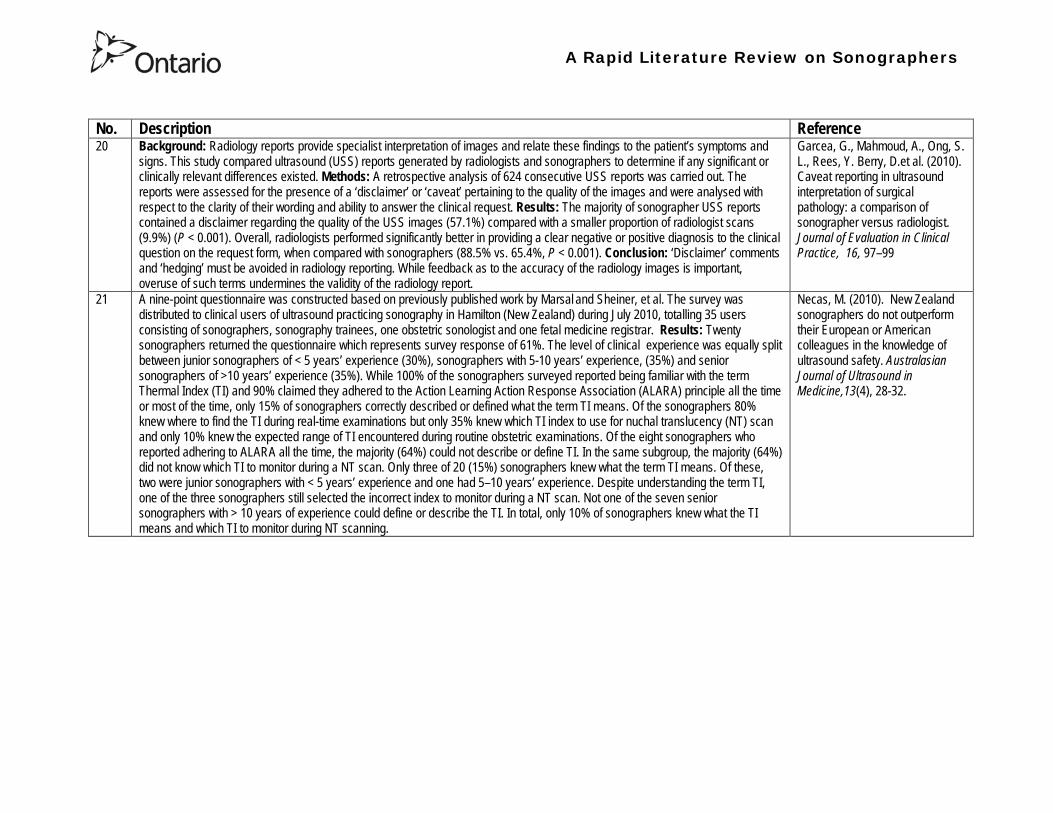
A Rapid Literature Review on Sonographers
No Description Reference 20 Background Radiology reports provide specialist interpretation of images and relate these findings to the patientrsquos symptoms and
signs This study compared ultrasound (USS) reports generated by radiologists and sonographers to determine if any significant or clinically relevant differences existed Methods A retrospective analysis of 624 consecutive USS reports was carried out The reports were assessed for the presence of a lsquodisclaimerrsquo or lsquocaveatrsquo pertaining to the quality of the images and were analysed with respect to the clarity of their wording and ability to answer the clinical request Results The majority of sonographer USS reports contained a disclaimer regarding the quality of the USS images (571) compared with a smaller proportion of radiologist scans (99) (P lt 0001) Overall radiologists performed significantly better in providing a clear negative or positive diagnosis to the clinical question on the request form when compared with sonographers (885 vs 654 P lt 0001) Conclusion lsquoDisclaimerrsquo comments and lsquohedgingrsquo must be avoided in radiology reporting While feedback as to the accuracy of the radiology images is important overuse of such terms undermines the validity of the radiology report
Garcea G Mahmoud A Ong S L Rees Y Berry Det al (2010) Caveat reporting in ultrasound interpretation of surgical pathology a comparison of sonographer versus radiologist Journal of Evaluation in Clinical Practice 16 97ndash99
21 A nine-point questionnaire was constructed based on previously published work by Marsal and Sheiner et al The survey was distributed to clinical users of ultrasound practicing sonography in Hamilton (New Zealand) during July 2010 totalling 35 users consisting of sonographers sonography trainees one obstetric sonologist and one fetal medicine registrar Results Twenty sonographers returned the questionnaire which represents survey response of 61 The level of clinical experience was equally split between junior sonographers of lt 5 yearsrsquo experience (30) sonographers with 5-10 yearsrsquo experience (35) and senior sonographers of gt10 yearsrsquo experience (35) While 100 of the sonographers surveyed reported being familiar with the term Thermal Index (TI) and 90 claimed they adhered to the Action Learning Action Response Association (ALARA) principle all the time or most of the time only 15 of sonographers correctly described or defined what the term TI means Of the sonographers 80 knew where to find the TI during real-time examinations but only 35 knew which TI index to use for nuchal translucency (NT) scan and only 10 knew the expected range of TI encountered during routine obstetric examinations Of the eight sonographers who reported adhering to ALARA all the time the majority (64) could not describe or define TI In the same subgroup the majority (64) did not know which TI to monitor during a NT scan Only three of 20 (15) sonographers knew what the term TI means Of these two were junior sonographers with lt 5 yearsrsquo experience and one had 5ndash10 yearsrsquo experience Despite understanding the term TI one of the three sonographers still selected the incorrect index to monitor during a NT scan Not one of the seven senior sonographers with gt 10 years of experience could define or describe the TI In total only 10 of sonographers knew what the TI means and which TI to monitor during NT scanning
Necas M (2010) New Zealand sonographers do not outperform their European or American colleagues in the knowledge of ultrasound safety Australasian Journal of Ultrasound in Medicine13(4) 28-32
A Rapid Literature Review on Sonographers
No Description Reference 22 Musculoskeletal (MSK) ultrasound performed by non-medical practitioners is a developing practice The aim of this paper is
(1) to audit an experienced sonographerrsquos (SJ Riley one of the authors) performance after one yearrsquos independent reporting against two experienced MSK radiologists (CJ Grove amp M Chandramohan the other two authors) and (2) to establish an audit standard against which such role development might be contrived Images and reports from 250 MSK ultrasound examinations performed by a sonographer were reviewed independently by two consultant MSK radiologists The examinations were graded for discrepancy eg grade 1 ndash agree with the sonographer report grade 2 ndash minor discrepancy unlikely to alter patient care grade 3 ndash potentially significant discrepancy grade 4 ndash definite significant discrepancy likely to have adverse consequences for patient care Two of 250 (08) cases were excluded Both radiologists agreed completely with the sonographer (grade 1) in 235 of the 248 cases (948) In 13 cases there was discrepancy between the reports of SJR and the radiologists The discrepancy was grade 2 in six of the 248 cases (24) grade 3 in six of the 248 cases (24) and grade 4 in one of the 248 cases (04) In conclusion this audit shows a high level of agreement between the sonographer and the consultant MSK radiologist reporting of MSK ultrasound This level of agreement may set the standard for future quality assurance audit of sonographer MSK reporting
Riley S J Groves C J Chandramohan M (2010) Musculoskeletal ultrasound audit of sonographer reporting Ultrasound 18 36ndash40
23 Background Growing demand for ultrasound examinations and higher quality requirements motivate searching for routines combining the diagnostic accuracy of radiologist-performed examinations with the economical advantages of sonographer-performed examinations One possible approach is to use strictly standardized acquisition and documentation schemes that give the radiologist access to all relevant information after the examination Purpose To compare a recently introduced routine combining acquisition by a radiographer (more specifically a sonographer with two years experience) documentation as standardized cine-loops and review by a radiologist (lsquostandardized methodrsquo) with the formerly used routine where the diagnosis is made bedside by the radiologist (lsquotraditional methodrsquo) Material and Methods In 64 policlinic patients the kidneys (n = 27) or the gallbladder (n = 37) were examined with both the standardized and the traditional method The radiologistsrsquo findings of hydronephrosis tumors cysts echogenicity changes and cortical thickness (in the kidneys) and wall thickness concrements and polyps (in the gallbladder) were compared between the methods with respect to agreement (proportion of agreement and kappa coefficient) as well as systematic differences (McNemarrsquos test) Results The findings at the gallbladder examination showed a median agreement of 97 (86ndash100 kappa = 064ndash100) and those at the kidney examination an agreement of 90 (78ndash100 kappa = 069ndash 100) There were no significant systematic differences between the methods Conclusion The satisfactory agreement in this preliminary study indicates that the new workflow with ultrasound examinations performed by a radiographer (sonographer) and analyzed off-line by a radiologist is promising and motivates further studies
Stenman C Thorelius L Knutsson A amp Smedby Ouml (2010) Radiographer-acquired and radiologist-reviewed ultrasound examination ndash agreement with radiologistrsquos bedside evaluation Acta Radiologica 52 70ndash74
A Rapid Literature Review on Sonographers
No Description Reference 24 The troubling practice of fetal sex selection has historically been considered an Asian phenomenon However recent evidence shows
that a similar situation is emerging in North America albeit on a smaller scale The Society of Obstetricians and Gynaecologists of Canada (SOGC) has firmly stated its opposition to sex selection for non-medical reasons as well as to the use of any technology used solely for the purpose of determining fetal sex However because fetal sex may be disclosed to the parents at the time of ultrasound examination if they request this information guidance for health care professionals to assist in discouraging fetal sex selection would be useful Because no declaration of motives or reasons is required when a woman seeks a termination of pregnancy the authors suggest that health care professionals need not disclose the sex of a fetus until it has reached a gestational age at which abortion for non-medical purposes would not be possible This proposal would facilitate consistency between clinical practice and the values of Canadian citizens the SOGC the Canadian Medical Association and other professional organizations while still respecting current laws pertaining to disclosure of patient information and patientsrsquo rights to autonomous decision-making
Thiele A T amp Leier B (2010) Towards an Ethical Policy for the Prevention of Fetal Sex Selection in Canada J Obstet Gynaecol Can 32(1) 54ndash57
25 Background The extent to which increases in the use of prenatal ultrasonography over time reflect changes in maternal risk (ie an increase in high-risk pregnancies over time) is unknown In this population-based study the authors examined the use of prenatal ultrasonography from 1996 to 2006 in Ontario Methods With fiscal year 199697 as the baseline the authors evaluated the relative risk (RR) and 95 confidence interval (CI) for the change in rates of ultrasonography for each subsequent year The RR was adjusted for maternal age income rural residence maternal comorbidities receipt of genetics consultation or amniocentesis mdash all in the index pregnancy mdash and history of complications in a prior pregnancy Results The study sample consisted of 1 399 389 singleton deliveries The rate of prenatal ultrasonography increased from 2055 per 1000 pregnancies in 1996 to 3264 per 1000 in 2006 (adjusted RR 155 95 CI 154ndash155) The rate increased among both women with low-risk pregnancies (adjusted RR 154 95 CI 153ndash155) and those with high-risk pregnancies (adjusted RR 155 95 CI 154ndash157) The proportion of pregnancies with at least four ultrasound examinations in the second or third trimesters rose from 64 in 1996 to 187 in 2006 (adjusted RR 268 95 CI 261ndash274) Paradoxically this increase was more pronounced among low-risk pregnancies (adjusted RR 292 95 CI 283ndash301) than among high-risk pregnancies (adjusted RR 225 95 CI 216ndash235) Interpretation Substantial increases in the use of prenatal ultrasonography over the past decade do not appear to reflect changes in maternal risk Nearly one in five women now undergo four or more ultrasound examinations during the second and third trimesters Efforts to promote more appropriate use of prenatal ultrasonography for singleton pregnancies appear warranted
You J J Alter D A Stukel T A McDonald S D Laupacis A et al (2010) Proliferation of prenatal ultrasonography CMAJ 182(2) 143-151
A Rapid Literature Review on Sonographers
No Description Reference 26 Aims The aim of this study was to assess the cost-effectiveness of using certified sonographers and miniaturized echocardiography
systems to perform echocardiograms at bedside in comparison to moving inpatients from the admission department to the echocardiography laboratory (echo-lab) Methods and Results From 26 September 2005 to 27 October 2005 112 patients admitted in six hospital wards in Italy connected through a 100 Mbit LAN to the echo-lab were scanned within the admission ward by sonographers using a miniaturized echo system Logistical data were collected and results were compared with those obtained from 194 consecutive patients coming from the same wards and studied in the echo-lab with high-end machines between 8 March 2005 and 15 April 2005 Performing echocardiograms in the admission department avoided long waiting time of the inpatients in the echo-lab before and after the study increased the percentage of patients studied within 3 and 5 days from request (88 vs 77 and 100 vs 95 respectively P = 003) increased both sonographer (by 339 P lt 0001) and echo-lab productivity (by 41 P lt 0001) and reduced costs of echocardiograms by 29 Conclusion Implementation of digital echocardiography certified sonographers and a miniaturized echo system allowed improvement of the cost-effectiveness of the service provided by the echo-lab for inpatients and avoided patientsrsquo discomfort derived from prolonged waiting time before and after the exam
Badano L Gaetano N Stacul S Gianfagna P Pericoli M et al (2009) Improved workflow sonographer productivity and cost-effectiveness of echocardiographic service for inpatients by using miniaturized systems European Journal of Echocardiography 10 537ndash542
27 The present report is a proposal from the American Society of Echocardiography Advanced Practice Task Force that identifies the potential of cardiac sonographers to achieve the Advanced Cardiovasculat Sonographer (ACS) level According to this report echocardiographic examinations require a well-trained and competent sonographer to obtain proper anatomic and physiologic data to establish an accurate diagnosis for clinical decision-making and patient management Although the formal education and training of cardiovascular sonographers are evolving many entry-level and staff sonographers may not have sufficient practical or clinical knowledge of the necessary components of the echocardiographic study for the individual patientrsquos clinical presentation In many clinical settings echocardiograms are read after the patient has left the laboratory Thus there is a role for a sonographer who can practice at an advanced level in a cardiovascular ultrasound laboratory to ensure a proper echocardiographic examination is performed on every patient In this setting an Advanced Cardiovascular Sonographer (ACS) would be able to review the indication for and quality of the examination If additional images were needed the ACS would assist the sonographer in obtaining these images which would lead to the performance of a complete and fully diagnostic examination before the patient had left the echocardiography laboratory In clinical practice the quality of the examinations performed would improve advancements in echocardiographic methods could be taught and incorporated into daily practice and patients would be better served
Mitchell C Miller F A Bierig S M Bremer M L Ehler D et al (2009) Advanced Cardiovascular Sonographer A Proposal of the American Society of Echocardiography Advanced Practice Sonographer Task Force J Am Soc Echocardiogr 22 1409-13
A Rapid Literature Review on Sonographers
No Description Reference 28 Sonographer practitioner development involves the expansion and extension of the sonographer role to include reporting on
ultrasound examinations Australian sonographers have not seen the same degree of role extension and expansion as their counterparts in the United Kingdom despite increasing levels of discussion regarding sonographer practitioner development The aim of this study was to determine if Australian sonographers want to extend their professional role and what they consider are the important issues associated with role extension This paper reports on qualitative data derived from a survey of Australian sonographers and investigates if Australian sonographers are interested in extending and expanding their professional role and responsibilities and if they do what might be necessary or desirable from a professional point of view for this change to occur A survey was mailed to all members of the Australian Sonographers Association (ASA) in October 2006 The 31-item survey included 28 closed-ended and three opened-ended items to provide both quantitative and qualitative data The quantitative data will be reported separately Qualitative data was derived from responses to the opened-ended questions which asked respondents to elaborate on their attitudes and feelings about role extension and development Analysis used Nvivo7 software to aid in uncovering common themes from the qualitative data The analysis focused on the reported incentives or motivations for becoming a sonographer practitioner as well as disincentives or perceived hurdles that would discourage respondents from becoming sonographer practitioners The three most reported incentives or motivations for becoming a sonographer practitioner were professional recognition remuneration and increased knowledge The three most commonly reported disincentives or perceived hurdles that would discourage respondents from becoming sonographer practitioners were legal issues insurance and further study
McGregor R OrsquoLoughlin K Cox J Clarke J Snowden A (2009) Sonographer practitioner development in Australia Qualitative analysis of an Australian sonographersrsquo survey Radiography15 313-319
29 Introduction This study describes the effect of introducing a cardiac sonographer led follow up clinic within a hospital cardiology department for patients with stable heart valve disease The two years before and after the instigation of the valve clinic were audited Materials and methods The clinic was conducted in a single centre and undertaken in the cardiology department of a district general hospital A total of 382 patients with 397 clinically significant valve lesions but for whom surgery was not yet indicated but follow up required were seen in a cardiac sonographer run clinics These patients no longer attended a medical follow up clinic unless there was clinical or echocardiographic deterioration Effectiveness was judged by the percentage treated according to current best practice guidelines the number of echocardiograms performed and the number of hospital outpatient visits attended In addition mortality data for the subjects in the clinic was collected Results The proportion followed up according to best practice guidelines rose from 157 (41) to 354 (92) (p lt 001) The total number of echocardiograms performed fell from 807 to 550 Total number of outpatient visits fell from 998 to 31 A total of 11 patients died in the two year study period none from progressive valve disease Discussion This study demonstrates that a protocol driven sonographer led heart valve disease follow up clinic significantly improved the quality of follow up while bringing about a major reduction in outpatients visits without compromising patient safety
Taggu W Topham A Hart L Carr-White G Sulke N et al (2009) A cardiac sonographer led follow up clinic for heart valve disease International Journal of Cardiology 132 240 ndash243
30 Role development for imaging allied health professionals has progressed significantly over the last three decades with many major developments pioneered in the field of ultrasound This paper reviews the developing role of the radiographer in ultrasound (in some countries such as the UK sonographers are considered a type of radiographer) and some of the clinical professional and educational implications of such development As the published evidence base underpinning the latest developments is scarce several contemporary case study examples which have been presented verbally at study days attended by the authors have been included In conclusion sonographer role development is considered within the professional contexts of extended role advanced and consultant practice
Hart A amp Dixon A (2008) Sonographer Role Extension and Career Development a Review of the Evidence Ultrasound 16(1) 31-35
A Rapid Literature Review on Sonographers
No Description Reference 31 Objective The main goal of this study was to determine end usersrsquo knowledge regarding safety aspects of diagnostic ultrasound
during pregnancy End usersrsquo attitudes toward the use of ultrasound in low-risk pregnancies were also assessed Methods A questionnaire was distributed to ultrasound end users attending review courses and hospital grand rounds between April and June 2006 in Chicago Results One hundred thirty end users completed the questionnaires (63 response rate) Sixty-three percent were physicians (n = 84) most of them obstetricians (817) About 18 of participants routinely performed Doppler ultrasound examinations during the first trimester Fifty percent of end users thought that the number of ultrasound examinations in low-risk pregnancy should be limited to 1 to 3 (mean plusmn SD 26 plusmn 09) Almost 70 disapproved of ldquokeepsakeentertainmentrdquo ultrasound Although 322 of the participants were familiar with the term thermal index only 177 actually gave the correct answer to the question on the nature of the thermal index About 22 were familiar with the term mechanical index but only 38 described it properly Almost 80 of end users did not know where to find the acoustic indices Only 208 were aware that they are displayed on the sonographic monitor during the examinations End users with higher knowledge of safety issues thought that there should be limitations on the number of ultrasound examinations in low-risk pregnancies (odds ratio 33 95 confidence interval 11ndash100 P = 028) Likewise these end users were more likely to respond that ultrasound might have adverse effects during pregnancy (odds ratio 32 95 confidence interval 11ndash125 P = 045) Conclusions Ultrasound end users are poorly informed regarding safety issues during pregnancy Further efforts in the realm of education and training are needed to improve end user knowledge about the acoustic output of the machines and safety issues
Sheiner E Shoham-Vardi I Abramowicz J S (2007) What Do Clinical Users Know Regarding Safety of Ultrasound During Pregnancy J Ultrasound Med 26 319ndash325
A Rapid Literature Review on Sonographers
No Description Reference 32 In an attempt to survey the knowledge among ultrasound users of some safety aspects of diagnostic ultrasound a questionnaire was
distributed to professionals using ultrasound for fetal examinations The questionnaire was answered by the participants and faculty members of six postgraduate courses in obstetric ultrasound Doppler ultrasound and fetal echocardiography given in Sweden Norway and Austria in 2003 Furthermore in Sweden the questionnaire was distributed to the staff meeting participants of two major obstetric ultrasound units and of one unit for fetal echocardiography and at a meeting of the Working Group for Ultrasound of the Swedish Society of Obstetrics and Gynecology In total 199 questionnaires were anonymously answered by doctors (n = 145) sonographers (n = 22) and midwives trained in ultrasound (n = 32) All of them were using diagnostic ultrasound on a daily or weekly basis The respondents were from nine European countries In none of the countries is formal certification for the use of diagnostic ultrasound in pregnancy or otherwise required The doctors who answered the questionnaire were specialists in obstetrics and gynecology (including some of the internationally recognized experts in fetal ultrasound) pediatric cardiology radiology or clinical physiology The respondents were not informed about the questionnaire in advance and they had a limited time to answer the 16 questions typically 10ndash15 min The questions concerned explanation of the safety indices and their relation to various ultrasound modes ie real-time B-mode M-mode three-dimensional (3D) spectral Doppler color and power Doppler ultrasound The respondents were also asked which ultrasound system they used most often and where on the system the information on thermal index (TI) and mechanical (MI) was displayed (eg in the top right corner of the screen in a separate window etc) Furthermore the respondents had to indicate how they adjust the output energy level on their own machine For all questions there was the optional answer lsquoDonrsquot knowrsquo Results About one-third of the ultrasound experts were able to define the abbreviations TI and MI For the subclasses of the TI the correct answer was found in only 3-8 of cases Of those who knew what the TI and MI meant only two-thirds and one-third of respondents respectively were able to give a simple explanation of the indices In 49 of questionnaires the Doppler modes were correctly ranked as generally producing higher ultrasound exposure than the imaging modes (B-mode 3D) Some 28 of the respondents correctly indicated where on their own machine the information on safety indices is displayed However not all of them knew how to control the output energy level For all items in the questionnaire there were no significant differences in the results between the three categories of respondents (ie physicians sonographers and midwives) The results of the questionnaire were anything but encouraging and they indicated that the users who are supposed to be responsible for controlling exposure of the fetus to ultrasound had a very poor knowledge of the basic safety aspects of ultrasound
Maršaacutel K (2005) The output display standard has it missed its target Ultrasound Obstet Gynecol 25 211ndash214
33 Purpose This study was performed to evaluate the accuracy of upper abdominal ultrasound (US) scanning performed by sonographers in a district general hospital to identify potential areas of weakness and to make recommendations to improve the service Materials and Methods Upper abdominal US examinations performed and reported by sonographers over a 4-week period were retrospectively reviewed The accuracy of the imaging findings and reports were assessed against other imaging surgical histological or laboratory findings and against clinical outcome Results A heterogenous group of 104 patients were included in the study 62 of whom had an US abnormality Errors of scanning or interpretation were identified in 10 patients (96) of whom five (48) were felt to be potentially significant Conclusions The sonographersrsquo accuracy in reporting upper abdominal US scans was 90 However on the basis of this study the authors have implemented specific recommendations to improve the quality of the service
Dongola N Guy R Ward S amp Giles J A (2003) Can sonographers offer an accurate upper abdominal ultrasound service in a district general hospital Radiography 9 29ndash33
A Rapid Literature Review on Sonographers
No Description Reference 34 Aim To compare the accuracy of radiographers and radiologists in routine abdominal ultrasound Materials and Methods One
hundred consecutive patients attending for routine abdominal ultrasound were included Each patient was examined by both a radiographer and radiologist Both operators noted their fndings and wrote a concluding report without conferring Reports were compared Where there was disagreement the patient was either re-examined by another radiologist or had further investigation Results Of 100 patients 52 were men and 48 were women The age range was 19plusmn88 years (median 52 years) Thirty-seven patients had renal tract ultrasound one had an aortic ultrasound and 62 had general upper abdominal ultrasound In 44 cases both operators reported the examination as normal In 49 cases both operators reported the examinations as abnormal and there was complete agreement between the operators In seven cases there was not complete agreement between operators Three of these disagreements were considered minor and four major In three of the seven cases the radiographer was correct and in four the radiologist was correct Conclusion Experienced radiographers and radiologists are highly accurate in performing and interpreting routine abdominal sonography Both operators missed a small minority of abnormalities There was no statistically significant difference in the accuracy of radiographers and radiologist
Leslie A Lockyer H amp Virjee J P (2000) Who Should be Performing Routine Abdominal Ultrasound A Prospective Double-Blind Study Comparing the Accuracy of Radiologist and Radiographer Clinical Radiology 55 606-609
Grey Literature 35 The ARRT The American Registry of Radiologic Technologists tests certifies and annually registers more than a quarter-of-a-million
radiologic technologists to promote high standards of patient care They award the Registered Technologist or RT designation to individuals who have completed the prescribed classroom and clinical education passed the appropriate exam and met the ethics requirements And they ensure their continuing education and ongoing compliance with their high standards by requiring annual registration of their certificate Certification by ARRT is voluntary not mandatory But employers state licensing agencies and federal regulators view ARRTrsquos credential as an indicator that a person has met recognized national standards for medical imaging and radiation therapy professionals How Are Radiologic Technologists Deemed Qualified First comes initial certification Certification is the process of initially recognizing individuals who have satisfied certain standards within a profession A person is certified by ARRT after completing an approved educational program complying with the ethics standards and passing a certification exam Then comes annual registration Registration is the yearly procedure required to maintain certification ARRT registrants are those who having already fulfilled the requirements for initial certification continue to meet the requirements for annual registration The ARRT annually registers the certificates of individuals who meet the following three criteria agreeing to comply with the ARRT Rules and Regulations continuing to comply with the ARRT Standards of Ethics and meeting our continuing education requirements Only technologists who are currently registered (which means that they have renewed within the past year) may designate themselves as ARRT Registered Technologists and use the initials RT after their names Federal laws regulate personnel qualifications in some modalities (mammography for example) but they accredit the health care facility rather than licensing the individual radiologic technologist who is providing patient care And while many states have laws regulating who can perform radiologic procedures they are typically based on ARRT certification Some employers and state agencies may require ARRT certification but not necessarily current registration Unless they require current registration by ARRT employers cannot assume that the person has complied with the ethics requirements or the continuing education requirements
American Registry of Radiologic Technologists (ARRT) (2013a) Patient Page Accessed April 2013
A Rapid Literature Review on Sonographers
No Description Reference 36 This document outlines certification requirements for sonography There are two pathways (primary and post-primary) For the
primary pathway it notes that sonography educational programs must be accredited by a mechanism acceptable to the ARRT ARRT generally recognizes only accreditation agencies that are recognized by the Council for Higher Education Accreditation andor United States Department of Education
American Registry of Radiologic Technologists (ARRT) (2013b) Sonography Certification Accessed April 2013
A Rapid Literature Review on Sonographers
No Description Reference 37 In Australia sonographers and student sonographers are required to be accredited by Australian Sonographer Accreditation Registry
(ASAR) in order to practise This requirement is contained within the Australian Governmentrsquos Diagnostic Imaging Accreditation Scheme standards and Medicare legislation Accreditation of sonographers is linked only to achieving - or in the case of students studying towards - an accredited entry-level qualification completion of continuing professional development and payment of an annual fee to ASAR The ASA contends that the lack of a requirement to ensure each sonographer is lsquofit to practisersquo is an unacceptable gap in current regulation undermining efforts to ensure the quality of sonographic services and failing to protect patients from harm The ASA supports and advocates for nationally-enforceable regulation of sonographers to meet our patientrsquos expectations National registration The National Registration and Accreditation Scheme (NRAS) for health workers commenced on 1 July 2010 The ten professions which comprised the initial intake into this scheme were those that had registration requirements in all Australian states or territories as well as those in which legislated regulation is deemed to be in the public interest An additional four professions - including medical radiation practitioners which are defined as radiographers radiation therapists and nuclear medicine technologists - will be included in the NRAS commencing on 1 July 2012 As sonography did not have registration in any states or territories prior to the commencement of NRAS the profession has not been in a position to negotiate entry into NRAS In early August 2011 the ASA lodged a submission with the Health Workforce Principal Committee requesting a profession-specific Sonography Board of Australia be formed under NRAS This submission was prepared based on information that consideration of professions which were unregistered would commence More recently the Australian Health Workforce Ministerial Council (AHWMC) issued a communiqueacute stating in part lsquoMinisters have commissioned work on unregistered professions and future directions for national registration Given this Ministers agreed to defer consideration of the inclusion of additional professions into the NRAS Ministers agreed that it would be premature to consider further applications for the scheme until this work has been completed other than paramedicsrsquo Options for unregistered health practitioners including sonographers Policy work is continuing at a federal level to establish an alternative regulatory framework The National Alliance for Self-Regulating Health Professions (NASRHP) consisting of the peak professional associations of eight self-regulating health professions including the ASA representing sonography released a proposal lsquoHarnessing self-regulation to support safety and quality in healthcare deliveryrsquo in March 2012 This proposal concluded that the framework that regulates all health practitioners should be expanded beyond NRAS to include
bull authorised self-regulating professions via a health professions panel bull negative licensing to withdraw the ability to practise from registered and self-regulated practitioners who do not meet the
regulatory requirements or unregulated practitioners due to poor practice As with all advocacy initiatives consideration by the federal government of the introduction of the NASRHPrsquos proposal may be prolonged and the outcomes may not match the expectations of NASRHPrsquos member professional associations including the ASA The ASA will continue both independently and collaboratively to lobby on behalf of our members and the public for the introduction of a timely and equitable solution to health practitioner regulation In the interim as there is no regulatory model with the rigour the ASA believes is required for regulation of sonographers and other health practitioners other than NRAS the ASA continues to support its Position Statement published in early 2010
Australian Sonographer Association (ASA) (2013) Sonographer Regulation Accessed April 2013
A Rapid Literature Review on Sonographers
No Description Reference 38 This publication contains information about Cardiovascular Credentialing International (CCI) It also provides the necessary
information and references concerning the process by which candidates may earn certificate or registry-level credentials CCI is a not-for-profit corporation established for the purpose of administering credentialing examinations as an independent credentialing agency CCI began credentialing cardiovascular professionals in 1968 CCI offers eight credentials which when earned demonstrate that the registrant holds fundamental knowledge in the particular cardiovascular specialty These include RCCS ndash Registered Congenital Cardiac Sonographer RCS ndash Registered Cardiac Sonographer and RPhS ndash Registered Phlebology Sonographer
Cardiovascular Credentialing International (2013) 2013 Examination Application amp Overview Accessed April 2013
39 Oregon Governor Signs Sonographer Licensure Legislation On July 28 2009 Oregon Governor Ted Kulongoski signed a sonographer licensure bill (HB 2245) into law The new law makes Oregon only the second state in the United States to require licensure of sonographers The Society of Diagnostic Medical Sonographers (SDMS) has worked closely with other sonography-related organizations and Oregon sonographers to help craft the language in new law The law renames and restructures the Oregon Board of Radiologic Technologists to the Oregon Board of Medical Imaging (OBMI) and ensures representation of sonographers on the Board The sonographer licensure law requires Oregon sonographers to become licensed by OBMI effective July 1 2010 Beginning on January 1 2014 all sonography licensees would be required to hold a national sonography certificationcredential (or be currently enrolled as a student) While the Oregon bill is far more detailed than the sonographer licensure law that passed in New Mexico earlier this year additional administrative rulesregulations will be developed over the next year to fully implement the new law SDMS will be continue to monitor developments regarding sonographer licensure in other states
Society of Diagnostic Medical Sonographers (SDMS) (2013a) News Accessed April 2013
A Rapid Literature Review on Sonographers
No Description Reference 40 On April 6 2009 New Mexico (NM) Governor Bill Richardson signed the historic bill that for the first time in the United States
requires licensure of sonographers The bill adds sonographers (and MRI technologists) to the list of medical imaging professionals licensed by the State of New Mexico Until now anyone could perform sonograms in New Mexico Prior to the passage of the New Mexico licensure law virtually all other health care providers and professionals had to be licensed As a result the public is often surprised to learn that the person performing a medical sonogram does not have any state licensure or national certification requirements The responsibility for administering the NM sonographer licensure program would rest with the New Mexico Environment Department Although this was not the first choice in most sonographersrsquo minds the Environment Department currently manages the radiologic technology licensure program so the administrative infrastructure is already in place By ensuring the creation of an advisory council that includes representation by each medical imaging and radiation therapy modality regulated the bill helps ensure that the Departmentrsquos other areas of emphasis do not detract from sonographer licensure Because sonographers have never had to be licensed before they frequently do not see or understand the distinction between state licensure and national certification National certifications or credentials issued by voluntary organizations such as the American Registry for Diagnostic Medical Sonography (ARDMS) Cardiovascular Credentialing International (CCI) and the American Registry of Radiologic Technologists (ARRT) have little legal weight The certification process and resulting credential are valuable tools in ensuring sonographers have met minimum education and knowledge requirements but do not really regulate anyonersquos ability to perform sonography In an ideal world no additional regulation of sonography would be required However as everyone knows more and more people are taking weekend courses and buying ultrasound equipment without understanding the ramifications of using ultrasound technology (eg ALARA) Under the bill passed by the New Mexico legislature national credentialscertifications should provide evidence of meeting the statersquos standards All sonographers who are performing these critical medical imaging services in New Mexico will be required to meet national certification standards (ie have certifications or credentials issued by ARDMS CCI or ARRT) As a result there would be no additional exams in New Mexico beyond the national certification exams required by ARDMS CCI or ARRT The bill also requires the state to recognize the continuing medical education completed for national certification renewal The most visible impact on New Mexico sonographers will be a license fee However the legislation caps the license fee at $100 per two-year license In addition only the New Mexico Legislature can change this cap
SDMS (2013b) New Mexico Sonographer Licensure Law Signed Accessed April 2013
A Rapid Literature Review on Sonographers
No Description Reference 41 Preamble The purpose of this document is to define the Scope of Practice for Diagnostic Ultrasound Professionals in the US and to
specify their roles as members of the health care team acting in the best interest of the patient This scope of practice is a living document that will evolve as the technology expands Definition of the Profession The Diagnostic Ultrasound Profession is a multi-specialty field comprised of Diagnostic Medical Sonography (with subspecialties in abdominal neurologic obstetricalgynecologic and ophthalmic ultrasound) Diagnostic Cardiac Sonography (with subspecialties in adult and pediatric echocardiography) Vascular Technology and other emerging fields These diverse specialties are distinguished by their use of diagnostic medical ultrasound as a primary technology in their daily work Certification is considered the standard of practice in ultrasound Individuals who are not yet certified should reference the Scope as a professional model and strive to become certified Scope of Practice of the Profession The Diagnostic Ultrasound Professional is an individual qualified by professional credentialing and academic and clinical experience to provide diagnostic patient care services using ultrasound and related diagnostic procedures The scope of practice of the Diagnostic Ultrasound Professional includes those procedures acts and processes permitted by law for which the individual has received education and clinical experience and in which heshe has demonstrated competency Diagnostic Ultrasound Professionals
bull Perform patient assessments bull Acquire and analyze data obtained using ultrasound and related diagnostic technologies bull Provide a summary of findings to the physician to aid in patient diagnosis and management bull Use independent judgment and systematic problem solving methods to produce high quality diagnostic information and
optimize patient care
Society of Diagnostic Medical Sonography [SDMS] (2013c) Scope of Practice for the Diagnostic Ultrasound Professional Accessed April 2013
42 This website notes that in the US most employers in the US prefer to hire sonographers who have professional certification A sonographer can get certification and passing an exam Most exams relate to the specialty that the sonographer is most interested inmdashfor example an exam to become certified in abdominal sonography A few states require diagnostic medical sonographers to be licensed Typically professional certifications required for licensure other requirements vary by state Sonographers must take continuing education to keep their certification current
Bureau of Labor Statistics US Department of Labor (2012) How to become a Diagnostic Medical Sonographer Occupational Outlook Handbook 2012-13 Edition Accessed April 2013
A Rapid Literature Review on Sonographers
No Description Reference 43 The American Registry for Diagnostic Medical Sonography (ARDMS) is an independent not-for-profit organization founded in 1975
that administers examinations and awards the following credentials bull RDMSreg ndash Registered Diagnostic Medical Sonographerreg bull RDCSreg ndash Registered Diagnostic Cardiac Sonographerreg bull RVTreg ndash Registered Vascular Technologistreg bull RPVIreg ndash Registered Physician in Vascular Interpretationreg bull RMSKtrade - Registered in Musculoskeletaltrade sonography
ARDMS is the globally recognized standard of excellence in sonography and has certified more than 75000 individuals ARDMS is composed of three key groups
bull Board of Directors ndash Sonographers vascular technologists physicians research scientists and a member of the public are volunteer members of the board that determines the direction and policies of our organization
bull Examination Development Task Forces ndash A group of sonographers vascular technologists physicians and scientists volunteer their time and expertise to produce our examinations
bull Professional Staff ndash A paid highly-skilled staff administers the programs and assists applicants and registrants Credentials awarded by ARDMS document personal achievement of recognized professional standards and are widely accepted in the medical community by sonography and vascular professional organizations However ARDMS does not validate the day-to-day job performance of any sonography professional ARDMS has earned the prestigious ANSI-ISO 17024 accreditation for certifying bodies from the International Organization for Standardization (ISO) Accreditation is granted through the American National Standards Institute (ANSI) ARDMS has also earned accreditation with the National Commission for Certifying Agencies (NCCA) The NCCA is the accrediting arm of the National Organization for Competency Assurance (NOCA) Established in 1977 as a nonprofit organization NOCA is a leader in establishing quality standards for certifying organizations
American Registry for Diagnostic Medical Sonography (ARDMS) (2012a) Overview of ARDMS Accessed April 2013
A Rapid Literature Review on Sonographers
No Description Reference 44 ARDMS is developing a program to assure that Registrants continue to meet the basic competency requirements of core clinical skills
in present day practice bull Each Registrant who holds an active ARDMS certification as of December 31 2011 will be assigned the 2012 ndash 2021 10-
year recertification assessment period bull The 10-year period for new Registrants will begin the year following the attainment of the credential bull If a Registrant earns a new credential or specialty after January 1 2012 then that credentialspecialty will have a separate
10-year recertification assessment period that will begin the year following the attainment of the credentialspecialty ARDMS will develop tools and resources to assist Registrants in tracking and meeting the recertification assessment program requirements
bull Registrants who do not wish to take a recertification assessment in a specific credentialspecialty may voluntarily relinquish the credentialspecialty
As an ARDMS Registrant individuals are committing to demonstrate the highest level of competency in the modality of sonography by meeting and maintaining the standards set by the ARDMS The pathway of maintenance your certification is comprised of Paying the ARDMS annual renewal fee each year Each year ARDMS assesses Registrants an annual renewal fee By paying your annual renewal fee you are contributing to the ARDMS mission of promoting quality care and patient safety through the certification and continuing competency of ultrasound professionals In order to maintain your credentials you must pay the annual renewal fee of $75 USD or $100 USD (for RPVI and MSK Registrants with MDDODPM) designation on or before December 31 of each year in order to stay active for the following year Attesting to ARDMS rules and policies Each year when you pay your annual renewal fee you are attesting that you are in compliance with all ARDMS rules and discipline policies and are in good standing with the ARDMS Earning a minimum of 30 ARDMS-accepted CMEs during your three-year CME period ARDMS verifies its Registrants compliance with continuing medical education (CME) requirements using an audit process Earning your CMEs is an integral component of maintaining your ARDMS credential(s) and demonstrates your ongoing commitment to lifelong learning RecertificationContinued Commitment to Excellence Following the attainment of the ARDMS credential Registrants are assigned a 10-year recertification period the year after the credential is earned ARDMS is committed to developing a recertification program that is easy for Registrants to comply with
American Registry for Diagnostic Medical Sonography (ARDMS) (2012b) Recertification General Information
A Rapid Literature Review on Sonographers
No Description Reference 45 This document reviews Sonography Principles and Instrumentation (SPI) Examination Requirements offered through ARDMS in
order to achieve sonography certification and the various prerequisites needed before taking the exam For example one prerequisite entails (1) Education Graduate of a program accredited by an agency recognized by the Council for Higher Education Accreditation (CHEA) United States Department of Education (USDOE) or Canadian Medical Association (CMA) that specifically conducts programmatic accreditation for diagnostic medical sonographydiagnostic cardiac sonographyvascular technology Currently the only organizations that offer programmatic accreditation under the aforementioned associations are the Commission on Accreditation of Allied Health Education Programs (CAAHEP) and the Canadian Medical Association (CMA) (2) Required Clinical UltrasoundVascular Experience No additional experience is required (3) Documentation Required with Application 1) Copy of diploma from ultrasoundvascular program or an official transcript indicating the date the degree was conferred 2) Original letter signed by program director andor medical director indicating date of graduation or successful completion of the program4 Program directors must use the mandatory formatted sample letter available on ARDMSorg sampleletters 3) The CV form is not required if the application is submitted and received in the ARDMS office within one year after successful completion of the program Otherwise an original signed and completed CV form for each appropriate specialty area(s) must be submitted 4) Photocopy of a non-expired government issued photo identification with signature the name on the identification must exactly match the name under which you are applying for ARDMS examination
American Registry for Diagnostic Medical Sonography (ARDMS) (2012c) SPI Requirement and General Prerequisites Accessed April 2013
46 This page notes the five credentials offered by the ARDMS and the corresponding exams and specialty exams required bull RDMS requires the SPI exam (sonography principles and instrumentation) and a specialty exam in abdomen breast fetal
echocardiography neurosonolgoy or obstetricsgynecology bull RDCS requires the SPI exam and a speciality exam in adult echocardiography fetal echocardiography pediatric
echocardiography bull RVT requires the SPI and a speciality exam in vascular technology bull RMSK requires the musculoskeletal sonography exam bull RPVI requires the physiciansrsquo vascular interpretation exam
American Registry for Diagnostic Medical Sonography (ARDMS) (2012d) Credentials and Examinations Accessed April 2013
A Rapid Literature Review on Sonographers
No Description Reference 47 This article summarizes some of the many trends in fetal pediatric and adult echocardiography that SDMS members are excited
about Fetal Echocardiography Sonography has been used since the mid-1980s to help identify congenital heart defects during the second trimester The ldquobasicrdquo examination of the fetal heart involves a four-chamber view ndash evaluation of the right and left atrial and ventricular chambers and their respective valves In the past several years there has been a movement in the obstetric ultrasound community to encourage obstetric sonographers to perform the ldquoextended basicrdquo exam which includes the cardiac outflow tracts The outflow tracts consist of the main pulmonary artery exiting the right ventricle and the aorta exiting the left ventricle The fetal echocardiography community also is abuzz over three-dimensional (3D) and four-dimensional (4D) ultrasound and the potential of this technology to significantly improve the evaluation of the fetal heart Spatiotemporal Image Correlation (STIC) a 4D application allows for evaluation and manipulation of the heart in real-time Allen says Todayrsquos ultrasound equipment allows sonographers to gather a volume of data from which they can create detailed 3D images of the tiny fetal heart which is constantly in motion With computer manipulation of the data sets the images also can be viewed in 4D allowing physicians to see the entire fetal heart cycle from start to finish Pediatric Echocardiography One of the most significant advances in pediatric echo over the last decade is moving to a digital format The advent of pediatric transesophageal (TEE) probes also has been a big advance for pediatric echo Transesophageal probes provide an unimpeded sonographic visualization of the intracardiac structures Theyrsquore useful in visualizing very fine valvular abnormalities the thoracic aorta vegetations and thrombus in the atrial appendage It is also useful in detecting subtle anatomic variations seen in congenital heart disease Pediatric-sized TEE probes make the exams possible In the past it wasnrsquot possible because the adult probe was just too big to put down the babyrsquos or childrsquos esophagus Now the probe is small enough to successfully manipulate it in children and get superb images Because the new sonographic imaging equipment and better surgical techniques allow clinicians to detect and repair more cardiac abnormalities earlier patients are living longer Adult Echocardiography The adult echo world is abuzz about 2D and 3D speckle tracking relatively new techniques for the assessment of heart function and strain imaging Speckle tracking evaluates both regional and global function by measuring the deformation of the heart muscle
Orenstein B W (2010) Current Trends in Echocardiography SDMS New Wave February 1-5 Accessed April 2013
48 This document describes the requirements for admission to the Register of Accredited Medical Sonographer in Australia Requirements include Continuation of Accreditation In order to remain on the Register an accredited Sonographer must satisfy the ASAR continuing professional development (CPD) requirements and pay the prescribed annual fee Student Sonographers apart from paying the prescribed annual fee should provide evidence of their student status every year when they renew their ASAR accreditation
Australian Sonographer Accreditation Registry (ASAR) (no date A) Sonographer Accreditation Accessed April 2013
A Rapid Literature Review on Sonographers
No Description Reference 49 ASARrsquos functions
1 Accredit sonography education programs within Australia 2 Maintain a register of Accredited Sonographers and Accredited Student Sonographers 3 Establish minimum standards of Continuing Professional Development (CPD) and to monitor the CPD requirements for
Sonographers to remain on the register 4 To support activities that advance the profession of sonography 5 To provide registration information about Sonographers to Medicare Australia 6 To provide public access to the name registration number State or Territory current type of accreditation and area of
specialty for active Sonographers ASARrsquos Role ASAR is not a registration board ASAR does not have the authority to remove a Sonographer from the Registry due to professional misconduct nor the ability to prevent a Sonographer from practicing ASAR is only able to remove a Sonographer from the Registry if they
bull fail to meet CPD requirements and or bull fail to pay annual fees
ASAR (no date B) About ASAR Accessed April 2013
50 Regulatory bodies A Regulatory Body maintains a register of individuals who have achieved the required qualification of that profession to practice and can evidence continuing competence at the required level For a doctor to practice in the UK he or she must be registered with the General Medical Council (GMC) This is the regulatory body for medical practitioners The majority of sonographers who are employed in the UK come from a background of radiography or midwifery To practice radiography in the UK a qualified radiographer must be registered with the Health Professions Council (HPC) Similarly a midwife must be registered with the National Midwifery Council to practice as a midwife Sonographer registration in the UK There are a number of professions allied to medicine that are regulated by the HPC for example radiography physiotherapy However sonography is not recognised as a profession by the HPC and therefore there is no mechanism whereby an individual can register with the HPC as a sonographer or be regulated by them as a sonographer It is a common misunderstanding that sonographers have to be registered with the HPC to practice This comes from the fact that most sonographers are also qualified radiographers and maintain their registration with the HPC because they wish to remain on the HPC register as a radiographer Professional bodies An individual can be a member of a Professional Body such as the Royal College of Obstetricians amp Gynecologists (RCOG) Royal College of Radiologists (RCR) Society amp College of Radiographers (SCoR) or the Royal College of Midwives (RCM) but this is not the same as being accepted onto the register of one of the regulatory bodies The professional bodies are responsible for setting and maintaining the standards of their members providing a code of conduct etc They may also act in the capacity of a trades union and can also provide professional indemnity insurance The British Medical Ultrasound Society BMUS is not a professional body but is a scientific society so will offer a different range of benefits Whether or not one wishes to join the society has no impact on their ability to work as a sonographer in the UK although from a continuing professional development point of view it can be beneficial The College of Radiographers are currently working with the HPC to get sonography recognized as a separate profession
British Medical Ultrasound Society (BMUS) (no date) Information on Sonographer Regulation in the UK Accessed April 2013
A Rapid Literature Review on Sonographers
No Description Reference 51 This document included sections on the role of the sonographer and the use of sonography for entertainment Role of the
Sonographer and Technical Impressions The role of the sonographer is to perform the ultrasound scan and to document observations The sonographerrsquos written technical impressions are intended as a form of communication between the sonographer and the reporting physician These observations must be reviewed and subsequently reported by the reporting physician Issuing of a final reportdiagnosis represents the practice of medicine and is therefore the domain of the reporting physician Use of Ultrasound for Entertainment or Non-Diagnostic Purposes In contrast to this justified form of assessment ultrasound for entertainment is simply an elaborate version of a portrait For a variable fee the surface of the fetus is skimmed over and clients are provided with keepsake images or a DVD of their fetus There are no measurements taken during this exam no morphological assessment performed and no dictated diagnostic report of findings Essentially there are no diagnostic benefits to the exam and uninformed parents may be falsely reassured that the beautiful portrait indicates a normal fetus Entertainment ultrasound facilities may operate outside of medical guidelines and without any controls which may result in a lack of technical safeguards operator expertise or governance of technical competency These facilities do not comply with the ALARA principle which could result in increased fetal exposures The Canadian Society of Diagnostic Medical Sonograph (CSDMS) does not support persons or facilities that participate in ultrasound for entertainment activities Liability insurance purchased through the CSDMS group insurance plan does not cover any activities carried out for entertainment ultrasound purposes Other leaders in Canadian medicine support this statement In 2005 the SOGC (Society of Obstetrics and Gynaecologists of Canada) published an article entitled ldquoObstetrical Ultrasound Biological Effects and Safetyrdquo and their conclusion was that ldquothe theoretical risk of adverse bio-effects even from standard 2D obstetrical ultrasound makes it hard to justify its use for non-medical reasons such as sex-determination making non-medical photos or video or for commercial purposerdquo The SOGC recommends that ultrasound be used prudently and that energy exposure be limited to the minimum that is medically necessary The SOGC further recommends a complete ban on the non-medical use of fetal ultrasound and encourages government to join with society to find appropriate means to deal with this public health issue The CAR (Canadian Association of Radiologists) makes a similar statement ldquoThe CAR strongly opposes the use of diagnostic ultrasound equipment for non-medical purposes and considers the use of medical ultrasound for entertainment to be a misuse of the technology especially if fetal subjects are involvedrdquo
Canadian Society of Diagnostic Medical Sonography (no date) Professional Practice Guidelines and Policy Statements For Canadian Sonography Accessed April 2013
A Rapid Literature Review on Sonographers
No Description Reference 52 In 1993 the Consortium for the Accreditation of Sonographic Education (CASE) was formed through the common desire to ensure
that the education and training of sonographers in the United Kingdom was delivered at the highest level All members share common concerns that standards of service provision and education are developed in parallel with increasing demand and improving technology All members strive to create an environment in which education and training is maximised This will make validation and accreditation to be as robust and straightforward as possible CASEs philosophy is to promote best ultrasound practise through the accreditation of those training programmes that develop safe and competent ultrasound practitioners This should be delivered at postgraduate level The majority of its activities relate to supporting those Institutions that offer or wish to offer courses leading to the award of a Masters Postgraduate DiplomaCertificate in Medical or clinical Ultrasound In addition to offering accreditation of new and re-accreditation of established ultrasound education programmes CASE undertakes annual monitoring of the courses it has accredited The information obtained is fed back to the Institutions both on an individual basis and via the annual CASE report Each member organisation is unique and has its own obligations to its membership and its own agenda while CASE brings them together to provide a forum for the interchange of ideas in which shared views may be synthesised and thus inform education and training programmes
Consortium for the Accreditation of Sonographic Education (no date) Home Accessed April 2013
A Rapid Literature Review on Sonographers
No Description Reference 53 The Medical Radiation Technologists Board (the Board) is one of sixteen New Zealand health registration authorities appointed by
the Minister of Health under the Health Practitioners Competence Assurance Act 2003 (the Act) The Board is responsible for the administration of the Act in regard to the profession of medical radiation technology The primary responsibility of the Board is to protect the health and safety of the New Zealand public by ensuring practitioners registered in the profession of medical radiation technology are competent and fit to practice In accordance with Section 118 of the Act the Board is responsible for fulfilling a number of functions
bull 01 To prescribe the qualifications required for scopes of practice within the profession of medical radiation technology and for that purpose to accredit and monitor educational institutions and degrees courses of studies or programmes
bull 02 To authorise the registration of medical radiation technologists under the Act and to maintain registers bull 03 To consider applications for annual practising certificates bull 04 To review and promote the competence of medical radiation technologists bull 05 To recognise accredit and set programmes to ensure the ongoing competence of medical radiation technologists bull 06 To receive and act on information from health practitioners employers and the Health and Disability Commissioner
about the competence of medical radiation technologists bull 07 To notify employers the Accident Compensation Corporation the Director-General of Health and the Health and
Disability Commissioner that the practice of a medical radiation technologist may pose a risk of harm to the public bull 08 To consider the cases of medical radiation technologists who may be unable to perform the functions required for the
practice of the medical radiation technology profession bull 09 To set standards of clinical competence cultural competence and ethical conduct to be observed by medical radiation
technologists bull 10 To liaise with other authorities appointed under the Act about matters of common interest bull 11 To promote education and training in the profession of medical radiation technology bull 12 To promote public awareness of the responsibilities of the Board bull 13 To exercise and perform any other functions powers and duties that are conferred or imposed on it by or under the Act
or any other enactment
Medical Radiation Technologists Board (MRTB) (no date A) What we do Accessed April 2013
54 Qualifications To be considered for registration as a medical radiation technologist in any of the specified scopes of practice an individual must hold a qualification for that particular scope as prescribed by the Board or approved by the Board as being substantially equivalent to the prescribed qualifications In addition to having an approved qualification individuals may also be required to demonstrate that they have adequate experience in the relevant scope of practice Sonographer The Board has prescribed one New Zealand postgraduate qualification for a person to be eligible for registration in the scope of practice of Sonographer Postgraduate Diploma in Health Science (Ultrasound) issued by the Unitec Institute of Technology Required Experience If an individual is applying for registration in Nuclear Medicine Ultrasound or Magnetic Resonance Imaging in addition to holding a relevant postgraduate qualification they must also be able to provide evidence that you have a minimum of 3360 hours of clinical experience in the relevant scope of practice you are applying for registration in
Medical Radiation Technologists Board (MRTB) (no date B) Qualifications Accessed April 2013
A Rapid Literature Review on Sonographers
No Description Reference 55 Recertification is a mechanism used by the Medical Radiation Technologists Board in New Zealand to ensure that registered medical
radiation technologists continue to be competent to practise within their scopes of practice Annual Practising Certificates Each year if an individual wishes to continue practising in your scope(s) of practice in New Zealand they must apply for and be issued with an annual practising certificate (APC) A condition of being issued with an APC is that one must provide evidence of enrolment and satisfactory progress in a Board-approved recertification programme Alternatively the Board may issue them with an interim practising certificate (IPC) until it is satisfied the individual demonstrates that they are competent to practice in the scope(s) of practice they applied for Working with an IPC will require an individual to work under the supervision of a registered medical radiation technologist or other relevant registered health practitioner Recertification Programmes The Boards recertification programme framework is based on a continuing professional development (CPD) model Individuals have the option of enrolling in one of seven recertification programmes for medical radiation technologists that are currently approved by the Board They must meet the requirements of the particular recertification programme in which they are enrolled Recertification Audits An essential component of the CPD-based recertification programme adopted by the Board is the audit of CPD recordsThe Board undertakes an annual audit of approximately 10 of its registrant database to assess individual medical radiation technologistsrsquo compliance with the Boarrsquods recertification requirements
Medical Radiation Technologists Board (MRTB) (no date C) Recertification Accessed April 2013
56 There are two types of radiography - diagnostic and therapeutic Both need considerable knowledge of technology anatomy physiology and pathology to carry out their work Many also undertake further training to become a sonographer Some of the imaging technology that a diagnostic radiographer may use are
bull X-Ray - looks through tissues to examine bones cavities and foreign objects bull Fluoroscopy - images the digestive system providing a real-time image bull CT (Computed Tomography) - which provides cross-sectional views (slices) of the body bull MRI (Magnetic Resonance Imaging) - builds a 2-D or 3-D map of the different tissue types within the body bull Ultrasound - well known for its use in obstetrics and gynaecology Also used to check circulation and examine the heart bull Angiography - used to investigate blood vessels
Sonographyultrasonography Ultrasound is used in various settings in a hospital including abdominal scanning and breast ultrasound Ultrasound imaging is the use of high frequency sound in excess of human hearing to produce images of structures of the human body that may be observed on a screen These images may subsequently be transferred to photographic film or paper or onto video or CD forming part of the patients record of their examination There are no direct entry routes into ultrasound Most sonographers train as a radiographer then undertake an approved post-registration course Offered by higher education institutions The courses are a minimum of one academic year and prepare sonographers clinically and academically for practice Normally a pre-requisite for acceptance is access to a clinical department with supervised practice for students
National Health Service (NHS) (no date A) Radiographer Accessed April 2013
57 To practice as a radiographer in the UK an individual must be registered with the Health and Care Professions Council (HCPC) To register individuals must have successfully completed an approved training programme in diagnostic or therapeutic radiography These are offered at undergraduate degree (full-time and part-time) and at postgraduate degreediploma level at a number of universities across the UK
NHS (no date B) Entry requirements and training in radiography Accessed April 2013
Health Professions Regulatory Advisory Council 56 Wellesley Street West 12th Floor
Toronto ON M5S 2S3
Telephone 416-326-1550 Toll-Free 1-888-377-7746
Fax 416-326-1549
Website wwwhpracorg
Email hpracwebmasterontarioca
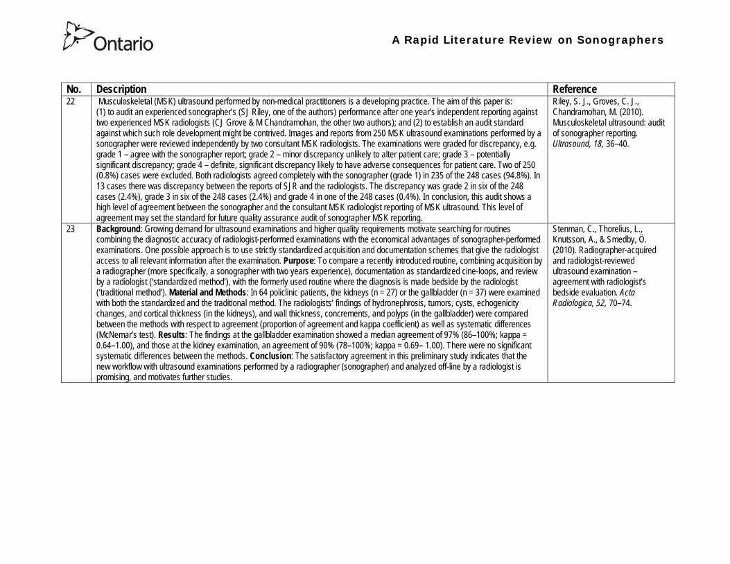
A Rapid Literature Review on Sonographers
No Description Reference 22 Musculoskeletal (MSK) ultrasound performed by non-medical practitioners is a developing practice The aim of this paper is
(1) to audit an experienced sonographerrsquos (SJ Riley one of the authors) performance after one yearrsquos independent reporting against two experienced MSK radiologists (CJ Grove amp M Chandramohan the other two authors) and (2) to establish an audit standard against which such role development might be contrived Images and reports from 250 MSK ultrasound examinations performed by a sonographer were reviewed independently by two consultant MSK radiologists The examinations were graded for discrepancy eg grade 1 ndash agree with the sonographer report grade 2 ndash minor discrepancy unlikely to alter patient care grade 3 ndash potentially significant discrepancy grade 4 ndash definite significant discrepancy likely to have adverse consequences for patient care Two of 250 (08) cases were excluded Both radiologists agreed completely with the sonographer (grade 1) in 235 of the 248 cases (948) In 13 cases there was discrepancy between the reports of SJR and the radiologists The discrepancy was grade 2 in six of the 248 cases (24) grade 3 in six of the 248 cases (24) and grade 4 in one of the 248 cases (04) In conclusion this audit shows a high level of agreement between the sonographer and the consultant MSK radiologist reporting of MSK ultrasound This level of agreement may set the standard for future quality assurance audit of sonographer MSK reporting
Riley S J Groves C J Chandramohan M (2010) Musculoskeletal ultrasound audit of sonographer reporting Ultrasound 18 36ndash40
23 Background Growing demand for ultrasound examinations and higher quality requirements motivate searching for routines combining the diagnostic accuracy of radiologist-performed examinations with the economical advantages of sonographer-performed examinations One possible approach is to use strictly standardized acquisition and documentation schemes that give the radiologist access to all relevant information after the examination Purpose To compare a recently introduced routine combining acquisition by a radiographer (more specifically a sonographer with two years experience) documentation as standardized cine-loops and review by a radiologist (lsquostandardized methodrsquo) with the formerly used routine where the diagnosis is made bedside by the radiologist (lsquotraditional methodrsquo) Material and Methods In 64 policlinic patients the kidneys (n = 27) or the gallbladder (n = 37) were examined with both the standardized and the traditional method The radiologistsrsquo findings of hydronephrosis tumors cysts echogenicity changes and cortical thickness (in the kidneys) and wall thickness concrements and polyps (in the gallbladder) were compared between the methods with respect to agreement (proportion of agreement and kappa coefficient) as well as systematic differences (McNemarrsquos test) Results The findings at the gallbladder examination showed a median agreement of 97 (86ndash100 kappa = 064ndash100) and those at the kidney examination an agreement of 90 (78ndash100 kappa = 069ndash 100) There were no significant systematic differences between the methods Conclusion The satisfactory agreement in this preliminary study indicates that the new workflow with ultrasound examinations performed by a radiographer (sonographer) and analyzed off-line by a radiologist is promising and motivates further studies
Stenman C Thorelius L Knutsson A amp Smedby Ouml (2010) Radiographer-acquired and radiologist-reviewed ultrasound examination ndash agreement with radiologistrsquos bedside evaluation Acta Radiologica 52 70ndash74
A Rapid Literature Review on Sonographers
No Description Reference 24 The troubling practice of fetal sex selection has historically been considered an Asian phenomenon However recent evidence shows
that a similar situation is emerging in North America albeit on a smaller scale The Society of Obstetricians and Gynaecologists of Canada (SOGC) has firmly stated its opposition to sex selection for non-medical reasons as well as to the use of any technology used solely for the purpose of determining fetal sex However because fetal sex may be disclosed to the parents at the time of ultrasound examination if they request this information guidance for health care professionals to assist in discouraging fetal sex selection would be useful Because no declaration of motives or reasons is required when a woman seeks a termination of pregnancy the authors suggest that health care professionals need not disclose the sex of a fetus until it has reached a gestational age at which abortion for non-medical purposes would not be possible This proposal would facilitate consistency between clinical practice and the values of Canadian citizens the SOGC the Canadian Medical Association and other professional organizations while still respecting current laws pertaining to disclosure of patient information and patientsrsquo rights to autonomous decision-making
Thiele A T amp Leier B (2010) Towards an Ethical Policy for the Prevention of Fetal Sex Selection in Canada J Obstet Gynaecol Can 32(1) 54ndash57
25 Background The extent to which increases in the use of prenatal ultrasonography over time reflect changes in maternal risk (ie an increase in high-risk pregnancies over time) is unknown In this population-based study the authors examined the use of prenatal ultrasonography from 1996 to 2006 in Ontario Methods With fiscal year 199697 as the baseline the authors evaluated the relative risk (RR) and 95 confidence interval (CI) for the change in rates of ultrasonography for each subsequent year The RR was adjusted for maternal age income rural residence maternal comorbidities receipt of genetics consultation or amniocentesis mdash all in the index pregnancy mdash and history of complications in a prior pregnancy Results The study sample consisted of 1 399 389 singleton deliveries The rate of prenatal ultrasonography increased from 2055 per 1000 pregnancies in 1996 to 3264 per 1000 in 2006 (adjusted RR 155 95 CI 154ndash155) The rate increased among both women with low-risk pregnancies (adjusted RR 154 95 CI 153ndash155) and those with high-risk pregnancies (adjusted RR 155 95 CI 154ndash157) The proportion of pregnancies with at least four ultrasound examinations in the second or third trimesters rose from 64 in 1996 to 187 in 2006 (adjusted RR 268 95 CI 261ndash274) Paradoxically this increase was more pronounced among low-risk pregnancies (adjusted RR 292 95 CI 283ndash301) than among high-risk pregnancies (adjusted RR 225 95 CI 216ndash235) Interpretation Substantial increases in the use of prenatal ultrasonography over the past decade do not appear to reflect changes in maternal risk Nearly one in five women now undergo four or more ultrasound examinations during the second and third trimesters Efforts to promote more appropriate use of prenatal ultrasonography for singleton pregnancies appear warranted
You J J Alter D A Stukel T A McDonald S D Laupacis A et al (2010) Proliferation of prenatal ultrasonography CMAJ 182(2) 143-151
A Rapid Literature Review on Sonographers
No Description Reference 26 Aims The aim of this study was to assess the cost-effectiveness of using certified sonographers and miniaturized echocardiography
systems to perform echocardiograms at bedside in comparison to moving inpatients from the admission department to the echocardiography laboratory (echo-lab) Methods and Results From 26 September 2005 to 27 October 2005 112 patients admitted in six hospital wards in Italy connected through a 100 Mbit LAN to the echo-lab were scanned within the admission ward by sonographers using a miniaturized echo system Logistical data were collected and results were compared with those obtained from 194 consecutive patients coming from the same wards and studied in the echo-lab with high-end machines between 8 March 2005 and 15 April 2005 Performing echocardiograms in the admission department avoided long waiting time of the inpatients in the echo-lab before and after the study increased the percentage of patients studied within 3 and 5 days from request (88 vs 77 and 100 vs 95 respectively P = 003) increased both sonographer (by 339 P lt 0001) and echo-lab productivity (by 41 P lt 0001) and reduced costs of echocardiograms by 29 Conclusion Implementation of digital echocardiography certified sonographers and a miniaturized echo system allowed improvement of the cost-effectiveness of the service provided by the echo-lab for inpatients and avoided patientsrsquo discomfort derived from prolonged waiting time before and after the exam
Badano L Gaetano N Stacul S Gianfagna P Pericoli M et al (2009) Improved workflow sonographer productivity and cost-effectiveness of echocardiographic service for inpatients by using miniaturized systems European Journal of Echocardiography 10 537ndash542
27 The present report is a proposal from the American Society of Echocardiography Advanced Practice Task Force that identifies the potential of cardiac sonographers to achieve the Advanced Cardiovasculat Sonographer (ACS) level According to this report echocardiographic examinations require a well-trained and competent sonographer to obtain proper anatomic and physiologic data to establish an accurate diagnosis for clinical decision-making and patient management Although the formal education and training of cardiovascular sonographers are evolving many entry-level and staff sonographers may not have sufficient practical or clinical knowledge of the necessary components of the echocardiographic study for the individual patientrsquos clinical presentation In many clinical settings echocardiograms are read after the patient has left the laboratory Thus there is a role for a sonographer who can practice at an advanced level in a cardiovascular ultrasound laboratory to ensure a proper echocardiographic examination is performed on every patient In this setting an Advanced Cardiovascular Sonographer (ACS) would be able to review the indication for and quality of the examination If additional images were needed the ACS would assist the sonographer in obtaining these images which would lead to the performance of a complete and fully diagnostic examination before the patient had left the echocardiography laboratory In clinical practice the quality of the examinations performed would improve advancements in echocardiographic methods could be taught and incorporated into daily practice and patients would be better served
Mitchell C Miller F A Bierig S M Bremer M L Ehler D et al (2009) Advanced Cardiovascular Sonographer A Proposal of the American Society of Echocardiography Advanced Practice Sonographer Task Force J Am Soc Echocardiogr 22 1409-13
A Rapid Literature Review on Sonographers
No Description Reference 28 Sonographer practitioner development involves the expansion and extension of the sonographer role to include reporting on
ultrasound examinations Australian sonographers have not seen the same degree of role extension and expansion as their counterparts in the United Kingdom despite increasing levels of discussion regarding sonographer practitioner development The aim of this study was to determine if Australian sonographers want to extend their professional role and what they consider are the important issues associated with role extension This paper reports on qualitative data derived from a survey of Australian sonographers and investigates if Australian sonographers are interested in extending and expanding their professional role and responsibilities and if they do what might be necessary or desirable from a professional point of view for this change to occur A survey was mailed to all members of the Australian Sonographers Association (ASA) in October 2006 The 31-item survey included 28 closed-ended and three opened-ended items to provide both quantitative and qualitative data The quantitative data will be reported separately Qualitative data was derived from responses to the opened-ended questions which asked respondents to elaborate on their attitudes and feelings about role extension and development Analysis used Nvivo7 software to aid in uncovering common themes from the qualitative data The analysis focused on the reported incentives or motivations for becoming a sonographer practitioner as well as disincentives or perceived hurdles that would discourage respondents from becoming sonographer practitioners The three most reported incentives or motivations for becoming a sonographer practitioner were professional recognition remuneration and increased knowledge The three most commonly reported disincentives or perceived hurdles that would discourage respondents from becoming sonographer practitioners were legal issues insurance and further study
McGregor R OrsquoLoughlin K Cox J Clarke J Snowden A (2009) Sonographer practitioner development in Australia Qualitative analysis of an Australian sonographersrsquo survey Radiography15 313-319
29 Introduction This study describes the effect of introducing a cardiac sonographer led follow up clinic within a hospital cardiology department for patients with stable heart valve disease The two years before and after the instigation of the valve clinic were audited Materials and methods The clinic was conducted in a single centre and undertaken in the cardiology department of a district general hospital A total of 382 patients with 397 clinically significant valve lesions but for whom surgery was not yet indicated but follow up required were seen in a cardiac sonographer run clinics These patients no longer attended a medical follow up clinic unless there was clinical or echocardiographic deterioration Effectiveness was judged by the percentage treated according to current best practice guidelines the number of echocardiograms performed and the number of hospital outpatient visits attended In addition mortality data for the subjects in the clinic was collected Results The proportion followed up according to best practice guidelines rose from 157 (41) to 354 (92) (p lt 001) The total number of echocardiograms performed fell from 807 to 550 Total number of outpatient visits fell from 998 to 31 A total of 11 patients died in the two year study period none from progressive valve disease Discussion This study demonstrates that a protocol driven sonographer led heart valve disease follow up clinic significantly improved the quality of follow up while bringing about a major reduction in outpatients visits without compromising patient safety
Taggu W Topham A Hart L Carr-White G Sulke N et al (2009) A cardiac sonographer led follow up clinic for heart valve disease International Journal of Cardiology 132 240 ndash243
30 Role development for imaging allied health professionals has progressed significantly over the last three decades with many major developments pioneered in the field of ultrasound This paper reviews the developing role of the radiographer in ultrasound (in some countries such as the UK sonographers are considered a type of radiographer) and some of the clinical professional and educational implications of such development As the published evidence base underpinning the latest developments is scarce several contemporary case study examples which have been presented verbally at study days attended by the authors have been included In conclusion sonographer role development is considered within the professional contexts of extended role advanced and consultant practice
Hart A amp Dixon A (2008) Sonographer Role Extension and Career Development a Review of the Evidence Ultrasound 16(1) 31-35
A Rapid Literature Review on Sonographers
No Description Reference 31 Objective The main goal of this study was to determine end usersrsquo knowledge regarding safety aspects of diagnostic ultrasound
during pregnancy End usersrsquo attitudes toward the use of ultrasound in low-risk pregnancies were also assessed Methods A questionnaire was distributed to ultrasound end users attending review courses and hospital grand rounds between April and June 2006 in Chicago Results One hundred thirty end users completed the questionnaires (63 response rate) Sixty-three percent were physicians (n = 84) most of them obstetricians (817) About 18 of participants routinely performed Doppler ultrasound examinations during the first trimester Fifty percent of end users thought that the number of ultrasound examinations in low-risk pregnancy should be limited to 1 to 3 (mean plusmn SD 26 plusmn 09) Almost 70 disapproved of ldquokeepsakeentertainmentrdquo ultrasound Although 322 of the participants were familiar with the term thermal index only 177 actually gave the correct answer to the question on the nature of the thermal index About 22 were familiar with the term mechanical index but only 38 described it properly Almost 80 of end users did not know where to find the acoustic indices Only 208 were aware that they are displayed on the sonographic monitor during the examinations End users with higher knowledge of safety issues thought that there should be limitations on the number of ultrasound examinations in low-risk pregnancies (odds ratio 33 95 confidence interval 11ndash100 P = 028) Likewise these end users were more likely to respond that ultrasound might have adverse effects during pregnancy (odds ratio 32 95 confidence interval 11ndash125 P = 045) Conclusions Ultrasound end users are poorly informed regarding safety issues during pregnancy Further efforts in the realm of education and training are needed to improve end user knowledge about the acoustic output of the machines and safety issues
Sheiner E Shoham-Vardi I Abramowicz J S (2007) What Do Clinical Users Know Regarding Safety of Ultrasound During Pregnancy J Ultrasound Med 26 319ndash325
A Rapid Literature Review on Sonographers
No Description Reference 32 In an attempt to survey the knowledge among ultrasound users of some safety aspects of diagnostic ultrasound a questionnaire was
distributed to professionals using ultrasound for fetal examinations The questionnaire was answered by the participants and faculty members of six postgraduate courses in obstetric ultrasound Doppler ultrasound and fetal echocardiography given in Sweden Norway and Austria in 2003 Furthermore in Sweden the questionnaire was distributed to the staff meeting participants of two major obstetric ultrasound units and of one unit for fetal echocardiography and at a meeting of the Working Group for Ultrasound of the Swedish Society of Obstetrics and Gynecology In total 199 questionnaires were anonymously answered by doctors (n = 145) sonographers (n = 22) and midwives trained in ultrasound (n = 32) All of them were using diagnostic ultrasound on a daily or weekly basis The respondents were from nine European countries In none of the countries is formal certification for the use of diagnostic ultrasound in pregnancy or otherwise required The doctors who answered the questionnaire were specialists in obstetrics and gynecology (including some of the internationally recognized experts in fetal ultrasound) pediatric cardiology radiology or clinical physiology The respondents were not informed about the questionnaire in advance and they had a limited time to answer the 16 questions typically 10ndash15 min The questions concerned explanation of the safety indices and their relation to various ultrasound modes ie real-time B-mode M-mode three-dimensional (3D) spectral Doppler color and power Doppler ultrasound The respondents were also asked which ultrasound system they used most often and where on the system the information on thermal index (TI) and mechanical (MI) was displayed (eg in the top right corner of the screen in a separate window etc) Furthermore the respondents had to indicate how they adjust the output energy level on their own machine For all questions there was the optional answer lsquoDonrsquot knowrsquo Results About one-third of the ultrasound experts were able to define the abbreviations TI and MI For the subclasses of the TI the correct answer was found in only 3-8 of cases Of those who knew what the TI and MI meant only two-thirds and one-third of respondents respectively were able to give a simple explanation of the indices In 49 of questionnaires the Doppler modes were correctly ranked as generally producing higher ultrasound exposure than the imaging modes (B-mode 3D) Some 28 of the respondents correctly indicated where on their own machine the information on safety indices is displayed However not all of them knew how to control the output energy level For all items in the questionnaire there were no significant differences in the results between the three categories of respondents (ie physicians sonographers and midwives) The results of the questionnaire were anything but encouraging and they indicated that the users who are supposed to be responsible for controlling exposure of the fetus to ultrasound had a very poor knowledge of the basic safety aspects of ultrasound
Maršaacutel K (2005) The output display standard has it missed its target Ultrasound Obstet Gynecol 25 211ndash214
33 Purpose This study was performed to evaluate the accuracy of upper abdominal ultrasound (US) scanning performed by sonographers in a district general hospital to identify potential areas of weakness and to make recommendations to improve the service Materials and Methods Upper abdominal US examinations performed and reported by sonographers over a 4-week period were retrospectively reviewed The accuracy of the imaging findings and reports were assessed against other imaging surgical histological or laboratory findings and against clinical outcome Results A heterogenous group of 104 patients were included in the study 62 of whom had an US abnormality Errors of scanning or interpretation were identified in 10 patients (96) of whom five (48) were felt to be potentially significant Conclusions The sonographersrsquo accuracy in reporting upper abdominal US scans was 90 However on the basis of this study the authors have implemented specific recommendations to improve the quality of the service
Dongola N Guy R Ward S amp Giles J A (2003) Can sonographers offer an accurate upper abdominal ultrasound service in a district general hospital Radiography 9 29ndash33
A Rapid Literature Review on Sonographers
No Description Reference 34 Aim To compare the accuracy of radiographers and radiologists in routine abdominal ultrasound Materials and Methods One
hundred consecutive patients attending for routine abdominal ultrasound were included Each patient was examined by both a radiographer and radiologist Both operators noted their fndings and wrote a concluding report without conferring Reports were compared Where there was disagreement the patient was either re-examined by another radiologist or had further investigation Results Of 100 patients 52 were men and 48 were women The age range was 19plusmn88 years (median 52 years) Thirty-seven patients had renal tract ultrasound one had an aortic ultrasound and 62 had general upper abdominal ultrasound In 44 cases both operators reported the examination as normal In 49 cases both operators reported the examinations as abnormal and there was complete agreement between the operators In seven cases there was not complete agreement between operators Three of these disagreements were considered minor and four major In three of the seven cases the radiographer was correct and in four the radiologist was correct Conclusion Experienced radiographers and radiologists are highly accurate in performing and interpreting routine abdominal sonography Both operators missed a small minority of abnormalities There was no statistically significant difference in the accuracy of radiographers and radiologist
Leslie A Lockyer H amp Virjee J P (2000) Who Should be Performing Routine Abdominal Ultrasound A Prospective Double-Blind Study Comparing the Accuracy of Radiologist and Radiographer Clinical Radiology 55 606-609
Grey Literature 35 The ARRT The American Registry of Radiologic Technologists tests certifies and annually registers more than a quarter-of-a-million
radiologic technologists to promote high standards of patient care They award the Registered Technologist or RT designation to individuals who have completed the prescribed classroom and clinical education passed the appropriate exam and met the ethics requirements And they ensure their continuing education and ongoing compliance with their high standards by requiring annual registration of their certificate Certification by ARRT is voluntary not mandatory But employers state licensing agencies and federal regulators view ARRTrsquos credential as an indicator that a person has met recognized national standards for medical imaging and radiation therapy professionals How Are Radiologic Technologists Deemed Qualified First comes initial certification Certification is the process of initially recognizing individuals who have satisfied certain standards within a profession A person is certified by ARRT after completing an approved educational program complying with the ethics standards and passing a certification exam Then comes annual registration Registration is the yearly procedure required to maintain certification ARRT registrants are those who having already fulfilled the requirements for initial certification continue to meet the requirements for annual registration The ARRT annually registers the certificates of individuals who meet the following three criteria agreeing to comply with the ARRT Rules and Regulations continuing to comply with the ARRT Standards of Ethics and meeting our continuing education requirements Only technologists who are currently registered (which means that they have renewed within the past year) may designate themselves as ARRT Registered Technologists and use the initials RT after their names Federal laws regulate personnel qualifications in some modalities (mammography for example) but they accredit the health care facility rather than licensing the individual radiologic technologist who is providing patient care And while many states have laws regulating who can perform radiologic procedures they are typically based on ARRT certification Some employers and state agencies may require ARRT certification but not necessarily current registration Unless they require current registration by ARRT employers cannot assume that the person has complied with the ethics requirements or the continuing education requirements
American Registry of Radiologic Technologists (ARRT) (2013a) Patient Page Accessed April 2013
A Rapid Literature Review on Sonographers
No Description Reference 36 This document outlines certification requirements for sonography There are two pathways (primary and post-primary) For the
primary pathway it notes that sonography educational programs must be accredited by a mechanism acceptable to the ARRT ARRT generally recognizes only accreditation agencies that are recognized by the Council for Higher Education Accreditation andor United States Department of Education
American Registry of Radiologic Technologists (ARRT) (2013b) Sonography Certification Accessed April 2013
A Rapid Literature Review on Sonographers
No Description Reference 37 In Australia sonographers and student sonographers are required to be accredited by Australian Sonographer Accreditation Registry
(ASAR) in order to practise This requirement is contained within the Australian Governmentrsquos Diagnostic Imaging Accreditation Scheme standards and Medicare legislation Accreditation of sonographers is linked only to achieving - or in the case of students studying towards - an accredited entry-level qualification completion of continuing professional development and payment of an annual fee to ASAR The ASA contends that the lack of a requirement to ensure each sonographer is lsquofit to practisersquo is an unacceptable gap in current regulation undermining efforts to ensure the quality of sonographic services and failing to protect patients from harm The ASA supports and advocates for nationally-enforceable regulation of sonographers to meet our patientrsquos expectations National registration The National Registration and Accreditation Scheme (NRAS) for health workers commenced on 1 July 2010 The ten professions which comprised the initial intake into this scheme were those that had registration requirements in all Australian states or territories as well as those in which legislated regulation is deemed to be in the public interest An additional four professions - including medical radiation practitioners which are defined as radiographers radiation therapists and nuclear medicine technologists - will be included in the NRAS commencing on 1 July 2012 As sonography did not have registration in any states or territories prior to the commencement of NRAS the profession has not been in a position to negotiate entry into NRAS In early August 2011 the ASA lodged a submission with the Health Workforce Principal Committee requesting a profession-specific Sonography Board of Australia be formed under NRAS This submission was prepared based on information that consideration of professions which were unregistered would commence More recently the Australian Health Workforce Ministerial Council (AHWMC) issued a communiqueacute stating in part lsquoMinisters have commissioned work on unregistered professions and future directions for national registration Given this Ministers agreed to defer consideration of the inclusion of additional professions into the NRAS Ministers agreed that it would be premature to consider further applications for the scheme until this work has been completed other than paramedicsrsquo Options for unregistered health practitioners including sonographers Policy work is continuing at a federal level to establish an alternative regulatory framework The National Alliance for Self-Regulating Health Professions (NASRHP) consisting of the peak professional associations of eight self-regulating health professions including the ASA representing sonography released a proposal lsquoHarnessing self-regulation to support safety and quality in healthcare deliveryrsquo in March 2012 This proposal concluded that the framework that regulates all health practitioners should be expanded beyond NRAS to include
bull authorised self-regulating professions via a health professions panel bull negative licensing to withdraw the ability to practise from registered and self-regulated practitioners who do not meet the
regulatory requirements or unregulated practitioners due to poor practice As with all advocacy initiatives consideration by the federal government of the introduction of the NASRHPrsquos proposal may be prolonged and the outcomes may not match the expectations of NASRHPrsquos member professional associations including the ASA The ASA will continue both independently and collaboratively to lobby on behalf of our members and the public for the introduction of a timely and equitable solution to health practitioner regulation In the interim as there is no regulatory model with the rigour the ASA believes is required for regulation of sonographers and other health practitioners other than NRAS the ASA continues to support its Position Statement published in early 2010
Australian Sonographer Association (ASA) (2013) Sonographer Regulation Accessed April 2013
A Rapid Literature Review on Sonographers
No Description Reference 38 This publication contains information about Cardiovascular Credentialing International (CCI) It also provides the necessary
information and references concerning the process by which candidates may earn certificate or registry-level credentials CCI is a not-for-profit corporation established for the purpose of administering credentialing examinations as an independent credentialing agency CCI began credentialing cardiovascular professionals in 1968 CCI offers eight credentials which when earned demonstrate that the registrant holds fundamental knowledge in the particular cardiovascular specialty These include RCCS ndash Registered Congenital Cardiac Sonographer RCS ndash Registered Cardiac Sonographer and RPhS ndash Registered Phlebology Sonographer
Cardiovascular Credentialing International (2013) 2013 Examination Application amp Overview Accessed April 2013
39 Oregon Governor Signs Sonographer Licensure Legislation On July 28 2009 Oregon Governor Ted Kulongoski signed a sonographer licensure bill (HB 2245) into law The new law makes Oregon only the second state in the United States to require licensure of sonographers The Society of Diagnostic Medical Sonographers (SDMS) has worked closely with other sonography-related organizations and Oregon sonographers to help craft the language in new law The law renames and restructures the Oregon Board of Radiologic Technologists to the Oregon Board of Medical Imaging (OBMI) and ensures representation of sonographers on the Board The sonographer licensure law requires Oregon sonographers to become licensed by OBMI effective July 1 2010 Beginning on January 1 2014 all sonography licensees would be required to hold a national sonography certificationcredential (or be currently enrolled as a student) While the Oregon bill is far more detailed than the sonographer licensure law that passed in New Mexico earlier this year additional administrative rulesregulations will be developed over the next year to fully implement the new law SDMS will be continue to monitor developments regarding sonographer licensure in other states
Society of Diagnostic Medical Sonographers (SDMS) (2013a) News Accessed April 2013
A Rapid Literature Review on Sonographers
No Description Reference 40 On April 6 2009 New Mexico (NM) Governor Bill Richardson signed the historic bill that for the first time in the United States
requires licensure of sonographers The bill adds sonographers (and MRI technologists) to the list of medical imaging professionals licensed by the State of New Mexico Until now anyone could perform sonograms in New Mexico Prior to the passage of the New Mexico licensure law virtually all other health care providers and professionals had to be licensed As a result the public is often surprised to learn that the person performing a medical sonogram does not have any state licensure or national certification requirements The responsibility for administering the NM sonographer licensure program would rest with the New Mexico Environment Department Although this was not the first choice in most sonographersrsquo minds the Environment Department currently manages the radiologic technology licensure program so the administrative infrastructure is already in place By ensuring the creation of an advisory council that includes representation by each medical imaging and radiation therapy modality regulated the bill helps ensure that the Departmentrsquos other areas of emphasis do not detract from sonographer licensure Because sonographers have never had to be licensed before they frequently do not see or understand the distinction between state licensure and national certification National certifications or credentials issued by voluntary organizations such as the American Registry for Diagnostic Medical Sonography (ARDMS) Cardiovascular Credentialing International (CCI) and the American Registry of Radiologic Technologists (ARRT) have little legal weight The certification process and resulting credential are valuable tools in ensuring sonographers have met minimum education and knowledge requirements but do not really regulate anyonersquos ability to perform sonography In an ideal world no additional regulation of sonography would be required However as everyone knows more and more people are taking weekend courses and buying ultrasound equipment without understanding the ramifications of using ultrasound technology (eg ALARA) Under the bill passed by the New Mexico legislature national credentialscertifications should provide evidence of meeting the statersquos standards All sonographers who are performing these critical medical imaging services in New Mexico will be required to meet national certification standards (ie have certifications or credentials issued by ARDMS CCI or ARRT) As a result there would be no additional exams in New Mexico beyond the national certification exams required by ARDMS CCI or ARRT The bill also requires the state to recognize the continuing medical education completed for national certification renewal The most visible impact on New Mexico sonographers will be a license fee However the legislation caps the license fee at $100 per two-year license In addition only the New Mexico Legislature can change this cap
SDMS (2013b) New Mexico Sonographer Licensure Law Signed Accessed April 2013
A Rapid Literature Review on Sonographers
No Description Reference 41 Preamble The purpose of this document is to define the Scope of Practice for Diagnostic Ultrasound Professionals in the US and to
specify their roles as members of the health care team acting in the best interest of the patient This scope of practice is a living document that will evolve as the technology expands Definition of the Profession The Diagnostic Ultrasound Profession is a multi-specialty field comprised of Diagnostic Medical Sonography (with subspecialties in abdominal neurologic obstetricalgynecologic and ophthalmic ultrasound) Diagnostic Cardiac Sonography (with subspecialties in adult and pediatric echocardiography) Vascular Technology and other emerging fields These diverse specialties are distinguished by their use of diagnostic medical ultrasound as a primary technology in their daily work Certification is considered the standard of practice in ultrasound Individuals who are not yet certified should reference the Scope as a professional model and strive to become certified Scope of Practice of the Profession The Diagnostic Ultrasound Professional is an individual qualified by professional credentialing and academic and clinical experience to provide diagnostic patient care services using ultrasound and related diagnostic procedures The scope of practice of the Diagnostic Ultrasound Professional includes those procedures acts and processes permitted by law for which the individual has received education and clinical experience and in which heshe has demonstrated competency Diagnostic Ultrasound Professionals
bull Perform patient assessments bull Acquire and analyze data obtained using ultrasound and related diagnostic technologies bull Provide a summary of findings to the physician to aid in patient diagnosis and management bull Use independent judgment and systematic problem solving methods to produce high quality diagnostic information and
optimize patient care
Society of Diagnostic Medical Sonography [SDMS] (2013c) Scope of Practice for the Diagnostic Ultrasound Professional Accessed April 2013
42 This website notes that in the US most employers in the US prefer to hire sonographers who have professional certification A sonographer can get certification and passing an exam Most exams relate to the specialty that the sonographer is most interested inmdashfor example an exam to become certified in abdominal sonography A few states require diagnostic medical sonographers to be licensed Typically professional certifications required for licensure other requirements vary by state Sonographers must take continuing education to keep their certification current
Bureau of Labor Statistics US Department of Labor (2012) How to become a Diagnostic Medical Sonographer Occupational Outlook Handbook 2012-13 Edition Accessed April 2013
A Rapid Literature Review on Sonographers
No Description Reference 43 The American Registry for Diagnostic Medical Sonography (ARDMS) is an independent not-for-profit organization founded in 1975
that administers examinations and awards the following credentials bull RDMSreg ndash Registered Diagnostic Medical Sonographerreg bull RDCSreg ndash Registered Diagnostic Cardiac Sonographerreg bull RVTreg ndash Registered Vascular Technologistreg bull RPVIreg ndash Registered Physician in Vascular Interpretationreg bull RMSKtrade - Registered in Musculoskeletaltrade sonography
ARDMS is the globally recognized standard of excellence in sonography and has certified more than 75000 individuals ARDMS is composed of three key groups
bull Board of Directors ndash Sonographers vascular technologists physicians research scientists and a member of the public are volunteer members of the board that determines the direction and policies of our organization
bull Examination Development Task Forces ndash A group of sonographers vascular technologists physicians and scientists volunteer their time and expertise to produce our examinations
bull Professional Staff ndash A paid highly-skilled staff administers the programs and assists applicants and registrants Credentials awarded by ARDMS document personal achievement of recognized professional standards and are widely accepted in the medical community by sonography and vascular professional organizations However ARDMS does not validate the day-to-day job performance of any sonography professional ARDMS has earned the prestigious ANSI-ISO 17024 accreditation for certifying bodies from the International Organization for Standardization (ISO) Accreditation is granted through the American National Standards Institute (ANSI) ARDMS has also earned accreditation with the National Commission for Certifying Agencies (NCCA) The NCCA is the accrediting arm of the National Organization for Competency Assurance (NOCA) Established in 1977 as a nonprofit organization NOCA is a leader in establishing quality standards for certifying organizations
American Registry for Diagnostic Medical Sonography (ARDMS) (2012a) Overview of ARDMS Accessed April 2013
A Rapid Literature Review on Sonographers
No Description Reference 44 ARDMS is developing a program to assure that Registrants continue to meet the basic competency requirements of core clinical skills
in present day practice bull Each Registrant who holds an active ARDMS certification as of December 31 2011 will be assigned the 2012 ndash 2021 10-
year recertification assessment period bull The 10-year period for new Registrants will begin the year following the attainment of the credential bull If a Registrant earns a new credential or specialty after January 1 2012 then that credentialspecialty will have a separate
10-year recertification assessment period that will begin the year following the attainment of the credentialspecialty ARDMS will develop tools and resources to assist Registrants in tracking and meeting the recertification assessment program requirements
bull Registrants who do not wish to take a recertification assessment in a specific credentialspecialty may voluntarily relinquish the credentialspecialty
As an ARDMS Registrant individuals are committing to demonstrate the highest level of competency in the modality of sonography by meeting and maintaining the standards set by the ARDMS The pathway of maintenance your certification is comprised of Paying the ARDMS annual renewal fee each year Each year ARDMS assesses Registrants an annual renewal fee By paying your annual renewal fee you are contributing to the ARDMS mission of promoting quality care and patient safety through the certification and continuing competency of ultrasound professionals In order to maintain your credentials you must pay the annual renewal fee of $75 USD or $100 USD (for RPVI and MSK Registrants with MDDODPM) designation on or before December 31 of each year in order to stay active for the following year Attesting to ARDMS rules and policies Each year when you pay your annual renewal fee you are attesting that you are in compliance with all ARDMS rules and discipline policies and are in good standing with the ARDMS Earning a minimum of 30 ARDMS-accepted CMEs during your three-year CME period ARDMS verifies its Registrants compliance with continuing medical education (CME) requirements using an audit process Earning your CMEs is an integral component of maintaining your ARDMS credential(s) and demonstrates your ongoing commitment to lifelong learning RecertificationContinued Commitment to Excellence Following the attainment of the ARDMS credential Registrants are assigned a 10-year recertification period the year after the credential is earned ARDMS is committed to developing a recertification program that is easy for Registrants to comply with
American Registry for Diagnostic Medical Sonography (ARDMS) (2012b) Recertification General Information
A Rapid Literature Review on Sonographers
No Description Reference 45 This document reviews Sonography Principles and Instrumentation (SPI) Examination Requirements offered through ARDMS in
order to achieve sonography certification and the various prerequisites needed before taking the exam For example one prerequisite entails (1) Education Graduate of a program accredited by an agency recognized by the Council for Higher Education Accreditation (CHEA) United States Department of Education (USDOE) or Canadian Medical Association (CMA) that specifically conducts programmatic accreditation for diagnostic medical sonographydiagnostic cardiac sonographyvascular technology Currently the only organizations that offer programmatic accreditation under the aforementioned associations are the Commission on Accreditation of Allied Health Education Programs (CAAHEP) and the Canadian Medical Association (CMA) (2) Required Clinical UltrasoundVascular Experience No additional experience is required (3) Documentation Required with Application 1) Copy of diploma from ultrasoundvascular program or an official transcript indicating the date the degree was conferred 2) Original letter signed by program director andor medical director indicating date of graduation or successful completion of the program4 Program directors must use the mandatory formatted sample letter available on ARDMSorg sampleletters 3) The CV form is not required if the application is submitted and received in the ARDMS office within one year after successful completion of the program Otherwise an original signed and completed CV form for each appropriate specialty area(s) must be submitted 4) Photocopy of a non-expired government issued photo identification with signature the name on the identification must exactly match the name under which you are applying for ARDMS examination
American Registry for Diagnostic Medical Sonography (ARDMS) (2012c) SPI Requirement and General Prerequisites Accessed April 2013
46 This page notes the five credentials offered by the ARDMS and the corresponding exams and specialty exams required bull RDMS requires the SPI exam (sonography principles and instrumentation) and a specialty exam in abdomen breast fetal
echocardiography neurosonolgoy or obstetricsgynecology bull RDCS requires the SPI exam and a speciality exam in adult echocardiography fetal echocardiography pediatric
echocardiography bull RVT requires the SPI and a speciality exam in vascular technology bull RMSK requires the musculoskeletal sonography exam bull RPVI requires the physiciansrsquo vascular interpretation exam
American Registry for Diagnostic Medical Sonography (ARDMS) (2012d) Credentials and Examinations Accessed April 2013
A Rapid Literature Review on Sonographers
No Description Reference 47 This article summarizes some of the many trends in fetal pediatric and adult echocardiography that SDMS members are excited
about Fetal Echocardiography Sonography has been used since the mid-1980s to help identify congenital heart defects during the second trimester The ldquobasicrdquo examination of the fetal heart involves a four-chamber view ndash evaluation of the right and left atrial and ventricular chambers and their respective valves In the past several years there has been a movement in the obstetric ultrasound community to encourage obstetric sonographers to perform the ldquoextended basicrdquo exam which includes the cardiac outflow tracts The outflow tracts consist of the main pulmonary artery exiting the right ventricle and the aorta exiting the left ventricle The fetal echocardiography community also is abuzz over three-dimensional (3D) and four-dimensional (4D) ultrasound and the potential of this technology to significantly improve the evaluation of the fetal heart Spatiotemporal Image Correlation (STIC) a 4D application allows for evaluation and manipulation of the heart in real-time Allen says Todayrsquos ultrasound equipment allows sonographers to gather a volume of data from which they can create detailed 3D images of the tiny fetal heart which is constantly in motion With computer manipulation of the data sets the images also can be viewed in 4D allowing physicians to see the entire fetal heart cycle from start to finish Pediatric Echocardiography One of the most significant advances in pediatric echo over the last decade is moving to a digital format The advent of pediatric transesophageal (TEE) probes also has been a big advance for pediatric echo Transesophageal probes provide an unimpeded sonographic visualization of the intracardiac structures Theyrsquore useful in visualizing very fine valvular abnormalities the thoracic aorta vegetations and thrombus in the atrial appendage It is also useful in detecting subtle anatomic variations seen in congenital heart disease Pediatric-sized TEE probes make the exams possible In the past it wasnrsquot possible because the adult probe was just too big to put down the babyrsquos or childrsquos esophagus Now the probe is small enough to successfully manipulate it in children and get superb images Because the new sonographic imaging equipment and better surgical techniques allow clinicians to detect and repair more cardiac abnormalities earlier patients are living longer Adult Echocardiography The adult echo world is abuzz about 2D and 3D speckle tracking relatively new techniques for the assessment of heart function and strain imaging Speckle tracking evaluates both regional and global function by measuring the deformation of the heart muscle
Orenstein B W (2010) Current Trends in Echocardiography SDMS New Wave February 1-5 Accessed April 2013
48 This document describes the requirements for admission to the Register of Accredited Medical Sonographer in Australia Requirements include Continuation of Accreditation In order to remain on the Register an accredited Sonographer must satisfy the ASAR continuing professional development (CPD) requirements and pay the prescribed annual fee Student Sonographers apart from paying the prescribed annual fee should provide evidence of their student status every year when they renew their ASAR accreditation
Australian Sonographer Accreditation Registry (ASAR) (no date A) Sonographer Accreditation Accessed April 2013
A Rapid Literature Review on Sonographers
No Description Reference 49 ASARrsquos functions
1 Accredit sonography education programs within Australia 2 Maintain a register of Accredited Sonographers and Accredited Student Sonographers 3 Establish minimum standards of Continuing Professional Development (CPD) and to monitor the CPD requirements for
Sonographers to remain on the register 4 To support activities that advance the profession of sonography 5 To provide registration information about Sonographers to Medicare Australia 6 To provide public access to the name registration number State or Territory current type of accreditation and area of
specialty for active Sonographers ASARrsquos Role ASAR is not a registration board ASAR does not have the authority to remove a Sonographer from the Registry due to professional misconduct nor the ability to prevent a Sonographer from practicing ASAR is only able to remove a Sonographer from the Registry if they
bull fail to meet CPD requirements and or bull fail to pay annual fees
ASAR (no date B) About ASAR Accessed April 2013
50 Regulatory bodies A Regulatory Body maintains a register of individuals who have achieved the required qualification of that profession to practice and can evidence continuing competence at the required level For a doctor to practice in the UK he or she must be registered with the General Medical Council (GMC) This is the regulatory body for medical practitioners The majority of sonographers who are employed in the UK come from a background of radiography or midwifery To practice radiography in the UK a qualified radiographer must be registered with the Health Professions Council (HPC) Similarly a midwife must be registered with the National Midwifery Council to practice as a midwife Sonographer registration in the UK There are a number of professions allied to medicine that are regulated by the HPC for example radiography physiotherapy However sonography is not recognised as a profession by the HPC and therefore there is no mechanism whereby an individual can register with the HPC as a sonographer or be regulated by them as a sonographer It is a common misunderstanding that sonographers have to be registered with the HPC to practice This comes from the fact that most sonographers are also qualified radiographers and maintain their registration with the HPC because they wish to remain on the HPC register as a radiographer Professional bodies An individual can be a member of a Professional Body such as the Royal College of Obstetricians amp Gynecologists (RCOG) Royal College of Radiologists (RCR) Society amp College of Radiographers (SCoR) or the Royal College of Midwives (RCM) but this is not the same as being accepted onto the register of one of the regulatory bodies The professional bodies are responsible for setting and maintaining the standards of their members providing a code of conduct etc They may also act in the capacity of a trades union and can also provide professional indemnity insurance The British Medical Ultrasound Society BMUS is not a professional body but is a scientific society so will offer a different range of benefits Whether or not one wishes to join the society has no impact on their ability to work as a sonographer in the UK although from a continuing professional development point of view it can be beneficial The College of Radiographers are currently working with the HPC to get sonography recognized as a separate profession
British Medical Ultrasound Society (BMUS) (no date) Information on Sonographer Regulation in the UK Accessed April 2013
A Rapid Literature Review on Sonographers
No Description Reference 51 This document included sections on the role of the sonographer and the use of sonography for entertainment Role of the
Sonographer and Technical Impressions The role of the sonographer is to perform the ultrasound scan and to document observations The sonographerrsquos written technical impressions are intended as a form of communication between the sonographer and the reporting physician These observations must be reviewed and subsequently reported by the reporting physician Issuing of a final reportdiagnosis represents the practice of medicine and is therefore the domain of the reporting physician Use of Ultrasound for Entertainment or Non-Diagnostic Purposes In contrast to this justified form of assessment ultrasound for entertainment is simply an elaborate version of a portrait For a variable fee the surface of the fetus is skimmed over and clients are provided with keepsake images or a DVD of their fetus There are no measurements taken during this exam no morphological assessment performed and no dictated diagnostic report of findings Essentially there are no diagnostic benefits to the exam and uninformed parents may be falsely reassured that the beautiful portrait indicates a normal fetus Entertainment ultrasound facilities may operate outside of medical guidelines and without any controls which may result in a lack of technical safeguards operator expertise or governance of technical competency These facilities do not comply with the ALARA principle which could result in increased fetal exposures The Canadian Society of Diagnostic Medical Sonograph (CSDMS) does not support persons or facilities that participate in ultrasound for entertainment activities Liability insurance purchased through the CSDMS group insurance plan does not cover any activities carried out for entertainment ultrasound purposes Other leaders in Canadian medicine support this statement In 2005 the SOGC (Society of Obstetrics and Gynaecologists of Canada) published an article entitled ldquoObstetrical Ultrasound Biological Effects and Safetyrdquo and their conclusion was that ldquothe theoretical risk of adverse bio-effects even from standard 2D obstetrical ultrasound makes it hard to justify its use for non-medical reasons such as sex-determination making non-medical photos or video or for commercial purposerdquo The SOGC recommends that ultrasound be used prudently and that energy exposure be limited to the minimum that is medically necessary The SOGC further recommends a complete ban on the non-medical use of fetal ultrasound and encourages government to join with society to find appropriate means to deal with this public health issue The CAR (Canadian Association of Radiologists) makes a similar statement ldquoThe CAR strongly opposes the use of diagnostic ultrasound equipment for non-medical purposes and considers the use of medical ultrasound for entertainment to be a misuse of the technology especially if fetal subjects are involvedrdquo
Canadian Society of Diagnostic Medical Sonography (no date) Professional Practice Guidelines and Policy Statements For Canadian Sonography Accessed April 2013
A Rapid Literature Review on Sonographers
No Description Reference 52 In 1993 the Consortium for the Accreditation of Sonographic Education (CASE) was formed through the common desire to ensure
that the education and training of sonographers in the United Kingdom was delivered at the highest level All members share common concerns that standards of service provision and education are developed in parallel with increasing demand and improving technology All members strive to create an environment in which education and training is maximised This will make validation and accreditation to be as robust and straightforward as possible CASEs philosophy is to promote best ultrasound practise through the accreditation of those training programmes that develop safe and competent ultrasound practitioners This should be delivered at postgraduate level The majority of its activities relate to supporting those Institutions that offer or wish to offer courses leading to the award of a Masters Postgraduate DiplomaCertificate in Medical or clinical Ultrasound In addition to offering accreditation of new and re-accreditation of established ultrasound education programmes CASE undertakes annual monitoring of the courses it has accredited The information obtained is fed back to the Institutions both on an individual basis and via the annual CASE report Each member organisation is unique and has its own obligations to its membership and its own agenda while CASE brings them together to provide a forum for the interchange of ideas in which shared views may be synthesised and thus inform education and training programmes
Consortium for the Accreditation of Sonographic Education (no date) Home Accessed April 2013
A Rapid Literature Review on Sonographers
No Description Reference 53 The Medical Radiation Technologists Board (the Board) is one of sixteen New Zealand health registration authorities appointed by
the Minister of Health under the Health Practitioners Competence Assurance Act 2003 (the Act) The Board is responsible for the administration of the Act in regard to the profession of medical radiation technology The primary responsibility of the Board is to protect the health and safety of the New Zealand public by ensuring practitioners registered in the profession of medical radiation technology are competent and fit to practice In accordance with Section 118 of the Act the Board is responsible for fulfilling a number of functions
bull 01 To prescribe the qualifications required for scopes of practice within the profession of medical radiation technology and for that purpose to accredit and monitor educational institutions and degrees courses of studies or programmes
bull 02 To authorise the registration of medical radiation technologists under the Act and to maintain registers bull 03 To consider applications for annual practising certificates bull 04 To review and promote the competence of medical radiation technologists bull 05 To recognise accredit and set programmes to ensure the ongoing competence of medical radiation technologists bull 06 To receive and act on information from health practitioners employers and the Health and Disability Commissioner
about the competence of medical radiation technologists bull 07 To notify employers the Accident Compensation Corporation the Director-General of Health and the Health and
Disability Commissioner that the practice of a medical radiation technologist may pose a risk of harm to the public bull 08 To consider the cases of medical radiation technologists who may be unable to perform the functions required for the
practice of the medical radiation technology profession bull 09 To set standards of clinical competence cultural competence and ethical conduct to be observed by medical radiation
technologists bull 10 To liaise with other authorities appointed under the Act about matters of common interest bull 11 To promote education and training in the profession of medical radiation technology bull 12 To promote public awareness of the responsibilities of the Board bull 13 To exercise and perform any other functions powers and duties that are conferred or imposed on it by or under the Act
or any other enactment
Medical Radiation Technologists Board (MRTB) (no date A) What we do Accessed April 2013
54 Qualifications To be considered for registration as a medical radiation technologist in any of the specified scopes of practice an individual must hold a qualification for that particular scope as prescribed by the Board or approved by the Board as being substantially equivalent to the prescribed qualifications In addition to having an approved qualification individuals may also be required to demonstrate that they have adequate experience in the relevant scope of practice Sonographer The Board has prescribed one New Zealand postgraduate qualification for a person to be eligible for registration in the scope of practice of Sonographer Postgraduate Diploma in Health Science (Ultrasound) issued by the Unitec Institute of Technology Required Experience If an individual is applying for registration in Nuclear Medicine Ultrasound or Magnetic Resonance Imaging in addition to holding a relevant postgraduate qualification they must also be able to provide evidence that you have a minimum of 3360 hours of clinical experience in the relevant scope of practice you are applying for registration in
Medical Radiation Technologists Board (MRTB) (no date B) Qualifications Accessed April 2013
A Rapid Literature Review on Sonographers
No Description Reference 55 Recertification is a mechanism used by the Medical Radiation Technologists Board in New Zealand to ensure that registered medical
radiation technologists continue to be competent to practise within their scopes of practice Annual Practising Certificates Each year if an individual wishes to continue practising in your scope(s) of practice in New Zealand they must apply for and be issued with an annual practising certificate (APC) A condition of being issued with an APC is that one must provide evidence of enrolment and satisfactory progress in a Board-approved recertification programme Alternatively the Board may issue them with an interim practising certificate (IPC) until it is satisfied the individual demonstrates that they are competent to practice in the scope(s) of practice they applied for Working with an IPC will require an individual to work under the supervision of a registered medical radiation technologist or other relevant registered health practitioner Recertification Programmes The Boards recertification programme framework is based on a continuing professional development (CPD) model Individuals have the option of enrolling in one of seven recertification programmes for medical radiation technologists that are currently approved by the Board They must meet the requirements of the particular recertification programme in which they are enrolled Recertification Audits An essential component of the CPD-based recertification programme adopted by the Board is the audit of CPD recordsThe Board undertakes an annual audit of approximately 10 of its registrant database to assess individual medical radiation technologistsrsquo compliance with the Boarrsquods recertification requirements
Medical Radiation Technologists Board (MRTB) (no date C) Recertification Accessed April 2013
56 There are two types of radiography - diagnostic and therapeutic Both need considerable knowledge of technology anatomy physiology and pathology to carry out their work Many also undertake further training to become a sonographer Some of the imaging technology that a diagnostic radiographer may use are
bull X-Ray - looks through tissues to examine bones cavities and foreign objects bull Fluoroscopy - images the digestive system providing a real-time image bull CT (Computed Tomography) - which provides cross-sectional views (slices) of the body bull MRI (Magnetic Resonance Imaging) - builds a 2-D or 3-D map of the different tissue types within the body bull Ultrasound - well known for its use in obstetrics and gynaecology Also used to check circulation and examine the heart bull Angiography - used to investigate blood vessels
Sonographyultrasonography Ultrasound is used in various settings in a hospital including abdominal scanning and breast ultrasound Ultrasound imaging is the use of high frequency sound in excess of human hearing to produce images of structures of the human body that may be observed on a screen These images may subsequently be transferred to photographic film or paper or onto video or CD forming part of the patients record of their examination There are no direct entry routes into ultrasound Most sonographers train as a radiographer then undertake an approved post-registration course Offered by higher education institutions The courses are a minimum of one academic year and prepare sonographers clinically and academically for practice Normally a pre-requisite for acceptance is access to a clinical department with supervised practice for students
National Health Service (NHS) (no date A) Radiographer Accessed April 2013
57 To practice as a radiographer in the UK an individual must be registered with the Health and Care Professions Council (HCPC) To register individuals must have successfully completed an approved training programme in diagnostic or therapeutic radiography These are offered at undergraduate degree (full-time and part-time) and at postgraduate degreediploma level at a number of universities across the UK
NHS (no date B) Entry requirements and training in radiography Accessed April 2013
Health Professions Regulatory Advisory Council 56 Wellesley Street West 12th Floor
Toronto ON M5S 2S3
Telephone 416-326-1550 Toll-Free 1-888-377-7746
Fax 416-326-1549
Website wwwhpracorg
Email hpracwebmasterontarioca
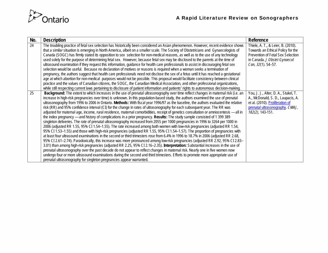
A Rapid Literature Review on Sonographers
No Description Reference 26 Aims The aim of this study was to assess the cost-effectiveness of using certified sonographers and miniaturized echocardiography
systems to perform echocardiograms at bedside in comparison to moving inpatients from the admission department to the echocardiography laboratory (echo-lab) Methods and Results From 26 September 2005 to 27 October 2005 112 patients admitted in six hospital wards in Italy connected through a 100 Mbit LAN to the echo-lab were scanned within the admission ward by sonographers using a miniaturized echo system Logistical data were collected and results were compared with those obtained from 194 consecutive patients coming from the same wards and studied in the echo-lab with high-end machines between 8 March 2005 and 15 April 2005 Performing echocardiograms in the admission department avoided long waiting time of the inpatients in the echo-lab before and after the study increased the percentage of patients studied within 3 and 5 days from request (88 vs 77 and 100 vs 95 respectively P = 003) increased both sonographer (by 339 P lt 0001) and echo-lab productivity (by 41 P lt 0001) and reduced costs of echocardiograms by 29 Conclusion Implementation of digital echocardiography certified sonographers and a miniaturized echo system allowed improvement of the cost-effectiveness of the service provided by the echo-lab for inpatients and avoided patientsrsquo discomfort derived from prolonged waiting time before and after the exam
Badano L Gaetano N Stacul S Gianfagna P Pericoli M et al (2009) Improved workflow sonographer productivity and cost-effectiveness of echocardiographic service for inpatients by using miniaturized systems European Journal of Echocardiography 10 537ndash542
27 The present report is a proposal from the American Society of Echocardiography Advanced Practice Task Force that identifies the potential of cardiac sonographers to achieve the Advanced Cardiovasculat Sonographer (ACS) level According to this report echocardiographic examinations require a well-trained and competent sonographer to obtain proper anatomic and physiologic data to establish an accurate diagnosis for clinical decision-making and patient management Although the formal education and training of cardiovascular sonographers are evolving many entry-level and staff sonographers may not have sufficient practical or clinical knowledge of the necessary components of the echocardiographic study for the individual patientrsquos clinical presentation In many clinical settings echocardiograms are read after the patient has left the laboratory Thus there is a role for a sonographer who can practice at an advanced level in a cardiovascular ultrasound laboratory to ensure a proper echocardiographic examination is performed on every patient In this setting an Advanced Cardiovascular Sonographer (ACS) would be able to review the indication for and quality of the examination If additional images were needed the ACS would assist the sonographer in obtaining these images which would lead to the performance of a complete and fully diagnostic examination before the patient had left the echocardiography laboratory In clinical practice the quality of the examinations performed would improve advancements in echocardiographic methods could be taught and incorporated into daily practice and patients would be better served
Mitchell C Miller F A Bierig S M Bremer M L Ehler D et al (2009) Advanced Cardiovascular Sonographer A Proposal of the American Society of Echocardiography Advanced Practice Sonographer Task Force J Am Soc Echocardiogr 22 1409-13
A Rapid Literature Review on Sonographers
No Description Reference 28 Sonographer practitioner development involves the expansion and extension of the sonographer role to include reporting on
ultrasound examinations Australian sonographers have not seen the same degree of role extension and expansion as their counterparts in the United Kingdom despite increasing levels of discussion regarding sonographer practitioner development The aim of this study was to determine if Australian sonographers want to extend their professional role and what they consider are the important issues associated with role extension This paper reports on qualitative data derived from a survey of Australian sonographers and investigates if Australian sonographers are interested in extending and expanding their professional role and responsibilities and if they do what might be necessary or desirable from a professional point of view for this change to occur A survey was mailed to all members of the Australian Sonographers Association (ASA) in October 2006 The 31-item survey included 28 closed-ended and three opened-ended items to provide both quantitative and qualitative data The quantitative data will be reported separately Qualitative data was derived from responses to the opened-ended questions which asked respondents to elaborate on their attitudes and feelings about role extension and development Analysis used Nvivo7 software to aid in uncovering common themes from the qualitative data The analysis focused on the reported incentives or motivations for becoming a sonographer practitioner as well as disincentives or perceived hurdles that would discourage respondents from becoming sonographer practitioners The three most reported incentives or motivations for becoming a sonographer practitioner were professional recognition remuneration and increased knowledge The three most commonly reported disincentives or perceived hurdles that would discourage respondents from becoming sonographer practitioners were legal issues insurance and further study
McGregor R OrsquoLoughlin K Cox J Clarke J Snowden A (2009) Sonographer practitioner development in Australia Qualitative analysis of an Australian sonographersrsquo survey Radiography15 313-319
29 Introduction This study describes the effect of introducing a cardiac sonographer led follow up clinic within a hospital cardiology department for patients with stable heart valve disease The two years before and after the instigation of the valve clinic were audited Materials and methods The clinic was conducted in a single centre and undertaken in the cardiology department of a district general hospital A total of 382 patients with 397 clinically significant valve lesions but for whom surgery was not yet indicated but follow up required were seen in a cardiac sonographer run clinics These patients no longer attended a medical follow up clinic unless there was clinical or echocardiographic deterioration Effectiveness was judged by the percentage treated according to current best practice guidelines the number of echocardiograms performed and the number of hospital outpatient visits attended In addition mortality data for the subjects in the clinic was collected Results The proportion followed up according to best practice guidelines rose from 157 (41) to 354 (92) (p lt 001) The total number of echocardiograms performed fell from 807 to 550 Total number of outpatient visits fell from 998 to 31 A total of 11 patients died in the two year study period none from progressive valve disease Discussion This study demonstrates that a protocol driven sonographer led heart valve disease follow up clinic significantly improved the quality of follow up while bringing about a major reduction in outpatients visits without compromising patient safety
Taggu W Topham A Hart L Carr-White G Sulke N et al (2009) A cardiac sonographer led follow up clinic for heart valve disease International Journal of Cardiology 132 240 ndash243
30 Role development for imaging allied health professionals has progressed significantly over the last three decades with many major developments pioneered in the field of ultrasound This paper reviews the developing role of the radiographer in ultrasound (in some countries such as the UK sonographers are considered a type of radiographer) and some of the clinical professional and educational implications of such development As the published evidence base underpinning the latest developments is scarce several contemporary case study examples which have been presented verbally at study days attended by the authors have been included In conclusion sonographer role development is considered within the professional contexts of extended role advanced and consultant practice
Hart A amp Dixon A (2008) Sonographer Role Extension and Career Development a Review of the Evidence Ultrasound 16(1) 31-35
A Rapid Literature Review on Sonographers
No Description Reference 31 Objective The main goal of this study was to determine end usersrsquo knowledge regarding safety aspects of diagnostic ultrasound
during pregnancy End usersrsquo attitudes toward the use of ultrasound in low-risk pregnancies were also assessed Methods A questionnaire was distributed to ultrasound end users attending review courses and hospital grand rounds between April and June 2006 in Chicago Results One hundred thirty end users completed the questionnaires (63 response rate) Sixty-three percent were physicians (n = 84) most of them obstetricians (817) About 18 of participants routinely performed Doppler ultrasound examinations during the first trimester Fifty percent of end users thought that the number of ultrasound examinations in low-risk pregnancy should be limited to 1 to 3 (mean plusmn SD 26 plusmn 09) Almost 70 disapproved of ldquokeepsakeentertainmentrdquo ultrasound Although 322 of the participants were familiar with the term thermal index only 177 actually gave the correct answer to the question on the nature of the thermal index About 22 were familiar with the term mechanical index but only 38 described it properly Almost 80 of end users did not know where to find the acoustic indices Only 208 were aware that they are displayed on the sonographic monitor during the examinations End users with higher knowledge of safety issues thought that there should be limitations on the number of ultrasound examinations in low-risk pregnancies (odds ratio 33 95 confidence interval 11ndash100 P = 028) Likewise these end users were more likely to respond that ultrasound might have adverse effects during pregnancy (odds ratio 32 95 confidence interval 11ndash125 P = 045) Conclusions Ultrasound end users are poorly informed regarding safety issues during pregnancy Further efforts in the realm of education and training are needed to improve end user knowledge about the acoustic output of the machines and safety issues
Sheiner E Shoham-Vardi I Abramowicz J S (2007) What Do Clinical Users Know Regarding Safety of Ultrasound During Pregnancy J Ultrasound Med 26 319ndash325
A Rapid Literature Review on Sonographers
No Description Reference 32 In an attempt to survey the knowledge among ultrasound users of some safety aspects of diagnostic ultrasound a questionnaire was
distributed to professionals using ultrasound for fetal examinations The questionnaire was answered by the participants and faculty members of six postgraduate courses in obstetric ultrasound Doppler ultrasound and fetal echocardiography given in Sweden Norway and Austria in 2003 Furthermore in Sweden the questionnaire was distributed to the staff meeting participants of two major obstetric ultrasound units and of one unit for fetal echocardiography and at a meeting of the Working Group for Ultrasound of the Swedish Society of Obstetrics and Gynecology In total 199 questionnaires were anonymously answered by doctors (n = 145) sonographers (n = 22) and midwives trained in ultrasound (n = 32) All of them were using diagnostic ultrasound on a daily or weekly basis The respondents were from nine European countries In none of the countries is formal certification for the use of diagnostic ultrasound in pregnancy or otherwise required The doctors who answered the questionnaire were specialists in obstetrics and gynecology (including some of the internationally recognized experts in fetal ultrasound) pediatric cardiology radiology or clinical physiology The respondents were not informed about the questionnaire in advance and they had a limited time to answer the 16 questions typically 10ndash15 min The questions concerned explanation of the safety indices and their relation to various ultrasound modes ie real-time B-mode M-mode three-dimensional (3D) spectral Doppler color and power Doppler ultrasound The respondents were also asked which ultrasound system they used most often and where on the system the information on thermal index (TI) and mechanical (MI) was displayed (eg in the top right corner of the screen in a separate window etc) Furthermore the respondents had to indicate how they adjust the output energy level on their own machine For all questions there was the optional answer lsquoDonrsquot knowrsquo Results About one-third of the ultrasound experts were able to define the abbreviations TI and MI For the subclasses of the TI the correct answer was found in only 3-8 of cases Of those who knew what the TI and MI meant only two-thirds and one-third of respondents respectively were able to give a simple explanation of the indices In 49 of questionnaires the Doppler modes were correctly ranked as generally producing higher ultrasound exposure than the imaging modes (B-mode 3D) Some 28 of the respondents correctly indicated where on their own machine the information on safety indices is displayed However not all of them knew how to control the output energy level For all items in the questionnaire there were no significant differences in the results between the three categories of respondents (ie physicians sonographers and midwives) The results of the questionnaire were anything but encouraging and they indicated that the users who are supposed to be responsible for controlling exposure of the fetus to ultrasound had a very poor knowledge of the basic safety aspects of ultrasound
Maršaacutel K (2005) The output display standard has it missed its target Ultrasound Obstet Gynecol 25 211ndash214
33 Purpose This study was performed to evaluate the accuracy of upper abdominal ultrasound (US) scanning performed by sonographers in a district general hospital to identify potential areas of weakness and to make recommendations to improve the service Materials and Methods Upper abdominal US examinations performed and reported by sonographers over a 4-week period were retrospectively reviewed The accuracy of the imaging findings and reports were assessed against other imaging surgical histological or laboratory findings and against clinical outcome Results A heterogenous group of 104 patients were included in the study 62 of whom had an US abnormality Errors of scanning or interpretation were identified in 10 patients (96) of whom five (48) were felt to be potentially significant Conclusions The sonographersrsquo accuracy in reporting upper abdominal US scans was 90 However on the basis of this study the authors have implemented specific recommendations to improve the quality of the service
Dongola N Guy R Ward S amp Giles J A (2003) Can sonographers offer an accurate upper abdominal ultrasound service in a district general hospital Radiography 9 29ndash33
A Rapid Literature Review on Sonographers
No Description Reference 34 Aim To compare the accuracy of radiographers and radiologists in routine abdominal ultrasound Materials and Methods One
hundred consecutive patients attending for routine abdominal ultrasound were included Each patient was examined by both a radiographer and radiologist Both operators noted their fndings and wrote a concluding report without conferring Reports were compared Where there was disagreement the patient was either re-examined by another radiologist or had further investigation Results Of 100 patients 52 were men and 48 were women The age range was 19plusmn88 years (median 52 years) Thirty-seven patients had renal tract ultrasound one had an aortic ultrasound and 62 had general upper abdominal ultrasound In 44 cases both operators reported the examination as normal In 49 cases both operators reported the examinations as abnormal and there was complete agreement between the operators In seven cases there was not complete agreement between operators Three of these disagreements were considered minor and four major In three of the seven cases the radiographer was correct and in four the radiologist was correct Conclusion Experienced radiographers and radiologists are highly accurate in performing and interpreting routine abdominal sonography Both operators missed a small minority of abnormalities There was no statistically significant difference in the accuracy of radiographers and radiologist
Leslie A Lockyer H amp Virjee J P (2000) Who Should be Performing Routine Abdominal Ultrasound A Prospective Double-Blind Study Comparing the Accuracy of Radiologist and Radiographer Clinical Radiology 55 606-609
Grey Literature 35 The ARRT The American Registry of Radiologic Technologists tests certifies and annually registers more than a quarter-of-a-million
radiologic technologists to promote high standards of patient care They award the Registered Technologist or RT designation to individuals who have completed the prescribed classroom and clinical education passed the appropriate exam and met the ethics requirements And they ensure their continuing education and ongoing compliance with their high standards by requiring annual registration of their certificate Certification by ARRT is voluntary not mandatory But employers state licensing agencies and federal regulators view ARRTrsquos credential as an indicator that a person has met recognized national standards for medical imaging and radiation therapy professionals How Are Radiologic Technologists Deemed Qualified First comes initial certification Certification is the process of initially recognizing individuals who have satisfied certain standards within a profession A person is certified by ARRT after completing an approved educational program complying with the ethics standards and passing a certification exam Then comes annual registration Registration is the yearly procedure required to maintain certification ARRT registrants are those who having already fulfilled the requirements for initial certification continue to meet the requirements for annual registration The ARRT annually registers the certificates of individuals who meet the following three criteria agreeing to comply with the ARRT Rules and Regulations continuing to comply with the ARRT Standards of Ethics and meeting our continuing education requirements Only technologists who are currently registered (which means that they have renewed within the past year) may designate themselves as ARRT Registered Technologists and use the initials RT after their names Federal laws regulate personnel qualifications in some modalities (mammography for example) but they accredit the health care facility rather than licensing the individual radiologic technologist who is providing patient care And while many states have laws regulating who can perform radiologic procedures they are typically based on ARRT certification Some employers and state agencies may require ARRT certification but not necessarily current registration Unless they require current registration by ARRT employers cannot assume that the person has complied with the ethics requirements or the continuing education requirements
American Registry of Radiologic Technologists (ARRT) (2013a) Patient Page Accessed April 2013
A Rapid Literature Review on Sonographers
No Description Reference 36 This document outlines certification requirements for sonography There are two pathways (primary and post-primary) For the
primary pathway it notes that sonography educational programs must be accredited by a mechanism acceptable to the ARRT ARRT generally recognizes only accreditation agencies that are recognized by the Council for Higher Education Accreditation andor United States Department of Education
American Registry of Radiologic Technologists (ARRT) (2013b) Sonography Certification Accessed April 2013
A Rapid Literature Review on Sonographers
No Description Reference 37 In Australia sonographers and student sonographers are required to be accredited by Australian Sonographer Accreditation Registry
(ASAR) in order to practise This requirement is contained within the Australian Governmentrsquos Diagnostic Imaging Accreditation Scheme standards and Medicare legislation Accreditation of sonographers is linked only to achieving - or in the case of students studying towards - an accredited entry-level qualification completion of continuing professional development and payment of an annual fee to ASAR The ASA contends that the lack of a requirement to ensure each sonographer is lsquofit to practisersquo is an unacceptable gap in current regulation undermining efforts to ensure the quality of sonographic services and failing to protect patients from harm The ASA supports and advocates for nationally-enforceable regulation of sonographers to meet our patientrsquos expectations National registration The National Registration and Accreditation Scheme (NRAS) for health workers commenced on 1 July 2010 The ten professions which comprised the initial intake into this scheme were those that had registration requirements in all Australian states or territories as well as those in which legislated regulation is deemed to be in the public interest An additional four professions - including medical radiation practitioners which are defined as radiographers radiation therapists and nuclear medicine technologists - will be included in the NRAS commencing on 1 July 2012 As sonography did not have registration in any states or territories prior to the commencement of NRAS the profession has not been in a position to negotiate entry into NRAS In early August 2011 the ASA lodged a submission with the Health Workforce Principal Committee requesting a profession-specific Sonography Board of Australia be formed under NRAS This submission was prepared based on information that consideration of professions which were unregistered would commence More recently the Australian Health Workforce Ministerial Council (AHWMC) issued a communiqueacute stating in part lsquoMinisters have commissioned work on unregistered professions and future directions for national registration Given this Ministers agreed to defer consideration of the inclusion of additional professions into the NRAS Ministers agreed that it would be premature to consider further applications for the scheme until this work has been completed other than paramedicsrsquo Options for unregistered health practitioners including sonographers Policy work is continuing at a federal level to establish an alternative regulatory framework The National Alliance for Self-Regulating Health Professions (NASRHP) consisting of the peak professional associations of eight self-regulating health professions including the ASA representing sonography released a proposal lsquoHarnessing self-regulation to support safety and quality in healthcare deliveryrsquo in March 2012 This proposal concluded that the framework that regulates all health practitioners should be expanded beyond NRAS to include
bull authorised self-regulating professions via a health professions panel bull negative licensing to withdraw the ability to practise from registered and self-regulated practitioners who do not meet the
regulatory requirements or unregulated practitioners due to poor practice As with all advocacy initiatives consideration by the federal government of the introduction of the NASRHPrsquos proposal may be prolonged and the outcomes may not match the expectations of NASRHPrsquos member professional associations including the ASA The ASA will continue both independently and collaboratively to lobby on behalf of our members and the public for the introduction of a timely and equitable solution to health practitioner regulation In the interim as there is no regulatory model with the rigour the ASA believes is required for regulation of sonographers and other health practitioners other than NRAS the ASA continues to support its Position Statement published in early 2010
Australian Sonographer Association (ASA) (2013) Sonographer Regulation Accessed April 2013
A Rapid Literature Review on Sonographers
No Description Reference 38 This publication contains information about Cardiovascular Credentialing International (CCI) It also provides the necessary
information and references concerning the process by which candidates may earn certificate or registry-level credentials CCI is a not-for-profit corporation established for the purpose of administering credentialing examinations as an independent credentialing agency CCI began credentialing cardiovascular professionals in 1968 CCI offers eight credentials which when earned demonstrate that the registrant holds fundamental knowledge in the particular cardiovascular specialty These include RCCS ndash Registered Congenital Cardiac Sonographer RCS ndash Registered Cardiac Sonographer and RPhS ndash Registered Phlebology Sonographer
Cardiovascular Credentialing International (2013) 2013 Examination Application amp Overview Accessed April 2013
39 Oregon Governor Signs Sonographer Licensure Legislation On July 28 2009 Oregon Governor Ted Kulongoski signed a sonographer licensure bill (HB 2245) into law The new law makes Oregon only the second state in the United States to require licensure of sonographers The Society of Diagnostic Medical Sonographers (SDMS) has worked closely with other sonography-related organizations and Oregon sonographers to help craft the language in new law The law renames and restructures the Oregon Board of Radiologic Technologists to the Oregon Board of Medical Imaging (OBMI) and ensures representation of sonographers on the Board The sonographer licensure law requires Oregon sonographers to become licensed by OBMI effective July 1 2010 Beginning on January 1 2014 all sonography licensees would be required to hold a national sonography certificationcredential (or be currently enrolled as a student) While the Oregon bill is far more detailed than the sonographer licensure law that passed in New Mexico earlier this year additional administrative rulesregulations will be developed over the next year to fully implement the new law SDMS will be continue to monitor developments regarding sonographer licensure in other states
Society of Diagnostic Medical Sonographers (SDMS) (2013a) News Accessed April 2013
A Rapid Literature Review on Sonographers
No Description Reference 40 On April 6 2009 New Mexico (NM) Governor Bill Richardson signed the historic bill that for the first time in the United States
requires licensure of sonographers The bill adds sonographers (and MRI technologists) to the list of medical imaging professionals licensed by the State of New Mexico Until now anyone could perform sonograms in New Mexico Prior to the passage of the New Mexico licensure law virtually all other health care providers and professionals had to be licensed As a result the public is often surprised to learn that the person performing a medical sonogram does not have any state licensure or national certification requirements The responsibility for administering the NM sonographer licensure program would rest with the New Mexico Environment Department Although this was not the first choice in most sonographersrsquo minds the Environment Department currently manages the radiologic technology licensure program so the administrative infrastructure is already in place By ensuring the creation of an advisory council that includes representation by each medical imaging and radiation therapy modality regulated the bill helps ensure that the Departmentrsquos other areas of emphasis do not detract from sonographer licensure Because sonographers have never had to be licensed before they frequently do not see or understand the distinction between state licensure and national certification National certifications or credentials issued by voluntary organizations such as the American Registry for Diagnostic Medical Sonography (ARDMS) Cardiovascular Credentialing International (CCI) and the American Registry of Radiologic Technologists (ARRT) have little legal weight The certification process and resulting credential are valuable tools in ensuring sonographers have met minimum education and knowledge requirements but do not really regulate anyonersquos ability to perform sonography In an ideal world no additional regulation of sonography would be required However as everyone knows more and more people are taking weekend courses and buying ultrasound equipment without understanding the ramifications of using ultrasound technology (eg ALARA) Under the bill passed by the New Mexico legislature national credentialscertifications should provide evidence of meeting the statersquos standards All sonographers who are performing these critical medical imaging services in New Mexico will be required to meet national certification standards (ie have certifications or credentials issued by ARDMS CCI or ARRT) As a result there would be no additional exams in New Mexico beyond the national certification exams required by ARDMS CCI or ARRT The bill also requires the state to recognize the continuing medical education completed for national certification renewal The most visible impact on New Mexico sonographers will be a license fee However the legislation caps the license fee at $100 per two-year license In addition only the New Mexico Legislature can change this cap
SDMS (2013b) New Mexico Sonographer Licensure Law Signed Accessed April 2013
A Rapid Literature Review on Sonographers
No Description Reference 41 Preamble The purpose of this document is to define the Scope of Practice for Diagnostic Ultrasound Professionals in the US and to
specify their roles as members of the health care team acting in the best interest of the patient This scope of practice is a living document that will evolve as the technology expands Definition of the Profession The Diagnostic Ultrasound Profession is a multi-specialty field comprised of Diagnostic Medical Sonography (with subspecialties in abdominal neurologic obstetricalgynecologic and ophthalmic ultrasound) Diagnostic Cardiac Sonography (with subspecialties in adult and pediatric echocardiography) Vascular Technology and other emerging fields These diverse specialties are distinguished by their use of diagnostic medical ultrasound as a primary technology in their daily work Certification is considered the standard of practice in ultrasound Individuals who are not yet certified should reference the Scope as a professional model and strive to become certified Scope of Practice of the Profession The Diagnostic Ultrasound Professional is an individual qualified by professional credentialing and academic and clinical experience to provide diagnostic patient care services using ultrasound and related diagnostic procedures The scope of practice of the Diagnostic Ultrasound Professional includes those procedures acts and processes permitted by law for which the individual has received education and clinical experience and in which heshe has demonstrated competency Diagnostic Ultrasound Professionals
bull Perform patient assessments bull Acquire and analyze data obtained using ultrasound and related diagnostic technologies bull Provide a summary of findings to the physician to aid in patient diagnosis and management bull Use independent judgment and systematic problem solving methods to produce high quality diagnostic information and
optimize patient care
Society of Diagnostic Medical Sonography [SDMS] (2013c) Scope of Practice for the Diagnostic Ultrasound Professional Accessed April 2013
42 This website notes that in the US most employers in the US prefer to hire sonographers who have professional certification A sonographer can get certification and passing an exam Most exams relate to the specialty that the sonographer is most interested inmdashfor example an exam to become certified in abdominal sonography A few states require diagnostic medical sonographers to be licensed Typically professional certifications required for licensure other requirements vary by state Sonographers must take continuing education to keep their certification current
Bureau of Labor Statistics US Department of Labor (2012) How to become a Diagnostic Medical Sonographer Occupational Outlook Handbook 2012-13 Edition Accessed April 2013
A Rapid Literature Review on Sonographers
No Description Reference 43 The American Registry for Diagnostic Medical Sonography (ARDMS) is an independent not-for-profit organization founded in 1975
that administers examinations and awards the following credentials bull RDMSreg ndash Registered Diagnostic Medical Sonographerreg bull RDCSreg ndash Registered Diagnostic Cardiac Sonographerreg bull RVTreg ndash Registered Vascular Technologistreg bull RPVIreg ndash Registered Physician in Vascular Interpretationreg bull RMSKtrade - Registered in Musculoskeletaltrade sonography
ARDMS is the globally recognized standard of excellence in sonography and has certified more than 75000 individuals ARDMS is composed of three key groups
bull Board of Directors ndash Sonographers vascular technologists physicians research scientists and a member of the public are volunteer members of the board that determines the direction and policies of our organization
bull Examination Development Task Forces ndash A group of sonographers vascular technologists physicians and scientists volunteer their time and expertise to produce our examinations
bull Professional Staff ndash A paid highly-skilled staff administers the programs and assists applicants and registrants Credentials awarded by ARDMS document personal achievement of recognized professional standards and are widely accepted in the medical community by sonography and vascular professional organizations However ARDMS does not validate the day-to-day job performance of any sonography professional ARDMS has earned the prestigious ANSI-ISO 17024 accreditation for certifying bodies from the International Organization for Standardization (ISO) Accreditation is granted through the American National Standards Institute (ANSI) ARDMS has also earned accreditation with the National Commission for Certifying Agencies (NCCA) The NCCA is the accrediting arm of the National Organization for Competency Assurance (NOCA) Established in 1977 as a nonprofit organization NOCA is a leader in establishing quality standards for certifying organizations
American Registry for Diagnostic Medical Sonography (ARDMS) (2012a) Overview of ARDMS Accessed April 2013
A Rapid Literature Review on Sonographers
No Description Reference 44 ARDMS is developing a program to assure that Registrants continue to meet the basic competency requirements of core clinical skills
in present day practice bull Each Registrant who holds an active ARDMS certification as of December 31 2011 will be assigned the 2012 ndash 2021 10-
year recertification assessment period bull The 10-year period for new Registrants will begin the year following the attainment of the credential bull If a Registrant earns a new credential or specialty after January 1 2012 then that credentialspecialty will have a separate
10-year recertification assessment period that will begin the year following the attainment of the credentialspecialty ARDMS will develop tools and resources to assist Registrants in tracking and meeting the recertification assessment program requirements
bull Registrants who do not wish to take a recertification assessment in a specific credentialspecialty may voluntarily relinquish the credentialspecialty
As an ARDMS Registrant individuals are committing to demonstrate the highest level of competency in the modality of sonography by meeting and maintaining the standards set by the ARDMS The pathway of maintenance your certification is comprised of Paying the ARDMS annual renewal fee each year Each year ARDMS assesses Registrants an annual renewal fee By paying your annual renewal fee you are contributing to the ARDMS mission of promoting quality care and patient safety through the certification and continuing competency of ultrasound professionals In order to maintain your credentials you must pay the annual renewal fee of $75 USD or $100 USD (for RPVI and MSK Registrants with MDDODPM) designation on or before December 31 of each year in order to stay active for the following year Attesting to ARDMS rules and policies Each year when you pay your annual renewal fee you are attesting that you are in compliance with all ARDMS rules and discipline policies and are in good standing with the ARDMS Earning a minimum of 30 ARDMS-accepted CMEs during your three-year CME period ARDMS verifies its Registrants compliance with continuing medical education (CME) requirements using an audit process Earning your CMEs is an integral component of maintaining your ARDMS credential(s) and demonstrates your ongoing commitment to lifelong learning RecertificationContinued Commitment to Excellence Following the attainment of the ARDMS credential Registrants are assigned a 10-year recertification period the year after the credential is earned ARDMS is committed to developing a recertification program that is easy for Registrants to comply with
American Registry for Diagnostic Medical Sonography (ARDMS) (2012b) Recertification General Information
A Rapid Literature Review on Sonographers
No Description Reference 45 This document reviews Sonography Principles and Instrumentation (SPI) Examination Requirements offered through ARDMS in
order to achieve sonography certification and the various prerequisites needed before taking the exam For example one prerequisite entails (1) Education Graduate of a program accredited by an agency recognized by the Council for Higher Education Accreditation (CHEA) United States Department of Education (USDOE) or Canadian Medical Association (CMA) that specifically conducts programmatic accreditation for diagnostic medical sonographydiagnostic cardiac sonographyvascular technology Currently the only organizations that offer programmatic accreditation under the aforementioned associations are the Commission on Accreditation of Allied Health Education Programs (CAAHEP) and the Canadian Medical Association (CMA) (2) Required Clinical UltrasoundVascular Experience No additional experience is required (3) Documentation Required with Application 1) Copy of diploma from ultrasoundvascular program or an official transcript indicating the date the degree was conferred 2) Original letter signed by program director andor medical director indicating date of graduation or successful completion of the program4 Program directors must use the mandatory formatted sample letter available on ARDMSorg sampleletters 3) The CV form is not required if the application is submitted and received in the ARDMS office within one year after successful completion of the program Otherwise an original signed and completed CV form for each appropriate specialty area(s) must be submitted 4) Photocopy of a non-expired government issued photo identification with signature the name on the identification must exactly match the name under which you are applying for ARDMS examination
American Registry for Diagnostic Medical Sonography (ARDMS) (2012c) SPI Requirement and General Prerequisites Accessed April 2013
46 This page notes the five credentials offered by the ARDMS and the corresponding exams and specialty exams required bull RDMS requires the SPI exam (sonography principles and instrumentation) and a specialty exam in abdomen breast fetal
echocardiography neurosonolgoy or obstetricsgynecology bull RDCS requires the SPI exam and a speciality exam in adult echocardiography fetal echocardiography pediatric
echocardiography bull RVT requires the SPI and a speciality exam in vascular technology bull RMSK requires the musculoskeletal sonography exam bull RPVI requires the physiciansrsquo vascular interpretation exam
American Registry for Diagnostic Medical Sonography (ARDMS) (2012d) Credentials and Examinations Accessed April 2013
A Rapid Literature Review on Sonographers
No Description Reference 47 This article summarizes some of the many trends in fetal pediatric and adult echocardiography that SDMS members are excited
about Fetal Echocardiography Sonography has been used since the mid-1980s to help identify congenital heart defects during the second trimester The ldquobasicrdquo examination of the fetal heart involves a four-chamber view ndash evaluation of the right and left atrial and ventricular chambers and their respective valves In the past several years there has been a movement in the obstetric ultrasound community to encourage obstetric sonographers to perform the ldquoextended basicrdquo exam which includes the cardiac outflow tracts The outflow tracts consist of the main pulmonary artery exiting the right ventricle and the aorta exiting the left ventricle The fetal echocardiography community also is abuzz over three-dimensional (3D) and four-dimensional (4D) ultrasound and the potential of this technology to significantly improve the evaluation of the fetal heart Spatiotemporal Image Correlation (STIC) a 4D application allows for evaluation and manipulation of the heart in real-time Allen says Todayrsquos ultrasound equipment allows sonographers to gather a volume of data from which they can create detailed 3D images of the tiny fetal heart which is constantly in motion With computer manipulation of the data sets the images also can be viewed in 4D allowing physicians to see the entire fetal heart cycle from start to finish Pediatric Echocardiography One of the most significant advances in pediatric echo over the last decade is moving to a digital format The advent of pediatric transesophageal (TEE) probes also has been a big advance for pediatric echo Transesophageal probes provide an unimpeded sonographic visualization of the intracardiac structures Theyrsquore useful in visualizing very fine valvular abnormalities the thoracic aorta vegetations and thrombus in the atrial appendage It is also useful in detecting subtle anatomic variations seen in congenital heart disease Pediatric-sized TEE probes make the exams possible In the past it wasnrsquot possible because the adult probe was just too big to put down the babyrsquos or childrsquos esophagus Now the probe is small enough to successfully manipulate it in children and get superb images Because the new sonographic imaging equipment and better surgical techniques allow clinicians to detect and repair more cardiac abnormalities earlier patients are living longer Adult Echocardiography The adult echo world is abuzz about 2D and 3D speckle tracking relatively new techniques for the assessment of heart function and strain imaging Speckle tracking evaluates both regional and global function by measuring the deformation of the heart muscle
Orenstein B W (2010) Current Trends in Echocardiography SDMS New Wave February 1-5 Accessed April 2013
48 This document describes the requirements for admission to the Register of Accredited Medical Sonographer in Australia Requirements include Continuation of Accreditation In order to remain on the Register an accredited Sonographer must satisfy the ASAR continuing professional development (CPD) requirements and pay the prescribed annual fee Student Sonographers apart from paying the prescribed annual fee should provide evidence of their student status every year when they renew their ASAR accreditation
Australian Sonographer Accreditation Registry (ASAR) (no date A) Sonographer Accreditation Accessed April 2013
A Rapid Literature Review on Sonographers
No Description Reference 49 ASARrsquos functions
1 Accredit sonography education programs within Australia 2 Maintain a register of Accredited Sonographers and Accredited Student Sonographers 3 Establish minimum standards of Continuing Professional Development (CPD) and to monitor the CPD requirements for
Sonographers to remain on the register 4 To support activities that advance the profession of sonography 5 To provide registration information about Sonographers to Medicare Australia 6 To provide public access to the name registration number State or Territory current type of accreditation and area of
specialty for active Sonographers ASARrsquos Role ASAR is not a registration board ASAR does not have the authority to remove a Sonographer from the Registry due to professional misconduct nor the ability to prevent a Sonographer from practicing ASAR is only able to remove a Sonographer from the Registry if they
bull fail to meet CPD requirements and or bull fail to pay annual fees
ASAR (no date B) About ASAR Accessed April 2013
50 Regulatory bodies A Regulatory Body maintains a register of individuals who have achieved the required qualification of that profession to practice and can evidence continuing competence at the required level For a doctor to practice in the UK he or she must be registered with the General Medical Council (GMC) This is the regulatory body for medical practitioners The majority of sonographers who are employed in the UK come from a background of radiography or midwifery To practice radiography in the UK a qualified radiographer must be registered with the Health Professions Council (HPC) Similarly a midwife must be registered with the National Midwifery Council to practice as a midwife Sonographer registration in the UK There are a number of professions allied to medicine that are regulated by the HPC for example radiography physiotherapy However sonography is not recognised as a profession by the HPC and therefore there is no mechanism whereby an individual can register with the HPC as a sonographer or be regulated by them as a sonographer It is a common misunderstanding that sonographers have to be registered with the HPC to practice This comes from the fact that most sonographers are also qualified radiographers and maintain their registration with the HPC because they wish to remain on the HPC register as a radiographer Professional bodies An individual can be a member of a Professional Body such as the Royal College of Obstetricians amp Gynecologists (RCOG) Royal College of Radiologists (RCR) Society amp College of Radiographers (SCoR) or the Royal College of Midwives (RCM) but this is not the same as being accepted onto the register of one of the regulatory bodies The professional bodies are responsible for setting and maintaining the standards of their members providing a code of conduct etc They may also act in the capacity of a trades union and can also provide professional indemnity insurance The British Medical Ultrasound Society BMUS is not a professional body but is a scientific society so will offer a different range of benefits Whether or not one wishes to join the society has no impact on their ability to work as a sonographer in the UK although from a continuing professional development point of view it can be beneficial The College of Radiographers are currently working with the HPC to get sonography recognized as a separate profession
British Medical Ultrasound Society (BMUS) (no date) Information on Sonographer Regulation in the UK Accessed April 2013
A Rapid Literature Review on Sonographers
No Description Reference 51 This document included sections on the role of the sonographer and the use of sonography for entertainment Role of the
Sonographer and Technical Impressions The role of the sonographer is to perform the ultrasound scan and to document observations The sonographerrsquos written technical impressions are intended as a form of communication between the sonographer and the reporting physician These observations must be reviewed and subsequently reported by the reporting physician Issuing of a final reportdiagnosis represents the practice of medicine and is therefore the domain of the reporting physician Use of Ultrasound for Entertainment or Non-Diagnostic Purposes In contrast to this justified form of assessment ultrasound for entertainment is simply an elaborate version of a portrait For a variable fee the surface of the fetus is skimmed over and clients are provided with keepsake images or a DVD of their fetus There are no measurements taken during this exam no morphological assessment performed and no dictated diagnostic report of findings Essentially there are no diagnostic benefits to the exam and uninformed parents may be falsely reassured that the beautiful portrait indicates a normal fetus Entertainment ultrasound facilities may operate outside of medical guidelines and without any controls which may result in a lack of technical safeguards operator expertise or governance of technical competency These facilities do not comply with the ALARA principle which could result in increased fetal exposures The Canadian Society of Diagnostic Medical Sonograph (CSDMS) does not support persons or facilities that participate in ultrasound for entertainment activities Liability insurance purchased through the CSDMS group insurance plan does not cover any activities carried out for entertainment ultrasound purposes Other leaders in Canadian medicine support this statement In 2005 the SOGC (Society of Obstetrics and Gynaecologists of Canada) published an article entitled ldquoObstetrical Ultrasound Biological Effects and Safetyrdquo and their conclusion was that ldquothe theoretical risk of adverse bio-effects even from standard 2D obstetrical ultrasound makes it hard to justify its use for non-medical reasons such as sex-determination making non-medical photos or video or for commercial purposerdquo The SOGC recommends that ultrasound be used prudently and that energy exposure be limited to the minimum that is medically necessary The SOGC further recommends a complete ban on the non-medical use of fetal ultrasound and encourages government to join with society to find appropriate means to deal with this public health issue The CAR (Canadian Association of Radiologists) makes a similar statement ldquoThe CAR strongly opposes the use of diagnostic ultrasound equipment for non-medical purposes and considers the use of medical ultrasound for entertainment to be a misuse of the technology especially if fetal subjects are involvedrdquo
Canadian Society of Diagnostic Medical Sonography (no date) Professional Practice Guidelines and Policy Statements For Canadian Sonography Accessed April 2013
A Rapid Literature Review on Sonographers
No Description Reference 52 In 1993 the Consortium for the Accreditation of Sonographic Education (CASE) was formed through the common desire to ensure
that the education and training of sonographers in the United Kingdom was delivered at the highest level All members share common concerns that standards of service provision and education are developed in parallel with increasing demand and improving technology All members strive to create an environment in which education and training is maximised This will make validation and accreditation to be as robust and straightforward as possible CASEs philosophy is to promote best ultrasound practise through the accreditation of those training programmes that develop safe and competent ultrasound practitioners This should be delivered at postgraduate level The majority of its activities relate to supporting those Institutions that offer or wish to offer courses leading to the award of a Masters Postgraduate DiplomaCertificate in Medical or clinical Ultrasound In addition to offering accreditation of new and re-accreditation of established ultrasound education programmes CASE undertakes annual monitoring of the courses it has accredited The information obtained is fed back to the Institutions both on an individual basis and via the annual CASE report Each member organisation is unique and has its own obligations to its membership and its own agenda while CASE brings them together to provide a forum for the interchange of ideas in which shared views may be synthesised and thus inform education and training programmes
Consortium for the Accreditation of Sonographic Education (no date) Home Accessed April 2013
A Rapid Literature Review on Sonographers
No Description Reference 53 The Medical Radiation Technologists Board (the Board) is one of sixteen New Zealand health registration authorities appointed by
the Minister of Health under the Health Practitioners Competence Assurance Act 2003 (the Act) The Board is responsible for the administration of the Act in regard to the profession of medical radiation technology The primary responsibility of the Board is to protect the health and safety of the New Zealand public by ensuring practitioners registered in the profession of medical radiation technology are competent and fit to practice In accordance with Section 118 of the Act the Board is responsible for fulfilling a number of functions
bull 01 To prescribe the qualifications required for scopes of practice within the profession of medical radiation technology and for that purpose to accredit and monitor educational institutions and degrees courses of studies or programmes
bull 02 To authorise the registration of medical radiation technologists under the Act and to maintain registers bull 03 To consider applications for annual practising certificates bull 04 To review and promote the competence of medical radiation technologists bull 05 To recognise accredit and set programmes to ensure the ongoing competence of medical radiation technologists bull 06 To receive and act on information from health practitioners employers and the Health and Disability Commissioner
about the competence of medical radiation technologists bull 07 To notify employers the Accident Compensation Corporation the Director-General of Health and the Health and
Disability Commissioner that the practice of a medical radiation technologist may pose a risk of harm to the public bull 08 To consider the cases of medical radiation technologists who may be unable to perform the functions required for the
practice of the medical radiation technology profession bull 09 To set standards of clinical competence cultural competence and ethical conduct to be observed by medical radiation
technologists bull 10 To liaise with other authorities appointed under the Act about matters of common interest bull 11 To promote education and training in the profession of medical radiation technology bull 12 To promote public awareness of the responsibilities of the Board bull 13 To exercise and perform any other functions powers and duties that are conferred or imposed on it by or under the Act
or any other enactment
Medical Radiation Technologists Board (MRTB) (no date A) What we do Accessed April 2013
54 Qualifications To be considered for registration as a medical radiation technologist in any of the specified scopes of practice an individual must hold a qualification for that particular scope as prescribed by the Board or approved by the Board as being substantially equivalent to the prescribed qualifications In addition to having an approved qualification individuals may also be required to demonstrate that they have adequate experience in the relevant scope of practice Sonographer The Board has prescribed one New Zealand postgraduate qualification for a person to be eligible for registration in the scope of practice of Sonographer Postgraduate Diploma in Health Science (Ultrasound) issued by the Unitec Institute of Technology Required Experience If an individual is applying for registration in Nuclear Medicine Ultrasound or Magnetic Resonance Imaging in addition to holding a relevant postgraduate qualification they must also be able to provide evidence that you have a minimum of 3360 hours of clinical experience in the relevant scope of practice you are applying for registration in
Medical Radiation Technologists Board (MRTB) (no date B) Qualifications Accessed April 2013
A Rapid Literature Review on Sonographers
No Description Reference 55 Recertification is a mechanism used by the Medical Radiation Technologists Board in New Zealand to ensure that registered medical
radiation technologists continue to be competent to practise within their scopes of practice Annual Practising Certificates Each year if an individual wishes to continue practising in your scope(s) of practice in New Zealand they must apply for and be issued with an annual practising certificate (APC) A condition of being issued with an APC is that one must provide evidence of enrolment and satisfactory progress in a Board-approved recertification programme Alternatively the Board may issue them with an interim practising certificate (IPC) until it is satisfied the individual demonstrates that they are competent to practice in the scope(s) of practice they applied for Working with an IPC will require an individual to work under the supervision of a registered medical radiation technologist or other relevant registered health practitioner Recertification Programmes The Boards recertification programme framework is based on a continuing professional development (CPD) model Individuals have the option of enrolling in one of seven recertification programmes for medical radiation technologists that are currently approved by the Board They must meet the requirements of the particular recertification programme in which they are enrolled Recertification Audits An essential component of the CPD-based recertification programme adopted by the Board is the audit of CPD recordsThe Board undertakes an annual audit of approximately 10 of its registrant database to assess individual medical radiation technologistsrsquo compliance with the Boarrsquods recertification requirements
Medical Radiation Technologists Board (MRTB) (no date C) Recertification Accessed April 2013
56 There are two types of radiography - diagnostic and therapeutic Both need considerable knowledge of technology anatomy physiology and pathology to carry out their work Many also undertake further training to become a sonographer Some of the imaging technology that a diagnostic radiographer may use are
bull X-Ray - looks through tissues to examine bones cavities and foreign objects bull Fluoroscopy - images the digestive system providing a real-time image bull CT (Computed Tomography) - which provides cross-sectional views (slices) of the body bull MRI (Magnetic Resonance Imaging) - builds a 2-D or 3-D map of the different tissue types within the body bull Ultrasound - well known for its use in obstetrics and gynaecology Also used to check circulation and examine the heart bull Angiography - used to investigate blood vessels
Sonographyultrasonography Ultrasound is used in various settings in a hospital including abdominal scanning and breast ultrasound Ultrasound imaging is the use of high frequency sound in excess of human hearing to produce images of structures of the human body that may be observed on a screen These images may subsequently be transferred to photographic film or paper or onto video or CD forming part of the patients record of their examination There are no direct entry routes into ultrasound Most sonographers train as a radiographer then undertake an approved post-registration course Offered by higher education institutions The courses are a minimum of one academic year and prepare sonographers clinically and academically for practice Normally a pre-requisite for acceptance is access to a clinical department with supervised practice for students
National Health Service (NHS) (no date A) Radiographer Accessed April 2013
57 To practice as a radiographer in the UK an individual must be registered with the Health and Care Professions Council (HCPC) To register individuals must have successfully completed an approved training programme in diagnostic or therapeutic radiography These are offered at undergraduate degree (full-time and part-time) and at postgraduate degreediploma level at a number of universities across the UK
NHS (no date B) Entry requirements and training in radiography Accessed April 2013
Health Professions Regulatory Advisory Council 56 Wellesley Street West 12th Floor
Toronto ON M5S 2S3
Telephone 416-326-1550 Toll-Free 1-888-377-7746
Fax 416-326-1549
Website wwwhpracorg
Email hpracwebmasterontarioca
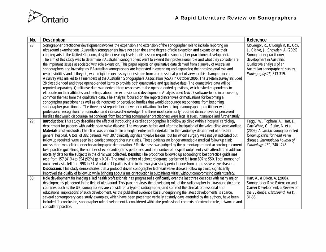
A Rapid Literature Review on Sonographers
No Description Reference 32 In an attempt to survey the knowledge among ultrasound users of some safety aspects of diagnostic ultrasound a questionnaire was
distributed to professionals using ultrasound for fetal examinations The questionnaire was answered by the participants and faculty members of six postgraduate courses in obstetric ultrasound Doppler ultrasound and fetal echocardiography given in Sweden Norway and Austria in 2003 Furthermore in Sweden the questionnaire was distributed to the staff meeting participants of two major obstetric ultrasound units and of one unit for fetal echocardiography and at a meeting of the Working Group for Ultrasound of the Swedish Society of Obstetrics and Gynecology In total 199 questionnaires were anonymously answered by doctors (n = 145) sonographers (n = 22) and midwives trained in ultrasound (n = 32) All of them were using diagnostic ultrasound on a daily or weekly basis The respondents were from nine European countries In none of the countries is formal certification for the use of diagnostic ultrasound in pregnancy or otherwise required The doctors who answered the questionnaire were specialists in obstetrics and gynecology (including some of the internationally recognized experts in fetal ultrasound) pediatric cardiology radiology or clinical physiology The respondents were not informed about the questionnaire in advance and they had a limited time to answer the 16 questions typically 10ndash15 min The questions concerned explanation of the safety indices and their relation to various ultrasound modes ie real-time B-mode M-mode three-dimensional (3D) spectral Doppler color and power Doppler ultrasound The respondents were also asked which ultrasound system they used most often and where on the system the information on thermal index (TI) and mechanical (MI) was displayed (eg in the top right corner of the screen in a separate window etc) Furthermore the respondents had to indicate how they adjust the output energy level on their own machine For all questions there was the optional answer lsquoDonrsquot knowrsquo Results About one-third of the ultrasound experts were able to define the abbreviations TI and MI For the subclasses of the TI the correct answer was found in only 3-8 of cases Of those who knew what the TI and MI meant only two-thirds and one-third of respondents respectively were able to give a simple explanation of the indices In 49 of questionnaires the Doppler modes were correctly ranked as generally producing higher ultrasound exposure than the imaging modes (B-mode 3D) Some 28 of the respondents correctly indicated where on their own machine the information on safety indices is displayed However not all of them knew how to control the output energy level For all items in the questionnaire there were no significant differences in the results between the three categories of respondents (ie physicians sonographers and midwives) The results of the questionnaire were anything but encouraging and they indicated that the users who are supposed to be responsible for controlling exposure of the fetus to ultrasound had a very poor knowledge of the basic safety aspects of ultrasound
Maršaacutel K (2005) The output display standard has it missed its target Ultrasound Obstet Gynecol 25 211ndash214
33 Purpose This study was performed to evaluate the accuracy of upper abdominal ultrasound (US) scanning performed by sonographers in a district general hospital to identify potential areas of weakness and to make recommendations to improve the service Materials and Methods Upper abdominal US examinations performed and reported by sonographers over a 4-week period were retrospectively reviewed The accuracy of the imaging findings and reports were assessed against other imaging surgical histological or laboratory findings and against clinical outcome Results A heterogenous group of 104 patients were included in the study 62 of whom had an US abnormality Errors of scanning or interpretation were identified in 10 patients (96) of whom five (48) were felt to be potentially significant Conclusions The sonographersrsquo accuracy in reporting upper abdominal US scans was 90 However on the basis of this study the authors have implemented specific recommendations to improve the quality of the service
Dongola N Guy R Ward S amp Giles J A (2003) Can sonographers offer an accurate upper abdominal ultrasound service in a district general hospital Radiography 9 29ndash33
A Rapid Literature Review on Sonographers
No Description Reference 34 Aim To compare the accuracy of radiographers and radiologists in routine abdominal ultrasound Materials and Methods One
hundred consecutive patients attending for routine abdominal ultrasound were included Each patient was examined by both a radiographer and radiologist Both operators noted their fndings and wrote a concluding report without conferring Reports were compared Where there was disagreement the patient was either re-examined by another radiologist or had further investigation Results Of 100 patients 52 were men and 48 were women The age range was 19plusmn88 years (median 52 years) Thirty-seven patients had renal tract ultrasound one had an aortic ultrasound and 62 had general upper abdominal ultrasound In 44 cases both operators reported the examination as normal In 49 cases both operators reported the examinations as abnormal and there was complete agreement between the operators In seven cases there was not complete agreement between operators Three of these disagreements were considered minor and four major In three of the seven cases the radiographer was correct and in four the radiologist was correct Conclusion Experienced radiographers and radiologists are highly accurate in performing and interpreting routine abdominal sonography Both operators missed a small minority of abnormalities There was no statistically significant difference in the accuracy of radiographers and radiologist
Leslie A Lockyer H amp Virjee J P (2000) Who Should be Performing Routine Abdominal Ultrasound A Prospective Double-Blind Study Comparing the Accuracy of Radiologist and Radiographer Clinical Radiology 55 606-609
Grey Literature 35 The ARRT The American Registry of Radiologic Technologists tests certifies and annually registers more than a quarter-of-a-million
radiologic technologists to promote high standards of patient care They award the Registered Technologist or RT designation to individuals who have completed the prescribed classroom and clinical education passed the appropriate exam and met the ethics requirements And they ensure their continuing education and ongoing compliance with their high standards by requiring annual registration of their certificate Certification by ARRT is voluntary not mandatory But employers state licensing agencies and federal regulators view ARRTrsquos credential as an indicator that a person has met recognized national standards for medical imaging and radiation therapy professionals How Are Radiologic Technologists Deemed Qualified First comes initial certification Certification is the process of initially recognizing individuals who have satisfied certain standards within a profession A person is certified by ARRT after completing an approved educational program complying with the ethics standards and passing a certification exam Then comes annual registration Registration is the yearly procedure required to maintain certification ARRT registrants are those who having already fulfilled the requirements for initial certification continue to meet the requirements for annual registration The ARRT annually registers the certificates of individuals who meet the following three criteria agreeing to comply with the ARRT Rules and Regulations continuing to comply with the ARRT Standards of Ethics and meeting our continuing education requirements Only technologists who are currently registered (which means that they have renewed within the past year) may designate themselves as ARRT Registered Technologists and use the initials RT after their names Federal laws regulate personnel qualifications in some modalities (mammography for example) but they accredit the health care facility rather than licensing the individual radiologic technologist who is providing patient care And while many states have laws regulating who can perform radiologic procedures they are typically based on ARRT certification Some employers and state agencies may require ARRT certification but not necessarily current registration Unless they require current registration by ARRT employers cannot assume that the person has complied with the ethics requirements or the continuing education requirements
American Registry of Radiologic Technologists (ARRT) (2013a) Patient Page Accessed April 2013
A Rapid Literature Review on Sonographers
No Description Reference 36 This document outlines certification requirements for sonography There are two pathways (primary and post-primary) For the
primary pathway it notes that sonography educational programs must be accredited by a mechanism acceptable to the ARRT ARRT generally recognizes only accreditation agencies that are recognized by the Council for Higher Education Accreditation andor United States Department of Education
American Registry of Radiologic Technologists (ARRT) (2013b) Sonography Certification Accessed April 2013
A Rapid Literature Review on Sonographers
No Description Reference 37 In Australia sonographers and student sonographers are required to be accredited by Australian Sonographer Accreditation Registry
(ASAR) in order to practise This requirement is contained within the Australian Governmentrsquos Diagnostic Imaging Accreditation Scheme standards and Medicare legislation Accreditation of sonographers is linked only to achieving - or in the case of students studying towards - an accredited entry-level qualification completion of continuing professional development and payment of an annual fee to ASAR The ASA contends that the lack of a requirement to ensure each sonographer is lsquofit to practisersquo is an unacceptable gap in current regulation undermining efforts to ensure the quality of sonographic services and failing to protect patients from harm The ASA supports and advocates for nationally-enforceable regulation of sonographers to meet our patientrsquos expectations National registration The National Registration and Accreditation Scheme (NRAS) for health workers commenced on 1 July 2010 The ten professions which comprised the initial intake into this scheme were those that had registration requirements in all Australian states or territories as well as those in which legislated regulation is deemed to be in the public interest An additional four professions - including medical radiation practitioners which are defined as radiographers radiation therapists and nuclear medicine technologists - will be included in the NRAS commencing on 1 July 2012 As sonography did not have registration in any states or territories prior to the commencement of NRAS the profession has not been in a position to negotiate entry into NRAS In early August 2011 the ASA lodged a submission with the Health Workforce Principal Committee requesting a profession-specific Sonography Board of Australia be formed under NRAS This submission was prepared based on information that consideration of professions which were unregistered would commence More recently the Australian Health Workforce Ministerial Council (AHWMC) issued a communiqueacute stating in part lsquoMinisters have commissioned work on unregistered professions and future directions for national registration Given this Ministers agreed to defer consideration of the inclusion of additional professions into the NRAS Ministers agreed that it would be premature to consider further applications for the scheme until this work has been completed other than paramedicsrsquo Options for unregistered health practitioners including sonographers Policy work is continuing at a federal level to establish an alternative regulatory framework The National Alliance for Self-Regulating Health Professions (NASRHP) consisting of the peak professional associations of eight self-regulating health professions including the ASA representing sonography released a proposal lsquoHarnessing self-regulation to support safety and quality in healthcare deliveryrsquo in March 2012 This proposal concluded that the framework that regulates all health practitioners should be expanded beyond NRAS to include
bull authorised self-regulating professions via a health professions panel bull negative licensing to withdraw the ability to practise from registered and self-regulated practitioners who do not meet the
regulatory requirements or unregulated practitioners due to poor practice As with all advocacy initiatives consideration by the federal government of the introduction of the NASRHPrsquos proposal may be prolonged and the outcomes may not match the expectations of NASRHPrsquos member professional associations including the ASA The ASA will continue both independently and collaboratively to lobby on behalf of our members and the public for the introduction of a timely and equitable solution to health practitioner regulation In the interim as there is no regulatory model with the rigour the ASA believes is required for regulation of sonographers and other health practitioners other than NRAS the ASA continues to support its Position Statement published in early 2010
Australian Sonographer Association (ASA) (2013) Sonographer Regulation Accessed April 2013
A Rapid Literature Review on Sonographers
No Description Reference 38 This publication contains information about Cardiovascular Credentialing International (CCI) It also provides the necessary
information and references concerning the process by which candidates may earn certificate or registry-level credentials CCI is a not-for-profit corporation established for the purpose of administering credentialing examinations as an independent credentialing agency CCI began credentialing cardiovascular professionals in 1968 CCI offers eight credentials which when earned demonstrate that the registrant holds fundamental knowledge in the particular cardiovascular specialty These include RCCS ndash Registered Congenital Cardiac Sonographer RCS ndash Registered Cardiac Sonographer and RPhS ndash Registered Phlebology Sonographer
Cardiovascular Credentialing International (2013) 2013 Examination Application amp Overview Accessed April 2013
39 Oregon Governor Signs Sonographer Licensure Legislation On July 28 2009 Oregon Governor Ted Kulongoski signed a sonographer licensure bill (HB 2245) into law The new law makes Oregon only the second state in the United States to require licensure of sonographers The Society of Diagnostic Medical Sonographers (SDMS) has worked closely with other sonography-related organizations and Oregon sonographers to help craft the language in new law The law renames and restructures the Oregon Board of Radiologic Technologists to the Oregon Board of Medical Imaging (OBMI) and ensures representation of sonographers on the Board The sonographer licensure law requires Oregon sonographers to become licensed by OBMI effective July 1 2010 Beginning on January 1 2014 all sonography licensees would be required to hold a national sonography certificationcredential (or be currently enrolled as a student) While the Oregon bill is far more detailed than the sonographer licensure law that passed in New Mexico earlier this year additional administrative rulesregulations will be developed over the next year to fully implement the new law SDMS will be continue to monitor developments regarding sonographer licensure in other states
Society of Diagnostic Medical Sonographers (SDMS) (2013a) News Accessed April 2013
A Rapid Literature Review on Sonographers
No Description Reference 40 On April 6 2009 New Mexico (NM) Governor Bill Richardson signed the historic bill that for the first time in the United States
requires licensure of sonographers The bill adds sonographers (and MRI technologists) to the list of medical imaging professionals licensed by the State of New Mexico Until now anyone could perform sonograms in New Mexico Prior to the passage of the New Mexico licensure law virtually all other health care providers and professionals had to be licensed As a result the public is often surprised to learn that the person performing a medical sonogram does not have any state licensure or national certification requirements The responsibility for administering the NM sonographer licensure program would rest with the New Mexico Environment Department Although this was not the first choice in most sonographersrsquo minds the Environment Department currently manages the radiologic technology licensure program so the administrative infrastructure is already in place By ensuring the creation of an advisory council that includes representation by each medical imaging and radiation therapy modality regulated the bill helps ensure that the Departmentrsquos other areas of emphasis do not detract from sonographer licensure Because sonographers have never had to be licensed before they frequently do not see or understand the distinction between state licensure and national certification National certifications or credentials issued by voluntary organizations such as the American Registry for Diagnostic Medical Sonography (ARDMS) Cardiovascular Credentialing International (CCI) and the American Registry of Radiologic Technologists (ARRT) have little legal weight The certification process and resulting credential are valuable tools in ensuring sonographers have met minimum education and knowledge requirements but do not really regulate anyonersquos ability to perform sonography In an ideal world no additional regulation of sonography would be required However as everyone knows more and more people are taking weekend courses and buying ultrasound equipment without understanding the ramifications of using ultrasound technology (eg ALARA) Under the bill passed by the New Mexico legislature national credentialscertifications should provide evidence of meeting the statersquos standards All sonographers who are performing these critical medical imaging services in New Mexico will be required to meet national certification standards (ie have certifications or credentials issued by ARDMS CCI or ARRT) As a result there would be no additional exams in New Mexico beyond the national certification exams required by ARDMS CCI or ARRT The bill also requires the state to recognize the continuing medical education completed for national certification renewal The most visible impact on New Mexico sonographers will be a license fee However the legislation caps the license fee at $100 per two-year license In addition only the New Mexico Legislature can change this cap
SDMS (2013b) New Mexico Sonographer Licensure Law Signed Accessed April 2013
A Rapid Literature Review on Sonographers
No Description Reference 41 Preamble The purpose of this document is to define the Scope of Practice for Diagnostic Ultrasound Professionals in the US and to
specify their roles as members of the health care team acting in the best interest of the patient This scope of practice is a living document that will evolve as the technology expands Definition of the Profession The Diagnostic Ultrasound Profession is a multi-specialty field comprised of Diagnostic Medical Sonography (with subspecialties in abdominal neurologic obstetricalgynecologic and ophthalmic ultrasound) Diagnostic Cardiac Sonography (with subspecialties in adult and pediatric echocardiography) Vascular Technology and other emerging fields These diverse specialties are distinguished by their use of diagnostic medical ultrasound as a primary technology in their daily work Certification is considered the standard of practice in ultrasound Individuals who are not yet certified should reference the Scope as a professional model and strive to become certified Scope of Practice of the Profession The Diagnostic Ultrasound Professional is an individual qualified by professional credentialing and academic and clinical experience to provide diagnostic patient care services using ultrasound and related diagnostic procedures The scope of practice of the Diagnostic Ultrasound Professional includes those procedures acts and processes permitted by law for which the individual has received education and clinical experience and in which heshe has demonstrated competency Diagnostic Ultrasound Professionals
bull Perform patient assessments bull Acquire and analyze data obtained using ultrasound and related diagnostic technologies bull Provide a summary of findings to the physician to aid in patient diagnosis and management bull Use independent judgment and systematic problem solving methods to produce high quality diagnostic information and
optimize patient care
Society of Diagnostic Medical Sonography [SDMS] (2013c) Scope of Practice for the Diagnostic Ultrasound Professional Accessed April 2013
42 This website notes that in the US most employers in the US prefer to hire sonographers who have professional certification A sonographer can get certification and passing an exam Most exams relate to the specialty that the sonographer is most interested inmdashfor example an exam to become certified in abdominal sonography A few states require diagnostic medical sonographers to be licensed Typically professional certifications required for licensure other requirements vary by state Sonographers must take continuing education to keep their certification current
Bureau of Labor Statistics US Department of Labor (2012) How to become a Diagnostic Medical Sonographer Occupational Outlook Handbook 2012-13 Edition Accessed April 2013
A Rapid Literature Review on Sonographers
No Description Reference 43 The American Registry for Diagnostic Medical Sonography (ARDMS) is an independent not-for-profit organization founded in 1975
that administers examinations and awards the following credentials bull RDMSreg ndash Registered Diagnostic Medical Sonographerreg bull RDCSreg ndash Registered Diagnostic Cardiac Sonographerreg bull RVTreg ndash Registered Vascular Technologistreg bull RPVIreg ndash Registered Physician in Vascular Interpretationreg bull RMSKtrade - Registered in Musculoskeletaltrade sonography
ARDMS is the globally recognized standard of excellence in sonography and has certified more than 75000 individuals ARDMS is composed of three key groups
bull Board of Directors ndash Sonographers vascular technologists physicians research scientists and a member of the public are volunteer members of the board that determines the direction and policies of our organization
bull Examination Development Task Forces ndash A group of sonographers vascular technologists physicians and scientists volunteer their time and expertise to produce our examinations
bull Professional Staff ndash A paid highly-skilled staff administers the programs and assists applicants and registrants Credentials awarded by ARDMS document personal achievement of recognized professional standards and are widely accepted in the medical community by sonography and vascular professional organizations However ARDMS does not validate the day-to-day job performance of any sonography professional ARDMS has earned the prestigious ANSI-ISO 17024 accreditation for certifying bodies from the International Organization for Standardization (ISO) Accreditation is granted through the American National Standards Institute (ANSI) ARDMS has also earned accreditation with the National Commission for Certifying Agencies (NCCA) The NCCA is the accrediting arm of the National Organization for Competency Assurance (NOCA) Established in 1977 as a nonprofit organization NOCA is a leader in establishing quality standards for certifying organizations
American Registry for Diagnostic Medical Sonography (ARDMS) (2012a) Overview of ARDMS Accessed April 2013
A Rapid Literature Review on Sonographers
No Description Reference 44 ARDMS is developing a program to assure that Registrants continue to meet the basic competency requirements of core clinical skills
in present day practice bull Each Registrant who holds an active ARDMS certification as of December 31 2011 will be assigned the 2012 ndash 2021 10-
year recertification assessment period bull The 10-year period for new Registrants will begin the year following the attainment of the credential bull If a Registrant earns a new credential or specialty after January 1 2012 then that credentialspecialty will have a separate
10-year recertification assessment period that will begin the year following the attainment of the credentialspecialty ARDMS will develop tools and resources to assist Registrants in tracking and meeting the recertification assessment program requirements
bull Registrants who do not wish to take a recertification assessment in a specific credentialspecialty may voluntarily relinquish the credentialspecialty
As an ARDMS Registrant individuals are committing to demonstrate the highest level of competency in the modality of sonography by meeting and maintaining the standards set by the ARDMS The pathway of maintenance your certification is comprised of Paying the ARDMS annual renewal fee each year Each year ARDMS assesses Registrants an annual renewal fee By paying your annual renewal fee you are contributing to the ARDMS mission of promoting quality care and patient safety through the certification and continuing competency of ultrasound professionals In order to maintain your credentials you must pay the annual renewal fee of $75 USD or $100 USD (for RPVI and MSK Registrants with MDDODPM) designation on or before December 31 of each year in order to stay active for the following year Attesting to ARDMS rules and policies Each year when you pay your annual renewal fee you are attesting that you are in compliance with all ARDMS rules and discipline policies and are in good standing with the ARDMS Earning a minimum of 30 ARDMS-accepted CMEs during your three-year CME period ARDMS verifies its Registrants compliance with continuing medical education (CME) requirements using an audit process Earning your CMEs is an integral component of maintaining your ARDMS credential(s) and demonstrates your ongoing commitment to lifelong learning RecertificationContinued Commitment to Excellence Following the attainment of the ARDMS credential Registrants are assigned a 10-year recertification period the year after the credential is earned ARDMS is committed to developing a recertification program that is easy for Registrants to comply with
American Registry for Diagnostic Medical Sonography (ARDMS) (2012b) Recertification General Information
A Rapid Literature Review on Sonographers
No Description Reference 45 This document reviews Sonography Principles and Instrumentation (SPI) Examination Requirements offered through ARDMS in
order to achieve sonography certification and the various prerequisites needed before taking the exam For example one prerequisite entails (1) Education Graduate of a program accredited by an agency recognized by the Council for Higher Education Accreditation (CHEA) United States Department of Education (USDOE) or Canadian Medical Association (CMA) that specifically conducts programmatic accreditation for diagnostic medical sonographydiagnostic cardiac sonographyvascular technology Currently the only organizations that offer programmatic accreditation under the aforementioned associations are the Commission on Accreditation of Allied Health Education Programs (CAAHEP) and the Canadian Medical Association (CMA) (2) Required Clinical UltrasoundVascular Experience No additional experience is required (3) Documentation Required with Application 1) Copy of diploma from ultrasoundvascular program or an official transcript indicating the date the degree was conferred 2) Original letter signed by program director andor medical director indicating date of graduation or successful completion of the program4 Program directors must use the mandatory formatted sample letter available on ARDMSorg sampleletters 3) The CV form is not required if the application is submitted and received in the ARDMS office within one year after successful completion of the program Otherwise an original signed and completed CV form for each appropriate specialty area(s) must be submitted 4) Photocopy of a non-expired government issued photo identification with signature the name on the identification must exactly match the name under which you are applying for ARDMS examination
American Registry for Diagnostic Medical Sonography (ARDMS) (2012c) SPI Requirement and General Prerequisites Accessed April 2013
46 This page notes the five credentials offered by the ARDMS and the corresponding exams and specialty exams required bull RDMS requires the SPI exam (sonography principles and instrumentation) and a specialty exam in abdomen breast fetal
echocardiography neurosonolgoy or obstetricsgynecology bull RDCS requires the SPI exam and a speciality exam in adult echocardiography fetal echocardiography pediatric
echocardiography bull RVT requires the SPI and a speciality exam in vascular technology bull RMSK requires the musculoskeletal sonography exam bull RPVI requires the physiciansrsquo vascular interpretation exam
American Registry for Diagnostic Medical Sonography (ARDMS) (2012d) Credentials and Examinations Accessed April 2013
A Rapid Literature Review on Sonographers
No Description Reference 47 This article summarizes some of the many trends in fetal pediatric and adult echocardiography that SDMS members are excited
about Fetal Echocardiography Sonography has been used since the mid-1980s to help identify congenital heart defects during the second trimester The ldquobasicrdquo examination of the fetal heart involves a four-chamber view ndash evaluation of the right and left atrial and ventricular chambers and their respective valves In the past several years there has been a movement in the obstetric ultrasound community to encourage obstetric sonographers to perform the ldquoextended basicrdquo exam which includes the cardiac outflow tracts The outflow tracts consist of the main pulmonary artery exiting the right ventricle and the aorta exiting the left ventricle The fetal echocardiography community also is abuzz over three-dimensional (3D) and four-dimensional (4D) ultrasound and the potential of this technology to significantly improve the evaluation of the fetal heart Spatiotemporal Image Correlation (STIC) a 4D application allows for evaluation and manipulation of the heart in real-time Allen says Todayrsquos ultrasound equipment allows sonographers to gather a volume of data from which they can create detailed 3D images of the tiny fetal heart which is constantly in motion With computer manipulation of the data sets the images also can be viewed in 4D allowing physicians to see the entire fetal heart cycle from start to finish Pediatric Echocardiography One of the most significant advances in pediatric echo over the last decade is moving to a digital format The advent of pediatric transesophageal (TEE) probes also has been a big advance for pediatric echo Transesophageal probes provide an unimpeded sonographic visualization of the intracardiac structures Theyrsquore useful in visualizing very fine valvular abnormalities the thoracic aorta vegetations and thrombus in the atrial appendage It is also useful in detecting subtle anatomic variations seen in congenital heart disease Pediatric-sized TEE probes make the exams possible In the past it wasnrsquot possible because the adult probe was just too big to put down the babyrsquos or childrsquos esophagus Now the probe is small enough to successfully manipulate it in children and get superb images Because the new sonographic imaging equipment and better surgical techniques allow clinicians to detect and repair more cardiac abnormalities earlier patients are living longer Adult Echocardiography The adult echo world is abuzz about 2D and 3D speckle tracking relatively new techniques for the assessment of heart function and strain imaging Speckle tracking evaluates both regional and global function by measuring the deformation of the heart muscle
Orenstein B W (2010) Current Trends in Echocardiography SDMS New Wave February 1-5 Accessed April 2013
48 This document describes the requirements for admission to the Register of Accredited Medical Sonographer in Australia Requirements include Continuation of Accreditation In order to remain on the Register an accredited Sonographer must satisfy the ASAR continuing professional development (CPD) requirements and pay the prescribed annual fee Student Sonographers apart from paying the prescribed annual fee should provide evidence of their student status every year when they renew their ASAR accreditation
Australian Sonographer Accreditation Registry (ASAR) (no date A) Sonographer Accreditation Accessed April 2013
A Rapid Literature Review on Sonographers
No Description Reference 49 ASARrsquos functions
1 Accredit sonography education programs within Australia 2 Maintain a register of Accredited Sonographers and Accredited Student Sonographers 3 Establish minimum standards of Continuing Professional Development (CPD) and to monitor the CPD requirements for
Sonographers to remain on the register 4 To support activities that advance the profession of sonography 5 To provide registration information about Sonographers to Medicare Australia 6 To provide public access to the name registration number State or Territory current type of accreditation and area of
specialty for active Sonographers ASARrsquos Role ASAR is not a registration board ASAR does not have the authority to remove a Sonographer from the Registry due to professional misconduct nor the ability to prevent a Sonographer from practicing ASAR is only able to remove a Sonographer from the Registry if they
bull fail to meet CPD requirements and or bull fail to pay annual fees
ASAR (no date B) About ASAR Accessed April 2013
50 Regulatory bodies A Regulatory Body maintains a register of individuals who have achieved the required qualification of that profession to practice and can evidence continuing competence at the required level For a doctor to practice in the UK he or she must be registered with the General Medical Council (GMC) This is the regulatory body for medical practitioners The majority of sonographers who are employed in the UK come from a background of radiography or midwifery To practice radiography in the UK a qualified radiographer must be registered with the Health Professions Council (HPC) Similarly a midwife must be registered with the National Midwifery Council to practice as a midwife Sonographer registration in the UK There are a number of professions allied to medicine that are regulated by the HPC for example radiography physiotherapy However sonography is not recognised as a profession by the HPC and therefore there is no mechanism whereby an individual can register with the HPC as a sonographer or be regulated by them as a sonographer It is a common misunderstanding that sonographers have to be registered with the HPC to practice This comes from the fact that most sonographers are also qualified radiographers and maintain their registration with the HPC because they wish to remain on the HPC register as a radiographer Professional bodies An individual can be a member of a Professional Body such as the Royal College of Obstetricians amp Gynecologists (RCOG) Royal College of Radiologists (RCR) Society amp College of Radiographers (SCoR) or the Royal College of Midwives (RCM) but this is not the same as being accepted onto the register of one of the regulatory bodies The professional bodies are responsible for setting and maintaining the standards of their members providing a code of conduct etc They may also act in the capacity of a trades union and can also provide professional indemnity insurance The British Medical Ultrasound Society BMUS is not a professional body but is a scientific society so will offer a different range of benefits Whether or not one wishes to join the society has no impact on their ability to work as a sonographer in the UK although from a continuing professional development point of view it can be beneficial The College of Radiographers are currently working with the HPC to get sonography recognized as a separate profession
British Medical Ultrasound Society (BMUS) (no date) Information on Sonographer Regulation in the UK Accessed April 2013
A Rapid Literature Review on Sonographers
No Description Reference 51 This document included sections on the role of the sonographer and the use of sonography for entertainment Role of the
Sonographer and Technical Impressions The role of the sonographer is to perform the ultrasound scan and to document observations The sonographerrsquos written technical impressions are intended as a form of communication between the sonographer and the reporting physician These observations must be reviewed and subsequently reported by the reporting physician Issuing of a final reportdiagnosis represents the practice of medicine and is therefore the domain of the reporting physician Use of Ultrasound for Entertainment or Non-Diagnostic Purposes In contrast to this justified form of assessment ultrasound for entertainment is simply an elaborate version of a portrait For a variable fee the surface of the fetus is skimmed over and clients are provided with keepsake images or a DVD of their fetus There are no measurements taken during this exam no morphological assessment performed and no dictated diagnostic report of findings Essentially there are no diagnostic benefits to the exam and uninformed parents may be falsely reassured that the beautiful portrait indicates a normal fetus Entertainment ultrasound facilities may operate outside of medical guidelines and without any controls which may result in a lack of technical safeguards operator expertise or governance of technical competency These facilities do not comply with the ALARA principle which could result in increased fetal exposures The Canadian Society of Diagnostic Medical Sonograph (CSDMS) does not support persons or facilities that participate in ultrasound for entertainment activities Liability insurance purchased through the CSDMS group insurance plan does not cover any activities carried out for entertainment ultrasound purposes Other leaders in Canadian medicine support this statement In 2005 the SOGC (Society of Obstetrics and Gynaecologists of Canada) published an article entitled ldquoObstetrical Ultrasound Biological Effects and Safetyrdquo and their conclusion was that ldquothe theoretical risk of adverse bio-effects even from standard 2D obstetrical ultrasound makes it hard to justify its use for non-medical reasons such as sex-determination making non-medical photos or video or for commercial purposerdquo The SOGC recommends that ultrasound be used prudently and that energy exposure be limited to the minimum that is medically necessary The SOGC further recommends a complete ban on the non-medical use of fetal ultrasound and encourages government to join with society to find appropriate means to deal with this public health issue The CAR (Canadian Association of Radiologists) makes a similar statement ldquoThe CAR strongly opposes the use of diagnostic ultrasound equipment for non-medical purposes and considers the use of medical ultrasound for entertainment to be a misuse of the technology especially if fetal subjects are involvedrdquo
Canadian Society of Diagnostic Medical Sonography (no date) Professional Practice Guidelines and Policy Statements For Canadian Sonography Accessed April 2013
A Rapid Literature Review on Sonographers
No Description Reference 52 In 1993 the Consortium for the Accreditation of Sonographic Education (CASE) was formed through the common desire to ensure
that the education and training of sonographers in the United Kingdom was delivered at the highest level All members share common concerns that standards of service provision and education are developed in parallel with increasing demand and improving technology All members strive to create an environment in which education and training is maximised This will make validation and accreditation to be as robust and straightforward as possible CASEs philosophy is to promote best ultrasound practise through the accreditation of those training programmes that develop safe and competent ultrasound practitioners This should be delivered at postgraduate level The majority of its activities relate to supporting those Institutions that offer or wish to offer courses leading to the award of a Masters Postgraduate DiplomaCertificate in Medical or clinical Ultrasound In addition to offering accreditation of new and re-accreditation of established ultrasound education programmes CASE undertakes annual monitoring of the courses it has accredited The information obtained is fed back to the Institutions both on an individual basis and via the annual CASE report Each member organisation is unique and has its own obligations to its membership and its own agenda while CASE brings them together to provide a forum for the interchange of ideas in which shared views may be synthesised and thus inform education and training programmes
Consortium for the Accreditation of Sonographic Education (no date) Home Accessed April 2013
A Rapid Literature Review on Sonographers
No Description Reference 53 The Medical Radiation Technologists Board (the Board) is one of sixteen New Zealand health registration authorities appointed by
the Minister of Health under the Health Practitioners Competence Assurance Act 2003 (the Act) The Board is responsible for the administration of the Act in regard to the profession of medical radiation technology The primary responsibility of the Board is to protect the health and safety of the New Zealand public by ensuring practitioners registered in the profession of medical radiation technology are competent and fit to practice In accordance with Section 118 of the Act the Board is responsible for fulfilling a number of functions
bull 01 To prescribe the qualifications required for scopes of practice within the profession of medical radiation technology and for that purpose to accredit and monitor educational institutions and degrees courses of studies or programmes
bull 02 To authorise the registration of medical radiation technologists under the Act and to maintain registers bull 03 To consider applications for annual practising certificates bull 04 To review and promote the competence of medical radiation technologists bull 05 To recognise accredit and set programmes to ensure the ongoing competence of medical radiation technologists bull 06 To receive and act on information from health practitioners employers and the Health and Disability Commissioner
about the competence of medical radiation technologists bull 07 To notify employers the Accident Compensation Corporation the Director-General of Health and the Health and
Disability Commissioner that the practice of a medical radiation technologist may pose a risk of harm to the public bull 08 To consider the cases of medical radiation technologists who may be unable to perform the functions required for the
practice of the medical radiation technology profession bull 09 To set standards of clinical competence cultural competence and ethical conduct to be observed by medical radiation
technologists bull 10 To liaise with other authorities appointed under the Act about matters of common interest bull 11 To promote education and training in the profession of medical radiation technology bull 12 To promote public awareness of the responsibilities of the Board bull 13 To exercise and perform any other functions powers and duties that are conferred or imposed on it by or under the Act
or any other enactment
Medical Radiation Technologists Board (MRTB) (no date A) What we do Accessed April 2013
54 Qualifications To be considered for registration as a medical radiation technologist in any of the specified scopes of practice an individual must hold a qualification for that particular scope as prescribed by the Board or approved by the Board as being substantially equivalent to the prescribed qualifications In addition to having an approved qualification individuals may also be required to demonstrate that they have adequate experience in the relevant scope of practice Sonographer The Board has prescribed one New Zealand postgraduate qualification for a person to be eligible for registration in the scope of practice of Sonographer Postgraduate Diploma in Health Science (Ultrasound) issued by the Unitec Institute of Technology Required Experience If an individual is applying for registration in Nuclear Medicine Ultrasound or Magnetic Resonance Imaging in addition to holding a relevant postgraduate qualification they must also be able to provide evidence that you have a minimum of 3360 hours of clinical experience in the relevant scope of practice you are applying for registration in
Medical Radiation Technologists Board (MRTB) (no date B) Qualifications Accessed April 2013
A Rapid Literature Review on Sonographers
No Description Reference 55 Recertification is a mechanism used by the Medical Radiation Technologists Board in New Zealand to ensure that registered medical
radiation technologists continue to be competent to practise within their scopes of practice Annual Practising Certificates Each year if an individual wishes to continue practising in your scope(s) of practice in New Zealand they must apply for and be issued with an annual practising certificate (APC) A condition of being issued with an APC is that one must provide evidence of enrolment and satisfactory progress in a Board-approved recertification programme Alternatively the Board may issue them with an interim practising certificate (IPC) until it is satisfied the individual demonstrates that they are competent to practice in the scope(s) of practice they applied for Working with an IPC will require an individual to work under the supervision of a registered medical radiation technologist or other relevant registered health practitioner Recertification Programmes The Boards recertification programme framework is based on a continuing professional development (CPD) model Individuals have the option of enrolling in one of seven recertification programmes for medical radiation technologists that are currently approved by the Board They must meet the requirements of the particular recertification programme in which they are enrolled Recertification Audits An essential component of the CPD-based recertification programme adopted by the Board is the audit of CPD recordsThe Board undertakes an annual audit of approximately 10 of its registrant database to assess individual medical radiation technologistsrsquo compliance with the Boarrsquods recertification requirements
Medical Radiation Technologists Board (MRTB) (no date C) Recertification Accessed April 2013
56 There are two types of radiography - diagnostic and therapeutic Both need considerable knowledge of technology anatomy physiology and pathology to carry out their work Many also undertake further training to become a sonographer Some of the imaging technology that a diagnostic radiographer may use are
bull X-Ray - looks through tissues to examine bones cavities and foreign objects bull Fluoroscopy - images the digestive system providing a real-time image bull CT (Computed Tomography) - which provides cross-sectional views (slices) of the body bull MRI (Magnetic Resonance Imaging) - builds a 2-D or 3-D map of the different tissue types within the body bull Ultrasound - well known for its use in obstetrics and gynaecology Also used to check circulation and examine the heart bull Angiography - used to investigate blood vessels
Sonographyultrasonography Ultrasound is used in various settings in a hospital including abdominal scanning and breast ultrasound Ultrasound imaging is the use of high frequency sound in excess of human hearing to produce images of structures of the human body that may be observed on a screen These images may subsequently be transferred to photographic film or paper or onto video or CD forming part of the patients record of their examination There are no direct entry routes into ultrasound Most sonographers train as a radiographer then undertake an approved post-registration course Offered by higher education institutions The courses are a minimum of one academic year and prepare sonographers clinically and academically for practice Normally a pre-requisite for acceptance is access to a clinical department with supervised practice for students
National Health Service (NHS) (no date A) Radiographer Accessed April 2013
57 To practice as a radiographer in the UK an individual must be registered with the Health and Care Professions Council (HCPC) To register individuals must have successfully completed an approved training programme in diagnostic or therapeutic radiography These are offered at undergraduate degree (full-time and part-time) and at postgraduate degreediploma level at a number of universities across the UK
NHS (no date B) Entry requirements and training in radiography Accessed April 2013
Health Professions Regulatory Advisory Council 56 Wellesley Street West 12th Floor
Toronto ON M5S 2S3
Telephone 416-326-1550 Toll-Free 1-888-377-7746
Fax 416-326-1549
Website wwwhpracorg
Email hpracwebmasterontarioca
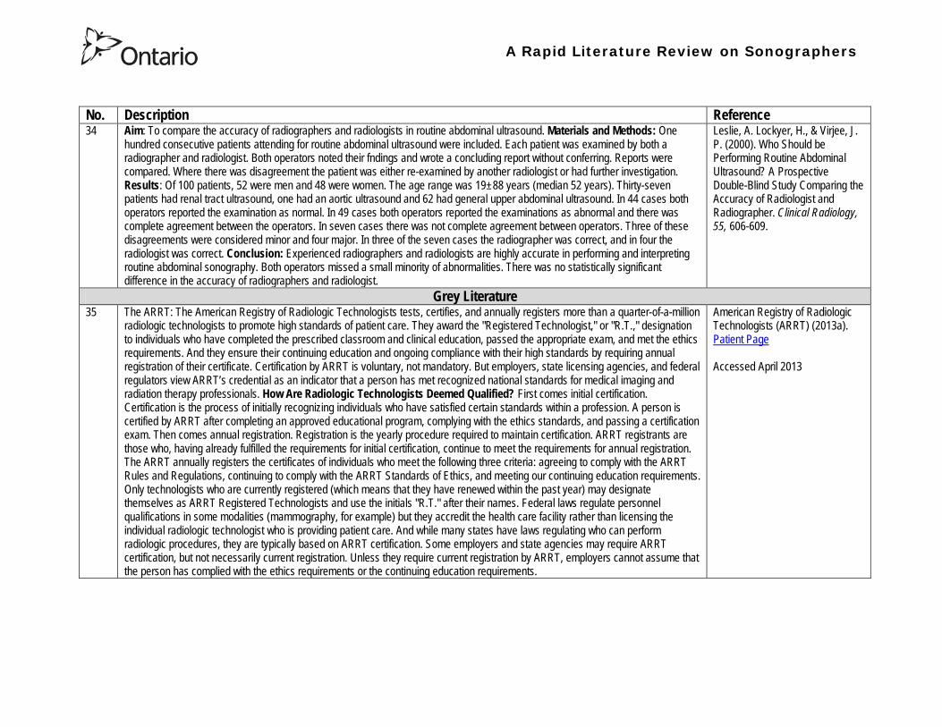
A Rapid Literature Review on Sonographers
No Description Reference 37 In Australia sonographers and student sonographers are required to be accredited by Australian Sonographer Accreditation Registry
(ASAR) in order to practise This requirement is contained within the Australian Governmentrsquos Diagnostic Imaging Accreditation Scheme standards and Medicare legislation Accreditation of sonographers is linked only to achieving - or in the case of students studying towards - an accredited entry-level qualification completion of continuing professional development and payment of an annual fee to ASAR The ASA contends that the lack of a requirement to ensure each sonographer is lsquofit to practisersquo is an unacceptable gap in current regulation undermining efforts to ensure the quality of sonographic services and failing to protect patients from harm The ASA supports and advocates for nationally-enforceable regulation of sonographers to meet our patientrsquos expectations National registration The National Registration and Accreditation Scheme (NRAS) for health workers commenced on 1 July 2010 The ten professions which comprised the initial intake into this scheme were those that had registration requirements in all Australian states or territories as well as those in which legislated regulation is deemed to be in the public interest An additional four professions - including medical radiation practitioners which are defined as radiographers radiation therapists and nuclear medicine technologists - will be included in the NRAS commencing on 1 July 2012 As sonography did not have registration in any states or territories prior to the commencement of NRAS the profession has not been in a position to negotiate entry into NRAS In early August 2011 the ASA lodged a submission with the Health Workforce Principal Committee requesting a profession-specific Sonography Board of Australia be formed under NRAS This submission was prepared based on information that consideration of professions which were unregistered would commence More recently the Australian Health Workforce Ministerial Council (AHWMC) issued a communiqueacute stating in part lsquoMinisters have commissioned work on unregistered professions and future directions for national registration Given this Ministers agreed to defer consideration of the inclusion of additional professions into the NRAS Ministers agreed that it would be premature to consider further applications for the scheme until this work has been completed other than paramedicsrsquo Options for unregistered health practitioners including sonographers Policy work is continuing at a federal level to establish an alternative regulatory framework The National Alliance for Self-Regulating Health Professions (NASRHP) consisting of the peak professional associations of eight self-regulating health professions including the ASA representing sonography released a proposal lsquoHarnessing self-regulation to support safety and quality in healthcare deliveryrsquo in March 2012 This proposal concluded that the framework that regulates all health practitioners should be expanded beyond NRAS to include
bull authorised self-regulating professions via a health professions panel bull negative licensing to withdraw the ability to practise from registered and self-regulated practitioners who do not meet the
regulatory requirements or unregulated practitioners due to poor practice As with all advocacy initiatives consideration by the federal government of the introduction of the NASRHPrsquos proposal may be prolonged and the outcomes may not match the expectations of NASRHPrsquos member professional associations including the ASA The ASA will continue both independently and collaboratively to lobby on behalf of our members and the public for the introduction of a timely and equitable solution to health practitioner regulation In the interim as there is no regulatory model with the rigour the ASA believes is required for regulation of sonographers and other health practitioners other than NRAS the ASA continues to support its Position Statement published in early 2010
Australian Sonographer Association (ASA) (2013) Sonographer Regulation Accessed April 2013
A Rapid Literature Review on Sonographers
No Description Reference 38 This publication contains information about Cardiovascular Credentialing International (CCI) It also provides the necessary
information and references concerning the process by which candidates may earn certificate or registry-level credentials CCI is a not-for-profit corporation established for the purpose of administering credentialing examinations as an independent credentialing agency CCI began credentialing cardiovascular professionals in 1968 CCI offers eight credentials which when earned demonstrate that the registrant holds fundamental knowledge in the particular cardiovascular specialty These include RCCS ndash Registered Congenital Cardiac Sonographer RCS ndash Registered Cardiac Sonographer and RPhS ndash Registered Phlebology Sonographer
Cardiovascular Credentialing International (2013) 2013 Examination Application amp Overview Accessed April 2013
39 Oregon Governor Signs Sonographer Licensure Legislation On July 28 2009 Oregon Governor Ted Kulongoski signed a sonographer licensure bill (HB 2245) into law The new law makes Oregon only the second state in the United States to require licensure of sonographers The Society of Diagnostic Medical Sonographers (SDMS) has worked closely with other sonography-related organizations and Oregon sonographers to help craft the language in new law The law renames and restructures the Oregon Board of Radiologic Technologists to the Oregon Board of Medical Imaging (OBMI) and ensures representation of sonographers on the Board The sonographer licensure law requires Oregon sonographers to become licensed by OBMI effective July 1 2010 Beginning on January 1 2014 all sonography licensees would be required to hold a national sonography certificationcredential (or be currently enrolled as a student) While the Oregon bill is far more detailed than the sonographer licensure law that passed in New Mexico earlier this year additional administrative rulesregulations will be developed over the next year to fully implement the new law SDMS will be continue to monitor developments regarding sonographer licensure in other states
Society of Diagnostic Medical Sonographers (SDMS) (2013a) News Accessed April 2013
A Rapid Literature Review on Sonographers
No Description Reference 40 On April 6 2009 New Mexico (NM) Governor Bill Richardson signed the historic bill that for the first time in the United States
requires licensure of sonographers The bill adds sonographers (and MRI technologists) to the list of medical imaging professionals licensed by the State of New Mexico Until now anyone could perform sonograms in New Mexico Prior to the passage of the New Mexico licensure law virtually all other health care providers and professionals had to be licensed As a result the public is often surprised to learn that the person performing a medical sonogram does not have any state licensure or national certification requirements The responsibility for administering the NM sonographer licensure program would rest with the New Mexico Environment Department Although this was not the first choice in most sonographersrsquo minds the Environment Department currently manages the radiologic technology licensure program so the administrative infrastructure is already in place By ensuring the creation of an advisory council that includes representation by each medical imaging and radiation therapy modality regulated the bill helps ensure that the Departmentrsquos other areas of emphasis do not detract from sonographer licensure Because sonographers have never had to be licensed before they frequently do not see or understand the distinction between state licensure and national certification National certifications or credentials issued by voluntary organizations such as the American Registry for Diagnostic Medical Sonography (ARDMS) Cardiovascular Credentialing International (CCI) and the American Registry of Radiologic Technologists (ARRT) have little legal weight The certification process and resulting credential are valuable tools in ensuring sonographers have met minimum education and knowledge requirements but do not really regulate anyonersquos ability to perform sonography In an ideal world no additional regulation of sonography would be required However as everyone knows more and more people are taking weekend courses and buying ultrasound equipment without understanding the ramifications of using ultrasound technology (eg ALARA) Under the bill passed by the New Mexico legislature national credentialscertifications should provide evidence of meeting the statersquos standards All sonographers who are performing these critical medical imaging services in New Mexico will be required to meet national certification standards (ie have certifications or credentials issued by ARDMS CCI or ARRT) As a result there would be no additional exams in New Mexico beyond the national certification exams required by ARDMS CCI or ARRT The bill also requires the state to recognize the continuing medical education completed for national certification renewal The most visible impact on New Mexico sonographers will be a license fee However the legislation caps the license fee at $100 per two-year license In addition only the New Mexico Legislature can change this cap
SDMS (2013b) New Mexico Sonographer Licensure Law Signed Accessed April 2013
A Rapid Literature Review on Sonographers
No Description Reference 41 Preamble The purpose of this document is to define the Scope of Practice for Diagnostic Ultrasound Professionals in the US and to
specify their roles as members of the health care team acting in the best interest of the patient This scope of practice is a living document that will evolve as the technology expands Definition of the Profession The Diagnostic Ultrasound Profession is a multi-specialty field comprised of Diagnostic Medical Sonography (with subspecialties in abdominal neurologic obstetricalgynecologic and ophthalmic ultrasound) Diagnostic Cardiac Sonography (with subspecialties in adult and pediatric echocardiography) Vascular Technology and other emerging fields These diverse specialties are distinguished by their use of diagnostic medical ultrasound as a primary technology in their daily work Certification is considered the standard of practice in ultrasound Individuals who are not yet certified should reference the Scope as a professional model and strive to become certified Scope of Practice of the Profession The Diagnostic Ultrasound Professional is an individual qualified by professional credentialing and academic and clinical experience to provide diagnostic patient care services using ultrasound and related diagnostic procedures The scope of practice of the Diagnostic Ultrasound Professional includes those procedures acts and processes permitted by law for which the individual has received education and clinical experience and in which heshe has demonstrated competency Diagnostic Ultrasound Professionals
bull Perform patient assessments bull Acquire and analyze data obtained using ultrasound and related diagnostic technologies bull Provide a summary of findings to the physician to aid in patient diagnosis and management bull Use independent judgment and systematic problem solving methods to produce high quality diagnostic information and
optimize patient care
Society of Diagnostic Medical Sonography [SDMS] (2013c) Scope of Practice for the Diagnostic Ultrasound Professional Accessed April 2013
42 This website notes that in the US most employers in the US prefer to hire sonographers who have professional certification A sonographer can get certification and passing an exam Most exams relate to the specialty that the sonographer is most interested inmdashfor example an exam to become certified in abdominal sonography A few states require diagnostic medical sonographers to be licensed Typically professional certifications required for licensure other requirements vary by state Sonographers must take continuing education to keep their certification current
Bureau of Labor Statistics US Department of Labor (2012) How to become a Diagnostic Medical Sonographer Occupational Outlook Handbook 2012-13 Edition Accessed April 2013
A Rapid Literature Review on Sonographers
No Description Reference 43 The American Registry for Diagnostic Medical Sonography (ARDMS) is an independent not-for-profit organization founded in 1975
that administers examinations and awards the following credentials bull RDMSreg ndash Registered Diagnostic Medical Sonographerreg bull RDCSreg ndash Registered Diagnostic Cardiac Sonographerreg bull RVTreg ndash Registered Vascular Technologistreg bull RPVIreg ndash Registered Physician in Vascular Interpretationreg bull RMSKtrade - Registered in Musculoskeletaltrade sonography
ARDMS is the globally recognized standard of excellence in sonography and has certified more than 75000 individuals ARDMS is composed of three key groups
bull Board of Directors ndash Sonographers vascular technologists physicians research scientists and a member of the public are volunteer members of the board that determines the direction and policies of our organization
bull Examination Development Task Forces ndash A group of sonographers vascular technologists physicians and scientists volunteer their time and expertise to produce our examinations
bull Professional Staff ndash A paid highly-skilled staff administers the programs and assists applicants and registrants Credentials awarded by ARDMS document personal achievement of recognized professional standards and are widely accepted in the medical community by sonography and vascular professional organizations However ARDMS does not validate the day-to-day job performance of any sonography professional ARDMS has earned the prestigious ANSI-ISO 17024 accreditation for certifying bodies from the International Organization for Standardization (ISO) Accreditation is granted through the American National Standards Institute (ANSI) ARDMS has also earned accreditation with the National Commission for Certifying Agencies (NCCA) The NCCA is the accrediting arm of the National Organization for Competency Assurance (NOCA) Established in 1977 as a nonprofit organization NOCA is a leader in establishing quality standards for certifying organizations
American Registry for Diagnostic Medical Sonography (ARDMS) (2012a) Overview of ARDMS Accessed April 2013
A Rapid Literature Review on Sonographers
No Description Reference 44 ARDMS is developing a program to assure that Registrants continue to meet the basic competency requirements of core clinical skills
in present day practice bull Each Registrant who holds an active ARDMS certification as of December 31 2011 will be assigned the 2012 ndash 2021 10-
year recertification assessment period bull The 10-year period for new Registrants will begin the year following the attainment of the credential bull If a Registrant earns a new credential or specialty after January 1 2012 then that credentialspecialty will have a separate
10-year recertification assessment period that will begin the year following the attainment of the credentialspecialty ARDMS will develop tools and resources to assist Registrants in tracking and meeting the recertification assessment program requirements
bull Registrants who do not wish to take a recertification assessment in a specific credentialspecialty may voluntarily relinquish the credentialspecialty
As an ARDMS Registrant individuals are committing to demonstrate the highest level of competency in the modality of sonography by meeting and maintaining the standards set by the ARDMS The pathway of maintenance your certification is comprised of Paying the ARDMS annual renewal fee each year Each year ARDMS assesses Registrants an annual renewal fee By paying your annual renewal fee you are contributing to the ARDMS mission of promoting quality care and patient safety through the certification and continuing competency of ultrasound professionals In order to maintain your credentials you must pay the annual renewal fee of $75 USD or $100 USD (for RPVI and MSK Registrants with MDDODPM) designation on or before December 31 of each year in order to stay active for the following year Attesting to ARDMS rules and policies Each year when you pay your annual renewal fee you are attesting that you are in compliance with all ARDMS rules and discipline policies and are in good standing with the ARDMS Earning a minimum of 30 ARDMS-accepted CMEs during your three-year CME period ARDMS verifies its Registrants compliance with continuing medical education (CME) requirements using an audit process Earning your CMEs is an integral component of maintaining your ARDMS credential(s) and demonstrates your ongoing commitment to lifelong learning RecertificationContinued Commitment to Excellence Following the attainment of the ARDMS credential Registrants are assigned a 10-year recertification period the year after the credential is earned ARDMS is committed to developing a recertification program that is easy for Registrants to comply with
American Registry for Diagnostic Medical Sonography (ARDMS) (2012b) Recertification General Information
A Rapid Literature Review on Sonographers
No Description Reference 45 This document reviews Sonography Principles and Instrumentation (SPI) Examination Requirements offered through ARDMS in
order to achieve sonography certification and the various prerequisites needed before taking the exam For example one prerequisite entails (1) Education Graduate of a program accredited by an agency recognized by the Council for Higher Education Accreditation (CHEA) United States Department of Education (USDOE) or Canadian Medical Association (CMA) that specifically conducts programmatic accreditation for diagnostic medical sonographydiagnostic cardiac sonographyvascular technology Currently the only organizations that offer programmatic accreditation under the aforementioned associations are the Commission on Accreditation of Allied Health Education Programs (CAAHEP) and the Canadian Medical Association (CMA) (2) Required Clinical UltrasoundVascular Experience No additional experience is required (3) Documentation Required with Application 1) Copy of diploma from ultrasoundvascular program or an official transcript indicating the date the degree was conferred 2) Original letter signed by program director andor medical director indicating date of graduation or successful completion of the program4 Program directors must use the mandatory formatted sample letter available on ARDMSorg sampleletters 3) The CV form is not required if the application is submitted and received in the ARDMS office within one year after successful completion of the program Otherwise an original signed and completed CV form for each appropriate specialty area(s) must be submitted 4) Photocopy of a non-expired government issued photo identification with signature the name on the identification must exactly match the name under which you are applying for ARDMS examination
American Registry for Diagnostic Medical Sonography (ARDMS) (2012c) SPI Requirement and General Prerequisites Accessed April 2013
46 This page notes the five credentials offered by the ARDMS and the corresponding exams and specialty exams required bull RDMS requires the SPI exam (sonography principles and instrumentation) and a specialty exam in abdomen breast fetal
echocardiography neurosonolgoy or obstetricsgynecology bull RDCS requires the SPI exam and a speciality exam in adult echocardiography fetal echocardiography pediatric
echocardiography bull RVT requires the SPI and a speciality exam in vascular technology bull RMSK requires the musculoskeletal sonography exam bull RPVI requires the physiciansrsquo vascular interpretation exam
American Registry for Diagnostic Medical Sonography (ARDMS) (2012d) Credentials and Examinations Accessed April 2013
A Rapid Literature Review on Sonographers
No Description Reference 47 This article summarizes some of the many trends in fetal pediatric and adult echocardiography that SDMS members are excited
about Fetal Echocardiography Sonography has been used since the mid-1980s to help identify congenital heart defects during the second trimester The ldquobasicrdquo examination of the fetal heart involves a four-chamber view ndash evaluation of the right and left atrial and ventricular chambers and their respective valves In the past several years there has been a movement in the obstetric ultrasound community to encourage obstetric sonographers to perform the ldquoextended basicrdquo exam which includes the cardiac outflow tracts The outflow tracts consist of the main pulmonary artery exiting the right ventricle and the aorta exiting the left ventricle The fetal echocardiography community also is abuzz over three-dimensional (3D) and four-dimensional (4D) ultrasound and the potential of this technology to significantly improve the evaluation of the fetal heart Spatiotemporal Image Correlation (STIC) a 4D application allows for evaluation and manipulation of the heart in real-time Allen says Todayrsquos ultrasound equipment allows sonographers to gather a volume of data from which they can create detailed 3D images of the tiny fetal heart which is constantly in motion With computer manipulation of the data sets the images also can be viewed in 4D allowing physicians to see the entire fetal heart cycle from start to finish Pediatric Echocardiography One of the most significant advances in pediatric echo over the last decade is moving to a digital format The advent of pediatric transesophageal (TEE) probes also has been a big advance for pediatric echo Transesophageal probes provide an unimpeded sonographic visualization of the intracardiac structures Theyrsquore useful in visualizing very fine valvular abnormalities the thoracic aorta vegetations and thrombus in the atrial appendage It is also useful in detecting subtle anatomic variations seen in congenital heart disease Pediatric-sized TEE probes make the exams possible In the past it wasnrsquot possible because the adult probe was just too big to put down the babyrsquos or childrsquos esophagus Now the probe is small enough to successfully manipulate it in children and get superb images Because the new sonographic imaging equipment and better surgical techniques allow clinicians to detect and repair more cardiac abnormalities earlier patients are living longer Adult Echocardiography The adult echo world is abuzz about 2D and 3D speckle tracking relatively new techniques for the assessment of heart function and strain imaging Speckle tracking evaluates both regional and global function by measuring the deformation of the heart muscle
Orenstein B W (2010) Current Trends in Echocardiography SDMS New Wave February 1-5 Accessed April 2013
48 This document describes the requirements for admission to the Register of Accredited Medical Sonographer in Australia Requirements include Continuation of Accreditation In order to remain on the Register an accredited Sonographer must satisfy the ASAR continuing professional development (CPD) requirements and pay the prescribed annual fee Student Sonographers apart from paying the prescribed annual fee should provide evidence of their student status every year when they renew their ASAR accreditation
Australian Sonographer Accreditation Registry (ASAR) (no date A) Sonographer Accreditation Accessed April 2013
A Rapid Literature Review on Sonographers
No Description Reference 49 ASARrsquos functions
1 Accredit sonography education programs within Australia 2 Maintain a register of Accredited Sonographers and Accredited Student Sonographers 3 Establish minimum standards of Continuing Professional Development (CPD) and to monitor the CPD requirements for
Sonographers to remain on the register 4 To support activities that advance the profession of sonography 5 To provide registration information about Sonographers to Medicare Australia 6 To provide public access to the name registration number State or Territory current type of accreditation and area of
specialty for active Sonographers ASARrsquos Role ASAR is not a registration board ASAR does not have the authority to remove a Sonographer from the Registry due to professional misconduct nor the ability to prevent a Sonographer from practicing ASAR is only able to remove a Sonographer from the Registry if they
bull fail to meet CPD requirements and or bull fail to pay annual fees
ASAR (no date B) About ASAR Accessed April 2013
50 Regulatory bodies A Regulatory Body maintains a register of individuals who have achieved the required qualification of that profession to practice and can evidence continuing competence at the required level For a doctor to practice in the UK he or she must be registered with the General Medical Council (GMC) This is the regulatory body for medical practitioners The majority of sonographers who are employed in the UK come from a background of radiography or midwifery To practice radiography in the UK a qualified radiographer must be registered with the Health Professions Council (HPC) Similarly a midwife must be registered with the National Midwifery Council to practice as a midwife Sonographer registration in the UK There are a number of professions allied to medicine that are regulated by the HPC for example radiography physiotherapy However sonography is not recognised as a profession by the HPC and therefore there is no mechanism whereby an individual can register with the HPC as a sonographer or be regulated by them as a sonographer It is a common misunderstanding that sonographers have to be registered with the HPC to practice This comes from the fact that most sonographers are also qualified radiographers and maintain their registration with the HPC because they wish to remain on the HPC register as a radiographer Professional bodies An individual can be a member of a Professional Body such as the Royal College of Obstetricians amp Gynecologists (RCOG) Royal College of Radiologists (RCR) Society amp College of Radiographers (SCoR) or the Royal College of Midwives (RCM) but this is not the same as being accepted onto the register of one of the regulatory bodies The professional bodies are responsible for setting and maintaining the standards of their members providing a code of conduct etc They may also act in the capacity of a trades union and can also provide professional indemnity insurance The British Medical Ultrasound Society BMUS is not a professional body but is a scientific society so will offer a different range of benefits Whether or not one wishes to join the society has no impact on their ability to work as a sonographer in the UK although from a continuing professional development point of view it can be beneficial The College of Radiographers are currently working with the HPC to get sonography recognized as a separate profession
British Medical Ultrasound Society (BMUS) (no date) Information on Sonographer Regulation in the UK Accessed April 2013
A Rapid Literature Review on Sonographers
No Description Reference 51 This document included sections on the role of the sonographer and the use of sonography for entertainment Role of the
Sonographer and Technical Impressions The role of the sonographer is to perform the ultrasound scan and to document observations The sonographerrsquos written technical impressions are intended as a form of communication between the sonographer and the reporting physician These observations must be reviewed and subsequently reported by the reporting physician Issuing of a final reportdiagnosis represents the practice of medicine and is therefore the domain of the reporting physician Use of Ultrasound for Entertainment or Non-Diagnostic Purposes In contrast to this justified form of assessment ultrasound for entertainment is simply an elaborate version of a portrait For a variable fee the surface of the fetus is skimmed over and clients are provided with keepsake images or a DVD of their fetus There are no measurements taken during this exam no morphological assessment performed and no dictated diagnostic report of findings Essentially there are no diagnostic benefits to the exam and uninformed parents may be falsely reassured that the beautiful portrait indicates a normal fetus Entertainment ultrasound facilities may operate outside of medical guidelines and without any controls which may result in a lack of technical safeguards operator expertise or governance of technical competency These facilities do not comply with the ALARA principle which could result in increased fetal exposures The Canadian Society of Diagnostic Medical Sonograph (CSDMS) does not support persons or facilities that participate in ultrasound for entertainment activities Liability insurance purchased through the CSDMS group insurance plan does not cover any activities carried out for entertainment ultrasound purposes Other leaders in Canadian medicine support this statement In 2005 the SOGC (Society of Obstetrics and Gynaecologists of Canada) published an article entitled ldquoObstetrical Ultrasound Biological Effects and Safetyrdquo and their conclusion was that ldquothe theoretical risk of adverse bio-effects even from standard 2D obstetrical ultrasound makes it hard to justify its use for non-medical reasons such as sex-determination making non-medical photos or video or for commercial purposerdquo The SOGC recommends that ultrasound be used prudently and that energy exposure be limited to the minimum that is medically necessary The SOGC further recommends a complete ban on the non-medical use of fetal ultrasound and encourages government to join with society to find appropriate means to deal with this public health issue The CAR (Canadian Association of Radiologists) makes a similar statement ldquoThe CAR strongly opposes the use of diagnostic ultrasound equipment for non-medical purposes and considers the use of medical ultrasound for entertainment to be a misuse of the technology especially if fetal subjects are involvedrdquo
Canadian Society of Diagnostic Medical Sonography (no date) Professional Practice Guidelines and Policy Statements For Canadian Sonography Accessed April 2013
A Rapid Literature Review on Sonographers
No Description Reference 52 In 1993 the Consortium for the Accreditation of Sonographic Education (CASE) was formed through the common desire to ensure
that the education and training of sonographers in the United Kingdom was delivered at the highest level All members share common concerns that standards of service provision and education are developed in parallel with increasing demand and improving technology All members strive to create an environment in which education and training is maximised This will make validation and accreditation to be as robust and straightforward as possible CASEs philosophy is to promote best ultrasound practise through the accreditation of those training programmes that develop safe and competent ultrasound practitioners This should be delivered at postgraduate level The majority of its activities relate to supporting those Institutions that offer or wish to offer courses leading to the award of a Masters Postgraduate DiplomaCertificate in Medical or clinical Ultrasound In addition to offering accreditation of new and re-accreditation of established ultrasound education programmes CASE undertakes annual monitoring of the courses it has accredited The information obtained is fed back to the Institutions both on an individual basis and via the annual CASE report Each member organisation is unique and has its own obligations to its membership and its own agenda while CASE brings them together to provide a forum for the interchange of ideas in which shared views may be synthesised and thus inform education and training programmes
Consortium for the Accreditation of Sonographic Education (no date) Home Accessed April 2013
A Rapid Literature Review on Sonographers
No Description Reference 53 The Medical Radiation Technologists Board (the Board) is one of sixteen New Zealand health registration authorities appointed by
the Minister of Health under the Health Practitioners Competence Assurance Act 2003 (the Act) The Board is responsible for the administration of the Act in regard to the profession of medical radiation technology The primary responsibility of the Board is to protect the health and safety of the New Zealand public by ensuring practitioners registered in the profession of medical radiation technology are competent and fit to practice In accordance with Section 118 of the Act the Board is responsible for fulfilling a number of functions
bull 01 To prescribe the qualifications required for scopes of practice within the profession of medical radiation technology and for that purpose to accredit and monitor educational institutions and degrees courses of studies or programmes
bull 02 To authorise the registration of medical radiation technologists under the Act and to maintain registers bull 03 To consider applications for annual practising certificates bull 04 To review and promote the competence of medical radiation technologists bull 05 To recognise accredit and set programmes to ensure the ongoing competence of medical radiation technologists bull 06 To receive and act on information from health practitioners employers and the Health and Disability Commissioner
about the competence of medical radiation technologists bull 07 To notify employers the Accident Compensation Corporation the Director-General of Health and the Health and
Disability Commissioner that the practice of a medical radiation technologist may pose a risk of harm to the public bull 08 To consider the cases of medical radiation technologists who may be unable to perform the functions required for the
practice of the medical radiation technology profession bull 09 To set standards of clinical competence cultural competence and ethical conduct to be observed by medical radiation
technologists bull 10 To liaise with other authorities appointed under the Act about matters of common interest bull 11 To promote education and training in the profession of medical radiation technology bull 12 To promote public awareness of the responsibilities of the Board bull 13 To exercise and perform any other functions powers and duties that are conferred or imposed on it by or under the Act
or any other enactment
Medical Radiation Technologists Board (MRTB) (no date A) What we do Accessed April 2013
54 Qualifications To be considered for registration as a medical radiation technologist in any of the specified scopes of practice an individual must hold a qualification for that particular scope as prescribed by the Board or approved by the Board as being substantially equivalent to the prescribed qualifications In addition to having an approved qualification individuals may also be required to demonstrate that they have adequate experience in the relevant scope of practice Sonographer The Board has prescribed one New Zealand postgraduate qualification for a person to be eligible for registration in the scope of practice of Sonographer Postgraduate Diploma in Health Science (Ultrasound) issued by the Unitec Institute of Technology Required Experience If an individual is applying for registration in Nuclear Medicine Ultrasound or Magnetic Resonance Imaging in addition to holding a relevant postgraduate qualification they must also be able to provide evidence that you have a minimum of 3360 hours of clinical experience in the relevant scope of practice you are applying for registration in
Medical Radiation Technologists Board (MRTB) (no date B) Qualifications Accessed April 2013
A Rapid Literature Review on Sonographers
No Description Reference 55 Recertification is a mechanism used by the Medical Radiation Technologists Board in New Zealand to ensure that registered medical
radiation technologists continue to be competent to practise within their scopes of practice Annual Practising Certificates Each year if an individual wishes to continue practising in your scope(s) of practice in New Zealand they must apply for and be issued with an annual practising certificate (APC) A condition of being issued with an APC is that one must provide evidence of enrolment and satisfactory progress in a Board-approved recertification programme Alternatively the Board may issue them with an interim practising certificate (IPC) until it is satisfied the individual demonstrates that they are competent to practice in the scope(s) of practice they applied for Working with an IPC will require an individual to work under the supervision of a registered medical radiation technologist or other relevant registered health practitioner Recertification Programmes The Boards recertification programme framework is based on a continuing professional development (CPD) model Individuals have the option of enrolling in one of seven recertification programmes for medical radiation technologists that are currently approved by the Board They must meet the requirements of the particular recertification programme in which they are enrolled Recertification Audits An essential component of the CPD-based recertification programme adopted by the Board is the audit of CPD recordsThe Board undertakes an annual audit of approximately 10 of its registrant database to assess individual medical radiation technologistsrsquo compliance with the Boarrsquods recertification requirements
Medical Radiation Technologists Board (MRTB) (no date C) Recertification Accessed April 2013
56 There are two types of radiography - diagnostic and therapeutic Both need considerable knowledge of technology anatomy physiology and pathology to carry out their work Many also undertake further training to become a sonographer Some of the imaging technology that a diagnostic radiographer may use are
bull X-Ray - looks through tissues to examine bones cavities and foreign objects bull Fluoroscopy - images the digestive system providing a real-time image bull CT (Computed Tomography) - which provides cross-sectional views (slices) of the body bull MRI (Magnetic Resonance Imaging) - builds a 2-D or 3-D map of the different tissue types within the body bull Ultrasound - well known for its use in obstetrics and gynaecology Also used to check circulation and examine the heart bull Angiography - used to investigate blood vessels
Sonographyultrasonography Ultrasound is used in various settings in a hospital including abdominal scanning and breast ultrasound Ultrasound imaging is the use of high frequency sound in excess of human hearing to produce images of structures of the human body that may be observed on a screen These images may subsequently be transferred to photographic film or paper or onto video or CD forming part of the patients record of their examination There are no direct entry routes into ultrasound Most sonographers train as a radiographer then undertake an approved post-registration course Offered by higher education institutions The courses are a minimum of one academic year and prepare sonographers clinically and academically for practice Normally a pre-requisite for acceptance is access to a clinical department with supervised practice for students
National Health Service (NHS) (no date A) Radiographer Accessed April 2013
57 To practice as a radiographer in the UK an individual must be registered with the Health and Care Professions Council (HCPC) To register individuals must have successfully completed an approved training programme in diagnostic or therapeutic radiography These are offered at undergraduate degree (full-time and part-time) and at postgraduate degreediploma level at a number of universities across the UK
NHS (no date B) Entry requirements and training in radiography Accessed April 2013
Health Professions Regulatory Advisory Council 56 Wellesley Street West 12th Floor
Toronto ON M5S 2S3
Telephone 416-326-1550 Toll-Free 1-888-377-7746
Fax 416-326-1549
Website wwwhpracorg
Email hpracwebmasterontarioca
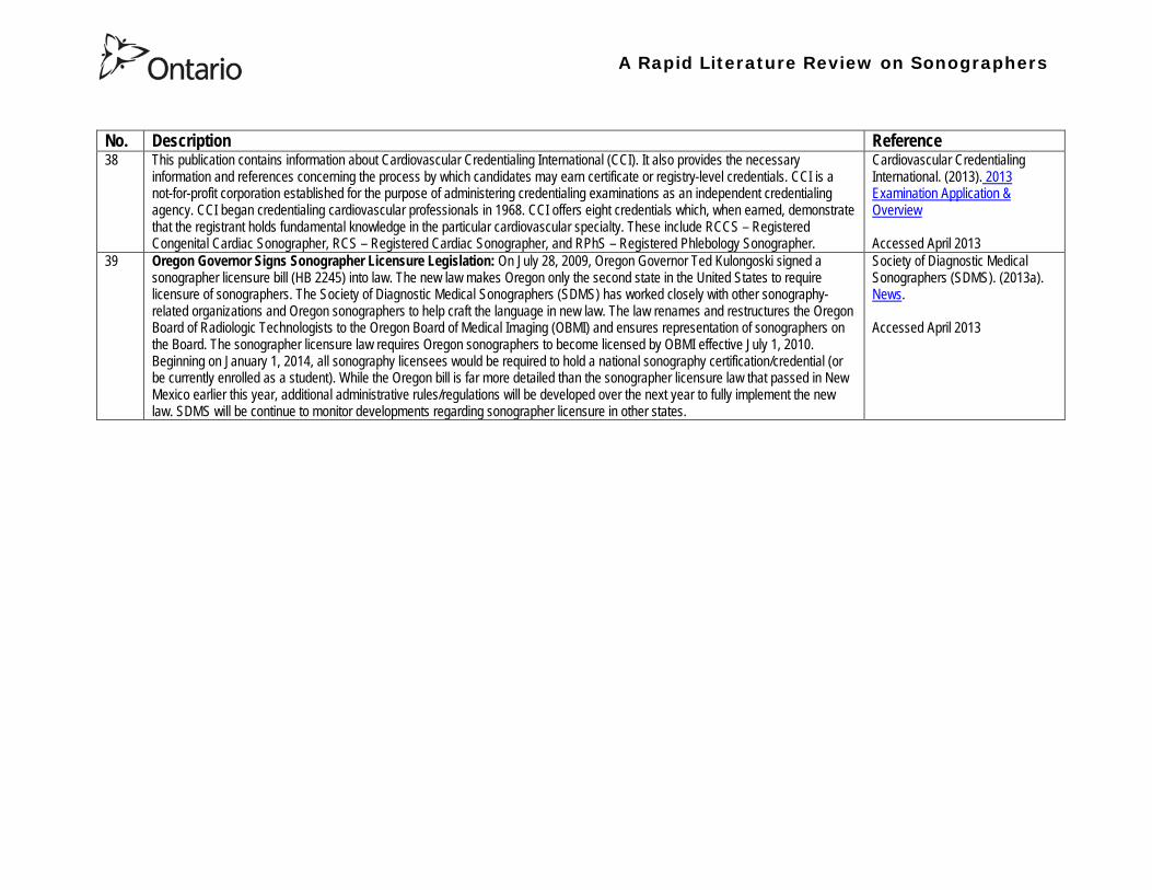
A Rapid Literature Review on Sonographers
No Description Reference 38 This publication contains information about Cardiovascular Credentialing International (CCI) It also provides the necessary
information and references concerning the process by which candidates may earn certificate or registry-level credentials CCI is a not-for-profit corporation established for the purpose of administering credentialing examinations as an independent credentialing agency CCI began credentialing cardiovascular professionals in 1968 CCI offers eight credentials which when earned demonstrate that the registrant holds fundamental knowledge in the particular cardiovascular specialty These include RCCS ndash Registered Congenital Cardiac Sonographer RCS ndash Registered Cardiac Sonographer and RPhS ndash Registered Phlebology Sonographer
Cardiovascular Credentialing International (2013) 2013 Examination Application amp Overview Accessed April 2013
39 Oregon Governor Signs Sonographer Licensure Legislation On July 28 2009 Oregon Governor Ted Kulongoski signed a sonographer licensure bill (HB 2245) into law The new law makes Oregon only the second state in the United States to require licensure of sonographers The Society of Diagnostic Medical Sonographers (SDMS) has worked closely with other sonography-related organizations and Oregon sonographers to help craft the language in new law The law renames and restructures the Oregon Board of Radiologic Technologists to the Oregon Board of Medical Imaging (OBMI) and ensures representation of sonographers on the Board The sonographer licensure law requires Oregon sonographers to become licensed by OBMI effective July 1 2010 Beginning on January 1 2014 all sonography licensees would be required to hold a national sonography certificationcredential (or be currently enrolled as a student) While the Oregon bill is far more detailed than the sonographer licensure law that passed in New Mexico earlier this year additional administrative rulesregulations will be developed over the next year to fully implement the new law SDMS will be continue to monitor developments regarding sonographer licensure in other states
Society of Diagnostic Medical Sonographers (SDMS) (2013a) News Accessed April 2013
A Rapid Literature Review on Sonographers
No Description Reference 40 On April 6 2009 New Mexico (NM) Governor Bill Richardson signed the historic bill that for the first time in the United States
requires licensure of sonographers The bill adds sonographers (and MRI technologists) to the list of medical imaging professionals licensed by the State of New Mexico Until now anyone could perform sonograms in New Mexico Prior to the passage of the New Mexico licensure law virtually all other health care providers and professionals had to be licensed As a result the public is often surprised to learn that the person performing a medical sonogram does not have any state licensure or national certification requirements The responsibility for administering the NM sonographer licensure program would rest with the New Mexico Environment Department Although this was not the first choice in most sonographersrsquo minds the Environment Department currently manages the radiologic technology licensure program so the administrative infrastructure is already in place By ensuring the creation of an advisory council that includes representation by each medical imaging and radiation therapy modality regulated the bill helps ensure that the Departmentrsquos other areas of emphasis do not detract from sonographer licensure Because sonographers have never had to be licensed before they frequently do not see or understand the distinction between state licensure and national certification National certifications or credentials issued by voluntary organizations such as the American Registry for Diagnostic Medical Sonography (ARDMS) Cardiovascular Credentialing International (CCI) and the American Registry of Radiologic Technologists (ARRT) have little legal weight The certification process and resulting credential are valuable tools in ensuring sonographers have met minimum education and knowledge requirements but do not really regulate anyonersquos ability to perform sonography In an ideal world no additional regulation of sonography would be required However as everyone knows more and more people are taking weekend courses and buying ultrasound equipment without understanding the ramifications of using ultrasound technology (eg ALARA) Under the bill passed by the New Mexico legislature national credentialscertifications should provide evidence of meeting the statersquos standards All sonographers who are performing these critical medical imaging services in New Mexico will be required to meet national certification standards (ie have certifications or credentials issued by ARDMS CCI or ARRT) As a result there would be no additional exams in New Mexico beyond the national certification exams required by ARDMS CCI or ARRT The bill also requires the state to recognize the continuing medical education completed for national certification renewal The most visible impact on New Mexico sonographers will be a license fee However the legislation caps the license fee at $100 per two-year license In addition only the New Mexico Legislature can change this cap
SDMS (2013b) New Mexico Sonographer Licensure Law Signed Accessed April 2013
A Rapid Literature Review on Sonographers
No Description Reference 41 Preamble The purpose of this document is to define the Scope of Practice for Diagnostic Ultrasound Professionals in the US and to
specify their roles as members of the health care team acting in the best interest of the patient This scope of practice is a living document that will evolve as the technology expands Definition of the Profession The Diagnostic Ultrasound Profession is a multi-specialty field comprised of Diagnostic Medical Sonography (with subspecialties in abdominal neurologic obstetricalgynecologic and ophthalmic ultrasound) Diagnostic Cardiac Sonography (with subspecialties in adult and pediatric echocardiography) Vascular Technology and other emerging fields These diverse specialties are distinguished by their use of diagnostic medical ultrasound as a primary technology in their daily work Certification is considered the standard of practice in ultrasound Individuals who are not yet certified should reference the Scope as a professional model and strive to become certified Scope of Practice of the Profession The Diagnostic Ultrasound Professional is an individual qualified by professional credentialing and academic and clinical experience to provide diagnostic patient care services using ultrasound and related diagnostic procedures The scope of practice of the Diagnostic Ultrasound Professional includes those procedures acts and processes permitted by law for which the individual has received education and clinical experience and in which heshe has demonstrated competency Diagnostic Ultrasound Professionals
bull Perform patient assessments bull Acquire and analyze data obtained using ultrasound and related diagnostic technologies bull Provide a summary of findings to the physician to aid in patient diagnosis and management bull Use independent judgment and systematic problem solving methods to produce high quality diagnostic information and
optimize patient care
Society of Diagnostic Medical Sonography [SDMS] (2013c) Scope of Practice for the Diagnostic Ultrasound Professional Accessed April 2013
42 This website notes that in the US most employers in the US prefer to hire sonographers who have professional certification A sonographer can get certification and passing an exam Most exams relate to the specialty that the sonographer is most interested inmdashfor example an exam to become certified in abdominal sonography A few states require diagnostic medical sonographers to be licensed Typically professional certifications required for licensure other requirements vary by state Sonographers must take continuing education to keep their certification current
Bureau of Labor Statistics US Department of Labor (2012) How to become a Diagnostic Medical Sonographer Occupational Outlook Handbook 2012-13 Edition Accessed April 2013
A Rapid Literature Review on Sonographers
No Description Reference 43 The American Registry for Diagnostic Medical Sonography (ARDMS) is an independent not-for-profit organization founded in 1975
that administers examinations and awards the following credentials bull RDMSreg ndash Registered Diagnostic Medical Sonographerreg bull RDCSreg ndash Registered Diagnostic Cardiac Sonographerreg bull RVTreg ndash Registered Vascular Technologistreg bull RPVIreg ndash Registered Physician in Vascular Interpretationreg bull RMSKtrade - Registered in Musculoskeletaltrade sonography
ARDMS is the globally recognized standard of excellence in sonography and has certified more than 75000 individuals ARDMS is composed of three key groups
bull Board of Directors ndash Sonographers vascular technologists physicians research scientists and a member of the public are volunteer members of the board that determines the direction and policies of our organization
bull Examination Development Task Forces ndash A group of sonographers vascular technologists physicians and scientists volunteer their time and expertise to produce our examinations
bull Professional Staff ndash A paid highly-skilled staff administers the programs and assists applicants and registrants Credentials awarded by ARDMS document personal achievement of recognized professional standards and are widely accepted in the medical community by sonography and vascular professional organizations However ARDMS does not validate the day-to-day job performance of any sonography professional ARDMS has earned the prestigious ANSI-ISO 17024 accreditation for certifying bodies from the International Organization for Standardization (ISO) Accreditation is granted through the American National Standards Institute (ANSI) ARDMS has also earned accreditation with the National Commission for Certifying Agencies (NCCA) The NCCA is the accrediting arm of the National Organization for Competency Assurance (NOCA) Established in 1977 as a nonprofit organization NOCA is a leader in establishing quality standards for certifying organizations
American Registry for Diagnostic Medical Sonography (ARDMS) (2012a) Overview of ARDMS Accessed April 2013
A Rapid Literature Review on Sonographers
No Description Reference 44 ARDMS is developing a program to assure that Registrants continue to meet the basic competency requirements of core clinical skills
in present day practice bull Each Registrant who holds an active ARDMS certification as of December 31 2011 will be assigned the 2012 ndash 2021 10-
year recertification assessment period bull The 10-year period for new Registrants will begin the year following the attainment of the credential bull If a Registrant earns a new credential or specialty after January 1 2012 then that credentialspecialty will have a separate
10-year recertification assessment period that will begin the year following the attainment of the credentialspecialty ARDMS will develop tools and resources to assist Registrants in tracking and meeting the recertification assessment program requirements
bull Registrants who do not wish to take a recertification assessment in a specific credentialspecialty may voluntarily relinquish the credentialspecialty
As an ARDMS Registrant individuals are committing to demonstrate the highest level of competency in the modality of sonography by meeting and maintaining the standards set by the ARDMS The pathway of maintenance your certification is comprised of Paying the ARDMS annual renewal fee each year Each year ARDMS assesses Registrants an annual renewal fee By paying your annual renewal fee you are contributing to the ARDMS mission of promoting quality care and patient safety through the certification and continuing competency of ultrasound professionals In order to maintain your credentials you must pay the annual renewal fee of $75 USD or $100 USD (for RPVI and MSK Registrants with MDDODPM) designation on or before December 31 of each year in order to stay active for the following year Attesting to ARDMS rules and policies Each year when you pay your annual renewal fee you are attesting that you are in compliance with all ARDMS rules and discipline policies and are in good standing with the ARDMS Earning a minimum of 30 ARDMS-accepted CMEs during your three-year CME period ARDMS verifies its Registrants compliance with continuing medical education (CME) requirements using an audit process Earning your CMEs is an integral component of maintaining your ARDMS credential(s) and demonstrates your ongoing commitment to lifelong learning RecertificationContinued Commitment to Excellence Following the attainment of the ARDMS credential Registrants are assigned a 10-year recertification period the year after the credential is earned ARDMS is committed to developing a recertification program that is easy for Registrants to comply with
American Registry for Diagnostic Medical Sonography (ARDMS) (2012b) Recertification General Information
A Rapid Literature Review on Sonographers
No Description Reference 45 This document reviews Sonography Principles and Instrumentation (SPI) Examination Requirements offered through ARDMS in
order to achieve sonography certification and the various prerequisites needed before taking the exam For example one prerequisite entails (1) Education Graduate of a program accredited by an agency recognized by the Council for Higher Education Accreditation (CHEA) United States Department of Education (USDOE) or Canadian Medical Association (CMA) that specifically conducts programmatic accreditation for diagnostic medical sonographydiagnostic cardiac sonographyvascular technology Currently the only organizations that offer programmatic accreditation under the aforementioned associations are the Commission on Accreditation of Allied Health Education Programs (CAAHEP) and the Canadian Medical Association (CMA) (2) Required Clinical UltrasoundVascular Experience No additional experience is required (3) Documentation Required with Application 1) Copy of diploma from ultrasoundvascular program or an official transcript indicating the date the degree was conferred 2) Original letter signed by program director andor medical director indicating date of graduation or successful completion of the program4 Program directors must use the mandatory formatted sample letter available on ARDMSorg sampleletters 3) The CV form is not required if the application is submitted and received in the ARDMS office within one year after successful completion of the program Otherwise an original signed and completed CV form for each appropriate specialty area(s) must be submitted 4) Photocopy of a non-expired government issued photo identification with signature the name on the identification must exactly match the name under which you are applying for ARDMS examination
American Registry for Diagnostic Medical Sonography (ARDMS) (2012c) SPI Requirement and General Prerequisites Accessed April 2013
46 This page notes the five credentials offered by the ARDMS and the corresponding exams and specialty exams required bull RDMS requires the SPI exam (sonography principles and instrumentation) and a specialty exam in abdomen breast fetal
echocardiography neurosonolgoy or obstetricsgynecology bull RDCS requires the SPI exam and a speciality exam in adult echocardiography fetal echocardiography pediatric
echocardiography bull RVT requires the SPI and a speciality exam in vascular technology bull RMSK requires the musculoskeletal sonography exam bull RPVI requires the physiciansrsquo vascular interpretation exam
American Registry for Diagnostic Medical Sonography (ARDMS) (2012d) Credentials and Examinations Accessed April 2013
A Rapid Literature Review on Sonographers
No Description Reference 47 This article summarizes some of the many trends in fetal pediatric and adult echocardiography that SDMS members are excited
about Fetal Echocardiography Sonography has been used since the mid-1980s to help identify congenital heart defects during the second trimester The ldquobasicrdquo examination of the fetal heart involves a four-chamber view ndash evaluation of the right and left atrial and ventricular chambers and their respective valves In the past several years there has been a movement in the obstetric ultrasound community to encourage obstetric sonographers to perform the ldquoextended basicrdquo exam which includes the cardiac outflow tracts The outflow tracts consist of the main pulmonary artery exiting the right ventricle and the aorta exiting the left ventricle The fetal echocardiography community also is abuzz over three-dimensional (3D) and four-dimensional (4D) ultrasound and the potential of this technology to significantly improve the evaluation of the fetal heart Spatiotemporal Image Correlation (STIC) a 4D application allows for evaluation and manipulation of the heart in real-time Allen says Todayrsquos ultrasound equipment allows sonographers to gather a volume of data from which they can create detailed 3D images of the tiny fetal heart which is constantly in motion With computer manipulation of the data sets the images also can be viewed in 4D allowing physicians to see the entire fetal heart cycle from start to finish Pediatric Echocardiography One of the most significant advances in pediatric echo over the last decade is moving to a digital format The advent of pediatric transesophageal (TEE) probes also has been a big advance for pediatric echo Transesophageal probes provide an unimpeded sonographic visualization of the intracardiac structures Theyrsquore useful in visualizing very fine valvular abnormalities the thoracic aorta vegetations and thrombus in the atrial appendage It is also useful in detecting subtle anatomic variations seen in congenital heart disease Pediatric-sized TEE probes make the exams possible In the past it wasnrsquot possible because the adult probe was just too big to put down the babyrsquos or childrsquos esophagus Now the probe is small enough to successfully manipulate it in children and get superb images Because the new sonographic imaging equipment and better surgical techniques allow clinicians to detect and repair more cardiac abnormalities earlier patients are living longer Adult Echocardiography The adult echo world is abuzz about 2D and 3D speckle tracking relatively new techniques for the assessment of heart function and strain imaging Speckle tracking evaluates both regional and global function by measuring the deformation of the heart muscle
Orenstein B W (2010) Current Trends in Echocardiography SDMS New Wave February 1-5 Accessed April 2013
48 This document describes the requirements for admission to the Register of Accredited Medical Sonographer in Australia Requirements include Continuation of Accreditation In order to remain on the Register an accredited Sonographer must satisfy the ASAR continuing professional development (CPD) requirements and pay the prescribed annual fee Student Sonographers apart from paying the prescribed annual fee should provide evidence of their student status every year when they renew their ASAR accreditation
Australian Sonographer Accreditation Registry (ASAR) (no date A) Sonographer Accreditation Accessed April 2013
A Rapid Literature Review on Sonographers
No Description Reference 49 ASARrsquos functions
1 Accredit sonography education programs within Australia 2 Maintain a register of Accredited Sonographers and Accredited Student Sonographers 3 Establish minimum standards of Continuing Professional Development (CPD) and to monitor the CPD requirements for
Sonographers to remain on the register 4 To support activities that advance the profession of sonography 5 To provide registration information about Sonographers to Medicare Australia 6 To provide public access to the name registration number State or Territory current type of accreditation and area of
specialty for active Sonographers ASARrsquos Role ASAR is not a registration board ASAR does not have the authority to remove a Sonographer from the Registry due to professional misconduct nor the ability to prevent a Sonographer from practicing ASAR is only able to remove a Sonographer from the Registry if they
bull fail to meet CPD requirements and or bull fail to pay annual fees
ASAR (no date B) About ASAR Accessed April 2013
50 Regulatory bodies A Regulatory Body maintains a register of individuals who have achieved the required qualification of that profession to practice and can evidence continuing competence at the required level For a doctor to practice in the UK he or she must be registered with the General Medical Council (GMC) This is the regulatory body for medical practitioners The majority of sonographers who are employed in the UK come from a background of radiography or midwifery To practice radiography in the UK a qualified radiographer must be registered with the Health Professions Council (HPC) Similarly a midwife must be registered with the National Midwifery Council to practice as a midwife Sonographer registration in the UK There are a number of professions allied to medicine that are regulated by the HPC for example radiography physiotherapy However sonography is not recognised as a profession by the HPC and therefore there is no mechanism whereby an individual can register with the HPC as a sonographer or be regulated by them as a sonographer It is a common misunderstanding that sonographers have to be registered with the HPC to practice This comes from the fact that most sonographers are also qualified radiographers and maintain their registration with the HPC because they wish to remain on the HPC register as a radiographer Professional bodies An individual can be a member of a Professional Body such as the Royal College of Obstetricians amp Gynecologists (RCOG) Royal College of Radiologists (RCR) Society amp College of Radiographers (SCoR) or the Royal College of Midwives (RCM) but this is not the same as being accepted onto the register of one of the regulatory bodies The professional bodies are responsible for setting and maintaining the standards of their members providing a code of conduct etc They may also act in the capacity of a trades union and can also provide professional indemnity insurance The British Medical Ultrasound Society BMUS is not a professional body but is a scientific society so will offer a different range of benefits Whether or not one wishes to join the society has no impact on their ability to work as a sonographer in the UK although from a continuing professional development point of view it can be beneficial The College of Radiographers are currently working with the HPC to get sonography recognized as a separate profession
British Medical Ultrasound Society (BMUS) (no date) Information on Sonographer Regulation in the UK Accessed April 2013
A Rapid Literature Review on Sonographers
No Description Reference 51 This document included sections on the role of the sonographer and the use of sonography for entertainment Role of the
Sonographer and Technical Impressions The role of the sonographer is to perform the ultrasound scan and to document observations The sonographerrsquos written technical impressions are intended as a form of communication between the sonographer and the reporting physician These observations must be reviewed and subsequently reported by the reporting physician Issuing of a final reportdiagnosis represents the practice of medicine and is therefore the domain of the reporting physician Use of Ultrasound for Entertainment or Non-Diagnostic Purposes In contrast to this justified form of assessment ultrasound for entertainment is simply an elaborate version of a portrait For a variable fee the surface of the fetus is skimmed over and clients are provided with keepsake images or a DVD of their fetus There are no measurements taken during this exam no morphological assessment performed and no dictated diagnostic report of findings Essentially there are no diagnostic benefits to the exam and uninformed parents may be falsely reassured that the beautiful portrait indicates a normal fetus Entertainment ultrasound facilities may operate outside of medical guidelines and without any controls which may result in a lack of technical safeguards operator expertise or governance of technical competency These facilities do not comply with the ALARA principle which could result in increased fetal exposures The Canadian Society of Diagnostic Medical Sonograph (CSDMS) does not support persons or facilities that participate in ultrasound for entertainment activities Liability insurance purchased through the CSDMS group insurance plan does not cover any activities carried out for entertainment ultrasound purposes Other leaders in Canadian medicine support this statement In 2005 the SOGC (Society of Obstetrics and Gynaecologists of Canada) published an article entitled ldquoObstetrical Ultrasound Biological Effects and Safetyrdquo and their conclusion was that ldquothe theoretical risk of adverse bio-effects even from standard 2D obstetrical ultrasound makes it hard to justify its use for non-medical reasons such as sex-determination making non-medical photos or video or for commercial purposerdquo The SOGC recommends that ultrasound be used prudently and that energy exposure be limited to the minimum that is medically necessary The SOGC further recommends a complete ban on the non-medical use of fetal ultrasound and encourages government to join with society to find appropriate means to deal with this public health issue The CAR (Canadian Association of Radiologists) makes a similar statement ldquoThe CAR strongly opposes the use of diagnostic ultrasound equipment for non-medical purposes and considers the use of medical ultrasound for entertainment to be a misuse of the technology especially if fetal subjects are involvedrdquo
Canadian Society of Diagnostic Medical Sonography (no date) Professional Practice Guidelines and Policy Statements For Canadian Sonography Accessed April 2013
A Rapid Literature Review on Sonographers
No Description Reference 52 In 1993 the Consortium for the Accreditation of Sonographic Education (CASE) was formed through the common desire to ensure
that the education and training of sonographers in the United Kingdom was delivered at the highest level All members share common concerns that standards of service provision and education are developed in parallel with increasing demand and improving technology All members strive to create an environment in which education and training is maximised This will make validation and accreditation to be as robust and straightforward as possible CASEs philosophy is to promote best ultrasound practise through the accreditation of those training programmes that develop safe and competent ultrasound practitioners This should be delivered at postgraduate level The majority of its activities relate to supporting those Institutions that offer or wish to offer courses leading to the award of a Masters Postgraduate DiplomaCertificate in Medical or clinical Ultrasound In addition to offering accreditation of new and re-accreditation of established ultrasound education programmes CASE undertakes annual monitoring of the courses it has accredited The information obtained is fed back to the Institutions both on an individual basis and via the annual CASE report Each member organisation is unique and has its own obligations to its membership and its own agenda while CASE brings them together to provide a forum for the interchange of ideas in which shared views may be synthesised and thus inform education and training programmes
Consortium for the Accreditation of Sonographic Education (no date) Home Accessed April 2013
A Rapid Literature Review on Sonographers
No Description Reference 53 The Medical Radiation Technologists Board (the Board) is one of sixteen New Zealand health registration authorities appointed by
the Minister of Health under the Health Practitioners Competence Assurance Act 2003 (the Act) The Board is responsible for the administration of the Act in regard to the profession of medical radiation technology The primary responsibility of the Board is to protect the health and safety of the New Zealand public by ensuring practitioners registered in the profession of medical radiation technology are competent and fit to practice In accordance with Section 118 of the Act the Board is responsible for fulfilling a number of functions
bull 01 To prescribe the qualifications required for scopes of practice within the profession of medical radiation technology and for that purpose to accredit and monitor educational institutions and degrees courses of studies or programmes
bull 02 To authorise the registration of medical radiation technologists under the Act and to maintain registers bull 03 To consider applications for annual practising certificates bull 04 To review and promote the competence of medical radiation technologists bull 05 To recognise accredit and set programmes to ensure the ongoing competence of medical radiation technologists bull 06 To receive and act on information from health practitioners employers and the Health and Disability Commissioner
about the competence of medical radiation technologists bull 07 To notify employers the Accident Compensation Corporation the Director-General of Health and the Health and
Disability Commissioner that the practice of a medical radiation technologist may pose a risk of harm to the public bull 08 To consider the cases of medical radiation technologists who may be unable to perform the functions required for the
practice of the medical radiation technology profession bull 09 To set standards of clinical competence cultural competence and ethical conduct to be observed by medical radiation
technologists bull 10 To liaise with other authorities appointed under the Act about matters of common interest bull 11 To promote education and training in the profession of medical radiation technology bull 12 To promote public awareness of the responsibilities of the Board bull 13 To exercise and perform any other functions powers and duties that are conferred or imposed on it by or under the Act
or any other enactment
Medical Radiation Technologists Board (MRTB) (no date A) What we do Accessed April 2013
54 Qualifications To be considered for registration as a medical radiation technologist in any of the specified scopes of practice an individual must hold a qualification for that particular scope as prescribed by the Board or approved by the Board as being substantially equivalent to the prescribed qualifications In addition to having an approved qualification individuals may also be required to demonstrate that they have adequate experience in the relevant scope of practice Sonographer The Board has prescribed one New Zealand postgraduate qualification for a person to be eligible for registration in the scope of practice of Sonographer Postgraduate Diploma in Health Science (Ultrasound) issued by the Unitec Institute of Technology Required Experience If an individual is applying for registration in Nuclear Medicine Ultrasound or Magnetic Resonance Imaging in addition to holding a relevant postgraduate qualification they must also be able to provide evidence that you have a minimum of 3360 hours of clinical experience in the relevant scope of practice you are applying for registration in
Medical Radiation Technologists Board (MRTB) (no date B) Qualifications Accessed April 2013
A Rapid Literature Review on Sonographers
No Description Reference 55 Recertification is a mechanism used by the Medical Radiation Technologists Board in New Zealand to ensure that registered medical
radiation technologists continue to be competent to practise within their scopes of practice Annual Practising Certificates Each year if an individual wishes to continue practising in your scope(s) of practice in New Zealand they must apply for and be issued with an annual practising certificate (APC) A condition of being issued with an APC is that one must provide evidence of enrolment and satisfactory progress in a Board-approved recertification programme Alternatively the Board may issue them with an interim practising certificate (IPC) until it is satisfied the individual demonstrates that they are competent to practice in the scope(s) of practice they applied for Working with an IPC will require an individual to work under the supervision of a registered medical radiation technologist or other relevant registered health practitioner Recertification Programmes The Boards recertification programme framework is based on a continuing professional development (CPD) model Individuals have the option of enrolling in one of seven recertification programmes for medical radiation technologists that are currently approved by the Board They must meet the requirements of the particular recertification programme in which they are enrolled Recertification Audits An essential component of the CPD-based recertification programme adopted by the Board is the audit of CPD recordsThe Board undertakes an annual audit of approximately 10 of its registrant database to assess individual medical radiation technologistsrsquo compliance with the Boarrsquods recertification requirements
Medical Radiation Technologists Board (MRTB) (no date C) Recertification Accessed April 2013
56 There are two types of radiography - diagnostic and therapeutic Both need considerable knowledge of technology anatomy physiology and pathology to carry out their work Many also undertake further training to become a sonographer Some of the imaging technology that a diagnostic radiographer may use are
bull X-Ray - looks through tissues to examine bones cavities and foreign objects bull Fluoroscopy - images the digestive system providing a real-time image bull CT (Computed Tomography) - which provides cross-sectional views (slices) of the body bull MRI (Magnetic Resonance Imaging) - builds a 2-D or 3-D map of the different tissue types within the body bull Ultrasound - well known for its use in obstetrics and gynaecology Also used to check circulation and examine the heart bull Angiography - used to investigate blood vessels
Sonographyultrasonography Ultrasound is used in various settings in a hospital including abdominal scanning and breast ultrasound Ultrasound imaging is the use of high frequency sound in excess of human hearing to produce images of structures of the human body that may be observed on a screen These images may subsequently be transferred to photographic film or paper or onto video or CD forming part of the patients record of their examination There are no direct entry routes into ultrasound Most sonographers train as a radiographer then undertake an approved post-registration course Offered by higher education institutions The courses are a minimum of one academic year and prepare sonographers clinically and academically for practice Normally a pre-requisite for acceptance is access to a clinical department with supervised practice for students
National Health Service (NHS) (no date A) Radiographer Accessed April 2013
57 To practice as a radiographer in the UK an individual must be registered with the Health and Care Professions Council (HCPC) To register individuals must have successfully completed an approved training programme in diagnostic or therapeutic radiography These are offered at undergraduate degree (full-time and part-time) and at postgraduate degreediploma level at a number of universities across the UK
NHS (no date B) Entry requirements and training in radiography Accessed April 2013
Health Professions Regulatory Advisory Council 56 Wellesley Street West 12th Floor
Toronto ON M5S 2S3
Telephone 416-326-1550 Toll-Free 1-888-377-7746
Fax 416-326-1549
Website wwwhpracorg
Email hpracwebmasterontarioca
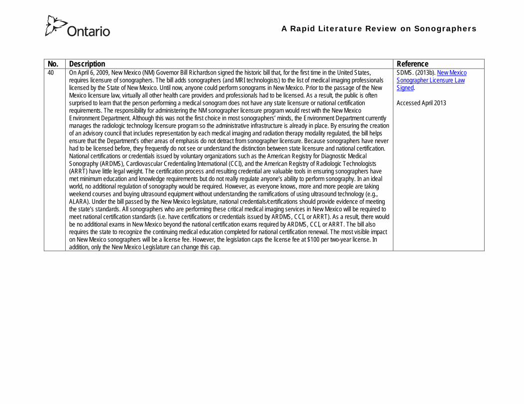
A Rapid Literature Review on Sonographers
No Description Reference 40 On April 6 2009 New Mexico (NM) Governor Bill Richardson signed the historic bill that for the first time in the United States
requires licensure of sonographers The bill adds sonographers (and MRI technologists) to the list of medical imaging professionals licensed by the State of New Mexico Until now anyone could perform sonograms in New Mexico Prior to the passage of the New Mexico licensure law virtually all other health care providers and professionals had to be licensed As a result the public is often surprised to learn that the person performing a medical sonogram does not have any state licensure or national certification requirements The responsibility for administering the NM sonographer licensure program would rest with the New Mexico Environment Department Although this was not the first choice in most sonographersrsquo minds the Environment Department currently manages the radiologic technology licensure program so the administrative infrastructure is already in place By ensuring the creation of an advisory council that includes representation by each medical imaging and radiation therapy modality regulated the bill helps ensure that the Departmentrsquos other areas of emphasis do not detract from sonographer licensure Because sonographers have never had to be licensed before they frequently do not see or understand the distinction between state licensure and national certification National certifications or credentials issued by voluntary organizations such as the American Registry for Diagnostic Medical Sonography (ARDMS) Cardiovascular Credentialing International (CCI) and the American Registry of Radiologic Technologists (ARRT) have little legal weight The certification process and resulting credential are valuable tools in ensuring sonographers have met minimum education and knowledge requirements but do not really regulate anyonersquos ability to perform sonography In an ideal world no additional regulation of sonography would be required However as everyone knows more and more people are taking weekend courses and buying ultrasound equipment without understanding the ramifications of using ultrasound technology (eg ALARA) Under the bill passed by the New Mexico legislature national credentialscertifications should provide evidence of meeting the statersquos standards All sonographers who are performing these critical medical imaging services in New Mexico will be required to meet national certification standards (ie have certifications or credentials issued by ARDMS CCI or ARRT) As a result there would be no additional exams in New Mexico beyond the national certification exams required by ARDMS CCI or ARRT The bill also requires the state to recognize the continuing medical education completed for national certification renewal The most visible impact on New Mexico sonographers will be a license fee However the legislation caps the license fee at $100 per two-year license In addition only the New Mexico Legislature can change this cap
SDMS (2013b) New Mexico Sonographer Licensure Law Signed Accessed April 2013
A Rapid Literature Review on Sonographers
No Description Reference 41 Preamble The purpose of this document is to define the Scope of Practice for Diagnostic Ultrasound Professionals in the US and to
specify their roles as members of the health care team acting in the best interest of the patient This scope of practice is a living document that will evolve as the technology expands Definition of the Profession The Diagnostic Ultrasound Profession is a multi-specialty field comprised of Diagnostic Medical Sonography (with subspecialties in abdominal neurologic obstetricalgynecologic and ophthalmic ultrasound) Diagnostic Cardiac Sonography (with subspecialties in adult and pediatric echocardiography) Vascular Technology and other emerging fields These diverse specialties are distinguished by their use of diagnostic medical ultrasound as a primary technology in their daily work Certification is considered the standard of practice in ultrasound Individuals who are not yet certified should reference the Scope as a professional model and strive to become certified Scope of Practice of the Profession The Diagnostic Ultrasound Professional is an individual qualified by professional credentialing and academic and clinical experience to provide diagnostic patient care services using ultrasound and related diagnostic procedures The scope of practice of the Diagnostic Ultrasound Professional includes those procedures acts and processes permitted by law for which the individual has received education and clinical experience and in which heshe has demonstrated competency Diagnostic Ultrasound Professionals
bull Perform patient assessments bull Acquire and analyze data obtained using ultrasound and related diagnostic technologies bull Provide a summary of findings to the physician to aid in patient diagnosis and management bull Use independent judgment and systematic problem solving methods to produce high quality diagnostic information and
optimize patient care
Society of Diagnostic Medical Sonography [SDMS] (2013c) Scope of Practice for the Diagnostic Ultrasound Professional Accessed April 2013
42 This website notes that in the US most employers in the US prefer to hire sonographers who have professional certification A sonographer can get certification and passing an exam Most exams relate to the specialty that the sonographer is most interested inmdashfor example an exam to become certified in abdominal sonography A few states require diagnostic medical sonographers to be licensed Typically professional certifications required for licensure other requirements vary by state Sonographers must take continuing education to keep their certification current
Bureau of Labor Statistics US Department of Labor (2012) How to become a Diagnostic Medical Sonographer Occupational Outlook Handbook 2012-13 Edition Accessed April 2013
A Rapid Literature Review on Sonographers
No Description Reference 43 The American Registry for Diagnostic Medical Sonography (ARDMS) is an independent not-for-profit organization founded in 1975
that administers examinations and awards the following credentials bull RDMSreg ndash Registered Diagnostic Medical Sonographerreg bull RDCSreg ndash Registered Diagnostic Cardiac Sonographerreg bull RVTreg ndash Registered Vascular Technologistreg bull RPVIreg ndash Registered Physician in Vascular Interpretationreg bull RMSKtrade - Registered in Musculoskeletaltrade sonography
ARDMS is the globally recognized standard of excellence in sonography and has certified more than 75000 individuals ARDMS is composed of three key groups
bull Board of Directors ndash Sonographers vascular technologists physicians research scientists and a member of the public are volunteer members of the board that determines the direction and policies of our organization
bull Examination Development Task Forces ndash A group of sonographers vascular technologists physicians and scientists volunteer their time and expertise to produce our examinations
bull Professional Staff ndash A paid highly-skilled staff administers the programs and assists applicants and registrants Credentials awarded by ARDMS document personal achievement of recognized professional standards and are widely accepted in the medical community by sonography and vascular professional organizations However ARDMS does not validate the day-to-day job performance of any sonography professional ARDMS has earned the prestigious ANSI-ISO 17024 accreditation for certifying bodies from the International Organization for Standardization (ISO) Accreditation is granted through the American National Standards Institute (ANSI) ARDMS has also earned accreditation with the National Commission for Certifying Agencies (NCCA) The NCCA is the accrediting arm of the National Organization for Competency Assurance (NOCA) Established in 1977 as a nonprofit organization NOCA is a leader in establishing quality standards for certifying organizations
American Registry for Diagnostic Medical Sonography (ARDMS) (2012a) Overview of ARDMS Accessed April 2013
A Rapid Literature Review on Sonographers
No Description Reference 44 ARDMS is developing a program to assure that Registrants continue to meet the basic competency requirements of core clinical skills
in present day practice bull Each Registrant who holds an active ARDMS certification as of December 31 2011 will be assigned the 2012 ndash 2021 10-
year recertification assessment period bull The 10-year period for new Registrants will begin the year following the attainment of the credential bull If a Registrant earns a new credential or specialty after January 1 2012 then that credentialspecialty will have a separate
10-year recertification assessment period that will begin the year following the attainment of the credentialspecialty ARDMS will develop tools and resources to assist Registrants in tracking and meeting the recertification assessment program requirements
bull Registrants who do not wish to take a recertification assessment in a specific credentialspecialty may voluntarily relinquish the credentialspecialty
As an ARDMS Registrant individuals are committing to demonstrate the highest level of competency in the modality of sonography by meeting and maintaining the standards set by the ARDMS The pathway of maintenance your certification is comprised of Paying the ARDMS annual renewal fee each year Each year ARDMS assesses Registrants an annual renewal fee By paying your annual renewal fee you are contributing to the ARDMS mission of promoting quality care and patient safety through the certification and continuing competency of ultrasound professionals In order to maintain your credentials you must pay the annual renewal fee of $75 USD or $100 USD (for RPVI and MSK Registrants with MDDODPM) designation on or before December 31 of each year in order to stay active for the following year Attesting to ARDMS rules and policies Each year when you pay your annual renewal fee you are attesting that you are in compliance with all ARDMS rules and discipline policies and are in good standing with the ARDMS Earning a minimum of 30 ARDMS-accepted CMEs during your three-year CME period ARDMS verifies its Registrants compliance with continuing medical education (CME) requirements using an audit process Earning your CMEs is an integral component of maintaining your ARDMS credential(s) and demonstrates your ongoing commitment to lifelong learning RecertificationContinued Commitment to Excellence Following the attainment of the ARDMS credential Registrants are assigned a 10-year recertification period the year after the credential is earned ARDMS is committed to developing a recertification program that is easy for Registrants to comply with
American Registry for Diagnostic Medical Sonography (ARDMS) (2012b) Recertification General Information
A Rapid Literature Review on Sonographers
No Description Reference 45 This document reviews Sonography Principles and Instrumentation (SPI) Examination Requirements offered through ARDMS in
order to achieve sonography certification and the various prerequisites needed before taking the exam For example one prerequisite entails (1) Education Graduate of a program accredited by an agency recognized by the Council for Higher Education Accreditation (CHEA) United States Department of Education (USDOE) or Canadian Medical Association (CMA) that specifically conducts programmatic accreditation for diagnostic medical sonographydiagnostic cardiac sonographyvascular technology Currently the only organizations that offer programmatic accreditation under the aforementioned associations are the Commission on Accreditation of Allied Health Education Programs (CAAHEP) and the Canadian Medical Association (CMA) (2) Required Clinical UltrasoundVascular Experience No additional experience is required (3) Documentation Required with Application 1) Copy of diploma from ultrasoundvascular program or an official transcript indicating the date the degree was conferred 2) Original letter signed by program director andor medical director indicating date of graduation or successful completion of the program4 Program directors must use the mandatory formatted sample letter available on ARDMSorg sampleletters 3) The CV form is not required if the application is submitted and received in the ARDMS office within one year after successful completion of the program Otherwise an original signed and completed CV form for each appropriate specialty area(s) must be submitted 4) Photocopy of a non-expired government issued photo identification with signature the name on the identification must exactly match the name under which you are applying for ARDMS examination
American Registry for Diagnostic Medical Sonography (ARDMS) (2012c) SPI Requirement and General Prerequisites Accessed April 2013
46 This page notes the five credentials offered by the ARDMS and the corresponding exams and specialty exams required bull RDMS requires the SPI exam (sonography principles and instrumentation) and a specialty exam in abdomen breast fetal
echocardiography neurosonolgoy or obstetricsgynecology bull RDCS requires the SPI exam and a speciality exam in adult echocardiography fetal echocardiography pediatric
echocardiography bull RVT requires the SPI and a speciality exam in vascular technology bull RMSK requires the musculoskeletal sonography exam bull RPVI requires the physiciansrsquo vascular interpretation exam
American Registry for Diagnostic Medical Sonography (ARDMS) (2012d) Credentials and Examinations Accessed April 2013
A Rapid Literature Review on Sonographers
No Description Reference 47 This article summarizes some of the many trends in fetal pediatric and adult echocardiography that SDMS members are excited
about Fetal Echocardiography Sonography has been used since the mid-1980s to help identify congenital heart defects during the second trimester The ldquobasicrdquo examination of the fetal heart involves a four-chamber view ndash evaluation of the right and left atrial and ventricular chambers and their respective valves In the past several years there has been a movement in the obstetric ultrasound community to encourage obstetric sonographers to perform the ldquoextended basicrdquo exam which includes the cardiac outflow tracts The outflow tracts consist of the main pulmonary artery exiting the right ventricle and the aorta exiting the left ventricle The fetal echocardiography community also is abuzz over three-dimensional (3D) and four-dimensional (4D) ultrasound and the potential of this technology to significantly improve the evaluation of the fetal heart Spatiotemporal Image Correlation (STIC) a 4D application allows for evaluation and manipulation of the heart in real-time Allen says Todayrsquos ultrasound equipment allows sonographers to gather a volume of data from which they can create detailed 3D images of the tiny fetal heart which is constantly in motion With computer manipulation of the data sets the images also can be viewed in 4D allowing physicians to see the entire fetal heart cycle from start to finish Pediatric Echocardiography One of the most significant advances in pediatric echo over the last decade is moving to a digital format The advent of pediatric transesophageal (TEE) probes also has been a big advance for pediatric echo Transesophageal probes provide an unimpeded sonographic visualization of the intracardiac structures Theyrsquore useful in visualizing very fine valvular abnormalities the thoracic aorta vegetations and thrombus in the atrial appendage It is also useful in detecting subtle anatomic variations seen in congenital heart disease Pediatric-sized TEE probes make the exams possible In the past it wasnrsquot possible because the adult probe was just too big to put down the babyrsquos or childrsquos esophagus Now the probe is small enough to successfully manipulate it in children and get superb images Because the new sonographic imaging equipment and better surgical techniques allow clinicians to detect and repair more cardiac abnormalities earlier patients are living longer Adult Echocardiography The adult echo world is abuzz about 2D and 3D speckle tracking relatively new techniques for the assessment of heart function and strain imaging Speckle tracking evaluates both regional and global function by measuring the deformation of the heart muscle
Orenstein B W (2010) Current Trends in Echocardiography SDMS New Wave February 1-5 Accessed April 2013
48 This document describes the requirements for admission to the Register of Accredited Medical Sonographer in Australia Requirements include Continuation of Accreditation In order to remain on the Register an accredited Sonographer must satisfy the ASAR continuing professional development (CPD) requirements and pay the prescribed annual fee Student Sonographers apart from paying the prescribed annual fee should provide evidence of their student status every year when they renew their ASAR accreditation
Australian Sonographer Accreditation Registry (ASAR) (no date A) Sonographer Accreditation Accessed April 2013
A Rapid Literature Review on Sonographers
No Description Reference 49 ASARrsquos functions
1 Accredit sonography education programs within Australia 2 Maintain a register of Accredited Sonographers and Accredited Student Sonographers 3 Establish minimum standards of Continuing Professional Development (CPD) and to monitor the CPD requirements for
Sonographers to remain on the register 4 To support activities that advance the profession of sonography 5 To provide registration information about Sonographers to Medicare Australia 6 To provide public access to the name registration number State or Territory current type of accreditation and area of
specialty for active Sonographers ASARrsquos Role ASAR is not a registration board ASAR does not have the authority to remove a Sonographer from the Registry due to professional misconduct nor the ability to prevent a Sonographer from practicing ASAR is only able to remove a Sonographer from the Registry if they
bull fail to meet CPD requirements and or bull fail to pay annual fees
ASAR (no date B) About ASAR Accessed April 2013
50 Regulatory bodies A Regulatory Body maintains a register of individuals who have achieved the required qualification of that profession to practice and can evidence continuing competence at the required level For a doctor to practice in the UK he or she must be registered with the General Medical Council (GMC) This is the regulatory body for medical practitioners The majority of sonographers who are employed in the UK come from a background of radiography or midwifery To practice radiography in the UK a qualified radiographer must be registered with the Health Professions Council (HPC) Similarly a midwife must be registered with the National Midwifery Council to practice as a midwife Sonographer registration in the UK There are a number of professions allied to medicine that are regulated by the HPC for example radiography physiotherapy However sonography is not recognised as a profession by the HPC and therefore there is no mechanism whereby an individual can register with the HPC as a sonographer or be regulated by them as a sonographer It is a common misunderstanding that sonographers have to be registered with the HPC to practice This comes from the fact that most sonographers are also qualified radiographers and maintain their registration with the HPC because they wish to remain on the HPC register as a radiographer Professional bodies An individual can be a member of a Professional Body such as the Royal College of Obstetricians amp Gynecologists (RCOG) Royal College of Radiologists (RCR) Society amp College of Radiographers (SCoR) or the Royal College of Midwives (RCM) but this is not the same as being accepted onto the register of one of the regulatory bodies The professional bodies are responsible for setting and maintaining the standards of their members providing a code of conduct etc They may also act in the capacity of a trades union and can also provide professional indemnity insurance The British Medical Ultrasound Society BMUS is not a professional body but is a scientific society so will offer a different range of benefits Whether or not one wishes to join the society has no impact on their ability to work as a sonographer in the UK although from a continuing professional development point of view it can be beneficial The College of Radiographers are currently working with the HPC to get sonography recognized as a separate profession
British Medical Ultrasound Society (BMUS) (no date) Information on Sonographer Regulation in the UK Accessed April 2013
A Rapid Literature Review on Sonographers
No Description Reference 51 This document included sections on the role of the sonographer and the use of sonography for entertainment Role of the
Sonographer and Technical Impressions The role of the sonographer is to perform the ultrasound scan and to document observations The sonographerrsquos written technical impressions are intended as a form of communication between the sonographer and the reporting physician These observations must be reviewed and subsequently reported by the reporting physician Issuing of a final reportdiagnosis represents the practice of medicine and is therefore the domain of the reporting physician Use of Ultrasound for Entertainment or Non-Diagnostic Purposes In contrast to this justified form of assessment ultrasound for entertainment is simply an elaborate version of a portrait For a variable fee the surface of the fetus is skimmed over and clients are provided with keepsake images or a DVD of their fetus There are no measurements taken during this exam no morphological assessment performed and no dictated diagnostic report of findings Essentially there are no diagnostic benefits to the exam and uninformed parents may be falsely reassured that the beautiful portrait indicates a normal fetus Entertainment ultrasound facilities may operate outside of medical guidelines and without any controls which may result in a lack of technical safeguards operator expertise or governance of technical competency These facilities do not comply with the ALARA principle which could result in increased fetal exposures The Canadian Society of Diagnostic Medical Sonograph (CSDMS) does not support persons or facilities that participate in ultrasound for entertainment activities Liability insurance purchased through the CSDMS group insurance plan does not cover any activities carried out for entertainment ultrasound purposes Other leaders in Canadian medicine support this statement In 2005 the SOGC (Society of Obstetrics and Gynaecologists of Canada) published an article entitled ldquoObstetrical Ultrasound Biological Effects and Safetyrdquo and their conclusion was that ldquothe theoretical risk of adverse bio-effects even from standard 2D obstetrical ultrasound makes it hard to justify its use for non-medical reasons such as sex-determination making non-medical photos or video or for commercial purposerdquo The SOGC recommends that ultrasound be used prudently and that energy exposure be limited to the minimum that is medically necessary The SOGC further recommends a complete ban on the non-medical use of fetal ultrasound and encourages government to join with society to find appropriate means to deal with this public health issue The CAR (Canadian Association of Radiologists) makes a similar statement ldquoThe CAR strongly opposes the use of diagnostic ultrasound equipment for non-medical purposes and considers the use of medical ultrasound for entertainment to be a misuse of the technology especially if fetal subjects are involvedrdquo
Canadian Society of Diagnostic Medical Sonography (no date) Professional Practice Guidelines and Policy Statements For Canadian Sonography Accessed April 2013
A Rapid Literature Review on Sonographers
No Description Reference 52 In 1993 the Consortium for the Accreditation of Sonographic Education (CASE) was formed through the common desire to ensure
that the education and training of sonographers in the United Kingdom was delivered at the highest level All members share common concerns that standards of service provision and education are developed in parallel with increasing demand and improving technology All members strive to create an environment in which education and training is maximised This will make validation and accreditation to be as robust and straightforward as possible CASEs philosophy is to promote best ultrasound practise through the accreditation of those training programmes that develop safe and competent ultrasound practitioners This should be delivered at postgraduate level The majority of its activities relate to supporting those Institutions that offer or wish to offer courses leading to the award of a Masters Postgraduate DiplomaCertificate in Medical or clinical Ultrasound In addition to offering accreditation of new and re-accreditation of established ultrasound education programmes CASE undertakes annual monitoring of the courses it has accredited The information obtained is fed back to the Institutions both on an individual basis and via the annual CASE report Each member organisation is unique and has its own obligations to its membership and its own agenda while CASE brings them together to provide a forum for the interchange of ideas in which shared views may be synthesised and thus inform education and training programmes
Consortium for the Accreditation of Sonographic Education (no date) Home Accessed April 2013
A Rapid Literature Review on Sonographers
No Description Reference 53 The Medical Radiation Technologists Board (the Board) is one of sixteen New Zealand health registration authorities appointed by
the Minister of Health under the Health Practitioners Competence Assurance Act 2003 (the Act) The Board is responsible for the administration of the Act in regard to the profession of medical radiation technology The primary responsibility of the Board is to protect the health and safety of the New Zealand public by ensuring practitioners registered in the profession of medical radiation technology are competent and fit to practice In accordance with Section 118 of the Act the Board is responsible for fulfilling a number of functions
bull 01 To prescribe the qualifications required for scopes of practice within the profession of medical radiation technology and for that purpose to accredit and monitor educational institutions and degrees courses of studies or programmes
bull 02 To authorise the registration of medical radiation technologists under the Act and to maintain registers bull 03 To consider applications for annual practising certificates bull 04 To review and promote the competence of medical radiation technologists bull 05 To recognise accredit and set programmes to ensure the ongoing competence of medical radiation technologists bull 06 To receive and act on information from health practitioners employers and the Health and Disability Commissioner
about the competence of medical radiation technologists bull 07 To notify employers the Accident Compensation Corporation the Director-General of Health and the Health and
Disability Commissioner that the practice of a medical radiation technologist may pose a risk of harm to the public bull 08 To consider the cases of medical radiation technologists who may be unable to perform the functions required for the
practice of the medical radiation technology profession bull 09 To set standards of clinical competence cultural competence and ethical conduct to be observed by medical radiation
technologists bull 10 To liaise with other authorities appointed under the Act about matters of common interest bull 11 To promote education and training in the profession of medical radiation technology bull 12 To promote public awareness of the responsibilities of the Board bull 13 To exercise and perform any other functions powers and duties that are conferred or imposed on it by or under the Act
or any other enactment
Medical Radiation Technologists Board (MRTB) (no date A) What we do Accessed April 2013
54 Qualifications To be considered for registration as a medical radiation technologist in any of the specified scopes of practice an individual must hold a qualification for that particular scope as prescribed by the Board or approved by the Board as being substantially equivalent to the prescribed qualifications In addition to having an approved qualification individuals may also be required to demonstrate that they have adequate experience in the relevant scope of practice Sonographer The Board has prescribed one New Zealand postgraduate qualification for a person to be eligible for registration in the scope of practice of Sonographer Postgraduate Diploma in Health Science (Ultrasound) issued by the Unitec Institute of Technology Required Experience If an individual is applying for registration in Nuclear Medicine Ultrasound or Magnetic Resonance Imaging in addition to holding a relevant postgraduate qualification they must also be able to provide evidence that you have a minimum of 3360 hours of clinical experience in the relevant scope of practice you are applying for registration in
Medical Radiation Technologists Board (MRTB) (no date B) Qualifications Accessed April 2013
A Rapid Literature Review on Sonographers
No Description Reference 55 Recertification is a mechanism used by the Medical Radiation Technologists Board in New Zealand to ensure that registered medical
radiation technologists continue to be competent to practise within their scopes of practice Annual Practising Certificates Each year if an individual wishes to continue practising in your scope(s) of practice in New Zealand they must apply for and be issued with an annual practising certificate (APC) A condition of being issued with an APC is that one must provide evidence of enrolment and satisfactory progress in a Board-approved recertification programme Alternatively the Board may issue them with an interim practising certificate (IPC) until it is satisfied the individual demonstrates that they are competent to practice in the scope(s) of practice they applied for Working with an IPC will require an individual to work under the supervision of a registered medical radiation technologist or other relevant registered health practitioner Recertification Programmes The Boards recertification programme framework is based on a continuing professional development (CPD) model Individuals have the option of enrolling in one of seven recertification programmes for medical radiation technologists that are currently approved by the Board They must meet the requirements of the particular recertification programme in which they are enrolled Recertification Audits An essential component of the CPD-based recertification programme adopted by the Board is the audit of CPD recordsThe Board undertakes an annual audit of approximately 10 of its registrant database to assess individual medical radiation technologistsrsquo compliance with the Boarrsquods recertification requirements
Medical Radiation Technologists Board (MRTB) (no date C) Recertification Accessed April 2013
56 There are two types of radiography - diagnostic and therapeutic Both need considerable knowledge of technology anatomy physiology and pathology to carry out their work Many also undertake further training to become a sonographer Some of the imaging technology that a diagnostic radiographer may use are
bull X-Ray - looks through tissues to examine bones cavities and foreign objects bull Fluoroscopy - images the digestive system providing a real-time image bull CT (Computed Tomography) - which provides cross-sectional views (slices) of the body bull MRI (Magnetic Resonance Imaging) - builds a 2-D or 3-D map of the different tissue types within the body bull Ultrasound - well known for its use in obstetrics and gynaecology Also used to check circulation and examine the heart bull Angiography - used to investigate blood vessels
Sonographyultrasonography Ultrasound is used in various settings in a hospital including abdominal scanning and breast ultrasound Ultrasound imaging is the use of high frequency sound in excess of human hearing to produce images of structures of the human body that may be observed on a screen These images may subsequently be transferred to photographic film or paper or onto video or CD forming part of the patients record of their examination There are no direct entry routes into ultrasound Most sonographers train as a radiographer then undertake an approved post-registration course Offered by higher education institutions The courses are a minimum of one academic year and prepare sonographers clinically and academically for practice Normally a pre-requisite for acceptance is access to a clinical department with supervised practice for students
National Health Service (NHS) (no date A) Radiographer Accessed April 2013
57 To practice as a radiographer in the UK an individual must be registered with the Health and Care Professions Council (HCPC) To register individuals must have successfully completed an approved training programme in diagnostic or therapeutic radiography These are offered at undergraduate degree (full-time and part-time) and at postgraduate degreediploma level at a number of universities across the UK
NHS (no date B) Entry requirements and training in radiography Accessed April 2013
Health Professions Regulatory Advisory Council 56 Wellesley Street West 12th Floor
Toronto ON M5S 2S3
Telephone 416-326-1550 Toll-Free 1-888-377-7746
Fax 416-326-1549
Website wwwhpracorg
Email hpracwebmasterontarioca
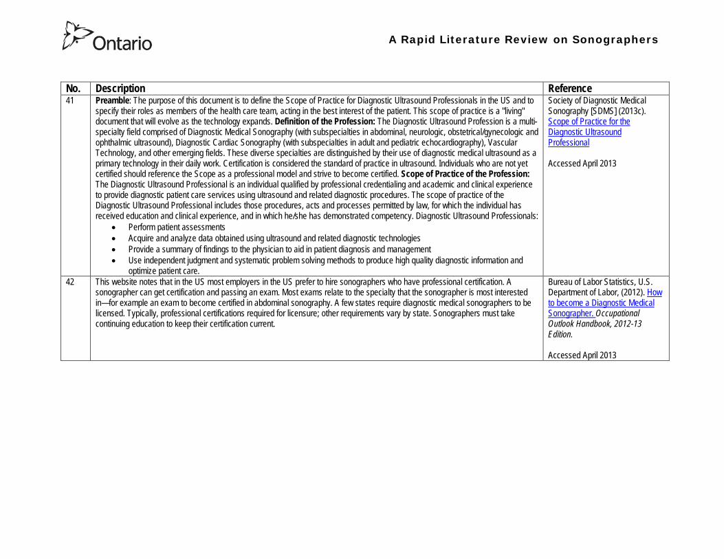
A Rapid Literature Review on Sonographers
No Description Reference 43 The American Registry for Diagnostic Medical Sonography (ARDMS) is an independent not-for-profit organization founded in 1975
that administers examinations and awards the following credentials bull RDMSreg ndash Registered Diagnostic Medical Sonographerreg bull RDCSreg ndash Registered Diagnostic Cardiac Sonographerreg bull RVTreg ndash Registered Vascular Technologistreg bull RPVIreg ndash Registered Physician in Vascular Interpretationreg bull RMSKtrade - Registered in Musculoskeletaltrade sonography
ARDMS is the globally recognized standard of excellence in sonography and has certified more than 75000 individuals ARDMS is composed of three key groups
bull Board of Directors ndash Sonographers vascular technologists physicians research scientists and a member of the public are volunteer members of the board that determines the direction and policies of our organization
bull Examination Development Task Forces ndash A group of sonographers vascular technologists physicians and scientists volunteer their time and expertise to produce our examinations
bull Professional Staff ndash A paid highly-skilled staff administers the programs and assists applicants and registrants Credentials awarded by ARDMS document personal achievement of recognized professional standards and are widely accepted in the medical community by sonography and vascular professional organizations However ARDMS does not validate the day-to-day job performance of any sonography professional ARDMS has earned the prestigious ANSI-ISO 17024 accreditation for certifying bodies from the International Organization for Standardization (ISO) Accreditation is granted through the American National Standards Institute (ANSI) ARDMS has also earned accreditation with the National Commission for Certifying Agencies (NCCA) The NCCA is the accrediting arm of the National Organization for Competency Assurance (NOCA) Established in 1977 as a nonprofit organization NOCA is a leader in establishing quality standards for certifying organizations
American Registry for Diagnostic Medical Sonography (ARDMS) (2012a) Overview of ARDMS Accessed April 2013
A Rapid Literature Review on Sonographers
No Description Reference 44 ARDMS is developing a program to assure that Registrants continue to meet the basic competency requirements of core clinical skills
in present day practice bull Each Registrant who holds an active ARDMS certification as of December 31 2011 will be assigned the 2012 ndash 2021 10-
year recertification assessment period bull The 10-year period for new Registrants will begin the year following the attainment of the credential bull If a Registrant earns a new credential or specialty after January 1 2012 then that credentialspecialty will have a separate
10-year recertification assessment period that will begin the year following the attainment of the credentialspecialty ARDMS will develop tools and resources to assist Registrants in tracking and meeting the recertification assessment program requirements
bull Registrants who do not wish to take a recertification assessment in a specific credentialspecialty may voluntarily relinquish the credentialspecialty
As an ARDMS Registrant individuals are committing to demonstrate the highest level of competency in the modality of sonography by meeting and maintaining the standards set by the ARDMS The pathway of maintenance your certification is comprised of Paying the ARDMS annual renewal fee each year Each year ARDMS assesses Registrants an annual renewal fee By paying your annual renewal fee you are contributing to the ARDMS mission of promoting quality care and patient safety through the certification and continuing competency of ultrasound professionals In order to maintain your credentials you must pay the annual renewal fee of $75 USD or $100 USD (for RPVI and MSK Registrants with MDDODPM) designation on or before December 31 of each year in order to stay active for the following year Attesting to ARDMS rules and policies Each year when you pay your annual renewal fee you are attesting that you are in compliance with all ARDMS rules and discipline policies and are in good standing with the ARDMS Earning a minimum of 30 ARDMS-accepted CMEs during your three-year CME period ARDMS verifies its Registrants compliance with continuing medical education (CME) requirements using an audit process Earning your CMEs is an integral component of maintaining your ARDMS credential(s) and demonstrates your ongoing commitment to lifelong learning RecertificationContinued Commitment to Excellence Following the attainment of the ARDMS credential Registrants are assigned a 10-year recertification period the year after the credential is earned ARDMS is committed to developing a recertification program that is easy for Registrants to comply with
American Registry for Diagnostic Medical Sonography (ARDMS) (2012b) Recertification General Information
A Rapid Literature Review on Sonographers
No Description Reference 45 This document reviews Sonography Principles and Instrumentation (SPI) Examination Requirements offered through ARDMS in
order to achieve sonography certification and the various prerequisites needed before taking the exam For example one prerequisite entails (1) Education Graduate of a program accredited by an agency recognized by the Council for Higher Education Accreditation (CHEA) United States Department of Education (USDOE) or Canadian Medical Association (CMA) that specifically conducts programmatic accreditation for diagnostic medical sonographydiagnostic cardiac sonographyvascular technology Currently the only organizations that offer programmatic accreditation under the aforementioned associations are the Commission on Accreditation of Allied Health Education Programs (CAAHEP) and the Canadian Medical Association (CMA) (2) Required Clinical UltrasoundVascular Experience No additional experience is required (3) Documentation Required with Application 1) Copy of diploma from ultrasoundvascular program or an official transcript indicating the date the degree was conferred 2) Original letter signed by program director andor medical director indicating date of graduation or successful completion of the program4 Program directors must use the mandatory formatted sample letter available on ARDMSorg sampleletters 3) The CV form is not required if the application is submitted and received in the ARDMS office within one year after successful completion of the program Otherwise an original signed and completed CV form for each appropriate specialty area(s) must be submitted 4) Photocopy of a non-expired government issued photo identification with signature the name on the identification must exactly match the name under which you are applying for ARDMS examination
American Registry for Diagnostic Medical Sonography (ARDMS) (2012c) SPI Requirement and General Prerequisites Accessed April 2013
46 This page notes the five credentials offered by the ARDMS and the corresponding exams and specialty exams required bull RDMS requires the SPI exam (sonography principles and instrumentation) and a specialty exam in abdomen breast fetal
echocardiography neurosonolgoy or obstetricsgynecology bull RDCS requires the SPI exam and a speciality exam in adult echocardiography fetal echocardiography pediatric
echocardiography bull RVT requires the SPI and a speciality exam in vascular technology bull RMSK requires the musculoskeletal sonography exam bull RPVI requires the physiciansrsquo vascular interpretation exam
American Registry for Diagnostic Medical Sonography (ARDMS) (2012d) Credentials and Examinations Accessed April 2013
A Rapid Literature Review on Sonographers
No Description Reference 47 This article summarizes some of the many trends in fetal pediatric and adult echocardiography that SDMS members are excited
about Fetal Echocardiography Sonography has been used since the mid-1980s to help identify congenital heart defects during the second trimester The ldquobasicrdquo examination of the fetal heart involves a four-chamber view ndash evaluation of the right and left atrial and ventricular chambers and their respective valves In the past several years there has been a movement in the obstetric ultrasound community to encourage obstetric sonographers to perform the ldquoextended basicrdquo exam which includes the cardiac outflow tracts The outflow tracts consist of the main pulmonary artery exiting the right ventricle and the aorta exiting the left ventricle The fetal echocardiography community also is abuzz over three-dimensional (3D) and four-dimensional (4D) ultrasound and the potential of this technology to significantly improve the evaluation of the fetal heart Spatiotemporal Image Correlation (STIC) a 4D application allows for evaluation and manipulation of the heart in real-time Allen says Todayrsquos ultrasound equipment allows sonographers to gather a volume of data from which they can create detailed 3D images of the tiny fetal heart which is constantly in motion With computer manipulation of the data sets the images also can be viewed in 4D allowing physicians to see the entire fetal heart cycle from start to finish Pediatric Echocardiography One of the most significant advances in pediatric echo over the last decade is moving to a digital format The advent of pediatric transesophageal (TEE) probes also has been a big advance for pediatric echo Transesophageal probes provide an unimpeded sonographic visualization of the intracardiac structures Theyrsquore useful in visualizing very fine valvular abnormalities the thoracic aorta vegetations and thrombus in the atrial appendage It is also useful in detecting subtle anatomic variations seen in congenital heart disease Pediatric-sized TEE probes make the exams possible In the past it wasnrsquot possible because the adult probe was just too big to put down the babyrsquos or childrsquos esophagus Now the probe is small enough to successfully manipulate it in children and get superb images Because the new sonographic imaging equipment and better surgical techniques allow clinicians to detect and repair more cardiac abnormalities earlier patients are living longer Adult Echocardiography The adult echo world is abuzz about 2D and 3D speckle tracking relatively new techniques for the assessment of heart function and strain imaging Speckle tracking evaluates both regional and global function by measuring the deformation of the heart muscle
Orenstein B W (2010) Current Trends in Echocardiography SDMS New Wave February 1-5 Accessed April 2013
48 This document describes the requirements for admission to the Register of Accredited Medical Sonographer in Australia Requirements include Continuation of Accreditation In order to remain on the Register an accredited Sonographer must satisfy the ASAR continuing professional development (CPD) requirements and pay the prescribed annual fee Student Sonographers apart from paying the prescribed annual fee should provide evidence of their student status every year when they renew their ASAR accreditation
Australian Sonographer Accreditation Registry (ASAR) (no date A) Sonographer Accreditation Accessed April 2013
A Rapid Literature Review on Sonographers
No Description Reference 49 ASARrsquos functions
1 Accredit sonography education programs within Australia 2 Maintain a register of Accredited Sonographers and Accredited Student Sonographers 3 Establish minimum standards of Continuing Professional Development (CPD) and to monitor the CPD requirements for
Sonographers to remain on the register 4 To support activities that advance the profession of sonography 5 To provide registration information about Sonographers to Medicare Australia 6 To provide public access to the name registration number State or Territory current type of accreditation and area of
specialty for active Sonographers ASARrsquos Role ASAR is not a registration board ASAR does not have the authority to remove a Sonographer from the Registry due to professional misconduct nor the ability to prevent a Sonographer from practicing ASAR is only able to remove a Sonographer from the Registry if they
bull fail to meet CPD requirements and or bull fail to pay annual fees
ASAR (no date B) About ASAR Accessed April 2013
50 Regulatory bodies A Regulatory Body maintains a register of individuals who have achieved the required qualification of that profession to practice and can evidence continuing competence at the required level For a doctor to practice in the UK he or she must be registered with the General Medical Council (GMC) This is the regulatory body for medical practitioners The majority of sonographers who are employed in the UK come from a background of radiography or midwifery To practice radiography in the UK a qualified radiographer must be registered with the Health Professions Council (HPC) Similarly a midwife must be registered with the National Midwifery Council to practice as a midwife Sonographer registration in the UK There are a number of professions allied to medicine that are regulated by the HPC for example radiography physiotherapy However sonography is not recognised as a profession by the HPC and therefore there is no mechanism whereby an individual can register with the HPC as a sonographer or be regulated by them as a sonographer It is a common misunderstanding that sonographers have to be registered with the HPC to practice This comes from the fact that most sonographers are also qualified radiographers and maintain their registration with the HPC because they wish to remain on the HPC register as a radiographer Professional bodies An individual can be a member of a Professional Body such as the Royal College of Obstetricians amp Gynecologists (RCOG) Royal College of Radiologists (RCR) Society amp College of Radiographers (SCoR) or the Royal College of Midwives (RCM) but this is not the same as being accepted onto the register of one of the regulatory bodies The professional bodies are responsible for setting and maintaining the standards of their members providing a code of conduct etc They may also act in the capacity of a trades union and can also provide professional indemnity insurance The British Medical Ultrasound Society BMUS is not a professional body but is a scientific society so will offer a different range of benefits Whether or not one wishes to join the society has no impact on their ability to work as a sonographer in the UK although from a continuing professional development point of view it can be beneficial The College of Radiographers are currently working with the HPC to get sonography recognized as a separate profession
British Medical Ultrasound Society (BMUS) (no date) Information on Sonographer Regulation in the UK Accessed April 2013
A Rapid Literature Review on Sonographers
No Description Reference 51 This document included sections on the role of the sonographer and the use of sonography for entertainment Role of the
Sonographer and Technical Impressions The role of the sonographer is to perform the ultrasound scan and to document observations The sonographerrsquos written technical impressions are intended as a form of communication between the sonographer and the reporting physician These observations must be reviewed and subsequently reported by the reporting physician Issuing of a final reportdiagnosis represents the practice of medicine and is therefore the domain of the reporting physician Use of Ultrasound for Entertainment or Non-Diagnostic Purposes In contrast to this justified form of assessment ultrasound for entertainment is simply an elaborate version of a portrait For a variable fee the surface of the fetus is skimmed over and clients are provided with keepsake images or a DVD of their fetus There are no measurements taken during this exam no morphological assessment performed and no dictated diagnostic report of findings Essentially there are no diagnostic benefits to the exam and uninformed parents may be falsely reassured that the beautiful portrait indicates a normal fetus Entertainment ultrasound facilities may operate outside of medical guidelines and without any controls which may result in a lack of technical safeguards operator expertise or governance of technical competency These facilities do not comply with the ALARA principle which could result in increased fetal exposures The Canadian Society of Diagnostic Medical Sonograph (CSDMS) does not support persons or facilities that participate in ultrasound for entertainment activities Liability insurance purchased through the CSDMS group insurance plan does not cover any activities carried out for entertainment ultrasound purposes Other leaders in Canadian medicine support this statement In 2005 the SOGC (Society of Obstetrics and Gynaecologists of Canada) published an article entitled ldquoObstetrical Ultrasound Biological Effects and Safetyrdquo and their conclusion was that ldquothe theoretical risk of adverse bio-effects even from standard 2D obstetrical ultrasound makes it hard to justify its use for non-medical reasons such as sex-determination making non-medical photos or video or for commercial purposerdquo The SOGC recommends that ultrasound be used prudently and that energy exposure be limited to the minimum that is medically necessary The SOGC further recommends a complete ban on the non-medical use of fetal ultrasound and encourages government to join with society to find appropriate means to deal with this public health issue The CAR (Canadian Association of Radiologists) makes a similar statement ldquoThe CAR strongly opposes the use of diagnostic ultrasound equipment for non-medical purposes and considers the use of medical ultrasound for entertainment to be a misuse of the technology especially if fetal subjects are involvedrdquo
Canadian Society of Diagnostic Medical Sonography (no date) Professional Practice Guidelines and Policy Statements For Canadian Sonography Accessed April 2013
A Rapid Literature Review on Sonographers
No Description Reference 52 In 1993 the Consortium for the Accreditation of Sonographic Education (CASE) was formed through the common desire to ensure
that the education and training of sonographers in the United Kingdom was delivered at the highest level All members share common concerns that standards of service provision and education are developed in parallel with increasing demand and improving technology All members strive to create an environment in which education and training is maximised This will make validation and accreditation to be as robust and straightforward as possible CASEs philosophy is to promote best ultrasound practise through the accreditation of those training programmes that develop safe and competent ultrasound practitioners This should be delivered at postgraduate level The majority of its activities relate to supporting those Institutions that offer or wish to offer courses leading to the award of a Masters Postgraduate DiplomaCertificate in Medical or clinical Ultrasound In addition to offering accreditation of new and re-accreditation of established ultrasound education programmes CASE undertakes annual monitoring of the courses it has accredited The information obtained is fed back to the Institutions both on an individual basis and via the annual CASE report Each member organisation is unique and has its own obligations to its membership and its own agenda while CASE brings them together to provide a forum for the interchange of ideas in which shared views may be synthesised and thus inform education and training programmes
Consortium for the Accreditation of Sonographic Education (no date) Home Accessed April 2013
A Rapid Literature Review on Sonographers
No Description Reference 53 The Medical Radiation Technologists Board (the Board) is one of sixteen New Zealand health registration authorities appointed by
the Minister of Health under the Health Practitioners Competence Assurance Act 2003 (the Act) The Board is responsible for the administration of the Act in regard to the profession of medical radiation technology The primary responsibility of the Board is to protect the health and safety of the New Zealand public by ensuring practitioners registered in the profession of medical radiation technology are competent and fit to practice In accordance with Section 118 of the Act the Board is responsible for fulfilling a number of functions
bull 01 To prescribe the qualifications required for scopes of practice within the profession of medical radiation technology and for that purpose to accredit and monitor educational institutions and degrees courses of studies or programmes
bull 02 To authorise the registration of medical radiation technologists under the Act and to maintain registers bull 03 To consider applications for annual practising certificates bull 04 To review and promote the competence of medical radiation technologists bull 05 To recognise accredit and set programmes to ensure the ongoing competence of medical radiation technologists bull 06 To receive and act on information from health practitioners employers and the Health and Disability Commissioner
about the competence of medical radiation technologists bull 07 To notify employers the Accident Compensation Corporation the Director-General of Health and the Health and
Disability Commissioner that the practice of a medical radiation technologist may pose a risk of harm to the public bull 08 To consider the cases of medical radiation technologists who may be unable to perform the functions required for the
practice of the medical radiation technology profession bull 09 To set standards of clinical competence cultural competence and ethical conduct to be observed by medical radiation
technologists bull 10 To liaise with other authorities appointed under the Act about matters of common interest bull 11 To promote education and training in the profession of medical radiation technology bull 12 To promote public awareness of the responsibilities of the Board bull 13 To exercise and perform any other functions powers and duties that are conferred or imposed on it by or under the Act
or any other enactment
Medical Radiation Technologists Board (MRTB) (no date A) What we do Accessed April 2013
54 Qualifications To be considered for registration as a medical radiation technologist in any of the specified scopes of practice an individual must hold a qualification for that particular scope as prescribed by the Board or approved by the Board as being substantially equivalent to the prescribed qualifications In addition to having an approved qualification individuals may also be required to demonstrate that they have adequate experience in the relevant scope of practice Sonographer The Board has prescribed one New Zealand postgraduate qualification for a person to be eligible for registration in the scope of practice of Sonographer Postgraduate Diploma in Health Science (Ultrasound) issued by the Unitec Institute of Technology Required Experience If an individual is applying for registration in Nuclear Medicine Ultrasound or Magnetic Resonance Imaging in addition to holding a relevant postgraduate qualification they must also be able to provide evidence that you have a minimum of 3360 hours of clinical experience in the relevant scope of practice you are applying for registration in
Medical Radiation Technologists Board (MRTB) (no date B) Qualifications Accessed April 2013
A Rapid Literature Review on Sonographers
No Description Reference 55 Recertification is a mechanism used by the Medical Radiation Technologists Board in New Zealand to ensure that registered medical
radiation technologists continue to be competent to practise within their scopes of practice Annual Practising Certificates Each year if an individual wishes to continue practising in your scope(s) of practice in New Zealand they must apply for and be issued with an annual practising certificate (APC) A condition of being issued with an APC is that one must provide evidence of enrolment and satisfactory progress in a Board-approved recertification programme Alternatively the Board may issue them with an interim practising certificate (IPC) until it is satisfied the individual demonstrates that they are competent to practice in the scope(s) of practice they applied for Working with an IPC will require an individual to work under the supervision of a registered medical radiation technologist or other relevant registered health practitioner Recertification Programmes The Boards recertification programme framework is based on a continuing professional development (CPD) model Individuals have the option of enrolling in one of seven recertification programmes for medical radiation technologists that are currently approved by the Board They must meet the requirements of the particular recertification programme in which they are enrolled Recertification Audits An essential component of the CPD-based recertification programme adopted by the Board is the audit of CPD recordsThe Board undertakes an annual audit of approximately 10 of its registrant database to assess individual medical radiation technologistsrsquo compliance with the Boarrsquods recertification requirements
Medical Radiation Technologists Board (MRTB) (no date C) Recertification Accessed April 2013
56 There are two types of radiography - diagnostic and therapeutic Both need considerable knowledge of technology anatomy physiology and pathology to carry out their work Many also undertake further training to become a sonographer Some of the imaging technology that a diagnostic radiographer may use are
bull X-Ray - looks through tissues to examine bones cavities and foreign objects bull Fluoroscopy - images the digestive system providing a real-time image bull CT (Computed Tomography) - which provides cross-sectional views (slices) of the body bull MRI (Magnetic Resonance Imaging) - builds a 2-D or 3-D map of the different tissue types within the body bull Ultrasound - well known for its use in obstetrics and gynaecology Also used to check circulation and examine the heart bull Angiography - used to investigate blood vessels
Sonographyultrasonography Ultrasound is used in various settings in a hospital including abdominal scanning and breast ultrasound Ultrasound imaging is the use of high frequency sound in excess of human hearing to produce images of structures of the human body that may be observed on a screen These images may subsequently be transferred to photographic film or paper or onto video or CD forming part of the patients record of their examination There are no direct entry routes into ultrasound Most sonographers train as a radiographer then undertake an approved post-registration course Offered by higher education institutions The courses are a minimum of one academic year and prepare sonographers clinically and academically for practice Normally a pre-requisite for acceptance is access to a clinical department with supervised practice for students
National Health Service (NHS) (no date A) Radiographer Accessed April 2013
57 To practice as a radiographer in the UK an individual must be registered with the Health and Care Professions Council (HCPC) To register individuals must have successfully completed an approved training programme in diagnostic or therapeutic radiography These are offered at undergraduate degree (full-time and part-time) and at postgraduate degreediploma level at a number of universities across the UK
NHS (no date B) Entry requirements and training in radiography Accessed April 2013
Health Professions Regulatory Advisory Council 56 Wellesley Street West 12th Floor
Toronto ON M5S 2S3
Telephone 416-326-1550 Toll-Free 1-888-377-7746
Fax 416-326-1549
Website wwwhpracorg
Email hpracwebmasterontarioca
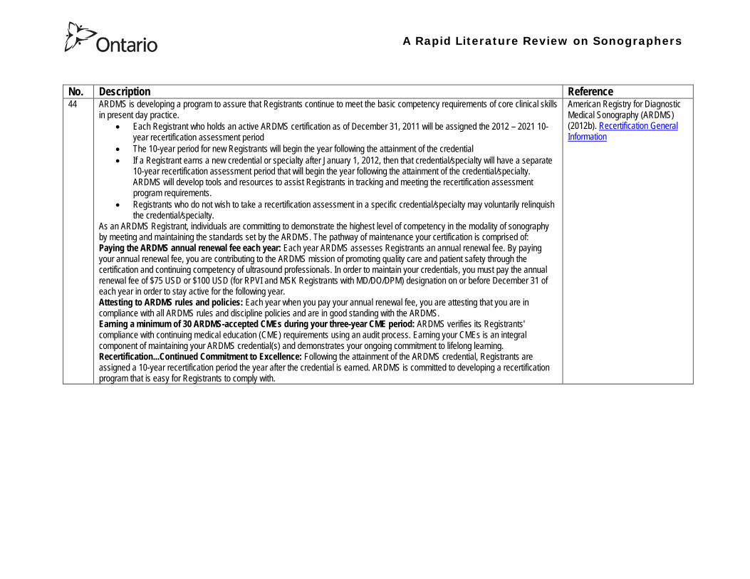
A Rapid Literature Review on Sonographers
No Description Reference 44 ARDMS is developing a program to assure that Registrants continue to meet the basic competency requirements of core clinical skills
in present day practice bull Each Registrant who holds an active ARDMS certification as of December 31 2011 will be assigned the 2012 ndash 2021 10-
year recertification assessment period bull The 10-year period for new Registrants will begin the year following the attainment of the credential bull If a Registrant earns a new credential or specialty after January 1 2012 then that credentialspecialty will have a separate
10-year recertification assessment period that will begin the year following the attainment of the credentialspecialty ARDMS will develop tools and resources to assist Registrants in tracking and meeting the recertification assessment program requirements
bull Registrants who do not wish to take a recertification assessment in a specific credentialspecialty may voluntarily relinquish the credentialspecialty
As an ARDMS Registrant individuals are committing to demonstrate the highest level of competency in the modality of sonography by meeting and maintaining the standards set by the ARDMS The pathway of maintenance your certification is comprised of Paying the ARDMS annual renewal fee each year Each year ARDMS assesses Registrants an annual renewal fee By paying your annual renewal fee you are contributing to the ARDMS mission of promoting quality care and patient safety through the certification and continuing competency of ultrasound professionals In order to maintain your credentials you must pay the annual renewal fee of $75 USD or $100 USD (for RPVI and MSK Registrants with MDDODPM) designation on or before December 31 of each year in order to stay active for the following year Attesting to ARDMS rules and policies Each year when you pay your annual renewal fee you are attesting that you are in compliance with all ARDMS rules and discipline policies and are in good standing with the ARDMS Earning a minimum of 30 ARDMS-accepted CMEs during your three-year CME period ARDMS verifies its Registrants compliance with continuing medical education (CME) requirements using an audit process Earning your CMEs is an integral component of maintaining your ARDMS credential(s) and demonstrates your ongoing commitment to lifelong learning RecertificationContinued Commitment to Excellence Following the attainment of the ARDMS credential Registrants are assigned a 10-year recertification period the year after the credential is earned ARDMS is committed to developing a recertification program that is easy for Registrants to comply with
American Registry for Diagnostic Medical Sonography (ARDMS) (2012b) Recertification General Information
A Rapid Literature Review on Sonographers
No Description Reference 45 This document reviews Sonography Principles and Instrumentation (SPI) Examination Requirements offered through ARDMS in
order to achieve sonography certification and the various prerequisites needed before taking the exam For example one prerequisite entails (1) Education Graduate of a program accredited by an agency recognized by the Council for Higher Education Accreditation (CHEA) United States Department of Education (USDOE) or Canadian Medical Association (CMA) that specifically conducts programmatic accreditation for diagnostic medical sonographydiagnostic cardiac sonographyvascular technology Currently the only organizations that offer programmatic accreditation under the aforementioned associations are the Commission on Accreditation of Allied Health Education Programs (CAAHEP) and the Canadian Medical Association (CMA) (2) Required Clinical UltrasoundVascular Experience No additional experience is required (3) Documentation Required with Application 1) Copy of diploma from ultrasoundvascular program or an official transcript indicating the date the degree was conferred 2) Original letter signed by program director andor medical director indicating date of graduation or successful completion of the program4 Program directors must use the mandatory formatted sample letter available on ARDMSorg sampleletters 3) The CV form is not required if the application is submitted and received in the ARDMS office within one year after successful completion of the program Otherwise an original signed and completed CV form for each appropriate specialty area(s) must be submitted 4) Photocopy of a non-expired government issued photo identification with signature the name on the identification must exactly match the name under which you are applying for ARDMS examination
American Registry for Diagnostic Medical Sonography (ARDMS) (2012c) SPI Requirement and General Prerequisites Accessed April 2013
46 This page notes the five credentials offered by the ARDMS and the corresponding exams and specialty exams required bull RDMS requires the SPI exam (sonography principles and instrumentation) and a specialty exam in abdomen breast fetal
echocardiography neurosonolgoy or obstetricsgynecology bull RDCS requires the SPI exam and a speciality exam in adult echocardiography fetal echocardiography pediatric
echocardiography bull RVT requires the SPI and a speciality exam in vascular technology bull RMSK requires the musculoskeletal sonography exam bull RPVI requires the physiciansrsquo vascular interpretation exam
American Registry for Diagnostic Medical Sonography (ARDMS) (2012d) Credentials and Examinations Accessed April 2013
A Rapid Literature Review on Sonographers
No Description Reference 47 This article summarizes some of the many trends in fetal pediatric and adult echocardiography that SDMS members are excited
about Fetal Echocardiography Sonography has been used since the mid-1980s to help identify congenital heart defects during the second trimester The ldquobasicrdquo examination of the fetal heart involves a four-chamber view ndash evaluation of the right and left atrial and ventricular chambers and their respective valves In the past several years there has been a movement in the obstetric ultrasound community to encourage obstetric sonographers to perform the ldquoextended basicrdquo exam which includes the cardiac outflow tracts The outflow tracts consist of the main pulmonary artery exiting the right ventricle and the aorta exiting the left ventricle The fetal echocardiography community also is abuzz over three-dimensional (3D) and four-dimensional (4D) ultrasound and the potential of this technology to significantly improve the evaluation of the fetal heart Spatiotemporal Image Correlation (STIC) a 4D application allows for evaluation and manipulation of the heart in real-time Allen says Todayrsquos ultrasound equipment allows sonographers to gather a volume of data from which they can create detailed 3D images of the tiny fetal heart which is constantly in motion With computer manipulation of the data sets the images also can be viewed in 4D allowing physicians to see the entire fetal heart cycle from start to finish Pediatric Echocardiography One of the most significant advances in pediatric echo over the last decade is moving to a digital format The advent of pediatric transesophageal (TEE) probes also has been a big advance for pediatric echo Transesophageal probes provide an unimpeded sonographic visualization of the intracardiac structures Theyrsquore useful in visualizing very fine valvular abnormalities the thoracic aorta vegetations and thrombus in the atrial appendage It is also useful in detecting subtle anatomic variations seen in congenital heart disease Pediatric-sized TEE probes make the exams possible In the past it wasnrsquot possible because the adult probe was just too big to put down the babyrsquos or childrsquos esophagus Now the probe is small enough to successfully manipulate it in children and get superb images Because the new sonographic imaging equipment and better surgical techniques allow clinicians to detect and repair more cardiac abnormalities earlier patients are living longer Adult Echocardiography The adult echo world is abuzz about 2D and 3D speckle tracking relatively new techniques for the assessment of heart function and strain imaging Speckle tracking evaluates both regional and global function by measuring the deformation of the heart muscle
Orenstein B W (2010) Current Trends in Echocardiography SDMS New Wave February 1-5 Accessed April 2013
48 This document describes the requirements for admission to the Register of Accredited Medical Sonographer in Australia Requirements include Continuation of Accreditation In order to remain on the Register an accredited Sonographer must satisfy the ASAR continuing professional development (CPD) requirements and pay the prescribed annual fee Student Sonographers apart from paying the prescribed annual fee should provide evidence of their student status every year when they renew their ASAR accreditation
Australian Sonographer Accreditation Registry (ASAR) (no date A) Sonographer Accreditation Accessed April 2013
A Rapid Literature Review on Sonographers
No Description Reference 49 ASARrsquos functions
1 Accredit sonography education programs within Australia 2 Maintain a register of Accredited Sonographers and Accredited Student Sonographers 3 Establish minimum standards of Continuing Professional Development (CPD) and to monitor the CPD requirements for
Sonographers to remain on the register 4 To support activities that advance the profession of sonography 5 To provide registration information about Sonographers to Medicare Australia 6 To provide public access to the name registration number State or Territory current type of accreditation and area of
specialty for active Sonographers ASARrsquos Role ASAR is not a registration board ASAR does not have the authority to remove a Sonographer from the Registry due to professional misconduct nor the ability to prevent a Sonographer from practicing ASAR is only able to remove a Sonographer from the Registry if they
bull fail to meet CPD requirements and or bull fail to pay annual fees
ASAR (no date B) About ASAR Accessed April 2013
50 Regulatory bodies A Regulatory Body maintains a register of individuals who have achieved the required qualification of that profession to practice and can evidence continuing competence at the required level For a doctor to practice in the UK he or she must be registered with the General Medical Council (GMC) This is the regulatory body for medical practitioners The majority of sonographers who are employed in the UK come from a background of radiography or midwifery To practice radiography in the UK a qualified radiographer must be registered with the Health Professions Council (HPC) Similarly a midwife must be registered with the National Midwifery Council to practice as a midwife Sonographer registration in the UK There are a number of professions allied to medicine that are regulated by the HPC for example radiography physiotherapy However sonography is not recognised as a profession by the HPC and therefore there is no mechanism whereby an individual can register with the HPC as a sonographer or be regulated by them as a sonographer It is a common misunderstanding that sonographers have to be registered with the HPC to practice This comes from the fact that most sonographers are also qualified radiographers and maintain their registration with the HPC because they wish to remain on the HPC register as a radiographer Professional bodies An individual can be a member of a Professional Body such as the Royal College of Obstetricians amp Gynecologists (RCOG) Royal College of Radiologists (RCR) Society amp College of Radiographers (SCoR) or the Royal College of Midwives (RCM) but this is not the same as being accepted onto the register of one of the regulatory bodies The professional bodies are responsible for setting and maintaining the standards of their members providing a code of conduct etc They may also act in the capacity of a trades union and can also provide professional indemnity insurance The British Medical Ultrasound Society BMUS is not a professional body but is a scientific society so will offer a different range of benefits Whether or not one wishes to join the society has no impact on their ability to work as a sonographer in the UK although from a continuing professional development point of view it can be beneficial The College of Radiographers are currently working with the HPC to get sonography recognized as a separate profession
British Medical Ultrasound Society (BMUS) (no date) Information on Sonographer Regulation in the UK Accessed April 2013
A Rapid Literature Review on Sonographers
No Description Reference 51 This document included sections on the role of the sonographer and the use of sonography for entertainment Role of the
Sonographer and Technical Impressions The role of the sonographer is to perform the ultrasound scan and to document observations The sonographerrsquos written technical impressions are intended as a form of communication between the sonographer and the reporting physician These observations must be reviewed and subsequently reported by the reporting physician Issuing of a final reportdiagnosis represents the practice of medicine and is therefore the domain of the reporting physician Use of Ultrasound for Entertainment or Non-Diagnostic Purposes In contrast to this justified form of assessment ultrasound for entertainment is simply an elaborate version of a portrait For a variable fee the surface of the fetus is skimmed over and clients are provided with keepsake images or a DVD of their fetus There are no measurements taken during this exam no morphological assessment performed and no dictated diagnostic report of findings Essentially there are no diagnostic benefits to the exam and uninformed parents may be falsely reassured that the beautiful portrait indicates a normal fetus Entertainment ultrasound facilities may operate outside of medical guidelines and without any controls which may result in a lack of technical safeguards operator expertise or governance of technical competency These facilities do not comply with the ALARA principle which could result in increased fetal exposures The Canadian Society of Diagnostic Medical Sonograph (CSDMS) does not support persons or facilities that participate in ultrasound for entertainment activities Liability insurance purchased through the CSDMS group insurance plan does not cover any activities carried out for entertainment ultrasound purposes Other leaders in Canadian medicine support this statement In 2005 the SOGC (Society of Obstetrics and Gynaecologists of Canada) published an article entitled ldquoObstetrical Ultrasound Biological Effects and Safetyrdquo and their conclusion was that ldquothe theoretical risk of adverse bio-effects even from standard 2D obstetrical ultrasound makes it hard to justify its use for non-medical reasons such as sex-determination making non-medical photos or video or for commercial purposerdquo The SOGC recommends that ultrasound be used prudently and that energy exposure be limited to the minimum that is medically necessary The SOGC further recommends a complete ban on the non-medical use of fetal ultrasound and encourages government to join with society to find appropriate means to deal with this public health issue The CAR (Canadian Association of Radiologists) makes a similar statement ldquoThe CAR strongly opposes the use of diagnostic ultrasound equipment for non-medical purposes and considers the use of medical ultrasound for entertainment to be a misuse of the technology especially if fetal subjects are involvedrdquo
Canadian Society of Diagnostic Medical Sonography (no date) Professional Practice Guidelines and Policy Statements For Canadian Sonography Accessed April 2013
A Rapid Literature Review on Sonographers
No Description Reference 52 In 1993 the Consortium for the Accreditation of Sonographic Education (CASE) was formed through the common desire to ensure
that the education and training of sonographers in the United Kingdom was delivered at the highest level All members share common concerns that standards of service provision and education are developed in parallel with increasing demand and improving technology All members strive to create an environment in which education and training is maximised This will make validation and accreditation to be as robust and straightforward as possible CASEs philosophy is to promote best ultrasound practise through the accreditation of those training programmes that develop safe and competent ultrasound practitioners This should be delivered at postgraduate level The majority of its activities relate to supporting those Institutions that offer or wish to offer courses leading to the award of a Masters Postgraduate DiplomaCertificate in Medical or clinical Ultrasound In addition to offering accreditation of new and re-accreditation of established ultrasound education programmes CASE undertakes annual monitoring of the courses it has accredited The information obtained is fed back to the Institutions both on an individual basis and via the annual CASE report Each member organisation is unique and has its own obligations to its membership and its own agenda while CASE brings them together to provide a forum for the interchange of ideas in which shared views may be synthesised and thus inform education and training programmes
Consortium for the Accreditation of Sonographic Education (no date) Home Accessed April 2013
A Rapid Literature Review on Sonographers
No Description Reference 53 The Medical Radiation Technologists Board (the Board) is one of sixteen New Zealand health registration authorities appointed by
the Minister of Health under the Health Practitioners Competence Assurance Act 2003 (the Act) The Board is responsible for the administration of the Act in regard to the profession of medical radiation technology The primary responsibility of the Board is to protect the health and safety of the New Zealand public by ensuring practitioners registered in the profession of medical radiation technology are competent and fit to practice In accordance with Section 118 of the Act the Board is responsible for fulfilling a number of functions
bull 01 To prescribe the qualifications required for scopes of practice within the profession of medical radiation technology and for that purpose to accredit and monitor educational institutions and degrees courses of studies or programmes
bull 02 To authorise the registration of medical radiation technologists under the Act and to maintain registers bull 03 To consider applications for annual practising certificates bull 04 To review and promote the competence of medical radiation technologists bull 05 To recognise accredit and set programmes to ensure the ongoing competence of medical radiation technologists bull 06 To receive and act on information from health practitioners employers and the Health and Disability Commissioner
about the competence of medical radiation technologists bull 07 To notify employers the Accident Compensation Corporation the Director-General of Health and the Health and
Disability Commissioner that the practice of a medical radiation technologist may pose a risk of harm to the public bull 08 To consider the cases of medical radiation technologists who may be unable to perform the functions required for the
practice of the medical radiation technology profession bull 09 To set standards of clinical competence cultural competence and ethical conduct to be observed by medical radiation
technologists bull 10 To liaise with other authorities appointed under the Act about matters of common interest bull 11 To promote education and training in the profession of medical radiation technology bull 12 To promote public awareness of the responsibilities of the Board bull 13 To exercise and perform any other functions powers and duties that are conferred or imposed on it by or under the Act
or any other enactment
Medical Radiation Technologists Board (MRTB) (no date A) What we do Accessed April 2013
54 Qualifications To be considered for registration as a medical radiation technologist in any of the specified scopes of practice an individual must hold a qualification for that particular scope as prescribed by the Board or approved by the Board as being substantially equivalent to the prescribed qualifications In addition to having an approved qualification individuals may also be required to demonstrate that they have adequate experience in the relevant scope of practice Sonographer The Board has prescribed one New Zealand postgraduate qualification for a person to be eligible for registration in the scope of practice of Sonographer Postgraduate Diploma in Health Science (Ultrasound) issued by the Unitec Institute of Technology Required Experience If an individual is applying for registration in Nuclear Medicine Ultrasound or Magnetic Resonance Imaging in addition to holding a relevant postgraduate qualification they must also be able to provide evidence that you have a minimum of 3360 hours of clinical experience in the relevant scope of practice you are applying for registration in
Medical Radiation Technologists Board (MRTB) (no date B) Qualifications Accessed April 2013
A Rapid Literature Review on Sonographers
No Description Reference 55 Recertification is a mechanism used by the Medical Radiation Technologists Board in New Zealand to ensure that registered medical
radiation technologists continue to be competent to practise within their scopes of practice Annual Practising Certificates Each year if an individual wishes to continue practising in your scope(s) of practice in New Zealand they must apply for and be issued with an annual practising certificate (APC) A condition of being issued with an APC is that one must provide evidence of enrolment and satisfactory progress in a Board-approved recertification programme Alternatively the Board may issue them with an interim practising certificate (IPC) until it is satisfied the individual demonstrates that they are competent to practice in the scope(s) of practice they applied for Working with an IPC will require an individual to work under the supervision of a registered medical radiation technologist or other relevant registered health practitioner Recertification Programmes The Boards recertification programme framework is based on a continuing professional development (CPD) model Individuals have the option of enrolling in one of seven recertification programmes for medical radiation technologists that are currently approved by the Board They must meet the requirements of the particular recertification programme in which they are enrolled Recertification Audits An essential component of the CPD-based recertification programme adopted by the Board is the audit of CPD recordsThe Board undertakes an annual audit of approximately 10 of its registrant database to assess individual medical radiation technologistsrsquo compliance with the Boarrsquods recertification requirements
Medical Radiation Technologists Board (MRTB) (no date C) Recertification Accessed April 2013
56 There are two types of radiography - diagnostic and therapeutic Both need considerable knowledge of technology anatomy physiology and pathology to carry out their work Many also undertake further training to become a sonographer Some of the imaging technology that a diagnostic radiographer may use are
bull X-Ray - looks through tissues to examine bones cavities and foreign objects bull Fluoroscopy - images the digestive system providing a real-time image bull CT (Computed Tomography) - which provides cross-sectional views (slices) of the body bull MRI (Magnetic Resonance Imaging) - builds a 2-D or 3-D map of the different tissue types within the body bull Ultrasound - well known for its use in obstetrics and gynaecology Also used to check circulation and examine the heart bull Angiography - used to investigate blood vessels
Sonographyultrasonography Ultrasound is used in various settings in a hospital including abdominal scanning and breast ultrasound Ultrasound imaging is the use of high frequency sound in excess of human hearing to produce images of structures of the human body that may be observed on a screen These images may subsequently be transferred to photographic film or paper or onto video or CD forming part of the patients record of their examination There are no direct entry routes into ultrasound Most sonographers train as a radiographer then undertake an approved post-registration course Offered by higher education institutions The courses are a minimum of one academic year and prepare sonographers clinically and academically for practice Normally a pre-requisite for acceptance is access to a clinical department with supervised practice for students
National Health Service (NHS) (no date A) Radiographer Accessed April 2013
57 To practice as a radiographer in the UK an individual must be registered with the Health and Care Professions Council (HCPC) To register individuals must have successfully completed an approved training programme in diagnostic or therapeutic radiography These are offered at undergraduate degree (full-time and part-time) and at postgraduate degreediploma level at a number of universities across the UK
NHS (no date B) Entry requirements and training in radiography Accessed April 2013
Health Professions Regulatory Advisory Council 56 Wellesley Street West 12th Floor
Toronto ON M5S 2S3
Telephone 416-326-1550 Toll-Free 1-888-377-7746
Fax 416-326-1549
Website wwwhpracorg
Email hpracwebmasterontarioca
A Rapid Literature Review on Sonographers
No Description Reference 47 This article summarizes some of the many trends in fetal pediatric and adult echocardiography that SDMS members are excited
about Fetal Echocardiography Sonography has been used since the mid-1980s to help identify congenital heart defects during the second trimester The ldquobasicrdquo examination of the fetal heart involves a four-chamber view ndash evaluation of the right and left atrial and ventricular chambers and their respective valves In the past several years there has been a movement in the obstetric ultrasound community to encourage obstetric sonographers to perform the ldquoextended basicrdquo exam which includes the cardiac outflow tracts The outflow tracts consist of the main pulmonary artery exiting the right ventricle and the aorta exiting the left ventricle The fetal echocardiography community also is abuzz over three-dimensional (3D) and four-dimensional (4D) ultrasound and the potential of this technology to significantly improve the evaluation of the fetal heart Spatiotemporal Image Correlation (STIC) a 4D application allows for evaluation and manipulation of the heart in real-time Allen says Todayrsquos ultrasound equipment allows sonographers to gather a volume of data from which they can create detailed 3D images of the tiny fetal heart which is constantly in motion With computer manipulation of the data sets the images also can be viewed in 4D allowing physicians to see the entire fetal heart cycle from start to finish Pediatric Echocardiography One of the most significant advances in pediatric echo over the last decade is moving to a digital format The advent of pediatric transesophageal (TEE) probes also has been a big advance for pediatric echo Transesophageal probes provide an unimpeded sonographic visualization of the intracardiac structures Theyrsquore useful in visualizing very fine valvular abnormalities the thoracic aorta vegetations and thrombus in the atrial appendage It is also useful in detecting subtle anatomic variations seen in congenital heart disease Pediatric-sized TEE probes make the exams possible In the past it wasnrsquot possible because the adult probe was just too big to put down the babyrsquos or childrsquos esophagus Now the probe is small enough to successfully manipulate it in children and get superb images Because the new sonographic imaging equipment and better surgical techniques allow clinicians to detect and repair more cardiac abnormalities earlier patients are living longer Adult Echocardiography The adult echo world is abuzz about 2D and 3D speckle tracking relatively new techniques for the assessment of heart function and strain imaging Speckle tracking evaluates both regional and global function by measuring the deformation of the heart muscle
Orenstein B W (2010) Current Trends in Echocardiography SDMS New Wave February 1-5 Accessed April 2013
48 This document describes the requirements for admission to the Register of Accredited Medical Sonographer in Australia Requirements include Continuation of Accreditation In order to remain on the Register an accredited Sonographer must satisfy the ASAR continuing professional development (CPD) requirements and pay the prescribed annual fee Student Sonographers apart from paying the prescribed annual fee should provide evidence of their student status every year when they renew their ASAR accreditation
Australian Sonographer Accreditation Registry (ASAR) (no date A) Sonographer Accreditation Accessed April 2013
A Rapid Literature Review on Sonographers
No Description Reference 49 ASARrsquos functions
1 Accredit sonography education programs within Australia 2 Maintain a register of Accredited Sonographers and Accredited Student Sonographers 3 Establish minimum standards of Continuing Professional Development (CPD) and to monitor the CPD requirements for
Sonographers to remain on the register 4 To support activities that advance the profession of sonography 5 To provide registration information about Sonographers to Medicare Australia 6 To provide public access to the name registration number State or Territory current type of accreditation and area of
specialty for active Sonographers ASARrsquos Role ASAR is not a registration board ASAR does not have the authority to remove a Sonographer from the Registry due to professional misconduct nor the ability to prevent a Sonographer from practicing ASAR is only able to remove a Sonographer from the Registry if they
bull fail to meet CPD requirements and or bull fail to pay annual fees
ASAR (no date B) About ASAR Accessed April 2013
50 Regulatory bodies A Regulatory Body maintains a register of individuals who have achieved the required qualification of that profession to practice and can evidence continuing competence at the required level For a doctor to practice in the UK he or she must be registered with the General Medical Council (GMC) This is the regulatory body for medical practitioners The majority of sonographers who are employed in the UK come from a background of radiography or midwifery To practice radiography in the UK a qualified radiographer must be registered with the Health Professions Council (HPC) Similarly a midwife must be registered with the National Midwifery Council to practice as a midwife Sonographer registration in the UK There are a number of professions allied to medicine that are regulated by the HPC for example radiography physiotherapy However sonography is not recognised as a profession by the HPC and therefore there is no mechanism whereby an individual can register with the HPC as a sonographer or be regulated by them as a sonographer It is a common misunderstanding that sonographers have to be registered with the HPC to practice This comes from the fact that most sonographers are also qualified radiographers and maintain their registration with the HPC because they wish to remain on the HPC register as a radiographer Professional bodies An individual can be a member of a Professional Body such as the Royal College of Obstetricians amp Gynecologists (RCOG) Royal College of Radiologists (RCR) Society amp College of Radiographers (SCoR) or the Royal College of Midwives (RCM) but this is not the same as being accepted onto the register of one of the regulatory bodies The professional bodies are responsible for setting and maintaining the standards of their members providing a code of conduct etc They may also act in the capacity of a trades union and can also provide professional indemnity insurance The British Medical Ultrasound Society BMUS is not a professional body but is a scientific society so will offer a different range of benefits Whether or not one wishes to join the society has no impact on their ability to work as a sonographer in the UK although from a continuing professional development point of view it can be beneficial The College of Radiographers are currently working with the HPC to get sonography recognized as a separate profession
British Medical Ultrasound Society (BMUS) (no date) Information on Sonographer Regulation in the UK Accessed April 2013
A Rapid Literature Review on Sonographers
No Description Reference 51 This document included sections on the role of the sonographer and the use of sonography for entertainment Role of the
Sonographer and Technical Impressions The role of the sonographer is to perform the ultrasound scan and to document observations The sonographerrsquos written technical impressions are intended as a form of communication between the sonographer and the reporting physician These observations must be reviewed and subsequently reported by the reporting physician Issuing of a final reportdiagnosis represents the practice of medicine and is therefore the domain of the reporting physician Use of Ultrasound for Entertainment or Non-Diagnostic Purposes In contrast to this justified form of assessment ultrasound for entertainment is simply an elaborate version of a portrait For a variable fee the surface of the fetus is skimmed over and clients are provided with keepsake images or a DVD of their fetus There are no measurements taken during this exam no morphological assessment performed and no dictated diagnostic report of findings Essentially there are no diagnostic benefits to the exam and uninformed parents may be falsely reassured that the beautiful portrait indicates a normal fetus Entertainment ultrasound facilities may operate outside of medical guidelines and without any controls which may result in a lack of technical safeguards operator expertise or governance of technical competency These facilities do not comply with the ALARA principle which could result in increased fetal exposures The Canadian Society of Diagnostic Medical Sonograph (CSDMS) does not support persons or facilities that participate in ultrasound for entertainment activities Liability insurance purchased through the CSDMS group insurance plan does not cover any activities carried out for entertainment ultrasound purposes Other leaders in Canadian medicine support this statement In 2005 the SOGC (Society of Obstetrics and Gynaecologists of Canada) published an article entitled ldquoObstetrical Ultrasound Biological Effects and Safetyrdquo and their conclusion was that ldquothe theoretical risk of adverse bio-effects even from standard 2D obstetrical ultrasound makes it hard to justify its use for non-medical reasons such as sex-determination making non-medical photos or video or for commercial purposerdquo The SOGC recommends that ultrasound be used prudently and that energy exposure be limited to the minimum that is medically necessary The SOGC further recommends a complete ban on the non-medical use of fetal ultrasound and encourages government to join with society to find appropriate means to deal with this public health issue The CAR (Canadian Association of Radiologists) makes a similar statement ldquoThe CAR strongly opposes the use of diagnostic ultrasound equipment for non-medical purposes and considers the use of medical ultrasound for entertainment to be a misuse of the technology especially if fetal subjects are involvedrdquo
Canadian Society of Diagnostic Medical Sonography (no date) Professional Practice Guidelines and Policy Statements For Canadian Sonography Accessed April 2013
A Rapid Literature Review on Sonographers
No Description Reference 52 In 1993 the Consortium for the Accreditation of Sonographic Education (CASE) was formed through the common desire to ensure
that the education and training of sonographers in the United Kingdom was delivered at the highest level All members share common concerns that standards of service provision and education are developed in parallel with increasing demand and improving technology All members strive to create an environment in which education and training is maximised This will make validation and accreditation to be as robust and straightforward as possible CASEs philosophy is to promote best ultrasound practise through the accreditation of those training programmes that develop safe and competent ultrasound practitioners This should be delivered at postgraduate level The majority of its activities relate to supporting those Institutions that offer or wish to offer courses leading to the award of a Masters Postgraduate DiplomaCertificate in Medical or clinical Ultrasound In addition to offering accreditation of new and re-accreditation of established ultrasound education programmes CASE undertakes annual monitoring of the courses it has accredited The information obtained is fed back to the Institutions both on an individual basis and via the annual CASE report Each member organisation is unique and has its own obligations to its membership and its own agenda while CASE brings them together to provide a forum for the interchange of ideas in which shared views may be synthesised and thus inform education and training programmes
Consortium for the Accreditation of Sonographic Education (no date) Home Accessed April 2013
A Rapid Literature Review on Sonographers
No Description Reference 53 The Medical Radiation Technologists Board (the Board) is one of sixteen New Zealand health registration authorities appointed by
the Minister of Health under the Health Practitioners Competence Assurance Act 2003 (the Act) The Board is responsible for the administration of the Act in regard to the profession of medical radiation technology The primary responsibility of the Board is to protect the health and safety of the New Zealand public by ensuring practitioners registered in the profession of medical radiation technology are competent and fit to practice In accordance with Section 118 of the Act the Board is responsible for fulfilling a number of functions
bull 01 To prescribe the qualifications required for scopes of practice within the profession of medical radiation technology and for that purpose to accredit and monitor educational institutions and degrees courses of studies or programmes
bull 02 To authorise the registration of medical radiation technologists under the Act and to maintain registers bull 03 To consider applications for annual practising certificates bull 04 To review and promote the competence of medical radiation technologists bull 05 To recognise accredit and set programmes to ensure the ongoing competence of medical radiation technologists bull 06 To receive and act on information from health practitioners employers and the Health and Disability Commissioner
about the competence of medical radiation technologists bull 07 To notify employers the Accident Compensation Corporation the Director-General of Health and the Health and
Disability Commissioner that the practice of a medical radiation technologist may pose a risk of harm to the public bull 08 To consider the cases of medical radiation technologists who may be unable to perform the functions required for the
practice of the medical radiation technology profession bull 09 To set standards of clinical competence cultural competence and ethical conduct to be observed by medical radiation
technologists bull 10 To liaise with other authorities appointed under the Act about matters of common interest bull 11 To promote education and training in the profession of medical radiation technology bull 12 To promote public awareness of the responsibilities of the Board bull 13 To exercise and perform any other functions powers and duties that are conferred or imposed on it by or under the Act
or any other enactment
Medical Radiation Technologists Board (MRTB) (no date A) What we do Accessed April 2013
54 Qualifications To be considered for registration as a medical radiation technologist in any of the specified scopes of practice an individual must hold a qualification for that particular scope as prescribed by the Board or approved by the Board as being substantially equivalent to the prescribed qualifications In addition to having an approved qualification individuals may also be required to demonstrate that they have adequate experience in the relevant scope of practice Sonographer The Board has prescribed one New Zealand postgraduate qualification for a person to be eligible for registration in the scope of practice of Sonographer Postgraduate Diploma in Health Science (Ultrasound) issued by the Unitec Institute of Technology Required Experience If an individual is applying for registration in Nuclear Medicine Ultrasound or Magnetic Resonance Imaging in addition to holding a relevant postgraduate qualification they must also be able to provide evidence that you have a minimum of 3360 hours of clinical experience in the relevant scope of practice you are applying for registration in
Medical Radiation Technologists Board (MRTB) (no date B) Qualifications Accessed April 2013
A Rapid Literature Review on Sonographers
No Description Reference 55 Recertification is a mechanism used by the Medical Radiation Technologists Board in New Zealand to ensure that registered medical
radiation technologists continue to be competent to practise within their scopes of practice Annual Practising Certificates Each year if an individual wishes to continue practising in your scope(s) of practice in New Zealand they must apply for and be issued with an annual practising certificate (APC) A condition of being issued with an APC is that one must provide evidence of enrolment and satisfactory progress in a Board-approved recertification programme Alternatively the Board may issue them with an interim practising certificate (IPC) until it is satisfied the individual demonstrates that they are competent to practice in the scope(s) of practice they applied for Working with an IPC will require an individual to work under the supervision of a registered medical radiation technologist or other relevant registered health practitioner Recertification Programmes The Boards recertification programme framework is based on a continuing professional development (CPD) model Individuals have the option of enrolling in one of seven recertification programmes for medical radiation technologists that are currently approved by the Board They must meet the requirements of the particular recertification programme in which they are enrolled Recertification Audits An essential component of the CPD-based recertification programme adopted by the Board is the audit of CPD recordsThe Board undertakes an annual audit of approximately 10 of its registrant database to assess individual medical radiation technologistsrsquo compliance with the Boarrsquods recertification requirements
Medical Radiation Technologists Board (MRTB) (no date C) Recertification Accessed April 2013
56 There are two types of radiography - diagnostic and therapeutic Both need considerable knowledge of technology anatomy physiology and pathology to carry out their work Many also undertake further training to become a sonographer Some of the imaging technology that a diagnostic radiographer may use are
bull X-Ray - looks through tissues to examine bones cavities and foreign objects bull Fluoroscopy - images the digestive system providing a real-time image bull CT (Computed Tomography) - which provides cross-sectional views (slices) of the body bull MRI (Magnetic Resonance Imaging) - builds a 2-D or 3-D map of the different tissue types within the body bull Ultrasound - well known for its use in obstetrics and gynaecology Also used to check circulation and examine the heart bull Angiography - used to investigate blood vessels
Sonographyultrasonography Ultrasound is used in various settings in a hospital including abdominal scanning and breast ultrasound Ultrasound imaging is the use of high frequency sound in excess of human hearing to produce images of structures of the human body that may be observed on a screen These images may subsequently be transferred to photographic film or paper or onto video or CD forming part of the patients record of their examination There are no direct entry routes into ultrasound Most sonographers train as a radiographer then undertake an approved post-registration course Offered by higher education institutions The courses are a minimum of one academic year and prepare sonographers clinically and academically for practice Normally a pre-requisite for acceptance is access to a clinical department with supervised practice for students
National Health Service (NHS) (no date A) Radiographer Accessed April 2013
57 To practice as a radiographer in the UK an individual must be registered with the Health and Care Professions Council (HCPC) To register individuals must have successfully completed an approved training programme in diagnostic or therapeutic radiography These are offered at undergraduate degree (full-time and part-time) and at postgraduate degreediploma level at a number of universities across the UK
NHS (no date B) Entry requirements and training in radiography Accessed April 2013
Health Professions Regulatory Advisory Council 56 Wellesley Street West 12th Floor
Toronto ON M5S 2S3
Telephone 416-326-1550 Toll-Free 1-888-377-7746
Fax 416-326-1549
Website wwwhpracorg
Email hpracwebmasterontarioca

A Rapid Literature Review on Sonographers
No Description Reference 49 ASARrsquos functions
1 Accredit sonography education programs within Australia 2 Maintain a register of Accredited Sonographers and Accredited Student Sonographers 3 Establish minimum standards of Continuing Professional Development (CPD) and to monitor the CPD requirements for
Sonographers to remain on the register 4 To support activities that advance the profession of sonography 5 To provide registration information about Sonographers to Medicare Australia 6 To provide public access to the name registration number State or Territory current type of accreditation and area of
specialty for active Sonographers ASARrsquos Role ASAR is not a registration board ASAR does not have the authority to remove a Sonographer from the Registry due to professional misconduct nor the ability to prevent a Sonographer from practicing ASAR is only able to remove a Sonographer from the Registry if they
bull fail to meet CPD requirements and or bull fail to pay annual fees
ASAR (no date B) About ASAR Accessed April 2013
50 Regulatory bodies A Regulatory Body maintains a register of individuals who have achieved the required qualification of that profession to practice and can evidence continuing competence at the required level For a doctor to practice in the UK he or she must be registered with the General Medical Council (GMC) This is the regulatory body for medical practitioners The majority of sonographers who are employed in the UK come from a background of radiography or midwifery To practice radiography in the UK a qualified radiographer must be registered with the Health Professions Council (HPC) Similarly a midwife must be registered with the National Midwifery Council to practice as a midwife Sonographer registration in the UK There are a number of professions allied to medicine that are regulated by the HPC for example radiography physiotherapy However sonography is not recognised as a profession by the HPC and therefore there is no mechanism whereby an individual can register with the HPC as a sonographer or be regulated by them as a sonographer It is a common misunderstanding that sonographers have to be registered with the HPC to practice This comes from the fact that most sonographers are also qualified radiographers and maintain their registration with the HPC because they wish to remain on the HPC register as a radiographer Professional bodies An individual can be a member of a Professional Body such as the Royal College of Obstetricians amp Gynecologists (RCOG) Royal College of Radiologists (RCR) Society amp College of Radiographers (SCoR) or the Royal College of Midwives (RCM) but this is not the same as being accepted onto the register of one of the regulatory bodies The professional bodies are responsible for setting and maintaining the standards of their members providing a code of conduct etc They may also act in the capacity of a trades union and can also provide professional indemnity insurance The British Medical Ultrasound Society BMUS is not a professional body but is a scientific society so will offer a different range of benefits Whether or not one wishes to join the society has no impact on their ability to work as a sonographer in the UK although from a continuing professional development point of view it can be beneficial The College of Radiographers are currently working with the HPC to get sonography recognized as a separate profession
British Medical Ultrasound Society (BMUS) (no date) Information on Sonographer Regulation in the UK Accessed April 2013
A Rapid Literature Review on Sonographers
No Description Reference 51 This document included sections on the role of the sonographer and the use of sonography for entertainment Role of the
Sonographer and Technical Impressions The role of the sonographer is to perform the ultrasound scan and to document observations The sonographerrsquos written technical impressions are intended as a form of communication between the sonographer and the reporting physician These observations must be reviewed and subsequently reported by the reporting physician Issuing of a final reportdiagnosis represents the practice of medicine and is therefore the domain of the reporting physician Use of Ultrasound for Entertainment or Non-Diagnostic Purposes In contrast to this justified form of assessment ultrasound for entertainment is simply an elaborate version of a portrait For a variable fee the surface of the fetus is skimmed over and clients are provided with keepsake images or a DVD of their fetus There are no measurements taken during this exam no morphological assessment performed and no dictated diagnostic report of findings Essentially there are no diagnostic benefits to the exam and uninformed parents may be falsely reassured that the beautiful portrait indicates a normal fetus Entertainment ultrasound facilities may operate outside of medical guidelines and without any controls which may result in a lack of technical safeguards operator expertise or governance of technical competency These facilities do not comply with the ALARA principle which could result in increased fetal exposures The Canadian Society of Diagnostic Medical Sonograph (CSDMS) does not support persons or facilities that participate in ultrasound for entertainment activities Liability insurance purchased through the CSDMS group insurance plan does not cover any activities carried out for entertainment ultrasound purposes Other leaders in Canadian medicine support this statement In 2005 the SOGC (Society of Obstetrics and Gynaecologists of Canada) published an article entitled ldquoObstetrical Ultrasound Biological Effects and Safetyrdquo and their conclusion was that ldquothe theoretical risk of adverse bio-effects even from standard 2D obstetrical ultrasound makes it hard to justify its use for non-medical reasons such as sex-determination making non-medical photos or video or for commercial purposerdquo The SOGC recommends that ultrasound be used prudently and that energy exposure be limited to the minimum that is medically necessary The SOGC further recommends a complete ban on the non-medical use of fetal ultrasound and encourages government to join with society to find appropriate means to deal with this public health issue The CAR (Canadian Association of Radiologists) makes a similar statement ldquoThe CAR strongly opposes the use of diagnostic ultrasound equipment for non-medical purposes and considers the use of medical ultrasound for entertainment to be a misuse of the technology especially if fetal subjects are involvedrdquo
Canadian Society of Diagnostic Medical Sonography (no date) Professional Practice Guidelines and Policy Statements For Canadian Sonography Accessed April 2013
A Rapid Literature Review on Sonographers
No Description Reference 52 In 1993 the Consortium for the Accreditation of Sonographic Education (CASE) was formed through the common desire to ensure
that the education and training of sonographers in the United Kingdom was delivered at the highest level All members share common concerns that standards of service provision and education are developed in parallel with increasing demand and improving technology All members strive to create an environment in which education and training is maximised This will make validation and accreditation to be as robust and straightforward as possible CASEs philosophy is to promote best ultrasound practise through the accreditation of those training programmes that develop safe and competent ultrasound practitioners This should be delivered at postgraduate level The majority of its activities relate to supporting those Institutions that offer or wish to offer courses leading to the award of a Masters Postgraduate DiplomaCertificate in Medical or clinical Ultrasound In addition to offering accreditation of new and re-accreditation of established ultrasound education programmes CASE undertakes annual monitoring of the courses it has accredited The information obtained is fed back to the Institutions both on an individual basis and via the annual CASE report Each member organisation is unique and has its own obligations to its membership and its own agenda while CASE brings them together to provide a forum for the interchange of ideas in which shared views may be synthesised and thus inform education and training programmes
Consortium for the Accreditation of Sonographic Education (no date) Home Accessed April 2013
A Rapid Literature Review on Sonographers
No Description Reference 53 The Medical Radiation Technologists Board (the Board) is one of sixteen New Zealand health registration authorities appointed by
the Minister of Health under the Health Practitioners Competence Assurance Act 2003 (the Act) The Board is responsible for the administration of the Act in regard to the profession of medical radiation technology The primary responsibility of the Board is to protect the health and safety of the New Zealand public by ensuring practitioners registered in the profession of medical radiation technology are competent and fit to practice In accordance with Section 118 of the Act the Board is responsible for fulfilling a number of functions
bull 01 To prescribe the qualifications required for scopes of practice within the profession of medical radiation technology and for that purpose to accredit and monitor educational institutions and degrees courses of studies or programmes
bull 02 To authorise the registration of medical radiation technologists under the Act and to maintain registers bull 03 To consider applications for annual practising certificates bull 04 To review and promote the competence of medical radiation technologists bull 05 To recognise accredit and set programmes to ensure the ongoing competence of medical radiation technologists bull 06 To receive and act on information from health practitioners employers and the Health and Disability Commissioner
about the competence of medical radiation technologists bull 07 To notify employers the Accident Compensation Corporation the Director-General of Health and the Health and
Disability Commissioner that the practice of a medical radiation technologist may pose a risk of harm to the public bull 08 To consider the cases of medical radiation technologists who may be unable to perform the functions required for the
practice of the medical radiation technology profession bull 09 To set standards of clinical competence cultural competence and ethical conduct to be observed by medical radiation
technologists bull 10 To liaise with other authorities appointed under the Act about matters of common interest bull 11 To promote education and training in the profession of medical radiation technology bull 12 To promote public awareness of the responsibilities of the Board bull 13 To exercise and perform any other functions powers and duties that are conferred or imposed on it by or under the Act
or any other enactment
Medical Radiation Technologists Board (MRTB) (no date A) What we do Accessed April 2013
54 Qualifications To be considered for registration as a medical radiation technologist in any of the specified scopes of practice an individual must hold a qualification for that particular scope as prescribed by the Board or approved by the Board as being substantially equivalent to the prescribed qualifications In addition to having an approved qualification individuals may also be required to demonstrate that they have adequate experience in the relevant scope of practice Sonographer The Board has prescribed one New Zealand postgraduate qualification for a person to be eligible for registration in the scope of practice of Sonographer Postgraduate Diploma in Health Science (Ultrasound) issued by the Unitec Institute of Technology Required Experience If an individual is applying for registration in Nuclear Medicine Ultrasound or Magnetic Resonance Imaging in addition to holding a relevant postgraduate qualification they must also be able to provide evidence that you have a minimum of 3360 hours of clinical experience in the relevant scope of practice you are applying for registration in
Medical Radiation Technologists Board (MRTB) (no date B) Qualifications Accessed April 2013
A Rapid Literature Review on Sonographers
No Description Reference 55 Recertification is a mechanism used by the Medical Radiation Technologists Board in New Zealand to ensure that registered medical
radiation technologists continue to be competent to practise within their scopes of practice Annual Practising Certificates Each year if an individual wishes to continue practising in your scope(s) of practice in New Zealand they must apply for and be issued with an annual practising certificate (APC) A condition of being issued with an APC is that one must provide evidence of enrolment and satisfactory progress in a Board-approved recertification programme Alternatively the Board may issue them with an interim practising certificate (IPC) until it is satisfied the individual demonstrates that they are competent to practice in the scope(s) of practice they applied for Working with an IPC will require an individual to work under the supervision of a registered medical radiation technologist or other relevant registered health practitioner Recertification Programmes The Boards recertification programme framework is based on a continuing professional development (CPD) model Individuals have the option of enrolling in one of seven recertification programmes for medical radiation technologists that are currently approved by the Board They must meet the requirements of the particular recertification programme in which they are enrolled Recertification Audits An essential component of the CPD-based recertification programme adopted by the Board is the audit of CPD recordsThe Board undertakes an annual audit of approximately 10 of its registrant database to assess individual medical radiation technologistsrsquo compliance with the Boarrsquods recertification requirements
Medical Radiation Technologists Board (MRTB) (no date C) Recertification Accessed April 2013
56 There are two types of radiography - diagnostic and therapeutic Both need considerable knowledge of technology anatomy physiology and pathology to carry out their work Many also undertake further training to become a sonographer Some of the imaging technology that a diagnostic radiographer may use are
bull X-Ray - looks through tissues to examine bones cavities and foreign objects bull Fluoroscopy - images the digestive system providing a real-time image bull CT (Computed Tomography) - which provides cross-sectional views (slices) of the body bull MRI (Magnetic Resonance Imaging) - builds a 2-D or 3-D map of the different tissue types within the body bull Ultrasound - well known for its use in obstetrics and gynaecology Also used to check circulation and examine the heart bull Angiography - used to investigate blood vessels
Sonographyultrasonography Ultrasound is used in various settings in a hospital including abdominal scanning and breast ultrasound Ultrasound imaging is the use of high frequency sound in excess of human hearing to produce images of structures of the human body that may be observed on a screen These images may subsequently be transferred to photographic film or paper or onto video or CD forming part of the patients record of their examination There are no direct entry routes into ultrasound Most sonographers train as a radiographer then undertake an approved post-registration course Offered by higher education institutions The courses are a minimum of one academic year and prepare sonographers clinically and academically for practice Normally a pre-requisite for acceptance is access to a clinical department with supervised practice for students
National Health Service (NHS) (no date A) Radiographer Accessed April 2013
57 To practice as a radiographer in the UK an individual must be registered with the Health and Care Professions Council (HCPC) To register individuals must have successfully completed an approved training programme in diagnostic or therapeutic radiography These are offered at undergraduate degree (full-time and part-time) and at postgraduate degreediploma level at a number of universities across the UK
NHS (no date B) Entry requirements and training in radiography Accessed April 2013
Health Professions Regulatory Advisory Council 56 Wellesley Street West 12th Floor
Toronto ON M5S 2S3
Telephone 416-326-1550 Toll-Free 1-888-377-7746
Fax 416-326-1549
Website wwwhpracorg
Email hpracwebmasterontarioca
A Rapid Literature Review on Sonographers
No Description Reference 51 This document included sections on the role of the sonographer and the use of sonography for entertainment Role of the
Sonographer and Technical Impressions The role of the sonographer is to perform the ultrasound scan and to document observations The sonographerrsquos written technical impressions are intended as a form of communication between the sonographer and the reporting physician These observations must be reviewed and subsequently reported by the reporting physician Issuing of a final reportdiagnosis represents the practice of medicine and is therefore the domain of the reporting physician Use of Ultrasound for Entertainment or Non-Diagnostic Purposes In contrast to this justified form of assessment ultrasound for entertainment is simply an elaborate version of a portrait For a variable fee the surface of the fetus is skimmed over and clients are provided with keepsake images or a DVD of their fetus There are no measurements taken during this exam no morphological assessment performed and no dictated diagnostic report of findings Essentially there are no diagnostic benefits to the exam and uninformed parents may be falsely reassured that the beautiful portrait indicates a normal fetus Entertainment ultrasound facilities may operate outside of medical guidelines and without any controls which may result in a lack of technical safeguards operator expertise or governance of technical competency These facilities do not comply with the ALARA principle which could result in increased fetal exposures The Canadian Society of Diagnostic Medical Sonograph (CSDMS) does not support persons or facilities that participate in ultrasound for entertainment activities Liability insurance purchased through the CSDMS group insurance plan does not cover any activities carried out for entertainment ultrasound purposes Other leaders in Canadian medicine support this statement In 2005 the SOGC (Society of Obstetrics and Gynaecologists of Canada) published an article entitled ldquoObstetrical Ultrasound Biological Effects and Safetyrdquo and their conclusion was that ldquothe theoretical risk of adverse bio-effects even from standard 2D obstetrical ultrasound makes it hard to justify its use for non-medical reasons such as sex-determination making non-medical photos or video or for commercial purposerdquo The SOGC recommends that ultrasound be used prudently and that energy exposure be limited to the minimum that is medically necessary The SOGC further recommends a complete ban on the non-medical use of fetal ultrasound and encourages government to join with society to find appropriate means to deal with this public health issue The CAR (Canadian Association of Radiologists) makes a similar statement ldquoThe CAR strongly opposes the use of diagnostic ultrasound equipment for non-medical purposes and considers the use of medical ultrasound for entertainment to be a misuse of the technology especially if fetal subjects are involvedrdquo
Canadian Society of Diagnostic Medical Sonography (no date) Professional Practice Guidelines and Policy Statements For Canadian Sonography Accessed April 2013
A Rapid Literature Review on Sonographers
No Description Reference 52 In 1993 the Consortium for the Accreditation of Sonographic Education (CASE) was formed through the common desire to ensure
that the education and training of sonographers in the United Kingdom was delivered at the highest level All members share common concerns that standards of service provision and education are developed in parallel with increasing demand and improving technology All members strive to create an environment in which education and training is maximised This will make validation and accreditation to be as robust and straightforward as possible CASEs philosophy is to promote best ultrasound practise through the accreditation of those training programmes that develop safe and competent ultrasound practitioners This should be delivered at postgraduate level The majority of its activities relate to supporting those Institutions that offer or wish to offer courses leading to the award of a Masters Postgraduate DiplomaCertificate in Medical or clinical Ultrasound In addition to offering accreditation of new and re-accreditation of established ultrasound education programmes CASE undertakes annual monitoring of the courses it has accredited The information obtained is fed back to the Institutions both on an individual basis and via the annual CASE report Each member organisation is unique and has its own obligations to its membership and its own agenda while CASE brings them together to provide a forum for the interchange of ideas in which shared views may be synthesised and thus inform education and training programmes
Consortium for the Accreditation of Sonographic Education (no date) Home Accessed April 2013
A Rapid Literature Review on Sonographers
No Description Reference 53 The Medical Radiation Technologists Board (the Board) is one of sixteen New Zealand health registration authorities appointed by
the Minister of Health under the Health Practitioners Competence Assurance Act 2003 (the Act) The Board is responsible for the administration of the Act in regard to the profession of medical radiation technology The primary responsibility of the Board is to protect the health and safety of the New Zealand public by ensuring practitioners registered in the profession of medical radiation technology are competent and fit to practice In accordance with Section 118 of the Act the Board is responsible for fulfilling a number of functions
bull 01 To prescribe the qualifications required for scopes of practice within the profession of medical radiation technology and for that purpose to accredit and monitor educational institutions and degrees courses of studies or programmes
bull 02 To authorise the registration of medical radiation technologists under the Act and to maintain registers bull 03 To consider applications for annual practising certificates bull 04 To review and promote the competence of medical radiation technologists bull 05 To recognise accredit and set programmes to ensure the ongoing competence of medical radiation technologists bull 06 To receive and act on information from health practitioners employers and the Health and Disability Commissioner
about the competence of medical radiation technologists bull 07 To notify employers the Accident Compensation Corporation the Director-General of Health and the Health and
Disability Commissioner that the practice of a medical radiation technologist may pose a risk of harm to the public bull 08 To consider the cases of medical radiation technologists who may be unable to perform the functions required for the
practice of the medical radiation technology profession bull 09 To set standards of clinical competence cultural competence and ethical conduct to be observed by medical radiation
technologists bull 10 To liaise with other authorities appointed under the Act about matters of common interest bull 11 To promote education and training in the profession of medical radiation technology bull 12 To promote public awareness of the responsibilities of the Board bull 13 To exercise and perform any other functions powers and duties that are conferred or imposed on it by or under the Act
or any other enactment
Medical Radiation Technologists Board (MRTB) (no date A) What we do Accessed April 2013
54 Qualifications To be considered for registration as a medical radiation technologist in any of the specified scopes of practice an individual must hold a qualification for that particular scope as prescribed by the Board or approved by the Board as being substantially equivalent to the prescribed qualifications In addition to having an approved qualification individuals may also be required to demonstrate that they have adequate experience in the relevant scope of practice Sonographer The Board has prescribed one New Zealand postgraduate qualification for a person to be eligible for registration in the scope of practice of Sonographer Postgraduate Diploma in Health Science (Ultrasound) issued by the Unitec Institute of Technology Required Experience If an individual is applying for registration in Nuclear Medicine Ultrasound or Magnetic Resonance Imaging in addition to holding a relevant postgraduate qualification they must also be able to provide evidence that you have a minimum of 3360 hours of clinical experience in the relevant scope of practice you are applying for registration in
Medical Radiation Technologists Board (MRTB) (no date B) Qualifications Accessed April 2013
A Rapid Literature Review on Sonographers
No Description Reference 55 Recertification is a mechanism used by the Medical Radiation Technologists Board in New Zealand to ensure that registered medical
radiation technologists continue to be competent to practise within their scopes of practice Annual Practising Certificates Each year if an individual wishes to continue practising in your scope(s) of practice in New Zealand they must apply for and be issued with an annual practising certificate (APC) A condition of being issued with an APC is that one must provide evidence of enrolment and satisfactory progress in a Board-approved recertification programme Alternatively the Board may issue them with an interim practising certificate (IPC) until it is satisfied the individual demonstrates that they are competent to practice in the scope(s) of practice they applied for Working with an IPC will require an individual to work under the supervision of a registered medical radiation technologist or other relevant registered health practitioner Recertification Programmes The Boards recertification programme framework is based on a continuing professional development (CPD) model Individuals have the option of enrolling in one of seven recertification programmes for medical radiation technologists that are currently approved by the Board They must meet the requirements of the particular recertification programme in which they are enrolled Recertification Audits An essential component of the CPD-based recertification programme adopted by the Board is the audit of CPD recordsThe Board undertakes an annual audit of approximately 10 of its registrant database to assess individual medical radiation technologistsrsquo compliance with the Boarrsquods recertification requirements
Medical Radiation Technologists Board (MRTB) (no date C) Recertification Accessed April 2013
56 There are two types of radiography - diagnostic and therapeutic Both need considerable knowledge of technology anatomy physiology and pathology to carry out their work Many also undertake further training to become a sonographer Some of the imaging technology that a diagnostic radiographer may use are
bull X-Ray - looks through tissues to examine bones cavities and foreign objects bull Fluoroscopy - images the digestive system providing a real-time image bull CT (Computed Tomography) - which provides cross-sectional views (slices) of the body bull MRI (Magnetic Resonance Imaging) - builds a 2-D or 3-D map of the different tissue types within the body bull Ultrasound - well known for its use in obstetrics and gynaecology Also used to check circulation and examine the heart bull Angiography - used to investigate blood vessels
Sonographyultrasonography Ultrasound is used in various settings in a hospital including abdominal scanning and breast ultrasound Ultrasound imaging is the use of high frequency sound in excess of human hearing to produce images of structures of the human body that may be observed on a screen These images may subsequently be transferred to photographic film or paper or onto video or CD forming part of the patients record of their examination There are no direct entry routes into ultrasound Most sonographers train as a radiographer then undertake an approved post-registration course Offered by higher education institutions The courses are a minimum of one academic year and prepare sonographers clinically and academically for practice Normally a pre-requisite for acceptance is access to a clinical department with supervised practice for students
National Health Service (NHS) (no date A) Radiographer Accessed April 2013
57 To practice as a radiographer in the UK an individual must be registered with the Health and Care Professions Council (HCPC) To register individuals must have successfully completed an approved training programme in diagnostic or therapeutic radiography These are offered at undergraduate degree (full-time and part-time) and at postgraduate degreediploma level at a number of universities across the UK
NHS (no date B) Entry requirements and training in radiography Accessed April 2013
Health Professions Regulatory Advisory Council 56 Wellesley Street West 12th Floor
Toronto ON M5S 2S3
Telephone 416-326-1550 Toll-Free 1-888-377-7746
Fax 416-326-1549
Website wwwhpracorg
Email hpracwebmasterontarioca

A Rapid Literature Review on Sonographers
No Description Reference 52 In 1993 the Consortium for the Accreditation of Sonographic Education (CASE) was formed through the common desire to ensure
that the education and training of sonographers in the United Kingdom was delivered at the highest level All members share common concerns that standards of service provision and education are developed in parallel with increasing demand and improving technology All members strive to create an environment in which education and training is maximised This will make validation and accreditation to be as robust and straightforward as possible CASEs philosophy is to promote best ultrasound practise through the accreditation of those training programmes that develop safe and competent ultrasound practitioners This should be delivered at postgraduate level The majority of its activities relate to supporting those Institutions that offer or wish to offer courses leading to the award of a Masters Postgraduate DiplomaCertificate in Medical or clinical Ultrasound In addition to offering accreditation of new and re-accreditation of established ultrasound education programmes CASE undertakes annual monitoring of the courses it has accredited The information obtained is fed back to the Institutions both on an individual basis and via the annual CASE report Each member organisation is unique and has its own obligations to its membership and its own agenda while CASE brings them together to provide a forum for the interchange of ideas in which shared views may be synthesised and thus inform education and training programmes
Consortium for the Accreditation of Sonographic Education (no date) Home Accessed April 2013
A Rapid Literature Review on Sonographers
No Description Reference 53 The Medical Radiation Technologists Board (the Board) is one of sixteen New Zealand health registration authorities appointed by
the Minister of Health under the Health Practitioners Competence Assurance Act 2003 (the Act) The Board is responsible for the administration of the Act in regard to the profession of medical radiation technology The primary responsibility of the Board is to protect the health and safety of the New Zealand public by ensuring practitioners registered in the profession of medical radiation technology are competent and fit to practice In accordance with Section 118 of the Act the Board is responsible for fulfilling a number of functions
bull 01 To prescribe the qualifications required for scopes of practice within the profession of medical radiation technology and for that purpose to accredit and monitor educational institutions and degrees courses of studies or programmes
bull 02 To authorise the registration of medical radiation technologists under the Act and to maintain registers bull 03 To consider applications for annual practising certificates bull 04 To review and promote the competence of medical radiation technologists bull 05 To recognise accredit and set programmes to ensure the ongoing competence of medical radiation technologists bull 06 To receive and act on information from health practitioners employers and the Health and Disability Commissioner
about the competence of medical radiation technologists bull 07 To notify employers the Accident Compensation Corporation the Director-General of Health and the Health and
Disability Commissioner that the practice of a medical radiation technologist may pose a risk of harm to the public bull 08 To consider the cases of medical radiation technologists who may be unable to perform the functions required for the
practice of the medical radiation technology profession bull 09 To set standards of clinical competence cultural competence and ethical conduct to be observed by medical radiation
technologists bull 10 To liaise with other authorities appointed under the Act about matters of common interest bull 11 To promote education and training in the profession of medical radiation technology bull 12 To promote public awareness of the responsibilities of the Board bull 13 To exercise and perform any other functions powers and duties that are conferred or imposed on it by or under the Act
or any other enactment
Medical Radiation Technologists Board (MRTB) (no date A) What we do Accessed April 2013
54 Qualifications To be considered for registration as a medical radiation technologist in any of the specified scopes of practice an individual must hold a qualification for that particular scope as prescribed by the Board or approved by the Board as being substantially equivalent to the prescribed qualifications In addition to having an approved qualification individuals may also be required to demonstrate that they have adequate experience in the relevant scope of practice Sonographer The Board has prescribed one New Zealand postgraduate qualification for a person to be eligible for registration in the scope of practice of Sonographer Postgraduate Diploma in Health Science (Ultrasound) issued by the Unitec Institute of Technology Required Experience If an individual is applying for registration in Nuclear Medicine Ultrasound or Magnetic Resonance Imaging in addition to holding a relevant postgraduate qualification they must also be able to provide evidence that you have a minimum of 3360 hours of clinical experience in the relevant scope of practice you are applying for registration in
Medical Radiation Technologists Board (MRTB) (no date B) Qualifications Accessed April 2013
A Rapid Literature Review on Sonographers
No Description Reference 55 Recertification is a mechanism used by the Medical Radiation Technologists Board in New Zealand to ensure that registered medical
radiation technologists continue to be competent to practise within their scopes of practice Annual Practising Certificates Each year if an individual wishes to continue practising in your scope(s) of practice in New Zealand they must apply for and be issued with an annual practising certificate (APC) A condition of being issued with an APC is that one must provide evidence of enrolment and satisfactory progress in a Board-approved recertification programme Alternatively the Board may issue them with an interim practising certificate (IPC) until it is satisfied the individual demonstrates that they are competent to practice in the scope(s) of practice they applied for Working with an IPC will require an individual to work under the supervision of a registered medical radiation technologist or other relevant registered health practitioner Recertification Programmes The Boards recertification programme framework is based on a continuing professional development (CPD) model Individuals have the option of enrolling in one of seven recertification programmes for medical radiation technologists that are currently approved by the Board They must meet the requirements of the particular recertification programme in which they are enrolled Recertification Audits An essential component of the CPD-based recertification programme adopted by the Board is the audit of CPD recordsThe Board undertakes an annual audit of approximately 10 of its registrant database to assess individual medical radiation technologistsrsquo compliance with the Boarrsquods recertification requirements
Medical Radiation Technologists Board (MRTB) (no date C) Recertification Accessed April 2013
56 There are two types of radiography - diagnostic and therapeutic Both need considerable knowledge of technology anatomy physiology and pathology to carry out their work Many also undertake further training to become a sonographer Some of the imaging technology that a diagnostic radiographer may use are
bull X-Ray - looks through tissues to examine bones cavities and foreign objects bull Fluoroscopy - images the digestive system providing a real-time image bull CT (Computed Tomography) - which provides cross-sectional views (slices) of the body bull MRI (Magnetic Resonance Imaging) - builds a 2-D or 3-D map of the different tissue types within the body bull Ultrasound - well known for its use in obstetrics and gynaecology Also used to check circulation and examine the heart bull Angiography - used to investigate blood vessels
Sonographyultrasonography Ultrasound is used in various settings in a hospital including abdominal scanning and breast ultrasound Ultrasound imaging is the use of high frequency sound in excess of human hearing to produce images of structures of the human body that may be observed on a screen These images may subsequently be transferred to photographic film or paper or onto video or CD forming part of the patients record of their examination There are no direct entry routes into ultrasound Most sonographers train as a radiographer then undertake an approved post-registration course Offered by higher education institutions The courses are a minimum of one academic year and prepare sonographers clinically and academically for practice Normally a pre-requisite for acceptance is access to a clinical department with supervised practice for students
National Health Service (NHS) (no date A) Radiographer Accessed April 2013
57 To practice as a radiographer in the UK an individual must be registered with the Health and Care Professions Council (HCPC) To register individuals must have successfully completed an approved training programme in diagnostic or therapeutic radiography These are offered at undergraduate degree (full-time and part-time) and at postgraduate degreediploma level at a number of universities across the UK
NHS (no date B) Entry requirements and training in radiography Accessed April 2013
Health Professions Regulatory Advisory Council 56 Wellesley Street West 12th Floor
Toronto ON M5S 2S3
Telephone 416-326-1550 Toll-Free 1-888-377-7746
Fax 416-326-1549
Website wwwhpracorg
Email hpracwebmasterontarioca
A Rapid Literature Review on Sonographers
No Description Reference 53 The Medical Radiation Technologists Board (the Board) is one of sixteen New Zealand health registration authorities appointed by
the Minister of Health under the Health Practitioners Competence Assurance Act 2003 (the Act) The Board is responsible for the administration of the Act in regard to the profession of medical radiation technology The primary responsibility of the Board is to protect the health and safety of the New Zealand public by ensuring practitioners registered in the profession of medical radiation technology are competent and fit to practice In accordance with Section 118 of the Act the Board is responsible for fulfilling a number of functions
bull 01 To prescribe the qualifications required for scopes of practice within the profession of medical radiation technology and for that purpose to accredit and monitor educational institutions and degrees courses of studies or programmes
bull 02 To authorise the registration of medical radiation technologists under the Act and to maintain registers bull 03 To consider applications for annual practising certificates bull 04 To review and promote the competence of medical radiation technologists bull 05 To recognise accredit and set programmes to ensure the ongoing competence of medical radiation technologists bull 06 To receive and act on information from health practitioners employers and the Health and Disability Commissioner
about the competence of medical radiation technologists bull 07 To notify employers the Accident Compensation Corporation the Director-General of Health and the Health and
Disability Commissioner that the practice of a medical radiation technologist may pose a risk of harm to the public bull 08 To consider the cases of medical radiation technologists who may be unable to perform the functions required for the
practice of the medical radiation technology profession bull 09 To set standards of clinical competence cultural competence and ethical conduct to be observed by medical radiation
technologists bull 10 To liaise with other authorities appointed under the Act about matters of common interest bull 11 To promote education and training in the profession of medical radiation technology bull 12 To promote public awareness of the responsibilities of the Board bull 13 To exercise and perform any other functions powers and duties that are conferred or imposed on it by or under the Act
or any other enactment
Medical Radiation Technologists Board (MRTB) (no date A) What we do Accessed April 2013
54 Qualifications To be considered for registration as a medical radiation technologist in any of the specified scopes of practice an individual must hold a qualification for that particular scope as prescribed by the Board or approved by the Board as being substantially equivalent to the prescribed qualifications In addition to having an approved qualification individuals may also be required to demonstrate that they have adequate experience in the relevant scope of practice Sonographer The Board has prescribed one New Zealand postgraduate qualification for a person to be eligible for registration in the scope of practice of Sonographer Postgraduate Diploma in Health Science (Ultrasound) issued by the Unitec Institute of Technology Required Experience If an individual is applying for registration in Nuclear Medicine Ultrasound or Magnetic Resonance Imaging in addition to holding a relevant postgraduate qualification they must also be able to provide evidence that you have a minimum of 3360 hours of clinical experience in the relevant scope of practice you are applying for registration in
Medical Radiation Technologists Board (MRTB) (no date B) Qualifications Accessed April 2013
A Rapid Literature Review on Sonographers
No Description Reference 55 Recertification is a mechanism used by the Medical Radiation Technologists Board in New Zealand to ensure that registered medical
radiation technologists continue to be competent to practise within their scopes of practice Annual Practising Certificates Each year if an individual wishes to continue practising in your scope(s) of practice in New Zealand they must apply for and be issued with an annual practising certificate (APC) A condition of being issued with an APC is that one must provide evidence of enrolment and satisfactory progress in a Board-approved recertification programme Alternatively the Board may issue them with an interim practising certificate (IPC) until it is satisfied the individual demonstrates that they are competent to practice in the scope(s) of practice they applied for Working with an IPC will require an individual to work under the supervision of a registered medical radiation technologist or other relevant registered health practitioner Recertification Programmes The Boards recertification programme framework is based on a continuing professional development (CPD) model Individuals have the option of enrolling in one of seven recertification programmes for medical radiation technologists that are currently approved by the Board They must meet the requirements of the particular recertification programme in which they are enrolled Recertification Audits An essential component of the CPD-based recertification programme adopted by the Board is the audit of CPD recordsThe Board undertakes an annual audit of approximately 10 of its registrant database to assess individual medical radiation technologistsrsquo compliance with the Boarrsquods recertification requirements
Medical Radiation Technologists Board (MRTB) (no date C) Recertification Accessed April 2013
56 There are two types of radiography - diagnostic and therapeutic Both need considerable knowledge of technology anatomy physiology and pathology to carry out their work Many also undertake further training to become a sonographer Some of the imaging technology that a diagnostic radiographer may use are
bull X-Ray - looks through tissues to examine bones cavities and foreign objects bull Fluoroscopy - images the digestive system providing a real-time image bull CT (Computed Tomography) - which provides cross-sectional views (slices) of the body bull MRI (Magnetic Resonance Imaging) - builds a 2-D or 3-D map of the different tissue types within the body bull Ultrasound - well known for its use in obstetrics and gynaecology Also used to check circulation and examine the heart bull Angiography - used to investigate blood vessels
Sonographyultrasonography Ultrasound is used in various settings in a hospital including abdominal scanning and breast ultrasound Ultrasound imaging is the use of high frequency sound in excess of human hearing to produce images of structures of the human body that may be observed on a screen These images may subsequently be transferred to photographic film or paper or onto video or CD forming part of the patients record of their examination There are no direct entry routes into ultrasound Most sonographers train as a radiographer then undertake an approved post-registration course Offered by higher education institutions The courses are a minimum of one academic year and prepare sonographers clinically and academically for practice Normally a pre-requisite for acceptance is access to a clinical department with supervised practice for students
National Health Service (NHS) (no date A) Radiographer Accessed April 2013
57 To practice as a radiographer in the UK an individual must be registered with the Health and Care Professions Council (HCPC) To register individuals must have successfully completed an approved training programme in diagnostic or therapeutic radiography These are offered at undergraduate degree (full-time and part-time) and at postgraduate degreediploma level at a number of universities across the UK
NHS (no date B) Entry requirements and training in radiography Accessed April 2013
Health Professions Regulatory Advisory Council 56 Wellesley Street West 12th Floor
Toronto ON M5S 2S3
Telephone 416-326-1550 Toll-Free 1-888-377-7746
Fax 416-326-1549
Website wwwhpracorg
Email hpracwebmasterontarioca
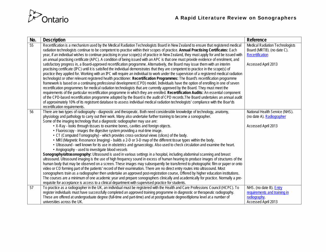

Health Professions Regulatory Advisory Council 56 Wellesley Street West 12th Floor
Toronto ON M5S 2S3
Telephone 416-326-1550 Toll-Free 1-888-377-7746
Fax 416-326-1549
Website wwwhpracorg
Email hpracwebmasterontarioca