Embed Size (px)
Citation preview
1
Head & Neck Cancer
II. Putting the Pieces
© 2004 A
.D.A
.M., Inc.
Together
Division of Cancer Prevention and ControlNCCDPHP, CoCHP
Centers for Disease Control and PreventionAtlanta, Georgia
Head and Neck Site Groups
Lip and Oral Cavity LipGumMouth SubsitesPalates
Ph
LarynxGlottisSupraglottisSubglottis
Nasal CavitySi
2
PharynxOropharynxHypopharynxNasopharynx
Salivary Glands
SinusesThyroidMiddle Ear
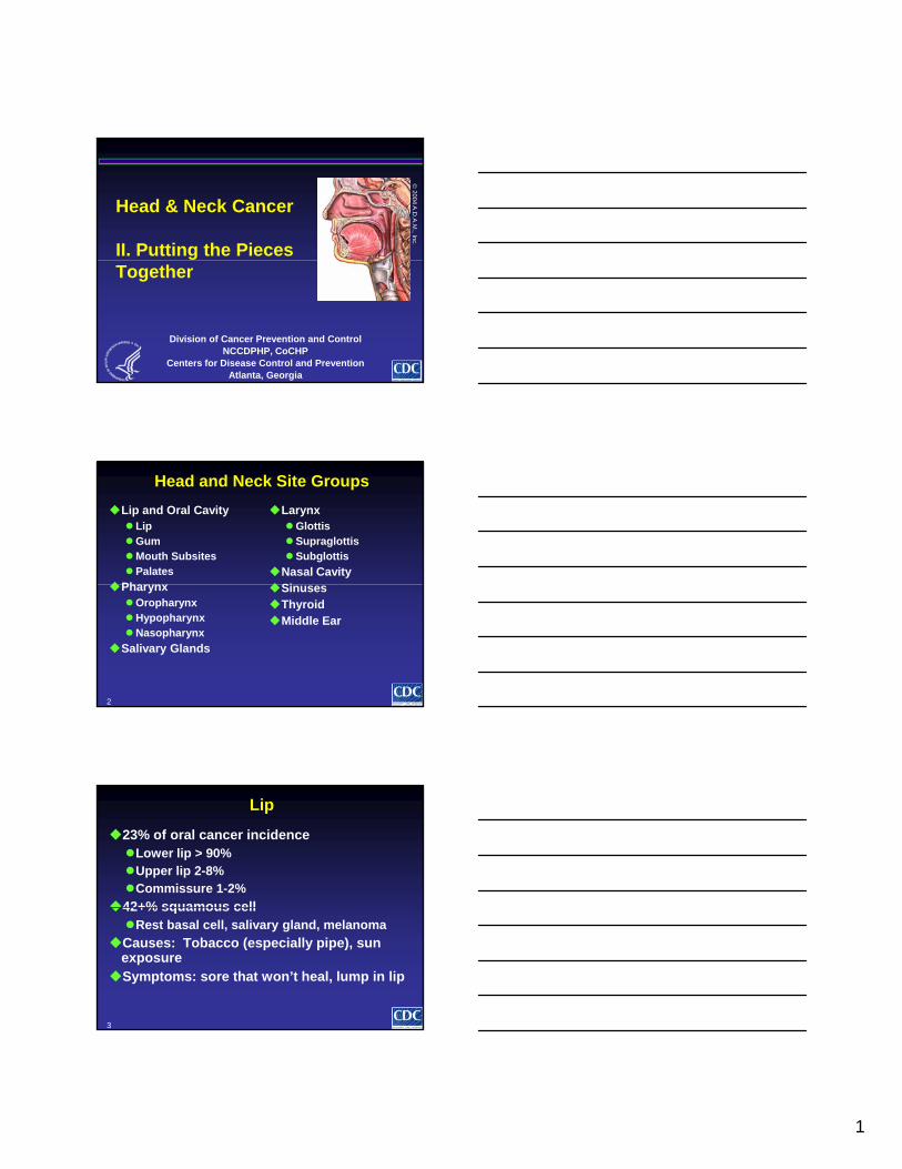
Lip
23% of oral cancer incidenceLower lip > 90%Upper lip 2-8%Commissure 1-2%
42+% squamous cell
3
42+% squamous cellRest basal cell, salivary gland, melanoma
Causes: Tobacco (especially pipe), sun exposure
Symptoms: sore that won’t heal, lump in lip

2
Lip (C00.x)
Parts: skin, vermilion border, mucosa, frenulum lip, commissureLower lip has better
Cutaneous C44.x
4
Lower lip has better prognosisC00.0, .1, .2 externalC00.3, .4, .5 mucosaC00.6 commissureC00.8 overlapping
VermilionCommissure
T2N0M0 Squamous cell C00.1
Use
d w
ith p
erm
issi
on a
nd
copy
right
ed b
y w
ww
.ent
usa.
com
Basal cell C44.0
5
Used w
ith permission and
copyrighted by ww
w.entusa.com
Mobile Tongue (C02.x)
28% oral cancer incidenceEst. 7,320 cases USA 2004 (0.3%)China 34,954; India 28,662
Parts: Tip, anterior 2/3 tongue, ventral & dorsal surface, frenulum linguae
6
dorsal surface, frenulum linguaeHistology: Squamous cell 90%WorkupH&PCT neck

3
Tongue, dorsal surface
Tongue, ventral surface
7
Frenulum
© 2004 A
.D.A
.M., Inc.
© 2004 A.D.A.M., Inc.
Tongue, right lateral border
Border
M.,
Inc.
8
© 2
004
A.D
.A.M
on a
nd c
opyr
ight
ed b
y w
ww
.ent
usa.
com
9
Squamous cell CA Tongue
Use
d w
ith p
erm
issi
o

4
CS Extension/Tongue Muscles
IntrinsicWithin tongue (no
bony attachment)Also called lingual
muscles If involved, code in CS
ExtrinsicAnchor tongue in
mouthAttached to mandible,
hyoid bone, styloid process of temporal b l i
10
,Ext 20 range (NOT T4 category)
bone, or palatine aponeurosis (posterior border of hard palate)
If involved, code in CS Ext 75 except C04, C08 (maps to T4)
Gum (C03.x)
Parts: gingiva, alveolar ridge, periodontalC03.0 upperC03.1 lowerC03.9 NOS
Snuff users 50 x risk (92% users male)
11
Snuff users 50 x risk (92% users male)2-3 times level of nicotine1100 mg sodium (1500 = RDA)grit & sand to abrase gums & teeth enamel
Upper Gum
12
Normal Gingiva (Gums)
© 2
004
A.D
.A.M
., In
c.
Lower Gum

5
Floor of Mouth/FOM (C04.x)
16% of all oral cancersSymptomsLesionDecreased tongue mobility
< 50% local at diagnosis
13
< 50% local at diagnosis
© 200
Frenulum
Floor of Mouth
14
Floor of Mouth
04 A.D
.A.M
., Inc.
Mouth
Hard Palate (C05.0, .8, .9)
Parts: Roof of mouth (NOT soft palate or uvula)
Histology (74% malignant, 26% benign)Squamous cell 53% Adenocarcinoma 4%Adenoid cystic 15% Anaplastic CA 4%
15
Adenoid cystic 15% Anaplastic CA 4%Mucoepidermoid 10% Other 14%
Reverse smoking70% tumors extend beyond hard palate
6
Normal Hard Palate
© 2004 A.D.A.M., Inc.
© 2
004
A.D
.A.M
., In
c.
16
Hard PalateSoft Palate
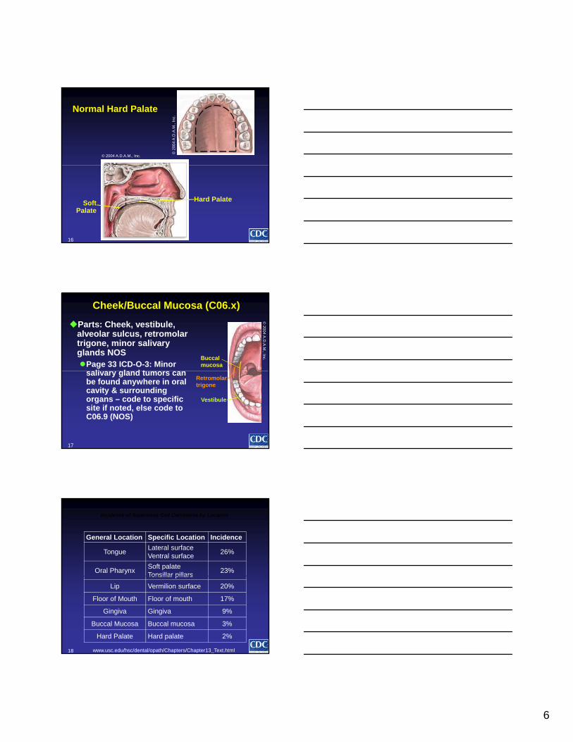
Cheek/Buccal Mucosa (C06.x)
Parts: Cheek, vestibule, alveolar sulcus, retromolar trigone, minor salivary glands NOSPage 33 ICD-O-3: Minor
li l d t
Buccal mucosa
© 2004 A
.D.A
.M., Inc.
17
salivary gland tumors can be found anywhere in oral cavity & surrounding organs – code to specific site if noted, else code to C06.9 (NOS)
Retromolartrigone
Vestibule
Incidence of Squamous Cell Carcinoma by Location
General Location Specific Location Incidence
TongueLateral surface Ventral surface
26%
Oral PharynxSoft palate Tonsillar pillars
23%
18
Tonsillar pillars
Lip Vermilion surface 20%
Floor of Mouth Floor of mouth 17%
Gingiva Gingiva 9%
Buccal Mucosa Buccal mucosa 3%
Hard Palate Hard palate 2%
www.usc.edu/hsc/dental/opath/Chapters/Chapter13_Text.html
7
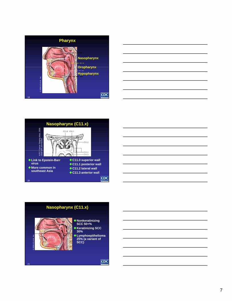
Pharynx
Nasopharynx
Oropharynx
19
Oropharynx
Hypopharynx
© 2
004
A.D
.A.M
., In
c.
Nasopharynx (C11.x)
Ca
nce
r S
tagi
ng
Atla
s, 2
00
6.
with
pe
rmis
sio
n.
20
Link to Epstein-Barr virus
More common in southeast Asia
C11.0 superior wallC11.1 posterior wallC11.2 lateral wallC11.3 anterior wall
AJC
C C
Use
d w
Nonkeratinizing SCC 50+%
Keratinizing SCC 30%
Nasopharynx (C11.x)
21
Lymphoepithelioma 25% (a variant of SCC)
© 2
004
A.D
.A.M
., In
c.

8
Parts of Oropharynx (C01.9, C05.1, C05.2, C09.x, C10.x)
C01.9 Base of tongue C05.1 Soft palate C05.2 UvulaC09.x Tonsils (not
shown)
C05.1
C05.2
C01.9
22
shown)C10.0 ValleculaC10.2 Oropharynx
wall (walls of throat) NOT C10.1 Anterior
surface epiglottis
C05.2
C10.0
C10.2
© 2004 A.D.A.M., Inc.
Base of Tongue (C01.9)
Posterior 1/3Usually larger
when diagnosed60-80% positive
k LN (40%
Lingual tonsil
Circum-
Palatine tonsil
23
neck LNs (40% bilateral)
vallate papillae
Mobile tongue
© 2004 A.D.A.M., Inc.
Tonsillar pillar
24
Sites of Oropharynx
Posterior wall
© 2
004
A.D
.A.M
., In
c.

9
Oropharyngeal Tumor Spread
Tonsil – extends down to base of tongue or onto soft palate
Base of tongue – into deep tongue muscle. Advanced lesions into rest of tongue or floor of mouth OR down toward glottis
25
Soft palate – involves uvula, cross midlinePharyngeal wall – spreads up/down rest of
pharynx OR laterally to other pharynx subsites
Parts of Hypopharynx (C12.9, C13.x)
C12.9 Pyriform sinus (most common, most lethal)
C13.0 PostcricoidC13 1 Hypo0
06
.
26
C13.1 Hypo-pharyngeal aspect of aryepiglottic fold
C13.2 Posterior wall C15.0 Esophagus
AJC
C C
an
cer
Sta
gin
g A
tlas,
20
Use
d w
ith p
erm
issi
on
.
87– year – old female
Tumor on tongue and right floor
Case Scenario: Multiple Primaries
hted
by
ww
w.e
ntus
a.co
m
27
mouth (extends onto anterior tonsillar pillar)
Needs commando procedure
Use
d w
ith p
erm
issi
on a
nd
copy
righ
10
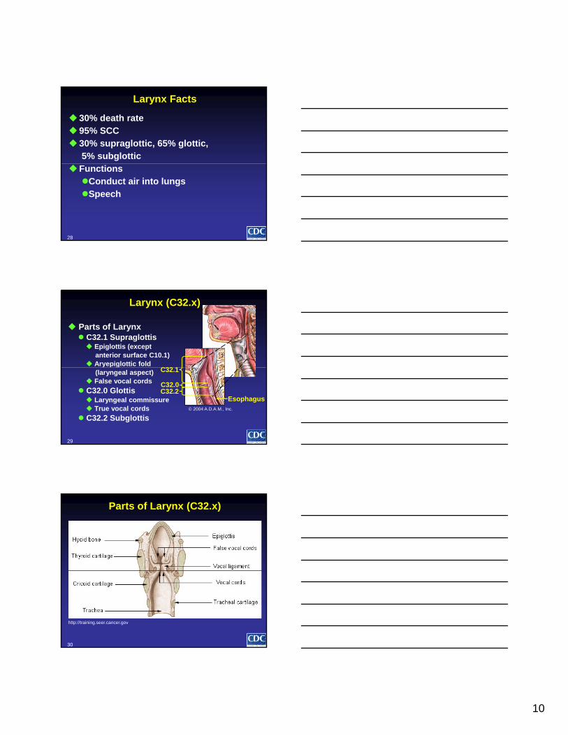
Larynx Facts
30% death rate95% SCC30% supraglottic, 65% glottic,
5% subglottic
28
FunctionsConduct air into lungsSpeech
Larynx (C32.x)
Parts of Larynx C32.1 Supraglottis Epiglottis (except
anterior surface C10.1) Aryepiglottic fold
C32 1
29
y p g(laryngeal aspect)
False vocal cords
C32.0 Glottis Laryngeal commissure True vocal cords
C32.2 Subglottis
C32.1
C32.0C32.2
© 2004 A.D.A.M., Inc.
Esophagus
Parts of Larynx (C32.x)
30
http://training.seer.cancer.gov

11
Laryngeal Cartilages (C32.3)
Single
1. Thyroid cartilage (a.k.a. Adam’s apple)
2. Cricoid cartilage 1
5
3
31
3. Epiglottis Paired
4. Arytenoid cartilage5. Corniculate
cartilage6. Cuneiform cartilage
(not shown)
5
2
4©
20
04
A.D
.A.M
., I
nc.
Laryngoscopic View of Larynx
Epiglottis
V l d
Anterior commissure
Vallecula (behind epiglottis)
32
Vocal cordAryepiglottic foldPosterior
commissure Arytenoid
False cord
AJCC Cancer Staging Atlas, 2006. Used with permission.
CS Staging: Larynx
Glottis 1 vs 2 vocal cords Impaired cord mobilityCartilage involvementMixed within hierarchy
(Local NOS = 45)
SubglottisDriven by extension
to other organs
Larynx cartilage, overlapping larynx and lar n NOS
33
(Local NOS = 45)
SupraglottisCartilages have lower
numbers in the Extension codes
and larynx, NOSSimilar to glottis
All subsites have T4a and T4b output

12
Nasal Cavity (C30.0)
Frontal sinusSphenoid sinus
Superior nasal concha
Internal narisMiddle nasal
h
34
© 2004 A.D.A.M., Inc.
Inferior nasal concha
External naris(vestibule)
Internal naris(choana)
concha
Nasopharynx
Maxillary Sinus (C31.0) Ethmoid Sinus (C31.1)Frontal Sinus (C31.2)
35
Maxillary sinus
Ethmoid sinuses
Frontal sinus
Nasal cavity
© 2
00
4 A
.D.A
.M.,
In
c.
CS Staging: Sinuses
All sinusesTumor Extension very dependent on
documenting regional organs (not size)
36

13
Major Salivary Glands (C07.9, C08.x)
Accessory parotid gland
37
Parotid duct (Stensen’s duct)
Sublingual gland
Frontal sinus
Parotid gland
© 2004 A.D.A.M., Inc.
Parotid Gland (C07.9)
Parts: Stensen duct, parotid gland duct 75% tumors benign Glands wrap around facial veins, arteries,
nerves SX: Swollen cheek facial nerve paralysis
38
SX: Swollen cheek, facial nerve paralysis
Distribution of Histologic Types of Tumor, Salivary Gland*
Cell Type %Acinic cell 7Adenocarcinoma 18
39
Adenoid cystic 22Malignant mixed 13Mucoepidermoid 33SCC 4Other 3
*Cancer of the Head and Neck, 4th ed., 2003, pg 483

14
Lymph Node Levels for Head and Neck
I Submental and submandibular
II Upper jugularIII Middle jugularIV Lower jugular and
supraclavicular
40
supraclavicularV Superficial cervicalVI Anterior
compartment (prelaryngeal, paratracheal)
VII Upper mediastinal(not shown)
AJCC Cancer Staging Atlas, 2006. Used with permission.
Head and Neck Lymph Nodes: Other
1 Occipital2 Mastoid3, 4, 7 Parotid5, 6 AuricularFacial (8 Buccinator,
Listed in SSF 3 – SSF 6
41
(9 Nasolabial, 10 Mandibular)
11 Jugular (Level II) 12 Cervical (Level V)Parapharyngeal*Retropharyngeal** not shown
http://training.seer.cancer.gov
Relationship Primary to Nodes
Submental (level I)
Anterior alveolar ridge, FOM, lower lip, anterior tongue,
Submandibular (level I)
Maxillary sinus, nasal cavity, oral cavity, submandibular gland
Level II Nasal cavity, oral cavity, parotid gland, pharynx
42
pharynx
Level III Larynx, oral cavity, pharynx
Level IV Cervical esophagus, hypopharynx, larynx
Level V Nasopharynx, oropharynx
Level VI Cervical esophagus, larynx (glottis, subglottis), pyriform sinus (apex), thyroid

15
CS Lymph Nodes
Regional lymph node (LN) data includes what nodes are involved, how many, and laterality Code the highest level possible
Cure rate drops 50% with positive regional LN 2005 CS clarification: supraclavicular LN can
43
be Level IV or Level V Add to Code 12 (CS LN)
Excludes nasopharynx and thyroid If can’t decide between levels, choose level
V
SSF 1, SSF 2
SSF1 = Size LN (largest diameter)If perinodal spread OR matted nodes, use
largest sizeIf cN, use entire node
SSF2 = Extracapsular extension
44
pAmorphous spiculated margins; involvement
of internodal fat; loss of normal oval to round shape888 if nodes are negative
SSF 1, SSF 2
SSF1 = Size of lymph node (largest diameter)If perinodal spread OR matted nodes,
use largest sizeIf cN use entire node
45
If cN, use entire nodeSSF2 = Extracapsular extensionAmorphous spiculated margins;
involvement of internodal fat; loss of normal oval to round shape888 if nodes are negative

16
SSF 3 – 6
LRND: 2 + parotid node (< 3 cm with extracapsular exten), 1 + buccal (facial) node (2 cm), and 1 + submandibular node (2 cm)
SSF3 Levels I-III 1 0 0I II III
SSF4 Levels IV V 0 0 0
46
SSF4 Levels IV-V, Retropharyngeal (RP)
0 0 0IV V RP
SSF5 Levels VI-VII, Facial (F)
0 0 1VI VII F
SSF6 Parapharyngeal (PP), Parotid (PA), Suboccipital (S)
0 1 0PP PA S
SSF 3-6 Hints
Do not mix “9” with “0”, “1” choicesEX: Level I LN positive, no comment about
Level II or III is coded as 100, not 199Code “9” could be used for entire group EX: Level IV, V unknown is 999 when less
47
,than radical neck dissection
CS Mets
Note about change of supraclavicular LNFollows usual pattern10 Distant LN40 Distant organs50 (10 + 40)
48
50 (10 + 40)

17
CS Evaluation Fields
Use Standard TableWHERE did info come
from?NOT was something
done?
Answer to EX:
EXAMPLE PE: 4 cm tonsillar
mass, cervical neck adenopathy
CT scan: Multiple enlarged cervical LNs
49
CS Size/Ext = 0
Size came from PE NOT because a scope & bx was done
CS Reg Nodes Eval = 0
Info from PE & CT NOT because scope was done
PET: SUV 17 in neck area, negative elsewhere
Endoscopy & bx tonsil = SCC
Comorbidities preclude surgery. 70 cGy to tonsil and bilat neck
CS Eval Field: Mets
CS Mets EvalUse positive findingsIf negative, use farthest finding
50
Thyroid (C73.9)
51
lm1

Slide 51
lm1 Delete the song---poor taste.epe9, 6/13/2007

18
Thyroid Cancer Types
Papillary Follicular Medullary Anaplastic
78% 17% 4% 1%
Develop in hormone-producing
Develop in hormone-producing
Develop in parafollicular cells (C cells)
Most aggressive
52
producing cells
producing cells
cells (C-cells)
Variety of papillary subtypes
Includes Hurthle cell
May be related to genetic syndrome
M:F 2:1
Common Histology Coding Mistakes
Papillary carcinoma, NOS - 8050/3 Papillary carcinoma of thyroid - 8260/3Micropapillary thyroid cancer OR
papillary microcarcinoma
53
p p y(< 1 cm) 8341/3, if > 1cm 8260/3
Two malignant nodules in thyroid (one papillary, one follicular), code as one tumor 8340/3
Papillary and medullary - 8347/3
Additional Resources
www.bcm.eduwww.pathologyoutlines.comMyers E, Suen J, Myers J, Hanna E. Cancer
of the Head and Neck. 4th ed. PA: Saunders; 2003
54
Harrison LB, Sessions RB, Hong WK, Mendenhall W, Medina J, Kies MS, Wenig B, O’Malley B. Head and Neck Cancer: A Multidisciplinary Approach. 2nd ed. PA: Lippincott; 2004

19
Additional Resources
ImagesAJCC Cancer Staging Illustrations in PowerPoint
from the AJCC Cancer Staging Atlas, sixth ed (2002). Springer-New York, 2007. Used with permission.
A D A M Interactive Anatomy 4 A D A M Inc 2004
55
A.D.A.M. Interactive Anatomy 4, A.D.A.M., Inc., 2004. Used with licensed permission.
ENTUSA.com. Used with permission
Next: Part III: Treatment and Survival
The findings and conclusions in this presentation are those of the authors and do not necessarily represent the views of the Centers for Disease Control and Prevention
56
Control and Prevention.
For information about CDC’sCancer Prevention and Control Programs
and the National Program of Cancer Registries
Please visit
www cdc gov/cancer/npcrwww.cdc.gov/cancer/npcr