Embed Size (px)
Citation preview

He, X., Hsiao, M-S., Boott, C., Harniman, R., Nazemi, A., Li, X., ...Manners, I. (2017). Two-dimensional assemblies from crystallizablehomopolymers with charged termini. Nature Materials, 16(4), 481–488. DOI:10.1038/nmat4837
Peer reviewed version
License (if available):Unspecified
Link to published version (if available):10.1038/nmat4837
Link to publication record in Explore Bristol ResearchPDF-document
This is the author accepted manuscript (AAM). The final published version (version of record) is available onlinevia Nature athttp://www.nature.com/nmat/journal/vaop/ncurrent/full/nmat4837.html?WT.feed_name=subjects_soft-materials.Please refer to any applicable terms of use of the publisher.
University of Bristol - Explore Bristol ResearchGeneral rights
This document is made available in accordance with publisher policies. Please cite only the publishedversion using the reference above. Full terms of use are available:http://www.bristol.ac.uk/pure/about/ebr-terms

1
Customized 2D Assemblies from Crystallizable Homopolymers with Charged
Termini
Xiaoming He,1 Ming-Siao Hsiao,2,# Charlotte E. Boott,1,# Robert L. Harniman,1,# Ali
Nazemi,1 Xiaoyu Li,1,3 Mitchell A. Winnik,4 and Ian Manners1,*
1School of Chemistry, University of Bristol, Bristol BS8 1TS, United Kingdom
2UES, Inc. and Materials & Manufacturing Directorate, Air Force Research Laboratory, Wright-
Patterson AFB, OH 45433, USA
3Department of Polymer Materials, School of Material Science and Technology, Beijing Institute of
Technology, Beijing 100081, PR China
4Department of Chemistry, University of Toronto, Toronto, Ontario M5S 3H6, Canada
#These authors contributed equally to this work
*To whom correspondence should be addressed: [email protected]
The creation of shaped, uniform, and colloidally-stable two-dimensional (2D) assemblies by
bottom-up methods represents a challenge of widespread current interest for a variety of
applications. We describe the utilization of surface charge to stabilize self-assembled planar
structures that are formed from crystallizable polymer precursors by a seeded growth
approach. Thus, addition of crystallizable homopolymers with charged end groups to seeds
generated by the sonication of block copolymer micelles with crystalline cores yields uniform
platelet micelles with controlled dimensions. Significantly, the seeded growth approach is
characterized by a morphology “memory” effect whereby the origin of the seed, which can
involve a quasi-hexagonal or rectangular 2D platelet precursor, dictates the observed 2D
platelet shape. The epitaxial nature of the growth process was confirmed in each case by
selected area electron diffraction. The new approach was illustrated using two different polymer
systems and opens the door to the construction of shaped 2D hierarchical structures with broad
utility.

2
Natural and synthetic two-dimensional (2D) planar structures, as exemplified by clay nanosheets and
graphene, respectively, have gained extensive recent attention as a result of their unique properties
that originate from their ultrathin and flat morphology.1-3 “Bottom-up” self-assembly of crystalline
homopolymers and block copolymers (BCPs) represents a promising route to analogous 2D materials
based on soft matter.4-7 However, the development of synthetic protocols that permit access to
colloidally stable 2D materials with uniform structures and with shape and dimensional control
represents a key contemporary challenge.
Crystalline homopolymers generally form 2D thin lamellae which, unless functionalized with
solvophilic substituents, are generally not colloidally stable.8,9 Recent advances have demonstrated the
use of thiol-terminated crystalline homopolymers that allow the peripheral nanoparticle patterning on
the 2D platelet,10,11 as well as the fabrication of alternate rings of a homopolymer and BCP.12,13 In a
further advance, nanosheets formed by the crystallization of precursors containing silsesquioxane
clusters bound to hyperbranched polymers can be fragmented into seeds using sonication which can
be used to control the growth of 2D platelets on subsequent precursor addition.14
We have shown that addition of a BCP with a crystallizable poly(ferrocenyldimethylsilane)
(PFS) core-forming block15 and a short complementary corona-forming block to seeds derived from
the sonication of 1D cylindrical PFS BCP micelles allows access to lenticular platelet micelles.16
Sequential addition of different PFS BCPs to seeds yields lenticular platelet block comicelles with
concentric regions of different coronal chemistry. In contrast, analogous seeded growth of a
crystallizable blend of PFS homopolymer and a PFS BCP with a long corona-forming segment affords
rectangular platelet micelles.17 Moreover, sequential addition of different blends to generate block
comicelles followed by spatially selective processing allows their programmed disassembly to yield
perforated or hollow rectangular morphologies. In this case, the presence of a corona-forming block of
significant length improves the colloidal stability of the resulting 2D structures, but the blend
approach adds a degree of complexity because of the need to determine the optimum homopolymer
and BCP ratio.
Inspired by the well-established use of electrostatic forces to stabilize particulate dispersions in
colloid science, we have explored the use of charged groups in place of coronal chains in the
formation of 2D platelets from BCPs using seeded growth processes. We anticipated that
incorporation of a charged group at the terminus of a crystalline homopolymer, aggregation of the
resulting platelets would be prevented by repulsive forces leading to colloidal stability.

3
Seeded growth of PFS homopolymers with charged end-groups using 1D seeds
Seeded-growth of crystallizable BCPs from 1D cylindrical seeds prepared by sonication of long,
polydisperse cylinders with a crystalline core provides a recently established route to low dispersity
cylindrical micelles of controlled length18-20 and also complex architectures such as block
comicelles,21-23 branched cylindrical micelles,24 and multi-armed micelles.25 The key to this approach
is that the core termini of the seeds remain active to the addition of further BCP unimer, leading to a
process that is analogous to a living covalent polymerization of molecular monomers.18 Successful
implementation relies on the use of core-forming blocks with close crystal lattice matching.26
Significantly, the resulting micelles are kinetically-trapped as a consequence of the crystallization of
the core and the constituent BCP chains are resistant to dynamic exchange between micelles under the
conditions used which would otherwise lead to substantial dispersities in length.
In order to explore the use of a charged terminal group in place of a corona-forming block we
have studied the seeded growth of a near monodisperse PFS homopolymer possessing a degree of
polymerization of 20 and with terminal phosphonium groups and iodide counteranions,
PFS20[PPh2Me]I (Fig. 1). This material was prepared by methylation of a neutral precursor,
PFS20PPh2. Addition of a solution of molecularly dissolved (unimeric) PFS20[PPh2Me]I in THF to
long cylindrical PFS25-b-P2VP250 micelle seeds (P2VP = poly(2-vinylpyridine), the subscripts refer to
the number average degree of polymerization) of number average length Ln = 840 nm (length
dispersity Lw/Ln = 1.03, Lw is the weight average length) in iPrOH afforded highly uniform rectangular
platelet micelles (Fig. 2a and Supplementary Fig. 1). The area of the platelets was found to be linearly
dependent on the unimer-to-seed mass ratio (munimer/mseed) and the area dispersity was very low (Aw/An
= 1.02, where Aw is the weight-average area and An is the number-average area). Significantly, the
platelet micelles were readily dispersible in iPrOH and the solution was colloidally stable, with no
evidence for a change in size or shape or for precipitation after three months. The aspect ratio (Ln/Wn)
was found to decrease non-linearly (from 14.0 to 4.1) upon increasing the ratio of munimer/mseed (from 4
to 40) (Supplementary Fig. 1j), indicating the preferential tendency for growth in the lateral direction
(perpendicular to the seed axis).
As shown in Fig. 2a, transmission electron microscopy (TEM) clearly indicated that the PFS25-
b-P2VP250 seed was located at the center of the platelets as a dark region of high electron density as a
consequence of seeded-growth both at the seed ends and the seed sides. Unlike our previous report on
the formation of lenticular micelles using seeded growth of BCPs, preferential growth off the ends of

4
the cylinder was not observed.16 The facile lateral growth observed may be a consequence of the
lower steric demands of PFS20[PPh2Me]I. Thus, the absence of long coronal chains may minimize the
hindrance to lateral addition expected from the corona of the cylindrical PFS25-b-P2VP250 seed
micelles. The heights of the platelet and the central cylindrical seed by atomic force microscopy
(AFM) were found to be 10 nm and 20 nm, respectively (Supplementary Fig. 1c). The larger height of
the central seed is at least partly attributed to presence of corona chains. Given that the average
distance between two adjacent main chain iron centers in a PFS chain is 0.65 nm,27 the extended chain
length of PFS20 is calculated to be 13 nm. Therefore in the 2D platelet structure surrounding the seed,
the PFS chains appear to be extended with a single fold and interdigitated, perpendicular to the planar
surface.
To characterize the positive surface of the platelets, a zeta potential measurement
(Supplementary Fig. 3) was performed for a sample of rectangular 2D assemblies in iPrOH (Ln = 1955
nm, Wn = 348 nm). This gave a positive value of +35 mV, indicative of the presence of a positive
surface which would be expected to provide good colloidal stability.28 These platelet micelles with a
positively charged surface function as a template for loading and patterning negatively charged
nanostructures, such as silica nanoparticles (Supplementary Fig. 4, see supplementary information for
further discussion). In order to clarify the importance of a terminal positive charge on the formation of
uniform and colloidal stable platelet micelles from the seeded growth of PFS20[PPh2Me]I, a control
experiment was carried out using neutral analogue PFS20PPh2. Following the same protocol of
addition of a unimer solution of PFS20PPh2 in THF to the cylindrical PFS25-b-P2VP250 seeds (Ln = 840
nm, Lw/Ln = 1.03) in iPrOH, very different results were obtained. Unlike the colloidally stable 2D
platelets formed from the seeded growth of PFS20[PPh2Me]I, obvious precipitation was observed on
addition of PFS20PPh2 in THF to the same PFS25-b-P2VP250 seeds in iPrOH. Moreover, TEM
indicated that irregular 2D plate structures formed with uncontrolled aggregation (Supplementary Fig.
5). This indicated that the introduction of a charged group at the terminus of the homopolymer is
necessary to permit the colloidal stability required for a controlled growth process.
The addition of PFS20[PPh2Me]I in THF to shorter PFS25-b-P2VP330 seeds (Ln = 58 nm, Lw / Ln
= 1.02) in iPrOH also led to the formation of 2D rectangular platelets with the aspect ratio showing a
similar trend of decreasing values (from 7.6 to 4.3) as the munimer/mseed ratio increased (from 4 to 20)
(Supplementary Fig. 6). However, the aspect ratio for the platelets formed by the shorter seeds was
significantly smaller. This indicated that the dimensions of the 2D rectangular platelets can be

5
controlled by seed length and the munimer/mseed ratio. Moreover, providing that the solvent is suitably
adjusted, the system is tolerant to considerable mismatches in the degrees of polymerization of the
PFS segments in the unimer and seed and also the counteranion used (Supplementary Fig. 7 and 8, see
supplementary information for further details). The generality of the method was also illustrated by
the observation that BCPs with a charged terminus, such as PFS BCP PI25-b-PFS22[PPh2Me]I, with a
short isoprene (PI) block at one end and a charged phosphonium group at the other, also formed well-
defined rectangular platelets via seeded growth in iPrOH (Supplementary Fig. 9). Furthermore, the
use of a PFS homopolymer with a negatively charged terminus, PFS20-SO3Li, also led to the
formation of rectangular platelets using 1D cylindrical seeds further illustrating the pervasive nature
of this new approach (Supplementary Fig. 10).
Seeded growth of PFS homopolymers with charged end-groups using 2D seeds
In the absence of cylindrical seeds, addition of PFS20[PPh2Me]I in THF to iPrOH resulted in the
formation of a precipitate of aggregated large 2D platelets (Supplementary Fig. 11a). The platelets
formed were more colloidally stable in MeOH and were found to possess a disk-like morphology
(Supplementary Fig. 11b). Sonication led to cleavage into small, polydisperse platelet fragments (An =
7985 nm2, Aw/An = 1.53; Supplementary Fig. 12). Next, we explored the seeded growth of
PFS20[PPh2Me]I using these 2D seeds (2D SeedHD; where the subscript ‘HD’ refers to the quasi-
hexagonal disk-like morphology of the seed precursor) as initiators. Interestingly, addition of unimer
PFS20[PPh2Me]I in THF to the 2D seedHD in MeOH led to the formation of monodisperse, quasi-
hexagonal disk-like 2D structures (Fig. 2b and Supplementary Fig.13), reminiscent of the platelet
aggregate precursors (Supplementary Fig. 11b). Similar quasi-hexagonal disk-like 2D platelets can
also be prepared via seeded growth in iPrOH (Supplementary Fig. 14). As for the case of the
rectangular platelet analogues, the area of these quasi-hexagonal disk-like platelets also followed a
linear relationship to the munimer/mseed ratio (Supplementary Fig. 13). With increasing unimer, the disk-
like platelets become increasingly uniform, with the polydispersity decreasing from 1.54 for the small
2D seeds before unimer addition to 1.03 (munimer/mseed = 40). AFM analysis indicated that these disks
are very flat, the average height of ca. 10 nm, again suggesting a interdigitated structure with a single
chain fold (Supplementary Fig. 13f-g).
The quasi-hexagonal disk-like platelets synthesized by the seeded growth method share a
similar morphology with their “parent” 2D aggregates used to form the seeds. Such a “memory” effect
is a likely consequence of identical crystal packing in these 2D structures and also the preference for

6
similar growth rates in the different crystallographic directions (see further discussions below and in
the Supplementary Information page S10).29 We envisaged that if the 2D seeds possess a “memory”
of the shape of the “parent” platelets, it should be possible to prepare 2D platelets using 2D seeds
derived from platelets of different shape. We therefore explored the use of 2D seeds derived from the
rectangular platelets (Fig. 2a) prepared using cylindrical seed precursors. By sonication of the
“parent” rectangular platelets, the 1st generation of 2D seeds was obtained (2D SeedR1, Supplementary
Fig. 15; where the subscript R1 refers to the rectangular morphology and seed generation).
Subsequent addition of a unimer solution PFS20[PPh2Me]I in THF to the 2D SeedR1 in MeOH
afforded uniform rectangular platelets of well-controlled size (Fig. 2c and Supplementary Fig. 16).
Very few quasi-hexagonal platelets (< 5 %) were present in the sample. Furthermore, we also found
that the “memory” was maintained in the 2nd generation of the 2D rectangular seeds (2D SeedR2,
Supplementary Fig. 15) which was prepared by sonication of the rectangular platelets formed by the
addition of unimeric PFS20[PPh2Me]I in THF to 2D SeedR1 in MeOH. Once again, rectangular
platelets of controlled dimensions could be prepared by varying the ratio of PFS20[PPh2Me]I unimer
to the 2D SeedR2 (Supplementary Fig. 17). AFM analysis showed that the rectangular platelets grown
from 2D SeedR1 and 2D SeedR2 were very flat with a height of 10 nm (Supplementary Fig. 16b-c). In
contrast to the rectangular platelets with cylindrical seeds at the center, the aspect ratio of the
rectangular platelet grown from 2D rectangular seeds possessed an almost constant value of ca. 2
irrespective of the munimer/mseed ratio. Analogous rectangular platelets with a similar aspect ratio can
also be prepared from 2D rectangular seeds via seeded growth in iPrOH (Supplementary Fig. 18).
Significantly, the seeded growth method we describe here is compatible with previously
reported seeded growth approaches that use BCPs and homopolymer/BCP blends and allows the
preparation of complex hierarchical structures.16,17 Moreover, the use of 2D platelets of different
shape as building blocks permits an additional level of control. For example, addition of the polymer
blend (PFS25/PFS25-b-P2VP250 = 1:1 based on mass ratio) to quasi-hexagonal disk-like platelets
(grown from 2D seedHD) or 2D rectangular analogues (prepared either from 1D PFS25-b-P2VP250
seeds or 2D seedR1) yielded block coplatelets in which the precursor shape was retained (Fig. 3). AFM
images showed the obvious height difference where the second block with PFS25/PFS25-b-P2VP250
polymer blend has 20 nm higher than the first block. By sequential alternating addition of a
PFS25/PFS25-b-P2VP250 blend (1:1, mass ratio) and PFS20[PPh2Me]I, concentric segmented platelet
comicelles with four distinct regions of excellent contrast were formed based on TEM (Supplementary
Fig. 19).

7
Although morphology “memory” is well-established for the growth of 2D semiconducting
nanosheets through epitaxial growth,30,31 reports involving soft matter are much less common.13,29
Furthermore, to our knowledge, the use of different seeds from the same polymer to induce the
formation of 2D platelets of different shape via seeded growth in solution is unprecedented. Analysis
by selected area electron diffraction (SAED) analysis revealed that the quasi-hexagonal disk-like and
rectangular platelets (grown from 2D seedHD and 2D seedR1, respectively) have near identical ED
patterns with three pairs of diffraction spots, confirming the presence of a single crystalline PFS core
with monoclinic symmetry (Fig. 4 and Supplementary Fig 20).27,32 The formation of isotropic disk-
like quasi-hexagonal or anisotropic rectangular shaped platelet micelles is attributed to the difference
in growth rate33 along the (110), (100) and (010) normal of the crystalline PFS core (Fig 4, see
supplementary information page S10 for additional discussion).34,35 The growth rate of the planes
(G(hkl)) for disk-like quasi-hexagonal platelets follows the trend G(010) G(100) = G(110) while
for rectangular plates, the growth rate follows the order G(110) G(100) = G(010). Electron
diffraction analysis for the spatially distinct regions of 2D platelet block comicelles with different
shapes confirmed that the crystalline structure for the PFS core was identical in each case (Fig. 4,
Supplementary Fig. 20), confirming the epitaxial nature of the growth process.
In order to confirm that self-assembly occurred in the solution phase rather than on solvent
evaporation and to monitor the formation of the different segments, laser scanning confocal
microscopy (LSCM) was used to characterize platelets formed in solution from PFS homopolymers
end-functionalized with a green fluorescent dye, PFS22-G (Fig. 1). Via addition of a blend of
PFS20[PPh2Me]I and PFS22-G (10:1, mass ratio) to quasi-hexagonal disk-like or rectangular platelets,
very uniform fluorescent almost circular, quasi-hexagonal or rectangular rings were prepared
(Supplementary Fig. 22).
The use of platelet micelles with different shapes as initiators enables the formation of
hierarchical and complex structures. Addition of the BCP PFS20-b-P2VP140 with a long corona-
forming block to the rectangular platelet (prepared from 1D PFS25-b-P2VP250 seed) in iPrOH led to
scarf-like structures, where the added BCP grew as the tassel fibers selectively at both long-axis
termini (Supplementary Fig. 23a). Amphiphilic 2D scarf-like structure can be similarly accessed by
addition of PFS25-b-PMVS170 unimer with a hydrophobic corona-forming segment in iPrOH/hexane
(3:1, v/v) (Supplementary Fig. S23c). Interestingly, increasing the polarity of the solvent induced the
2D amphiphilic objects to form oligomers via a type of step-growth polymerization process through

8
favorable hydrophobic interactions of the tassels. Similar scarf-like structures can also be prepared
upon addition of PFS20-b-P2VP140 unimer to rectangular platelets grown from 2D seedR1
(Supplementary Fig. 24a). When quasi-hexagonal disk-like platelets were used as initiators, however,
addition of PFS20-b-P2VP140 led to unusual, kinked micelles with less direction selectivity
(Supplementary Fig. 24b). Moreover, cross-shape platelets can be fabricated by seeded growth of
PFS20[PPh2Me]I from cylindrical cross-shape micelles prepared from the self-assembly of
amphiphilic cylindrical triblock comicelles M(PFS-b-PtBA)-b-M(PFS-b-PDMS)-b-M(PFS-b-PtBA)
(Supplementary Fig. 25).36
Concept Extension to Polylactide Homopolymers
In order to explore the generality of the new seeded growth approach to shaped 2D assemblies that
utilises homopolymers with charged terminal groups, we applied an analogous design strategy to a
semicrystalline organic polymer,37 poly(L-lactide) (PLLA).19,38,39 Two homopolymers with
phosphonium groups at a chain terminus were prepared, PLLA24[PPh2Me]I and PLLA34[PPh2Me]I
(see Fig. 1). To create suitable seeds, unimer solutions of PLLA24[PPh2Me]I and PLLA34[PPh2Me]I
were added to polar solvents (such as iPrOH or MeOH). In a mixture of iPrOH (or MeOH) and CHCl3
(10:1, v/v), formation of aggregates of 2D diamond-shape platelets of variable size (final c = 0.1
mg/mL) was observed at 23 C in 2 h based on TEM analysis (Supplementary Fig. 26).
The controlled fabrication of uniform 2D PLLA platelets was also achieved by seeded growth.
Sonication of 2D platelet of PLLA24[PPh2Me]I (0.1 mg/mL) in iPrOH/CHCl3 (10:1) produced
relatively small, almost 1D fragments of low dispersity that function as seeds (Ln = 200 nm, Lw/Ln =
1.09; Supplementary Fig. 27) whereas under the same conditions sonication of the 2D platelet of
PLLA34[PPh2Me]I in iPrOH/CHCl3 (10:1) gave 2D seeds with large size and high polydispersity (An =
13,980 nm2, Aw/An = 1.75). By using MeOH in place of iPrOH, relatively small, lower dispersity 2D
seeds of PLLA34[PPh2Me]I (An = 6542 nm2, Aw/An = 1.38; Supplementary Fig. 28) were successfully
obtained. Addition of unimeric PLLA24[PPh2Me]I in CHCl3 to the almost 1D seeds of
PLLA24[PPh2Me]I in iPrOH gave very uniform 2D diamond platelets with controlled size obtained by
varying the munimer/mseed (Fig. 5). The vast majority (over 95 %) of the formed platelets were found to
have clearly visible 1D seed present at the center by TEM. This indicated that 2D platelet formation
involving seeded-growth with the virtual absence of spontaneous nucleation as a competing process.
The 2D diamond platelet structure was also confirmed by AFM (Fig. 5f). The average height of the
platelets was about 10 nm, and the central seed and four edges were found to be ca. 3 nm higher than

9
the remaining regions (12 nm vs 9 nm). Analogous to the seeded-growth for the PFS20[PPh2Me]I, the
area of the platelets was found to linearly dependent on the munimer/mseed (Fig. 5h, Supplementary
Table 4). Seeded growth of PLLA34[PPh2Me]I from 2D seeds of PLLA34[PPh2Me]I also resulted in
2D diamond-shaped platelets (Supplementary Fig. 29). In addition, polymer PLLA34[PPh2Me]I with a
different polymer chain length could also be successfully grown into diamond platelets from 1D seeds
of PLLA24[PPh2Me]I, indicating the adaptability of the seeded growth process. By addition of a blend
of PLLA24[PPh2Me]I and PLLA21-G (or PLLA21-R) (10:1, mass ratio) to a solution of diamond-
shaped platelets of PLLA24[PPh2Me]I (where PLLA21-G and PLLA21-R refer to PLLA homopolymer
end-functionalized with green and red fluorescent dyes, respectively, Fig. 1), concentric fluorescent
2D diamond platelet multiple block comicelles with spatially defined regions of fluorescence were
prepared, further illustrating the versatility of the process (Fig. 5j-k).
Conclusions
In summary, we report a simple approach to the preparation of uniform, colloidally stable 2D platelets
with different shapes from homopolymers with charged end-groups by a combination of seeded-
growth and morphology “memory” effect. A variety of morphologies, such as rectangular, quasi-
hexagonal, and diamond platelet micelles have been fabricated for two different polymeric materials.
The seeded growth process is likely to be extendable to a wide range of crystallizable polymers,
including -conjugated and biodegradable materials,19,23 and allows precise control of dimensions and
access to hierarchical 2D segmented comicelles and also other complex architectures. The resulting
shaped 2D platelets possess a lipophilic charged surface and offer potential applications as carriers for
nanoparticles, biomolecules, and therapeutic agents, as liquid crystals and adhesives, and in composite
reinforcement. Many of these areas of prospective utility are currently under investigation in our
laboratory.
Acknowledgements
X.M.H., A.N. and X.L. are grateful to the European Union (EU) for Marie Curie Postdoctoral
Fellowships. C.E.B. thanks the Bristol Chemical Synthesis Centre for Doctoral Training, founded by
the Engineering and Physical Sciences Research Council (EPSRC), for a PhD studentship. M.A.W.
thanks the Natural Sciences and Engineering Research Council (NSERC) of Canada for financial
support. I.M. thanks the EPSRC for support. PeakForce atomic force microscopy was carried out in
the Chemical Imaging Facility, University of Bristol with equipment funded by EPSRC. Dr. George

10
R. Whittell is thanked for helpful discussions.
Author contributions
X.M.H and I.M. conceived the project with input from A.N. X.M.H. synthesized the polymers,
performed the experiments. X.M.H. and C.E.B. performed the LSCM imaging. X.M.H. and R.L.H.
performed the AFM analysis. X.L. provided the PFS25-b-P2VP250 seeds. M.S.H. performed the
imaging and analysis of ED and EDS mapping. X.M.H., M.S.H. and I.M. prepared the manuscript
with input from all the other authors. The project was supervised by I. M.
Additional information
Supplementary information is available in the online version of the paper. Reprints and permissions
information is available online at www.nature.com/reprints. Correspondence and requests for
materials should be addressed to I. M.
Competing financial interests
The authors declare no competing financial interests.

11
Figure 1 | Chemical structures of homopolymers used for seeded growth.

12

13
Figure 2 | Formation of 2D platelet micelles with different morphologies and controlled area
prepared by seeded growth of PFS20[PPh2Me]I. a. Schematic representation of the formation of 2D
rectangular platelets through seeded growth of PFS20[PPh2Me]I from 1D cylindrical seeds. TEM
images of a representative sample prepared by the seeded growth of unimer PFS20[PPh2Me]I in THF
from PFS25-b-P2VP250 seed micelles (Ln = 840 nm, Lw / Ln = 1.03) in iPrOH at 23 C, with unimer to
seed mass ratios (munimer/mseed) of 20:1. Linear dependence of micelle area on the munimer/mseed. Error
bars, standard deviation of measured areas. b. Schematic representation of the generation of 2D
SeedHD and formation of uniform quasi-hexagonal disk-like platelet through seeded growth. TEM
images of a representative sample formed through seeded growth of unimer PFS20[PPh2Me]I in THF
from 2D seedHD in MeOH at 23 C, with munimer/mseed of 40:1. Linear dependence of micelle area on
the munimer/mseed. Error bars, standard deviation of measured areas. c. Schematic representation of the
1st generation of 2D rectangular seed (2D SeedR1) and formation of uniform rectangular platelet
through seeded growth. TEM images of a representative sample formed through seeded growth of
unimer PFS20[PPh2Me]I in THF from 2D seedR1 in MeOH at 23 C, with munimer/mseed of 40:1. Linear
dependence of micelle area on the munimer/mseed. Error bars, standard deviation of measured areas.

14
Figure 3 | Formation of platelet block comicelles of different shape. a-c, Schematic representation,
TEM images, AFM topography images and corresponding height profiles of block comicelles with 2D
morphologies of (a) rectangular platelet grown from 1D PFS25-b-P2VP250 seed, (b) quasi-hexagonal
disk-like platelet grown from 2D seedHD and (c) rectangular platelet grown from 2D seedR1. These
platelet block comicelles were prepared by addition of PFS25-b-P2VP250/PFS25 (1:1, mass ratio) blend
unimer in THF to three types 2D platelets which were formed by the addition of PFS20[PPh2Me]I
unimer in THF to 1D PFS-b-P2VP250 seed in iPrOH/hexane (3:1, v/v), and 2D seedHD or 2D seedR1 in
MeOH/hexane (3:1, v/v) at 23 C.

15
Figure 4 | Selected area electron diffraction (SAED) pattern for platelet block comicelles. a,b.
TEM image and SAED patterns for a quasi-hexagonal disk-like platelet block comicelle (a), as well as
the schematic representation of the growth mechanism for the micelle and block comicelle including
four blue arrows presenting the growth direction of the (110) planes and two black arrows indicating
the growth direction of the (100) planes (b). c,d. TEM image and SAED patterns for a rectangular
platelet block comicelle (c), as well as the schematic representation of the growth mechanism for the
micelle and block comicelle containing two red arrows presenting the growth direction of the (010)
planes and two black arrows indicating the growth direction of the (100) planes. Exhaustion of the
faster growing (110) planes first during crystal growth leads to an absence of (110) facets in a
rectangular platelet (d). The platelet block comicelles were prepared by addition of PFS25-b-
P2VP250/PFS25 (1:1, mass ratio) blend unimer in THF to 2D quasi-hexagonal disk-like or rectangular
platelets which were formed by the addition of PFS20[PPh2Me]I unimer in THF to 2D seedHD or 2D
seedR1 in MeOH/hexane (3:1, v/v) at 23 C. G indicates the growth direction of the platelet.

16
Figure 5 | Uniform diamond platelet micelles of controlled area prepared by seeded growth of
PLLA24[PPh2Me]I. a, Schematic representation of the formation of diamond platelets through
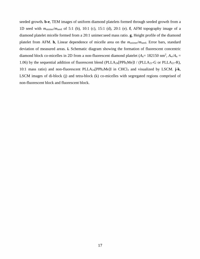
17
seeded growth. b-e, TEM images of uniform diamond platelets formed through seeded growth from a
1D seed with munimer/mseed of 5:1 (b), 10:1 (c), 15:1 (d), 20:1 (e). f, AFM topography image of a
diamond platelet micelle formed from a 20:1 unimer:seed mass ratio. g, Height profile of the diamond
platelet from AFM. h, Linear dependence of micelle area on the munimer/mseed. Error bars, standard
deviation of measured areas. i. Schematic diagram showing the formation of fluorescent concentric
diamond block co-micelles in 2D from a non-fluorescent diamond platelet (An= 182150 nm2, Aw/An =
1.06) by the sequential addition of fluorescent blend (PLLA24[PPh2Me]I / (PLLA21-G or PLLA21-R),
10:1 mass ratio) and non-fluorescent PLLA24[PPh2Me]I in CHCl3 and visualized by LSCM. j-k,
LSCM images of di-block (j) and tetra-block (k) co-micelles with segregated regions comprised of
non-fluorescent block and fluorescent block.

18
References
1. Zhang, X. & Xie, Y. Recent advances in free-standing two-dimensional crystals with
atomic thickness: design, assembly and transfer strategies. Chem. Soc. Rev. 42, 8187-8199 (2013).
2. Boott, C.E., Nazemi, A. & Manners, I. Synthetic Covalent and Non-Covalent 2D Materials. Angew. Chem. Int. Ed. 54, 13876-13894 (2015).
3. Zhuang, X., Mai, Y., Wu, D., Zhang, F. & Feng, X. Two-Dimensional Soft Nanomaterials: A Fascinating World of Materials. Adv. Mater. 27, 403-427 (2015).
4. Rizis, G., van de Ven, T.G.M. & Eisenberg, A. “Raft” Formation by Two-Dimensional Self-Assembly of Block Copolymer Rod Micelles in Aqueous Solution. Angew. Chem. Int. Ed. 53, 9000-9003 (2014).
5. Yang, J.-X., et al. Hydrogen-Bonding-Mediated Fragmentation and Reversible Self-assembly of Crystalline Micelles of Block Copolymer. Macromolecules 49, 367-372 (2016).
6. Lee, I.-H., et al. Nanostar and Nanonetwork Crystals Fabricated by in Situ Nanoparticlization of Fully Conjugated Polythiophene Diblock Copolymers. J. Am. Chem. Soc. 135, 17695-17698 (2013).
7. Yin, L. & Hillmyer, M.A. Disklike Micelles in Water from Polyethylene-Containing Diblock Copolymers. Macromolecules 44, 3021-3028 (2011).
8. Keller, A. Polymer single crystals. Polymer 3, 393-421 (1962). 9. Geil, P. Polymer Single Crystals. Robert Krieger Pub. Huntington, NY Press (1973). 10. Li, B. & Li, C.Y. Immobilizing Au Nanoparticles with Polymer Single Crystals, Patterning
and Asymmetric Functionalization. J. Am. Chem. Soc. 129, 12-13 (2007). 11. Dong, B., Zhou, T., Zhang, H. & Li, C.Y. Directed Self-Assembly of Nanoparticles for
Nanomotors. ACS Nano 7, 5192-5198 (2013). 12. Chen, W.Y., et al. “Chemically Shielded” Poly(ethylene oxide) Single Crystal Growth and
Construction of Channel-Wire Arrays with Chemical and Geometric Recognitions on a Submicrometer Scale. Macromolecules 37, 5292-5299 (2004).
13. Zheng, J.X., et al. Onsets of Tethered Chain Overcrowding and Highly Stretched Brush Regime via Crystalline−Amorphous Diblock Copolymers. Macromolecules 39, 641-650 (2006).
14. Yu, B., Jiang, X. & Yin, J. Size-Tunable Nanosheets by the Crystallization-Driven 2D Self-Assembly of Hyperbranched Poly(ether amine) (hPEA). Macromolecules 47, 4761-4768 (2014).
15. Hailes, R.L.N., Oliver, A.M., Gwyther, J., Whittell, G.R. & Manners, I. Polyferrocenylsilanes: synthesis, properties and applications. Chem. Soc. Rev., DOI: 10.1039/c1036cs00155f (2016).
16. Hudson, Z.M., et al. Tailored hierarchical micelle architectures using living crystallization-driven self-assembly in two dimensions. Nat. Chem. 6, 893-898 (2014).
17. Qiu, H., et al. Uniform patchy and hollow rectangular platelet micelles from crystallizable polymer blends. Science 352, 697-701 (2016).
18. Gilroy, J.B., et al. Monodisperse cylindrical micelles by crystallization-driven living self-assembly. Nat. Chem. 2, 566-570 (2010).
19. Petzetakis, N., Dove, A.P. & O'Reilly, R.K. Cylindrical micelles from the living crystallization-driven self-assembly of poly(lactide)-containing block copolymers. Chem. Sci. 2, 955-960 (2011).

19
20. Schmelz, J., Karg, M., Hellweg, T. & Schmalz, H. General Pathway toward Crystalline-Core Micelles with Tunable Morphology and Corona Segregation. ACS Nano 5, 9523-9534 (2011).
21. Wang, X., et al. Cylindrical Block Copolymer Micelles and Co-Micelles of Controlled Length and Architecture. Science 317, 644-647 (2007).
22. Schmelz, J., Schedl, A.E., Steinlein, C., Manners, I. & Schmalz, H. Length Control and Block-Type Architectures in Worm-like Micelles with Polyethylene Cores. J. Am. Chem. Soc. 134, 14217-14225 (2012).
23. Qian, J., et al. Uniform, High Aspect Ratio Fiber-like Micelles and Block Co-micelles with a Crystalline π-Conjugated Polythiophene Core by Self-Seeding. J. Am. Chem. Soc. 136, 4121-4124 (2014).
24. Qiu, H., et al. Branched micelles by living crystallization-driven block copolymer self-assembly under kinetic control. J. Am. Chem. Soc. 137, 2375-2385 (2015).
25. Qiu, H., Cambridge, G., Winnik, M.A. & Manners, I. Multi-Armed Micelles and Block Co-micelles via Crystallization-Driven Self-Assembly with Homopolymer Nanocrystals as Initiators. J. Am. Chem. Soc. 135, 12180-12183 (2013).
26. Gadt, T., Ieong, N.S., Cambridge, G., Winnik, M.A. & Manners, I. Complex and hierarchical micelle architectures from diblock copolymers using living, crystallization-driven polymerizations. Nat. Mater. 8, 144-150 (2009).
27. Gilroy, J.B., et al. Probing the Structure of the Crystalline Core of Field-Aligned, Monodisperse, Cylindrical Polyisoprene-block-Polyferrocenylsilane Micelles in Solution Using Synchrotron Small- and Wide-Angle X-ray Scattering. J. Am. Chem. Soc. 133, 17056-17062 (2011).
28. Hanaor, D., Michelazzi, M., Leonelli, C. & Sorrell, C.C. The effects of carboxylic acids on the aqueous dispersion and electrophoretic deposition of ZrO2. J. Eur. Ceram. Soc. 32, 235-244 (2012).
29. Xu, J., Ma, Y., Hu, W., Rehahn, M. & Reiter, G. Cloning polymer single crystals through self-seeding. Nat. Mater. 8, 348-353 (2009).
30. Tan, C. & Zhang, H. Epitaxial Growth of Hetero-Nanostructures Based on Ultrathin Two-Dimensional Nanosheets. J. Am. Chem. Soc. 137, 12162-12174 (2015).
31. Huang, X., et al. Solution-phase epitaxial growth of noble metal nanostructures on dispersible single-layer molybdenum disulfide nanosheets. Nat. Commun. 4, 1444 (2013).
32. Hsiao, M.-S., Yusoff, S.F.M., Winnik, M.A. & Manners, I. Crystallization-Driven Self-Assembly of Block Copolymers with a Short Crystallizable Core-Forming Segment: Controlling Micelle Morphology through the Influence of Molar Mass and Solvent Selectivity. Macromolecules 47, 2361-2372 (2014).
33. Passaglia, E. & Khoury, F. Crystal growth kinetics and the lateral habits of polyethylene crystals. Polymer 25, 631-644 (1984).
34. Chen, Z., et al. Structure of Poly(ferrocenyldimethylsilane) in Electrospun Nanofibers. Macromolecules 34, 6156-6158 (2001).
35. Papkov, V.S., et al. Crystalline Structure of Some Poly(ferrocenylenedialkylsilylenes). Macromolecules 33, 7107-7115 (2000).
36. Li, X., et al. “Cross” Supermicelles via the Hierarchical Assembly of Amphiphilic Cylindrical Triblock Comicelles. J. Am. Chem. Soc. 138, 4087-4095 (2016).
37. He, W.-N. & Xu, J.-T. Crystallization assisted self-assembly of semicrystalline block copolymers. Prog. Polym. Sci. 37, 1350-1400 (2012).

20
38. Sun, L., et al. Structural reorganization of cylindrical nanoparticles triggered by polylactide stereocomplexation. Nat. Commun. 5(2014).
39. Sun, L., et al. Core functionalization of semi-crystalline polymeric cylindrical nanoparticles using photo-initiated thiol-ene radical reactions. Polym. Chem. 7, 2337-2341 (2016).