Embed Size (px)
Citation preview


1
Hawaiian Hilton Village, Honolulu, Hawaii
The American Association of Clinical Anatomists officially began on October 17, 1983 to
advance the science and art of Clinical Anatomy, to encourage research and
publication in the field and to maintain high standards in the teaching of Anatomy. The
International Society for Plastination was founded in 1986 as a multidisciplinary
organization that included people within all fields of science interested in the technique
of Plastination. Plastination refers to the use of polymers to infiltrate and preserve any
material for teaching, research or diagnostic purposes.

2
Message from the AACA President
July 4, 2010
Dear Members and Attendees,
It is my honor and pleasure to welcome you to the joint 27th Annual Scientific Meeting of the
AACA and the 15th Annual International Conference on Plastination. There are many reasons to
anticipate that our time in Honolulu, made possible by our generous and gracious host Scott
Lozanoff, will be a memorable event for everyone. For the AACA, it is our second visit to Hawaii
and only the second meeting in our history in which registration has exceeded 300! AACA‘s
previous meeting in Honolulu in 1997 was its 14th meeting and attracted more registrants than
any previous meeting. For the International Society for Plastination, this meeting is ISP‘s first
annual conference in the United States since 1988 and its first visit to the Pacific area since 1996
when it met in Australia. Mahalo nui loa to Scott, his dedicated faculty and staff, and the
University for your invitation and the many weeks of work to make this event a success.
AACA members will quickly appreciate that the 2010 program book is very different in both
format and content from previous years. The 2010 Annual Meeting Committee believes the
larger size and inclusion of reports and information from our committees will greatly enhance its
value. Active and creative committees that address the interests and needs of the membership
are the heart and musculoskeletal system of all vital and growing associations. I consider the
substantial work done by our many committees to be the single most significant achievement
since I became President a year ago. In no small measure, I believe the creativeness and
productivity of our committees equally reflect the strengths of its membership and their abilities
to work as a team. Please take a few minutes to read your committees‘ reports. Learn about
their purpose, membership, activities, and maybe contact one of the members to get involved
yourself. I have more to say about committees in my Executive Committee report.
Another significant introduction by the 2010 Annual Meeting Committee was its decision to
eliminate all concurrent meetings and events of our Special Interest Groups. These groups and
the committees that oversee their activities are a core component of the AACA because they
each address a central mission of the Association. This year for the first time, all attendees will be
able to participate in all events organized by each of our three Special Interest Groups!
This year we are also blessed by the presence of Carlos Machado, MD, who will deliver the 2010
Council Presentation. Dr. Machado was scheduled as last year‘s presidential speaker but was
unfortunately at the last minute unable to attend the meeting. Welcome Carlos and we are all
looking forward to your presentation.
Last year, we were lucky to see Dr. Sam Scott return to the AACA fold after a number years in
exile in the neuroscience community. Those of you at the Cleveland meeting will remember Sam
as the man with the camera. Sam spent most his time, when not attending sessions, taking
spectacular portrait pictures to update member‘s AACA Online Directory entry. Sam had
planned to attend this year but a new job in Louisville has made it impossible. You will see him
with his camera setup at The Ohio State University in 2011 if you missed him last year.
No mere welcome or greeting letter could possibly express the gratitude that the membership
and Association owe Mark Seifert, our Program Secretary, for his tireless and determined mastery
of the many nuances of our new Meeting Organization and Program Planning Committee. As
chair, Mark has had a lot of cooperation and input but he deserves much of the credit for
reorganizing the meeting structure and creating a new process essential for the planning of

3
future meetings. As a direct result, you will be pleased to learn that all of the special events
scheduled for next year in Columbus have been finalized! Another individual who deserves
special thanks is Dave Bolender who ably chaired the 2010 Annual Meeting Committee. He and
his committee also deserve our appreciation and thanks. The third leg of our meeting triumvirate
is our Meeting Manager, Julie Hewett. Julie and her team at JulNet Solutions had a hand in
every aspect of meeting preparation and implementation. If you have a question or problem
this week, you will probably end up speaking with Julie. Please thank all of the above individuals
for making this meeting informative, pleasant, and memorable.
Finally, I am asking the membership to please take time during the meeting to visit the Exhibition
Area. I cannot overstate the importance and value of the time you spend with our exhibitors.
The Exhibitors are an essential part of each meeting. They are here to see and to hear from YOU
about how they can improve upon what they do for YOU. The exhibitors not only contribute to
the financial success of our meetings but more importantly their presence adds content and
value to benefit everyone who attends. Please thank them for participating and supporting this
year‘s AACA and ISP meeting.
Make it a great meeting,
Todd R. Olson, President

4
This page was intentionally left blank.

5
Table of Contents
Officers of AACA 7
Officers of ISP 8
Clinical Anatomy 9
Journal of Plastination 10
Sponsors 11
Program 13
Post Graduate Course 29
Annual Banquet 31
2010 Honored Member 33
2010 Presidential Speaker 35
Harmon Bickley Memorial Lecture 36
Vincent J De Feo Memorial Lecture 38
AACA Council Presentation 41
CDC Symposium 43
Anatomical Services Symposium 44
Educational Affairs Symposium 45
Educational Affairs Presentation 46
Annual Committee Reports 47
Poster Listings 63
Abstracts – Platform Presentations 85
Abstracts – Posters Presentations 101
Hilton Hawaiian Village Property Map 172

6
This page was intentionally left blank.
7
Officers of the AACA Council
President
Todd R. Olson, Ph.D.
President-Elect
Anne M. Agur, Ph.D.
Secretary
Brian R. MacPherson, Ph.D.
Treasurer
Neil S. Norton, Ph.D.
Past-President
Lawrence M. Ross, M.D., Ph.D.
Program Secretary
Mark F. Seifert, Ph.D.
Councilors
Brion Benninger, M.D., M.S.
David L. Bolender, Ph.D.
Thomas R. Gest, Ph.D.
Noelle Granger, Ph.D.
Carol S. Lomneth, Ph.D.
David J. Porta, Ph.D.
Rustin E. Reeves, Ph.D.
Alan T. Richards, M.D.
Brandi J. Schmitt, M.S.

8
Officers of the International Society for Plastination
President
Carlos A. C. Baptista, M.D., M.S., Ph.D., MPH
Vice-President
Rafael Latorre, D.V.M., Ph.D.
Secretary
Christoph von Horst, Ph.D.
Treasurer
Ameed Raoof, M.D., Ph.D.
Past-Presidents
Harmon C. Bickley, Ph.D., D.D.S. (1986-1995)
Robert W. Henry, D.V.M., Ph.D. (1996 -1997)
Andreas H. Weiglein, M.D. (1997-2003)
Mircea-Constantin Sora, M.D. (2003-2008)

9
Clinical Anatomy
The Official Journal of the American Association of Clinical Anatomists, the British Association of
Clinical Anatomists, the Australian and New Zealand Association of Clinical Anatomists, and the
Anatomical Society of Southern Africa
Editor-in-Chief – Stephen W. Carmichael
AACA Co-Editors –
Sherry A. Downie, Thomas R. Gest, John T. Hansen, Mark F. Seifert, Robert J. Spinner,
R. Shane Tubbs, and Joel A. Vilensky
BACA Editor – Stuart McDonald
ASSA Editor – Nirusha Lachman
ANZACA Editor – Helen Nicholson
Editorial Assistant – Beverly Northouse
Founding Editors: Ralph Ger and Ray J. Scothorne
Editorial Board – 2010
Associate Editors
Marwan Abu-Hijleh
Robert D. Acland
Philip Adds
Anne M.R. Agur
Seyedhossein
Aharinejad
Peter C. Amadio
Robert H. Anderson
Nihal Apaydin
David H. Bernanke
David R. Bowsher
Christopher Briggs
Jennifer Brueckner
H. Hamdi Celik
Carmine D. Clemente
Gene L. Colborn
John M. Cooke
Arthur F. Dalley II
Peter H. Dangerfield
Raffaele DeCaro
Adrian K. Dixon
Fabrice Duparc
Harold Ellis
Georg Feigl
Christian J. Fontaine
Ralph Ger
Daniel O. Graney
Noelle A. Granger
N. Alan Green
Malcolm Howard Hast
Marie-Noëlle Hébert-Blouin
David J. A. Heylings
David A. Hogg
David B. Jenkins
D. Gareth Jones
Jan L. Kasperbauer
Hee-Jin Kim
Subramaniam Krishnan
J. Thomas Lambrecht
Robert J. Leonard
Marios Loukas
Graham Louw
Scott Lozanoff
Sarah Mackay
Terence P. Ma
Pasuk Mahakkanukrauh
Vishy Mahadevan
Jan H. Meiring
W. Stanley Monkhouse
Keith L. Moore
Janusz Moryś
Robert P. Myers
Richard L. M. Newell
Wojciech Pawlina
Kathryn E. Peek
Sepp Poisel
Andrew T. Raftery
Cornelius Rosse
Jose R. Sanudo
Tatsuo Sato
Kapil S. Satyapal
Louise Scheuer
Hans-Martin Schmidt
Carol E. H.
Scott-Conner
Mohammadali M. Shoja
Shizuko Shoumura
Reon Somana
Mark D. Stringer
Ernest F. Talarico, Jr
Phillip R. Waggoner
Marvin Wagner
Anil H. Walji
Huan Wang
Andreas H. Weiglein
Peter L.T. Willan
Gary E. Wise
DaChuan Xu
John L. Zeller
Shao-Xiang Zhang
Changman Zhou
10
Journal of Plastination
The Official Journal of the International Society for Plastination
Editor-in-Chief - Ming Zhang
Co-Editors –
Robert W. Henry
Selcuk Tunali
Editorial Board – 2010
Renu Dhingra
Geoffrey D. Guttmann
M.S.A. Kumar
Rafael Latorre
Ameed Raoof
Mircea-Constantin Sora
Hong Jin Sui

11
Sponsors/Commercial Exhibitors
Generous donations and/or commercial exhibitor fees paid by the following companies and
organizations have substantially reduced the Association‘s expenses in presenting this meeting.
You are encouraged to visit the exhibits available for viewing in Palace Lounge
4D Anatomy Inc.
A.D.A.M. Software
American Association of Anatomists
Bone Clones, Inc.
Elsevier
Holt Anatomical, Inc.
Mopec
Thieme Medical Publishers
Touch of Life Technologies
Wolters Kluwer Health - LWW

12
This page was intentionally left blank.

13
Program
Monday July 19
9:00 – 5:00 AACA Council Meeting Ilima
Boardroom
3:00 – 6:00 Registration
Tapa Tower
5:00 – 6:30 Mentor Reception
Village Green
6:30 – 8:00 Welcome Reception
Sponsored by Elsevier
Tapa Cafe

14
This page was intentionally left blank.

15
Scientific Program
Tuesday July 20
7:00-4:30 Registration Desk Open Tapa Tower
7:00-8:30 Continental Breakfast Tapa Tower
7:00-8:00 Career Development Breakfast Iolani 5 & 6
8:00-8:30 Opening Remarks
Todd R. Olson
Carlos Baptista
Scott Lozanoff
Tapa Ballroom
8:30-9:30 AACA Presidential Speaker
Arthur F. Dalley
Quo Vadis, Anatomy?
Introduction by Todd R. Olson
Tapa Ballroom
9:30-10:30 Harmon Bickley Memorial Lecture
Charleen M. Moore and C. Mackenzie Brown
Can Human Dignity Be Preserved?
Ethical Issues Surrounding Plastinated Specimens
Introduction by Carlos Baptista
Tapa Ballroom
10:30-10:45 Transition Break
10:45-11:30 AACA Platform Session I – Muscle
Moderator: Rick Clemente
Tapa Ballroom
10:45-11:00 Transformation of muscle architecture at the fiber bundle level to fit
parametric b-spline volumes. RAVICHANDIRAN, Mayoorendra,
Kajeandra RAVICHANDIRAN, Jacobo SCHUSTER, Azam KHAN,
Nancy MCKEE, Anne AGUR. Division of Anatomy, Department of
Surgery, University of Toronto, Toronto, ON, M5S 1A8, CA.
11:00-11:15 Architecture and innervation of the vastus medialis muscle: a three-
dimensional modeling study. GUENETTE, Melanie, Judi LAPRADE,
Kate SAUKS, James JUNG, Erin BOYNTON, Anne AGUR. Division of
Anatomy, Department of Surgery, University of Toronto, Toronto,
ON, M5S 1A8, CA.
11:15-11:30 3D simulation of the masticatory system by analyses of the TMJ
movement and masticatory muscle functions. KIM Hee-Jin1, Il-
Kwang SIM2, Sang-Hee LEE2, Dong-Kyu YANG2, Jin-Sung KIM2, and
Kyung-Seok HU1. 1Division in Anatomy and Developmental Biology,
Department of Oral Biology, Brain Korea 21 Project, Human
Identification Research Center, Yonsei University College of
Dentistry, 250 Seongsanno, Seodaemoon-gu, Seoul, 120-752, Korea.
16
11:30-1:00 Lunch on your own
Poster Board Set-up – Session I
11:30-1:00 Editorial Board Lunch Iolani 6
11:30-1:00 Past Presidents Lunch Iolani 5
1:00-2:20 ISP Platform Session 1 – Principles of Plastination
Moderator: Carlos Baptista
Tapa Ballroom
1:00-1:20 Fundamentals of plastination – The silicone technique. HENRY,
Robert W. College of Veterinary Medicine, The University of
Tennessee, Knoxville, TN 37996, USA.
1:20-1:40 Principles of epoxy plastination technique (E12): Sheet plastination.
SORA, Mircea-Constantin1, Petru MATUSZ2, Radu JILAVU1, Jan
DRESENKAMP1. 1Center for Anatomy and Cell Biology, Medical
University of Vienna, Austria. 2Anatomical Department, University of
Medicine and Pharmacy "Victor Babes" Timisoara, Romania.
1:40-2:00 Principles of polyester plastination technique (P40). LATORRE,
Rafael. Dept. of Anatomy and Comparative Pathology, and Dept.
of Medicine and Surgery, Veterinary Faculty, University of Murcia,
Spain.
2:00-2:20 Room temperature plastination. RAOOF, Ameed. Division of
Anatomical Sciences/Plastination, University of Michigan, Ann
Arbor, MI 48109, USA.
2:20-2:30 Transition Break
2:30-4:30 Poster & Exhibitor Viewing Session 1
Categories: Extremities, Reproductive System, Associate Member
Posters
Tapa Ballroom
4:30-5:30 AACA Platform Session 2 – Education 1
Moderator: David Bolender
Tapa Ballroom
4:30-4:45 Don't cremate that body yet! PORTA, David J. Bellarmine
University, Louisville, KY 40205, USA.
4:45-5:00 Cadavers as models: Putting the best face forward?
DECKER, Summer J., Jonathan M. FORD, and Don R. HILBELINK.
Dept. of Pathology & Cell Biology, University of South Florida
College of Medicine, Tampa, FL 33612, USA.

17
Scientific Program
Tuesday July 20
5:00-5:15 Cadavers: virtually necessary? HILBELINK, Don R., Summer J.
DECKER, and Jonathan M. FORD. Dept. of Pathology & Cell
Biology, University of South Florida College of Medicine, Tampa, FL
33612, USA.
5:15-5:30 Teaching cross-sectional anatomy and radiology with clinical cases
based on CT scans of body donors. GEST, Thomas R., Webster
FRANCOIS, and Michael BOHL. University of Michigan Medical
School, Ann Arbor, MI, 48109, USA.
5:30 AACA Executive Council Meeting Iolani 6

18
This page was intentionally left blank.

19
Scientific Program
Wednesday July 21
7:00-4:00 Registration Desk Open Tapa Tower
7:00-8:00 Anatomical Services Breakfast Iolani 6
7:00-8:30 Continental Breakfast
Poster Set-up Session 2
Tapa Tower
7:55-8:00 Morning Welcome and Announcements Tapa Ballroom
8:00-9:00 Vincent J. De Feo Memorial Lecture
Ben Young
The Role of Anatomical Education in Hawaiian Medical History
Introduction by Scott Lozanoff
Tapa Ballroom
9:00-10:00 Poster & Exhibitor Viewing Session 2
Categories: Education, Neuroscience, Willed Body
Tapa Ballroom
1 0:00-10:40 Tech Fair Session
Moderator: Rusty Reeves
Tapa Ballroom
10:00-10:10 Development of an independent learning tool from MRI
reconstructions demonstrating the developmental anatomy of
the CNS. DETTON, Alan J., Douglas J. GOULD. The Ohio State
University Division of Anatomy, Columbus, OH 43210, USA.
10:10-10:20 Creation of 3D Finite Element Hip Model from the Visible Human
Male. FORD, Jonathan M., Summer J. DECKER, and Don R.
HILBELINK. Dept. of Pathology & Cell Biology, University of South
Florida College of Medicine, Tampa, FL 33612, USA.
10:20-10:30 Clinical Anatomy: as easy as x, y, z. HILBELINK, Don R. Dept. of
Pathology & Cell Biology, University of South Florida College of
Medicine, Tampa, FL 33612, USA.
10:30-10:40 A multimodal approach to teaching human development and
improving prenatal health. STILLWELL, Brian J.1 and Mark J.
HOLTERMAN1,2. 1The Endowment for Human Development,
Concord NH 03301. 2 The University of Illinois College of Medicine,
Chicago, IL 60612, USA.
10:40-11:00 Transition Break
20
11:00-12:00 AACA Platform Session 3 – Lymphatics, Vasculature
Moderator: Nihal Apaydin
Tapa Ballroom
11:00-11:15 A novel microsurgical injection technique for investigating the
lymphatic system in a cadaver.
SUAMI, Hiroo, and David W. CHANG. The University of Texas M. D.
Anderson Cancer Center, Houston, TX 77030, USA.
11:15-11:30 DVD demonstration of a minute dissection of the mediastinal
lymphatics from the posterior approach. SATO, T. Tokyo Ariake
University of Medical and Health Sciences, Tokyo 135-0063 JAPAN.
11:30-11:45 Exploring 3D morphology of the common, internal and external
iliac vessels as it applies to renal transplantation.
RAVICHANDIRAN Kajeandra, Siavash BOLOURANI, Yonah
KRAKOWSKY, Mayoorendra RAVICHANDIRAN, Robert STEWART
and Anne AGUR. Department of Surgery, University of Toronto,
Ontario, CA.
11:45-12:00 Modification of the right subclavian vein catheterization and its
anatomic basis and techniques. GUANGHUI, Luo. Department of
General Surgery, Xinhui City People‘s Hospital, Xinhui 529100,
Guangdong, China.
12:00-1:30 Tech Fair Hands On / Lunch on your own Tapa Ballroom
12:00-1:30 Clinical Anatomical Terminology Committee Meeting Iolani 6
1:30-3:00 ISP Platform Session 2 - Plastination in Education and Research
Moderator: Ming Zhang
Tapa Ballroom
1:30-1:45 Public education with plastinated specimen. SUI, Hongjin, and
Shengbo YU. Department of Anatomy, Dalian Medical University,
Dalian 116044, P.R. China
1:45-2:00 The potential of a plastinated horse for veterinary education.
YU, Shengbo, Jianfei ZHANG, Yanyan CHI, Haibin GAO, Jie LIU, Jin
GONG, and Hongjin SUI. Department of Anatomy, Dalian Medical
University, Dalian 116044, P.R. China
2:00-2:15 Plastination of fresh and old embalmed human lungs using
modified S-10 technique. DHINGRA Renu1, Sankat MOCHAN1,
Sanjeev LALWANI2 and Rani KUMAR1 1Department of Anatomy, 2Department of Forensic Medicine, All India Institute of Medical
Sciences, New Delhi, India
21
Scientific Program
Wednesday July 21
2:15-2:30 Plastinated knee: A model for arthroscopy and diagnostic
purposes. KUMAR, Rani1, Neha JAIN1 , Sanjeev LALWANI2 and
Renu DHINGRA1. 1Department of Anatomy, 2Department of
Forensic Medicine. All India Institute of Medical Sciences, Ansari
Nagar, New Delhi – 110 029. India.
2:30-2:45 Room temperature plastination of stained brain slices.
ADDS, Philip, Lynda PHILLIPSON and Mandeep SAGOO. St
George‘s Hospital Medical School, London SW17 0RE, United
Kingdom
2:45-3:00 The strategy for the three dimensional reconstruction of
anatomical structures by using plastinated cross-sections.
SORA, Mircea-Constantin1, Petru MATUSZ2, Radu JILAVU1, Jan
DRESENKAMP1. 1Center for Anatomy and Cell Biology, Medical
University of Vienna, Austria. 2 Anatomical Department, University
of Medicine and Pharmacy "Victor Babes" Timisoara, Romania.
3:00-4:00 Poster & Exhibitor Viewing Session 2
Categories: Education, Neuroscience, Willed Body
Tapa Ballroom
4:00-5:30 Career Development Committee Symposium Tapa Ballroom
Advancements in Medical Education Research
Current Issues in Medical Education
Richard T. Kasuya
University of Hawaii John A. Burns School of Medicine
The Anatomy of Medical Education Research
Rebecca Lufler, Tufts University School of Medicine
The Future of Medical Education: It’s More Than Medical
Knowledge – Anatomy Lab as Opportunity for Competency-
Based Instruction
Kimberly Topp, University of California, San Francisco
Is it Research? How to Elevate Observational Phenomena to
Educational Research and Scholarship
Kitt Shaffer
Boston University School of Medicine
Moderators: Todd Hoagland & Brion Benninger

22
This page was intentionally left blank.

23
Scientific Program
Thursday July 22
7:00-8:00 Educational Affairs Breakfast Iolani 6
7:00-4:00 Registration Desk Open Tapa Tower
7:00 – 8:30 Continental Breakfast
Poster Setup – Session 3
Tapa Tower
7:55-8:00 Morning Welcome and Announcements Tapa Ballroom
8:00-9:00 AACA Council Presentation
Carlos A.G. Machado
Following the Trail of Frank Netter: Master Medical Illustrator
Introduction by David Bolender
Tapa Ballroom
9:00-10:00 Poster & Exhibitor Viewing Session 3
Categories: Head and Neck, Thorax, Abdomen
Tapa Ballroom
10:00-11:45 AACA Platform Session 4 – Head & Neck, Abdomen, Lower Limb
Moderator: Peter Ward
Tapa Ballroom
10:00-10:15 Innervation of temporalis muscle: a three-dimensional study.
CANTELMI, David, Jonathan J. WISCO1, Joel C. DAVIES, Jayc C.
SEDLMAYR2, and Anne AGUR3. 3Division of Anatomy, Department
of Surgery, University of Toronto, Toronto, ON, M5S 1A8, Canada. 1Department of Pathology and Laboratory Medicine, Division of
Integrative Anatomy, David Geffen School of Medicine at UCLA,
Los Angeles, CA, 90095-1732, USA. 2Department of Cell Biology
and Anatomy, LSU Health Science Center, New Orleans, LA,
70112, USA.
10:15-10:30 Localization of the superior cervical ganglion for targeted
anesthetic blockade. WISCO, Jonathan J.1, M. Elena STARK1 and
Siamak RAHMAN2. 1Division of Integrative Anatomy, Department
of Pathology and Laboratory Medicine and 2Department of
Anesthesiology, David Geffen School of Medicine at UCLA, Los
Angeles, CA 90095, USA.
10:30-10:45 Anatomical research on laparoscopic surgical plane of
retroperitoneal fascia space. ZI-HAI, Ding, Wu TAO, Lei SHANG-
TONG, Zhang CE, Qiu JIAN-GUANG and Li GUO-XIN.Anatomical
Institute of Minimal Invasive Surgery, Southern Medical University,
Guangzhou, 510515, China.

24
10:45-11:00 Surgical view of the lumbar arteries and their branches: An
anatomical study with potential clinical application. COMERT,
Ayhan , Mehmet ARSLAN, Halil I. ACAR, Mevci OZDEMIR, Alaittin
ELHAN, Ibrahim TEKDEMIR, R. Shane TUBBS, and Hasan C. UGUR.
Ankara University, Faculty of Medicine, Department of Anatomy,
Ankara, Turkey.
11:00-11:15 Cone beam computed tomography of the pharynx in patients
with obstructive sleep apnea. LOZANOFF, Scott1,2, Neil NORTON3,
Karra MOTO1, Michael L. FARRELL2, Gurdev D. SINGH2. 1Dept. of
Anatomy, Biochemistry & Physiology, Honolulu, HI; 2OnChip
Technologies, Portland OR; 3Oral Biology, School of Dentistry,
Creighton University, Omaha, NE, USA.
11:15-11:30 Surgical anatomy of the superior gluteal nerve and landmarks for
its localization during minimally invasive approaches to the hip.
APAYDIN, Nihal, Simel KENDIR, Marios LOUKAS, R. Shane TUBBS.
Ankara University School of Medicine, Ankara, Turkey.
11:30-11:45 The normal and pathologic MRI appearance of the tibialis
anterior motor branch. HÉBERT-BLOUIN, Marie-Noëlle, Kimberly K.
AMRAMI, Robert J. SPINNER, Mayo Clinic, Departments of
Neurologic Surgery and Radiology, Rochester, MN 55905, USA.
11:45-1:30 Lunch on your own
1:30-2:30 Educational Affairs Presentation
D. Gareth Jones
Finding a Context for Plastination within the Development of
Anatomy: Aberration or Pathfinder?
Introduction by Tom Gest
Tapa Ballroom
2:30-3:30 Poster & Exhibitor Viewing Session 3
Categories: Head and Neck, Thorax, Abdomen
Tapa Ballroom
25
Scientific Program
Thursday July 22
3:30-4:30 Anatomical Services Committee Symposium
Consent, Tracking, and Disposition of Anatomical Collections with
Long Term Retention Periods: Focus on Plastination
Consent
Christina Strong
Law Offices of Christina Strong, Belle Mead, NJ
Tracking
Charlotte Wacker
University of California, Davis
Disposition
Darrell Petersen
Loma Linda University
Tapa Ballroom
4:30-6:00 AACA Business Meeting Tapa Ballroom
4:30-6:00 ISP Business Meeting Iolani 2-3-4
6:30-7:00 Reception Tapa Ballroom 1
7:00-9:00 Banquet Tapa Ballroom 1

26
This page was intentionally left blank.

27
Scientific Program
Friday July 23
7:00-8:30 2011 Program Committee Meeting Iolani 6
7:00-12:00 Registration Desk Open Tapa Tower
8:25-8:30 Morning Welcome and Announcements Tapa Ballroom
8:30-10:00 AACA Platform Session 5 – Education 2
Moderator: Mark Hankin
Tapa Ballroom
8:30-8:45 The documented benefits of using extended matching format
exams for medical gross anatomy. CROSS, Neal A., Randal
BATCHELOR, Shannon KING and Stanley ILIFF. Lincoln Memorial
University-DeBusk College of Osteopathic Medicine Harrogate TN
37752, USA.
8:45-9:00 Anatomy and art: The use of ―Christina‘s World‖ and the audience
response system to assess and foster clinical observation skills.
BAPTISTA, Carlos A. C., Department of Neurosciences, University of
Toledo, College of Medicine, Toledo, OH, 43614, USA.
9:00-9:15 Heavy reliance on medical school faculty and facilities in teaching
North American dental students. LAMBERT, H. Wayne, Douglas J.
GOULD, Dorothy T. BURK, Lisa M.J. LEE, Stavros ATSAS, Bob
HUTCHINS. West Virginia University, Morgantown, WV 26506-9128,
USA.
9:15-9:30 Teaching anatomy in an increasingly crowded medical
curriculum: a survey of current practices. MCANDREW, Darryl J.,
Steven J., CRAIG, David BOERS, Noel TAIT. Graduate School of
Medicine, University of Wollongong, Wollongong, New South
Wales, 2522, Australia.
9:30-9:45 First-year medical students‘ approaches to study and
performance in the gross anatomy course. WARD, Peter J., Ph.D.
West Virginia School of Osteopathic Medicine. Lewisburg, WV.
24901, USA.
9:45-10:00 Emotional experiences of medical students employed as
anatomical embalmers. WALKER, Rowan, Shaveen
KANAKARATNE, Chi Kit SO and A. STEWART, Prof. M.D. Fiona
STEWART. University of New England School of Rural Medicine,
Armidale, NSW, Australia.
28
10:00-12:00 Educational Affairs Committee Symposium
Plastination in Anatomy Education
Using Student-Produced Plastinated Specimens to Improve
Anatomical Expertise
Peter Ward
West Virginia School of Osteopathic Medicine
Plastinated Specimens as an Essential Resource in Anatomy
Education: Our Experience at the University of Michigan Medical
School, Ann Arbor, Over Two Decades
Ameed Raoof
University of Michigan School of Medicine
Use of Plastinated Specimens to Convey Learning Concepts in
Sports Medicine and Kinesiology
Kaori Tamura
University of Hawaii
Plastinated Prosections for Regional Study and Student
Performance on Identification Exams
Marc Pizzimenti
University of Iowa
Moderator: Noelle Granger
Tapa Ballroom
12:00-12:15 Closing Remarks / Adjournment Tapa Ballroom
12:30-1:30 New AACA Council Meeting Iolani 6
29
Scientific Program – Post Graduate Course
Saturday – July 24
Hot Topics in the Tropics: Plastination and Anatomical Education
Post Graduate Workshop Presented by the International Society for Plastination and the
American Association of Clinical Anatomists
Description: This workshop will introduce the participant to the method of plastination. The
purpose of plastination is to replace tissue fluid (water and fat) with a curable polymer (Silicone,
Polyester and Epoxy). The participants will be introduced to this process by actively performing
the general plastination steps including Specimen Preparation, Dehydration, Degreasing,
Impregnation and Curing. P40 (polyester) and room temperature S10 (silicone) techniques will
be undertaken. Participants will be divided into 4 groups of approximately 15 individuals.
Groups will rotate through stations in the laboratory and participants will perform aspects of the
basic plastination steps assisted by instructors assigned to the corresponding station. Participants
are expected to actively engage themselves and should complete both P40 plastination of thin
sections and S10 whole organ methods.
Objective. The joint AACA/ISP meeting held in Honolulu (July 19-24) represents the first ever
meeting of these two groups. Several research presentations will provide both introductory and
advanced information concerning plastination during the scientific session (July 19-23). The
objective of the postgraduate course is to facilitate a unique and ―hands-on‖ experience of the
method complementing the didactic information communicated in the scientific session. By the
end of the course, participants should be able to undertake P40 and S10 methods, comprehend
the underlying theoretical aspects of plastination, appreciate the tools required to set up a
plastination laboratory, and understand basic safety issues pertaining to the plastination
method.
Meeting Time and Activity Schedule:
7:00 Bus departs from Hilton Hawaiian Village for JABSOM
7:30-8:00 Continental Breakfast, MEB 314
8:00-9:00 Introductions and Plastination Overview Lecture, MEB 314
9:00AM -12:00 Laboratory Activities: Tissue Preparation, Vacuuming, Impregnation,
P40 Casting, BSB 107
12:00-1:00 Lunch
1:00-4:30 Laboratory Activities, continued, BSB 107
4:30-5:00 Questions and Wrap-up, BSB 107
5:00 Bus returns to Hilton Hawaiian Village from JABSOM

30
This page was intentionally left blank.
31
Annual Banquet
Thursday, July 22, 2010
Tapa Ballroom 1
6:30 pm - Reception
7:00 pm - Dinner with AACA & ISP Awards Presentations
9:00 pm - Closing Remarks by Local Host
Previous Honored Members of the AACA
*W. Henry Hollinshead, 1984
*Chester B. McVay, 1985
*Donald James Gray, 1986
*Russell T. Woodburne, 1987
*Oliver Beahrs, 1988
N. Alan Green, 1989
*Frank H. Netter, 1990
Ralph Ger, 1991
M. Roy Schwartz, 1992
Carmine D. Clemente, 1993
Keith L. Moore, 1994
*Ray J. Scothorne, 1995
Robert A. Chase, 1996
Tatsuo Sato, 1997
*John E. Skandalakis, 1998
Donald R. Cahill, 1999
*Sandy C. Marks, Jr., 2000
David G. Whitlock, 2001
Robert D. Acland, 2002
Arthur F. Dalley, II, 2003
*John V. Basmajian, 2004
Ian Whitmore, 2005
Peter H. Abrahams, 2006
Gary G. Wind, 2007
Vid Persaud, 2008
Richard S. Snell, 2009
* deceased
32
This page was intentionally left blank.
33
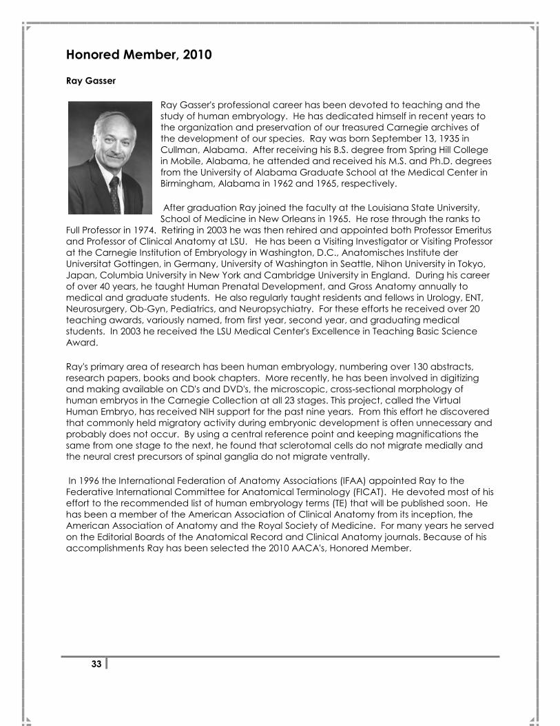
Honored Member, 2010
Ray Gasser
Ray Gasser's professional career has been devoted to teaching and the
study of human embryology. He has dedicated himself in recent years to
the organization and preservation of our treasured Carnegie archives of
the development of our species. Ray was born September 13, 1935 in
Cullman, Alabama. After receiving his B.S. degree from Spring Hill College
in Mobile, Alabama, he attended and received his M.S. and Ph.D. degrees
from the University of Alabama Graduate School at the Medical Center in
Birmingham, Alabama in 1962 and 1965, respectively.
After graduation Ray joined the faculty at the Louisiana State University,
School of Medicine in New Orleans in 1965. He rose through the ranks to
Full Professor in 1974. Retiring in 2003 he was then rehired and appointed both Professor Emeritus
and Professor of Clinical Anatomy at LSU. He has been a Visiting Investigator or Visiting Professor
at the Carnegie Institution of Embryology in Washington, D.C., Anatomisches Institute der
Universitat Gottingen, in Germany, University of Washington in Seattle, Nihon University in Tokyo,
Japan, Columbia University in New York and Cambridge University in England. During his career
of over 40 years, he taught Human Prenatal Development, and Gross Anatomy annually to
medical and graduate students. He also regularly taught residents and fellows in Urology, ENT,
Neurosurgery, Ob-Gyn, Pediatrics, and Neuropsychiatry. For these efforts he received over 20
teaching awards, variously named, from first year, second year, and graduating medical
students. In 2003 he received the LSU Medical Center's Excellence in Teaching Basic Science
Award.
Ray's primary area of research has been human embryology, numbering over 130 abstracts,
research papers, books and book chapters. More recently, he has been involved in digitizing
and making available on CD's and DVD's, the microscopic, cross-sectional morphology of
human embryos in the Carnegie Collection at all 23 stages. This project, called the Virtual
Human Embryo, has received NIH support for the past nine years. From this effort he discovered
that commonly held migratory activity during embryonic development is often unnecessary and
probably does not occur. By using a central reference point and keeping magnifications the
same from one stage to the next, he found that sclerotomal cells do not migrate medially and
the neural crest precursors of spinal ganglia do not migrate ventrally.
In 1996 the International Federation of Anatomy Associations (IFAA) appointed Ray to the
Federative International Committee for Anatomical Terminology (FICAT). He devoted most of his
effort to the recommended list of human embryology terms (TE) that will be published soon. He
has been a member of the American Association of Clinical Anatomy from its inception, the
American Association of Anatomy and the Royal Society of Medicine. For many years he served
on the Editorial Boards of the Anatomical Record and Clinical Anatomy journals. Because of his
accomplishments Ray has been selected the 2010 AACA's, Honored Member.

34
This page was intentionally left blank.
35
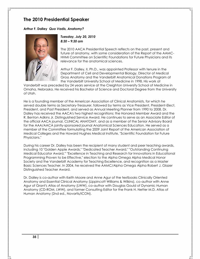
The 2010 Presidential Speaker
Arthur F. Dalley Quo Vadis, Anatomy?
Tuesday, July 20, 2010
8:30 – 9:30 am
The 2010 AACA Presidential Speech reflects on the past, present and
future of anatomy, with some consideration of the Report of the AAMC-
HHMI Committee on Scientific Foundations for Future Physicians and its
relevance for the anatomical sciences.
Arthur F. Dalley, II, Ph.D., was appointed Professor with tenure in the
Department of Cell and Developmental Biology, Director of Medical
Gross Anatomy and the Vanderbilt Anatomical Donations Program at
the Vanderbilt University School of Medicine in 1998. His work at
Vanderbilt was preceded by 24 years service at the Creighton University School of Medicine in
Omaha, Nebraska. He received his Bachelor of Science and Doctoral Degree from the University
of Utah.
He is a founding member of the American Association of Clinical Anatomists, for which he
served double terms as Secretary-Treasurer, followed by terms as Vice President, President-Elect,
President, and Past President, and served as Annual Meeting Planner from 1990 to 2008. Dr.
Dalley has received the AACA's two highest recognitions: the Honored Member Award and the
R. Benton Adkins Jr. Distinguished Service Award. He continues to serve as an Associate Editor of
the official AACA journal, CLINICAL ANATOMY, and as a member of the Senior Advisory Board
for the AAA/AACA jointly-sponsored journal Anatomical Sciences Education. He served as a
member of the Committee formulating the 2009 Joint Report of the American Association of
Medical Colleges and the Howard Hughes Medical Institute, "Scientific Foundation for Future
Physicians."
During his career Dr. Dalley has been the recipient of many student and peer teaching awards,
including 10 "Golden Apple Awards," "Dedicated Teacher Award," "Outstanding Continuing
Medical Educator Award,‖ "Excellence in Teaching and Research for Innovations in Educational
Programming Proven to be Effective," election to the Alpha Omega Alpha Medical Honor
Society and the Vanderbilt Academy for Teaching Excellence, and recognition as a Master
Basic Sciences Teacher. In 2004, he received the AAMC/Alpha Omega Alpha Robert J. Glazer
Distinguished Teacher Award.
Dr. Dalley is co-author with Keith Moore and Anne Agur of the textbooks Clinically Oriented
Anatomy and Essential Clinical Anatomy (Lippincott Williams & Wilkins), co-author with Anne
Agur of Grant's Atlas of Anatomy (LWW), co-author with Douglas Gould of Dynamic Human
Anatomy (CD-ROM, LWW), and former Consulting Editor for the Frank H. Netter M.D. Atlas of
Human Anatomy (2nd ed., Novartis/ICON).
36
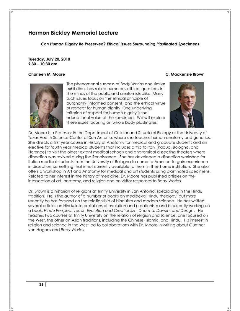
Harmon Bickley Memorial Lecture
Can Human Dignity Be Preserved? Ethical Issues Surrounding Plastinated Specimens
Tuesday, July 20, 2010
9:30 – 10:30 am
Charleen M. Moore C. Mackenzie Brown
The phenomenal success of Body Worlds and similar
exhibitions has raised numerous ethical questions in
the minds of the public and anatomists alike. Many
such issues focus on the ethical principle of
autonomy (informed consent) and the ethical virtue
of respect for human dignity. One underlying
criterion of respect for human dignity is the
educational value of the specimen. We will explore
these issues focusing on whole body plastinates.
Dr. Moore is a Professor in the Department of Cellular and Structural Biology at the University of
Texas Health Science Center at San Antonio, where she teaches human anatomy and genetics.
She directs a first year course in History of Anatomy for medical and graduate students and an
elective for fourth year medical students that includes a trip to Italy (Padua, Bologna, and
Florence) to visit the oldest extant medical schools and anatomical dissecting theaters where
dissection was revived during the Renaissance. She has developed a dissection workshop for
Italian medical students from the University of Bologna to come to America to gain experience
in dissection; something that is not currently available to them in their home institution. She also
offers a workshop in Art and Anatomy for medical and art students using plastinated specimens.
Related to her interest in the history of medicine, Dr. Moore has published articles on the
intersection of art, anatomy, and religion and on visitor responses to Body Worlds.
Dr. Brown is a historian of religions at Trinity University in San Antonio, specializing in the Hindu
tradition. He is the author of a number of books on mediaeval Hindu theology, but more
recently he has focused on the relationship of Hinduism and modern science. He has written
several articles on Hindu interpretations of evolution and creationism and is currently working on
a book, Hindu Perspectives on Evolution and Creationism: Dharma, Darwin, and Design. He
teaches two courses at Trinity University on the relation of religion and science, one focused on
the West, the other on Asian traditions, including the Chinese, Islamic, and Hindu. His interest in
religion and science in the West led to collaborations with Dr. Moore in writing about Gunther
von Hagens and Body Worlds.

37
Harmon C. Bickley Jr.
Harmon C. Bickley Jr. Ph.D., DDS was a former professor of pathology at the
Mercer University School of Medicine in Macon. He graduated in Dentistry at
the University of Michigan, and obtained his doctorate of pathology from the
University of Rochester. Dr. Bickley taught pathology in the medical schools of
the University of Kentucky at Lexington, the University of Iowa, and the
University of Texas at San Antonio. He was the father of Plastination in the
United States. In 1981 plastination was used by Dr. Bickley in the Department
of Pathology at the University of Texas Medical School at San Antonio. In 1986
he found the International Society for Plastination during the Third
International Conference on Plastination held in San Antonio, Texas. Dr. Bickley served as the
Executive Director of The International Society for Plastination from 1986-1995. He published a
textbook series for pathology courses at universities across the country and was a founding
member of the Group for Research in Pathology Education.

38
Vincent J. De Feo Memorial Lecture
Ben Young The Role of Anatomical Education in Hawaiian Medical History
Wednesday, July 21, 2010
8:00 – 9:00 am
Ben Young was born and raised in Honolulu, Hawaii, and graduated from
Roosevelt High School. He received his undergraduate degree in English
literature from Milligan College, Tennessee, and completed studies in
church history at Pepperdine University. He graduated from Howard
University, Washington, DC, with his medical degree and trained in
psychiatry at the University of Hawaii Integrated Residency Program. He
was former dean of students at the John A. Burns School of Medicine;
former vice president of student affairs, University of Hawaii-Manoa; and
chief of staff at Castle Medical Center, Kailua, Oahu, Hawaii. He served as
chairman of the Department of Psychiatry at Castle Medical Center for
many years. His last position was executive director of the Native Hawaiian
Center of Excellence, John A. Burns School of Medicine, from which he
retired in 2007. While at the medical school, he was responsible for bringing in over $10 million in
funding for several programs in research and training. He was appointed to former US Surgeon
General David Satcher's Advisory Committee on the Prevention of Violence and was national
chairman for deans of student affairs for all medical schools in the United States. For several
years, he was president of the National Council for Diversity in the Health Professions. In 1972, he
was one of only 10 licensed native Hawaiian physicians in Hawaii. He began efforts to increase
the numbers of native Hawaiians in medicine and today, because of programs that he initiated,
there are now over 300 Hawaiian physicians. He received many awards including the title of
Distinguished Historian by the Hawaiian Historical Society, was named a Living Treasure of Hawaii
by the Honpa Hongwanji, and was presented with the Distinguished Hawaiian Award by the
Queen Emma Hawaiian Civic Club. His contributions to improving the health of Hawaiians
resulted in the Kaonohi Award being given to him by the community organization Papa Ola
Lokahi. In the early 1970s, he helped build the voyaging canoe Hokule`a and was president of
the Polynesian Voyaging Society. He was the physician on Hokule`a's maiden voyage in 1976
from Tahiti to Hawaii and is currently writing a book on Hawaii's Medical History.
39
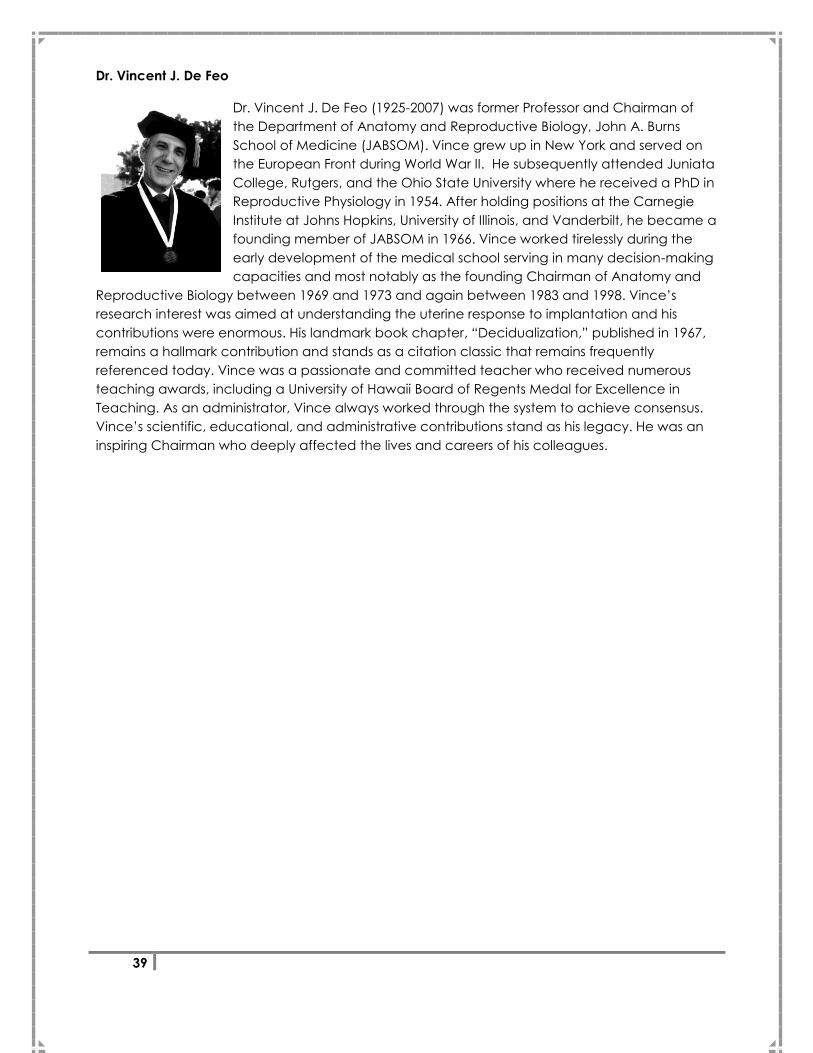
Dr. Vincent J. De Feo
Dr. Vincent J. De Feo (1925-2007) was former Professor and Chairman of
the Department of Anatomy and Reproductive Biology, John A. Burns
School of Medicine (JABSOM). Vince grew up in New York and served on
the European Front during World War II. He subsequently attended Juniata
College, Rutgers, and the Ohio State University where he received a PhD in
Reproductive Physiology in 1954. After holding positions at the Carnegie
Institute at Johns Hopkins, University of Illinois, and Vanderbilt, he became a
founding member of JABSOM in 1966. Vince worked tirelessly during the
early development of the medical school serving in many decision-making
capacities and most notably as the founding Chairman of Anatomy and
Reproductive Biology between 1969 and 1973 and again between 1983 and 1998. Vince‘s
research interest was aimed at understanding the uterine response to implantation and his
contributions were enormous. His landmark book chapter, ―Decidualization,‖ published in 1967,
remains a hallmark contribution and stands as a citation classic that remains frequently
referenced today. Vince was a passionate and committed teacher who received numerous
teaching awards, including a University of Hawaii Board of Regents Medal for Excellence in
Teaching. As an administrator, Vince always worked through the system to achieve consensus.
Vince‘s scientific, educational, and administrative contributions stand as his legacy. He was an
inspiring Chairman who deeply affected the lives and careers of his colleagues.

40
This page was intentionally left blank.
41
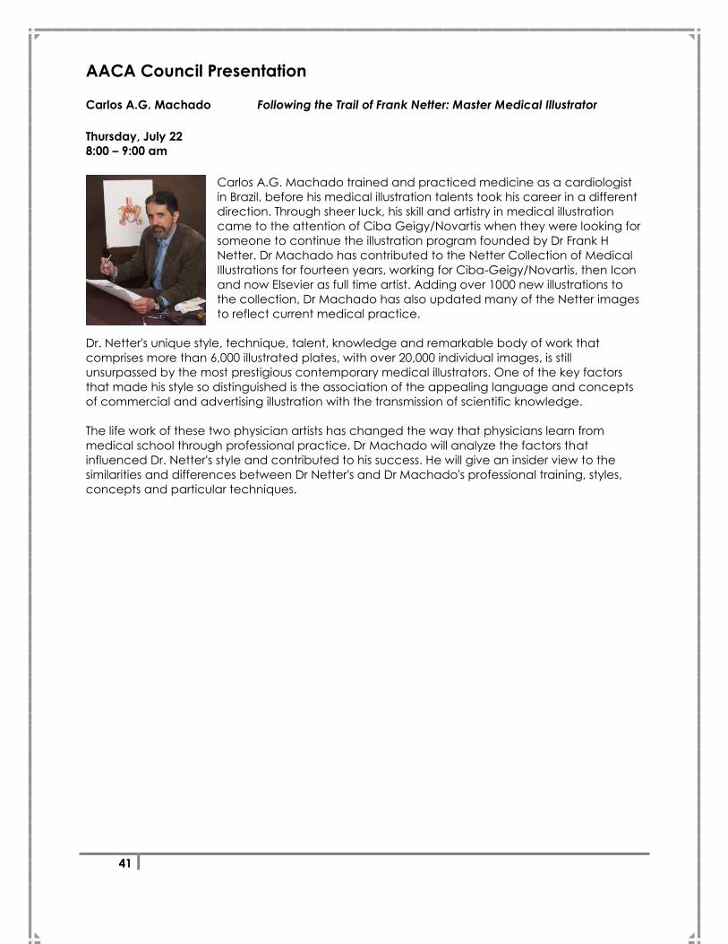
AACA Council Presentation
Carlos A.G. Machado Following the Trail of Frank Netter: Master Medical Illustrator
Thursday, July 22
8:00 – 9:00 am
Carlos A.G. Machado trained and practiced medicine as a cardiologist
in Brazil, before his medical illustration talents took his career in a different
direction. Through sheer luck, his skill and artistry in medical illustration
came to the attention of Ciba Geigy/Novartis when they were looking for
someone to continue the illustration program founded by Dr Frank H
Netter. Dr Machado has contributed to the Netter Collection of Medical
Illustrations for fourteen years, working for Ciba-Geigy/Novartis, then Icon
and now Elsevier as full time artist. Adding over 1000 new illustrations to
the collection, Dr Machado has also updated many of the Netter images
to reflect current medical practice.
Dr. Netter's unique style, technique, talent, knowledge and remarkable body of work that
comprises more than 6,000 illustrated plates, with over 20,000 individual images, is still
unsurpassed by the most prestigious contemporary medical illustrators. One of the key factors
that made his style so distinguished is the association of the appealing language and concepts
of commercial and advertising illustration with the transmission of scientific knowledge.
The life work of these two physician artists has changed the way that physicians learn from
medical school through professional practice. Dr Machado will analyze the factors that
influenced Dr. Netter's style and contributed to his success. He will give an insider view to the
similarities and differences between Dr Netter's and Dr Machado's professional training, styles,
concepts and particular techniques.

42
This page was intentionally left blank.
43
Career Development Committee Symposium
Wednesday, July 21, 2010
4:00 – 5:30 pm
Advancements in Medical Education Research
Current Issues in Medical Education
Richard T. Kasuya
University of Hawaii John A. Burns School of Medicine
The Anatomy of Medical Education Research
Rebecca Lufler
Tufts University School of Medicine
The Future of Medical Education: It’s More Than Medical Knowledge – Anatomy Lab as
Opportunity for Competency-Based Instruction
Kimberly Topp
University of California, San Francisco
Is it Research? How to Elevate Observational Phenomena to Educational Research and
Scholarship
Kitt Shaffer
Boston University School of Medicine
Moderators: Todd Hoagland
Brion Benninger
44
Anatomical Services Committee Symposium
Thursday, July 22, 2010
3:30 – 4:30 pm
Consent, Tracking, and Disposition of Anatomical Collections with Long
Term Retention Periods: Focus on Plastination
Presenters:
Consent
Christina Strong
Law Offices of Christina Strong, Belle Mead, NJ
Tracking
Charlotte Wacker
University of California, Davis
Disposition
Darrell Petersen
Loma Linda University

45
Educational Affairs Committee Symposium
Friday, July 23, 2010
10:00 – 12:00 pm
Plastination in Anatomy Education
Using Student-Produced Plastinated Specimens to Improve Anatomical Expertise
Peter Ward
West Virginia School of Osteopathic Medicine
Plastinated Specimens as an Essential Resource in Anatomy Education: Our Experience at the
University of Michigan Medical School, Ann Arbor, Over Two Decades
Ameed Raoof
University of Michigan School of Medicine
Use of Plastinated Specimens to Convey Learning Concepts in Sports Medicine and Kinesiology
Kaori Tamura
University of Hawaii
Plastinated Prosections for Regional Study and Student Performance on Identification Exams
Marc Pizzimenti
University of Iowa
Moderator: Noelle Granger
46
Educational Affairs Presentation
Thursday, July 22, 2010
1:30 – 2:30 pm
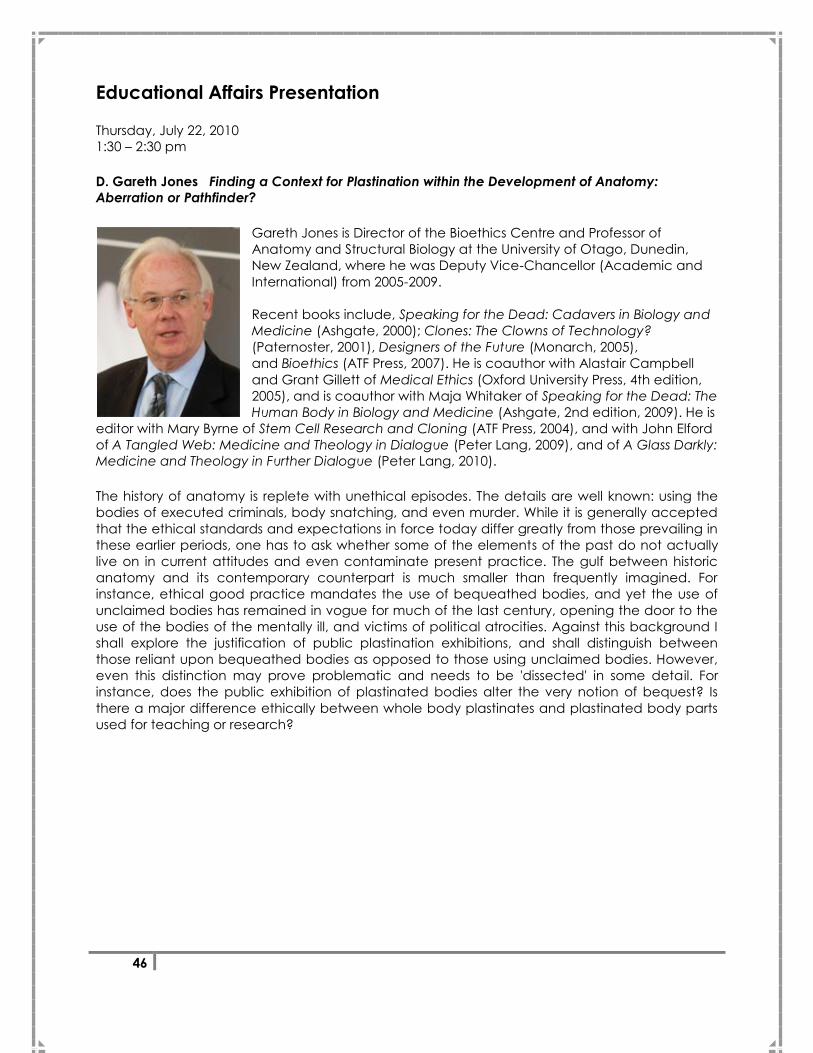
D. Gareth Jones Finding a Context for Plastination within the Development of Anatomy:
Aberration or Pathfinder?
Gareth Jones is Director of the Bioethics Centre and Professor of
Anatomy and Structural Biology at the University of Otago, Dunedin,
New Zealand, where he was Deputy Vice-Chancellor (Academic and
International) from 2005-2009.
Recent books include, Speaking for the Dead: Cadavers in Biology and
Medicine (Ashgate, 2000); Clones: The Clowns of Technology?
(Paternoster, 2001), Designers of the Future (Monarch, 2005),
and Bioethics (ATF Press, 2007). He is coauthor with Alastair Campbell
and Grant Gillett of Medical Ethics (Oxford University Press, 4th edition,
2005), and is coauthor with Maja Whitaker of Speaking for the Dead: The
Human Body in Biology and Medicine (Ashgate, 2nd edition, 2009). He is
editor with Mary Byrne of Stem Cell Research and Cloning (ATF Press, 2004), and with John Elford
of A Tangled Web: Medicine and Theology in Dialogue (Peter Lang, 2009), and of A Glass Darkly:
Medicine and Theology in Further Dialogue (Peter Lang, 2010).
The history of anatomy is replete with unethical episodes. The details are well known: using the
bodies of executed criminals, body snatching, and even murder. While it is generally accepted
that the ethical standards and expectations in force today differ greatly from those prevailing in
these earlier periods, one has to ask whether some of the elements of the past do not actually
live on in current attitudes and even contaminate present practice. The gulf between historic
anatomy and its contemporary counterpart is much smaller than frequently imagined. For
instance, ethical good practice mandates the use of bequeathed bodies, and yet the use of
unclaimed bodies has remained in vogue for much of the last century, opening the door to the
use of the bodies of the mentally ill, and victims of political atrocities. Against this background I
shall explore the justification of public plastination exhibitions, and shall distinguish between
those reliant upon bequeathed bodies as opposed to those using unclaimed bodies. However,
even this distinction may prove problematic and needs to be 'dissected' in some detail. For
instance, does the public exhibition of plastinated bodies alter the very notion of bequest? Is
there a major difference ethically between whole body plastinates and plastinated body parts
used for teaching or research?
47
Committee Reports
Executive Committee Report
Anatomical Services Committee
Bylaws Committee
Career Development Committee
Clinical Anatomical Terminology Committee
Educational Affairs Committee
Financial Affairs Committee
Journal Committee
Meeting Oversight and Planning Committee
Membership Committee
Membership Development Committee
Membership Outreach Committee
Nominating Committee
Professional Association Management Committee

48
Executive Committee Report
The Executive Committee (ExecComm), consisting of AACA‘s 6 officers, is charged in the Bylaws'
Article IV – Section 1 with making decisions and taking actions on behalf of the Council between
its meetings. At the 2009 meeting, Council directed the Executive Committee to hold monthly
teleconference meetings in order to dispense with the day to day business of the Association
that often burdens the Council when it meets and prevents it from fully deliberating topics of
broader concern for the future. The ExecComm held its first monthly meeting on August 4th less
than 3 weeks after the Cleveland meeting and has met monthly thereafter. In addition to our 12
monthly meetings, the ExecComm held 4 extra-monthly meetings. On April 15th, the Executive
Committee also called a special meeting of the Council as a whole to debate the terms and
conditions of the proposed Editorial Agreement drafted by the Journal Committee. In total, 14
members of Council participated in this meeting.
In addition to these teleconference meetings, ExecComm members exchanged over 200 e-
mails on the dedicated Listserve created by Julie Hewett. Each of our committees now has its
own listserve to facilitate communication amongst its members and to archive correspondence.
Since these lists came online in August, 1600+ emails have been exchanged to coordinate and
supplement committee teleconferences and other activities. The ExecComm received regular
reports from committees about their work and progress. As the chair of the ExexComm, I wish to
express my personal thanks to ExecComm members. Nearly every teleconference meeting had
100% of the committee present and participating for 1-2 hours.
From December through June, the major topic on the ExecComm‘s agenda was the ongoing
negotiations between the Journal Committee, chaired by Anne Agur, and Stephen Carmichael
over his reappointment to a third term as our journal‘s Editor-In-Chief. I am pleased to report that
these negotiations are completed and all parties have signed the agreement finalizing his
reappointment. This is a truly historic moment for Stephen and AACA since at the end of his term
in 2013 he will have served as the EIC of Clinical Anatomy for over half of our journal‘s existence.
Congratulations, Stephen. At the same time as these negotiations were taking place, the
ExecComm and myself, were chagrined to discover that the AACA had been acting out of
compliance with the legal agreement signed in 2007 with our publisher. After discussions with
John Wiley and the BACA President, Louise Scheuer, a new section was drafted for the Bylaws
redefining the composition and authority of the Journal Committee that placed the Association
in legal conformity with our Publisher Agreement. This Section was part of the Bylaws just
approved by the membership.
The Bylaws Committee, chaired by Carol Lomneth, did superb work and many of its
recommendations were present in the recently approved Bylaws amendments. There remain
additional items that need attention and a new charge will be issued in August. The reports to
the ExecComm by the Special Interest Committees: Anatomical Services, Career Development,
and Educational Affairs were particularly substantial and important. In addition to redefining
their scope and purpose – approved in the revised Bylaws – all three are well along in program
planning for their committee activities through 2012. This is very exciting news and will let us
organize and promote better meetings in the coming years. In the Fall, the Membership
Committee proposed a new process and documentation to be completed by applicants.
Again, these changes are reflected in the new Bylaws. The Nominating Committee, for the first
time in its history, submitted its list of nominees by the Jan. 1 deadline. This was greatly
appreciated by all concerned as it created ample time for the election to unfold. We owe a
vote of appreciation to all of the candidates who were willing to serve as well as to Neil Norton,
Tom Gest, Wayne Lambert and Shane Tubbs who were elected.
49
The Meeting Organization and Program Planning (MOPP) Committee met over 20 times in the
past 12 months. The first objective was to coordinate planning for future meetings beyond the
one we are now attending. Secondly, it worked closely with the Annual Meeting Committees
that have specific responsibility for each meeting. Since all members of the ExecComm are also
members of the MOPP, the ExecComm was kept closely in the loop of planning and decision
making. Your approval of the formal structure and charge of the MOPP and Annual Meeting
Committees in the recent Bylaws amendments provides us with a proven and robust means for
planning and implementing future meetings. Again, my congratulations to everyone involved.
The Clinical Anatomic Terminology Ad hoc Committee, chaired by Sherry Downie, has proven to
me that it has the potential to become a permanent substantial addition to our repertoire of
Standing Committees. The Committee‘s report, containing this recommendation to Council, is
printed in the Meeting Program. Finally, an important reason for the reports to be included here
is to reduce presentation time and increase discussion at Thursday‘s Annual Business Meeting
that you all are encouraged to attend.
As President, I am charge with announcing my appointments to fill committee vacancies prior to
the adjournment of Thursday‘s Annual Business Meeting. This is both the appropriate place and
time to announce my appointments to the following committees. The Bylaws are silent on
whether the President must designate committee chairs before or after the election of members
at the Special Interest Group or Business meetings, I have decided to wait until the New Council
Meeting on Friday, July 23rd to announce who will fill the committee chair vacancies.
Anatomical Services:
Len Cleary to full 3-year term
Carol Lomneth to 2-year unexpired term of now ex officio member Brandi Schmitt
Career Development:
Rebecca Lufler to full 3-year term
Anthony Olinger to 2-year unexpired elected term of Rebecca Pratt
Educational Affairs:
Rebecca Pratt to full 3-year term
Nominating:
Reappoint to 1-year terms, Shane Tubbs and Sherry Downie
Appoint to 1-year term, retiring Special Councilor David Porta
Clinical Anatomic Terminology Ad Hoc Committee:
Mark Hankin for 1-year term
50
Anatomical Services Committee (ASC)
The Anatomical Services Committee (ASC) represents both academic and technical members
of the Association. This special interest group functions to serve the Association‘s membership
through the development of symposia, courses and guidance documents, and promotes
technical and academic aspects of human anatomical specimens use in health care and
university education and research. The group advocates informed, ethical, safe operations for
students, faculty and researchers who contribute to health and education through the use of
anatomical materials. The ASC meets many times a year outside of the annual meeting by
conference call and in person to discuss agenda items that range from current legal or media
issues to the Association‘s revised committee descriptions and proposed bylaw changes to
topics for future symposia.
The AACA maintains an active listserv specific to anatomical services, which helps to facilitate
open discussion of pertinent issues. We use this listserv to raise awareness, communicate with
colleagues and to gather information. Surveys are distributed via this listserv as well as through
the main AACA listserv and response data is collected. The ASC regularly interacts with members
of other professional societies on topics of mutual interest. This past year has seen three of the
committee members serving as subject matter experts to the American Association of Tissue
Banks committee on non-transplantable tissue standards.
Other recent accomplishments and ongoing efforts of the ASC include the authorship of a
guidance document entitled ―Best Practices in Whole Body Donations Programs,‖ the
development and maintenance of contact information for all institutional whole body donation
programs, as well as the creation of a database to help disseminate information on access to
anatomical materials and related professional aspects of those who work in anatomical services
careers.
The 2010 symposium will take place on Thursday, July 22 from 3:30 to 4:30 and is entitled:
Consent, Tracking and Disposition of Anatomical Collections with Long Term Retention Periods:
Focus on Plastination
Please come to the ASC symposium, stop by our breakfast meeting on Wednesday morning or
get in touch with one of our committee members to learn more about how this Committee
serves the Association and its members. The current ASC committee includes:
Dean Fisher
Jon Jackson
Leon Martino
Angela McArthur
Darrell Petersen
Brandi Schmitt

51
Bylaws Committee
The Bylaws Committee met monthly by teleconference between August and February.
Amendments to the Bylaws were submitted to the Executive Committee on February 15, 2010,
were approved by the Executive Committee on May 18, 2010, and posted to the AACA website
for membership approval on May 18, 2010.
The 2010 Amendments to the Bylaws include both minor and major changes. There were two
substantial changes related to Article VIII which describes the purpose and organizational
structure of various committees. First, the Meeting Oversight and Program Planning (MOPP)
Committee, which has operated for the last year as an ad hoc committee, is now a Standing
Committee of the Association. In contrast to the current ―Program Committee‖, which is largely
concerned with annual meeting events, the breadth and scope of the MOPP Committee
extends over multiple years. This change will allow for more effective long-range planning of
meetings and programs. A second change to Article VIII is a new description and charge of the
Journal Committee. The AACA, BACA and John Wiley and Sons are in a contractual agreement
to publish Clinical Anatomy. In this agreement, AACA and BACA are joint owners of the journal
and management authority is proportional to the number of members authorized to receive the
journal. The new description of the Journal Committee is now consistent with the Publisher‘s
Agreement. Other, more minor changes in Article VIII include new descriptions of the Special
Interest Groups (SIGs). Members of the Education Committee, Career Development Committee
and Anatomical Services Committee were asked to review and update the description of their
respective SIGs. The new SIG descriptions reflect current practices.
There is a significant governance change in the Bylaws in Article IV Section 1, Subsection A. The
Executive Committee has the authority to act in behalf of the Council between Council
Meetings. The amendment adds a check and balance by requiring that the minutes of the
Executive Committee Meeting are to be provided to the Council in a timely manner and allows
Council to call for a meeting to review an action taken by the Executive Council on their behalf.
Another change of the Bylaws worth noting is an amendment submitted by the Membership
Committee which simplifies the application criteria for membership by eliminating a required
letter of recommendation. This change in membership criteria will facilitate electronic submission
of membership applications, making it easier for applicants to apply and will expedite the ability
of the Membership Committee to process the applications.
Finally, an important change in the Bylaws regarding elections is a transition clause that allows
for the staggered election of Council members. This amendment is necessary to adjust for a
previous change in the bylaws which affected the election sequence. The adjusted terms of
office are described in Article VII, Section 8 of the revised Bylaws.
Bylaws Committee Members: Jennifer Burgoon, Doug Gould, Craig Goodmurphy, Carol
Lomneth (Chairperson), Todd Hoagland

52
Career Development Committee
The Career Development Committee (CDC) plays an important role in the Association as a
committee designated to address career issues and the advancement of clinical anatomy
knowledge for an individual at any stage in their career. The CDC promotes fostering between
career anatomists and clinicians. We coordinate with the Educational Affairs Committee and
the Special Society Outreach & the Liaison Community Committee to provide an annual
symposium that enhances each committee‘s strengths. We recently developed and will
maintain a website for the AACA that displays current anatomy courses available in North
America and the Caribbean to enable anyone to expand their clinical anatomy skills.
The CDC is planning on developing a website to provide current postings and career
opportunities for those newly qualified as anatomists as well as clinicians who now are returning
to education to teach clinical anatomy. The following list describes the activities during this past
year.
1. Reviewed, updated and codified CDC charge for the bylaws of the AACA.
2. Developed and organized CDC symposium for Hawaii Conference.
3. Set out a 3-year CDC symposium calendar.
4. Coordinating with Educational Affairs Committee and the Special Society Outreach and
the Liaison Community Committee.
5. Developed and are maintaining an Anatomy Course website to enable anyone to
locate an anatomy course outside of those taught as part of a classic medical, dental,
osteopathic, chiropractic, PA, PT, and naturopathic curriculum. The website covers the
USA, Canada, and the Caribbean.
6. A website is being updated and developed for the AACA to enable people to place
current job opportunities and career advice.
7. Mentorship program is being rejuvenated with higher visibility with posting images of
mentors and areas of clinical expertise and research interests. These will be posted as a
link on the AACA website.
8. Will organize and host the Mentor reception and raffle at the Hawaii meeting.
9. Review and update judging forms and organize judging of The Ralph Ger Platform and
The Sandy Marks Poster Presentations.
Current Committee Members:
Chairs: Brion Benninger, OHSU and Todd Hoagland, BU
Members: Carol Lomneth, UNMC, Rebecca Pratt, MSU, Bill Swartz, LSU, Richie Nikfarjam, AECOM
The CDC 2010 would like to extend a special thanks to all the Publishers - Elsevier, Wolters
Kluwer/LWW, and Thieme - for their generous support towards the AACA Annual Mentor
Reception. Special thanks to Bone Clones for their generous support as well. Please take the
time to thank these people and the authors and mentors who are always supportive.

53
Clinical Anatomical Terminology (CAT) Ad Hoc Committee
The charge to the Clinical Anatomical Terminology (CAT) ad hoc committee was to investigate
the usage of clinical anatomical terms and propose mechanisms by which both clinical and
anatomical systems of nomenclature could be cross referenced in the same widely accessible
resource in order to (1) improve the educational experience for students and faculty, (2) simplify
communication between a broad spectrum of professionals in medical education, and (3) give
credence to the history of both of these closely related, but distinct, sets of terms.
The CAT committee met monthly by teleconference and, after extensive investigation,
submitted a proposal to Council outlining two different approaches to disseminating information
about synonymous clinical and anatomical terms. The first approach is web site delivery. We
have been in contact with the authors of two anatomical terminology web sites: Tom Gest at
the University of Michigan and Paul Gobee at Leiden University in the Netherlands. These authors
have invited us to add clinical anatomical terms to their already existing web sites. If approved
by Council, the AACA logo would be displayed on each of these sites and the AACA web site
would feature links to both sites. In addition, the AACA web site would host an interface where
visitors could submit terminology to the CAT committee for consideration of posting on the web
sites.
The second approach is publication of short essays in Clinical Anatomy. These 1-2 page essays,
ideally co-authored by an anatomist and a clinician, would present some interesting aspect of
the clinical and anatomical terms that are used to refer to the same body structure. It is hoped
that these essays would shed light on the development of both sets of terminology and foster
appreciation for their usage. The CAT committee would assume the responsibility for soliciting
and reviewing manuscripts for publication.
Because of the enormity of the proposed tasks and the on-going nature of these activities, the
CAT committee has requested of Council that it be instated as a standing committee of the
AACA.
If you are interested in becoming an active member of the Clinical Anatomical Terminology
committee, please contact one of its members.
2010-2011 CAT Committee Continuing Members:
Sherry Downie, Chair
Brion Benninger Tom Gest
Todd Hoagland Ahmed Khan
Marios Loukas Bradford Martin
Bill Rennie Alan Richards
Bill Swartz Pat Tank
Shane Tubbs
54
Educational Affairs Committee
The role of the Educational Affairs Committee (EAC) is to present current and developing
information about anatomical education and to hold discussions of clinical anatomy teaching
and learning that are relevant to members of the AACA. At the annual AACA meetings, we
sponsor a keynote speaker to highlight a current issue in anatomical education. This year's
speaker, Dr. Gareth Jones, is a world-famous medical ethicist who will be addressing the issue of
the ethics of plastination in his talk, "Finding a Context for Plastination Within the Development of
Anatomy: Aberration or Pathfinder?" EAC also organizes a symposium focused on anatomy
education at the annual meetings. This year's symposium will explore the educational uses of
plastinated anatomical material, while next year in Columbus the topic will be educational
research methods.
Each annual meeting features an Educational Affairs Breakfast (Thursday 7-8am this year), at
which various educational topics are discussed in small groups. This year in Hawaii, our focused
discussions will include assessment, curriculum planning, educational technology, educating the
next generation of anatomists, incorporating radiology and clinical relevance, and teaching
methods and curricular models. The Educational Affairs Breakfast is the official Educational
Affairs Committee meeting at the annual meetings, and an important function of this breakfast
meeting is the election of a new member of the Educational Affairs Committee. We encourage
all interested members to attend the breakfast meeting and become involved in the discussions
and election. The current members of the AACA Educational Affairs Committee members are:
Tom Gest (Chair) (2012)
Noelle Granger (2012)
Nirusha Lachman (2011)
Ameed Raoof (2011)
Cristian Stefan (2010)
Peter Ward (2010)
When not busy organizing our component of the annual meetings, the EAC is involved in
promoting clinical anatomy education through other means, including additions to the AACA
web site, such as the Frequently Asked Questions (currently under development), or articles in
Clinical Anatomy that address current concerns among anatomy educators.

55
Financial Affairs Committee
The Financial Affairs Committee oversees the budget and fiscal health of the Association. Part of
the charge is to oversee the establishment and management of an endowment fund, make
recommendations to the Council for maximizing financial resources, and review annually the
Association's financial records.
The Financial Affairs Committee has met regularly this past year to discuss continued financial
enhancements to the AACA. In particular, discussions have centered on establishing a process
for financial bequeathals to the AACA by members. Due to confidentiality, current details
relating to members cannot be discussed. The FAC will present their final recommendations to
Council.
Committee Members:
Lawrence M. Ross, Lonie R. Salkowski, Kenneth H. Jones, and Neil S. Norton, chair

56
Meeting Oversight and Program Planning (MOPP) Committee
The MOPP Committee represents the ―top of
the pyramid‖ in terms of planning and
overseeing annual meetings and
postgraduate courses. It is responsible for the
overall organization, general content, and
budget of each Annual Scientific Meeting and
Postgraduate Course, and plans and sets the
Association‘s long-term programmatic
objectives for future meetings.
The MOPP Committee was created by the
Council in late 2008 as an ad hoc committee
to replace the Program Committee. It has
since met virtually monthly via conference call.
With a year ―under our belts,‖ we have worked
to formalize the structure and activities of the
MOPP Committee into the Association‘s
Bylaws, specifically, Section 5 of Article VIII.
Organizational and planning decisions of the
MOPP are implemented through its Annual
Meeting Committee structure, and include:
preparing the Call for Abstracts, overseeing
abstract review, selection, and author
notification, organizing the content of
platform, techfair, and poster sessions, and
meeting program booklet preparation.
Considerable effort this year has gone into the
development of guidelines and procedures for
abstract submission and review, nomination
and selection of annual meeting program
chairs, and ―Call for Hosts‖ guidelines for
preparing a formal invitation to host a future
meeting. In addition, we have elevated the
role of SIG committees at the annual meetings
in recognition of their unique scholarly
contributions to our meetings and
opportunities for leadership development and
involvement by our membership.
The MOPP Committee has already begun
advanced planning for the 2011 Columbus
meeting and is finalizing their review of a
formal invitation for the 2012 meeting site.
The MOPP Committee is comprised of all
officers of the Association, the Chairs of the SIG
committees, the Chair of each Annual
Meeting Committee, and the Local Host for
each annual meeting. The Meeting Manager
serves as an ex officio non-voting member and
the Program Secretary serves as the Chair.
This year‘s MOPP Committee members are:
Executive Committee Members
Program Secretary, Mark Seifert
President, Todd R. Olson
Past President, Larry Ross
President-Elect, Anne Agur
Treasurer, Neil Norton
Association Secretary, Brian MacPherson
Special Interest Group Committee Chairs/Co-
Chairs
Anatomical Services, Brandi Schmitt
Anatomical Services, Leon Martino
Career Development, Brion Benninger
Career Development, Todd Hoagland
Educational Affairs, Tom Gest
Annual Program Committee Chairs
2009 Program Chair, Scott Lozanoff
2010 Program Chair, David Bolender
2011 Program Chair, Greg Smith
Local Hosts
2010 Hawaii, Scott Lozanoff
2011 Columbus, Kenneth Jones
AACA Meeting Manager
Julie Hewett, CMP
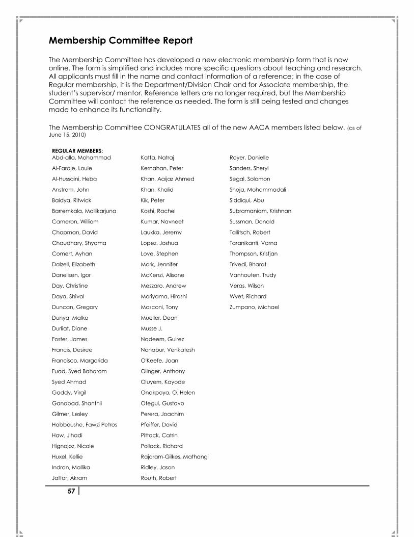
57
Membership Committee Report
The Membership Committee has developed a new electronic membership form that is now
online. The form is simplified and includes more specific questions about teaching and research.
All applicants must fill in the name and contact information of a reference; in the case of
Regular membership, it is the Department/Division Chair and for Associate membership, the
student‘s supervisor/ mentor. Reference letters are no longer required, but the Membership
Committee will contact the reference as needed. The form is still being tested and changes
made to enhance its functionality.
The Membership Committee CONGRATULATES all of the new AACA members listed below. (as of
June 15, 2010)
REGULAR MEMBERS:
Abd-alla, Mohammad Katta, Natraj Royer, Danielle
Al-Faraje, Louie Kernahan, Peter Sanders, Sheryl
Al-Hussaini, Heba Khan, Aaijaz Ahmed Segal, Solomon
Anstrom, John Khan, Khalid Shoja, Mohammadali
Baidya, Ritwick Kik, Peter Siddiqui, Abu
Barremkala, Mallikarjuna Koshi, Rachel Subramaniam, Krishnan
Cameron, William Kumar, Navneet Sussman, Donald
Chapman, David Laukka, Jeremy Tallitsch, Robert
Chaudhary, Shyama Lopez, Joshua Taranikanti, Varna
Comert, Ayhan Love, Stephen Thompson, Kristjan
Dalzell, Elizabeth Mark, Jennifer Trivedi, Bharat
Danelisen, Igor McKenzi, Alisone Vanhouten, Trudy
Day, Christine Meszaro, Andrew Veras, Wilson
Daya, Shival Moriyama, Hiroshi Wyet, Richard
Duncan, Gregory Mosconi, Tony Zumpano, Michael
Dunya, Malko Mueller, Dean
Durliat, Diane Musse J.
Foster, James Nadeem, Gulrez
Francis, Desiree Nonabur, Venkatesh
Francisco, Margarida O'Keefe, Joan
Fuad, Syed Baharom Olinger, Anthony
Syed Ahmad Oluyem, Kayode
Gaddy, Virgil Onakpoya, O. Helen
Ganabad, Shanthii Otegui, Gustavo
Gilmer, Lesley Perera, Joachim
Habboushe, Fawzi Petros Pfeiffer, David
Haw, Jihadi Pittack, Catrin
Hignojoz, Nicole Pollock, Richard
Huxel, Kellie Rajaram-Gilkes, Mathangi
Indran, Mallika Ridley, Jason
Jaffar, Akram Routh, Robert

58
AFFILIATE MEMBER:
Trivedi, Bharat
ASSOCIATE MEMBERS:
Alkhalili, Sereen Myers, Candice
Barrett, Richard Orsini, David
Bennet, Gregory Pace, Anna
Bishop, Amanda Patel, Shama
Bohl, Michael Patel, Bonny
Buchler, Lucas Peterson, Andrew
Burkholder, Alison Robinson Jr., William
Carr-Boyd, Peter Ross, Allen
Carroll, Melissa Sauks, Kate
Chen, Jian Schultz, Jason
Cromeens, Barrett Schwar, Angelika
Davies, Joel Shah, Suzanne
Delamarter, Taylor Shapir, David
Detton, Alan Splittgerber, Ryan
Eggum, Julianne Stone, Jonathan
Elswick, Lorna Strauchler, Daniel
Ford, Jonathan Takacs, Judit
Garrison, Ian Tamura, Kaori
Guenette, Melanie Tavangari, Ricky
Hartman, Lara Wenger, Lindsay
Johnson, Taylor Wilson, Axel
Kennedy, Bradley Wolpert, Joseph
Kloenne, Jessica Zhang, Haowei
Kummerling, Marissa
Lee, Francisco
Lee, Katherine
Littl, Jarrode
Maertins, Benjamin
Malloy, Kyle
Moore, Colin
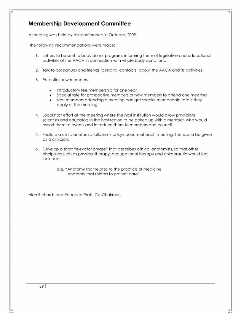
59
Membership Development Committee
A meeting was held by teleconference in October, 2009.
The following recommendations were made:
1. Letters to be sent to body donor programs informing them of legislative and educational
activities of the AACA in connection with whole body donations.
2. Talk to colleagues and friends (personal contacts) about the AACA and its activities.
3. Potential new members.
Introductory fee membership for one year
Special rate for prospective members or new members to attend one meeting
Non members attending a meeting can get special membership rate if they
apply at the meeting.
4. Local host effort at the meeting where the host institution would allow physicians,
scientists and educators in the host region to be paired up with a member, who would
escort them to events and introduce them to members and council.
5. Feature a clinic-anatomic talk/seminar/symposium at each meeting. This would be given
by a clinician.
6. Develop a short ―elevator phrase‖ that describes clinical anatomists, so that other
disciplines such as physical therapy, occupational therapy and chiropractic would feel
included.
e.g. ―Anatomy that relates to the practice of medicine‖
―Anatomy that relates to patient care‖
Alan Richards and Rebecca Pratt, Co-Chairmen
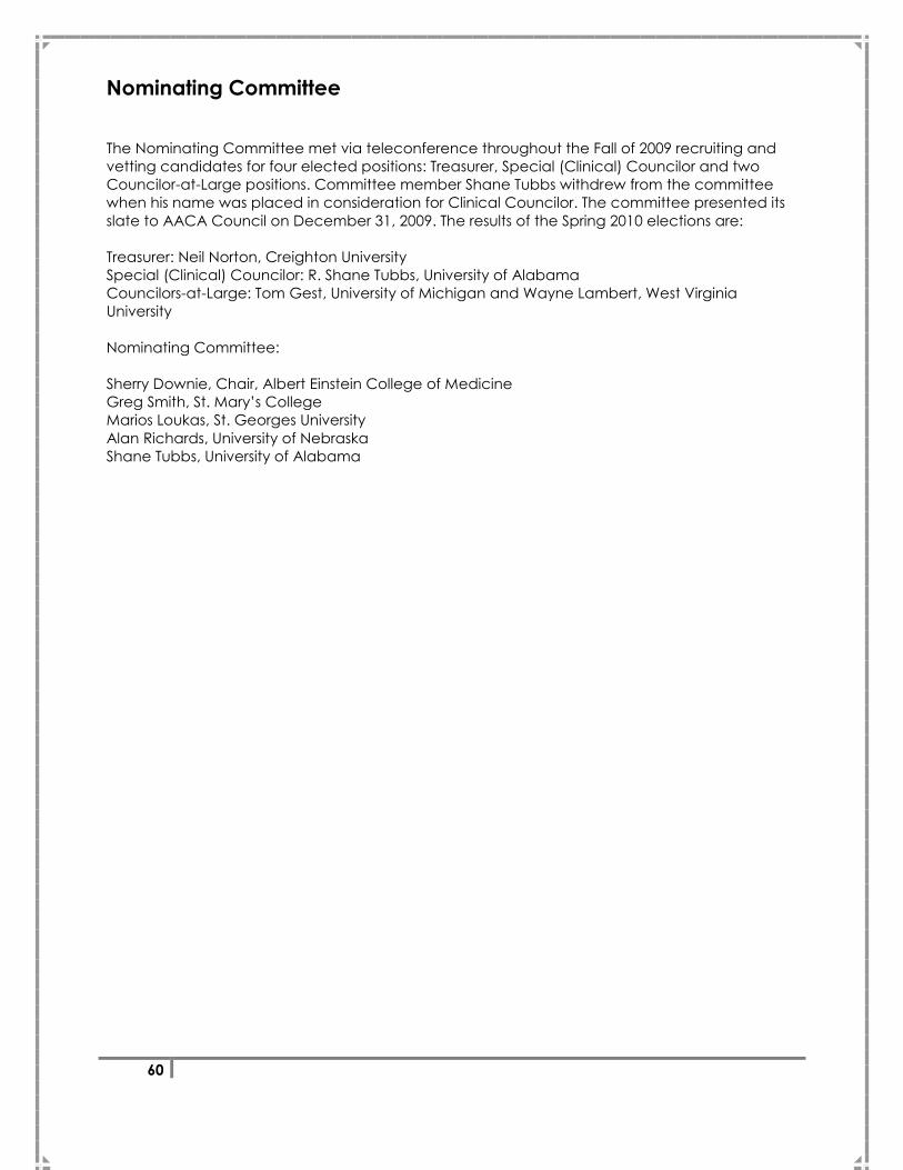
60
Nominating Committee
The Nominating Committee met via teleconference throughout the Fall of 2009 recruiting and
vetting candidates for four elected positions: Treasurer, Special (Clinical) Councilor and two
Councilor-at-Large positions. Committee member Shane Tubbs withdrew from the committee
when his name was placed in consideration for Clinical Councilor. The committee presented its
slate to AACA Council on December 31, 2009. The results of the Spring 2010 elections are:
Treasurer: Neil Norton, Creighton University
Special (Clinical) Councilor: R. Shane Tubbs, University of Alabama
Councilors-at-Large: Tom Gest, University of Michigan and Wayne Lambert, West Virginia
University
Nominating Committee:
Sherry Downie, Chair, Albert Einstein College of Medicine
Greg Smith, St. Mary‘s College
Marios Loukas, St. Georges University
Alan Richards, University of Nebraska
Shane Tubbs, University of Alabama
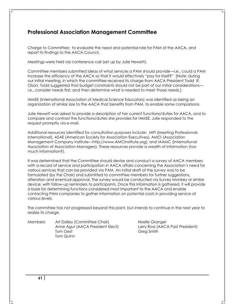
61
Professional Association Management Committee
Charge to Committee: to evaluate the need and potential role for PAM of the AACA, and
report its findings to the AACA Council.
Meetings were held via conference call (set up by Julie Hewett).
Committee members submitted ideas of what services a PAM should provide—i.e., could a PAM
increase the efficiency of the AACA so that it would effectively ―pay for itself?‖ [Note: during
our initial meeting, in which the committee received its charge from AACA President Todd R.
Olson, Todd suggested that budget constraints should not be part of our initial considerations—
i.e., consider needs first, and then determine what is needed to meet those needs.]
IAMSE (International Association of Medical Science Educators) was identified as being an
organization of similar size to the AACA that benefits from PAM, to enable some comparisons.
Julie Hewett was asked to provide a description of her current functions/duties for AACA, and to
compare and contrast the functions/duties she provides for IAMSE. Julie responded to the
request promptly via e-mail.
Additional resources identified for consultation purposes include: MPI (Meeting Professionals
International), ASAE (American Society for Association Executives), AMCI (Association
Management Company Institute—http://www.AMCInstitute.org), and IAAMC (International
Association of Association Managers). These resources provide a wealth of information (too
much information?).
It was determined that the Committee should devise and conduct a survey of AACA members
with a record of service and participation in AACA affairs concerning the Association‘s need for
various services that can be provided via PAM. An initial draft of the survey was to be
formulated (by the Chair) and submitted to committee members for further suggestions,
alteration and eventual approval. The survey would be conducted via Survey Monkey or similar
device, with follow-up reminders to participants. Once this information is gathered, it will provide
a basis for determining functions considered most important to the AACA and enable
contacting PAM companies to gather information on potential costs in providing service at
various levels.
The committee has not progressed beyond this point, but intends to continue in the next year to
realize its charge.
Members: Art Dalley (Committee Chair) Noelle Granger
Anne Agur (AACA President Elect) Larry Ross (AACA Past President)
Tom Gest Greg Smith
Tom Quinn
62
This page was intentionally left blank.
63
Poster Listing *Sandy Marks Award Nominee
+Ralph Ger Award Nominee
Poster Session 1 – Associate Member Posters, Extremities, Reproductive System
10-101*
Multifidus morphology and its relationship to common surgical back
approaches. SHAH, Suzanne, Jackson JONES and Brion BENNINGER.
Department of Surgery, Department of Integrative Biosciences,
Department of Oral Maxillofacial Surgery, Department of Orthopaedic
surgery, Oregon Health & Science University, Portland, OR, USA.
10-102*
Resident perceptions of anatomy education: A survey of medical school
alumni. BOHL, Michael A. and Thomas GEST, Ph.D. University of Michigan
Medical School, Anatomical Sciences. Ann Arbor, MI 48109, USA.
10-103*
Development of a learning module for imaging of the thorax with
anatomical correlation. RAVICHANDIRAN, Nisanthini, and Anne AGUR.
Division of Anatomy, Department of Surgery, University of Toronto, Toronto,
ON, M5S 1A8, CA.
10-104*
Use of plastinated specimens to convey learning concepts in Sports
Medicine and Kinesiology. TAMURA, Kaori. Department of Kinesiology and
Rehabilitation Science, University of Hawaii at Manoa, Honolulu, HI 96813,
USA.
10-105*
An active website listing current courses and symposia enabling career
development for a clinical anatomist. BURKHOLDER, Alison and Brion
BENNINGER. Department of Surgery, Department of Integrative
Biosciences, Department of Oral Maxillofacial Surgery, Oregon Health &
Science University, Portland, OR, USA.
10-106*
‗The Anatomy Grammys‘ – should graduate anatomy education include
skills of filming, production, directing, and editing using the Sky Eye
camera. ROSS, Allen and Brion BENNINGER. Department of Integrative
Biosciences, Department of Surgery, Department of Oral Maxillofacial
Surgery, Oregon Health & Science University, Portland, OR, USA.
10-107*
Does ultrasound training during the head and neck anatomy lab improve
dissection experience, dissection quality and examination scores.
BURKHOLDER, Alison and Brion BENNINGER. Department of Surgery,
Department of Oral Maxillofacial Surgery, Department of Integrative
Biosciences, Oregon Health & Science University, Portland, OR, USA.
64
10-108*
A three dimensional analysis of the innervation of the human lateral
pterygoid muscle. DAVIES, Joel C., Charles MAKEPEACE, David CANTELMI,
Bernie LIEBGOTT and Anne M. AGUR. Division of Anatomy, Department of
Surgery, University of Toronto, Toronto, ON, M5S 1A8, CA.
10-109*
Middle ear and hearing outcomes in cleft palate children: A Children‘s
Hospital Westmead study. CARR-BOYD, D. Peter and Fiona STEWART. The
Sydney Medical School, The University of Sydney, NSW 2006, Australia.
10-110*
What is the Schneiderian membrane, its historical account and clinical
importance? MALLOY, Kyle and Brion BENNINGER. Department of Surgery,
Department of Oral Maxillofacial Surgery, Department of Integrative
Biosciences, Oregon Health & Science University, Portland, OR, USA.
10-111*
The posteroinferior sub-parotid incision approach for condylar process
(subcondylar) fractures. SCHULTZ, Jason, Kevin ARCE and Brion
BENNINGER. Department of Surgery, Department of Oral Maxillofacial
Surgery, Department of Integrative Biosciences, Oregon Health & Science
University, Portland, OR, USA.
10-112*
Clinical anatomy of the nasal valve, does it merit anatomical recognition.
LEE, Katherine and Brion BENNINGER. Department of Surgery, Department
of Oral Maxillofacial Surgery, Department of Integrative Biosciences,
Oregon Health & Science University, Portland, OR, USA.
10-113*
Assessing validity of actual tooth height from cone beam images of
cadavers with subsequent dissection. PETERSON, Andrew and Brion
BENNINGER. Department of Oral Maxillofacial Surgery, Department of
Integrative Biosciences, Department of Surgery, Oregon Health & Science
University, Portland, OR, USA.
10-114*
A comparative analysis of popliteus muscle morphology. THAI, Al1, Enrique
J. ROBERTS2, Piroska L. SZABO3 and Anne M. AGUR1. 1Division of Anatomy,
Department of Surgery, University of Toronto, Toronto, Ontario, Canada. 2Faculty of Arts & Science, University of Toronto. 3Physical Therapy
Department, Touro College School of Health Sciences, Bay Shore, New
York, USA.
10-115*
Correlation of qualitative rating of ultrasound images to functional
performance in Duchenne Muscular Dystrophy. RAVICHANDIRAN,
Nisanthini, Christopher YUEN, Nancy MCKEE, and Anne AGUR. Division of
Anatomy, Department of Surgery, University of Toronto, Toronto, ON, M5S
1A8, CA.
65
10-116*
Intramuscular innervation of vastus medialis (VM): can VM be divided into
a longus and obliquus part? SAUKS, Kate, Melanie GUENETTE, Jane MENG,
Erin BOYNTON, Anne AGUR. Division of Anatomy, Department of Surgery,
University of Toronto, Toronto, ON, M5S 1A8, CA.
10-117 The posteromedial neurovascular bundle of the ankle: an anatomic study
using plastinated slices. SORA, Mircea-Constantin1, Petru MATUSZ2 , Radu
JILAVU1, Jan DRESENKAMP1. 1 Center for Anatomy and Cell Biology,
Medical University of Vienna, Austria. 2 Anatomical Department, University
of Medicine and Pharmacy "Victor Babes" Timisoara, Romania.
10-118 Novel application of an emerging technology in the rehabilitation of lower
extremity fractures. TAKACS, Judit1, Jeff LEITER1,2, and Jason D. PEELER1,2. 1Department of Human Anatomy and Cell Science, University of Manitoba,
Winnipeg, MB R3E 0J9, CA. 2Pan Am Clinic, 75 Poseidon Bay, Winnipeg, MB
R3M 3E4, CA.
10-119
Performing an oblique incision ACL reconstruction: Is there an infra-patellar
branch of saphenous nerve safe zone? PEELER, Jason1,2, and Jeff LEITER1,2. 1
Department of Human Anatomy and Cell Science, University of Manitoba,
Winnipeg, MB R3E 0J9, CA. 2 Pan Am Clinic, 75 Poseidon Bay, Winnipeg, MB
R3M 3E4, CA.
10-120
Case study: An anatomical variation in the tarsal tunnel. FITZSIMMONS,
John, M.D., Yohan JANG. Department of Radiology, College of
Osteopathic Medicine, Michigan State University, East Lansing, MI, USA.
10-121
Ending confusion in the sole. LENINGTON, Jake D., Mark BASSO, Erik
WALLIN, Jessica PELPHREY, Dennis E. MORSE, and Mark H. HANKIN.
University of Toledo, College of Medicine, Toledo, OH, USA.
10-122 Saving feet and legs from amputation through a knowledge and
application of clinical anatomy. GER, Ralph1, Kathy DOOLEY1,2, Sherry A.
DOWNIE1,3, and Todd R. OLSON1. 1Department of Anatomy and Structural
Biology, 3Department of Physical Medicine and Rehabilitation, Albert
Einstein College of Medicine, Bronx, NY 10461; 2New York Chiropractic
College, Seneca Falls, NY 13148, USA.
66
10-123*
Does trochlear shape modify the relationship between patella alta and
patelloefemoral joint cartilage damage? The MOST Study. STEFANIK,
Joshua J.1, Yanyan ZHU1, Ann C. ZUMWALT1, K. Douglas GROSS1, 2, Margaret
CLANCY1, John A. LYNCH3, Laura A. FREY-LAW4, Cora E. LEWIS5, Frank W.
ROEMER1, Christopher M. POWERS6, Ali GUERMAZI1, and David T. FELSON1. 1Boston University School of Medicine, Boston, Massachusetts, 02118. 2MGHIHP, Boston, Massachusetts, 02129. 3 UCSF, San Francisco, CA, 94107. 4UIowa, Iowa City, IA, 52242. 5C.E. Lewis, UAB, Birmingham, AL, 35205; 6USC,
Los Angeles, CA, 90089, USA.
10-124 Intraneural Ganglion Cysts in the Hip and Pelvic Region. SPINNER, Robert J.,
Marie-Noëlle HÉBERT-BLOUIN, and Kimberly K. AMRAMI. Mayo Clinic,
Rochester, MN 55905, USA.
10-125*
Clinical importance and accurate terminology of the distal
semimembranosus-muscle-tendon complex. DELAMARTER, Taylor and
Brion BENNINGER. Department of Surgery, Department of Integrative
Biosciences, Department of Oral Maxillofacial Surgery, Oregon Health &
Science University, Portland, OR, USA.
10-126*
Interpreting the clinical importance of the ‗os peroneum‘ by comparing
the rate in cadavers versus chronological roentogram rates. KLOENNE,
Jessica and Brion BENNINGER. Department of Surgery, Department of
Integrative Biosciences, Department of Oral Maxillofacial Surgery, Oregon
Health & Science University, Portland, OR, USA.
10-127*
Anatomical joint recognition from an antalgic gait as a helpful clinical
examination skill. JOHNSTONE, Taylor and Brion BENNINGER. Department of
Surgery, Department of Integrative Biosciences, Department of Oral
Maxillofacial Surgery, Oregon Health & Science University, Portland, OR,
USA.
10-128 Anatomic basis of medial calf perforator ablation. RUSSO, Alejandro M.,
Juan M. DEL CASTILLO, José A. STEFFANI, Andrea DE MARTINI, Eduardo
OLIVERA, Verónica BENTANCOUR. Anatomy Department, Universidad de
la República, Avenida General Flores 2125 ZC 11800, Montevideo,
Uruguay.
10-129 Anatomy of the plantaris muscle and its application to extraction and
repair of tendons. RUSSO, Alejandro M.1, María C. SIENRA1, Pablo V. VILLA1,
Alejandra NEIRREITTER1, Cecilia RUIBAL1, Marie C. POUMAYRAC1, Prof. Dr.
Víctor R. SORIA2. 1Anatomy Department, 2Facultad de Medicina,
Universidad de la República, Avenida General Flores 2125 ZC 11800,
Montevideo, Uruguay.
67
10-130 Anatomo – Ultrasonographic correlation of the medial calf perforators and
its application to endolaser surgery. RUSSO, Alejandro M.1, Juan M. DEL
CASTILLO1, José A. STEFFANI1, Andrea DE MARTINI1, Eduardo OLIVERA1, Prof.
Dr. Víctor R. SORIA2. 1Anatomy Department, 2Facultad de Medicina,
Universidad de la República, Avenida General Flores 2125 PC 11800,
Montevideo, Uruguay.
10-131 Distally based hemisoleus flap: Correlation between cadaveric and in vivo
studies. DEL CASTILLO, Juan M.1, Daniel A. WOLFF1, Gustavo ARMAND
UGON1, Jesús MANZANI1, Dr. Víctor R. SORIA2. 1Anatomy Department, 2Facultad de Medicina, Universidad de la República, Avenida General
Flores 2125 ZC 11800, Montevideo, Uruguay.
10-132*
Ligament and articular surface anatomic variability in the hindfoot.
CROMEENS, Barrett P., Cara FISHER, Harold J. SHEEDLO, Rustin E. REEVES.
Department of Cell Biology and Anatomy, University of North Texas Health
Science Center, Fort Worth, TX, 76107, USA.
10-133 Morphological variations in the pes anserinus: A cadaveric study on a
Dutch and American population. WICKS, Eric, Lucas SMITH and Lance G.
NASH. American University of the Caribbean School of Medicine,
Copecoy, Sint Maarten, Netherlands Antilles.
10-134*
Dermatome man – a 3D representation from evidence based spinal nerve
cutaneous mapping. DELAMARTER, Taylor, Brion BENNINGER and Mark D.
STRINGER. Department of Surgery, Department of Oral Maxillofacial
Surgery, Department of Integrative Biosciences, Oregon Health & Science
University, Portland, OR, USA. Department of Anatomy and Structural
Biology, Otago School of Medical Sciences, University of Otago, Dunedin,
New Zealand.
10-135 Concomitant antioxidant supplementation inhibits a combination therapy
of bleomycin, etoposide and cisplatin-induced oxidative stress and
apoptosis of germ cells in the testis. KILARKAJE, Narayana1, Maie AL-
BADER2, Alyaa MOUSA1, Khalid M. KHAN1. Department of Anatomy1 and
Physiology2, Faculty of Medicine, Kuwait University, PO Box 24923, Safat
13110, Kuwait.
10-136 Immunohistochemical localization of endothelial nitric oxide synthase in
human testis. GAWISH, Shefa M.1, Nadia M. EL-ROUBY2. 1Anatomy
Department, 2Histology Department, Dubai Medical College, Dubai, UAE.
68
10-137 Oviductal fluid can modulate the success of in vitro fertilization. A study in
the pig model. COY, Pilar, Francisco A. GARCÍA-VÁZQUEZ, Irene
MONDÉJAR and Carmen MATÁS. Department of Physiology, Faculty of
Veterinary, University of Murcia. Murcia 30071, Spain.
10-138 Effect of circulating sFlt-1 on trophoblastic cell line: a potential role in
preeclampsia. DHINGRA Renu1., Betsy VARUGHESE1, Kalpana LUTHRA2 ,
Neerja BHATLA3 ,Rajesh KALRA2 and Rani KUMAR1. 1Department of
Anatomy, 2 Department of Biochemistry, 3 Department of Obs and
Gynecology, All India Institute of Medical Sciences, New Delhi , India.
10-139 Oxidative stress and apoptosis in placentas of preeclamptic women. RANI,
Neerja1, Renu DHINGRA1, Neerja BHATLA2, Dharmveer Singh ARYA3, Mani
KALAIWANI4 and Rani KUMAR1. Department of Anatomy1, Gynecology2,
Pharmacology3 and Biostatistics4.All India Institute of Medical Sciences,
Ansari Nagar, New Delhi-110029, India.
10-140 Cadaveric conservation: Comparative study between formaldhyde and
non formaldehyde solution in human placenta. DEL CASTILLO, Juan M.1,
Daniel A WOLFF1, Gustavo J. ARMAND UGON1, Pablo V. VILLA1, Gabriela
NOVELLO1, Mariana FRIPP1, Andrea DE MARTINI1, Andrea BANCHERO1,
Nicole SPECKER1, Prof. Dr. Víctor R. SORIA2. 1Anatomy Department , 2Facultad de Medicina, Universidad de la República, Avenida General
Flores 2125 ZC 11800, Montevideo, Uruguay.
10-141*
Morphology and patterns of the internal intercostal muscle and their
relationship with rib fractures TAVANGARI, Ricky, Brion BENNINGER, Philip
HOMIER and Anthony OLINGER. Department of Surgery, Department of
Integrative Biosciences, Department of Oral Maxillofacial Surgery, Oregon
Health & Science University, Portland, OR, USA.
10-142 Comparative anatomical study of the lymphatic system of the upper
extremity in canine and human. SUAMI, Hiroo, Donghyeok SHIN, Fatih
UYGUR, and David W. CHANG. The University of Texas M. D. Anderson
Cancer Center, Houston, TX 77030, USA.
10-143 A possible relationship between reliability of thoracic outlet syndrome
diagnostic testing and the position of the axillary artery. Part II: The effect
of diagnostic testing on intra-arterial pressure. OLINGER, Anthony. Kansas
City University of Medicine and Biosciences, Kansas City, MO 64106, USA.

69
10-144 Penetration of the digital nerves by the common palmar digital arteries in
Koreans. KIM, Jeong-Nam, Ju-Young LEE, Yoo-Ri KIM, Wu-Chul SONG, and
Ki-Seok KOH. Department of Anatomy, School of Medicine, Konkuk
University, Seoul 143-701, South Korea.
10-145 Anastomosis of ulnar and median nerve in upper extremity. GIL,
Youngchun, Heejun YANG, Seongin JOHN, Hyejin CHO and Hyeyeon LEE.
Yonsei University, Seoul 120-752, South Korea.
10-146 Tridimensional anatomical model of the elbow joint of the dog. LATORRE,
Rafael1, Jorge ARREDONDO2, Ricardo SARRIA1, Mircea-Constantine SORA3,
Scott LOZANOFF4, María D. AYALA1, and Octavio LÓPEZ-ALBORS1. 1Department of Anatomy and Compared Pathologic Anatomy University
of Murcia, 30100, Spain. 2Department of Anatomy, Faculty of Veterinary
Medicine and Zootechny, Autonomous University of the State of Mexico,
50000, México. 3The Medical University of Vienna, A-1090, Austria.
4Department of Anatomy, Biochemistry and Physiology, John A. Burns
School of Medicine, Hawai, 96813, USA.
10-147 Case Study: An unusual axillary origin of the radial artery. FITZSIMMONS,
John, M.D., Amanda BISHOP. Department of Radiology, College of Human
Medicine, Michigan State University, East Lansing, MI, USA.
10-148 Two cadaver-cases of accessory subscapularis muscles with multiple
brachial plexus anomalies and axillary artery variations. DOWNIE1,2, Sherry
A., Ming-Ming LEE3, Anna PACE3, Michelle E. PARK3, David SHAPIRO3,
Jonathan A. STONE3, Eric W. BAKER1,5, Nogah HARAMATI4, and Todd R.
OLSON1. 1Department of Anatomy and Structural Biology, 2Department of
Physical Medicine and Rehabilitation, 3Medical Student Class of 2013, 4Department of Radiology, Albert Einstein College of Medicine, Bronx, NY
10461; 5Basic Science and Craniofacial Biology, New York University
College of Dentistry, New York, NY 10010, USA.
10-149 Demystifying Thoracic Outlet Syndrome (TOS): Symptoms, Anatomy and
Imaging. COLLINS, James D., E.H. SAXTON, H. GELABERT, Samuel S. AHN,
A.E. CARNES. Departments of Neurology, Radiological Sciences and
Vascular Surgery, UCLA, Los Angeles, CA, 90095, USA.
70
Poster Session 2 – Education, Neuroscience, Willed Body
10-150
Learner exam performance and preferences using interactive Web-based
instructional techniques. REILLY, Frank D. Department of Neurobiology and
Anatomy, West Virginia University School of Medicine, Morgantown, WV
26505-9128, USA.
10-151
Both perceived participation and satisfaction scores higher in small-group
sessions of 5 compared to 15 students. KOOLOOS, Jan, Tim KLAASSEN,
Mayke VEREIJKEN, Sacha VAN KUPPELVELD and Marc VORSTENBOSCH.
Radboud University Medical Centre, 6525 EZ, Nijmegen, The Netherlands.
10-152
Surgeons as educators: An untapped resource. HAUBERT, Lisa M., Robert
DEPHILIP, Julie Y. BISHOP, Marty TAM, Kenneth JONES, and Susan D.
MOFFATT-BRUCE. The Ohio State University Medical Center, Columbus, OH
43210, USA.
10-153
‖Post-natal‖ development of the Virtual Human Embryo project. CORK, R.
John, and Raymond F. GASSER. LSU Health Sciences Center, New Orleans,
LA 70112, USA.
10-154
The evaluation of the success of programmes in clinical anatomy. LOUW,
Graham J. Department of Human Biology, Faculty of Health Sciences,
University of Cape Town, Cape Town, Western Cape, Republic of South
Africa.
10-155
Maximizing Cadaver Usage and Student Dissection Time in Tight Curricular
Schedules. MACPHERSON, Brian R. and Pamela A. STEIN. Anatomy &
Neurobiology, The University of Kentucky, Lexington, KY 40536, USA.
10-156
Undergraduate student dissection courses in North America – are they
more widely needed? WHITMORE, Ian, John GOSLING and Sakti
SRIVASTAVA. Stanford School of Medicine, Stanford, CA 94305, USA.
10-157
The role of gender in first-year medical student self-efficacy for the
anatomy curriculum. BURGOON1, Jennifer M. and Noelle A. GRANGER2. 1Division of Anatomy, The Ohio State University College of Medicine,
Columbus, OH, USA. 2Department of Cell and Developmental Biology, The
University of North Carolina School of Medicine, Chapel Hill, NC, USA.
10-158
The Ohio State University college of medicine faculty teaching scholars
program: a foundational science perspective. GOULD, Douglas J. The
Ohio State University Division of Anatomy, Columbus, OH 43210, USA.

71
10-159
A Ten Year Longitudinal Experience With Interprofessional Education in
Gross Anatomy. MACPHERSON, Brian R., and Samuel R. FRANKLIN.
Anatomy & Neurobiology, University of Kentucky, Lexington, KY 40536,
USA.
10-160
Pacemaker or defibrillator? safe or dangerous? SILVERMAN, Bruce, David
J. ELIOT and Donald R. WILSON. Touro University California, Vallejo Ca.
94592, USA.
10-161
How did you get over there? The need for a commonly accepted
definition of ‗migration‘ when talking about embryonic development.
GASSER, Raymond F., and R. John CORK. LSU Health Sciences Center,
New Orleans, LA 70112, USA.
10-162
The Post Cardiac Surgery Cadaver—Teaching Opportunities and
Challenges WILSON, Donald R.1, Pedro B. NAVA1, David J. ELIOT2. 1Loma
Linda University, CA 92350. 2Touro University, CA 94592, USA.
10-163
Ten year experience with a clinical anatomy master‘s program. QUINN,
Thomas H, John A. YEE. Department of Biomedical Sciences, Creighton
University School of Medicine, Omaha, NE 68178, USA.
10-164
Trends in embryology and histology education in North American dental
schools. LEE, Lisa M.J., Dorothy T. BURK, H. Wayne LAMBERT. Ohio State
University, Columbus, OH 43210, USA.
10-165
A comparison of dissection versus plastinated prosections to teach the
anatomy of the hand. BENNETT-CLARKE Carol A., Carlos A. BAPTISTA, and
Richard D. LANE. Department of Neurosciences, University of Toledo
College of Medicine, Toledo, OH 43614, USA.
10-166
Developing an undergraduate curriculum in Second LifeTM. RICHARDSON,
April D., Jennifer K. BRUECKNER, and Brian R. MACPHERSON. Anatomy &
Neurobiology Department, College of Medicine, University of Kentucky,
Lexington, KY 40536, USA.
10-167
Elimination of cadaver dissection caused by hurricane Katrina: a look back
four years later. SWARTZ, William and Richard WHITWORTH, JR. Department
of Cell Biology and Anatomy, LSU Health Sciences Center, New Orleans, LA
70112, USA.

72
10-168
SmartPen®-Plastinate Integrated Tutorial System (S-PITS): A Self-directed
Learning Tool. BAPTISTA, Carlos A. C. Department of Neurosciences,
University of Toledo, College of Medicine, Toledo, OH, 43614, USA.
10-169
Gross Anatomy Mentors Program: Implementation of a newly designed
anatomy preparatory system. DOLL, Sara, Ralph NAWROTZKI, Jens STRELAU,
Anne SERWE, Joachim KIRSCH. University of Heidelberg, Institute of
Anatomy and Cell Biology, 69121 Heidelberg, Germany.
10-170
Use of heart plastinated slices to assist echorcardiography interpretation in
the dog. LATORRE, Rafael, Alejandro GÓMEZ, María J. FERNÁNDEZ DEL
PALACIO, Ricardo SARRIÁ, and Octavio LÓPEZ ALBORS. Dept. of Anatomy
and Comparative Pathology, and Dept. of Medicine and Surgery,
Veterinary Faculty, University of Murcia, Spain.
10-171
Testing medical students‘ knowledge of clinically-relevant surface
anatomy. HANKIN, Mark H.1, Carlos A.C. BAPTISTA1, Richard D. LANE1,
Dennis E. MORSE1, Sheryl A. MILZ2, Carol A. BENNETT-CLARKE1. 1Neurosciences, 2Public Health and Preventive Medicine, University of
Toledo, Toledo, OH, USA.
10-172
Student perceptions of an anatomy dissection experience before & after
hospital rotation placements. LARKIN, Theresa A., Darryl J. MCANDREW.
Graduate School of Medicine, University of Wollongong, Wollongong NSW
2522, Australia.
10-173
An example of the integration of anatomical and clinical skills education
within a medical curriculum. MCANDREW, Darryl J., Theresa A. LARKIN, Lisa
BROWN. Graduate School of Medicine, University of Wollongong,
Wollongong, New South Wales, 2522, Australia.
10-174
Methods and practicalities of administering an extended choice gross
anatomy lab practical exam. KING, Shannon M., Stanley ILIFF, Randal
BATCHELOR and Neal CROSS. Lincoln Memorial University- DeBusk College
of Osteopathic Medicine, Harrogate, TN 37752, USA.
10-175
Student learning styles and their impact on performance in the study of
human anatomy. SALKOWKSI, Lonie R.1,2, Nalin THAPAR4, Alejandro MUNOZ
DEL RIO1,3, and Karen KRABBENHOFT2. 1University of Wisconsin School of
Medicine and Public Health, Department of Radiology, 2Department of
Anatomy and 3Department of Medical Physics, and 4University of
Wisconsin, College of Letters and Science, Madison, Wisconsin 53792, USA.
73
10-176
Building a new integrated curriculum in clinical anatomy by blending
traditional teaching with clinical cases. COOKE, John M. Department of
Cell Biology, University of Massachusetts Medical School, Worcester, MA
01655, USA.
10-177
A biomedical skills research and education laboratory for anatomy
training and outreach. REEVES, Rustin E., Robin BELCHER, Cara FISHER, and
Harold J. SHEEDLO. Department of Cell Biology and Anatomy, University of
North Texas Health Science Center, Fort Worth, TX, 76107, USA.
10-178
Effectiveness of an Anatomy Video Dissector as a Guide to Dissection
CHICO, Diane E. and Thomas J. COLLINS. Dept. of Neuroscience and Cell
Biology, The University of Texas Medical Branch, Galveston, TX 77555-1069,
USA.
10-179
Staining of sections planned for sheet plastination. DALL, Annette M. and
John CHEMNITZ. Neurobiological Science, University of Southern Denmark,
5230 Odense M, Denmark.
10-180
Integrating human clinical and comparative anatomy with marine science
during interactive public shark necropsies as an outreach to K-12 students,
science teachers and the public. BENNINGER, Brion and William
HANSHUMAKER. Department of Surgery, Department of Integrative
Biosciences, Department of Oral Maxillofacial Surgery, Oregon Health &
Science University, Portland, OR, Hatfield Marine Science Center, Oregon
Sea Grant Oregon State University, Newport OR, USA.
10-181
Does ultrasound training in head and neck anatomy lab during the
dissection of extra-oral structures improve dissection confidence and
proficiency in learning extra-oral examinations? LEE, Ilkyu and Brion
BENNINGER. Department of Oral Maxillofacial Surgery, Department of
Surgery, Department of Integrative Biosciences, Oregon Health & Science
University, Portland, OR, USA.
10-182
Confidence and competence of physician assistant student‘s
performance of knee aspiration/injection using lightly embalmed
cadavers vs. video training: A pilot study. KEIM JANSSEN, Sarah A., and
Stephane P. VANDERMEULEN. Department of Genetics, Cell Biology and
Anatomy and the Division of Physician Assistants, University of Nebraska
Medical Center, Omaha, NE 68198, USA.
10-183
Introducing a novel team-based approach in embryology teaching and
learning. SCOVILLE Elizabeth Ann1, Nirusha LACHMAN2 and Wojciech
PAWLINA2. 1Mayo Medical School and 2Department of Anatomy, Mayo
Medical School, Rochester, MN, 55901, USA.
74
10-184
Evaluations in the clinical anatomy course and its projection into the Final
Exam. DEL CASTILLO, Juan M.1, Eduardo OLIVERA1, Víctor R. SORIA2,
Gustavo ARMAND UGÓN1, Sebastián LAZA1, Alejandro RUSSO1, Natalia
FREIJIDO1, Alejandra JAUME1. 1Departamento de Anatomía, 2Facultad de
Medicina, Universidad de la República. Montevideo, Uruguay.
10-185
Clinical workshop as a tool for teaching in anatomy: evaluation by the
students of the second generation of Clinical Anatomy. LAZA Sebastián1,
María E. VERGARA1, Fernando MARTINEZ1, Germán DECUADRO1, Natalia
SORRENTI1, Inés DOASSANS1, Marie C. POUMAYRAC1, Fernanda NOZAR1,
Gustavo ARMAND UGON1, Prof. Dr. Víctor R. SORIA2. 1Anatomy
Department, 2Facultad de Medicina, Universidad de la República,
Avenida General Flores 2125 PC 11800, Montevideo, Uruguay.
10-186
Using anatomical models. Radiological anatomical correlation in cardiac
electrophysiology. LAZA Sebastian1, Natalia SORRENTI1, Maria E.
VERGARA1, Marie C. POUMAYRAC1, Diego FREIRE1, Prof. Dr. Víctor R.
SORIA2, Dr. Alvaro RIvara, M.D. 1Anatomy Department, 2Facultad de
Medicina, Universidad de la República, Avenida General Flores 2125 PC
11800, Montevideo, Uruguay.
10-187
Pioneers in Cardiovascular Imagiology. FRANCISCO, Margarida F., Antonio
F. FRANCISCO. Faculdade de Medicina de Lisboa, Portugal.
10-188
The Brain Book: Plastination and Lamination of Brain Cross Sections. MITRA,
Aditi, Linda SAAB, Lauren MARCHESE, Adriane MARCHESE, Ameed RAOFF.
Division of Anatomical Sciences/Plastination, University of Michigan, Ann
Arbor, MI 48109, USA.
10-189
Effect of lead on expression of serine/threonine phosphatases in rat
hippocampus: an immunohistochemical study. KHAN, Khalid M.1, Tahany
E. AL-SHAMARY1, Saju S. JACOB1, Sreeja ATTUR 1, Islam KHAN2, Abdur
RAHMAN3. Department of Anatomy1 and Biochemistry2, Faculty of
Medicine, Department of Family Sciences3, College for Women, Kuwait
University, Kuwait, P.O. Box 2492.
10-190
Effects of concomitant administration of lithium and cadmium on the
corpus striatum of rat brain - A histological study. ALI, M. Hassan.
Department of Anatomy, Jawahar Lal Medical College, Aligarh Muslim
University, Aligarh, UP 2002002, India.
75
10-191
A study of lateralization among professional footballers. MCNEIL, T. Rosie,
Ewere, EKWERE O, Olaleke ONI. School of Anatomical Sciences, University
of the Witwatersrand, Johannesburg. Department of Anatomy, University
of Jos, Jos, Nigeria.
10-192
The amygdala and its vascular relationship DOASSANS Ines, Natalia
SORRENTI, Marie C. POUMAYRAC, Sebastian LAZA. Departamento de
Anatomía , Facultad de Medicina, Universidad de la República.
Montevideo, Uruguay.
10-193
Proposal for standardized infection control guidelines in the gross anatomy
laboratory: a golden teaching opportunity. BEALE, Elmus G., and Gary
SIMPSON. Department of Medical Education, Paul L. Foster School of
Medicine, Texas Tech University Health Sciences Center, El Paso, TX 79905,
USA.
10-194
Identifying the ethical principles that guide leaders of human body
donation programs. MUELLER, Dean A. University of Michigan Medical
School, Ann Arbor, MI 48109, USA.

76
This page was intentionally left blank.

77
Poster Session 3: Abdomen, Head and Neck, Thorax
10-195
An anatomical study of the blood flow patterns of the TRAM flap and a
new theory on the blood flow. KOGA, Noriyuki, Koichi WATANEBE, Tsuyoshi
SAGA, Moriyoshi NAKAMURA, Kei-ichiro NAKAMURA, Koh-ichi YAMAKI.
Department of Anatomy, Kurume University School of Medicine, Kurume,
Japan.
10-196
Anatomical Variations in the Extra-Hepatic Biliary Apparatus. NADEEM,
Gulrez. Department Of Basic Medical Sciences, College Of Medicine,
University Of Sharjah, Sharjah, UAE.
10-197
MRI anatomy of the porcine pancreas. LATORRE, Rafael1, Wendy
HERNÁNDEZ2, Ester PÁRRAGA1, Gregorio RAMÍREZ1, Ricardo SARRIÁ1,
Francisco SÁNCHEZ MARGALLO3 and Octavio LÓPEZ ALBORS1. 1Dept.
Anatomy and Comparative Pathology, University of Murcia, Spain. 2Department of Anatomy, Faculty of Veterinary Medicine and Zootechny,
Autonomous University of the State of Mexico, México. 3Minimally Invasive
Surgery Centre Jesús Usón, Cáceres, Spain.
10-198
Middle hepatic vein in the economy of venous drainage of the liver: Study
on corrosion casts. MATUSZ, Petru, Agneta Maria PUSZTAI, and Eniko-
Christine HORDOVAN. Department of Anatomy, ―Victor Babes‖ University
of Medicine and Pharmacy, Timisoara 300041, Romania.
10-199
Numerical correlation of afferent elements of caudate lobe: Study on
corrosion casts. MATUSZ, Petru. Department of Anatomy, ―Victor Babes‖
University of Medicine and Pharmacy, Timisoara 300041, Romania.
10-200
Cadaveric case study of a hemodialysis patient with end stage renal
disease and multiple arteriovenous fistulas. OLSON, Todd R.1, Marissa L.
KUMMERLING2, Jasmine STEPHENS5, Vaughn W. FOLKERT3, and Sherry A.
DOWNIE 1,4. 1Department of Anatomy and Structural Biology, 2Medical
Class of 2012, 3Department of Medicine, 4Department of Physical Medicine
and Rehabilitation, Albert Einstein College of Medicine. Bronx, NY 10461. 5Clarkson University, Potsdam, NY 13699, USA.
10-201
Variation in the relationship of Right Hepatic Artery to Common Hepatic
Duct in Indian Population – A Comparative Cadaveric Study. MISHALL, Priti
L. and Lakshmi RAJGOPAL. Department of Anatomy, Seth G.S Medical
College and K.E.M. Hospital, Parel, Mumbai - 400012, MH, India.
10-202
Pathology in the transverse colon associated with pancreatitis. PATEL, H.
Bonny, and Shama PATEL. Ross University, Dominica, West Indies.
78
10-203
Anatomic basis of the thoracoscpic splanchnicectomy for the treatment
of solaralgias. VERGARA Maria E., Pablo DIBARBOURE, Laura BORGNO,
Varina SCANNIELLO, Guillermo CARRIQUIRY.
10-204
Nature of deep cervical fascia: What did the original investigations tell us?
ZHANG, Ming. Department of Anatomy, Beijing Capital Medical University,
China, and Department of Anatomy and Structural Biology, University of
Otago, Dunedin, New Zealand.
10-205
Proprioception in the human larynx. WEIR, Kristy A., Seamus DELAHUNTY,
and Stuart B. MAZZONE. School of Biomedical Sciences, The University of
Queensland, QLD, 4072, Australia.
10-206
A study on the incidence of atheromas of the internal carotid artery in four
anatomical regions of the head and neck using cone beam computed
tomography. NORTON, Neil S., Paul C. EDWARDS, Tarnjit S. SAINI, Laura C.
BARRITT, and Margaret A. JERGENSON. Creighton University, Omaha, NE
68178, USA.
10-207
Functional anatomy of human scalene musculature: rotation of the
cervical spine. OLINGER, Anthony, Phillip HOMIER. Kansas City University of
Medicine and Biosciences, Kansas City, MO 64106, USA.
10-208
Histological analysis of tooth development in a conditional Dicer knockout
mouse. BARRITT, C. Laura1, Joseph M. MILLER1, Garret A. SOUKUP2, and
Sonia M. ROCHA-SANCHEZ1. Department of Oral Biology1, Biomedical
Sciences2, Creighton University, Omaha, NE 68178, USA.
10-209
Comparison of cranial measurements between real bone and three
dimensional model. LEE, U-Young1, In-Beom KIM1, Ki-Hwan HAN2, Jung-Ho
CHA1, Jin KIM1, Myung-Hoon CHUN1, and Seung-Ho HAN1. 1Catholic
Institute for Applied Anatomy and Department of Anatomy, College of
Medicine, The Catholic University of Korea, 505 Banpo-dong, Seocho-gu,
Seoul, 137701, Republic of Korea. 2Department of Anatomy, Ehwa
Womans University School of Medicine, 911-1 Mok-dong, Yancheon-gu,
Seoul, 158710, Republic of Korea.
10-210
Triangulation applied to facial and recurrent laryngeal nerve localization: Is
math the answer? SEIFERT, Mark, Mark ROYER, Ahmed SUFYAN, and Mimi
KOKOSKA. Department of Anatomy and Cell Biology, Department of
Otolaryngology-Head and Neck Surgery, Indiana University School of
Medicine and Surgical Services, Richard L. Roudebush VA Medical Center,
Indianapolis, IN 46202, USA.
79
10-211
Merkel cell carcinoma metastatic to superficial anterior jugular node
detected by sentinel lymph node mapping. DEARING, S. Elizabeth, David
A. SLOAN, Richard A. POLLOCK, Adria HARTMAN. Chandler Medical
Center, University Kentucky, Lexington, KY 40536-0232, USA.
10-212
Microbuttresses of the orbit: preannular shelf, optic strut, and other features
germane to operative intervention. POLLOCK, Richard A. Chandler
Medical Center, University of Kentucky, Lexington, KY 40536-0232, USA.
10-213
An anatomic study of the inferior bundle (fourth band) of the buccinator
and the incisivus labii inferioris muscle. HUR Mi-Sun1, Jung-Su WOO1, Kyung-
Seok HU2, Hee-Jin KIM2, and Kyu-Seok LEE1. 1Department of Anatomy,
Kwandong University College of Medicine, Naegok-Dong 522, Gangneung
210-701, Korea. 2Division in Anatomy & Developmental Biology,
Department of Oral Biology, Human Identification Research Center, Brain
Korea 21 project, Yonsei University College of Dentistry, 250 Seongsanno,
Seodaemoon-gu, Seoul, 120-752, South Korea.
10-214
Microanatomy of infraorbital and its associated small canals in the maxilla.
KIM2, Soo-Young, Ja-Young YOO2, Soon-Cheol PARK1, Wu-Chul SONG2,
and Ki-Seok KOH2. Department of Anatomy, School of Medicine, Konkuk
University, Seoul 143-701, 1 Department of Life Science, ChungAng
University, Seoul 156-756, South Korea.
10-215
Analysis of the midface, focusing on the nose: an anthropometric study in
young Koreans. KIM, Soon-Heum2, Euna WHANG2, Hyun Gon CHOI2, Dong
Hyeok SHIN2, Ki Il UHM2, Wu-Chul SONG1, and Ki-Seok KOH2. 2Department of
Plastic and Reconstructive Surgery, 1Department of Anatomy, School of
Medicine, Konkuk University, Seoul 143-701, South Korea.
10-216
An anatomic study of the nasal skeletons in Korean. KIM, Yoo-Ri1, Ju-Young
LEE1, Jeong-Nam KIM1, Wu-Chul SONG1, Ki-Seok KOH1, and Soon-Heum
KIM2. 1Department of Anatomy, 2Department of Plastic and Reconstructive
Surgery, School of Medicine, Konkuk University, Seoul 143-701, South Korea.
10-217
Topographic relationship among transverse facial artery, zygomatic
branch and buccal branch of facial nerve, and parotid duct in the
midface. LEE, Ju-Young1, Yoo-Ri KIM1, Jeong-Nam KIM1, Wu-Chul SONG1,
Ki-Seok KOH1, and Hyun Gon CHOI2. 1Department of Anatomy, 2Department of Plastic and Reconstructive Surgery, School of Medicine,
Konkuk University, Seoul 143-701, South Korea.
10-218
Bilateral anomalies of the internal carotid artery. JERGENSON, Margaret A.,
Neil S. NORTON, and Laura C. BARRITT. Creighton University School of
Dentistry, Omaha, NE 68178, USA.

80
10-219
Clinical anatomy of the relation between the temporomandibular joint
and the mandibular nerve in the cat. LATORRE, Rafael1, Jorge
ARREDONDO2, María J. RODRÍGUEZ1, Amalia AGUT1, José M. VAZQUEZ-
AUTÓN1 and Octavio LÓPEZ-ALBORS1. Department of Anatomy and
Compared Pathologic Anatomy and Department of Animal Medicine and
Surgery, University of Murcia, 30100, Spain1. Department of Anatomy,
Faculty of Veterinary Medicine and Zootechny, Autonomous University of
the State of Mexico, 50000, México2.
10-220
Clinical anatomy of the relation between the temporomandibular joint
and the middle ear in horses. LATORRE, Rafael1, María J RODRÍGUEZ 1,
Octavio LÓPEZ-ALBORS1, Jorge ARREDONDO2, Francisco GIL1 and Amalia
AGUT1. 1Dept. of Anatomy and Comparative Pathology, and Dept. of
Medicine and Surgery, Veterinary Faculty, University of Murcia, Spain. 2Dept Anatomy, Faculty of Veterinary Medicine and Zootecny,
Autonomous University of the State of México, México.
10-221
Gross anatomical observations on supernumerary fibers of superior
pharyngeal constrictor. SAKAMOTO, Yujiro. Tokyo Medical and Dental
University. Tokyo 113-8549, Japan.
10-222
Topographical anatomy of the transverse facial artery. YANG, Hee-Jun,
Young-Chun GIL and Hye-yeon LEE. Department of Anatomy, Yonsei
University Medical College, Seoul, 120-752, Korea.
10-223
Does whitening adversely affect tooth strength?PORTA, David, Becca
YOUNG, Valerie KEY and Blake GARR. Bellarmine University, Louisville, KY
40205, USA.
10-224
An anatomic study of lateral muscular band of orbicularis oculi muscle.
PARK, Jong-Tae1, Kwan-Hyun YOON1, Yong-Ho KIM2, Hyun-Ho KWAK2,
Kyung-Seok HU1, and Hee-Jin KIM1. 1Division in Anatomy & Developmental
Biology, Department of Oral Biology, Human Identification Research
Center, Brain Korea 21 Project, Yonsei University College of Dentistry, 250
Seongsanno, Seodaemoon-gu, Seoul, 120-752, Korea. 2Department of Oral
Anatomy, School of Dentistry, Pusan National University, Busan, Korea.
10-225
Topography of the arteries supplying the masseter; dissection and Sihler's
staining. WON, Sung-Yoon1, Da-Yae CHOI1, Yong-Ho KIM2, Hyun-Ho KWAK2,
Hee-Jin KIM1, and Kyung-Seok HU1. 1Division in Anatomy & Developmental
Biology, Department of Oral Biology, Human Identification Research
Center, Brain Korea 21 project, Yonsei University College of Dentistry, Seoul,
Korea, 2Department of Oral Anatomy, School of Dentistry, Pusan National
University, Busan, Korea.

81
10-226
Morphometric analysis of Korean mandibular ramus for distraction
osteogenesis using micro-CT. LEE, Jae-Gi1, Da-Yae CHOI1, Jong-TAE PARK1,
Young-Woo KIM2, Yun-Hwa JUNG2, Soo-Yeon KIM2, Kyung-Seok HU1, and
Hee-Jin KIM1. 1Division in Anatomy and Developmental Biology,
Department of Oral Biology, Human Identification Research Center, Brain
Korea 21 Project, 2Undergraduate student, Yonsei University College of
Dentistry and School of Dentistry, Seoul, Korea.
10-227
Morphometric analysis of alveolar regions for the miniscrewing and
immediate implantation. KIM, Da-Hye, Sung-Yoon WON, Kyung-Seok HU,
and Hee-Jin KIM. Division in Anatomy & Developmental Biology,
Department of Oral Biology, Human Identification Research Center, Brain
Korea 21 project, Yonsei University College of Dentistry, 250 Seongsanno,
Seodaemoon-gu, Seoul, 120-752, Korea.
10-228
Morphometric analysis of the condyle of Korean mandible using micro-CT.
CHOI, Da-Yae, Sung-Yoon WON, Jae-Gi LEE, Kyung-Seok HU, and Hee-Jin
KIM. Division in Anatomy & Developmental Biology, Department of Oral
Biology, Human Identification Research Center, Brain Korea 21 project,
Yonsei University College of Dentistry, 250 Seongsanno, Seodaemoon-gu,
Seoul, 120-752, Korea.
10-229
Positional relationship between the orbital rim and orbicularis oculi muscle.
KIM, Hyun-Ju1, Kwan-Hyun YOON1, Mi-Sun HUR2, Yong-Ho KIM3, Hyun-Ho
KWAK3, Jong-Tae PARK1, Kyung-Seok HU1, and Hee-Jin KIM1. 1Division in
Anatomy & Developmental Biology, Department of Oral Biology, Human
Identification Research Center, Brain Korea 21 project, Yonsei University
College of Dentistry, 250 Seongsanno, Seodaemoon-gu, Seoul, 120-752,
Korea. 2Department of Anatomy, Kwandong University College of
Medicine, Naegok-Dong 522, Gangneung 210-701, Korea. 3Department of
Oral Anatomy, School of Dentistry, Pusan National University, Busan, Korea.
10-230
Morphological patterns of the zygomaticus minor and its relationship with
the orbicularis oculi mucle. KANG Hyun-Ju1, Kwan-Hyun YOON1, Mi-Sun
HUR2, Yong-Ho KIM3, Hyun-Ho KWAK3, Jong-Tae PARK1, Kyung-Seok HU1,
and Hee-Jin KIM1. 1Division in Anatomy & Developmental Biology,
Department of Oral Biology, Human Identification Research Center, Brain
Korea 21 project, Yonsei University College of Dentistry, 250 Seongsanno,
Seodaemoon-gu, Seoul, 120-752, Korea. 2Department of Anatomy,
Kwandong University College of Medicine, Naegok-Dong 522, Gangneung
210-701, Korea. 3Department of Oral Anatomy, School of Dentistry, Pusan
National University, Busan, Korea.

82
10-231
Anatomy – Ultrasound correlation of the larynx. POUMAYRAC, Marie C,
Ines DOASSANS, Natalia SORRENTI, Sebastian LAZA, Jose A. STEFFANI.
Departamento de Anatomía , Facultad de Medicina, Universidad de la
República. Montevideo, Uruguay.
10-232
A novel tool to navigate to the round window: A cadaveric pilot study.
PRAETORIUS, Mark, Philipp VAN DE WEYER, Ioana HERISANU, Peter K.
PLINKERT, Sara DOLL and Stefan ROHDE. University of Heidelberg Medical
Center, D-69120 Heidelberg, Germany.
10-233
Anatomical localization of submandibular gland for botulinum toxin
injection. LEE Je-hun1, Be-na LEE2, Yong-seok NAM1, Dai-soon KWAK1, Mi-
sun LEE1, Seung-Ho HAN1. 1Department of Anatomy ∙ Catholic Institute for
Applied Anatomy, College of Medicine, The Catholic University of Korea,
Seoul, Korea. 2Department of Rehabilitation Medicine, College of
Medicine, The Catholic University of Korea, Seoul, Korea.
10-234
Anatomic variations of the nasal fossae and sinuses with implementation in
endoscopic nasosinus surgery. SAIBENE, Andrés1, Agustín STEFFANI1, Jorge
RAPALINI1, Alejandro RUSSO1, Victor R. SORIA VARGAS2. 1Anatomy
Department, 2Facultad de Medicina, Universidad de la República,
Avenida General Flores 2125 PC 11800, Montevideo, Uruguay.
10-235
A semiquantitative approach to variation of the azygos vein course.
BALES, Gerald S. Department of Anatomy, Western University of Health
Sciences, Pomona, CA, 91766 USA.
10-236
The relationship of the paired anterior and posterior intercostal arteries: A
cadaveric study. LANE, Richard D., David R. KUHLMAN, Mark H. HANKIN,
and Carol A. BENNETT-CLARKE. Department of Neurosciences, University of
Toledo College of Medicine, Toledo, OH 43614, USA.
10-237
The evolution of the surgical management of chest wall deformities in
children. WEBER, Thomas R.1,2, Michael J. DINGELDEIN3, Ami SHAH1 and
Mark J. HOLTERMAN 1,2,3. 1University of Illinois College of Medicine,
Chicago, IL 60612. 2Advocate Hope Children‘s Hospital, Oak Lawn IL
60453. 3Rush University College of Medicine, Chicago, IL 60612, USA.
10-238
Anatomical considerations for vascular access in children. HOLTERMAN,
Mark J.1,2, Ami SHAH1, Michael J. DINGELDEIN3, and Thomas R. Weber1,2.
1University of Illinois College of Medicine, Chicago, IL 60612. 2Advocate
Hope Children‘s Hospital, Oak Lawn IL 60453 and 3Rush University College
of Medicine, Chicago, IL, USA.

83
10-239
Standardized rib fracture classification system as a future paradigm for
treating single, multiple and flail segment fractures. BENNINGER, Brion, John
MAYBERRY. Department of Surgery, Department of Oral Maxillofacial
Surgery, Department of Integrative Biosciences, Oregon Health & Science
University, Portland, OR, USA.
10-240
A case study of the collateral central venous circulation associated with
superior vena caval occlusion. BEST, M. Irwin. Emory University School of
Medicine, Atlanta, GA 30033, USA.
10-241
A venous access challenge associated with central occlusion and
possible duplication of the azygous vein. BEST, M. Irwin. Emory University
School of Medicine, Atlanta, GA 30033, USA.
10-242
The distribution of phrenic nerve in diaphragm processed with Sihler‘s
staining. AN, Xiaochun, Je-hun LEE, Bin YUE, Junfeng YE, Yong-seok NAM,
U-young LEE, and Seung-ho HAN. Department of Anatomy Catholic
Institute for Applied Anatomy, College of Medicine, The Catholic University
of Korea, Seoul, Korea.

84
This page was intentionally left blank.
85
Abstracts – Platform Presentations
ADDS, Philip, Lynda PHILLIPSON and Mandeep SAGOO. St George’s Hospital Medical School, London SW17 0RE, United Kingdom Room temperature plastination of stained brain slices. INTRODUCTION. The standard method for plastination with Biodur® S10/S3 silicone involves low-temperature dehydration in a volatile intermediary solvent (acetone or methylene chloride) followed by forced impregnation under vacuum at -15°. However, some institutions have been reluctant to install low-temperature impregnation equipment because of health and safety concerns. Room-temperature plastination has the advantages of low cost and simplicity of set-up, and avoids the potential safety hazards associated with low-temperature impregnation. Previous studies at St George’s have shown that a low-temperature dehydration/ room temperature impregnation protocol for Biodur® S10/S3 can produce results comparable, if not equal, to the standard low-temperature method. Studies on brain tissue have shown that slices impregnated at room temperature retain excellent colour definition. Shrinkage was below 5%. METHODS. In this study, formalin-fixed brain slices were first stained with Mulligan’s stain for grey matter, before undergoing dehydration in acetone at -30° C and vacuum impregnation with S10 at room temperature. Measurements of the slices were taken at each stage of the process to monitor shrinkage. RESULTS. Colour definition of the stained grey matter remained good after plastination. Shrinkage was acceptable, and did not detract from the value of the slices for neuroanatomy teaching. The stain has thus far not faded on exposure to light. CONCLUSIONS. Cold-temperature dehydration and room-temperature impregnation can be used to plastinate brain slices stained with Mulligan’s stain. This further extends the potential applications of room-temperature plastination. APAYDIN, Nihal, Simel KENDIR, Marios LOUKAS, R. Shane TUBBS. Ankara University School of Medicine, Ankara, Turkey. Surgical anatomy of the superior gluteal nerve and landmarks for its localization during minimally invasive approaches to the hip. INTRODUCTION. The superior gluteal nerve (SGN) is vulnerable to damage during hip arthroplasty and pelvic surgeries. Recently introduced minimally invasive approaches (MIA) to the hip show promise for less muscle trauma compared to conventional approaches. However, the risk of damaging the SGN has not been well documented for such approaches. As there is scant information in the literature regarding the course and anatomic relationships of the SGN, we aimed to investigate its anatomic course and to define anatomical landmarks that may used by surgeons during MIA to the hip. METHODS. Twenty-eight gluteal regions of 14 cadavers were dissected and the course and the distances of the SGN and its branches to the tip of the greater trochanter (GT) were measured. The landmarks for standardizing the course of the SGN included the posterior inferior iliac spine (PSIS), GT and a line (PSIS-GT) connecting these two points. RESULTS. The exit of the SGN was found to be at the medial one third of the PSIS-GT line and 3.4 cm from the GT. The branches of the SGN were distributed above a PSIS-GT line and the closest mean distance to the branch to the gluteus medius(GM), gluteus minimus(Gm) and tensor fascia latae(TFL) from the GT was 3.1, 4.4 and 7.1 cm, respectively. CONCLUSION. The safe zone for the SGN was smaller than previously reported. Posterior, lateral or anterolateral minimally invasive approaches to the hip should take into account the point of exit of the SGN and the area of distribution of its branches. Use of a minimally invasive direct posterior or lateral approach to this region puts the branches to the GM and anterolateral approach to TFL at risk. Localization of the SGN and its branches using the anatomic landmarks defined in this study may decrease surgical morbidity.

86
BAPTISTA, Carlos A., and Richard D. LANE. Department of Neurosciences, University of Toledo College of Medicine, Toledo, OH 43614, USA. Anatomy and art: The use of “Christina’s World” and the audience response system to assess and foster clinical observation skills. INTRODUCTION. The ability to accurately observe small details is a skill important to clinicians. The identification of students weak in observational skills is a feature that should be assessed early in their medical training so that supplemental training can be provided. METHODS. A classroom learning exercise utilizing the painting Christina’s World by Andrew Wyeth in a power point presentation linked with an audience response system (ARS) was developed to help medical students appreciate and test their observational skills for detecting and diagnosing illnesses in their patients. Students were given a brief (2 sec) view of the painting. They were then asked to indicate via their ARS if they had seen the painting before and were familiar with the story behind the painting. This was followed by six questions regarding specific details about the painting as well as their emotional response, interpretation of what Christina is doing and her medical status. The students were then given a second, longer (10 sec) opportunity to view the painting and then asked the same six questions regarding the painting. At the end of the exercise the students were given the story behind the painting and Christina’s medical condition that was the inspiration for the painting. The RESULTS. A majority (85%) of the 122 students participating in the exercise were unfamiliar with the painting. Most students performed poorly on the detail questions after the first viewing and much better after the second viewing. The student’s diagnosis of Christina’s medical condition improved markedly with longer viewing. CONCLUSIONS. The exercise successfully demonstrated how careful and thorough observation can provide subtle clues which can lead to an accurate diagnosis. CANDREW, Darryl J., Steven J., CRAIG, David BOERS, Noel TAIT. Graduate School of Medicine, University of Wollongong, Wollongong, New South Wales, 2522, Australia. Teaching anatomy in an increasingly crowded medical curriculum: a survey of current practices. INTRODUCTION. Changes to medical education include a proliferation of medical schools, shorter courses, shifts toward problem-based learning, and large-scale medical knowledge expansion. Students also spend less time on university campuses and more time at clinical teaching sites which are often non-specialised, regional and remote from the parent university. These changes leave little room for teaching anatomy as a pure discipline. The aim of this research was to analyse contemporary anatomy teaching and assessment in Australasian medical schools. METHODS. An online questionnaire / survey was competed by 19 Australian and New Zealand medical schools, examining the time-allocation, content, delivery and assessment of anatomy for 2008. RESULTS. The results indicate that considerable variability exists within the current delivery of anatomical education in Australasian medical schools. This is an indication of the current climate in which basic sciences, including gross anatomy, are taught in Australian and New Zealand medical schools. There is no national curriculum for the teaching of gross anatomy; the instruction and assessment of gross anatomy is entirely at the discretion of each individual institution. CONCLUSIONS. Currently, without reliable evidence, the degree of divergence between anatomy curricula at the various Australasian medical schools is unclear. The questionnaire survey findings have clarified this, and could be used to inform a Royal Australian College of Surgeons policy or consensus statement on anatomy teaching in our medical schools. A clinically integrated approach to teaching anatomy has been developed to extend anatomy teaching from a pure to a clinically integrated discipline, providing a pedagogical benefit to anatomical and surgical educators.
87
CANTELMI, David, Jonathan J. WISCO1, Joel C. DAVIES, Jayc C. SEDLMAYR2, and Anne AGUR3. 3Division of Anatomy, Department of Surgery, University of Toronto, Toronto, ON, M5S 1A8, Canada. 1Department of Pathology and Laboratory Medicine, Division of Integrative Anatomy, David Geffen School of Medicine at UCLA, Los Angeles, CA, 90095-1732, USA. 2Department of Cell Biology and Anatomy, LSU Health Science Center, New Orleans, LA, 70112, USA. Innervation of temporalis muscle: a three-dimensional study. INTRODUCTION. Knowledge of nerve distribution patterns within the human temporalis muscle is clinically important for muscle flap procedures in reconstructive and cosmetic surgery. Previous studies have focused on the extramuscular innervations of the temporalis, with only a few descriptive studies of the intramuscular innervation; however, results from these studies have been contradictory. The purpose of the present study was to define the extramuscular and intramuscular innervation of the temporalis using 3D modeling. METHODS. Branches of the mandibular nerve (V3) supplying the temporalis muscle were identified and sequentially digitized from foramen ovale to the muscle belly in ten formalin embalmed cadaveric specimens. Each intramuscular branch was dissected and digitized until no longer visible under the microscope. At each level of dissection the surface of the muscle was digitized to obtain volumetric data. The digitized nerve data and muscle volume data were reconstructed into a 3D model using Maya®. Using the models the nerve distribution of each specimen was analyzed and patterns of innervation were identified. RESULTS. The extramuscular innervation included one to four deep temporal nerves originating directly from V3. In addition, in some specimens, temporal branches from the masseteric and/or buccal nerves were also found. Intramuscularly, the region supplied by each branch differed depending on the number of branches innervating the muscle. The intramuscular branches were interconnected by perfuse nerve anastomoses. CONCLUSION. These results provide a detailed mapping of innervation patterns which, combined with muscle architecture, can help optimize muscle flap procurement. COMERT, Ayhan , Mehmet ARSLAN, Halil I. ACAR, Mevci OZDEMIR, Alaittin ELHAN, Ibrahim TEKDEMIR, R. Shane TUBBS, and Hasan C. UGUR. Ankara University, Faculty of Medicine, Department of Anatomy, Ankara, Turkey. Surgical view of the lumbar arteries and their branches: An anatomical study with potential clinical application. INTRODUCTION. Although injury to the lumbar arteries during anterior spinal approaches is often encountered, there are no published articles regarding the relationship between the lumbar arteries and spinal cord ischemia. As it is important to avoid injury to these structures, the following study was performed. METHODS. With the aid of a surgical microscope, eighty lumbar arteries in ten formalin fixed male cadavers were studied. Measurements of these structures were made and relationships observed. RESULTS. The spinal artery was usually the first branch of the lumbar artery. The greatest lumbar artery diameter was at L4 and had a mean diameter of 3.25 mm; the smallest diameter was identified at L2 and had a mean diameter of 2.05 mm. The largest spinal artery diameter was at the L3 and the smallest at L1. The largest anastomotic artery diameter was at L4 and the smallest, at L1. For right and left sides, the mean greatest distance between the origin of the lumbar artery and the tendinous arch was at L4 and the least at L1. The mean of the greatest distance between the anastomotic branch and the base of the transverse process of the lumbar vertebrae was at L4 and the smallest, at L1. CONCLUSIONS. These anatomical findings of the lumbar segmental arteries would be useful for elucidating the mechanisms of spinal cord injury.
88
CROSS, Neal A., Randal BATCHELOR, Shannon KING and Stanley ILIFF. Lincoln Memorial University-DeBusk College of Osteopathic Medicine Harrogate TN 37752, USA The documented benefits of using extended matching format exams for medical gross anatomy. INTRODUCTION: Grading anatomy lab practicals is wrought with scoring inconsistencies and errors. The process also takes considerable time. We ran a controlled study comparing standard fill-in answers to extended matching answer sheets. METHOD: We dicieded the class into four groups assigned randomly. Two versions of the exams were created so each student would take half the exam in extended matching and the other half in fill-in. After the first and third test-taking groups the question sets were switched. In addition to generating item-analysis stats for both vrsions, the scoring results were compared using a t-test. Midterm and end of term course surveys asked students which format they preferred. RESULTS: Item analysis statistics indicated that both fill-in and extending matching questions were good discriminators; however, poorly discriminating extended matching questions could easily be identified and removed. Student scores were slightly higher for the extended matching questions. Statistically significant, albeit small, differences were found in the scores of two of four question sets. The midterm survey suggested that students had no clear preference for either format. On the end of course survey 60% of students preferred the extended matching format. CONCLUSIONS: Using the extended matching format, we were able to assess item quality and remove poorly performing questions. Machine scoring eliminated most error in grading the extended matching exams and faculty time was better spent setting up and evaluating exams rather than hand grading. We have implemented the extended- matching exams throughout our anatomy curricula. DECKER, Summer J., Jonathan M. FORD, and Don R. HILBELINK. Dept. of Pathology & Cell Biology, University of South Florida College of Medicine, Tampa, FL 33612, USA. Cadavers as models: Putting the best face forward? INTRODUCTION. Forensic facial approximation is a common method of analysis in death investigation and human identification. Most known indices of facial geometry utilized by today’s facial reconstruction practitioners originate from cadaver soft tissue studies. This study employed three-dimensional computer modeling to determine if traditional cadaveric facial studies are relevant to modern forensic cases. METHODS. A series of embalmed cadaveric heads were scanned using computed tomography (CT) at slice intervals of 0.625 mm. After scanning, soft tissue depth measurements were manually taken for each specimen following field standard protocols of placing pins at set landmarks on the face. These measurements were compared to virtual soft tissue depths captured from CT scans. Photographs from the faces were also taken to document the location of tissue depth markers. RESULTS. Soft tissue measurements demonstrated that anatomical features of the cadaver facial region were significantly distorted and did not provide accurate data. Inaccurate soft tissue facial data may result in misleading facial reconstruction to a level unacceptable for forensic use. CONCLUSIONS. If facial approximations are to remain a tool in human identification, then more accurate soft tissue depth data will be needed. With advances in medical imaging and computer modeling, soft tissue depth analyses can be conducted virtually with increased accuracy over the traditional pin methods.
89
DHINGRA Renu1, Sankat MOCHAN1, Sanjeev LALWANI2 and Rani KUMAR1 1Department of Anatomy, 2Department of Forensic medicine, All India Institute of Medical Sciences, New Delhi, India Plastination of fresh and old embalmed human lungs using modified S-10 technique.
INTRODUCTION. Well preserved gross specimens become an integral part for understanding their three dimensional anatomy, thereby giving clarity to spatial co-relations to an aspiring surgeon. Many fixatives are used for preservation of biological tissues, formalin being the most popular globally. Formalin fixed specimens have certain limitations like formalin fumes, use of gloves, masks, deterioration with handling etc. To overcome these disadvantages, plastination is being used as these specimens are dry, odourless and require minimal aftercare . We have successfully plastinated specimens like liver, kidney, heart and knee joints using S10 technique with certain improvisations METHODS. In the present study, we planned to standardize the plastination of human lungs using S 10 technique.as they contain air spaces which can cause considerable shrinkage. Twenty one fresh lungs (group I) and 15 old embalmed lungs (group II) were divided into subgroups based on variation in forced impregnation mixture . All the lungs were then subjected to dehydration, forced impregnation ( with or without solvent) and curing. RESULTS.The morphological features of these lungs were evaluated using qualitative ( colour, flexibility) and quantitative criterion ( shrinkage % in surface area and volume). The difference in mean %shrinkage of surface area and volume was found to be statistically significant (P<0.05) among with and without solvent group in both fresh and old embalmed lungs.CONCLUSIONS. The fresh lungs impregnated with solvent in the impregnation mixture showed superior colour preservation,were more flexible and had less shrinkage as compared to fresh lungs impregnated without solvent in impregnation mixture and old embalmed lungs. GEST, Thomas R., Webster FRANCOIS, and Michael BOHL. University of Michigan Medical School, Ann Arbor, MI, 48109, USA. Teaching cross-sectional anatomy and radiology with clinical cases based on CT scans of body donors. INTRODUCTION: The goal of our project was to devise, implement, and evaluate a novel format for introducing radiology and clinical relevance into the anatomy curriculum by using clinical cases based on CT scans of the cadavers used in the anatomy course. METHODS: Thirty body donors were selected for the course, and medical records were obtained, summarized, made anonymous, and presented on a password protected part of the anatomy website. Whole body CT scans of all cadavers were obtained, the CT datasets were converted to QuickTime movies, and these movies were presented as part of the medical histories. Selected CT movies that revealed evidence of interesting pathologies or surgeries were used to develop clinical cases. These movies were placed in an alphanumeric grid so that instructions could guide the students directly to the points of interest on the CT scans. The clinical cases were chosen and developed to correlate with and emphasize key anatomical knowledge within the M1 curriculum. RESULTS: A web-based evaluation form was used to collect student opinion on the effectiveness of this method of presenting radiology in correlation with dissection of the bodies from which these radiological images come. We also collected student opinions on the effect of presenting cadaver medical histories. CONCLUSION: The web-based presentation of clinical cases based on whole body CT data correlated with anatomical dissection provides a new and improved method to introduce first year medical students to clinical anatomy and radiology. These cases together with the medical histories serve to emphasize the role of the cadaver as the medical student's first patient.

90
GUANGHUI, Luo. Department of General Surgery, Xinhui City People’s Hospital, Xinhui 529100, Guangdong, China. Modification of the right subclavian vein catheterization and its anatomic basis and techniques. Introduction Subclavian venipuncture requires the position of a deep vein to be identified with only surface landmarks. But the traditional right subclavian vein (RSV) catheterization (primitive procedures) is not the answer for all patients. The precise location of the vein is not known, and it is important to select the most appropriate method to achieve central venous access safely in any given patient. To modify the primitive procedures of the RSV catheterization for greater success and reduce the complications, anatomic studies and ultrasonography were conducted and clinical applications were validated.Methods Anatomical observation and measurement of the RSV and its adjacent structures were performed on 20 adult cadavers according to modified procedures. The RSV catheterization of 2900 cases was carried out by the modified procedure, 500 of these were observed by ultrasonography after the operation. Results The anatomical studies and clinical application showed that the insertion point differs from the bodily form of fatness or leptosome. The clinical data revealed that in the 2900 cases which were performed with the modified approach, the success rate was 98.90%, and the complication rate is 0.79%, and the catheterization time is (31.2±10.5) minutes. 560 cases of the RSV catheterization were carried out by the recommended insertion procedure; the results were compared with the modified approach and the traditional approach. The successful rate of the traditional approach was 73.0%, of which the complication rate was 6.1%; the two approaches were significantly different. Conclusions The modified RSV catheterization is characterized with a higher success rate and less complications, and the insertion procedure differs from the bodily form of fatness or leptosome. GUENETTE, Melanie, Judi LAPRADE, Kate SAUKS, James JUNG, Erin BOYNTON, Anne AGUR. Division of Anatomy, Department of Surgery, University of Toronto, Toronto, ON, M5S 1A8, CA. Architecture and innervation of the vastus medialis muscle: a three-dimensional modeling study. INTRODUCTION. Many authors support the existence of the vastus medialis obliquus (VMO) muscle: the most distal fibres of the vastus medialis (VM). Some also believe the VMO can be trained in isolation from the rest of the muscle belly, the vastus medialis longuus (VML), to correct abnormal patellar alignment. Lin et al. (2004) found that, in an extended knee, medial displacement and rotation of the patella were the primary actions of VMO while proximal displacement was accomplished by the VML. The purpose of this study was to determine if muscle architecture and intramuscular innervation provide a basis for these findings. METHODS. The VM and femoral nerve were exposed by removing superficial tissues. Nine formalin embalmed cadaveric specimens were used: four to define muscle architecture and five to investigate innervation. To quantify muscle architecture (fibre bundle length and pennation angle) from muscle models, fibre bundles were digitized and reconstructed in 3-D using MAYA®. The intramuscular innervation was also digitized and modeled within the muscle volume. RESULTS. The fibre bundles of VMO have bony attachments only, i.e. from the femur to the medial patella, whereas the VML has extensive attachment to the fascia lata and quadriceps tendon. The fibre bundles of VML and VMO are similar in length but have significantly different pennation angles. Medial and lateral branches of the femoral nerve overlap in their supply of the VMO and VML. CONCLUSION. The VMO has a direct, uninterrupted medial pull on the patella as the fibre bundles do not attach to the fascia lata. The VML has extensive fascial attachment to the quadriceps tendon, fascia lata, femur and patella, making it a powerful knee extensor. Innervation patterns do not support the bipartite nature of VM.
91
HÉBERT-BLOUIN, Marie-Noëlle, Kimberly K. AMRAMI, Robert J. SPINNER, Mayo Clinic, Departments of Neurologic Surgery and Radiology, Rochester, MN 55905, USA. The normal and pathologic MRI appearance of the tibialis anterior motor branch. INTRODUCTION. Foot drop, the clinical manifestation of tibialis anterior muscle dysfunction, may be caused by pathologies involving selectively the tibialis anterior motor nerve branches. Recently, in patients with fibular intraneural ganglia, cystic involvement of the tibialis anterior motor branch from the articular branch was demonstrated. However, no imaging characteristics of this functionally important tibialis anterior motor branch have been reported. We hypothesized that this particular proximal tibialis anterior motor branch could be visualized and characterized on magnetic resonance imaging (MRI) in both normal and pathologic cases. METHODS. Twenty-six consecutive routine knee MRI examinations and 4 illustrative pathologic cases imaged to evaluate pathology of the common fibular nerve and its branches (specifically the proximal tibialis anterior motor branch) were retrospectively reviewed. RESULTS. In cases without fibular nerve pathology, the proximal tibialis anterior motor branch was visualized in 21 of the 26 patients. In the cases in which the nerve was not visualized, the imaging techniques could explain the non-visualization of the branch. The involvement of the proximal branch to the tibialis anterior was evident in the 4 patients with pathology, which included a perineurioma, a fibular intraneural cyst, a plexiform neurofibroma, and an inflammation of the nerve. CONCLUSIONS. In both normal and pathologic cases, the functionally important proximal tibialis anterior motor branch originating from the articular branch can be visualized on MRI. This branch can be selectively affected by a variety of pathologies and its identification on imaging may help elucidate the predominant tibialis anterior muscle involvement in these patients. HILBELINK, Don R., Summer J. DECKER, and Jonathan M. FORD. Dept. of Pathology & Cell Biology, University of South Florida College of Medicine, Tampa, FL 33612, USA. Cadavers: virtually necessary? INTRODUCTION. Cadaver dissection has been at the center of anatomy education for centuries. There recently has been increased interest by medical educational institutions to CT and/or MR image cadavers prior to dissection. During the past three years we have implemented this technology and examined its usefulness for incorporation of resulting digital content into the medical education curriculum. METHODS. In 2007, 34 cadavers scheduled for dissection in gross anatomy were CT scanned (3 mm). In 2008, 37 cadavers were CT scanned (0.625 mm). Each year several cadavers were also MR whole body imaged. All scan data was saved in raw DICOM format, and visualized, modeled and analyzed using state of the art computer technology by individuals with extensive experience with this technology. RESULTS. Three-dimensional anatomical models have been developed from the scans for use in education and research but these required a great deal of time and effort. Medical students were provided with cadaver CT image data prior to dissection to permit imaging and cross sectional anatomy correlation during actual dissection. CONCLUSIONS. In general cadaver imaging is of poorer quality than that of live patients. Even with state of the art computer technology segmentation and 3D modeling of structures other than skeletal was of limited value. Time in the existing curriculum that can be committed for full implementation of all possible uses of resulting digital content will most likely be less than optimal. It is highly recommended that medical institutions seriously evaluate issues of time, cost and potential educational value before initiating a major cadaver scanning program.
92
KIM, Hee-Jin1, Il-Kwang SIM2, Sang-Hee LEE2, Dong-Kyu YANG2, Jin-Sung KIM2, and Kyung-Seok HU1. 1Division in Anatomy and Developmental Biology, Department of Oral Biology, Brain Korea 21Project, Human Identification Research Center, 2Undergraduate student, Yonsei University College of Dentistry and School of Dentistry, Seoul, Korea. 3D simulation of the masticatory system by analyses of the TMJ movement and masticatory muscle functions. INTRODUCTIONS. Masticatory movement related to the oral motor behavior is done based on the complicated masticatory muscle and TMJ movements. In this reason, multi-disciplinary studies are essential to understand and evaluate the masticatory system with reference to the TMJ. In this study, the authors tried to simulate the 3D movement of masticatory system to provide the basic virtual reality masticatory model for the clinicians and students as well. METHODS. To simulate the masticatory movement, CT images were taken from one Korean volunteer (male, age: 30). The skull image data were three dimensionally reconstructed (Mimics, Materialise Co., Belgium) and 3D-reconstructed masticatory muscles were attached to the 3D skull model. And the masticatory movement was animated (MAYA, Autodesk Co., USA) based on the mandibular motion path. RESULTS. During unilateral chewing, the mandible moves laterally to the functional side by contracting the contralateral lateral pterygoid and ipsilateral temporalis. During the initial mouth opening, only hinge movement occurred at TMJ. At this period, whole mandible rotates approximately 130 to the bicondylar horizontal plane. When the mandible continued to move into the full mouth opening, the sliding and hinge movement took place at the same time, and mandible rotated about 170 to the center of the mandibular ramus. CONCLUSIONS. These results can be provided the basic data in facial animation, virtual reality simulation system, and learning materials. (This study was supported by a grant of the Korea Healthcare technology R&D Project, Ministry for Health, Welfare & Family Affairs, Republic of Korea. (A090353)) KUMAR, Rani1, Neha JAIN1, Sanjeev LALWANI2 and Renu DHINGRA1. 1Department of Anatomy, 2Department of Forensic Medicine. All India Institute of Medical Sciences, Ansari Nagar, New Delhi – 110 029. India. Plastinated knee :A model for arthroscopic and diagnostic purposes. INTRODUCTION. Plastinated specimens serve as excellent models not only for teaching purpose but also for hands-on for aspiring surgeons. Plastinated cross sections of body and brain also serve as excellent well comparable models for radiologist for analyzing MRI, CT and Sonography images. The present study is undertaken to plastinate the knee region for arthroscopic study of knee joint. METHODS.Freshly embalmed knee regions were plastinated, for the arthrosopic study. Also the sections of knee region were cut to compare the anatomy of knee joint with MRI images. 15 knee regions were collected from department of forensic medicine at AIIMS. They were washed, cleaned and fixed in 5-8% formalin. The joint was filled with 120-250 ml of fixative. The knee region was plastinated using standard S-10 silicon technique. RESULTS. The interior of knee joint was viewed through arthroscope by making a port on the anterolateral side of the joint. The structures seen were ligamentum mucosum, menisci intercondylar notch, infrapatellar pad of fat, anterior and posterior cruciate ligaments. By making more ports and changing the position of arthoscope and manipulating the freshly embalmed plastinated knee joint, the interior of joint was viewed for clinical assessment without disturbing its anatomy. Further the coronal and sagittal sections of plastinated knee were made and compared with MRI images of knee joint. CONCLUSION. Thus plastinated specimens can serve as very good model for surgical procedures and for MRI images.
93
LAMBERT, H. Wayne, Douglas J. GOULD, Dorothy T. BURK, Lisa M.J. LEE, Stavros ATSAS, Bob HUTCHINS. West Virginia University, Morgantown, WV 26506-9128, USA. Heavy reliance on medical school faculty and facilities in teaching North American dental students. INTRODUCTION. The Basic Science Survey Series for Dentistry was implemented to assess how North American dental students are taught the basic sciences in their pre-clinical, undergraduate education. The purpose of this educational survey project was to help dental faculties in their curricular planning efforts by establishing, for the first time, what topics are taught, faculty involvement, and pedagogy in the dental basic sciences. METHODS. Three online surveys were launched on Survey Monkey, and course directors from the 67 North American dental schools were invited to participate. RESULTS. 241 faculty representing all of the US and Canadian dental schools have responded to surveys revealing the content of their dental anatomy, neuroanatomy, histology, and/or embryology courses. Only 37.6% of survey respondents had a primary affiliation within the dental school. The remaining faculty indicated they were employed by a medical school, allied health department, or even subcontracted to teach these courses. 51 (76.1%) dental schools indicated their gross anatomy laboratory was located within contiguous medical schools. Anatomical sciences faculty also reported an average of 23.2 years experience. CONCLUSIONS. North American dental schools have a heavy reliance on medical school faculty and facilities to teach undergraduate dental students the anatomical sciences. With many instructors approaching retirement age, dental schools may experience a shortage of qualified faculty to teach these courses. Detailed analysis of the surveys will be presented with special emphasis on trends within teaching the anatomical sciences to pre-clinical dental students and future implications of these data. (Sponsored by an educational grant from the American Dental Education Association) LOZANOFF, Scott1,2, Neil NORTON3, Karra IMOTO1, Michael L. FARRELL2, Gurdev D. SINGH2. 1Dept. of Anatomy, Biochemistry & Physiology, Honolulu, HI; 2OnChip Technologies, Portland OR; 3Oral Biology, School of Dentistry, Creighton University, Omaha, NE, USA. Cone beam computed tomography of the pharynx in patients with obstructive sleep apnea. INTRODUCTION. Obstructive Sleep Apnea (OSA) affects millions of Americans. Its pathogenesis involves progressive alterations in size and shape of the upper airway. Advances in imaging technology suggest that cone beam computed tomography (CBCT) can be applied for rapid clinical assessment. The purpose of this study was to test that hypothesis that size and shape of the upper airway in OSA patients differs from control patients based on CBCT. METHODS. A software program (AccuCBCT) was developed to generate computerized surface models of the upper airway. Nvida/Cuda technology was used to perform boundary extraction and model rendering on the graphics processing unit (GPU). Automatic identification of the airway axis was followed by thresholding to detect the airway boundary. Anatomical landmarks were identified manually and digitized. Morphometric methods were applied capturing size and shape features of control and OSA patients. RESULTS. GPU computations were performed 28 times faster compared to the CPU (.04 s, 11.10 s). Threshold values defining the airway did not differ significantly between patients. Preliminary data revealed that OSA patient airways demonstrated large volumes with altered morphology centered on the aboral region of the oropharynx. CONCLUSIONS: Results demonstrate that CBCT modeling of the airway is performed significantly faster on the GPU. OSA airways show a primary difference centralizing in the aboral region of the oropharynx.
94
PORTA, David J. Bellarmine University, Louisville, KY 40205, USA. Don't cremate that body yet! INTRODUCTION. It is assumed that people who donate their bodies to medical science expect their gift to be maximized. Dissecting embalmed cadaveric tissue is an integral and generally accepted part of learning anatomy for students in healthcare professions. Some art programs encourage their students to take gross anatomy as well. However, this tissue can also be used to enhance the education of many other students. Undergraduate students wishing to learn about biomechanical research, as they ponder career paths in medicine versus engineering for example, can utilize embalmed tissue after traditional gross anatomy students have completed their work. METHODS. When attempting to determine the tolerance to trauma of a particular human anatomical structure, there are numerous experimental design issues that often take significant blocks of time to sort out. Embalmed tissue is a relatively safe and nonperishable model that allows for careful experimental protocol design and testing. At our university, several skeletal system components from bodies dissected in a traditional doctoral physical therapy gross course have been subsequently used to study a variety of traumas including hip, knee, ankle, and spine injuries. Heads have been impacted to study mandible and teeth fractures, and heart-lung blocks were subjected to deceleration forces in an effort to examine the role of the ligamentum arteriosum in aortic rupture. RESULTS. The use of embalmed tissues, secondary to a traditional gross course, has allowed students to gain valuable experience in a field that ultimately utilizes fresh tissue to truly define human tolerance. CONCLUSION. Students from outside healthcare curricula can learn from cadaveric tissue and thus increase the value of the donor’s gift. RAVICHANDIRAN Kajeandra, Siavash BOLOURANI, Yonah KRAKOWSKY, Mayoorendra RAVICHANDIRAN, Robert STEWART and Anne AGUR. Department of Surgery, University of Toronto, Toronto, ON, M5S 1A8, CA. Exploring 3D morphology of the common, internal and external iliac vessels as it applies to renal transplantation. INTRODUCTION. Renal transplants are performed by placing the donor kidney in the recipient’s pelvis and anastomosing the renal vessels to the common iliac vessels and less frequently to the internal iliac. In practice, most transplant surgeons place the donor kidney in the right pelvis of the recipient even though surgical literature suggest the donor kidney be placed contralaterally. Therefore the purpose of this study is to assess whether the placement of the donor kidney on the right side is supported by the three-dimensional morphology of the aorta, inferior vena cava (IVC), common, internal and external iliac vessels. METHODS. Twenty two formalin embalmed cadaveric specimens were used in this study. Each of the vessels listed above were exposed and injected with a latex like material until the vessels were fully dilated. The vessels were excised and scanned as a 3D point cloud using a FARO arm laser scanner. The 3D models were used to measure and compare length between the bifurcation of the aorta and the common iliac vessels, vessel diameter, angle of bifurcation and overlap of venous and arterial vessels. RESULTS. Preliminary results indicate that the right common iliac arteries and veins has a smaller diameter than the left. The angle of bifurcation relative to midline of the IVC is similar whereas the aorta has a larger angle of bifurcation on the left than on the right. The length and surface area of exposure between the aorta/IVC and the common iliac bifurcations were greater on the right side than on the left. CONCLUSION. The results suggest that for renal transplants the vessels are more accessible on the right side of the body due to the greater area of exposure, longer vessels and a steeper angle of vessel orientation.
95
RAVICHANDIRAN, Mayoorendra, Kajeandra RAVICHANDIRAN, Jacobo SCHUSTER, Azam KHAN, Nancy MCKEE, Anne AGUR. Division of Anatomy, Department of Surgery, University of Toronto, Toronto, ON, M5S 1A8, CA. Transformation of muscle architecture at the fiber bundle level to fit parametric b-spline volumes. INTRODUCTION. Skeletal muscle architecture is an important determinant of function. It has been found that the architecture of muscles is consistent between individuals of the same species. Volumetric fiber bundle modeling provides the most detailed architectural information to date. However, the process of data collection is time consuming and could be made more efficient by the capability to transfer architectural data between muscle volumes. The purpose of this study was to develop an algorithm to fit digitized fiber bundle data from one specimen into muscle volume and intramuscular nerve distribution data from other specimens. METHODS. To develop appropriate algorithms, several approaches were considered: iterative closest point, cross-sectional fitting and Coherent Point Drift (CPD) algorithms. RESULTS. CPD algorithm was successfully adapted to fit fiber bundle architecture of one specimen of ECRL and ECRB to muscle volume and intramuscular nerve distribution for seven other specimens. CONCLUSIONS. Fitting the digitized fiber bundle data to a different muscle volume resulted in change in fiber bundle length but pennation angle (PA) remained within the standard deviations of the original specimen. The consistency seen in the PA supported integrity of this algorithm because it demonstrated that the fiber bundle arrangement within the muscle volume was maintained after being fit into a new muscle volume. These methods can be used to delineate neuromuscular compartments based on muscle architecture and nerve distribution. Functionally, the identification of neuromuscular compartments is important since compartments within a muscle may be differentially activated, and this may translate to differences in the magnitude, direction and distribution of forces. SATO, T. Tokyo Ariake University of Medical and Health Sciences, Tokyo 135-0063 JAPAN. DVD demonstration of a minute dissection of the mediastinal lymphatics from the posterior approach. INTRODUCTION. In function-preserving operations of lung cancer and esophageal cancer precise knowledge of the topographic anatomy of the mediastinal lymphatics is crucial. However, due to the deep location and the difficulty of dissection, demonstration of minute dissection is necessary to obtain a clearer understanding of the detailed relationships of the structures. A special cadaveric dissection from the unique perspective of the posterior approach was performed. Methods and Results: One adult male cadaver was dissected. After removal of the vertebral column and ribs, the thoracic aorta was cut and removed. By careful removal of the surrounding connective tissues, a few thin lymph vessels which arose from the esophagus and drained into the thoracic duct were detected. The lymphatics along the esophagus were identified and separated. After removal of the esophagus lymphatics surrounding the bronchi and trachea were dissected and traced to their termination into the venous angles. As this DVD clearly demonstrates the complex mediastinal lymphatics, it may be valuable to enhance topographic structural comprehension for students and also facilitate the development of future surgical techniques.
96
SORA, Mircea-Constantin1, Petru MATUSZ2, Radu JILAVU1, Jan DRESENKAMP1. 1Center for Anatomy and Cell Biology, Medical University of Vienna, Austria. 2 Anatomical Department, University of Medicine and Pharmacy "Victor Babes" Timisoara, Romania. The strategy for the three dimensional reconstruction of anatomical structures by using plastinated cross-section. Background: Computerized reconstruction of anatomical structures is becoming very useful for developing anatomical teaching modules and animations. Although databases exist consisting of serial sections derived from frozen cadaver material, plastination represents an alternate method for developing anatomical data useful for computerized reconstruction.The purpose of this study was to describe a method for developing a computerized model of different anatomical specimens by using plastinated slices. Materials and Methods: Different anatomical specimens (ankle, lumbar spine, skull, shoulder joint) were used for this study. A tissue block containing the desired region was removed from the cadaver, than dehydrated, degreased and finally impregnated with aresine mixture E12/ E6/ E600. Using a band saw the E12 block was cut into 1 mm slices. Once scanned, these images of the plastinated slices are loaded into WinSURF and traced from the monitor. After all contours are traced, the reconstruction is rendered and visualized.Results:The generated 3D models display a morphology corresponding qualitatively to the actual cadaver specimen. The quality of the reconstructed images appeared distinct, especially, the spatial positions and complicated relationships of contiguous structures. Soft tissue features were easily seen when displayed with the bones positioned in the background. All reconstructed structures can be displayed in groups or as a whole and interactively rotated in 3D space.Conclusion:Plastination provides a useful alternative for generating anatomical databases. The reconstructed model can be used for residency education, testing an unusual surgery, and for the development of new surgical approaches. SUAMI, Hiroo, and David W. CHANG. The University of Texas M. D. Anderson Cancer Center, Houston, TX 77030, USA. A novel microsurgical injection technique for investigating the lymphatic system in a cadaver. INTRODUCTION. The gross anatomy of the human lymphatic system is one of the least studied areas in anatomy. Current knowledge about this system still largely depends on works by anatomists in 19th century. However, recent clinical date that are obtained by lymphoscintigraphy often contradict this conventional information. Reevaluation and update are needed for making a precise diagnosis for patients. METHODS. A pilot study was commenced on using animal carcasses and then it was applied to fresh human cadavers. The definitive technique utilized hydrogen peroxide to identify lymphatic vessels and to inflate them. The individual channels were cannulated under the surgical microscope with the aid of micromanipulator. The channels were injected with a radio-opaque lead oxide suspension antegradely, and recorded on radiographs. Each channel was meticulously dissected and its course examined in relation to the regional lymph nodes. RESULTS. On the upper limb studies, most of the superficial lymph vessels, especially on the anterior side, flowed into one dominant lymph node in the lateral axillary region. However, it is in the posterior side that there is the possibility of vessels bypassing the main node and going straight into the second tier nodes. On the anterior upper torso studies, the superficial lymphatic pathways showed no significant difference between sexes. Most of them passed over and some through breast parenchyma. We found perforating lymph vessels that coursed beside the branches of the internal mammary vessels, draining into the ipsilateral internal mammary lymphatics. CONCLUSIONS. We developed an effective method to identify the lymphatic system and to delineate them photographically and radiographically.

97
SUI, Hongjin, and Shengbo YU. Department of Anatomy, Dalian Medical University, Dalian 116044, P.R. China Public education with plastinated specimens. Although the development of anatomy is more than five hundred years, the structures of human body remain unknown and mysterious to most of ordinary people. Public education of anatomical science is a strong desire for anatomists in the history. Anatomy is an unique subject since its research findings are achieved mainly by dissecting real human bodies. The story of the systematic dissection of human corpses began in Alexandria time. Many anatomical museums in the world which still open to the public have a history of hundreds of years. Most specimens in these old museums are preserved with formalin. Those who are not medical investigators might be scared of these specimens. Public education of anatomical science reflects a considerable far-reaching significance. But it is not as easy as many people thought to conduct a public education of anatomical science. A few visitors might be scared and some even can not accept it. The dispute about the public education of anatomical science has never stopped until now. In china, application of plastinated specimen is contributed to the merit of public education. The plastinated specimens are dry and smell no odor comparing with specimens preserved with formalin. So the public have a chance to observe and to learn human bodies more directly and carefully. At the same time the public education with plastinated specimen also has a promoting effect on body donation. Chinese have a stronger traditional view for human bodies. Now a large portion of the Chinese visitors’ opinions have been changed because of our public education with plastinated specimen. The application of plastination will be very wide in the future. Plastination technique creates a new age for the public education of anatomical science. WALKER, Rowan, Shaveen KANAKARATNE, Chi Kit SO and A. STEWART, Prof. M.D. Fiona STEWART. University of New England School of Rural Medicine, Armidale, NSW, Australia. Emotional experiences of medical students employed as anatomical embalmers. INTRODUCTION. The School of Rural Medicine, in Australian country town, Armidale, opened in 2008 and began teaching an anatomy dissection elective using a city university’s reject cadavers. For such teaching, a cadaver donor program and anatomical embalming are essential. Sourcing appropriate embalmer employees from the town’s population was difficult. We learned our anatomy by dissection. The experience was invaluable. Wanting our peers to have that experience, we volunteered to train for the anatomical embalmer position, accepting responsibility for “on-call”• receipt of donors. Initially “apprentice-trained”•, supervised by our professor, and now “the embalmers”, we present our reflections upon this experience. METHOD. Three males, age 18 - 22, of varied ethnic, religious and cultural origins, resident in college, families in distant rural towns. We reflected upon and analysed our impressions, attitudes, and emotions in relation to this activity, particularly with respect to confronting the recently dead and handling them. We examined our self-perceptions, anxieties, rumination, effects upon personal relationships (family, peers, close), sleep patterns, mood changes, coping mechanisms, emotional ventilation, personal and peer perceptions and inter-peer relationships. RESULTS. Family-expressed ‘concerns’ (2/3), initial anxieties related to performing the work (2/3), sleep disturbances (0), caution about disclosure to peers (3/3), satisfaction and value to personal and professional development experience (3/3). CONCLUSIONS. This work did not cause us sleep or emotional disturbance. Educationally valuable, it benefited our personal and professional development. Ethnic and religious backgrounds affected our family’s acceptance of our work.

98
WARD, Peter J. West Virginia School of Osteopathic Medicine. Lewisburg, WV. 24901, USA. First-year medical students’ approaches to study and performance in the gross anatomy course. INTRODUCTION: Students’ approaches to study have a demonstrable effect on their performance and retention of class material. However, the ways in which these approaches to study change over time has not been studied in detail. In addition, the phenomenographic method, well known in Europe and Australia, has not been applied extensively within the United States. METHODS: In this study, the ASSIST tool was used to assess the approaches to study (deep, surface, strategic) used by students at the beginning of their first year at an American Medical school and at the end of their first year. The impact of their approaches was correlated with their success in the gross anatomy course. RESULTS: No significant changes in anatomy grade were due to study approaches at the beginning of the year but there were several correlations between end of year study approaches and performance in gross anatomy. Students using a deep approach were in the clear majority with strategic and surface approaches following behind (55.7%, 34.4% and 9.9%, respectively at the end of the year). Students who adopted a strategic approach throughout the first year had significantly better performance than average (p < 0.01) while students who began as deep or strategic but later adopted a surface approach had significantly poorer performance than average (p < 0.05) in the gross anatomy course. CONCLUSIONS: Approaches to study are not personality characteristics but context-dependent choices that can be changed through motivation and course design. These results will assist medical educators in understanding how students perceive their classes and assist them in their studies. WISCO, Jonathan J.1, M. Elena STARK1 and Siamak RAHMAN2. 1Division of Integrative Anatomy, Department of Pathology and Laboratory Medicine and 2Department of Anesthesiology, David Geffen School of Medicine at UCLA, Los Angeles, CA 90095, USA. Localization of the superior cervical ganglion for targeted anesthetic blockade. INTRODUCTION. Cerebral vasospasm is the leading cause of death and disability in patients with aneurysmal subarachnoid hemorrhage. Stellate ganglion block is one of the recommended treatments for cerebral vasospasm, but it is associated with many potential complications. Superior cervical ganglion (SCG) block is an alternative therapy, but the ganglion is difficult to locate clinically. METHODS. We dissected the SCG in 60 embalmed (from 30 subjects) and six unembalmed (from three subjects) cadaveric specimens and localized its relationship to the common carotid artery bifurcation (CCAB), greater horn of the hyoid bone (GHHB) and surrounding fascias of the parapharyngeal space. RESULTS. The SCG was consistently found between the CCAB and GHHB. The ganglion was located, with 95% confidence, 1.09–1.96 cm superior to, and 0.69–1.92 cm inferior to the CCAB; and 1.01–1.78 cm superior to, and 1.03–1.91 cm inferior to the GHHB. Height, weight and gender (but not age or side) each predicted the distance between the SCG and the CCAB after controlling for other independent variables. Dissections demonstrated and confirmed that the alar fascia thickens and compartmentalizes the SCG against the outside of the carotid sheath, thereby making the ganglion accessible to anesthetic but isolated from the vagus nerve. CONCLUSIONS. We recommend that ultrasound or CT guided injection of 2.5 cc or less anesthetic in the parapharyngeal space at the level of, or just superior to the CCAB will precisely target the SCG for blockade. If imaging is unavailable, the GHHB is also a reliable landmark to locate the ganglion. We present a reference chart to determine the highest likelihood of locating the SCG relative to the CCAB for targeted blockade, taking height, weight and gender into account.
99
YU, Shengbo, Jianfei ZHANG, Yanyan CHI, Haibin GAO, Jie LIU, Jin GONG, and Hongjin SUI. Department of Anatomy, Dalian Medical University, Dalian 116044, P.R. China The potential of a plastinated horse for veterinary education. INTRODUCTION. In the perspective of the animal anatomy, antiseptic treatment of the large animal as an entity is very difficult, and large animals that are immersed in fixative solution are thought to be inconvenient for teaching and learning of anatomy. In this study, the benefits of the whole plastination equine specimen in veterinary education were evaluated. METHODS. A formalin-preserved horse was dissected to display the nervous and muscular systems. The dissected horse was dehydrated by the freeze substitution method. The horse specimen was precooled at +5℃, and placed respectively in the
four baths of acetone with consentration of 85% at -25℃, 90% at -15℃,95% at room temperature, and 99.9% at room temperature, each bath for about one month. The specimen was thereafter subjected to silicon impregnation. The horse was placed into a silicone base material (Hoffen R1), containing a thickener (Hoffen R3) at -15℃in a deep freezer. The absolute pressure was slowly decreased to from 20 through 0 mmHg. Then the reinforced steel framework was built and fitted into the horse according to its spring posture on the forelegs. Thereafter the anatomical structures were returned to previous positions. After modeling and replacing, the horse was exposed to hardener vapors (Hoffen R6) at 35℃for curing. RESULTS. The result was a clean, dry, odorless, and durable real whole horse. The flexibility of the nerve and muscle tissues after plastination was maintained, and not only muscles but also nerve structures were easily discriminated. CONCLUSIONS. The present technique furnishes a persistently dry, odorless and durable whole equine specimen as an ideal whole specimen for anatomical learning. Hoffen Silicone made in china is fit to plastination for a whole specimen of large animal. ZI-HAI, Ding, Wu TAO, Lei SHANG-TONG, Zhang CE, Qiu JIAN-GUANG and Li GUO-XIN.Anatomical Institute of Minimal Invasive Surgery, Southern Medical University, Guangzhou, 510515, China. Anatomical research on laparoscopic surgical plane of retroperitoneal fascia space. INTRODUCTION. To explore topography and anatomical nature of retroperitoneal fascia and fascia space in laparoscopic left colectomy, right colectomy, or nephrectomy and set up methodologies to identify surgical plane. METHODS. Five unbalmed adult cadavers were observed laparoscopically. 30 patients undergoing laparoscopic left or right colectomy, as well as 95 patients undergoing laparoscopic nephrectomy were observed on the fusion fascia and fusion fascia space about their location, distribution and topography. RESULTS. Between visceral and parietal peritoneum lateral to the ascending or the descending colon, there was a Yellow-White Borderline, along which cutting into the peritoneum and extraperitoneal tissue unveiled fusion fascia. The fusion fascia and prerenal fascia enclosed a fusion fascia space full of loose connective tissue. Dissection along the fusion fascia space mobilized easily the ascending and descending colon and their primitive mesocolon, then unveiled prerenal fascia posteriorly. Anterior pararenal spaces were delineated by the prerenal fascia, the lateroconal fascia and the lateral extension of the fusion fascia, posterior pararenal space by the posterior layer of renal faascia, the lateroconal fascia and the lumbar quadrate muscular fascia. Mobilization of kidney was achieved by dissecting in the anterior and the posterior pararenal space to the renal hilum. CONCLUSIONS. The Yellow-White Borderlines can be regarded as landmarks to access the fusion fascia space, anterior and posterior pararenal spaces, which is ideal surgical plane for laparoscopic left colectomy, right colectomy or rephrectomy, due to its avascularity and extensibility.

100
This page was intentionally left blank.
101
Abstracts – Poster Presentations ALI, M. Hassan. Department of Anatomy, Jawahar Lal Medical College, Aligarh Muslim University, Aligarh, UP 2002002, India. Effects of concomitant administration of lithium and cadmium on the corpus striatum of rat brain - A histological study. INTRODUCTION. Lithium is used in the patients of bipolar disorder because of its neuroprotective role on brain. Cadmium has proven degenerative effects on the brain. The aim of the present study was to determine the histological changes in the corpus striatum of adult rat brain after administration of lithium and cadmium simultaneously. METHODS. Twelve adult albino rats of either sex were divided into control and experimental groups of animal each. The experimental group received injection of cadmium chloride,2mg/kg weight intraperitoneally on alternate day for 30 days. Intracardiac perfusion with 10 % formalin and immediately after perfusion the rats were decapitated and the corpus striatum was removed. Half of the brain tissue was processed for paraffin blocks and the other half was processed for celloidion blocks. 5-7 µm thick sections were stained with thionin and haematoxylene & eosin.150-200 µm thick sections were stained with Golgi technique. RESULTS. The tissues examined under light microscope showed degenerative changes. Neuronal shrinkage, clumping and atrophy of nerve fiber, appearance of concentric spaces around the nerve fibre bundles, spongification, shrunken neuron somata, stunted basal dendrites and the reduced dendritic arborization were appreciated in the mentioned staining methods. CONCLUSIONS.It is quite evident that exposure to lithium and cadmium have resulted in all round damage to the neurons. Degeneration of neurons and nerve fibres in the corpus striatum have been extensively reported following cadmium administration and despite the reported neuroprotective effect of lithium its concomitants administration has not been able to prevent the neuronal damage caused by cadmium.
AN, Xiaochun, Je-hun LEE, Bin YUE, Junfeng YE, Yong-seok NAM, U-young LEE, and Seung-ho HAN. Department of Anatomy Catholic Institute for Applied Anatomy, College of Medicine, The Catholic University of Korea, Seoul, Korea. The distribution of phrenic nerve in diaphragm processed with Sihler’s staining. INTRODUCTION. The aim of this study was to demonstrate the branching pattern and map the detailed phrenic nerve distribution in diaphragm for better understanding of their anatomy and function. Compared with the method of dissection, Sihler’s staining can demonstrate the entire phrenic nerve course and branches in diaphragm without inflicting any injury. METHODS. Fifteen human diaphragm specimens were processed with modified Sihler’s staining. RESULTS. The end result was an almost transparent muscle with its intramuscular nerve branches stained clearly deep blue. We found that: (1) On both sides of all diaphragms, there must be a single anterior and a single posterior primary branch. Only the number of the lateral primary branches was variable while this number was one or two. (2) The distribution of intramuscular branches from each kind of primary branch confined to localized subvolumes and three such kind of localized subvolumes existed in diaphragm. (3) Each primary branch subdivided into multiple smaller branches and filaments and they anastomosed widely with each other, and formed characteristic “neural net”. (4) We did not find any obvious nerve branch in the periphery of the diaphragm. Only occasionally we found very small and fine nerve filaments, which were likely of intercostal nerve origination and always accompanied the peripheral vessels of the diaphragm. (5) On the left side, the nerve branches supplying the hiatal diaphragm ran in a more vertical direction and located more closely to the edge of esophageal hiatus than the corresponding branches on the right. CONCLUSIONS. Results of this research could provide useful information for anatomists, surgeons and physiologists.

102
BALES, Gerald S. Department of Anatomy, Western University of Health Sciences, Pomona, CA, 91766 USA. A semiquantitative approach to variation of the azygos vein course. INTRODUCTION.The azygos vein (AV) is typically described and shown as ascending along the right sides of thoracic vertebrae (T12-T4). Variations have been noted, but most studies have focused on tributary patterns. AV usually deviates leftward. This study statistically documents the AV course independent of other variations in a sample of 84 cadavers, providing a most-probable course for any given case. Having a baseline expected location of AV at a given level should facilitate thoracic surgeons and radiologists iin cases where AV location is critical. METHODS. Thoracic viscera were removed between the aortic hiatus and tracheal bifurcation in 84 teaching cadavers to expose the AV course along the vertebrae. Subjectively nonpathological specimens were digitally photographed anteriorly. On each image, a superimposed grid scaled to fit was used to mark the horizontal position of the AV center within each of five vertical levels. Tallies of the horizontal positions provided a distribution of AV positions for each level. RESULTS. ~5% of the AVs ascended in the classic right side position, ~30% did not cross the midline, and ~70% included part or all of their course left of the midline, with ~14% reaching the extreme left side. ~50% of the left extremes occurred at midcourse. . In several cases, the leftward maximum was associated with large left-side connections, suggesting they may exert tension on the AV. CONCLUSIONS. The average AV course, based on weighted means and modal positions, begins at or to the right of the midline, deviates leftward, crosses the midline in the the lower half of the course, reaches a maximum at about the upper 3/5 of its course, then devates rightward, often only just reaching the midline before arching under the lung root.
BAPTISTA, Carlos A. C. Department of Neurosciences, University of Toledo, College of Medicine, Toledo, OH, 43614, USA. SmartPen®-Plastinate Integrated Tutorial System (S-PITS): A self-directed learning tool. INTRODUCTION. Self-directed learning has been identified as a promising methodology for life-long learning in health education. The LCME (Liaison Committee on Medical Education) is recommending flexible and innovated approaches that foster self-directed learning. The use of plastinated anatomical/pathological specimens provides a unique opportunity for self-directed learning. Plastinates are dry, odorless and free of formaldehyde. Therefore, they can be removed from the dissection/autopsy laboratories and brought to the classrooms/libraries. In addition, the integration of digital technology to plastinates promotes meaningful learning by providing guidance and more robust knowledge. METHODS. Design a software application to integrate the LiveScribe® Smart Pen® to plastinates. RESULTS. The S-PITS prototype consisted of a series of educational objects on anatomy created by a capture/play device using a dot-positioning system (DPS) in standard paper with printed microdots on its surface. The paper was attached to a silicon plastinate through a thumbtack using a permanent bonding agent. A series of narrative anatomy modules were recorded in wave format and encoded to LiveScribe® proprietary format. Two S-PITS modules were designed to teach the anatomy of the brain and the hand. CONCLUSIONS. S-PITS is a platform for self-directed learning that integrates plastinates with digital technology providing flexibility for the study of anatomy and pathology outside the usual academic settings. In addition, they provide an interactive environment, structure and guidance to the student and a powerful educational tool to promote meaningful learning through the integration of words, sounds and visuals.

103
BARRITT, C. Laura1, Joseph M. MILLER1, Garret A. SOUKUP2, and Sonia M. ROCHA-SANCHEZ1. Department of Oral Biology1, Biomedical Sciences2, Creighton University, Omaha, NE 68178, USA. Histological analysis of tooth development in a conditional Dicer knockout mouse. INTRODUCTION: Mature miRNAs are small, non-coding RNAs, approximately 21 nucleotides in length, which function to post-transcriptionally regulate gene expression by interfering with mRNA translation. The generation of a functional miRNAs depends on the action of Dicer, a cytoplasmic ribonuclease belonging to the RNAse III family. In humans, mutations in Dicer have been linked to several diseases, while in mice, a mutation results in early embryonic lethality. Presumably, these defects are due to disruptions in miRNA processing and the functional loss miRNA—mediated gene regulation. Previously, the role of Dicer during mammalian craniofacial development was investigated by generating a knockout mouse (Dicer/Pax2-Cre loxP/loxP) with a tissue-specific deletion of the Dicer gene. The data indicate Dicer mutant mice live only to embryonic day (E) 17.5 and exhibit craniofacial dysmorphism, including a secondary palatal cleft. Given the correlation between craniofacial abnormalities and tooth development, we sought to gain insight into the role of Dicer during tooth development. The aim of the present study is to provide an initial histological assessment of tooth development at various stages of embryonic development in a conditional Dicer knockout mouse. METHODS: Tissue obtained from Dicer mutant or age- matched wild type littermate embryos were fixed in 4% paraformaldehyde (PFA) and processed routinely for embedding in paraffin. Serial sections taken in a coronal plane through the head at 10um were then stained by Hematoxylin and Eosin. RESULTS: Tooth morphogenesis may be delayed in Dicer mutant embryos when compared to aged-matched wild type littermates. CONCLUSIONS: Craniofacial abnormalities associated with Dicer mutant embryos may affect normal tooth development. (Sponsored by HFF Faculty Development Award- Dr. Rocha-Sanchez)
BEALE, Elmus G., and Gary SIMPSON. Department of Medical Education, Paul L. Foster School of Medicine, Texas Tech University Health Sciences Center, El Paso, TX 79905, USA. Proposal for standardized infection control guidelines in the gross anatomy laboratory: a golden teaching opportunity. INTRODUCTION: The risk of acquiring an infectious disease from cadavers has been the topic of publications and past AACA Listserve discussions, but a uniform policy for dealing with the risk still does not exist. We propose adoption of ‘universal barrier precautions’ as official AACA guidelines. METHODS: Anatomical embalming effectively inactivates many pathogens. Thus living persons pose a greater risk than cadavers. OSHA prescribes universal barrier precautions to minimize risk in clinical settings. We have adopted these procedures in our anatomy lab. RESULTS: Risk mitigation in clinical settings is based on data showing that if one is stuck with a needle that contains the blood of an infected person then the likelihood of becoming infected is 30% for hepatitis B virus (HBV), 3% for hepatitis C virus (HCV) and 0.3% for the AIDS virus (HIV) (CDC data). Furthermore, 1.8% of Americans have been infected with HCV (NHANES Study). Healthcare workers reduce the risk of infection through behavior that comes from assuming that every patient is infected, and by wearing personal protective equipment (PPE: gloves, safety glasses, and aprons). If aerosols can be generated then a mask is also worn. One must also practice good hygiene including hand washing, frequent glove changing and proper clothing. CONCLUSIONS: There is a risk of acquiring an infectious disease in the Anatomy lab albeit lower than in clinical settings. We should use the anatomy lab to prepare students for their future encounters with living persons by teaching them the real world risks and the value of PPE as universal barrier precautions. In addition, bodies should be rejected as donors if they are known to have died of an infectious disease, died with active tuberculosis, or are infected with HBV, HCV or HIV.

104
BENNETT-CLARKE Carol A., Carlos A. BAPTISTA, and Richard D. LANE. Department of Neurosciences, University of Toledo College of Medicine, Toledo, OH 43614, USA. A comparison of dissection versus plastinated prosections to teach the anatomy of the hand. INTRODUCTION. Cadaver dissection of certain body regions such as the hands and feet can be very challenging, time consuming, and frequently unrewarding for medical students. The present study was designed to examine the impact of plastinated prosections on the efficiency of student learning in the gross anatomy laboratory. METHODS. Fifty-two first year medical students participated in a study conducted during a single lab session covering the dissection of the hand. All students were given a pretest, them randomly placed in 3 groups that either completed the standard dissection of the hand (group 1), completed the dissection as group 1 but with access to plastinated prosections (group 2), or used the laboratory session to study plastinated prosections of hands without performing a dissection (group 3). All groups used the same dissection guide during the session. At the end of this laboratory, all students completed a posttest followed by a brief survey. A faculty member evaluated final dissections completed by groups 1 and 2. RESULTS. No significant differences were noted in either the pre- or post-test results between the three groups. However, the mean time needed to complete the lab using plastinated specimens only (group 3) was half of that required by the two groups. There was no significant difference in time of spent or quality of the dissection noted between the two dissection groups (1 and 2). The students in the “prosections only” (group 3) were significantly more satisfied with the laboratory experience based on ease of learning and time productivity measures. CONCLUSION. These results suggest that plastinated prosections provide an effective alternative to student dissection of technically difficult regions.
BENNINGER, Brion and William HANSHUMAKER. Department of Surgery, Department of Integrative Biosciences, Department of Oral Maxillofacial Surgery, Oregon Health & Science University, Portland, OR, Hatfield Marine Science Center, Oregon Sea Grant Oregon State University, Newport, OR, USA. Integrating human clinical and comparative anatomy with marine science during interactive public shark necropsies as an outreach to K-12 students, science teachers and the public. INTRODUCTION. Integrating biology and marine science with the study of human anatomy by way of interactive shark necropsy for K-12 students, their public school teachers, graduate students and the public-at-large creates a memorable learning environment. To view an actual dissection of a shark and learn the anatomical similarities shared by sharks and humans is a valuable tool from basic information for younger students to critical comparative analysis for graduate students. This combination of emphasis on marine sciences and human structure is one approach to maximize study of human anatomy. Another beneficial outcome is the development of interest in anatomy and science at an early age. METHODS. A literature search was conducted to identify any studies or programs that integrate clinical and comparative anatomy at sites that encourage public attendance while addressing K-12 students. A trained clinical anatomist and marine science educator collaborated by organizing public live performance necropsies of large sharks creating an interactive (tactile during shark dissection) environment between K-12 and graduate students, the public at large and themselves. RESULTS. No published journal articles were located during a literature search. Hundreds of K-12 students, their teachers and the community attended the public dissections. Audience participated by touching shark structures and asking questions related to comparative human and shark anatomy. CONCLUSION. Human and shark structures and their functions were emphasized during an interactive shark dissection, demonstrating similarities, adaptive behavior and current research. This project was a successful endeavor integrating the public-at-large, K-12 students and their teachers, OSU graduate students and OHSU.
105
BENNINGER, Brion, John MAYBERRY. Department of Surgery, Department of Oral Maxillofacial Surgery, Department of Integrative Biosciences, Oregon Health & Science University, Portland, OR, USA. Standardized rib fracture classification system as a future paradigm for treating single, multiple and flail segment fractures. INTRODUCTION. Rib fractures are common and have debilitating effects on one’s health, work and lifestyle. Traditionally, rib fractures have been treated conservatively unless a flail segment is involved. There was no need for a rib fracture classification system because treatment was non-surgical. Recent pilot studies and current multicenter surgical trials using open reduction internal fixation (ORIF) to repair fractured ribs have shown success. Multiple factors should be considered when creating a paradigm to aid decision-making regarding conservative or surgical treatment. The type of fracture and location may be relevant factors in developing a rib fracture classification system. The objective of this study was to analyze and design an anatomical based rib fracture classification system from known fracture types and sites. METHODS. A literature search was conducted of anatomical and specialty texts, atlases, journals and websites to evaluate any previous known rib fracture classification systems that are anatomically based. Rib fractures were diagnosed by radiologists and/or surgeons reading chest roentograms (CXR) and computed tomography (CT) scans with respect to fracture location anteriorly, laterally and posteriorly. The site data was then placed on an anatomical chest grid. RESULTS. Literature search revealed no known published anatomically based rib fracture classification systems. CXR and CT analysis demonstrated significant differences regarding location of fracture sites when applied to an anterior, lateral and posterior chest grid. CONCLUSION. This study suggests that an anatomically based rib fracture classification system could be used for future research in developing a paradigm for healthcare professionals in the treatment of fractured ribs.
BEST, M. Irwin. Emory University School of Medicine, Atlanta, GA 30033, USA. A case study of the collateral central venous circulation associated with superior vena caval occlusion. INTRODUCTION: Chronic central venous access delivers life sustaining therapies to millions of patients annually. However, it is associated with central venous occlusion from access at multiple sites due to Infectious or thrombotic complications. Fortunately, many occlusions develop slowly allowing the collateral pathways to develop and reroute the venous blood. This patient with short gut syndrome on intravenous nutrition (TPN) illustrates these points. METHODS: A 39-year-old woman with short gut syndrome was maintained on TPN. She has had bilateral venous catheters. She came to the fluoroscopy suite for a new central venous access. The right external jugular vein was accessed under ultrasound and a guide wire passed with difficulty. A venogram was obtained. The superior vena cava (SVC) was occluded. A large centrally placed posterior vein was noted. The catheter was passed distally towards the diaphragm and the venogram repeated. RESULTS: A 5-French tunneled central venous catheter was placed and secured in place. A review of prior magnetic resonance imaging revealed a large subcutaneous midline vessel from the substernal area to the mid sternum on coronal imaging. The Hepatic veins and Inferior vena cava (IVC) are widely patent as well as the suprahepatic IVC. However on recent MRI of the thorax, a large vessel is seen medial to the aorta anterior to the spine. This vessel bifurcates in the distal thorax and contributes to the venous drainage of the intercostal veins. This is beautifully illustrated with the axial imaging, and venography at the same level. DISCUSSION: Collateral venous pathways are of little significance in clinical medicine. However, as the reach of venous interventions and medical therapies is extended, these collateral pathways develop
106
BEST, M. Irwin. Emory University School of Medicine, Atlanta, GA 30033, USA. A venous access challenge associated with central occlusion and possible duplication of the azygous vein. INTRODUCTION: Central venous access is common place in current medical practice. Approximately, five million venous catheters are inserted annually. Significant alterations to the central venous circulation might result as this case illustrates. The clinician must understand not only common variants in the central venous circulation but also the multiple collateral central pathways that might result. METHODS: A 69-year-old man with a history of refractory myeloma since 1993 presented for a peripherally inserted central catheters (PICC) repositioning under fluoroscopy. The PICC was exchanged for a 5French catheter and multiple venograms obtained. No contrast was seen in the right atrium. RESULTS: A large sigmoid shaped collateral vessel was seen to the left of the spine below the left supreme intercostal vein. A dual lumen PICC was placed at this location. Prior imaging was reviewed. Computed tomography three years earlier did not reveal any prominent collaterals. However, a contrast enhanced thoracic CT from the previous year showed a large collateral network associated with the azygous system. DISCUSSION: Duplication of the azygous system has been reported by several authors. In a dissection of 200 cadavers, Bergman et al have observed 21 different arrangements of the azygous system. However, when the venous anatomy is altered as a result of central and peripheral venous occlusion, a rather confusing pattern of veins might emerge complicating venous access. The nature of these collaterals remains in debate but a functional venous catheter was placed. Since this ascending limb from the left sigmoid loop communicates with the main terminal trunk of the azygous vein a good case can be made for duplication of the azygous vein. CONCLUSION: It is imperative that clinician caring for complicated venous patients with significant challenges related to central venous occlusions, familiarize themselves with the varied normal anatomy related to not only the great vessels in the chest but also the minor collateral pathways represented by the Azygous system and its many variants.
BOHL, Michael A. and Thomas GEST, Ph.D. University of Michigan Medical School, Anatomical Sciences, Ann Arbor, MI 48109, USA. Resident perceptions of anatomy education: A survey of medical school alumni. INTRODUCTION. In 2004, the U of Michigan Medical School reduced the hours of anatomy instruction and number of dissections in its curriculum. To determine the effect of this shorter curriculum on resident perceptions of their clinical preparedness, we surveyed a population of alumni that included residents from both the original and shortened curricula. METHODS. A Likert-scale survey was sent to four classes of medical school graduates. Respondents were divided first into old curriculum (OC) and new curriculum (NC) groups, then into surgical specialty (SS) and non-surgical specialty (NS) groups. Group mean response scores were compared using independent samples T-Tests. RESULTS. As a single population (n=110), respondents felt their anatomy education prepared them well for residency, they showed a slight bias toward a more robust anatomy curriculum, felt that dissection was important to their residency preparation, and felt that a 4th year anatomy elective was effective in expanding their anatomy education and preparing them for residency. No significant difference existed between the OC and NC groups. The SS group felt dissection was more important to their residency preparation than the NS group (p=0.001), and also that a more robust anatomy curriculum would have better prepared them for residency (p=0.001). 30% of SS respondents who did not take a 4th year elective commented that they wish they had. CONCLUSIONS. Residents pursuing surgical careers felt more strongly about the importance of dissection, suggesting that schools hoping to graduate surgical residents should continue using this method. Also, 4th year anatomy electives were highly valued by residents, and should be offered to students as a way of synthesizing one’s didactic and clinical anatomy training.
107
BURGOON1, Jennifer M. and Noelle A. GRANGER2. 1Division of Anatomy, The Ohio State University College of Medicine, Columbus, OH, USA. 2Department of Cell and Developmental Biology, The University of North Carolina School of Medicine, Chapel Hill, NC, USA. The role of gender in first-year medical student self-efficacy for the anatomy curriculum. INTRODUCTION. Self-efficacy, an individual's own judgment of their abilities to successfully perform a task, influences student motivation and impacts such areas as academic achievement, selection of activities, and persistence. This study investigates whether gender differences exist with first-year medical student self-efficacy for the anatomy curriculum. METHODS. Students at the University of North Carolina School of Medicine completed surveys at the beginning and end of the first-year human gross anatomy course. Survey data included anatomical self-efficacy ratings and anatomical experiences prior to medical school. MCAT data was also collected. All data was analyzed using ANCOVA and ANOVA. RESULTS. When controlling for academic ability (defined as the sum on the Physical Sciences and Biological Sciences sections of the MCAT), females had significantly lower anatomical self-efficacy at the beginning of the anatomy course than did their male classmates [F(1, 136) = 7.554, p = .007]. However, there was no significant difference [F(1, 137) = 1.575, p = .212)] in anatomical experiences prior to medical school between genders, even though it is these personal performances that provide the most reliable and influential information for accessing one's self-efficacy. At the end of the anatomy course, the female students continued to have significantly lower anatomical self-efficacy than did their male classmates [F(1, 102) = 8.135, p = .005]. CONCLUSIONS. Female medical students were found to have lower self-efficacy for the anatomy curriculum than their male classmates at the beginning and end of the first-year human anatomy course. [Sponsored by grant P116B010181 from the US Department of Education, Fund for the Development of Post-Secondary Education (FIPSE).]
BURKHOLDER, Alison and Brion BENNINGER. Department of Surgery, Department of Integrative Biosciences, Department of Oral Maxillofacial Surgery, Oregon Health & Science University, Portland, OR, USA. An active website listing current courses and symposia enabling career development for a clinical anatomist. INTRODUCTION. It takes time to evolve into a clinical anatomist. Initially, a terminal degree as a Ph.D, MD, DO, and DMD is acquired. The Ph.D expert begins teaching anatomy immediately while maintaining research responsibilities. The MD, DO and DMD expert initially begins seeing patients and generally later in their career acquire the responsibility for teaching anatomy. Aspiring clinical anatomists can augment their education and training by attending extra curricular courses and seminars. Courses are offered but are poorly advertised. An efficient website could provide the link to career developing courses. The objective of this study was to locate current clinical anatomy development courses and build a website allowing autonomy for career development as a clinical anatomist. METHODS. A literature search was conducted, identifying the major academic sites of all medical, osteopathic, dental, physician's assistant, physical therapy and naturopathic schools in the United States, Canada and the Caribbean. A five key word search was conducted. Courses identified were linked to the institution’s website. In addition, a list serve request to anatomy society members was sent to identify courses. Statistical analysis was applied. RESULTS. Literature search and a list serve revealed 12 courses. Manual search revealed links to all healthcare institutions stated above and navigation located departments where anatomy is taught. This was hyperlinked to identified extra curricular courses. CONCLUSION. This study revealed few anatomy courses are offered for development in clinical anatomy and even more difficult to locate when
108
searching the web. Therefore a newly developed website with linked Institutions, departments and courses was named "Clinical Anatomy Career Development."
BURKHOLDER, Alison and Brion BENNINGER. Department of Surgery, Department of Oral Maxillofacial Surgery, Department of Integrative Biosciences, Oregon Health & Science University, Portland, OR, USA. Does ultrasound training during the head and neck anatomy lab improve dissection experience, dissection quality and examination scores INTRODUCTION. Cadaver dissection has proven to be beneficial; however, dissection is traditionally conducted blindly. This causes students to dissect tentatively, looking for structures. Performing ultrasonography on oneself and on the cadaver provides real time morphology of the structures to be dissected, providing a canvas of architecture prior and during dissection. The objective of this study was to assess if dissection quality and confidence to dissect improved with students exposed to ultrasound during a head and neck anatomy course compared to groups without ultrasound exposure during dissection. METHODS. A literature search was conducted on the use of ultrasonography during anatomy labs and clinics with dental students. A Phillips Ultrasound Machine (HD11 XE) grant was awarded. Ultrasonography training was provided for 75 students in groups of 3-4 during lab dissection. All students dissected at the same rate and the same structures. We compared cadaver dissection quality with previous years with no ultrasound training and conducted a questionnaire regarding confidence to conduct a clinical exam. Dissection grade quality accounted for 20% of the overall course grade. Dissection quality was based on a 4 point form. RESULTS. Literature revealed no previous studies with ultrasound training during anatomy dissections. Average dissection scores were 4.6% higher than in previous years. The exam scores increased by 3.2%. Students’ average response to a confidence questionnaire was 4.4 out of a 5-point scale. CONCLUSION. This study suggests ultrasonography during lab augments dissection experience, dissection quality and may improve scores. Enthusiasm for using an ultrasound machine and perception of confidence in clinic was exceptional. Further studies are warranted.
CARR-BOYD, D. Peter and Fiona STEWART. The Sydney Medical School, The University of Sydney, NSW 2006, Australia. Middle ear and hearing outcomes in cleft palate children: A Children’s Hospital Westmead study. INTRODUCTION: Familiarity with palatal development and anatomy is pertinent to understanding palatal clefts and their consequent functional restrictions. This study introduces the embryology and anatomy of the palate, middle ear and associated structures and aims to determine whether: (i) the timing of first ventilation tube insertion, (ii) the number of ventilation tube insertions, and (iii) the cleft severity, affected hearing outcome at, or beyond, five years of age. METHODS: Data were drawn from a computerized Cleft Palate database, and analysed retrospectively. The selection criteria were: (i) cleft palate or cleft lip/palate, (ii) primary palate repair performed before 18 months age, and (iii) at least one audiogram at, or after, 5 years of age. RESULTS: 145 children were analysed. At a mean 7.4 year follow-up: 73% (211/289) of ears had normal hearing. Ears that did not receive ventilation tubes had significant better hearing (11.14dBHL) than ears that received two (20.21dBHL, p=0.001) and three ventilation tubes (20.44dBHL, p=0.001). No significant difference was found with regards timing of first ventilation tubes insertion or cleft severity. Children with syndromes had significantly poorer hearing with an average estimated hearing deficit of 2.88dBHL greater than the non-syndrome group (p=0.039). CONCLUSIONS: Early prophylactic insertion of ventilation tubes may be appropriate for children with syndromes and more severe clefts. Children with milder anatomical defects, who have no clinical

109
manifestation of middle ear effusion prior to, or at, primary palate repair may benefit from a conservative management protocol. (Acknowledgements: CUROTTA, John, and FITZSOMONS, Dave)
CHICO, Diane E. and Thomas J. COLLINS. Dept. of Neuroscience and Cell Biology, The University of Texas Medical Branch, Galveston, TX 77555-1069, USA. Effectiveness of an anatomy video dissector as a guide to dissection. INTRODUCTION: We developed instructional dissection videos for use in the laboratory component of a clinical anatomy course. The study evaluated the visual impact, clarity, and usefulness of the dissection videos. METHODS: We produced 19 videos (13 narrated; 6 non-narrated) covering selected dissections of the male body. The videos, accompanied by brief text, were posted on the websites of the 8-wk medical and 15-wk physician assistant/physical therapy (PAPT) first-year gross anatomy courses during AY 2009-10. All students had access to both recommended Hansen’s Essential Anatomy Dissector (2e) and the dissection videos. We distributed an anonymous online 22-item post-course survey designed to evaluate clarity of images and text, demonstration of dissection techniques, and overall usefulness of the videos. RESULTS: Fifty-seven student participants (n = 33 of 227 medical; n = 24 of 100 PAPT) completed the online survey. Students agreed that the videos gave clear dissection instructions (87.9% medical, mean = 4.1; 66.7% PAPT, mean = 4.0), clearly displayed images of key individual structures (81.8% medical, mean = 4.1; 83.3% PAPT, mean = 4.1), clearly demonstrated useful dissection techniques (78.7% medical, mean = 4.1; 70.8% PAPT, mean = 4.2), and helped prepare them for the dissection laboratory (90.9% medical, mean = 4.2; 70.8% PAPT, mean = 4.1). CONCLUSION: Student responses indicated that the videos allowed users to observe and learn dissection techniques, suggesting their value as a resource for guiding the dissection process. (Funded by the Junior Faculty Development Grant and the Faculty Development and Innovations Grant from the UTMB Academy of Master Teachers.)
CHOI, Da-Yae, Sung-Yoon WON, Jae-Gi LEE, Kyung-Seok HU, and Hee-Jin KIM. Division in Anatomy & Developmental Biology, Department of Oral Biology, Human Identification Research Center, Brain Korea 21 project, Yonsei University College of Dentistry, 250 Seongsanno, Seodaemoon-gu, Seoul, 120-752, Korea. Morphometric analysis of the condyle of Korean mandible using micro-CT. INTRODUCTION. The aim of this study was to investigate the difference of the trabecular volume (TV) between dentate and edentate states of the mandible. METHODS. In order to analyze the trabecular structure of the mandibular condyle, 23 Korean mandibles (10 males, 13 females, age of 67.7±14.4) were scanned with micro-CT system. From the 3D-reconstructed images, the mandibular condyle was sectioned vertically passing through the medial and lateral poles of the condyle. RESULTS. The more the missing teeth were, the more TV of mandibular condyle tended to be decreased. In every incisor, premolar and molar region, the TV of group having more than one tooth was larger than that of group of edentate state. And there were statistically significant differences in ipsilateral incisor, collateral incisor and collateral premolar region. The greater value of TV was observed in males than in females, however there was no statistically significant difference. TV of the condyle decreased gradually by aging (p<0.05). CONCLUSIONS. The quality of the trabecular bone showed differences according to gender, region, and existence of teeth. The data obtained in this study could provide the information about the bone quality and quantity of the micro-structure of mandibular condyle with reference to the missing state of the teeth. (This research was supported by Basic Science Research Program through the National Research Foundation of Korea (NRF) funded by Ministry of Education, Science and Technology (R13-2003-013-03001-0))

110
COLLINS, James D., E.H. SAXTON, H. GELABERT, Samuel S. AHN, A.E. CARNES. Departments of Neurology, Radiological Sciences and Vascular Surgery, UCLA, Los Angeles, CA, 90095, USA.Demystifying thoracic outlet syndrome (TOS): Symptoms, anatomy and imaging. INTRODUCTION: Patients with thoracic outlet syndrome (TOS) and migraine complain of upper extremity pain, numbness and tingling, and autonomic symptoms with temperature and color changes, along with the headache. These symptoms can be associated with other diseases and conditions. A differential diagnosis, made by the treating physician, dictates the course of the patient’s work up, including the choice of proper imaging study by the radiologist. METHODS: Bilateral multiplanar MRI/MRA/MRV of the brachial plexus is conducted on a 1.5 Tesla GE Signa LX, 9.0, 512x256 matrix, 44x44cm field of view, saline water bags alongside the neck to enhance signal to noise ratio, 4 mm thickness without contrast (Clin.Anat.1995; 8:1-16). Changes in blood flow are displayed according to proton density. The 2D-Time Of Flight (TOF) MRA and MRV in-phase codes blood such that diminished blood flow is displayed as decreased proton density. The field of view includes the dural sinuses. Plain chest radiographs are included in each study. This presentation includes Fast Spin Echo (FSE), to display breast implant integrity, lymphedema and for tumor differentiation. RESULTS: This study presents two TOS patients of more than 300 patients with breast cancer and other tumors who present with TOS directly related to the neoplasm or to the treatment of the cancer. Both patients had silicone breast implants that ruptured and breast cancer. The initial computerized radiographs were improperly contrasted and missed the diagnosis of metastatic breast cancer. CONCLUSIONS: This study demonstrates that the radiologist must use 1) proper contrast of plain films; 2) proper windowing, contrast, magnification and inversion to best evaluate the landmark anatomy to display the pathology captured by the images.
COOKE, John M. Department of Cell Biology, University of Massachusetts Medical School, Worcester, MA 01655, USA. Building a new integrated curriculum in clinical anatomy by blending traditional teaching with clinical cases. INTRODUCTION. In August 2010, the first year of a new learner-centered integrated medical curriculum (LINC) will be rolled out at the University of Massachusetts Medical School. The new curriculum will dramatically affect the way faculty teach and the way students learn the basic sciences. Both dissection time and lectures will be cut back radically in order to allow more time for clinical problem solving and clinical imaging. MATERIALS. Clinical case sets compiled from a variety of published sources including the New England Journal of Medicine will accompany dissection of specific body regions and include brief summaries of each patient’s history, physical exam and chief complaint along with pertinent diagnostic imaging. RESULTS. Clinical case sets include ones designed around a uniform clinical presentation, e.g., four patients with Unknown Masses in the Neck evident on physical exam. Other sets include patients related by similar diagnostic imaging findings such as a Widened Mediastinum evident on CTscan or plain radiographs. Other case sets include patients with different presentations of common clinical problems encountered regularly in the dissection lab, e.g., Abdominal Aortic Aneurysm. Discussion of clinical case sets will be accompanied by selected Images in Clinical Anatomy that have been discovered as incidental findings by UMass students dissecting their first patient. CONCLUSIONS. Anatomical dissection, discussion of related clinical cases and analysis of pertinent medical images are natural partners in facilitating students’ acquisition of competency in clinical problem solving.

111
CORK, R. John, and Raymond F. GASSER. LSU Health Sciences Center, New Orleans, LA 70112, USA. “Post-natal” development of the Virtual Human Embryo project. INTRODUCTION. The Virtual Human Embryo project (VHE) aims to make examples of all 23 Carnegie stage embryos available to embryologists everywhere. A large series of databases, containing the fully annotated serial section images of representative specimens, are being made available on disk and online at http://virtualhumanembryo.lsuhsc.edu. It is expected that the databases for all 23 stages will be completed by mid-2011. The presentation will describe the VHE project, outline the plans for its future development and solicit input from the meeting participants as to what features they would like to see incorporated into it. METHODS. Plans for the further development of the VHE focus on the expansion of the HEIRLOOM website. Several new features will be added to enable all of the labeled high-resolution sections to be browsed online. The VHE data will also be made available, together with additional educational material, through the website of the National Museum of Health and Medicine, the home of the Carnegie collection. The HEIRLOOM website will also be linked to the Edinburgh Mouse Atlas (http://www.emouseatlas.org) and the associated HUDSEN databases (http://www.hudsen.org). These links will enable users to compare mouse and human embryo morphology and to correlate the anatomy to patterns of gene expression or cell lineage. CONCLUSIONS. It is hoped that these added features will make the VHE databases a greater resource for educators, students and clinicians looking to understand human embryology. The databases should also be useful for developmental biologists investigating the fundamental processes of human development.(The VHE project is funded by grants from the NICHD (HD37811) and had additional support from the NLM (LM07591)
COY, Pilar, Francisco A. GARCÍA-VÁZQUEZ, Irene MONDÉJAR and Carmen MATÁS. Department of Physiology, Faculty of Veterinary, University of Murcia. Murcia 30071, Spain. Oviductal fluid can modulate the success of in vitro fertilization. A study in the pig model. INTRODUCTION. The World Health Organization estimates that, on a worldwide scale, 50-80 million people suffer from infertility. One of the most common causes of female infertility are anatomical abnormalities such as damaged or blocked fallopian tubes. In-vitro fertilization (IVF) can help to overcome this problem. However, by using IVF, the oocyte lacks its otherwise natural contact with the oviductal fluid and a concern remains about how can this affect the final success of the technique. METHODS. Pig was used as animal model. Oocytes (N=177) were in vitro matured and, 30 minutes before insemination, they were exposed or not (control) to three different batches of oviductal fluid collected from sows close to the ovulation time. The oocytes were then washed from the fluid and transferred to insemination wells. Fresh ejaculated boar spermatozoa were selected by Percoll® gradient and added to the wells at a concentration of 2 x 10⁵ cells/ml. The rates of fertilization, monospermy, mean number of sperm penetrating each oocyte, mean number of sperm attached to the zona pellucida (ZP) and the ZP digestion time (ZPdt) in 0.5% pronase were assessed. RESULTS. The ZP of oocytes took 1 min (Control), 3, 7 and 25 min, respectively to be digested depending on the oviductal fluid batch used. The monospermy rates were increasing as the ZPdt did it, whereas the mean number of sperm per oocyte and the number of sperm attached to the ZP decreased following the same criteria. CONCLUSIONS. The contact of the oocyte for a short period of time with the oviductal fluid modulates the success of fertilization and the components in the oviductal fluid increasing the ZPdt are directly involved in this success (Sponsored by Grant AGL2009-12512-C02-01 from the MEC/FEDER).

112
CROMEENS, Barrett P., Cara FISHER, Harold J. SHEEDLO, Rustin E. REEVES. Department of Cell Biology and Anatomy, University of North Texas Health Science Center, Fort Worth, TX, 76107 USA. Ligament and articular surface anatomic variability in the hindfoot. INTRODUCTION. Osteoarthritis (OA) is the most common joint disease resulting in painful degeneration in diarthrodial joints and occurs due to aberrant forces on a joint surface including those caused by variable ligament and articular surface anatomy. The aim of this study is to establish in the hindfoot the prevalence of ligament and articular surface variants, ligament tears and osteochondral lesions. METHODS. Thirty-one embalmed foot and ankle specimens were dissected. All soft tissue was removed with the exception of the ligaments of interest. Ligament presence or absence, number of ligament fascicles, gross positional ligament attachment, and ligament tears were documented. The bones of the hindfoot were then disarticulated and articular facet orientation, location of osteochondral lesions, and Outerbridge classification of osteochondral lesions were documented. RESULTS. Seventeen of 30 studied ligaments were always present and 27 were present in greater than 70% of specimens. Eight ligaments had variable numbers of fascicles and 2 had varying positional attachments. Fifteen of the 30 ligaments had tears, amounting to 25 total tears. Ten different articular surface conformations were observed. Ninety-seven osteochondral lesions occurred in four joints, most commonly the talocrural joint. CONCLUSIONS. Reported prevalence of ligament tears, osteochondral lesions, and ligament and articular surface variability is lacking. This study produced previously unknown data regarding presence or absence of ligaments, number of fascicles forming each ligament, variable ligament attachments, ligament tears, articular surface variability, and osteochondral lesion prevalence and location. This knowledge can now be used in further studies linking hindfoot ligament anatomy to OA.
DALL, Annette M. and John CHEMNITZ. Neurobiological Science, University of Southern Denmark, 5230 Odense M, Denmark. Staining of sections planned for sheet plastination. INTRODUCTION. Sheet plastinated sections provide an exceptional good filling of the gap between macroscopic and microscopic anatomy. They are normally unstained but will get an even wider field of application if stained. The conventional protocols for histological staining are developed for thin tissue sections applicable for light microscopy, which are only a few µm thick. Sections made for sheet plastination is usually 2-5 mm thick, and hence the staining protocols have to be changed to secure solid colouring of the tissue. METHODS. We focused on the van Gieson protocol labelling collagen fibres in the connective tissue and the Klüver & Barrera method for myelinated nervefibres. The relation between incubation time and colour penetrance of the tissue was studied for the solutions required according to the staining protocols. RESULTS. Staining protocols for conventional thin tissue sections were modified to be suitable for thick tissue slices planned for sheet plastination. CONCLUSION. Staining of connective tissue and myelinated nervefibres in thick sections may contribute to the understanding of the myofascial system.
113
DAVIES, Joel C., Charles MAKEPEACE, David CANTELMI, Bernie LIEBGOTT and Anne M. AGUR. Division of Anatomy, Department of Surgery, University of Toronto, Toronto, ON, M5S 1A8, CA. A three dimensional analysis of the innervation of the human lateral pterygoid muscle. INTRODUCTION. The lateral pterygoid muscle (LPM) is divided into superior and inferior heads. A portion of the superior head attaches directly to the TMJ disc and has been linked to temporomandibular joint (TMJ) dysfunctions. Recent studies have examined muscle architecture of LPM; however, the pattern of extramuscular and intramuscular innervation is poorly defined. The purpose of this study was to determine the patterns of intramuscular and extramuscular innervation to the LPM using 3D modelling. METHODS. The LPM was exposed superiorly and laterally in ten formalin embalmed cadaveric specimens. The extramuscular branches of the mandibular nerve (V3) were defined and those innervating the LPM were isolated and digitized. Each branch was followed intramuscularly and digitized until it was no longer visible using a microscope. Muscle volume, the surrounding bone and the TMJ disc were also digitized. The digitized data were reconstructed into a 3D model using Maya®, and used to determine the innervation patterns of LPM. RESULTS. Both heads of the LPM were found to share common innervations from the long buccal nerve. In addition, the superior head also received independent innervations from the deep temporal and masseteric nerves, while the inferior head received direct branches from V3. Preliminary observations have indicated that branches of the masseteric nerve innervate the superior fibre bundles of the superior head of the LPM along its medial aspect. CONCLUSIONS. Based on cadaveric dissection, digitization and three dimensional modelling of the innervation within the muscle volume, the LPM has numerous sources of innervation. The masseteric nerve may have an important role in TMJ dysfunction as it innervates the fibres attaching to the disc.
DEARING, S. Elizabeth, David A. SLOAN, Richard A. POLLOCK, Adria HARTMAN. Chandler Medical Center, University Kentucky, Lexington, KY 40536-0232, USA. Merkel cell carcinoma metastatic to superficial anterior jugular node detected by sentinel lymph node mapping. INTRODUCTION. Although metastases to external jugular nodes from cutaneous malignancies are occasionally encountered, metastasis to an anterior jugular lymph node is not described in the literature.METHOD. Lymphoscintigraphy and intraoperative sentinel lymph node mapping detected metastasis to a superficial lymph node immediately adjacent to the right anterior jugular vein. RESULTS.Identification of metastasis to a nodal location not typically addressed with neck dissection allowed targeted and aggressive dissection of the regional nodal basin. CONCLUSIONS: This case of metastasis to an unusual location underscores the value of dynamic lymphoscintigraphy and sentinel lymph node mapping in the management of patients with Merkel cell carcinoma.

114
DEL CASTILLO, Juan M.1, Daniel A. WOLFF1, Gustavo ARMAND UGON1, Jesús MANZANI1, Dr. Víctor R. SORIA2. 1Anatomy Department, 2Facultad de Medicina, Universidad de la República, Avenida General Flores 2125 ZC 11800, Montevideo, Uruguay. Distally based hemisoleus flap: Correlation between cadaveric and in vivo studies. INTRODUCTION: The success of this procedure is based on the existence of soleous intramuscular anastomosis. Due to this we study the distal accessory vascular anatomy of the soleous muscle in cadavers, and correlate this findings with those founds during surgical reconstruction of soft tissue defects of the ankle. METHODS: 5 formol fixed lower limbs were injected with latex and dissected in order to study soleus´s distal vascular supply and its intramuscular anastomosis with proximal main pedicles. These connections were sought in an effort to clarify the anatomical basis of this flap. 5 distally based hemisouleus flaps were performed in 5 patients, correlating the cadaveric findings with those seen in clinical cases. RESULTS: we found in the cadaveric group that soleous vascular supply comes from both posterior tibial and fibular artery. The last vessel was found a 6,32 cm from intermaleolar line and more often came from posterior tibial artery. The existence of intramuscular anastomosis were not demonstrable in cadaveric dissections, however in vivo these anastomosis were evident given the survival of flaps. CONCLUSIONS: Cadaveric data was similar to those reported previously by other authors and comparable to in vivo findings. This information was useful regarding surgical approach and localization of the distal pedicle used to nourish the flap. Anastomosis were not found in anatomical dissections but are evident in clinical cases. Our work shows that matching anatomical with clinical data is paramount in order to develop new surgical techniques, but surgeons need to know which anatomical data is relevant to clinical cases and which is not.
DEL CASTILLO, Juan M.1,Daniel A WOLFF1, Gustavo J. ARMAND UGON1, Pablo V. VILLA1, Gabriela NOVELLO1, Mariana FRIPP1, Andrea DE MARTINI1, Andrea BANCHERO1, Nicole SPECKER1, Prof. Dr. Víctor R. SORIA2. 1Anatomy Department , 2Facultad de Medicina, Universidad de la República, Avenida General Flores 2125 ZC 11800, Montevideo, Uruguay. Cadaveric conservation: Comparative study between formaldhyde and non formaldehyde solution in human placenta. INTRODUCTION: When fixing cadaveric material, anatomist use formaldehyde solutions due to its lower price. Some disadvantages of this kind of fixation are: irritating steam, less flexibility tissue, colours change, teratogenic and mutagenic potential. That is why we look forward to develop non formaldehyde fixation solutions. The purpouse of this paper, using the placenta as experimental model, is to compare two different methods of cadaveric fixation, one with formaldheyde solution (Montevideo solution) and the other one with non formaldehyde (Prives solution). METHODS: We collected 46 placentas at term from the Pereira Rosell Hospital from Montevideo Uruguay. After vascular runoff. 22 of this were inyected throught the vein with Montevideo solution (water, formaldehyde, methyl and ethyl alcohol, phenol, potassium nitrate) and 24 were inyected with Prives solution (water, thymol, glycerin and potassium acetate). For two weeks each group of placentas were inmersed in the solution which respectively inyected and then they were taken out and left on disection table for other two weeks. We measured weight, diameter, consistence, colour, smell and the presence of micro and macroorganisms. Data collection was done at the begining, at the second, third and fourth weeks. RESULTS: Related to fresh placenta, the one inyected with Prives solution result in better diameter, consistence and colour preservation with less irritating steam. In any of the 46 placentas micro or macroorganism growth was detected. CONCLUSIONS: Prives solution demonstrate better preservation of the organ. Both solutions stop the putrefaction process until the first month. Since this investigation we can extrapolate this results to more complex tissue as the human cadaver.

115
DEL CASTILLO, Juan M.1, Eduardo OLIVERA1, Víctor R. SORIA2, Gustavo ARMAND UGÓN1, Sebastián LAZA1, Alejandro RUSSO1, Natalia FREIJIDO1, Alejandra JAUME1. 1Departamento de Anatomía , Facultad de Medicina, Universidad de la República. Montevideo, Uruguay. Evaluations in the clinical anatomy course and its projection into the final exam. INTRODUCTION: During the University of Medicine´s (UDELAR) clinical anatomy course, the student’s performance is evaluated by monthly tests and the final exam. The present study aimed to correlate the student’s performance during the clinical anatomy course and compare it with the results obtained in the final exam. METHODS: 479 students went on the 2007 Clinical Anatomy course. Six monthly tests took place corresponding to the six modules of the course: Neuroanatomy, Musculoskeletal, Head and Neck, Thorax, Abdomen and Pelvis. In order to pass the tests, the students are required to reach a minimum of 40%. To gain the course they cannot fail more than two tests and they must achieve an average of 40% through out the six tests. We study the results of the first and second period final exam taking into account that final exams students need to reach a 60% to pass. RESULTS: 208 students (43%) reached an average of > = 60% in the six monthly tests. From this group, 74% passed the exam in the first period and 90% between both periods. While 271 students (57%) achieved an average of <60% in monthly tests, only 25.8% passed the first period and 45% between both first and second periods. Finally only 24% of the students with monthly tests that average between 40% -50% passed the final exam between both periods. CONCLUSIONS: We found in monthly tests an objective tool to measure the student ´s performance during the course and to extrapolate their results into the final exam. According to this we conclude that it is advisable to elevate the difficulty of the monthly tests and motivate the students with poor performance during the course.
DELAMARTER, Taylor and Brion BENNINGER. Department of Surgery, Department of Integrative Biosciences, Department of Oral Maxillofacial Surgery, Oregon Health & Science University, Portland, OR, USA. Clinical importance and accurate terminology of the distal semimembranosus-muscle-tendon complex. INTRODUCTION. Clinician’s view the semimembranosus muscle-tendon-unit (SMTU) as an important posterior stabilizer of the knee due to its multiple distal expansions and insertions. Current anatomy texts and atlases do not fully divulge the complexity of its distal insertion. Research articles support three main distal insertions and various fibrous expansions, one of which is the oblique popliteal ligament. The objective of this study was to confirm and clarify these distal insertions and challenge the terminology from an expansion of the SMTU as the oblique popliteal ligament. METHODS. A literature search of anatomical and clinical texts, atlases, journals and websites was conducted to assess the distal morphology of the SMTU muscle. Dissection of 27 embalmed cadavers was performed prone(20cm proximal and distal to the knee joint line) identifying the distal SMTU morphology. Statistical analysis was applied. RESULTS. Literature search of anatomical texts and atlases revealed a single distal insertion. Orthopaedic texts and journals contradict the single insertion. All referred to an oblique popliteal ligament. Cadaver dissections revealed multiple distal expansions from the SMTU. The main branches were two bony insertions (anterior and direct arms) and the oblique popliteal ligament was a direct extension from the SMTU. CONCLUSION. Regarding posterior knee stability, the distal SMTU complex is recognized as a clinically important structure. This study, supported by literature, cadaver dissection and statistical analysis suggests that texts and atlases could provide a more detailed description to better emphasize its importance. The oblique popliteal ligament should be considered the oblique popliteal tendon since it is a direct extension of the SMTU free of bony origin.
116
DELAMARTER, Taylor, Brion BENNINGER and Mark D. STRINGER. Department of Surgery, Department of Oral Maxillofacial Surgery, Department of Integrative Biosciences, Oregon Health & Science University, Portland, OR. Department of Anatomy and Structural Biology, Otago School of Medical Sciences, Univeristy of Otago, Dunedin, New Zealand. Dermatome man – a 3D representation from evidence based spinal nerve cutaneous mapping. INTRODUCTION. The dermatome and its assessment is one of the most useful clinical tools available to healthcare practitioners, demonstrating cutaneous sensation, referred pain and aiding differential diagnosis. One of the core concepts in anatomy, the dermatome is effectively defined as the cutaneous area supplied by one spinal nerve through both rami. There is remarkable variability between dermatome maps in anatomical texts from the past 50 years. Classic and contemporary dermatome maps illustrate an anterior and posterior view but fail to reveal a lateral view. Addressing the large variability and the lateral perspective would benefit learners, educators and clinicians. METHODS. A literature search of anatomical and clinical texts, journals and websites was carried out to assess the definition, structure, classification and function of human dermatomes. An analysis was conducted on all contemporary or commonly used dermatome maps to assess lateral distribution. A volunteer was used as a human canvas and painted to represent all spinal nerve cutaneous distributions over the entire body. RESULTS. The literature search revealed significant variation between dermatome maps. Using the most accurate evidence based dermatome map currently available, the human canvas allows the distribution of dermatomes to be visualized from all perspectives, including the neglected lateral view. CONCLUSION. The dermatome is one of the most clinically relevant and functional aspects of anatomy that a professional healthcare student learns; therefore, dermatome maps should be accurate and comprehensive. Future texts and atlases could include a lateral view of dermatomes and consider an evidence based dermatome map.
DHINGRA, Renu1., Betsy VARUGHESE1, Kalpana LUTHRA2, Neerja BHATLA3, Rajesh KALRA2 and Rani KUMAR1. 1Department of Anatomy, 2Department of Biochemistry, Department of Obs and Gynecology, All India Institute of Medical Sciences , New Delhi, India. Effect of circulating sFlt-1 on trophoblastic cell line: a potential role in preeclampsia. INTRODUCTION.Preeclampsia, a life threatening pregnancy specific syndrome, is characterized by the presence of hypertension and proteinuria. Though its etiology remains an enigma , multiple factors have been proposed. sFLT-1(soluble fms like tyrosine kinase) a circulating toxin, functions as a decoy receptor during placental development and prevents the pro-angiogenic factor (vascular endothelial growth factor) from binding to its signalling receptor (vascular endothelial growth factor receptor-2 ), leading to endothelial dysfunction which results in locally exacerbated oxidative stress and apoptosis. METHODS.The study was aimed to evaluate the role of sFlt-1 in the pathogenesis of preeclampsia. Serum samples of 40 preeclamptic and 40 normotensive pregnant women were analyzed for the concentration of sFlt-1 using ELISA. The effect of sFlt-1 on placenta was studied by treating JAR cell line with these sera. The cell morphology was studied using Hematoxylin & Eosin staining and the cell viability using Cell Titer 96 Aqueous One Solution Cell Proliferation Assay. RESULTS.The concentration of sFlt-1 was significantly increased in the sera of preeclamptic women compared to the sera of normotensive pregnant women (median 11295.25 Vs 2936.2, P<0.0001). Distortion in the cell structure and significant reduction in the cell viability was observed in the preeclamptic serum treated group as compared to the control group (59.19 + 8.90 Vs 80.95 + 8.05, p<0.0001). CONCLUSIONS.These findings suggest that serum from women with pre-eclampsia may induce cytotoxicity of the trophoblastic cell line (JAR) suggesting sFlt-1 in the maternal blood may have a role in the pathogenesis of the syndrome.
117
DOASSANS, Ines, Natalia SORRENTI, Marie C. POUMAYRAC, Sebastian LAZA. Departamento de Anatomía , Facultad de Medicina, Universidad de la República. Montevideo, Uruguay.The amygdala and its vascular relationship. INTRODUCTION. The surgical approach of the amygdale requires the knowledge of the hierarchical surrounding vascular structures. The present study aimed to determine the average distance between the amygdale and the Ascending Palatine Artery (APA), the Tonsillar Artery (TA), the Facial Artery (FA), the Ascending Pharyngeal Artery (APhA), the External and the Internal Carotid Artery (ECA and ICA respectively), and to find the sectors in which these distances are smaller, Upper Pole (UP), Medium Sector (MS), or Lower Pole (LP), also to register the anatomic vascular variations of the ICA at that level. METHODS. Dissection of 14 tonsils and peripharyngeal regions of adult formol-fixed cadavers. RESULTS. The mean distance between the ICA and the amygdala was of 1.8 cm in UP, 1.7 cm in MS and 1.6 cm in LP. For the ECA this distances were of 2.2 cm, 2.0 cm, and 1.5 cm. The mean distance from the APhA to the LP was of 2.4 cm, from the FA to the LP was of 1.7 cm. The APA was attached to the pharyngeal wall in the LP. In 4 cases curvatures of the ICA were seen, 50% of them were at the UP. CONCLUSIONS. The LP is the sector of the amygdala that is closest to the ECA and to the ICA, unless when the ICA has curvatures that approach it to the UP. The FA also presents close relations with the LP. The APA ascends directly attached to the pharyngeal wall.
DOLL, Sara, Ralph NAWROTZKI, Jens STRELAU, Anne SERWE, Joachim KIRSCH. University of Heidelberg, Institute of Anatomy and Cell Biology, 69121 Heidelberg, Germany. Gross Anatomy Mentors Program: Implementation of a newly designed anatomy preparatory system. INTRODUCTION. Medical students receive anatomy instruction during the first semester of the first year at the University of Heidelberg. Reduction of laboratory contact hours and an increase in teaching faculty trained as researchers with limited gross anatomy knowledge challenge successful delivery of course content. A new program was established to train medical student mentors to meet these challenges. METHODS. Medical students (46 3rd semester) were selected based on academic achievement. A short dissection course was conducted to refresh their dissecting skills. Additional sessions were conducted by Educational Specialists addressing pedagogical skills, didactics, small group interaction, and indepedent learning strategies. Participants maintained an instructional portfolio that was evaluated at the end of the program. Assessment was performed to determine the effectiveness of the program as well as mentor proficiency. RESULTS. Medical student mentors were evaluated highly by both the freshman students and the Mentor Program organizers. Medical Student Mentors rated the program highly, were enthusiastic about the instructional material, and felt confident about their teaching ability during laboratory sessions. CONCLUSIONS. Freshmen performance not only improved as a result of the mentoring program, mentors rated the program highly leading to an overwhelming number of program applications for next year. The Gross Anatomy Mentoring Program was highly successful and will be expanded next year.

118
DOWNIE1,2, Sherry A., Ming-Ming LEE3, Anna PACE3, Michelle E. PARK3, David SHAPIRO3, Jonathan A. STONE3, Eric W. BAKER1,5, Nogah HARAMATI4, and Todd R. OLSON1. 1Department of Anatomy and Structural Biology, 2Department of Physical Medicine and Rehabilitation, 3Medical Student Class of 2013, 4Department of Radiology, Albert Einstein College of Medicine, Bronx, NY 10461; 5Basic Science and Craniofacial Biology, New York University College of Dentistry, New York, NY 10010, USA. Two cadaver-cases of accessory subscapularis muscles with multiple brachial plexus anomalies and axillary artery variations. INTRODUCTION. The accessory subscapularis (AS) muscle is a variable-sized muscle band that arises distally from the lateral border of the subscapularis m., traverses the anterior surface of subscapularis m. and attaches on the anterior aspect of the glenohumeral joint capsule. The muscle passes obliquely through the axilla in close proximity to the terminal brachial plexus and frequently entraps plexus cords, branches and nerves. The AS muscle has been rarely described and only two sources provide prevalence data (5%, Gruber, 1857; 3.7%, Kameda, 1976). METHODS. Observations were made and photographs taken during a first year full-body dissection course. RESULTS. Two of forty cadavers (5%) had AS muscles. Cadaver T15, an 85-year-old male, had bilateral AS muscles with entrapment of the axillary and lower subscapular nerves. Cadaver T13, a 51-year-old male, had a left AS muscle that entrapped part of the radial nerve as well as axillary and lower subscapular nerves. Multiple other brachial plexus anomalies and axillary artery variations were present in both cadavers. CONCLUSIONS. The clinical relevance of AS variations and associated entrapments are, to date, only hypothesized since they are only known from cadaver cases that were not correlated with a clinical history. The lack of associated patient data and the probability that AS would produce clinically significant conditions motivated us to initiate a collaboration with a clinician (NH) in our Department of Radiology in order to: (1) document the prevalence of AS muscles in a live patient population and (2) determine, using medical records, histories and interviews, the clinical signs (e.g. pain, paraesthesia, muscular weakness, or limitations in shoulder joint motion) that may be associated with this condition.
FITZSIMMONS, John, M.D., Amanda BISHOP. Department of Radiology, College of Human Medicine, Michigan State University, East Lansing, MI, USA. Case study: An unusual axillary origin of the radial artery. INTRODUCTION: During observation in the MSU medical school anatomy lab, a female cadaver was found with a right unilateral anomalous branching of the radial artery from the axillary artery. A brachial origin has been reported many times but the axillary origin is rare. The brachial artery, with its typical branches in the arm, continued distally in the forearm as the ulnar artery. The anomalous axillary branch passed through the arm superficial to the brachial artery and continued into the forearm where it followed the typical radial artery path. This atypical vascular arrangement was not associated with any additional anomalies distal to the elbow. CONCLUSIONS: Knowledge of such vascular anomalies is of clinical importance during surgical intervention, access procedures, and diagnostic procedures.
119
FITZSIMMONS, John, M.D., Yohan JANG. Department of Radiology, College of Osteopathic Medicine, Michigan State University, East Lansing, MI, USA. Case study: An anatomical variation in the tarsal tunnel. INTRODUCTION: During a routine offering of the Medical Gross Anatomy course at Michigan State university a specimen was found with a fourth tendon traversing the tarsal tunnel bilaterally. Variations of the tarsal tunnel are unusual. Most of the reported variations are related to the flexor digitorum longus muscle (flexor digitorum accessorious). There are also reports of the peroneocalcaneus muscle which is similar to our finding. In our specimen the tendon arises from the belly of the flexor hallicus longus. The tendon of the accessory muscle then lies lateral to the tendon of the FHL and attaches to the inferior surface of the calcaneus, just below the sustentaculum tali. This particular anomalous muscles has not, to our knowledge been reported in the literature. CONCLUSIONS: Accessory tendons may increase the risk of tarsal tunnel syndrome and intraoperative confusion. The usefulness of accessory tendons for tendon transfer procedures is considered. Also considered is the potential role of this particular anomaly in the etiology of pes cavus.
FRANCISCO, Margarida F., Antonio F. FRANCISCO. Faculdade de Medicina de Lisboa, Portugal. Pioneers in cardiovascular imagiology. INTRODUCTION: In which country was a systematic approach to brain angiography (1928), abdominal aortography (1928), extremity arteriography (1928), pulmonary angiography (1931), lymphography (1931), phlebography (1938), and splenoportography (1949) first developed? The country is Portugal. A small group of Portuguese physicians inspired by Medicine Nobel Prize winner Egas Moniz (1949) introduced the science of visualization of the arteries, veins, and lymphatics throughout the body. The group became known as the Portuguese School of Angiography. METHODS: A literature review was made concerning biography data and papers published by this school of researchers. RESULTS AND CONCLUSIONS: In a time when so much is expected from new imaging techniques and new endovascular therapeutic strategies are being developed based on angiography, it could be relevant to remember the first steps and the wealth of knowledge gained in the clinical cardiovascular field (and clinical cardiovascular anatomy) with the angiography pioneers.
120
GAWISH SHEFA M.1, Nadia M. EL-ROUBY2. 1Anatomy Department, 2Histology Department, Dubai Medical College, Dubai, UAE. Immunohistochemical localization of endothelial nitric oxide synthase in human testis. INTRODUCTION Many studies reported that nitric oxide has been implicated in several aspects of male genital physiology including erectile function and androgen secretion, as well as affects sperm motility and capacitation. The objective of this study was to characterize the distribution of e NOS in normal human testis and in some cases of infertility. METHODS NOS protein was localized immunohistochemically using rabbit cytoplasmic polycolonal antibody against e NOS. the control group consisted of testicular biopsies obtained from cadavers, while the patients group was obtained from archival paraffin blocks. The patients’ group was further subdivided into mild, moderate, severe hypospermatogenic subgroups and Sertoli cell only syndrome. RESULTS Endothelial NOS protein co-localized to the cytoplasm of Leydig cells and Sertoli cells at all stages of spermatogenesis. It was also, localized to endothelial cells of interstitial blood vessels and fibrocytes. It was not detected in intact germ cells. However, e NOS was detected in the dislodged germ cells and arrested spermatids. It is concluded that NO is produced by various cytotypes in human testis. The mean area percent of immunoexpression proved that cases of impaired fertility showed high area percent than control. CONCLUSION The over expression of e NOS immunostaining which means over production of e NO has a role in infertility. Subsequently, it is advisable to use NOS blockers to treat some cases of fertility problems.
GER, Ralph1, Kathy DOOLEY1,2, Sherry A. DOWNIE1,3, and Todd R. OLSON1. 1Department of Anatomy and Structural Biology, 3Department of Physical Medicine and Rehabilitation, Albert Einstein College of Medicine, Bronx, NY 10461; 2New York Chiropractic College, Seneca Falls, NY 13148, USA. Saving feet and legs from amputation through a knowledge and application of clinical anatomy. INTRODUCTION. The Centers for Disease Control (2009) reported that nearly 24 million (8%) of Americans are diabetic and 5.5 per 1,000 of these individuals will likely undergo a lower extremity amputation (LEA) during her or his lifetime. The number, costs and complications related to LEAs are significant both in terms of the human toll and the drain on healthcare resources. METHODS. Five conditions associated with ischema and LEA, due to diabetes mellitus and peripheral vascular disease, are presented in a case study format. The clinical anatomy of each case is described and evaluated in terms of current treatment practices and operative procedures. RESULTS. Several cases, particularly transmetatarsal amputations (TMA), demonstrate that current surgical procedures do not appropriately take into account the complexities of the foot's vascular anatomy and variability and thus contribute to the increased probability of amputation of the entire foot or leg. Following TMA procedures, the appearance of gangrenous adjoining toe(s) is a frequent complication that is often inappropriately ascribed to the underlying disease condition rather than the consequence of the surgery. CONCLUSION. The underlying causes of preventable complications and subsequent further amputations are a result of a misunderstanding or lack of appreciation of basic anatomy and variability. In TMA cases, it is suggested that the spread of gangrene to adjacent toes is due to damage of the bifurcation of the dorsal metatarsal arteries rather than an idiopathic consequence of diabetes. Finally, we propose that improvements in the management and operative approach to lower limb lesions will produce efficacious results if the clinical anatomy of the distal lower limb is more fully appreciated and utilized.

121
GIL, Youngchun, Heejun YANG, Seongin JOHN, Hyejin CHO and Hyeyeon LEE. Yonsei University, Seoul 120-752, Korea Anastomosis of ulnar and median nerve in upper extremity. INTRODUCTION. The composition of the ulnar nerve can be altered by the accessory ulnar root from the lateral cord of brachial plexus or the connection with the median nerve. However, connections between the ulnar and median nerves in the forearm and/or hand might compensate the compositional change. Even though many reports regarding variations of the accessory ulnar root or the connection between the ulnar and median nerve have already been made, the correlation between them in one upper extremity has not yet been revealed. This study was performed to clarify the relationship between the two variations. METHOD. Three hundred seven sides of upper extremities from 157 Korean cadavers were dissected. The presence of accessory ulnar roots and the connection between the median and ulnar nerves in the forearm and hand was observed. The relationship between the presence of the accessory ulnar root and the median-ulnar anastomosis was analyzed. RESULTS. The accessory ulnar root in the brachial plexus was found in 37.1%. It was observed more frequently in females (43.9%) than in males (31.5 %). The Median-ulnar anastomosis in the forearm was found in 23.8% of the cases which had an accessory ulnar root. While it was observed in 56.3% of the cases which did not have an accessory ulnar root. Whereas the anastomosis between the deep branch of the ulnar nerve and median nerve in the hand was observed more frequently in case that had an accessory ulnar root (61.9%) than in case that did not have an accessory ulnar root (43.8%). CONCLUSIONS. These results suggest that the accessory ulnar root lead to changes in components of the ulnar nerve and that this change might be compensated by the communicants between the median nerve and the deep branch of ulnar nerve in the hand.
GOULD, Douglas J. The Ohio State University Division of Anatomy, Columbus, OH 43210, USA. The Ohio State University college of medicine faculty teaching scholars program: a foundational science perspective. INTRODUCTION. The Ohio State University’s newly established Faculty Teaching Scholars Program is focused on developing better educators and educational researchers. Faculty Scholars not only receive instruction in educational research, but are expected to complete and present a scholarly project by the end of the 15-month program. METHODS. Monthly interactive, educational research sessions were provided to the initial class of OSU Faculty Teaching Scholars. Early on, each Scholar met individually with educational research specialists to conceive of an appropriate scholarly project. Thereafter, each Scholar developed a research protocol, submitted an IRB, implemented a scholarly project, and analyzed their resulting data with assistance from the educational specialists. Pre and post self-assessment surveys of Scholars’ skills in 27 areas of scholarship were administered to assess the impact of the Faculty Teaching Scholars Program on perceived skills. RESULTS. Eleven of the 12 Scholars completed and presented a scholarly project, resulting in a host of presentations that have been accepted for national or international meetings. All 12 Scholars responded to the pre-assessment and eleven of twelve responded to the post-assessment survey. One-tailed paired t-tests analyses compared pre to post ability for each skill. Pre to post improvement was demonstrated for 24 of the 27 items with significant improvement for 8 of the items. CONCLUSIONS. The scholarship portion of our Teaching Scholars Program was successful based on the Scholars’ improvement in perceived scholarship skills and presentations. (supported by the OSU Center for Education and Scholarship)

122
HANKIN, Mark H.1, Carlos A.C. BAPTISTA1, Richard D. LANE1, Dennis E. MORSE1, Sheryl A. MILZ2, Carol A. BENNETT-CLARKE1. 1Neurosciences, 2Public Health and Preventive Medicine, University of Toledo, Toledo, OH, USA. Testing medical students’ knowledge of clinically-relevant surface anatomy. INTRODUCTION. Knowledge of surface anatomy is needed for diagnosis and management. For many students, “instruction” in this subject is limited to an introductory lecture. While surface anatomy is available on undissected cadavers, dissection unavoidably removes skin, making it difficult to subsequently correlate relationships between deeper structures and their surface projections. Thus, students’ knowledge of surface anatomy is rarely assessed on practical exams but measured, instead, in a limited way on written exams. METHODS. This report describes the use of paired, subject-matched, multiple-choice questions on written and practical exams given to first-year medical students (N=173) to assess their knowledge of surface anatomy. The practical exam utilized undissected cadavers. Associations between performance on these questions and overall exam scores were assessed using Spearman non-parametric analysis. RESULTS. (1) Strong correlations between overall performance on written and practical Exam 1 (r=0.735, p<0.001) and Exam 2 (r=0.793, p<0.001); (2) statistically significant correlations between performance on surface anatomy-matched questions for both Exam 1 (r=0.294, p<0.001) and for Exam 2 (r=0.295, p<0.001); (3) performance on matched questions correlated with overall performance for both exams (0.180<r<0.499, 0.000<p<0.018); (4) percentage of students answering correctly each pair of questions decreased from Exam 1 (61.8%) to Exam 2 (52.9%). CONCLUSION. This data suggests that it is possible to accurately assess surface anatomy knowledge on a practical exam. Since physical examination is an important clinical application of surface anatomy, it is critical that we can accurately assess our students' ability to recognize clinically-relevant surface landmarks.
HAUBERT, Lisa M., Robert DEPHILIP, Julie Y. BISHOP, Marty TAM, Kenneth JONES, and Susan D. MOFFATT-BRUCE. The Ohio State University Medical Center, Columbus, OH 43210, USA. Surgeons as educators: An untapped resource. INTRODUCTION. Surgeons, in general, tend to be an unexplored educational resource for medical students. We hypothesized that surgeons can transition into the role of educators and can function to increase interest in surgery, improve opinions of surgeons and improve anatomy grades. METHODS. We have designed and implemented 5 programs where surgeons perform as educators. The first program, begun in the fall of 2008, formalized participation of surgical faculty, fellows, and residents in the first-year medical student anatomy dissection labs. Secondly, a Surgical Clinical Correlates Program in Anatomy was implemented with faculty-led operations on cadavers. Thirdly, Table Rounds, implemented in the fall of 2009, is a surgeon-led anatomy review using clinical scenarios that the students are quizzed during anatomy dissection lab. Fourthly, Clinical Pathologic Conferences in Anatomy, is a surgeon-led monthly conference which combines actual clinical cases with anatomy, radiology and pathology. Lastly, we have established a mentorship program which matches first-year female medical students interested in surgery with both a surgical attending and resident. RESULTS. All five of these programs were successfully integrated into the medical school curriculum. CONCLUSIONS. By the successful implementation of these programs, we conclude that surgeons can easily be transitioned into the role of educators. We will follow the medical students and surgeons throughout their careers in medical school and measure the educational impact surgeons can have as formalized educators.

123
HOLTERMAN, Mark J.1,2, Ami SHAH1, Michael J. DINGELDEIN3, and Thomas R. WEBER1,2. 1University of Illinois College of Medicine, Chicago,IL 60612, 2Advocate Hope Children’s Hospital, Oak Lawn IL 60453 and 3Rush University College of Medicine, Chicago, IL 60612, USA. Anatomical considerations for vascular access in children. INTRODUCTION: A critical component of modern medical therapy is reliable prolonged central venous access for the administration of IV medications including blood products, antibiotics, chemotherapy, hemodialysis and parenteral nutrition. Insertion of these devices in adults is routine with few associated risks. Vascular access in children, especially premature babies, presents an anatomical and technical challenge. Pediatric surgeons are often called to implant long term vascular devices that must meet a wide variety of clinical needs in children of all sizes. In addition, these devices may become infected and/or the vein may become blocked due to thrombosis necessitating access to less accessible veins. Occasionally this necessitates direct placement of venous catheters into the superior vena cava, hepatic or azygos veins. METHODS: We have reviewed our greater than thirty years of clinical experience with securing vascular access in children. We have placed a particular emphasis on the surgical techniques employed, the anatomical options available, the vein imaging technologies available and the evolution of the implanted vascular devices. RESULTS: Important advances have developed over the last four decades in surgical techniques with fewer catheter placements requiring venous cutdown procedures, a wider range of vessels being used for vascular access, a greater reliance on vascular imaging technology such as fluoroscopy and ultrasound and an ever more complex array of implantable venous access devices. CONCLUSION: After decades of advancement in surgical techniques, imaging modalities and medical device development, vascular device implantation has become a safe, effective and essential component of clinical practice even for the smallest patients.
HUR Mi-Sun1, Jung-Su WOO1, Kyung-Seok HU2, Hee-Jin KIM2, and Kyu-Seok LEE1. 1Department of Anatomy, Kwandong University College of Medicine, Naegok-Dong 522, Gangneung 210-701, Korea. 2Division in Anatomy & Developmental Biology, Department of Oral Biology, Human Identification Research Center, Brain Korea 21 project, Yonsei University College of Dentistry, 250 Seongsanno, Seodaemoon-gu, Seoul, 120-752, Korea. An anatomic study of the inferior bundle (fourth band) of the buccinator and the incisivus labii inferioris muscle. INTRODUCTION. The aim of this study was to clarify the morphological and spatial relationships of the inferior bundle of the buccinator and the incisivus labii inferioris muscle (ILI) and their surrounding structures. METHODS. The inferior bundle of the buccinator and the ILI were investigated in 40 hemifaces from Korean cadavers. RESULTS. The inferior bundle (or fourth band) of the buccinator muscle was observed in 14 of the 40 specimen sides (35%). The ILI was found in 39 of the 40 specimen sides (97.5%). The spatial relationships of the ILI with the buccinator muscle and the orbicularis oris muscle were classified into four categories based on the existence of the inferior bundle of the buccinator. When the inferior bundle of the buccinator was absent, the location of the ILI was related to the lower extent of the orbicularis oris muscle. The ILI arising from the mandible ascended posterolaterally below the orbicularis oris muscle in eight specimen sides (20%). The ILI was located deep to the inferior margin of the orbicularis oris muscle in 18 specimen sides (45%). When the inferior bundle of the buccinator was present, the location of the ILI was related to the lower extent of the inferior bundle of the buccinator. The ILI ascended posterolaterally below the inferior bundle of the buccinator in 6 of the 40 specimen sides (15%), and in 7 sides (17.5%) the ILI was located deep to the inferior margin of the inferior bundle of the buccinator. CONCLUSIONS. These observations indicate that the lower portion of the buccinator including the third and fourth inferior bundles and the ILI could

124
affect the alveolar bone of the mandible or occlusion during these muscular movements. (Sponsored by Grant No. NRF-2009-0071338)
JERGENSON, Margaret A., Neil S. NORTON, and Laura C. BARRITT. Creighton University School of Dentistry, Omaha, NE 68178, USA. Bilateral anomalies of the internal carotid artery. INTRODUCTION. Though the cervical part of the internal carotid artery (ICA)most commonly takes a straight course to the skull base, tortuosity of this vessel does occur and has been classified from curved to coiled. Coiling and kinking of the ICA are of more rare occurrence. Both have been well documented in imaging studies but are infrequently observed in dissection. METHODS. During dissection of the neck of a 78 year old male cadaver, anomalies of the ICA were encountered bilaterally. RESULTS. On the right side, the ICA exhibited a complete coil approximately 5 cm superior to the bifurcation of the common carotid. On the left side of the same subject, a kink was found at a similar level. CONCLUSIONS. Imaging studies estimate the occurrence of kinking and coiling of the ICA combined is 5-6%. This case is illustrative of both types of anomaly. The etiology of ICA anomalies is theorized to be either congenital or the result of degenerative changes in the vessels. The proximity of these anomalies to the pharyngeal wall has critical importance in surgical procedures in this area.
JOHNSTONE, Taylor and Brion BENNINGER. Department of Surgery, Department of Integrative Biosciences, Department of Oral Maxillofacial Surgery, Oregon Health & Science University, Portland, OR, USA. Anatomical joint recognition from an antalgic gait as a helpful clinical examination skill. INTRODUCTION. Antalgic gait patterns can be a useful clinical examination tool for educational purposes regarding lower limb joint anatomy. Antalgic gait is a painful gait and can represent both acute and chronic pathoanatomy. Current clinical examination literature describes the morphology of an injured joint (e.g. osteoarthritis) and describes how gait is affected but does not describe foot strike in detail. Observing the foot strike of gait patterns can be a diagnostic sign to identify the joint in question. During clinical examination, pattern recognition aids diagnosis and is used widely in the medical field. The objective of this study was to understand whether the gait associated with acute and chronic conditions of the classic lower limb joint regions (lumbarsacral, hip, knee, ankle) had a consistent pattern that could be developed as a skill set for healthcare providers. METHODS. A literature search of an antalgic gait was performed to identify if a relationship exists between foot strike and joint anatomy of the lower back, hip, knee, and ankle. Antalgic gaits were observed and the affected joint was predicted. The predictions were cross-referenced with actual diagnosis. Statistical analysis was applied. RESULTS. Literature search revealed no observation on gait patterns regarding foot strike and joint anatomy in antalgic gaits. The observation study revealed a correlation between experience and predictability of the relationship between the affected joint anatomy and an antalgic gait. CONCLUSION. This study suggests that using the anatomy of the foot strike can predict the affected joint in an antalgic gait, which could be a useful examination tool. Healthcare providers would benefit from this skill set.

125
KANG, Hyun-Ju1, Kwan-Hyun YOON1, Mi-Sun HUR2, Yong-Ho KIM3, Hyun-Ho KWAK3, Jong-Tae PARK1, Kyung-Seok HU1, and Hee-Jin KIM1. 1Division in Anatomy & Developmental Biology, Department of Oral Biology, Human Identification Research Center, Brain Korea 21 project, Yonsei University College of Dentistry, 250 Seongsanno, Seodaemoon-gu, Seoul, 120-752, Korea. 2Department of Anatomy, Kwandong University College of Medicine, Naegok-Dong 522, Gangneung 210-701, Korea. 3Department of Oral Anatomy, School of Dentistry, Pusan National University, Busan, Korea. Morphological patterns of the zygomaticus minor and its relationship with the orbicularis oculi mucle.
INTRODUCTION. The purpose of this study was to clarify distinctive topographical anatomy of the zygomaticus minor (Zmi), and to demonstrate the relationship with the orbicularis oculi muscle (OOc). METHODS. Forty one dissections were performed from Korean adult embalmed cadavers (male: 24 sides, female: 17 sides, and average age: 62.6). RESULTS. The cases that Zmi originated from the zygomatic bone were observed 96.2%. The lateral fibers of the orbital portion of OOc were merged with Zmi in 92.3%, and the breadth of the merging fibers was 5.4 mm in average. The muscle fibers of the Zmi ascended and merged with the inferomedial part of the OOc in 57.7%, and the breadth of the merging fibers was 3.6 mm in average. The origin of Zmi was classified into three categories depending on the relationship with OOc and zygomatic bone. When Zmi originated from the zygomatic bone, the origin of Zmi was located superficial to ZMj in 7.7%, separated from ZMj in 65.4%, and bifurcated in 23.1%. In one case (3.8%), the Zmi originated only from the muscle fibers of OOc without any bony origin. The origin site of Zmi was located 6.2 mm below the Frankfort horizontal Plane (FP). The breadth of the Zmi was 6.7 mm in average at the origin, and became 10.3 mm in average after merging with lateral muscle fibers of OOc. CONCLUSIONS. Zmi was running and inserted into the upper lip as the merged muscle fibers originated from the bony origin and from the lateral muscle fibers of OOc, respectively. It is thought that these morphological findings play in an important role in preventing the sagging of sub OOc fat. (This work was supported by National Research Foundation of Korea Grant funded by the Korean Government (2009-0064849))
KEIM JANSSEN, Sarah A., and Stephane P. VANDERMEULEN. Department of Genetics, Cell Biology and Anatomy and the Division of Physician Assistants, University of Nebraska Medical Center, Omaha, NE 68198, USA. Confidence and competence of physician assistant student’s performance of knee aspiration/injection using lightly embalmed cadavers vs. video training: A pilot study.
INTRODUCTION. Physician assistant (PA) students have a limited time in which to learn and master a variety of clinical procedures including knee aspirations/injections. This pilot study aimed to determine if the training provided in clinical rotations and by video was adequate for our PA students to become competent and confident in their performance of joint aspiration/injections, or if hands-on training was needed to achieve competency in performing joint aspiration/injections. METHODS. A total of 40 PA students beginning their clinical rotations were randomly allocated to receive the hands-on intervention using lightly embalmed cadavers (Group A n=20) or serve as wait-list controls (Group B n=20). Both groups received video instruction while the intervention group received an additional three hour hands-on training session using the lightly embalmed cadavers. Three months later both groups underwent Round 1 testing using the lightly embalmed cadavers. The control group then received crossover training three months after Round 1 testing, while the intervention group received no training. Three months later both groups underwent Round 2 testing. RESULTS. Students were evaluated for competency by completing 13 steps on a checklist. Students who had the hands-on training (Group A) performed significantly better than students with only video training (Group B). In addition, students
126
self assessed confidence after training on the cadavers was much higher. 95% were confident they could perform a joint aspiration/injection in the clinic, while only 11% were confident they could with video only training. CONCLUSIONS. Hands-on training for joint aspiration/injections using lightly embalmed cadavers was beneficial for improving competency in PA students.
KHAN, Khalid M.1, Tahany E. AL-SHAMARY1, Saju S. JACOB1, Sreeja ATTUR1, Islam KHAN2, Abdur RAHMAN3. Department of Anatomy1 and Biochemistry2, Faculty of Medicine, Department of Family Sciences3, College for Women, Kuwait University, Kuwait, P.O. Box 24923, Safat 13110, Kuwait. Effect of lead on expression of serine/threonine phosphatases in rat hippocampus: an immunohistochemical study. INTRODUCTION. Lead, a persistent environmental toxin, preferentially accumulates in hippocampus, an area associated with learning and memory. Serine/threonine protein phosphatases, when over-activated, function as a constraint on learning and memory. In this study we attempted to study the effect of lead on expression of these enzymes using immunohistochemistry technique. METHODS. Pups were culled to 10/litter at birth and exposed to 0.2% lead acetate via their dams’ drinking water from postnatal day 1 to 21 and 1 to 30 days. Control group was given regular drinking water. Lead levels in brain was measured by atomic absorption spectrophotometer. For immunohistochemical localization of phosphatases PP1, PP2A, PP2B and PP5 in hippocampus, animals were transcardially perfused and brain was dissected and processed for paraffin embedding. RESULTS. Levels of lead in brain was 0.23 µg/g in control and 0.50 µg/g in lead-exposed 21-day old rats, whereas, it was 0.21 µg/g and 0.58 µg/g in 30-day old control and lead-exposed rats, respectively. Immunohistochemical localization results showed that in lead-exposed 21-day old rats more neurons were immunoreactive to PP1 and PP5, but fewer neurons showed PP2A and PP2B immunoreactivity. Exposure of rats to lead for 30 days did not show an appreciable difference in the number of immunoreactive cells in comparison to 21-day old rats. CONCLUSIONS. These results suggest that lead exposure increases the expression of some phosphatases in young rats. These phosphatases may be involved in lead-induced impairment of learning and memory. (This work was supported by Kuwait University Research Grant # WF 01/07)
KILARKAJE Narayana1, Maie AL-BADER2, Alyaa MOUSA1, Khalid M. KHAN1. Department of Anatomy1 and Physiology2, Faculty of Medicine, Kuwait University, PO Box 24923, Safat 13110, Kuwait. Concomitant antioxidant supplementation inhibits a combination therapy of bleomycin, etoposide and cisplatin-induced oxidative stress and apoptosis of germ cells in the testis. INTRODUCTION: Disadvantages of combination chemotherapy protocols are their adverse effects on non-target organs. The present study was designed to investigate the testicular effects of bleomycin (B; 1.5mg/kg), etoposide (E; 15mg/kg) and cisplatin (P; 3mg/kg) treated similar to that in clinical setting and their prevention by antioxidants in a non-cancerous animal model. METHODS: Adult male Sprague-Dawley rats (13-15-week-old) were treated (i. p.) with 0 (Group I); or with the antioxidants (Group II; L-ascorbic acid 50mg/kg; α-tocopherol 100 mg/kg; Zn 40 mg/L and Se 100µg/L), or with EP on first day, BEP on second day, EP on third day and EP on fourth day at intervals of 24 h (Group III); or with BEP regimen similar to the group III + antioxidants (Group IV). Animals were sacrificed within 24h after the treatment and the testis was processed for estimation of total antioxidants and lipid peroxidation by spectrophotometry, apoptosis by the TUNEL assay, and expression of p53, Bax and Bcl-2 by western blotting and immunohistochemistry. Data were analyzed by one way ANOVA followed by Bonferroni’s post hoc test. RESULTS: The BEP regimen decreased the testis weight and total antioxidant status and increased the lipid peroxidation, apoptotic cells (P<0.05) and expression of Bax but not that of p53 and Bcl-2. The antioxidants were able to recover total antioxidant status, to decrease the lipid peroxidation and apoptosis (P<0.05). CONCLUSION: The BEP regimen induces oxidative stress, apoptosis and

127
expression of a pro-apoptotic protein-Bax, which are significantly reversed by the antioxidants. (This work was supported by Kuwait University Research Grant No. MA02/08)
KIM, Da-Hye, Sung-Yoon WON, Kyung-Seok HU, and Hee-Jin KIM. Division in Anatomy & Developmental Biology, Department of Oral Biology, Human Identification Research Center, Brain Korea 21 project, Yonsei University College of Dentistry, 250 Seongsanno, Seodaemoon-gu, Seoul, 120-752, Korea. Morphometric analysis of alveolar regions for the miniscrewing and immediate implantation. INTRODUCTION. The purposes of this study were to elucidate the relationship between the roots and surrounding tissues and to ensure the optimal sites for placement of dental surgical devices including implant fixture and miniscrew. METHODS. We performed 5 items of measurement and analyzed on the horizontal cross-sections in 1mm interval from 20 Korean mandibles. RESULTS. The following results were obtained: (1) The facial plate at the mandibular canine was the thinnest (0.5-0.7mm) compared to the other region and it became thicker toward to the molar region. (2) The thickness of the facial and lingual cortical bone at the interdental region increased from anterior to posterior and from coronal to apical aspect, respectively. (3) The diameter of all roots of each section, the buccolingual root width was less than 4mm at depths greater than 8mm and 9mm and the maximum mesiodistal root width was 3.0mm and 3.3mm in the central and lateral incisor. (4) The interroot distance increased from anterior to posterior and from coronal to apical aspect, respectively. (5) On the sections of the 1st and 2nd molar, the diameter of the septal bone ranged from 4.2 to 7.9mm buccolingually and ranged from 1.3 to 3.3mm mesiodistally, respectively. CONCLUSIONS. In order to achieve successful placement of the miniscrew and implant fixture, an accurate understanding of the anatomic structure of the installation site is indispensable. These anatomic data might contribute to a successful result of treatments and give the crucial information prior to or during placement of the dental surgical devices. (This study was supported by a grant of the Korea Healthcare technology R&D Project, Ministry for Health, Welfare & Family Affairs, Republic of Korea. (Grant No.: A090353))
KIM, Hyun-Ju1, Kwan-Hyun YOON1, Mi-Sun HUR2, Yong-Ho KIM3, Hyun-Ho KWAK3, Jong-Tae PARK1, Kyung-Seok HU1, and Hee-Jin KIM1. 1Division in Anatomy & Developmental Biology, Department of Oral Biology, Human Identification Research Center, Brain Korea 21 project, Yonsei University College of Dentistry, 250 Seongsanno, Seodaemoon-gu, Seoul, 120-752, Korea. 2Department of Anatomy, Kwandong University College of Medicine, Naegok-Dong 522, Gangneung 210-701, Korea. 3Department of Oral Anatomy, School of Dentistry, Pusan National University, Busan, Korea. Positional relationship between the orbital rim and orbicularis oculi muscle. INTRODUCTION. The purpose of this study was to determine the location and the extent of the orbicularis oculi muscle (OOc) to the outer orbital rim through the direct measurement. METHODS. Dissections were performed from 41 Korean adult embalmed cadavers (male: 24 sides, female: 17 sides, and average age: 62.6). From the dissected specimens, we performed 10 items of measurement and calculated the 8 items of index. RESULTS. The mean width and height of the outer orbital rim was 43.6mm and 37.8mm, respectively (orbital index (height/width): 87.0). The mean width and height of OOc was 69.2mm and 65.0mm, respectively (OOc index (height/width): 93.9). A proportion of the orbital height to the OOc height was 58.3%, and a proportion of the OOc to the midfacial height (glabella~subnasale) was 97.5%. A distance ratio of the supraorbital margin to the infraorbital margin from palpebral fissure was 1.2 to 1. Similarly, a distance ratio of the superior border of OOc to inferior border of OOc from palpebral fissure was 1.2 to 1. In addition, the supraorbital margin was the half of the distance from palpebral fissure to superior border of OOc. CONCLUSIONS. When comparing the
128
shape of orbital rim and orbicularis oculi, the orbital rim was a horizontally long rectangle, while orbicularis oculi was close to a square.
KIM, Jeong-Nam, Ju-Young LEE, Yoo-Ri KIM, Wu-Chul SONG, and Ki-Seok KOH. Department of Anatomy, School of Medicine, Konkuk University, Seoul 143-701, South Korea. Penetration of the digital nerves by the common palmar digital arteries in Koreans. INTRODUCTION. Anatomic variations of the neurovascular distribution of the hand are frequently observed. However, digital nerve loop penetrations, which form where a digital artery passes through a digital nerve, have received little attention and little is known about them, although these structures are occasionally encountered in hand surgery. The aim of the present study was to investigate the incidence of digital nerve loop penetration in Koreans, and to classify digital nerve loop penetrations into types and forms according their topography and morphology. METHODS. One hundred and twenty-one palms (from 57 right hands and 64 left hands) were dissected from 70 preserved cadavers (50 males and 20 females; mean age 66.1 years). RESULTS. Approximately 81% of hands had digital nerve loop penetrations (98/121 palms). A total of 184 cases of digital nerve loop penetrations was observed. Digital nerve loop penetrations could be classified into four types according to their positional relationship to the digital arteries: ulnar (n=68, 36.9%), radial (n=55, 29.9%), common (n=38, 20.7%), and bridge (n=15, 8.1%). Digital nerve loops could also be classified into three types according to their size and morphology: spindle (n=86, 46.8%), buttonhole (n=77, 41.8%), and needle eye (n=21, 11.4%). The mean nerve loop lengths of these three types were 16.1, 7.2, and 3.0 mm, respectively. CONCLUSIONS. It has been confirmed that the little-known digital nerve loop penetration variation is indeed a common occurrence in the Korean palm. This knowledge about digital-artery digital nerve loop penetration provides useful information for both hand surgeons and anatomists in clinical and developmental applications.
KIM, Soon-Heum2, Euna WHANG2, Hyun Gon CHOI2, Dong Hyeok SHIN2, Ki Il UHM2, Wu-Chul SONG1, and Ki-Seok KOH2. 2Department of Plastic and Reconstructive Surgery, 1Department of Anatomy, School of Medicine, Konkuk University, Seoul 143-701, South Korea. Analysis of the midface, focusing on the nose: an anthropometric study in young Koreans. INTRODUCTION. Improvements in both living standards and incomes have meant that many more people are now able to undergo elective plastic surgery for facial improvement. The nose and midface are the most frequent surgical target in Korea. However, the lack of reliable and precise anthropological data is a hindrance for effective surgical applications in this area. METHODS. In the present study, 21 different lengths and angles of the midface and nose were measured in 2,065 volunteers. The data were analyzed to establish sex and racial differences. RESULTS. Most of the measurements obtained from male volunteers were 5–10% greater than for the females, exceptions being the nasofrontal, nasolabial, and nasal tip angles. The biggest sex difference was found for depth of pupil and the smallest difference was for intercanthal distance. For each measurement, the ratio of proportional differences was similar to that for Caucasians. Many Koreans exhibited a columella protrusion of 4–5 mm; that parameter measures 2–3 mm in Caucasians. The prevention of a hanging columella is important in nasal tip surgery. The intercanthal distance was also relatively large compared to the facial width. In females, the intercanthal distance was similar to the length of the nasal dorsum and the width of the nose. CONCLUSIONS. Surgeons may find this analysis useful for surgical planning. The present study provides basic clues for planning and practice in reconstructive and esthetic facial surgery.
129
KIM, Soo-Young, Ja-Young YOO, Soon-Cheol PARK, Wu-Chul SONG, and Ki-Seok KOH. Department of Anatomy, School of Medicine, Konkuk University, Seoul 143-701, Department of Life Science, ChungAng University, Seoul 156-756, South Korea. Microanatomy of infraorbital and its associated small canals in the maxilla. INTRODUCTION. The infraorbital nerve, the branch of the trigeminal nerve, passes through infraorbital canal in the maxilla with infraorbital vessels. The branches of the infraorbital nerve run along the internal surface of maxillary sinus, and they finally distributed to the maxillary teeth. The small canals associated with infraorbital canal in the maxilla which the nerves and vessels pass through were not well known. The present study was to investigate the distribution and pattern of the canals in the maxilla including infraorbital canal. METHODS. The materials of the study were twenty-eight side maxillae harvested from preserved human cadaver. The samples were scanned with microCT, and microCT images were three-dimensionally reconstructed using the computer software. RESULTS. The division point of the canal of anterior superior alveolar nerve from infraorbital canal was about anterior one-third of the infraorbital canal. The number of the canal was generally one, but cases with two canals were also existed. In cases of one canal, the division direction was lateral (21/28) and inferior (7/28) to infraorbital canal. There was a canal located in the inferior lateral border of the piriform aperture in all cases, it continued from the canal of the anterior superior alveolar nerve. The canal named to lateral nasal canal was first reported in the present study. CONCLUSIONS. The distribution of the canals in the maxilla was represented indirectly the course and distribution of the nerve and vessels supplying to maxillary teeth and bone.
KIM, Yoo-Ri1, Ju-Young LEE1, Jeong-Nam KIM1, Wu-Chul SONG1, Ki-Seok KOH1, and Soon-Heum KIM2. 1Department of Anatomy, 2Department of Plastic and Reconstructive Surgery, School of Medicine, Konkuk University, Seoul 143-701, South Korea. An anatomic study of the nasal skeletons in Korean. INTRODUCTION. The shape of the nose is determined by the shape and size of nasal cartilages. In the present study, we analyzed the size and connection pattern of cartilages that can be used to determine the topographic relationship between the nasal cartilages and bone in Korean. METHODS. Twenty-two fixed cadavers and 7 fresh cadavers were used for dissection. The average age was 66 years. The nasal bone, lower lateral cartilages, upper lateral cartilages, septal cartilage and accessory cartilages were exposed. The width, length and thickness of each side of bone and cartilages were measured with digital calipers. The connection pattern of cartilages was classified. The location and the number of anterior ethmoidal nerves were investigated. RESULTS. There was no significant difference between male and female in all cases. The connection pattern between septal cartilage and upper lateral cartilages was classified by 4 types; left dominance, right dominance, all connection, and disconnection. The left dominance type was the most common as about 45.0%. The connection pattern of lower lateral cartilages and upper lateral cartilages was classified 5 types; end-to-end, scroll, membrane, overlap, and disconnection. The end-to-end type was the most common. The average overlap length between nasal bone and upper lateral cartilages was 7.5mm. The accessory cartilages were not present in 51.7% of cases. The average distance from the midline to the emerging point of anterior ethmoidal nerve from the nasal bone was about 4.4mm. CONCLUSIONS. The present study analyzed the average size of nasal cartilages and determined the connection pattern of nasal cartilages. The present study will be expected to be helpful for plastic operation of the nose.

130
KING, Shannon M., Stanley ILIFF, Randal BATCHELOR and Neal CROSS. Lincoln Memorial University- DeBusk College of Osteopathic Medicine, Harrogate, TN 37752, USA. Methods and practicalities of administering an extended choice gross anatomy lab practical exam. INTRODUCTION. Medical gross anatomy lab practical examinations traditionally are administered using fill-in-the-blank. This format typically involves lengthy grading time, grading inconsistency, and errors. Previous studies have suggested that implementing an effective extended matching examination format could resolve these problems. METHODS. A database of extended choice sheets, ten choices per sheet, was created for all major sections of the medical gross anatomy course i.e.; back and upper limb, lower limb, etc. Throughout the past two years, these sheets have been appropriately edited for content and to include more plausible and fewer confusing distracters. Customary laboratory testing methods otherwise continued utilizing cadavers, models, etc. with the addition of extended matching. Small, efficient sheets were placed near the corresponding question. They were graded electronically with scantron. RESULTS. Extended matching is an easier, more accurate system for testing medical students in lab practicals. The use of scantron reduced grading time and eliminated inconsistencies and errors. Item analysis can be easily generated to assess individual questions and finer distinctions can be measured in student knowledge. The data can be used to enhance the effectiveness of the matching lists. CONCLUSIONS. While the implementation of extended choice examinations was initially time consuming, using the new exam format has simplified the creation of a general database from which to draw effective extended choice examination sheets. As the project progressed, we found no disadvantages. In the future, we plan to continue to use the extended choice format for all medical gross anatomy lab practicals and hope to find possible partnerships with other medical colleges.
KLOENNE, Jessica and Brion BENNINGER. Department of Surgery, Department of Integrative Biosciences, Department of Oral Maxillofacial Surgery, Oregon Health & Science University, Portland, OR, USA. Interpreting the clinical importance of the ‘os peroneum’ by comparing the rate in cadavers versus chronological roentogram rates. INTRODUCTION. The 'os peroneum' (OP) or sesamoid bone in the fibularis (peroneus) longus (FL) has a history of controversy regarding its terminology and frequency. Recent histological studies have proven sesamoid terminology. Cadaver studies have revealed high incidence rates (IR), yet virtually all texts and atlases exclude it. Clinicians recognize it in routine foot x-rays. No studies have compared IRs between cadavers and injured patients of the general population. METHODS. Literature search of texts, atlases, journals and websites was conducted identifying incidence of OP within the FL. Dissection of 58 embalmed cadavers (M 35, F 23) identified the IR of the OP. Oblique foot x-rays from 1025 individuals were examined. RESULTS. Literature search revealed OP in 10% of atlases, 7.69% in texts, and previous cadaver study results are 30%, 90%, and 14.9%. This study’s cadaver results reported an IR of 89.09% with an average age of 78.15 (45–89yrs). Radiographic results revealed 15.12% incidence with an average age 41.97 (10 – 89yrs). The average IR from 10 to 70 years was 13.32%. From 70 onwards it increased to 32.98%. The p-value per decade from radiographic analysis was 0.001. CONCLUSION. This study suggests there is a high IR of an OP in cadavers (89.09%). This may be a result of the average age of cadavers 76.25 and the technique used to locate the OP. Interestingly, radiographic results were significantly lower and may be explained by an age factor. Radiographs reviewed were from an emergency room where the majority of patients receiving foot x-rays were between the ages of 19 – 61. The clinical importance has been understated regarding the area of the os peroneum, which can be mistaken for styloid and Jones’ fractures.

131
KOGA, Noriyuki, Koichi WATANEBE, Tsuyoshi SAGA, Moriyoshi NAKAMURA, Kei-ichiro NAKAMURA, Koh-ichi YAMAKI. Department of Anatomy, Kurume University School of Medicine, Kurume, Japan. An anatomical study of the blood flow patterns of the TRAM flap and a new theory on the blood flow. INTRODUCTION. With regard to the peripheral zone (Zone IV) of a transverse rectus abdominis musculocutaneous (TRAM) flap, it is likely that the superior epigastric artery would become necrotic and that the deep inferior epigastric artery would survive. The purpose of this paper is to clarify the reason for the difference in anatomy, and based thereon, to propose a new theory regarding blood flow in angiosomes. METHODS. We examined the 3-dimensional blood flow in the TRAM flap by observing and measuring 5 fresh cadavers as well as 10 photo images reported by other researchers. RESULTS. Inside the TRAM flap, three anastomoses types were identified: (1) a true anastomosis between the superior epigastric artery and the deep inferior epigastric artery; (2) a choke anastomosis of perforators on both sides of the deep inferior epigastric artery; and (3) a true anastomosis between the perforators of the deep inferior epigastric artery and the branches of the superficial inferior epigastric artery. CONCLUSIONS. We herein present a new theory by adding new information to the conventional theory, namely, a concept of hypoperfusion caused by true anastomoses. It is believed that, not only does the new theory make it possible to logically explain the difference between the survival area of Zone IV of the upper pedicle and that of the lower pedicle, but it may also serve as a useful indicator for determining both the survival area and the necessity of also performing microvascular augmentation.
KOOLOOS, Jan, Tim KLAASSEN, Mayke VEREIJKEN, Sacha VAN KUPPELVELD and Marc VORSTENBOSCH. Radboud University Medical Centre, 6525 EZ, Nijmegen, The Netherlands. Both perceived participation and satisfaction scores higher in small-group sessions of 5 compared to 15 students. INTRODUCTION. In Nijmegen, small-group sessions include 15 students who solve cases all together, assisted by an expert. In these sessions not all students participate actively in the verbal discussion. To reduce the number of silent students (although they certainly may learn) smaller group sizes are gradually introduced. This study explores the effect of group size and of assignment structure by pre-posttest and course test comparison, and by perceived participation and satisfaction of the students. METHODS. 391 students first year students, divided into 27 groups of 14-15 students, were enrolled. The goal of the assignment was to learn the system of the branches of the abdominal aorta and to discuss the functional effects of three cases: aneurysm, atherosclerosis, and accessory renal artery. The 27 groups were randomly assigned to one of the following categories: A: All 15 students in the group working together on all three cases. B: Three teams of 5 students working on all three cases. C: Three teams of 5 students working on one of the cases, informing each other at the end. All three tests were EMQ’s around the same schematic drawing of the abdominal aorta. The questionnaires held items about organization, success, and participation, and an overall mark was given. RESULTS. Perceived participation of the students is different between A and B (ANOVA p< .001) and also between A and C (p< .001), but not between B and C (group size effect). The students’ satisfaction shows differences between A and C (p< .003) and between B and C (p < .001), but not between A and B (assignment effect). A trend in the results of the EMQ’s points at better results for students in smaller groups. CONCLUSION. The smaller group as well as the smaller assignment are definitively preferred.

132
LANE, Richard D., David R. KUHLMAN, Mark H. HANKIN, and Carol A. BENNETT-CLARKE. Department of Neurosciences, University of Toledo College of Medicine, Toledo, OH 43614, USA. The relationship of the paired anterior and posterior intercostal arteries: A cadaveric study. INTRODUCTION. The anterior and posterior intercostal arteries are the principal blood supply to the intercostal spaces and ribs. The anastomoses between these paired vessels can provide important alternate routes of blood flow for individuals with clinical conditions such as coarctation of the aorta, or when the internal thoracic artery is harvested for a coronary bypass graft. The clinical significance of these vessels raises questions regarding their relationships in normal subjects such as: Is there a correlation between the relative diameter of paired intercostal arteries? Is there a relationship between vessel diameter and the specific intercostal space it supplies? The present study was designed to investigate the relationship between the diameters of paired intercostal vessels and the influence of their relative position in the thoracic cage. METHODS. Samples of the 2nd through the 6th anterior and posterior intercostal arteries of an initial 10 adult human cadavers were collected at, or near, their origin and their diameters measured. The length of each intercostal space was also measured. RESULTS. Both the anterior (R2= 0.9695) and posterior (R2= 0.9385) vessel diameters showed a linear relationship with the intercostal space number. This increase in diameter also correlated with the length of the intercostal spaces. The estimated contribution of blood from the anterior intercostal arteries compared with the paired posterior arteries similarly increased from 12% to 30% from the 2nd to the 6th intercostal space. CONCLUSIONS. The length of the intercostal space and, hence their location along the superior-inferior axis of the thorax, appears to be a major factor in determining the diameter of both the anterior and posterior intercostal arteries.
LARKIN, Theresa A., Darryl J. MCANDREW. Graduate School of Medicine, University of Wollongong, Wollongong NSW 2522, Australia. Student perceptions of an anatomy dissection experience before and after hospital rotation placements. INTRODUCTION. In our 4 year Medical Degree, the only laboratory-based formal anatomy teaching occurs in the first 1.5 years, which are medical science-based. Cadaveric prosections and models are the predominant teaching aids; whole body dissection is not used. The final 2.5 years of the degree are clinical-based, consisting of hospital rotation placements with an ever decreasing medical science curriculum component. Without an integrated practical anatomy component during the final 2.5 years, it is essential that students have sufficient anatomical knowledge prior to, and an ability to apply this to patient cases during, their hospital rounds. METHODS. We trialled a 3 week guided “Dissection Experience” for students, timetabled to occur prior to the hospital rotations. Specifically, students dissected a cubital fossa, femoral triangle and subclavian region to enhance their regional anatomical knowledge of these clinically-relevant areas. Students were further encouraged to open and explore the abdominal and thorax cavities. Students were surveyed at the end of the 3 week dissection block and again after their 2nd year hospital rotations on their perceptions of the usefulness and relevance of these skills in their hospital rotations. RESULTS. Student feedback was very positive with students thriving in this more investigative learning environment. Student visualisation and subsequent discussion of 3D concepts was a highlight. Previous anatomical knowledge was reinforced in addition to an increased understanding of skin layers, fascias and anatomical relations of structures. CONCLUSIONS. To prepare students for their hospital rotation placements, dissection experience is useful in understanding the anatomical underpinning of medical knowledge and its clinical application.

133
LATORRE, Rafael, Alejandro GÓMEZ, María J. FERNÁNDEZ DEL PALACIO, Ricardo SARRIÁ, and Octavio LÓPEZ ALBORS. Dept. of Anatomy and Comparative Pathology, and Dept. of Medicine and Surgery, Veterinary Faculty, University of Murcia, Spain. Use of heart plastinated slices to assist echocardiography interpretation in the dog. INTRODUCTION. The present work was aimed at obtaining a collection of plastinated slices of the dog heart correspondent to the standard protocol of the bidimensional (2D) echocardiography. This study is justified by the lack of abundant material in this subject and by both its educational and clinical appliances. METHODS. Thirteen fresh normal hearts were fixed by dilation and processed by S10 silicon plastination (Biodur®). Also, two dogs absent of cardiopathies were explored by 2D echocardiography so as to obtain standard right parasternal (short and long axis), subcostal and left parasternal, caudal and cranial images. The plastinated hearts were knife sectioned according to the correspondent echo-images. RESULTS. The collection of plastinated slices displayed the heart anatomy of standard 2D echocardiographic planes with great detail and accuracy. Nevertheless, due to the process of fixation (by dilation) no comparison could be done with echocardiographs in ventricular systole. CONCLUSIONS. The plastinated slices were anatomy accurate, durable and easy to manage, which is very appropriate for 2D echocardiographic training and clinical assessment.
LATORRE, Rafael1, Jorge ARREDONDO2, Ricardo SARRIA1, Mircea-Constantine SORA3, Scott LOZANOFF4, María D. AYALA1, and Octavio LÓPEZ-ALBORS1. 1Department of Anatomy and Compared Pathologic Anatomy University of Murcia, 30100, Spain. 2Department of Anatomy, Faculty of Veterinary Medicine and Zootechny, Autonomous University of the State of Mexico, 50000, México. 3The Medical University of Vienna, A-1090, Austria. 4Department of Anatomy, Biochemistry and Physiology, John A. Burns School of Medicine, Hawai, 96813, USA. Tridimensional anatomical model of the elbow joint of the dog. INTRODUCTION. Development pathologies and traumatic lesions of the elbow joint are very common in the dog. Most of them have a surgical solution; however, surgical planning is difficult due to the high complexity of this joint. 3D computed tomography models have been proved useful when planning surgical approaches but not always offer a good definition of soft tissue structures such as blood vessels and nerves. The use of epoxy ultrathin plastinated slices (aprox. 400 µm) allow accurate descriptions of complex anatomical regions with a great definition and, in the end can be used for three-dimentional reconstructions. The aim of this work was setting up a 3D computer model of the anatomical structures of the elbow joint of the dog from plastinated ultrathin sections. METHODS. One elbow joint of a dog was used in this study. The whole fore limb was removed from the cadaver and the axillary artery injected with epoxy resin. Then, it was frozen at -30ºC for 48 hours and a block containing the elbow joint removed. The block was plastinated by epoxy impregnation E12-E1-E600 (Biodur®) and then cut into 0.4-0.6 mm thick slices with a contact point diamond band saw (Exakt®). The plastinated slices were scanned and the images uploaded into the Windsurf® 3D reconstruction software. RESULTS. The thin plastinated slices provided a good anatomical detail of the elbow joint. In the 3D model bony structures were particularly well reproduced. Subtraction of specific structures was possible; so all the elements in the model could be displayed in groups or as a whole, as well as rotated in the simulated 3D space. This facility increased the understanding of the anatomy of the elbow joint of the dog and may be useful to assess surgical or clinical problems in this complex joint. CONCLUSIONS. The 3D model of the elbow joint of the dog obtained from ultrathin plastinated sections is a reliable tool for the study of this joint and could become useful for planning standard and alternative surgical approaches in this or other species.

134
LATORRE, Rafael1, María J RODRÍGUEZ 1, Octavio LÓPEZ-ALBORS1, Jorge ARREDONDO2, Francisco GIL1 and Amalia AGUT1. 1Dept. of Anatomy and Comparative Pathology, and Dept. of Medicine and Surgery, Veterinary Faculty, University of Murcia, Spain. 2Dept Anatomy, Faculty of Veterinary Medicine and Zootecny, Autonomous University of the State of México, México. Clinical anatomy of the relation between the temporomandibular joint and the middle ear in horses. INTRODUCTION: In horses, some of the clinical signs shown by the temporomandibular joint (TMJ) disorders, such as headshaking or head leaning are similar to those described for certain of the neurological and middle ear pathologies. The aim of this study was to demonstrate the relationship between the TMJ and the middle ear and its surroundings in horses. METHODS: Heads from 6 Purebred Spanish horses were frozen at -30ºC for 48 hours. Blocks containing the TMJ and its surroundings were then plastinated by epoxy impregnation E12-E1-E600 (Biodur®). The epoxy blocks were cut in oblique sagittal 0.4-0.6 mm ultrathin slices with a contact point diamond blade saw (Exakt ®). RESULTS: Fibres from the articular disc and the articular capsule of the TMJ, especially from the caudal ligament, were observed running through the petrotympanic fissure and attaching onto middle ear. The mandibular nerve was also observed in close proximity to the medial aspect of the dorsal synovial pouch and the fibrous caudal expansion of the articular disc. CONCLUSIONS: There is a direct relationship between the TMJ and the middle ear in horses which may explain the aforementioned clinical signs. The close proximity of the mandibular nerve to the medial aspect of the TMJ components may also explain some of the malocclusion problems affecting horses with TMJ disorders.
LATORRE, Rafael1, Wendy HERNÁNDEZ2, Ester PÁRRAGA1, Gregorio RAMÍREZ1, Ricardo SARRIÁ1, Francisco SÁNCHEZ MARGALLO3 and Octavio LÓPEZ ALBORS1. 1Dept. Anatomy and Comparative Pathology, University of Murcia, Spain. 2Department of Anatomy, Faculty of Veterinary Medicine and Zootechny, Autonomous University of the State of Mexico, México. 3Minimally Invasive Surgery Centre Jesús Usón, Cáceres, Spain. MRI anatomy of the porcine pancreas. INTRODUCION. Pigs are large animal models for research of several human diseases. In both species the pancreas is almost retroperitoneal, and the pancreatic body wraps the portal vein. Also, the firmness of the pig pancreatic parenchyma is similar to the gland in humans. Investigation of diabetes needs animal models for the development of effective treatments, as islet transplantation or genetic engineering. It is likely that magnetic resonance imaging (MRI) will become fundamental for experimental studies of the pancreatic disease, and to monitoring the distribution of cells used in cell therapy procedures. Until now, correspondent anatomical and MRI studies have not been carried out in the pig. This work was aimed at documenting the normal sectional anatomy of the pig pancreatic region by MRI and plastinated anatomic sections. METHODS. 4 juvenile pigs (25kgr) were examined by MRI and then euthanized according to human procedure. Thin transversal cryosections (2-3mm) containing the pancreas were plastinated by the E12 method (Biodur®). RESULTS. Good correspondence of anatomical details was found between plastinated and MRI images. MRI allowed the identification of the body, the right and left lobes, as well as the relationship of the pancreas with other organs, such as spleen, kidneys, stomach, caudal cava and portal veins. CONCLUSIONS. Information provided in this paper should aid clinicians in the interpretation and following diagnosis of the specific portions of the porcine pancreas by MRI images. (Sponsored by Grant PI070712 from I. S. Carlos III, and BIO-MED 07/08-0019 from CARM).

135
LATORRE, Rafael1, Jorge ARREDONDO2, María J. RODRÍGUEZ1, Amalia AGUT1, José M. VAZQUEZ-AUTÓN1 and Octavio LÓPEZ-ALBORS1. Department of Anatomy and Compared Pathologic Anatomy and Department of Animal Medicine and Surgery, University of Murcia, 30100, Spain1. Department of Anatomy, Faculty of Veterinary Medicine and Zootechny, Autonomous University of the State of Mexico, 50000, México2. Clinical anatomy of the relation between the temporomandibular joint and the mandibular nerve in the cat. INTRODUCTION. Temporomandibular joint (TMJ) anatomy of the cat has received little attention particularly in questions about its topographic relationship with other important anatomical structures. TMJ pathologies are frequent in the cat and surgical procedures are common in this specie. A good knowledge of the anatomy of this joint and its relation with other important structures, as nerves, is basic to minimize the incidence of surgical iatrogenic lesions. The main objective of this work was to describe the precise localization of the mandibular nerve and its branches in relation to the TMJ. METHODS. Medial mIcrodissection of eight TMJ´s was performed. The medial approach was obtained by cutting the head in the midline with a speed band saw. Previously, vascular repletion with red latex was made through the common carotid arteries. A sectional anatomic study of this region was also made. For this, 2-3 mm cryosections of five heads in the sagital, transversal and dorsal planes were obtained and then plastinated by the E12 and P40 methods. The plastinated specimens were analyzed with a stereoscope (Stemi DV4, Carl Seizz). RESULTS. Medial dissection showed a close relationship of the medial aspect of the articular capsule with the mandibular nerve, the timpanic cord and maxillary artery. Plastinated specimens allowed identifying the topography of the mandibular nerve respect to the TMJ. Branches of the mandibular nerve related to the TMJ in the cat were the auriculotemporal nerve, running mediocaudally to the joint, the pterygoid nerve, running throw the lateral pterygoid muscle and medially to the articular capsule, and the masticator nerve, running mediorostrally to the joint and passing through the mandibular scotadure. Other branches of the mandibular nerve such as lingual, buccal and inferior alveolar nerves were also identified. CONCLUSIONS. The anatomical information in this work may be useful while planning a surgical approach to the TMJ that minimizes iatrogenic lesions. Sheet plastination allowed to observe the real topography of the nerves related to the TMJ. LAZA Sebastián1, María E. VERGARA1, Fernando MARTINEZ1, Germán DECUADRO1, Natalia SORRENTI1, Inés DOASSANS1, Marie C. POUMAYRAC1, Fernanda NOZAR1, Gustavo ARMAND UGON1, Prof. Dr. Víctor R. SORIA2. 1Anatomy Department, 2Facultad de Medicina, Universidad de la República, Avenida General Flores 2125 PC 11800, Montevideo, Uruguay. Clinical workshop as a tool for teaching in anatomy: evaluation by the students of the second generation of Clinical Anatomy. INTRODUCTION: Most medical schools are re-structuring their educational programs in basic sciences, heading for the basic-clinical integration. The mechanisms of integration include: discussion of clinic case, problems based learning, computer assisted instruction, etc. In our Department we use a methodology based on workshops in which teaching is done in small groups with tutors. The present study aimed to describe the teaching experience of the Department of Anatomy, Faculty of Medicine of Montevideo with the addition of workshops on teaching clinical anatomy and presenting the results of the evaluation of these innovations by the second generation of students placed with this methodology. METHODS: We assessed the students' opinion on the methodology used, after to complete the course and exams. For this, 200 Forms were given to students who studied Clinical Anatomy and Neuroanatomy in the year 2004. RESULTS: 93% of forms were received and processed whole. The 93.2% of students considered that the workshops and teaching methodology are important, very important or essential. On the use of medical records and analysis of semiology, 95.3% think that is important, very
136
important or essential. The 98.9% of students understand that imaging as a methodology for the teaching of anatomy is important, very important or essential. 33.7% thought it essential. CONCLUSIONS: The workshop methodology is evaluated by students as a useful tool in the basic-clinical integration. LAZA Sebastian1, Natalia SORRENTI1, Maria E. VERGARA1, Marie C. POUMAYRAC1, Diego FREIRE1, Prof. Dr. Víctor R. SORIA2, Dr. Alvaro RIvara, M.D., 1Anatomy Department, 2Facultad de Medicina, Universidad de la República, Avenida General Flores 2125 PC 11800, Montevideo, Uruguay. Using anatomical models. Radiological anatomical correlation in cardiac electrophysiology. INTRODUCTION. The knowledge of cardiac anatomy is a fundamental fact in electrophysiology invasive procedures (EP). These have become increasingly complex and the anatomical difficulties are important. The present study aimed to create and evaluate radiological -anatomic cardiac models in EP. METHODS. Using cadaver dissection with cardiopulmonary block, was performed opening of the heart and proceeded to put cardiac catheter in site in regular access structures in EP. By injection of radiopaque polyester resin were filled cardiac chambers and great vessels, including catheters. Setting was then submitted to the extraction of parts, corrosion and diafanización. They underwent fluoroscopy at different incidences. We evaluated imaging, radiological and anatomical correlation as well as the importance of knowledge for different processes of EP. RESULTS.Fluoroscopy of the models allowed to observe the different heart chambers, and its geometry, the great vessels and catheters with good definition. One could observe the course of the catheter and its position in the various structures that are accessed on a regular basis in Proceedings of EP, the relative distances and relations between different structures, and radiological incidents favorable choice for each particular procedure. CONCLUSIONS.Fluoroscopic evaluations of the models developed are useful for the evaluation of cardiac chambers and great vessels, to assess their relations and for understanding the location and positioning of catheters during procedures EP. It allows three-dimensional analysis and reconstruction in space, and its correlation with electrophysiologic procedures radioscopic.
LEE Je-hun1, Be-na LEE2, Yong-seok NAM1, Dai-soon KWAK1, Mi-sun LEE1, Seung-Ho HAN1. 1Department of Anatomy ∙ Catholic Institute for Applied Anatomy, College of Medicine, The Catholic University of Korea, Seoul, Korea. 2Department of Rehabilitation Medicine, College of Medicine, The Catholic University of Korea, Seoul, Korea. Anatomical localization of submandibular gland for botulinum toxin injection. INTRODUCTION. The aim of this study was to document the anatomical landmarks of the submandibular gland (SMG) for a botulinum toxin injection. METHODS. Thirty-four SMGs from 20 cadavers were examined. RESULTS. The mean length of a reference line between the angle of the mandible and the gnathion was 94.8 ± 5.9 mm, the proximal and distal point of the SMG from the angle of the mandible was 10.6% (11.5 ± 3.5 mm) and 41.8% (40.9 ± 5.2 mm), respectively. The facial artery came out of the SMG at 11.6% (14.6 ± 3.4 mm) and the position of the intersection of the facial artery with the inferior border of the mandible was located at 24.4% (28.0 ± 5.5 mm) from the angle of the mandible. The shape of the SMG was generally triangular or irregular round on the anatomical position. The mean superior- inferior diameter, anterior-posterior diameter and medial-lateral diameter of the gland was 28.8 ± 4.1 mm, 30.0 ± 6.1 mm and 15.1 ± 3.5 mm, respectively. CONCLUSIONS. The safety zone for the injection was 20~35% from the mandible angle on the inferior view and 1.5 cm below the inferior line of the mandible on the lateral view. In addition, the needle should be inserted to a depth of 2.0 cm from the skin surface on the inferior view. These results may assist in determining a more accurate localization of injection sites for the SMG, particularly for injections without ultrasound guidance.
137
LEE, Ilkyu and Brion BENNINGER. Department of Oral Maxillofacial Surgery, Department of Surgery, Department of Integrative Biosciences, Oregon Health & Science University, Portland, OR, USA. Does ultrasound training in head and neck anatomy lab during the dissection of extra-oral structures improve dissection confidence and proficiency in learning extra-oral examinations? INTRODUCTION. Performing an efficient extra-oral physical examination is a diminishing skill amongst training and recently qualified dentists. This is thought to be partially due to availability of improved imaging mediums. For a training dental student, conducting an extra- oral exam is a difficult concept to execute with confidence and proficiency. Dental clinical educators teach the extra- oral exam didactically and provide a hands-on example. The objective of this study was to assess if ultrasound is a beneficial tool for the student to appreciate the anatomy and it’s positioning during an extra–oral examination. METHOD. A literature search was conducted on the use of ultrasonography during anatomy labs and clinics with dental students. A Phillips Ultrasound Machine (HD11 XE) was acquired through a grant. First year dental students were placed into small groups of 4-5 and provided ultrasound training during a head and neck anatomy course. All of the students dissected extra-oral structures at the same rate. Students assessed the use of ultrasonography during an extra-oral dissection and were asked if it aided dissection and provided confidence during physical exams in the clinic. RESULTS. Literature search revealed no known articles integrating dental anatomy, ultrasonography and clinical skills. Students responded with excellent ratings regarding the experience of ultrasound training during the dissecting lab and the perception of confidence when asked to conduct an extra-oral physical examination. CONCLUSION. This study suggests that ultrasound training during a head and neck anatomy lab has many benefits including 1) Visualizing 3-D Anatomy, 2) Operating Ultrasound Equipment, and 3) Proficiency in extra-oral examination.
LEE, Jae-Gi1, Da-Yae CHOI1, Jong-TAE PARK1, Young-Woo KIM2, Yun-Hwa JUNG2, Soo-Yeon KIM2, Kyung-Seok HU1, and Hee-Jin KIM1. 1Division in Anatomy and Developmental Biology, Department of Oral Biology, Human Identification Research Center, Brain Korea 21 Project, 2Undergraduate student, Yonsei University College of Dentistry and School of Dentistry, Seoul, Korea. Morphometric analysis of Korean mandibular ramus for distraction osteogenesis using micro-CT. INTRODUCTION. When performing the distraction osteogenesis, the osteotomy is done most frequently on the cortical bone posterior to the mandibular second molar. We performed the measurements on the topographic thickness of the cortical and trabecular bone of mandibular ramus and on the location of the mandibular canal in order to provide critical anatomical data to minimize complications and elucidate the appropriate site for the distractor placement. METHODS. 40 sides of mandibles were prepared from 20 Korean cadavers (10 males, 10 females, and average ages of 68). The specimens were scanned and reconstructed into 3D images using a micro-CT system. The coronal and horizontal section images of mandibular ramus were taken in 2mm thickness from the reconstructed 3D images. By using an image analysis software, the thickness of cortical and trabecular bone was measured and the location of mandibular canal within the body and mandibular ramus was identified on each section. RESULTS. The average thickness of buccal cortical plate was 2.9mm (male: 3.0mm, female: 2.8mm) and that of the trabecular bone was 9.1mm (male: 9.8mm, female: 8.5mm). The distance from the buccal surface of mandible to the mandibular canal was increased from 5.3 to 10.0mm (male: 5.3-10.0mm, female: 5.3-9.1mm) as it goes progressively forward from mandibular angle region. CONCLUSIONS. The morphometric analyses of the mandibular ramus from our results can provide the crucial data in performing the mandibular osteotomy and in locating an appropriate placement site for a distractor device during the distraction osteogenesis procedure. (This study was supported by a grant of the Korea Healthcare technology R&D Project, Ministry for Health, Welfare & Family Affairs, Republic of Korea (A090353))

138
LEE, Ju-Young1, Yoo-Ri KIM1, Jeong-Nam KIM1, Wu-Chul SONG1, Ki-Seok KOH1, and Hyun Gon CHOI2. 1Department of Anatomy, 2Department of Plastic and Reconstructive Surgery, School of Medicine, Konkuk University, Seoul 143-701, South Korea. Topographic relationship among transverse facial artery, zygomatic branch and buccal branch of facial nerve, and parotid duct in the midface. INTRODUCTION. The aims of the present study were to clarify the topographic relationship among structures in the lateral midface and to provide the important anatomical information for face lifting or treatment for damage of midface structure based on numerization and objectification of the location. METHODS. Total twelve fixed cadavers were dissected (9 males and 3 females, mean age: 61.7 years) and eighteen midface were used (8 right and 10 left faces). Transverse facial artery, zygomatic branch and buccal branches of facial nerve, and parotid duct were dissected. The structures of lateral midface were measured from the zygion and tragion. RESULTS. The vertical average distances from zygion to each structure have following order; zygomatic branch of the facial nerve was the closest and transverse facial artery was second, parotid duct was third and buccal branch of the facial nerve was the furthest. The horizontal length from tragion to the emerging point from the parotid gland was measured. The transverse facial artery was the shortest and the parotid duct was the longest. The angles between the horizontal line passing tragion and zygomatic branch of facial nerve, transverse facial artery, parotid duct, buccal branch of facial nerve were +2.3°, -6.5°, -0.7° and -5.8°, respectively. CONCLUSIONS. The results could demonstrate the exact location and the course of the important midface structures and might be the valuable data for face lifting and reconstruction of the facial nerve and parotid duct.
LEE, Katherine and Brion BENNINGER. Department of Surgery, Department of Oral Maxillofacial Surgery, Department of Integrative Biosciences, Oregon Health & Science University, Portland, OR, USA. Clinical anatomy of the nasal valve, does it merit anatomical recognition. INTRODUCTION. The nasal valve is located within the nose and is not formally recognized by the terminologia anatomica, nor is it found in conventional anatomy texts and atlases. Clinicians examine and surgically repair the “nasal valve.” The nasal valve is further divided into external and internal valves. The internal valve is also known as the limen vestibuli or the os internum. Testing has proven that this is the narrowest point of the nasal cavity marking the division between the external naries and the external turbinates. The external and internal valves are assessed clinically as separate entities. Each valve works as a collective unit and has clinical relevance regarding the pathology of the nose. The objective of this study was to identify the anatomy of the nasal valve and assess whether it could be consistently located and recognized as an anatomical unit or region. METHODS. A literature search of anatomical texts, atlases, clinical texts, journals and websites was carried out to assess the definition and structure of the nasal valve. Thirty-two cadavers were dissected and analyzed to identify the nasal valve. RESULTS. Literature search revealed no acknowledgement of a nasal valve in contemporary anatomy texts and atlases; however, it is common in otolaryngology and physiology literature. Cadaver dissections revealed components of the nasal valve. The external valve consists of the ala lobule, lower lateral cartilage, caudal septum and piriform aperture. The internal valve consists of the upper lateral cartilage and nasal septum. CONCLUSION. This study suggests that the nasal valve could be considered in clinical anatomy texts and atlases to help with the training of healthcare professionals to appreciate its anatomy, function and surgical importance.

139
LEE, Lisa M.J., Dorothy T. BURK, H. Wayne LAMBERT. Ohio State University, Columbus, OH 43210, USA. Trends in embryology and histology education in North American dental schools. INTRODUCTION. As a part of the Basic Science Survey Series for Dentistry, a survey was designed to assess how embryology and histology are taught to dental students across North America. The purpose of this study was to examine current trends in embryology and histology education with respect to course content, teaching methods, and other pertinent information that will serve as a useful guideline in many aspects of dental education and administration. METHODS. An online survey was created on SurveyMonkey and course directors from the 67 North American dental schools were invited to participate. RESULTS. 93 faculty representing 86.6% (58 of 67) of the US and Canadian dental schools have responded. Preliminary data reveal the following trends in embryology and histology education: 1) 55.9% of the dental instructors have primary appointments in the medical school; 2) faculty teaching experience average approximately 20 years; 3) over half of the North American dental schools underwent recent curricular changes; 4) approximately 70% and 40% of the dental schools don’t have formal embryology laboratory or histology laboratory, respectively; 5) embryology and histology topics that are devoted most or least curricular hours are revealed; 6) embryology and histology curricular hours are decreasing; 7) use of computer-assisted instruction (CAI) is on the rise. CONCLUSIONS. Continued reliance upon medical school faculty to teach dental students and the advanced experience levels of the instructors implicate future demand for new faculty. Curricular changes are ongoing and becoming more integrated accompanied by decreased contact hours and increased use of CAI. Detailed analysis of the survey will be presented. (Sponsored by American Dental Education Association)
LEE, U-Young1, In-Beom KIM1, Ki-Hwan HAN2, Jung-Ho CHA1, Jin KIM1, Myung-Hoon CHUN1, and Seung-Ho HAN1. 1Catholic Institute for Applied Anatomy and Department of Anatomy, College of Medicine, The Catholic University of Korea, 505 Banpo-dong, Seocho-gu, Seoul, 137701, Republic of Korea. 2Department of Anatomy, Ehwa Womans University School of Medicine, 911-1 Mok-dong, Yancheon-gu, Seoul, 158710, Republic of Korea. Comparison of cranial measurements between real bone and three dimensional model. INTRODUCTION. The tendency measuring on 3D model is increased, instead of real object. Authors are involved in the project to construct the database for Korean skull morphology using 3D measurements. Then, it is a curious point whether the metric data obtained from 3D model are reliable enough to compare with the metric data on the real bone. METHOD. 41 measurements suggested by American Academy of Forensic Sciences were investigated with 5 skulls. The metric data on real bone using digital calipers and mandibulometer were compared with the metric data on 3D cranial model created from computed tomography using MIMICS software. RESULTS. The metric data on real bone was smaller than 3D metric data, as the average difference in 39 measurements of length was -1.18 ± 3.97mm. The average difference in left and right mandibular angle was 0.19 ± 1.25˚. In paired t-test, 21 measurements had no differences and 20 measurements had significant differences. The measurements which include the landmarks with insufficient definition of location like Glabella, Eurion, Zygion and Gonion, had more differences. Especially, the 3D metric values about piriform aperture were longer than the metric values on real bone, as low reproducibility of 3D model. CONCLUSIONS. This study is preliminary for verification of 3D metric data to compare with the metric data on real bone. It needs to collect more cranial samples to ensure normal distribution and measure repeatedly for intra- and inter-observer error. Nevertheless, this study showed that it is expected to need some correction in using 3D metric data to compare with metric data on real bone. Sponsored by Grant No. 2009-0083946 from the National Research Foundation of Korea (NRF).

140
LENINGTON, Jake D., Mark BASSO, Erik WALLIN, Jessica PELPHREY, Dennis E. MORSE, and Mark H. HANKIN. University of Toledo, College of Medicine, Toledo, Ohio, USA. Ending confusion in the sole. INTRODUCTION. Dissection of the plantar aspect of the foot is part of most traditional medical anatomy courses. The dissection is challenging due to the tough plantar fascia, tightly packed muscle layers, and the intricate branching of nerves and blood vessels. It is widely accepted that the posterior tibial artery terminates as medial and lateral plantar arteries, which distribute to this aspect of the foot. Unlike the hand, however, where the radial and ulnar arteries contribute to both a superficial and deep palmar arterial arch, the lateral plantar artery gives rise to the deep plantar arch. The medial plantar artery does not typically contribute to this vessel. A superficial arch is mentioned in the 1998 Terminolgia Anatomica (TA), but neither its degree of variability nor its source is clear. While TA has brought some consistency this nomenclature, inconsistent nomenclature exists. This results in confusion for both students and faculty. METHODS. The authors examine these vessels and compare their dissections with descriptions in current textbooks and atlases. RESULTS. A survey of current textbooks and atlases reveals wide variability in nomenclature for branches of the medial and lateral plantar arteries. Examples: (1) the deep plantar arch (TA) is given four different names in eight references; (2) plantar metatarsal arteries (TA), branches of the deep plantar arch have other names, including “proper original arteries” and “plantar digital aa”. CONCLUSION. In light of an expected update of TA, and a recent published article regarding inconsistent nomenclature (Gest et al. ASE 2009), we offer a proposal for consistent nomenclature for the branching patterns of the medial and lateral plantar arteries.
LOUW, Graham J. Department of Human Biology, Faculty of Health Sciences, University of Cape Town, Cape Town, Western Cape, Republic of South Africa. The evaluation of the success of programmes in clinical anatomy. INTRODUCTION. Evaluation of the extent to which effective teaching is occurring within programmes may be used to help improve the quality of teaching. Anatomists should take responsibility for keeping up with global developments in student learning of anatomy, as well as monitor the effects of their teaching from the perspective of their students. METHODS. Monitoring should include a student evaluation questionnaire addressing the critical issues that impact on learning together with informal ways of tapping into students’ experiences and outcomes. RESULTS. Medical students often have a much more favourable perception of tutorial teaching provided by recent medical graduates, who may be studying for their primary examinations, compared to that provided by some of the more senior staff. The ‘inexperienced tutors’ are within easy access of the students and are always enthusiastic and willing to help with any problems. This is consistent with the findings that teaching techniques per se have little direct effect on approaches to learning, compared to the indirect effects connecting students’ perceptions of what teachers do. The anatomy department should be regarded by tutors as a future post-graduate resource centre. This can be capitalised on and linked to further collaboration in surgical and anatomical research as well as teaching, coupled with recruitment of clinical anatomy teachers. Even if the human body may not seem to change, ways of viewing it, conceptualising it and intervening on it, certainly do. CONCLUSIONS. Clinical anatomists should fill the vacuum left by the gross anatomists of the past. If not, the teaching of anatomy will be left in the hands of staff without a grounding in anatomy or any clinical experience, which will have serious implications for the associated specialties that rely on graduates having a foundation of anatomy. Anatomy teaching should not be deferred to these specialist programmes, as they aim rather to refresh and advance knowledge. For anatomy to reinvent itself as a subject, anatomists need to reinvent themselves as clinical anatomists.
141
MACPHERSON, Brian R. and Pamela A. STEIN. Anatomy & Neurobiology, The University of Kentucky, Lexington, KY 40536, USA. Maximizing cadaver usage and student dissection time in tight curricular schedules. INTRODUCTION. The first year dental curriculum at the University of Kentucky (UK) is extremely compressed . Dental students dissect the majority of the body except for the perineum and lower limbs - matching NDBE gross anatomy topic regions. Our PA and PT students do full body dissection in a merged course that meets the requirement of both programs. The head and neck is covered in a 3 week period that makes student dissection difficult. METHODS. In an effort to reduce expenditures on cadaver usage, we took the opportunity to pilot share cadavers between these two classes. This approach reduces dissection in areas not of critical importance to the specific group(s). RESULTS. Both groups dissect the superficial back and upper limb, the dental students doing a superficial dissection, the PA/PTs doing a deeper dissection. The PA/PTs move on to thorax, abdomen, pelvis, perineum, lower limbs while the dental students move into neck and then up into the head. During the last 3 weeks of both courses, the dental students do the thorax, abdomen and pelvis by prosection and the PA/PTs do the head and neck by prosection. This lightens the laboratory experience for the dental students now further into a busy semester when they do the non-head and neck regions and allows the PA/PTs to easily complete their head and neck experience in the 3 weeks allotted. CONCLUSIONS. Initial feedback from both groups has been positive – allowing a dissection experience that realistically fits with the time allotted in the curriculum. Results on the laboratory exams (heavily weight in both courses) has not been significantly different from previous years when the cadavers were not shared. Student compliance in maintaining cadavers in a moist, draped condition is essential.
MACPHERSON, Brian R., and Samuel R. FRANKLIN. Anatomy & Neurobiology, University of Kentucky, Lexington, KY 40536, USA. A ten year longitudinal experience with interprofessional education in gross anatomy. INTRODUCTION. Clinical education has documented improved outcomes when interprofessional approaches are employed. Physician Assistant and Physical Therapy students have received an integrated gross anatomy course for over ten years at the University of Kentucky. METHOD. We used a survey tool to assess participant attitudes toward the value of interprofessional eduction (IPE) before and after their gross anatomy experience. Approximately 40 Master of Science in Physician Assistant Studies (PAS) and 45 Doctor of Physical Therapy (DPT) students were surveyed at the start of their gross anatomy course, at the end of the course and at the end of their professional program. The electronically-administered survey tool was designed to assess their attitude toward IPE and cooperation among health care professionals. CONCLUSIONS. Both groups of students were cautiously optimistic regarding opportunities for encouraging a better understanding of the scope and nature of each other’s programs and developing a teamwork syngery. This was more strongly expressed at the end of their gross anatomy course - particularly the teamwork experience they had during the course in their mixed dissection groups. Students near the end of their program looked back at their gross anatomy experience as one of the highlights of their program and acknowledged the value of their introduction to IPE as extremely positive.
142
MALLOY, Kyle and Brion BENNINGER. Department of Surgery, Department of Oral Maxillofacial Surgery, Department of Integrative Biosciences, Oregon Health & Science University, Portland, OR, USA. What is the Schneiderian membrane, its historical account and clinical importance? INTRODUCTION. The Schneiderian membrane refers to the membrane of the maxillary sinus floor. Conradus Victor Schneider [1614-1680] is credited with the term Schneiderian membrane. The membrane in the floor of the maxillary sinus is a paramount structure in periodontal surgical procedures. It has been investigated and proven to be morphologically thicker and enriched with increased vascularity. German terminology refers to the membrane of the floor of the maxillary sinus as the Schneiderian membrane, yet this is not the case in English literature. The objective of this study was to investigate the historical account of the Schneiderian membrane, its histological and radiological evidence and cadaver dissection. METHODS. A literature search of English and German anatomical and clinical texts, journals, websites and historical societies was carried out to assess the definition and structure of the SM. Radiological assessment was investigated. Dissection and observation on 30 cadaver sides of the maxillary sinus was conducted. RESULTS. Regarding the Schneiderian membrane, English literature search revealed no articles with SM in the title. German literature search revealed multiple references with SM in the title. Radiographic mediums are used to aid diagnosis and monitor treatment of the maxillary sinus and the SM. Cadaver dissection revealed an obvious difference of the floor membrane compared to the walls and the roof. CONCLUSION. Several Periodontal surgical techniques rely on preserving the SM both for surgical completeness and to expedite healing. This study suggests that anatomical texts and atlases could give credit to this clinically relevant structure that has been clinically used in Europe for approximately 350 years.
MATUSZ, Petru, Agneta Maria PUSZTAI, and Eniko-Christine HORDOVAN. Department of Anatomy, “Victor Babes” University of Medicine and Pharmacy, Timisoara 300041, Romania. Middle hepatic vein in the economy of venous drainage of the liver: Study on corrosion casts. INTRODUCTION. The middle hepatic vein (MHV) is placed in the plane of the main portal fissure, participating in venous drainage of the left medial division (segment IV), right medial division (segments V and VIII) and the caudate lobe (segment I). The present study aimed to investigate the morphological aspects and quantitative development of the MHV tributaries, depending on liver parenchymal drainage. METHODS. A total of 150 pieces of liver corrosion casts were analyzed in terms of: the drainage of the MHV in the retrohepatic portion of the inferior vena cava, the division in morphological types of the MHV according to the development of the origin tributaries, and participation of the middle heptic vein in the venous drainage of the caudate lobe. The corrosion casts were prepared by injecting the hepatic vasculo-ductal systems with plastic, followed by parenchyma corrosion with hydrochloric acid. RESULTS. In 84% of the cases, the MHV and left hepatic vein form a common drainage trunk draining in the retrohepatic portion of the inferior vena cava. According to the quantitative development of the origin tributaries the MHV was classified into three morphological types: in 21.33% of cases with predominant development of right anterior vein, 60.67% of cases with balanced development of the right and left anterior veins, and 18.00% of cases with predominant development of left anterior vein. The majority of the caudate lobe veins (59.27%) drain into the upper part of middle hepatic vein. CONCLUSIONS. MHV presents a great morphological variability in terms of origin tributaries and of those of its distal portion. Consideration of these morphological aspects is essential in planning and achieving the liver transplantation. (Sponsored by Grant No 41092/2007 from CNMP).

143
MATUSZ, Petru. Department of Anatomy, “Victor Babes” University of Medicine and Pharmacy, Timisoara 300041, Romania. Numerical correlation of afferent elements of caudate lobe: Study on corrosion casts. INTRODUCTION. In liver parenchyma the branches of hepatic portal vein (HPV) represent the main distribution elements, attaching arterial and bile ducts system elements. The present study aimed to investigate the ways of linking of the association of portal, arterial and bile duct system in the portal triad at the macroscopic level in the caudate lobe (CL). METHODS. A total of 150 pieces of liver corrosion casts were analyzed in terms of: the caudate portal branches, arteries of CL and caudate bile ducts. The corrosion casts were prepared by injecting the hepatic vasculo-ductal systems with plastic, followed by parenchyma corrosion with hydrochloric acid. RESULTS. In this study 390 portal caudate branches were revealed (with variation of branches between 1 and 6, and an average of 2.60 branches/piece), distributed in 27 morphological types (MT). A number of 287 arteries of caudate lobe have been highlighted (with variation of caudate arteries between 1 and 4, and an average of 1.91 caudate arteries/piece), distributed in 26 MT. Also a number of 307 caudate bile ducts have been highlighted (with variation of 1 and 3, and an average of 2.04 caudate bile ducts/piece), distributed in 21 MT. Correlation between the number of arterial and the portal branches of CL was achieved by early bifurcation of the caudate lobe arteries. Correlation with the number of bile ducts and the portal branches of CL was achieved by early confluence of the caudate bile ducts. CONCLUSIONS. The spatial distribution of the afferent vasculo-ductal elements in the CL presents a great morphological variability. The portal caudate branches represent the main elements to which are associated arterial and biliary ducts elements of the CL. (Sponsored by Grant No 41092/2007 from CNMP).
MCANDREW, Darryl J., Theresa A. LARKIN, Lisa BROWN. Graduate School of Medicine, University of Wollongong, Wollongong, New South Wales, 2522, Australia. An example of the integration of anatomical and clinical skills education within a medical curriculum. INTRODUCTION. The desire and drive to integrate the many components of a modern day medical curriculum poses a considerable dilemma for academics. One such task is the integration of anatomical and clinical skills education. Medical students at the Graduate School of Medicine, University of Wollongong attended, within a two week Case Based Learning scenario, two practical based anatomy sessions on limb musculature and peripheral nerves and a clinical skills session on the neurological examination of the upper and lower limbs. METHODS. To design a series of sessions to provide the student with a sound understanding of the relevant anatomy and the practical skills required for a competent examination, with the overall clinical learning objective based around peripheral nerve lesions. RESULTS. Anatomy sessions occurred within the wet lab and utilised a station format, which students rotated through for the two hour session. Students completed three tasks at each station: muscle and nerve identification, myotome distribution (motor supply) and dermatome distribution (sensory reception). Learning tasks were supported with cadaveric specimens, models, and diagrams. Clinical skills sessions comprised of a brief discussion of the importance of the neurological examination of the upper and lower limbs, its technique and the relevant anatomical and physiological principles, a tutor demonstration of the examination with running commentary and explanation and then extended student practice of the examination technique on each other and patient volunteers. CONCLUSIONS. By contextualising medical curriculum content, through the integration of anatomy and clinical skills, overall student performance in clinical competency examinations were enhanced.

144
MCNEIL, T. Rosie, Ewere EKWERE O, Olaleke ONI. School of Anatomical Sciences, University of the Witwatersrand, Johannesburg Department of Anatomy, University of Jos, Jos, Nigeria. A study of lateralization among professional footballers. INTRODUCTION: Fifty professional footballers ( 25 right handed and 25 left handed), with age range of 18-46 years were randomly selected for this work. METHODS: They were studied with regard to their leg preference in playing soccer. All information was obtained using a standard questionnaire and the data generated analyzed using ANOVA. RESULTS: The result showed that 84% of right-handed footballers are also right footed, 4% were left footed, and 12% were mixed footed with no particular foot preference. Of the 25 left handed footballers, 28% were right footed while 72% were left footed. This shows that the left handers were significantly less lateralized than right handers. In other words, more of the left handers used their right foot in playing football than right handers use their left foot. CONCLUSION: Nevertheless, statistical analysis of the results showed that the observed difference between the two groups was not statistically significant. We therefore concluded that the observed cross lateral preference between the right handers and the left handers is not significant.
MISHALL, Priti L. and Lakshmi RAJGOPAL. Department of Anatomy, Seth G.S Medical College and K.E.M. Hospital, Parel, Mumbai - 400012, MH, India. Variation in the relationship of right hepatic artery to common hepatic duct in Indian population – A comparative cadaveric study. INTRODUCTION: Recognizing the relationship of the right hepatic artery to the common hepatic duct is important in various surgeries performed in the extrahepatic biliary region, especially laparoscopic cholecystectomy. Failure to recognize a variation from the typical arterial pattern increases the risk of iatrogenic injury to the hepatic vessels. The right hepatic artery usually passes posterior to the common hepatic duct, before it enters Calot’s triangle, where it gives the cystic artery for the gallbladder. In a review of the literature, three studies conducted in the USA indicated a range of prevalent variation from 12 - 43%, while a study conducted in UK reported 12.5% prevalence and a study in the Japanese population reported 38.5% prevalance.The present study was undertaken to document the prevalence of right hepatic artery variation in relation to the common hepatic duct in the Indian population. METHODS: The right hepatic arteries of 60 cadavers belonging to a heterogeneous population residing in Mumbai, India’s most cosmopolitican city were dissected using an anterior abdominal approach. The vessels and the common hepatic duct were cleaned of all fascial tissues and the relationship of the artery and duct were photographed. RESULTS: The right hepatic artery was found anterior to the common hepatic duct in 8 of 60 cases (13.33 %). CONCLUSION: In the present study, the prevalence of right hepatic artery lying anterior to the common hepatic duct was 13.3%. That places it in the low end of the range of variation reported in the literature and suggests that the prevalence is no more or less than the average USA and UK prevalence.
145
MITRA, Aditi, Linda SAAB, Lauren MARCHESE, Adriane MARCHESE, Ameed RAOFF. Division of Anatomical Sciences/Plastination, University of Michigan Medical School, Ann Arbor, MI 48109, USA. The Brain Book: Plastination and lamination of brain cross sections. INTRODUCTION: Plastination has proven to be an invaluable asset to the education of anatomy. With its use, specimens have been preserved in a way that is both durable and conducive to learning anatomy much more effectively than two-dimensional pictures and plastic models alone. In particular, brain slicing has been an effective method of visualizing the numerous tracts and deep internal structures held within the cortex. However, organization and fragility of such fine slices, as well as the vast amount of anatomy found within the brain, has hindered students’ ability to sufficiently navigate these resources. As a result, it is important to develop a system which not only processes thinly sliced brain sections, but also organizes them in a way that both safeguards them for long term use and presents anatomical structures in a more interactive way. METHODS: Our objective is to enhance anatomy education by developing a hands-on, interactive, teaching tool for the University of Michigan’s anatomy education needs. Through the collaboration of students well versed in anatomical research and medicine, we created a series of slides in which human brains have been thinly sliced transversely, sagittaly, and coronally. Next, each slice is plastinated and laminated using plastic lamination sheets. This ensures the slices are protected while also maintaining their ability to see the brain clearly. In addition to the brain slices, transparencies labeled with specific areas of the brain are placed over the plastinated brain slices thereby highlighting various anatomical structures. These areas are labeled using colored markers and each color represents a different landmark. A color legend is laminated along with the brain slice that correlates the color and anatomical structures. RESULTS: By doing so, we are able to create a tangible brain atlas. We believe that these models would allow students to better visualize and understand the complexity of the human brain, and would provide a useful resource to facilitate anatomy comprehension with specimens that can be used independently or in conjunction with transparencies.CONCLUSIONS: The brain atlas will improve the ability of students to learn brain anatomy and comprehend the dynamics of its intricacy. It will not only serve as a useful tool for visualizing the anatomical structures of the brain, but also provide a tangible and durable piece of anatomy that students can hold. MUELLER, Dean A. University of Michigan Medical School, Ann Arbor, MI 48109, USA. Identifying the ethical principles that guide leaders of human body donation programs. INTRODUCTION: This research explores what ethical principles guide leaders of human body donation programs. Historical and modern day examples of abuse and success are discussed to explain the need to have quality leadership in control of present day programs. METHODS: Nine leaders of anatomical donations programs were interviewed from nine different states and their responses were analyzed. RESULTS: Common themes emerged around concern for their program, methods for handling important issues, external guiding forces, methods of avoiding problems, passion for their work and lastly internal guiding forces. These discussions began to uncover the personal and professional opinions and actions that leaders employ today in such programs. CONCLUSIONS: Tactics used by these leaders to secure an ethical foundation in the programs are discussed, along with strategies that are employed to protect the donor, the donor's family, the institution, the institution's employees and the end-users of the cadaver. Recommendations are offered for further research in ethics, business, and laws as they pertain to the leadership, the donation institution, the end-users and the donors that co-exist to create the rapidly changing profession of human body donation. (Acknowledgements: Lourdes College, Sylvania, Ohio - Master Theses Project, Jennifer Sader, Ph.D. - Adviser)
146
NADEEM Gulrez. Department of Basic Medical Sciences, College of Medicine, University of Sharjah, Sharjah, UAE. Anatomical variations in the extra-hepatic biliary apparatus. INTRODUCTION: The extra hepatic biliary apparatus, its normal and variations are forming an interesting field of study and has gained importance anatomically and surgically. The biliary tract surgeries have drawn the attraction of surgeons because of lots of interesting and numerous variations of the extra hepatic biliary apparatus. METHODS: In the present study, 100 specimens of human livers and biliary apparatus were taken. The chemicals used were absolute alcohol, xylene and formalin. The specimens were preserved in 10% formalin. Bile duct, hepatic artery and portal vein were identified and dissected using scalpel, blades, forceps, scissors, measuring tape and artery forceps etc. The gall bladder was separated from the fossa of gallbladder and cystic duct was identified. RESULTS: It showed various anatomical variations pertaining to it. The variations were in relation to length and breadth of gallbladder, relation of gallbladder to inferior margin of liver, length and diameter of cystic duct, hepatic duct and bile duct. Presence of Hartman’s pouch, hourglass constriction, bilobed gall bladder and gallbladder with mesentery. Some other variations related to cystic artery and hepatic arteries were also seen. CONCLUSION: These variations are to be taken into account for radiological studies, investigative procedures, surgical interventions, clinical implications, embryological explanations and comparative anatomy. Refinements in operative and diagnostic techniques demand a detailed knowledge of these anomalies. Therefore, there should be a closer co-operation between the operating room, anatomy laboratory and pathology labs.
NORTON, Neil S., Paul C. EDWARDS, Tarnjit S. SAINI, Laura C. BARRITT, and Margaret A. JERGENSON. Creighton University, Omaha, NE 68178, USA. A study on the incidence of atheromas of the internal carotid artery in four anatomical regions of the head and neck using cone beam computed tomography. INTRODUCTION. Atherosclerosis is characterized by the formation of atheromas, or plaques, on the tunica intima of blood vessels. This study aimed to investigate the incidence of atheromas within the internal carotid artery at four anatomical locations of the head and neck. METHODS. Cone beam computed tomography scans were performed on a total of 850 patients. The exposure technique used 120KV, 24mA, 20 seconds, with a field of view of 13cm generating images at 0.3mm resolution. The internal carotid artery was assessed in coronal, sagittal, and axial planes and evaluated for the presence of atheromas in the internal carotid artery at four locations: 1.) at the bifurcation, 2.) cervical portion (within the carotid sheath), 3.) petrous portion (within the petrous portion of the temporal bone), and 4.) cavernous portion (within the sinus). RESULTS. In 57% of the patients, atheromas were observed in the internal carotid artery. These atheromas were not limited to one area of the internal carotid artery and were often found in multiple regions of the head and neck. When present, the majority of the lesions (78%) were observed in the cavernous sinus portion. We also observed atheromas at the bifurcation (27%), cervical (15%), and petrous portions (2%) of the internal carotid. CONCLUSIONS. Results demonstrate that cone beam computed tomography accurately show the incidence of atheromas within the internal carotid artery.

147
OLINGER, Anthony, Phillip HOMIER. Kansas City University of Medicine and Biosciences, Kansas City, MO 64106, USA. Functional anatomy of human scalene musculature: rotation of the cervical spine. INTRODUCTION. The actions of the scalene muscles include flexion of the cervical spine, lateral flexion of the cervical spine, and elevation of the first and second ribs. The cervical rotational qualities of the scalene muscles remain unclear. Textbooks and recent studies report contradictory findings with respect to the cervical rotational qualities of the scalene muscles. The present study was designed to take a straight forward, mechanical approach to determining whether the scalene muscles produce rotation of the cervical spine. METHODS. The scalene muscles of seven preserved and two fresh cadavers were isolated, removed and replaced by a durable suture material, with a length of suture to represent each distinct tendinous origin. The suture was then passed through a hole on the corresponding rib near the central point of the insertion. The suture material was pulled down through the corresponding costal insertion hole to mimic flexion of each muscle. RESULTS. In addition to the accepted main actions of scalene musculature, the simulated anterior, middle and posterior scalene muscles, working independently and jointly produced a slight ipsilateral rotation of the cervical spine in preserved and fresh cadavers. CONCLUSIONS. Experimental simulations of the anterior, middle and posterior scalene muscles, working together and independently are capable of producing a slight ipsilateral rotation of the cervical spine.
OLINGER, Anthony. Kansas City University of Medicine and Biosciences, Kansas City, MO 64106, USA. A possible relationship between reliability of thoracic outlet syndrome diagnostic testing and the position of the axillary artery. Part II: The effect of diagnostic testing on intra-arterial pressure. INTRODUCTION. Publications describe anomalous spatial relationships between the axillary and brachial plexus, that are clinically significant since the axillary artery can be compressed by the median nerve roots when the upper extremity hyper-abducted. Previous work described and quantified an anomalous positioning of the axillary artery anterior to the brachial plexus. The purpose of this study was to evaluate intra-arterial pressure in axillary arteries of classical and anomalous cadavers when the upper extremity is hyper-abducted. METHODS. Four embalmed human cadavers were selected based on the relationship between each specimen’s axillary artery and brachial plexus. A small inflatable latex device was inserted into the axillary artery to evaluate intra-arterial pressure. RESULTS. A pressure increase was observed in the one cadaver that had a more proximal union of the median nerve roots and a classical spatial relationship between the axillary artery and median nerve. No pressure change was observed in the cadavers that possessed a more distal union of the median nerve roots or the anomaly that placed the axillary artier anterior to the median nerve roots. CONCLUSION. Anatomical variations involving the axillary artery and the brachial plexus can decrease the reliability of provocative tests for thoracic outlet syndrome.

148
OLSON, Todd R.1, Marissa L. KUMMERLING2, Jasmine STEPHENS5, Vaughn W. FOLKERT3, and Sherry A. DOWNIE1,4. 1Department of Anatomy and Structural Biology, 2Medical Class of 2012, 3Department of Medicine, 4Department of Physical Medicine and Rehabilitation, Albert Einstein College of Medicine. Bronx, NY 10461. 5Clarkson University, Potsdam, NY 13699, USA. Cadaveric case study of a hemodialysis patient with end stage renal disease and multiple arteriovenous fistulas. INTRODUCTION. End stage renal disease (ESRD) is a common condition (> 500,000 Americans (2006)) where renal function is ≤ 5% of normal. In ESRD, kidneys fail to regulate body fluid volume and composition, uremic toxins, and renal hormone production. Symptoms include hypertension, edema, azotemia, anemia, malaise, and GI and CV signs. Diseases associated with ESRD include diabetes, hypertension and polycystic kidney disease. ESRD patients undergoing hemodialysis require surgically induced vascular enlargements, arteriovenous fistulae (AVF), to permit frequent venous access. AVF formation is initiated by the surgical union of a superficial artery and vein in the distal region of the non-dominant forearm. AVFs can become dysfunctional aneurysms requiring creation of a new AVF. METHODS. The surface anatomy of a 55-year-old-male cadaver’s left upper limb revealed multiple subcutaneous aneurysms suggesting he was an ESRD hemodialysis patient. Dissection was undertaken to verify AFVs. A literature review of hemodialysis and kidney disease was done. Interviews with ESRD patients and their physicians were conducted. RESULTS. Dissection revealed three AVFs and two AV grafts as well as evidence of central line cannulation. The cadaver’s kidneys were small and pathologic in central structure consistent with an ESRD diagnosis. The cause of ESRD in this case was undetermined; however, polycystic kidney disease was ruled out based on gross renal morphology. The interviews generated first-hand knowledge of the life history and challenges of ESRD patients. CONCLUSION. The educational value of this dissection case is that it provided evidence of a prevalent and important medical problem and stimulated students to seek information about ESRD's impact on patients and treating physicians.
PARK, Jong-Tae1, Kwan-Hyun YOON1, Yong-Ho KIM2, Hyun-Ho KWAK2, Kyung-Seok HU1, and Hee-Jin KIM1. 1Division in Anatomy & Developmental Biology, Department of Oral Biology, Human Identification Research Center, Brain Korea 21 Project, Yonsei University College of Dentistry, 250 Seongsanno, Seodaemoon-gu, Seoul, 120-752, Korea. 2Department of Oral Anatomy, School of Dentistry, Pusan National University, Busan, Korea. An anatomic study of lateral muscular band of orbicularis oculi muscle. INTRODUCTION. An anatomic study is essential to confirm the variation and morphology of muscles around the OOc (orbicularis oculi muscle) related to the facial expression and surgical procedures. However, little has not been known about the muscular band variation of OOc region. The present study aims to provide an anatomical basis on the morphological patterns of the lateral muscular bands of OOc. METHODS. Forty-one hemifaces from embalmed Korean adult cadavers (24 males, 17 females; ranged age 45-76 years) were dissected to demonstrate the anatomic features of the orbicularis oris region. RESULTS. The lateral muscular band originating from the superficial temporal fascia lateral to the OOc was observed in 51% of the cases. It terminated at the zygomatic arch region in 10 cases (type A, 24.4%), at the cheek region in 7 cases (type B, 17.1%), and at the angle of mouth in 4 cases (type C, 9.8%). When the linear length from the lateral canthus to the tragion was set as 100, the length from the lateral canthus to the lateral edge of OOc was 35.4 (male 35.4, female 35.2), and the width between the lateral edge of OOc and the lateral muscular bands of OOc was 6.6 (male 7.3, female 5.2). CONCLUSIONS. The results of this study suggested that lateral muscular bands of OOc might play a significant role in facial animation and dimple formation. In addition, it could participate in preventing the drooping of OOc.
149
(This study was supported by a grant of the Korea Healthcare technology R&D Project, Ministry for Health, Welfare & Family Affairs, Republic of Korea (A090353)) PATEL, H. Bonny, and Shama PATEL. Ross University, Dominica, West Indies. Pathology in the transverse colon associated with pancreatitis. INTRODUCTION. Involvement of the transverse colon secondary to acute pancreatitis is a rare complication but has a high mortality rate. The peritoneal reflections which arise from the anterior surface of the pancreas allow for the aggressive spread of pancreatic enzymes to the transverse colon; this is of major clinical interest. In the embalmed cadaver of a 65-year-old woman, whose cause of death was recorded as renal failure with sepsis, an abnormal section of the colon was found upon dissection. METHODS. A tissue section was obtained of the focal pathology, located in the transverse colon, as well as its adjacent areas. The samples were processed histologically and stained with hemotoxylin and eosin. RESULTS. Upon histopathological examination the mucosal layer was intact. However, the serosa had abundant neutrophils, necrosis, and hemorrhaging with fibrinous exudate. Cancer was ruled out because there were no polyps seen grossly and histologically, there was no cellular atypia, hyperplasia or hyperchromatic nuclei. Due to the intact mucosa, location in the transverse colon and the extremely focal nature of this pathology, various gastrointestinal disorders such as Crohn’s disease, ulcerative colitis and diverticulitis were ruled out. CONCLUSION. This case study corroborates earlier research associating colonic involvement with pancreatitis due to the anatomical relationship of the transverse colon and the pancreas. Since pancreatitis is usually treated without surgery, it is vital that physicians recognize these colonic complications of pancreatitis. Early diagnosis can allow for immediate surgery, reducing the risk of sepsis, thereby decreasing morbidity and mortality.
PEELER, Jason 1,2, and Jeff LEITER 1,2. 1Department of Human Anatomy and Cell Science, University of Manitoba, Winnipeg, MB R3E 0J9, Canada; 2Pan Am Clinic, 75 Poseidon Bay, Winnipeg, MB R3M 3E4, Canada. Performing an oblique incision ACL reconstruction: Is there an infra-patellar branch of saphenous nerve safe zone? INTRODUCTION. The infra-patellar branch of the saphenous nerve (IBSN) is commonly traumatized during ACL reconstruction, with injury causing post-operative numbness, dysesthesia, and painful neuroma about the knee. While a vertical incision is routinely used to harvest a semitendonosis autograft, recent surgical evidence suggests that an oblique incision may be superior for reducing IBSN injury. The purpose of this investigation was to determine an IBSN “safe zone” when performing an oblique incision to harvest a semitendonosis autograft during ACL surgery. METHODS. Following dissection of 18 embalmed cadavers, the course and distribution of the IBSN was detailed. Surgical landmarks were then identified and marked. Digital images were made of each dissection, and ImageJ processing and analysis software was used to perform standardized measurements about an X/Y coordinate system, with the IBSN’s distribution being measured at angles of 15, 30, 45, 60, 75, and 90º. RESULTS. Four variations in IBSN distribution were noted: eighteen had 1 branch; fourteen had 2 branches; six had 3 branches; and one had 4 branches. Mean (SE) distance from inferior pole of the patella to the joint line was 1.83 cm (.163); from the joint line to the superior aspect of the tibial tubercle was 1.93 cm (.166). Mean distances for branch #1 ranged from 1.95 cm at 15º to 1.19 cm at 90º; branch #2 from 2.70 cm at 15º to 2.75 cm at 90º; branch #3 from 2.73 cm at 15º to 2.83 cm at 90º; & branch #4 from 2.74 cm at 15º to 3.23 cm at 90º. CONCLUSIONS. Results provide detailed information about the course and distribution of the IBSN, and should assist surgeons with the establishment of

150
oblique incision guidelines to avoid IBSN injury when performing an ACL reconstruction using a semitendonosis autograft.
PETERSON, Andrew and Brion BENNINGER. Department of Oral Maxillofacial Surgery, Department of Integrative Biosciences, Department of Surgery. Oregon Health & Science University, Portland, OR, USA. Assessing validity of actual tooth height from cone beam images of cadavers with subsequent dissection. INTRODUCTION. Cone-beam computed technology (CBCT) is a relatively new medium for maxillofacial conditions. Developed in 1998, commercial cone beam technology has been commonly used since 2000. CBCT offers less radiation than computed tomography (CT) in 3D image construction. With the meteoric rise in the number of dental implant surgeries, CBCT could become a common machine in dental offices. The objective of this study is to validate the accuracy of CBCT tooth measurements. METHODS. A literature search was conducted on the validity and use of CBCT regarding tooth measurements for implant surgery. Twelve embalmed cadavers had a complete CBCT of the head and neck region. Ninety-six teeth (8 per cadaver) were extracted in total, but only 69 were collected and measured. Exclusion factors included crown or root fracture damage during extraction. iCAT measurements were used for imaged teeth. CEN-TECH electronic calipers were used to measure the actual teeth extracted. RESULTS. A literature search revealed studies that validated bone measurements using 14 different location points on the maxilla and 17 anatomical landmarks on the skull. Both studies validated bone measurements on CBCT. However, no studies were identified measuring teeth lengths for implant surgery. A two-tailed, paired t-test compared the iCAT image measurements to the actual teeth measurements. There was no statistical significance between the two measurements (P=.11). CONCLUSION. This study suggests using iCAT measurements on teeth from CBCT imaging would reflect the actual tooth length and could be beneficial for implant surgery.
POLLOCK, Richard A. Chandler Medical Center, University of Kentucky, Lexington, KY 40536-0232, USA. Microbuttresses of the orbit: preannular shelf, optic strut, and other features germane to operative intervention. INTRODUCTION. Clinical anatomy of the orbit has not been thoroughly studied since Dupuytren and Desmarres, in the 19th century, and Converse, in the 20th century. METHODS. 20 cadaver skulls were taken at random, and the anatomical features of the three sections of the orbit measured and analyzed using high resolution CT and 3-dimensional reformatting. Densely osseus structures were isolated with the aid of cutting burrs, then photographed,labeled, and illustrated. RESULTS. Orbital contours, cants, and juts are described, and dense struts of bone, such as the preannular shelf and optic strut, are identified. CONCLUSIONS: The osseus anatomy of orbit is complicated by a myriad of contours and microbuttresses that would profoundly affect orbital volume and results of orbital repair and reconstruction.

151
PORTA, David, Becca YOUNG, Valerie KEY and Blake GARR. Bellarmine University, Louisville, KY 40205, USA. Does whitening adversely affect tooth strength? INTRODUCTION. Whitening is a popular cosmetic dental service- but does it affect tooth strength? Previous in vitro studies examined dentin strength and tooth fracture toughness after bleaching. Our controlled studies investigate the fracture strength of in situ whole human teeth subject to impact. METHODS. Geriatric, embalmed human cadavers were tested in 3 different set-ups. 1) Mandibles from 5 specimens were sectioned so one side served as control and the other side was subjected to the maximum recommended treatment regimen of an over-the-counter whitening product (16 teeth in each group). A ¼” bolt threaded onto an Omega DLC101-5k force transducer was hammered onto each tooth at low speed. Peak force (N) for all tests was recorded using Instrunet software and testing was captured on high-speed video (up to 600 fps). 2) Intact maxillae and mandibles were removed from 3 cadavers and each was potted in casting resin. One side of each specimen served as a control and the other was subjected to professional dental whitening treatments via custom-molded bleaching trays. Seven control and 8 bleached teeth were impacted. 3) The jaws from 3 cadavers were prepped and bleached the same as the previous experiment; however, multiple teeth were impacted simultaneously by a small bar. There were 10 impacts to control sides and 12 to bleached sides. RESULTS. Peak forces were compared via two-sample t-Tests assuming unequal variances. In all three test scenarios, there was no significant difference (p>0.05) in the fracture forces between the controls and the bleached teeth. Fracture patterns and teeth ashing results appear to corroborate this finding. CONCLUSION. Pilot data from our 3 studies of cadaver teeth do not support the notion that bleaching decreases fracture strength.
POUMAYRAC, Marie C., Ines DOASSANS, Natalia SORRENTI, Sebastian LAZA, Jose A. STEFFANI. Departamento de Anatomía , Facultad de Medicina, Universidad de la República. Montevideo, Uruguay. Anatomy – Ultrasound correlation of the larynx. INTRODUCTION: Ultrasound scan is hardly used to study the larynx.It has high spatial resolution which allows in depth study of the components of the larynx, and also real-time study to evaluate its functionality. The present study aimed to perform a correlation between the anatomic components of the larynx and the findings of the larynx´s ultrasound. METHODS: 50 randomized patients were studied with high definition ultrasound using high frequency linear transducer /10MHz),Siemmens Acusson equipment.Ultrasound scans were performed to identify: thyroid cartilage,anterior cricoids ring,arithenoid cartilage,preepiglotic space,true and false vocal cords,glottis,and subglottic space.The anatomic correlation was performed using 5 anatomic pieces of pathology free adult formoled cadavers,doing the same cuts as the ones performed with the ultrasound scan. RESULTS: Thyroid and cricoids cartilages are visualized in 100% of the patients,while the arithenoid cartilages are seen in 75% of the cases.The thyro-hyoid-epiglottic space (THE) was visualized 100% of the time,as well as the crico-thyroid space, while the anterior comissure was seen in 67.5%,and the peri-cricoid space in 65% of the cases.The vocal cords and the ventricular bands were both seen in 72.5% of the cases.The crico-thyroid membrane and the thyro-hyoid membrane were visualized in 100% of the patients.The crico-thyroid muscle and the thyro-hyoid muscles were seen in 100% of the patients. The thyroarytenoid muscles were seen in 80% of the patients.The ultrasound scan findings correlate in 100% of the cases with the anatomical pieces. CONCLUSIONS: The ultrasound scan allows the assessment of the most relevant components of the larynx,from both anatomical and functional standings.The ultrasound scan is an adequate method to study the larynx, and given its high spatial resolution it provides anatomic details that can not be obtained with other imaging methods.

152
PRAETORIUS, Mark, Philipp VAN DE WEYER, Ioana HERISANU, Peter K. PLINKERT, Sara DOLL and Stefan ROHDE. University of Heidelberg Medical Center, D-69120 Heidelberg, Germany. A novel tool to navigate to the round window: A cadaveric pilot study. INTRODUCTION. Disorders affecting the inner ear as hearing loss, tinnitus and vertigo are affecting an estimated 13 million people in Germany alone. Current remedies are technical. For acute interventions, local delivery of drugs as corticosteroids is under current investigation. The therapeutic approach uses the round window membrane as entry to the inner ears’ fluid spaces. The present pilot study aimed at facilitating the application with a navigational tool to guide a 23 g needle through the outer ear canal. METHODS. Five adult heads were fitted with a plastic tube with lid and reversibly fixed in the conchal cave with dental rubber. The heads were scanned with computational tomography and the angle and the distance to the round window niche was calculated. RESULTS. 10 ears were investigated. Only in 7 ears, an unobstructed access to the Round window could be achieved, the three others had bony exostosis which blocked a straight access. The mean angles were in the coronary plane right 86 degrees, in the transversal plane 91.5 degrees, The mean distance was 4.76 cm. On the left, these figures were 84.5 degrees, 99 degrees and 4.68 cm, respectively. CONCLUSIONS. We could show the feasibility of an low budget navigational tool to determine the angle and depth for a needle driven application to the round window niche.
QUINN, Thomas H, John A. YEE. Department of Biomedical Sciences, Creighton University School of Medicine, Omaha, NE 68178, USA. Ten year experience with a clinical anatomy master’s program. INTRODUCTION. The Clinical Anatomy Master’s Program was initiated ten years ago to address the challenge posed by the shortage of anatomists. Since many Ph.D. programs now limit access to formal didactic courses, it was felt that the training could best be placed at the master’s degree level. In this way, students are brought to a crossroads from which many career avenues can be chosen. Training and the resulting placement of the graduates of the program are discussed. METHODS. The curriculum, in addition to the “classical courses,” was designed to include a wide range of clinical experiences in surgery, radiology, and pathology. Opportunities to lecture and to teach in the gross laboratory were enhanced by courses on teaching technique; assessment and constructive critiques of student lectures were included. The gross anatomy experience includes the team dissection of two cadavers and the preparation of gross and histopathologic reports. Students are also expected to prepare prosections and to present pre-laboratory demonstrations which are recorded on DVD and available on-line to the medical and graduate students. Clinical rotations, reports and seminars, and experience teaching undergraduate health science students also enhance the use of clinical anatomy. RESULTS. The program has produced 76 graduates; their career choices are: Faculty Positions (7); Medical Students or Residents (47); Dentists or Dental Students (5); other health care fields (7); Ph.D. in Anatomy programs (3); Status Pending (7). CONCLUSIONS. The Master’s Degree in Clinical Anatomy prepares students with the necessary skills and experiences to become excellent teachers of clinical anatomy either in the classroom and laboratory or in the clinics.

153
RANI, Neerja1, Renu DHINGRA1, Neerja BHATLA2, Dharmveer Singh ARYA3, Mani KALAIWANI4 and Rani KUMAR1. Department of Anatomy1, Gynecology2, Pharmacology3 and Biostatistics4. All India Institute of Medical Sciences, Ansari Nagar, New Delhi 110029, India. Oxidative stress and apoptosis in placentas of preeclamptic women. INTRODUCTION. Preeclampsia is one of the major causes of maternal deaths globally and affects 8-10% of pregnancies in India.In order to gain an insight into the molecular basis of its pathophysiology , the present study was done to investigate the role of oxidative stress markers [malondialdehyde (MDA) and glutathione (GSH)] and apoptosis in preeclamptic patients. The estimation of these oxidative markers evaluated in this study may be used as a baseline for future studies. METHODS. A total of 60 placentas were collected ,of these 30 were from normotensive pregnant women and 30 from pregnancies with preeclampsia as per ISSHP criterion. The oxidative stress levels were estimated using standard enzymatic assays and apoptosis was studied using TUNEL assay. RESULTS. The MDA levels were significantly higher (12.21±4.1 vs. 4.7 ±2.1 nmol/gm tissue, p<0.0001) whereas GSH levels were significantly lower (0.46±0.37 vs. 1.03±0.43 µmol/gm tissue, p< 0.0001) in the placentas of preeclamptic women when compared to those of normotensive women. The apoptotic index of TUNEL positive cells was also significantly higher (12.6% vs 7.6%) in placentas of preeclamptic women. The apoptotic rate was further correlated with oxidative stress markers and it was observed that the level of MDA was higher and GSH was lower in preeclamptic placentas with the progression of apoptosis. CONCLUSION. The present study showed increased oxidative stress along with increased apoptosis in preeclampsia which could be due to hypoxia caused by less availability of villous surface area.
RAVICHANDIRAN, Nisanthini, and Anne AGUR. Division of Anatomy, Department of Surgery, University of Toronto, Toronto, ON, M5S 1A8, CA. Development of a learning module for imaging of the thorax with anatomical correlation. INTRODUCTION. Computer-assisted learning is an effective supplement to traditional teaching of medical imaging. A review of existing programs and the literature indicates that user requirements for computer-assisted learning include a multimedia format, interactive learning, and relation of imaging to anatomy and pathology. The purpose of this study is: (1) to develop a learning module to assist medical students in learning basic imaging of the thorax, and (2) to assess the effectiveness of this learning module with first-year medical students. METHODS. Content for the tutorial was compiled from textbooks, online resources and previously designed tutorials, and appropriate images were collected. The learning module was designed used Adobe Flash(TM). A questionnaire to assess ease of use and learning value has been developed for a cohort of first-year medical students, who will assess the module by answering the questionnaire and giving verbal feedback. RESULTS. The learning module includes content on thoracic radiology, computed tomography and magnetic resonance imaging. Didactic textual content, images, animations and opportunities for user interaction are integrated. The module relates thoracic imaging to anatomical sections of cadaveric specimens, and includes examples of pathology. CONCLUSIONS. Teaching of clinical imaging is particularly suited to computer-assisted learning, since information technology allows presentation of higher quality images, greater accessibility to normal and pathological images and more interactive learning than would be possible in a print format.
154
RAVICHANDIRAN, Nisanthini, Christopher YUEN, Nancy MCKEE, and Anne AGUR. Division of Anatomy, Department of Surgery, University of Toronto, Toronto, ON, M5S 1A8, CA. Correlation of qualitative rating of ultrasound images to functional performance in Duchenne muscular dystrophy. INTRODUCTION. Duchenne Muscular Dystrophy (DMD) causes progressive degeneration of skeletal muscle, which eventually leads to fibrosis and fatty infiltration of muscle tissue and functional decline. The purpose of this study was to determine the correlation between functional performance and the qualitative score of ultrasound images in DMD subjects. METHODS. 18 DMD and 18 control boys were recruited, and ultrasound images of the gastrocnemius and soleus muscles were obtained. A new qualitative scale was developed to categorize ultrasound images based on echogenicity and morphological appearance (0-normal, 1-mild, 2-moderate, 3-severe, 4-obliterated). Each image was rated by three raters who were blinded to the condition of the subjects. Timed function test (TFT) data for walking 30 feet and climbing 4 steps were obtained from chart reviews. RESULTS. For boys ≤10 years of age, qualitative rating scores were significantly correlated (p<0.05) to age (ρ=0.633) and TFT results for walking (ρ=0.679). For boys ≥11 years of age, qualitative rating scores were significantly correlated (p<0.05) to TFT results for stairs (ρ=1.00). Older subjects on steroids had similar or better TFT scores than younger subjects not on steroids. Finally, a comparison between ambulatory and non-ambulatory boys also yielded a significant difference in the qualitative rating scores of the two groups (p<0.05). CONCLUSIONS. Scores from visual grading of US images correlate well with results from timed function tests used in clinic to monitor changes in function of children with DMD. Qualitative rating of ultrasound images may be used as a viable method to monitor the progression of DMD, as well as the effects of treatment and training programmes.
Raymond F. GASSER , and R. John CORK. LSU Health Sciences Center, New Orleans, LA 70112, USA. How did you get over there? The need for a commonly accepted definition of ‘migration’ when talking about embryonic development. INTRODUCTION. A quick glance at any embryology textbook will reveal the word ‘migration’ used in the context of many developmental processes; however, the term is rarely, if ever, defined. The dictionary definition of ‘migration’ is simply, ‘the action of moving from one region or habitat to another (Oxford English Dictionary)’ although when it is usually used in cell biology it often appears to be used in the sense of ‘active movement of cells that follow guidance cues towards a distinct target region that is a relatively large distance away from where the cells originated’. The implications of such an expanded definition have formed the basis for a great deal of research in cell and developmental biology. Given that a number of studies have shown that migration is either unnecessary or has only a minor role in growing embryos, it is important that there is a commonly accepted definition of what migration is inside an expanding embryo. METHODS. In this study we looked at a number of examples where migration is deemed to be taking place in human embryos. We used these examples to construct a strict definition of ‘migration’ and examined what criteria need to be met to use the term. We also defined other alternative terms and studied their usage similarly. CONCLUSION. It is hoped that by clearly defining what is meant by ‘migration’ we will provoke some developmental biologists into reexamining some of their data and rethinking some of the processes that occur during embryonic development. (Funded in part by Grants HD37811 and LM07591 from the National Institutes of Health)
155
REEVES, Rustin E., Robin BELCHER, Cara FISHER, and Harold J. SHEEDLO. Department of Cell Biology and Anatomy, University of North Texas Health Science Center, Fort Worth, TX, 76107 USA. A biomedical skills research and education laboratory for anatomy training and outreach. INTRODUCTION. The Biomedical Skills Research and Education Laboratory or Bioskills lab was constructed to promote biomedical research and anatomy education. Graduate students in the Structural Anatomy PhD program, medical students, clinicians, and residents in north Texas use the facility. Anatomy outreach programs such as teacher workshops, science camps, field trips, and anatomy videoconferences will be hosted there, too. METHODS. The Bioskills lab accommodates up to 30 individuals at 6 workstations and is used to train medical students, physicians, and residents in various surgical techniques, graduate students for intricate orthopedic dissections, and K-12 students and teachers in the anatomical sciences. RESULTS. In February, a private company hosted an endoscope workshop for orthopedic surgeons and residents. They severed the biceps tendon, scraped the bicipital groove, and inserted the tendon into a hole drilled in this groove. Graduate students from the Medical Sciences program were invited to participate. Other surgical skills workshops are planned for knee and hip replacement, tendon repair, and muscle ultrasound training. In April, area science teachers will perform a cadaveric dissection as part of the “Teachers As Scholars” outreach program. In June, 100 8th-grade students will participate in a 4-week long “Anatomy Academy” as part of the Texas Academy of Biomedical Sciences program. CONCLUSIONS. For medical and graduate students, clinicians and residents, the Bioskills lab serves as a resource for educational research in developing surgical and dissection skills. In addition, this facility serves area K-12 science teachers and students to develop an appreciation for the anatomical sciences through hands-on experiences in a unique science outreach venue.
REILLY, Frank D. Department of Neurobiology and Anatomy, West Virginia University School of Medicine, Morgantown, WV 26505-9128 USA. Learner exam performance and preferences using interactive Web-based instructional techniques. INTRODUCTION: The effects of interactive instructional techniques were re-examined in a Web-based peripheral nervous system component of a first-year medical school human anatomy course. These methods, i.e., patient case studies, review games, simulated interactive patients (SIP), flashcards, and quizzes complement didactic and practical (dissection) activities in human structure. METHODS: Retrospective review of existing data from 13 years of instruction involving 1279 students and 294 written exam questions were used to evaluate (1) the effect of Web-based interactive instructional techniques on exam item performance, and (2) differences among learner opinions of the benefit level of the interactive learning objects. New data (2006-09) were compared to published data. Statistical analyses included one-way analysis of variance and Tukey post-hoc tests. RESULTS: Exam item analysis scores remained significantly higher (p <0.05) for learners receiving the instructional treatment incorporating Web-based interactive learning objects than for those not receiving this treatment. The students reported favorably on the benefit level of all learning objects, selecting SIP as the most beneficial of all. They also rated the SIP and patient case studies significantly higher (p <0.05) and review games significantly lower (p <0.05) than in previous years indicating a significant (p <0.05) change in learner preferences in 2006-09. CONCLUSIONS: Learners continue to benefit from successful integration of Web-based interactive courseware into the human anatomy curriculum. Incorporating evidence-based instructional approaches into other component parts of human structure should prove equally beneficial to learners. (Supported in part by WVU account#:491030050, fund#:12305495.)
156
RICHARDSON, April D., Jennifer K. BRUECKNER, and Brian R. MACPHERSON. Anatomy & Neurobiology Department, College of Medicine, University of Kentucky, Lexington, KY 40536, USA. Developing an undergraduate curriculum in Second LifeTM. INTRODUCTION. Second Life™ (SL), a virtual web-based world initially created for social networking, has emerged as a novel venue that may be used for educational purposes. Pilot studies using SL to create a virtual anatomy lab environment have shown promise in the response of the millennial student to this particular instructional format. To address this positive feedback, SL will be used to teach advanced anatomy education for undergraduates in transition to their respective medical professional programs. METHODS. To develop an anatomy curriculum in SL that incorporates a team-based learning (TBL) approach for learning important anatomical principles. The current undergraduate ANA 209 will be a prerequisite for this course. Independent study of regional anatomy is a component of the course, and TBL sessions in SL are used to reinforce anatomical concepts presented in the independent study objectives. RESULTS. An anatomy curriculum was designed for undergraduates in transition from college level anatomy to more extensive medical professional gross anatomy. This curriculum is presented as an introduction to regional anatomy, in contrast to the systems-based format used in the current undergraduate curriculum. This approach more closely parallels the method of anatomy instruction students will experience in their respective professional programs. We present an example of this curriculum and how TBL sessions using clinical correlations in SL are used to reinforce anatomical concepts. CONCLUSIONS. SL appears to be embraced by the millenial student. Furthermore, SL imparts a novel medium to introduce regional anatomy to undergraduates as an intermediate anatomy course that addresses more difficult anatomical principles through TBL sessions.
ROSS, Allen and Brion BENNINGER. Department of Integrative Biosciences, Department of Surgery, Department of Oral Maxillofacial Surgery, Oregon Health & Science University, Portland, OR, USA. ‘The Anatomy Grammys’ – should graduate anatomy education include skills of filming, production, directing, and editing using the Sky Eye camera. INTRODUCTION. Generally today’s anatomy graduate students are millennials or younger. They are recipients’ of educational technology and feel comfortable using it as a primary teaching tool. Face-to-face interaction has been replaced with animated pixels. Transition from ‘Blackboards-to-Videos’ often describes the mentor-student technology gap. Technology enables a base campus to reach a satellite site where filming skills would be beneficial. The objective of this study was to investigate if basic filming skills are taught to training clinical anatomists and then provide an example. METHODS. Literature search of texts, journals, courses and websites was carried out to assess whether filming was included in a post-graduate anatomy curriculum. The Sky Eye camera and Panasonic DMR-E55 HDD recorder were used to create a video in NTSC MPEG2 at 720p. The video was converted to MP4 format for compatibility of PC and MAC video editing software using Any Video Converter. AVS was used to edit the video and repackaged as a flash video for security and regulation. Format of the video was converted by use of Any Video Converter program. Video editing was performed using AVS Suites. Following edits, the AVS program allowed us to package the video in any format and resolution required. Sensitivity of the content required the flash format because it only allows users to view the video via streaming from the internet, which allowed us to place videos on a restricted website thereby regulating viewing. RESULTS. Literature search revealed no terminal anatomy degrees with a filming discipline. An example was provided. CONCLUSION. This study lends weight to the concept that filming skills improve the delivery and reach of clinical anatomy and therefore should be considered in curricula.
157
RUSSO, Alejandro M. 1, Juan M. DEL CASTILLO1, José A. STEFFANI1, Andrea DE MARTINI1, Eduardo OLIVERA1, Verónica BENTANCOUR1, Prof. Dr. Víctor R. SORIA2. 1Anatomy Department, 2Facultad de Medicina, Universidad de la República, Avenida General Flores 2125 PC 11800, Montevideo, Uruguay.Anatomic basis of medial calf perforator ablation. INTRODUCTION: Techniques for percutaneous ablation of perforating veins (PV) are evolving, with promising results in short-term studies. In order to successfully accomplish this procedure, the proper anatomic knowledge is required. METHODS: 34 formalin fixed lower limbs of adult cadavers were studied. None had pathologic evidence of venous disease. In order to visualize the leg´s venous system, blue latex solution was inyected into the greater and lesser saphenous vein. We registered: number of PV, type (direct or indirect), length (distance in cm, measured from origin to ending), location (distances in cm, from the medial malleolus (D1) and from the posteromedial edge of the tibia (D2), caliber (<1mm, 1 - 2mm and >2mm) and presence of a satellite artery. RESULTS: 235 PV (mean 7.1, range 4 – 13) were found. 54 (23%) were indirect PV and 181 (77%) direct PV. 100% had a length greater than 1cm (mean 2.3, range 1.0-5.1). 38 PV (16%) had a caliber >2mm. In the distal half of the leg, 78 (33%) direct PV were found, clustered in 3 groups: Cocket I: D1, 4 – 6cm (31 PV), Cocket II: D1, 7 – 9cm (21 PV), Cocket III: D1, 10 – 12cm (26 PV). In the proximal half, 157 (67%) PV were found, of which 104 (66%) were direct PV and 53 (33%) were indirect PV; 4 clusters of PV were registered: (D1, 14 a 17cm (28 VP), D1 18 a 22cm (35 VP), D1 23 a 27cm (49 VP), D1 28 a 32cm (45 VP). 211 (90%) A satellite artery accompanied PV. CONCLUSIONS: All of the PV encountered were >1cm in length. We found a greater number of direct PV, and all of the indirect PV were located in the proximal half of the leg. A satellite artery accompanied the vast majority of the PV.
RUSSO, Alejandro M. 1, María C. SIENRA1, Pablo V. VILLA1, Alejandra NEIRREITTER1, Cecilia RUIBAL1, Marie C. POUMAYRAC1, Prof. Dr. Víctor R. SORIA2. 1Anatomy Department, 2Facultad de Medicina, Universidad de la República, Avenida General Flores 2125 ZC 11800, Montevideo, Uruguay. Anatomy of the plantaris muscle and its application to extraction and repair of tendons. INTRODUCTION: Various tendons can be used for transplants and reconstructions.The tendon of the plantaris muscle (PM) is used in flexor muscles tendon repairs of the upper extremity and reinforcements of the glenohumeral joint. In order to successfully accomplish this type of procedure, the proper anatomic knowledge is required. METHODS: 28 formalin fixed lower limbs of adult cadavers were studied (n=28). The length of the muscle body (LMB), the maximum width on the muscle body (MWMB), the length of the tendon (LT) and the maximum width of the tendon (MWT) of the PM were registered. RESULTS: We found the PM in 27 (96%) of the 28 lower limbs studied. The LMB was 7,5 cm in average (range 4,5cm -10cm). The LT was mean 34,65cm (range 33cm -38cm). The MWMB was 1,68cm in average (from 0,9 cm to 3 cm). The MWT was 0,27 cm in average (from 0,4 to 0,1 cm). The distal insertion was into the calcaneus in 24 legs (85%) and into Achilles tendon in the remaining 4 lower limbs (15%). CONCLUSIONS: The distal insertion of the PM was, in the majority of cases into the calcaneus bone. The morphometric characteristics of the PM were according with the ones found in the international literature .The anatomical knowledge, as well as the previous evaluation of the PM, has it’s implications on the different procedures of tendon reconstructions.

158
RUSSO, Alejandro M.1, Juan M. DEL CASTILLO1, José A. STEFFANI1, Andrea DE MARTINI1, Eduardo OLIVERA1, Prof. Dr. Víctor R. SORIA. 1Anatomy Department, 2Facultad de Medicina, Universidad de la República, Avenida General Flores 2125 PC 11800, Montevideo, Uruguay. Anatomo – Ultrasonographic correlation of the medial calf perforators and its application to endolaser surgery. INTRODUCTION: Early success using medial calf perforator endolaser surgery has been reported. In order to avoid complications, the literature suggests that the laser tip should be placed 0,5-1cm away from the deep venous system, without any anatomic description that supports this statement. The present study aimed to investigate the anatomy in both cadaveric and in vivo of the medial calf perforating veins (PV) and provide information regarding its treatment with endolaser surgery. METHODS: We used 25 formalin fixed lower limbs of adult cadavers. None had pathologic evidence of venous disease. In addition, we studied with duplex ultrasonography the venous anatomy of 25 lower limbs of patients with suspicion of venous pathology. In both groups we registered: number of PV, type (direct or indirect), length (distance in cm, measured from origin to ending), location (distances in cm, from the medial malleolus (D1) and from the posteromedial edge of the tibia (D2)), and caliber (<1mm, 1 - 2mm and >2mm). RESULTS: In the cadaveric limbs we found 170 PV (mean 6.5, range 4-12). 43 (25%) were indirect PV and 127 (75%) direct PV. 100% had a length greater than 1cm (mean 2.7, range 1.0-6.8). 34 PV (20%) had a caliber >2mm. In patients 52 PV were detected with duplex ultrasonography. 13 PV (25%) were indirect, 39 PV (75%) direct. We found 1 PV (2%) which measured 0.8cm of length, the mean was 2.0 (range 0.8-3.6). 39 PV (75%) showed a caliber >2mm. CONCLUSIONS: We found less number of PV in the patient group due to the fact that the clinically relevant VP are sized >2mm. PV found in both groups had in average a length greater than 1cm, which allows to perform endolaser surgery with a safe margin of 0.5-1cm as the literature suggests.
SAIBENE, Andrés1, Agustín STEFFANI1, Jorge RAPALINI1, Alejandro RUSSO1, Victor R. SORIA VARGAS2. 1Anatomy Department, 2Facultad de Medicina, Universidad de la República, Avenida General Flores 2125 PC 11800, Montevideo, Uruguay. Anatomic variations of the nasal fossae and sinuses with implementation in endoscopic nasosinus surgery. INTRODUCTION: In recent years knowledge of the phisiopathology of naso sinusal disease and the rapid development of endoscopic naso sinus surgery have revolutionized their treatment. This requires a pre-therapeutic anatomical evaluation of the nasal fossae, through tomographic studies in order to reduce complications. The present paper aimed to study the clinical anatomy of naso sinus surgery in order to avoid complications. METHODS: 10 formol fixed heads were cut in the coronal plane with 1 cm of separation, parallel to the clivus, at the level of the nasal fossae. RESULTS: There were registered different variables found in 20 nasal fossae and their percentage. The variables were: Agger Nassi wells, 10%; ethmoidal Bulla 100%; Haller wells, 20%; Onodi wells, 10%; deviated septum, 20%; uncinate neumatized, 0%; uncinate diverted, 5%; turbinate invested, 5%; concha bullous, 45%; ethmoidal roof following the classification of Keros, finding 30 % of type 1, 60% of type 2, 0 % of type 3, and 10 % of type 4. It also took the measure of the distance among the ethmoidal roof and higher ethmoidal wells, being this measure of 2 to 10 mm and asymmetrical in 90 % of cases. CONCLUSIONS: We contrast results with the data obtained by other authors, such as: Lang and Reiter, Zinreich et al., Bolger, Lloyd, Van Alyea, Messerklinger, Lebovits, Scribano et al., Wanamaker, Driben et al., Tonai and Baba, Yoseum, Perez-Pinas and col., Wienber et al. We stress that none of this authors measured the ethmoid roof like we did, which has better surgical application. SAKAMOTO, Yujiro. Tokyo Medical and Dental University. Tokyo 113-8549, Japan. Gross anatomical observations on supernumerary fibers of superior pharyngeal constrictor.

159
INTRODUCTION. Supernumerary muscle fibers are sometimes found around the superior pharyngeal constrictor. The purpose of this study was to clarify the relationships between the anomalous fibers and the superior constrictor. METHODS. The attachments and innervations of the anomalous fibers adjacent to the superior constrictor were gross anatomically examined under a binocular microscope in 10 Japanese cadavers. RESULTS. The superior constrictor arose from the pterygoid hamulus, the posterior end of the buccinator, the posterior area of the mylohyoid line and the side of the tongue, and was inserted into the median pharyngeal raphe. However, the pterygomandibular and pharyngeal raphes were not always apparent. In addition, the uppermost fibers of the superior constrictor arose from the soft palate. It was occasionally found that some fibers arise from the sheath of styloid process of the temporal bone and join the superior or middle constrictor. Some fibers frequently descended anterior- or posteriorly from the bony area between the opening of carotid canal and the lateral wall of the pharyngeal recess and joined the superior constrictor. The pharyngeal plexus supplied the superior constrictor from its dorsal surface. The anomalous fibers were innervated by the pharyngeal plexus from their ventral or dorsal surface. CONCLUSIONS. It is known that the palatopharyngeal sphincter or the petropharyngeal muscle is found in this pharyngeal region. However, the positional relationships with the superior constrictor and innervations indicated that the anomalous fibers in this study include different fibers from these two muscles. The findings suggest that they are intermediate fibers of the superior constrictor with the levator veli palatini and the palato- and stylopharyngei.
SALKOWKSI, Lonie R 1,2, Nalin THAPAR4, Alejandro MUNOZ DEL RIO1,3, and Karen KRABBENHOFT2. 1University of Wisconsin School of Medicine and Public Health, Department of Radiology, 2Department of Anatomy and 3Department of Medical Physics, and 4University of Wisconsin, College of Letters and Science, Madison, Wisconsin 53792, USA. Student learning styles and their impact on performance in the study of human anatomy. INTRODUCTION: The VARK inventory assesses a student’s learning style that reflects their strengths in absorbing information. Four sensory learning modalities (Visual, Auditory, Reading/writing, and Kinesthetic) are evaluated. We investigated whether learning style preference impacted performance in graduate medical anatomy. METHODS: This study was IRB-approved. Complete class data was available for 151 of 164 students (92.1%) who voluntarily participated. VARK results were grouped into unimodal or multimodal (bi-, tri- or quad-) learning preferences. Demographic and graded elements (written, laboratory and radiology exams) were compared to the inventory results. Analysis was performed using Fisher’s exact and Kruskal-Wallis tests, and linear regression. RESULTS: Of the 82 female and 69 male students (median age 23), 70.9% are multimodal learners and 29.1% are unimodal. Unimodal learners performed better on all exams. As the number of modalities increased, performance declined (p>0.27). Unimodal males predominate in the visual and kinesthetic modalities, and females predominate in auditory and reading (p>0.12). Males performed better on all exams, written (p=0.14), laboratory (p<0.05) and radiology (p=0.41). Time elapsed to complete the image exam was inversely proportional to the performance on the written and lab (p<0.01) and radiology (p<0.05) exams. CONCLUSIONS: Learning styles are not significantly impacted by gender or exam performance (written and radiology). Performance decreased as the number of modalities increased. A larger sample size is needed to analyze specific characteristics of the 15 VARK subtypes to avoid generalizations within uni-, bi- and trimodal predominance. (Sponsored by UW Department of Radiology R & D award.)
160
SAUKS, Kate, Melanie GUENETTE, Jane MENG, Erin BOYNTON, Anne AGUR. Division of Anatomy, Department of Surgery, University of Toronto, Toronto, ON, M5S 1A8, CA. Intramuscular innervation of vastus medialis (VM): can VM be divided into a longus and obliquus part? INTRODUCTION. Clinical and basic science literature suggests that the vastus medialis (VM) may consist of two separate muscles, vastus medialis longus and vastus medialis obliquus, the distal part of the muscle belly that attaches to the patella. However, the results of anatomical and clinical studies are inconsistent. The purpose of this study was to determine if the VM can be partitioned based on muscle innervation. METHODS. Femoral nerve branches entering the muscle were identified and then each branch was followed intramuscularly and sequentially digitized throughout the muscle volume using a dissection microscope (n=5). The digitized nerve data along with muscle volume data were reconstructed and modelled in 3D using MAYA®. Nerve distribution pattern was analyzed in each specimen. RESULTS. Dissection findings: In all specimens, the proximal and middle parts of VM attached extensively to the fascia lata, while the distal part had no fascial attachment, and the fibre bundles spanned directly from the femur to patella. Only in one specimen was there a fascial separation between the two parts. Innervation: In four specimens a medial and lateral branch were found entering the muscle belly, but in one specimen there was a single branch. The lateral branch supplied the proximal and middle parts of VM, and the medial branch the middle and distal parts. In the case of the single branch, there were more collateral branches along the length of the muscle belly. CONCLUSION. Based on the results of this pilot study, the VM muscle belly does not seem to be partitioned by intramuscular innervation but rather by attachment to the fascia lata. Further research is needed to investigate the functional implications of fibre bundle attachment sites in the VM.
SCHULTZ, Jason, Kevin ARCE and Brion BENNINGER. Department of Surgery, Department of Oral Maxillofacial Surgery, Department of Integrative Biosciences, Oregon Health & Science University, Portland, OR, USA. The posteroinferior sub-parotid incision approach for condylar process (subcondylar) fractures. INTRODUCTION. Subcondylar fractures are common and have two traditional approaches, the retromandibular and submandibular. The retromandibular approach dissects through the parotid gland, and its neurovascular structures (facial nerve, retromandibular vein, external carotid artery). The submandibular approach dissects the superficial layer of the deep cervical fascia to locate the pteryomassteric musclar sling, which must be incised to expose the body and ramus of the mandible. This approach exposes major neurovascular structures (marginal mandibular nerve,facial artery/vein, facial scar). The objective of the study is to see if there was an alternative, safe anatomical approach for subcondylar fractures. METHODS. A literature search of anatomical structures and clinical texts, journals and websites was conducted to identify and assess current and alternative approaches for subcondylar fractures. Dissection was conducted on 30 embalmed cadaver sides exposing the condylar process using an alternative incision, the posteroinferior sub-partoid(PISP) approach and measuring the greater auricular nerve(GA) from the mastoid process to medial border of sternocleiodmastoid. This approach was also performed in the operating room on 50 patients. RESULTS. Literature search revealed no articles with titles describing alternative approaches. A surgical text mentions a posterior ramus approach, with no example. Cadaver results revealed a practical and safe anatomical approach to the subcondylar process with the GA nerve potentially in harms way. CONCLUSION. This study suggests the PISP incision approach to reduce subcondylar fractures is safe with fewer neurovascular structures in harms way compared to the current approaches. Surgical and anatomical texts could include this technique.
161
SCOVILLE Elizabeth Ann1, Nirusha LACHMAN2 and Wojciech PAWLINA2. 1Mayo Medical School and 2Department of Anatomy, Mayo Medical School, Rochester, MN, 55901, USA. Introducing a novel team-based approach in embryology teaching and learning. INTRODUCTION. A novel learning approach to embryology was introduced in the human structure block at Mayo Medical School. METHODS. The model utilized peer and near-peer teaching, continuous self-assessment, and inter-professional collaboration to maximize embryology curriculum time in this integrated course. Embryology topics were discussed in teams and then presented to the class during designated sessions. Through the guidance of teaching assistants and collaborators from the Patient Education Department, each team produced a pamphlet related to a specific congenital disorder. RESULTS. Students were provided with formative feedback on their learning progress during the course. Pamphlets were graded and contributed toward the group projects portion of the final grade. CONCLUSIONS. Assessment of student satisfaction and utilization of near-peer and inter-professional resources showed an overall positive learning experience.
SEIFERT, Mark, Mark ROYER, Ahmed SUFYAN, and Mimi KOKOSKA. Department of Anatomy and Cell Biology, Department of Otolaryngology-Head and Neck Surgery, Indiana University School of Medicine and Surgical Services, Richard L. Roudebush VA Medical Center, Indianapolis, IN 46202, USA. Triangulation applied to facial and recurrent laryngeal nerve localization: Is math the answer? INTRODUCTION: Medical literature, surgical textbooks, and clinical experience underscore the critical importance of efficient, accurate identification of the facial (CN VII) and recurrent laryngeal (RLN) nerves at surgery and reveal inadequacies of current intraoperative methods for identifying these structures. Objective 1. Develop a novel framework that provides a reliable, surgically relevant method for identifying these small caliber nerves. Objective 2. Review and compare the precision of nerve localization methods in publications versus our proposed model. METHODS: Using mathematical principles (multiplanar intersections and mathematical triangulation) and key anatomical landmarks, we present a reliable, surgically relevant method for identifying these nerves. A comprehensive review of the current literature and commonly referenced surgical textbooks and atlases was completed. RESULTS: This novel method provides the surgeon with a consistent “address” rather than the traditional “neighborhood” for CN VII and RLN identification during parotidectomy and thyroidectomy, respectively. The previous literature and surgical references do not provide a precise and reliable method for intraoperative localization of these critical nerves. CONCLUSIONS: In the current climate of reduced resident training hours per week, it is imperative that concise, reliable and efficient surgical instruction is widely available. The integration of multiplanar localization with surgical anatomy provides a reliable method for a surgeon to consistently and rapidly identify the CN VII and RLN, which inherently reduces the risk for inadvertent injury to these nerves. The triangulation methodology could also be applied to identify other critical structures in a surgically relevant manner.

162
SHAH, Suzanne, Jackson JONES and Brion BENNINGER. Department of Surgery, Department of Integrative Biosciences, Department of Oral Maxillofacial Surgery, Department of Orthopaedic surgery, Oregon Health & Science University, Portland, OR, USA. Multifidus morphology and its relationship to common surgical back approaches. INTRODUCTION. Multifidus muscle is part of the spinotransverse muscle group found along the cervical, thoracic and lumbar vertebrae. Its attachment is described; however, its thickness and width at each lumbar vertebrae has not been studied. There is a recognized contradiction between lab observations and text representation. The purpose of this study is to provide a detailed analysis of multifidus morphology within the lumbar region. METHODS. A literature search was conducted of texts, atlases, journals, and websites regarding multifidus muscle morphology. Dissection and measurements of 30 cadaver sides was conducted in the lumbar region. The inter-muscular septum between multifidus and longissimus was identified. Measurements were taken from the lateral edge and the midline of the spinous process to the beginning of the intermuscular septum on the dorsal surface of the paraspinals. The widest point and thickness of multifidus was measured. RESULTS. Literature revealed multifidus muscle measurements of mass, tendon, and cross-sectional muscle, but not measurements at each vertebral level. Observation revealed the multifidus as the dominant muscle at the posterior lumbar spine. An analysis of variance test demonstrated statistical significance in the width and thickness of the multifidus muscle between the upper lumbar levels L1-L3 to the lower lumbar levels, L4-L5 (P < 0.05). CONCLUSION. The multifidus starts as a very thick muscle in the lower lumbar spine, but progressively decreases in caliber caudal to L3. Longissimus does not become the most medial structure until it reaches the upper lumbar spine. This study suggests that the multifidus muscle, which has implications for spinal surgery, be reviewed for future publications.
SILVERMAN, Bruce, David J. ELIOT and Donald R. WILSON. Touro University California, Vallejo Ca. 94592, USA. Pacemaker or defibrillator? safe or dangerous? INTRODUCTION. Implantable pacemakers and defibrillators are vital life support devices that can be very dangerous to anatomical service workers, anatomists and medical students. They carry electrical current and both may explode when incinerated. Defibrillators present a serious electrical shock hazard. Pacemakers have been found in anatomy labs for years, and their leads have been cut routinely. Defibrillators are now showing up in the cadaver lab; cutting the high voltage lead poses significant risks including nerve injury, paralysis and fatal cardiac arrest. With technological advances it may be difficult to distinguish between the two. METHOD. Pacemaker and defibrillator manufacturers were identified by Internet search. Five manufacturers were consulted about the diversity of models, risks, removal procedures and safety. Photos of pacemakers and defibrillators were requested from all manufacturers. RESULTS. Photos show difficulties in distinguishing pacemakers and defibrillators. Both devices are becoming smaller but older pacemakers may be similar in size to more recent defibrillators. Since defibrillators require capacitors even the latest models are at least 8-10 mm thick compared to 3-7 mm for most modern pacemakers. Wires leaving the device will be 3 or more for defibrillators mostly 2 for pacemakers. CONCLUSION. A cardiac magnet may be used to temporarily disable a defibrillator while its high voltage lead is cut. All five manufacturers stated that serious risk including death can occur if a defibrillator is removed incorrectly, and they recommended that defibrillators be turned off by a company representative. Implantables are evolving, therefore any device found in the lab over 7mm thick should be suspect and investigated before leads are cut.
163
SORA, Mircea-Constantin1, Petru MATUSZ2 , Radu JILAVU1, Jan DRESENKAMP1. 1Center for Anatomy and Cell Biology, Medical University of Vienna, Austria. 2Anatomical Department, University of Medicine and Pharmacy "Victor Babes" Timisoara, Romania. The posteromedial neurovascular bundle of the ankle: an anatomic study using plastinated slices. INTRODUCTION: The aim of this study was to evaluate the topography of the posteromedial neurovascular bundle of the ankle. The anatomic relationship of the posteriormedial neurovascular bundle at different levels of the ankle was studied as an aid in planning minimal invasive surgery. A thorough knowledge of the local anatomy is a prerequisite prior to attempting release of the tibial nerve, or when using the posteromedial portal for ankle arthroscopy.METHODS: A slice anatomy study was performed on twelve intact right male cadaver lower limbs. The distal third of each limb was cut and the foot positioned in the neutral position. The measurements were performed at the level of the tibiotalar joint, tip of the medial malleolus and at the sustentaculum tali.RESULTS: The tibial nerve is predicted to be 11.8 ± 2.4 mm and the posterior tibial artery 16.7 ± 3.8 mm anterior from the calcaneal tendon at the level of the tibiotalar joint. At the tip of the malleolus medialis, the tibial nerve is 14.3 ± 2.5 mm and the posterior tibilal artery 22.1 ± 4.1 mm anterior to the Achilles tendon. The medial plantar nerve is situated at the sustentaculum tali level 8.4 ± 3.4 mm and the lateral plantar nerve 16.1 ± 3.1 mm posterior to the sustentaculum.CONCLUSION: Based on our anatomic data, a posteromedial portal made at the level of the tip of the medial malleolous seems to be safe, effective and reproducible. Therefore a portal at this level would be advantageous for an endoscopic tarsal tunnel release or when using the posteromedial portal for ankle arthroscopy. Keywords: posteromedial portal, ankle, plastination, tibial nerve, posterior tibial artery.
SPINNER, Robert J., Marie-Noëlle HÉBERT-BLOUIN, and Kimberly K. AMRAMI. Mayo Clinic, Rochester, MN 55905, USA. Intraneural ganglion cysts in the hip and pelvic region. INTRODUCTION. Intraneural ganglion cysts are relatively rare mucinous cysts located within peripheral nerves. Recent evidence at common sites has substantiated an articular (synovial) origin with cyst propagation along an articular branch into the parent nerve. When intraneural cysts occur at unusual sites, their joint connection may be difficult to establish. Joint connection identification is of importance as failure to recognize and treat it frequently leads to intraneural recurrences. We hypothesized that cases of intraneural ganglion cysts in the hip and pelvic region would arise from the hip joint. METHODS. MRIs of four patients with 5 sciatic intraneural ganglion cysts (1 bilateral) in the buttock and pelvis were reviewed. All other reported cases of intraneural ganglion cysts occurring in this region were sought. Three cases were identified with a sciatic, superior gluteal and obturator intraneural ganglion cyst respectively. In none was a joint connection detected. The original MRIs were reinterpreted. RESULTS. We identified hip joint connections in all cases: the sciatic examples arose from the posteromedial aspect of the hip; the superior gluteal example, from the posterocentral aspect; and the obturator example, from the anteromedial aspect. CONCLUSIONS. This is the first demonstration that intraneural ganglion cysts arise from the hip joint. The fact that all had joint connections lends further evidence that the articular theory is a unifying concept. Knowledge of articular branch anatomy is fundamental to understanding the mechanism of formation and propagation of intraneural cysts and helps formulate a surgical strategy that improves neurologic outcomes, minimizes risks and decreases recurrences.
164
STEFANIK, Joshua J.1, Yanyan ZHU1, Ann C. ZUMWALT1, K. Douglas GROSS1,2, Margaret CLANCY1, John A. LYNCH3, Laura A. FREY-LAW4, Cora E. LEWIS5, Frank W. ROEMER1, Christopher M. POWERS7, Ali GUERMAZI1, and David T. FELSON1. 1Boston University School of Medicine, Boston, Massachusetts, 02118. 2MGHIHP, Boston, Massachusetts, 02129. 3UCSF, San Francisco, CA, 94107. 4UIowa, Iowa City, IA, 52242. 5UAB, Birmingham, AL, 35205; 6USC, Los Angeles, CA, 90089, USA. Does trochlear shape modify the relationship between patella alta and patelloefemoral joint cartilage damage? The MOST Study. INTRODUCTION: Patella alta (PA), a high riding patella, is associated with cartilage damage in the lateral patellofemoral joint (PFJ). The shape of the femoral trochlea may modify this relationship. When the medial trochlea is more anterior relative to the lateral trochlea, the patella may be more likely to displace laterally and damage cartilage in the lateral PFJ, especially in knees with PA. We hypothesized that those with PA and medial trochleas more anterior than lateral trochleas would be more likely to have cartilage damage in the lateral PFJ. METHODS: We measured the Insall-Salvati ratio (ISR) [patellar tendon length/patellar length] in 603 knees from a cohort of persons aged 50-79 years. Trochlear morphology was measured using the trochlear angle (TA), an angle between a line along the most anterior medial and lateral trochlea and a line connecting the posterior femoral condyles. ISRs greater than 1.2 were considered to have PA and TAs were dichotomized into high and low using the median value as the cut-point. PFJ cartilage damage was graded on MRI and dichotomized into presence or absence. Using logistic regression, we determined the association between the TA and cartilage damage among those with and without PA, adjusting for age, sex, and BMI. RESULTS: Among those with PA, subjects with low TAs (medial trochlea more anterior) had 2.7 (95% C.I. 1.6, 4.6, p = 0.0003) times the odds of lateral PFJ cartilage damage compared with knees with high TAs. Among those without PA, subjects with low TAs had 1.2 (95% C.I. 0.9, 1.8, p = 0.23) times the odds of lateral PFJ cartilage damage compared with knees with low TAs. CONCLUSIONS: Among knees with PA, but not in other knees, a lower TA increased the risk of cartilage damage.
SUAMI, Hiroo, Donghyeok SHIN, Fatih UYGUR, and David W. CHANG. The University of Texas M. D. Anderson Cancer Center, Houston, TX 77030, USA. Comparative anatomical study of the lymphatic system of the upper extremity in canine and human. INTRODUCTION. Despite a critical need for better understanding of breast cancer related lymphedema and evaluating efficacy of various surgical treatments for lymphedema, there is no accepted animal model for investigating the lymphatic system. The aim of this study is to investigate the lymphatic system of the forelimb in canine and to compare with our previous studies of human lymphatic system in the upper extremity. METHODS. Six upper extremities from three mongrel dogs were investigated. After animals were euthanized, our novel microsurgical injection technique was applied for demonstrating the lymphatic vessels. 3% hydrogen peroxide was injected into the dermis and subcutaneous tissue in the searching area. The lymphatic vessels were inflated by fine oxygen bubbles and identified under the microscope. Individual lymph channels were cannulated with 30G needle or 24G cannula and filled with a radio-opaque lead oxide suspension. RESULTS. Using our injection technique, the superficial and deep lymphatic vessels could be demonstrated. The calibers of canine lymphatic vessels varied from 0.3 mm to 1.2 mm. These were similar to human lymphatic vessels and were sizable for demonstrating on radiographs. The radiographs revealed that major difference between human and canine lymphatic system was the size of the supraclavicular lymph node territory. The territory in canine was much larger than that in human and most of the lymphatic vessels from the lateral aspect connected to the node. CONCLUSIONS. We succeeded to map the lymphatic vessels in the upper extremity in canine. The canine model is versatile to mimic the lymphatic system in human

165
because of having the superficial and deep lymphatic system and readiness for manipulating the lymphatic vessels.(Sponsored by Kyte Plastic Surgery Research Fund)
SWARTZ, William and Richard WHITWORTH, JR. Department of Cell Biology and Anatomy, LSU Health Sciences Center, New Orleans, LA 70112, USA. Elimination of cadaver dissection caused by hurricane Katrina: a look back four years later. INTRODUCTION. Hurricane Katrina created logistical problems which caused a disruption in the normal presentation of the gross anatomy course to the first year medical students at our institution in 2005. Students were unable to dissect as previous classes. Follow-up evaluations were planned to determine if the absence of dissection affected the performance of these students during the remaining years of medical school. METHODS. A set of questions was prepared for a population of 28 senior students enrolled in a Surgical Anatomy elective who were first year students in 2005. These students, who represented the largest enrollment in this elective in the eleven year history of this course, were asked questions regarding the effects of their non-dissection course on specific aspects of their medical education. RESULTS. Of the twenty-six students responding to the survey, no significant difference was found between YES and NO responses as to whether the lack of a dissection experience in the first year affected their performance on the USMLE exam. When asked if they were adequately prepared for their clinical surgical clerkships in the light of a limited cadaver dissection, 40% responded NO, 32% YES and 28% were unsure. However, 92% of the respondents perceived hands-on dissection to be necessary to learning anatomy. CONCLUSIONS. The results of this survey, while indicating that a lack of dissection might not necessarily negatively affect board scores, demonstrate a strong conviction among this group of students that dissection is essential to obtaining an appreciation of three-dimensional anatomical relationships and raise concerns as to whether students, who do not have a significant dissection experience, are adequately prepared for their surgical clerkships.
TAKACS, Judit1, Jeff LEITER1,2, and Jason D. PEELER1,2. 1Department of Human Anatomy and Cell Science, University of Manitoba, Winnipeg, MB R3E 0J9, Canada; 2Pan Am Clinic, 75 Poseidon Bay, Winnipeg, MB R3M 3E4, Canada. Novel application of an emerging technology in the rehabilitation of lower extremity fractures. INTRODUCTION. A 41 year-old male multiple-fracture patient was recruited to examine the effect of a novel rehabilitation device, the G-trainer (Alter-G, Menlo Park, CA) anti-gravity treadmill, on injury healing and ambulation. The patient suffered from an avulsion fracture of the right medial malleolus, comminution fracture of the left talus, and fractures of the left proximal and distal tibia and fibula after a mountain climbing accident. The patient suffered from pain during daily activities including walking. This new technology has been shown to be useful in the rehabilitation of musculoskeletal injuries by “un-weighting” patients against gravity; however the anatomical effects on healing are unknown. METHODS. The patient completed three 20 minute treadmill walking sessions per week, for 6 months. He was instructed to walk at a speed and Lower Body Positive Pressure (LBPP) level (body weight support, with lower percentages indicating less support), which maintained a level of pain ≤ 2 on a Visual Analog Scale of 0-10 (0 being no pain). The patient completed the Foot and Ankle Module of the AAOS Lower Limb Outcomes Assessment Instruments once a week. RESULTS. Maximum speed and minimum LBPP reached were 2.2 mph and 10%, respectively, with no pain. Fracture healing on radiograph was observed. There was a significant change in physical function scores on the AAOS scale (p = 0.004). CONCLUSIONS. Our results indicate that the G-trainer may be a useful rehabilitation tool which supports the early resumption of pain free ambulatory activities during the treatment of lower
166
extremity injuries. Future research should be directed at establishing the limitations of this emerging technology in the treatment of musculoskeletal disorders of the lower extremity.
TAMURA, Kaori. Department of Kinesiology and Rehabilitation Science, University of Hawaii at Manoa, Honolulu, HI 96813 USA. Use of plastinated specimens to convey learning concepts in Sports Medicine and Kinesiology. INTRODUCTION. Reduction of anatomy contact hours, budget reductions, and changes in student learning approaches have prompted novel instructional approaches to maximize efficiency. While computer-assisted instruction provides an important supplemental approach, “hands-on” opportunities remain critical for fully appreciating anatomical concepts. The purpose of this study was to develop a series of plastinated specimens to demonstrate common sport injuries and assess their usability for learning musculoskeletal injury evaluation. METHODS. Dissected specimens were dehydrated, degreased, vacuumed, and cured following standard room temperature plastination methods. An assessment tool using a Likert scale was designed to evaluate the usefulness of the plastinates for conveying associated learning issues. Students from various programs and backgrounds were presented with the models following didactic presentation of injury mechanism, symptom, and clinical testing. RESULTS. Plastinates provided realistic representations of injuries. Preliminary data suggested that the plastinates were helpful in understanding the injured structure and its surrounding relationships. Most health-profession students believed the injury models would enhance their skills in palpation and special testing. CONCLUSIONS. Plastination may be a cost effective method for exposing students to unique human anatomical materials. The plastinated injury models are useful learning tools that enhance the students’ understanding of sports injuries from the anatomical perspective.
TAVANGARI, Ricky, Brion BENNINGER, Philip HOMIER and Anthony OLINGER. Department of Surgery, Department of Integrative Biosciences, Department of Oral Maxillofacial Surgery, Oregon Health & Science University, Portland, OR, USA. Morphology and patterns of the internal intercostal muscle and their relationship with rib fractures. INTRODUCTION. Classically, the morphology of the internal intercostal muscle (IIM) is depicted with its fibers oblique, caudal and dorsal at a consistent angle from medial to lateral. Pain is associated with rib fractures due to spasm of the IIM at the medial and lateral ends of the fracture. The objective of this study is to measure the patterns of the IIM to determine a relationship within an intercostal space and from one space to another and see if this relates to rib fractures. METHODS. A literature search was conducted of anatomical and specialty texts, atlases, journals and websites regarding the morphology of the IIM. Dissection was then conducted on 40 embalmed human cadavers. Using a goniometer, orientations of the IIM fibers were recorded at 5cm and 15cm from the midpoint of the sternum along the ribs bilaterally. In addition, any acute changes in angles were documented. Exclusion criteria included pathological conditions that grossly altered the normal architecture of the muscle or rib at the measuring point.RESULTS. Literature search revealed a consistent pattern of the IIM fibers between and along each rib space. Cadaver dissections contradicted three aspects of this pattern. First, there was significant difference in muscle fiber orientation at 5cm and 15cm. Secondly, there was no consistent pattern between each successive rib space at both 5cm and 15cm. Thirdly, the fibers made a single acute alteration in their angle and this change occurred further along the rib medially at each descending rib space. CONCLUSION. This study suggests that future texts and atlases could consider the morphology of the IIM. Fully understanding the morphology of the IIM could help better understand fracture sites, morbidity, and potential surgical therapy of rib fractures.
167
THAI, Al1, Enrique J. ROBERTS2, Piroska L. SZABO3 and Anne M. AGUR1. 1Division of Anatomy, Department of Surgery, University of Toronto, Toronto, Ontario, Canada. 2Faculty of Arts & Science, University of Toronto. 3Physical Therapy Department, Touro College School of Health Sciences, Bay Shore, New York, USA. A comparative analysis of popliteus muscle morphology. INTRODUCTION: In humans, the popliteus muscle has been found to consist of two distinct parts: (1) a quadrangular portion attaching proximally to the joint capsule with some projections extending to the lateral meniscus, (2) and a deep triangular portion attaching to the popliteal main tendon by one or more intramuscular tendons(Thai et al, 2008, 2009). Comparative studies of the popliteus in the literature mainly concern terrestrial species. PURPOSE: The purpose of this study was to compare the morphology of the popliteus in 20 species of mammals. METHODS: 20 formalin embalmed mammalian specimens (4 aerial, 5 arboreal, 9 terrestrial, 2 subterranean) were obtained from the Royal Ontario Museum (Toronto, Ontario). The popliteus muscle was isolated in each specimen, using a dissecting microscope when necessary. Each specimen was photographed with attachment sites, size, and shape of the muscle belly and tendon documented. A comparison between species examined in this study and those described in the existing literature was made. RESULTS: The muscle belly shape appeared as either triangular or band-like with a proximal attachment on the femoral epicondyle and a distal attachment on the posteromedial tibia in all species examined. The length of the tibial attachment ranged from 1-25mm with a tibial length of 15-55mm. The popliteus attachment length appeared to be more extensive in arboreal mammals relative to the length of the tibia. CONCLUSION: Worth noting is that all mammals have been reported to have a popliteus muscle. This study revealed some variation in the morphological features of the popliteus across the species examined. The results imply that the action of this muscle in the locomotion of various types of mammals differs from that described in the human.
VERGARA Maria E., Pablo DIBARBOURE, Laura BORGNO, Varina SCANNIELLO, Guillermo CARRIQUIRY. Anatomic basis of the thoracoscopic splanchnicectomy for the treatment of solaralgias. INTRODUCTION. Splanchnicetomy is nowadays the surgical procedure of election as pallaitive to pancreatic origin pain. It entails a minimally invasuve approach and is therefore less aggressive, faster and offers better comfort rate patients. This paper is directed at the anatomic study of formations patterns, branches of origin anastomosis and symmetry of major and minor splanchnics nerves. METHODS. We disected 30 sympathetic thoracic ganglia chains in 17 adult formoled cadavers in order to confront classic and modern anatomic aspects with the diversity of procedures in performing this type of surgery.RESULTS. Formation of the major splanchnic was variable between T3 and T10 and the nerves exist in 100% of cases. Minor splanchnic formation was found between T8 and T11 and existing in 86.6% of specimens. In terms of symmetry major splanchnic is a symmetry structure in 6.5% of specimes and the minor splachnic in 7.6%.CONCLUSIONS. We concluded that because the multiples variations in formations patterns and branches of origins the best way to perform the thoracic splanchnicectomy is identifying the trunk of the nerve in the posterior mediastinal compartment and cutting it at this level.

168
WEBER, Thomas R1,2, Michael J. DINGELDEIN3, Ami SHAH1 and Mark J. HOLTERMAN1,2,3. 1University of Illinois College of Medicine, Chicago,IL 60612. 2Advocate Hope Children’s Hospital, Oak Lawn IL 60453. 3Rush University College of Medicine, Chicago, IL 60612, USA. The evolution of the surgical management of chest wall deformities in children. INTRODUCTION: Children with chest wall deformities such as Pectus Excavatum (PE) Pectus Carinatum (PC), Poland Syndrome (PS) and Thoracic Dystrophy (TD) often suffer from significant physical and emotional discomfort and present to pediatric surgeons for surgical correction. Recent advances in our understanding of the physiology and anatomy of these problems have led to new less invasive therapeutic interventions. We asked the question as to the effectiveness of these new techniques. METHODS: We have reviewed our clinical experience with this spectrum of congenital and acquired deformities with particular emphasis on the evolution of the clinical strategies for repair of these various deformities the morbidity associated with the various procedures and the final cosmetic and physiologic outcomes. RESULTS: Our review of over thirty years of clinical experience with chest wall deformities has revealed some important trends including decreased morbidity and improved outcomes. The preferred surgical approach has changed to minimally invasive techniques including internal and external orthotic rearrangement of the chest wall for PE and PC. New procedures for the correction of PS and TD have been developed which combine orthopedic and plastic surgical techniques. CONCLUSIONS: Effective new therapies have emerged to care for difficult chest wall deformities with less morbidity and excellent results.
WEIR, Kristy A., Seamus DELAHUNTY, and Stuart B. MAZZONE. School of Biomedical Sciences, The University of Queensland, QLD, 4072, Australia. Proprioception in the human larynx. INTRODUCTION. It is well accepted that auditory feedback plays an important role in monitoring our speech however relatively little is known regarding the proprioceptive capability of the larynx and its importance for speech and swallowing. This study aimed to provide a comprehensive histological description of the presence (or absence), size, density and distribution of muscle spindles (MS) within intrinsic and extrinsic laryngeal muscles. METHODS. The right cricothryoid (CT) and sternothyroid (ST) muscles were removed from six embalmed cadavers (3 male, 3 female). The right thyroarytenoid (TA) muscle was collected by removing the entire vocal fold. All specimens were paraffin embedded and the muscles were sectioned transversely at a thickness of 5µm throughout their length. Sections were collected every 1mm for the CT and TA and every 4mm for the ST. Sections were then stained with haematoxylin and eosin. RESULTS. No MS were found in the two intrinsic muscles. The number of MS within the ST muscles ranged from two to twelve (mean ± SD; 7.5 ± 4.6). There was no consistent localisation of MS to a particular region of the muscle (proximal to distal). The size of the MS was highly variable with an average cross sectional area of 2677 ± 2260µm. The number of intrafusal fibres within the MS ranged between one and eight. CONCLUSIONS. This study demonstrates the presence of MS within an extrinsic laryngeal muscle but not within two intrinsic muscles. Given that the ST muscle contracts to pull the thyroid cartilage inferiorly, MS within this muscle may provide indirect feedback on the level of stretch within the vocal folds. Further work is currently being conducted in order to confirm whether MS are absent from all intrinsic muscles of the larynx.

169
WHITMORE, Ian, John GOSLING and Sakti SRIVASTAVA. Stanford School of Medicine, Stanford, CA 94305, USA. Undergraduate student dissection courses in North America – are they more widely needed? INTRODUCTION. Stanford Medical School has been running an annual Anatomy course for more than 40 years which is offered to undergraduate students across the University. In its present form the course consists of lectures followed by dissection of the human cadaver excluding head and neck for a total of 72 hours. There are no prerequisites for the course and it is not a requirement for any other programme. In recent years the popularity of this course has increased dramatically; from an original 15 enrolees it is now being oversubscribed when capped at 120 despite being restricted to senior undergraduate and graduate students. METHODS. A questionnaire was used to assess student perceptions and the value of the course to their ongoing career plans. These were related to the major subject for each student. Enquiries were also made to assess the prevalence of similar courses in the USA and Canada. RESULTS. Irrespective of the student’s major subject at the undergraduate level, the vast majority of students were planning to pursue a career in medicine. It remains one of very few in the USA offering dissection to undergraduate students not already taking vocational training. CONCLUSIONS. The popularity of this course, despite taking place with up to 10 students per cadaver, continues to exceed all expectations; enrolment was full on the opening day for registration which occurs 3 months prior to start of the course. Clearly there is considerable demand for such courses and other universities might consider offering similar programmes.
WICKS, Eric, Lucas SMITH and Lance G. NASH. American University of the Caribbean School of Medicine, Copecoy, Sint Maarten, Netherlands Antilles. Morphological variations in the pes anserinus: A cadaveric study on a Dutch and American population. INTRODUCTION. The Pes Anserinus, or “goose foot”• is classically described as being formed by the tendinous intersection of the Gracilis (GT), Sartorius (ST) and Semitendinosus (STT). The aim of this study was to more precisely determine the topographic and anatomical arrangement and examine possible contributions from other medial thigh compartment muscles. METHODS. Forty-four lower limbs from 22 (American: 9 males; 6 females; aged 47 ““ 93 yrs; Dutch: 2 males; 5 females; aged 77 ““ 95 yrs.) 10% formalin fixed cadavers were examined by progressive layer-by-layer dissection. RESULTS. Two individuals displayed bilateral classic patternation. No statistical differences were observed in prevalence of the anatomical variations, between genders or sidedness, within and between the two sub-populations. Five sub-categories were observed including: 1. ST bifurcation, 11 (25%); 2a. Semimembranosus tendon (SMT) with a double split, 31 (70%); 2b. SMT with a triple split, 8 (18%); superficial banding along medial aspect of the tibia, 20 (45%); 4a. GT bifurcation attaching to the SMT, 11 (25%); 4b. GT passing deep to SMT attaching to the STT, 2 (4.5%); 4c. GT interdigitating and passing through SMT fibers, 1 (2%) and 5. SMT attaching with STT, 23 (52%). CONCLUSIONS. The results of this study indicate that several morphological variations in the arrangement of the Pes Anserinus exist where significant contributions from the SMT occur. In addition, accessory-banding patterns arose from three recognized tendon insertions. The GT and SMT regularly interdigitated with other medial thigh musculature seemingly to provide increased biomechanical stability when the knee is extended.
170
WILSON, Donald R.1, Pedro B. NAVA1, David J. ELIOT2. 1Loma Linda University, CA 92350. 2Touro University, CA 94592, USA. The post cardiac surgery cadaver—Teaching opportunities and challenges. INTRODUCTION Most medical school anatomy labs will have cadavers with prior heart surgery. These donors offer teaching opportunities which should not be neglected. METHODS Donor bodies at 3 medical schools (LLU, CA, Touro, CA, Touro, NV) were examined and 15 (12 %) were found to have undergone major cardiac surgical procedures. All 15 hearts were studied and surgical changes photographed. All specimens were used for student teaching. RESULTS There was a high level of interest from students, who had many questions relating to the surgery. It is clearly desirable that instructors have some surgical knowledge and understanding to maximize the learning process. This presentation will include images of the operated hearts and brief video clips of each type of surgery. The challenges of dissection in these cases, and typical student questions will be addressed. CONCLUSION Donors with “mended hearts” constitute a valuable resource for medical student education.
WON, Sung-Yoon1, Da-Yae CHOI1, Yong-Ho KIM2, Hyun-Ho KWAK2, Hee-Jin KIM1, and Kyung-Seok HU1. 1Division in Anatomy & Developmental Biology, Department of Oral Biology, Human Identification Research Center, Brain Korea 21 project, Yonsei University College of Dentistry, Seoul, Korea, 2Department of Oral Anatomy, School of Dentistry, Pusan National University, Busan, Korea. Topography of the arteries supplying the masseter; dissection and Sihler's staining. INTRODUCTION. The purpose of this study was to provide a new and detailed anatomic description of the blood supply of the masseter, which represents critical information for various surgical procedures. METHODS. Twenty-nine embalmed Korean cadavers (males: 66.6, females: 59.2, and average age: 64.7) injected with the coloring latex were dissected. We observed the arterial branching patterns and counted the number of arterial branches originated from surrounding arteries. Among the dissected specimens, ten specimens were stained with modified Sihler's technique to investigate the intramuscular blood supplying pattern of the masseter. RESULTS. The masseter was supplied by 7 branches from the external carotid artery: (1) the masseteric branch of the facial artery (MbFA), (2) the masseteric branch of the deep facial artery (MbDFA), (3) the masseteric branch of the transverse facial artery (MbTFA), (4) the masseteric artery of the maxillary artery (MA), (5) the masseteric branch of the external carotid artery (MbECA, previously undescribed), (6) the masseteric branch of the superficial temporal artery (MbSTA), and (7) the masseteric branch of the deep temporal artery (MbDTA). The mean diameters of the MbFA, MbDFA, MbTFA, MbECA, MA, MbSTA, and MbDTA were 0.9mm, 1.06mm, 2.12mm, 1.41mm, 0.65mm, 1.72mm and 0.65mm, respectively. From the results on the intramuscular blood supplying patterns of the masseter, MbSTA among seven branches distributed at the most lateral half of masseter. CONCLUSIONS. When considering the arterial diameter and distribution area, MbSTA branch can be considered as main arterial branch among 7 branches from external carotid artery. (This work was supported by National Research Foundation of Korea Grant funded by the Korean Government (2009-0076303))
171
YANG, Hee-Jun, Young-Chun GIL and Hye-yeon LEE. Department of Anatomy, Yonsei University Medical College, Seoul, 120-752, Korea. Topographical anatomy of the transverse facial artery. INTRODUCTION. The transverse facial artery (TFA) is found in the lateral face and supplies the parotid gland and duct, facial nerve, facial muscles and skin. METHODS. To better understand cutaneous vascularization of the lateral face and to better characterize the topography and other anatomical features of the TFA, microsurgical dissection was performed in 44 cadavers. RESULTS. The number of TFAs present ranged from one to three, and a single TFA was most common (70.5%). The TFA originated from the superficial temporal artery at or above the level of crossing by the temporofacial trunk of the facial nerve in the parotid gland (57.6%). The TFA divided into superior and inferior trunks in the gland, and continued as emerging branch. The superior emerging branch emerged from the gland superior to the parotid duct and divided many branches. It supplied the malar area, crossed the parotid duct, terminated as perforator, vasa nervorum, or artery to the parotid duct or muscle. The inferior trunk in 72.5% continued as emerging branch instead of terminating in the gland. TFAs were classified into four types; the most common type was type A in which the superior and inferior emerging branches and the duct-crossing branch were present. The mean number of perforators to the superficial cutaneous layer was 1.9. Most perforators extended from the superior emerging branches (77.9%). The most common perforating site was below the duct on the anterior third of the masseter muscle. In two cases, the TFA formed an anastomosis with the facial artery. CONCLUSIONS. This study provides useful informations about the number, course and perforating pattern of transverse facial artery for surgical approach to the face.
ZHANG, Ming. Department of Anatomy, Beijing Capital Medical University, China, and Department of Anatomy and Structural Biology, University of Otago, Dunedin, New Zealand. Nature of deep cervical fascia: What did the original investigations tell us? INTRODUCTION. Clinical importance of deep cervical fascia (DCF) has long been appreciated. Although numerous attempts have been made to define DCF anatomy, it still appears “in a new form under the pen of each author who attempts to describe them” (Malgaine, 1838). The aim of this presentation was to conduct a systematic review of original studies on the nature of DCF. METHODS. The review was undertaken using a combination of literature search, re-analysis of data and technologies and re-examination of connective tissue architecture in the neck with modern anatomical technologies. RESULTS. The number of original investigations was limited. Based on their results and methodologies, the investigations can be classified into three groups. (1) The majority of investigations used anatomical dissection or injection methods and identified 3 or 4 layers of DCF, which is the conventional wisdom in most anatomical and clinical texts. (2) The clinical observations from surgical dissection or medical images demonstrated several well-localized cervical spaces, presumably bounded by the layered DCF but with considerable variability from cadaveric observations. (3) The studies with histology or sectional anatomy methods revealed no existence of the layered DCF and believed that the DCF planes were artificially produced by dissection or under pathological situations. The recent studies with modern anatomical technologies, e.g. sheet plastination and confocal microscopy, support view (3). CONCLUSIONS. The lack of the solid anatomical evidence, rather than variability of terminology or definitions, contributes to the centuries-long confusion on DCF. Further systematic in situ investigations on DCF are strongly warranted. (Sponsored by Grant No.279585 from NZ Lottery Health Research.)

172
![WCMISST June 3-7 2008 - joimax · Program & Abstract WCMISST The of Minimally Invasive Spine Surgery & Techniques [WCMISST] June 3-7, 2008 Hilton Hawaiian Village, Honolulu June 3-4,](https://img.dokumen.tips/doc/110x75/5c0219d509d3f20a538da0da/wcmisst-june-3-7-2008-joimax-program-abstract-wcmisst-the-of-minimally-invasive.jpg)