Embed Size (px)
Citation preview

2Fluorophores and Fluorescent Labels
2.1Natural Fluorophores
Substances that display significant fluorescence generally possess delocalizedelectrons, formally present in conjugated double bonds. Most proteins and allnucleic acids are colorless in the visible region of the spectrum. However, theyexhibit absorption and emission in the ultraviolet (UV) region. Natural fluorophoresin tissue include the reduced form of nicotinamide adenine dinucleotide (NADH)and flavin adenine dinucleotide (FAD), structural proteins such as collagen, elastin,and their crosslinks, and the aromatic amino acids, each of which has a characteristicwavelength for excitation with an associated characteristic emission. Within theproteins the aromatic amino acids tryptophan, tyrosine, and phenylalanine areresponsible for the fluorescence signal emitted (Figure 2.1) [1].
Among the three aromatic amino acids, tryptophan is themost highly fluorescent.Owing to differences in fluorescence quantum yield and resonance energy transferfrom proximal phenylalanine to tyrosine or tyrosine to tryptophan, fluorescence ofproteins is usually dominated by tryptophan fluorescence. The tryptophan residuesof proteins generally account for about 90% of the total fluorescence of proteins. Anaturalfluorophore is highly sensitive to the polarity of its surrounding environment.In general, proteins absorb light at 280 nm, andfluorescence emissionmaxima rangefrom 320 to 350 nm. The fluorescence quantum yield for tryptophan in differentproteins is virtually unpredictable, and may lie anywhere in a range varying from<0.01 to around 0.35, with lifetimes well below 1ns up to �7 ns. One seriouslimitation of native fluorescence detection of aromatic amino acids is their lowphotostability under one- and two-photon excitation conditions, which renders theapplication of native fluorescence for highly sensitive detection schemes, for exam-ple, single-molecule fluorescence spectroscopy, almost impossible [2, 3]. In addition,the intrinsic fluorescence of tryptophan or tyrosine residues is generally muchweaker compared with conventional fluorescent dyes.
Nucleotides and nucleic acids are generally nonfluorescent at room temperature(Wf< 10�4) in aqueous solvents, but show a slightly increasing fluorescence yieldwith decreasing temperature [4, 5]. However, some modified derivatives, such as
Handbook of Fluorescence Spectroscopy and Imaging. M. Sauer, J. Hofkens, and J. EnderleinCopyright � 2011 WILEY-VCH Verlag GmbH & Co. KGaA, WeinheimISBN: 978-3-527-31669-4
j31

7-methylguanosine and 7-methylinosine are strongly fluorescent at room temper-ature [5]. Also deoxyadenosine derivatives bearing alkenyl side chains at C(8) arehighly fluorescent upon excitation at �280 nm with emission maxima at�400 nm [6].
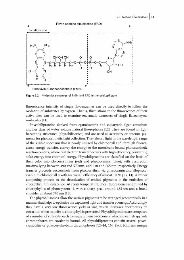
Nicotinamide adenine dinucleotide (NADH) and flavin adenine dinucleotide(FAD) play important roles in the energy metabolism of cells. The reduced cofactorNADH is highly fluorescent, with absorption and emission maxima at 340 and450 nm, respectively. On the other hand, the oxidized formNADþ is nonfluorescent.The fluorescent group in NADH is the reduced nicotinamide ring, and its fluores-cence is partially quenched by collisions with the adenine moiety. Upon binding toproteins, the quantum yield of NADH generally increases drastically, thus enablingthe relatively sensitive detection of native proteins carrying an NADH residue. Sincethe isolation of theOld Yellow Enzyme byWarburg and Christian in 1932, the numberof known flavoproteins has increased considerably [7, 8]. Flavoenzymes, that is,enzymes that contain the naturally fluorescent flavin cofactor as the redox-activeprosthetic group, are involved in numerous redox processes in metabolic oxidation–reduction, photobiology, and biological electron transport [9]. Flavin cofactors arederivatives of riboflavin, a compound better known as vitamin B2 (Figure 2.2).
Riboflavin is synthesized by bacteria and plants but has to be absorbed by higherorganisms. The enzymes flavokinase and FAD can convert riboflavin into flavinmononucleotide (FMN) and flavin adenine dinucleotide (FAD), the cofactors com-monly found in flavoproteins. The essential part of the flavin cofactor is theisoalloxazine ring (Figure 2.2). FMN and FAD exhibit a characteristic yellow color,that is, they absorb light in the visible range at �450 nm, and emit around 515 nm.Thefluorescence spectra as well as the fluorescence quantum yield offlavins stronglydepend on the environment. In aqueous solution, riboflavin and FMN possess arather high fluorescence quantum yield, Wf, of 0.26 [10]. On the other hand, FADexhibits a much smaller fluorescence quantum yield, Wf, of 0.03, because of theformation of an intramolecular quenching complex between the flavin and adeninemoiety [10]. As FAD is only naturally fluorescent in its oxidized form, fluctuations in
CH2
COO-
HCH3+N
CH2
COO-
HH3+N C
OH
H
CH2
COO-
HCH3+N
N
Phenylalanine Tyrosine Tryptophan
Figure 2.1 Molecular structures of the aromatic amino acids.
32j 2 Fluorophores and Fluorescent Labels
fluorescence intensity of single flavoenzymes can be used directly to follow theoxidation of substrates by oxygen. That is, fluctuations in the fluorescence of theiractive sites can be used to examine enzymatic turnovers of single flavoenzymemolecules [11].
Phycobiliproteins derived from cyanobacteria and eukaryotic algae constituteanother class of water soluble natural fluorophores [12]. They are found in lightharvesting structures (phycobilisomes) and are used as accessory or antenna pig-ments for photosynthetic light collection. They absorb light in the wavelength rangeof the visible spectrum that is poorly utilized by chlorophyll and, through fluores-cence energy transfer, convey the energy to the membrane-bound photosyntheticreaction centers, where fast electron transfer occurs with high efficiency, convertingsolar energy into chemical energy. Phycobiliproteins are classified on the basis oftheir color into phycoerythrins (red) and phycocyanins (blue), with absorptionmaxima lying between 490 and 570 nm, and 610 and 665 nm, respectively. Energytransfer proceeds successively from phycoerythrin via phycocyanin and allophyco-cyanin to chlorophyll a with an overall efficiency of almost 100% [13, 14]. A minorcompeting process to the deactivation of excited pigments is the emission ofchlorophyll a fluorescence. At room temperature, most fluorescence is emitted bychlorophyll a of photosystem II, with a sharp peak around 685 nm and a broadshoulder at about 740 nm [15].
The phycobilisomes allow the various pigments to be arranged geometrically in amanner that helps to optimize the capture of light and transfer of energy. Accordingly,they have a very low fluorescence yield in vivo, which increases enormously onextractionwhen transfer to chlorophyll is prevented. Phycobiliproteins are composedof a number of subunits, each having a protein backbone to which linear tetrapyrrolechromophores are covalently bound. All phycobiliproteins contain several phyco-cyanobilin or phycoerythrobilin chromophores [12–14, 16]. Each bilin has unique
O
OH
OH
CH2
OPOPOCH2
CH
CH
CH
CH2
N
NH
N
O
O
N
C CH3
OH OH OH O
O–
O
O–
N
NNH2
NN
Isoalloxazine
Riboflavin-5’-monophosphate (FMN)
Flavin adenine dinucleotide (FAD)
H3
Figure 2.2 Molecular structures of FMN and FAD in the oxidized state.
2.1 Natural Fluorophores j33

spectral characteristics, which may be further modified by interactions of thesubunits and of the chromophore with the apoprotein (Table 2.1).
B-phycoerythrin is compromised of three polypeptide subunits forming anaggregate containing a total of 34 bilin chromophores. It exhibits an absorptioncross-section equivalent to that of �20 rhodamine 6G chromophores and has thehighestfluorescence quantumyield of all phycobiliproteins, 0.98,with afluorescencelifetime of 2.5 ns [17–19]. B-phycoerythrin was the first species to be detected at thesingle-molecule level using laser-induced fluorescence [19–21]. However, singleallophycocyanin molecules with a smaller fluorescence quantum yield of 0.68 [22]can also be easily visualized when immobilized on a cover glass surface underaqueous buffer [23]. Therefore, phycobiliproteins are often used as fluorescent labelsin bioanalytical applications requiring high sensitivity.
Phytochromes are biliprotein photosensors that regulate many physiologicalprocesses in green plants, enabling them to adapt to fluctuating light environments.A red, far-red reversible chromoprotein, phytochrome, was the first photoreceptor tobe identified [24, 25]. Phytochromes are large proteins with covalently bound lineartetrapyrrole, that is, bilin, chromophores that transduce light signals by reversiblyphotointerconverting between red-light-absorbing and far-red-light-absorbing spe-cies, a process that typically initiates a transcriptional signaling cascade [26]. Becauseof efficient double-bond isomerization in the bilin chromophores, the excited state israpidly depopulated. Therefore, phytochromes are comparatively poorly fluorescentbiliproteins [27]. However, by introduction of bilin analogs that lack the photoisome-rizing double bond, strongly yellow-orange fluorescent holoproteins (phytofluors)have been produced [28]. The ability to tag proteins of interest through fusionwith anapophytochrome gene and to produce phytofluors within living cells has attracted agreat deal of interest and considerable effort has been spent on the investigation ofthe fluorescence properties of red-emitting phytofluors using fluorescence correla-tion spectroscopy (FCS) [29].Acomparative studywith standard organicfluorophoresdemonstrated that a specific mutant of a phytochrome (PR1: phytofluor red 1)enables even single-molecule detection upon excitation at 632.8 nm using a con-ventional helium:neon laser [30, 31].
While phytochromes monitor the red and far-red regions of the electromagneticspectrum, UV-A/blue light perception is mediated by the cryptochromes, photo-
Table 2.1 Spectroscopic characteristics (absorptionmaximum, labs, emissionmaximum, lem, andmolecular weight, MW) of some prominent phycobiliproteins in aqueous buffer.
Pigment labs (nm) lem (nm) MW (kDa)
R-Phycoerythrin 565 (495) 575 240B-Phycoerythrin 545 575 240C-Phycocyanin 615 647 220R-Phycocyanin 617 (555) 637 100Allophycocyanin 652 660 100
34j 2 Fluorophores and Fluorescent Labels
receptors regulating stomatal aperture in response to blue light, and the chromo-protein encoded by the NPH1 gene. NPH1 apoprotein noncovalently binds FMN toform the holoprotein nph1. The N-terminal region of the protein contains twodomains of about 110 amino acids (LOV1 and LOV2), which are regulated byenvironmental factors that affect their redox status: light, oxygen, or voltage(LOV) [32–34]. Moreover, both domains show bright fluorescence at�500 nm uponexcitation at �450 nm and might therefore be used as efficient alternatives to greenfluorescent proteins for genetic labeling.
2.2Organic Fluorophores
Organic fluorophores or fluorescent dyes are characterized by a strong absorptionand emission band in the visible region of the electromagnetic spectrum. Thelong-wavelength absorption band of a fluorophore is attributed to the transitionfrom the electronic ground state S0 to the first excited singlet state S1. As thetransition moment for this process is typically very large, the correspondingabsorption bands exhibit oscillator strengths of the order of unity. The reverseprocess S1 ! S0 is responsible for spontaneous emission known as fluorescenceand for stimulated emission. Much of the knowledge we have today stems from thetime when new organic fluorescent dyes were developed as amplifying media fordye lasers [35].
However, since the use of organic fluorescent dyes for qualitative and quantitativedetermination of analytemolecules (especially for automatedDNAsequencing in the80th), their importance for bioanalytical applications has increased considerably [36–39].Owing to the enhanced demand forfluorescentmarkers, the development of newfluorescent dyes has increased significantly in recent years [40–45].Usingfluorescentdyes, extremely high sensitivity down to the single-molecule level can be achieved.Furthermore, time- and position-resolved detection without contact with the analyteis possible. Typically, the fluorescent probes or markers are identified and quantifiedby their absorption and fluorescence emission wavelengths and intensities. Thesensitivity achievable with a fluorescent label is directly proportional to the molarextinction coefficient for the absorption and the quantum yield of the fluorescence.The extinction coefficient typically hasmaximumvalues of about 105 lmol�1 cm�1 inorganic fluorophores and the quantum yield may approach values close to 100%.Absorption and emission spectra, in addition to the fluorescence quantum yield andlifetime, are dependent on environmental factors. Furthermore, for labeling ofbiological compounds, for example, antibodies or DNA/RNA, the dye must carrya functional group suitable for a mild covalent coupling reaction, preferentially withfree amino or thiol groups of the analyte. In addition, the fluorophore should be ashydrophilic as possible to avoid aggregation and nonspecific binding in aqueoussolvents.
Suitable organic fluorescent dyes are distinguished by a high fluorescencequantum yield. That is, upon excitation into the excited singlet state and subsequent
2.2 Organic Fluorophores j35
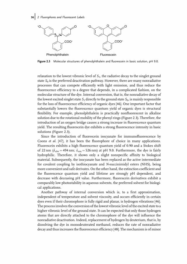
relaxation to the lowest vibronic level of S1, the radiative decay to the singlet groundstate S0 is the preferred deactivation pathway. However, there are many nonradiativeprocesses that can compete efficiently with light emission, and thus reduce thefluorescence efficiency to a degree that depends, in a complicated fashion, on themolecular structure of the dye. Internal conversion, that is, the nonradiative decay ofthe lowest excited singlet state S1 directly to the ground state S0, ismainly responsiblefor the loss of fluorescence efficiency of organic dyes [46]. One important factor thatsubstantially lowers the fluorescence quantum yield of organic dyes is structuralflexibility. For example, phenolphthalein is practically nonfluorescent in alkalinesolution due to the rotational mobility of the phenyl rings (Figure 2.3). Therefore, theintroduction of an oxygen bridge causes a strong increase in fluorescence quantumyield. The resulting fluorescein dye exhibits a strong fluorescence intensity in basicsolutions (Figure 2.3).
Since the introduction of fluorescein isocyanate for immunofluorescence byCoons et al. [47], it has been the fluorophore of choice in many applications.Fluorescein exhibits a high fluorescence quantum yield of 0.90 and a Stokes shiftof 22 nm (labs¼ 494 nm; lem¼ 526 nm) at pH 9.0. Furthermore, the dye is fairlyhydrophilic. Therefore, it shows only a slight nonspecific affinity to biologicalmaterial. Subsequently, the isocyanate has been replaced as the active intermediatefor covalent coupling by isothiocyanate and N-succinimidyl esters (NHS), beingmore convenient and safe derivates. On the other hand, the extinction coefficient andthe fluorescence quantum yield and lifetime are strongly pH dependent, anddecrease with deceasing pH value. Furthermore, fluorescein derivatives exhibit acomparably low photostability in aqueous solvents, the preferred solvent for biologi-cal applications.
Another pathway of internal conversion which is, to a first approximation,independent of temperature and solvent viscosity, and occurs efficiently in certaindyes even if their chromophore is fully rigid and planar, is hydrogen vibrations [46].The process involves the conversion of the lowest vibronic level of the excited state to ahigher vibronic level of the ground state. It can be expected that only those hydrogenatoms that are directly attached to the chromophore of the dye will influence thenonradiative deactivation. Indeed, replacement of hydrogen by deuterium, that is, bydissolving the dye in monodeuterated methanol, reduces the rate of nonradiativedecay and thus increases thefluorescence efficiency [48]. Themechanism is ofminor
COO-
O-O OO-O
COO-
Phenolphthalein Fluorescein
Figure 2.3 Molecular structures of phenolphthalein and fluorescein in basic solution, pH 9.0.
36j 2 Fluorophores and Fluorescent Labels
importance in dyes that emit in the visible range, but can seriously reduce thefluorescence efficiency of infrared dyes. Therefore, long-wavelength (>700 nm)absorbing fluorophores generally exhibit only very poor fluorescence intensities,especially in aqueous surrounding.
Besides fluorescence quenching via photoinduced electron transfer (PET) orfluorescence resonance energy transfer (FRET), a molecule excited to S1 may entera triplet state and relax to the lowest level T1. The occupation of triplet states isundesirable with respect to single-molecule fluorescence applications for variousreasons. Firstly, owing to the relatively long lifetimes of triplet states, the chromo-phore might be excited into higher excited triplet states and undergo irreversiblechemical reactions, that is, it might photobleach. Secondly, provided the dye residesin the triplet state, no fluorescence photon can be detected, thus decreasing thenumber of detected fluorescence photons. On the other hand, photoinduced reverseintersystem crossing might occur, which induces complicated photophysics andrenders the interpretation of single-molecule data more difficult. That is, excitationinto higher excited triplet states might repopulate the singlet manifold, becausesinglet and triplet energies are better matched in higher excited states, and/or someof the restrictions of intersystem crossing are relaxed due to the different nature ofthe triplet symmetry [49, 50]. Therefore, in some organic fluorescent dyes the tripletlifetime can be reduced by increasing the excitation intensity, that is, with increasingexcitation intensity the dye is pumped into higher excited triplet states, thus inducingreverse intersystem crossing [51–54].
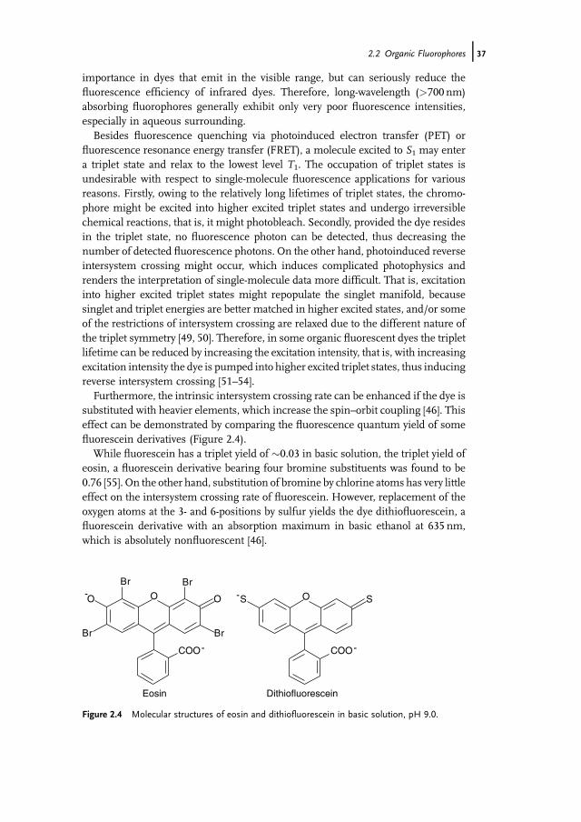
Furthermore, the intrinsic intersystem crossing rate can be enhanced if the dye issubstituted with heavier elements, which increase the spin–orbit coupling [46]. Thiseffect can be demonstrated by comparing the fluorescence quantum yield of somefluorescein derivatives (Figure 2.4).
While fluorescein has a triplet yield of �0.03 in basic solution, the triplet yield ofeosin, a fluorescein derivative bearing four bromine substituents was found to be0.76 [55]. On the other hand, substitution of bromine by chlorine atoms has very littleeffect on the intersystem crossing rate of fluorescein. However, replacement of theoxygen atoms at the 3- and 6-positions by sulfur yields the dye dithiofluorescein, afluorescein derivative with an absorption maximum in basic ethanol at 635 nm,which is absolutely nonfluorescent [46].
OOO
BrBr
Br Br
O SS
COO
- -
-COO-
Eosin Dithiofluorescein
Figure 2.4 Molecular structures of eosin and dithiofluorescein in basic solution, pH 9.0.
2.2 Organic Fluorophores j37
2.3Different Fluorophore Classes
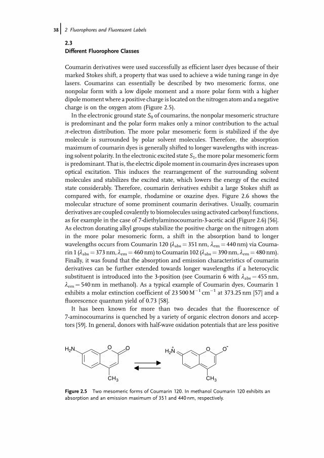
Coumarin derivatives were used successfully as efficient laser dyes because of theirmarked Stokes shift, a property that was used to achieve a wide tuning range in dyelasers. Coumarins can essentially be described by two mesomeric forms, onenonpolar form with a low dipole moment and a more polar form with a higherdipolemomentwhere a positive charge is located on the nitrogen atomand a negativecharge is on the oxygen atom (Figure 2.5).
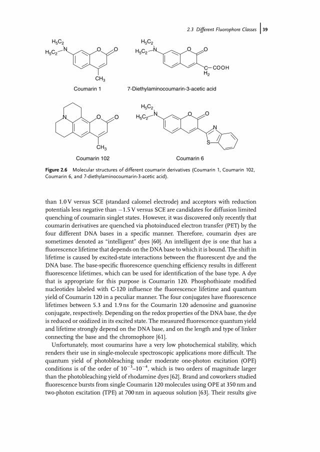
In the electronic ground state S0 of coumarins, the nonpolar mesomeric structureis predominant and the polar form makes only a minor contribution to the actualp-electron distribution. The more polar mesomeric form is stabilized if the dyemolecule is surrounded by polar solvent molecules. Therefore, the absorptionmaximum of coumarin dyes is generally shifted to longer wavelengths with increas-ing solvent polarity. In the electronic excited state S1, themore polarmesomeric formis predominant. That is, the electric dipolemoment in coumarin dyes increases uponoptical excitation. This induces the rearrangement of the surrounding solventmolecules and stabilizes the excited state, which lowers the energy of the excitedstate considerably. Therefore, coumarin derivatives exhibit a large Stokes shift ascompared with, for example, rhodamine or oxazine dyes. Figure 2.6 shows themolecular structure of some prominent coumarin derivatives. Usually, coumarinderivatives are coupled covalently to biomolecules using activated carboxyl functions,as for example in the case of 7-diethylaminocoumarin-3-acetic acid (Figure 2.6) [56].As electron donating alkyl groups stabilize the positive charge on the nitrogen atomin the more polar mesomeric form, a shift in the absorption band to longerwavelengths occurs from Coumarin 120 (labs¼ 351 nm, lem¼ 440 nm) via Couma-rin 1 (labs¼ 373 nm, lem¼ 460 nm) toCoumarin 102 (labs¼ 390 nm, lem¼ 480 nm).Finally, it was found that the absorption and emission characteristics of coumarinderivatives can be further extended towards longer wavelengths if a heterocyclicsubstituent is introduced into the 3-position (see Coumarin 6 with labs¼ 455 nm,lem¼ 540 nm in methanol). As a typical example of Coumarin dyes, Coumarin 1exhibits a molar extinction coefficient of 23 500M�1 cm�1 at 373.25 nm [57] and afluorescence quantum yield of 0.73 [58].
It has been known for more than two decades that the fluorescence of7-aminocoumarins is quenched by a variety of organic electron donors and accep-tors [59]. In general, donors with half-wave oxidation potentials that are less positive
O OH2N H2N+ -O O
CH3CH3
Figure 2.5 Two mesomeric forms of Coumarin 120. In methanol Coumarin 120 exhibits anabsorption and an emission maximum of 351 and 440 nm, respectively.
38j 2 Fluorophores and Fluorescent Labels
than 1.0 V versus SCE (standard calomel electrode) and acceptors with reductionpotentials less negative than �1.5 V versus SCE are candidates for diffusion limitedquenching of coumarin singlet states. However, it was discovered only recently thatcoumarin derivatives are quenched via photoinduced electron transfer (PET) by thefour different DNA bases in a specific manner. Therefore, coumarin dyes aresometimes denoted as �intelligent� dyes [60]. An intelligent dye is one that has afluorescence lifetime that depends on theDNAbase to which it is bound. The shift inlifetime is caused by excited-state interactions between the fluorescent dye and theDNA base. The base-specific fluorescence quenching efficiency results in differentfluorescence lifetimes, which can be used for identification of the base type. A dyethat is appropriate for this purpose is Coumarin 120. Phosphothioate modifiednucleotides labeled with C-120 influence the fluorescence lifetime and quantumyield of Coumarin 120 in a peculiar manner. The four conjugates have fluorescencelifetimes between 5.3 and 1.9 ns for the Coumarin 120 adenosine and guanosineconjugate, respectively. Depending on the redox properties of the DNA base, the dyeis reduced or oxidized in its excited state. Themeasured fluorescence quantum yieldand lifetime strongly depend on the DNA base, and on the length and type of linkerconnecting the base and the chromophore [61].
Unfortunately, most coumarins have a very low photochemical stability, whichrenders their use in single-molecule spectroscopic applications more difficult. Thequantum yield of photobleaching under moderate one-photon excitation (OPE)conditions is of the order of 10�3–10�4, which is two orders of magnitude largerthan the photobleaching yield of rhodamine dyes [62]. Brand and coworkers studiedfluorescence bursts from single Coumarin 120 molecules using OPE at 350 nm andtwo-photon excitation (TPE) at 700 nm in aqueous solution [63]. Their results give
O ON
H5C2 H5C2
H5C2
H5C2
H5C2
CH3
H5C2OON
CH2
COOH
N OO
CH3
S
N
O ON
Coumarin 1
Coumarin 102 Coumarin 6
7-Diethylaminocoumarin-3-acetic acid
Figure 2.6 Molecular structures of different coumarin derivatives (Coumarin 1, Coumarin 102,Coumarin 6, and 7-diethylaminocoumarin-3-acetic acid).
2.3 Different Fluorophore Classes j39
clear evidence that OPE at a high irradiance results in two-step photolysis via the firstelectronic excited singlet and triplet states, S1 and T1, producing dye radical ions andsolvated electrons. Hence, this additional photobleaching pathway limits the appli-cable irradiance for OPE. Using coherent TPE for single-molecule detection, satu-ration of the fluorescence was observed for a high quasi-CW (continuous wave)irradiance (108Wcm�2), whichmay also be caused by photobleaching. Furthermore,TPE is deteriorated by other competing nonlinear processes (e.g., continuumgeneration in the solvent), which only occur above a certain threshold irradiance.They concluded that the single-molecule detection sensitivity of Coumarin 120molecules is enhanced substantially by using TPE, primarily due to the higherbackground with OPE at UV wavelengths.
Alexa Fluor 350 and 430, and the two dyes ATTO 390 and ATTO 425, belong to aclass of commercially available coumarin derivatives for biological labeling applica-tions. Their absorption maxima are reflected in their names, that is, the absorptionsmaxima are at�350, 430, 390, and 425 nm, respectively. The emissionmaxima of thethree coumarins are located around 445, 545, 480, and 485 nm, respectively. Theyexhibit extinction coefficients in the range of 20 000–50 000M�1 cm�1 with fluores-cence quantum yields of up to 0.90 and lifetimes of between 3 and 4 ns.
Today, most (bio)analytical applications requiring high sensitivity use xanthenedyes that absorb and emit in the wavelength region from 500 to 700 nm. Owing totheir structural rigidity, xanthene dyes show high fluorescence quantum yields. Aswithfluorescein dyes, xanthenes exhibit a small Stokes shift of about 20–30 nm. Theyare applied as complementary probes together with other fluorophores in double-label staining, as energy donors and acceptors in FRET experiments, and asfluorescent markers in DNA sequencing and immunoassays. Xanthene dyes are ingeneral far more stable than fluorescein and coumarin derivatives under aqueousconditions [45, 64]. As most commercially available ATTO and Alexa dyes absorbingand emitting in the wavelength range between 480 and 630 nm belong to the class ofxanthene dyes, this fluorophore class will be introduced in more detail. To increasethe water solubility of the fluorophores for bioanalytical applications, the xanthenechromophores are often modified by the attachment of sulfonate groups.
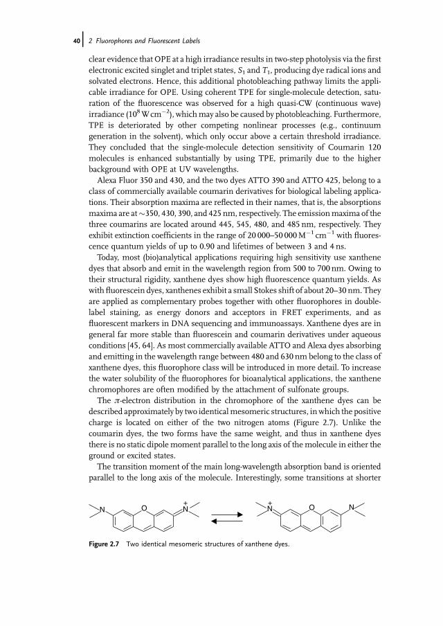
The p-electron distribution in the chromophore of the xanthene dyes can bedescribed approximately by two identicalmesomeric structures, inwhich the positivecharge is located on either of the two nitrogen atoms (Figure 2.7). Unlike thecoumarin dyes, the two forms have the same weight, and thus in xanthene dyesthere is no static dipole moment parallel to the long axis of themolecule in either theground or excited states.
The transition moment of the main long-wavelength absorption band is orientedparallel to the long axis of the molecule. Interestingly, some transitions at shorter
NN O O NN+ +
Figure 2.7 Two identical mesomeric structures of xanthene dyes.
40j 2 Fluorophores and Fluorescent Labels
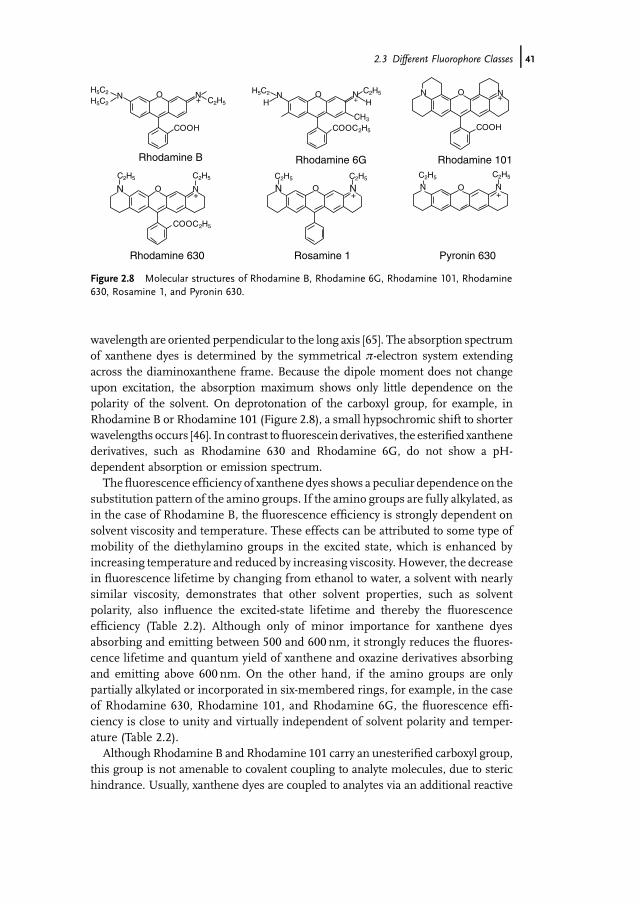
wavelength are oriented perpendicular to the long axis [65]. The absorption spectrumof xanthene dyes is determined by the symmetrical p-electron system extendingacross the diaminoxanthene frame. Because the dipole moment does not changeupon excitation, the absorption maximum shows only little dependence on thepolarity of the solvent. On deprotonation of the carboxyl group, for example, inRhodamine B or Rhodamine 101 (Figure 2.8), a small hypsochromic shift to shorterwavelengths occurs [46]. In contrast tofluorescein derivatives, the esterified xanthenederivatives, such as Rhodamine 630 and Rhodamine 6G, do not show a pH-dependent absorption or emission spectrum.
Thefluorescence efficiency of xanthene dyes shows a peculiar dependence on thesubstitution pattern of the amino groups. If the amino groups are fully alkylated, asin the case of Rhodamine B, the fluorescence efficiency is strongly dependent onsolvent viscosity and temperature. These effects can be attributed to some type ofmobility of the diethylamino groups in the excited state, which is enhanced byincreasing temperature and reduced by increasing viscosity. However, the decreasein fluorescence lifetime by changing from ethanol to water, a solvent with nearlysimilar viscosity, demonstrates that other solvent properties, such as solventpolarity, also influence the excited-state lifetime and thereby the fluorescenceefficiency (Table 2.2). Although only of minor importance for xanthene dyesabsorbing and emitting between 500 and 600 nm, it strongly reduces the fluores-cence lifetime and quantum yield of xanthene and oxazine derivatives absorbingand emitting above 600 nm. On the other hand, if the amino groups are onlypartially alkylated or incorporated in six-membered rings, for example, in the caseof Rhodamine 630, Rhodamine 101, and Rhodamine 6G, the fluorescence effi-ciency is close to unity and virtually independent of solvent polarity and temper-ature (Table 2.2).
Although Rhodamine B and Rhodamine 101 carry an unesterified carboxyl group,this group is not amenable to covalent coupling to analyte molecules, due to sterichindrance. Usually, xanthene dyes are coupled to analytes via an additional reactive
NN O+
N O+
NN O
COOH
+
NN O+
O NN
COOH
H5C2 H5C2
HH5C2
C2H5 C2H5 C2H5
NC2H5 C2H5 C2H5
C2H5
CH3
C2H5+O NN
COOC2H5
COOC2H5
H+
Rhodamine B
Rhodamine 630
Rhodamine 6G
Rosamine 1
Rhodamine 101
Pyronin 630
Figure 2.8 Molecular structures of Rhodamine B, Rhodamine 6G, Rhodamine 101, Rhodamine630, Rosamine 1, and Pyronin 630.
2.3 Different Fluorophore Classes j41

carboxyl group (typically activated as NHS) attached to one of the amino groups or tothe carboxyphenyl ring. As the carboxyphenyl substituent is held in a position almostperpendicular to the xanthene moiety, it is not part of the chromophore system.Hence it has only minor influence on the absorption and emission spectrum ofthe dye.
Removal of the bulky o-carboxyl group leads to so-called rosamines, which showreduced quantumefficiency influid solvents such as ethanol,methanol, orwater [44].In Rosamine 1 (Figure 2.8) the steric hindrance for torsion of the phenyl group isreduced, leading to a configuration inwhich the planes of the phenyl substituents andthe xanthene ring system can nearly approach coplanarity in the first excited state.This process can result in a state with charge transfer character and a reduced S1�S0energy gap. Hence, the internal conversion rate increases as demonstrated by areduced fluorescence efficiency and lifetime (Table 2.2). In this double-potentialminimum model, the height of the potential barrier and the energy differencebetween the initial and the final states are controlled by solvent viscosity and polarityas well as by steric and electronic properties of the chromophore and the phenylsubstituents [44]. Therefore, rosamine derivatives with strong electron donating oraccepting phenyl substituents, for example, with p-amino or p-nitro phenyl sub-stituents, show only weak fluorescence efficiency.
Rhodamine dyes that carry a free o-carboxyl group can exist in several forms. Thedeprotonation is enhanced by dilution or by adding a small amount of a base. Thedeprotonated zwitterionic form exhibits an absorption maximum shifted to shorterwavelengths (3–10 nm). In nonpolar solvents such as acetone, the zwitterionic formis not stable and forms an intramolcular lactone in a reversible fashion. The lactone iscolorless because the p-electron system of the dye is interrupted. A further inter-esting characteristic of xanthene derivatives consists in the fact that they areselectively quenched upon contact formation with the DNA base guanine and theamino acid tryptophan via photoinduced electron transfer [66–74]. Both guanine and
Table 2.2 Spectroscopic characteristics(absorption maximum, labs, emissionmaximum, lem, and fluorescence lifetime, t) ofvarious xanthene derivatives in aqueous bufferand ethanol at 25 �C. Fluorescence quantumyields are given only for ethanolic dye solutions.To ensure protonation of the o-carboxyl group inRhodamine B and Rhodamine 101,
measurements were performed in aqueousbuffer at pH 3.0 and upon addition of a drop oftrifluoroacetic acid to 1ml of alcoholic dyesolution, respectively. Typically, xanthene dyesexhibit extinction coefficients in the range of1� 105 to 1.3� 105M�1 cm�1 in alcoholicsolutions.
Xanthene derivative labs (nm) lem (nm) t (ns) Wf (in ethanol)
Rhodamine B 557/552 578/579 1.43/2.28 0.55Rhodamine 6G 526/530 556/556 3.89/3.79 0.95Rhodamine 101 579/574 608/601 4.17/4.28 0.92Rhodamine 630 564/563 588/587 4.04/4.06 0.97Rosamine 1 565/562 592/589 2.68/3.76 0.88Pyronin 630 559/559 579/579 3.69/3.72 0.92
42j 2 Fluorophores and Fluorescent Labels

tryptophan act as efficient electron donors quenching the excited singlet state ofxanthene derivatives efficiently.
There are several functional xanthene derivatives commercially available forbiological labeling: ATTO 488, ATTO 520 and ATTO 532 related to Rhodamine6G, ATTO 550 related to Rhodamine B, ATTO 565 related to Rhodamine 630, ATTO590, and ATTO 594. The molecular structures of the Alexa derivatives Alexa 488,Alexa 532, Alexa 546, Alexa 568, Alexa 594, and Alexa 633 are also based on thexanthene chromophores shown in Figure 2.8 [75].
Owing to the small number of compounds that demonstrate intrinsic fluores-cence above 600 nm, the use of near-infrared (NIR) fluorescence detection inbioanalytical samples is a desirable alternative to visible fluorescence detection.This fact has prompted current efforts towards the use of NIR dyes for bioanalyticalapplications. However, there are few chromophores that show sufficient fluores-cence quantum yields in the NIR region, especially in aqueous surroundings, andthat can be coupled covalently to analyte molecules [76]. This finding is due to thefact that at longer wavelengths the fluorescence efficiency tends to decrease withdecreasing energy difference between S1 and S0. As already mentioned, hydrogenvibrations in particular lead to a decreased quantum yield in the red-near-IR region.The quanta of hydrogen vibrations have the highest energies for organic com-pounds. Thus, hydrogen vibrations are most likely to contribute to internalconversion between S1 and S0. This mechanism, which is expected only for thosehydrogens that are attached directly to the chromophore, seriously reduces thefluorescence efficiency of infrared dyes [45, 46]. Therefore, only a limited numberof fluorescent dyes with sufficient quantum yields that absorb at wavelengths above620 nm are known, and fewer still are available in reactive form for conjugationwith biomolecules.
There are several other advantages to using fluorescent dyes that absorb in the redover those that absorb at shorter blue and green wavelengths. Themost important ofthese advantages is the reduction of the background signal, which ultimatelyimproves the sensitivity achievable. There are three major sources of background:(i) elastic scattering, that is, Rayleigh scattering, (ii) inelastic scattering, that is,Raman scattering, and (iii) fluorescence from impurities. The efficiency of bothRayleigh and Raman scattering are dramatically reduced by shifting to longerwavelength excitation (scales with 1/l4). Likewise, the number of fluorescentimpurities is significantly reduced with longer excitation and detection wavelengths.
One class of red-absorbing fluorophores constitute cyanine dyes [77–80]. Cyaninederivatives belong to the class of polymethine dyes, that is, planar fluorophores withconjugated double bonds where all atoms of the conjugated chain lie in a commonplane linked by s-bonds (see Section 1.1). Cyanine dyes can be best described bytwo identical mesomeric structures R2N[CH¼CH]nCH¼NþR2$R2N
þ¼CH[CH¼CH]nNR2, where n is a small number that defines the longest wavelengthabsorption, and the nitrogen and part of the conjugated chain usually form part of aheterocyclic system, such as imidazole, pyridine, pyrrole, quinoline, or thiazole.Figure 2.9 gives the molecular structure of some commercially available symmetric(e.g., Cy5) and asymmetric (e.g., Dy-630) cyanine derivatives. As in the case of
2.3 Different Fluorophore Classes j43

xanthene dyes, there is no static dipolemoment parallel to the long axis of symmetriccyanine derivatives in either the ground or the excited states, because the twomesomeric forms have the same weight.
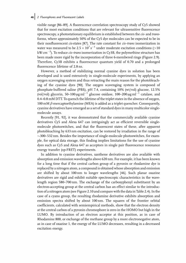
Cyanine dyes are widely used in ultrasensitive imaging and spectroscopy, espe-cially for biological applications. The absorption can be tuned through the visible andnear-infrared region by variation of the length of the polymethine chain joining thetwo heads of the cyanine dye. As can be seen in Table 2.3 each additional double bondshifts the absorption maximum by roughly 100 nm towards the red (see Cy3 !Cy5 ! Cy7). Themolar extinction coefficients of cyanine derivatives are comparablyhigh and lie between 1.2 and 2.5� 105M�1 cm�1 [81–83]. On the other hand, thefluorescence quantum yield only varies between 0.04 and 0.4 with lifetimes in therange of a few hundred picoseconds (0.2–1.0 ns). In addition, cyanine dyes are lessphotostable than xanthene dyes, especially under single-molecule conditions [84] andsome derivatives (Cy5) are destroyed by environmental ozone [85].
Fluorescence experiments have revealed several expected and also unexpectedphotophysical phenomena of cyanine derivatives (especially Cy5), such as cis–transisomerization, off-states in addition to triplet formation, and complex photobleach-ing pathways including nonfluorescent intermediates that still absorb light in the
NN
COOH
+ +N N
O
HOOC
+NN
COOH
+NN
HOOC
+N
COOH
N+
O
N
N
COOH
Cy3 Cy3B
Alexa 647Cy5
Cy5.5 Dy-630
SO-3
SO-3
SO-3
SO-3
SO-3
SO-3
SO-3
-O S3
-O S3
-O S3-O S3
-O S3
-O S3
-O S3
Figure 2.9 Molecular structures of the cyanine derivatives Cy3, Cy3B, Cy5, Alexa 647, Cy5.5, andDy-630.
44j 2 Fluorophores and Fluorescent Labels
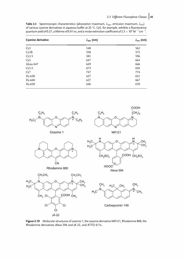
Table 2.3 Spectroscopic characteristics (absorption maximum, labs, emission maximum, lem)of various cyanine derivatives in aqueous buffer at 25 �C. Cy5, for example, exhibits a fluorescencequantumyield of 0.27, a lifetimeof 0.91 ns, and amolar extinction coefficient of 2.5� 105M�1 cm�1.
Cyanine derivative labs (nm) lem (nm)
Cy3 548 562Cy3B 558 572Cy3.5 581 596Cy5 647 664Alexa 647 649 666Cy5.5 673 692Cy7 747 774Dy-630 627 651Dy-640 627 667Dy-650 646 670
Figure 2.10 Molecular structures of oxazine 1, the oxazine derivativeMR121, Rhodamine 800, theRhodamine derivatives Alexa 594 and JA 22, and ATTO 611x .
2.3 Different Fluorophore Classes j45
visible range [86–89]. A fluorescence correlation spectroscopy study of Cy5 showedthat for most excitation conditions that are relevant for ultrasensitive fluorescencespectroscopy, a photostationary equilibrium is established between the cis- and trans-forms, where approximately 50% of the Cy5 dye molecules can be expected to be intheir nonfluorescent cis-states [87]. The rate constant for cis–trans isomerization inwater was measured to be 2.5� 107 s�1 under moderate excitation conditions (>10kWcm�2). To reduce cis–trans isomerization in Cy3B, the polymethine structure hasbeen made more rigid by the incorporation of three 6-membered rings (Figure 2.9).Therefore, Cy3B exhibits a fluorescence quantum yield of 0.70 and a prolongedfluorescence lifetime of 2.8 ns.
However, a method of stabilizing normal cyanine dyes in solution has beendeveloped and is used extensively in single-molecule experiments, by applying anoxygen scavenging system and thus retracting the main reason for the photobleach-ing of the cyanine dyes [90]. The oxygen scavenging system is composed ofphosphate-buffered saline (PBS), pH 7.4, containing 10% (wt/vol) glucose, 12.5%(vol/vol) glycerin, 50–100 mgml�1 glucose oxidase, 100–200 mgml�1 catalase, and0.4–0.8mMDTT. To quench the lifetime of the triplet states in the absence of oxygen,100mM b-mercaptoethylamine (MEA) is added as a triplet quencher. Consequently,cyanine derivatives have emerged as a set of standard dyes inmanymulticolor single-molecule assays.
Recently [91, 92], it was demonstrated that the commercially available cyaninederivatives Cy5 and Alexa 647 can intriguingly act as efficient reversible single-molecule photoswitches, and that the fluorescent states of these, after apparentphotobleaching by 633 nm excitation, can be restored by irradiation in the range of�300–532 nm. Besides the importance of single-molecule photoswitches, for exam-ple, for optical data storage, this finding implies limitations for the use of cyaninedyes such as Cy5 and Alexa 647 as acceptors in single pair fluorescence resonanceenergy transfer (sp-FRET) experiments.
In addition to cyanine derivatives, xanthene derivatives are also available withabsorption and emissionwavelengths above 620 nm. For example, it has been knownfor a long time that if the central carbon group of a pyronin or rhodamine dye isreplaced by a nitrogen atom, a compound is obtainedwhose absorption and emissionare shifted by about 100 nm to longer wavelengths [46]. Such planar oxazinederivatives are rigid and exhibit suitable spectroscopic characteristics in the wave-length region 580–700 nm. The exchange of the carboxyphenyl substituent by anelectron-accepting group at the central carbon has an effect similar to the introduc-tion of a nitrogen atom (see Figure 2.10 and comparewith the data inTable 2.4). In thecase of a cyano group, the resulting rhodamine derivative exhibits absorption andemission spectra shifted by about 100 nm. The squares of the frontier orbitalcoefficients, calculated with semiempirical methods, show that the electron densityat the central carbon of a pyronin chromophore is zero in the HOMO but high in theLUMO. By introduction of an electron acceptor at this position, as in case ofRhodamine 800, or exchange of the methane group by a more electronegative atom,as in case of oxazine 1, the energy of the LUMO decreases, resulting in a decreasedexcitation energy.
46j 2 Fluorophores and Fluorescent Labels
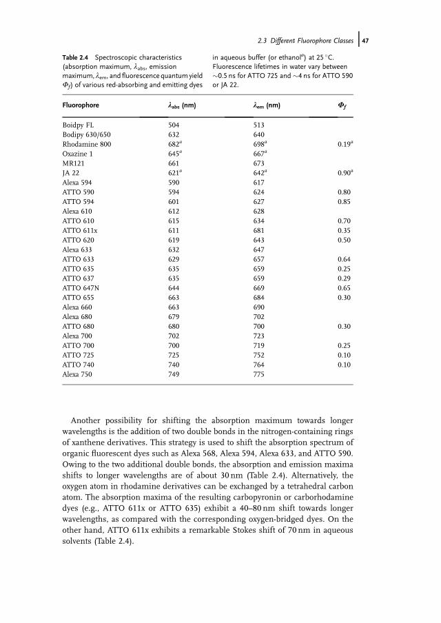
Another possibility for shifting the absorption maximum towards longerwavelengths is the addition of two double bonds in the nitrogen-containing ringsof xanthene derivatives. This strategy is used to shift the absorption spectrum oforganic fluorescent dyes such as Alexa 568, Alexa 594, Alexa 633, and ATTO 590.Owing to the two additional double bonds, the absorption and emission maximashifts to longer wavelengths are of about 30 nm (Table 2.4). Alternatively, theoxygen atom in rhodamine derivatives can be exchanged by a tetrahedral carbonatom. The absorption maxima of the resulting carbopyronin or carborhodaminedyes (e.g., ATTO 611x or ATTO 635) exhibit a 40–80 nm shift towards longerwavelengths, as compared with the corresponding oxygen-bridged dyes. On theother hand, ATTO 611x exhibits a remarkable Stokes shift of 70 nm in aqueoussolvents (Table 2.4).
Table 2.4 Spectroscopic characteristics(absorption maximum, labs, emissionmaximum, lem, and fluorescence quantum yieldWf) of various red-absorbing and emitting dyes
in aqueous buffer (or ethanola) at 25 �C.Fluorescence lifetimes in water vary between�0.5 ns for ATTO 725 and �4 ns for ATTO 590or JA 22.
Fluorophore labs (nm) lem (nm) Wf
Boidpy FL 504 513Bodipy 630/650 632 640Rhodamine 800 682a 698a 0.19a
Oxazine 1 645a 667a
MR121 661 673JA 22 621a 642a 0.90a
Alexa 594 590 617ATTO 590 594 624 0.80ATTO 594 601 627 0.85Alexa 610 612 628ATTO 610 615 634 0.70ATTO 611x 611 681 0.35ATTO 620 619 643 0.50Alexa 633 632 647ATTO 633 629 657 0.64ATTO 635 635 659 0.25ATTO 637 635 659 0.29ATTO 647N 644 669 0.65ATTO 655 663 684 0.30Alexa 660 663 690Alexa 680 679 702ATTO 680 680 700 0.30Alexa 700 702 723ATTO 700 700 719 0.25ATTO 725 725 752 0.10ATTO 740 740 764 0.10Alexa 750 749 775
2.3 Different Fluorophore Classes j47

To summarize, red-absorbing rhodamine, oxazine, and carborhodamine orcarbopyronin derivatives exhibit a high fluorescence quantum yield of up to0.90 with lifetimes of 2.0–4.0 ns in alcoholic solutions. Usually, the fluorescencelifetime of xanthene derivatives absorbing and emitting above 620 nm shows amarked decrease on changing from ethanol to an aqueous environment [45].Exceptions are dyes derived from JA 22 (Figure 2.9). This rhodamine derivativeJA 22, with the tetrachlorocarboxyphenyl substituents, also shows a fluorescenceefficiency of close to unity in an aqueous buffer. Finally, it has to be noted that alllong-wavelength absorbing rhodamine, oxazine, and carborhodamine orcarbopyronin derivatives show a much higher photostability than their relatedcyanine derivatives.
Finally, bora-diaza-indacene derivatives have to be considered, which spanthe visible spectrum from 500 to �650 nm. These so-called Bodipy dyes areunusual in that they are relatively nonpolar, they exhibit only a small Stokesshift, and the chromophore is electrically neutral. Therefore, they tend to bindnonspecifically to biomolecules in aqueous surroundings. On the otherhand, they exhibit extinction coefficients and quantum yields comparable toxanthene derivatives.
Interestingly, some of the red-absorbing xanthene derivatives show a loss ofabsorption and emission under basic conditions, for example, while coupling theactivated dye to amino functions of a biomolecule at pH �9.0. Unfortunately, alltriphenylmethane and related dyes show a tendency to react at the central carbonwith nucleophiles, for example, hydroxide ions, if this carbon is stericallyavailable. Therefore, some of the above mentioned derivatives become colorlesson addition of a base to their aqueous solutions, due to the formation of a so-calledpseudobase. In the pseudobase thep-electron system is interrupted and thereforethe long-wavelength absorption is lost. Although this process is reversible,subsequent reactions, for example, with oxygen, may lead to an irreversibledestruction of the dye. It has to be pointed out that the tendency for the formationof the pseudobase is strongly controlled by the structure of the dye. Furthermore,other destructive, that is, irreversible, reactions also tend to increase withincreasing absorptionmaximum. In the case of �normal� rhodamines, the centralcarbon of the chromophore is protected by the carboxyphenyl substituent.Therefore, rhodamine derivatives, such as JA 22 or Alexa 594, are completelystable even in strong basic solutions. Due to the decrease in fluorescencequantum yield with increasing absorption wavelength, it is advisable to workwith red-absorbing fluorescent dyes whose absorption and emission maxima liebetween 620 and 700 nm.
Owing to their outstanding chemical and photochemical stabilities and their highfluorescence quantum yields, rylene derivatives have been established as alternativefluorophores, especially in single-molecule fluorescence spectroscopy. The compa-rably high photostability of rylene dyes immobilized in different polymeric matricesenables the observation of photophysical processes, for example, photon antibunch-ing and electron or energy transfer, at the single-molecule level over extended periodsof time [93–98]. Perylenetetracarboxdiimide (PDI) represents the key structure from
48j 2 Fluorophores and Fluorescent Labels

which new types of fluorophores, important intermediates, and various functionalperylene dyes are derived. Depending on the substitution pattern, PDI exhibits anabsorption maximum between 520 and 580 nm with a typical Stokes shift of30–40 nm. The fluorescence quantum yield is close to unity with the fluorescencelifetime in the range of 4–5 ns. Extension of PDI by one naphthalene results interrylenediimide derivatives (TDI) (Figure 2.11) with absorption maxima shifted byabout 100 nm to the red. By extending the aromatic p-system, the molar extinctioncoefficient also increases from 60 000M�1 cm�1 (PDI) to 93 000M�1 cm�1 (TDI).One limitation of perylene derivatives is represented by their low water solubility.Therefore, strategies based on the incorporation of sulfonated phenoxy groups havebeen developed to make new red-absorbing water-soluble TDI derivatives availablefor biological labeling applications [93].
2.4Multichromophoric Labels
As an alternative to conventional organic fluorescent dyes, fluorescent nano-and microspheres imbedded with organic fluorescent dyes can be used. Theyhave been developed and then conjugated to antibodies or streptavidin forimmunological studies using flow cytometry, fluorescence microscopy, andELISAs (enzyme-linked immunosorbent assay) [99–102]. Furthermore, fluo-rescent spheres or beads can be synthesized with other surface functionalgroups, for example, carboxy-, aldehyde-, sulfate-, or amino-modified, to enable
N
N
OO
R
R
OO
N
N OO
R
R
OO
PDI TDI
Figure 2.11 Basic molecular structures of the perylene dyes perylenetetracarboxdiimide (PDI) andterrylenediimide (TDI).
2.4 Multichromophoric Labels j49
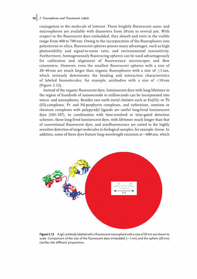
conjugation to the molecule of interest. These brightly fluorescent nano- andmicrospheres are available with diameters from 20 nm to several mm. Withrespect to the fluorescent dyes embedded, they absorb and emit in the visiblerange from 400 to 700 nm. Owing to the incorporation of the fluorophores intopolystyrene or silica, fluorescent spheres posses many advantages, such as highphotostability and signal-to-noise ratio, and environmental insensitivity.Furthermore, homogeneously fluorescing spheres can be used advantageouslyfor calibration and alignment of fluorescence microscopes and flowcytometers. However, even the smallest fluorescent spheres with a size of20–40 nm are much larger than organic fluorophores with a size of �1 nm,which seriously deteriorates the binding and interaction characteristicsof labeled biomolecules, for example, antibodies with a size of <10 nm(Figure 2.12).
Instead of the organic fluorescent dyes, luminescent dyes with long lifetimes inthe region of hundreds of nanoseconds to milliseconds can be incorporated intomicro- and nanospheres. Besides rare earth metal chelates such as Eu(III)- or Tb(III)-complexes, Pt- and Pd-porphyrin complexes, and ruthenium, osmium orrhenium complexes with polypyridyl ligands are useful long-lived luminescentdyes [103–107]. In combination with time-resolved or time-gated detectionschemes, these long-lived luminescent dyes, with lifetimes much longer than thatof conventional fluorescent dyes, and autofluorescence are suited to the highlysensitive detection of targetmolecules in biological samples, for example, tissue. Inaddition, some of these dyes feature long-wavelength emission at�600 nm, which
Figure 2.12 A IgGantibody labeledwith a fluorescent nanospherewith a size of 20 nmare shown toscale. Comparison of the size of the fluorescent dyes embedded (�1 nm) and the sphere (20 nm)clarifies the different proportions.
50j 2 Fluorophores and Fluorescent Labels
is well separated from the excitation peak at <400 nm. On the other hand, thesecompounds are prone to quenching by oxygen. Therefore, these materials have tobe encapsulated inmaterial that is impermeable to oxygen. An additional limitationto the use of luminescent nano- or microspheres is their long lifetime,which seriously limits the maximum number of photons detectable within agiven time.
The need to detect as many different analyte molecules as possible simulta-neously has driven the development of multicolor or multiparameter strategies,that is, the development of fluorescent labels that can be excited at single wave-lengths with comparable efficiency, but emit at different wavelengths. This can beachieved, for example, by the incorporation of two or more differently absorbingand emitting organic fluorophores into microspheres that can undergo fluores-cence resonance energy transfer (FRET). Each microsphere contains one class ofdyes that absorbs at the desired excitation wavelength. In addition, each micro-sphere contains one or more longer-wavelength dyes, which are carefully chosen tocreate an energy transfer cascade that guarantees efficient energy transfer from theinitially excited dye to the longest-wavelength dye. Ideally, the excitation energy istransferred quantitatively fromdye to dye so that only the longest-wavelength dye inthe cascade emits significant fluorescence. As these microspheres exhibit a largeStokes shift, they are ideally suited to detection in samples with significant Rayleighor Raman scattering or strong autofluorescence. Depending on the dye composi-tion, 40 nm nanospheres (so-called TransFluoSpheres) can be prepared that can beexcited efficiently at 488 nm but emit at 560, 605, 645, 685, or 720 nm, respectively.Besides applications in multiparameter experiments, such as energy transfer,nano- or microspheres can be used advantageously in high-resolution multicolorcolocalization experiments [108]. Furthermore, bioimaging with luminescentnanoparticle probes has recently attracted widespread interest in biology andmedicine. Because luminescent nanoparticles are better in terms of photostabilityand sensitivity, they are suitable for real time tracking andmonitoring of biologicalevents at the cellular level, which may not be accomplished using regular fluo-rescent dyes.
Alternatively, so-called tandem dyes or resonance energy transfer (RET) dyeshave been synthesized to increase the Stokes shift of common fluorophores. Theconjugates rely on efficient fluorescence resonance energy transfer between twofluorophores. Inmost cases tandemdyes based on phycobiliproteins, serving as theenergy transfer donors, have been developed, particularly for immunofluorescenceapplications that apply flow cytometry [109, 110]. For the sake of completeness, thedevelopment of light harvesting dendrimers has to be discussed. Tree-like multi-chromophoric dendrimers with spectrally different fluorophores featuring variousabsorption and emission spectra have been synthesized and studied at the single-molecule level. The multichromophoric dendrimers show strong absorption overthe whole visible spectral range, but only the longest-wavelength absorbingfluorophore shows strong fluorescence [111, 112]. Unfortunately, the hithertosynthesized energy transfer dendrimers are not suitable for mild covalent labelingof biomolecules.
2.4 Multichromophoric Labels j51
2.5Nanocrystals
One class of fluorescent nanoparticles with great expectations are semiconductornanocrystals (NC), such as core-shell CdSe/ZnS NCs [113]. Their unique opticalproperties – tunable narrow emission spectrum, broad excitation spectrum, highphotostability and long fluorescence lifetime (of the order of tens of nanoseconds) –make these bright probes attractive in experiments involving long observation times,multicolor and time-gated detection. Furthermore, the relatively long fluorescencelifetime of CdSe nanocrystals, in the region of several tens of nanoseconds, can beused advantageously to enhance the fluorescence biological imaging contrast andsensitivity using time-gated detection [114].
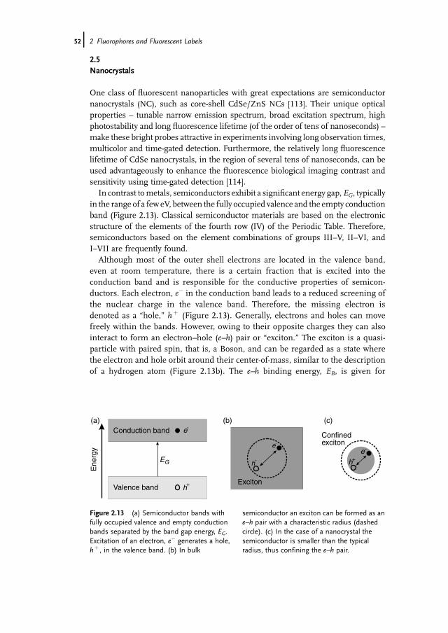
In contrast tometals, semiconductors exhibit a significant energy gap,EG, typicallyin the range of a few eV, between the fully occupied valence and the empty conductionband (Figure 2.13). Classical semiconductor materials are based on the electronicstructure of the elements of the fourth row (IV) of the Periodic Table. Therefore,semiconductors based on the element combinations of groups III–V, II–VI, andI–VII are frequently found.
Although most of the outer shell electrons are located in the valence band,even at room temperature, there is a certain fraction that is excited into theconduction band and is responsible for the conductive properties of semicon-ductors. Each electron, e� in the conduction band leads to a reduced screening ofthe nuclear charge in the valence band. Therefore, the missing electron isdenoted as a �hole,� hþ (Figure 2.13). Generally, electrons and holes can movefreely within the bands. However, owing to their opposite charges they can alsointeract to form an electron–hole (e–h) pair or �exciton.� The exciton is a quasi-particle with paired spin, that is, a Boson, and can be regarded as a state wherethe electron and hole orbit around their center-of-mass, similar to the descriptionof a hydrogen atom (Figure 2.13b). The e–h binding energy, EB, is given for
Ene
rgy
Valence band
(a) (b) (c)
EG
e-
h+
Conduction band
Exciton
e-
h+
Confinedexciton
e-
h+
Figure 2.13 (a) Semiconductor bands withfully occupied valence and empty conductionbands separated by the band gap energy, EG.Excitation of an electron, e� generates a hole,hþ , in the valence band. (b) In bulk
semiconductor an exciton can be formed as ane–h pair with a characteristic radius (dashedcircle). (c) In the case of a nanocrystal thesemiconductor is smaller than the typicalradius, thus confining the e–h pair.
52j 2 Fluorophores and Fluorescent Labels

the nth state by
EB ¼ m�e4
2 4pe0e�hð Þ21n2
where
m� denotes the reduced mass of the electron and holee is the electric permittivity of the semiconductor.
The average distance between e and h, that is, the exciton radius, aX, in the groundstate, ismuch larger than the Bohr radius of hydrogen of aH¼ 0.5� 10�10m, and liesin the region of 1–13 nm (e.g., aX¼ 1.7, 6.1, and 12.5 nm for ZnS, CdSe, and GaAs,respectively).
Significant deviations from bulk semiconductor properties occur when the crystalsize is reduced in one or more dimensions to the diameter of the exciton radius(Figure 2.13c) [115–117]. The strong influence of the reduction in size on thesemiconductor properties can be understood by considering that both the electronand the hole can theoretically only exist within the semiconductor material, whichmeans, its interface constitutes an infinite potential. For such a particle within aboundary, that is, a particle in the box, the allowed energy states can be calculatedaccording to (Section 1.1)
Ec ¼ h2
8m�a21n2
where
a is the �length� of the box.
While for sizes a� aX this is of minor importance for the energy levels of the e–hpair, in the case of a� aX, the confinement regime, the exciton energy is forced intoa higher energy level and discrete absorption bands appear (Figure 2.14). Thus, theexciton energy can be controlled by size reduction. In principle, confinement ispossible in all three dimensions. Therefore, different structures exist, such as�quantum wells� (1-dimensional confinement), �quantum wires� (2-dimensionalconfinement), and �quantum dots� (3-dimensional confinement). Although allthree types of confinement are important, it is mainly quantum dots (QDs), that is,colloidal nanocrystals (NCs) that are used for external labeling of analytemolecules.
The crucial step for fabrication of such small particles is the controlled growingprocedure, which ideally leads to crystals of similar composition, similar structureand nearly equal size (monodisperse particles). Growing of particles starts from anatomic precursor of both materials involved. As soon as clusters have been formed,there are two competing reactions in the supersaturated solution: growth of initiallyformed clusters and formation of new nuclei. As the latter will lead to increasingheterodispersity, the nucleation process has to be interrupted instantaneously, forexample, by a sudden change in temperature. Once a sufficient number of nucleihave been produced, further growth can continue under milder conditions, where
2.5 Nanocrystals j53

no new nucleation occurs [113,118] . For the most widely used material, CdSe, theprecursor substances generally consist of dimethylcadmium (Me2Cd) and tri-n-octylphosphine selenide [(C8H17)3PSe], both of which rapidly disintegrate intonon-reactive organic compounds and Cd and Se atoms upon heating to 250–300 �Cin tri-n-octylphosphine oxide, TOPO [118]. Cooling of the reaction mixture totemperatures<200 �C prevents further nucleation, and growth continues for severalhours under heating at 250 �C. The progress of the reaction can be easily followed byabsorption spectroscopy. A more sophisticated procedure is size-selective precipi-tation, where, under careful conditions, only the largest NCs in solution areprecipitated and removed. This method can be successfully used to synthesize CdSeNCs with a diameter of 1.2–11.5 nm, corresponding to photoluminescence emissionin the range of 400–700 nm. NCs synthesized by this method exhibit a wurtzite-likecrystal structure and are more or less spherical. However, because the e–h pairscreated upon excitation strongly interact with the environment, resulting inincreased non-radiative deactivitation via �traps�, the photoluminescence quantumyield is less than 0.10. Therefore, the �core� of aNC is nowadays coated with a �shell,�that is, an insulator material, to yield a stabilized core–shell NC with high quantumyield [119, 120]. The best material for this purpose has been found to be ZnS, whichcan be grown onto the NCs in an analogous manner to the NC preparation, byaddition of precursors (dimethylzinc, Me2Zn, and hexamethyldisilathiane,[Me3Si)2S] to a heated NC solution in TOPO. As ZnS has a much higher bandgapenergy than CdSe (3.91 versus 2.51 eV), interactions of the carriers with theenvironment are greatly reduced, resulting in photoluminescence quantum yieldsin solution of >0.50.
350 450 550 650
Wavelength (nm)
Abs
orpt
ion
CdSe
CdSe
Figure 2.14 Absorption (black) and emissionspectra of CdSe NCs of various sizes (green:2.2 nm; red: 5 nm). The emission maximum isonly slightly red-shifted to the longest-wavelength absorption shoulder(corresponding to the ground state of theexciton). The absorption in NCs stems from thephotoinduced creation of discrete e–h pair
combinations. Hence, the absorption wouldprobably be expected to consist of narrow bandsinstead of a broad band distribution. However,owing to the slight heterodispersity of thesample, inhomogeneous line width broadeningresults. The overlap of the broadened bandswith different absorption cross-sections resultsin the observed absorption spectra.
54j 2 Fluorophores and Fluorescent Labels

For biological applications the water solubility of the rather hydrophobic NCs hasto be increased. For example, surface modification with thiols with charged residuesare the simplest approach to increasing the water solubility. On the other hand, thiolcoatings exhibit only a limited stability, which renders their application for long-termstudies in living systems difficult [121, 122]. Other materials used to increase thewater solubility, and through this enabling biological imaging applications to bemade, include coating with silanes [113, 123], peptides [124–126], and ambiphilicpolymers [127–130].
Although reports showing NC biocompatibility actually appeared a few yearsago [113, 121], their breakthrough for biological targeting was only very recent[130–136]. At present, NCs can be easily functionalized, for example with streptavidin,to facilitate mild coupling to biotinylated biomolecules (Figure 2.15). Immunofluo-rescent labeling of cancermarkers andother cellular targets on the surface offixed andlive cancer cells in addition to stainingof actin andmicrotubulefibers in the cytoplasm,and the detection of nuclear antigens inside the nucleus, have been successfullydemonstrated [136]. Therefore, NCs might also be useful for in vivo single-moleculestudies and the first successful results have already been presented [137, 138].
However, NCs also posses serious limitations. One disadvantage of NCs is thedifficulty to engineer them with single binding sites that can be specifically conju-gated to only one molecule of interest. Instead, during the labeling step, NCs tend tobind to several molecules simultaneously. Another problem is blinking, which isstrongly controlled by the excitation intensity (Figure 2.15b), and often obeys a powerlaw [139, 140]. Although blinking of NCs is efficiently reduced in the presence of1–10mM mercaptoethanol, the addition of reducing agents is not compatible withlive cell imaging [141]. In addition, not all semiconductor particles are generallyactive, that is, luminescent, but they can be photoactivated [142]. To explain the strongblinking phenomenon it is assumed that trapping of an electron occurs, induced byphoton absorption. Trappingmeans that the electron is emitted into the surroundingof the NC, thus leaving a charged NC behind. The charged NC can still be excited,
Core
Shell
Polymercoating
Streptavidin
10-15 nm
200 40 60 80 100 120Time (s)
Flu
ores
cenc
e In
tens
ity
(a) (b)
Figure 2.15 (a) Assembling of water soluble quantum dots carrying a streptavidin layer for labelingof biotinylated biomolecules. (b) Fluorescence trajectory of a single QD605 immobilized on a drycover slide surface (1ms bin�1).
2.5 Nanocrystals j55
resulting in the formation of an e–h pair, which is efficiently quenched by Augerinteractions. After a certain time, the electron can be released back into the NC,neutralizing the charge and resulting in an emissive NC. Typical photoluminescencelifetimes measured for core-shell CdSe or CdTe NCs are in the range of 10–80 ns[143, 144]. A systematic study of the radiative and non-radiative rate fluctuations insingle NCs revealed a correlation of photoluminescence emission maxima, lifetime,and intensity of individual NCs withmillisecond time resolution [144]. Furthermore,the technique that was applied enabled the exact determination of the photolumi-nescence quantum yield of single NCs. In this study, average quantum yields of 0.82for QD 705 and 0.85 for QD 605 were obtained. Very recently [145], it was shown thatsingle NCs show multiexciton emission under high power excitation. This meansthat it is possible to create more than one e–h pair per NC. In contrast to organic dyese–h pairs can coexist in NCs because they are Bosons.
Another issue is whether such particles, which are composed of seemingly toxicmaterial, are well suited for in vivo studies and whether they retain biological func-tionality. For example, besides their core–shell structure, commercially available NCshave a third layer– an organic surface coating – to provide chemical and photophysicalstability, inertness in different environments, buffer solubility, and to introducereactive groups for linking to biomolecules. Ultimately, this results in particle sizesof 15–25nm in diameter (Figure 2.15a), that is, about 15–25 times larger thanconventional fluorophores used to tag biomolecules (for comparison see Figure 2.12).
A new class of water-soluble nanocrystals or nanodots made from small numbersof gold or silver atoms could be the basis for a new biological labeling system withnarrower excitation spectra, smaller particle size, and fluorescence comparable tosystems based on semiconductor nanocrystals. Nontoxic noble metal nanoclusterscomposed of only a few atoms also show very strong, robust, discrete, size-dependentemission but with much smaller sizes than those of semiconductor nanocrys-tals [146–149]. For example, gold nanodots are made up of 5, 8, 13, 23 or 31 atoms,each size fluorescing at a different wavelength between �350 and �850 nm toproduce ultraviolet, blue, green, red and infrared emissions, respectively [149, 150].Thefluorescence energy varies according to the radius of the crystal, with the smalleststructures being the most efficient at light emission.
References
1 Teale, F.W.J. and Weber, G. (1957)Biochem. J., 65, 476–482.
2 Lippitz, M., Erker, W., Decker, H., vanHolde, K.E., and Basch�e, T. (2002) Proc.Natl. Acad. Sci. USA, 99, 2772–2777.
3 Sch€uttpelz,M.,M€uller, C.,Neuweiler,H.,and Sauer, M. (2006) Anal. Chem., 78,663–669.
4 Vigny, P. and Favre, A. (1974) Photochem.Photobiol., 20, 345–349.
5 Morgan, J.P. and Daniels, M. (1980)Photochem. Photobiol., 31, 101–113;Kulikowska, E., Bzowska, A.,Wierzchowski, J., and Shugar, D. (1986)Biochim. Biophys. Acta, 874, 355–363.
6 Seela, F., Zuluaf, M., Sauer, M., andDeimel, M. (2000) Helv. Chim. Acta, 83,910–927.
7 Warburg, O. and Christian, W. (1932)Biochem. Z., 254, 438.
56j 2 Fluorophores and Fluorescent Labels
8 Wellner, D. (1967) Annu. Rev. Biochem.,36, 669–690.
9 M€uller, F. (ed.) (1991) Chemistry andBiochemistry of Flavoenzymes, vol. I, II, III,CRC Press, Boca Raton.
10 Weber, G. (1950)Biochem. J., 47, 114–121.11 Lu, H.P., Xun, L., and Xie, X.S. (1998)
Science, 282, 1877–1882.12 MacColl, R. and Guard-Friar, D. (1987)
Phycobiliproteins, CRC Press, Boca Raton,Florida.
13 Glazer, A.N. (1982) Annu. Rev. Microbiol.,36, 173–198.
14 Glazer, A.N. (1984) Biochim. Biophys.Acta, 768, 29–51.
15 Krause, G.H. and Weis, E. (1991) Annu.Rev. Plant Physiol. Plant Mol. Biol., 42,313–349.
16 Kronick, M.N. (1986) J. Immunol.Methods, 92, 1–13.
17 Mathies, R.A. and Peck, K. (1990) Anal.Chem., 62, 1786.
18 Wehrmeyer, W., Wendler, J., andHolzwarth, A.R. (1985) Eur. J. Cell. Biol.,36, 17.
19 Wu, M., Goodwin, P.M., Ambrose, W.P.,and Keller, R.A. (1996) J. Phys. Chem.,100, 17406–17409.
20 Nguyen, D.C., Keller, R.A., Jett, J.H., andMartin, J.C. (1987)Anal. Chem., 59, 2158.
21 Peck, K., Stryer, L., Glazer, A.N., andMathies, R.A. (1989) Proc. Natl. Acad. Sci.USA, 86, 4087.
22 Oi, V., Glazer, A.N., and Stryer, L. (1982)J. Cell Biol., 93, 981.
23 Ying, L. and Xie, X.S. (1998) J. Phys.Chem. B, 102, 10399–10409.
24 Butler, W.L., Norris, K.H., Siegelman,H.A., and Hendricks, S.B. (1959) Proc.Natl. Acad. Sci. USA, 45, 1703–1708.
25 Briggs,W.R. andOlney,M.A. (2001)PlantPhysiol., 125, 85–88.
26 Rockwell, N.C., Su, Y.S., and Lagarias,J.C. (2006) Annu. Rev. Plant Biol., 57,837–858.
27 Braslavsky, S.E. (2003) Photochroism:Molecules and Systems (eds. H. D€urr andH. Bouas-Laurent) Elsevier Science,Amsterdam, pp. 738–755.
28 Murphy, J.T. and Lagarias, J.C. (1997)Curr. Biol., 7, 870–876.
29 Bose, G., Schwille, P., and Lamparter, T.(2004) Biophys. J., 87, 2013–2021.
30 Fischer, A.J. and Lagarias, J.C. (2004)Proc. Natl. Acad. Sci. USA, 101,17334–17339.
31 Miller, A.E., Fischer, A.J., Laurence, T.,Hollars, C.W., Saykally, R.J., Lagarias,J.C., andHuser, T. (2006)Proc.Natl. Acad.Sci. USA, 103, 11136–11141.
32 Christie, J.M., Salomon, M., Nozue, K.,Wada, M., and Briggs, W.R. (1999) Proc.Natl. Acad. Sci. USA, 96, 8779–8783.
33 Kottke, T., Heberle, J., Hehn, D., Dicks,B., and Hegemann, P. (2004) Biophys. J.,84, 1192–1201.
34 Kottke, T., Dick, B., Hegemann, P., andHeberle, J. (2006) Biopolymers, 82,373–378.
35 Sch€afer, F.P. (ed.) (1973) Topics in AppliedPhysics �Dye Lasers�, vol. 1, Springer-Verlag, Berlin, Heidelberg, New York.
36 Smith, L.M., Fung, S., Hunkapillar,M.W., andHood, L.E. (1985)Nucleic AcidsRes., 13, 2399.
37 Prober, J.M., Trainer, G.L., Dam, R.J.,Hobbs, F.W., Robertson, C.W., Zagursky,R.J., Cocuzza, A.J., Jensen, M.A., andBaumeister, K. (1987) Science, 238, 336.
38 Smith, L.M. (1991) Nature, 349, 812.39 Ansorge, W., Sproat, B., Stegemann, J.,
Schwager, C., and Zenke, M. (1987)Nucleic Acids Res., 15, 4593.
40 Hemmil€a, I.A. (1989) Appl. Fluoresc.Technol., 1, 1–16.
41 Whitaker, J.A., Haugland, R.P., Ryan, D.,Hewitt, P.C., and Prendergast, F.G.(1992) Anal. Biochem., 207, 267–279.
42 Zen, J.M. and Patonay, G. (1991) Anal.Chem., 63, 2934–2938.
43 Ernst, L.A., Gupta, R.K.,Mujumdar, R.B.,and Waggoner, A.S. (1989) Cytometry, 10,3–10.
44 Sauer, M., Han, K.T., M€uller, R., Schulz,A., Tadday, R., Seeger, S., Wolfrum, J.,Arden-Jacob, J., Deltau, G., Marx, N.J.,and Drexhage, K.H. (1993) J. Fluoresc., 3,131–139.
45 Sauer, M., Han, K.T., M€uller, R., Nord, S.,Schulz, A., Seeger, S., Wolfrum, J.,Arden-Jacob, J., Deltau, G., Marx, N.J.,Zander, C., and Drexhage, K.H. (1995)J. Fluoresc., 5, 247–261.
46 Drexhage, K.H. (1973) Structure andproperties of laser dyes, in Topics inApplied Physics �Dye Lasers�, vol. 1
References j57

(ed. F.P. Sch€afer) Springer-Verlag, Berlin,Heidelberg, New York, pp. 144–179.
47 Coons, H., Creech, H.J., Jones, R.N., andBerliner, E. (1942) J. Immunol. Methods,45, 159.
48 Drexhage, K.H. (1973) Laser Focus, 9, 35.49 El-Sayed, M.A. (1986) Acc. Chem. Res.,
8, 1.50 Tinnefeld, P., Hofkens, J., Herten, D.P.,
Masuo, S., Vosch, T., Cotlet, M., Habuch,S., M€ullen, K., De Schryver, F.C., andSauer, M. (2004) ChemPhysChem, 5,1786–1790.
51 English,D.S.,Harbron, E.J., andBarbara,P.F. (2000) J. Phys. Chem. A, 104, 9057.
52 Widengren, J. and Seidel, C.A.M. (2000)ChemPhysPhysChem., 2, 3435.
53 Fleury, L., Segura, J.M., Zumofen, G.,Hecht, B., and Wild, U.P. (2000) Phys.Rev. Lett., 84, 1148.
54 Tinnefeld, P., Buschmann, V., Weston,K.D., and Sauer, M. (2003) J. Phys. Chem.A, 107, 323–327.
55 Soep, B., Kellmann, A., Martin, M., andLindquist, L. (1972) Chem. Phys. Lett., 13,241.
56 Corrie, J.E.T. (1994) J. Chem. Soc. PerkinTrans. 1, 2975–2982.
57 Reynolds, G.A. and Drexhage, K.H.(1975) Optics Commun., 13, 222–225.
58 Jones, G., Jackson, W.R., Choi, C., andBergmark,W.R. (1985) J. Phys. Chem., 89,294–300.
59 Jones, G., Griffin, S.F., Choi, C.Y., andBergmark, W.R. (1984) J. Org. Chem., 49,2705–2708.
60 Seidel, C.A.M., Schulz, A., and Sauer,M. (1996) J. Phys. Chem., 105, 5541–5553.
61 Han, K.T., Sauer, M., Schulz, A., Seeger,S., andWolfrum, J. (1993) Ber. Bunsenges.Phys. Chem., 97, 1728–1730.
62 Eggeling, C., Brand, L., and Seidel,C.A.M. (1997) Bioimaging, 5, 105–115.
63 Brand, L., Eggeling, C., Zander, C.,Drexhage, K.H., and Seidel, C.A.M.(1997) J. Phys. Chem., 101, 4313–4321.
64 Davidson, R.S. and Hilchenbach, M.M.(1990) Photochem. Photobiol., 52, 431.
65 Jakobi, H. and Kuhn, H. (1962) Z.Elektrochem. Ber. Bundenges. Phys. Chem.,46, 46.
66 Knemeyer, J.-P., Marm�e, N., and Sauer,M. (2000) Anal. Chem., 72, 3717–3724.
67 Piestert, O., Barsch, H., Buschmann, V.,Heinlein, T., Knemeyer, J.P., Weston,K.D., and Sauer, M. (2003) Nano Lett., 3,979–982.
68 Heinlein, T., Knemeyer, J.P., Piestert, O.,and Sauer, M. (2003) J. Phys. Chem. B,107, 7957–7964.
69 Marm�e, N., Knemeyer, J.P., Wolfrum, J.,and Sauer, M. (2003) Bioconjugate Chem.,14, 1133–1139.
70 Sauer, M. (2003) Angew. Chem. Int. Ed.,115, 1790–1793.
71 Doose, S., Neuweiler, H., and Sauer, M.(2005) ChemPhysChem, 6, 2277–2285.
72 Neuweiler, H., Schulz, A., B€ohmer, A.,Enderlein, J., and Sauer, M. (2003) J. Am.Chem. Soc., 125, 5324–5330.
73 Neuweiler, H. and Sauer, M. (2004) Curr.Pharm. Biotechnol., 5, 285–298.
74 Neuweiler, H., Doose, S., and Sauer, M.(2005) Proc. Natl. Acad. Sci. USA, 102,16650–16655.
75 Panchuk-Voloshina, N., Haugland, R.P.,Bishop-Stewart, J., Bhalgat, M.K.,Millard, P.J., Mao, F., Leung, W.L., andHaugland, R.P. (1999) J. Histochem.Cytochem., 47, 1179–1188.
76 Soper, S.A. andMattingly, J. (1994) J. Am.Chem. Soc., 116, 3744.
77 Boyer, A.E., Lipowska, M., Zen, J.M.,Patonay, G., and Tsung, V.C.W. (1992)Anal. Lett., 25, 415.
78 Southwick, P.L., Ernst, L.A., Tauriello,E.V., Parker, S.R., Mujumdar, R.B.,Mujumdar, S.R., Clever, H.A., andWaggoner, A.S. (1990)Cytometry, 11, 418.
79 Mujumdar, R.B., Ernst, L.A., Mujumdar,S.R., and Waggoner, A.S. (1989)Cytometry, 10, 11.
80 Terpetschnig, E., Szmacinski, H.,Ozinskas, A., and Lakowicz, J.R. (1994)Anal. Biochem., 217, 197.
81 Mujumdar, R.B., Ernst, L.A., Mujumdar,S.R., Lewis, C.J., and Waggoner, A.S.(1993) Bioconjugate Chem., 4, 105–111.
82 Mujumdar, S.R., Mujumdar, R.B., Grant,C.M., and Waggoner, A.S. (1996)Bioconjugate Chem., 7, 356.
83 Buschmann, V.,Weston, K.D., and Sauer,M. (2003) Bioconjugate Chem., 14,195–204.
84 Tinnefeld, P.,Herten,D.P., andSauer,M.(2001) J. Phys. Chem A, 105, 7989–8003.
58j 2 Fluorophores and Fluorescent Labels
85 Fare, T.L., Coffey, E.M., Dai, H., He, Y.D.,Kessler, D.A., Kilian, K.A., Koch, J.E.,LeProust, E., Marton, M.J., Meyer, M.R.,Stouhton, R.B., Tokiwa, G.Y., and Wang,Y. (2003) Anal. Chem., 75, 4672–4675.
86 Ha, T., Ting, A.Y., Liang, J., Deniz, A.A.,Chemla, D.S., Schultz, P.G., and Weiss,S. (1999) Chem. Phys., 247, 107.
87 Widengren, J. and Schwille, P. (2000)J. Phys. Chem. A, 104, 6416–6428.
88 Tinnefeld, P., Buschmann, V., Weston,K.D., and Sauer, M. (2003) J. Phys. Chem.A, 107, 323.
89 Ha, T. andXu, J. (2003)Phys. Rev. Lett., 90,223002.
90 Ha, T., Rasnik, I., Chemg, W., Babcock,H.P., Gauss, G.H., Lohman, T.M., andChu, S. (2002) Nature, 419, 638.
91 Heilemann, M., Margeat, E., Kasper, R.,Sauer,M., and Tinnefeld, P. (2005) J. Am.Chem. Soc., 127, 3801–3806.
92 Bates, M., Blosser, T.R., and Zhuang, X.(2005) Phys. Rev. Lett., 94, 108101.
93 Herrmann, A. and M€ullen, K. (2006)Chem. Lett., 35, 978–985.
94 Hofkens, J.,Maus,M., Gensch, T., Vosch,T., Cotlet, M., Kohn, F., Herrmann, A.,M€ullen, K., and De Schryver, F. (2000)J. Am. Chem. Soc., 122, 9278–9288.
95 Vosch, T., Hofkens, J., Cotlet, M., Kohn,F., Fujiwara, H., Gronheid, R., Van DerBiest, K.,Weil, T., Herrmann, A.,M€ullen,K., and De Schryver, F. (2001) Angew.Chem. Int Ed., 40, 4643–4646.
96 Tinnefeld, P., Weston, K.D., Vosch, T.,Cotlet, M., Weil, M., Hofkens, J., M€ullen,K., De Schryver, F., and Sauer, M. (2002)J. Am. Chem. Soc., 124, 14310–14311.
97 Hofkens, J., Cotlet, M., Vosch, T.,Tinnefeld, P., Weston, K.D., Ego, C.,Grimsdaler, A., M€ullen, K., Beljonne, D.,Bredas, J.L., Jordens, S., Schweitzer, G.,Sauer, M., and De Schryver, F. (2003)Proc. Natl. Acad. Sci. USA, 100,13146–13151.
98 Vosch, T., Cotlet,M.,Hofkens, J., VanDerBiest, K., Lor, M., Weston, K.D.,Tinnefeld, P., Sauer, M., Latterini, L.,M€ullen, K., and De Schryver, F. (2003)J. Phys. Chem. A, 107, 6920–6931.
99 Vogt, R.F., Cross, G.D., Phillips, D.L.,Henderson, L.O., and Hannon, W.H.(1991) Cytometry, 12, 525–536.
100 Bhalgat, M.K., Haugland, R.P., Pollack,J.S., Swan, S., and Haugland, R.P. (1998)J. Immunol. Methods, 219, 57–68.
101 Zhao, X., Hillard, L.R., Mechery, S.J.,Wang, Y., Bagwe, R.P., Jin, S., and Tan,W.(2004) Proc. Natl. Acad. Sci. USA, 101,15027–15032.
102 Schaertl, S., Meyer-Almes, F., Lopez-Calle, E., Siemers, A., and Kr€amer, J.(2000) J. Biolmol. Screen., 5, 389–397.
103 K€urner, J.M., Klimant, I., Krause, C.,Preu, H., Kunz, W., and Wolfbeis, O.S.(2001) Bioconjugate Chem., 12, 883–889.
104 Diamanidis, E.P. and Christopoulos, T.K.(1990) Anal. Chem., 62, 1149A–1157A.
105 Kessler, M.A. (1999) Anal. Chem., 71,1540–1543.
106 Juris, A., Balzani, V., Barigelletti, F.,Campagna, S., Belser, P., and Zelewsky,A.V. (1988)Coord. Chem. Rev., 84, 85–277.
107 Liebsch, G., Klimant, I., and Wolfbeis,O.S. (1999) Adv. Mater., 11, 1296–1299.
108 Lacoste, T.D., Michalet, X., Pinaud, F.,Chemla, D.S., Alivisatos, A.P., andWeiss,S. (2000) Proc. Natl. Acad. Sci. USA, 97,9461–9466.
109 Roederer, M., Kantor, A.B., Parks, D.R.,and Herzenberg, L.A. (1996) Cytometry,24, 191–197.
110 Gerstner, A.O., Lenz, D., Laffers, W.,Hoffman, R.A., Steinbrecher, M., Bootz,F., and Tarnok, A. (2002) Cytometry, 48,115–123.
111 Cotlet, M., Vosch, T., Habuchi, S., Weil,T., M€ullen, K., Hofkens, J., and DeSchryver, F. (2005) J. Am. Chem. Soc., 127,9760–9768.
112 Nantalaksakul, A., Reddy, D.R., Bardeen,C.J., and Thayumanavan, S. (2006)Photosynth. Res., 87, 133–150.
113 Bruchez, M., Moronne, M., Gin, P.,Weiss, S., and Alivisatos, A.P. (1998)Science, 281, 2013–2016.
114 Dahan, M., Laurence, T., Pinaud, F.,Chemla, D.S., Alivisatos, A.P., Sauer, M.,andWeiss,S. (2001)Opt.Lett.,26, 825–827.
115 Henglein, A. (1982) Ber. Bunsenges., 86,301–305.
116 Ekimov, A.I. and Onushchenko, A.A.(1982) Fiz. I Tekhnika Poluprovodnikov, 16,1215–1219.
117 Efros, A.L. (1982) Fiz. I TekhnikaPoluprovodnikov, 16, 1209–1214.
References j59
118 Murray, C.B., Norris, D.J., and Bawendi,M.G. (1993) J. Am. Chem. Soc., 115,8706–8715.
119 Hines, M.A. and Guyot-Sionnest, P.(1996) J. Phys. Chem., 100, 468–471.
120 Dabbousi, B.O., Rodriguez-Viejo, J.,Mikulec, F.V., Heine, J.R., Mattoussi, H.,Ober, R., Jensen, K.F., andBawendi,M.G.(1997) J. Phys. Chem. B, 101, 9463–9475.
121 Chan, W.C.W. and Nile, S. (1998) Science,281, 2016–2018.
122 Aldana, J., Wang, Y.A., and Peng, X.(2001) J. Am. Chem. Soc., 123, 8844–8850.
123 Gerion, D., Pinaud, F., Williams, S.C.,Parak, W.J., Zanchet, D., Weiss, S., andAlivisatos, A.P. (2001) J. Phys. Chem. B,105, 8861–8871.
124 Akerman, M.E., Chan, W.C.W.,Laakkonen, P., Bhatia, S.N., andRuoslahti, E. (2002) Proc. Natl. Acad. Sci.USA, 99, 12617–12621.
125 Tsay, J.M., Doose, S., Pinaud, R., andWeiss, S. (2005) J. Phys. Chem. B, 109,1669–1674.
126 Doose, S., Tsay, J.M., Pinaud, F., andWeiss, S. (2005) Anal. Chem., 77,2235–2242.
127 Jovin, T.M. (2003) Nat. Biotechnol., 21,32–33.
128 Michalet, X., Pinaud, F., Bentolila, L.A.,Tsay, J.M., Doose, S., Li, J.J., Sundaresan,G., Wu, A.M., Gambhir, S.S., and Weiss,S. (2005) Science, 307, 538–544.
129 Han, M., Gao, X., Su, J.Z., and Nie, S.(2001) Nat. Biotechnol., 19, 631–635.
130 Larson, D.R., Zipfel, W.R., Williams,R.M., Clark, S.W., Bruchez, M.P., Wise,F.W., andWebb,W.W. (2003)Science, 300,1434–1437.
131 Dubertret, B., Skourides, P., Norris, D.J.,Noireaux, V., Brivanlou, A.H., andLibchaber, A. (2002) Science, 298,1759–1762.
132 Jaiswal, J.K., Mattoussi, H., Mauro, J.M.,and Simon, S.M. (2003) Nat. Biotechnol.,21, 47–51.
133 Lingerfelt, B.M., Mattoussi, H.,Goldman, E.R., Mauro, J.M., andAnderson, G.P. (2003) Anal. Chem., 75,4043–4049.
134 Goldman, E.R., Balighian, E.D.,Mattoussi, H., Kuno, M.K., Mauro, J.M.,Tran, P.T., and Anderson, G.P. (2002)J. Am. Chem. Soc., 124, 6378–6382.
135 Mattoussi, H., Mauro, J.M., Goldman,E.R., Anderson, G.P., Sundar, V.C.,Mikulec, F.V., and Bawendi, M.G. (2000)J. Am. Chem. Soc., 122, 12142–12150.
136 Wu, X., Liu, H., Liu, J., Haley, K.N.,Treadway, J.A., Larson, J.P., Ge, N., Peale,F., and Bruchez, M.P. (2003) Nat.Biotechnol., 21, 41–46.
137 Pinaud, F., King, D., Michalet, X., Doose,S., and Weiss, S. (2003) Eur. Biophys. J.,32, 261.
138 Dahan, M., Levi, S., Luccardini, C.,Rostaing, P., Riveau, B., and Triller, A.(2003) Science, 302, 442–445.
139 Kuno, M., Fromm, D.P., Hamann, H.F.,Gallagher, A., and Nesbitt, D.J. (2000)J. Chem. Phys., 112, 3117–3120.
140 Kuno, M., Fromm, D.P., Hamann, H.F.,Gallagher, A., and Nesbitt, D.J. (2001)J. Chem. Phys., 115, 1028–1040.
141 Hohng, S. andHa, T. (2004) J. Am. Chem.Soc., 126, 1324–1325.
142 Cordero, S.R., Carson, P.J., Estabrook,R.A., Strouse, G.F., and Buratto, S.K.(2000) J. Phys. Chem. B, 104,12137–12142.
143 Brokmann, X., Coolen, L., Dahan, M.,and Hermier, J.P. (2004) Phys. Rev. Lett.,93, 107403.
144 Biebricher, A., Sauer, M., and Tinnefeld,P. (2006) J. Phys. Chem. B, 110,5174–5178.
145 Fisher, B., Caruge, J.M., Zehnder, D., andBawendi, M. (2005) Phys. Rev. Lett., 94,087403.
146 Zheng, J. andDickson, R.M. (2002) J. Am.Chem. Soc., 124, 13982–13983.
147 Wilcoxon, J.P.,Martin, J.E., Parsapour, F.,Wiedenman, B., and Kelley, D.F. (1998)J. Chem. Phys., 108, 9137–9143.
148 Rabin, I., Schulze, W., and Ertl, G. (1999)Chem. Phys. Lett., 312, 394–398.
149 Zheng, J., Petty, J.T., and Dickson, R.M.(2003) J. Am. Chem. Soc., 125, 7780–7781.
150 Zheng, J., Zhang, C., and Dickson, R.M.(2004) Phys. Rev. Lett., 93, 077402.
60j 2 Fluorophores and Fluorescent Labels