Embed Size (px)
Citation preview

HAMAZA ABUFARSAKH
OLA NAWAFLEH
MANAR HAJEER

During this lecture we will discuss:
1- the pattern of reversable and irreversible cell injuries
2- morphologic changes
3- patterns of necrosis
CELL INJURY : GENERAL VIEW Happens when the cell is unable to adapt to the stimulus and if the stimulus is removed
the cell can go back to its normal condition, they are non-functional cells, because
structural alterations happen.
As in the picture below, you can see that the
injured cell is swollen and you can see that
plasma membrane is still intact but it’s blebbing,
which can mimic the shape of the micro villi, and
notice that mitochondria and ER are swollen,
with the appearance of micro-densities in
addition to the accumulation of injured
phospholipids which is called “Myelin figure”
and this is reversible cell injury.
If that cell is exposed to a higher stress, then
it develops to an irreversible injury which
leads to necrotic death of the cell.
Hallmarks of necrotic death of a cell
1- The rupture of the plasma membrane
(the main hallmark)
2- Inflammatory cells arrive to clear the necrotic cells and prepare for the
tissue reparation
HAMZA ABUFARASAKH
OLA NAWAFLEH
MANAR HAJEER
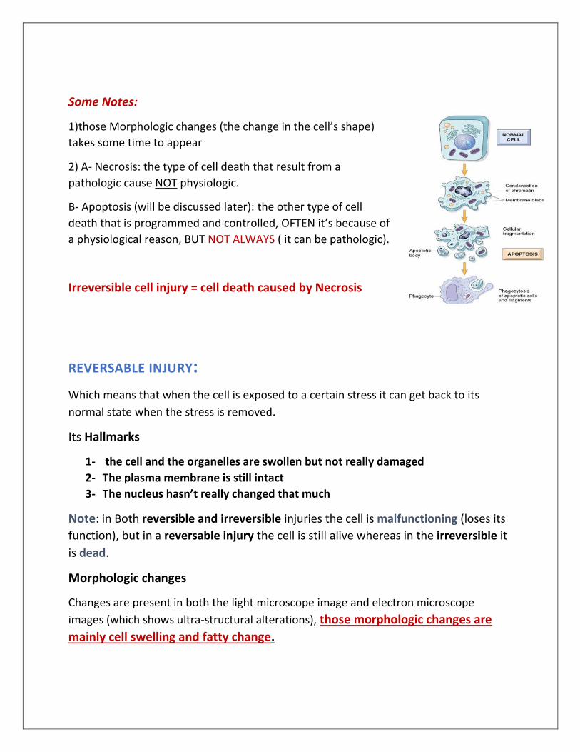
Some Notes:
1)those Morphologic changes (the change in the cell’s shape)
takes some time to appear
2) A- Necrosis: the type of cell death that result from a
pathologic cause NOT physiologic.
B- Apoptosis (will be discussed later): the other type of cell
death that is programmed and controlled, OFTEN it’s because of
a physiological reason, BUT NOT ALWAYS ( it can be pathologic).
Irreversible cell injury = cell death caused by Necrosis
REVERSABLE INJURY:
Which means that when the cell is exposed to a certain stress it can get back to its
normal state when the stress is removed.
Its Hallmarks
1- the cell and the organelles are swollen but not really damaged
2- The plasma membrane is still intact
3- The nucleus hasn’t really changed that much
Note: in Both reversible and irreversible injuries the cell is malfunctioning (loses its
function), but in a reversable injury the cell is still alive whereas in the irreversible it
is dead.
Morphologic changes
Changes are present in both the light microscope image and electron microscope
images (which shows ultra-structural alterations), those morphologic changes are
mainly cell swelling and fatty change.
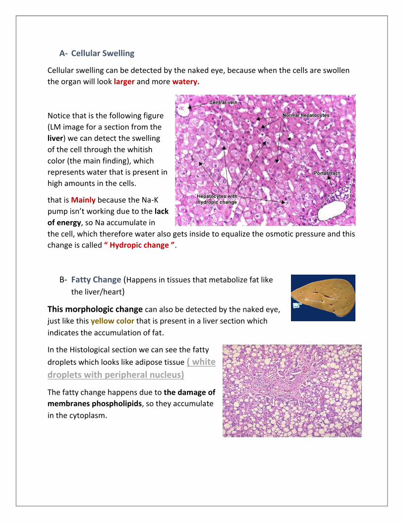
A- Cellular Swelling
Cellular swelling can be detected by the naked eye, because when the cells are swollen
the organ will look larger and more watery.
Notice that is the following figure
(LM image for a section from the
liver) we can detect the swelling
of the cell through the whitish
color (the main finding), which
represents water that is present in
high amounts in the cells.
that is Mainly because the Na-K
pump isn’t working due to the lack
of energy, so Na accumulate in
the cell, which therefore water also gets inside to equalize the osmotic pressure and this
change is called “ Hydropic change ”.
B- Fatty Change (Happens in tissues that metabolize fat like
the liver/heart)
This morphologic change can also be detected by the naked eye,
just like this yellow color that is present in a liver section which
indicates the accumulation of fat.
In the Histological section we can see the fatty
droplets which looks like adipose tissue ( white
droplets with peripheral nucleus)
The fatty change happens due to the damage of
membranes phospholipids, so they accumulate
in the cytoplasm.
Ultra-structural changes that can be seen under electron microscope include:
1- The occurrence of blebs in the plasma membrane, but it’s not damaged and still
intact.
2- Swelling of the mitochondria and the appearance of black densities because of
the damaged phospholipids.
3- Swelling of the endoplasmic reticulum.
4- clumping of the chromatin in the nucleus.
5- The occurrence of myelin figures ( an accumulation for phospholipids because of
membrane damage ).
IRREVERSIBLE CELL INJURY If the stress is very severe the cell may not handle it, so that will develop to an
irreversible cell energy and then necrosis.
Irreversible injury features:
1- Irreversible mitochondrial dysfunction ( THE MAIN characteristic at the ultra-
structural level )
2- The loss of the plasma membrane and organelles membrane (discontinued
membrane). When some organelles like mitochondria or lysosomes lose their
membranes, their enzymes will be leaked and lost.
3- Disappearance of DNA, which make it impossible for the cell to go back to
normal
Morphologic Changes : Irreversible injury
The first thing we notice is increased eosinophilia in the cytoplasm when it’s stained
with H&E, and that’s because
1) The binding between eosin and denatured proteins increases.
2) The disappearance of RNA (which is basophilic) because there is no transcription ,
contribute to the eosinophility.

Nuclear changes take place also take place in irreversible injury, and it’s divided into 3
stages :
A- “pyknosis” or the shrinkage of the chromatin and increased basophilia so it looks
like a dark blue spot
B- “karyorrhexis” (chromatin fragmentation)
C- “karyolysis” (chromatin disappearance/basophilia fades)
Note : those 3 steps are not ordered , for example sometimes B happens before A
Remember that the reversibly injured cells are whitish due to the existence of
water.
Other changes are similar between the reversible and irreversible injuries but more
severe in the irreversible, like: (The ER and mitochondria dilatation, the loss of the
membranes and the accumulation of the black densities and myelin figures).
You can see here three sections for the kidney;
1- the first one from the left is the normal tissue
2- the middle one is on a reversible injury; the cells are swollen, and you can see
the whitish color they’ve turned into. Some cells will look very eosinophilic
preparing to enter the irreversible injury status
3- the right one is on an irreversible injury where some cells have lost their nucleus
and got ruptured.

Types of cell death: Necrosis & apoptosis
each one occurs depending on the nature and severity of the injury.
Necrosis: it’s caused by ischemia, toxins, infection or trauma. It usually happens when
the injury is rapid, uncontrollable and lead to severe disturbances.
Apoptosis ( cell suicide ): it’s the controlled cell death and it’s caused by sun induced
injury or growth factor deficiency, can be modified by certain substances and it’s the
basic mechanism of some chemotherapeutic agents that are used to treat cancer
In some cases like ischemia, both Necrosis & apoptosis occur and it’s called
“Necroptosis”. Where some cells die because of Necrosis and others die because
apoptosis
Note on the table:
1- Sometimes some macrophages digest apoptotic cells, but generally apoptosis does
NOT cause inflammation
2- In Apoptosis the cell goes under shrinkage , but in Necrosis it is swollen
3- Necrosis happens to a big number of cells once a time, but apoptosis happens to
selective cells

CLINICAL IMPLICATIONS
Studying cell injuries helps us in diagnosing certain diseases. for example, when a cell is
injured, its enzymes will be leaked into the blood, so when testing a blood sample we
can tell where the injury has taken place, for elaboration:
1- when we suspect a myocardial infarction, it can be detected by measuring the
cardiac enzymes in the blood.
2- We can detect hepatitis when abnormal measurements are detected for hepatic
enzymes in the blood.
And the same goes for Pancreatitis, etc. ….
MORPHOLOGIC PATTERNS OF TISSUE NECROSIS
When a big portion of the tissue is affected by necrosis, it will result in certain gross
morphologic patterns, which can give a clue about the etiology of necrosis (the cause of
it), which leads us to talk about the Types of Necrosis:
1- Coagulative necrosis:
This type is usually caused by ischemia to all solid organs except in the brain
characterized by a wedge-shaped (kind of triangle) pale area corresponding to
the decrease in blood supply.
For elaboration, imagine the thick Red line
as an artery and the two arrows as its
branches, so if the big artery is blocked (
ischemic ) then its branches won’t have any
blood supply, therefore the area between
them ( the blue triangle area ) will go under
necrosis.

Under the LM the are will look pale but its preserved in its shape for many hours or even
days, WHY?
Because the decrease of blood supply causes the
enzymes to denature , so the enzymatic digestion of
the molecules in this area won’t happen.
Conclusion: the shape of the tissue’s cells wont
change that much, but you can notice the
disappearance of the nucleus.
B = Necrosis C = normal
2- Liquefactive necrosis:
It’s a viscous fluid like material appears
on the affected area, it may be caused
by:
A- bacterial or fungal infections which lead
to the formation of a creamy yellowish
fluid called pus
B- ischemia or infractions to the CNS

Macroscopically In this section of the lung ( the yellow lung ) we have this cavity
filled with the yellowish fluid
Microscopically it is characterized by the rapid accumulation of inflammatory cells
like neutrophils ( AKA: abscess )
In the brain this is a focal area
containing a liquefactive viscous fluid
corresponds to an area on infarction
in the brain.
3- Gangrenous necrosis
It’s coagulative necrosis but this is a medical term. It occurs because tissues at different
planes of the organ get affected by coagulative necrosis. We see this a lot in diabetic
patients
The black color appears because of the ischemic necrosis of the skin, underlying
tissues and the bones. It can be dry or wet if there’s a super imposed infection on
it. (infection on top of an infection )

4- Caseous Necrosis
This type appears as a cheese like yellow material in the area of infarction and :
A- It isn’t preserved at all so it’s characterized by an acellular ( no living cells )
center that stains pink (eosinophilic)
B- It’s surrounded by macrophages, inflammatory cells and multi-nucleated giant
cells and we call this granuloma
Very Important Note: This type of necrosis is seen associated with
tuberculosis. (An infection caused by mycobacterium TB)
5- Fat Necrosis
Fat necrosis is associated with acute pancreatitis, which is considered a top emergency.
Elaboration:
Pancreatic enzymes mainly lipases will be released to the peritoneum (which is
filled with fat) and will digest the fat in the abdomen releasing fatty acids from
triglycerides, so there’s fat destruction and they can bind to Ca2+ which leads to
the appearance of these whitish foci that look like a chalky material
(saponification) which helps the surgeon to identify fat necrosis
Saponification: Binding of fatty acids to Ca 2+.

Under the LM, we see shadows of these fatty cells with lost nucleus
6- Fibrinoid Necrosis This is the only type that can be seen just under the microscope, it’s caused by
the deposition of fibrin that contain pink material in the walls of blood vessels,
which occur due to an antigen-antibody reaction in the wall of blood vessels
leading to this deposition.
It’s usually associated with vasculitis
Vasculitis: auto immune disease that characterized by the
inflammation of the walls of blood vessels, leading to the
accumulation of this pink material
“It always seems impossible until it's done.”
― Nelson Mandela
دعوة من قلبك