Embed Size (px)
Citation preview
Neurol Clin 25 (2007) 809–832
Normal Pressure Hydrocephalus
Neill R. Graff-Radford, MBBCh, FRCPMayo Clinic Jacksonville, 4500 San Pablo Road, Jacksonville, FL 32224, USA
Epidemiology
At this time, there is no definitive information on the incidence or prevalenceof normal pressure hydrocephalus (NPH). There are a few studies in the liter-ature that address this issue. In one involving the evaluation of all elderly per-sons in Republic of San Marino (smallest independent country; in Italy), theresearchers found that 2 of 396 persons had idiopathic NPH (INPH), a 0.5%prevalence of persons over age 65 [1]. In a door-to-door surveyof parkinsonism,Trenkwalder and colleagues [2] found 4 of 982 people who had NPH, againa prevalence of 0.5%. In the 2000 United States census, there were 35 millionpersons ages 65 and older. Based on the two studiesmentioned previously, thereis an estimated prevalence of 175,000 persons who have NPH in the UnitedStates. Another way to estimate the United States prevalence is from the studyby Clarfield [3], who report, in a meta-analysis of 37 dementia studies, a 1%NPH prevalence in more than 5000 dementia patients. Based on this studyand the estimated 4 to 6million persons in theUnited Stateswho have dementia[4], there would be approximately 40,000 to 60,000 persons who have NPH.
Vanneste and colleagues [5] estimated the incidence in Holland to be 1.3 to2.2 per million per year, whereas Krauss andHalve [6], in a survey of neurosur-geons, calculated NPH incidence in Germany to be 1.8 per 100,000 per year.Theynote that this probably is an underestimate because it is basedon referrals.
Why the diagnosis is difficult
Doctors find the diagnosis and treatment of patients who have NPHparticularly difficult. The reasons for this are that no combinations of the
This study was funded in part by grant P50 AG16574 and by the Robert and Clarice
Smith and Abigail Van Buren Alzheimer’s Disease Research Program.
This chapter is based in part on the chapter, ‘‘Normal Pressure Hydrocephalus’’ by Dr.
Graff-Radford published in the April 2007 Dementia issue of Continuum: Lifelong Learning
in Neurology (13[2]:144–164) and is used with permission from the American Academy of
Neurology.
E-mail address: [email protected]
0733-8619/07/$ - see front matter � 2007 Elsevier Inc. All rights reserved.
doi:10.1016/j.ncl.2007.03.004 neurologic.theclinics.com
810 GRAFF-RADFORD
cardinal findings (gait difficulty, cognitive decline, incontinence of urine, andenlarged ventricles) are pathognomonic for the diagnosis; each of the cardi-nal symptoms is common in the elderly and has many causes (discussedlater); all of the diagnostic tests described give false-positive and false-neg-ative results; the surgical treatment carries significant short- and long-termrisks, and the cause or pathogenesis of many NPH cases is not known.
Discussing the cardinal features:
1. Gait difficulty: one study found that 20% of persons ages 75 and olderhad gait abnormality and this was related to future development of de-mentia [7].
2. Cognitive decline: one study estimated that 4.5 million persons over age65 had Alzheimer’s disease in the United States in 2000 [4].
3. Incontinence: in 2006, Anger and colleagues [8] reported the overallprevalence of incontinence in older women as 38% and Stothers andcolleagues [9] reported a prevalence of 17% in men over age 60.
4. Enlarged ventricles: Barron and colleagues [10] showed that ventriclesize increases with age, and Jack and colleagues [11] have shown ventri-cle size increases faster in patients who have AD compared withcontrols.
Thus, all the cardinal features are common in the elderly and have manycauses.
Differential diagnosis
When evaluating patients, keep in mind a practical differential diagnosis(Box 1), which helps focus the history and examination.
Box 1. Differential diagnosis of normal pressure hydrocephalus
� Combinations of ventriculomegaly, dementia, and factorsaffecting gait (eg, cervical spondylosis, large joint arthritis,peripheral neuropathy, impaired vision, vestibular dysfunction,and antipsychotics)
� Vascular dementia, including subcortical ischemicencephalopathy or Binswanger’s disease
� Parkinson’s disease dementia and enlarged ventricles� Parkinsonian syndromes (Lewy body disease, corticobasal
ganglionic degeneration, progressive supranuclear palsy, andmultiple system atrophy) and enlarged ventricles
� Frontotemporal dementia with caudate atrophy
811NORMAL PRESSURE HYDROCEPHALUS
Clinical evaluation
General factorsClinicians should evaluate patients’ general medical health because this
may be important when considering surgery. Factors theoretically thatcould aggravate hydrocephalus include systemic hypertension (see discus-sion later of hydrocephalus association with hypertension) and a recenthead injury (which is pertinent particularly in individuals who have gait dif-ficulty). Evaluate for sleep apnea, congestive heart failure, lung disease, andobesity, all of which could increase jugular venous pressure and decrease ce-rebrospinal fluid (CSF) flow into the cerebral venous sinuses. If patients areon long-term anticoagulants, such as coumadin for atrial fibrillation, takethis into account for the theoretic increased risk for brain hemorrhage dur-ing and after surgery. Look for evidence of arthritis of the hips and knees,cervical myelopathy, lumbar stenosis and radiculopathy, visual impairment,vestibular dysfunction, and peripheral neuropathy, all of which could impairgait.
History related to hydrocephalusThere are several specific questions that should be asked when taking
a history from these patients and their families.Ask how long patients have been demented. If this is more than 2
years, it is less likely that patients will respond to surgery [12,13]. Notethat the question is not how long patients have had gait abnormalitybut how long patients have been demented. In the author and colleagues’series, this question predicted 5 of 7 unimproved and 21 of 23 improvedpatients [12].
Ask which started first, gait abnormality or dementia. If the gait abnor-mality began before or at the same time as dementia, then there is a betterchance for successful surgery, whereas if dementia started before gait abnor-mality, shunting is less likely to help. In the author and colleagues’ series thisquestion predicted 3 of 7 unimproved and 23 of 23 improved patients [12].This observation has been reported previously by Fisher [14].
Ask if patients abused alcohol, because alcohol abuse is a poor prognos-tic indicator [15].
Ask if there are secondary causes for hydrocephalus, such as subarach-noid hemorrhage, meningitis, prior brain surgery, and head injury. If anyof these is present, the chances of improvement with surgery are better[13,15,16].
Ask if patients have a large head size, as evidenced by needing a large hat.This may indicate that patients suffer from congenital hydrocephalus thathas become symptomatic in later life [17,18]. The author and colleagueshave found that 10% to 20% of persons diagnosed with so-called‘‘INPH’’ have large heads, indicating that they may have congenital hydro-cephalus that becomes symptomatic in later life [17,18].

812 GRAFF-RADFORD
ExaminationOn examination, the following issues should be addressed.Measure the head circumference. If greater than 59 cm in males or 57.5
cm in females, suspect that patients could have an element of congenitalhydrocephalus that has become symptomatic in later life [17,18].
Look for signs of diseases that may mimic NPH (see Box 1). Theseinclude AD with extrapyramidal features, Parkinson’s disease dementia,parkinsonian syndromes (progressive supranuclear palsy, corticobasal de-generation, and multiple system atrophy), diffuse Lewy body disease, fron-totemporal dementia, cerebrovascular disease, and phenothiazine use. Also,look for cervical spondylosis with spinal cord compression, lumbar stenosisor radiculopathy, arthritis of the hips and knees, and multiple factors thatimpair gait abnormality (as might occur in diabetics who have peripheralneuropathy and visual impairment, alcoholics who have peripheral neurop-athy and cerebellar atrophy, or the elderly who have vestibular and visualdysfunction and arthritis).
NeuropsychologyLook for evidence of aphasia. If there is evidence of aphasia (eg,
anomia), this is a poor prognostic indicator for surgical success [12,15].Ogino and colleagues recently reported the typical neuropsychology pat-tern of patients who have NPH compared with those who have AD [19].Of 42 patients who had AD and 21 who had NPH who were matchedfor age, gender, and Mini–Mental State Examination [20], the patientswho had NPH scored better on orientation and on the delayed recallof the Wechsler Memory Scale–Revised (WMS-R) [21] but significantlylower on the attention and concentration subtests of the WMS-R andon the digit span, arithmetic, block design, and digit symbol substitutionsubtest of the Wechsler Adult Intelligence Scale–Revised [22]. In sum-mary, the cognitive deficits are characterized by psychomotor slowing,memory impairment, and impaired executive function with preserved cor-tical tests, such as naming.
Radiologic evaluation
CTSince the advent of CT, the documentation of ventriculomegaly has be-
come easier. Patients have ventriculomegaly (above the 95th percentile)when the modified Evan’s index (maximum width of the frontal horns/mea-sure of the inner table at the same place) is greater than 0.31 [23]. The ven-tricles normally enlarge with age [10], a point to be taken into account whendiagnosing hydrocephalus. There is slow ventricular enlargement to age 60years and then the rate of enlargement increases. In Barron and colleagues’study [10], the mean ventricular size was 5.2% (percent of intracranial area)in the decade 50 to 59 years, 6.4% in 60 to 69 years, 11.5% in 70 to 79 years,and 14.1% in 80 to 89 years.
813NORMAL PRESSURE HYDROCEPHALUS
In one study, the greater the sulcal enlargement, the less the chance of im-provement with surgery [24]. Patients still might improve with surgery, how-ever, even if there is sulcal enlargement and hydrocephalus. Borgesen andGjerris [24] measured the largest sulcus in the high frontal or parietal regionand found that if the cortical sulci were less than 1.9 mm, 17 of 17 patientsshunted improved; if the sulci were 1.9 to 5 mm, 17 of 20 shunted improved;and if the sulci were 5 mm or more, 15 of 27 shunted improved.
MRIDetecting congenital hydrocephalus. MRI is an excellent method for evalu-ating patients who have possible symptomatic hydrocephalus and is the neu-roimaging study of choice in patients who have NPH. It has the advantageof being able to visualize relevant structures in the posterior fossa, includingcerebral aqueduct stenosis, cerebellar tonsil herniation, and infarctions inthe brainstem. Further, MRI can be used to obtain volumetric measuresof medial temporal lobe structures, a technique that has been shown to beuseful in separating patients who have AD from normal elderly controls[25].
Approximately 10% to 20% of patients who have symptomatic hydro-cephalus after age 60 years may have congenital hydrocephalus that be-comes symptomatic in later years [17,18]. A clinical clue to this is thatpatients have a large head size. On MRI, the ventricular enlargement showsno or little associated periventricular increased signal on T2-weighted imag-ing, indicating a chronic process. In addition, a cause for the congenital hy-drocephalus rarely is found, such as an Arnold-Chiari malformation oraqueductal stenosis.
White matter lesions and normal pressure hydrocephalus. Although someearly reports using CT indicated the presence of transependymal flowmay be related to a good surgical prognosis [24] (Borgesen and Gjerris’study reported that 16 of 16 who had periventricular hypodensity on CTimproved with surgery), later studies did not confirm this or found theopposite. Bradley [26] reported that on MRI the presence or extent ofdeep white matter changes did not correlate with outcome, whereas Kraussand colleagues [27] reported that the degree of improvement after shuntsurgery depends on the extent and severity of white matter lesions (ie,the more extensive the white matter lesions, the less the improvement).In a subsequent report [28], they compared the MRI findings in NPH toan age-matched controlled group and found that in both groups theperiventricular white matter lesions correlated with the deep white matterlesions. In the control group, the white matter lesions correlated signifi-cantly with age and the anterior horn index (frontal horn width dividedby the horizontal intracranial width). In contrast, in the NPH group, therewas no correlation of white matter lesions with age and there was a sig-nificant negative correlation between the white matter lesions and the
814 GRAFF-RADFORD
frontal horn index (ie, the wider the frontal horns, the fewer white matterlesions present). They argue that the white matter lesions do not cause thehydrocephalus but that the common link between the frequent coexistenceof INPH and vascular encephalopathy (as evidenced by white matterlesions) is arterial hypertension (the association of systemic hypertensionand NPH is discussed later).
In a postmortem MRI study of autopsied brains and a histologic analysisof the same brains, Munoz and colleagues [29] found the white matterchanges seen on MRI correlate with decreased density of axons and myelin-ated fibers, diffuse vacuolation of white matter (so-called ‘‘spongiosis’’), anddecreased density of glia. Infarctions were not common in these areas. Al-though this study does not necessarily apply to the white matter changesseen in hydrocephalic patients, it does indicate that white matter MRI find-ings do not necessarily indicate irreversible periventricular infarctions,which would make shunt surgery unlikely to be effective.
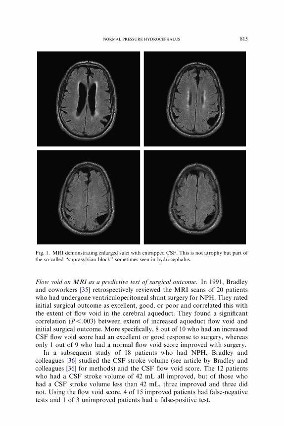
MRI differentiation of normal pressure hydrocephalus and Alzheimer’s disease.Traditionally, the presence of ventriculomegaly without sulcal enlargementhas been a radiologic finding believed to indicate NPH when accompaniedby the typical clinical triad. Studies by Holodny and colleagues [30] andKitagaki and colleagues [31], however, have pointed out the occasionaloccurrence of focally dilated sulci over the convexity or medial surface ofa hemisphere in patients who have NPH, unlike the diffuse sulcal enlarge-ment seen in AD (Fig. 1). Kitagaki and colleagues’ report [31] also indicatedsignificantly greater sylvian CSF volume in patients who had INPH com-pared with AD. He believed this was a sign supportive of NPH, indicatinga ‘‘suprasylvian block.’’
An area where MRI has the potential to be helpful for surgical prognosisis volumetric measurements of certain structures in the temporal lobe. Thereis enlargement of the temporal horns of the lateral ventricles in patients whohave NPH and those who have AD. Jack and colleagues [25] developeda technique for measuring the volumes of structures in the anterior temporallobe and hippocampal formation. Holodny and colleagues [32] measured theCSF volumes of the perihippocampal fissures and the ventricular volumes.They showed that the perihippocampal fissures were enlarged significantlyin patients who had AD compared with patients who had NPH, whereasthe ventricles were larger in NPH. This was detectable by visible inspectionand computer volumetrics.
MR-based volumetric measurements of the hippocampal formation havebeen shown to be useful in discriminating between AD and normal elderlycontrols [25]. Although Golomb and colleagues [33] found smaller hippo-campal volumes in patients who had NPH compared with controls, Savolai-nen and colleagues [34] found only a minor left-side decrease. Savolainenand colleagues, however, detected significantly larger hippocampi in patientswho had NPH compared with patients who had AD.
815NORMAL PRESSURE HYDROCEPHALUS
Flow void on MRI as a predictive test of surgical outcome. In 1991, Bradleyand coworkers [35] retrospectively reviewed the MRI scans of 20 patientswho had undergone ventriculoperitoneal shunt surgery for NPH. They ratedinitial surgical outcome as excellent, good, or poor and correlated this withthe extent of flow void in the cerebral aqueduct. They found a significantcorrelation (P!.003) between extent of increased aqueduct flow void andinitial surgical outcome. More specifically, 8 out of 10 who had an increasedCSF flow void score had an excellent or good response to surgery, whereasonly 1 out of 9 who had a normal flow void score improved with surgery.
In a subsequent study of 18 patients who had NPH, Bradley andcolleagues [36] studied the CSF stroke volume (see article by Bradley andcolleagues [36] for methods) and the CSF flow void score. The 12 patientswho had a CSF stroke volume of 42 mL all improved, but of those whohad a CSF stroke volume less than 42 mL, three improved and three didnot. Using the flow void score, 4 of 15 improved patients had false-negativetests and 1 of 3 unimproved patients had a false-positive test.
Fig. 1. MRI demonstrating enlarged sulci with entrapped CSF. This is not atrophy but part of
the so-called ‘‘suprasylvian block’’ sometimes seen in hydrocephalus.
816 GRAFF-RADFORD
Krauss and colleagues [37] report that that the flow void in the cerebralaqueduct of 37 patients who had INPH was not significantly different fromthat in 37 age-matched controls. Further, the extent of the flow void exten-sion into the third, fourth, and lateral ventricles did not correlate withamount of improvement in these patients but correlated with rather thewidth of the ventricles.
Hakim and Black [38], in a small study of 12 patients of whom 10 im-proved, found that the MRI-CSF flow studies were correct in six, but fivehad false negatives and one a false positive.
Dixon and colleagues [39] also found in 49 patients that CSF flowthrough the cerebral aqueduct did not reliably predict those who improvedwith shunt surgery. These last three studies cast doubt on CSF flow as a di-agnostic test in NPH.
Summary of factors to be addressed when looking at the CT or MRIHydrocephalus must be present. The modified Evans ratio should be
greater than 0.31 [23]. Is cortical atrophy prominent? If there is extensivecortical atrophy, this reduces but does not eliminate the chance of improve-ment with surgery [13,24]. Avoid calling entrapped sulci or suprasylvianblock brain atrophy [30,31].
The pattern of atrophy may be useful diagnostically (eg, Does it involvethe medial temporal lobes as seen in AD?). Although data on this point arelacking, it may be that prominent medial temporal cortical atrophy de-creases the chances for surgical cognitive improvement because these pa-tients may have AD [25,30].
Is there evidence of congenital hydrocephalus? For example, is there aque-ductal stenosis or an Arnold-Chiari malformation and are the ventricles largewith little white matter abnormality indicating a chronic process [17,18]?
NewerMRI techniques, such as cine MRI, involving the analysis of a CSFflow void in the aqueduct of Sylvius, were first believed to be helpful [35,36]but unfortunately have not been found to be so in subsequent studies [37–39].
Regional cerebral blood flow
It has been reported that regional cerebral blood flow (rCBF) is decreasedin the frontal areas in hydrocephalus [40] and in the parietotemporal areas inAD [41]. On the presumption that many of the nonimproved group haveAD (Bech and colleagues have shown more recently [42] that 25% at timeof shunt surgery brain biopsy show AD pathology), the author and col-leagues tried to differentiate those who will respond to shunt surgery fromthose who will not, based on the pattern of preoperative rCBF [43]. Todo this, frontal over posterior regional blood flow was calculated, expectinga lower frontal-posterior ratio in true symptomatic hydrocephalus anda higher ratio in pseudosymptomatic hydrocephalus patients who haveAD. This has been a good method in predicting surgical outcome: the ratio
817NORMAL PRESSURE HYDROCEPHALUS
predicted 5 of 7 unimproved and 22 of 23 improved patients in the authorand colleagues’ series [12]. Granado and colleagues [44] also found thatthose suspected of having NPH with and Alzheimer pattern did not improvebut those who had frontal hypoperfusion did. Unfortunately, a review of theliterature on CBF in NPH shows no clear-cut use at this time [45].
Cisternography
The literature suggests there are many cases of a positive test (radioisotopeseen within the ventricles 48 to 72 hours after being injected in the lumberarea) with no improvement with surgery and patients who have equivocalor negative tests who do improve. Further, the test itself may be difficult tointerpret [46]. Black [46], in a review of his experience with this test, foundthe following: of 11 patients who had a positive test, nine improved andtwo did not; of six patients who had mixed results, three improved and threedid not; of six who had negative results, four improved and two did not. Hesuggests a positive test is helpful but an equivocal or negative test is not. Amore recent study by Vanneste and colleagues [5] reported, ‘‘cisternographydid not improve the accuracy of combined clinical and computerizedtomography in patients with presumed normal-pressure hydrocephalus.’’
Cerebrospinal drainage procedures
Lumbar puncture
If a patient’s gait improves after removing a large quantity of CSF bylumbar puncture (LP) (30 to 50 mL; this can be repeated daily), this personis a good candidate for shunt surgery [47]. Malm and colleagues [48] foundno predictive values of a spinal tap test but Walchenbach and colleagues[49] found a poor sensitivity (9 of 35 cases with a positive test had improve-ment with shunt surgery) but a good positive predictive value (9 of 9 witha positive test improved). One of the issues is that in both of these studiesthe gait was evaluated 4 to 6 hours after the LP. In patients who do notleak CSF after the LP, the CSF is replaced at 0.3 mL per minute would be re-plenished in 2 hours. When the author and coworkers evaluate patients afteran LP, they made a videotape of the gait immediately after the LP. Later theycompared this to the videotape done prior to the LP. At this time, it is reason-able to presume that a positive LP test has a good positive predictive value butis not sensitive.
External cerebrospinal fluid drainage
A modification of this technique also has been reported and is continuousCSF drainage via a catheter placed in the lumbar CSF space [50]. In theguidelines for the diagnosis and management of INPH [51], the guidelinesare based on three studies [48–50]. These studies showed a range of
818 GRAFF-RADFORD
sensitivity from 50% to 100%, specificity 60% to 100%, positive predictivevalue 80% to 100%, negative predictive value 36% to 100%, and accuracy58% to 100%. In one study [49], two of 38 developed meningitis and fivepulled out their drains. In another study [50], two of 22 had infections,four removed their drains, and three had root irritation.
Although this test is more sensitive than a large-volume spinal tap, theinvestigators caution that it should be undertaken by an experienced teamthat can limit the complication rate.
Cerebrospinal fluid infusion tests
There are several ways of measuring the resistance to absorption (Ro) orthe reciprocal to this, called the conductance. In Katzman and Hussey’s test[52], fluid was infused through an LP needle into the CSF at a known rateuntil a steady state pressure is reached. The Ro is calculated as the new pres-sure minus the initial pressure divided by the infused flow rate and is givenas mm Hg per mL per minute. Another way of doing this is the bolusmethod, where 4 mL are infused at 1 mL per second and the new pressuremeasured. Lastly, Ro can be calculated as described by Borgesen and Gjer-ris [24]. They called the reciprocal of the Ro the conductance. In thismethod, CSF absorption is measured at different CSF pressures. The con-cept is that the greater the pressure needed to obtain an amount of absorp-tion, the better the chances of the patient improving with shunt surgery.Absorption is calculated by infusing fluid through an LP needle for a giventime (5 minutes) while catching the overflow from a ventricular catheter.There is some evidence to show that the amount of CSF produced doesnot vary much at different CSF pressures and is approximately 0.4 mLper minute. Because one knows how much is infused through the LP needleand how much overflows through the ventricular catheter, one can calculatethe amount absorbed in this time period. The following equation gives ab-sorption: Absorption ¼ Infused (measured) þ Produced (assumed) � Over-flow (measured).
The overflow pressure for the ventricular catheter then is raised andabsorption then is calculated at this new pressure. Between six and eight ab-sorptions at different pressures are obtained in this way and then absorptionis plotted against pressure and the slope of the line calculated (ie, absorp-tion/pressure). The slope of this line is called the conductance. Borgersenand Gjerris reported that a conductance of less than 0.08 predicted a favor-able outcome. A conductance of 0.08 ¼ Rcsf of 12.5 mm Hg/mL/min, whereRcsf is resistance to CSF outflow.
Boon and colleagues [53] report the first multiple center randomizedstudy evaluating the predictive value for shunt surgery of measuring Rcsf.They enrolled 101 patients (both idiopathic and secondary, although theynote most were idiopathic) and measured the Rcsf by infusing salinethrough 19-gage LP needle at 1.4 to 1.6 mL per minute until a stable

819NORMAL PRESSURE HYDROCEPHALUS
pressure plateau was reached or the pressure exceeded 50 mm Hg. The Rcsfwas calculated as the difference between the plateau and the baseline pres-sure divided by the infusion rate. They randomized the patients to receiveeither low or medium pressure valves. Outcome measures were an NPHscale (sum of gait and dementia measures) and the modified Rankin scale.Follow-up was at 1, 3, 6, 9, and 12 months. Intention to treat analysiswas performed on all 101 patients and 57% showed improvement in theNPH scale and 59% in the modified Rankin scale at 1 year. When all knownserious events unrelated to NPH that clearly interfered with neurologicfunction were excluded, 95 patients were left. In these patients, 76% hada meaningful improvement on the NPH scale and 69% improved one gradeon the modified Rankin scale. Using a cutoff of the Rcsf of less than 18 mmHg per mL per minute, 20 of 59 had no improvement on the NPH scale.Above this cutoff, 3 of 36 had no improvement on the NPH scale.
The investigators conclude that the Rcsf obtained by lumbar CSF infu-sion is a reliable method for selecting patients for shunt surgery if theRcsf cutoff is 18 mm Hg per mL per minute or greater. Patients who havean rCBF less than 18 mm Hg per mL per minute should undergo shuntplacement only when characteristic clinical features of NPH are present.Unfortunately, the majority of patients (O60%) under consideration forshunt surgery have a Rcsf of less than 18 mm Hg per mL per minute andthis diagnostic test is not helpful in this group. In the guidelines [54], theynote the data are limited and ‘‘the results, methods and thresholds are centerspecific and subject to wide variation.’’
Cerebrospinal fluid pressure monitoring
There are reports of a significant relationship between measures of intra-cranial CSF pressure monitoring and surgical outcome for symptomatichydrocephalus (eg, in the Borgesen and Gjerris study and in the authorand colleagues’ study [12,24], the greater the percentage of time B waveswere present, the greater the chance of a good outcome). In Williams’ andcolleagues study, the sensitivity using a threshold of 25% time of B wavesthe sensitivity in predicting surgical out come was 78% but the specificitywas 40%. From all the studies, using only the percentage of time B wavesare present would be inadequate in predicting surgical outcome. The con-sensus guidelines [51] note that there is insufficient evidence to use pressuremonitoring for prognostic purposes.
One point to be evaluated in the future is the prognostic value of how longthe pressure is highwhen patients aremonitored. In the author and colleagues’series, the longer the pressure was more than 15 mmHg, the better the chanceof successful surgery [12]. This may be important because it implies thatincreased pressure may be pathogenic in symptomatic hydrocephalus.
These data raise the issue about what is meant by NPH. Does it mean nor-mal pressure at one spinal tap or does it imply the pressure remains normal all
820 GRAFF-RADFORD
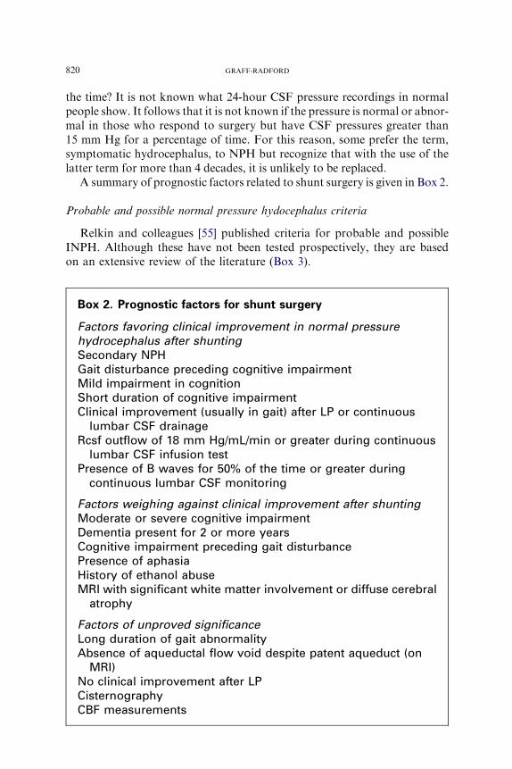
the time? It is not known what 24-hour CSF pressure recordings in normalpeople show. It follows that it is not known if the pressure is normal or abnor-mal in those who respond to surgery but have CSF pressures greater than15 mm Hg for a percentage of time. For this reason, some prefer the term,symptomatic hydrocephalus, to NPH but recognize that with the use of thelatter term for more than 4 decades, it is unlikely to be replaced.
A summary of prognostic factors related to shunt surgery is given in Box 2.
Probable and possible normal pressure hydocephalus criteria
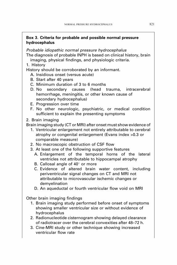
Relkin and colleagues [55] published criteria for probable and possibleINPH. Although these have not been tested prospectively, they are basedon an extensive review of the literature (Box 3).
Box 2. Prognostic factors for shunt surgery
Factors favoring clinical improvement in normal pressurehydrocephalus after shuntingSecondary NPHGait disturbance preceding cognitive impairmentMild impairment in cognitionShort duration of cognitive impairmentClinical improvement (usually in gait) after LP or continuous
lumbar CSF drainageRcsf outflow of 18 mm Hg/mL/min or greater during continuous
lumbar CSF infusion testPresence of B waves for 50% of the time or greater during
continuous lumbar CSF monitoring
Factors weighing against clinical improvement after shuntingModerate or severe cognitive impairmentDementia present for 2 or more yearsCognitive impairment preceding gait disturbancePresence of aphasiaHistory of ethanol abuseMRI with significant white matter involvement or diffuse cerebral
atrophy
Factors of unproved significanceLong duration of gait abnormalityAbsence of aqueductal flow void despite patent aqueduct (on
MRI)No clinical improvement after LPCisternographyCBF measurements
821NORMAL PRESSURE HYDROCEPHALUS
Box 3. Criteria for probable and possible normal pressurehydrocephalus
Probable idiopathic normal pressure hydrocephalusThe diagnosis of probable INPH is based on clinical history, brain
imaging, physical findings, and physiologic criteria.1. HistoryHistory should be corroborated by an informant.
A. Insidious onset (versus acute)B. Start after 40 yearsC. Minimum duration of 3 to 6 monthsD. No secondary causes (head trauma, intracerebral
hemorrhage, meningitis, or other known cause ofsecondary hydrocephalus)
E. Progression over timeF. No other neurologic, psychiatric, or medical condition
sufficient to explain the presenting symptoms
2. Brain imagingBrain imaging study (CT or MRI) after onset must show evidence of
1. Ventricular enlargement not entirely attributable to cerebralatrophy or congenital enlargement (Evans index >0.3 orcomparable measure)
2. No macroscopic obstruction of CSF flow3. At least one of the following supportive features
A. Enlargement of the temporal horns of the lateralventricles not attributable to hippocampal atrophy
B. Callosal angle of 40� or moreC. Evidence of altered brain water content, including
periventricular signal changes on CT and MRI notattributable to microvascular ischemic changes ordemyelination
D. An aqueductal or fourth ventricular flow void on MRI
Other brain imaging findings1. Brain imaging study performed before onset of symptoms
showing smaller ventricular size or without evidence ofhydrocephalus
2. Radionucleotide cisternogram showing delayed clearanceof radiotracer over the cerebral convexities after 48–72 h.
3. Cine-MRI study or other technique showing increasedventricular flow rate
822 GRAFF-RADFORD
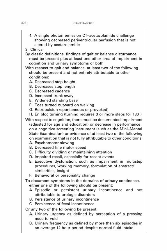
4. A single photon emission CT–acetazolamide challengeshowing decreased periventricular perfusion that is notaltered by acetazolamide
3. ClinicalBy classic definitions, findings of gait or balance disturbance
must be present plus at least one other area of impairment incognition and urinary symptoms or both
With respect to gait and balance, at least two of the followingshould be present and not entirely attributable to otherconditions:A. Decreased step heightB. Decreases step lengthC. Decreased cadenceD. Increased trunk swayE. Widened standing baseF. Toes turned outward on walkingG. Retropulsion (spontaneous or provoked)H. En bloc turning (turning requires 3 or more steps for 180�)
With respect to cognition, there must be documented impairment(adjusted for age and education) or decrease in performanceon a cognitive screening instrument (such as the Mini–MentalState Examination) or evidence of at least two of the followingon examination that is not fully attributable to other conditions:A. Psychomotor slowingB. Decreased fine motor speedC. Difficulty dividing or maintaining attentionD. Impaired recall, especially for recent eventsE. Executive dysfunction, such as impairment in multistep
procedures, working memory, formulation of abstract/similarities, insight
F. Behavioral or personality change
To document symptoms in the domains of urinary continence,either one of the following should be present:A. Episodic or persistent urinary incontinence and not
attributable to urologic disordersB. Persistence of urinary incontinenceC. Persistence of fecal incontinence
Or any two of the following be present:A. Urinary urgency as defined by perception of a pressing
need to voidB. Urinary frequency as defined by more than six episodes in
an average 12-hour period despite normal fluid intake
823NORMAL PRESSURE HYDROCEPHALUS
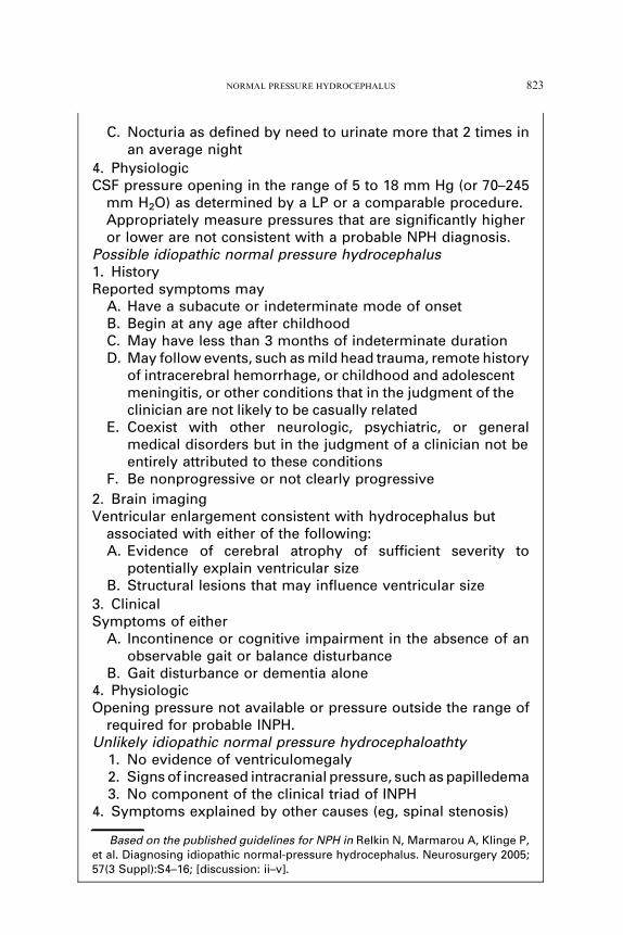
C. Nocturia as defined by need to urinate more that 2 times inan average night
4. PhysiologicCSF pressure opening in the range of 5 to 18 mm Hg (or 70–245
mm H2O) as determined by a LP or a comparable procedure.Appropriately measure pressures that are significantly higheror lower are not consistent with a probable NPH diagnosis.
Possible idiopathic normal pressure hydrocephalus1. HistoryReported symptoms may
A. Have a subacute or indeterminate mode of onsetB. Begin at any age after childhoodC. May have less than 3 months of indeterminate durationD. May follow events, such as mild head trauma, remote history
of intracerebral hemorrhage, or childhood and adolescentmeningitis, or other conditions that in the judgment of theclinician are not likely to be casually related
E. Coexist with other neurologic, psychiatric, or generalmedical disorders but in the judgment of a clinician not beentirely attributed to these conditions
F. Be nonprogressive or not clearly progressive
2. Brain imagingVentricular enlargement consistent with hydrocephalus but
associated with either of the following:A. Evidence of cerebral atrophy of sufficient severity to
potentially explain ventricular sizeB. Structural lesions that may influence ventricular size
3. ClinicalSymptoms of either
A. Incontinence or cognitive impairment in the absence of anobservable gait or balance disturbance
B. Gait disturbance or dementia alone4. PhysiologicOpening pressure not available or pressure outside the range of
required for probable INPH.Unlikely idiopathic normal pressure hydrocephaloathty
1. No evidence of ventriculomegaly2. Signs of increased intracranial pressure, such as papilledema3. No component of the clinical triad of INPH
4. Symptoms explained by other causes (eg, spinal stenosis)
Based on the published guidelines for NPH in Relkin N, Marmarou A, Klinge P,et al. Diagnosing idiopathic normal-pressure hydrocephalus. Neurosurgery 2005;57(3 Suppl):S4–16; [discussion: ii–v].
824 GRAFF-RADFORD
How to assess patient improvement
Older studies measure patient improvement on a five-point rating scale[24,46]. This may be problematic, because levels on the scale overlap andit is a subjective judgment into which level patients fall. More recently, mea-sures of outcome have included more objective measures of gait change[12,53]dscales measuring change in activities of daily living, such as theKatz index [12] or the Rankin scale [53], both of which are sensitive to def-icits in NPH.
One of the crucial unknowns in NPH is if patients improve, how long dothey stay improved? One study addressing this issue was published by Malmand colleagues [56]. They found when they prospectively followed 84 surgi-cally treated patients who had INPH, 64% were improved at 3 months butonly 26% remained improved at 3 years. In Aygok and colleagues’ [54],study they followed 50 patients who had INPH and noted a decline ingait improvement from 90% at 3 months to 75% at 3 years, but cognitiveimprovement remained steady at 80% at 3 months and 3 years, and incon-tinence improved from 70% at 3 months to 82.5% at 3 years.
Shunts
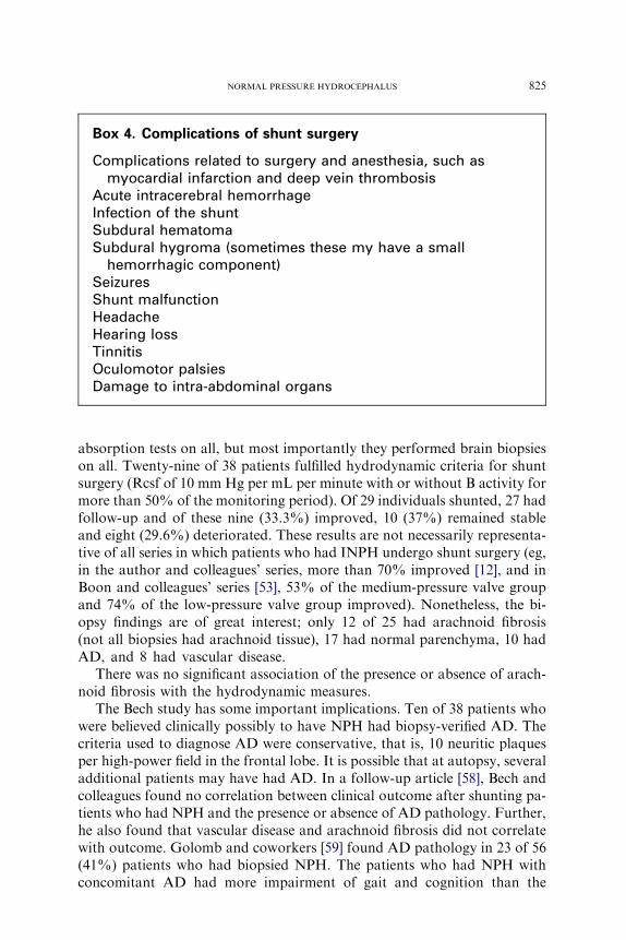
There is no good guidance in the literature to advise patients on the com-plication rate and the type of shunt that should be used. Some of the olderstudies report a complication rate from 30% to 40% [13,16]. The best wayfor doctors to be able to advise patients about the complication rate of shuntsurgery is for them to become familiar with the complication rate at the cen-ter they practice. All complications are important but some effect patientsmore. A list of the complications encountered more commonly is given isBox 4 and is based on the surgical management article in the INPH guide-lines [57]. Before adjustable valves becoming available, one prospectivestudy compared low and medium pressure valves. In this study, 35 of 49 pa-tients receiving low pressure and 16 of 47 patients receiving medium pres-sure valves developed subdural collections. More patients in the LPVgroup improved compared with the MPV group (P!.06). In recent years,the use of adjustable shunts has made the management of subdural hy-gromas and hematomas easier and allowed adjustment down of the openingpressure if patients do not improve. Accurate information on shunt compli-cations and which shunt to use is sorely needed.
Histopathology of the brains of patients who have idiopathic normal
pressure hydrocephalus
In a unique study, Bech and colleagues [42] reported their experience with38 consecutive patients who had INPH. They monitored and performed
825NORMAL PRESSURE HYDROCEPHALUS
absorption tests on all, but most importantly they performed brain biopsieson all. Twenty-nine of 38 patients fulfilled hydrodynamic criteria for shuntsurgery (Rcsf of 10 mm Hg per mL per minute with or without B activity formore than 50% of the monitoring period). Of 29 individuals shunted, 27 hadfollow-up and of these nine (33.3%) improved, 10 (37%) remained stableand eight (29.6%) deteriorated. These results are not necessarily representa-tive of all series in which patients who had INPH undergo shunt surgery (eg,in the author and colleagues’ series, more than 70% improved [12], and inBoon and colleagues’ series [53], 53% of the medium-pressure valve groupand 74% of the low-pressure valve group improved). Nonetheless, the bi-opsy findings are of great interest; only 12 of 25 had arachnoid fibrosis(not all biopsies had arachnoid tissue), 17 had normal parenchyma, 10 hadAD, and 8 had vascular disease.
There was no significant association of the presence or absence of arach-noid fibrosis with the hydrodynamic measures.
The Bech study has some important implications. Ten of 38 patients whowere believed clinically possibly to have NPH had biopsy-verified AD. Thecriteria used to diagnose AD were conservative, that is, 10 neuritic plaquesper high-power field in the frontal lobe. It is possible that at autopsy, severaladditional patients may have had AD. In a follow-up article [58], Bech andcolleagues found no correlation between clinical outcome after shunting pa-tients who had NPH and the presence or absence of AD pathology. Further,he also found that vascular disease and arachnoid fibrosis did not correlatewith outcome. Golomb and coworkers [59] found AD pathology in 23 of 56(41%) patients who had biopsied NPH. The patients who had NPH withconcomitant AD had more impairment of gait and cognition than the
Box 4. Complications of shunt surgery
Complications related to surgery and anesthesia, such asmyocardial infarction and deep vein thrombosis
Acute intracerebral hemorrhageInfection of the shuntSubdural hematomaSubdural hygroma (sometimes these my have a small
hemorrhagic component)SeizuresShunt malfunctionHeadacheHearing lossTinnitisOculomotor palsiesDamage to intra-abdominal organs

826 GRAFF-RADFORD
patients who had ‘‘pure’’ NPH. Only 18% of the patients who had GlobalDeterioration Scores of 3 and below had AD-positive biopsies, whereas75% of those who had Global Deterioration Scores 6 or above were ADpositive. There was comparable improvement in gait velocity in patientswho had NPH regardless of the presence of AD pathology. No consistentcognitive improvement occurred in either group after shunting. Savolainenand colleagues [60] found concomitant AD pathology by biopsy in 31% of51 consecutive patients who had NPH.
These three studies [42,59,60] show that AD pathology is frequent in pa-tients diagnosed with INPH. Further, some of these patients have gait im-provement after shunting. The implication is that NPH and AD mayoccur simultaneously in the same patient, and families should be madeaware of this. In Braak and Braak’s classic study [61] of autopsies ofmore than 2000 persons from the medical examiner, the prevalence of ADpathology in the age group 71 to 75 was 30% and 76 to 80 was 40%, sothe AD biopsy rate of patients who have NPH is similar to that patientsnot selected for being demented (ie, from the medical examiner). Futurestudies should investigate if CSF markers, such as the amyloid-beta protein1-42 and tau protein [62], may be helpful in these cases.
The relationship of idiopathic normal pressure hydrocephalus and systemic
hypertension
Several lines of evidence in the literature now point to a relationship be-tween hydrocephalus and systemic hypertension.
Clinical and autopsy studies of normal pressure hydrocephalus patients
Several postmortem examinations of patients who had NPH and casecontrol studies have reported the association of systemic hypertension andNPH [42,63–67]. In the author and Godersky’s series [68], a significantlyhigher prevalence of systemic hypertension was found in patients who hadINPH compared with matched, demented controls and to the publishedprevalence of hypertension in the United States population, matched forage. A more recent, much larger case control study [27] of 65 patientswho had INPH versus 70 matched control patients found a prevalence of83% of systemic hypertension in the patients who had NPH comparedwith 36% in the control group. This was highly significant (P!.001).
Boon and colleagues [69] showed that cerebrovascular risk factors (hy-pertension, diabetes mellitus, cardiac disease, peripheral vascular disease,male gender, and advancing age) did not influence outcome after shuntplacement. The presence of cerebrovascular disease (history of stroke, cere-bral infarction noted on CT, or moderate-to-severe white matter hypodenselesions on CT), however, was an important predictor of poor outcome.Nonetheless, even though 74% of those who did not have concomitant
827NORMAL PRESSURE HYDROCEPHALUS
cerebrovascular disease improved with shunting, 49% who had it also im-proved. Four of the seven patients who had the most severe white matterhypodense lesions responded favorably to shunting.
At this time, regarding hypertension, subcortical arteriosclerotic enceph-alopathy, and NPH, there are more questions than answers. Are hyperten-sion, white matter changes, and cerebrovascular disease merely frequentconcomitants of NPH? Do NPH and subcortical arteriosclerotic encepha-lopathy represent a spectrum (as suggested by Gallassi and colleagues)[70]? Is one causative of the other?
Hydrocephalus after subarachnoid hemorrhage
Another line of evidence showing that systemic hypertension and hydro-cephalus may be related comes from the Cooperative Aneurysm Study [71].In more than 3000 patients who had subarachnoid hemorrhage, it wasfound that a preoperative history of hypertension, the admission bloodpressure measurement, and sustained hypertension during hospitalizationafter surgery, all were highly significantly related to patients developinghydrocephalus.
Hypertension in patients who have aqueductal stenosis
Greitz and colleagues [72] found a high prevalence of systemic hyperten-sion in patients who had hydrocephalus from aqueductal stenosis.
Hydrocephalus in the spontaneously hypertensive rat
The association of hypertension and hydrocephalus is corroborated byreports in the animal literature. Ritter and Dinh [72] showed that the spon-taneously hypertensive rat develops hydrocephalus.
Experimental models of hydrocephalus, ventricular pulse pressure,and systemic hypertension
Portnoy and colleagues [73] showed, in dogs, that infusing dopamine andnorepinephrine led to increased systemic blood pressure, which, in turn,resulted in an increased CSF pressure and pulse pressure. Experimentallycreating an increased CSF pulse pressure with an inflatable balloon in thelateral ventricle of sheep leads to hydrocephalus within hours [74]. Beringand Salibi [75] performed seminal work on hydrocephalus in dogs. Theytied off the jugular veins in the dogs and measured the intraventricular pulsepressure. They concluded that the mechanism involved in the ventricularenlargement seemed to be a combination of at least two factors: ‘‘Onewas the possible failure of CSF absorption in the face of increased superiorsagittal sinus venous pressure, and the other the increased intraventricularpulse pressure from the choroid plexus.’’
828 GRAFF-RADFORD
Thus, a body of information is accumulating that systemic hypertensionand hydrocephalus are associated. It remains to be shown whether or nothypertension causes hydrocephalus or hydrocephalus causes hypertension,or both.
Large head size and hydrocephalus
Approximately 10% to 20% of patients diagnosed with INPH havea large head size [17,18]. This raises the possibility that one of the causesof so-called ‘‘INPH’’ may be patients born with hydrocephalus who becomesymptomatic late in life.
Summary
When confronted with patients who have possible NPH, use the follow-ing systematic approach. Keeping in mind the differential diagnosis, look forpertinent factors in the history and examination and neuropsychologic eval-uation that have a bearing on diagnosis and surgical prognosis. On theMRI, look at the amount and pattern of atrophy and white matter changes.Perform between one and three spinal taps and evaluate the effect of this onthe gait. Consider sending the CSF for AD markers (tau and amyloid beta1-42). At this time you may wish to advise about surgery. If your center per-forms external CSF drainage or intracranial pressure monitoring, this maybe helpful. Patients and families must be aware of the possible benefits andthe risks for the surgery. If they choose surgery, follow patients carefully tosee if there is improvement and to detect possible surgical complications. Ifyou are uncertain whether or not patients should undergo surgery or if fam-ilies and patients choose that a patient should not undergo surgery, makesure you have established a baseline from which you can follow the patient.Ideally, this includes a video of the gait, a brain MRI, and neuropsychologictesting. Follow patients at 3-month intervals with serial videotaping of gait.Stability or deterioration of the gait often helps families and doctors decideon further action.
References
[1] Casmiro M, Benassi G, Cacciatore FM, et al. Frequency of idiopathic normal pressure
hydrocephalus. Arch Neurol 1989;46(6):608.
[2] Trenkwalder C, Schwarz J, Gebhard J, et al. Starnberg trial on epidemiology of Parkinson-
ism and hypertension in the elderly. Prevalence of Parkinson’s disease and related disorders
assessed by a door-to-door survey of inhabitants older than 65 years. Arch Neurol 1995;
52(10):1017–22.
[3] Clarfield AM. The decreasing prevalence of reversible dementias: an updated meta-analysis.
Arch Intern Med 2003;163(18):2219–29.

829NORMAL PRESSURE HYDROCEPHALUS
[4] Hebert LE, Scherr PA, Bienias JL, et al. AlzheimerDisease in the US population: prevalence
estimates using the 2000 census. Arch Neurol 2003;60(8):1119–22.
[5] Vanneste J, Augustijn P, Dirven C, et al. Shunting normal-pressure hydrocephalus: do the
benefits outweigh the risks? A multicenter study and literature review. Neurology 1992;
42(1):54–9.
[6] Krauss JK, Halve B. Normal pressure hydrocephalus: survey on contemporary diagnostic
algorithms and therapeutic decision-making in clinical practice. Acta Neurochir (Wien)
2004;146(4):379–88 [discussion: 388].
[7] Verghese J, LiptonRB,Hall CB, et al. Abnormality of gait as a predictor of non-Alzheimer’s
dementia. N Engl J Med 2002;347(22):1761–8.
[8] Anger JT, Saigal CS, LitwinMS. The prevalence of urinary incontinence among community
dwelling adult women: results from theNational Health andNutrition Examination Survey.
J Urol 2006;175(2):601–4.
[9] Stothers L, ThomD, Calhoun E. Urologic diseases in America project: urinary incontinence
in males–demographics and economic burden. J Urol 2005;173(4):1302–8.
[10] Barron SA, Jacobs L, Kinkel WR. Changes in size of normal lateral ventricles during aging
determined by computerized tomography. Neurology 1976;26(11):1011–3.
[11] Jack CR Jr, ShiungMM, Gunter JL, et al. Comparison of different MRI brain atrophy rate
measures with clinical disease progression in AD. Neurology 2004;62(4):591–600.
[12] Graff-Radford NR, Godersky JC, Jones M. Variables predicting surgical outcome in symp-
tomatic hydrocephalus in the elderly. Neurology 1989;39:1601–4.
[13] Petersen RC, Mokri B, Laws ER Jr. Surgical treatment of idiopathic hydrocephalus in
elderly patients. Neurology 1985;35(3):307–11.
[14] Fisher CM. The clinical picture in occult hydrocephalus. Clin Neurosurg 1977;24:270–84.
[15] De Mol J. Prognostic factors for therapeutic outcome in normal-pressure hydrocephalus.
Review of the literature and personal study [French]. Acta Neurol Belg 1985;85(1):13–29.
[16] Black PM, Ojemann RG, Tzouras A. CSF shunts for dementia, incontinence, and gait
disturbance. Clin Neurosurg 1985;32:632–51.
[17] Graff-Radford NR, Godersky JC. Symptomatic congenital hydrocephalus in the elderly
simulating normal pressure hydrocephalus. Neurology 1989;39(12):1596–600.
[18] Krefft TA, Graff-Radford NR, Lucas JA, et al. Normal pressure hydrocephalus and large
head size. Alzheimer Dis Assoc Disord 2004;18(1):35–7.
[19] Ogino A, Kazui H, Miyoshi N, et al. Cognitive impairment in patients with idiopathic
normal pressure hydrocephalus. Dement Geriatr Cogn Disord 2006;21(2):113–9.
[20] Folstein MF, Folstein SE, McHugh PR. ‘‘Mini-mental state’’. A practical method for grad-
ing the cognitive state of patients for the clinician. J Psychiatr Res 1975;12(3):189–98.
[21] Wechsler D. Wechsler Memory Scale-Revised. New York: The Psychological Corporation;
1987.
[22] Wechsler D. Wechsler Adult Intelligence Scale, 3rd Edition administration and scoring
manual. Orlando: The Psychological Corporation; 1997.
[23] Gyldenstad C. Measurements of the normal ventricular system and hemispheric sulci of 100
adults with computerizes tomography. Neuroradiology 1977;14:183–92.
[24] Borgesen SE, Gjerris F. The predictive value of conductance to outflow of CSF in normal
pressure hydrocephalus. Brain 1982;105(Pt 1):65–86.
[25] Jack CR Jr, Petersen RC, O’Brien PC, et al. MR-based hippocampal volumetry in the diag-
nosis of Alzheimer’s disease. Neurology 1992;42(1):183–8.
[26] Bradley WG. Normal pressure hydrocephalus and deep white matter ischemia: which is the
chicken, and which is the egg? AJNR Am J Neuroradiol 2001;22(9):1638–40.
[27] Krauss JK, Regel JP, Vach W, et al. Vascular risk factors and arteriosclerotic disease in
idiopathic normal-pressure hydrocephalus of the elderly. Stroke 1996;27(1):24–9.
[28] Krauss JK, Regel JP, VachW, et al. White matter lesions in patients with idiopathic normal
pressure hydrocephalus and in an age-matched control group: a comparative study. Neuro-
surgery 1997;40(3):491–5 [discussion: 495–6].
830 GRAFF-RADFORD
[29] Munoz DG, Hastak SM, Harper B, et al. Pathologic correlates of increased signals of the
centrum ovale on magnetic resonance imaging. Arch Neurol 1993;50(5):492–7.
[30] Holodny AI, George AE, de Leon MJ, et al. Focal dilation and paradoxical collapse of
cortical fissures and sulci in patients with normal-pressure hydrocephalus. J Neurosurg
1998;89(5):742–7.
[31] Kitagaki H,Mori E, Ishii K, et al. CSF spaces in idiopathic normal pressure hydrocephalus:
morphology and volumetry. AJNR Am J Neuroradiol 1998;19(7):1277–84.
[32] Holodny AI, Waxman R, George AE, et al. MR differential diagnosis of normal-pressure
hydrocephalus and Alzheimer disease: significance of perihippocampal fissures. AJNR
Am J Neuroradiol 1998;19(5):813–9.
[33] Golomb J, de Leon MJ, George AE, et al. Hippocampal atrophy correlates with severe
cognitive impairment in elderly patients with suspected normal pressure hydrocephalus.
J Neurol Neurosurg Psychiatry 1994;57(5):590–3.
[34] Savolainen S, LaaksoMP, Paljarvi L, et al.MR imaging of the hippocampus in normal pres-
sure hydrocephalus: correlations with cortical Alzheimer’s disease confirmed by pathologic
analysis. AJNR Am J Neuroradiol 2000;21(2):409–14.
[35] Bradley WG Jr, Whittemore AR, Kortman KE, et al. Marked cerebrospinal fluid void:
indicator of successful shunt in patients with suspected normal-pressure hydrocephalus.
Radiology 1991;178(2):459–66.
[36] Bradley WG Jr, Scalzo D, Queralt J, et al. Normal-pressure hydrocephalus: evaluation with
cerebrospinal fluid flow measurements at MR imaging. Radiology 1996;198(2):523–9.
[37] Krauss JK, Regel JP, Vach W, et al. Flow void of cerebrospinal fluid in idiopathic normal
pressure hydrocephalus of the elderly: can it predict outcome after shunting? Neurosurgery
1997;40(1):67–73 [discussion: 73–4].
[38] Hakim R, Black PM. Correlation between lumbo-ventricular perfusion andMRI-CSF flow
studies in idiopathic normal pressure hydrocephalus. Surg Neurol 1998;49(1):14–9 [discus-
sion: 19–20].
[39] Dixon GR, Friedman JA, Luetmer PH, et al. Use of cerebrospinal fluid flow rates measured
by phase-contrast MR to predict outcome of ventriculoperitoneal shunting for idiopathic
normal-pressure hydrocephalus. Mayo Clin Proc 2002;77(6):509–14.
[40] Jagust WJ, Friedland RP, Budinger TF. Positron emission tomography with [18F] fluoro-
deoxyglucose differentiates normal pressure hydrocephalus from Alzheimer-type dementia.
J Neurol Neurosurg Psychiatry 1985;48(11):1091–6.
[41] Mosconi L. Brain glucose metabolism in the early and specific diagnosis of Alzheimer’s
disease. FDG-PET studies in MCI and AD. Eur J Nucl Med Mol Imaging 2005;32(4):
486–510.
[42] Bech RA, JuhlerM,Waldemar G, et al. Frontal brain and leptomeningeal biopsy specimens
correlated with cerebrospinal fluid outflow resistance and B-wave activity in patients sus-
pected of normal-pressure hydrocephalus. Neurosurgery 1997;40(3):497–502.
[43] Graff-Radford NR, Rezai K, Godersky JC, et al. Regional cerebral blood flow in normal
pressure hydrocephalus. J Neurol Neurosurg Psychiatry 1987;50(12):1589–96.
[44] Granado JM, Diaz F, Alday R. Evaluation of brain SPECT in the diagnosis and prognosis
of the normal pressure hydrocephalus syndrome. Acta Neurochir (Wien) 1991;112(3–4):
88–91.
[45] Owler BK, Pickard JD. Normal pressure hydrocephalus and cerebral blood flow: a review.
Acta Neurol Scand 2001;104(6):325–42.
[46] Black PM. Normal-pressure hydrocephalus: current understanding of diagnostic tests and
shunting. Postgrad Med 1982;71(2):57–61, 65–7.
[47] Wikkelso C, Andersson H, Blomstrand C, et al. Normal pressure hydrocephalus. Predictive
value of the cerebrospinal fluid tap-test. Acta Neurol Scand 1986;73(6):566–73.
[48] Malm J, Kristensen B, Karlsson T, et al. The predictive value of cerebrospinal fluid dynamic
tests in patients with th idiopathic adult hydrocephalus syndrome. Arch Neurol 1995;52(8):
783–9.
831NORMAL PRESSURE HYDROCEPHALUS
[49] Walchenbach R, Geiger E, Thomeer RT, et al. The value of temporary external lumbar CSF
drainage in predicting the outcome of shunting on normal pressure hydrocephalus. J Neurol
Neurosurg Psychiatry 2002;72(4):503–6.
[50] Haan J, Thomeer RT. Predictive value of temporary external lumbar drainage in normal
pressure hydrocephalus. Neurosurgery 1988;22(2):388–91.
[51] MarmarouA, BergsneiderM, Klinge P, et al. The value of supplemental prognostic tests for
the preoperative assessment of idiopathic normal-pressure hydrocephalus. Neurosurgery
2005;57(3 Suppl):S17–28 [discussion: ii–v].
[52] KatzmanR,Hussey F. A simple constant-infusionmanometric test formeasurement of CSF
absorption. I. Rationale and method. Neurology 1970;20(6):534–44.
[53] BoonAJ, Tans JT, Delwel EJ, et al. Dutch normal-pressure hydrocephalus study: prediction
of outcome after shunting by resistance to outflow of cerebrospinal fluid. J Neurosurg 1997;
87(5):687–93.
[54] Aygok G, Marmarou A, Young HF. Three-year outcome of shunted idiopathic NPH
patients. Acta Neurochir Suppl 2005;95:241–5.
[55] Relkin N,Marmarou A, Klinge P, et al. Diagnosing idiopathic normal-pressure hydroceph-
alus. Neurosurgery 2005;57(3 Suppl):S4–16 [discussion: ii–v].
[56] Malm J, Kristensen B, Stegmayr B, et al. Three-year survival and functional outcome of
patients with idiopathic adult hydrocephalus syndrome. Neurology 2000;55(4):576–8.
[57] Bergsneider M, Black PM, Klinge P, et al. Surgical management of idiopathic normal-
pressure hydrocephalus. Neurosurgery 2005;57(3 Suppl):S29–39 [discussion: ii–v].
[58] Bech RA, Waldemar G, Gjerris F, et al. Shunting effects in patients with idiopathic normal
pressure hydrocephalus; correlation with cerebral and leptomeningeal biopsy findings. Acta
Neurochir (Wien) 1999;141(6):633–9.
[59] Golomb J, Wisoff J, Miller DC, et al. Alzheimer’s disease comorbidity in normal pressure
hydrocephalus: prevalence and shunt response. J Neurol Neurosurg Psychiatry 2000;
68(6):778–81.
[60] Savolainen S, Paljarvi L, Vapalahti M. Prevalence of Alzheimer’s disease in patients inves-
tigated for presumed normal pressure hydrocephalus: a clinical and neuropathological
study. Acta Neurochir (Wien) 1999;141(8):849–53.
[61] Braak H, Braak E. Frequency of stages of Alzheimer-related lesions in different age
categories. Neurobiol Aging 1997;18(4):351–7.
[62] Blennow K, Hampel H. CSF markers for incipient Alzheimer’s disease [review]. Lancet
Neurol 2003;2:605–13.
[63] Koto A, Rosenberg G, Zingesser LH, et al. Syndrome of normal pressure hydrocephalus:
possible relation to hypertensive and arteriosclerotic vasculopathy. J Neurol Neurosurg
Psychiatry 1977;40(1):73–9.
[64] Earnest MP, Fahn S, Karp JH, et al. Normal pressure hydrocephalus and hypertensive
cerebrovascular disease. Arch Neurol 1974;31(4):262–6.
[65] Haidri NH, Modi SM. Normal pressure hydrocephalus and hypertensive cerebrovascular
disease. Dis Nerv Syst 1977;38(11):918–21.
[66] Shukla D, Singh BM, Strobos RJ. Hypertensive cerebrovascular disease and normal
pressure hydrocephalus. Neurology 1980;30(9):998–1000.
[67] Casmiro M, D’Alessandro R, Cacciatore FM, et al. Risk factors for the syndrome of
ventricular enlargement with gait apraxia (idiopathic normal pressure hydrocephalus): a
case-control study. J Neurol Neurosurg Psychiatry 1989;52(7):847–52.
[68] Graff-Radford NR, Godersky JC. Idiopathic normal pressure hydrocephalus and systemic
hypertension. Neurology 1987;37(5):868–71.
[69] Boon AJ, Tans JT, Delwel EJ, et al. Dutch Normal-Pressure Hydrocephalus Study: the role
of cerebrovascular disease. J Neurosurg 1999;90(2):221–6.
[70] Gallassi R, Morreale A, Montagna P, et al. Binswanger’s disease and normal-pressure
hydrocephalus. Clinical and neuropsychological comparison. Arch Neurol 1991;48(11):
1156–9.

832 GRAFF-RADFORD
[71] Graff-Radford NR, Torner J, Adams HP Jr, et al. Factors associated with hydrocephalus
after subarachnoid hemorrhage. A report of the Cooperative Aneurysm Study. ArchNeurol
1989;46(7):744–52.
[72] Ritter S, Dinah TT. Progressive postnatal dilation of brain ventricles in spontaneously hy-
pertensive rats. Brain Res 1986;340:327–32.
[73] Portnoy HD, Chopp M, Branch C. Hydraulic model of myogenic autoregulation and the
cerebrovascular bed: the effects of altering systemic arterial pressure. Neurosurgery 1983;
13(5):482–98.
[74] Pettorossi V,DiRocci C,Mancinelli R, et al. Communicating hydrocephalus induced byme-
chanically increased amplitude of the intraventricular cerebrospinal fluid pulse pressure; ra-
tionale and method. Exp Neurol 1978;59:30–9.
[75] Bering RJ, Salibi B. Production of hydrocephalus by increased cephalic-venous pressure.
Arch Neurol Psychiatry 1959;81:693–8.