Embed Size (px)
Citation preview

Cytoplasmic transport and nuclear import of plasmid DNA
Haiqing Bai, Gillian M. Schiralli Lester, Laura C. Petishnok, and David A. Dean
Division of Neonatology
Department of Pediatrics
University of Rochester
601 Elmwood Avenue, Box 850
Rochester, NY 14642
tel: 585-276-3933
fax: 585-756-7780
e-mail: [email protected]
AC
CE
PT
ED
MA
NU
SC
RIP
T
10.1042/BSR20160616. Please cite using the DOI 10.1042/BSR20160616http://dx.doi.org/up-to-date version is available at
encouraged to use the Version of Record that, when published, will replace this version. The most this is an Accepted Manuscript, not the final Version of Record. You are:Bioscience Reports
). http://www.portlandpresspublishing.com/content/open-access-policy#ArchivingPolicy of Portland Press (which the article is published. Archiving of non-open access articles is permitted in accordance with the Archiving Use of open access articles is permitted based on the terms of the specific Creative Commons Licence under

2
ABSTRACT
Productive transfection and gene transfer requires not simply entry of DNA into cells
and subsequent transcription from an appropriate promoter, but also a number of
intracellular events that allow the DNA to move from the extracellular surface of the cell
into and through the cytoplasm, and ultimately across the nuclear envelope and into the
nucleus before any transcription can initiate. Immediately upon entry into the cytoplasm,
naked DNA, either delivered through physical techniques or after disassembly of DNA-
carrier complexes, associates with a large number of cellular proteins that mediate
subsequent interactions with the microtubule network for movement towards the
microtubule organizing center and the nuclear envelope. Plasmids then enter the
nucleus either upon the mitotic disassembly of the nuclear envelope or through nuclear
pore complexes in the absence of cell division, using a different set of proteins. This
review will discuss our current understanding of these pathways used by naked DNA
during the transfection process. While much has been elucidated on these processes,
much remains to be discerned, but with the development of a number of model systems
and approaches, great progress is being made.

3
Introduction
Productive transfection and gene transfer requires extracellular DNA to cross the
plasma membrane, traffic through the cytoplasm, enter the nucleus, be transcribed, and
have the resulting mRNA be exported into the cytoplasm for translation and the ultimate
modification and localization of the expressed protein. While most development of
transfection reagents and strategies has focused largely on carriers for plasma
membrane targeting and entry and the choice of the "best" promoter, these other steps
of intracellular trafficking are crucially important for successful gene transfer.
Formation of host protein-DNA complexes following internalization.
Once DNA has been released successfully into the cytoplasm, it must traffic to the
nucleus in order for gene expression to occur. This translocation across the cytoplasm
represents a significant barrier to gene delivery. First, the cytoplasm contains
nucleases that will degrade free DNA. Studies have demonstrated that plasmid DNA is
degraded in the cytoplasm of HeLa and COS cells with a half-life of 50 to 90 minutes (1).
This poses a large problem for delivery of naked DNA and DNA-carrier complexes that
are believed to dissociate prior to nuclear entry. However, the terms "naked DNA" or
"free" DNA may not actually describe the state of the DNA once released into the
cytoplasm. Rather, any "naked" DNA is rapidly complexed with a number of host
cellular proteins to form a protein-DNA complex that mediates subsequent interactions
4
with intercellular pathways, condenses the plasmid to reduce its effective size, and
shields the DNA from rapid degradation.
When naked DNA is delivered to adherent cells by electroporation and isolated as early
as 15 minutes later, significant numbers of proteins can be found associated with the
plasmid (2-4). In the case of the green fluorescent protein (GFP) expression vector
pEGFPN1 (Clontech), over 600 proteins are found to associate with the plasmid in each
of three independent experiments (2). While many of these proteins bind non-
specifically to DNA or associate weakly by simple electrostatic interactions, a large
number of proteins interact specifically with plasmids that productively traffic through the
cell. Analysis of protein-DNA complexes over time in transfected cells also show that
these complexes are dynamic with certain proteins remaining bound to the DNA for at
least 4 hours and others that bind early but come off at later times or those that come
on at later times only (2). Thus, it is likely that many of these complexes serve multiple
roles in cytoplasmic movement along the cytoskeleton, protection from cytoplasmic
nucleases, and nuclear localization (Figure 1).
Cytoplasmic movement
The cytoplasm is a viscous environment crowded with molecules, which results in
decreased mobility of macromolecules (5-7). Spot-photobleaching and other studies
have demonstrated that small solutes can diffuse freely and rapidly in the cytoplasm
and the nucleus (5, 8). However, similar studies looking at the movement of labeled

5
DNA fragments have shown that while small DNA can diffuse, those larger than 2000
bps were effectively unable to diffuse to any degree in the cytoplasm in any reasonable
physiological time frame (6). This is due to the existence of the cytoskeleton and the
large numbers of actin filaments, microtubules, and intermediate filaments that form a
highly cross-linked gel-sol (9-13). Because of this system of networks, if DNA is
released from an endosome at a site distant from the nucleus, the DNA cannot simply
diffuse toward its desired location. This has been demonstrated in the case of liposome
transfections where some DNA is left free in the cytoplasm and never reaches the
nucleus (14, 15). Although it has been shown that lipoplex-containing endosomes
themselves traffic toward the nucleus and the interior of the cell, there is still quite a lot
of distance for the free DNA following endosomal release to move before it reaches the
nucleus.
Since diffusion cannot account for the movement of plasmids to the nucleus, the only
alternative is directed active transport (Figure 1). Indeed, we and others have shown
that DNA in the cytoplasm utilizes the microtubule network and the molecular motor,
dynein (16-18) for its trafficking to the nucleus. Early studies pointed to microtubules as
being involved in the directed movement of plasmids based on the fact that upon
microinjection into the cytoplasm of TC7 cells, GFP-expressing plasmids were able to
drive high level GFP expression in non-dividing cells when untreated but showed greatly
reduced GFP expression upon treatment with nocodazole, a microtubule disrupter (17).
By contrast, treatment with drugs that affected actin dynamics had no effect on
transgene expression (17). Further, co-injection of the DNA with inhibitory antibodies

6
against dynein also reduced subsequent gene expression, suggesting that without
either intact microtubules or nuclear-directed dynein, cytoplasmic plasmids were unable
to make their way to the nucleus for gene expression (17). In vitro binding assays
confirmed that plasmids do indeed interact with microtubules through cytoplasmic
adapter proteins, including dynein and transcription factors (17, 19). Interestingly, when
different plasmids were analyzed for their ability to interact with microtubules in this
binding assay, it was found that binding sites for the transcription factor CREB were
required for the interaction; when plasmids lacking CREB sites were used, no
interactions were detected, suggesting some degree of DNA sequence-specificity in the
interaction and for movement (19).
These studies were confirmed by following individual fluorescently-labeled plasmids in
microinjected cells using particle tracking (19). When plasmids carrying cAMP response
element binding protein (CREB) binding sites, which are fortuitously present in multiple
copies in the Cytomegalovirus (CMV) immediate early promoter, were cytoplasmically
microinjected, directed movement was observed with initial velocities of 150 nm/sec and
upwards to 380 nm/sec, indicative of directed, dynein-driven movement of proteins and
organelles along microtubules (20-23). By contrast, when a plasmid lacking CREB sites
was followed, the observed velocities were less than 50 nm/sec (24), a rate of
movement indicative of random Brownian motion or limited diffusion. Similar rates and
directionality of plasmid movement also have been seen following electroporation-
mediated delivery of plasmids in cultured cells, largely confirming our earlier findings
(25). This study also showed that at early times after electroporation, the actin network

7
and associated motors may also play a role in DNA movement from the periphery of the
cell to the microtubules themselves since treatment of cells with drugs that affect actin
dynamics reduced plasmid velocities and displacement of the particles but did not
greatly affect total plasmid movement (25). Proteomic studies from our laboratory have
found that several actin-based motors (myosin 1B, 1C, and 9) are found in protein-
plasmid complexes at early times after electroporation (15 min) along with a number of
different microtubule-based motors (2). This supports a possible role for actin-based
movement of DNA particles, at least at times between entry of the DNA into the cytosol
and its binding to microtubules (Figure 1). However, since the actin network and its
associated motors are known to play critical roles in the internalization of endosomes
and their subsequent intracellular movement, it is also possible that the effects of actin
filament disruption could be due to movement of vesicles, rather than the cytoplasmic
DNA itself.
Directed trafficking of plasmids in the cytoplasm
Since DNA has not been shown to bind directly to dynein, the mechanism of this
interaction was investigated and was found to involve a multiprotein complex that
bridges the DNA to microtubules and their associated motors. In vitro binding assays
revealed that plasmid DNA can interact with microtubules only in the presence of
cellular extracts (17). When plasmids carrying different eukaryotic promoters were
tested for their ability to interact with microtubules in this assay, it was found that while
plasmids carrying either the CMV or Cauliflower mosaic virus 35S promoter bound

8
efficiently to the microtubules in the presence of cell extract, plasmids carrying either no
promoter or a number of other different RNA polymerase II promoters failed to do so
(19). Analysis of the transcription factor binding sites present in these DNAs revealed
that binding sites for the transcription factor CREB mediated this interaction. An in vivo
role for this binding was demonstrated by pull-down assays in transfected cells.
Plasmids containing CREB binding sites co-precipitated with CREB as early as 15
minutes after electroporation of adherent cells, but plasmids without CREB binding sites
showed no such interaction.
The functional consequence of this interaction was shown by investigating the initial
velocities, through particle tracking, of microinjected plasmids with or without CREB
binding sites (19). A bacterial plasmid containing no eukaryotic promoter showed very
limited movement following microinjection, indicative of limited diffusive motion. By
contrast, plasmids carrying CREB sites in the CMV promoter showed rapid and
directional movement consistent with microtubule-based trafficking. When another
plasmid carrying the simian virus 40 (SV40) enhancer but no CMV promoter or CREB
binding sites was injected, the plasmids also showed directional active transport,
although at lower rates than seen with the CREB site containing plasmids. When CREB
was depleted from cells using siRNA, no change in the initial velocities were seen for
the bacterial plasmid or the SV40 enhancer only plasmid, but a statistically significant
drop in velocity was detected for plasmids carrying CREB binding sites, suggesting that
the binding of this protein has functional consequences for cytoskeletal movement of
the DNA.

9
In studies on the composition of the protein-DNA complexes that traffic through the
cytoplasm, a number of other proteins have been found to associate with DNA and also
may play a role in microtubule-based movement (2). Moreover, most of these proteins
are much more abundantly associated (from 3- to 250-fold) with trafficking plasmids
compared to those plasmids that displayed no directed movement, based on their
sequence makeup. Several of these are known microtubule-associated proteins, or
MAPs, including MAP1B and MAP4, a microtubule-actin crosslinking factor, and several
known motor proteins, including kinesin-1, kinesin 5B, and dynein-1. Additional proteins
in the trafficking DNA complex included the nuclear localization signal receptor proteins
importin 1, importin 4, importin 7, importin 1 and importin 2, as well as numerous
DNA binding proteins and chaperones. While not all have been tested for their roles in
cytoplasmic DNA movement, several of these proteins have been shown to play an
active role in cytoskeletal trafficking, since their depletion by siRNA treatment, greatly
inhibited the ability of plasmids to move in the cytoplasm. For example, siRNA silencing
of importin 1 blocked all directed movement of injected plasmids and reduced the
movement of the plasmid to less than 50 nm/sec, whereas depletion of importin 7 or
importin 1 had no effect on plasmid velocities in the cytoplasm (2). Another member of
the importin family was also found in the trafficking DNA complexes: exportin, also
called Crm1. This protein does not bind to nuclear localization signals (NLSs) to
facilitate nuclear import, but rather is the major nuclear export signal (NES) receptor that
drives nuclear export of proteins. As in the case of importin 1, inhibition of NES
binding by exportin using the drug leptomycin B abolished the ability of plasmids to

10
move along microtubules in the cytoplasm, suggesting that this protein, perhaps in
association with key exportin cargo proteins, is actively involved in cytoskeletal
trafficking of DNA. This is not unprecedented, since exportin has also been shown to
be needed for movement of adenoviral particles from the microtubule organizing center
to nuclear pores in the nuclear envelope, although not movement on the microtubules,
per se (26).
Another recent study has implicated several kinesin family members as playing a role in
the movement of small double-stranded DNA fragments along microtubules (27). When
32 bp oligonucleotides labeled with quantum dots were internalized by cells, a fraction
of them showed directed movement consistent with active transport, but while roughly
half were directed toward the nucleus, the other half was directed toward the cell
periphery. When biotinylated oligos were used for pull-downs, three plus- (periphery)
directed kinesins (KIF1C, KIF4A and KIF14), the minus-directed C-terminal kinesin
KIFC1 and the cytoplasmic dynein-1 were found to be among the proteins in the DNA
complexes(27). FRET experiments were suggestive of a direct interaction between
KIFC1 and the DNA, although indirect interactions could not be ruled out. Given that
KIFC1 drives movement toward the nucleus, it is possible that this motor may play a
role in plasmid movement to the nucleus as well.
Modulation of microtubule-based DNA movement

11
The cytoskeleton is a highly dynamic network that is influenced by external and internal
forces. A host of post-translational modifications to actin or tubulin monomers and to the
filaments and microtubules themselves can have profound effects on loading and
unloading of cargoes and their movement once bound to these networks. One such
modification is the hyperacetylation of microtubules which leads to increased
recruitment of both kinesin and dynein motors and their bound cargo (28-30). Several
viruses, including HIV, some herpesviruses, and circoviruses, also show greater
cytoplasmic trafficking and enhanced infection when microtubules are hyperacetylated
(30-34). We found that exposure of cultured cells to mild levels of cyclic stretch lead to
the hyperacetylation of microtubules and increased binding of plasmid DNA to
microtubules in the cell, more rapid movement to the nucleus along the microtubules,
and greater transfection efficiency (35, 36). This effect was mediated through the
inhibition of histone deacetylase 6 (HDAC6), a cytoplasmic deacetylase whose major
target is tubulin; silencing of HDAC6 by siRNA or inhibition of HDAC6 by drugs lead to
the same result, but overexpression of HDAC6 could alleviate the enhanced DNA
binding, cytoplasmic trafficking, and increased transfection efficiency seen with cyclic
stretch. When particle tracking was used to quantify the effects of microtubule
hyperacetylation on plasmid movement in cells, it was found that increased tubulin
acetylation not only lead to increased numbers of plasmids loading onto the
microtubules, but also lead to greater processivity of DNA transport along the
microtubules (35). Similar increased gene transfer and expression with cyclic stretch or
HDAC6 inhibition were seen in the lungs of living mice following brief, high tidal volume
ventilation or treatment with HDAC6 inhibitors when plasmid DNA was delivered using

12
transthoracic electroporation (37, 38). These results demonstrate that cytoplasmic
trafficking is important for gene delivery in both cells and tissues.
Nuclear envelope as a barrier to transfection
For years, the nuclear envelope has been proposed to be one of the most substantial
barriers for DNA delivery in cells and tissues. Mario Capecchi carried out an elegant set
of experiments in 1980 in which he microinjected plasmids into either the nuclear or
cytoplasmic compartment of non-dividing cells and assayed their ability to express their
gene product. When pBR322-based plasmids were injected into the cytoplasm, 100%
of the injected cells showed no gene expression, but when the plasmids were injected
into the nucleus, between 50-100% of cells showed gene expression (39). Over the next
20 years, several other groups demonstrated similar findings in multiple cell types and
even Xenopus oocytes, showing that the nuclear envelope was a major impediment to
effective gene transfer in the absence of cell division (40-43).
Despite these studies, it is clear that transfected DNA does reach the nucleus, although
the amount that does may be small. It has been estimated that between 2000 and
10,000 plasmids are delivered per cell following lipofection, but that only between 20
and 1000 are detected in the nucleus by 24-36 hours following DNA addition (44-46).
Even in actively dividing cells, microinjection into the cytoplasm takes between 30 and
100 times more plasmid compared to injection into the nucleus to give equivalent levels

13
of gene expression (47, 48). These studies and others clearly illustrate both that
cytoplasmic trafficking is inefficient and that nuclear entry can be a significant issue.
Nuclear localization in mitotic cells
In the absence of cell division, the intact nuclear envelope remains largely impermeable
to plasmids and plasmid-carrier complexes. However, during mitosis, the nuclear
envelope breaks down, allowing cytoplasmic plasmids near the nucleus access to the
nuclear space. During mitosis, chromosomes are highly condensed by a number of
resident nuclear proteins that interact largely though non-specific interactions with DNA.
Many of these proteins, most notably histones, become dephosphorylated by several
protein phosphatases that are upregulated during mitosis. This dephosphorylation
leads to the recruitment of sets of proteins that facilitate interaction with nuclear
envelope proteins present in the disassembled envelope (as sheets and vesicles) for
engulfment of the chromatin and subsequent re-formation and sealing of the nuclear
envelope (49). Since plasmids become rapidly chromatinized once internalized (50-53),
they too may provide a scaffold on which the nuclear envelope is deposited and
reformed, as has been shown in vitro (54, 55). More efficient nuclear localization and
transfection efficiency in dividing cells also has been noted in transfections using a
number of different carriers, including liposomes, polyethyleneimine, and nanoparticles
(56, 57) (58, 59). For example, one study demonstrated that actively dividing cells were
ten times more likely to express the transferred gene product than cells that had not
divided (60). In another study that looked at the redistribution of transfected plasmids
14
following subsequent mitosis, it was shown that even plasmids that cannot enter the
non-dividing nucleus (see below) partition relatively equally to daughter nuclei,
suggesting that the plasmids are trapped in the reforming nuclei and not transported in
after nuclear formation (61).
Nuclear localization in the absence of cell division
Although nuclear localization of plasmids is much more efficient in dividing cells and
leads to much higher transfection efficiencies using almost all techniques, DNA is able
to enter the nuclei of non-dividing cells, albeit to a much less extent. Indeed, if this were
not the case, there would be effectively no gene transfer to any tissue in living animals
or humans since the majority of cells in tissues are either terminally differentiated or
divide with doubling times of weeks to months. In the absence of mitosis and the
subsequent breakdown of the nuclear envelope, the only way for proteins and protein-
DNA complexes to enter the nucleus is through nuclear pore complexes (NPCs). The
NPC is an aqueous channel in the nuclear envelope through which proteins,
ribonucleoproteins, and all macromolecules in the cell can traffic between the cytoplasm
and nucleus (62). The pores are large (~125 Mdal) multiprotein complexes that are
composed of upwards of 100 distinct proteins present in multiple copies (63). Transport
through the NPC occurs either by signal-independent diffusion in the case of small
molecules such as nucleotides, ions, and solutes, or by signal-mediated facilitated
diffusion.

15
Nuclear entry of plasmids in the absence of cell division was first directly observed after
plasmids were microinjected into the cytoplasm of primary myotubes (64). Wolff and
colleagues showed that the biotin-labeled plasmids were able to enter nuclei of
myotubes and to direct gene expression. This nuclear import was a facilitated process
that was dose- and energy-dependent and was inhibited by agents that block transport
through the NPC. This suggested either that karyophilic proteins containing nuclear
localization signals (NLSs) enabled the injected plasmids to enter nucleus or that the
DNA itself may have an intrinsic signal for nuclear pore recognition and nuclear entry.
They also demonstrated that greater levels of gene expression (hence trafficking) were
obtained when the plasmids were injected near as opposed to far away from the nuclei
(64). Using a similar approach, our laboratory demonstrated that plasmids do indeed
traffic into the nucleus through the NPC by localizing the injected DNA by in situ
hybridization (65) or directly using plasmids labeled with fluorescent peptide nucleic acid
(PNA) clamps (66) in a process that is inhibited by agents that block signal-mediated
transport through the NPC. More recently, experiments have shown that this import is
size-dependent as well, with smaller plasmids or those compacted with protamine being
able to localize to the nucleus and express their products faster than larger ones (67).
In the case of proteins, the signals needed for transport are either the nuclear
localization signal (NLS) for nuclear import or the nuclear export signal (NES) for
nuclear export. Although a number of microinjection studies have shown that proteins
less than 60 kdal in size have the ability to diffuse through the NPC for nuclear entry
(68), even small nuclear proteins contain NLSs that are required for nuclear localization.

16
RNAs, including mRNA, snRNAs, miRNAs, and tRNAs, all exist as ribonucleoprotein
complexes that require both RNA and protein signals for their export or import into the
nucleus. In all cases, the signals needed for nuclear import or export are recognized by
a class of receptors termed the karyopherins (69-71). These fall into two general
classes: importins for nuclear import and exportins for nuclear export. In the case of the
importins, the NLS on a cargo protein is recognized either directly by a beta subunit (of
which there are 11 in humans) with 1 being the prototype, or by an alpha subunit (7
isoforms in humans) that then binds to 1 for translocation across the NPC. Similar to
importin , other factors can recognize motifs on RNA, such as the trimethylguanosine
(m3G) cap structure of the U snRNA which is recognized by snurportin 1, and interact
with importin subunits for complex import. In the case of nuclear export, one of 7
exportins interacts directly with the NES to facilitate export. The "directionality" of
transport is controlled by the small Ras-like GTPase Ran in its bound GTP or GDP state.
GTP exchange factors reside in the nucleus while GTPase activating proteins are
maintained in the cytoplasm, leading to a gradient of Ran-GTP in the nucleus and Ran-
GDP in the cytoplasm. Assembly of export complexes (NES cargo bound to exportin) is
driven by association with Ran-GTP in the nucleus while disassembly of import
complexes (NLS cargo bound to importin ) is promoted by binding to Ran-GTP in the
nucleus, thus leading to this directionality.
Since DNA itself is not a protein, it contains no intrinsic NLS, and thus cannot simply
use the importin machinery directly for its nuclear import. The simplest model for DNA
movement through the NPC would be for the DNA to attract and associate with NLSs.

17
This strategy has been used in many iterations in attempts to increase the nuclear
targeting of plasmids during transfections. NLS peptides, either free or as conjugates,
exogenous NLS-containing proteins, importin domains, ligands for nuclear hormone
receptors, and other approaches have all been tested to increase DNA nuclear import,
all to varying degrees of success (Figure 2)(72, 73). This NLS attachment also can be
achieved by the DNA interacting with endogenous DNA-binding proteins, such as
transcription factors, bound for the nucleus while both the proteins and the DNA are in
the cytoplasm (Figure 3). To function, transcription factors consist of at least three
domains or activities: an NLS for nuclear entry, a DNA binding domain to interact with
specific DNA sequence(s), and a domain that regulates transcription in some manner.
If transcription factors encounter DNA in the cytoplasm, they can (and do) form protein-
DNA complexes that may have one or more exposed NLSs that can in turn be
recognized by the importins to carry the entire complex through the NPC into the
nucleus. If this model is correct, most plasmids should be able to enter the nucleus in
the absence of cell division, since many DNA binding proteins and factors involved in
transcription in the cell have the ability to bind to DNA either specifically or non-
specifically and should create these transport-competent plasmid complexes. However,
this does not appear to be the case.
DNA nuclear targeting sequences
The first suggestion that plasmids may contain specific sequences that drive nuclear
import better than others came from microinjection and injection studies in the 1980s.

18
SV40 is a DNA tumor virus that infects non-dividing cells and expresses Large T-
antigen, a protein with transforming activity, which can lead to transformation and
tumorigenesis. In studies to understand the mechanisms of expression of these viral
genes, various eukaryotic control regions of the SV40 virus were cloned into plasmids
and evaluated for expression. When plasmids were injected directly into the nucleus of
dividing cells, those containing the SV40 enhancer showed only modestly increased
expression compared to those injected into the cytoplasmic. However, when the
enhancer sequence was not present in a plasmid, injection into the cytoplasm showed
30-fold lower levels of expression compared to delivery directly into the nucleus (40).
These studies suggested that the SV40 enhancer may have some type of "helper"
activity to aid in viral infection and expression, although the mechanisms were not
elucidated or proposed. In later microinjection and in situ hybridization studies to
investigate movement of the DNA itself, it was found that plasmids containing the entire
SV40 genome (5243 bp) or just 50 bp of the SV40 enhancer were actually able to enter
the nuclei of growth-arrested cells prior to cell division, while those lacking the enhancer
remained in the cytoplasm until the cells divided (47, 65). This sequence-specificity of
plasmid nuclear import has been seen in all cultured cells tested to date, including
established cell lines and primary cells from multiple species and multiple cell and tissue
lineages, suggesting that this is a general phenomenon.
The SV40 enhancer is a 72 bp sequence that is present in two tandem copies in the
SV40 genome. It contains binding sites for at least 10 different transcription factors,
AP1, AP2, AP3, NF-B, Oct-1, TEF-I and TEF-II, all of which are general factors that

19
are ubiquitously expressed in mammalian (and most eukaryotic) cells (74-76). The
ubiquity of these transcription factors could explain why sequence-specific plasmid
nuclear import is seen in all cell types tested so far. The need for this sequence for
effective plasmid nuclear import also fits nicely with the model for DNA nuclear import,
since the enhancer provides a scaffold for transcription factor binding to "coat" the
plasmid, or at least one region of it, with NLS-containing proteins. Sequences such as
the SV40 enhancer have been termed DNA nuclear Targeting Sequences, or DTSs (65).
Based on the activity of the SV40 enhancer and the model for plasmid nuclear import in
which binding of transcription factors to the DNA can facilitate access to the nucleus, a
likely possibility is that any eukaryotic promoter or transcription factor binding site
placed into a plasmid could elicit the same function. However, this does not seem to be
the case. Several strong viral promoters have been tested for their nuclear targeting
ability, but none of them showed similar "DTS" activity. The immediate early promoter
and enhancer from CMV, the Rous sarcoma virus long terminal repeat (LTR), the
Moloney murine leukemia virus LTR, and the herpes simplex virus thymidylate kinase
(TK) promoter are unable to mediate plasmid nuclear import in non-dividing cells (40, 47,
77). One possible reason is that these other promoters do not contain binding sites for
a specific transcription factor that may be responsible for transport activity. Alternatively,
a specific combination of factors is needed that is also not built on these other
sequences. These two possibilities suggest that not all transcription factors are capable
of transporting bound DNA into the nucleus.

20
The normal "life cycle" of a transcription factor is to be synthesized in the cytoplasm and
then be transported into the nucleus using its NLS (either immediately after translation
or upon signal-mediated stimulation such as tumor necrosis factor- (TNF- stimulation
of NF-B). Only after the transcription factor has entered the nucleus does it encounter
the DNA and bind to it. Many transcription factors, including many basic helix-loop-helix
and zinc-finger proteins, contain NLSs and DNA binding domains that are coincident
and overlapping. For example, the NLS of the fos and jun proteins that make up the
AP1 transcription factor are encoded within the DNA binding domains of this helix-loop-
helix factor. By contrast, other transcription factors, such as NF-B, have spatially
distinct domains for their NLS and DNA binding activities so that both can function
simultaneously to facilitate the cytoplasmic trafficking and nuclear import of plasmids
from the cytoplasm. Indeed, NF-B has been shown to mediate the nuclear import of
plasmids and the presence of tandem NF-B binding sites within a plasmid acts as a
general DTS that is functional in all cell types (16, 78-81).
To date, several general DTSs have been identified, largely on the basis of
microinjection and subcellular localization studies, although several have been identified
based on their ability to increase gene expression following microinjection or
transfection (Table 1). Apart from the ~50 bp SV40 DTS that contains binding sites for
multiple transcription factors and the NF-B DTS composed of tandem NF-B binding
sties on a plasmid, binding sites for the glucocorticoid receptor have also been reported
to have similar DNA import activity (82). This glucocorticoid receptor DTS activity was
shown to improve nuclear localization and gene expression in confluent cells as well as

21
in the lungs of mice following tail vein injection of cationic liposome-DNA complexes or
aerosol delivery of polyethyleneimine -DNA complexes (82). These studies are based
on the earlier findings that complexation of plasmids with the glucocorticoid receptor by
conjugating dexamethasone (a glucocorticoid receptor agonist) to the DNA resulted in
greater DNA nuclear localization and gene expression (83, 84).
One caveat to the use of the DTS strategy is that the DTS only works as anticipated in
cells that are not dividing. When the cell goes through mitosis, the nuclear envelope
breaks down and reassembles after cell division. Thus, any plasmid that is in the
cytoplasm during mitosis is no longer prevented from entering the nuclear space and
when cell division is finished, plasmids wind up being trapped within the new nuclear
space. In this case, a DTS is not required for nuclear uptake (61). Further, while the
dependence on the DTS for plasmid nuclear import appears almost absolute in cultured
cells, a number of studies have shown that in many tissues, especially skeletal muscle,
robust gene transfer and expression can be obtained using plasmids lacking any
nuclear import sequence. It is likely that when the cytoplasm becomes filled with large
concentrations of plasmids, at least some of the plasmids can make their way to the
nuclear envelope and be imported into the nucleus independent of any DTS. Indeed,
when linear DNA is brought close enough to the nuclear envelope using laser tweezers,
it is pulled in (85). It also has been shown that when DTS-lacking plasmids are
delivered to the cytoplasm of a mouse myotube in vivo, no gene expression is observed
until 1,000,000 plasmids or more are injected, suggesting that mass action could
account for the nuclear localization (86).

22
Several studies aimed at evaluating the effects of the DTS on gene expression following
transfection have reported seeing limited to no benefit from the DTS (87, 88). In several
cases, gene expression was followed as the readout for nuclear entry, whereas the
studies showing that a DTS (such as the SV40 DTS or tandem NF-B sites) promoted
nuclear import relied on direct imaging of plasmids following microinjection to isolate the
step of nuclear import or by quantifying levels of nuclear DNA (47, 65, 79, 81, 89).
However, changes in gene expression can be affected at multiple points during the
transfection process, from binding to the plasma membrane and internalization,
compaction, release from carriers, nuclear import, and transcription itself. One elegant
study from the Crommelin and Mastrobattista groups at Utrecht showed that no
increased gene expression was seen from CMV promoter-driven expression plasmids
with the SV40 DTS in A431 and HeLa cells transfected by a number of different
techniques (87). The authors proposed that the use of the strong CMV promoter
obscured or overcame any effects of preferential DNA nuclear import provided by the
DTS and that any limitation in gene expression was at the level of post-transcriptional
processing or translation, not pre-transcriptional events. By contrast, they postulated
that when a weak promoter is used, the transcription machinery would not be saturated,
and any increases in nuclear delivery of the plasmids would lead to increased
transcription and translation.
Cell-specific and regulated DNA nuclear entry
23
Based on the models of bound transcription factor-mediated DNA nuclear import, it was
postulated that cell specific nuclear targeting sequences may exist (47). It is possible
that binding of cell-specific transcription factors to a plasmid could lead to cell-specific
nuclear import in the cells in which these particular transcription factors are expressed
(Figure 3). In order to identify such sequences, multiple cell-specific promoters were
cloned into plasmids and tested for their ability to facilitate plasmid nuclear entry
following microinjection in the specific cell types in which the promoters are
transcriptionally active, but not in other cell types in which the promoters do not function.
The first such cell-specific DTS identified was a 176 bp fragment of the smooth muscle
specific smooth muscle gamma actin (SMGA) promoter (77). This fragment of the
SMGA promoter was able to drive nuclear import of a plasmid in primary smooth muscle
cells from multiple species, but did not cause nuclear import in other cell types including
endothelial cells, fibroblasts, or epithelial cells (77, 90). Nuclear import of the SMGA
DTS is mediated by two transcription factors that are preferentially co-expressed in
smooth muscle, Nkx3.1/3.2 and serum response factor (SRF), are both necessary and
sufficient for DNA nuclear uptake in these cells (77, 90). When the binding sites for
these factors were mutated within the SMGA promoter, plasmids containing the mutant
DTS remained in the cytoplasm of microinjected cells. Similarly, when Nkx3.1/3.2 and
SRF were knocked down in smooth muscle cells through the use of siRNA, nuclear
import of plasmids carrying the wild type SMGA promoter was abolished, again showing
that these factors are necessary for DNA nuclear import (90). Sufficiency of these two
transcription factors alone was shown by expressing the factors in bacteria, complexing
the purified proteins with SMGA DTS plasmids prior to cytoplasmic microinjection, and

24
obtaining nuclear import in non-smooth muscle cells that do not normally express these
factors (90). To date, at least 5 other cell-specific DTSs have been identified, including
those active in endothelial cells (91), osteoblasts (92), type I alveolar epithelial cells (93),
type 2 alveolar epithelial cells (94), and embryonic stem (ES) cells (95)(Table 1).
Using the same concept of coupling specific transcription factors and their binding sites
for DNA nuclear import, an intriguing study from a group in Brazil demonstrated that
binding sites for the hypoxia-inducible factor HIF-1 could function as a DTS in a
controlled manner (96). A293T cells were transfected with plasmids carrying either the
72 bp SV40 DTS or a 50 bp hypoxia-responsive element (HRE) using calcium
phosphate and their nuclear plasmid content quantified by PCR 4 hours later. Under
normoxic conditions, approximately twice as many SV40 DTS plasmids were present in
the nuclei of transfected cells compared to the HRE plasmid, which was only slightly
enriched in the nuclei of normoxic cells compared to a plasmid containing no DTS (20%
more HRE plasmids in the nucleus compared to no DTS plasmid). When cells were
grown under hypoxic conditions following transfection, the numbers of SV40 DTS
plasmids in the nucleus dropped to the level of those seen with no DTS plasmid while
the HRE plasmid showed a 2-fold increase in nuclear plasmid.
Effect of DNA nuclear targeting sequences in animal models
DTS elements have also been shown to act in living animals as they do in cultured cells.
In studies of vascular gene transfer, plasmids containing the SV40 DTS increased gene

25
expression 40- to 200-fold after electroporation-mediated transfer into the mesenteric
vessels of living rats (97). Similar results were seen when plasmids expressing GFP or
luciferase were quantified. In situ hybridization verified that the increased reporter gene
expression in rats electroporated with constructs containing the SV40 DTS vs.
constructs lacking this sequence was a result of increased nuclear import and
localization of the DTS plasmids (97). As stated above, these DTS have benefit only in
cells in the absence of cell division. When we denuded endothelial cells from the rat
carotid by angioplasty and induced rapid smooth muscle and endothelial cell
proliferation, no difference in the levels of gene expression were seen with plasmids
carrying or lacking an SV40 DTS (Young and Dean, unpublished). Similar effects of the
SV40 DTS on increasing gene expression have also been reported in skeletal muscle
using injection of DNA and electroporation for gene delivery, although these studies
only followed expression levels and did not correlate nuclear plasmid levels with
increased gene expression (98, 99).
Cell-specific DTSs also have been shown to be cell-selective in animals as they are in
cultured cells. When the rat mesenteric vasculature was electroporated with CMV-
promoter driven GFP plasmids that contained either no DTS, the SV40 DTS, or the
SMGA DTS downstream of the GFP gene and evaluated two days later for gene
expression, little to no expression was seen in vessels receiving the no DTS plasmids
(100). By contrast, those electroporated with the SV40 DTS plasmid showed robust
expression throughout the neurovascular bundle (including smooth muscle cells,
endothelial cells, fibroblasts, and adventitial cells). The SMGA plasmids also showed

26
robust gene expression, but the expression was limited only to smooth muscle cells.
When in situ hybridization was used to localize the delivered plasmids, the SV40 DTS
plasmid could be seen in all cells within the vessel wall, but the SMGA DTS plasmid
was only detected in smooth muscle cells and not in any other cell type, mimicking the
expression results (100). Taken together, these results demonstrated that the DTS
enhanced nuclear localization of the transfected plasmids which in turn lead to
increased gene expression. Similar results have been seen in the lung for both type I-
and type II-alveolar epithelial cell-specific DTSs: whereas the type II cell-specific
surfactant protein C (SPC) DTS promoted nuclear import and gene expression
disproportionately in type II cells, the type I cell-specific T1 DTS was restricted in its
expression to type I alveolar epithelial cells, with over 80% of the gene expressing cells
being type I cells (93, 94). Again, as in the dividing carotids following angioplasty, DTS
function in the lung was also dependent on cell division. Although the majority of the
cell-specific DTS activity was indeed in the corresponding target cells, some gene
expression was detected in off-target cells. However, when animals were labeled with
BrdU during the gene transfer process, off-target cells showing gene expression were
also those that were BrdU positive, indicating that they had divided during the course of
the experiment (94).
Co-factors involved in DNA nuclear transport

27
The core proteins that serve as linkers between the DNA and the importin machinery
are the specific transcription factors that interact with the various DTS. In the case of
the SV40 DTS, these include a number of ubiquitously expressed mammalian
transcription factors such as AP1, AP2, NF-B, Oct1, TEF-1, and SP1, while in the case
of the various cell-specific DTSs, cell specific transcription factors are required that vary
with the cell type and DTS involved. To dissect the machinery required for DNA nuclear
import, a number of experimental systems have been used to identify other protein
factors involved in this import. One of the most useful systems for reconstitution has
been the digitonin permeabilized HeLa cell system established by Adam and Gerace
(101). Incubation of cells with relatively low concentrations of the detergent digitonin
preferentially permeabilizes the plasma membrane, allowing the cytosolic contents of
the cell to be removed. By adding back purified components of the nuclear import
machinery (e.g., importin , importin , Ran, etc.), the roles of specific proteins and
importins can be determined. Using this system, we showed that the nuclear import of
plasmids fluorescently labeled at a specific site with a PNA conjugated to fluorescein
was able to accumulate in the nucleus of cells in the presence of complete cytoplasmic
and nuclear extracts (66). Import was detectable as early as 60 min after addition of
DNA and saturated by 4 hours. This import was energy-dependent and could be
blocked with either lectins or antibodies that bind to and block NLS-mediated movement
through the NPC. When bacterially expressed and purified importin , importin , and
Ran were added in lieu of cytoplasmic extracts, nuclear import of an NLS-containing
protein was reconstituted, but that of plasmid DNA was not unless nuclear extracts were
also supplied. Like nuclear import in microinjected cells, plasmid nuclear import in the

28
permeabilized cells was sequence-dependent. While plasmids carrying the SV40 DTS
showed robust nuclear accumulation in the presence of importin and Ran, plasmids
without the SV40 sequence failed to accumulate (66).
This requirement for the soluble NLS-import machinery for plasmid nuclear localization
appears to be in contrast to the results of Wolff and colleagues who found that the
nuclear import of fluorescently-labeled linear DNA fragments was energy-dependent
and inhibited by wheat germ agglutinin but occurred optimally in the absence of
cytoplasmic proteins (102). One likely reason for this disparity is that the uniformly-
labeled, short, linear DNA used in the Wolff study is a very different substrate compared
to plasmid labeled at a discrete site, and as such, may utilize different pathways for
entry.
Proteomics has been used to identify the proteins that complex with plasmids either in
solution or in cells during the transfection process (2, 3, 103). In in vitro studies, cell
extracts were subjected to affinity chromatography using supercoiled plasmids
immobilized onto columns at a single, specified site through the use sepharose-
conjugated PNAs (3, 103). Subsequent mass spectrometry analysis identified a
number of proteins that bound to plasmids able to enter the nuclei of non-dividing cells
(i.e., containing the SV40 DTS) or those that did not (pBR322). In the Miller study,
several proteins involved in NLS-mediated nuclear import were identified to bind to the
SV40 DTS plasmid but not pBR322, including importin 1, importin 7, Ran, RanBP1,
and RanBP2, as were numbers of transcription factors, DNA processing factors,

29
chromatin assembly proteins, and cytoskeletal proteins and associated motors (3).
Smooth muscle cells were transfected and DNA pull-downs were performed at later
times and several key proteins were evaluated by Western blot. Several of the import
factors found in the in vitro assay also bound in vivo to both the SV40 DTS plasmid as
well as an SMGA DTS plasmid, but not pBR322. These factors included importin 1,
importin 7, and Ran. Since our previous data using the permeabilized cell model pointed
to a role for importin 1 in plasmid nuclear localization (66), we depleted importin 1 or
importin 7 from the cells using siRNA. Depletion of importin 1 abolished nuclear import
of plasmids carrying either the SV40 DTS or the SMGA DTS following cytoplasmic
microinjection. By contrast, depletion of importin 7 (>90% depletion by Western) had no
discernable effect on the nuclear import of either plasmid in smooth muscle cells,
suggesting that importin 7 does not play an active role in plasmid nuclear localization, at
least in smooth muscle cells.
Ran was also identified as interacting with plasmid DNA in another in vitro study using a
similar affinity chromatography approach but using extracts from HeLa cells (103).
Other proteins shown to bind to a DTS-containing supercoiled plasmid included several
chaperones, transcription factors, cytoskeletal proteins, histone H2B and NM23-H2, a
transcription factor and metastasis suppressor (103). When these proteins were
depleted from the HeLa cytoplasmic extracts that support plasmid nuclear import in
permeabilized cells, the depleted extracts no longer promoted plasmid nuclear import.
Further, when purified NM23-H2 and Hsc70 (a chaperone shown to bind to the plasmid
during affinity chromatography) were added back to the depleted HeLa cytoplasmic

30
extract, plasmid nuclear import was completely restored. Whether NM23-H2 mediates
its effects on DNA nuclear import through its DNA-binding activity or through its kinase
activity remains to be determined, but it appears to have clear effects on DNA uptake.
Other studies have also identified importin family members as playing a role in DNA
nuclear uptake. Several studies looking at the nuclear targeting of HIV reverse
transcribed DNA complexes pointed to a role for importin 7 (104, 105). When importin 7
was knocked down in HeLa cells using shRNA, transfection of a linearized HIV-1
plasmid was reduced 5-fold compared to control cells (105). Based on this finding,
recombinant importin 7 was tested in a permeabilized cell assay and shown to support
the nuclear import of fluorescently labeled plasmids in the presence of energy and Ran
(106). Surprisingly, in this study, importin 1 also stimulated plasmid nuclear import,
although the authors indicated that the increase was not significantly increased over
that seen with Ran and energy alone. When importin 7 was depleted from cells using
shRNA (~90% knockdown) and the cells were transfected with an SV40 enhancer
containing plasmid complexed with poly-L-lysine, nuclear levels of DNA were decreased
by about 60% compared to control. These results are in contrast to those seen in our
studies where knockdown of importin 7 by the same degree in either smooth muscle
cells or A549 cells caused no change in nuclear accumulation of labeled plasmids (2, 3).
It is possible that complexation with poly-L-lysine and transfection as opposed to direct
injection into the cytoplasm could account for these differences. However, it has been
shown that histone H1 is imported into the nucleus by interacting with a heterodimeric
31
importin 1/importin 7 complex and it is possible that both importin 7 and 1 could play
roles in the import of H1 bound DNA complexes (107).
Alternatively, it is also possible that the fluorescent labeling methods could have an
effect on the nuclear import of the DNA. In our studies, fluorophore-labeled PNAs were
bound to plasmids in a triplex structure that is stable in cells and serum for several days
(66, 108-110), whereas in the study by Dhanoya, plasmids were labeled with the
intercalating YOYO-1 dye (106). While YOYO-1 binds to DNA with high affinity and is
extremely stable for electrophoresis at low salt concentrations, at physiological salt
concentrations, dissociation of YOYO-1 can occur within seconds to minutes (111, 112).
It has been found that labeling methods for plasmids can have profound effects on the
DNA in terms of transcriptional activity or subcellular localization. This is most notable
for dyes that covalently attach to the DNA at random sites, especially when labeled at a
high fluorophore to DNA ratio. Indeed, when plasmids were labeled at high labeling
ratios with one of several different commercially available photoactivatable or chemical
DNA labeling kits (Label IT or Fast Tag), the ability of the plasmids to enter the nucleus
was destroyed as was their transcriptional activity (61, 113). In either case, when bulky
adducts are added randomly to the DNA, it is not surprising that the ability of proteins to
bind to the DNA can be reduced, if the adducts are coincident with the binding sites.
Transportin (also known as importin 2) is another importin family member that
mediates the nuclear import of the M9 class of NLS found on the hnRNP A1 protein
(114, 115). Transportin also has been shown in vitro to mediate the nuclear import of

32
H2A, H2B, H3 and H4 histones (116). Nuclei reconstituted in vitro from Xenopus egg
extracts show high fidelity NLS-mediated nuclear import in a defined system that can
easily be manipulated (117). This system also has been used to study the nuclear
import of 400 bp to 1500 bp DNA fragments (118). When reconstituted from extracts
depleted of transportin, the nuclei failed to import DNA, while reconstitution of nuclei
from extracts devoid of importin 1 had little defect in this regard. A similar need for
transportin was also shown in digitonin-permeabilized HeLa cells (118). These authors
showed that the nuclear import mediated by transportin was through the binding of core
histones to the DNA. Interestingly, transportin and importin 4 were also identified by
mass spectrometry to bind to nuclear import competent plasmids in pull-down
experiments in electroporated cells, although the specific roles of these proteins in
nuclear import was not further investigated (2). Since all the core histones were also
shown to complex with the DNA in these transfected cells at early times after
electroporation (2), it is possible that the same mechanisms may apply to intact
plasmids.
Conclusions
It is vital that we understand how plasmids move inside the cell in order to develop
novel ways to enhance trafficking and improve gene delivery and expression. Although
the intracellular trafficking of exogenous DNA may not be a normal event in the cell,
mechanisms do exist for its transport. Some of these have evolved, as viruses and
other pathogens have perfected ways to invade the host, while others appear to be

33
fortuitous piracy, as in the case of the SV40 DTS, which binds to transcription factors
and other proteins destined for the nucleus.
By characterizing and understanding the mechanisms of the cytoplasmic trafficking and
nuclear import of plasmids, we can overcome our relative inability to target substantial
amounts of DNA to the nucleus and increase transfection efficiency and ultimately gene
therapy.

34
Table 1. Examples of DTS
DTS Cell Specificity Reference
SV40 enhancer general (65)
Glucocorticoid Receptor binding sites general (82)
NF-B binding sites general; inducible by TNF (78)
Hypoxia Response Elements general; inducible by hypoxia (96)
SMGA promoter smooth muscle cells (77)
Flk-1 promoter endothelial cells (91)
hcolA1 promoter osteoblasts (92)
SPC promoter type II alveolar epithelial cells (94)
Sox2 regulatory region 2 embryonic stem cells (95)
T1 promoter type I alveolar epithelial cells (93)

35
Figure Legends
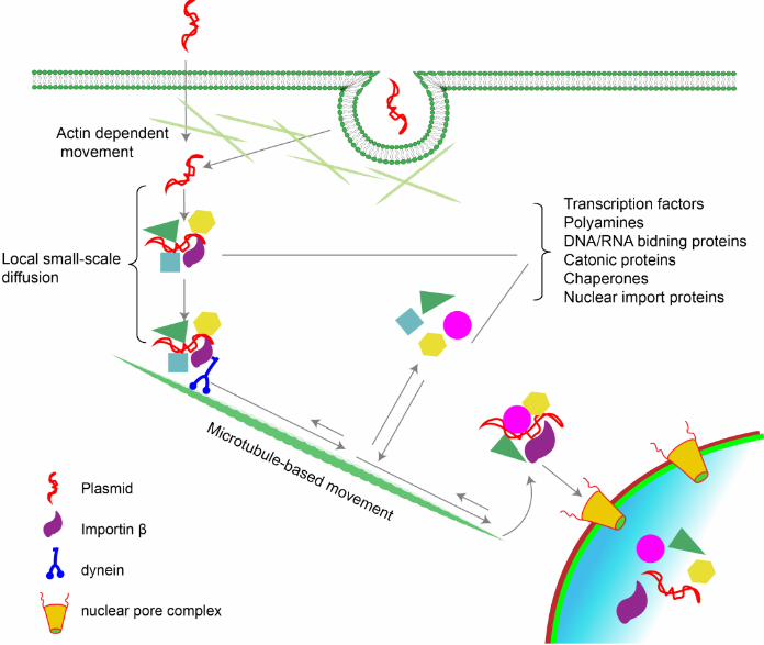
Figure 1. Intracellular trafficking of plasmids. Most plasmids enter the cell by either
endocytosis and/or direct entry into the cytosol at which point they must traverse the
cortical actin layer, perhaps using actin-based movement (25, 119). Once free in the
cytoplasm, plasmids are rapidly complexed by a number of DNA binding proteins in the
cytoplasm which in turn bind to other proteins to form large protein-DNA complexes (2).
Transcription factors bound to the DNA interact with importin and other proteins that
link the complex to dynein and kinesin for movement along microtubules toward the
nucleus (19). During this process, the DNA protein complexes appear to be dynamic,
with various proteins coming on an off the DNA at different times, perhaps to mediate
different processes. Nuclear entry is then mediated by importin in a sequence- and
importin-dependent manner through the nuclear pore complex (NPC) in non-dividing
cells or independent of importins and any DNA sequence requirement during mitosis
and the associated dissolution of the nuclear envelope.
Figure 2. Strategies to increase nuclear targeting of plasmids. A number of
different approaches have been used to improve the nuclear import of plasmids, all of
which center around attaching NLS peptides or NLS proteins or other importin
interacting peptides to the DNA. This can be also done by increasing the functional
diameter of the NPC itself by enhancing non-selective gating of the pore with the drug
TCHD (120, 121).

36
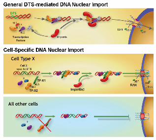
Figure 3. Model for general and cell-specific DNA nuclear import in non-dividing
cells. If plasmids containing sequences that act as scaffolds for transcription factors
and other DNA binding proteins (termed DTS, or DNA nuclear Targeting Sequences)
are deposited into the cytoplasm during transfection, they can form complexes with
these proteins, thereby attaching NLSs to the DNA. Some, but not all, of these NLSs
may be in a conformation able to interact with importins for transport of the DNA-protein
complex into the nucleus through nuclear pores. In the case of general DTSs, the
transcription factors that interact with the DTS are ubiquitously expressed and thus
allow DNA nuclear import in all cell types. By contrast, cell-specific DTSs bind to a
group of cell-specific transcription factors as well as some general transcription factors
to form import competent complexes in corresponding cell types (the cell type X DTS
binds cell type X-specific transcription factors in cell type X) . In other cell types, the
specific transcription factors are absent and consequently, plasmids fail to be imported
into the nuclei for expression.


37
REFERENCES
1. Lechardeur, D., K.-J. Sohn, M. Haardt, P.B. Joshi, M. Monck, R.W. Graham, B. Beatty, J. Squire, H. O’Brodovich, and G.L. Lukacs. (1999) Metabolic instability of plasmid DNA in the cytosol: a potential barrier to gene transfer. Gene Ther 6: 482–497
2. Badding, M.A., J.D. Lapek, A.E. Friedman, and D.A. Dean. (2013) Proteomic and functional analyses of protein-DNA complexes during gene transfer. Mol Ther 21: 775-85
3. Miller, A.M., F.M. Munkonge, E.W. Alton, and D.A. Dean. (2009) Identification of Protein Cofactors Necessary for Sequence-specific Plasmid DNA Nuclear Import. Mol Ther 17: 1897-1903
4. Munkonge, F.M., V. Amin, S.C. Hyde, A.M. Green, I.A. Pringle, D.R. Gill, J.W. Smith, R.P. Hooley, S. Xenariou, M.A. Ward, N. Leeds, K.Y. Leung, M. Chan, E. Hillery, D.M. Geddes, U. Griesenbach, E.H. Postel, D.A. Dean, M.J. Dunn, and E.W. Alton. (2009) Identification and functional characterization of cytoplasmic determinants of plasmid DNA nuclear import. J Biol Chem 284: 26978-87
5. Kao, H.P., J.R. Abney, and A.S. Verkman. (1993) Determinants of the translational mobility of a small solute in cell cytoplasm. J Cell Biol 120: 175-84
6. Lukacs, G.L., P. Haggie, O. Seksek, D. Lechardeur, N. Freedman, and A.S. Verkman. (2000) Size-dependent DNA mobility in cytoplasm and nucleus. J Biol Chem 275: 1625-9.
7. Dauty, E. and A.S. Verkman. (2005) Actin cytoskeleton as the principal determinant of size-dependent DNA mobility in cytoplasm: a new barrier for non-viral gene delivery. J Biol Chem 280: 7823-8
8. Bicknese, S., N. Periasamy, S.B. Shohet, and A.S. Verkman. (1993) Cytoplasmic viscosity near the cell plasma membrane: measurement by evanescent field frequency-domain microfluorimetry. Biophys J 65: 1272-82
9. Chambers, R., The micromanipulation of living cells, in The cell and protoplasm, F.R. Moulton, Editor 1940, Science press: Washington, DC. p. 20-30.
10. Niederman, R., P.C. Amrein, and J. Hartwig. (1983) Three-dimensional structure of actin filaments and of an actin gel made with actin-binding protein. J Cell Biol 96: 1400-13
11. Stossel, T.P. (1984) Contribution of actin to the structure of the cytoplasmic matrix. J Cell Biol 99: 15s-21s
12. Hou, L., F. Lanni, and K. Luby-Phelps. (1990) Tracer diffusion in F-actin and Ficoll mixtures. Toward a model for cytoplasm. Biophys J 58: 31-43
13. Luby-Phelps, K. (2000) Cytoarchitecture and physical properties of cytoplasm: volume, viscosity, diffusion, intracellular surface area. Int Rev Cytol 192: 189-221
14. Gao, X. and L. Huang. (1993) Cytoplasmic expression of a reporter gene by co-delivery of T7 RNA polymerase and T7 promoter sequence with cationic liposomes. Nucleic Acids Res 21: 2867-72

38
15. Zabner, J., A.J. Fasbender, T. Moninger, K.A. Poellinger, and M.J. Welsh. (1995) Cellular and molecular barriers to gene transfer by a cationic lipid. J Biol Chem 270: 18997-9007
16. Mesika, A., V. Kiss, V. Brumfeld, G. Ghosh, and Z. Reich. (2005) Enhanced intracellular mobility and nuclear accumulation of DNA plasmids associated with a karyophilic protein. Hum Gene Ther 16: 200-8
17. Vaughan, E.E. and D.A. Dean. (2006) Intracellular trafficking of plasmids during transfection is mediated by microtubules. Mol Ther 13: 422-8
18. Salman, H., A. Abu-Arish, S. Oliel, A. Loyter, J. Klafter, R. Granek, and M. Elbaum. (2005) Nuclear localization signal peptides induce molecular delivery along microtubules. Biophys J 89: 2134-45
19. Badding, M.A., E.E. Vaughan, and D.A. Dean. (2012) Transcription factor plasmid binding modulates microtubule interactions and intracellular trafficking during gene transfer. Gene Ther 19: 338-46
20. Kural, C., H. Kim, S. Syed, G. Goshima, V.I. Gelfand, and P.R. Selvin. (2005) Kinesin and dynein move a peroxisome in vivo: a tug-of-war or coordinated movement? Science 308: 1469-72
21. Paschal, B.M., H.S. Shpetner, and R.B. Vallee. (1987) MAP 1C is a microtubule-activated ATPase which translocates microtubules in vitro and has dynein-like properties. J Cell Biol 105: 1273-82
22. King, S.J. and T.A. Schroer. (2000) Dynactin increases the processivity of the cytoplasmic dynein motor. Nat Cell Biol 2: 20-4
23. Rapp, S., R. Saffrich, U. Jakle, W. Ansorge, K. Gorgas, and W.W. Just. (1996) Microtubule-mediated peroxisomal saltations. Ann N Y Acad Sci 804: 666-8
24. Ondrej, V., E. Lukásová, M. Falk, and S. Kozubek. (2007) The role of actin and microtubule networks in plasmid DNA intracellular trafficking. Acta Biochim Pol 54: 657-663
25. Rosazza, C., A. Buntz, T. Riess, D. Woll, A. Zumbusch, and M.P. Rols. (2013) Intracellular Tracking of Single-plasmid DNA Particles After Delivery by Electroporation. Mol Ther 21: 2217-26
26. Strunze, S., L.C. Trotman, K. Boucke, and U.F. Greber. (2005) Nuclear targeting of adenovirus type 2 requires CRM1-mediated nuclear export. Mol Biol Cell 16: 2999-3009
27. Farina, F., P. Pierobon, C. Delevoye, J. Monnet, F. Dingli, D. Loew, M. Quanz, M. Dutreix, and G. Cappello. (2013) Kinesin KIFC1 actively transports bare double-stranded DNA. Nucleic Acids Res 41: 4926-37
28. Dompierre, J.P., J.D. Godin, B.C. Charrin, F.P. Cordelieres, S.J. King, S. Humbert, and F. Saudou. (2007) Histone deacetylase 6 inhibition compensates for the transport deficit in Huntington's disease by increasing tubulin acetylation. J Neurosci 27: 3571-83
29. Chen, S., G.C. Owens, H. Makarenkova, and D.B. Edelman. (2010) HDAC6 regulates mitochondrial transport in hippocampal neurons. PLoS One 5: e10848
30. Reed, N.A., D. Cai, T.L. Blasius, G.T. Jih, E. Meyhofer, J. Gaertig, and K.J. Verhey. (2006) Microtubule acetylation promotes kinesin-1 binding and transport. Curr Biol 16: 2166-72

39
31. Cao, J., C. Lin, H. Wang, L. Wang, N. Zhou, Y. Jin, M. Liao, and J. Zhou. (2015) Circovirus transport proceeds via direct interaction of the cytoplasmic dynein IC1 subunit with the viral capsid protein. J Virol 89: 2777-91
32. Frampton, A.R., Jr., H. Uchida, J. von Einem, W.F. Goins, P. Grandi, J.B. Cohen, N. Osterrieder, and J.C. Glorioso. (2010) Equine herpesvirus type 1 (EHV-1) utilizes microtubules, dynein, and ROCK1 to productively infect cells. Vet Microbiol 141: 12-21
33. Naranatt, P.P., H.H. Krishnan, M.S. Smith, and B. Chandran. (2005) Kaposi's sarcoma-associated herpesvirus modulates microtubule dynamics via RhoA-GTP-diaphanous 2 signaling and utilizes the dynein motors to deliver its DNA to the nucleus. J Virol 79: 1191-206
34. Sabo, Y., D. Walsh, D.S. Barry, S. Tinaztepe, K. de Los Santos, S.P. Goff, G.G. Gundersen, and M.H. Naghavi. (2013) HIV-1 induces the formation of stable microtubules to enhance early infection. Cell Host Microbe 14: 535-46
35. Badding, M.A. and D.A. Dean. (2013) Highly acetylated tubulin permits enhanced interactions with and trafficking of plasmids along microtubules. Gene Ther 20: 616-24
36. Vaughan, E.E., R.C. Geiger, A.M. Miller, P.L. Loh-Marley, T. Suzuki, N. Miyata, and D.A. Dean. (2008) Microtubule Acetylation Through HDAC6 Inhibition Results in Increased Transfection Efficiency. Mol Ther 16: 1841-7
37. Geiger, R.C., C.D. Kaufman, A.P. Lam, G.R. Budinger, and D.A. Dean. (2009) Tubulin acetylation and histone deacetylase 6 activity in the lung under cyclic load. Am J Respir Cell Mol Biol 40: 76-82
38. Kaufman, C.D., R.C. Geiger, and D.A. Dean. (2010) Electroporation- and mechanical ventilation-mediated gene transfer to the lung. Gene Ther 17: 1098-104
39. Capecchi, M.R. (1980) High efficiency transformation by direct microinjection of DNA into cultured mammalian cells. Cell 22: 479-88
40. Graessman, M., J. Menne, M. Liebler, I. Graeber, and A. Graessman. (1989) Helper activity for gene expression, a novel function of the SV40 enhancer. Nucleic Acids Res. 17: 6603-6612
41. Zabner, J., A.J. Fasbender, T. Moninger, K.A. Poellinger, and M.J. Welsh. (1995) Cellular and molecular barriers to gene transfer by a cationic lipid. J. Biol. Chem. 270: 18997-19007
42. Mirzayans, R., A.A. Remy, and P.C. Malcom. (1992) Differential expression and stability of foreign genes introduced into human fibroblasts by nuclear versus cytoplasmic microinjection. Mutation Res. 281: 115-122
43. Thornburn, A.M. and A.S. Alberts. (1993) Efficient expression of miniprep plasmid DNA after needle micro-injection into somatic cells. Biotechniques 14: 356-358
44. Coonrod, A., F.Q. Li, and M. Horwitz. (1997) On the mechanism of DNA transfection: efficient gene transfer without viruses. Gene Ther 4: 1313-21
45. Tseng, W., F. Haselton, and T. Giorgio. (1997) Transfection by cationic liposomes using simultaneous single cell measurements of plasmid delivery and transgene expression. J Biol Chem 272: 25641-25647

40
46. James, M.B. and T.D. Giorgio. (2000) Nuclear-associated plasmid, but not cell-associated plasmid, is correlated with transgene expression in cultured mammalian cells. Mol Ther 1: 339-46.
47. Dean, D.A., B.S. Dean, S. Muller, and L.C. Smith. (1999) Sequence requirements for plasmid nuclear entry. Exp. Cell Res. 253: 713-722
48. Ludtke, J.J., M.G. Sebestyen, and J.A. Wolff. (2002) The effect of cell division on the cellular dynamics of microinjected DNA and dextran. Mol Ther 5: 579-88
49. LaJoie, D. and K.S. Ullman. (2017) Coordinated events of nuclear assembly. Curr Opin Cell Biol 46: 39-45
50. Griffith, J.D. (1975) Chromatin structure: deduced from a minichromosome. Science 187: 1202-1203
51. Laskey, R.A., B.M. Honda, A.D. Mills, N.R. Morris, A.H. Wyllie, J.E. Mertz, E.M. De Roberts, and J.B. Gurdon. (1978) Chromatin assembly and transcription in eggs and oocytes of Xenopus laevis. Cold Spring Harb Symp Quant Biol 42 Pt 1: 171-8
52. Reeves, R., C.M. Gorman, and B. Howard. (1985) Minichromosome assembly of non-integrated plasmid DNA transfected into mammalian cells. Nucleic Acids Res 13: 3599-615
53. Riu, E., Z.Y. Chen, H. Xu, C.Y. He, and M.A. Kay. (2007) Histone modifications are associated with the persistence or silencing of vector-mediated transgene expression in vivo. Mol Ther 15: 1348-55
54. Newmeyer, D.D., J.M. Lucocq, T.R. Bürglin, and E.M. De Robertis. (1986) Assembly in vitro of nuclei active in nuclear protein transport: ATP is required for nucleoplasmin accumulation. EMBO J. 5: 501-510
55. Newport, J. (1987) Nuclear reconstitution in vitro: stages of assembly around protein-free DNA. Cell 48: 205-217
56. Brunner, S., T. Sauer, S. Carotta, M. Cotten, M. Saltik, and E. Wagner. (2000) Cell cycle dependence of gene transfer by lipoplex, polyplex and recombinant adenovirus. Gene Therapy 7: 401-407
57. Tseng, W.C., F.R. Haselton, and T.D. Giorgio. (1999) Mitosis enhances transgene expression of plasmid delivered by cationic liposomes. Biochim Biophys Acta 1445: 53-64
58. Grosse, S., G. Thevenot, M. Monsigny, and I. Fajac. (2006) Which mechanism for nuclear import of plasmid DNA complexed with polyethylenimine derivatives? J Gene Med 8: 845-51
59. Chernousova, S. and M. Epple. (2017) Live-cell imaging to compare the transfection and gene silencing efficiency of calcium phosphate nanoparticles and a liposomal transfection agent. Gene Ther 24: 282-289
60. Fasbender, A., J. Zabner, B.G. Zeiher, and M.J. Welsh. (1997) A low rate of cell proliferation and reduced DNA uptake limit cationic lipid-mediated gene transfer to primary cultures of ciliated human airway epithelia. Gene Therapy 4: 1173-1180
61. Gasiorowski, J.Z. and D.A. Dean. (2005) Postmitotic Nuclear Retention of Episomal Plasmids Is Altered by DNA Labeling and Detection Methods. Mol Ther 12: 460-467
62. Gorlich, D. (1997) Nuclear protein import. Curr Opin Cell Biol 9: 412-9

41
63. Beck, M. and E. Hurt. (2017) The nuclear pore complex: understanding its function through structural insight. Nat Rev Mol Cell Biol 18: 73-89
64. Dowty, M.E., P. Williams, G. Zhang, J.E. Hagstrom, and J.A. Wolff. (1995) Plasmid DNA entry into postmitotic nuclei of primary rat myotubes. Proc. Natl. Acad. Sci. USA 92: 4572-4576
65. Dean, D.A. (1997) Import of plasmid DNA into the nucleus is sequence specific. Exp. Cell Res. 230: 293-302
66. Wilson, G.L., B.S. Dean, G. Wang, and D.A. Dean. (1999) Nuclear import of plasmid DNA in digitonin-permeabilized cells requires both cytoplasmic factors and specific DNA sequences. J. Biol. Chem. 274: 22025–22032
67. Akita, H., D. Kurihara, M. Schmeer, M. Schleef, and H. Harashima. (2015) Effect of the Compaction and the Size of DNA on the Nuclear Transfer Efficiency after Microinjection in Synchronized Cells. Pharmaceutics 7: 64-73
68. Feldherr, C.M. and D. Akin. (1990) The permeability of the nuclear envelope in dividing and nondividing cell cultures. J Cell Biol 111: 1-8
69. Christie, M., C.W. Chang, G. Rona, K.M. Smith, A.G. Stewart, A.A. Takeda, M.R. Fontes, M. Stewart, B.G. Vertessy, J.K. Forwood, and B. Kobe. (2016) Structural Biology and Regulation of Protein Import into the Nucleus. J Mol Biol 428: 2060-90
70. Cautain, B., R. Hill, N. de Pedro, and W. Link. (2015) Components and regulation of nuclear transport processes. FEBS J 282: 445-62
71. Forbes, D.J., A. Travesa, M.S. Nord, and C. Bernis. (2015) Nuclear transport factors: global regulation of mitosis. Curr Opin Cell Biol 35: 78-90
72. Hebert, E. (2003) Improvement of exogenous DNA nuclear importation by nuclear localization signal-bearing vectors: a promising way for non-viral gene therapy? Biol Cell 95: 59-68
73. Escriou, V., M. Carriere, D. Scherman, and P. Wils. (2003) NLS bioconjugates for targeting therapeutic genes to the nucleus. Adv Drug Deliv Rev 55: 295-306
74. Dynan, W.S. and S.A. Chervitz. (1989) Characterization of a minimal simian virus 40 late promoter: enhancer elements in the 72-base-pair repeat not required. J. Virol. 63: 1420-1427
75. Dynan, W.S. and R. Tjian. (1983) The promoter-specific transcription factor Sp1 binds to upstream sequences in the SV40 early promoter. Cell 35: 79-87
76. Wildeman, A.G. (1988) Regulation of SV40 early gene expression. Biochem Cell Biol 66: 567-77.
77. Vacik, J., B.S. Dean, W.E. Zimmer, and D.A. Dean. (1999) Cell-specific nuclear import of plasmid DNA. Gene Therapy 6: 1006-1014
78. Mesika, A., I. Grigoreva, M. Zohar, and Z. Reich. (2001) A regulated, NFkappaB-assisted import of plasmid DNA into mammalian cell nuclei. Mol Ther 3: 653-7.
79. Goncalves, C., M.Y. Ardourel, M. Decoville, G. Breuzard, P. Midoux, B. Hartmann, and C. Pichon. (2009) An optimized extended DNA kappa B site that enhances plasmid DNA nuclear import and gene expression. J Gene Med 11: 401-11
80. Cramer, F., C.L. Christensen, T.T. Poulsen, M.A. Badding, D.A. Dean, and H.S. Poulsen. (2012) Insertion of a nuclear factor kappa B DNA nuclear-targeting
42
sequence potentiates suicide gene therapy efficacy in lung cancer cell lines. Cancer Gene Ther 19: 675-83
81. Deng, Q., J.L. Chen, Q. Zhou, B. Hu, Q. Chen, J. Huang, and R.Q. Guo. (2013) Ultrasound microbubbles combined with the NFkappaB binding motif increase transfection efficiency by enhancing the cytoplasmic and nuclear import of plasmid DNA. Mol Med Rep 8: 1439-45
82. Dames, P., A. Laner, C. Maucksch, M.K. Aneja, and C. Rudolph. (2007) Targeting of the glucocorticoid hormone receptor with plasmid DNA comprising glucocorticoid response elements improves nonviral gene transfer efficiency in the lungs of mice. J Gene Med 9: 820-9
83. Rebuffat, A., A. Bernasconi, M. Ceppi, H. Wehrli, S.B. Verca, M. Ibrahim, B.M. Frey, F.J. Frey, and S. Rusconi. (2001) Selective enhancement of gene transfer by steroid-mediated gene delivery. Nat Biotechnol 19: 1155-61
84. Rebuffat, A.G., A.R. Nawrocki, P.E. Nielsen, A.G. Bernasconi, E. Bernal-Mendez, B.M. Frey, and F.J. Frey. (2002) Gene delivery by a steroid-peptide nucleic acid conjugate. Faseb J 16: 1426-8
85. Salman, H., D. Zbaida, Y. Rabin, D. Chatenay, and M. Elbaum. (2001) Kinetics and mechanism of DNA uptake into the cell nucleus. Proc Natl Acad Sci U S A 98: 7247-7252
86. Utvik, J.K., A. Nja, and K. Gundersen. (1999) DNA injection into single cells of intact mice. Hum Gene Ther 10: 291-300.
87. van Gaal, E.V., R.S. Oosting, R. van Eijk, M. Bakowska, D. Feyen, R.J. Kok, W.E. Hennink, D.J. Crommelin, and E. Mastrobattista. (2011) DNA nuclear targeting sequences for non-viral gene delivery. Pharm Res 28: 1707-22
88. Prasad, T.K. and N.M. Rao. (2005) The role of plasmid constructs containing the SV40 DNA nuclear-targeting sequence in cationic lipid-mediated DNA delivery. Cell Mol Biol Lett 10: 203-15
89. Breuzard, G., M. Tertil, C. Goncalves, H. Cheradame, P. Geguan, C. Pichon, and P. Midoux. (2008) Nuclear delivery of NFiB-assisted DNA/polymer complexes: plasmid DNA quantitation by confocal laser scanning microscopy and evidence of nuclear polyplexes by FRET imaging. Nucleic Acids Res 36
90. Miller, A.M. and D.A. Dean. (2008) Cell-specific nuclear import of plasmid DNA in smooth muscle requires tissue-specific transcription factors and DNA sequences. Gene Ther 15: 1107-1115
91. Young, J.L., J.N. Byrd, C.R. Wyatt, and D.A. Dean. (1999) Endothelial cell-specific plasmid nuclear import. Mol. Biol. Cell 10S: 443a
92. Strong, D.D., T.A. Linkhart, and D.A. Dean. 2010. Cell-specific molecule and Method for importing DNA into osteoblast nuclei. US Patent # 7741113.
93. Gottfried, L., X. Lin, M. Barravecchia, and D.A. Dean. (2016) Identification of an alveolar type I epithelial cell-specific DNA nuclear import sequence for gene delivery. Gene Ther 23: 734-742
94. Degiulio, J.V., C.D. Kaufman, and D.A. Dean. (2010) The SP-C promoter facilitates alveolar type II epithelial cell-specific plasmid nuclear import and gene expression. Gene Ther 17: 541-9
95. Funabashi, H., M. Takatsu, M. Saito, and H. Matsuoka. (2010) Sox2 regulatory region 2 sequence works as a DNA nuclear targeting sequence enhancing the

43
efficiency of an exogenous gene expression in ES cells. Biochem Biophys Res Commun 400: 554-8
96. Sacramento, C.B., J.Z. Moraes, P.M. Denapolis, and S.W. Han. (2010) Gene expression promoted by the SV40 DNA targeting sequence and the hypoxia-responsive element under normoxia and hypoxia. Braz J Med Biol Res 43: 722-7
97. Young, J.L., J.N. Benoit, and D.A. Dean. (2003) Effect of a DNA nuclear targeting sequence on gene transfer and expression of plasmids in the intact vasculature. Gene Ther 10: 1465-70
98. Blomberg, P., M. Eskandarpour, S. Xia, C. Sylven, and K.B. Islam. (2002) Electroporation in combination with a plasmid vector containing SV40 enhancer elements results in increased and persistent gene expression in mouse muscle. Biochem Biophys Res Commun 298: 505-10
99. Li, S., F.C. MacLaughlin, J.G. Fewell, M. Gondo, J. Wang, F. Nicol, D.A. Dean, and L.C. Smith. (2001) Muscle-specific enhancement of gene expression by incorporation of the SV40 enhancer in the expression plasmid. Gene Therapy 8: 494-497
100. Young, J.L., W.E. Zimmer, and D.A. Dean. (2008) Smooth muscle-specific gene delivery in the vasculature based on restriction of DNA nuclear import. Exp Biol Med 233: 840-848
101. Adam, S.A., R.S. Marr, and L. Gerace. (1990) Nuclear protein import in permeabilized mammalian cells requires soluble cytoplasmic factors. J. Cell Biol. 111: 807-816
102. Hagstrom, J.E., J.J. Ludtke, M.C. Bassik, M.G. Sebestyen, S.A. Adam, and J.A. Wolff. (1997) Nuclear import of DNA in digitonin-permeabilized cells. J. Cell Sci. 110: 2323-2331
103. Munkonge, F.M., V. Amin, S.C. Hyde, A.M. Green, I.A. Pringle, D.R. Gill, J.W. Smith, R.P. Hooley, S. Xenariou, M.A. Ward, N. Leeds, K.Y. Leung, M. Chan, E. Hillery, D.M. Geddes, U. Griesenbach, E.H. Postel, D.A. Dean, M.J. Dunn, and E.W. Alton. (2009) Identification and functional characterisation of cytoplasmic determinants of plasmid DNA nuclear import. J Biol Chem 284: 26978-26987
104. Fassati, A., D. Gorlich, I. Harrison, L. Zaytseva, and J.M. Mingot. (2003) Nuclear import of HIV-1 intracellular reverse transcription complexes is mediated by importin 7. EMBO J 22: 3675-85
105. Zaitseva, L., P. Cherepanov, L. Leyens, S.J. Wilson, J. Rasaiyaah, and A. Fassati. (2009) HIV-1 exploits importin 7 to maximize nuclear import of its DNA genome. Retrovirology 6: 11
106. Dhanoya, A., T. Wang, E. Keshavarz-Moore, A. Fassati, and B.M. Chain. (2013) Importin-7 mediates nuclear trafficking of DNA in mammalian cells. Traffic 14: 165-75
107. Jakel, S., W. Albig, U. Kutay, F.R. Bischoff, K. Schwamborn, D. Doenecke, and D. Gorlich. (1999) The importin beta/importin 7 heterodimer is a functional nuclear import receptor for histone H1. Embo J 18: 2411-23
108. Nielsen, P.E., M. Egholm, and O. Buchardt. (1994) Sequence-specific transcription arrest by peptide nucleic acid bound to the DNA template strand. Gene 149: 139-45

44
109. Demidov, V.V., V.N. Potaman, M.D. Frank-Kamenetskii, M. Egholm, O. Buchard, S.H. Sonnichsen, and P.E. Nielsen. (1994) Stability of peptide nucleic acids in human serum and cellular extracts. Biochem Pharmacol 48: 1310-3
110. Dean, D.A. (2000) Peptide nucleic acids: versatile tools for gene therapy strategies. Adv Drug Deliv Rev 44: 81-95.
111. Larsson, A. and B. Akerman. (1995) Period Times and Helix Alignment during the Cyclic Migration of DNA in Electrophoresis Gels Studied with Fluorescence Microscopy. Macromolecules 28: 4441-4454
112. Markstrom, M., K.D. Cole, and B. Akerman. (2002) DNA electrophoresis in gellan gels. The effect of electroosmosis and polymer additives. Journal of Physical Chemistry B 106: 2349-2356
113. Slattum, P.S., A.G. Loomis, K.J. Machnik, M.A. Watt, J.L. Duzeski, V.G. Budker, J.A. Wolff, and J.E. Hagstrom. (2003) Efficient in vitro and in vivo expression of covalently modified plasmid DNA. Mol Ther 8: 255-63
114. Nakielny, S., M.C. Siomi, H. Siomi, W.M. Michael, V. Pollard, and G. Dreyfuss. (1996) Transportin: nuclear transport receptor of a novel nuclear protein import pathway. Exp Cell Res 229: 261-6
115. Pollard, V.W., W.M. Michael, S. Nakielny, M.C. Siomi, F. Wang, and G. Dreyfuss. (1996) A novel receptor-mediated nuclear import pathway. Cell 86: 985-994
116. Baake, M., M. Bauerle, D. Doenecke, and W. Albig. (2001) Core histones and linker histones are imported into the nucleus by different pathways. European Journal of Cell Biology 80: 669-677
117. Newmeyer, D.D., D.R. Finlay, and D.J. Forbes. (1986) In vitro transport of a fluorescent nuclear protein and exclusion of non-nuclear proteins. J. Cell Biol. 103: 2091-2102
118. Lachish-Zalait, A., C.K. Lau, B. Fichtman, E. Zimmerman, A. Harel, M.R. Gaylord, D.J. Forbes, and M. Elbaum. (2009) Transportin mediates nuclear entry of DNA in vertebrate systems. Traffic 10: 1414-28
119. Rosazza, C., J.M. Escoffre, A. Zumbusch, and M.P. Rols. (2011) The actin cytoskeleton has an active role in the electrotransfer of plasmid DNA in mammalian cells. Mol Ther 19: 913-21
120. Vandenbroucke, R.E., B. Lucas, J. Demeester, S.C. De Smedt, and N.N. Sanders. (2007) Nuclear accumulation of plasmid DNA can be enhanced by non-selective gating of the nuclear pore. Nucleic Acids Res 35: e86
121. Griesenbach, U., K.M. Wilson, R. Farley, C. Meng, F.M. Munkonge, S.H. Cheng, R.K. Scheule, and E.W. Alton. (2012) Assessment of the nuclear pore dilating agent trans-cyclohexane-1,2-diol in differentiated airway epithelium. J Gene Med 14: 491-500