Embed Size (px)
Citation preview
Vol.:(0123456789)1 3
Exp Brain Res DOI 10.1007/s00221-017-5050-0
RESEARCH ARTICLE
Gulf War illness (GWI) as a neuroimmune disease
Apostolos P. Georgopoulos1,2,3,4,5 · Lisa M. James1,2,3,4 · Adam F. Carpenter1,5 · Brian E. Engdahl1,2,3,6 · Arthur C. Leuthold1,2 · Scott M. Lewis1,5
Received: 11 February 2017 / Accepted: 26 July 2017 © Springer-Verlag GmbH Germany (outside the USA) 2017
the SNI itself, as a basic measure of neural communica-tion (irrespective of specific connections) and compared it between GWI and seven other diseases that cover a broad spectrum of etiology and pathophysiology. Specifically, we sought to determine which, if any, of those diseases might resemble GWI SNI, overall and within the HLA protec-tive domain, and thus gain further knowledge regarding the nature of GWI brain abnormality. We studied a total of 962 participants from a healthy control population (N = 583) and eight different diseases, including GWI (N = 40), schizo-phrenia (SZ; N = 21), Alzheimer’s disease (AD; N = 66), posttraumatic stress disorder (PTSD; N = 159), major depressive disorder (MDD; N = 10), relapsing–remitting multiple sclerosis (RRMS; N = 43), Sjögren’s syndrome (SS; N = 32), and rheumatoid arthritis (RA; N = 8). They all underwent a resting-state magnetoencephalographic (MEG) scan to calculate SNIs. Data were analyzed using analysis of covariance (ANCOVA) with disease as fixed factor, and sex and age as covariates. We found that GWI SNIs dif-fered significantly from control SZ, AD, PTSD and MDD but not from RRMS, SS and RA. In addition, we compared GWI to RRMS, SS and RA with respect to SNIs of MEG sensor pairs that were related to the HLA alleles protec-tive for GWI (James et al., EBioMedicine 13:72–79, 2016). We found that GWI SNIs did not differ significantly from any of these three diseases but they did so from control SZ, AD, PTSD and MDD. These findings indicate that (a) GWI brain synchronicity does not differ significantly from that of known immune-related diseases (RRMS, SS, RA), and (b) that this SNI similarity is present within the HLA-related SNIs. In contrast, GWI SNIs differed significantly from those of the other diseases. We conclude that altered brain communication in GWI likely reflects immune-related pro-cesses, as postulated previously (James et al., EBioMedicine 13:72–79, 2016). By extension, these findings also indicate
Abstract Gulf War illness (GWI) is a chronic disease char-acterized by the involvement of several organs, including the brain (Christova et al., Exp Brain Res doi:10.1007/s00221-017-5010-8, 2017). In a previous study (Georgopoulos et al., J Neural Eng 4:349–355, 2015), we identified six protective alleles from Class II human leukocyte antigen (HLA) genes, and more recently, we investigated the brain correlates of this protection (James et al., EBioMedicine 13:72–79, 2016). Those and other studies (Israeli, Lupus, 21:190–194, 2012) suggested an involvement of the immune system in GWI. In a recent study (Engdahl et al., EBioMedicine doi:10.1016/j.ebiom.2016.08.030, 2016), we showed that the brain pat-tern of synchronous neural interactions (SNI; Georgopoulos et al., J Neural Eng 4:349–355, 2007) in GWI is distinctly different from that in healthy controls. Here we focused on
Adam F. Carpenter, Brian E. Engdahl, Arthur C. Leuthold, and Scott M. Lewis contributed equally and are listed in alphabetical order.
* Apostolos P. Georgopoulos [email protected]
1 Brain Sciences Center (11B), Minneapolis Veterans Affairs Health Care System, One Veterans Drive, Minneapolis, MN 55417, USA
2 Department of Neuroscience, University of Minnesota Medical School, Minneapolis, MN 55455, USA
3 Center for Cognitive Sciences, University of Minnesota, Minneapolis, MN 55455, USA
4 Department of Psychiatry, University of Minnesota Medical School, Minneapolis, MN 55455, USA
5 Department of Neurology, University of Minnesota Medical School, Minneapolis, MN 55455, USA
6 Department of Psychology, University of Minnesota, Minneapolis, USA
Exp Brain Res
1 3
that functional brain abnormalities in RRMS, SS and RA might be, in part, due to lack of protective HLA alleles as documented for GWI (Georgopoulos et al., EBioMedicine 3:79–85, 2015).
Keywords Gulf War illness (GWI) · Magnetoencephalography · Human leukocyte antigen (HLA) · Veterans · Schizophrenia · Alzheimer’s disease · Posttraumatic stress disorder · Major depressive disorder · Relapsing–remitting multiple sclerosis · Sjögren’s syndrome · Rheumatoid arthritis
Introduction
Gulf War illness (GWI)
Twenty-five years after the 1990–1991 Persian Gulf War, approximately 250,000 veterans continue to suffer from Gulf War illness (GWI), a condition characterized by chronic and diffuse physical and mental health symptoms that are not readily explained (White et al. 2016). Typical symptoms of GWI include widespread pain, fatigue, mood disruption, cognitive impairment and neurological abnormalities as well as skin rashes, respiratory complaints, and gastroin-testinal problems (Fukuda et al. 1998; Steele 2000). The etiology of GWI remains unknown and definitive pathophys-iological markers have not been identified. Recently, how-ever, several lines of research suggest a clear explanation, specifically, that GWI involves immune system disruption (Georgopoulos et al. 2015; Parkitny et al. 2015; Skowera et al. 2004; Whistler et al. 2009) which is reflected (in part) in altered brain function (Engdahl et al. 2016; James et al. 2016) in genetically vulnerable individuals (Georgopoulos et al. 2015). Here we seek to extend that line of research and clarify GWI’s relation to other immune-related conditions by comparing brain synchronicity in veterans with GWI to various immune- and non-immune-related diseases.
Synchronous neural interactions (SNI)
Several magnetic resonance imaging studies have identi-fied brain abnormalities associated with GWI (White et al. 2016), although various methodological differences have hampered identification of definitive GWI-related brain biomarkers. We have taken a different approach, focus-ing on SNIs derived from task-free magnetoencephalog-raphy (MEG). Healthy brain functioning is characterized by patterns of synchronized neural communications that are conserved across individuals (Langheim et al. 2006). In contrast, diseases involving the brain manifest character-istic aberrations in neural synchrony. To that end, we have demonstrated that SNIs successfully discriminate various
brain disorders including schizophrenia, chronic alcoholism, Sjögren’s syndrome, multiple sclerosis, Alzheimer’s disease temporomandibular joint disorder (Georgopoulos et al. 2007) and posttraumatic stress disorder (Georgopoulos et al. 2010; Engdahl et al. 2010) from each other and from healthy brain functioning. More recently, we demonstrated highly accurate discrimination of veterans with GWI from healthy controls based on regional SNI distributions (Engdahl et al. 2016), further substantiating the discriminatory power of SNI. In the current study, we compare SNI in GWI with that of healthy brain functioning and seven other diseases and to determine which, if any, resemble GWI.
Rationale of the study
In the present study, we test our hypothesis that GWI is a neuroimmune disorder by comparing GWI SNI, irrespective of its regional brain distribution, to seven other diseases with neurological-cognitive-mood (NCM) symptoms of diverse etiology: schizophrenia, Alzheimer’s disease, posttraumatic stress disorder, major depressive disorder, relapsing–remit-ting multiple sclerosis, Sjögren’s syndrome, and rheumatoid arthritis. We hypothesized that GWI SNI would be similar to the latter three known immune-related diseases but not to the other conditions. Based on our prior work demonstrating HLA- and non-HLA-related brain effects on GWI symptoms (James et al. 2016), we also compared SNI across diseases with regard to HLA status.
Materials and methods
Study participants
A total of 962 human subjects participated in this study as paid volunteers. The study protocol was approved by the rel-evant institutional review boards and informed consent was obtained prior to the study. Exclusionary criteria included cardiac pacemakers or implanted ferrous metal, central nervous system disorders (e.g., Parkinson’s disease, cer-ebrovascular accidents, a history of traumatic brain injury, etc.), and current alcohol or drug dependence. There were eight groups, including healthy controls (HC), patients with GWI, schizophrenia (SZ), Alzheimer’s disease (AD), post-traumatic stress disorder (PTSD), major depressive disor-der (MDD), relapsing–remitting multiple sclerosis (RRMS), Sjögren’s syndrome (SS), and rheumatoid arthritis (RA). Demographic information (age and sex) and counts per group of zero-lag partial cross-correlations (synchronous neural interactions, SNI) are given in Table 1. The diag-noses for each patient group were made by a specialist in the respective field of medicine at the time of the study, as follows. GWI patients met both Centers for Disease Control

Exp Brain Res
1 3
(Fukuda et al. 1998) and Kansas (Steele 2000) criteria. SZ patients were diagnosed based on DSM-IV criteria (APA 2000), had no history of electroconvulsive therapy, no past substance dependence, no current substance/alcohol depend-ence or abuse, and no medical conditions that effect the central nervous system (e.g., epilepsy). AD patients were diagnosed based on an interdisciplinary consensus diagnosis conference and determined to meet criteria for (1) a diag-nosis of dementia according to DSM-IV (APA 2000) and (2) possible or probable AD according to NINCDS-ARDA criteria (McKhann et al. 1984). PTSD was diagnosed using the Clinician-Administered PTSD Scale for DSM-IV (CAPS; Blake et al. 1995). MDD was diagnosed using the Struc-tured Clinical Interview for DSM-IV-TR Axis I Disorders (SCID; First et al. 2002). RRMS patients met the modified McDonald criteria (Polman et al. 2005), had greater than or equal to 10 T2 cerebral lesions, were at least 30 days post relapse or steroid burst, and had a clear relapsing–remitting MS subtype. SS patients were diagnosed based on the classi-fication criteria by the American-European consensus group for Sjögren’s syndrome (Vitali et al. 2002). They complained of cognitive dysfunction verified clinically by their physi-cians and by neuropsychological measurements. RA patients had their diagnosis established at the rheumatology clinic. Finally, the control group comprised age-matched subjects to the patient groups, as well as additional healthy subjects. Patients were receiving medications relevant to their brain illness; some of these medications were psychotropic.
Data acquisition
All participants underwent a magnetoencephalographic (MEG) scan. As described previously (Georgopoulos et al. 2007, 2010), subjects lay supine within the electro-magnetically shielded chamber and fixated their eyes on a spot ~65 cm in front of them, for 45–60 s. MEG data were acquired using a 248-channel axial gradiometer system
(Magnes 3600WH, 4-D Neuroimaging, San Diego, CA), band-filtered between 0.1 and 400 Hz, and sampled at 1017.25 Hz. Data with artifacts (e.g., from non-removable metal or excessive subject motion) were eliminated from further analysis.
Data analysis
Standard statistical methods were used to analyze the data, including analysis of covariance (ANCOVA). The follow-ing packages were employed: IBM-SPSS statistical package, version 23, Matlab (version R2015b), and ad hoc Fortran computer programs employing the International Mathemat-ics and Statistics Library (IMSL; Rogue Wave Software, Louisville, CO, USA) statistical and mathematical librar-ies. Prewhitening of the raw MEG series (see below) was performed using programs in Python (Mahan et al. 2015).
Single trial MEG time series from all sensors underwent ‘prewhitening’ (Box and Jenkins 1976; Priestley 1981) using a (50,1,3) ARIMA model (Mahan et al. 2015) to obtain inno-vations (i.e., residuals). All possible pairwise zero-lag cross-correlations (N = 30,628, given 248 sensors) were computed between the prewhitened MEG time series. Finally, the par-tial zero-lag cross-correlations PCC0
ij (SNI) between i and j
sensors were computed for all sensor pairs. PCCij0 was trans-
formed to zij0 using Fisher’s (Fisher 1958) z transformation to
normalize its distribution:
An analysis of covariance (ANCOVA) was used to evalu-ate SNI differences between GWI and the remaining eight groups. For that purpose, SNIs were pooled from all subjects in each group; the number of SNIs per group are given in Table 1. Since age and sex differed among groups (Table 1), and since the objective was to test whether GWI SNIs dif-fered significantly from those of the other groups, eight
(1)SNI = z0ij= atanh(PCC0
ij)
Table 1 Demographic and SNI information for study groups
SD standard deviation, N counts, GWI Gulf War illness, SZ schizophrenia, AD Alzheimer’s disease, PTSD posttraumatic stress disorder, MDD major depressive disorder, RRMS relapsing–remitting multiple sclero-sis, SS Sjögren’s syndrome, RA rheumatoid arthritis
Group Mean (years) SD N (participants) N (men) N (women) N (SNI) N (HLA-SNI)
Control 52.1 17.6 583 446 137 15531816 15012879GWI 50.0 7.7 40 36 4 997227 961726SZ 45.0 9.4 21 17 4 537775 520457AD 78.3 7.4 66 61 5 1600581 1556639PTSD 50.9 14.8 159 139 20 4109160 3973161MDD 50.5 11.9 10 9 1 193048 186546RRMS 41.3 10.3 43 12 31 1195130 1148449SS 55.3 11.0 32 4 28 867689 838534RA 63.2 15.5 8 6 2 215331 206581
Exp Brain Res
1 3
ANCOVAs were carried out, one between GWI and each of the eight groups, where the SNI was the dependent variable, GWI and a specific disease were the Group fixed factor, and sex and age were covariates.
Additional analyses were performed to assess differences between GWI and other diseases in a subset of sensor pairs (N = 29219) the SNIs of which were found previously to possess a significant relation to the presence of any one (or more) HLA alleles protective for GWI (James et al. 2016; Georgopoulos et al. 2015) with respect to NCM symptom severity. Therefore, eight additional ANCOVAs as above were performed for this HLA-related SNI subset.
Results
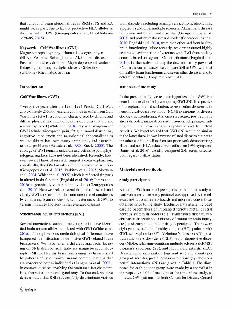
All sensor pairs (Fig. 1)
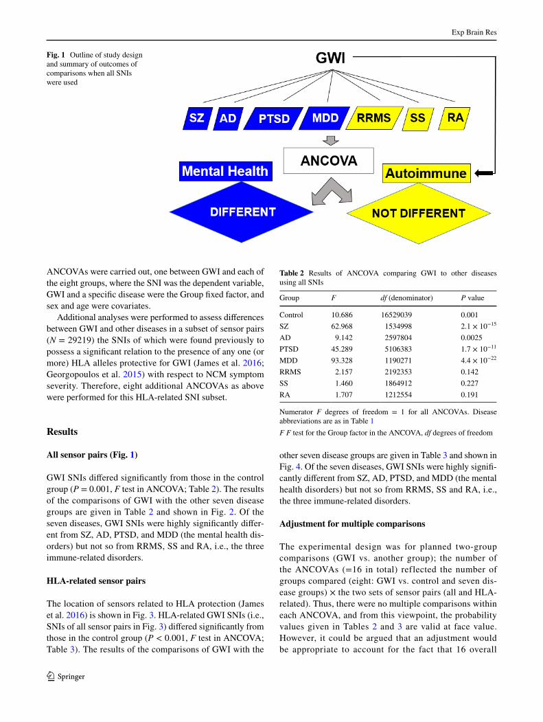
GWI SNIs differed significantly from those in the control group (P = 0.001, F test in ANCOVA; Table 2). The results of the comparisons of GWI with the other seven disease groups are given in Table 2 and shown in Fig. 2. Of the seven diseases, GWI SNIs were highly significantly differ-ent from SZ, AD, PTSD, and MDD (the mental health dis-orders) but not so from RRMS, SS and RA, i.e., the three immune-related disorders.
HLA‑related sensor pairs
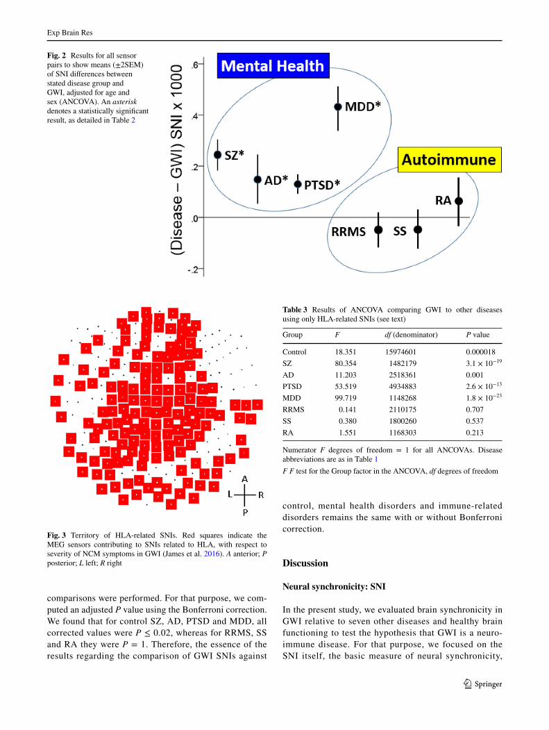
The location of sensors related to HLA protection (James et al. 2016) is shown in Fig. 3. HLA-related GWI SNIs (i.e., SNIs of all sensor pairs in Fig. 3) differed significantly from those in the control group (P < 0.001, F test in ANCOVA; Table 3). The results of the comparisons of GWI with the
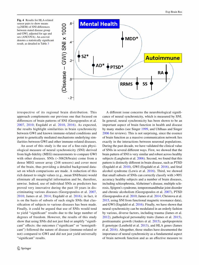
other seven disease groups are given in Table 3 and shown in Fig. 4. Of the seven diseases, GWI SNIs were highly signifi-cantly different from SZ, AD, PTSD, and MDD (the mental health disorders) but not so from RRMS, SS and RA, i.e., the three immune-related disorders.
Adjustment for multiple comparisons
The experimental design was for planned two-group comparisons (GWI vs. another group); the number of the ANCOVAs (=16 in total) reflected the number of groups compared (eight: GWI vs. control and seven dis-ease groups) × the two sets of sensor pairs (all and HLA-related). Thus, there were no multiple comparisons within each ANCOVA, and from this viewpoint, the probability values given in Tables 2 and 3 are valid at face value. However, it could be argued that an adjustment would be appropriate to account for the fact that 16 overall
Fig. 1 Outline of study design and summary of outcomes of comparisons when all SNIs were used
Table 2 Results of ANCOVA comparing GWI to other diseases using all SNIs
Numerator F degrees of freedom = 1 for all ANCOVAs. Disease abbreviations are as in Table 1F F test for the Group factor in the ANCOVA, df degrees of freedom
Group F df (denominator) P value
Control 10.686 16529039 0.001SZ 62.968 1534998 2.1 × 10⁻¹⁵
AD 9.142 2597804 0.0025PTSD 45.289 5106383 1.7 × 10⁻¹¹
MDD 93.328 1190271 4.4 × 10⁻²²
RRMS 2.157 2192353 0.142SS 1.460 1864912 0.227RA 1.707 1212554 0.191
Exp Brain Res
1 3
comparisons were performed. For that purpose, we com-puted an adjusted P value using the Bonferroni correction. We found that for control SZ, AD, PTSD and MDD, all corrected values were P ≤ 0.02, whereas for RRMS, SS and RA they were P = 1. Therefore, the essence of the results regarding the comparison of GWI SNIs against
control, mental health disorders and immune-related disorders remains the same with or without Bonferroni correction.
Discussion
Neural synchronicity: SNI
In the present study, we evaluated brain synchronicity in GWI relative to seven other diseases and healthy brain functioning to test the hypothesis that GWI is a neuro-immune disease. For that purpose, we focused on the SNI itself, the basic measure of neural synchronicity,
Fig. 2 Results for all sensor pairs to show means (±2SEM) of SNI differences between stated disease group and GWI, adjusted for age and sex (ANCOVA). An asterisk denotes a statistically significant result, as detailed in Table 2
Fig. 3 Territory of HLA-related SNIs. Red squares indicate the MEG sensors contributing to SNIs related to HLA, with respect to severity of NCM symptoms in GWI (James et al. 2016). A anterior; P posterior; L left; R right
Table 3 Results of ANCOVA comparing GWI to other diseases using only HLA-related SNIs (see text)
Numerator F degrees of freedom = 1 for all ANCOVAs. Disease abbreviations are as in Table 1F F test for the Group factor in the ANCOVA, df degrees of freedom
Group F df (denominator) P value
Control 18.351 15974601 0.000018SZ 80.354 1482179 3.1 × 10⁻¹⁹
AD 11.203 2518361 0.001PTSD 53.519 4934883 2.6 × 10⁻¹³
MDD 99.719 1148268 1.8 × 10⁻²³
RRMS 0.141 2110175 0.707SS 0.380 1800260 0.537RA 1.551 1168303 0.213
Exp Brain Res
1 3
irrespective of its regional brain distribution. This approach complements our previous one that focused on differences of brain patterns of SNI (Georgopoulos et al. 2007, 2010; Engdahl et al. 2010, 2016). As expected, the results highlight similarities in brain synchronicity between GWI and known immune-related conditions and point to genetically mediated mechanisms underlying sim-ilarities between GWI and other immune-related diseases.
An asset of this study is the use of a fine-rain physi-ological measure of neural synchronicity (SNI) derived from high-fidelity (MEG) measurements to compare GWI with other diseases. SNIs (~30628/brain) come from a dense MEG sensor array (248 sensors) and cover most of the brain, thus providing a detailed background data-set on which comparisons are made. A reduction of this rich dataset to single values (e.g., mean SNI/brain) would eliminate all meaningful information and be, therefore, unwise. Indeed, use of individual SNIs as predictors has proved very innovative during the past 10 years in dis-criminating various diseases (Georgopoulos et al. 2007, 2010; James et al. 2014; Engdahl et al. 2016); in fact, it is on the basis of subsets of such single SNIs that clas-sification of subjects to various diseases has been made. Finally, it could be argued that use of SNIs would tend to yield “significant” results due to the large number of degrees of freedom. However, the results of this study show that using SNIs did not just find or amplify “signifi-cant” effects: the outcomes (“significant” or “nonsignifi-cant”) followed the nature of disease (immune-related or not) compared to GWI and did not just yield universally “significant” results.
A different issue concerns the neurobiological signifi-cance of neural synchronicity, which is measured by SNI. In general, neural synchronicity has been shown to be an important aspect of brain function in health and disease by many studies (see Singer 1999, and Uhlhaas and Singer 2006 for reviews). This is not surprising, since the essence of brain function as a massive communication network lies exactly in the interactions between neuronal populations. During the past decade, we have validated the clinical value of SNIs in several different ways. First, we showed that the brain pattern of SNI is very similar and robust across healthy subjects (Langheim et al. 2006). Second, we found that this pattern is distinctly different in brain disease, such as PTSD (Engdahl et al. 2010), GWI (Engdahl et al. 2016), and fetal alcohol syndrome (Lewis et al. 2016). Third, we showed that small subsets of SNIs can correctly classify with >90% accuracy healthy subjects and a number of brain diseases, including schizophrenia, Alzheimer’s disease, multiple scle-rosis, Sjögren’s syndrome, temporomandibular joint disorder and chronic alcoholism (Georgopoulos et al. 2007), PTSD (Georgopoulos et al. 2010; James et al. 2014; Christova et al. 2015, using SNI from functional magnetic resonance data), and GWI (Engdahl et al. 2016). Finally, we have shown that neural synchronicity can be modulated in an orderly fashion by various, diverse factors, including trauma (James et al. 2012), pathological personality traits (James et al. 2015), posttraumatic growth (Anders et al. 2015), apolipoprotein E genotype (Leuthold et al. 2013), and HLA genes (James et al. 2016). Altogether, those studies have documented the importance of neural synchronicity as a fundamental aspect of brain network function and as an effective measure to
Fig. 4 Results for HLA-related sensor pairs to show means (±2SEM) of SNI differences between stated disease group and GWI, adjusted for age and sex (ANCOVA). An asterisk denotes a statistically significant result, as detailed in Table 3
Exp Brain Res
1 3
differentiate, quantify and evaluate the effects of disease and behavioral factors on integrative brain function.
Immune basis of GWI
A number of researchers have implicated immune system disruption in GWI (Hotopf et al. 2000; Israeli 2012; Moss 2013; Parkitny et al. 2015; Skowera et al. 2004; Toubi 2012; Whistler et al. 2009). To that end, we recently demonstrated genetic vulnerability involving human leukocyte antigen (HLA) genes in veterans with GWI (Georgopoulos et al. 2015). HLA genes, which are located in the Major Histo-compatibility Complex of chromosome 6, play a central role in immune system functioning (Meuer et al. 1982). We reported that six Class II HLA alleles discriminate veterans with GWI from healthy controls and are inversely related to GWI symptom severity, suggesting a protective effect (Georgopoulos et al. 2015). That is, veterans with GWI lack protection, thereby increasing the likelihood of immune-related reactions and other aberrant immune responses when exposed to environmental triggers. We also demonstrated that these HLA alleles interact with brain function to influ-ence symptoms of GWI including NCM (James et al. 2016). There, we concluded that in the absence of HLA protec-tion, immune-related brain abnormalities develop in GWI, perhaps via the development of antibodies to brain antigens resulting in cellular abnormalities, anomalies in neural com-munication, and symptomatology.
Brain dysfunction in GWI and other disorders with immune involvement
GWI is associated with structural brain abnormalities, notably subcortical brain atrophy (Christova et al. 2017). Functionally, more than half of veterans with GWI report at least moderate neurological/cognitive/mood (NCM) impairment (Steele 2000). Typical symptoms include memory and concentration difficulty, word-finding trouble, headaches, blurred vision, tremors, numbness, and mood alterations among others. Similar cognitive and neuropsy-chiatric symptoms have been associated with various con-ditions characterized by disruptions in immune function-ing including rheumatoid arthritis (Hanly et al. 2005; Shin et al. 2012, 2013; de Melo and Da-Silva 2012), systemic lupus erythematosus (Ainala et al. 2001; Antonchak et al. 2011; Carbotte et al. 1986; de Melo and Da-Silva 2012; Ginsburg et al. 1992; Hanly et al. 1994, 2005; Hay et al. 1992) Sjögren’s syndrome (Alexander and Provost 1987; Lafitte et al. 2001; Martinez et al. 2010; Segal et al. 2012, 2014), and multiple sclerosis (Amato et al. 2006; Chiara-valloti and DeLuca 2008; Denney et al. 2005; Rao et al. 1991). Although estimates vary, some studies have found that two thirds of patients with these disorders exhibit
cognitive impairment (Ainala et al. 2001; Hamed et al. 2012; Carbotte et al. 1986; Alexander and Provost 1987; Heaton et al. 1985). These deficits are observed in indi-viduals with no prior cognitive or psychiatric history and have been shown to be associated with markers of inflam-mation or autoimmunity (Alexander and Provost 1987; Kozora et al. 2001; Hamed et al. 2012). Thus, like GWI, these conditions appear to exhibit interacting effects on the nervous system and immune system that result in both NCM impairment and immune system disruption.
GWI SNI differences from other diseases
We have previously demonstrated the power of SNI brain patterns derived from task-free MEG in successfully dis-criminating various brain diseases (Georgopoulos et al. 2007, 2010; Engdahl et al. 2010, 2016; James et al. 2014). In the present study, we compared average GWI SNI, irre-spective of its brain distribution, to healthy brain func-tioning and other diseases of varied etiology, all of which involve NCM-related impairments. Results demonstrated that GWI SNI did not differ significantly from that of three immune-related diseases (SS, RRMS, and RA) but differed significantly from healthy brain functioning and from brain functioning in non-immune-related diseases (SZ, AD, PTSD, MDD), supporting our hypothesis that GWI is a neuroimmune disease. Although many research-ers have recently surmised that GWI is an immune-related condition, this is the first study to empirically demon-strate brain-related similarities between GWI and known immune diseases.
GWI differences within protective HLA‑related SNIs
In previous studies, we demonstrated HLA-involvement in GWI (Georgopoulos et al. 2015) as well as HLA-related neural influences on GWI symptoms (James et al. 2016). Here we sought to further evaluate SNI differences between GWI and the three immune-related diseases with regard to HLA status. The vast majority of SNIs (29219 out of 30628) were significantly related to HLA with respect to GWI NCM severity (James et al. 2016), high-lighting robust interactions of neural and immune systems in GWI. The SNIs involved were widespread although entirely absent in the right temporal region (Fig. 3) and sparse in the right temporal region. Within this subset of HLA-related SNIs, there were no significant differences between GWI and the three immune-related diseases: RA, RRMS, and SS, in contrast to significant differences present between GWI and the four non-immune-related diseases (SZ, AD, PTSD, MDD).

Exp Brain Res
1 3
Implications for possible HLA protective involvement in other diseases
The results of the present study highlight neuroimmune involvement in GWI and indicate brain-based similarities with other immune disorders, particularly with regard to HLA-related neural synchrony. Here, the focus is on disease and, with regard to HLA-related SNI, GWI is indistinguish-able from RRMS, RA, and SS. However, in as much as the absence of certain HLA alleles has been linked to enhanced vulnerability for GWI, the presence of those alleles confers protection (Georgopoulos et al. 2015). This suggests the possibility that these same alleles may confer protection for brain involvement in other neuroimmune diseases as well. Interestingly, DRB1*13:02, one of our six GWI protective alleles (Georgopoulos et al. 2015), has been found to con-fer protection to a wide variety of immune-related disorders (Furukawa et al. 2017). This adds further support to the link between GWI and lack of HLA protection (Georgopoulos et al. 2015).
Limitation of the study
The main limitation of the study is the relatively small number of participants in the disease groups. Although the number of SNIs was large and allowed valid com-parisons, the representation of adequate variety across participants with various diseases is important. Another possible limitation concerns the criteria used for diagno-sis. In the present study, disease diagnosis was made by expert clinician at the time of study but such criteria may change over time. This limitation holds for many clinical studies and trials.
Acknowledgements This work was partially supported by a service directed grant from the United States Department of Veterans Affairs, a grant for the United States Department of Defense (Award Num-ber W81XWH-15-1-0520), and the American Legion Brain Sciences Chair. The contents do not represent the views of the U.S. Department of Veterans Affairs or the United States Government.
Compliance with ethical standards
Conflict of interest The authors do not report any financial disclo-sures or conflicts of interest.
References
Ainala H, Loukkola J, Peltola J et al (2001) The prevalence of neu-ropsychiatric syndromes in systemic lupus erythematosus. Neu-rology 57:496–500
Alexander E, Provost TT (1987) Sjögren’s syndrome: association of cutaneous vasculitis with central nervous system disease. Arch Dermatol 123:801–810
Amato MP, Zipoli V, Portaccio E (2006) Multiple sclerosis-related cognitive changes: a review of cross-sectional and longitudinal studies. J Neurol Sci 245:41–46
American Psychiatric Association (2000) Diagnostic and statistical manual of mental disorders, 4th edn. APA, Washington, DC
Anders SL, Peterson CK, James LM et al (2015) Neural communica-tion in posttraumatic growth. Exp Brain Res 233:2013–2020
Antonchak MA, Saoudian M, Khan AR et al (2011) Cognitive dys-function in patients with systemic lupus erythematosus: a con-trolled study. J Rheumatol 38:1020–1025
Blake D, Weathers F, Nagy LM et al (1995) Clinician-Administered PTSD Scale. National Center for PTSD, Boston
Box GEP, Jenkins GM (1976) Time series analysis: forecasting and control. Holden-Day, San Francisco
Carbotte RM, Denburg SD, Denburg JA (1986) Prevalence of cog-nitive impairment in systemic lupus erythematosus. J Nervous Mental Dis 174:357–364
Chiaravalloti ND, DeLuca J (2008) Cognitive impairment in multiple sclerosis. Lancet Neurol 7:1139–1151
Christova P, James LM, Engdahl BE, Lewis SM, Georgopoulos AP (2015) Diagnosis of posttraumatic stress disorder (PTSD) based on correlations of prewhitened fMRI data: outcomes and areas involved. Exp Brain Res 233:2695–2705
Christova P, James LM, Engdahl BE et al (2017) Subcortical brain atrophy in Gulf War Illness. Exp Brain Res. doi:10.1007/s00221-017-5010-8 (Epub ahead of print)
de Melo LF, Da-Silva SL (2012) Neuropsychological assessment of cognitive disorders in patients with fibromyalgia, rheumatoid arthritis, and systemic lupus erythematosus. Brazil J Rheumatol 52:175–188
Denney DR, Sworowski LA, Lynch SG (2005) Cognitive impairment in three subtypes of multiple sclerosis. Arch Clin Neuropsychol 20:967–981
Engdahl BE, Leuthold A, Tan HR et al (2010) Post-traumatic stress disorder: a right temporal lobe syndrome? J Neural Eng 7:066005
Engdahl BE, James LM, Miller RD et al (2016) A magnetoencephalo-graphic (MEG) study of Gulf War Illness (GWI). EBioMedicine. doi:10.1016/j.ebiom.2016.08.030
First MB, Spitzer RL, Gibbon M et al (2002) Structural clinical inter-view for DSM-IV-TR axis I disorders, research version, non-patient edition (SCID-I/NP). Biometrics Research, New York State Psychiatric Institute, New York
Fisher RA (1958) Statistical methods for research workers, 13th edn. Oliver and Boyd, Edinburgh
Fukuda K, Nisenbaum R, Stewart G et al (1998) Chronic multisymp-tom illness affecting Air Force veterans of the Gulf War. JAMA 280:981–988
Furukawa H, Oka S, Tsuchiya N et al (2017) The role of common protective alleles HLA-DRB1*13 among systemic autoimmune diseases. Genes Immun 18:1–7
Georgopoulos AP, Karageorgiou E, Leuthold A et al (2007) Syn-chronous neural interactions assessed by magnetoencephalog-raphy: a functional biomarker for brain disorders. J Neural Eng 4:349–355
Georgopoulos AP, Tan HM, Lewis SM et al (2010) The synchronous neural interactions test as a functional neuromarker for post-trau-matic stress disorder (PTSD): a robust classification method based on the bootstrap. J Neural Engin 7:016011
Georgopoulos AP, James LM, Mahan MY et al (2015) Reduced Human Leukocyte Antigen (HLA) protection in Gulf War Illness (GWI). EBioMedicine 3:79–85
Ginsburg KS, Wright EA, Larson MG et al (1992) A controlled study of the prevalence of cognitive dysfunction in randomly selected patients with systemic lupus erythematosus. Arthritis Rheum 35:776–782

Exp Brain Res
1 3
Hamed SA, Selim ZI, Elattar AM et al (2012) Assessment of biocor-relates for brain involvement in female patients with rheumatoid arthritis. Clin Rheumatol 31:123–132
Hanly JG, Fisk JD, Eastwood B (1994) Brain reactive autoantibod-ies and cognitive impairment in systemic lupus erythematosus. Lupus 3:193–199
Hanly JG, Fisk JD, McCurdy G et al (2005) Neuropsychiatric syn-dromes in patients with systemic lupus erythematosus and rheu-matoid arthritis. J Rheumatol 32:1459–1466
Hay EM, Black D, Huddy A et al (1992) Psychiatric disorder and cognitive impairment in systemic lupus erythematosus. Arthritis Rheum 35:411–416
Heaton RK, Nelson LM, Thompson DS et al (1985) Neuropsycho-logical findings in relapsing-remitting and chronic-progressive multiple sclerosis. J Consul Clin Psychol 53:103
Hotopf M, David A, Hull L et al (2000) Role of vaccinations as risk factors for ill health in veterans of the Gulf war: cross-sectional study. BMJ 320:1363–1367
Israeli E (2012) Gulf War Syndrome as a part of the autoimmune (auto-inflammatory) syndrome induced by adjuvant (ASIA). Lupus 21:190–194
James LM, Engdahl BE, Leuthold AC et al (2012) Neural network modulation by trauma as a marker of resilience: differences between veterans with posttraumatic stress disorder and resilient controls. JAMA Psychiatry 70:410–418
James LM, Belitskaya-Levy I, Lu Y et al (2014) Development and application of a diagnostic algorithm for posttraumatic stress dis-order. Psychiatry Res 231:1–7
James LM, Engdahl BE, Leuthold AC, Krueger RF, Georgopoulos AP (2015) Pathological personality traits modulate neural interac-tions. Exp Brain Res 233:3543–3552
James LM, Engdahl BE, Leuthold AC, Georgopoulos AP (2016) Brain correlates of human leukocyte antigen (HLA) protection in Gulf War Illness (GWI). EBioMedicine 13:72–79
Kozora E, Laudenslager M, Lemieux A, West SG (2001) Inflammatory and hormonal measures predict neuropsychological functioning in systemic lupus erythematosus and rheumatoid arthritis patients. J Int Neuropsychol Soc 7:745–754
Lafitte C, Amoura Z, Cacoub P et al (2001) Neurological complications of primary Sjögren’s syndrome. J Neurol 248:577–584
Langheim FPJ, Leuthold AC, Georgopoulos AP (2006) Synchronous dynamic brain networks revealed by magnetoencephalography. Proc Natl Acad Sci USA 103:455–459
Leuthold AC, Mahan MY, Stanwyck JJ, Georgopoulos A, Georgopou-los AP (2013) The number of cysteine residues per mole in apoli-poprotein E affects systematically synchronous neural interactions in women’s healthy brains. Exp Brain Res 226:525–536
Lewis SM, Vydrová RR, Leuthold AC, Georgopoulos AP (2016) Cor-tical miscommunication after prenatal exposure to alcohol. Exp Brain Res 234:3347–3353
Mahan MY, Chorn CR, Georgopoulos AP (2015) White Noise Test: detecting autocorrelation and nonstationarities in long time series after ARIMA modeling. Proc 14th Python In Science Conference (Scipy 2015), Austin, TX
Martinez S, Caceres C, Mataro M et al (2010) Is there progressive cognitive dysfunction in Sjögren Syndrome? A preliminary study. Acta Neurol Scand 122:182–188
McKhann G, Drachman D, Folstein M et al (1984) Clinical diagnosis of Alzheimer’s disease Report of the NINCDS-ADRDA Work Group under the auspices of Department of Health and Human Services Task Force on Alzheimer’s Disease. Neurology 34:939–944
Meuer SC, Hussey RE, Hodgdon JC et al (1982) Surface structures involved in target recognition by human cytotoxic T lymphocytes. Science 218:471–473
Moss JI (2013) Gulf War illnesses are autoimmune illnesses caused by increased activity of the p38/MAPK pathway in CD4+ immune system cells, which was caused by nerve agent prophylaxis and adrenergic load. Med Hypotheses 81:1002–1003
Parkitny L, Middleton S, Baker K, Younger J (2015) Evidence for abnormal cytokine expression in Gulf War Illness: a prelimi-nary analysis of daily immune monitoring data. BMC Immunol. doi:10.1186/s12865-015-0122-z
Polman CH, Reingold SC, Edan G et al (2005) Diagnostic criteria for multiple sclerosis: 2005 revisions to the “McDonald Criteria”. Annals Neuro 58:840–846
Priestley MB (1981) Spectral analysis of time series. Academic, San Diego
Rao SM, Leo GJ, Bernardin L, Unverzagt F (1991) Cognitive dysfunc-tion in multiple sclerosis. I. Frequency, patterns, and prediction. Neurology 41:685–691
Segal BM, Pogatchnik B, Holker E et al (2012) Primary Sjogren’s syn-drome: cognitive symptoms, mood, and cognitive performance. Acta Neurol Scand 125:272–278
Segal BM, Rhodus N, Sivils KLM, Solid CA (2014) Validation of the brief cognitive symptoms index in sjögren syndrome. J Rheumatol 41:2027–2033
Shin SY, Katz P, Wallhagen M, Julian L (2012) Cognitive impair-ment in persons with rheumatoid arthritis. Arthritis Care Res 64:1144–1150
Shin SY, Julian L, Katz P (2013) The relationship between cognitive function and physical function in rheumatoid arthritis. J Rheu-matol 40:236–243
Singer W (1999) Neuronal synchrony: a versatile code for the definition of relations? Neuron 24:49–65
Skowera A, Hotopf M, Sawicka E et al (2004) Cellular immune activa-tion in Gulf War veterans. J Clin Immunol 24:66–73
Steele L (2000) Prevalence and patterns of Gulf War illness in Kan-sas veterans: association of symptoms with characteristics of person, place, and time of military service. Am J Epidemiol 152:992–1002
Uhlhaas PJ, Singer W (2006) Neural synchrony in brain disorders: relevance for cognitive dysfunctions and pathophysiology. Neuron 52:155–168
Vitali C, Bombardieri S, Jonsson R (2002) Classification criteria for Sjögren’s syndrome: a revised version of the European criteria proposed by the American-European Consensus Group. Annals Rheum Dis 61:554–558
Whistler T, Fletcher MA, Lonergan W et al (2009) Impaired immune function in Gulf War illness. BMC Med Genom 2:1. doi:10.1186/1755-8794-2-12
White RF, Steele L, O’Callaghan JP et al (2016) Recent research on Gulf War illness and other health problems in veterans of the 1991 Gulf War: effects of toxicant exposures during deployment. Cortex 74:449–475