Embed Size (px)
Citation preview
Guiding Principles in the Design of MolecularBioconjugates for Vaccine Applications
The MIT Faculty has made this article openly available. Please share how this access benefits you. Your story matters.
Citation Liu, Haipeng, and Darrell J. Irvine. “Guiding Principles in the Designof Molecular Bioconjugates for Vaccine Applications.” BioconjugateChemistry 26, no. 5 (May 20, 2015): 791–801. © 2015 AmericanChemical Society
As Published http://dx.doi.org/10.1021/acs.bioconjchem.5b00103
Publisher American Chemical Society (ACS)
Version Author's final manuscript
Citable link http://hdl.handle.net/1721.1/108144
Terms of Use Article is made available in accordance with the publisher'spolicy and may be subject to US copyright law. Please refer to thepublisher's site for terms of use.
Guiding Principles in the Design of Molecular Bioconjugates for Vaccine Applications
Haipeng Liu1,2,3 and Darrell J. Irvine4,5,6,7,8
1Department of Chemical Engineering and Materials Science, Wayne State University, Detroit, MI 48202.
2Department of Oncology, Wayne State University, Detroit, MI 48201.
3Tumor Biology and Microenvironment Program, Barbara Ann Karmanos Cancer Institute, Detroit, MI 48201.
4Department of Materials Science and Engineering, Massachusetts Institute of Technology, Cambridge, MA 02139.
5Department of Biological Engineering, Massachusetts Institute of Technology, Cambridge, MA 02139.
6Ragon Institute of Massachusetts General Hospital, Massachusetts Institute of Technology, and Harvard, Cambridge, MA 02139.
7Koch Institute for Integrative Cancer Research, Massachusetts Institute of Technology, Cambridge, MA 02139.
8Howard Hughes Medical Institute, Chevy Chase, MD 20815.
Abstract
Antigen- and adjuvant-based bioconjugates that can stimulate the immune system play an
important role in vaccine applications. Bioconjugates have demonstrated unique physicochemical
and biological properties, enabling vaccines to be delivered to key immune cells, to target specific
intracellular pathways, or to mimic immunogenic properties of natural pathogens. In this review
we highlight recent advances in such molecular immunomodulators, with an emphasis on the
structure-function relationships that provide the foundation for rational design of safe and
effective vaccines and immunotherapies.
1. Introduction
Vaccines remain the single most effective public health intervention ever developed, with
millions of lives saved every year through the array of pediatric and adult vaccines
administered globally.1-3 The immune response elicited by vaccination is a multi-step,
complex process that involves the coordinated action of diverse molecular signals and
immune cells within lymphoid organs4,5: first, antigen must be acquired by specialized
*Correspondence to: [email protected], [email protected].
HHS Public AccessAuthor manuscriptBioconjug Chem. Author manuscript; available in PMC 2015 December 29.
Published in final edited form as:Bioconjug Chem. 2015 May 20; 26(5): 791–801. doi:10.1021/acs.bioconjchem.5b00103.
Author M
anuscriptA
uthor Manuscript
Author M
anuscriptA
uthor Manuscript
sentinel cells known as antigen presenting cells (APCs). APCs can internalize antigen
directly in the tissue of the vaccination site or antigen can be transported through the
capillary lymphatic vessels to APCs or B-cells in the draining lymph nodes6 (Fig. 1).
Second, for T-cell activation, these APCs must degrade the antigen in appropriate
intracellular compartments and load resulting peptide fragments onto major
histocompatibility complex (MHC) molecules.4,5 These APCs must also be activated by
inflammatory cues (“danger signals”) elicited by the vaccine, which instruct the APCs to
mount an immune response against the acquired antigen.7 Third, in the lymph node, CD8+
T-cells and CD4+ T-cells with matching receptors recognize peptide fragments from the
antigen bound to MHC on APC surfaces, and if the APCs are properly activated, these T-
cells proliferate and differentiate into primed effector cells that can directly kill infected
cells (CD8+ “killer” T-cells) or secrete cytokines to coordinate microbe clearance by other
immune cells (CD4+ “helper” T-cells). In parallel, antigen is also recognized by antigen-
specific B-cells, which receive “help” signals from primed CD4+ T-cells to differentiate into
antibody-producing plasma cells that secrete copious amounts of antibody that will bind to
microbes and promote their clearance. For therapeutic vaccines administered in the presence
of ongoing disease, these effector T- and B-cell responses can provide immediate
therapeutic benefit. Following initial expansion, most (~90%) of the antigen-specific CD8+
T-cells, CD4+ T-cells, and B-cells generated during this early effector phase die off, but a
population of long-lived memory T-cells and B-cells develops. This pool of long-lived cells,
which can persist for many years in humans, is the basis of prophylactic vaccination; these
memory cells provide in some cases lifelong immunity against subsequent exposure to the
pathogen matching the vaccine antigen.4
The first licensed vaccines were comprised of inactivated or attenuated live microorganisms.
Though these whole-microbe vaccines have been successful in preventing many infectious
diseases, this approach is not applicable to some vaccine settings (e.g., therapeutic vaccines
for cancer) or may not be safe (e.g., vaccines for HIV). Further, most live-attenuated
vaccines were developed empirically without a clear understanding of their mechanisms of
action.8 In the modern era, the paramount importance of vaccine safety has made such an
approach problematic, and much of current vaccinology is based on the development of
subunit vaccines, which replace whole microbes with defined protein or polysaccharide
antigens that have no potential for infectivity or toxicity on their own.9,10 Subunit vaccines
are usually fully synthetic and have molecularly defined structures, which have advantages
in manufacturability, stability, and safety. However, subunit vaccines are poorly
immunogenic and require adjuvants to induce an adaptive immune response. Adjuvants
broadly defined are any substance added to a vaccine to augment the immune response to
the antigen, and include diverse compounds including microbe-derived products that trigger
conserved pathogen-recognition receptors; synthetic immunostimulatory molecules; and
nanoparticles, microparticles, or oil/water emulsions.11,12
Among these different approaches, one of the most attractive strategies to achieve well-
defined molecular vaccines is to incorporate additional functionality directly into the antigen
(or alternatively, into danger signal molecules) through bioconjugation.13-16 In fact,
bioconjugates have long had an important role in the development of vaccines against
infection, cancer and many other diseases. The most common bioconjugates are those where
Liu and Irvine Page 2
Bioconjug Chem. Author manuscript; available in PMC 2015 December 29.
Author M
anuscriptA
uthor Manuscript
Author M
anuscriptA
uthor Manuscript
vaccine components are covalently linked to a protein, peptide, lipid, oligonucleotide,
polymer, or nanoparticle, but in some cases antigens or molecular adjuvants are linked to
synthetic small molecules.17-19 Depending on their chemical and molecular nature,
bioconjugates can enhance vaccine efficacy via diverse mechanisms. Examples include
conjugation of antigen/adjuvant to a ligand to enable tissue/cell specific targeting;
conjugation of vaccines to polymers to provide new properties such as multivalency and/or
controlled release; vaccines conjugated to nanoparticles can also lead to changes in the
pathways by which antigens are processed by APCs. Thus, bioconjugates can be tailored and
functionalized according to vaccine-specific needs.
In this review, we summarize bioconjugate strategies being explored in preclinical research
and clinical development, with a focus on the guiding principles for rational design of
bioconjugates in vaccine applications. We have chosen to limit the scope to techniques and
approaches that can modulate the immune system via molecular conjugates; therefore, a
variety of important and novel systems, such as antigen/adjuvant encapsulated in nano/micro
particles that have been reviewed recently20,23 are not covered here. In addition, we will not
discuss polysaccharide/peptide/hapten conjugate vaccines (antigen conjugated to carrier
proteins), which have also been recently reviewed.15, 24-26
2. Targeting vaccines to the lymphatic system
For a vaccine to prime de novo immune responses, naive T-cells and B-cells that reside in
secondary lymphoid organs (lymph nodes and spleen) must be stimulated. Because of this
localization, Zinkernagel first enunciated the “geographical” concept of immunity, whereby
vaccines that do not reach the lymphoid organs are ignored by the immune system.27 Two
pathways for vaccine delivery to lymph nodes are possible: First, vaccine molecules can be
directly transported from injection sites (muscle, skin, or mucosal surfaces) to draining
lymph nodes (LNs) by lymph draining through lymphatic vessels (Fig. 1). Alternatively,
APCs (monocytes from the blood, or local tissue-resident dendritic cells) can internalize
vaccine antigens/adjuvant compounds at the injection site and actively carry them through
migration to the LNs. The latter pathway is relatively inefficient because few APCs migrate
to lymph nodes from a site of inflammation, but these migratory cells play an important role
in the evolving immune response in some settings.28 Bioconjugate strategies have thus been
explored that facilitate lymphatic uptake and capture of vaccines in the lymph nodes.
2.1. Targeting lymphoid tissues via macromolecular conjugates
The fate of molecules injected parenterally is strongly influenced by molecular size.
Connective tissues are perfused by blood and lymphatic vessels, which play a major role in
the clearance of proteins injected into the tissue. Fluid is both released and reabsorbed across
blood vessels, while lymphatic vessels provide for one-way transport of fluid out of tissue.
Blood vessels reabsorb ~10-fold more interstitial fluid from the tissue than lymphatics, but
the endothelial cells of the blood vessels are connected by tight junctions which block the
diffusion of particles greater than ~3-5 nm in size. Thus, small molecules/particles are
cleared from tissues primarily by the blood, while proteins show increasing efficiencies of
lymphatic uptake with increasing molecular weight (plateauing at masses greater than
~40-50 KDa).29,30 For vaccines, this size-dependent transport means that peptides, small
Liu and Irvine Page 3
Bioconjug Chem. Author manuscript; available in PMC 2015 December 29.
Author M
anuscriptA
uthor Manuscript
Author M
anuscriptA
uthor Manuscript
protein antigens, and a variety of molecular adjuvants will exhibit very poor lymph node
accumulation if injected as unformulated compounds. Thus, a number of approaches have
been developed to direct small molecular weight vaccine components to lymph nodes by
increasing their effective hydrodynamic size.
Antigens and molecular adjuvants conjugated to size-optimized nanoparticles (NPs) have
frequently been used to promote LN targeting. Reddy et al. showed that small polypropylene
sulfide (PPS) NPs (less than 45 nm in diameter) were able to drain efficiently to lymph
nodes for capture by LN-resident dendritic cells (DCs).31,32 Attaching subunit antigens or
adjuvants to such particles enhanced both humoral and antigen-specific CD8+ T cell
responses.32,33 Similar enhancements in immunogenicity were observed by Fifis et al. using
peptide antigens conjugated to 40 nm diam. polystyrene nanoparticles.34,35 Using
monodisperse polystyrene nanoparticles, Manolova et al. also demonstrated size-dependent
trafficking of NPs to the draining LNs: large particles (200-500 nm) were mainly associated
with DCs at the injection sites, but small particles (20-200 nm) were able to freely drain to
the lymph node and accumulate in LN-resident DCs and macrophages, suggesting an
optimum range for lymphatic uptake of injected nanoparticles.36 In each of these studies,
subsequent conjugation of antigen or adjuvant to lymph node-targeting NPs led to markedly
enhanced humoral and cell-mediated immune responses, demonstrating the potential of
nano-sized materials in vaccination.
Conjugation to water-soluble polymers can also increase the hydrodynamic radius of
compounds to promote lymphatic delivery. Because efficient lymph node accumulation is
also needed for sentinel lymph node mapping in cancer (a procedure where optical or
radioactive tracers with lymph node tropism are injected at a tumor site to identify tumor-
draining lymph nodes),37 a number of examples of lymph node-targeting conjugates
applicable to vaccines have been demonstrated in the context of delivering imaging agents to
lymph nodes. For example, Forrest and colleagues investigated LN retention of a series of
six different molecular weight hyaluronan (HA)-near-infrared dye (HA-IR820) conjugates in
mice over 2 weeks following subcutaneous injection.38 They discovered that 74 KDa HA-
IR820 had the largest net lymph node uptake. Enhanced lymphatic uptake and nodal
retention of HA conjugates suggest this natural biodegradable polymer could be an
interesting vaccine carrier, particularly given the fact that one of its receptors, CD44, is
expressed by APCs. Recently, the use of polymer conjugates to enhance LN uptake by
vaccines was shown for water-soluble N-trimethylaminoethylmethacrylate chitosan (TMC)-
protein antigen conjugates. TMC-antigen conjugates were shown to exhibit dramatic
increases in lymph node uptake relative to soluble antigen after nasal instillation.39 The
macromolecular conjugate also elicited 80-fold higher serum IgG responses compared to
mixtures of the same polymer with antigen. These results suggest bioconjugates are also
capable of targeting LN via mucosal routes of administration, when coupled to polymers
such as chitosan that promote penetration through the epithelial barriers at these sites.39
Apart from size, surface properties (i.e. surface charge, hydrophobicity) can affect the
delivery of macromolecules to the lymph node. It is widely believed that positively charged
surface leads to strong electrostatic interaction with the negatively charged interstitial
matrix, preventing lymphatic drainage. Thus, neutral or negatively charged molecules are
Liu and Irvine Page 4
Bioconjug Chem. Author manuscript; available in PMC 2015 December 29.
Author M
anuscriptA
uthor Manuscript
Author M
anuscriptA
uthor Manuscript

preferred in lymph node targeting. Takakura et al. demonstrated that neutral or anionic
polymers were more efficiently accumulated in the draining lymph nodes compared to
cationic polymers.40 In another study, Kaminskas et al. reported the influence of surface
PEGylation of a polylysine dendrimer in the absorption and lymphatic targeting following
SC administration in a rat model and found that increasing the PEG chain length (thereby
shielding the surface charge) promoted uptake in the lymphatics.41 The Hydrophobicity of
macromolecular carrier can impact the lymphatic uptake. Maintaining a balance between
surface hydrophilicity and hydrophobicity has been shown to govern the drainage from
injection sites and lymph nodes retention.42 Enhancing hydrophobicity leads to increased
molecular interaction with antigen presenting cells, thus increasing lymph node retention.
However, hydrophobic modification also limits the solubility and leads to aggregation at the
injection sites, reducing the drainage to the lymphatics. Thus, balancing the hydrophobicity/
hydrophilicity is critical in designing molecular conjugates to target lymph nodes.
Dendrimers are perhaps the most intensively investigated macromolecule for lymph node
targeting purposes.43 These compact polymeric structures are in an optimal size range to
avoid entry into blood vessels from tissue but still diffuse efficiently through the
extracellular matrix. They are transported to the lymphatics and trapped in the lymph node,
especially when their surface charge and hydrophobicity is appropriately modified.
Kobayashi and colleagues investigated the use of gadolinium-conjugated poly(amido amine)
(PAMAM) dendrimers as magnetic resonance lymphangiography agents.44 Increasing
hydrophobicity of the dendrimer led to enhanced lymphatic uptake. The same group also
conjugated 5-color near-infrared dyes and radionuclides to a generation-6 PAMAM
dendrimer and successfully applied these polymers in multi-modal and multicolor lymphatic
imaging.45 Together, materials that can efficiently target lymph node need to possess a small
size (5-100 nm), negative or neutral surface charge, and appropriate hydrophobicity.
A second size-based strategy for lymph node targeting is to design conjugates that non-
covalently associate with serum proteins that have intrinsically efficient lymphatic uptake.
The best-established example of this approach is ‘hitchhiking’ of dye compounds on
endogenous albumin following parenteral injection for sentinel lymph node mapping: A
variety of small-molecule dyes such as Evans blue were discovered empirically to stain
draining lymph nodes when injected subcutaneously in tissues or in tumor resection sites,
allowing visual identification of lymph nodes during tumor resection surgery.46 Subsequent
structure-function analyses of effective dyes revealed a common characteristic of effective
lymph node mapping dyes: high-affinity binding to albumin.47 Thus, upon injection, these
compounds associate with endogenous albumin in the interstitial fluid, forming a complex of
appropriate size to efficiently traffic to lymphatics. Inspired by this clinically-proven
approach for lymph node targeting, we recently developed ‘albumin hitchhiking’ vaccines,
where antigens or molecular adjuvants are covalently linked to a lipophilic albumin binding
domain (Fig. 2a).48 These amphiphile-vaccines, if appropriately designed to reduce
spontaneous cell membrane insertion while retaining effective association with albumin,
exhibited >10-fold increased accumulation in lymph nodes following subcutaneous
administration in mice (Fig. 2b-c). Our collective data to date suggests initial lymphatic
uptake and lymph node targeting is largely a size-based effect, whereby albumin, which is
large enough to show predominantly blood-to-lymph one-way trafficking out of tissues,
Liu and Irvine Page 5
Bioconjug Chem. Author manuscript; available in PMC 2015 December 29.
Author M
anuscriptA
uthor Manuscript
Author M
anuscriptA
uthor Manuscript

ferries the vaccine to lymph nodes. (Notably however, once in the lymph node, albumin
binding may lead to significantly altered trafficking, uptake, and antigen processing
compared to free vaccine). This greatly increased lymph node delivery in turn led to greatly
enhanced potency of these vaccines for promoting T-cell responses (Fig. 2d) and anti-tumor
immunity. In addition, this approach greatly increased the safety profile of molecular
adjuvants by effectively confining them to draining lymph nodes, reducing systemic
dissemination. Given the fact that lymph, which originates from interstitial fluid and
circulates throughout the lymphatic system, contains many substances, including plasma
proteins (i.e.—albumins, globulins, and fibrinogen), lipoproteins, complement components,
etc., it remains to be investigated whether other lymph components can be similarly
exploited for ‘hitchhiking’ of vaccines to lymph nodes.
2.2 Targeting immune cell receptors
In addition to the “passive targeting” strategies described above, which rely on the physical
properties of vaccine carriers to promote lymphatic uptake, “active targeting” based on
conjugation of vaccines with a specific ligand for APC surface receptors (e.g. Fc receptors,
CD40, C-type lectin receptors such as DC-SIGN, DEC-205, mannose receptor, etc.) can also
be used to augment lymph node retention.49-51 One of the first and most striking examples
of the capacity of ligand-mediated targeting to promote vaccine responses was shown with
protein antigens conjugated to an anti-DEC-205 antibody: anti-DEC-205-ovalbumin
conjugates injected in mice were taken up by CD11c+ DCs primarily in the lymph nodes
draining the injection site, leading to 400-fold greater CD8+ T-cell responses compared to
non-targeted ovalbumin protein.51 Recently, human anti-DEC-205 antibody fused with NY-
ESO-1, a full-length cancer-testis antigen overexpressed in diverse cancer types, was shown
to induce humoral and cellular immunity in patients with confirmed NY-ESO-1-expressing
tumors.52 Other members of the C-type lectin receptors, including DC-SIGN (CD209) and
the mannose receptor (CD206), recognize carbohydrates (mannose, fucose, glucose,
maltose, etc.) that are characteristic of pathogen surfaces, regulating the uptake of pathogens
and subsequent activation of adaptive immune responses. The high specificity of
carbohydrate-lectin interactions has been exploited for targeting a wide variety of antigen/
adjuvant formulations for vaccine applications. For example, mannosylated MUC1, a tumor-
associated mucin-like protein has been shown to induce strong Th1 or Th2 immune
responses, depending on the oxidative state of the mannose.53,54 Clinical studies with
oxidized mannan (a polymeric form of mannose)–MUC-1 conjugates demonstrated
induction of both humoral and cellular responses and evidence of protection against
recurrence in early stage breast cancer patients.55 Importantly, no adverse events were
observed, suggesting these polymer conjugates were safe in humans. Synthetic artificial
ligands, such as nucleic acid aptamers identified by in vitro selection, have also been shown
to specifically bind DEC-205 on DCs.56 Due to their unique chemical properties and low
immunogenicity, aptamers are promising alternatives to antibody-based targeting agents.
The DEC-205-targeted antigen was efficiently cross-presented and subsequently activated
CD8+ T cells.56 Clearly, active targeting to DCs enhances vaccine efficacy and safety and
might be included in the future as a safe immunotherapy regimen.
Liu and Irvine Page 6
Bioconjug Chem. Author manuscript; available in PMC 2015 December 29.
Author M
anuscriptA
uthor Manuscript
Author M
anuscriptA
uthor Manuscript
These two concepts of hydrodynamic size and receptor-specific targeting can also be
combined for enhanced LN targeting: Lymphoseek, a mannose-conjugated, dextran-based
lymphatic mapping polymeric agent has been recently approved by the FDA to assist in the
localization of lymph nodes draining a primary tumor site in patients with breast cancer or
melanoma.57 Lymphoseek has an appropriate size (7 nm) and carries multiple units of
mannose, which targets mannose receptors expressed on the surface of macrophages and
DCs.
3. Promoting antigen processing and presentation
Antigen presentation by APCs, whereby short peptide fragments of antigens are loaded into
MHC molecules and displayed on the APC surface to activate T-cells, plays a key role in the
induction of adaptive immune responses. Many of the targeting ligands discussed above for
promoting lymph node accumulation that can bind to APC surface receptors promote
antigen internalization or modulate antigen processing.49-51,53-55,58,59 However,
bioconjugate vaccines can be further designed to control antigen presentation by influencing
what intracellular compartments antigens are delivered to within APCs or directly changing
how antigens are proteolyzed and loaded onto MHC molecules.
3.1. Bioconjugate vaccines promoting cross presentation
Much effort has focused on promoting MHC-I presentation of antigens, in order to prime
CD8+ T-cell responses with vaccines. Class I MHC molecules are normally primarily loaded
with peptides generated in the cytosol, and thus antigens taken up from the extracellular
environment (and therefore transported into endosomes within APCs) are typically not
delivered to the MHC I antigen loading pathway. The process of extracellular antigens being
taken up by APCs and loaded on class I MHC is called cross presentation, a process that
may be critical for successful subunit vaccines against cancer and some infectious
diseases.60 One strategy to enhance class I MHC loading is to link antigens to endosome-
disrupting moieties that can deliver the macromolecules to the cytosol. For example, Stayton
and colleagues prepared pH-responsive, endosomolytic polymers to actively promote
antigen cross-presentation, based on amphiphilic diblock copolymers conjugated with
protein antigens through disulfide linkages (Fig. 3).61-63 In this elegant design, protonation
of the carboxylate and amine groups of these copolymers within endolysosomes leads to
their interaction with the endosomal membrane and/or a proton sponge effect, leading to
escape of the conjugates into the cytosol, where the disulfides are reduced to release the
antigen for “natural” class I MHC pathway processing.61 These copolymers yielded
markedly enhanced cellular responses in vivo.61-63 The vaccine's efficacy was further
improved when CpG DNA (a molecular adjuvant that stimulates APCs) was included.61
Another strategy explored for cytosolic delivery of antigens is through conjugation to cell-
penetrating peptides (CPPs). Certain CPPs are endosomolytic and conjugation of short
(~10-20 amino acid) CPP sequences to antigens has been shown to promote antigen uptake,
cytosolic localization, and antigen cross-presentation for potent cytotoxic CD8+ T cell
responses in vivo.64-66 Finally, activation of certain pattern-recognition receptors (reviewed
in the following section) enables efficient antigen cross-presentation via diverse
mechanisms, leading to potent CD8+ T cell stimulation.67 In summary, bioconjugates can be
Liu and Irvine Page 7
Bioconjug Chem. Author manuscript; available in PMC 2015 December 29.
Author M
anuscriptA
uthor Manuscript
Author M
anuscriptA
uthor Manuscript
designed to dramatically enhance antigen uptake and presentation, resulting in much lower
antigen doses required for immune cell activation and robust and T-cell proliferation.
3.2. Promoting tolerogenic antigen presentation
In addition to stimulating an immune response, bioconjugates can be used to promote
tolerogenic antigen presentation, in order to inhibit detrimental immune responses. This is a
potentially ideal treatment strategy for autoimmune diseases, allergies, and organ
transplants, providing antigen-specific immune tolerance without global
immunosuppression. Early studies focused on the use of apoptotic cells for the induction of
tolerance: Peptide self-antigens chemically conjugated to apoptotic cells were shown to be
effective and safe for the prevention and treatment of a wide variety of autoimmune diseases
including relapsing experimental autoimmune encephalomyelitis (EAE, a mouse model of
multiple sclerosis),68 type 1 diabetes69 and transplant rejection.70 Although the underlying
mechanisms are still under study, it is believed that several distinctive mechanisms, such as
suppression of costimulatory molecule expression on APCs, modulation of antigen
presentation, and production of immunosuppressive cytokines to promote T-cell clonal
depletion or anergy may act synergistically in such therapies. Recently, nanoparticles
conjugated with disease-associated peptide antigens were used to replace donor cells in this
approach in an attempt to avoid the complexities and cost associated with cell manipulation
in the clinic. A number of different nanoparticles have been covalently conjugated to
autoantigens and have shown promise in several autoimmune disease models.71,72 For
example, Getts and coworkers showed that intravenous infusion of antigen-decorated
particles (500-nm diameter) induced long-term T-cell tolerance in mice with relapsing
experimental autoimmune encephalomyelitis (EAE).72 Blockade of immune cell adhesion
during antigen recognition has been shown to suppress the inflammatory immune response
in autoimmune diseases. Chittasupho and colleagues used peptide-conjugated nanoparticles
to block immunological synapse formation between dendritic cells and T-cells. These
nanoparticles also altered cytokine production in cell culture when compared to
unconjugated ligands.73 In a separate study, soluble antigen arrays (SAgAs, hyaluronic acid
grafted with antigen and LABL peptide, an immune cell adhesion inhibitor) were shown to
be efficacious in experimental autoimmune encephalomyelitis.74 Promoting tolerogenic
antigen presentation has also been achieved by in situ binding of autoantigens to red blood
cells. Kontos and coworkers reported an innovative strategy where an antigen was
conjugated with an erythrocyte binding domain, with the goal of targeting autoantigens into
the normal pathways of tolerance present during clearance of aging red blood cells.75
Following i.v. injection, these RBC-binding constructs bound efficiently to erythrocytes in
the blood, inducing peripheral tolerance in an antigen specific manner.75 Instead of using
cells, this strategy uses molecularly-defined bioconjugates for in situ erythrocyte targeting,
which like the nanoparticle/microparticle-conjugate approach, should be more readily
translated to human studies.
4. Multivalent immunogens
Many pathogens such as viruses and bacteria exhibit a highly ordered, repetitive display of
antigens on their surfaces, which are thought to effectively engage and cluster antigen
Liu and Irvine Page 8
Bioconjug Chem. Author manuscript; available in PMC 2015 December 29.
Author M
anuscriptA
uthor Manuscript
Author M
anuscriptA
uthor Manuscript
receptors on B cells, stimulating antibody production more strongly than the same antigens
encountered as soluble proteins in solution.76-78 These observations have led to the idea that
the immunogenicity of subunit antigens can be greatly enhanced by a rigid, ordered
organization on surfaces, mimicking viral particles.76 This multivalency of antigen
presentation, together with the facilitation of immune cell recognition and antigen
internalization, has been explored as a strategy to enhance both humoral and cellular
immunity.
4.1. Multivalent antigens
Early studies with haptens (small molecule antigens that elicit T-cell-independent B-cell
responses) conjugated to water-soluble polymers suggested that T-independent antibody
responses in vivo are only elicited when at least ~20 haptens are coupled to each polymer
chain at a spacing of ~10 nm apart,79 providing early evidence for the importance of antigen
multivalency and clustering in B-cell triggering. Building on the principle that multivalency
can increase the immunogenicity of subunit antigens, it was shown that peptides
multimerized on a dendritic oligo-lysine scaffold (termed multiple antigenic peptides,
MAPs) elicited enhanced antibody responses.80 Mixing immunological adjuvants or
incorporating T-helper epitopes into the MAP system have been reported to greatly enhance
the efficacy of these vaccines.81 MAPs can be readily constructed using solid phase peptide
synthesis and have been shown to be effective in a variety of vaccines.82,83 Dendrimers are a
second platform widely used for multivalent antigen display. For example, Sheng et al.
prepared polyamidoamine (PAMAM) dendrimers chemically conjugated to ovalbumin and
found significant increases in both anti-OVA CD8+ T cells and OVA-specific IgG in mice
compared to soluble OVA vaccines.84 Liu et al. reported a star polymer-peptide conjugate
and found greatly enhanced cellular responses without the need for additional
immunological adjuvants.85 These self-adjuvanting conjugates were able to eradicate TC-1
tumors (a model of HPV-induced cervical cancer) in mice after a single immunization.85
Multimeric antigens can be also built on synthetic peptides linked with a polymerizable
double bond86,87 or derived from ring-opening metathesis polymerization (ROMP).88 For
example, Brandt et al. demonstrated a linear polypeptide derived from acryloyl modified
monomer had improved immunogenicity.87 Using a polymer-hapten conjugate system
derived from ROMP, Kiessling et.al., demonstrated that B cell activation was strongly
influenced by antigen valency; conjugates with high antigen valencies promoted stronger B
cell receptor signaling in vitro and greater antibody production in vivo.88 These studies,
provide evidence that antigen conjugates in a multivalent format can yield potent B- and T-
cell responses.
4.2. Self-assembled immunogens
As an alternative to direct conjugation/synthesis of pre-fabricated multivalent scaffolds,
multivalent immunogen display can also be achieved using individual antigens that undergo
programmed self-assembly. The licensed hepatitis B virus and human papilloma virus
vaccines are based on natural self-assembling proteins from viral capsids, which self-
assemble to form nanoparticles 40-60 nm in diameter displaying an ordered array of HBV
and HPV antigens, respectively.89 Recently, fully synthetic peptides have been explored as
self-assembling vaccine nanomaterials. For example, synthetic lipopeptides, containing
Liu and Irvine Page 9
Bioconjug Chem. Author manuscript; available in PMC 2015 December 29.
Author M
anuscriptA
uthor Manuscript
Author M
anuscriptA
uthor Manuscript
peptide antigens linked to a lipid-like molecule, are capable of self-assembling into
homogeneous nanoparticles90 (Fig. 4A) or cylindrical micelles91 (Fig. 4B) via hydrophobic
interactions. In addition to lipid conjugation, antigen epitopes may also be covalently linked
to peptide sequences that form ordered structures via molecular interactions including van
der Waals forces, ionic bonds, hydrogen bonds and hydrophobic forces.92-95 Engineered
peptide nanoparticles92 (Fig. 4C) or nanofibers93-95 (Fig. 4D) with repetitively displayed
antigen epitopes have been assembled utilizing the peptide molecular interactions and have
shown to be potent immunogens promoting both T-cell and antibody responses in vivo. A
common characteristic of these virus-like synthetic assemblies is their potency without the
need for addition of further adjuvants.91,94 This finding is even more striking given that
responses to these nanostructures have been formally shown to be independent of common
Toll like receptor-based innate immune recognition pathways.91,93 Yet these self-assembling
antigens elicit T-cell dependent, long-lived class-switched antibody responses,96 implying
that humoral immunity primed by these multivalent immunogens shares characteristics of
both T-cell-independent and T-cell-dependent antigens.
Self-assembling nanostructures can be designed to incorporate additional functionality
beyond antigen display alone. For example, coupling of antigens to particles through a
disulfide linkage promotes environment-sensitive release of the antigen for antigen
processing in the reductive endolysosomal pathway within APCs 97; this approach has been
used to link protein antigen to block copolymer micelles for intracellular release of antigen,
promoting cross presentation to T-cells.98 Moyle et al. developed an approach to couple
protein antigens to nanoparticle-forming amphiphiles that self-assembled via hydrophobic
lipid tails.99 The lipid tails of these multi-block amphiphiles were also designed to trigger
Toll-like receptors on APCs (discussed further below) and the hydrophilic block contained a
dendritic cell-targeting peptide, thus building antigen display, APC targeting, and APC
activation all into a single molecule. These diverse examples illustrate the capacity of
nanostructure-based vaccine platforms to display ordered arrays of antigen and regulate
antigen uptake and processing, using self-assembly-based synthesis approaches that are
attractive for well-defined large-scale manufacturing.
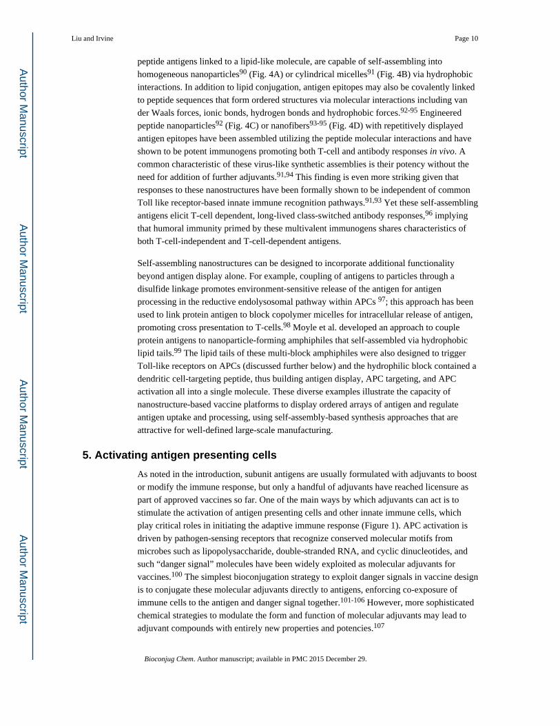
5. Activating antigen presenting cells
As noted in the introduction, subunit antigens are usually formulated with adjuvants to boost
or modify the immune response, but only a handful of adjuvants have reached licensure as
part of approved vaccines so far. One of the main ways by which adjuvants can act is to
stimulate the activation of antigen presenting cells and other innate immune cells, which
play critical roles in initiating the adaptive immune response (Figure 1). APC activation is
driven by pathogen-sensing receptors that recognize conserved molecular motifs from
microbes such as lipopolysaccharide, double-stranded RNA, and cyclic dinucleotides, and
such “danger signal” molecules have been widely exploited as molecular adjuvants for
vaccines.100 The simplest bioconjugation strategy to exploit danger signals in vaccine design
is to conjugate these molecular adjuvants directly to antigens, enforcing co-exposure of
immune cells to the antigen and danger signal together.101-106 However, more sophisticated
chemical strategies to modulate the form and function of molecular adjuvants may lead to
adjuvant compounds with entirely new properties and potencies.107
Liu and Irvine Page 10
Bioconjug Chem. Author manuscript; available in PMC 2015 December 29.
Author M
anuscriptA
uthor Manuscript
Author M
anuscriptA
uthor Manuscript

The most studied class of danger signals are ligands for a highly conserved family of
receptors known as the Toll-like receptors (TLRs), which are expressed by immune cells in
organisms ranging from flies to humans.101 TLR agonists are being employed in a variety of
novel ways by chemists to enhance prophylactic or therapeutic vaccines. For example,
irradiated tumor cells have been pursued in numerous clinical trials as candidate cancer
vaccines. Tom et al. synthesized succinimidyl ester-functionalized CpG DNA and
lipoteichoic acid, ligands for TLR-9 and TLR-2/6, respectively, and conjugated these
reactive ligands to cell surface proteins of tumor cells to provide danger signals that would
be guaranteed to be co-delivered into APCs during vaccination.108 Multimerization of TLR
agonists may also impact their function, by promoting receptor aggregation that alters
intracellular signaling. Mancini et al. showed that heterodimers of TLR-2 and TLR-9
agonists coupled via short poly(ethylene glycol) spacers endowed these danger signals with
a potent capacity to activate NF-κB signaling in APCs, while soluble mixtures of the same
ligands had almost no capacity to trigger this signaling network; this alteration of
intracellular signaling led to enhanced production of T-cell-stimulatory cytokines from
APCs.109 Covalently link small-molecule immune response modifiers (IRMs) to antigens is
a popular strategy to improve vaccine potency and adjuvant safety.102,103 Recently, a
rational approach which precisely control the pharmacology of IRMs was developed.110
IRMs were modified with polyethylene glycol (PEG) linker and terminal phophonate
groups. While PEG linker improves solubility at neutral pH, the phophonate group
facilitates the adsorption to Al(OH)3, restricting the systemic exposure. This conjugate
design leaded to increased in vivo potency with little or no systemic toxicity.110
Supramolecular approaches may open new opportunities for adjuvant design. For example,
synthetic peptides of the general sequence CSKKK containing one or more palmitoyl groups
appended to the cysteine thiol or N-terminus are agonists of TLR-2. These amphiphiles have
recently been shown to self-assemble as spherical or cylindrical micelles in solution
depending on the number of lipid tails;111 if stable in physiologic conditions, these different
structures could have significant implications for crosslinking of the receptors. A
structurally optimized TLR-2 specific monoacyl lipopeptide was also developed recently
with excellent adjuvant activity, safety and also water solubility.112 Type A CpG single-
stranded oligonucleotides (ligands for TLR-9) with palindromic nucleotide sequences are
known to potently induce the production of type I interferons (IFNs), key cytokines
promoting cellular and humoral immunity. However, the large-scale homogeneous
production of these oligos, which undergo uncontrolled base pairing-mediated aggregation
and self-assembly, is problematic. Gungor et al. recently demonstrated that non-palindromic
CpG oligos could be induced to self-assemble by condensation with cationic peptides
(derived from the Tat protein of HIV), forming well-defined “nanorings” that induced strong
IFN induction in dendritic cells, mimicking palindromic CpGs with a well-defined
nanomaterial.113 The development of nucleic acid-based adjuvants is an area where the
burgeoning field of DNA nanotechnology is ripe to have impact, given the capacity of self-
assembled DNA to form arbitrary, complex nanoscale structures. Early examples of CpG
delivered by nanosized DNA assemblies suggest that uptake and stimulation of TLR-9 can
be fine-tuned by DNA nanostructures.114-116
Liu and Irvine Page 11
Bioconjug Chem. Author manuscript; available in PMC 2015 December 29.
Author M
anuscriptA
uthor Manuscript
Author M
anuscriptA
uthor Manuscript

6. Conclusions and future outlook
The rational design of next-generation prophylactic and therapeutic vaccines will benefit
substantially from breakthrough advances in multidisciplinary fields including basic
immunology, engineering, chemistry and materials science. By linking two or more
molecules to form a complex having diverse functions absent in the individual components,
bioconjugate strategies provide exciting new ways to modulate the induction of immunity or
tolerance, bridging immunological features with a detailed understanding of synthetic
molecular functions. While this review outlines the bioconjugate approaches currently being
used to optimize vaccine efficacy, it underscores the promise of bioconjugates in the
development of future innovative vaccines. Undoubtedly as we gain more knowledge of the
human immune system, additional bioconjugate strategies not covered in this review will
emerge as new modalities for immune modulation. For example, bioconjugates might be
developed for immune checkpoint blockade to augment vaccine immunity. Bioconjugates
might also be engineered to program immune cellular differentiation and thus control
immune cell fates; or be used to mimic the antigen exposure kinetics of pathogens. In
addition, new types of bioconjugates fulfilling the above design criteria are also emerging as
novel vaccines or delivery systems. Therefore, future strategies to design bioconjugates that
can produce tailored immune responses against a specific disease will require an extension
of our current understanding of how to modulate the immune system. In the long term,
bioconjugates will continue to play key roles in rational design for improvement of our
current vaccines and for development of new vaccines against challenging pathogens and
diseases.
Acknowledgments
This work was supported in part by the Ragon Institute of MGH, MIT, and Harvard, the Bridge Project of the Dana Farber Cancer Institute and the Koch Institute for Integrative Cancer Research, the NIH (AI095109, AI091693, and AI104715 to DJI), (R56DK103651 to HL) and American Cancer Society (11-053-01-IRG to HL). DJI is an investigator of the Howard Hughes Medical Institute.
References
1. Scully T. The age of vaccines. Nature. 2014; 507:S2–S3. [PubMed: 24611167]
2. Ten great public health achievements—United States, 1900–1999. MMWR Morb Mortal Wkly Rep. 1999; 48:241–243. [PubMed: 10220250]
3. Rappuoli R, Mandl CW, Black S, De Gregorio E. Vaccines for the twenty-first century society. Nature Rev. Immunol. 2011; 11:865–872. [PubMed: 22051890]
4. Pulendran B, Ahmed R. Immunological mechanisms of vaccination. Nat. Immunol. 2011; 12:509–517. [PubMed: 21739679]
5. Mellman I, Coukos G, Dranoff G. Cancer immunotherapy comes of age. Nature. 2011; 480:480–489. [PubMed: 22193102]
6. Pal I, Ramsey JD. The role of the lymphatic system in vaccine trafficking and immune response. Adv. Drug. Delivery Rev. 2011; 63:909–922.
7. Gallucci S, Matzinger P. Danger signals: SOS to the immune system. Curr. Opin. Immunol. 2001; 1:114–9. [PubMed: 11154927]
8. Theiler M, Smith HH. The use of yellow fever virus modified by in vitro cultivation for human immunization. J. Exp. Med. 1937; 65:787–800. [PubMed: 19870634]
9. Ruechert C, Guzmán CA. Vaccines: From Empirical Development to Rational design. PLOS pathog. 2012; 8:e1003001. [PubMed: 23144616]
Liu and Irvine Page 12
Bioconjug Chem. Author manuscript; available in PMC 2015 December 29.
Author M
anuscriptA
uthor Manuscript
Author M
anuscriptA
uthor Manuscript
10. De Gregorio E, Rappuoli R. From empiricism to rational design: a personal perspective of the evolution of vaccine development. Nat. Rev. Immunol. 2014; 14:505–514. [PubMed: 24925139]
11. Guy B. The perfect mix: recent progress in adjuvant research. Nat. Rev. Microbiol. 2007; 5:505–517. [PubMed: 17558426]
12. García A, De Sanctis JB. An overview of adjuvant formulations and delivery systems. APMIS. 2014; 122:257–267. [PubMed: 23919674]
13. Fujita Y, Taguchi H. Overview and outlook of Toll-like receptor ligand-antigen conjugate vaccines. Ther. Deliv. 2012; 3:749–760. [PubMed: 22838070]
14. Heegaard PMH, Boas U, Sorensen NS. Dendrimers for vaccine and immunostimulatory uses. A review. Bioconjugate Chem. 2010; 21:405–418.
15. Goldblatt D. Conjugate vaccines. Clin. Exp. Immunol. 2000; 119:1–3. [PubMed: 10671089]
16. Moyle PM, Toth I. Modern Subunit Vaccines: Development, Components, and Research Opportunities. ChemMedChem. 2013; 8:360–376. [PubMed: 23316023]
17. Robbins JB. Schneerson, R. Polysaccharide-ptotein conjugates: a new generation of vaccines. J. Infect. Dis. 1990; 161:821–832. [PubMed: 2182727]
18. Slovin SF, Keding SJ, Ragupathi GR. Carbohydrate vaccines as immunotherapy for cancer. Immunol. Cell Biol. 2005; 83:418–428. [PubMed: 16033538]
19. Pilishvili T, Lexau C, Farley MM, Hadler J, Harrison LH, Bennett NM, Reingold A, Thomas A, Schaffner W, Craig AS, Smith PJ, Beall BW, Whitney CG, Moore MR. Sustained reductions in invasive pneumococcal disease in the era of conjugate vaccine. J. Infec. Dis. 2010; 201:32–41. [PubMed: 19947881]
20. Smith DM, Simon JK, Baker JR. Applications of nanotechnology for immunology. Nat. Rev. Immunol. 2013; 13:592–605. [PubMed: 23883969]
21. Moon JJ, Huang B, Irvine DJ. Engineering Nano- and Microparticles to tune immunity. Adv. Mater. 2012; 24:3724–3746. [PubMed: 22641380]
22. Leleux J, Roy K. Micro and nanoparticle-based delivery systems for vaccine immunotherapy: an immunological and materials perspective. Adv. Healthc. Mater. 2013; 2:72–94. [PubMed: 23225517]
23. Irvine DJ, Swartz MA, Szeto GL. Engineering synthetic vaccines using cues from natural immunity. Nat. Mater. 2013; 12:978–990. [PubMed: 24150416]
24. Avci FY. Novel strategies for development of next-generation glycoconjugate vaccines. Curr. Top Med. Chem. 2013; 13:2535–40. [PubMed: 24066893]
25. Hecht M, Stallforth P, Silva DV, Adibekian A, Seeberger PH. Recent advances in carbohydrate-based vaccines. Curr. Opin. Chem. Biol. 2009; 13:354–359. [PubMed: 19560394]
26. Astronomo RD, Bruton DR. Carbohydrate vaccines: developing sweet solutions to sticky situations? Nat. Rev. Drug. Dis. 2010; 9:308–324.
27. Zinkernagel RM, Ehl S, Aichele P, Oehen S, Kündig T, Hengartner H. Antigen localisation regulates immune responses in a dose- and time-dependent fashion: a geographical view of immune reactivity. Immunological Reviews. 1997; 156:199–209. [PubMed: 9176709]
28. Itano AA, McSorley SJ, Reinhardt RL, Ehst BD, Ingulli E, Rudensky AY, Jenkins MK. Distinct dendritic cell populations sequentially present antigen to CD4 T cells and stimulate different aspects of cell-mediated immunity. Immunity. 2003; 19:47–57. [PubMed: 12871638]
29. Wu F, Bhansali SG, Law WC, Bergey EJ, Prasad PN, Morris ME. Fluorescence imaging of the lymph node uptake of proteins in mice after subcutaneous injection: molecular weight dependence. Pharm. Res. 2012; 29:1843–53. [PubMed: 22373666]
30. McLennan DN, Porter CJH, Charman SA. Subcutaneous drug delivery and the role of the lymphatics. Drug Discovery Today: Technologies. 2005; 2:89–96. [PubMed: 24981760]
31. Reddy ST, Rehor A, Schmoekel HG, Hubbell JA, Swartz MA. In vivo targeting of dendritic cells in lymph nodes with poly(propylene sulfide) nanoparticles. J. Control Release. 2006; 112:26–34. [PubMed: 16529839]
32. Reddy ST, van der Vlies AJ, Simeoni E, Angeli V, Randolph GJ, O'Neil CP, Lee LK, Swartz MA, Hubbell JA. Exploiting lymphatic transport and complement activation in nanoparticle vaccines. Nat. Biotechnol. 2007; 25:1159–64. [PubMed: 17873867]
Liu and Irvine Page 13
Bioconjug Chem. Author manuscript; available in PMC 2015 December 29.
Author M
anuscriptA
uthor Manuscript
Author M
anuscriptA
uthor Manuscript

33. Thomas SN, Vokali E, Lund AW, Hubbell JA, Swartz MA. Targeting the tumor-draining lymph node with adjuvanted nanoparticles reshapes the anti-tumor immune response. Biomaterials. 2014; 35:814–824. [PubMed: 24144906]
34. Fifis T, Gamvrellis A, Crimeen-Irwin B, Pietersz G, Li J, Mottram P, McKenzie IF, Plebanski M. Size-dependent immunogenicity: therapeutic and protective properties of nano-vaccines against tumors. J. Immunol. 2004a; 173:3148–3154. [PubMed: 15322175]
35. Fifis T, Mottram P, Bogdanoska V, Hanley J, Plebanski M. Short peptide sequences containing MHC class I and/or class II epitopes linked to nano-beads induce strong immunity and inhibition of growth of antigen-specific tumor challenge in mice. Vaccine. 2004b; 23:258–266. [PubMed: 15531045]
36. Manolova V, Flace A, Bauer M, Schwarz K, Saudan P, Bachmann MF. Nanoparticles target distinct dendritic cell populations according to their size. Eur. J. Immunol. 2008; 38:1404–1413. [PubMed: 18389478]
37. Salhab M, Patani N, Mokbel K. Sentinel lymph node micrometastasis in human breast cancer: an update. Surg. Oncol. 2011; 20:e195–e206. [PubMed: 21788132]
38. Bagby TR, Cai S, Duan S, Thati S, Aires DJ, Forrest L. Impact of molecular weight on lymphatic drainage of a biopolymer-based imaging agent. Pharmaceutics. 2012; 4:276–95. [PubMed: 24300232]
39. Liu Q, Zhang C, Zheng X, Shao X, Zhang X, Zhang Q, Jiang X. Preparation and evaluation of antigen/N-trimethylaminoethylmethacrylate chitosan conjugates for nasal immunization. Vaccine. 2014; 32:2582–90. [PubMed: 24681230]
40. Takakura Y, Atsumi R, Hashida M, Sezaki H. Development of a novel polymeric prodrug of mitomycin C, mitomycin C-dextran conjugate with anionic charge. II. Disposition and pharmacokinetics following intravenous and intramuscular administration. Int. J. Pharm. 1987; 37:145–154.
41. Kaminskas LM, Kota J, McLeod VM, Kelly BD, Karellas P, Porter CJ. PEGylation of polylysine dendrimers improves absorption and lymphatic targeting following SC administration in rats. J. Control Release. 2009; 140:108–16. [PubMed: 19686787]
42. Rao DA, Forrest ML, Alani AW, Kwon GS, Robinson JR. Biodegradable PLGA based nanoparticles for sustained regional lymphatic drug delivery. J. Pharm. Sci. 2010; 99:2018–2031. [PubMed: 19902520]
43. Kaminskas LM, Porter CJ. Targeting the lymphatics using dendritic polymers (dendrimers). Adv. Drug Deliv. Rev. 2011; 63:890–900. [PubMed: 21683746]
44. Kobayashi H, Kawamoto S, Bernardo M, Brechbiel MW, Knopp MV, Choyke PL. Delivery of gadolinium-labeled nanoparticles to the sentinel lymph node: comparison of the sentinel node visualization and estimations of intra-nodal gadolinium concentration by the magnetic resonance imaging. J. Control Release. 2006; 111:343–51. [PubMed: 16490277]
45. Kobayashi H, Koyama Y, Barrett T, Hama Y, Regino CA, Shin IS, Jang B, Le N, Paik CH, Choyke PL, Urano Y. Multi-Modal Nano-Probes for Radionuclide and 5-color Near Infrared Optical Lymphatic Imaging. ACS Nano. 2007; 1:258–264. [PubMed: 19079788]
46. Wong JH, Cagle LA, Morton DL. Lymphatic drainage of skin to a sentinel lymph node in a feline model. Ann. Surg. 1991; 214:637–640. [PubMed: 1953118]
47. Tsopelas C, Sutton R. Why certain dyes are useful for localizing the sentinel lymph node. J. Nucl. Med. 2002; 43:1377–82. [PubMed: 12368377]
48. Liu H, Moynihan KD, Zheng Y, Szeto GL, Li AV, Huang B, Van Egeren DS, Park C, Irvine DJ. Structure-based programming of lymph-node targeting in molecular vaccines. Nature. 2014; 507:519–522. [PubMed: 24531764]
49. Apostolopoulos V, Thalhammer T, Tzakos AG, Stojanovska L. Targeting Antigens to Dendritic Cell Receptors for Vaccine Development. J. Drug Deliv. 2013; 2013:869718. [PubMed: 24228179]
50. Tacken PJ, de Vries IJ, Gijzen K, Joosten B, Wu D, Rother RP, Faas SJ, Punt CJ, Torensma R, Adema GJ, Figdor CG. Effective induction of naive and recall T-cell responses by targeting antigen to human dendritic cells via a humanized anti-DC-SIGN antibody. Blood. 2005; 106:1278–1285. [PubMed: 15878980]
Liu and Irvine Page 14
Bioconjug Chem. Author manuscript; available in PMC 2015 December 29.
Author M
anuscriptA
uthor Manuscript
Author M
anuscriptA
uthor Manuscript

51. Bonifaz LC, Bonnyay DP, Charalambous A, Darguste DI, Fujii S, Soares H, Brimnes MK, Moltedo B, Moran TM, Steinman RM. In vivo targeting of antigens to maturing dendritic cells via the DEC-205 receptor improves T cell vaccination. J. Exp. Med. 2004; 199:815–824. [PubMed: 15024047]
52. Dhodapkar MV, Sznol M, Zhao B, Wang D, Carvajal RD, Keohan ML, Chuang E, Sanborn RE, Lutzky J, Powderly J, Kluger H, Tejwani S, Green J, Ramakrishna V, Crocker A, Vitale L, Yellin M, Davis T, Keler T. Induction of antigen-specific immunity with a vaccine targeting NY-ESO-1 to the dendritic cell receptor DEC-205. Sci. Transl. Med. 2014; 6:232ra51.
53. Apostolopoulos V, Pietersz GA, McKenzie IF. Cell-mediated immune responses to MUC1 fusion protein coupled to mannan. Vaccine. 1996; 14:930–8. [PubMed: 8843637]
54. Apostolopoulos V, Pietersz GA, Gordon S, Martinez-Pomares L, McKenzie IF. Aldehyde-mannan antigen complexes target the MHC class I antigen-presentation pathway. European Journal of Immunology. 2000; 30:1714–1723. [PubMed: 10898509]
55. Vassilaros S, Tsibanis A, Tsikkinis A, Pietersz GA, McKenzie IF, Apostolopoulos V. Up to 15-year clinical follow-up of a pilot Phase III immunotherapy study in stage II breast cancer patients using oxidized mannan-MUC1. Immunotherapy. 2013; 5:1177–82. [PubMed: 24188672]
56. Wengerter BC, Katakowski JA, Rosenberg JM, Park CG, Almo SC, Palliser D, Levy M. Aptamer-targeted antigen delivery. Mol. Ther. 2014; 22:1375–87. [PubMed: 24682172]
57. Vera DR, Wallace AM, Hoh CK, Mattrey RF. A synthetic macromolecule for sentinel node detection: 99mTc-DTPA-Mannosyl-Dextran. J. Nucl. Med. 2001; 42:951–959. [PubMed: 11390562]
58. Bonifaz L, Bonnyay D, Mahnke K, Rivera M, Nussenzweig MC, Steinman RM. Efficient targeting of protein antigen to the dendritic cell receptor DEC-205 in the steady state leads to antigen presentation on major histocompatibility complex class I products and peripheral CD8+ T cell tolerance. J. Exp. Med. 2002; 196:1627–1638. [PubMed: 12486105]
59. van Kooyk Y, Unger WW, Fehres CM, Kalay H, García-Vallejo JJ. Glycan-based DC-SIGN targeting vaccines to enhance antigen cross-presentation. Mol. Immunol. 2013; 55:143–145. [PubMed: 23158834]
60. Joffre OP, Segura E, Savina A, Amigorena S. Cross-presentation by dendritic cells. Nat. Rev. Immunology. 2012; 12:557–569. [PubMed: 22790179]
61. Wilson JT, Keller S, Manganiello MJ, Cheng C, Lee C, Opara C, Convertine A, Stayton PS. pH-Responsive Nanoparticle Vaccines for Dual-Delivery of Antigens and Immunostimulatory Oligonucleotides. ACS Nano. 2013; 7:3912–3925. [PubMed: 23590591]
62. Keller S, Wilson JT, Patilea GI, Kern HB, Convertine AJ, Stayton PS. Neutral polymer micelle carriers with pH-responsive, endosome-releasing activity modulate antigen trafficking to enhance CD8(+) T cell responses. J. Control Release. 2014; 191:24–33. [PubMed: 24698946]
63. Foster S, Duvall CL, Crownover EF, Hoffman AS, Stayton PS. Intracellular delivery of a protein antigen with an endosomal-releasing polymer enhances CD8 T-cell production and prophylactic vaccine efficacy. Bioconjugate. Chem. 2010; 21:2205–2212.
64. Brooks NA, Pouniotis DS, Tang CK, Apostolopoulos V, Pietersz GA. Cell-penetrating peptides: application in vaccine delivery. Biochim. Biophys. Acta. 2010; 1805:25–34. [PubMed: 19782720]
65. Wang RF, Wang HY. Enhancement of antitumor immunity by prolonging antigen presentation on dendritic cells. Nat. Biotechnol. 2002; 20:149–154. [PubMed: 11821860]
66. Granadillo M, Vallespi MG, Batte A, Mendoza O, Soria Y, Lugo VM, Torrens I. A novel fusion protein-based vaccine comprising a cell penetrating and immunostimulatory peptide linked to human papillomavirus (HPV) type 16 E7 antigen generates potent immunologic and anti-tumor responses in mice. Vaccine. 2011; 5:920–930. [PubMed: 21145912]
67. Maurer T, Heit A, Hochrein H, Ampenberger F, O'Keeffe M, Bauer S, Lipford GB, Vabulas RM, Wagner H. CpG-DNA aided cross-presentation of soluble antigens by dendritic cells. Eur. J. Immunol. 2002; 32:2356–64. [PubMed: 12209649]
68. Kennedy MK, Tan LJ, Dal Canto MC, Tuohy VK, Lu ZJ, Trotter JL, Miller SD. Inhibition of murine relapsing experimental autoimmune encephalomyelitis by immune tolerance to proteolipid protein and its encephalitogenic peptides. J. Immunol. 1990; 144:909–15. [PubMed: 1688591]
Liu and Irvine Page 15
Bioconjug Chem. Author manuscript; available in PMC 2015 December 29.
Author M
anuscriptA
uthor Manuscript
Author M
anuscriptA
uthor Manuscript

69. Prasad S, Kohm AP, McMahon JS, Luo X, Miller SD. Pathogenesis of NOD diabetes is initiated by reactivity to the insulin B chain 9-23 epitope and involves functional epitope spreading. J. Autoimmun. 2012; 39:347–53. [PubMed: 22647732]
70. Luo X, Pothoven KL, McCarthy D, DeGutes M, Martin A, Getts DR, Xia G, He J, Zhang X, Kaufman DB, Miller SD. ECDI-fixed allogeneic splenocytes induce donor-specific tolerance for long-term survival of islet transplants via two distinct mechanisms. Proc. Natl. Acad. Sci. U S A. 2008; 105:14527–32. [PubMed: 18796615]
71. Hunter Z, McCarthy DP, Yap WT, Harp CT, Getts DR, Shea LD, Miller SD. A biodegradable nanoparticle platform for the induction of antigen-specific immune tolerance for treatment of autoimmune disease. ACS Nano. 2014; 8:2148–60. [PubMed: 24559284]
72. Getts DR, Martin AJ, McCarthy DP, Terry RL, Hunter ZN, Yap WT, Getts MT, Pleiss M, Luo X, King NJ, Shea LD, Miller SD. Microparticles bearing encephalitogenic peptides induce T-cell tolerance and ameliorate experimental autoimmune encephalomyelitis. Nat. Biotechnol. 2012; 30:1217–24. [PubMed: 23159881]
73. Chittasupho C, Shannon L, Siahaan TJ, Vines CM, Berkland C. Nanoparticles targeting dendritic cell surface molecules effectively block T cell conjugation and shift response. ACS Nano. 2011; 5:1693–1702. [PubMed: 21375342]
74. Sestak JO, Sullivan BP, Thati S, Northrup L, Hartwell B, Antunez L, Forrest ML, Vines CM, Siahaan TJ, Berkland C. Codelivery of antigen and an immune cell adhesion inhibitor is necessary for efficacy of soluble antigen arrays in experimental autoimmune encephalomyelitis. Molecular Therapy-Method & Clinical Development. 2014; 1:14008.
75. Kontos S, Kourtis IC, Dane KY, Hubbell JA. Engineering antigens for in situ erythrocyte binding induces T-cell deletion. Proc. Natl. Acad. Sci. U S A. 2013; 110:E60–E68. [PubMed: 23248266]
76. Bachmann MF, Rohrer UH, Kundig TM, Burki K, Hengartner H, Zinkernagel RM. The influence of antigen organization on B cell responsiveness. Science. 1993; 262:1448–1451. [PubMed: 8248784]
77. Fehr T, Bachmann MF, Bucher E, Kalinke U, Di Padova FE, Lang AB, Hengartner H, Zinkernagel RM. Role of repetitive antigen patterns for induction of antibodies against antibodies. J. Exp. Med. 1997; 185:1785–1792. [PubMed: 9151704]
78. Bachmann MF, Hengartner H, Zinkernagel RM. T helper cell-independent neutralizing B cell response against vesicular stomatitis virus: role of antigen patterns in B cell induction? Eur. J. Immunol. 1995; 25:3445–3451. [PubMed: 8566036]
79. Dintzis HM, Dintzis RZ, Vogelstein B. Molecular determinants of immunogenicity: the immunon model of immune response. Proc. Natl. Acad. Sci. U S A. 1976; 73:3671–3675. [PubMed: 62364]
80. Francis MJ, Hastings GZ, Brown F, McDermed J, Lu YA, Tam JP. Immunological evaluation of the multiple antigen peptide (MAP) system using the major immunogenic site of foot-and-mouth disease virus. Immunology. 1991; 73:249–54. [PubMed: 1652552]
81. Kumar A, Arora R, Kaur P, Chauhan VS, Sharma P. “Universal” T helper cell determinants enhance immunogenicity of a Plasmodium falciparum merozoite surface antigen peptide. J. Immunol. 1992; 148:1499–1505. [PubMed: 1371529]
82. Mahajan B, Berzofsky JA, Boykins RA, Majam V, Zheng H, Chattopadhyay R, de la Vega P, Moch JK, Haynes JD, Belyakov IM, Nakhasi HL, Kumar S. Multiple antigen peptide vaccines against Plasmodium falciparum malaria. Infect. Immun. 2010; 78:4613–24. [PubMed: 20823210]
83. Wang GZ, Tang XD, Lü MH, Gao JH, Liang GP, Li N, Li CZ, Wu YY, Chen L, Cao YL, Fang DC, Yang SM. Multiple antigenic peptides of human heparanase elicit a much more potent immune response against tumors. Cancer Prev. Res. 2011; 4:1285–95.
84. Sheng KC, Kalkanidis M, Pouniotis DS, Esparon S, Tang CK, Apostolopoulos V, Pietersz GA. Delivery of antigen using a novel mannosylated dendrimer potentiates immunogenicity in vitro and in vivo. Eur. J. Immunol. 2008; 38:424–36. [PubMed: 18200633]
85. Liu TY, Hussein WM, Jia Z, Ziora ZM, McMillan NA, Monteiro MJ, Toth I, Skwarczynski M. Self-adjuvanting polymer-peptide conjugates as therapeutic vaccine candidates against cervical cancer. Biomacromolecules. 2013; 14:2798–806. [PubMed: 23837675]
Liu and Irvine Page 16
Bioconjug Chem. Author manuscript; available in PMC 2015 December 29.
Author M
anuscriptA
uthor Manuscript
Author M
anuscriptA
uthor Manuscript

86. O'Brien-Simpson NM, Ede NJ, Brown LE, Swan J, Jackson DC. Polymerization of Unprotected Synthetic Peptides: A View toward Synthetic Peptide Vaccines. J. Am. Chem. Soc. 1997; 119:1183–1188.
87. Brandt ER, Sriprakash KS, Hobb RI, Hayman WA, Zeng W, Batzloff MR, Jackson DC, Good MF. New multi-determinant strategy for a group A streptococcal vaccine designed for the Australian Aboriginal population. Nat. Med. 2000; 6:455–9. [PubMed: 10742155]
88. Puffer EB, Pontrello JK, Hollenbeck JJ, Kink JA, Kiessling LL. Activating B cell signaling with defined multivalent ligands. ACS Chem. Biol. 2007; 24:252–262. [PubMed: 17432821]
89. Kushnir N, Streatfield SJ, Yusibov V. Virus-like particles as a highly efficient vaccine platform: diversity of targets and production systems and advances in clinical development. Vaccine. 2012; 31:58–83. [PubMed: 23142589]
90. Ghasparian A, Riedel T, Koomullil J, Moehle K, Gorba C, Svergun DI, Perriman AW, Mann S, Tamborrini M, Pluschke G, Robinson JA. Engineered synthetic virus-like particles and their use in vaccine delivery. Chembiochem. 2011; 12:100–109. [PubMed: 21132689]
91. Black M, Trent A, Kostenko Y, Lee JS, Olive C, Tirrell M. Self-assembled peptide amphiphile micelles containing a cytotoxic T-cell epitope promote a protective immune response in vivo. Adv. Mater. 2012; 24:3845–3849. [PubMed: 22550019]
92. Kaba SA, Brando C, Guo Q, Mittelholzer C, Raman S, Tropel D, Aebi U, Burkhard P, Lanar DE. A nonadjuvanted polypeptide nanoparticle vaccine confers long-lasting protection against rodent malaria. J. Immunol. 2009; 183:7268–7277. [PubMed: 19915055]
93. Rudra JS, Mishra S, Chong AS, Mitchell RA, Nardin EH, Nussenzweig V, Collier JH. Self-assembled peptide nanofibers raising durable antibody responses against a malaria epitope. Biomaterials. 2012; 33:6476–6484. [PubMed: 22695068]
94. Chen J, Pompano RR, Santiago FW, Maillat L, Sciammas R, Sun T, Han H, Topham DJ, Chong AS, Collier JH. The use of self-adjuvanting nanofiber vaccines to elicit high-affinity B cell responses to peptide antigens without inflammation. Biomaterials. 2013; 34:8776–8785. [PubMed: 23953841]
95. Hudalla GA, Sun T, Gasiorowski JZ, Han H, Tian YF, Chong AS, Collier JH. Gradated assembly of multiple proteins into supramolecular nanomaterials. Nature Materials. 2014; 13:829–836. [PubMed: 24930032]
96. Rudra JS, Sun T, Bird KC, Daniels MD, Gasiorowski JZ, Chong AS, Collier JH. Modulating Adaptive Immune Responses to Peptide Self-Assemblies. ACS Nano. 2012; 6:1557–1564. [PubMed: 22273009]
97. Hirosue S, Kourtis IC, van der Vlies AJ, Hubbell JA, Swartz MA. Antigen delivery to dendritic cells by poly(propylene sulfide) nanoparticles with disulfide conjugated peptides: Cross-presentation and T cell activation. Vaccine. 2010; 28:7897–7906. [PubMed: 20934457]
98. Eby JK, Dane KY, O'Neil CP, Hirosue S, Swartz MA, Hubbell JA. Polymer micelles with pyridyl disulfide-coupled antigen travel through lymphatics and show enhanced cellular responses following immunization. Acta Biomaterialia. 2012; 8:3210–3217. [PubMed: 22698945]
99. Moyle PM, Dai W, Zhang Y, Batzloff MR, Good MF, Toth I. Site-Specific Incorporation of Three Toll-Like Receptor 2 Targeting Adjuvants into Semisynthetic, Molecularly Defined Nanoparticles: Application to Group A Streptococcal Vaccines. Bioconjugate Chemistry. 2014; 25:965–978. [PubMed: 24712905]
100. Coffman RL, Sher A, Seder RA. Vaccine adjuvants: putting innate immunity to work. Immunity. 2010; 33:492–503. [PubMed: 21029960]
101. Steinhagen F, Kinjo T, Bode C, Klinman DM. TLR-based immune adjuvants. Vaccine. 2011; 29:3341–3355. [PubMed: 20713100]
102. Wille-Reece U, Flynn BJ, Lore K, Koup RA, Kedl RM, Mattapallil JJ, Weiss WR, Roederer M, Seder RA. HIV Gag protein conjugated to a Toll-like receptor 7/8 agonist improves the magnitude and quality of Th1 and CD8+ T cell responses in nonhuman primates. Proc. Natl. Acad. Sci. U S A. 2005; 102:15190–15194. [PubMed: 16219698]
103. Shukla NM, Lewis TC, Day TP, Mutz CA, Ukani R, Hamilton CD, Balakrishna R, David A. Toward self-adjuvanting subunit vaccines: Model peptide and protein antigens incorporating
Liu and Irvine Page 17
Bioconjug Chem. Author manuscript; available in PMC 2015 December 29.
Author M
anuscriptA
uthor Manuscript
Author M
anuscriptA
uthor Manuscript

covalently bound toll-like receptor-7 agonistic imidazoquinolines. Bioorganic & Medicinal Chemistry Letters. 2011; 21:3232–3236. [PubMed: 21549593]
104. Datta SK, Cho HJ, Takabayashi K, Horner AA, Raz E. Antigen-immunostimulatory oligonucleotide conjugates: mechanisms and applications. Immunol. Rev. 2004; 199:217–226. [PubMed: 15233737]
105. Zhou Z, Mondal M, Wu Z. Synthesis and evaluation of monophosphoryl lipid A derivatives as fully synthetic self-adjuvanting glycoconjugate cancer vaccine carriers. Org. Biomol. Chem. 2014; 12:3238–3245. [PubMed: 24728423]
106. Heit A, Schmitz F, O'Keeffe M, Staib C, Busch DH, Wagner H, Huster KM. Protective CD8 T cell immunity triggered by CpG-protein conjugates competes with the efficacy of live vaccines. J. Immunol. 2005; 174:4373–4380. [PubMed: 15778402]
107. Shukla NM, Salunke DB, Balakrishna R, Mutz CA, Malladi SS, David SA. Potent adjuvanticity of a pure TLR7-agonistic imidazoquinoline dendrimer. PLoS One. 2012; 7:e43612. [PubMed: 22952720]
108. Tom JK, Mancini RJ, Esser-Kahn AP. Covalent modification of cell surfaces with TLR agonists improves & directs immune stimulation. Chem. Commun. (Camb). 2013; 49:9618–20. [PubMed: 24022092]
109. Mancini RJ, Tom JK, Esser-Kahn AP. Covalently coupled immunostimulant heterodimers. Angew. Chem. Int. Ed. Engl. 2014; 53:189–92. [PubMed: 24259411]
110. Wu TY, Singh M. Rational design of small molecules as vaccine adjuvants. Sci. Transl. Med. 2014; 6:263ra160.
111. Hamley IW, Kirkham S, Dehsorkhi A, Castelletto V, Reza M, Ruokolainen J. Toll-like receptor agonist lipopeptides self-assemble into distinct nanostructures. Chem. Commun. (Camb). 2014; 50:15948–15951. [PubMed: 25382300]
112. Salunke DB, Connelly SW, Shukla NM, Hermanson AR, Fox LM, David SA. Design and development of stable, water-soluble, human Toll-like receptor 2 specific monoacyl lipopeptides as candidate vaccine adjuvants. J. Med Chem. 2013; 56:5885–900. [PubMed: 23795818]
113. Gungor B, Yagci FC, Tincer G, Bayyurt B, Alpdundar E, Yildiz S, Ozcan M, Gursel I, Gursel M. CpG ODN nanorings induce IFNalpha from plasmacytoid dendritic cells and demonstrate potent vaccine adjuvant activity. Sci. Transl. Med. 2014; 6:235ra61.
114. Liu X, Xu Y, Yu T, Clifford C, Liu Y, Yan H, Chang Y. A DNA nanostructure platform for directed assembly of synthetic vaccines. Nano Lett. 2012; 12:4254–4259. [PubMed: 22746330]
115. Li J, Pei H, Zhu B, Liang L, Wei M, He Y, Chen N, Li D, Huang Q, Fan C. ACS Nano. 2011; 5:8783–8789. [PubMed: 21988181]
116. Mohri K, Nishikawa M, Takahashi N, Shiomi T, Matsuoka N, Ogawa K, Endo M, Hidaka K, Sugiyama H, Takahashi Y, Takakura Y. Design and development of nanosized DNA assemblies in polypod-like structures as efficient vehicles for immunostimulatory CpG motifs to immune cells. ACS Nano. 2012; 6:5931–5940. [PubMed: 22721419]
Liu and Irvine Page 18
Bioconjug Chem. Author manuscript; available in PMC 2015 December 29.
Author M
anuscriptA
uthor Manuscript
Author M
anuscriptA
uthor Manuscript
Figure 1. Mechanisms by which bioconjugates enhance the activation of immune system through vaccinationActivation of the immune response begins when vaccines are introduced to the body.
Bioconjugates have been engineered to target vaccines to antigen-presenting cells (APCs) in
the lymph node (1); to enhance the antigen uptake and presentation (2); and to activate
APCs (3).
Liu and Irvine Page 19
Bioconjug Chem. Author manuscript; available in PMC 2015 December 29.
Author M
anuscriptA
uthor Manuscript
Author M
anuscriptA
uthor Manuscript

Figure 2. Targeting lymph nodes with ‘albumin-hitchhiking’ vaccinesa) Schematic of the design of albumin-binding amphiphiles. Antigen-amphiphiles contain a
lipophilic albumin-binding diacyl lipid tail, PEG solubilizing linker and a vaccine cargo
(peptide or other antigen, or adjuvant compound). b-c) Fluorophore-conjugated amphiphiles
were injected s.c. in mice, and draining LNs were isolated and imaged 24 hours post
injection. Albumin-binding amphiphiles accumulated in LNs in a lipid- (b, fixed PEG length
48 EG units) and PEG- (c, fixed C18 diacyl lipid tails) molecular weight-dependent manner.
d) Following vaccination, ‘albumin-hitchhiking’ vaccines (“Lipo” conjugates) elicited
enhanced antigen-specific CD8+ T-cells responses compared to traditional peptide vaccines
adjuvanted with the Toll-like receptor agonist CpG DNA. Shown are frequencies of antigen-
specific cytokine-producing T-cells among all CD8+ T-cells in peripheral blood 7 days post
boost. Reprinted by permission from Macmillan Publishers Ltd: ref. 48, copyright 2014.
Liu and Irvine Page 20
Bioconjug Chem. Author manuscript; available in PMC 2015 December 29.
Author M
anuscriptA
uthor Manuscript
Author M
anuscriptA
uthor Manuscript
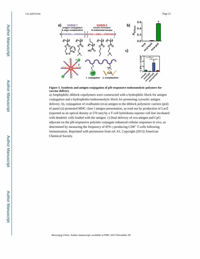
Figure 3. Synthesis and antigen conjugation of pH-responsive endosomolytic polymers for vaccine deliverya) Amphiphilic diblock copolymers were constructed with a hydrophilic block for antigen
conjugation and a hydrophobic/endosomolytic block for promoting cytosolic antigen
delivery. b), conjugation of ovalbumin (ova) antigen to the diblock polymeric carriers (pol)
of panel (a) promoted MHC class I antigen presentation, as read out by production of LacZ
(reported as an optical density at 570 nm) by a T-cell hybridoma reporter cell line incubated
with dendritic cells loaded with the antigen. c) Dual delivery of ova antigen and CpG
adjuvant on the pH-responsive polymer conjugate enhanced cellular responses in vivo, as
determined by measuring the frequency of IFN-γ-producing CD8+ T-cells following
immunization. Reprinted with permission from ref. 61, Copyright (2013) American
Chemical Society.
Liu and Irvine Page 21
Bioconjug Chem. Author manuscript; available in PMC 2015 December 29.
Author M
anuscriptA
uthor Manuscript
Author M
anuscriptA
uthor Manuscript
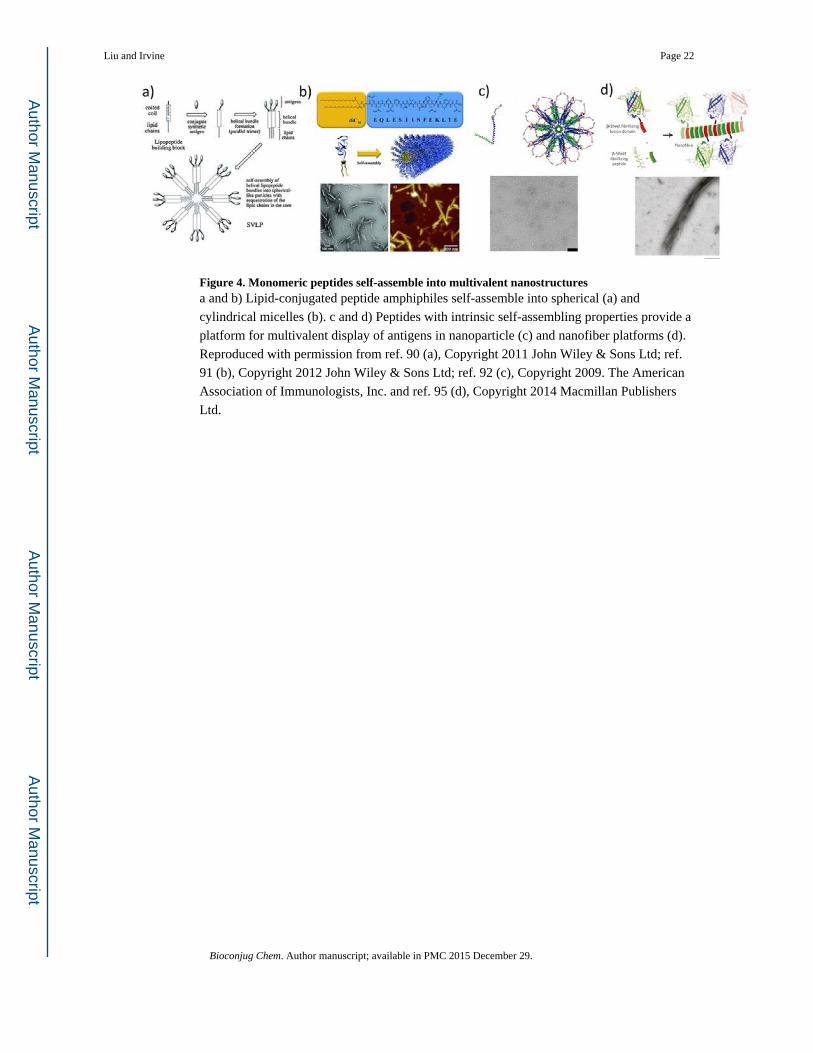
Figure 4. Monomeric peptides self-assemble into multivalent nanostructuresa and b) Lipid-conjugated peptide amphiphiles self-assemble into spherical (a) and
cylindrical micelles (b). c and d) Peptides with intrinsic self-assembling properties provide a
platform for multivalent display of antigens in nanoparticle (c) and nanofiber platforms (d).
Reproduced with permission from ref. 90 (a), Copyright 2011 John Wiley & Sons Ltd; ref.
91 (b), Copyright 2012 John Wiley & Sons Ltd; ref. 92 (c), Copyright 2009. The American
Association of Immunologists, Inc. and ref. 95 (d), Copyright 2014 Macmillan Publishers
Ltd.
Liu and Irvine Page 22
Bioconjug Chem. Author manuscript; available in PMC 2015 December 29.
Author M
anuscriptA
uthor Manuscript
Author M
anuscriptA
uthor Manuscript


![[GP1] Guiding Principles](https://img.dokumen.tips/doc/110x75/56816858550346895dde8352/gp1-guiding-principles.jpg)