Embed Size (px)
Citation preview

ESC Guidelines on the Diagnosis & Management of Pericardial Diseases - 2004ESC Guidelines on the Diagnosis & Management of Pericardial Diseases - 2004 1
GUIDELINES ON THE DIAGNOSIS AND MANAGEMENT
OF PERICARDIAL DISEASES
GUIDELINES ON THE DIAGNOSIS AND MANAGEMENT
OF PERICARDIAL DISEASESESC Task Force
Chairperson: Bernhard Maisch (Marburg, Germany),Petar M. Seferović (Belgrade, Serbia and Montenegro), Arsen D. Ristić, (Belgrade, Serbia and Montenegro), Raimund Erbel (Essen, Germany), Rainer Rienmüller (Graz, Austria), Yehuda Adler (Tel Hashomer, Israel), Witold Z. Tomkowski (Warsaw, Poland), Gaetano Thiene (Padua, Italy),
Magdi H. Yacoub (Harefield, UK)
Executive summary: The European Heart Journal 2004; 25: 587-610Full text guidelines: www.escardio.org
Pocket Guidelines: Available per request from the ESC
ESC Task ForceChairperson: Bernhard Maisch (Marburg, Germany),
Petar M. Seferović (Belgrade, Serbia and Montenegro), Arsen D. Ristić, (Belgrade, Serbia and Montenegro), Raimund Erbel (Essen, Germany), Rainer Rienmüller (Graz, Austria), Yehuda Adler (Tel Hashomer, Israel), Witold Z. Tomkowski (Warsaw, Poland), Gaetano Thiene (Padua, Italy),
Magdi H. Yacoub (Harefield, UK)
Executive summary: The European Heart Journal 2004; 25: 587-610Full text guidelines: www.escardio.org
Pocket Guidelines: Available per request from the ESC

ESC Guidelines on the Diagnosis & Management of Pericardial Diseases - 2004ESC Guidelines on the Diagnosis & Management of Pericardial Diseases - 2004 2
GUIDELINES ON THE DIAGNOSIS AND MANAGEMENT OF PERICARDIAL DISEASES
GUIDELINES ON THE DIAGNOSIS AND MANAGEMENT OF PERICARDIAL DISEASES
ESC Committee for Practice Guidelines (CPG)Chairperson: Silvia G. Priori, Italy
Maria Angeles Alonso Garcia, Spain; Jean-Jacques Blanc, France; Andrzej Budaj, Poland; Martin Cowie , UK; Veronica Dean, France; Jaap Deckers, The Netherlands; Enrique Fernandez Burgos, Spain;
John Lekakis, Greece; Bertil Lindahl, Sweden; Gianfranco Mazzotta, Italy; Keith McGregor, France; João Morais, Portugal; Ali Oto, Turkey;
Otto A. Smiseth, Norway
Document ReviewersCPG Review Coordinator: Gianfranco Mazzotta, Italy
Jean Acar, France; Eloisa Arbustini, Italy; Anton E. Becker, The Netherlands; Giacomo Chiaranda, Italy; Yonathan Hasin, Israel; Rolf Jenni, Switzerland;
Werner Klein, Austria; Irene Lang, Austria; Thomas F. Lüscher, Switzerland; Fausto J. Pinto, Portugal; Ralph Shabetai, USA; Maarten L. Simoons, The
Netherlands; Jordi Soler Soler, Spain; David H. Spodick, USA.

ESC Guidelines on the Diagnosis & Management of Pericardial Diseases - 2004ESC Guidelines on the Diagnosis & Management of Pericardial Diseases - 2004 3
Guidelines on the Diagnosis and Management of Pericardial Diseases
MAIN TOPICSGuidelines on the Diagnosis and Management of Pericardial Diseases
MAIN TOPICS
� Acute pericarditis � Pericardial effusion and
cardiac tamponade� Constrictive pericarditis� Viral pericarditis� Bacterial pericarditis� Pericarditis in renal failure� Autoreactive pericarditis
and pericardial involvement in systemic autoimmune diseases
� Acute pericarditis � Pericardial effusion and
cardiac tamponade� Constrictive pericarditis� Viral pericarditis� Bacterial pericarditis� Pericarditis in renal failure� Autoreactive pericarditis
and pericardial involvement in systemic autoimmune diseases
� The post-cardiac injury syndrome
� Postinfarction pericarditis� Traumatic pericardial
effusion� Haemopericardium in
aortic dissection � Neoplastic pericarditis� Pericardial diseases in
pregnancy� Drug- and toxin-related
pericardial disease
� The post-cardiac injury syndrome
� Postinfarction pericarditis� Traumatic pericardial
effusion� Haemopericardium in
aortic dissection � Neoplastic pericarditis� Pericardial diseases in
pregnancy� Drug- and toxin-related
pericardial disease

ESC Guidelines on the Diagnosis & Management of Pericardial Diseases - 2004ESC Guidelines on the Diagnosis & Management of Pericardial Diseases - 2004 4
� Level of evidence A: Multiple randomised clinical trials or meta-analyses.
� Level of evidence B: A single randomised trial or non-randomised studies.
� Level of evidence C: Consensus opinion of the experts.
� Level of evidence A: Multiple randomised clinical trials or meta-analyses.
� Level of evidence B: A single randomised trial or non-randomised studies.
� Level of evidence C: Consensus opinion of the experts.
Guidelines on the Diagnosis and Management of Pericardial Diseases
LEVEL OF EVIDENCEGuidelines on the Diagnosis and Management of Pericardial Diseases
LEVEL OF EVIDENCE

ESC Guidelines on the Diagnosis & Management of Pericardial Diseases - 2004ESC Guidelines on the Diagnosis & Management of Pericardial Diseases - 2004 5
� Class I: evidence and/or general agreement that a given procedure or treatment is useful and effective.
� Class II: conflicting evidence and/or a divergence of opinion about the usefulness/efficacy of a procedure or treatment.� Class IIa: Weight of evidence/opinion is in favour of
usefulness/efficacy. � Class IIb: Usefulness/efficacy is less well established by
evidence/opinion.
� Class III: evidence and/or general agreement that the procedure/treatment is not useful/effective and in some cases may be harmful.
Guidelines on the Diagnosis and Management of Pericardial Diseases
CLASSES OF RECOMMANDATIONSGuidelines on the Diagnosis and Management of Pericardial Diseases
CLASSES OF RECOMMANDATIONS

ESC Guidelines on the Diagnosis & Management of Pericardial Diseases - 2004ESC Guidelines on the Diagnosis & Management of Pericardial Diseases - 2004 6
ACUTE PERICARDITIS
ACUTE PERICARDITIS

ESC Guidelines on the Diagnosis & Management of Pericardial Diseases - 2004ESC Guidelines on the Diagnosis & Management of Pericardial Diseases - 2004 7
ACUTE PERICARDITISACUTE PERICARDITIS
ECHOCARDIOGRAPHYECHOCARDIOGRAPHY
Symptomatic managementHospitalisation and exercise restrictionPain management- Ibuprofen, 300-800 mg tid or qid - Colchicine, 0.5 mg bid
Symptomatic managementHospitalisation and exercise restrictionPain management- Ibuprofen, 300-800 mg tid or qid - Colchicine, 0.5 mg bid
PERICARDIOCENTESISPERICARDIAL DRAINAGE
(best with cardiac catheterisation)
PERICARDIOCENTESISPERICARDIAL DRAINAGE
(best with cardiac catheterisation)
SUBXIPHOID PERICARDIOTOMY
AND DRAINAGE
SUBXIPHOID SUBXIPHOID PERICARDIOTOMYPERICARDIOTOMY
AND DRAINAGEAND DRAINAGE
TAMPONADE orPE>20 mm in diastole
TAMPONADE orTAMPONADE orPE>20 mm in diastolePE>20 mm in diastole
NO TAMPONADEPE 10-20 mm in diastole
NO TAMPONADENO TAMPONADEPE 10PE 10--20 mm in diastole20 mm in diastole
Suspected purulent, TBCor neoplastic effusion
Suspected purulent, TBCor neoplastic effusion
NO TAMPONADEPE <10 mm in diastole
NO TAMPONADENO TAMPONADEPE <10 mm in diastolePE <10 mm in diastole
PERICARDIOSCOPYPERICARDIOSCOPY ANDANDPERICARDIAL/EPICARDIAL BIOPSYPERICARDIAL/EPICARDIAL BIOPSY
FOLLOW-UPECHOCARDIOGRAPHY
FOLLOW-UPECHOCARDIOGRAPHY
INTRAPERICARDIAL THERAPYINTRAPERICARDIAL THERAPY

ESC Guidelines on the Diagnosis & Management of Pericardial Diseases - 2004ESC Guidelines on the Diagnosis & Management of Pericardial Diseases - 2004 8
Guidelines on the Diagnosis and Management of Pericardial Diseases
ACUTE PERICARDITISDiagnostic pathway and sequence of performance
Guidelines on the Diagnosis and Management of Pericardial Diseases
ACUTE PERICARDITISDiagnostic pathway and sequence of performance
a.Typical lead involvement: I, II, aVL, aVF, and V3Typical lead involvement: I, II, aVL, aVF, and V3--V6. V6. b.Cardiac troponin is detectable in 32.2Cardiac troponin is detectable in 32.2--49%. An increase beyond 1.5 ng/ml is rare (7.649%. An increase beyond 1.5 ng/ml is rare (7.6--22%). 22%).
Ranging from normal to Ranging from normal to ��water bottlewater bottle�� heart shadow. heart shadow. Revealing additional pulmonary/mediastinal pathology.Revealing additional pulmonary/mediastinal pathology.
Chest x-ray
a) ESR, CRP, LDH, leukocytes (inflammation markers)a) ESR, CRP, LDH, leukocytes (inflammation markers)b) cTnI, CKb) cTnI, CK--MB (markers of myocardial lesion)MB (markers of myocardial lesion)b
Blood analyses
Effusion types BEffusion types B--D (Horowitz)D (Horowitz)Signs of tamponadeSigns of tamponade
Echocardiography
Stage IStage I:: anterior & inferior concave ST segment elevation. anterior & inferior concave ST segment elevation. PR segment deviations opposite to P polarity.PR segment deviations opposite to P polarity.Early stage II:Early stage II: ST junct. return to baseline, PR deviated.ST junct. return to baseline, PR deviated.Late stage II:Late stage II: T waves progressively flatten and invert T waves progressively flatten and invert Stage III:Stage III: generalised T wave inversions generalised T wave inversions Stage IV:Stage IV: ECG returns to prepericarditis state.ECG returns to prepericarditis state.
ECG a
Pericardial rub (monoPericardial rub (mono--, bi, bi--, or triphasic), or triphasic)AuscultationOBLIGATORY (class I, level of evidence B for all procedures):

ESC Guidelines on the Diagnosis & Management of Pericardial Diseases - 2004ESC Guidelines on the Diagnosis & Management of Pericardial Diseases - 2004 9
Guidelines on the Diagnosis and Management of Pericardial Diseases
ACUTE PERICARDITISDiagnostic pathway and sequence of performance
(level of evidence B for all procedures)
Guidelines on the Diagnosis and Management of Pericardial Diseases
ACUTE PERICARDITISDiagnostic pathway and sequence of performance
(level of evidence B for all procedures)
Establishing the specific aetiologyEstablishing the specific aetiologyPericardioscopy, pericardial biopsy
Effusions, periEffusions, peri--, and epicardium, and epicardiumMRI
Effusions, periEffusions, peri--, and epicardium, and epicardiumCT
OPTIONAL OR IF PREVIOUS TESTS INCONCLUSIVE (class IIa):
Pericardial fluid cytology, and cultures, PCRs and Pericardial fluid cytology, and cultures, PCRs and histochemistry for determination of infection or neoplasiahistochemistry for determination of infection or neoplasia
Pericardiocentesis and drainage
MANDATORY IN TAMPONADE (class I), OPTIONAL IN LARGE/RECURRENT EFFUSIONS OR IF PREVIOUS TESTS INCONCLUSIVE (class IIa) IN SMALL: EFFUSIONS (class IIb):

ESC Guidelines on the Diagnosis & Management of Pericardial Diseases - 2004ESC Guidelines on the Diagnosis & Management of Pericardial Diseases - 2004 10
Guidelines on the Diagnosis and Management of Pericardial Diseases
ACUTE PERICARDITISSymptomatic management
Guidelines on the Diagnosis and Management of Pericardial Diseases
ACUTE PERICARDITISSymptomatic management
� Exercise restriction
� Hospitalisation to determine the aetiology and observe for tamponade as well as the effect of treatment.
� Pain management
� Exercise restriction
� Hospitalisation to determine the aetiology and observe for tamponade as well as the effect of treatment.
� Pain management

ESC Guidelines on the Diagnosis & Management of Pericardial Diseases - 2004ESC Guidelines on the Diagnosis & Management of Pericardial Diseases - 2004 11
Guidelines on the Diagnosis and Management of Pericardial Diseases
ACUTE PERICARDITISPain management
Guidelines on the Diagnosis and Management of Pericardial Diseases
ACUTE PERICARDITISPain management
� NSAIDs are the mainstay (level of evidence B, class I).
� Ibuprofen is preferred (rare side-effects, favourable impact on the coronary flow, and the large dose range: 300-800 mg every 6-8 h)
� Aspirin 300-600 mg every 4-6 h� Indomethacin should be avoided in elderly patients
(flow reduction in the coronary arteries).
� Gastrointestinal protection must be provided.

ESC Guidelines on the Diagnosis & Management of Pericardial Diseases - 2004ESC Guidelines on the Diagnosis & Management of Pericardial Diseases - 2004 12
Guidelines on the Diagnosis and Management of Pericardial Diseases
ACUTE / RECURRENT PERICARDITISTreatment and prevention of recurrences
Guidelines on the Diagnosis and Management of Pericardial Diseases
ACUTE / RECURRENT PERICARDITISTreatment and prevention of recurrences
� Colchicine (0.5 mg bid) added to an NSAID or as monotherapy is well tolerated with fewer side effects than NSAIDs (level of evidence B, class IIa).
� Percutaneous balloon pericardiotomy In recurrences resistant to medical treatment (level of evidence B, class IIb).
� Colchicine (0.5 mg bid) added to an NSAID or as monotherapy is well tolerated with fewer side effects than NSAIDs (level of evidence B, class IIa).
� Percutaneous balloon pericardiotomy In recurrences resistant to medical treatment (level of evidence B, class IIb).

ESC Guidelines on the Diagnosis & Management of Pericardial Diseases - 2004ESC Guidelines on the Diagnosis & Management of Pericardial Diseases - 2004 13
Guidelines on the Diagnosis and Management of Pericardial Diseases
ACUTE / RECURRENT PERICARDITISTreatment and prevention of recurrences
Guidelines on the Diagnosis and Management of Pericardial Diseases
ACUTE / RECURRENT PERICARDITISTreatment and prevention of recurrences
� Corticosteroids � only in patients with poor general condition or in frequent
crises (level of evidence C, class IIa). � The recommended regimen is: prednisone 1-1.5 mg/kg, for
at least one month. � If patients do not respond adequately, azathioprine (75-100
mg/day) or cyclophosphamide can be added.
� Pericardiectomy - only in frequent and highly symptomatic recurrences resistant to medical treatment (level of evidence B, class IIa).

ESC Guidelines on the Diagnosis & Management of Pericardial Diseases - 2004ESC Guidelines on the Diagnosis & Management of Pericardial Diseases - 2004 14
CARDIAC TAMPONADE
CARDIAC TAMPONADE

ESC Guidelines on the Diagnosis & Management of Pericardial Diseases - 2004ESC Guidelines on the Diagnosis & Management of Pericardial Diseases - 2004 15
Guidelines on the Diagnosis and Management of Pericardial Diseases
CARDIAC TAMPONADEClinical presentation
Guidelines on the Diagnosis and Management of Pericardial Diseases
CARDIAC TAMPONADEClinical presentation
� Elevated systemic venous pressure� Jugular venous distension is less notable in hypovolemic
patients or in �surgical tamponade�.� An inspiratory increase or lack of fall of the pressure in the neck
veins (Kussmaul`s sign) indicates constriction� Hypotension
� Occasional pts are hypertensive especially if they have pre-existing hypertension
� Pulsus paradoxus� Absent in tamponade complicating atrial septal defect and
significant aortic regurgitation. � Tachycardia
� HR may be < 100 b/min in hypothyroidism and in uremic patients� Dyspnoea or tachypnoea with clear lungs

ESC Guidelines on the Diagnosis & Management of Pericardial Diseases - 2004ESC Guidelines on the Diagnosis & Management of Pericardial Diseases - 2004 16
Guidelines on the Diagnosis and Management of Pericardial Diseases
CARDIAC TAMPONADEPrecipitating factors
Guidelines on the Diagnosis and Management of Pericardial Diseases
CARDIAC TAMPONADEPrecipitating factors
� Drugs� Cyclosporine� Anticoagulants� Thrombolytics, etc.
� Injury� Recent cardiac surgery� Indwelling instrumentation� Blunt chest trauma
� Malignancies� Connective tissue disease� Renal failure� Septicaemia
� Drugs� Cyclosporine� Anticoagulants� Thrombolytics, etc.
� Injury� Recent cardiac surgery� Indwelling instrumentation� Blunt chest trauma
� Malignancies� Connective tissue disease� Renal failure� Septicaemia

ESC Guidelines on the Diagnosis & Management of Pericardial Diseases - 2004ESC Guidelines on the Diagnosis & Management of Pericardial Diseases - 2004 17
Guidelines on the Diagnosis and Management of Pericardial Diseases
CARDIAC TAMPONADEElectrocardiogram
Guidelines on the Diagnosis and Management of Pericardial Diseases
CARDIAC TAMPONADEElectrocardiogram
� Can be normal or � Non-specifically
changed (ST-T wave)� Electrical alternans
(QRS, rarely T)� Bradycardia (end-stage)� Electromechanical
dissociation (agonal phase)
� Can be normal or � Non-specifically
changed (ST-T wave)� Electrical alternans
(QRS, rarely T)� Bradycardia (end-stage)� Electromechanical
dissociation (agonal phase)

ESC Guidelines on the Diagnosis & Management of Pericardial Diseases - 2004ESC Guidelines on the Diagnosis & Management of Pericardial Diseases - 2004 18
Guidelines on the Diagnosis and Management of Pericardial Diseases
CARDIAC TAMPONADEChest X-ray
Guidelines on the Diagnosis and Management of Pericardial Diseases
CARDIAC TAMPONADEChest X-ray
! Enlarged cardiac silhouettewith clear lungs

ESC Guidelines on the Diagnosis & Management of Pericardial Diseases - 2004ESC Guidelines on the Diagnosis & Management of Pericardial Diseases - 2004 19
Guidelines on the Diagnosis and Management of Pericardial Diseases
CARDIAC TAMPONADEM-mode/2D echocardiogram
Guidelines on the Diagnosis and Management of Pericardial Diseases
CARDIAC TAMPONADEM-mode/2D echocardiogram
� Diastolic collapse of the anterior RV free wall� Can be absent in RV
hypertrophy or RV infarction.
� Diastolic collapse of the anterior RV free wall� Can be absent in RV
hypertrophy or RV infarction.
TsangTsang et al. et al. Herz 2000Herz 2000

ESC Guidelines on the Diagnosis & Management of Pericardial Diseases - 2004ESC Guidelines on the Diagnosis & Management of Pericardial Diseases - 2004 20
Guidelines on the Diagnosis and Management of Pericardial Diseases
CARDIAC TAMPONADEM-mode/2D echocardiogram
Guidelines on the Diagnosis and Management of Pericardial Diseases
CARDIAC TAMPONADEM-mode/2D echocardiogram
� RA collapse� LA and very rarely LV
collapse� Increased LV diastolic wall
thickness "pseudohypertrophy�
� VCI dilatation (no collapse in inspirium)
� �Swinging heart"
� RA collapse� LA and very rarely LV
collapse� Increased LV diastolic wall
thickness "pseudohypertrophy�
� VCI dilatation (no collapse in inspirium)
� �Swinging heart"Joffe et al. Joffe et al. Circulation 1996Circulation 1996

ESC Guidelines on the Diagnosis & Management of Pericardial Diseases - 2004ESC Guidelines on the Diagnosis & Management of Pericardial Diseases - 2004 21
Guidelines on the Diagnosis and Management of Pericardial Diseases
CARDIAC TAMPONADEDoppler echocardiography
Guidelines on the Diagnosis and Management of Pericardial Diseases
CARDIAC TAMPONADEDoppler echocardiography
� Tricuspid flow increases and mitral flow decreases during inspiration (reverse in expiration)
� Systolic and diastolic flows are reduced in systemic veins in expirium and reverse flow with atrial contraction is increased.
M-mode colour Doppler
� Large respiratory fluctuations in mitral/tricuspid flows
� Tricuspid flow increases and mitral flow decreases during inspiration (reverse in expiration)
� Systolic and diastolic flows are reduced in systemic veins in expirium and reverse flow with atrial contraction is increased.
MM--mode colour Dopplermode colour Doppler
� Large respiratory fluctuations in mitral/tricuspid flows
Oh et al. Mayo Clin ProcOh et al. Mayo Clin Proc 19931993

ESC Guidelines on the Diagnosis & Management of Pericardial Diseases - 2004ESC Guidelines on the Diagnosis & Management of Pericardial Diseases - 2004 22
Guidelines on the Diagnosis and Management of Pericardial Diseases
CARDIAC TAMPONADECardiac catheterisation
Guidelines on the Diagnosis and Management of Pericardial Diseases
CARDIAC TAMPONADECardiac catheterisation
� Confirmation and quantification of the haemodynamic compromise.
� Documenting haemodynamic improvement after pericardiocentesis.
� Detection of the coexisting haemodynamic abnormalities (LV failure, constriction, pulmonary hypertension).
� Detection of associated coronary artery disease or cardiomyopathy.
Murgo 1982Murgo 1982

ESC Guidelines on the Diagnosis & Management of Pericardial Diseases - 2004ESC Guidelines on the Diagnosis & Management of Pericardial Diseases - 2004 23
Guidelines on the Diagnosis and Management of Pericardial Diseases
CARDIAC TAMPONADECardiac catheterisation - haemodynamics
Guidelines on the Diagnosis and Management of Pericardial Diseases
CARDIAC TAMPONADECardiac catheterisation - haemodynamics
� Confirmation and quantification of the haemodynamic compromise:� RA pressure is elevated (preserved systolic x descent and
absent or diminished diastolic y descent).� Intrapericardial pressure is also elevated and virtually
identical to RA pressure (both pressures fall in inspiration).� RV mid-diastolic pressure elevated and equal to the RA and
pericardial pressures (no dip-and-plateau configuration).� PA diastolic pressure is slightly elevated. � LV systolic and aortic pressures may be normal or reduced.

ESC Guidelines on the Diagnosis & Management of Pericardial Diseases - 2004ESC Guidelines on the Diagnosis & Management of Pericardial Diseases - 2004 24
Guidelines on the Diagnosis and Management of Pericardial Diseases
CARDIAC TAMPONADEGuidelines on the Diagnosis and Management of Pericardial Diseases
CARDIAC TAMPONADE
RV/LV angiography� Atrial collapse� Small hyperactive
ventricular chambers.
Coronary angiography� Coronary compression
in diastole.
RV/LV angiography� Atrial collapse� Small hyperactive
ventricular chambers.
Coronary angiography� Coronary compression
in diastole.
ESC Guidelines on the Diagnosis & Management of Pericardial Diseases - 2004ESC Guidelines on the Diagnosis & Management of Pericardial Diseases - 2004 25
Guidelines on the Diagnosis and Management of Pericardial Diseases
CARDIAC TAMPONADEGuidelines on the Diagnosis and Management of Pericardial Diseases
CARDIAC TAMPONADE
Computertomography
� No visualisation of subepicardial fat along both ventricles, which show tube-like configuration and anteriorly drawn atrias
Computertomography
� No visualisation of subepicardial fat along both ventricles, which show tube-like configuration and anteriorly drawn atrias
ChilesChiles et al. et al. Radiographics 2001Radiographics 2001

ESC Guidelines on the Diagnosis & Management of Pericardial Diseases - 2004ESC Guidelines on the Diagnosis & Management of Pericardial Diseases - 2004 26
PERICARDIOCENTESISPERICARDIOCENTESIS

ESC Guidelines on the Diagnosis & Management of Pericardial Diseases - 2004ESC Guidelines on the Diagnosis & Management of Pericardial Diseases - 2004 27
Guidelines on the Diagnosis and Management of Pericardial Diseases
PERICARDIAL EFFUSION / TAMPONADEClasses of recommendations for pericardiocentesis
Guidelines on the Diagnosis and Management of Pericardial Diseases
PERICARDIAL EFFUSION / TAMPONADEClasses of recommendations for pericardiocentesis
Class I� Cardiac tamponade � Effusions >20 mm in echocardiography (diastole)� Suspected purulent or tuberculous pericardial effusion
Class IIa � Effusions 10-20 mm in echocardiography in diastole for
diagnostic purposes other than purulent pericarditis or tuberculosis (pericardial fluid and tissue analyses, pericardioscopy, and epicardial/pericardial biopsy)
� Suspected neoplastic pericardial effusion

ESC Guidelines on the Diagnosis & Management of Pericardial Diseases - 2004ESC Guidelines on the Diagnosis & Management of Pericardial Diseases - 2004 28
Class IIb � Effusions <10 mm in echocardiography in dialstole for
diagnostic purposes other than purulent, neoplastic or tuberculous pericarditis
Contraindications (Class III)� Aortic dissection� Relative contraindications include uncorrected coagulopathy,
anticoagulant therapy, thrombocytopenia <50000/mm3, small, posterior, and loculated effusions.
� If the diagnosis can be made otherwise or the effusions are small and resolving under anti-inflammatory treatment.
Guidelines on the Diagnosis and Management of Pericardial Diseases
PERICARDIAL EFFUSION / TAMPONADEClasses of recommendations for pericardiocentesis
Guidelines on the Diagnosis and Management of Pericardial Diseases
PERICARDIAL EFFUSION / TAMPONADEClasses of recommendations for pericardiocentesis

ESC Guidelines on the Diagnosis & Management of Pericardial Diseases - 2004ESC Guidelines on the Diagnosis & Management of Pericardial Diseases - 2004 29
Pericardiocentesis guided by fluoroscopy
� Current and reliable echocardiography before the procedure
� Cardiac catheterisation laboratory.� Local anaesthesia. � Subxiphoid approach (long needle
directed towards the left shoulder at a 30° angle to the skin).
Pericardiocentesis guided by fluoroscopy
� Current and reliable echocardiography before the procedure
� Cardiac catheterisation laboratory.� Local anaesthesia. � Subxiphoid approach (long needle
directed towards the left shoulder at a 30° angle to the skin).
Guidelines on the Diagnosis and Management of Pericardial Diseases
PERICARDIAL EFFUSION / TAMPONADEHow to perform pericardiocentesis
Guidelines on the Diagnosis and Management of Pericardial Diseases
PERICARDIAL EFFUSION / TAMPONADEHow to perform pericardiocentesis
B. Maisch and A.D. Ristic Heart 2003B. Maisch and A.D. Ristic Heart 2003

ESC Guidelines on the Diagnosis & Management of Pericardial Diseases - 2004ESC Guidelines on the Diagnosis & Management of Pericardial Diseases - 2004 30
Pericardiocentesis guided by echocardiography� Bedside, intensive care unit, cardiac
cath. lab., or operating theatre.� Echocardiography should identify the
shortest route where the pericardium can be entered intercostally (usually in the sixth or seventh rib space in the anterior axillary line).
� Intercostal arteries should be avoided.
Pericardiocentesis guided by echocardiography� Bedside, intensive care unit, cardiac
cath. lab., or operating theatre.� Echocardiography should identify the
shortest route where the pericardium can be entered intercostally (usually in the sixth or seventh rib space in the anterior axillary line).
� Intercostal arteries should be avoided.TsangTsang et alet al, JACC 1998, JACC 1998
Guidelines on the Diagnosis and Management of Pericardial Diseases
PERICARDIAL EFFUSION / TAMPONADEHow to perform pericardiocentesis
Guidelines on the Diagnosis and Management of Pericardial Diseases
PERICARDIAL EFFUSION / TAMPONADEHow to perform pericardiocentesis

ESC Guidelines on the Diagnosis & Management of Pericardial Diseases - 2004ESC Guidelines on the Diagnosis & Management of Pericardial Diseases - 2004 31
� Strict aseptic conditions, ECG, and blood pressure monitoring have to be provided.
� Direct ECG monitoring from the puncturing needle is not an adequate safeguard.
� Right-heart catheterisation can be performed simultaneously, allowing exclusion of constriction.
� Strict aseptic conditions, ECG, and blood pressure monitoring have to be provided.
� Direct ECG monitoring from the puncturing needle is not an adequate safeguard.
� Right-heart catheterisation can be performed simultaneously, allowing exclusion of constriction.
Guidelines on the Diagnosis and Management of Pericardial Diseases
PERICARDIAL EFFUSION / TAMPONADEHow to perform pericardiocentesis
Guidelines on the Diagnosis and Management of Pericardial Diseases
PERICARDIAL EFFUSION / TAMPONADEHow to perform pericardiocentesis

ESC Guidelines on the Diagnosis & Management of Pericardial Diseases - 2004ESC Guidelines on the Diagnosis & Management of Pericardial Diseases - 2004 32
� The needle approaches pericardium slowly.
� Steady manual aspiration is essential.
� Stop the needle as soon as the effusion is aspirated.
� Exchange for soft J-tip guidewire and after dilatation for a multi-holed pigtail catheter.
� The needle approaches pericardium slowly.
� Steady manual aspiration is essential.
� Stop the needle as soon as the effusion is aspirated.
� Exchange for soft J-tip guidewire and after dilatation for a multi-holed pigtail catheter.
Guidelines on the Diagnosis and Management of Pericardial Diseases
PERICARDIAL EFFUSION / TAMPONADEHow to perform pericardiocentesis
Guidelines on the Diagnosis and Management of Pericardial Diseases
PERICARDIAL EFFUSION / TAMPONADEHow to perform pericardiocentesis
B. Maisch et al. Am J B. Maisch et al. Am J Cardiol Cardiol 20012001

ESC Guidelines on the Diagnosis & Management of Pericardial Diseases - 2004ESC Guidelines on the Diagnosis & Management of Pericardial Diseases - 2004 33
� Drain the fluid in <1 l steps to avoid the acute right-ventricular dilatation.
� Perform prolonged pericardial drainage (several days) until <25 ml/day.
� Drain the fluid in <1 l steps to avoid the acute right-ventricular dilatation.
� Perform prolonged pericardial drainage (several days) until <25 ml/day.
Guidelines on the Diagnosis and Management of Pericardial Diseases
PERICARDIAL EFFUSION / TAMPONADEHow to perform pericardiocentesis
Guidelines on the Diagnosis and Management of Pericardial Diseases
PERICARDIAL EFFUSION / TAMPONADEHow to perform pericardiocentesis

ESC Guidelines on the Diagnosis & Management of Pericardial Diseases - 2004ESC Guidelines on the Diagnosis & Management of Pericardial Diseases - 2004 34
Guidelines on the Diagnosis and Management of Pericardial Diseases
PERICARDIAL EFFUSION ANALYSESShould be ordered according to the clinical presentation
CLASS I
Guidelines on the Diagnosis and Management of Pericardial Diseases
PERICARDIAL EFFUSION ANALYSESShould be ordered according to the clinical presentation
CLASS I� Suspected malignant effusion: CYTOLOGY.
� Suspected tuberculous effusion: ACID-FAST BACILLI STAINING, mycobacterium CULTURE (preferably with radiometric growth detection e.g., BACTEC-460), adenosine deaminase, IFN-gamma, pericardial lysozyme, PCR analyses
� Suspected bacterial infection: at least three cultures of pericardial fluid for aerobes and anaerobes as well as three blood cultures. Positive cultures should be followed by sensitivity tests for antibiotics.
� Suspected malignant effusion: CYTOLOGY.
� Suspected tuberculous effusion: ACID-FAST BACILLI STAINING, mycobacterium CULTURE (preferably with radiometric growth detection e.g., BACTEC-460), adenosine deaminase, IFN-gamma, pericardial lysozyme, PCR analyses
� Suspected bacterial infection: at least three cultures of pericardial fluid for aerobes and anaerobes as well as three blood cultures. Positive cultures should be followed by sensitivity tests for antibiotics.

ESC Guidelines on the Diagnosis & Management of Pericardial Diseases - 2004ESC Guidelines on the Diagnosis & Management of Pericardial Diseases - 2004 35
Guidelines on the Diagnosis and Management of Pericardial Diseases
PERICARDIAL EFFUSION ANALYSESShould be ordered according to the clinical presentation
CLASS IIa
Guidelines on the Diagnosis and Management of Pericardial Diseases
PERICARDIAL EFFUSION ANALYSESShould be ordered according to the clinical presentation
CLASS IIa
� Viral vs. autoreactive pericarditis: PCR analyses for cardiotropic viruses.
� Suspected neoplastic pericarditis: Tumour markers (CEA, AFP, CA 125, CA 72-4, CA 15-3, CA 19-9, CD-30, CD-25...).
� Benign reactive mesothelial cells vs. adenocarcinoma:Combination of epithelial membrane antigen, CEA, and vimentin immunocytochemical staining.

ESC Guidelines on the Diagnosis & Management of Pericardial Diseases - 2004ESC Guidelines on the Diagnosis & Management of Pericardial Diseases - 2004 36
Guidelines on the Diagnosis and Management of Pericardial Diseases
PERICARDIAL EFFUSION ANALYSESShould be ordered according to the clinical presentation
CLASS IIb
Guidelines on the Diagnosis and Management of Pericardial Diseases
PERICARDIAL EFFUSION ANALYSESShould be ordered according to the clinical presentation
CLASS IIb
� Exudate vs. transudate:� Pericardial fluid specific gravity (>1015)� Protein level (>3.0 g/dl; fluid/serum ratio >0.5)� LDH (>200mg/dL; serum/fluid >0.6), and � Glucose (exudates vs. transudates = 77.9±41.9
vs. 96.1±50.7 mg/dl)
� Exudate vs. transudate:� Pericardial fluid specific gravity (>1015)� Protein level (>3.0 g/dl; fluid/serum ratio >0.5)� LDH (>200mg/dL; serum/fluid >0.6), and � Glucose (exudates vs. transudates = 77.9±41.9
vs. 96.1±50.7 mg/dl)

ESC Guidelines on the Diagnosis & Management of Pericardial Diseases - 2004ESC Guidelines on the Diagnosis & Management of Pericardial Diseases - 2004 37
CONSTRICTIVE PERICARDITISCONSTRICTIVE PERICARDITIS

ESC Guidelines on the Diagnosis & Management of Pericardial Diseases - 2004ESC Guidelines on the Diagnosis & Management of Pericardial Diseases - 2004 38
Clinical presentationClinical presentation
� Severe chronic systemic venous congestion
� Jugular venous distension� Hypotension� Low pulse pressure� Abdominal distension� Oedema� Muscle wasting
� Severe chronic systemic venous congestion
� Jugular venous distension� Hypotension� Low pulse pressure� Abdominal distension� Oedema� Muscle wasting
Guidelines on the Diagnosis and Management of Pericardial Diseases
CONSTRICTIVE PERICARDITISGuidelines on the Diagnosis and Management of Pericardial Diseases
CONSTRICTIVE PERICARDITIS
Courtesy of P. Cocco and G. Thiene

ESC Guidelines on the Diagnosis & Management of Pericardial Diseases - 2004ESC Guidelines on the Diagnosis & Management of Pericardial Diseases - 2004 39
� Not always equal to constrictive physiology
� May also be absent in proven constriction (18% of 143 surgically proven cases, Talreja et al. Circulation 2003).
� Not always equal to constrictive physiology
� May also be absent in proven constriction (18% of 143 surgically proven cases, Talreja et al. Circulation 2003).
Guidelines on the Diagnosis and Management of Pericardial Diseases
CONSTRICTIVE PERICARDITISThickening of the pericardium
Guidelines on the Diagnosis and Management of Pericardial Diseases
CONSTRICTIVE PERICARDITISThickening of the pericardium
7 mm7 mm
1.5 mm1.5 mm
Talreja et al. Circulation 2003Talreja et al. Circulation 2003

ESC Guidelines on the Diagnosis & Management of Pericardial Diseases - 2004ESC Guidelines on the Diagnosis & Management of Pericardial Diseases - 2004 40
Guidelines on the Diagnosis and Management of Pericardial Diseases
CONSTRICTIVE PERICARDITISTransient forms
Guidelines on the Diagnosis and Management of Pericardial Diseases
CONSTRICTIVE PERICARDITISTransient forms
Haley Haley et alet al. J Am Coll Cardiol 2004. J Am Coll Cardiol 2004

ESC Guidelines on the Diagnosis & Management of Pericardial Diseases - 2004ESC Guidelines on the Diagnosis & Management of Pericardial Diseases - 2004 41
ElectrocardiogramElectrocardiogram
Can be normal or:� Low QRS voltage� Generalized T-wave
inversion/flattening� LA abnormalities� Atrial fibrillation� AV block� Intraventricular
conduction defects� Pseudoinfarction
pattern (rarely)
Can be normal or:� Low QRS voltage� Generalized T-wave
inversion/flattening� LA abnormalities� Atrial fibrillation� AV block� Intraventricular
conduction defects� Pseudoinfarction
pattern (rarely)
Guidelines on the Diagnosis and Management of Pericardial Diseases
CONSTRICTIVE PERICARDITISGuidelines on the Diagnosis and Management of Pericardial Diseases
CONSTRICTIVE PERICARDITIS

ESC Guidelines on the Diagnosis & Management of Pericardial Diseases - 2004ESC Guidelines on the Diagnosis & Management of Pericardial Diseases - 2004 42
� Pericardial calcifications
� Pleural effusions
� Pericardial calcifications
� Pleural effusions
Guidelines on the Diagnosis and Management of Pericardial Diseases
CONSTRICTIVE PERICARDITISChest X-ray
Guidelines on the Diagnosis and Management of Pericardial Diseases
CONSTRICTIVE PERICARDITISChest X-ray
Ling et al. Ann Intern Med 2000Ling et al. Ann Intern Med 2000

ESC Guidelines on the Diagnosis & Management of Pericardial Diseases - 2004ESC Guidelines on the Diagnosis & Management of Pericardial Diseases - 2004 43
� Pericardial thickening and calcifications
� Indirect signs of constriction
� Pericardial thickening and calcifications
� Indirect signs of constriction
Guidelines on the Diagnosis and Management of Pericardial Diseases
CONSTRICTIVE PERICARDITISM-mode/2D echocardiogram
Guidelines on the Diagnosis and Management of Pericardial Diseases
CONSTRICTIVE PERICARDITISM-mode/2D echocardiogram
Oxantenko et al, Oxantenko et al, J Clin Gastroenterol 2002J Clin Gastroenterol 2002

ESC Guidelines on the Diagnosis & Management of Pericardial Diseases - 2004ESC Guidelines on the Diagnosis & Management of Pericardial Diseases - 2004 44
� RA & LA enlargement with normal ventricles, and systolic function.
� Early pathological outward and inward movement of the interventricular septum (�dip-plateau phenomenon�)
� Flattering waves at the LV posterior wall� LV diameter is not increasing after the early
rapid filling phase. � VCI and the hepatic veins are dilated with
restricted respiratory fluctuations.
Guidelines on the Diagnosis and Management of Pericardial Diseases
CONSTRICTIVE PERICARDITISIndirect echo signs of constriction
Guidelines on the Diagnosis and Management of Pericardial Diseases
CONSTRICTIVE PERICARDITISIndirect echo signs of constriction

ESC Guidelines on the Diagnosis & Management of Pericardial Diseases - 2004ESC Guidelines on the Diagnosis & Management of Pericardial Diseases - 2004 45
� Restricted filling of both ventricles with respiratory variation(>25% over the AV-valves).� In mixed constriction-restriction and increased atrial pressures
respiratory changes are <25%.� In atrial fibrillation flow velocity pattern is inconclusive, but hepatic
diastolic vein flow reversal in expirium is observed.� Provocation test with head-up tilting or sitting position with
decrease of preload may unmask the constrictive pericarditis.
Guidelines on the Diagnosis and Management of Pericardial Diseases
CONSTRICTIVE PERICARDITISDoppler echocardiography
Guidelines on the Diagnosis and Management of Pericardial Diseases
CONSTRICTIVE PERICARDITISDoppler echocardiography
MITRAL INFLOW DOPPLER, MITRAL INFLOW DOPPLER, BOONYARATAVEJ ET AL. JACC BOONYARATAVEJ ET AL. JACC 19981998

ESC Guidelines on the Diagnosis & Management of Pericardial Diseases - 2004ESC Guidelines on the Diagnosis & Management of Pericardial Diseases - 2004 46
� �Dip and plateau� or �square route� sign in the RV and/or LV pressure curve.
� Equalisation of LV/RV end-diastolic pressures.
� In occult constriction rapid infusion of 1-2 l of normal saline may reveal the diagnosis.
� �Dip and plateau� or �square route� sign in the RV and/or LV pressure curve.
� Equalisation of LV/RV end-diastolic pressures.
� In occult constriction rapid infusion of 1-2 l of normal saline may reveal the diagnosis.
Guidelines on the Diagnosis and Management of Pericardial Diseases
CONSTRICTIVE PERICARDITISCardiac catheterisation
Guidelines on the Diagnosis and Management of Pericardial Diseases
CONSTRICTIVE PERICARDITISCardiac catheterisation
Myers and Spodick. Am Heart J 1999Myers and Spodick. Am Heart J 1999

ESC Guidelines on the Diagnosis & Management of Pericardial Diseases - 2004ESC Guidelines on the Diagnosis & Management of Pericardial Diseases - 2004 47
RV/LV angiography� The reduction of RV & LV size and increase
of RA & LA size� Dip-plateau - rapid early filling with stop of
further enlargement in diastole
Coronary angiography� Indicated in all patients over 35 years and in
pts with a history of mediastinal irradiation, regardless of the age.
RV/LV angiography� The reduction of RV & LV size and increase
of RA & LA size� Dip-plateau - rapid early filling with stop of
further enlargement in diastole
Coronary angiography� Indicated in all patients over 35 years and in
pts with a history of mediastinal irradiation, regardless of the age.
Guidelines on the Diagnosis and Management of Pericardial Diseases
CONSTRICTIVE PERICARDITISGuidelines on the Diagnosis and Management of Pericardial Diseases
CONSTRICTIVE PERICARDITIS

ESC Guidelines on the Diagnosis & Management of Pericardial Diseases - 2004ESC Guidelines on the Diagnosis & Management of Pericardial Diseases - 2004 48
� Thickened and/or calcified pericardium
� Tube-like configuration of one or both ventricles
� Enlargement of one or both atria
� Narrowing of one or both atrio-ventricular grooves
� Congestion of the caval veins
� Thickened and/or calcified pericardium
� Tube-like configuration of one or both ventricles
� Enlargement of one or both atria
� Narrowing of one or both atrio-ventricular grooves
� Congestion of the caval veins
Guidelines on the Diagnosis and Management of Pericardial Diseases
CONSTRICTIVE PERICARDITISCT / MRI
Guidelines on the Diagnosis and Management of Pericardial Diseases
CONSTRICTIVE PERICARDITISCT / MRI
Cine TruFISP
Courtesy of Courtesy of R. Maksimović and T. DillR. Maksimović and T. Dill

ESC Guidelines on the Diagnosis & Management of Pericardial Diseases - 2004ESC Guidelines on the Diagnosis & Management of Pericardial Diseases - 2004 49
Guidelines on the Diagnosis and Management of Pericardial DiseasesCONSTRICTIVE PERICARDITIS VS. RESTRICTIVE CARDIOMYOPATHY
Guidelines on the Diagnosis and Management of Pericardial DiseasesCONSTRICTIVE PERICARDITIS VS. RESTRICTIVE CARDIOMYOPATHY
METHOD RESTRICTIVE CARDIOMYOPATHY
CONSTRICTIVE PERICARDITIS
Physical findings
Kussmaul�s sign ±, apical impulse +++
Kussmaul�s sign +, apical impulse -
S3 (advanced), S4 (early disease), regurgitant murmurs ++
pericardial knock+, regurgitant murmurs -
ECG Low voltage, pseudoinfarction, left-axis deviation, AF, conduction disturbances
Low voltage (<50%)
Chest radiography
No calcifications Calcifications may be present (low diagnostic accuracy)

ESC Guidelines on the Diagnosis & Management of Pericardial Diseases - 2004ESC Guidelines on the Diagnosis & Management of Pericardial Diseases - 2004 50
Guidelines on the Diagnosis and Management of Pericardial DiseasesCONSTRICTIVE PERICARDITIS VS. RESTRICTIVE CARDIOMYOPATHY
Guidelines on the Diagnosis and Management of Pericardial DiseasesCONSTRICTIVE PERICARDITIS VS. RESTRICTIVE CARDIOMYOPATHY
METHOD RESTRICTIVE CARDIOMYOPATHY
CONSTRICTIVE PERICARDITIS
2D-Echocardio-graphy
Small LV cavity with large atria Increased wall thickness sometimes present (especially thickened interatrial septum in amyloidosis) Thickened valves and granular sparkling (amyloidosis)
Normal wall thickness Pericardial thickening, prominent early diastolic filling with abrupt displacement of IVS
Tissue Doppler Echocardio-graphy
Peak early velocity of longitudinal expansion (peak Ea) of ≥8.0 cm/s (89% sensitivity and 100% specificity)
Negative

ESC Guidelines on the Diagnosis & Management of Pericardial Diseases - 2004ESC Guidelines on the Diagnosis & Management of Pericardial Diseases - 2004 51
Guidelines on the Diagnosis and Management of Pericardial DiseasesCONSTRICTIVE PERICARDITIS VS. RESTRICTIVE CARDIOMYOPATHY
Guidelines on the Diagnosis and Management of Pericardial DiseasesCONSTRICTIVE PERICARDITIS VS. RESTRICTIVE CARDIOMYOPATHY
DOPPLER STUDIES
RESTRICTIVE CARDIOMYOPATHY
CONSTRICTIVE PERICARDITIS
Mitral inflow No respiration variation of mitral inflow E wave velocity, IVRT E/A ratio >2, short DT, diastolic regurgitation
INSPIRATION: decreased inflow E wave velocity, prolonged IVRT EXPIRATION: opposite changes, short DT, diastolic regurgitation
Pulmonary vein
Blunted S/D ratio (0.5), prominent and prolonged AR No respiration variation, D wave
S/D ratio = 1, INSPIRATION: decreased PV S and D waves EXPIRATION: opposite changes
Tricuspid inflow
Mild respiration variation of tricuspid inflow E wave velocity, E/A ratio >2, TR peak velocity, no significant respiration change
INSPIRATION: increased tricuspid inflow E wave velocity, increased TR peak velocity, EXPIRATION: opposite

ESC Guidelines on the Diagnosis & Management of Pericardial Diseases - 2004ESC Guidelines on the Diagnosis & Management of Pericardial Diseases - 2004 52
Guidelines on the Diagnosis and Management of Pericardial DiseasesCONSTRICTIVE PERICARDITIS VS. RESTRICTIVE CARDIOMYOPATHY
Guidelines on the Diagnosis and Management of Pericardial DiseasesCONSTRICTIVE PERICARDITIS VS. RESTRICTIVE CARDIOMYOPATHY
DOPPLER STUDIES RESTRICTIVE CARDIOMYOPATHY
CONSTRICTIVE PERICARDITIS
Hepatic veins Blunted S/D ratio, increased inspiratory reversals
INSPIRATION: minimally increased HV, S and D
EXPIRATION: opposite changes
Inferior vena cava Plethoric Plethoric
Colour M-mode Slow flow propagation Rapid flow propagation (>100 cm/s)
Mitral annular motion Low-velocity early filling (<8 cm/s)
High-velocity early filling (>8 cm/s)
ESC Guidelines on the Diagnosis & Management of Pericardial Diseases - 2004ESC Guidelines on the Diagnosis & Management of Pericardial Diseases - 2004 53
Guidelines on the Diagnosis and Management of Pericardial DiseasesCONSTRICTIVE PERICARDITIS VS. RESTRICTIVE CARDIOMYOPATHY
Guidelines on the Diagnosis and Management of Pericardial DiseasesCONSTRICTIVE PERICARDITIS VS. RESTRICTIVE CARDIOMYOPATHY
METHOD RESTRICTIVE CARDIOMYOPATHY
CONSTRICTIVE PERICARDITIS
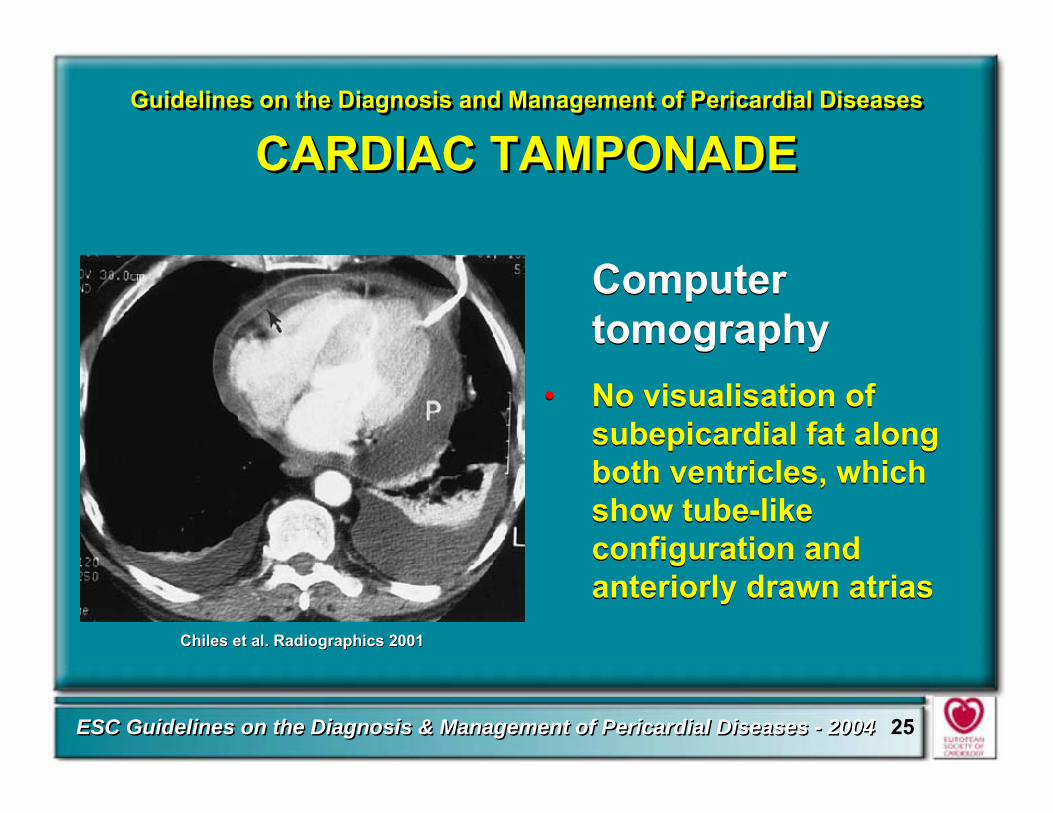
Cardiac catheterisation
Dip and plateau LVEDP often >5 mmHg greater than RVEDP, but may be identical, RV systolic pressure >50 mmHg RVEDP<1/3 RVSP
Dip and plateau, RVEDP and LVEDP usually equal, INSPIRATION: Increase in RV systolic pressure Decrease in LV systolic pressure, with EXPIRATION, opposite changes
EMB May reveal specific cause of restrictive cardiomyopathy
May be normal or show nonspecific hypertrophy or fibrosis
CT/MRI Pericardium usually normal Pericardium thickened or calcified.

ESC Guidelines on the Diagnosis & Management of Pericardial Diseases - 2004ESC Guidelines on the Diagnosis & Management of Pericardial Diseases - 2004 54
PERCUTANEOUS BALLOON
PERICARDIOTOMY
PERCUTANEOUS BALLOON
PERICARDIOTOMY
PERICARDIECTOMYPERICARDIECTOMY
ECHOCARDIOGRAPHYECHOCARDIOGRAPHYIf symptomaticfor >2 years
If symptomaticIf symptomaticfor >2 yearsfor >2 years CARDIAC CATHETERIZATIONCARDIAC CATHETERIZATION
RECURRENT PERICARDITISRECURRENT PERICARDITIS
CONSTRICTIVE PERICARDITIS
CONSTRICTIVE PERICARDITIS
CHRONIC PERICARDIAL EFFUSION
CHRONIC PERICARDIAL EFFUSION
EFFUSIVE-CONSTRICTIVEPERICARDITIS
EFFUSIVE-CONSTRICTIVEPERICARDITIS
Symptomatic managementHospitalisation and exercise restrictionPain management- Ibuprofen, 300-800 mg tid or qid - Colchicine, 0.5 mg bid- Prednisone 1-1.5 mg qd
Symptomatic managementHospitalisation and exercise restrictionPain management- Ibuprofen, 300-800 mg tid or qid - Colchicine, 0.5 mg bid- Prednisone 1-1.5 mg qd
CLINICAL SUSPICION FOR:CLINICAL SUSPICION FOR:CLINICAL SUSPICION FOR:
Congestive heart failure therapy
Congestive heart failure therapy

ESC Guidelines on the Diagnosis & Management of Pericardial Diseases - 2004ESC Guidelines on the Diagnosis & Management of Pericardial Diseases - 2004 55
Guidelines on the Diagnosis and Management of Pericardial Diseases
CONSTRICTIVE PERICARDITISPericardiectomy
Guidelines on the Diagnosis and Management of Pericardial Diseases
CONSTRICTIVE PERICARDITISPericardiectomy
� The only treatment for permanent constriction.
� Antero-lateral thoracotomy� Median sternotomy (faster access to the aorta and
right atrium for extracorporeal circulation). � Primary installation of cardiopulmonary
bypass is not recommended (diffuse bleeding following systemic heparinisation).
� Areas of strong calcification or dense scaring may be left as islands to avoid major bleeding.

ESC Guidelines on the Diagnosis & Management of Pericardial Diseases - 2004ESC Guidelines on the Diagnosis & Management of Pericardial Diseases - 2004 56
Guidelines on the Diagnosis and Management of Pericardial Diseases
PERICARDIECTOMY FOR CONSTRICTIVE PERICARDITIS
Guidelines on the Diagnosis and Management of Pericardial Diseases
PERICARDIECTOMY FOR CONSTRICTIVE PERICARDITIS
� Acute perioperative cardiac insufficiency (should be treated by fluid substitution and catecholamines, high doses of digitalis, and intraaortic balloon pump in most severe cases).
� Ventricular wall rupture.� Mortality (in properly selected cases 6-12%, but >40% in
unselected patients).
� If indication for surgery was established early, long-term survival after pericardiectomy corresponds to that of the general population
Major complicationsMajor complications
Long term resultsLong term results

ESC Guidelines on the Diagnosis & Management of Pericardial Diseases - 2004ESC Guidelines on the Diagnosis & Management of Pericardial Diseases - 2004 57
Guidelines on the Diagnosis and Management of Pericardial Diseases
PERICARDIECTOMY FOR VARIOUS PATHOANATOMICAL FORMS OF CONSTRICTION
Guidelines on the Diagnosis and Management of Pericardial Diseases
PERICARDIECTOMY FOR VARIOUS PATHOANATOMICAL FORMS OF CONSTRICTION
Rienmüller et al. J Thorac Imaging 1993
Annular form Left-sided form Right sided form
Global form Global form with Global form with
MYOCARDIAL ATROPHYMYOCARDIAL ATROPHYGlobal form withGlobal form with
MYOCARDIAL FIBROSISMYOCARDIAL FIBROSIS
Very high mortality in Very high mortality in pericardiectomypericardiectomyRienmüller et al. J Thorac Imaging 1993
Annular form Left-sided form Right sided formAnnular form Left-sided form Right sided form
Global form Global form with Global form with
MYOCARDIAL ATROPHYMYOCARDIAL ATROPHYGlobal form withGlobal form with
MYOCARDIAL FIBROSISMYOCARDIAL FIBROSIS
Very high mortality in Very high mortality in pericardiectomypericardiectomy
� Exclusion of patients with extensive myocardial fibrosis and/or atrophy significantly reduces the mortality rate
� Exclusion of patients with extensive myocardial fibrosis and/or atrophy significantly reduces the mortality rate

ESC Guidelines on the Diagnosis & Management of Pericardial Diseases - 2004ESC Guidelines on the Diagnosis & Management of Pericardial Diseases - 2004 58
VIRALPERICARDITIS
VIRALPERICARDITIS

ESC Guidelines on the Diagnosis & Management of Pericardial Diseases - 2004ESC Guidelines on the Diagnosis & Management of Pericardial Diseases - 2004 59
� Not possible without the evaluation of pericardial effusion and/or pericardial/epicardial tissue, preferably by PCR or in-situ hybridisation (level of evidence B, class IIa).
� A four-fold rise in serum antibody levels (two samples within 3-4 weeks) is suggestive but not diagnostic for viral pericarditis (level of evidence B, class IIb).
ADV PCR
ADV SOUTHERN BLOT
ADV PCR
ADV SOUTHERN BLOT
ADV PCR
ADV SOUTHERN BLOT
Guidelines on the Diagnosis and Management of Pericardial Diseases
VIRAL PERICARDITISDiagnosis
Guidelines on the Diagnosis and Management of Pericardial Diseases
VIRAL PERICARDITISDiagnosis
Courtesy of S. Pankuweit

ESC Guidelines on the Diagnosis & Management of Pericardial Diseases - 2004ESC Guidelines on the Diagnosis & Management of Pericardial Diseases - 2004 60
� In most cases the disease is self-limiting and no specific treatment is necessary.
� Symptomatic treatment for chest pain, and eventual rhythm disorders and congestive heart failure is indicated.
� In most cases the disease is self-limiting and no specific treatment is necessary.
� Symptomatic treatment for chest pain, and eventual rhythm disorders and congestive heart failure is indicated.CMV in situCMV in situ
Guidelines on the Diagnosis and Management of Pericardial Diseases
VIRAL PERICARDITISManagement
Guidelines on the Diagnosis and Management of Pericardial Diseases
VIRAL PERICARDITISManagement
B. Maisch et al. B. Maisch et al. Clin Cardiol Clin Cardiol 19991999

ESC Guidelines on the Diagnosis & Management of Pericardial Diseases - 2004ESC Guidelines on the Diagnosis & Management of Pericardial Diseases - 2004 61
� In patients with chronic or recurrent symptomatic pericardial effusion and confirmed viral infection the following specific treatment is under investigation:
� CMV pericarditis: hyperimmunoglobulin � one daily 4ml/kg on day 0, 4, and 8; 2 ml/kg on day 12 and 16;
� Coxsackie B pericarditis: Interferon alpha or beta 2,5 million. IU/m² surface area s.c. 3 x per week;
� Adenovirus and parvovirus B19 pericarditis: immunoglobulin treatment: 10 g intravenously at day 1 and 3 for 6-8 hours.
Guidelines on the Diagnosis and Management of Pericardial Diseases
VIRAL PERICARDITISManagement
Guidelines on the Diagnosis and Management of Pericardial Diseases
VIRAL PERICARDITISManagement

ESC Guidelines on the Diagnosis & Management of Pericardial Diseases - 2004ESC Guidelines on the Diagnosis & Management of Pericardial Diseases - 2004 62
Guidelines on the Diagnosis and Management of Pericardial Diseases
PERICARDITIS IN AIDSManagement
Guidelines on the Diagnosis and Management of Pericardial Diseases
PERICARDITIS IN AIDSManagement
� Symptomatic treatment
� Pericardiocentesis In large effusions/tamponade
� Standard (prolonged) anti-tuberculous regimens for TBC pericarditis in AIDS
� Use of rifampicin is precluded (if pts are treated with proteaseinhibitors or non-nucleoside reverse transcriptase inhibitors).
� Corticoid therapy as an adjunct to tuberculostatic treatment is allowed (level of evidence A, class I).

ESC Guidelines on the Diagnosis & Management of Pericardial Diseases - 2004ESC Guidelines on the Diagnosis & Management of Pericardial Diseases - 2004 63
BACTERIAL PERICARDITIS
BACTERIAL PERICARDITIS

ESC Guidelines on the Diagnosis & Management of Pericardial Diseases - 2004ESC Guidelines on the Diagnosis & Management of Pericardial Diseases - 2004 64
� Pericardiocentesis must be promptly performed.
� Pericardial fluid should undergo Gram, acid-fast, and fungal staining, followed by cultures for aerobes, anaerobes, and M. tuberculosis (preferably with radiometric growth detection).
� Drug sensitivity testing is essential for treatment selection.
� Pericardiocentesis must be promptly performed.
� Pericardial fluid should undergo Gram, acid-fast, and fungal staining, followed by cultures for aerobes, anaerobes, and M. tuberculosis (preferably with radiometric growth detection).
� Drug sensitivity testing is essential for treatment selection.
Guidelines on the Diagnosis and Management of Pericardial Diseases
BACTERIAL PERICARDITISDiagnosis
Guidelines on the Diagnosis and Management of Pericardial Diseases
BACTERIAL PERICARDITISDiagnosis

ESC Guidelines on the Diagnosis & Management of Pericardial Diseases - 2004ESC Guidelines on the Diagnosis & Management of Pericardial Diseases - 2004 65
� PCR analyses� Adenosine deaminase
>40 IU/L� Interferon-gamma
>200 pg/L� Pericardial lysozyme
>6.5 microg/dL
� PCR analyses� Adenosine deaminase
>40 IU/L� Interferon-gamma
>200 pg/L� Pericardial lysozyme
>6.5 microg/dL
Cost-effective only if the pre-test probability is high (populations with high incidence of tuberculosis).
Guidelines on the Diagnosis and Management of Pericardial Diseases
TUBERCULOUS PERICARDITISDiagnosis
Guidelines on the Diagnosis and Management of Pericardial Diseases
TUBERCULOUS PERICARDITISDiagnosis

ESC Guidelines on the Diagnosis & Management of Pericardial Diseases - 2004ESC Guidelines on the Diagnosis & Management of Pericardial Diseases - 2004 66
� Urgent pericardial drainage� Intravenous antibiotic therapy (e.g. vancomycin
1 g bid, ceftriaxone 1-2 g bid, and ciprofloxacin 400 mg/day (MIC and MBC need to be considered)
� Irrigation with urokinase or streptokinase, using large catheters, may liquefy the purulent exudate
� Open surgical drainage is preferable.
Guidelines on the Diagnosis and Management of Pericardial Diseases
BACTERIAL PERICARDITISManagement
Guidelines on the Diagnosis and Management of Pericardial Diseases
BACTERIAL PERICARDITISManagement

ESC Guidelines on the Diagnosis & Management of Pericardial Diseases - 2004ESC Guidelines on the Diagnosis & Management of Pericardial Diseases - 2004 67
� Respiratory isolation in active laryngeal or lung TBC.� The initial treatment:
� After two months most patients can be switched to two-drug regimen (isoniazid and rifampicin) for the total of 6 months.
� Respiratory isolation in active laryngeal or lung TBC.� The initial treatment:
� After two months most patients can be switched to two-drug regimen (isoniazid and rifampicin) for the total of 6 months.
� Isoniazid 300 mg/day� Rifampicin 600 mg/day� Pyrazinamide 15-30 mg/kg/day� Ethambutol 15-25 mg/kg/day.� Prednisone (1-2 mg/kg/day) may be given simultaneously with
antituberculous therapy for 5-7 days and progressively reduced to discontinuation in 6-8 weeks.
Guidelines on the Diagnosis and Management of Pericardial Diseases
TUBERCULOUS PERICARDITISManagement
Guidelines on the Diagnosis and Management of Pericardial Diseases
TUBERCULOUS PERICARDITISManagement

ESC Guidelines on the Diagnosis & Management of Pericardial Diseases - 2004ESC Guidelines on the Diagnosis & Management of Pericardial Diseases - 2004 68
� Recurrent effusions
� Constriction (continued elevation of central venous pressure after 4-6 weeks of antituberculous and corticosteroid therapy).
� Recurrent effusions
� Constriction (continued elevation of central venous pressure after 4-6 weeks of antituberculous and corticosteroid therapy).
Guidelines on the Diagnosis and Management of Pericardial Diseases
TUBERCULOUS PERICARDITISPericardiectomy
Guidelines on the Diagnosis and Management of Pericardial Diseases
TUBERCULOUS PERICARDITISPericardiectomy
Courtesy of P. Courtesy of P. PetrovićPetrović

ESC Guidelines on the Diagnosis & Management of Pericardial Diseases - 2004ESC Guidelines on the Diagnosis & Management of Pericardial Diseases - 2004 69
PERICARDITIS IN RENAL FAILUREPERICARDITIS IN RENAL FAILURE

ESC Guidelines on the Diagnosis & Management of Pericardial Diseases - 2004ESC Guidelines on the Diagnosis & Management of Pericardial Diseases - 2004 70
Guidelines on the Diagnosis and Management of Pericardial Diseases
PERICARDITIS IN RENAL FAILUREDiagnosis
Guidelines on the Diagnosis and Management of Pericardial Diseases
PERICARDITIS IN RENAL FAILUREDiagnosis
� Chest pain, pericardial friction rub and pericardial effusion in a patient with advanced renal failure.
� Patients on maintenance chronic haemodialysis or peritoneal dialysis can also be affected.
� Heart rate may remain slow (60�80 beats/min) during tamponade, despite fever and hypotension (uremic autonomic impairment).
� No ST/T elevations in ECG due to the lack of the myocardial inflammation.

ESC Guidelines on the Diagnosis & Management of Pericardial Diseases - 2004ESC Guidelines on the Diagnosis & Management of Pericardial Diseases - 2004 71
Guidelines on the Diagnosis and Management of Pericardial Diseases
PERICARDITIS IN RENAL FAILUREManagement
Guidelines on the Diagnosis and Management of Pericardial Diseases
PERICARDITIS IN RENAL FAILUREManagement
� Frequent (heparin-free) haemodialyses.
� Peritoneal dialysis (no heparinisation), may be therapeutic in pericarditis resistant to haemodialysis, or if heparin-free haemodialysis cannot be performed.
� NSAIDs and systemic corticosteroids have limited success when intensive dialysis is ineffective.
� Frequent (heparin-free) haemodialyses.
� Peritoneal dialysis (no heparinisation), may be therapeutic in pericarditis resistant to haemodialysis, or if heparin-free haemodialysis cannot be performed.
� NSAIDs and systemic corticosteroids have limited success when intensive dialysis is ineffective.Courtesy of P. Cocco and G. Thiene

ESC Guidelines on the Diagnosis & Management of Pericardial Diseases - 2004ESC Guidelines on the Diagnosis & Management of Pericardial Diseases - 2004 72
Guidelines on the Diagnosis and Management of Pericardial Diseases
PERICARDITIS IN RENAL FAILUREManagement
Guidelines on the Diagnosis and Management of Pericardial Diseases
PERICARDITIS IN RENAL FAILUREManagement
� Cardiac tamponade and large chronic effusions resistant to dialysis must be treated with pericardiocentesis (level of evidence B, class IIa).
� Large, non-resolving symptomatic effusions may be treated with intrapericardial instillation of corticosteroids (triamcinolone hexacetonide 50 mg every 6 h for 2-3 days).
� Pericardiectomy is indicated only in refractory, severely symptomatic patients.

ESC Guidelines on the Diagnosis & Management of Pericardial Diseases - 2004ESC Guidelines on the Diagnosis & Management of Pericardial Diseases - 2004 73
PERICARDITIS AND AUTOIMMUNITY
PERICARDITIS AND AUTOIMMUNITY

ESC Guidelines on the Diagnosis & Management of Pericardial Diseases - 2004ESC Guidelines on the Diagnosis & Management of Pericardial Diseases - 2004 74
Guidelines on the Diagnosis and Management of Pericardial Diseases
AUTOREACTIVE PERICARDITISDiagnosis I
Guidelines on the Diagnosis and Management of Pericardial Diseases
AUTOREACTIVE PERICARDITISDiagnosis I
� Pericardial effusion (PE) with >5000/mm3 lymphocytes and mononuclear cells (autoreactive lymphocytic), or antibodies against heart muscle tissue (autoreactive antibody-mediated).
� Inflammation in epicardial/endomyocardial biopsies by >14 cells/mm².
� Exclusion of active viral infection both in PE and endomyocardial/epimyocardial biopsies.
� No virus isolation.� No IgM-titer against cardiotropic viruses in PE.� Negative PCRs for major cardiotropic viruses.
B. Maisch et al. Eur Heart J 2002B. Maisch et al. Eur Heart J 2002

ESC Guidelines on the Diagnosis & Management of Pericardial Diseases - 2004ESC Guidelines on the Diagnosis & Management of Pericardial Diseases - 2004 75
Guidelines on the Diagnosis and Management of Pericardial Diseases
AUTOREACTIVE PERICARDITISDiagnosis II
Guidelines on the Diagnosis and Management of Pericardial Diseases
AUTOREACTIVE PERICARDITISDiagnosis II
� TBC, B. burgdorferi, C. pneumoniae, and other bacterial infection excluded by PCR and/or cultures
� Neoplastic infiltration absent in pericardial effusion and biopsy samples
� Exclusion of systemic, metabolic disorders, and renal failure
� TBC, B. burgdorferi, C. pneumoniae, and other bacterial infection excluded by PCR and/or cultures
� Neoplastic infiltration absent in pericardial effusion and biopsy samples
� Exclusion of systemic, metabolic disorders, and renal failure
B. Maisch et al. Eur Heart J 2002B. Maisch et al. Eur Heart J 2002

ESC Guidelines on the Diagnosis & Management of Pericardial Diseases - 2004ESC Guidelines on the Diagnosis & Management of Pericardial Diseases - 2004 76
Guidelines on the Diagnosis and Management of Pericardial Diseases
AUTOREACTIVE PERICARDITIS AND PERICARDIAL INVOLVEMENT IN SYSTEMIC AUTOIMMUNE DISEASES
Management
Guidelines on the Diagnosis and Management of Pericardial Diseases
AUTOREACTIVE PERICARDITIS AND PERICARDIAL INVOLVEMENT IN SYSTEMIC AUTOIMMUNE DISEASES
Management
� Intrapericardial treatment with triamcinolone plus colchicine per os 0.5 mg bid for six months is highly efficient with rare side effects (level of evidence B, class IIa).
� In systemic autoimmune diseases intensified treatment of the underlying disease and symptomatic management are indicated (evidence level B, class I).
� For tapering of prednisone, ibuprofen or colchicine should be introduced early.

ESC Guidelines on the Diagnosis & Management of Pericardial Diseases - 2004ESC Guidelines on the Diagnosis & Management of Pericardial Diseases - 2004 77
Guidelines on the Diagnosis and Management of Pericardial Diseases
POSTPERICARDIOTOMY SYNDROMEDiagnosis
Guidelines on the Diagnosis and Management of Pericardial Diseases
POSTPERICARDIOTOMY SYNDROMEDiagnosis
� Chest pain
� Pericardial friction rub
� ECG changes
� PE within days to months after cardiac, pericardial injury or both.
� Chest pain
� Pericardial friction rub
� ECG changes
� PE within days to months after cardiac, pericardial injury or both.
Courtesy of P. Cocco and G. Thiene

ESC Guidelines on the Diagnosis & Management of Pericardial Diseases - 2004ESC Guidelines on the Diagnosis & Management of Pericardial Diseases - 2004 78
Guidelines on the Diagnosis and Management of Pericardial Diseases
POSTPERICARDIOTOMY SYNDROMEManagement
Guidelines on the Diagnosis and Management of Pericardial Diseases
POSTPERICARDIOTOMY SYNDROMEManagement
� Symptomatic treatment as in acute pericarditis� In refractory forms long term (3-6 months) oral
corticoids or preferably intrapericardial instillation of triamcinolone (300 mg/m2)
� Redo surgery or pericardiectomy is rarely needed. � Primary prevention with short-term perioperative steroid
treatment or colchicine is under investigation.� Warfarin administration in patients with early
postoperative PE imposes greatest risk.

ESC Guidelines on the Diagnosis & Management of Pericardial Diseases - 2004ESC Guidelines on the Diagnosis & Management of Pericardial Diseases - 2004 79
� Detection of PE after acute myocardial infarction
� ECG changes are often overshadowed by myocardial infarction changes.
� Postinfarction PE >10 mm is most frequently associated with haemopericardium, and two thirds of these pts may develop tamponade/ free wall cardiac rupture.
� Detection of PE after acute myocardial infarction
� ECG changes are often overshadowed by myocardial infarction changes.
� Postinfarction PE >10 mm is most frequently associated with haemopericardium, and two thirds of these pts may develop tamponade/ free wall cardiac rupture.
Guidelines on the Diagnosis and Management of Pericardial Diseases
POSTINFARCTION PERICARDITIS (Pericarditis epistenocardica and Dressler�s syndrome)
Diagnosis
Guidelines on the Diagnosis and Management of Pericardial Diseases
POSTINFARCTION PERICARDITIS (Pericarditis epistenocardica and Dressler�s syndrome)
Diagnosis

ESC Guidelines on the Diagnosis & Management of Pericardial Diseases - 2004ESC Guidelines on the Diagnosis & Management of Pericardial Diseases - 2004 80
� Hospitalisation to observe for tamponade, differential diagnosis, and adjustments of treatment is needed.
� Ibuprofen, which increases coronary flow, is the agent of choice.
� Aspirin, up to 650 mg every 4 hours for 2 to 5 days has also been successfully applied (other NSAIDs risk thinning the infarction zone).
Guidelines on the Diagnosis and Management of Pericardial Diseases
POSTINFARCTION PERICARDITIS (Pericarditis epistenocardica and Dressler�s syndrome)
Management I
Guidelines on the Diagnosis and Management of Pericardial Diseases
POSTINFARCTION PERICARDITIS (Pericarditis epistenocardica and Dressler�s syndrome)
Management I

ESC Guidelines on the Diagnosis & Management of Pericardial Diseases - 2004ESC Guidelines on the Diagnosis & Management of Pericardial Diseases - 2004 81
� Corticosteroid therapy should be used for refractory symptoms only (potential delay in myocardial infarction healing).
� In cardiac rupture, urgent surgical treatment is life saving.
� If the immediate surgery is not possible pericardiocentesis and intrapericardial fibrin-glue instillation could be an alternative.
Guidelines on the Diagnosis and Management of Pericardial Diseases
POSTINFARCTION PERICARDITIS (Pericarditis epistenocardica and Dressler�s syndrome)
Management II
Guidelines on the Diagnosis and Management of Pericardial Diseases
POSTINFARCTION PERICARDITIS (Pericarditis epistenocardica and Dressler�s syndrome)
Management II

ESC Guidelines on the Diagnosis & Management of Pericardial Diseases - 2004ESC Guidelines on the Diagnosis & Management of Pericardial Diseases - 2004 82
PERICARDIAL EFFUSION IN TRAUMA AND AORTIC
DISSECTION
PERICARDIAL EFFUSION IN TRAUMA AND AORTIC
DISSECTION

ESC Guidelines on the Diagnosis & Management of Pericardial Diseases - 2004ESC Guidelines on the Diagnosis & Management of Pericardial Diseases - 2004 83
Guidelines on the Diagnosis and Management of Pericardial Diseases
TRAUMATIC PERICARDIAL EFFUSIONManagement
Guidelines on the Diagnosis and Management of Pericardial Diseases
TRAUMATIC PERICARDIAL EFFUSIONManagement
� Urgent echocardiography, if available TEE
� Rescue pericardiocentesis
� Autotransfusion
� Urgent thoracotomy and surgical repair
� Urgent echocardiography, if available TEE
� Rescue pericardiocentesis
� Autotransfusion
� Urgent thoracotomy and surgical repair

ESC Guidelines on the Diagnosis & Management of Pericardial Diseases - 2004ESC Guidelines on the Diagnosis & Management of Pericardial Diseases - 2004 84
� Echocardiography (both TTE and TEE)
� CT or MRI in complex cases
� Angiography (only in stable patients)
� Echocardiography (both TTE and TEE)
� CT or MRI in complex cases
� Angiography (only in stable patients)
PE
Guidelines on the Diagnosis and Management of Pericardial Diseases
HAEMOPERICARDIUM IN AORTIC DISSECTIONDiagnosis
Guidelines on the Diagnosis and Management of Pericardial Diseases
HAEMOPERICARDIUM IN AORTIC DISSECTIONDiagnosis

ESC Guidelines on the Diagnosis & Management of Pericardial Diseases - 2004ESC Guidelines on the Diagnosis & Management of Pericardial Diseases - 2004 85
Guidelines on the Diagnosis and Management of Pericardial Diseases
HAEMOPERICARDIUM IN AORTIC DISSECTIONManagement
Guidelines on the Diagnosis and Management of Pericardial Diseases
HAEMOPERICARDIUM IN AORTIC DISSECTIONManagement
� Pericardiocentesis is contraindicated (risk of intensified bleeding and extension of the dissection).
� Surgery should be performed immediately (evidence level B, class I).
� Pericardiocentesis is contraindicated (risk of intensified bleeding and extension of the dissection).
� Surgery should be performed immediately (evidence level B, class I).
ESC Guidelines on the Diagnosis & Management of Pericardial Diseases - 2004ESC Guidelines on the Diagnosis & Management of Pericardial Diseases - 2004 86
NEOPLASTIC PERICARDITISNEOPLASTIC
PERICARDITIS

ESC Guidelines on the Diagnosis & Management of Pericardial Diseases - 2004ESC Guidelines on the Diagnosis & Management of Pericardial Diseases - 2004 87
� Confirmation of the malignant infiltration within the pericardium (cytology, histology, tumour markers).
� In up to 2/3 of pts with documented malignancy PE is caused by non-malignant diseases, e.g. radiation pericarditis, or opportunistic infections.
� Confirmation of the malignant infiltration within the pericardium (cytology, histology, tumour markers).
� In up to 2/3 of pts with documented malignancy PE is caused by non-malignant diseases, e.g. radiation pericarditis, or opportunistic infections.
Guidelines on the Diagnosis and Management of Pericardial Diseases
NEOPLASTIC PERICARDITISDiagnosis
Guidelines on the Diagnosis and Management of Pericardial Diseases
NEOPLASTIC PERICARDITISDiagnosis
B. Maisch et al. Eur Heart J 2002B. Maisch et al. Eur Heart J 2002

ESC Guidelines on the Diagnosis & Management of Pericardial Diseases - 2004ESC Guidelines on the Diagnosis & Management of Pericardial Diseases - 2004 88
� Systemic antineoplastic therapy whenever possible (prevents recurrences in up to 67% of cases)
� Pericardiocentesis to relieve symptoms and establish diagnosis (level of evidence B, class IIa)
� Intrapericardial instillation of a cytostatic/sclerosing agent (level of evidence B, class IIa).
� Cisplatin (single instillation of 30 mg/m2) is preferred for pericardial metastases of the lung cancer and
� Intrapericardial instillation of thiotepa (15 mg on days 1,3, and 5) for breast cancer.
Guidelines on the Diagnosis and Management of Pericardial Diseases
NEOPLASTIC PERICARDITISManagement I
Guidelines on the Diagnosis and Management of Pericardial Diseases
NEOPLASTIC PERICARDITISManagement I

ESC Guidelines on the Diagnosis & Management of Pericardial Diseases - 2004ESC Guidelines on the Diagnosis & Management of Pericardial Diseases - 2004 89
Guidelines on the Diagnosis and Management of Pericardial Diseases
NEOPLASTIC PERICARDITISManagement II
Guidelines on the Diagnosis and Management of Pericardial Diseases
NEOPLASTIC PERICARDITISManagement II
� Prolonged pericardial drainage is recommended, in all pts with large effusions because of the high recurrence rates (40-70%)(level of evidence B, class I).
� In resistant cases percutaneous balloon pericardiotomy or rarely pericardiectomy may be indicated.
� Radiation therapy is very effective in controlling malignant effusion due to radiosensitive tumours (e.g. lymphomas and leukemias), may however, cause myocarditis and pericarditis by itself.

ESC Guidelines on the Diagnosis & Management of Pericardial Diseases - 2004ESC Guidelines on the Diagnosis & Management of Pericardial Diseases - 2004 90
PERICARDIAL DISEASES IN PREGNANCY
PERICARDIAL DISEASES IN PREGNANCY
ESC Guidelines on the Diagnosis & Management of Pericardial Diseases - 2004ESC Guidelines on the Diagnosis & Management of Pericardial Diseases - 2004 91
Guidelines on the Diagnosis and Management of Pericardial Diseases
PERICARDIAL DISEASES IN PREGNANCYDiagnosis
Guidelines on the Diagnosis and Management of Pericardial Diseases
PERICARDIAL DISEASES IN PREGNANCYDiagnosis
� Many pregnant women develop a minimal to moderate clinically silent hydropericardium by the third trimester. Cardiac compression is rare.
� ECG changes of acute pericarditis in pregnancy should be distinguished from the slight ST-segment depressions and T-wave changes seen in normal pregnancy.
� Occult constriction becomes manifest in pregnancy due to the increased blood volume.
ESC Guidelines on the Diagnosis & Management of Pericardial Diseases - 2004ESC Guidelines on the Diagnosis & Management of Pericardial Diseases - 2004 92
Guidelines on the Diagnosis and Management of Pericardial Diseases
PERICARDIAL DISEASES IN PREGNANCYManagement
Guidelines on the Diagnosis and Management of Pericardial Diseases
PERICARDIAL DISEASES IN PREGNANCYManagement
� Most pericardial disorders are managed as in nonpregnant pts.
� Caution is necessary while high-dose aspirin may prematurely close the ductus arteriosus.
� Colchicine is contraindicated in pregnancy.
� Pericardiotomy and pericardiectomy can be safely performed if necessary and do not impose a risk for subsequent pregnancies.
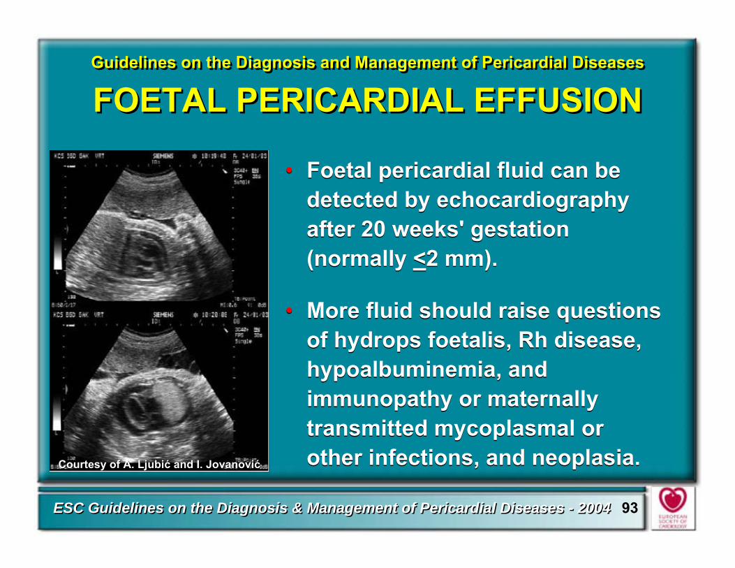
ESC Guidelines on the Diagnosis & Management of Pericardial Diseases - 2004ESC Guidelines on the Diagnosis & Management of Pericardial Diseases - 2004 93
� Foetal pericardial fluid can be detected by echocardiography after 20 weeks' gestation (normally <2 mm).
� More fluid should raise questions of hydrops foetalis, Rh disease, hypoalbuminemia, and immunopathy or maternally transmitted mycoplasmal or other infections, and neoplasia.
� Foetal pericardial fluid can be detected by echocardiography after 20 weeks' gestation (normally <2 mm).
� More fluid should raise questions of hydrops foetalis, Rh disease, hypoalbuminemia, and immunopathy or maternally transmitted mycoplasmal or other infections, and neoplasia.Courtesy of A. Courtesy of A. LjubiLjubićć and Iand I.. JovanoviJovanovićć
Guidelines on the Diagnosis and Management of Pericardial Diseases
FOETAL PERICARDIAL EFFUSIONGuidelines on the Diagnosis and Management of Pericardial Diseases
FOETAL PERICARDIAL EFFUSION

ESC Guidelines on the Diagnosis & Management of Pericardial Diseases - 2004ESC Guidelines on the Diagnosis & Management of Pericardial Diseases - 2004 94
DRUG- AND TOXIN-RELATED PERICARDIAL
DISEASE
DRUG- AND TOXIN-RELATED PERICARDIAL
DISEASE

ESC Guidelines on the Diagnosis & Management of Pericardial Diseases - 2004ESC Guidelines on the Diagnosis & Management of Pericardial Diseases - 2004 95
A. Drug-induced lupuserythematosus
� Procainamide� Tocainide� Hydralazine� Methyldopa� Mesalazine� Reserpine� Isoniazid� Hydantoins
A. Drug-induced lupuserythematosus
� Procainamide� Tocainide� Hydralazine� Methyldopa� Mesalazine� Reserpine� Isoniazid� Hydantoins
B. Hypersensitivity� Penicillins� Tryptophan� Cromolyn sodium
C. Idiosyncratic reaction or hypersensitivity
� Methysergide� Minoxidil � Practolol� Bromocriptine
B. Hypersensitivity� Penicillins� Tryptophan� Cromolyn sodium
C. Idiosyncratic reaction or hypersensitivity
� Methysergide� Minoxidil � Practolol� Bromocriptine
Guidelines on the Diagnosis and Management of Pericardial Diseases
DRUG- AND TOXIN-RELATED PERICARDIAL DISEASE
Guidelines on the Diagnosis and Management of Pericardial Diseases
DRUG- AND TOXIN-RELATED PERICARDIAL DISEASE

ESC Guidelines on the Diagnosis & Management of Pericardial Diseases - 2004ESC Guidelines on the Diagnosis & Management of Pericardial Diseases - 2004 96
C. Idiosyncratic reaction or hypersensitivity
� Psicofuranine� Polymer fume inhalation
(Teflon)� Cytarabine� Phenylbutazone� Amiodarone� Streptokinase� p-Aminosalicylic acid� Thiazides
C. Idiosyncratic reaction or hypersensitivity
� Psicofuranine� Polymer fume inhalation
(Teflon)� Cytarabine� Phenylbutazone� Amiodarone� Streptokinase� p-Aminosalicylic acid� Thiazides
� Streptomycin� Thiouracils� 5-Fluorouracil� Sulfa drugs� Cyclophosphamide� Cyclosporine� Mesalazine� Vaccines
(Smallpox, Yellow fever)� GM-CSF
� Streptomycin� Thiouracils� 5-Fluorouracil� Sulfa drugs� Cyclophosphamide� Cyclosporine� Mesalazine� Vaccines
(Smallpox, Yellow fever)� GM-CSF
Guidelines on the Diagnosis and Management of Pericardial Diseases
DRUG- AND TOXIN-RELATED PERICARDIAL DISEASE (continued)
Guidelines on the Diagnosis and Management of Pericardial Diseases
DRUG- AND TOXIN-RELATED PERICARDIAL DISEASE (continued)

ESC Guidelines on the Diagnosis & Management of Pericardial Diseases - 2004ESC Guidelines on the Diagnosis & Management of Pericardial Diseases - 2004 97
Guidelines on the Diagnosis and Management of Pericardial Diseases
DRUG- AND TOXIN-RELATED PERICARDIAL DISEASE (continued)
Guidelines on the Diagnosis and Management of Pericardial Diseases
DRUG- AND TOXIN-RELATED PERICARDIAL DISEASE (continued)
D. Anthracyclines� Doxorubicin� DaunorubicinE. Serum sickness� Foreign antisera
(e.g., antitetanus)� Blood products F. Venom� Scorpion fish sting
D. Anthracyclines� Doxorubicin� DaunorubicinE. Serum sickness� Foreign antisera
(e.g., antitetanus)� Blood products F. Venom� Scorpion fish sting
G. Foreign-substance reactions
� Talc (Mg silicate) � Silicones� Tetracycline/other
sclerosants� Asbestos� Iron in β-
thalasssemiaH. Haemopericardium� Anticoagulants� Thrombolytic agents
G. Foreign-substance reactions
� Talc (Mg silicate) � Silicones� Tetracycline/other
sclerosants� Asbestos� Iron in β-
thalasssemiaH. Haemopericardium� Anticoagulants� Thrombolytic agents

ESC Guidelines on the Diagnosis & Management of Pericardial Diseases - 2004ESC Guidelines on the Diagnosis & Management of Pericardial Diseases - 2004 98
Guidelines on the Diagnosis and Management of Pericardial Diseases
DRUG- AND TOXIN-RELATED PERICARDIAL DISEASE (continued)
Guidelines on the Diagnosis and Management of Pericardial Diseases
DRUG- AND TOXIN-RELATED PERICARDIAL DISEASE (continued)
I. Polymer fume fever � Inhalation of the burning fumes of polytetrafluoroethylene(Teflon)
I. Polymer fume fever � Inhalation of the burning fumes of polytetrafluoroethylene(Teflon)