Embed Size (px)
Citation preview

s
Guidelines for the Judicious Use of
Antibiotics for Upper Respiratory Infections
and Other Related Diseases in Children.
- Our Proposal -
Ver. 3 Apr. 10th2007 Ver. 2 May 1st 2006 Ver. 1 Nov. 1st 2005
Working Group for the Judicious Use of Antibiotics in Pediatric Ambulatory Practice Akira KUSAKARI (Kusakari Pediatric Clinic), Hajime TAKEUCHI (Mimihara General Hospital, Pediatric Department), Tatsuo NISHIMURA (Nishimura Pediatric Clinic), Mitsuru FUKAZAWA (Fukazawa Pediatric Clinic), Hitoshi YOSHIDA (Yoshida Pediatric Clinic; also Editor of this publication) JAPAN (In the order of the Japanese alphabet) : Contact us
Introduction ABSTRACT The common coldAntibiotics are not needed. Pharyngitis and tonsillitis1) Most cases of pharyngitis/tonsilitis are viral infections. There is no need for antibiotics except when group A beta-hemolytic streptococcus (GABHS) is the causative pathogen. 2) In the case of GABHS infection, benzylpenicillin benzathine is the first-line agent. Acute otitis mediaFollow up with symptomatic treatment only. If the symptoms (otalgia or fever) have not subsided within 48-72 hours, administrate an antibiotic. In case the symptoms (otalgia, fever or sickness) have worsened during the observation period, examine the patient immediately. Acute sinusitisMost cases of acute sinusitis are viral infections. Do not use antibiotics for the first 10-14 days, even if purulent nasal discharge is present. Cough illness/bronchitis1) Most of cough illness/bronchitis are caused by viral infections. Antibiotics are not justified for the treatment. 2) Erythromycin is recommended if pertussis is suspected. Fever without sourceObserve patient’s condition without administering antibiotics. However, when one of the findings below is present, intravenous ceftriaxone is our recommendation: 1) Infants of 3-12 months old with either a temperature of ≧40°C, or 38.5°C or more and leukocyte count of ≧15,000/µl ( or neutrophil count of ≧10,000/µl). 2) Infants of 12-36 months old with a temperature of ≧39°C and leukocyte count of ≧15,000/µl (or neutrophil count of ≧10,000/µl).
1

INTRODUCTION
Antibiotics, which have been developed through the wisdom and efforts of mankind, have been saving thousands of human lives. For a time, it seemed that antibiotics might possibly eradicate all bacterial infections. However, this hope was crushed with the emergence of antimicrobial-resistant bacteria. We have since learned that the use of antibiotics inevitably entails the appearance of more resistant strains of bacteria. Until recently, new antibiotics have been repeatedly created to combat the new strains of resistant bacteria. However, it is becoming impossible to continue using this method, because bacterial resistance is developing faster than new antibiotics 1).
The isolations of the resistant bacteria are closely related with the usage of antibiotics in each country. In Japan, not only are a large amount of antibiotics used, but broad-spectrum antibiotics are also used more widely than in other countries. As a result, the rate of resistant bacteria is very high, causing difficulty in treating severe bacterial infections such as meningitis. Therefore, in order to continue using antibiotics, it is imperative that we establish guidelines and share a basic policy for prescribing and using antibiotics. Prevalence of resistant strains of bacteria in Europe has been very low, compared to other parts of the world. Through the initiative of the Netherlands, the European Antimicrobial Resistance Surveillance System (EARSS) has been established leading to a full disclosure of information and the creation of educational programs focused on reducing antibiotic resistance 2). Penicillin has been principally used in Europe, whereas broad-spectrum antibiotics (such as cephalosporin) have been frequently used in Japan 3). The United States has also made medical guidelines, as one of their national projects, to help solve the problem of the resistant strains. These efforts are producing good results 4) 5).
Research data showed a wide variation in the prescribing rate of antibiotics for upper respiratory
infections by individual doctors in Japan and that many doctors over prescribed antibiotics. This might reflect the fact that they do not share the same standard for the use of antibiotics 6).
Our working group for the judicious use of antibiotics proposes these Guidelines, incorporating our own research with studies from other countries. Our objectives are the judicious use of antibiotics and the reduction of resistant strains, without adversely affecting patients. Therefore, our Guidelines are structured to cover not only upper respiratory infections (The Common Cold, Pharyngitis and Tonsillitis), but also other related diseases (Cough Illness/Bronchitis, Acute Sinusitis and Acute Otitis Media), and the risk control of fever (Fever without Source) as a part of the treatment method of serious bacterial infections.
The subjects of these Guidelines are children over the age of three months with no underlying conditions such as immunodeficiency, immunosuppressive state, broncho-pulmonary dysplasia, immotile cilia syndrome and so on. (When treating febrile infants aged less than 3 months, see the guidelines by Baraff et al. 7))
The Guidelines for the use of antibiotics are as follows: 1) Antibiotics should not be prescribed for viral diseases. Prescription for the prevention of a secondary bacterial infection should also be avoided. 2) Even if a bacterial infection is suspected, antibiotics should not be used when the risk of severe complications is low and a recovery without antibiotics is expected. 3) Antibiotics should be used when there is evidence of bacterial infection and the therapeutic effectiveness, of antibiotics, has been confirmed. 4) The use of antibiotics is appropriate when a patient has a fever and the laboratory data suggests a high risk of serious bacterial infection. 5) Narrow-spectrum antibiotics, whenever possible, should be the first-line of therapy for bacterial diseases. 6) When a disease such as sepsis, bacterial meningitis, or other severe bacterial infections is suspected, broad-spectrum antibiotics (sufficiently effective against possible resistant strains) should be used until the causative pathogen is revealed.
This set of guidelines is written from the viewpoint of practitioners who see patients on a daily basis, and is
2
aimed at being adaptable to outpatient settings. Because the clinical studies on which the Guidelines
are based may be more beneficial than the Guidelines themselves, we are including as many references as possible. We hope that the references cited here will be read as well as the Guidelines. (Note: It goes without saying that, when your patient follows an unexpected course, the plan of treatment should be modified depending on the patient’s condition. The ultimate decision of the treatment is left to the attending physician. )
3
Various opinions regarding the content of these Guidelines have been collected from extensive discussions at: Symposium at the Meeting of The Japan Pediatric Society (Fukuoka, 2003); Symposium at the Meeting of The Society of Ambulatory and General Pediatrics of Japan (Oita, 2004) and their workshops (5 times in total during 2000-2004) and conferences (Osaka, 2005; Yokohama, 2006). We will further investigate the validity of these Guidelines, seek opinions from various quarters, and revise editions regularly. This edition has been revised through the valuable contributions of 24 pediatricians in 18 countries.
Note: Leaflets for the patients (written in Japanese), “Antibiotics and Resistant Bacteria” “Acute Bronchitis” “Children and Otitis Media” from The Society of Ambulatory and General Pediatrics of Japan are available at the following: Noble Press; Tel: +81-3-3398-1904 Fax: +81-3-3398-1905 E-mail: [email protected] References 1. 吉川昌之介 (Yoshikawa M). 細菌の逆襲 中公新書
1234. 東京:中央公論社 1995:260-263 (In Japanese) 2. McKerrow W, et al. Management of sore throat and
indications for tonsillectomy. A National Clinical Guideline. SIGN Publication 1999;34:1-23 [Full Text]
3. SWEDRES 2001. A report on Swedish antibiotic utilisation and resistance in human medicine. STRAMA (the Swedish Strategic Programme for the Rational Use of Antimicrobial Agents) [Full Text]
4. Dowell F, et al. Principles of judicious use of antimicrobial agents for pediatric upper respiratory infections. Pediatrics 1998;101(suppl):163-165 [Full Text]
5. Bauchner H, et al. Promoting the appropriate use of oral antibiotics: There is some very good news.
Pediatrics 2003;111:668-670. [Full Text] 6. 草刈 章 (Kusakari A), 他. 小児科外来における上気道
炎診療調査―発病72時間以内の初診患者に対する抗
菌薬使用状況― . 外来小児科2004;7:122-127 (In Japanese)
7. Baraff LJ, et al. Practice guideline for the management of infants and children 0 to 36 months of age with Fever without source. Pediatrics 1993;92:1-12 [Abstract]
Return to the top
THE COMMON COLD
1. Introduction
The common cold heals without antibiotic treatment. However, any doctor may think, “Could it be an onset of serious infections?” or “Could it cause a secondary bacterial infection?” As a result, antibiotics are too often prescribed to prevent serious, but rare, bacterial infections. However, with the recent development of various immunodiagnostic methods (rapid diagnostic tests, for example), blood tests using a very small amount of capillary blood, and easier access to medical services in Japan, it is possible to offer patients good medical care without depending on antibiotics. 2. Diagnostic guideline
The common cold is an acute viral disease characterized by a generally good condition aside from the main symptoms of nasal discharge and congestion. Other associated symptoms may include a slight sore throat, a mild cough, and a fever (usually below 38.5 °C). (Note: see the Guidelines of Pharyngitis/Tonsillitis or Cough/ Bronchitis if there is marked redness of the throat or a moderate to severe cough.) 3. Treatment guideline
Antibiotics are not needed to treat the common cold.

4
4. Overview 1) Etiology, pathology and symptoms of the common cold
In some European and American countries, the term, “the common cold,” is used as a synonym of rhinosinusitis 1). However, there is another opinion that the term, “rhinosinusitis,” is not proper in the cases without symptoms suggesting sinusitis. The term, “nasopharyngitis,” may be more appropriate, especially in the case of children 2).
The causative pathogens of the common cold are viruses. The symptoms start when viruses adhere to the nasal and pharyngeal mucosa or palpebral conjunctiva and invade the epithelial cells. When macropharges in the submucous tissue recognize that the viruses have invaded the epithelial barrier, inflammation-promoting substances such as prostaglandin are released, causing capillary dilation and mucosal reddness. It also increases vascular permeability, which causes a swollen submucosa followed by pain and discomfort of the pharynx. In the nasal mucosa, viruses cause nasal congestion or clear nasal discharge. Inflammatory cytokines and chemokines secreted by macrophages and epithelial cells further promote these biological reactions, and enhance and sustain inflammatory response, by gathering neutrophils and monocytes in the blood until the viruses are completely eliminated. In this way, systemic symptoms such as fever, fatigue, headaches, loss of appetite, and muscle pain begin to appear 3). Within 1-3 days, nasal discharge starts to include epithelial cells, neutrophils, and normal flora, all of which become purulent without bacterial infection 4). Cough and nasal symptoms are normally resolved in 10-14 days, although they may occasionally linger over two weeks 5). The main causative organisms are: rhinoviruses (30%), coronaviruses (10%), RS virus, influenza viruses (type A and B), and adenoviruses collectively (10-15%) 6). In addition, influenza virus type C, enteroviruses, newly identified human metapneumovirus (hMPV), and novel human coronavirus can be causative agents 7). 2) Alleviating symptoms through antibiotics
Todd et al. studied a randomized control trial (RCT) in children with the common cold. It showed that the alleviation of purulent nasal discharge was observed after 5-6 days in 24% and 37% of the subjects, and
complications were found in 7% and 8% of the subjects in the antibiotics and the placebo groups, respectively. These findings represented no significant difference between the two groups 8). Furthermore, a meta-analysis of 6 RCTs showed no effect in the alleviation of symptoms and concluded that the use of antibiotics is not advisable for the common cold 9) 10). An epidemiological study also reported that most symptoms of the common cold lasted no longer than 10-14 days; therefore we advice that the course of the disease be monitored without the administration of antibiotics during this period 11) 12). (Note: see the Guidelines for “Acute Sinusitis” if the symptoms persist after this period.) 3) Preventing complications through antibiotics
Cronk et al. investigated the rate of second hospital visits for the penicillin group and the symptomatic treatment group in 2,177 university students. The rates were 26% and 20% respectively (no significant difference), and therefore the effectiveness of antibiotics were denied 13). An RCT which studied the preventive effects of antibiotics for complications in 845 children found no difference between a normal dosage, ¼ of a dosage, or no dosage of antibiotics in secondary occurrences of otitis media, pneumonia, or tonsillitis 14). Lexomboon et al. also disproved the effects of antibiotics because there was no difference in duration of fever or development of complications (5.1% and 4.6% in the antibiotics and the no treatment groups, respectively) 15). A meta-analysis of 5 studies (including this trial) also dismissed the preventive effects of complications 16). Dowell et al. reviewed 9 RCTs and reconfirmed that antibiotics did not have any effect on either the conditions such as nasal discharge or the occurrence of complications such as otitis media or pneumonia 17). 4) Differentiation from other diseases
Depending on the progression of symptoms of the common cold, it may be necessary to refer to each of the other Guidelines: When nasal discharge or congestion persists over 14 days, see “Acute Sinusitis”. When a sore throat is the main symptom, see “Pharyngitis / Tonsillitis”. When an ear pain is present, see “Acute Otitis Media”. When a cough is the chief complaint, see “Cough Illness/ Bronchitis”.

5
When there is a high fever for which the cause cannot be specified, see “Fever without Source”.
Initial symptoms of serious bacterial diseases are like those of the common cold. It is sometimes quite difficult to distinguish bacterial infection from the common cold. In cases of fever of unknown origin urine and blood tests should be taken, suspecting urinary tract infection or occult bacteremia. As the onset of Kawasaki disease is also just a fever, it is difficult to make a precise diagnosis until its characteristic symptoms such as conjunctival hyperemia or injected and dry fissured lips appear. Above all, bacterial meningitis is one of the most serious infections. Making a diagnosis on the first visit is difficult, as a patient’s chief complaint is only a fever without meningeal signs. Systemic conditions (whether or not the child looks reasonably well and calm, smiles, speaks, or plays) should be continually monitored. If conditions worsen, diagnostic tests should be performed immediately 18). An automatic prescription of oral antibiotics (for fear of serious disease) could do more harm than good to the patient. A study revealed that patients treated with oral antibiotics were more likely to have hearing loss or neural sequelae because the medication masked the presence of meningitis and delayed the start of appropriate treatment 19).
Careful observation of the patient’s condition (a “wait and see approach”), in which the patient could be seen whenever unusual symptoms are observed, produces a faster diagnosis (see “Fever without Source”). 5. Summary
The common cold heals without antibiotics. Antibiotics do not prevent secondary bacterial infection. Careful observation of the patient (a “wait and see approach”) is the best and quickest way to detect serious bacterial infections hidden in the common cold. Overuse of the administration of antibiotics delays both the correct diagnosis and the start of appropriate treatment. References 1. Turner RB, et al. The Common Cold. In:Behrman RE,
et al, eds. Nelson Textbook of Pediatrics. 17th ed. Philadelphia: Saunders, 2004:1389-1393
2. Puhakka T, et al. Sinusitis in the common cold. J Allergy Clin Immunol, 1998; 102:403-408 [Abstract]
3. Cherry JD. The Common Cold. In:Feigin RD, et al, eds. Textbook of Pediatric Infectious Diseases. 5th
ed. Philadelphia: Saunders, 2004:140-146 4. van Volkenburgh VA, et al. Acute minor respiratory
diseases prevailing in a group of families residing in Baltimore, Maryland, 1928-1930. Prevalence, distribution and clinical description of observed cases. Am J Hyg 1933; 17:122-153
5. Burroughs M, et al. Respiratory infections. In: Gershon AA, et al, eds. Krugman’s Infectious Diseases of Children. 11th ed. Philadelphia: Mosby, 2004:493-529
6. Gwaltney JM Jr. Virology and immunology of the common cold. Rhinology 1985; 23:265-271 [Medline]
7. Fouchier RAM, et al. A previously undescribed coronavirus associated with respiratory disease in humans. Proc Natl Acad Sci USA 2004;101:6212-6216 [Full Text]
8. Todd JK, et al. Bacteriology and treatment of purulent nasopharyngitis: a double blind, placebo-controlled evaluation. Pediatr Infect Dis 1984; 3:226-232 [Medline]
9. Morris P, et al. Antibiotics for persistent nasal discharge (rhinosinusitis) in children (Cochrane Review). Cochrane Database Syst Rev 2002;4:CD001094 [Medline]
10. Aroll b, et al. Antibiotics for the common cold and acute purulent rhinitis (Review). Cochrane Database of Syst Rev. 2005;3:CD000247
11. 草刈章 (Kuakari A), 他. チャートでみる抗菌薬適正
使用ガイドライン・急性副鼻腔炎 . 外来小児科
2006;9:211-214. (In Japanese) 12. Wald ER, et al. Upper respiratory tract infections in
young children: duration of and frequency of complications. Pediatrics 1991;87:129-133 [Medline]
13. Cronk GA, et al. A controlled study of the effect of oral penicillin G in the treatment of non-specific upper respiratory infections. Am J Med 1954;16:804-809
14. Townsend EH Jr. Chemophylaxis during respiratory infections in a private pediatric practice. Am J Dis Child 1960;99:566-573
15. Lexomboon U, et al. Evaluation of orally administered antibiotics for treatment of upper respiratory infections in Thai children. J Pediatr 1971;78:772-778
16. Gadomski AM. Potential interventions for preventing pneumonia among young children: lack of effect of antibiotic treatment for upper respiratory infections. Pediatr Infect Dis J 1993;12:115-120 [Medline]

17. Dowell SF, et al. Appropriate use of antibiotics for URIs in children: Part II. Cough, pharyngitis and the common cold. Am Family Physician 1998;58:1335-1342 [Full Text]
18. McCarthy PL, et al. Observation scales to identify serious illness in febrile children. Pediatrics 1982;70:802-809 [Abstract]
19. Kaplan SL, et al. Association between preadmission oral antibiotic therapy and cerebrospinal fluid findings and sequelae caused by Haemophilus influenzae type b meningitis. Pediatr Infect Dis 1986;5:626-632 [Medline]
Return to the top
6
PHARYNGITIS AND TONSILLITIS
1. Introduction
Pharyngitis and tonsillitis are often associated with high fever and white exudate on the tonsils; therefore they tend to be considered as a bacterial infection. However, many of their causative pathogens are viruses. Group A beta-hemolytic streptococcus (GABHS) infection is treated with antibiotics to prevent rheumatic fever (RF) or poststreptococcal acute glomerulonephritis (PSAGN). This infection accounts for only 10-20% of pharyngitis/tonsillitis at most. It should be noted that the pathogen colonizes in the tonsil of up to 15% of healthy children aged 5 to 12 years old.
Clinical findings of pharyngitis/tonsillitis are easily obtained as doctors can observe an affected region directly. It has also become possible to specify the causative pathogen using various rapid-test methods. 2. Diagnostic guideline
The main symptoms of pharyngitis/tonsillitis are sore throat and fever. The pharynx is reddish and the tonsils
are often swollen with exudate. Cough may also be present. 3. Treatment guideline a) Most cases of pharyngitis/tonsilitis are viral infections. There is no need for antibiotics except when GABHS is the causative pathogen. b) In the case of GABHS infection, benzylpenicillin benzathine (Bicillin G®, 400,000U/g, available in limited countries) 50,000U/kg/day (up to 1,600,000U=4g/day), 3 times orally for 10 days is the first-line agent. If the patient is allergic to beta-lactam antibiotics, erythromycin 50mg/kg/day (up to 1200mg/day), 3 times orally for 10 days is the second-line agent. 4. Overview
In Europe and America, pharyngitis and tonsillitis are collectively known as sore throat 1) 2). In Japan, they have often been separated on clinical diagnosis. However, there is no need to differentiate between them, because their main difference is the extent and degree of the inflammation. 1) Causative pathogens As far as the children’s tonsillitis with “exudates” are concerned, 12% of the causative pathogens are GABHS, 42% are various viruses, of which 45% are adenoviruses 3). Other microbes isolated and cultured are not identified as causative pathogens. The flora such Haemophilus influenzae, Staphylococcus aureus, and Streptococcus pneumoniae are often isolated from the throat of healthy children. They are considered to be carriers of these pathogens. When a causative organism is unknown, involvement of other viruses difficult to detect may be suspected. Hayden et al. isolated adenoviruses, rhinoviruses, coronaviruses, herpes simplex virus (HSV), influenza viruses, parainfluenza viruses, coxsackieviruses, Ebstein-Bar virus (EBV) and cytomegalovirus as causative organisms for acute pharyngitis 4). Other pathogens such as RS virus and human herpes viruses type 6&7, the causative virus of exanthema subitum, can also cause pharyngitis.
In 2004-2005, a multi-centered prospective study concerning about “exudative” tonsillitis was performed in Japan. The causative pathogens were viruses in 63/149(42%) and GABHS in 25/143(17%). Two

pathogens, virus and GABHS, were simultaneously detected in 4 cases. The detected viruses were adenoviruses in 48 cases, coxsackieviruses in 6 cases, influenza viruses and HSV in 3 cases, and parainfluenza viruses and echo viruses in 2 cases, respectively 5). (1) Adenoviruses (Fig 1.)
Adenoviruses are most frequently identified as a causative organism of pharyngitis/tonsillitis 3) 5). Although a typical pharyngoconjunctival fever can be diagnosed by clinical examination only, there are many atypical cases. Therefore, it is important to know the endemic situation in order to identify it. Adenovirus infection often causes a high fever of 40 °C or above which lasts 4–5 days. In more than half of the cases, leukocytes count of ≧15,000/µl was observed in the early stage of the fever, and CRP level also increased during the course 6). Fig 1. Tonsillitis due to Adenovirus
Boy , age 10
7
There are white exudates on both tonsils.
The rapid antigen-detection test is useful for diagnosis.
A test kit released in 2001 in Japan facilitated the diagnosis of adenovirus infection. The use of the test not only reduces the administration of antibiotics but also helps to reassure parents by giving the prospect of recovery. The sensitivity of the test is reported to be 70-94%, so a negative result does not completely rule out a possibility of adenovirus infection. The specificity, on the other hand, is 95-100%, and therefore a positive result confirms adenovirus infection 7). Our study comparing the rapid test with viral isolation also showed that the sensitivity was 83% and specificity was 98% 5).
Although there are reports of viral excretion for months or infection with mild symptoms only, the facts that the virus is rarely isolated from healthy school children (1/174; 0.6%) and that the results of the test become negative after the acute stage indicate that it is appropriate to consider that the positive result means adenovirus infection 8).
Because blood tests (leukocyte count, CRP level) often
show strong positive results, it is necessary to rule out a bacterial infection such as occult bacteremia. A study of patients with positive results of adenovirus (n=143) found only two cases of complications with bacterial infections: one with a urinary tract infection of E. coli, and the other with bacteremia by Moraxella catarrhalis (1.4% collectively) 9). The possibility of complications with bacterial infection, although requiring some attention, seems to be rare. (2) Other viruses
Pharyngitis/tonsillitis due to virus is often associated with cough and nasal discharge as well as pharyngeal symptoms. EBV usually exhibits “membranous” exudate on the tonsil while coxsackieviruses and HSV exhibit only “streaky” exudate. Many of EBV infections are associated with cervical lymphadenopathy and hepatosplenomegaly. Blood test often reveals leukocytosis with increased atypical lymphocytes. Diagnosis of EBV infection can be confirmed by serological antibody tests 10). The infection with coxsackieviruses is characterized by more than one ulcer of 2-3mm in the soft palate. The endemic often occurs in the summer. With HSV infection, gingival swelling/redness and aphthae on the oral mucosa and tongue are usually observed. (3) GABHS infection Although it is possible to start the treatment based on clinical symptoms and signs in a typical case, it is fundamental to treat this infection after confirming the diagnosis, particularly in an otherwise indistinguishable case, by a rapid antigen-detection test or a culture. However, a study found that the rapid test was positive in 18% of the healthy children in an elementary school. Thus, some children are carriers of GABHS, so it is not always easy to differentiate infection from colonization 11). As a carrier should not be treated, it is unnecessary to use antibiotics when there are no clinical manifestations or characteristic findings of the infection even if the test is positive 12) 13). The infection occurs most commonly in children of 3 years old or older. An endemic occurs often in winter, but the first term of the school year could be an endemic season because newcomers enter schools or kindergartens 13). The main symptoms are a fever and sore throat, but a headache, fatigue, stomachache, nausea and vomiting may also be present. If a cough, nasal discharge, stridor, tachypnea or diarrhea is seen, GABHS infection is not likely to exist 12). Conjunctivitis

could be added as a rule-out sign. A fever of 38.3°C or higher is found in 30% of the cases 14). The pharynx is reddish and the soft palate is often erythematous or petechial (Fig 2.) The tonsils are also swollen and red, sometimes covered with exudate. The tongue’s papillae may also be swollen and red (strawberry tongue). Anterior cervical lymphadenopathy with tenderness is an important diagnostic finding. It is called scarlet fever when a characteristic generalized exanthema appears, but there is no difference in the infectivity or prognosis. Fig 2. GABHS infection
Boy, age 5
8
The soft palate is erythematous and petechial.
The purpose of using antibiotics for GABHS is to
alleviate symptoms such as high fever and to prevent secondary RF. If an epidemic of GABHS occurs, 3% of the pathogen is thought to be certain serotype strains that are likely to cause a secondary RF (e.g. M protein 1, 3, 18 types), and therefore an RF outbreak also is suspected to occur. The prevalence of RF in school children in the United States is currently only 0.2/100,000, whereas in Africa the prevalence is 1,500 times higher (300/100,000) 15). It therefore can be reasoned that hygienic conditions and living environment are strongly associated with the prevalence. In 1985, an epidemic of RF started in Salt Lake City, Utah, and affected 198 patients over a 4-year period. However, no relationship between the living environment and the prevalence was seen 16). The contributing factors of the RF outbreak still remain to be unsolved.
The basis for use of antibiotics for the prevention of RF is derived from a controlled study conducted during the endemic of GABHS infection at an air base camp in Wyoming in 1949. The subjects were either injected intramuscularly with penicillin G or a placebo, and 2/798 in the former and 17/804 in the latter group developed RF, thus demonstrating the effectiveness of the preventive treatment 17). Other studies followed in the US to confirm preventive effects on initial or recurrent cases of RF, and based on these results, use of antibiotics for the prevention of RF has been considered as a standard 1).
On the contrary, in Scotland UK, where the yearly incidence of RF was 0.6/100,000 (27 cases out of 4,400,000), there was no significant difference between the group administered with antibiotics and the untreated group. Thus, it was not proved that antibiotics were effective to prevent RF. Based on these results, Scottish Intercollegiate Guidelines Network (SIGN) concluded that antibiotics were not necessary for the treatment of GABHS infection 2) 18). As there is a discrepancy between the two nations regarding the treatment of using antibiotics for RF prevention, it is desirable to review the necessity in Japan. However, it would be difficult to conduct an RCT because the treatment of GABHS infection with antibiotics has been already established 19). A preventive effect of antibiotics for PSAGN or Henoch-Schönlein purpura has been dismissed in Europe and America. In Japan, however, its effectiveness has been believed for a long time. In addition, atypical PSAGN with hypocomplementemia and mild hematuria occasionally occurs. Considering these possibilities and expert opinions in Japan 20) 21), it might be necessary to use antibiotics to prevent these diseases. Further reviews are waited.
(4) Bacteria other than GABHS Although the possibility of involvement by other
bacteria cannot be completely ruled out, other countries also don’t recommend antibiotics unless GABHS is present 22). 2) Retropharyngeal abscess and peritonsillar abscess
Although less frequent, retropharyngeal and peritonsillar abscess are important as complications or diseases to be differentiated. They are most likely to occur in boys up to 3-4 years old and are characterized by the lack of movement of the jaws, torticollis or neck stiffness in addition to nonspecific symptoms such as fever, decreased oral intake and salivation. It is important to consider a possibility of these two diseases and examine the oral cavity/pharynx carefully whenever seeing a patient. Although GABHS is a predominant cause of these conditions, Haemphilus influenzae, Klebsiella sp. or Mycobacterium sp. are also possible causative microorganisms 23). When these complications are suspected, refer the patient to a specialist of otolaryngology.

9
3) Treatment
There is no need to use antibiotics for viral infections. Even with a high fever, leukocytosis and increased CRP level, they will heal without antibiotics. Antibiotics are used for treating GABHS infection. Narrow-spectrum penicillin is the best choice 1) 12). In Japan, oral narrow-spectrum penicillin is only benzylpenicillin granule. Tablet or capsule is not available. Erythromycin is a universally accepted second-line agent when there is an allergy to beta-lactam antibiotics 24). Careful attention should be payed to the increase in bacteria strains resistant to macrolide antibiotics in Japan. Though cephalosporins are effective, they tend to increase the resistance of pneumococcus and other bacteria. It is therefore important to use as few cephalosporins as possible 14). Fever subsiding within 24 hours of the treatment is an important sign as to the efficacy of diagnosis and treatment. Some cases are refractory to treatment or the symptoms are recurrent even if the pathogen is susceptible to penicillin. There are some causes for these problems. The most common cause is an insufficient taking of the medicine. Therefore, a dosing instruction for the patient is very important. When a new infection with other serotype of GABHS is suspected, another medication of the same penicillin may be effective. When erythromycin has been used, suspect an emergence of a resistant strain against macrolides and change to a susceptible antibiotic after a culture. It is also possible that pharyngitis is caused by another pathogen such as a virus, and the patient is only a carrier of GABHS 25). This is likely to be the case if a typical finding in the pharynx is absent. Some studies reported that once GABHS invades the mucosal cell, it will not be eliminated by antibiotics and the patient becomes a carrier. However, in any case, antibiotics should not be used only on a carrier. Another study found that the streptococcus is protected by beta-lactamase produced by the normal flora in the pharynx: however, evidence to consider the presence of beta-lactamase is vague. 5. Summary Most of pharyngitis/tonsillitis are viral infections and only a part of them are due to GABHS. Antibiotics are indicated for GABHS infection, retropharyngeal abscess and peritonsillar abscess. We believe that a grasp of the endemic situations, precise observation of the patients’
pharynx and use of rapid tests will improve the accuracy of diagnoses and promote the judicious use of antibiotics. References 1. Hayden GF, et al. Acute Pharyngitis. In:Behrman RE,
et al, eds. Nelson Textbook of Pediatrics. 17th ed. Philadelphia: Saunders, 2004:1393-1394
2. McKerrow W, et al. Management of sore throat and indications for tonsillectomy. A National Clinical Guideline. SIGN Publication 1999; 34:1-23 [Full Text]
3. Putto A. Febrile exudative tonsillitis: viral or streptococcal? Pediatrics 1987; 80: 6-12 [Abstract]
4. Hayden GF, et al. Management of the ambulatory patient with a sore throat. Curr Clin Top Infect Dis 1988;9:62-75
5. Takeuchi H, et al. Exudative tonsillitis - viral isolation and bacterial cultures of 149 cases. The 24th Annual Meeting of the European Society for Paediatric Infectious Diseases (SPID). 2006 Basel [Abstract] (See: Scientific Program → Poster Sessions → Respiratory Tract Infection)
6. 武内 一 (Takeuchi H) . アデノウイルス 3 型感染症
―臨床症状・検査データと流行拡大の特徴―. 日児
誌 (in Japanese) 7. 原三千丸 (Hara M), 他. アデノウイルス迅速診断キ
ットチェック Ad® 改良品の有用性の検討. 小児科臨
床 2005; 58:221-223. (In Japanese) 8. Edwards KM, et al. Adenovirus infections in young
children. Pediatrics 1985;76:420-424 [Abstract] 9. Rocholl C, et al. Adenoviral infections in children:
The impact of rapid diagnosis. Pediatrics 2004; 113:e51-56 [Full Text]
10. 鹿野隆明 (Shikano T), 他. EB ウイルスによる小児
伝 染 性 単 核 球 症 の 臨 床 的 検 討 . 日 児 誌
2002;106:389-393, (In Japanese) 11. 中島邦夫 (Nakashima K), 他. 学童の咽頭分離溶血
性 レ ン サ 球 菌 の 疫 学 的 研 究 . 感 染 症 学 1983;57:1075-1082 (In Japanese)
12. Kumar S, et al. Why do general practitioners prescribe antibiotics for sore throat? Grounded theory interview study. Br Med J 2003;326:138-143
[Full Text] 13. Bisno AL, et al. Practice Guidelines for the diagnosis
and management of group A streptococcal pharyngitis. Clinical Infectious Diseases 2002;35:113-125 [Full Text]
14. McIsaac WJ. Practical experience with clinical

algorithms for reducing unnecessary antibiotic use in the management of streptococcal pharyngitis. Pechere JC, et al. eds. Streptococcal Pharyngitis Optimal Management. Issues Infectious Diseases. 1st ed. Basel: Karger, 2004:36-48 [Extract]
15. Guzman SV. Epidemiology of rheumatic fever and rheumatic heart diseases. In: Calleja HB, et al, eds. Rheumatic fever and rheumatic heart diseases. Epidemiology, clinical aspects, management and prevention and control programs. Manila: Philippine Foundation for the Prevention and Control of Rheumatic Fever/Rheumatic Heart Disease, 2001:1-12
16. Gerber MA. Group A Streptococcus. In:Behrman RE, et al, eds. Nelson Textbook of Pediatrics. 17th ed. Philadelphia: Saunders, 2004:870-879
17. Denny FW, et al. Prevention of rheumatic fever; treatment of the preceding streptococcic infection. JAMA 1950; 143:151-153
18. Howie JG, et al. Antibiotics, sore throats and rheumatic fever. JR Coll Gen Pract 1985;35:223-224
19. Danchin MH, et al. Treatment of sore throat in light of the Cochrane verdict: is the jury still out? MJA 2002; 177: 512-515 [Full Text]
20. 武田修明 (Takeda N), 他. 小児期急性糸球体腎炎の
非典型例. 小児科 1997;38:1093-1098 (In Japanese) 21. 武田修明 (Takeda N), 他. Henoch-Schönlein 紫斑病
と溶連菌感染との関連性について . 小児科臨床 2000;53: 1473-1477 (In Japanese)
22. Schwartz B, et al. Pharyngitis - principles of judicious use of antimicrobial agents. Pediatrics 1998;101(suppl):171-174 [Full Text]
23. Goldstein NA, et al. Peritonsillar, retropharyngeal, and parapharyngeal abscesses. In:Feigin RD, et al. eds. Textbook of Pediatric Infectious Diseases 5th ed. Philadelphia: Saunders, 2004:178-185
24. WHO Model Prescribing Information. Drugs used in the treatment of streptococcal pharyngitis and prevention of rheumatic fever. Geneva: WHO/EDM/PAR/1999.1:1-18
25. Kaplan EL, et al. Diagnosis of streptococcal pharyngitis: differentiation of active infection from the carrier state in the symptomatic child. J Infect Dis 1971;123:490-501
26. Neeman R, et al. Prevalence of internalisation-associated gene, prtF1, among persisting group-A streptococcus strains isolated from asymptomatic carriers. Lancet 1998;352:1974-1977 [Medline]
27. Gerber MA, et al. Potential mechanisms for failure to eradicate group A streptococci from the pharynx. Pediatrics 1999;104:911-917 [Full Text]
Return to the top
ACUTE OTITIS MEDIA
1. Introduction
Acute otitis media has been one of the disorders for which antibiotics were frequently prescribed in Japan. It is now an urgent problem that the disease is becoming intractable due to the increase of antibiotic resistant bacteria.
Since 1990, in the Netherlands, a policy of essentially not using antibiotics and following up patients with symptomatic treatment only has been practiced 1) 2) (modified in 1999). Owing to the principles of restricting the use of antibiotics, the rate of the resistant pneumococcus was 3% in the Netherlands, which was remarkably low 3). In the United States, administration of antibiotics had been the basis of therapy, but the 2004 guideline published by the American Academy of Pediatrics (AAP), for the first time, adopted a policy of restricting the use of antibiotics 4). Our Guideline also employs the same concept as the Netherlands-essentially not using antibiotics. 2. Diagnostic guideline
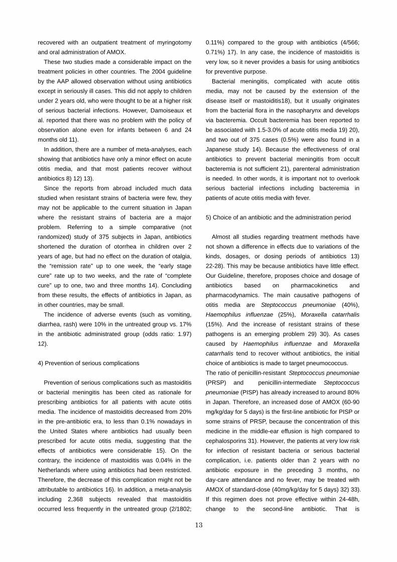
Acute otitis media is diagnosed by the presence of a purulent discharge (otorrhea) through a perforation of the eardrum, or middle-ear effusion plus symptom and/or findings of acute inflammation in the middle ear. The presence of the effusion is ascertained by a fullness of the eardrum or no mobility observed by pneumatic otoscopy. The symptom of acute inflammation is otalgia (causing crying, irritability or pulling of the ear in infants), and its findings are marked redness, strong bulge and bulla of the eardrum (Fig.)
10

Fig. Eardrum Findings
1) Normal eardrum (left)
11
This photo shows normal color (pearly gray) and normal shape of the eardrum.
2) Acute otitis media (right)
This eardrum is reddish and has a slight bulge due to middle ear effusion. But, it is difficult to distinguish acute otitis media from otitis media with effusion only by these
findings. The diagnosis of this disease was made by an additional presence of otalgia.
3) Acute otitis media (left)
This patient is diagnosed as acute otitis media based on a strong bulge of the entire eardrum, though the degree of redness is mild.
4) Acute otitis media (left)
This eardrum is slightly reddish and has a few bullas. These findings are often seen in infants.
It is sometimes difficult to distinguish acute otitis media from otitis media with effusion unless there are symptoms of otalgia. In an indistinguishable case, diagnose as otitis media with effusion. 3. Treatment guideline For children over 6 months of age: Basic rule: Follow up with symptomatic treatment only. If the symptoms (otalgia or fever) have not subsided within 48-72 hours, administrate an antibiotic. In case the symptoms (otalgia, fever or sickness) have worsened during the observation period, examine the patient
immediately. a) In case otorrhea is present: Follow up on the patient, only by cleaning the external auditory meatus for 7 days. If a fever or otalgia is present, follow the basic rule. b) In case otalgia is present: Prescribe acetaminophen 10-15mg/kg/dose orally or suppositorily as analgesic. For children over 2 years of age, ibuprofen 5mg/kg/dose (authorized dosage in Japan) is also an option. If otalgia has persisted or worsened, the presence of symptoms and signs suggesting mastoiditis, such as protrusion of the pinna, postauricular swelling, tenderness, pain and erythema, must be examined. c) In case fever is present: Consider the complications with another serious bacterial infection, such as bacteremia or meningitis. In particular, the patient with a fever of 39°C or above (in children under 3 years of age) or 38.5 °C or above (in children under 1 year of age), should be examined on leukocyte counts. The patient with leukocyte count of ≧15,000/µl (or neutrophil count of ≧10,000/µl) or with toxic appearance is categorized as a “high risk group” and should be treated according to the Guideline of “Fever without Source”. A culture of middle-ear effusion, by puncturing the tympanic membrane as a part of “sepsis work-up”, is an option. d) Initial antibiotic treatment: The first-line oral antibiotic is amoxicillin (AMOX). Administer 60-90mg/kg/day in 3 doses for 5 days. If the symptoms have not improved within 24-48 hours after administration, change to another oral or parenteral antibiotic. AMOX of standard-dose (40mg/kg/day for 5 days) should limitedly be used for patients who are at low risk for infection of resistant bacteria or serious bacterial complication. e) In case antibiotics are not effective: If the otalgia, fever or sickness have not subsided within 48-72 hours after the start of antibiotic treatment, a complication such as mastoiditis without obvious clinical signs (masked mastoiditis, or occult mastoiditis) may be suspected. In these cases, consultation with or referral to an otolaryngologist should be considered. For infants under 6 months of age: Basic rule: Administer antibiotics, especially for infants with a fever. There is little data available for the treatment of infants under 6 months of age. This recommendation is,

12
therefore, a tentative one. 4. Overview 1) Diagnosis of acute otitis media and the severity assessment
A correct diagnosis is a prerequisite for the judicious use of antibiotics. However, it might not be possible to differentiate acute otitis media clearly from otitis media with effusion requiring no antibiotics. Our diagnostic criteria are based on that of Bluestone et al. 5) and the guidelines of the AAP -- both of these require symptoms or findings suggesting acute illness. There is no accurate method of confirming acute onset from the eardrum findings; therefore, the findings of a marked redness, strong bulging or the presence of bulla are adopted as a diagnostic sign of acute illness. The findings of bulging, or bulla of the eardrum are important, because the redness of the eardrum is not frequently found in infants with acute otitis media. It is also essential to know that bilateral hyperemia of the eardrum is often observed in crying infants. There are some other guidelines on which a fever is one of diagnostic criteria for acute illness. Although fever is a symptom observed in 1/3-2/3 patients with acute otitis media, it is impossible to differentiate the fever caused by acute otitis media itself from that arising from the preceding viral infection. Therefore, we did not include the fever in our criteria.
To classify the grades of severity, a method combining otalgia and fever 6) or one of using the eardrum findings has been proposed. However, the most important aim of the therapy in the acute stage is to prevent complications of serious bacterial infection. For this reason, we will categorize “high risk group” under “Fever without Source”. 2) Assessment of treatment effectiveness It is necessary to evaluate treatment effectiveness, to facilitate comparison with other studies and provision of appropriate medical care 7). If symptoms of otalgia, fever and otorrhea have subsided, judge it as a ”remission” whether or not abnormal findings of the eardrum remain; evaluate after 24hours, 2-3days and 1week. If the aforementioned symptoms and the eardrum findings such as redness or bulging (except for the middle-ear effusion) have resolved, judge it as an “early stage cure”; evaluate after the first and second week. If the middle-ear effusion
has disappeared, judge it as a ”complete cure; evaluate after 2 weeks, 4 weeks, 2 months and 3 months.
These assessment criteria are not the treatment goal in actual medical practice. Acute otitis media treatment’s focus is not necessarily the complete disappearance of abnormal eardrum findings, but the disappearance of the symptoms 8). 3) Effectiveness of antibiotics
The Netherland’s guideline, that has drawn much attention from the world, is based on the two studies conducted by an otolaryngologist, van Buchem and his colleagues in Tilburg, a local city of a population of 150,000 at that time. In 1981, the authors compared an untreated group (n=40), a myringotomy-monotherapy group (n=36), an AMOX administration-monotherapy group (n=47) and a combination of myringotomy + AMOX group (n=48). Otalgia was found in 98%, 100%, 94%, and 94% of each group respectively at the first visit; after 24 hours it was reduced to 28%, 28%, 28% and 35%; and after 7 days 10%, 11%, 6% and 10%, representing no significant difference between the groups. The rates of abnormal eardrum findings observed in each group were 49%, 49%, 40% and 44% respectively after 7 days, and 28%, 25%, 10% and 11% after 14 days, showing a slightly larger decrease in groups treated with antibiotics, regardless of myringotomy. This study concluded that there was no significant difference on the effectiveness of antibiotics or myringotomy between these groups 9).
In 1985, van Buchem et al. published the results of 4,860 subjects aged 2-12 years treated with a therapeutic plan of “treat with analgesic only for the first 3-4 days and observe otorrhea for 14 days without antibiotics”; these results later became the base for the guideline of the Netherlands 10). Among these subjects, 126 (2.7%) whose otalgia or fever persisted for more than 3-4 days (“persistent cases”) were included in an RCT and divided into one of the following three groups: myringotomy-monotherapy, AMOX-monotherapy, and myringotomy+AMOX. There was no difference in the rate of otalgia between the three groups after 24 hours, 7 days and 14 days. Of all the subjects, 4.5% having another bacterial infection not related to acute otitis media, and 1.5% having otorrhea for more than 14 days, were treated with antibiotics. Including the persistent cases, more than 90% of all the subjects were resolved by the symptomatic treatment. Only two patients (0.04% of all the subjects) developed mastoiditis, however, they
13
recovered with an outpatient treatment of myringotomy and oral administration of AMOX.
These two studies made a considerable impact on the treatment policies in other countries. The 2004 guideline by the AAP allowed observation without using antibiotics except in seriously ill cases. This did not apply to children under 2 years old, who were thought to be at a higher risk of serious bacterial infections. However, Damoiseaux et al. reported that there was no problem with the policy of observation alone even for infants between 6 and 24 months old 11).
In addition, there are a number of meta-analyses, each showing that antibiotics have only a minor effect on acute otitis media, and that most patients recover without antibiotics 8) 12) 13).
Since the reports from abroad included much data studied when resistant strains of bacteria were few, they may not be applicable to the current situation in Japan where the resistant strains of bacteria are a major problem. Referring to a simple comparative (not randomized) study of 375 subjects in Japan, antibiotics shortened the duration of otorrhea in children over 2 years of age, but had no effect on the duration of otalgia, the “remission rate” up to one week, the “early stage cure” rate up to two weeks, and the rate of “complete cure” up to one, two and three months 14). Concluding from these results, the effects of antibiotics in Japan, as in other countries, may be small.
The incidence of adverse events (such as vomiting, diarrhea, rash) were 10% in the untreated group vs. 17% in the antibiotic administrated group (odds ratio: 1.97) 12). 4) Prevention of serious complications
Prevention of serious complications such as mastoiditis or bacterial meningitis has been cited as rationale for prescribing antibiotics for all patients with acute otitis media. The incidence of mastoiditis decreased from 20% in the pre-antibiotic era, to less than 0.1% nowadays in the United States where antibiotics had usually been prescribed for acute otitis media, suggesting that the effects of antibiotics were considerable 15). On the contrary, the incidence of mastoiditis was 0.04% in the Netherlands where using antibiotics had been restricted. Therefore, the decrease of this complication might not be attributable to antibiotics 16). In addition, a meta-analysis including 2,368 subjects revealed that mastoiditis occurred less frequently in the untreated group (2/1802;
0.11%) compared to the group with antibiotics (4/566; 0.71%) 17). In any case, the incidence of mastoiditis is very low, so it never provides a basis for using antibiotics for preventive purpose.
Bacterial meningitis, complicated with acute otitis media, may not be caused by the extension of the disease itself or mastoiditis18), but it usually originates from the bacterial flora in the nasopharynx and develops via bacteremia. Occult bacteremia has been reported to be associated with 1.5-3.0% of acute otitis media 19) 20), and two out of 375 cases (0.5%) were also found in a Japanese study 14). Because the effectiveness of oral antibiotics to prevent bacterial meningitis from occult bacteremia is not sufficient 21), parenteral administration is needed. In other words, it is important not to overlook serious bacterial infections including bacteremia in patients of acute otitis media with fever. 5) Choice of an antibiotic and the administration period
Almost all studies regarding treatment methods have not shown a difference in effects due to variations of the kinds, dosages, or dosing periods of antibiotics 13) 22-28). This may be because antibiotics have little effect. Our Guideline, therefore, proposes choice and dosage of antibiotics based on pharmacokinetics and pharmacodynamics. The main causative pathogens of otitis media are Steptococcus pneumoniae (40%), Haemophilus influenzae (25%), Moraxella catarrhalis (15%). And the increase of resistant strains of these pathogens is an emerging problem 29) 30). As cases caused by Haemophilus influenzae and Moraxella catarrhalis tend to recover without antibiotics, the initial choice of antibiotics is made to target pneumococcus. The ratio of penicillin-resistant Steptococcus pneumoniae (PRSP) and penicillin-intermediate Steptococcus pneumoniae (PISP) has already increased to around 80% in Japan. Therefore, an increased dose of AMOX (60-90 mg/kg/day for 5 days) is the first-line antibiotic for PISP or some strains of PRSP, because the concentration of this medicine in the middle-ear effusion is high compared to cephalosporins 31). However, the patients at very low risk for infection of resistant bacteria or serious bacterial complication, i.e. patients older than 2 years with no antibiotic exposure in the preceding 3 months, no day-care attendance and no fever, may be treated with AMOX of standard-dose (40mg/kg/day for 5 days) 32) 33). If this regimen does not prove effective within 24-48h, change to the second-line antibiotic. That is

14
cephalosporins with a low minimum inhibitory concentration (MIC) against resistant pneumococcus or Haemophilus influenzae. However, the actual clinical efficacy of these medicines is not clear. Period of medication should be five days, as the length of dosing does not correlate with the complete cure and a longer use of antibiotics may increase resistant strains 25) 28). 6) Medication on holidays
As acute otitis media is a common disease in the emergency medical facilities, the preparation for holiday cases is important. Cates, a general practitioner in the UK, recommended “Safety-Net Antibiotic Prescription” (SNAP): a method designed to achieve both the patients’ safety and the restricted use of antibiotics 34). Parents are handed over a prescription of antibiotics and a leaflet explaining that the efficacy of antibiotics is small. In addition, the decision to give the antibiotic to their children after 1-2 days is left to the parents. This method proved to be effective on the restriction of antibiotics in the follow-up studies 35) 36).
Compared to other countries, access to outpatient medical service is easy on weekdays in Japan. Therefore there is no urgent need to introduce the Cates’ method to our weekday program. However, considering the Japanese emergency care system on holidays, SNAP should be adopted only before holidays. 7) Comparison with the Netherland’s guideline
Our Guideline conforms to the guideline of the Netherlands 1). The main rationale for adopting a policy of restricting the use of antibiotics is the existence of this guideline that has been used for more than 10 years without problems. We made two modifications: first, the Netherland’s guideline allows the use of antibiotics only when otorrhea persists for 14 days; however we changed this to 7 days. Regarding otorrhea, 77% resolved after 7 days and 87% after 14 days in the report of van Buchem et al. 10), and 73% and 87% respectively in Japanese data 14). This means that the increase of dosing rate may be small even if the observation period was changed from 14 days to 7 days. Therefore, considering the circumstances in Japan, we decided to start administration after 7days. Second, the procedures for the treatment of patients with fever were obscure in the guideline of the Netherlands; however, our guideline clarified the approaches for patients with high fever or
serious illness. 8) Approach to recurrent otitis media
Repetitive episodes of acute otitis media increase the parent’s anxiety and burden them with frequent clinic visits. Recurrent otitis media is usually defined as 3 episodes of acute otitis media within 6 months or 4 or more episodes within 1 year. There are several RCTs studying the efficacy of different approaches, such as following the patients by observation (treatment is given when acute symptoms develop), administering preventive antibiotics, and insertion of tympanostomy tubes. Casselbrant et al. concluded that the preventive medication and tympanostomy tube were effective 37). However, Rosefeld et al. concluded from the results of 12 RCTs that acute otitis media had a tendency to resolve as the child grew, and the choice of treatment should be made carefully. As a long-term preventive medication or insertion of tympanostomy tube presents various problems, the optimal treatment has not yet been established. Our Guideline, therefore, recommends a “wait and see approach” for one year. Although the ultimate choice of the treatment is left to the attending physician, the decision should be made after explaining both the advantages and disadvantages to the parents, and obtaining their consent 7) 17). 5. Summary
Most cases of acute otitis media recover without antibiotics. The conclusions from many RCTs were that the administration of antibiotics did not provide a long-term effect directly related to the impairment of hearing (i.e., residual middle-ear effusion), though a short-term efficacy (i.e., improvement of otalgia) was slightly achieved by the therapy. Taking the recent increase of resistant bacteria into consideration, a policy of restricting the use of antibiotics should be standardized -- and the treatment should focus on both prevention of serious complications and reduction of the otalgia to improve the patient’s quality of life (QOL). Also, as access to outpatient medical services is easier in Japan than in other countries, it is quite possible to implement the policy of the judicious use of antibiotics without risk to the patients’ QOL and safety. References

15
1. Appelman CLM, et al. NHG standard otitis media acuta. (Guideline on acute otitis media of the Duch College of General Practitioners.) Huisarts Wet 1990; 33:242-245
2. Appelman CLM, et al. Otitis media acuta. NHG-standard. Huisarts Wet. 1999; 42:362-366
[Full Text] 3. Schilder AG, et al. International perspectives on
management of acute otitis media: a qualitative review. Int J Pediatr Otorhinolaryngol 2004;68:29-36 [Medline]
4. American Academy of Pediatrics Subcommittee on Management of Acute Otitis Media. Diagnosis and management of acute otitis media. Pediatrics 2004;113:1451-1465 [Full Text]
5. Bluestone CD, et al. Definitions, terminology, and classification. Diagnosis. In: Bluestone CD, et al, eds. Otitis Media in Infants and Children.3rd ed.Philadelphia: W.B. Saunders, 2001:1-15,120-179.
6. Kaleida PH, et al. Amoxicillin or myringotomy or both for acute otitis media: results of a randomized clinical trial. Pediatrics 1991;87:466-474 [Abstract]
7. Rosenfeld RM, et al. Clinical pathway for acute otitis media. In: Rosenfeld RM. et al, eds. Evidence-based Otitis Media. Hamilton: BC Decker,1999: 235-258
8. Burke P, et al. Acute red ear in children: controlled trial of non-antibiotic treatment in general practice. Br Med J 1991;303:558-562 [Full Text]
9. Van Buchem FL, et al. Therapy of acute otitis media: myringotomy, antibiotics, or neither? A double-blind study in children. Lancet 1981;2:883-887 [Medline]
10. Van Buchem FL, et al. Otitis media: a new treatment strategy. Br Med J 1985;290:1033-1037 [Full Text]
11. Damoiseaux RA, et al. Primary care based randomised, double blind trial of amoxicillin versus placebo for acute otitis media in children aged under 2 years. Br Med J 2000;320:350-354 [Full Text]
12. Del Mar C, et al. Are antibiotics indicated as initial treatment for children with acute otitis media? A meta-analysis. Br Med J 1997;314:1526-1529
[Full Text] 13. Rosenfeld RM, et al. Clinical efficacy of
antimicrobial drugs for acute otitis media: metaanalysis of 5400 children from thirty-three randomized trials. J Pediatr 1994;124:355-367 [Medline]
14. 深澤 満(Fukazawa M). 中耳炎治療における抗菌薬
の有効性.ワークショップ: 抗菌薬の適正使用 第13
回日本外来小児科学会学術集会. 仙台. 口述 2003 (in Japanese)
15. Berman S.Otitis media in children.N Engl J Med 1995:332;1560-1565 [Extract]
16. Van Buchem FL, et al. Otitis media in children.Correspondence.N Engl J Med 1995:333;1151-1152
17. Rosenfeld RM. Natural history of untreated otitis media. In: Rosenfeld RM et al, eds. Evidence-based Otitis Media. Hamilton: B.C. Decker,1999:157-177
18. Eavey RD, et al. Otologic features of bacterial meningitis of childhood. J Pediatr 1985;106:402-407 [Medline]
19. Schutzman SA, et al. Bacteremia with otitis media. Pediatrics 1991;87:48-53 [Abstract]
20. Powell KR. Fever without a focus. In: Behrman et al, eds. Nelson Textbook of Pediatrics 17th ed. Philadelphia: Saunders, 2004:841-846
21. Rothrock SG, et al. Do oral antibiotics prevent meningitis and serious bacterial infections in children with Streptococcus pneumoniae occult bacteremia? A meta-analysis. Pediatrics 1997;99:438-444 [Abstract]
22. Berman S, et al. Otitis media-related antibiotic prescribing patterns, outcomes, and expenditures in a pediatric Medicaid population. Pediatrics 1997;100:585-592 [Abstract]
23. Takata GS, et al. Evidence assessment of management of acute otitis media: I. The role of antibiotics in treatment of uncomplicated acute otitis media. Pediatrics 2001;108:239-247 [Full Text]
24. Hendrickse WA, et al. Five vs. ten days of therapy for acute otitis media. Pediatr Infect Dis J 1988;7:14-23 [Medline]
25. Kozyrskyj AL, et al. Treatment of acute otitis media with a shortened course of antibiotics: a meta-analysis. JAMA 1998;279:1736-1742 [Medline]
26. Cohen R, et al. Five vs. ten days of antibiotic therapy for acute otitis media in young children. Pediatr Infect Dis J 2000;19:458-463 [Medline]
27. Cohen R, et al. One dose ceftriaxone vs. ten days of amoxicillin/clavulanate therapy for acute otitis media: clinical efficacy and change in nasopharyngeal flora. Pediatr Infect Dis J 1999;18:403-409 [Medline]
28. Cohen R, et al. A multicenter, randomized, double-blind trial of 5 versus 10 days of antibiotic therapy for acute otitis media in young children. J

Pediatr 1998;133:634-639 [Medline] 29. Bluestone CD, et al. Ten-year review of otitis media
pathogens. Pediatr Infect Dis J 1992;11(suppl):S7-11 [Medline]
30. Block SL.Causative pathogens, antibiotic resistance and therapeutic considerations in acute otitis media. Pediatr Infect Dis J 1997;16:449-456 [Medline]
31. Dowell SF, et al. Acute otitis media: management and surveillance in an era of pneumococcal resistance--a report from the Drug-resistant Streptococcus pneumoniae. Pediatr Infect Dis J. 1999;18:1-9 [Medline]
32. Arason VA, et al. Do antimicrobials increase the carriage rate of penicillin resistant pneumococci in children? Cross sectional prevalence study. BMJ 1996;313:387-391 [Full Text]
33. Reichler MR, et al. The spread of multiply resistant Streptococcus pneumoniae at a day care center in Ohio. J Infect Dis 1992 ;166:1346-1353. [Medline]
34. Cates C. An evidence based approach to reducing antibiotic use in children with acute otitis media: controlled before and after study. Br Med J 1999; 318:715-716 [Full Text]
35. Siegel RM, et al. Treatment of otitis media with observation and a safety-net antibiotic prescription. Pediatrics 2003;112:527-531 [Full Text]
36. Little P, et al. Pragmatic randomised controlled trial of two prescribing strategies for childhood acute otitis media. Br Med J 2001;322:336-342 [Full Text]
37. Casselbrant ML, et al. Efficacy of antimicrobial prophylaxis and of tympanostomy tube insertion for prevention of recurrent acute otitis media: results of a randomized clinical trial. Pediatr Infect Dis J 1992;11:278-286 [Medline]
Return to the top
16
ACUTE SINUSITIS
1. Introduction
Acute sinusitis is a disease commonly complicated by
upper respiratory infection. Most cases are caused by viruses and recover without antibiotics. However, it seems that antibiotics tend to be prescribed, being based on the presence of purulent nasal discharge -- because of the difficulty in differentiating this condition from bacterial sinusitis. We will discuss the current diagnostic practices for this disease while referencing overseas clinical studies, and propose a treatment method appropriate for this era of resistant bacteria. 2. Diagnostic guideline The main symptoms of acute sinusitis are rhinorrhea and nasal congestion. Purulent nasal discharge and postnasal drip are also commonly found. Cough and/or fever may be present. 3. Treatment guidelines a) Most cases of acute sinusitis are viral infections. Do not use antibiotics for the first 10-14 days, even if purulent nasal discharge is present. b) When at least one of the following conditions is present, antibiotics should be considered: (1) Symptoms persist for 10-14 days without any sign of improvement (“the 10 day-mark”). (2) Facial swelling or pain develops. (3) Symptoms and findings worsen with a fever during the course of upper respiratory infection. (Note: the diagnosis of acute bacterial sinusitis can be made in children aged one year or older.) c) The diagnostic imaging is unnecessary, as it does not help to differentiate bacterial from viral origin. This diagnostic measure is only indicated for cases refractory to treatments, with complications, or repetitive. d) Even if it is a bacterial sinusitis, 60-80% of cases recover without antibiotics. When the general condition is not serious, observe the patient carefully without antibiotics. If an antibiotic is to be used, the first-line medicine is amoxicillin (AMOX). Administer 60-90mg/kg/day in 3 doses for 5 days. If the symptoms do not improve, change to intravenous ceftriaxione (CTRX) 50mg/kg/day for 1~3 days.
17
4. Overview
As the paranasal sinuses themselves are not fully developed in the infants aged less than one year, these Guidelines are applicable to children aged one year or more. 1) Pathophysiology
Paranasal sinuses are cavity structures surrounded by bone frame and lined with ciliated epithelial cells. Ethmoidal sinuses and maxillary sinuses are present at birth, though only the former is air-filled and the latter will not be until the age of four or later. Other sinuses develop even later; sphenoidal sinuses by around five years old and frontal sinuses at around seven, fully developing by puberty 1). Therefore it can be said that sinusitis is less likely to develop at younger ages.
Paranasal sinuses are normally kept aseptic due to the mucociliary cleaning function. Because the outlets of the cavities that open to the middle nasal meatus are very narrow passages of 1-3mm, it can be easily occluded by mucosal edema or certain mechanical factors. The most frequent causes of occlusion are viral infections of the upper respiratory tract, followed by allergic inflammation. Because an occluded paranasal sinus has a negative pressure, it brings in bacterial flora from the nasal meatus when the occlusion is released, and bacterial sinusitis may develop if the bacterial count is large 2). 2) Causative pathogens
The upper respiratory viruses including rhinoviruses, parainfluenza viruses, adenoviruses, and others are involved etiologically. The causative bacteria are Streptococcus pneumoniae (35-42%), Haemophilus influenzae (21-28%), and Moraxella catarrhalis (21-28%) -- similar to those of acute otitis media. Group A beta-hemolytic streptococcus (GABHS) (3-7%) and anaerobes (3-7%) are also observed 3). 2) Differential diagnosis of bacterial from viral sinusitis
Acute sinusitis is a very common condition. The common cold is also referred to as rhino-sinusitis in which paranasal sinuses are invaded by viruses causing inflammation. Viral infection itself causes mucosal swelling of the sinus and fluid accumulation, but it heals without antibiotics 4-6). Bacterial sinusitis occurs by a
secondary infection, but the frequency of the disease is very low-estimated about 0.5-5.0% 7-10). If the bacteria invade into the orbital or intracranial space, a rare but serious complication such as orbital cellulitis, sinus thrombosis or cerebral meningitis may happen1).
Unlike otitis media or pharyngitis/tonsillitis, the lesions of paranasal sinuses cannot be directly observed. Therefore, it is difficult to make a precise diagnosis. The chief symptoms are rhinorrhea, nasal congestion, daytime cough, and fever. In addition, if other symptoms including impairment or loss of smell, ozostomia, facial swelling or pain, toothache in the upper jaw, and a headache are present, bacterial sinusitis is suspected 11-13). However, characteristic symptoms like these are not often found in children 14). Furthermore, bacteria detected by the culture of nasal discharge cannot be identified as the causative pathogen. Aspiration of the maxillary sinus makes an accurate diagnosis possible, but it is too invasive to be performed at an outpatient clinic and should be used for critical cases only. 4) The 10 day-mark
While there is no simple method to differentiate bacterial from viral sinusitis, Wald et al. paid attention to the duration of clinical symptoms. Their study showed that common cold symptoms caused by viruses subsided or disappeared in 7-10 days in most cases 15). Radiographic examination of the patients whose symptoms persisted for 10-30 days revealed that 80% of them had the findings of sinusitis 16). Bacterial culture using aspiration of the maxillary sinus was performed in patients with radiographic evidence and significant bacteria were detected in 70% of them 17). From these results, Wald et al. thought that it would be important to use the duration of symptoms (10-14 days) as the basis for differentiating bacterial from viral sinusitis, and then called this “the 10 day-mark” 7) 18).
Further, the same authors conducted an RCT in which patients, diagnosed by the 10 day-mark and radiographic imaging, were divided into one of the following groups: amoxicillin (AMOX), amoxicillin potassium clavulanate, and placebo. After 10 days of treatment, the rates of recovery/improvement were 83%, 75%, and 60%, respectively. Although the antibiotics were effective, more than half of the patients improved without the help of the drugs 17).
Supported by these results, the 10 day-mark is a basis for the current overseas guidelines and textbooks 1) 19)

20). We adopted this standard, because our data also showed that symptoms such as cough and nasal discharge resolved within approximately 10 days in the cases of the uncomplicated common cold 21).
In rare cases symptoms such as purulent nasal discharge, headaches, and facial pain become worse, accompanied by a high fever of 39 °C or above. As these symptoms may indicate the onset of serious bacterial sinusitis, the imaging methods should be performed for the accurate diagnosis 1) 19) 20).
Fig. Findings of Acute Sinusitis with MRI
18
A: Normal finding B: The mucosal edema in both maxillary sinuses and right ethmoidal sinus C: The fluid accumulation in ethmoidal and maxillary sinuses
Kristo A, et al. examined 60 children with MRI with symptoms of acute respiratory infection 6). Approximately 60% of the children had major abnormalities in their maxillary and ethmoidal sinuses(C). The follow-up MRI scan, taken in the
most affected cases in the first scan, showed that the paranasal sinus abnormalities tended to resolve without antibiotics. (Reproduced with permission from Pediatrics,
111:e586-e589, CopyrightⒸ2003 by the AAP)
5) Diagnostic imaging
Many articles from Europe and America conclude that imaging methods such as plain radiography, echography, CT, and MRI are unnecessary. Kristo et al. analyzed the lesions of sinusitis in children with respiratory infection using MRI. The first MRI images showed that 36/60 (60%) had abnormal findings in the maxillary sinuses, though by the second imaging most of the findings
resolved or improved without antibiotics (Fig.) 6). This result demonstrated that diagnostic imaging does not support an indication of antibiotics, and should not be
used as a routine examination to avoid excessive X-ray exposure 19) 20). Imaging methods should only be used for diagnosis and evaluations in serious or complicated cases or when the symptoms are recurrent. 6) Treatments Sinusitis is the fifth common disease for which antibiotics are prescribed in adults, and it is the second or third (after otitis media) in children in the United States 3) 22). However, the medical effectiveness is controversial even in adults 23). In children, Wald et al. reported that the antibiotics were effective in their RCT 17). However, fifteen years later Garbutt et al. conducted an RCT with the patients chosen by the 10 day-mark only in accordance with the guidelines of the American Academy of Pediatrics 20), using the same medications as Wald et al. 24). After 14 days of the treatment, improvement of the symptoms was observed in 79% of amoxicillin group, 81% of amoxicillin potassium clavulanate group and 79% of the placebo group. These results showed that antibiotics were ineffective, in opposition to the data of Wald et al.
This inconsistency appears to be attributable to the uncertainty of the diagnostic criteria. That is, if 80% of the patients chosen by the 10 day-mark had sinusitis, of which 70% are of bacterial origin, 80% x 70% = 56% of all the patients are supposed to have bacterial sinusitis 19). Accordingly, about a half of the patients in this group had viral sinusitis, for which antibiotics are not expected to be effective. This means that an RCT could produce completely different results depending on a small difference in the choice of subjects.
As shown in these studies, screening of patients by the10day-mark is insufficient and presents problems in terms of prevention of resistant bacteria. Even in the 10 day-mark condition, a follow-up observation without the use of antibiotics (a “wait and see approach”) is recommended when the general physical condition is good, and there is no facial swelling/pain and other signs of severe symptoms 25). As orbital or intracranial complications are rare, careful observation of the patients is recommended, rather than a preventive administration of antibiotics. Attention should be paid to the development of fever, facial pain, bad temper, and periocular redness or swelling.
In case an antibiotic is used, the first-line medicine is AMOX. If the symptoms do not improve, change to intravenous CTRX. This therapy is similar to the

19
Guidelines of Acute Otitis Media, because not only are the causative pathogens of these two diseases the same, but the pathophysiology and healing process of them also resemble each other. Dosing period of AMOX should be five days as well, because there was not any data showing that the length of dosing correlated with the complete cure. The longer antibiotics are used, the more resistant strains increase. If this treatment is ineffective and the symptoms become severe, refer the patient to an otolaryngologist for a possible aspiration of the paranasal sinus. 5. Summary It is difficult to define an accurate use of antibiotics in the patients with acute sinusitis. “The 10 day-mark” is useful for the screening of patients who should be treated with antibiotics. It is important not to prescribe antibiotics for the initial 10-14 days even if there is purulent nasal discharge. Even though fulfilling the 10 day-mark, if the symptoms are not severe, the observation of patients without antibiotics (a “wait and see approach”) is recommended. Subjoinder:
Although long-term treatment with a small dose of macrolide is indicated for “chronic” sinusitis in Japan, there is no sufficient evidence for usage in children and furthermore, there is a great risk of increasing bacterial resistance. It should also be noted that macrolide-resistant GABHS, Streptococcus pneumoniae, and Helicobacter pylori are increasing rapidly as the result of this macrolide overuse. Therefore, macrolide should be used very sparingly. The efficacy of other treatments such as washing of maxillary sinuses or adenoidectomy is uncertain, and a study reports that most of sinusitis heals naturally by 10 years old without any of these treatments 26). References 1. Pappas DE, et al. Sinusitis. In:Behrman RE, et al,
eds. Nelson Textbook of Pediatrics 17th ed. Philadelphia: Saunders, 2004;1391-1393
2. Wald ER. Sinusitis. In:Jensen HB, et al, eds. Pediatric Infectious Diseases. Principles and Practice 2nd ed. Philadelphia: W.B. Saunders, 2002:760-770
3. Sinus and Allergy Health Partnership. Antimicrobial treatment guidelines for acute bacterial rhinosinusitis. Otolaryngology - Head and Neck Surgery 2000;123:S1-S32 2004;130:S1-45 [Full Text]
4. Puhakka T, et al. Sinusitis in the common cold. J Allergy Clin Immunol 1998;102:403-408 [Abstract]
5. Gwaltney JM Jr, et al. Computed tomographic study of the common cold. New Engl J Med 1994;330:25-30 [Abstract]
6. Kristo A, et al. Paranasal sinus findings in children during respiratory infection evaluated with magnetic resonance imaging. Pediatrics 2003;111:e586-589 [Full Text]
7. Ueda D, et al. The ten-day mark as a practical diagnostic approach for acute paranasal sinusitis in children. Pediatr Infect Dis J 1996;15:576-579 [Medline]
8. Gwaltney JM Jr. Acute community-acquired sinusitis. Clin Infect Dis 1996; 23:1209-1223
9. Dingle JH, et al. Illness in the home: a study of 25,000 illnesses in a group of Cleveland families. Cleveland, Ohaio. The Press of Western Reserve University, 1964:347
10. Berg O, et al. Occurrence of asymptomatic sinusitis in common cold and other acute ENT-infections. Rhinology 1986;24:223-225
11. Brook I, et al. Medical management of acute bacterial sinusitis. Recommendation of a clinical advisory committee on pediatric and adult sinusitis. Ann Otol Rhino Laryngol 2000;109:2-20 [Full Text]
12. Engels EA, et al. Meta-analysis of diagnostic tests for acute sinusitis. J Clin Epidemiol 2000;53:852-862 [Medline]
13. Young J, et al. The clinical diagnosis of acute bacterial rhinosinusitis in general practice and its therapeutic consequences. J Clin Epidemiol 2003;56:377-384 [Medline]
14. Dowell SF, et al. Appropriate use of antibiotics for URIs in children: PartⅠ. Otitis media and acute sinusitis. Am Fam. Physician 1998;58:1113-1118 [Full Text]
15. Wald ER, et al. Upper respiratory tract infections in young children: duration of and frequency of complications. Pediatrics 1991;87:129-133 [Abstract]
16. Wald ER, et al. Comparative effectiveness of amoxicillin and amoxicillin-clavulanate potassium in acute paranasal sinus infections in children: a

double-blind, placebo-controlled trial. Pediatrics 1986;77:795-800 [Abstract]
17. Wald ER, et al. Treatment of acute maxillary sinusitis in childhood: a comparative study of amoxicillin and cefaclor. J Pediatr 1984;104:297-302 [Medline]
18. Wald ER. Acute sinusitis in children. Adv Otolaryngol Head Neck Surg 1988;2:165-188
19. O’Brien KL, et al. Acute sinusitis - principles of judicious use of antimicrobial agents. Pediatrics 1998;101(suppl):174-177 [Full Text]
20. Subcommittee on Management of Sinusitis and Committee on Quality Improvement. Clinical practice guideline: management of sinusitis. Pediatrics 2001;108:798-808 [Full Text]
21. 草刈章 (Kuakari A), 他. チャートでみる抗菌薬適正
使用ガイドライン・急性副鼻腔炎 . 外来小児科
2006;9:211-214. (In Japanese) 22. Finkelstein JA, et al. Reduction in antibiotic use
among US children , 1996-2000 Pediatrics 2003;112:620-627 [Full Text]
23. Bucher HC, et al. Effect of amoxicillin-clavulanate in clinically diagnosed acute rhinosinusitis: a placebo-controlled, double-blind, randomized trial in general practice. Arch Intern Med 2003;163:1793-1798 [Medline]
24. Garbutt JM, et al. A randomized, placebo-controlled trial of antimicrobial treatment for children with clinically diagnosed acute sinusitis. Pediatrics 2001;107:619-625 [Full Text]
25. Kahn J, et al. ‘Sinusitis’? Pediatrics 2002;110:192-193 [Full Text]
26. Otten FW, et al. Long-term follow-up of chronic therapy resistant purulent rhinitis in children. Clin Otolaryngol 1992;17:32-33 [Medline]
Return to the top
COUGH ILLNESS/ BRONCHITIS
1. Introduction Cough illness/bronchitis are common diseases that
mostly heal without antibiotics. However, it is often difficult to predict at the early stage of the diseases whether they will develop into pneumonia, as their symptoms are very similar to each other, marked by coughing and a high fever -- and antibiotics are often prescribed as preventive measures 1). This approach is not appropriate for this current era of resistant bacteria. Based on studies in Japan and overseas, our Guidelines propose the principles for the judicious use of antibiotics for cough illness/bronchitis. 2. Diagnostic guideline Cough illness/bronchitis’ main symptom is a cough that may be accompanied by a fever, sputum or an abnormal breathing sound. There are no findings of pneumonia by chest radiography. 3. Treatment guideline a) Most of cough illness/bronchitis are caused by viral infections. Antibiotics are not justified for the treatment. b) Erythromycin 50mg/kg/day for 14 days is recommended if pertussis is suspected. 4. Overview
Acute bronchitis is not a defined condition and its clinical definition is vague. We therefore use the term “cough illness/bronchitis” to cover all symptoms associated with cough in children. 1) Causative pathogens
Most of the causative pathogens are viruses such as influenza viruses, parainfluenza viruses, RS virus, adenoviruses, rhinoviruses, coronaviruses, human metapneumovirus, etc.; and some are caused by Mycoplasma pneumoniae, Chlamydia pneumoniae or Bordetella pertussis 2). In Japan, although there are insufficient studies to support this idea, Streptococcus pneumoniae, Haemophilus influenzae, and Moraxella catarrhalis are also thought to be causative 3). Even if these pathogens can be isolated from sputum, they are not considered overseas to be causative in patients with uncomplicated acute bronchitis 4-6). They are limitedly defined in the Nelson Textbook of Pediatrics as causative pathogens of only secondary infections 7).
20

2) Differentiation from pneumonia
The most important process in the treatment of cough illness/bronchitis is the differentiation from bacterial or atypical pneumonia. Fortunately, Staphylococcus aureus pneumonia, which had been feared to be the cause of empyema is now rare 8). It is not too late to start the treatment of patients with pneumonia after its diagnosis is established, except in newborns, immuno-compromised patients or patients with respiratory congenital diseases.
Symptoms indicative of bacterial pneumonia are high fever, fatigue, wet cough, and rapid aggravation of the general conditions. Accompanying tachypnea, nasal alar breathing, retractive breathing, grunting and cyanosis are signs of severe infection 9) 10). Oxygen saturation can be conveniently checked by a pulse oximeter to evaluate the necessity of oxygen or hospitalization. Careful attention is required when there are physical findings like moist rale or the attenuation of breath sound. If a blood examination shows a high leukocyte count or elevated CRP level, the possibility of bacterial pneumonia is high 10) 11).
Pneumonia caused by atypical pathogens such as Mycoplasma pneumoniae is characterized by a severe, dry cough with various degrees of fever, though in infants the cough can also be wet. The only accurate method of
diagnosis is chest radiography (Fig.), as physical findings are often normal 6). This diagnostic step should be considered when the fever persists or the cough is severe even without fever. 2)
21
Fig. Mycoplasma pneumoniae pneumonia Boy, age 5
He had a dry cough and normal finding on auscultation. This chest radiography shows consolidation in the left lower lung field, examined on the 4th day of fever. 3) Efficacy of antibiotics – evidence for treatment guideline
The uncertain definition of acute bronchitis, lack of bacteriological data, and the high rate of spontaneous recovery interfere with the establishment of treatment guidelines. There are seven RCTs that studied the effectiveness of antibiotics on adult patients with wet cough. Of these, four trials showed that the use of antibiotics slightly shortened the duration of cough, and the patients could return to work sooner 12-15). The remaining three trials demonstrated no difference in effectiveness between the antibiotic group and the placebo group 16-18). The meta-analysis of these trials also showed a limited effectiveness of antibiotics 19-22). Specifically, the analysis of Fahey et al. found little difference in the rate of improvement of wet cough after 7-11 days of treatment, i.e., 64.0% (228/356) in the antibiotic group and 60.2% (207/344) in the placebo group 19). Bent et al. also reported that the duration of cough is shortened by only half a day in the antibiotic group with no statistically significant difference 20).
An RCT of 89 children with cough and nasal discharge compared the effectiveness of some antibiotics and a placebo, and found that antibiotics did not reduce symptoms 23).
Antibiotics are often prescribed to viral bronchitis to prevent secondary bacterial infection, but the effectiveness of this approach is also doubtful. Townsend et al. conducted an RCT on 781 children with viral upper respiratory infection using some antibiotics and symptomatic treatments to examine their preventive effects on secondary bacterial infection. The results showed that incidence of complications including pneumonia, otitis media and tonsillitis was 13.5% (57/423) in groups treated with antibiotics and 14.8% (53/358) in group with symptomatic treatment, respectively. No significant difference was detected. Both groups had the same number (5 cases) of pneumonia complcations 24). 4) Treatment guideline by causal agents (1) Viral bronchitis
Viral bronchitis is a respiratory infection that improves without antibiotics within approximately two weeks. Its main symptom is a cough that occasionally accompanies sputum and fever. Breath sound is normal, but there may be wet or dry rale and a prolonged expiration with wheezing. Chest radiography may be required to rule out pneumonia.

22
Fever is a common symptom in the viral bronchitis. Although it usually subsides in 2-3 days, it may last one week even with rhinoviruses. Human viral infection experiments and research showed that the symptoms started with a sneeze, fever, pharyngeal pain and myalgia, followed by a cough which lasted for more than 14 days in 17% of the subjects 25) 26). Even if the sputum becomes purulent, it does not necessarily indicate secondary bacterial infection 2). A prolonged cough accompanied by sputum also does not mean that it is a bacterial case; most cases are viral. Leukocyte counts and CRP level are usually normal; however adenoviruses or RS virus sometimes cause these levels to rise 27). Rapid tests of naso-pharyngeal smear are useful for the diagnosis of Influenza virues A/B, adenoviruses and RS virus. Most patients recover spontaneously, but caution is needed because the patient’s condition may worsen in some cases. RS virus infection may cause sudden deterioration of breathing or apnea in infants less than 3 months old. An accurate diagnosis by rapid test is essential, as hospitalization and oxygen administration may be required. Palivizumab (Synagis®) is recommended to prevent premature infants and infants with congenital heart diseases from serious RS virus infections in Japan. Cases of bronchitis caused by adenoviruses that progress to severe viral pneumonia should be hospitalized. Caution is necessary because an endemic of adenovirus type 7 pneumonia was reported in Japan 28).
However, cases with good general condition, normal chest radiography and blood test results, but with a persistent cough and fever are often observed. We would propose the “wait and see approach” without antibiotics in such cases. This may, needless to say, require frequent examinations depending on the patient’s age and condition. (2) Bronchitis caused by atypical pathogens
Although bronchitis due to Mycoplasma pneumoniae causes prolonged cough in school children or older 4), the data from PCR tests indicated that this pathogen only accounts for 6.3% (13/207) in children with acute bronchitis 29). Patients often have a fever in the early stage, but unlike viral bronchitis, nasal discharge is not usually seen. It was reported that erythromycin shortened the period of cough if taken orally within 4 days of onset of the disease, though a delay in the start of treatment reduced such effect 30-32). Some guidelines or textbooks
stipulate that there is no need of antibiotics unless the cough is severe or complicated by pneumonia 4) 33). An RCT also did not find out the efficacy of antibiotics 15). There was a report that this pathogen caused bronchiolitis in infants 34), but usually its symptoms were not severe.
Chlamydia pneumoniae bronchitis should also be suspected when the cough persists 35). Because the cough may linger for several weeks to months even with treatment by erythromycin, the effects of antibiotics are uncertain 10) 35).
It is difficult in the early stage of the disease to confirm the presence of bronchitis by these atypical pathogens. Since it usually heals without antibiotics, antibiotic treatment is unnecessary unless the chest radiography finding shows pneumonia. Considering the drug’s side effects and antibiotic resistance, administration for a small effect is not justified. (3) Pertussis
Pertussis is characterized by three stages including catarrhal, paroxysmal and convalescent stage. The most typical symptom is coughing in paroxysms accompanied by the characteristic whoop. It is not usually accompanied by a fever. The auscultatory finding of breath sounds is normal in most cases. In its early stage, pertussis is indistinguishable from a viral infection, and the diagnosis should be made based on an endemic of the disease, vaccination history, leukocytosis of lymphocyte predominance type, and serological tests or culture. Atypical cases without whoops or leukocytosis are often observed in school children and adolescents, in such cases a serological test is required 36).
Treatment with erythromycin during the catarrhal stage shortens the duration of the disease, though it may have no obvious effect if the patient is already in paroxysmal stage 36). Because the diagnosis during the catarrhal stage is difficult, early treatment is only possible in an endemic outbreak or secondary family infection. Antibiotic treatment during paroxysmal stage is for the purpose of preventing the spread of this infection. Infants of less than 6 months of age with symptoms of apnea or cyanosis require hospitalization. (4) Other causes
Prolonged cough may be caused by airway hypersensitivity or an allergy. Such patients respond to a bronchodilator and the cough will improve 37). If purulent

23
nasal discharge or postnasal drip is present, sinusitis should be suspected (see “Acute Sinusitis”). If a cough persists for 4-8 weeks, rare cases such as foreign material in the airways, gastroesopageal reflux, bronchiectasis, ciliary movement dysfunction, or pulmonary tuberculosis should also be suspected. Administration of antibiotics should be delayed, if possible, until a correct diagnosis-top priority-is made 4). 5. Summary
Most cough illness/bronchitis cases are caused by viral infections for which antibiotics are unnecessary. Many cases suffering from bronchitis caused by atypical pathogens recover without antibiotics. Concerning pneumonia, a detailed interview and careful examination, chest radiography, and watchful observation helps to make an early diagnosis. Uniform administration of antibiotics in order to prevent pneumonia will only cause a spread of resistant bacteria that will result in difficulty of treatment. Subjoinder: Croup (laryngotracheobronchitis) results from obstruction of the larynx with symptoms such as inspiratory stridor, dry cough and hoarseness. The pathogens of croup are viruses, foreign bodies or allergies. Therefore antibiotics are not indicated. However, it is necessary to make a closer observation of the respiratory condition or to transport to a hospital. In rare cases, croup is followed by bacterial tracheitis that may require an artificial airway such as intubation or tracheostomy.
Acute epiglottitis is caused by bacteria such as Haemophilus influenzae type b. Patient’s chief complaints are a fever, drooling, and pharyngeal pain. There is usually neither cough nor inspiratory stridor in the early stage. The respiratory condition may worsen suddenly and is impossible to treat with oral antibiotics. Intensive care, including airway management by intubation, takes top priority in this treatment 38). References 1. Kawamoto R, et al. A study of clinical features and
treatment of acute bronchitis by Japanese primary care physicians. Fam Pract 1998;15:244-251
[Full Text] 2. Carroll KC. Laboratory diagnosis of lower respiratory
tract infections: controversy and conundrums. J Clin Microbiol 2002;40:3115-3120 [Full Text]
3. Ishiwada N. et al. The etiology of acute lower respiratory infections in infants. Journal of Infection and Chemotherapy 1996;70:470-478 (Abstract written in English)
4. O’Brien KL, et al. Cough illness/bronchitis - principles of judicious use of antimicrobial agents. Pediatrics 1998;101(suppl):178-181 [Full Text]
5. Gonzales R, et al. Principles of appropriate antibiotic use for treatment of uncomplicated acute bronchitis: background. Ann Intern Med 2001;134:521-529 [Abstract]
6. Bartlett JG, et al. Practice guidelines for the management of community-acquired pneumonia in adults. Clinical Infectious Diseases 2000;31:347-382 [Full Text]
7. Orenstein DM. Bronchitis. In:Behrman RE, et al, eds. Nelson Textbook of Pediatrics 16th ed. Philadelphia: W.B. Saunders, 2000:1284-1285
8. 中村 明 (Nakamura A). 気管支肺感染症病因診断の
問題点ーEBM の時代を迎えてー. 日本小児科学会雑
誌 2003;107:1067-1073. (In Japanese) 9. Uehara S, et al. Guidelines for the management of
respiratory infectious diseases in children in Japan 2004:36-54 (In Japanese)
10. Baltimore RS. Pneumonia. In:Jensen HB, et al, eds. Pediatric Infectious Diseases. Principles and Practice 2nd ed. Philadelphia: W.B. Saunders 2002:794-831
11. Korppi M. Non-specific host response markers in the differentiation between pneumococcal and viral pneumonia: what is the most accurate combination? Pediatr Int 2004;46:545-550 [Medline]
12. Dunlay J, et al. A placebo-controlled, double-blind trial of erythromycin in adults with acute bronchitis. J Fam Pract 1987;25:137-141 [Medline]
13. Franks P, et al. The treatment of acute bronchitis with trimethoprim and sulfamethoxazole. J Fam Pract 1984;19:185-190 [Medline]
14. Verheij TJM, et al. Effects of doxycycline in patients with acute cough and purulent sputum: a double blind placebo controlled trial. Br J Gen Pract 1994;44:400-404 [Full Text]
15. King DE, et al. Effectiveness of erythromycin in the treatment of acute bronchitis. J Fam Pract 1996;42:601-605. [Medline]
16. Stott NC, et al. Randomised controlled trial of antibiotics in patients with cough and purulent

sputum. Br Med J 1976;2:556-559 [Full Text] 17. Williamson HA. A randomized, controlled trial of
doxycycline in the treatment of acute bronchitis. J Fam Pract 1984;19:481-486 [Medline]
18. Brickfield FX, et al. Erythromycin in the treatment of acute bronchitis in a community practice. J Fam Pract 1986;23:119-122 [Medline]
19. Fahey T, et al. Quantitative systematic review of randomised controlled trials comparing antibiotic with placebo for acute cough in adults. Br Med J 1998; 316:906-910 [Full Text]
20. Bent S, et al. Antibiotics in acute bronchitis: a meta-analysis. Am J Med 1999; 107:62-67 [Medline]
21. Orr PH, et al. Randomized placebo-controlled trials of antibiotics for acute bronchitis: a critical review of the literature. J Fam Pract 1993;36:507-512 [Medline]
22. Smucny JJ, et al. Are antibiotics effective treatment for acute bronchitis? A meta-analysis. J Fam Pract 1998;47:453-460 [Medline]
23. Gordon M, et al. The value of antibiotics in minor respiratory illness in children. A controlled trial. Med J Aust 1974;1:304-306
24
24. Townsend EH Jr, et al . Prevention of complications of respiratory illnesses in pediatric practice. A double-blind study. N Engl J Med 1962;266:683-689
25. Cate TR, et al. Production of tracheobronchitis in volunteers with rhinovirus in a small-particle aerosol. Am J Epidemiol 1965;81:95-105
26. Gwaltney JM Jr, et al. Rhinovirus infections in an industrial population. II. Characteristics of illness and antibody response. JAMA. 1967;202:494-500
27. 小西芳樹 (Konishi Y), 他. 当院における RS ウイル
ス迅速検査陽性入院児 2 年間のまとめ. 小児科臨床
2003;56:881-886. (In Japanese) 28. 西村章(Nishimura A), 他. 重症アデノウイルス感染
症の治療と管 . 小児内科 1999;31:184-189. (In Japanese)
29. Morokado M. et al. New PCR for rapid identification of Mycoplasma Pneumoniae in clinical specimens from children with respiratory tract infections. Journal of Infection and Chemotherapy 2003;51:289-298 (Abstract written in English)
[Full Text] 30. Broome CV, et al. An explosive outbreak of
Mycoplasma pneumoniae infection in a summer camp. Pediatrics 1980;66:884-888 [Abstract]
31. Stevens D, et al. Mycoplasma pneumoniae infections in children. Arch Dis Child 1978;53:38-42
[Abstract] 32. Broughton RA. Infections due to Mycoplasma
pneumoniae in childhood. Pediatr Infect Dis J 1986;5:71-85
33. Committee on Infectious Diseases 2001-2003. Mycoplasma pneumoniae infection. In:Abramson JS, et al, eds. Red Book. 26th ed. Elk Grove Village: American Academy of Pediatrics 2003:443-445
34. Henderson FW, et al. The etiologic and epidemiologic spectrum of bronchiolitis in pediatric practice. J Pediatr 1979;95:183-190
35. 沼崎 啓 (Numasaki H). クラミジア・ニューモニエ
肺炎. 小児科臨床 2002;55:645-649. (In Japanese) 36. Committee on Infectious Diseases 2001-2003.
Pertussis. In:Abramson JS, et al, eds. Red Book. 26th ed. Elk Grove Village: American Academy of Pediatrics 2003:472-486
37. König P. Hidden asthma in childhood. Am J Dis Child 1981;135:1053-1055 [Abstract]
38. Rotta AT, et al. Respiratory emergencies in children. Respir Care 2003;48:248-258; discussion 258-260 [Full Text]
Return to the top
FEVER WITHOUT SOURCE (FWS)
1. Introduction
Most fevers in children are caused by viral infection that can heal without antibiotics. However, there can be cases of rare but serious bacterial infections among febrile children. Early detection and treatment of this condition is one of the most important jobs for doctors in ambulatory care. However, it is difficult to specify the causes of the diseases, because many of them have no symptoms or findings other than fever at the early stage. A precise diagnosis of serious bacterial infection cannot be made until the specific signs of the disease begin to appear -- usually after the first visit to the doctor. In Japan, the doctors tend to prescribe antibiotics to children with
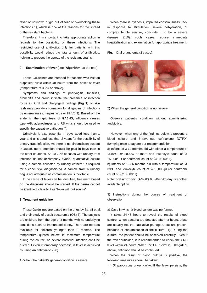
fever of unknown origin out of fear of overlooking these infections 1), which is one of the reasons for the spread of the resistant bacteria. Therefore, it is important to take appropriate action in regards to the possibility of these infections. The restricted use of antibiotics only for patients with this possibility would reduce the total amount of antibiotics, helping to prevent the spread of the resistant strains.
25
2. Examination of fever (see “Algorithm” at the end) These Guidelines are intended for patients who visit an outpatient clinic within 48 hours from the onset of fever (temperature of 38°C or above). Symptoms and findings of pharyngitis, tonsillitis, bronchitis and croup indicate the presence of infection focus 2). Oral and pharyngeal findings (Fig 1) or skin rash may provide information for diagnosis of infections by enteroviruses, herpes virus or HHV6 3). Based on the endemic, the rapid tests of GABHS, influenza viruses type A/B, adenoviruses and RS virus should be used to specify the causative pathogen 4). Urinalysis is also essential in boys aged less than 1 year and girls aged less than 2 years for the possibility of urinary tract infection. As there is no circumcision custom in Japan, more attention should be paid in boys than in the other countries. As 10-20% of cases with urinary tract infection do not accompany pyuria, quantitative culture using a sample collected by urinary catheter is required for a conclusive diagnosis 5). A sample from a urinary bag is not adequate as contamination is inevitable.
If the cause of fever can be identified, treatment based on the diagnosis should be started. If the cause cannot be identified, classify it as “fever without source”. 3. Treatment guideline
These Guidelines are based on the ones by Baraff et al. and their study of occult bacteremia (OB) 6). The subjects are children, from the age of 3 months with no underlying conditions such as immunodeficiency. There are no data available for children younger than 3 months. The temperature quoted below is maximum temperature during the course, as severe bacterial infection can’t be ruled out even if temporary decrease in fever is achieved by using an antipyretic 7) 8). 1) When the patient’s general condition is severe
When there is cyanosis, impaired consciousness, lack in response to stimulation, severe dehydration, or complex febrile seizure, conclude it to be a severe disease 9)10): such cases require immediate hospitalization and examination for appropriate treatment. Fig. Oral enanthema (2 cases)
2) When the general condition is not severe
Observe patient’s condition without administering antibiotics.
However, when one of the findings below is present, a blood culture and intravenous ceftriaxone (CTRX) 50mg/kg once a day are our recommendation: a) Infants of 3-12 months old with either a temperature of ≧40°C, or 38.5°C or more and leukocyte count of ≧15,000/µl ( or neutrophil count of ≧10,000/µl). b) Infants of 12-36 months old with a temperature of ≧39°C and leukocyte count of ≧15,000/µl (or neutrophil count of ≧10,000/µl). Note: oral amoxicillin (AMOX) 60-90mg/kg/day is another available option. 3) Instructions during the course of treatment or observation a) Case in which a blood culture was performed
It takes 24-48 hours to reveal the results of blood culture. When bacteria are detected after 48 hours, those are usually not the causative pathogen, but are present because of contamination of the culture 11). During the culture, the patient should be observed carefully. Even if the fever subsides, it is recommended to check the CRP level within 24 hours. When the CRP level is 5.0mg/dl or above, antibiotic should be continued.
When the result of blood culture is positive, the following measures should be taken: ⅰ) Streptococcus pneumoniae: If the fever persists, the
26
patient should be hospitalized for immediate treatment to prevent serious bacterial complication. If the fever subsides, there are two options: to hospitalize for intravenous antibiotic therapy, or to administer a susceptible antibiotic orally for seven days at an outpatient clinic. ⅱ ) Haemophilus influenzae type b (Hib): the patient should be hospitalized for immediate treatment to prevent serious bacterial complication even if the fever has subsided. Hib occasionally causes serious bacterial complication even after the fever subsides 12). If severe symptoms develop during the disease’s course, it is necessary to make an appropriate evaluation and treat immediately. ⅲ) Others: When meningococcus is detected, the rate of subsequent development of serious bacterial complication is high 13), and the patient in Japan also tends to have an underlying condition such as complement deficiency 14); thus hospitalization is indicated regardless of the patient’s general condition. When one of the following bacteria is detected: Salmonella, Group A beta-hemolytic streptococcus (GABHS), Group B streptococcus (GBS), Staphylococcus, Moraxella catarrhalis and Haemophilus influenzae other than Hib, the patient should be hospitalized for adequate evaluation, even though contamination is possible. b) Case in which a blood culture was not performed
Observation should be continued for 6-48 hours. When the fever persists, the CRP level should be checked. If the result is 5.0mg/dl or above, treatment should be started in consideration for the possibility of serious bacterial infection. 4. Overview 1) Occult Bacteremia and Guidelines
Although fever without source is mostly due to viral infection, a small number of the cause can be OB or other severe bacterial infection in an early stage. As OB is a disorder with relatively high frequency, it has been studied in many clinical trials as a model disease for the beginning of severe bacterial infection. Our Guidelines are also based on these OB findings.
OB is defined as bacteremia with the only symptom of fever, occasionally associated with the symptoms of the common cold or otitis media, and one with no findings of obvious localized infection or deteriorated general
condition 15). Serious localized infections such as meningitis, pneumonia, epiglottitis, cellulitis, suppurative arthritis and osteomyelitis occur secondarily in 5-15% of OB cases 16) 17). Among the patients with fever of unknown focus, OB occurs more frequently in children with higher body temperature, leukocyte count, neutrophil count and CRP level 18-20).
The first large-scale research on OB was conducted by McGowan et al. in 1973 21). They detected 31 patients (4.4%) with bacteremia from 708 febrile children with unknown focus who were in fairly stable general condition of rectal temperature of 37.8°C or more. The precise diagnosis of bacteremia was not possible only by physical examination, because they had no other characteristic symptoms and signs than fever. It was revealed later that serious bacterial infections such as bacterial meningitis occur secondarily in some OB cases, which led to various studies concerning the relationship between OB and clinical or laboratorial findings.
In 1983, Dershewitz et al. studied whether it was possible to predict the occurrence of OB in children by both interview and physical examination at the first visit in patients of 3-24 months old with temperatures higher than 39.5°C and without serious general condition 22). The results showed that the blood cultures were indeed positive in 10 of 110 cases (9.1%) suspected OB, whereas those were also positive in 15 of 578 cases (2.6%) not suspected OB. These results demonstrated that it was difficult to exactly predict OB in advance. Most of these cases had been misdiagnosed as upper respiratory infection, otitis media or others until positive blood culture results were obtained. One of these patients had bacterial meningitis 24 hours later.
Concerning the laboratory findings predictive of OB, Bass et al. examined children between 3 and 36 months of the age with fever without source in 1993 18). Blood cultures were obtained from children with (a) rectal temperature of 40°C or above, or (b) rectal temperature of 39.5°C or above and leukocyte count of 15,000/µl or above, and then diagnosed 60 cases of OB. Of these, 5/182 (2.7%) cases with leukocyte count of <15,000/µl had OB, whereas 55/331 (16.6%) cases with leukocyte count of ≧15,000/µl had OB. In addition, the incidence of OB increased in proportion to the rise of the leukocyte counts.
Fleisher et al., in their research on treatment efficacy on OB, compared CTRX 50mg/kg by intramuscular injection and AMOX 60mg/kg by oral administration 23). The results showed that CTRX prevented secondary
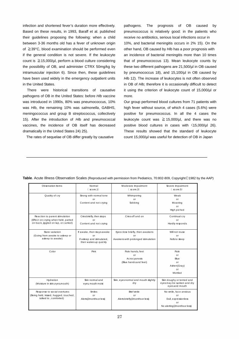
infection and shortened fever’s duration more effectively. Based on these results, in 1993, Baraff et al. published their guidelines proposing the following: when a child between 3-36 months old has a fever of unknown origin of ≧39°C, blood examination should be performed even if the general condition is not severe. If the leukocyte count is ≧15,000/µl, perform a blood culture considering the possibility of OB, and administer CTRX 50mg/kg by intramuscular injection 6). Since then, these guidelines have been used widely in the emergency outpatient units in the United States. There were historical transitions of causative pathogens of OB in the United States: before Hib vaccine was introduced in 1990s, 80% was pneumococcus, 10% was Hib, the remaining 10% was salmonella, GABHS, meningococcus and group B streptococcus, collectively 15). After the introduction of Hib and pneumococcal vaccines, the incidence of OB itself has decreased dramatically in the United States 24) 25).
The rates of sequelae of OB differ greatly by causative
27
pathogens. The prognosis of OB caused by pneumococcus is relatively good: in the patients who receive no antibiotics, serious local infections occur in 10%, and bacterial meningitis occurs in 2% 15). On the other hand, OB caused by Hib has a poor prognosis with an incidence of bacterial meningitis more than 10 times that of pneumococcus 13). Mean leukocyte counts by these two different pathogens are 21,500/µl in OB caused by pneumococcus 18), and 15,100/µl in OB caused by Hib 12). The increase of leukocytes is not often observed in OB of Hib; therefore it is occasionally difficult to detect it using the criterion of leukocyte count of 15,000/µl or more. Our group performed blood cultures from 71 patients with high fever without source, of which 4 cases (5.6%) were positive for pneumococcus. In all the 4 cases the leukocyte count was ≧ 15,000/µl, and there was no positive blood cultures in cases with <15,000/µl 26). These results showed that the standard of leukocyte count 15,000/µl was useful for detection of OB in Japan
Table. Acute Illness Observation Scales (Reproduced with permission from Pediatrics, 70:802-809, CopyrightⒸ1982 by the AAP)
No smile, face anxiousor
Dull, expressionlessor
No alerting(2months or less)
Brief smileor
Alerts briefly(2months or less)
Smilesor
Alerts(2months or less)
Response to social overtures(Being held, kissed, hugged, touched,
talked to , comforted)
Skin doughy or tented andeyes may be sunken and dry
eyes and mouth
Skin, eyes normal and mouth slightlydry
Skin normal andeyes, mouth moist
Hydration(Moisture in skin,eyes,mouth)
Paleor
Blueor
Ashen(Gray)or
Mottled
Pale hands, feetor
Acrocyanosis(Blue hands and feet)
PinkColor
Will not rouseor
Falls to sleep
Eyes close briefly, then awakensor
Awakens with prolonged stimulation
If awake, then stays awakeor
If asleep and stimulated,then wakes up quickly
State variation(Going from awake to asleep or
asleep to awake)
Continual cryor
Hardly responds
Cries off and onCries briefly, then stopsor
Content and not crying
Reaction to parent stimulation(Effect on crying when held, pattedon back, jiggled on lap, or carried)
Weakor
Moaningor
High piched
Whimperingor
Sobbing
Strong with normal toneor
Content and not crying
Quality of cry
Severe Impairment( score;5)
Moderate Impairment( score;3)
Normal( score;1)
Observation Items

as well. In addition, a study of 175 febrile children with leukocyte count ≧15,000/µl and fever without source found 20 cases of pneumococcal OB and 3 Hib cases. Bacterial meningitis followed one Hib case 27).The current incidence of OB is similar to that of the United States before the introduction of Hib vaccine and therefore OB appears to be a major cause of serious bacterial infections in Japan even now.
2) Limits of the Guidelines (1) Limits of clinical findings for detecting severe
bacterial infection When a doctor sees a patient with fever, an
assessment of systemic condition should be taken first. For an experienced doctor, it is not difficult to make an exact judgment by physical examination only, perhaps without the help of Table (Acute Illness Observation Scales: AIOS) 10). According to McCarthy who developed these scales, the sensitivity of them for detecting a serious bacterial infection when used with the patient’s clinical history, careful interviewing and physical examination is 90% 28). This means that 10% is undetectable from clinical findings.
28
Because medical services are so easily accessible in Japan, patients tend to visit clinics in the early stage of a fever, before serious symptoms have developed. In the research of Takeuchi et al., the rates of the symptoms observed on the first day of bacterial meningitis were as follows: vomiting (56.7%), convulsion (7.5%), and one or more meningeal signs such as neck stiffness and Kernig’s sign, or anterior fontanelle bulging (11.1%). Although vomiting is often observed from the early stage, this is not a symptom specific to meningitis. It is not frequent at all to observe the meningeal signs that are considered more diagnostic. It is therefore difficult to diagnose bacterial meningitis on the first day of the illness when the patient’s general condition is not serious 29).
In bacterial meningitis and other serious bacterial infections, the systemic conditions become worse as time goes by. Therefore, it is important to make an evaluation of severity at appropriate intervals. (2) Limits of laboratory finding for detecting OB These Guidelines use a leukocyte count of ≧15,000/µl as the criterion for administrating antibiotics. Thirty percent of the cases of fever without source confirm this
criterion 26), including 65% of all OB cases 15). Antibiotics are expected to improve prognosis for these patients.
The remaining 70% with leukocyte count of <15,000/µl includes 35% of the OB cases, though they are undetectable in our Guidelines. As the odds of these cases being OB are low, it is not practical to perform blood cultures and administer antibiotics for all these cases. The “wait and see approach” is recommended. 3) Blood examination
Blood examination data is required to decide treatment
options based on these Guidelines. Risk assessment by this method is essential (even though it may be painful and cost more). It is possible to measure both leukocyte/neutrophil counts and CRP level simultaneously using only 20µl of capillary blood sample by a commercially available instrument. The pain would be minimal if the blood were collected from a fingertip or an ear lobe using a blood-collecting needle for diabetes
(Fig 2.) 30).
Fig 2. How to collect blood from a fingertip 1) Prick the fingertip using a disposable lancet for diabetes. 2) Gently massage from the hand to near the puncture site to obtain the required blood volume, and draw up blood into a capillary tube - EDTA (or
heparin) coated. 3) Put blood into a micro tube.

4) The instrument draws up blood.
29
Blood culture is possible with only 0.5-2.0ml of blood
sample using the BACTEC® bottle for children. Except for special cases, the culture needs to target only aerobic bacteria. The CO2 sensor underneath the bottle reacts if the bacteria are cultured. The positive results are obtained after 12-24 hours. 4) Administration method
When a severe bacterial infection is suspected, antibiotics should be administered intravenously whenever possible. Since this method is more efficient than oral administration for taking the drug into the body, there is a quicker increase of the drug concentration in the blood. Therefore, better effects are expected. CTRX is recommended, as it has a long half-life of 7.5 hours, and sustains effective blood concentration for 24 hours after administration -- features suitable for outpatient treatment. 5. Instructions for the practice of these Guidelines
Careful observation on the course of the disease is most important for the judicious use of antibiotics. It is not possible to identify all cases of serious bacterial infection only by these programs. The Guidelines shouldn’t restrict or force the use of antibiotics on children with the possible bacterial infection. Doctors should examine each patient carefully and use their discretion to decide the optimal treatment. The ultimate decision of the use of antibiotics is left to each clinician. 6. Summary
It has been known that most children’s fevers are caused by viral infection. However, from the lack of data in daily practice and the fear of serious bacterial infections such as OB or meningitis, oral antibiotics have been prescribed to all febrile children. Such an approach
may have been effective for preventing possible complications secondary to OB in the era of a few or no resistant bacteria. However, the resistant strains of pneumococcus and Hib (common causative agents for OB) are increasing rapidly, and the conventional approach will soon be ineffective in Japan.
With the widespread use of medical equipment, it is now possible to manage a risk control of fever by both rapid blood examinations and rapid antigen tests in the outpatient settings. In these Guidelines, we propose a principle of not administering antibiotics to the cases with leukocyte count of <15,000/µl, with the exception of both infants of less than 12 months old with fever of ≧40°C and cases with bad general condition.
Prescribing oral antibiotics without risk assessment to all febrile children will result in an increase of resistant bacteria and difficulty in treatment of OB and other serious bacterial infections. Oral antibiotics have possible effects of slightly decreasing risks of pneumococcal OB 31). However, they are not only ineffective on OB due to Hib, but also the use of them may delay the correct diagnosis, and result in a poor prognosis if bacterial meningitis has already developed 32). Therefore, it is not recommended to prescribe antibiotics to all febrile children. Subjoinder:
Baraff’s guidelines (the basis for our Guidelines) are now intended in the United States only for children who have not received Hib and pneumococcus vaccines. These vaccines should also be introduced in Japan as soon as possible to prevent serious bacterial infections and to reduce the use of antibiotics. References 1. 草刈 章 (Kusakari A), 他. 小児科外来における上気
道炎診療調査―発病72時間以内の初診患者に対する
抗菌薬使用状況― . 外来小児科 2004;7:122-127. (In Japanese)
2. Greenes DS, et al. Low risk of bacteremia in febrile children with recognizable viral syndromes. Pediatr Infect Dis J 1999;18:258-261 [Medline]
3. 原三千丸 (Hara M). 症状から見たウイルス感染症診
断学. 小児内科 2005;37:31-37. (In Japanese) 4. 吉村 速 (Yoshimura H). 迅速診断キットの利用法.
小児内科 2005;37:38-41. (In Japanese) 5. Shaw KN, et al. Prevalence of urinary tract infection

in febrile young children in the emergency department. Pediatrics 1998;102:e16 [Full Text]
6. Baraff LJ, et al. Practice guideline for the management of infants and children 0 to 36 months of age with fever without source. Pediatrics 1993;92:1-12 [Abstract]
7. Mazur LJ, et al. Temperature response to acetaminophen and risk of occult bacteremia: a case-control study. J Pediatr 1989;115:888-891 [Medline]
8. Torrey SB, et al. Temperature response to antipyretic therapy in children: relationship to occult bacteremia. Am J Emerg Med 1985;3:190-192 [Medline]
9. Luszczak M. Evaluation and Management of infants and young children with fever. Am Fam Physician 2001;64:1219-1226 [Full Text]
10. McCarthy PL, et al. Observation scales to identify serious illness in febrile children. Pediatrics 1982;70:802-809 [Abstract]
11. Alpern ER, et al. Occult bacteremia from a pediatric emergency department: current prevalence, time to detection, and outcome. Pediatrics 2000;106:505-511 [Abstract]
12. Korones DN, et al. Outcome of children with occult bacteremia caused by Haemophilus influenzae type b. Pediatr Infect Dis J 1992;11:516-520 [Medline]
13. Shapiro ED, et al. Risk factors for development of bacterial meningitis among children with occult bacteremia. J Pediatr 1986;109:15-19 [Medline]
14. 原 寿郎 (Hara T). 免疫不全シリーズ 補体系の
異常と免疫不全症 . 免疫 Immunology Frontier 2001;11:179-181. (In Japanese)
15. Harper MB, et al. Occult bacteremia in the 3-month-old to 3-year-old age group. Pediatr Ann 1993;22:484,487-493 [Medline]
16. Bratton L, et al. Outcome of unsuspected pneumococcemia in children not initially admitted to the hospital. J Pediatr 1977;90:703-706 [Medline]
17. Marshall R, et al. Unsuspected bacteremia due to Haemophilus influenzae: outcome in children not initially admitted to hospital. J Pediatr 1979;95:690-695 [Medline]
18. Bass JW, et al. Antimicrobial treatment of occult bacteremia: a multicenter cooperative study. Pediatr Infect Dis J 1993;12:466-473 [Medline]
19. Kuppermann N, et al. Predictors of occult pneumococcal bacteremia in young febrile children. Ann Emerg Med 1998;31:679-687 [Medline]
20. Pulliam PN, et al. C-reactive protein in febrile children 1 to 36 months of age with clinically undetectable serious bacterial infection. Pediatrics 2001;108:1275-1279 [Abstract]
21. McGowan JE, et al. Bacteremia in febrile children seen in a "walk-in" pediatric clinic. N Engl J Med 1973;288:1309-1312
22. Dershewitz RA, et al. A comparative study of the prevalence, outcome, and prediction of bacteremia in children. J Pediatr 1983;103:352-358 [Medline]
23. Fleisher GR, et al. Intramuscular versus oral antibiotic therapy for the prevention of meningitis and other bacterial sequelae in young, febrile children at risk for occult bacteremia. J Pediatr 1994;124:504-512 [Medline]
24. Lee GM, et al. Risk of bacteremia for febrile young children in the post-Haemophilus influenzae type b era. Arch Pediatr Adolesc Med 1998; 152:624-628 [Medline]
25. Klein JO. Management of the febrile child without a focus of infection in the era of universal pneumococcal immunization. Pediatr Infect Dis J 2002;21:584-588 [Abstract]
26. 西村龍夫 (Nishimura T), 他. 小児科外来における
occult bacteremia の 前 方 視 的 調 査 . 日 児 誌 2004;108:620-624 (In Japanese) [Abstract]
27. 西村龍夫 (Nishimura T), 他. 小児科開業医で経験し
た occult bacteremia23例の臨床的検討 . 日児誌 2005;109:623-629. (In Japanese)
28. McCarthy PL. Fever. Pediatrics in Review 1998;19:401-408
29. 武内一 (Takeuchi H), 他. インフルエンザ菌・肺炎球
菌髄膜炎の早期スクリーニングの可能性 .日児誌 2006;110:1401-1408. (In Japanese) [Abstract]
30. 鈴江純史 (Suzue J). 診察時にCRPとWBCを迅速測
定する効果と意義 . 循環器情報処理研究会雑誌 2000;15:59-64. (In Japanese)
31. Rothrock SG, et al. Do oral antibiotics prevent meningitis and serious bacterial infections in children with Streptococcus pneumoniae occult bacteremia? A meta-analysis. Pediatrics 1997;99:438-444 [Abstract]
32. Kaplan SL, et al. Association between preadmission oral antibiotic therapy and cerebrospinal fluid findings and sequelae caused by Haemophilus influenzae type b meningitis. Pediatr Infect Dis 1986;5:626-632 [Medline]
Return to the top
30
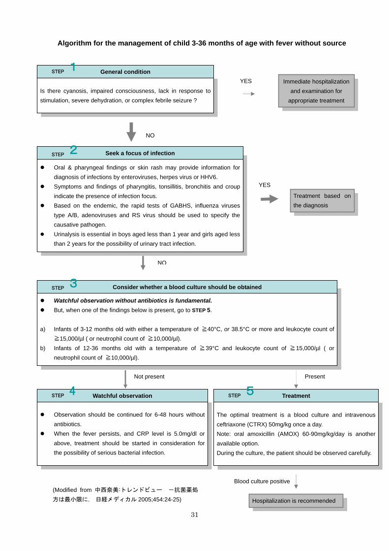
31
Algorithm for the management of child 3-36 months of age with fever without source
Observation should be continued for 6-48 hours without
antibiotics. When the fever persists, and CRP level is 5.0mg/dl or
above, treatment should be started in consideration for the possibility of serious bacterial infection.
Watchful observation without antibiotics is fundamental. But, when one of the findings below is present, go to STEP 5.
a) Infants of 3-12 months old with either a temperature of ≧40°C, or 38.5°C or more and leukocyte count of
≧15,000/µl ( or neutrophil count of ≧10,000/µl). b) Infants of 12-36 months old with a temperature of ≧39°C and leukocyte count of ≧15,000/µl ( or
neutrophil count of ≧10,000/µl).
YES
NO
Is there cyanosis, impaired consciousness, lack in response to stimulation, severe dehydration, or complex febrile seizure ?
1 General condition STEP
Immediate hospitalization and examination for
appropriate treatment
Oral & pharyngeal findings or skin rash may provide information for diagnosis of infections by enteroviruses, herpes virus or HHV6.
Symptoms and findings of pharyngitis, tonsillitis, bronchitis and croup indicate the presence of infection focus.
Based on the endemic, the rapid tests of GABHS, influenza virusestype A/B, adenoviruses and RS virus should be used to specify the causative pathogen.
Urinalysis is essential in boys aged less than 1 year and girls aged less than 2 years for the possibility of urinary tract infection.
2 Seek a focus of infection STEP
YES Treatment based on the diagnosis
NO
3 Consider whether a blood culture should be obtained STEP
PresentNot present
4 Watchful observation STEP
The optimal treatment is a blood culture and intravenous ceftriaxone (CTRX) 50mg/kg once a day. Note: oral amoxicillin (AMOX) 60-90mg/kg/day is another available option. During the culture, the patient should be observed carefully.
5 Treatment STEP
Blood culture positive (Modified from 中西奈美:トレンドビュー -抗菌薬処
方は最小限に. 日経メディカル 2005;454:24-25)
Hospitalization is recommended