Embed Size (px)
Citation preview

GUIDELINES FOR THE DIAGNOSIS OFLATENT TUBERCULOSIS INFECTION in the 21st Century
GUIDELINES FOR THE DIAGNOSIS OFLATENT TUBERCULOSIS INFECTION in the 21st Century
PO Box 1709 • 225 Warren Street • Newark, NJ 07101-1709
CME Certified Monograph2nd Edition

GUIDELINES FOR THE DIAGNOSIS OF LATENT
TUBERCULOSIS INFECTION IN THE 21st CENTURY
CONTINUING EDUCATION CERTIFIED MONOGRAPH
Sponsored by the University of Medicine & Dentistry of New Jersey (UMDNJ), and
the New Jersey Medical School Global Tuberculosis Institute
Original Release Date: June 2002 • Activity Update: April 1, 2008 • Expiration Date: March 31, 2010
Nursing credit for this activity will be provided through March 31, 2010
This activity is supported by educational grants from Monarch Pharmaceuticals for the first edition release and JHP
Pharmaceuticals for the release of the second edition.
TARGET AUDIENCE
This activity is designed for internists, pediatricians, pulmonologists, infectious disease specialists, public health and
preventive medicine specialists, nurses, and other health personnel interested or involved in tuberculosis diagnosis,
treatment and prevention of tuberculosis and latent tuberculosis infection.
LEARNING OBJECTIVES
Upon the completion of this activity, participants should be able to:
� Describe the role of tuberculin testing in low prevalence countries
� Review how tuberculins are developed, manufactured and validated
� Recognize minor disparities in commercially available tuberculins and the necessity of serial testing with the same antigen
� Explain the protocol for administering and reading tuberculin skin tests
� Correctly interpret repeated tuberculin skin tests
� Examine the role of tuberculin reactions produced by cross reactions with non-tuberculous mycobacteria
� Differentiate the use of interferon-γ release assays when compared to tuberculin skin testing
� Discuss the role of the nurse in the diagnosis of latent TB infection
METHOD OF INSTRUCTION
Participants should read the learning objectives and the activity in its entirety. After reviewing the material, complete
the post-test consisting of a series of multiple-choice questions.
Upon completing this activity as designed and achieving a passing score 70% or more on the post-test, participants
will receive a continuing education credit letter and test answer key four weeks after receipt of the registration and
evaluation materials.
Estimated time to complete this activity as designed is 3.5 hours.
c1

ACCREDITATION
Physicians: UMDNJ–Center for Continuing and Outreach Education is accredited by the Accreditation Council for
Continuing Medical Education to provide continuing education for physicians.
UMDNJ–Center for Continuing and Outreach Education designates this educational activity for a maximum of 3.5 AMA PRA
Category 1 Credit(s)™. Physicians should only claim credit commensurate with the extent of their participation in the activity.
Nurses: UMDNJ–Center for Continuing and Outreach Education is an approved provider of continuing nursing
education by NJSNA, an accredited approver by the American Nurses Credentialing Center’s Commission on Accreditation.
This activity is awarded 3.5 contact hours (60 minute CH).
Provider approved by the California Board of Registered Nursing, Provider Number CEP 13780.
This activity was peer-reviewed for relevance, accuracy of content and balance of presentation by Lee B. Reichman, MD,
MPH and Rajita Bhavaraju, MPH; and pilot-tested for time required for participation by Anju Budhwani, MD, Henry S.
Fraimow, MD, DJ McCabe, RN, MSN, and Lillian Pirog, RN, PNP.
DISCLOSURE
In accordance with the disclosure policies of UMDNJ and to conform with ACCME and FDA guidelines, individuals in a position
to control the content of this education activity are required to disclose to the activity participants: 1) the existence of any financial
interest or other relationships with propriety entities producing health care goods and services, with the exemption of non-profit or
government organizations and non-health care related companies, within the past 12 months; and 2) the identification of a
commercial product/device that is unlabeled for use or an investigational use of a product/device not yet approved.
The faculty and editors listed below have declared that they have no significant financial relationships or affiliations
to disclosure:
John-Manuel Andriote, MS Kitty Lambregts, MD, PhD, MPHRajita Bhavaraju, MPH Alfred Lardizabal, MDGeorge Comstock, MD, DrPH Richard Menzies, MD, MScKaren Galanowsky, RN, MPH Sheldon L. Morris, PhD
Elsa Villarino, MD, MPH
Lee B. Reichman, MD, MPH is a consultant for and shareholder of Cellestis, Inc.
The pilot-testers listed below have declared that they have no significant financial relationships or affiliations to disclosure:
Anju Budhwani, MD DJ McCabe, RN, MSNHenry S. Fraimow, MD Lillian Pirog, RN, PNP
OFF-LABEL USAGE DISCLOSURE
This publication contains information about the test, T-SPOT.TB, which has not been approved by the U.S. Food and
Drug Administration for the detection of M. tuberculosis as of the date of this publication.
DISCLAIMER
The views expressed in this publication are those of the faculty. It should not be inferred or assumed that they are
expressing the views of Monarch and JHP Pharmaceuticals, any other manufacturer of pharmaceuticals, or UMDNJ.
The drug selection and dosage information provided in this publication are believed to be accurate. However, the reader
is urged to consult the full prescribing information on any drug mentioned in this publication for recommended dosage,
indications, contraindications, warnings, precautions, and adverse effects before prescribing any medication. This is
particularly important when a drug is new or infrequently prescribed.
Copyright © 2008 UMDNJ-Center for Continuing and Outreach Education. All rights reserved including translation
into other languages. No part of this publication may be reproduced or transmitted in any form or by any means, electronic
or mechanical, including photocopying, recording, or any information storage and retrieval systems, without permission in
writing from UMDNJ-Center for Continuing and Outreach Education.
To review UMDNJ’s privacy policy: http://ccoe.umdnj.edu/general/privacypolicy.html.
Please direct continuing education related questions to UMDNJ at 800-227-4852 or email [email protected].––––––––––––––––––––––
This activity is accessible via Internet Explorer and Netscape browsers. A high speed connection is highly recommended.
You must have Adobe Acrobat Reader version 4 or above installed on your computer to view PDF files. If you do not have
Acrobat Reader, you can download it for free at Adobe.com.
Page composition and typography by Williams Production Group, Annandale, Virginia.

GUIDELINES FOR THE DIAGNOSIS OFLATENT TUBERCULOSIS INFECTION in the 21st Century
Continuing Education Certified Monograph2nd Edition
Edited by Lee B. Reichman, MD, MPH
John-Manuel Andriote, MS (First Edition)Rajita Bhavaraju, MPH (Second Edition)

IntroductionLee B. Reichman, M.D., M.P.H.
Relevance of the Tuberculin Test in Low-Prevalence CountriesKitty Lambregts, M.D., Ph.D, M.P.H.
Developing a Clinical Tuberculin Test: Tuberculin Characteristics, Reactivity and Potency
Sheldon L. Morris, Ph.D.
Randomized Clinical Trials of Specificities of Commercially Available Tuberculins
Elsa Villarino, M.D., M.P.H.
Administering and Reading Tuberculin Skin Tests;Interpreting Repeated Tuberculin Skin Tests
Richard Menzies, M.D., M.Sc.
Tuberculin Sensitivity Produced by MycobacteriaOther than the Mycobacterium Tuberculosis Complex
George Comstock, M.D., Dr.P.H.
Interferon-γ Release Assay for Detectionof Tuberculosis Infection
Alfred Lardizabal, M.D.
The Role of the Nurse inDiagnosing Latent TB InfectionKaren Galanowsky, R.N., M.P.H.
GUIDELINES FOR THE DIAGNOSIS OF LATENT
TUBERCULOSIS INFECTION IN THE 21st CENTURY

A special symposium was held in Miami Beach, Florida. It was
chaired by Lee B. Reichman, M.D., M.P.H., and the faculty featured
six distinguished authorities on the diagnosis of latent tuberculosis
infection.
This monograph presents suggested guidelines that emerged
from the meeting, which are intended for the practicing clinician,
and emphasize the administration and interpretation of
tuberculin tests as part of the diagnosis and treatment of latent
tuberculosis infection.
The symposium and monograph was supported by an
unrestricted educational grant from Monarch Pharmaceuticals.
Due to unprecedented interest, authors were asked to revise
and update chapters for a second edition, which was supported
by an unrestricted educational grant from JHP Pharmaceuticals.
John-Manuel Andriote was the technical editor of the original
monograph. Rajita Bhavaraju revised the 2nd edition.
SUGGESTED CITATION
To Cite the Entire Monograph
Reichman LB, Bhavaraju R, eds.Guidelines for the Diagnosis of Latent Tuberculosis Infection in
the 21st Century, 2nd Edition. Newark: New Jersey Medical School Global Tuberculosis
Institute, 2008.
To Cite a Specific Chapter
Menzies R. Interpreting Repeated Tuberculin Skin Tests. pages 38-46. in Reichman LB,
Bhavaraju R, eds: Guidelines for the Diagnosis of Latent Tuberculosis Infection in the 21st Century,
2nd Edition. Newark: New Jersey Medical School Global Tuberculosis Institute, 2008.

Guidelines for the Diagnosis of Latent Tuberculosis Infection in the 21st Century, 2nd Edition
George Comstock, M.D., Dr.P.H. passed away on July 15, 2007 after a long illness. We were particularly gratified that
even though quite ill, Dr. Comstock saw fit to bring his chapter, “Tuberculin Sensitivity Produced by Mycobacteria other
than Mycobacterium tuberculosis Complex,” up to date for the second edition of this monograph.
Much of our knowledge about latent tuberculosis infection is directly due to Dr. Comstock’s work. His research and
application of its results, has served as the basis of the diagnosis and management of this condition.
Dr. Comstock was a good friend and mentor to us all. It is with gratitude, appreciation, and respect that we dedicate this
monograph to his memory.
—Lee B. Reichman, M.D., M.P.H.
April 1, 2008
Dedication to George Comstock, M.D., Dr.P.H.

Guidelines for the Diagnosis of Latent Tuberculosis Infection in the 21st Century, 2nd Edition
Executive Summary . . . . . . . . . . . . . . . . . . . . . . . . . . . . . . . . . . . . . . . . . . . . . . . . . 1
Introduction . . . . . . . . . . . . . . . . . . . . . . . . . . . . . . . . . . . . . . . . . . . . . . . . . . . . . . . 7
Lee B. Reichman, M.D., M.P.H.
Relevance of the Tuberculin Test in Low-Prevalence Countries . . . . . . . . . . . . . . . 9
Kitty Lambregts, M.D., Ph.D., M.P.H.
Developing a Clinical Tuberculin Test: Tuberculin Characteristics,
Reactivity and Potency . . . . . . . . . . . . . . . . . . . . . . . . . . . . . . . . . . . . . . . . . . . . . 18
Sheldon L. Morris, Ph.D.
Randomized Clinical Trials of Specificities of Commercially
Available Tuberculins . . . . . . . . . . . . . . . . . . . . . . . . . . . . . . . . . . . . . . . . . . . . . . . 24
Elsa Villarino, M.D., M.P.H.
Administering and Reading Tuberculin Skin Tests . . . . . . . . . . . . . . . . . . . . . . . . 32
Dick Menzies, M.D., M.Sc.
Interpreting Repeated Tuberculin Skin Tests . . . . . . . . . . . . . . . . . . . . . . . . . . . . . 38
Dick Menzies, M.D., M.Sc.
Tuberculin Sensitivity Produced by Mycobacteria Other than the
Mycobacterium Tuberculosis Complex . . . . . . . . . . . . . . . . . . . . . . . . . . . . . . . . . . . 47
George Comstock, M.D., Dr.P.H.
Interferon-γ Release Assay for Detection of Tuberculosis Infection . . . . . . . . . . . 57
Alfred Lardizabal, M.D.
The Role of the Nurse in Diagnosing Latent TB Infection . . . . . . . . . . . . . . . . . . . 62
Karen Galanowsky R.N., M.P.H.
Glossary . . . . . . . . . . . . . . . . . . . . . . . . . . . . . . . . . . . . . . . . . . . . . . . . . . . . . . . . . 66
Activity Self-Assessment Test . . . . . . . . . . . . . . . . . . . . . . . . . . . . . . . . . . . . . . . . . 68
Registration Form . . . . . . . . . . . . . . . . . . . . . . . . . . . . . . . . . . . . . . . . . . . . . . . . . 69
Activity Evaluation Form . . . . . . . . . . . . . . . . . . . . . . . . . . . . . . . . . . . . . . . . . . . 71
CONTENTS


Guidelines for the Diagnosis of Latent Tuberculosis Infection in the 21st Century, 2nd Edition 1
For more than three decades, treatment of persons
with latent Mycobacterium tuberculosis infection to
prevent active disease has been an essential component of
TB control in the United States. The symposium on which
this monograph is based was organized to address the
confusion that has been generated by periodic outbreaks of
TB in the United States. Looking at these outbreaks, one
thing is clear: They were unnecessary and likely could
have been prevented if those at high risk were targeted,
tested and treated.
EVOLUTION OF TREATMENT GUIDELINES
In 1965, treatment for latent TB infection was first
recommended for those with previously untreated TB,
tuberculin skin test (TST) converters and all children
under age three with a positive tuberculin test result.
The recommendations were broadened in 1967 to
include all who were TST positive (>10mm) and their
close contacts.
Treatment guidelines were developed in 1974
regarding pre-treatment screening and monitoring to
minimize the risk for hepatitis and to exclude low-risk
persons over age 35 as candidates for treatment. The
guidelines were further revised in 1983 to recommend
routine clinical and laboratory monitoring for persons
older than 35 or with increased risk for hepatoxicity.
In the years since HIV/AIDS was first reported in 1981,
TB has reemerged as a tremendous threat to individuals
with compromised immune systems. Many of those
infected with HIV are co-infected with TB. For this reason,
two months of RIF and PZA (2RZ) were recommended in
1998 for co-infected HIV-positive persons, and
subsequently for those who were not HIV-infected.
Treatment guidelines for latent TB—what used to be
called “chemoprophylaxis”—have continued to be
refined. In 2000, nine months of Isoniazid was decreed
to be more effective that six months, and 2RZ were
deemed equal to nine months of Isoniazid. But in 2001
2RZ was de-emphasized, due to its liver toxicity, in favor
of nine months of Isoniazid. In 2004, rifampin alone for
4 months was also recommended.
TARGETED TUBERCULIN TESTING AND
TREATMENT OF LATENT TB INFECTION
As the rate of active tuberculosis in the United States
has decreased, identification and treatment of persons
with latent infection who are at high risk for active TB
have become essential components of the nation’s TB
elimination strategy.
In its 2000 report, Ending Neglect: The Elimination of
Tuberculosis in the United States, the Institute of Medicine
(IOM) recommended precisely what this symposium is
intended to address: an increased emphasis on targeted
tuberculin testing and treatment of latent TB infection.
“To begin advancing toward the elimination of
tuberculosis,” said the IOM report, “aggressive new
efforts must be implemented to identify those who are at
greatest risk of disease through targeted programs of
tuberculin skin testing coupled with treatment for latent
tuberculosis infection.” It continued, “The question now
confronting the United States is whether another cycle of
neglect will be allowed to begin or whether, instead,
decisive action will be taken.”
THE SYMPOSIUM AND GUIDELINES
The 2002 symposium, held in Miami Beach, Florida,
brought together clinicians and researchers with
considerable expertise on the diagnosis and treatment of
latent TB infection. Each of them prepared and
presented a paper that describes various aspects of
diagnosing and treating latent TB infection. These papers
are collected in this monograph, and have been updated
by the authors for this second edition.
Because the FDA approved an effective gamma
interferon release assay, we have added a chapter on
QuantiFERON®-TB Gold In-Tube.
EXECUTIVE SUMMARY

2 Guidelines for the Diagnosis of Latent Tuberculosis Infection in the 21st Century, 2nd Edition
RELEVANCE OF THE TUBERCULIN TEST IN
LOW-PREVALENCE COUNTRIES
The incidence of TB disease is declining in the United
States and other industrialized countries, and in fact the
U.S. has reached the elimination phase of the TB
epidemic. An important challenge, however, lies in the
fact that while the number of U.S.-born Americans
infected with TB continues to shrink, the pool of infected
foreign-born individuals grows larger. Among the
problems associated with imported TB are drug-resistant
bacteria, microepidemics among sub-populations
(including illegal immigrants, the homeless and
prisoners) and a reduction in the overall effectiveness of
treatment for latent TB infection.
The positive effect of adequate TB control—i.e.,
decreasing incidence—hampers TB control in the
elimination phase because professional expertise on TB
fades away. This results in diagnostic delay, improper
diagnostic tools, inadequate treatment, poor infection
control and inadequate guidance for patients. Other
challenges to diagnosing and treating latent TB infection
include the lack of cultural understanding among many
immigrants of treating a disease without symptoms. The
overlap of the TB and HIV/AIDS epidemics present
another major challenge.
Targeted tuberculin testing for latent TB infection is a
strategic component of TB control in low-prevalence
countries in that it identifies persons who are at high risk
for developing TB and would benefit from treatment of
latent TB infection. In particular, tuberculin skin testing
(TST) is useful for:
� Contact tracing
� Providing pre-exposure baseline TST results for
exposure groups, such as health care workers
� Screening of persons with clinical conditions
associated with progression to active disease
� Screening of risk groups for TB
� Screening travelers to high-prevalence countries
� Skin-testing of symptomatic patients, and
� Measuring annual risk of infection and program
impact
QuantiFERON®-TB Gold In-Tube has similar specific
advantages.
Despite its usefulness, the TST also has consequences
and limitations. In particular:
� A TST result, whether negative or positive, may
add valuable information in symptomatic patients
� A test result just below the cutoff point for a
specific target group should provide guidance to
both patient and health care providers by raising
the level of suspicion so they will consider TB if
there are symptoms
� In some instances—such as with liver disease or
among the elderly who may long have been
infected—the physician may decide not to start
treatment for latent TB infection but rather to
offer close monitoring and proper instruction to
the patient
� There may be false positive or negative reactions
due to either technical or biological causes
� Boosting of tuberculin sensitivity
DEVELOPING A CLINICAL TUBERCULIN TEST:TUBERCULIN CHARACTERISTICS, REACTIVITY
AND POTENCY
Tuberculins are complex mixtures of culture filtrate
components derived from sterilized cultures of tubercle
bacilli. The predominant form of tuberculin used in the
United States is Tuberculin PPD, a protein precipitate of
M. tuberculosis culture filtrate. Tuberculins are widely
used to detect exposure to M. tuberculosis because they
induce delayed-type hypersensitivity immune reactions
in individuals who have been sensitized to
mycobacterial antigens.
There are four primary stages in the clinical
development of new tuberculin preparations:
(1) Preclinical development and testing
(2) Investigational New Drug (IND) stage
(3) Biologics License Application stage
(4) Post-licensure stage
During the critical IND stage, the potency of the new
product is evaluated by comparing its skin test reactivity
to the skin test responses induced by a standard dose of
PPD-S, the U.S. Standard PPD. When the dose of the
new product that is bioequivalent to PPD-S has been
determined, the specificity and sensitivity of the new
tuberculin is often assessed by testing it in at least three
populations: persons known to be infected with M.
tuberculosis, persons living in areas with low
mycobacterial infection and persons living in areas with
high atypical mycobacterial infection rates.

Guidelines for the Diagnosis of Latent Tuberculosis Infection in the 21st Century, 2nd Edition 3
If the potency, specificity and sensitivity of the new
product are shown to be appropriate in these clinical trials,
these data become the foundation of a Biologics License
application. To ensure consistent reactivity after licensure,
the potency of each commercial tuberculin lot must be
shown to be bioequivalent to PPD-S prior to distribution.
RANDOMIZED CLINICAL TRIALS OF
SPECIFICITIES OF COMMERCIALLY AVAILABLE
TUBERCULINS
The tuberculin skin test (TST) is the standard method
for diagnosing infection with M. tuberculosis. The test
involves intracutaneous injection of 5 tuberculin units
(TU) of purified protein derivative (PPD) by the
Mantoux technique. Two companies manufacture
PPD tuberculin in the United States: JHP Pharmaceuticals
(Aplisol®) and Pasteur Mérieux Connaught (Tubersol®).
Despite FDA regulations for production and
standardization of PPD tuberculin, there have been
concerns that these commercial PPD products may vary
in performance. Clusters of unexpected positive
reactions or suspected false-positive results involving
both products have been reported in the medical
literature, to the FDA and to the Centers for Disease
Control and Prevention (CDC).
The accurate diagnosis of infection is important to
ensure that infected persons receive appropriate
evaluation and treatment, and that uninfected persons
are not exposed to unnecessary evaluation and treatment.
The possibility that one or both of the commercial PPD
products may have an unacceptably high rate of false-
positive reactions prompted a study. The study
compared the specificity—i.e., the percentage of
uninfected persons correctly categorized—and the
distribution of reaction sizes of the two commercial PPD
reagents among a population of subjects who, because of
their history, were at low risk for infection with M.
tuberculosis. Besides skin testing with the two
commercial PPD reagents, subjects were skin tested with
PPD-S, the “gold standard” PPD test.
The randomized, double-blinded trial among a total
of 1,596 volunteers revealed that:
� Testing with Tubersol® produced smaller
reactions, and with Aplisol®, larger reactions, than
PPD-S, but these differences did not affect TST
interpretations
� The specificities of Aplisol® and Tubersol® were
equally high and similar to that of PPD-S
� Both Aplisol® and Tubersol® correctly classified
comparable numbers of persons not infected
with TB
� Either commercial product may be used with
confidence for TST
These study findings suggest:
� It is important to remember that the biological
variability in response to TST, as well as technical
differences in admininistering and reading the test
will result in increases or decreases of <6 mm in
95% of subjects
� It is also important to remember that erythema and
bruising at the TST reaction site are not indicative
of positive reactions and should be disregarded
when interpreting the TST
� The sensitivity and specificity of the TST for the
detection of latent TB infection (LTBI) is unknown
because no test can provide formal proof of the
presence or absence of LTBI
� Even when done in the most reliable way possible,
the TST remains an imperfect diagnostic tool and
should not replace clinical judgment
� Tubersol® demonstrates the greatest number of
non-reactors and thus would appear to be the least
suited for a screening test. Most importantly,
routine TST in a low-TB incidence area is of limited
use because the majority of positive results will be
false-positive
� With respect to rates of false positive results and
based on the results of this study, there are
probably no differences between the different
tuberculins
� At present, the most accurate method to diagnose
LTBI is the Mantoux TST, which requires:
– Testing a targeted high-risk population
– Properly administering a dose of a standardized
tuberculin preparation
– A trained professional correctly interpreting any
observed reaction
� An increase in the percentage of positive skin test
results is possible after a change of tuberculin
preparations

4 Guidelines for the Diagnosis of Latent Tuberculosis Infection in the 21st Century, 2nd Edition
Results of the study show that the key to approach this
issue is to think probabilistically by:
� First, using the available information to estimate
the likelihood of disease
� Then assessing the potential benefits and risk of
the proposed interventions (x-rays, sputum test,
drug therapy)
� Recognizing that sometimes the best strategy is to
wait for more information via repeat testing
A new laboratory based blood test with good
specificity and sensitivity, QuantiFERON®-TB Gold In-
Tube, has recently been FDA approved and will likely
find its use in screening of low risk reactors as well as
contacts for whom Mantoux tuberculin skin testing is
indicated.
ADMINISTERING AND READING TUBERCULIN
SKIN TESTS
Injecting tuberculin material intradermally into a
person previously infected with M. tuberculosis will result
in infiltration of previously sensitized lymphocytes from
circulating peripheral blood. At the site of the injection,
CD4 and CD8 T-lymphocytes, monocytes and
macrophages will accumulate. These release
inflammatory mediators, which produce edema and
erythema. Although this results in increased blood flow,
the locally increased metabolic activity of these
inflammatory cells results in relative hypoxia and
acidosis, which may be severe enough to lead to
ulceration and necrosis.
Certain guiding principles can help in appropriately
administering and reading tuberculin skin tests:
� Indications
– Test only those in whom therapy for latent TB
infection is indicated—namely, the “high-risk”
reactors:
• Recent infections (contact, conversion)
• Increased reactivation risk (HIV, diabetes,
abnormal chest x-ray, etc.)
� Contraindications
– Known documented severe reaction to TST in
the past
– Documented prior positive test result (TST
should be done if there is a history of
undocumented prior positive TST result)
� Administration
– Use only the Mantoux method, i.e., intradermal
injection on forearm
– Use 5-TU of PPD—bio-equivalent to PPD-S
– Anergy testing is not useful nor recommended
� Reading
– All reading must be done by trained health
professionals
– All readings must be made at 48-72 hours
– Measure the transverse diameter of induration
– Record result in millimeters
� Adverse Reactions
– Severe adverse reactions are rare
– Local allergic reactions occur in 2-3%, and are
not related to the actual tuberculin result
– Strongly positive reactions with blistering:
• Should be covered with a dry dressing
(to prevent scratching)
• Cold compresses may be soothing
• Corticosteroids (topical cream) are not effective
INTERPRETING REPEATED TUBERCULIN
SKIN TESTS
The use of repeated tuberculin tests to detect new TB
infections in high-risk populations has often resulted in
problems of interpretation. This is because tuberculin
reactions may change size because of random variation of
the test, or because of a real biologic increase. But it also
may be due to boosting or conversion.
� Random (Non-Specific or Chance) Variation—
When multiple tuberculin tests are administered
and read, resulting test-to-test differences will
include differences due to administration and
reading as well as inherent biological variability.
– Can account for changes of 1 to 5 millimeters in
size (bigger or smaller)
– So 6 millimeters is criterion to distinguish true
biologic changes in reaction
� Boosting (Two-step testing)—This phenomenon
is defined as an increase in tuberculin skin reactions
of at least 6 mm following repeat tuberculin testing
and unrelated to new mycobacterial infection. It is
believed to occur when cell-mediated response has
waned, resulting in an initially negative tuberculin
reaction, but the tuberculin test stimulates

Guidelines for the Diagnosis of Latent Tuberculosis Infection in the 21st Century, 2nd Edition 5
anamnestic immune recall. Boosting is often seen
among elderly persons.
– Seen if two tuberculin tests repeated in absence
of new infection
– Maximum if interval between two TSTs is
one week
• Less if only two to three days between TST
• Still detected after one year or more
– Associated with old age and foreign birth
(remote TB infection)
• Also with BCG vaccination (common in
foreign born)
• And with non-tuberculous mycobacteria
(common in southern USA and foreign-born)
– Non-specific reaction—risk of true infection
is lower
• Risk of disease is also lower
– Management—medical evaluation and chest
x-ray
• Benefit of therapy, and, therefore, need for
therapy is less
� Conversion—Tuberculin conversion is defined as an
increase in tuberculin reactions of at least 6mm
following repeat tuberculin testing and is due to new
mycobacterial infection.
– Defined as an increase of TST following new
mycobacterial infection
• Could be true TB infection, or BCG
vaccination, or nontuberculous mycobacteria
– Definition based on TST size
• Increase of 6 mm—most sensitive but less
specific
• Increase of 10 mm—less sensitive but more
specific - generally used
• Increase of 15 mm—much less
sensitive but more specific—not generally
used
– Prognosis—high risk of disease
• Highest risk if conversion following known
TB contact
– Management—medical evaluation and chest
x-ray
• Therapy for LTBI strongly recommended for
all ages
TUBERCULIN SENSITIVITY PRODUCED BY
MYCOBACTERIA OTHER THAN THE
MYCOBACTERIUM TUBERCULOSIS COMPLEX
“Nontuberculous mycobacteria” is a name suggested
for this numerous and diverse group of mycobacteria. As
the name suggests, the group is defined by exclusion, i.e.,
mycobacteria other than M. tuberculosis. Although
nontuberculous mycobacteria were recognized as early as
1885, they were rarely considered in the first half of the
20th century. By century’s end, however, they had
become well known because of the widespread use of
diagnostic cultures of M. tuberculosis and because of the
disease they caused among persons whose immune
systems had been compromised by HIV.
Studies of the similar reactions caused by both TB and
non-TB mycobacteria sparked renewed interest in the
ability of nontuberculous mycobacteria to provoke
sensitivity on the tests used to detect TB infection much
like TB itself. The outcome was a clearer understanding
that not all tuberculin test reactions are caused by
infection with M. tuberculosis. In short:
� A positive tuberculin test is not always due to
M. tuberculosis: During the decades after World
War II, it was demonstrated that reactions to the
tuberculin tests were not always due to infections
with M. tuberculosis.
� Role of nontuberculous mycobacteria:
Infections with a variety of nontuberculous
mycobacteria are common, especially in the
warmer parts of the world. They cause reactions to
the tuberculin test that tend to be smaller than
those due to M. tuberculosis.
� Probability, not certainty: There is no way to
differentiate all individuals who are infected with
M. tuberculosis from those infected with other
mycobacteria (including M. bovis BCG). The size of
the tuberculin test reaction indicates the
probability that it was caused by infection with
M. tuberculosis.
� Accurate measurements of reactions are
essential: To find the optimal cut-point between
positive and negative reactions, or to apply the
CDC/ATS recommended cut-points with maximal
effectiveness, requires accurate measurements of
induration. Specifically, this means there should be
a smooth distribution of reaction sizes without
undue proportions of reaction sizes ending in 5 or 0.

6 Guidelines for the Diagnosis of Latent Tuberculosis Infection in the 21st Century, 2nd Edition
� What can be seen, can be measured: Margins
of induration should be visualized by examining
the arm in proper lighting or by marking the
margins by the ballpoint pen method. In either
case, it is highly desirable to use a gauge or calipers
that do not allow the scale to be seen until after the
measurement has been made.
� Selecting a locally optimal cut-point: The
mirror-image method of estimating the proportion
of reactions caused by M. tuberculosis indicates
the optimal cut-point between positive and
negative reactions, taking into consideration the
local relative frequency of tuberculous and
nontuberculous infections.
INTERFERON-γ RELEASE ASSAY FOR
DETECTION OF TUBERCULOSIS INFECTION
Until recently, the standard and only method for
immunologic diagnosis of M. tuberculosis infection has been
limited to the tuberculin skin test (TST). However, because
purified protein derivative of tuberculin contains many
antigens that are shared with other mycobacteria, the skin
test does not reliably distinguish LTBI from prior
immunization with Mycobacterium bovis bacilli Calmette-
Guérin (BCG) or infection with environmental
mycobacteria. False-negative results in the setting of host
immunosuppression has limited also its utility. In addition,
cutaneous sensitivity to tuberculin develops from 2 to 10
weeks after infection and the TST requires two encounters
with a health care professional which often causes logistical
problems if not inconvenience. Finally, skilled personnel are
essential for proper placement and interpretation of the test.
Countering many of the concerns associated with the
TST, the QuantiFERON®-TB test (QFT) was approved by
the US Food and Drug Administration (FDA) in 2001 and
the and current generation of this test, QuantiFERON®-
TB Gold In-Tube (QFT-G), received final approval from
the FDA in 2007. Like the TST, QFT measures a
component of cell-mediated immune reactivity (CMI) to
purified protein derivative (PPD) from M. tuberculosis as
well as M. avium intercellure. However, as a blood assay,
the QFT requires a single patient visit, and because it is an
ex vivo test, it does not boost anamnestic immune
responses. The interpretation of the whole-blood
interferon gamma release assay (IGRA) is less subjective
than the TST, and the test is less affected by prior BCG
vaccination and reactivity to non-tuberculous
mycobacteria than the TST.
The principles of QuantiFERON®-TB Gold In-Tube
Test are incubation of whole blood with antigens,
measurement of IFN-γ by ELISA, and interpretation of
test results.
THE ROLE OF THE NURSE IN DIAGNOSING
LATENT TB INFECTION
The nurse plays a vital role in the diagnosis of latent
TB infection by:
� Providing tuberculin skin testing for high-risk
individuals within a community, and
� Ensuring that those individuals who have a positive
tuberculin skin test are medically evaluated and
treated for latent TB infection to completion.
By doing these two things, future cases of TB will
be prevented.
Targeted tuberculin skin testing and treatment of
those individuals with latent TB infection is a public
health activity, which has significant health, social and
economic benefits. To accomplish this, the nurse must
integrate the core functions of public health into
interventions and strategies at the individual, community
and health care system levels. This requires knowledge
and competencies regarding:
� The nursing process
� The diagnosis of latent TB infection and disease
� Tuberculin skin testing administration, reading,
and interpretation
� Community assessment
� Adherence
� Epidemiology
� Regulations and legal mandates
� Patient education that is culturally and
linguistically appropriate
� Collaboration
� Networking
� Evaluation
Confronted with the challenges at the individual,
community and health care system level, nurses utilize
knowledge and competencies to intervene appropriately
and prevail to achieve significant outcomes.

Guidelines for the Diagnosis of Latent Tuberculosis Infection in the 21st Century, 2nd Edition 7
Lee B. Reichman, M.D.,
M.P.H.
Dr. Reichman is Professor of
Medicine, Preventive Medicine
and Community Health, and
Executive Director of the New
Jersey Medical School Global
Tuberculosis Institute in Newark,
New Jersey
For more than three decades, treatment of persons
with latent Mycobacterium tuberculosis infection to
prevent active disease has been an essential component of
TB control in the United States. This symposium was
organized to address the confusion that has been generated
by periodic outbreaks of TB in the United States. Looking
at these outbreaks, one thing is clear: they were
unnecessary and likely could have been prevented if those
at high risk were targeted, tested and treated.
CONFUSION ABOUT TB TESTING
There has been confusion about how to target
tuberculin testing and treat latent TB infection, evidenced
in written and e-mail correspondence received by the
New Jersey Medical School Global Tuberculosis Institute:
� “I have taken two tests and they both come out
puffy and red and the clinic said they are positive. I
just enrolled my daughter in preschool and now I’m
not allowed to help in the school. What is TB and
how come my tests are coming up positive?”
� “I am a physician and have a 20-year-old patient
recently arrived from Finland. As is the practice
there, she received BCG as a child approximately
18 months ago. A recent PPD resulted in an 18
mm reaction. Her chest x-ray is normal and she
has neither a history of TB exposure nor immuno-
suppressive conditions. She has, however, traveled
to Russia in the past few years on at least two
occasions. I am aware of the general
recommendations about BCG and PPD
interpretation but the Finish government has
provided this lady with written statements that her
PPD is a result of the BCG and that no treatment is
advised. What do you think? Are there
organizational recommendations (e.g., WHO) that
I can refer to in this matter?”
� “My sixteen-year-old daughter has tested positive
for TB. I am very much concerned. She was asked
to take Isoniazid 300 mg tablets for the next six
months. Is it really necessary to go through this?
She has no symptoms. According to our physician
she tested positive because of some immunization
shot given in India. What are the side effects of this
medicine? Is there any long, bad effect of this
medicine? Please guide us.”
� “My son’s health form was returned by his college
because he had not taken the PPD (Mantoux) test
within the past 12 months. He was born in
Honduras and exposed to TB. His tine test in
1992, as in earlier parts of his life, was positive.
His doctor said he would always test positive since
he was exposed. His doctor also said he has a clean
x-ray and is in perfect health (he is 18 and has not
been in Honduras since we adopted him at age
four). Is it not true that once you test positive you
will always test positive, as the doctor said? What
do I tell the school? I cannot imagine that after all
the hard work to get into the college they would
keep him out for a positive test—especially if he
will always test positive. Please help me. Give me
something to tell the college health department.”
� “I tested positive on the TB skin test but I heard
about a new blood test for TB and want to get it
done. They don’t have it here where I live and my
doctor doesn’t know anything about it. Where can
I get the blood test? I am willing to fly to any state
where it is available.”
EVOLUTION OF TREATMENT GUIDELINES
In 1965, treatment for latent TB infection was first
recommended for those with previously untreated TB,
TST converters and all children under age three with a
positive tuberculin test. The recommendations were
broadened in 1967 to include all who were TST-positive
(>10 mm) and their close contacts.
Treatment guidelines were developed in 1974
regarding pre-treatment screening and monitoring to
minimize the risk for hepatitis and exclusion of low-risk
persons over age 35 as candidates for treatment. The
guidelines were further revised in 1983 to recommend
INTRODUCTION

8 Guidelines for the Diagnosis of Latent Tuberculosis Infection in the 21st Century, 2nd Edition
routine clinical and laboratory monitoring for persons
older than 35 or with increased risk for hepatoxicity.
In the years since HIV/AIDS was first reported in
1981, TB has reemerged as a tremendous threat to those
with compromised immune systems. Many of those
infected with HIV are co-infected with TB which is the
largest killer of HIV-infected persons worldwide. For this
reason, two months of Rifampin (RIF) and Pyrazinamide
(PZA) were recommended in 1998 for co-infected HIV-
positive persons, and subsequently for those who were
not HIV-infected.
Treatment guidelines for latent TB—what used to be
called “chemoprophylaxis”—have continued to be
refined. In 2000, nine months of Isoniazid (INH) was
decreed to be more effective than six months, and
months of RIF and PZA (2RZ) were deemed equal to nine
months of Isoniazid. But in 2003, 2RZ was de-
emphasized, due to its liver toxicity, in favor of nine
months of INH. Finally, 4 months of RIF was suggested
as being equal to the 9 months of INH regimen in efficacy
as well as with less toxicity.
TARGETED TUBERCULIN TESTING AND
TREATMENT OF LATENT TB INFECTION
As the rate of active tuberculosis in the United States
has decreased, identification and treatment of persons
with latent infection who are at high risk for active TB
have become essential components of the nation’s TB
elimination strategy.
In its 2000 report Ending Neglect: The Elimination of
Tuberculosis in the United States, the Institute of Medicine
(IOM) recommended precisely what this symposium is
intended to address: an increased emphasis on targeted
TB testing and treatment of latent TB infection. Among
its recommendations the IOM said:
� There should be increased emphasis on the
use of targeted TB testing and treatment of
latent TB infection. The focus should be on
identified groups that have a high incidence of TB,
including persons exposed to infectious cases, HIV-
positive individuals, persons born in high-
incidence countries, prisoners and other groups at
particular risk.
“To begin advancing toward the elimination of
tuberculosis,” said the IOM report, “aggressive new
efforts must be implemented to identify those who are at
greatest risk of disease through targeted programs of
tuberculin skin testing coupled with treatment for latent
tuberculosis infection.” It continued, “The question now
confronting the United States is whether another cycle of
neglect will be allowed to begin or whether, instead,
decisive action will be taken (1).”
Finally, the elimination of TB requires advances in
new diagnostic tools. While the advent of
QuantiFERON®-TB Gold In-Tube meets this objective,
continued advances are needed.
This monograph represents a step toward taking the
decisive action this country will need to eliminate the
ancient scourge of tuberculosis from America in the
21st century.
References
(1) Lawrence Geiter, ed. Ending Neglect: The Elimination
of Tuberculosis in the United States. Washington, D.C.:
Institute of Medicine, 2000.

Guidelines for the Diagnosis of Latent Tuberculosis Infection in the 21st Century, 2nd Edition 9
Kitty Lambregts, M.D.,
Ph.D, M.P.H.
Dr. Lambregts is Senior
Consultant, International Unit
for the KNCV Tuberculosis
Foundation in The Hague,
Netherlands
There has not been much improvement in
tuberculosis testing since Green in 1951 spoke of
tuberculin in these mocking terms:
“It would surely simplify life for manufacturers if
Old Tuberculin were plainly described as any
witches’ brew, derived from evaporation of any
unspecified fluid medium in which any
unspecified strain of mammalian M. tuberculosis
had been grown, provided its potency matched
that of other witches’ brews, kept in Copenhagen
and called international standard, or any allegedly
equivalent substandard thereof, when tested on an
unspecified number of guinea pigs without
worrying too much about statistical analysis of
results” (1).
But as the tuberculin skin test is still an important
method for identifying infection with M. tuberculosis
in persons who do not (yet) have active disease, it
remains an important control-tool in the elimination
phase of tuberculosis.
EPIDEMIOLOGICAL SITUATION
As in all industrialized high-income countries, the
case rate in the U.S. is going down. The regular decline
in tuberculosis cases that resumed in 1993 reached an all
time low of 4.6 cases per 100,000 people (13,767 cases)
in 2006 (2). The U.S. has reached the elimination phase
of the TB epidemic (Figure 1).
As expected, case rates are not evenly distributed
across geographical areas, reflecting variations in both
sociodemographic and TB control situations. About 75%
of the new cases occur in the 99 metropolitan areas that
account for 62% of the total U.S. population. Although
the number of cases decreased 49% among individuals
born in the United States between 1992 and 1999, it
increased 2% among foreign-born persons. Individuals
from Mexico, The Philippines and Vietnam accounted for
nearly half of the foreign-born individuals with
tuberculosis (3). As a result the proportion of foreign-
born TB cases among all cases has increased and is likely
to continue increasing as the pool of infected Americans
becomes continues to shrink while the pool of infected
foreign-born persons becomes larger.
Figure 1
In some states, and nationally, the proportion of the
foreign-born with TB is larger than the proportion of TB
cases among patients born in the U.S. (Figure 2). High
percentages (>50%) of TB among the foreign-born are
seen in states with very low case rates (<3.5/100,000)
and in states with rates above the national average (2)
(Figure 3).
As expected, a relatively high proportion of TB in the
foreign-born is diagnosed within the first years of
immigration—reflecting recent infections in their
countries of origin. It is well known that progression from
infection to active disease is most likely to occur during
the first years of infection. However, TB cases will also
continue to occur among the growing pools of latently
infected foreign-born individuals, both legal and illegal,
who have resided in the U.S. for a longer period of time.
28,000
26,000
24,000
22,000
20,000
18,000
16,000
14,000
12,000
1982 1985 1990 1995 2000 2005
No
of C
ases
Year
Reported TB CasesUnited States, 1982–2006
RELEVANCE OF THE TUBERCULIN TESTIN LOW-PREVALENCE COUNTRIES

10 Guidelines for the Diagnosis of Latent Tuberculosis Infection in the 21st Century, 2nd Edition
Figure 2
Figure 3
In addition to the category “immigrants from high
prevalence TB countries,” other risk groups for TB have
been identified in the U.S. as well. Tuberculosis is
relatively common among homeless people and in
individuals who reside in congregate facilities and
correctional institutions. Substance abuse also is
common in individuals with tuberculosis (3).
As in the U.S., The Netherlands has entered the
elimination phase of the TB epidemic without the use of
the BCG vaccination. The prevalence of TB infection in
consecutive age-cohorts in The Netherlands shows that
within two decades the pool of infected Dutch (excluding
foreign-born) will nearly have been eliminated (Figure 4).
The uneven distribution of TB in the world may, however,
influence this favorable situation to some extent.
Although most research indicates that transmission of
imported TB to the native population is limited (4, 5).
The extent of transmission, and thus the effect on
infection prevalence in the indigenous population, is
unknown and will probably vary. The effect of
importation on the TB situation in a country will depend
on the quality of TB control in that country and on the
magnitude and quality—prevalence of TB and drug
resistance—of the migration flow.
PERCENTAGE OF TB CASESAMONG FOREIGN-BORN PERSONS,
UNITED STATES
Lake Ontario
Lake Erie
Lake Huron
Lake
Michigan
Lake
Superior
FL
NM
DEMD
TX
OK
KS
NE
SD
NDMT
WY
COUT
ID
AZ
NV
WA
CA
OR
KY
ME
NY
PA
MI
VT NH
MARICT
VA
WV
OH
IN
IL
NC
TN
SC
ALMS
AR
LA
MO
IA
MN
WI
NJ
GA
WashingtonDC
Alaska
1996 2006
≥50%25%–49%≤25%
DCDC
1994 1996 1998 2000 2002 2004 2006
20000
15000
10000
5000
0
U.S.-born Foreign-born
NUMBER OF TB CASES IN U.S.-BORN VS FOREIGN-BORN PERSONs,
UNITED STATES, 1994–2006
Figure 4
In The Netherlands, three-quarters of all drug-resistant
TB cases are among the foreign-born. In 1997 and 1998
all MDR TB cases were associated with recent
immigration. In a cohort of 7,738 patients the prevalence
of INH resistance in asylum-seekers, regular immigrants
and Dutch citizens were respectively 10.3%, 7% and
2.8% (Table 1). Clearly, asylum-seekers and refugees
coming from unstable countries and war situations are at
increased risk of drug-resistant TB compared with regular
immigrants. In the United States, drug-resistant TB also
is associated with the foreign-born. Studies show high
overall rates of INH resistance (12%) and even higher
rates in large immigrant groups such as the Vietnamese
and patients from The Philippines (6). This is especially
relevant, as these high rates will reduce the overall
effectiveness of latent tuberculosis infection (LTBI)
treatment with nine months of INH in those populations,
a possible justification for adopting 4 months of rifampin.
Table 1
TB CONTROL CHALLENGES AND
OPPORTUNITIES
The positive effect of adequate TB control—i.e.,
decreasing incidence—at the same time hampers TB
control in countries in the elimination phase because
Rates of drug-resistance to INH (H), streptomycin (S)Rifampicin (R) and to HR in a cohort of 7,738 patients
diagnosed with bacillary tuberculosisin The Netherlands, 1993–1999
Resistance to
HSR
HR
Asylum seekers(n=1488)
%
10.39.92.61.7
Immigrants(n=2962)
%
77
1.50.9
Dutch(n=3288)
%
2.83.10.50.3

Guidelines for the Diagnosis of Latent Tuberculosis Infection in the 21st Century, 2nd Edition 11
professional expertise and experience fade away.
Professional failure results in diagnostic delay, improper
use of diagnostic tools, inadequate treatment, poor
infection control and inadequate guidance of patients
(Figure 6). An analysis of TB diagnosis in The Netherlands
shows that a pulmonologist diagnoses on average 1.7 TB
cases a year. Laboratories diagnose an average of 16 new
culture-positive cases a year (Figure 5). At the same time
lack of interest among the general population and
policymakers results in patient delays, non-compliance
with treatment and lack of funds for TB control.
Figure 5
Figure 6
The combination of a “TB virgin population” and
diagnostic delay/improper treatment results in micro-
epidemics and concentration of TB in risk groups. An
analysis of well documented micro-epidemics in The
Netherlands shows that 1) micro-epidemics are associated
with Dutch nationality; 2) involve younger age-groups; 3)
occur all over the country, especially in lowest incidence
rural areas; and 4) are associated with long (often
combined) patient and doctor delays (Figure 6).
In addition to the waning expertise on TB, the
importation of (drug-resistant) TB is probably the
greatest challenge for industrialized countries (Figure 7).
Legal and illegal immigrants and especially those seeking
PROBLEMS AND CHALLENGES (1)
• Doctor’s/patient’s delay• Inadequate treatment• Inadequate infection control
• Micro-epidemics• Risk groups• Nosocomial infections
• Professionals
• Population
• Politicians ($)
Expertise
Disease incidence
{
DIAGNOSIS OF 6,511 PULMONARY TB CASESIN THE NETHERLANDS, 1993–1998
TB officer 2,383 37 35 11.3 Pulmonologist 3,422 53 344 1.7ID specialist 379 6 40 1.6Other 229 3 — —Unknown 98 1 — —
Cases Professionals Cases/year/ diagnosed registered professional N % N N
46 laboratories involved in mycobacteriology diagnose onaverage 16 culture positive cases/year/laboratory
asylum cause an unpredictable fluctuation in the TB
caseload. Transfers of immigrants in the country and
country-specific TB cultural stigma cause huge logistical
and transcultural problems and may thus hamper case
management. Mismanagement of these cases may lead to
prolonged transmission and spreading TB and drug
resistance. Another problem is that most immigrants are
not familiar with the concept of treating a disease that
does not cause symptoms, such as the treatment of LTBI.
The overlap of the HIV epidemic and the TB epidemic,
especially among substance abusers, homeless persons
and prisoners form a third important challenge for TB
control.
Figure 7
But some of the challenges mentioned above also offer
opportunities. After all, concentration of TB in certain
subpopulations allows for targeted interventions; These
interventions include: 1) targeted screening; 2) target
group-specific health education; and 3) target group-
specific treatment programs.
CONTROL INTERVENTIONS
TB control aims to break the chain of transmission by
(1) reducing the number of infectious sources in the
society by rapid diagnosis and adequate treatment of TB
cases, and (2) preventing the progression from infection
to disease by diagnosis and treatment of LTBI (Figures 8
and 9). There is a hierarchy here in that the identification
and treatment of active disease should have absolute
priority. Spending a lot of resources on LTBI treatment is
a waste if active tuberculosis is not adequately cared for.
This means that favorable conditions must exist for
passive case finding, including reducing the barriers to
care for all those suspected of carrying TB infection,
including illegal and/or uninsured individuals. In
addition, active case finding must focus on identified risk
groups for tuberculosis, such as recent immigrants and
PROBLEMS AND CHALLENGES (2)
Mismanagement anddefaulting
• Drug resistance• Transmission to Dutch
• Fluctuation caseload• Cultural barriers• Transfers
Import of TB

12 Guidelines for the Diagnosis of Latent Tuberculosis Infection in the 21st Century, 2nd Edition
prisoners. Cohort analysis of treatment outcome provides
an essential element of TB control program monitoring.
High default and failure rates should lead to problem
analysis and appropriate action. In countries where
active cases are properly taken care of, diagnosis and
treatment of LTBI must be introduced to “speed up” the
elimination of TB.
This brings us to the role of the tuberculin skin test,
which is only of limited importance in source reduction
but of utmost importance as a tool in the “prevention of
breakdown” process (Figure 8). Treatment of LTBI
reduces the pool of latently infected individuals and thus
the pool of future TB sources.
Figure 8
Figure 9
In addition to the control interventions mentioned
above, infection control measures are increasingly
important. The overlap of the HIV and the TB epidemics
in some groups and settings requires that sufficient
measures be taken to prevent transmission from
identified and unidentified TB sources.
MONITORING THE TB EPIDEMIC
prophylaxis
BCG
early diagnosis
adequatetreatment
infected contact
tuberculin survey
DNA–fingerprinting
TB source
tuberculin survey: transmissionmolecular epidemiology: transmission resulting in disease
CHAIN OF TRANSMISSION—INTERVENTION
Preventionof
breakdown (II)
Preventionof
infection (I)
Infectedcontact
Source ofinfection
‡ KNCV 2002
THE ROLE OF THE TUBERCULIN SKIN TEST
(TST)Targeted tuberculin testing for LTBI is a strategic
component of TB control in low-prevalence countries
that identifies persons at high risk for developing TB and
who would benefit by treatment of LTBI. Persons with
increased risk for developing TB include those who were
recently infected and those who have clinical conditions
that are associated with an increased risk for progression
of LTBI to active disease (7).
Sometimes, for example, during contact investigations
or in patients with symptoms—a positive skin test result
(combined with x-ray and sputum examinations)
identifies patients who have already developed active TB.
In such cases the tuberculin skin test supports either
active or passive case finding rather than LTBI diagnosis.
Knowledge of tuberculin test sensitivity and
specificity, as well as understanding of the predictive
value of the test in different populations, are required to
properly target and interpret skin tests. False positive
tests occur in persons who have been infected with non-
tuberculous mycobacteria and those who have received
BCG vaccination. For this reason targeted testing should
only be conducted among groups at high risk and
discouraged in those at low risk. Causes for false
negative and false positive reactions are listed in Table 2.
It is important to distinguish the seven different roles
of the tuberculin skin test before discussing its relevance
and limitations. It is used for:
(1) Contact tracing
(2) Screening of exposure groups
(3) Screening of persons with clinical conditions
associated with progression to active tuberculosis
(4) Screening of risk groups for tuberculosis
(5) Screening of travelers to high-prevalence
countries
(6) Additional diagnostic tool in symptomatic
patients
(7) Measuring annual risk of infection/program
impact
Exposure groups are defined as “individuals who, by the
nature of their (voluntary) work, run a considerable risk of
being exposed to (unscreened) TB sources.” Risk groups are
defined (in Europe) as well-described sub-populations with
a TB incidence or prevalence of >100/100,000.

Guidelines for the Diagnosis of Latent Tuberculosis Infection in the 21st Century, 2nd Edition 13
Table 2
As far as TB control impact is concerned, contact
tracing and screening of exposure groups are the most
important TST-related TB control interventions.
Contact tracing
In contact tracing, TST and QuantiFERON®-TB Gold
In-Tube offer great opportunities for TB control. A
confirmed contact with an infectious source strongly
increases the predictive value (PVP) of the test. Therefore,
a positive skin test result, and, moreso, a positive
QuantiFERON®-TB Gold In-Tube result, in contacts is
very likely to represent recent infection, and thus selects the
individuals who will benefit from LTBI treatment.
Diagnostic delay in TB sources may result in satellite cases
even before contact tracing is organized. In those situations
the TST selects individuals for x-ray and, therefore,
contributes to active case finding—but essentially comes
too late. However, timely diagnosis of symptomatic TB
patients and rapid initiation of contact investigations
usually allows contacts to be diagnosed with LTBI before
breakdown to active disease.
Contacts who have been exposed for more than two
months should not wait another eight weeks to get a TST
but should have one immediately and, if negative, a
second one 8-10 weeks after the last contact. In contrast,
contacts who were first exposed shortly before
identification of the source can wait for a single TST two
months later. But it is preferable to have a baseline TST
result to be able to document skin test conversion. All
contacts identified with LTBI should be offered LTBI
treatment. But in some cases—such as a 70-year-old
contact with liver disease—careful clinical monitoring
may be preferred.
The complexity of an extensive contact investigation
with different types of contacts requires specific skills,
understanding of TB pathogenesis and epidemiology and
central coordination.
CAUSES OF FALSE NEGATIVE OR FALSEPOSITIVE SKIN TEST
False negative
– BCG– MOTT
False positive– tuberculin used– administration–reading
biological – infections (not only HIV) – age – recent live virus vaccine – malnutrition – disease affecting T-cell function – drugs
technical
Exposure groups
In exposure groups, such as health care workers, it is
in the interest of the individuals involved to get a baseline
TST before exposure, which will serve as a reference
when these individuals are screened or take part in a
contact investigation after documented contact.
Whether members of exposure groups are periodically
screened or tested only after documented contact
depends on the local situation and risk assessment.
Documented skin test conversions should lead to
treatment of LTBI with recommended regimens (7). It is
of utmost importance that both the baseline and the
periodic/later TSTs are properly recorded for further
individual use and, as important, surveillance purposes.
TST-yield surveillance allows policymakers to judge the
necessity and consequences of TST surveillance in
specific groups. High levels of transmission, for instance,
should lead to evaluation of infection control measures
and, if found inadequate, measures for improvement.
Screening of persons with clinical conditions
associated with progression to active disease
Clinical conditions such as HIV-infection,
underweight, silicosis, diabetes mellitus, chronic renal
failure, gastrectomy, jejunoileal bypass, solid organ
transplantation and malignancies, and use of certain
drugs such as corticosteroids and other
immunosuppressive agents, increase the risk of breaking
down from LTBI to active disease (7). It needs to be
stressed that most of these clinical conditions may cause
false negative TST results (Table 2). But if LTBI is
diagnosed in these individuals, LTBI treatments should
be strongly considered. Patients with fibrotic lung
lesions, suggestive of inactive TB, should undergo careful
clinical examination (including TST and medical history)
and be offered LTBI treatment if active disease is
excluded by sputum examinations.
Risk groups for tuberculosis
In risk groups, such as immigrants coming from high-
prevalence TB countries, measures should first of all
focus on identification of active tuberculosis. TST can
serve as a selection mechanism for x-ray but, depending
on the target group, there may be different operational
and technical barriers—such as patients having to come
in twice, false positive reactions, boosting and false
negative reactions due to HIV or other conditions. This is
why x-ray screening may be preferred in most countries.

14 Guidelines for the Diagnosis of Latent Tuberculosis Infection in the 21st Century, 2nd Edition
Secondly, LTBI treatment of individuals with
radiographic evidence of inactive TB is an important and
probably cost-effective intervention in tuberculosis
control programs in low-prevalence countries (8-10).
Obviously it is of great importance that active
tuberculosis is adequately ruled out.
Thirdly, treatment of LTBI in immigrants without x-
ray abnormalities may be considered in the elimination
phase of the TB epidemic. Recent experiences with
targeted outreach programs, using outreach workers of
the same cultural background as the individuals
diagnosed with LTBI—such as a program operating in
Seattle—indicate that excellent results can be obtained
provided (labor-intensive) LTBI treatment is tailored to
the individual needs of the patients (11).
But the aggressive approach of mandatory TST
screening and mandatory LTBI treatment of legal
immigrants as recently advocated by the Institute of
Medicine committee is debatable (3, 12).
Screening travelers to high-prevalence countries
Depending on the country of destination, duration of
stay and exposure to the indigenous population in that
country, tuberculin testing of travelers should be
considered. Testing should take place before leaving and
eight weeks after returning. Proper instructions to
travelers are crucial as compliance with the second test is
usually poor. LTBI treatment should be offered to all
converters after excluding active disease.
Skin testing of symptomatic patients
The role of TST in symptomatic patients—such as
patients with lung infiltrates or pleurisy—is limited and
should be regarded as an additional diagnostic tool, which
cannot confirm nor exclude TB diagnosis. As
symptomatic patients are often cared for by general
practitioners in the private sector without sufficient
knowledge of TB epidemiology and the limitations of the
TST (e.g., false negative and positive results),
misinterpretation is common. On the other hand, an 18
mm skin test result in a 14-year-old American-born
patient with pleurisy or hilar lymphadenopathy can be
very helpful, especially as many of these cases are not
bacteriologically confirmed.
Measuring annual risk of infection and program
impact
Population-based surveys in both high- and low-
prevalence countries have proved to be very informative
in documenting the annual risk of infection and trend of
the epidemic, and thus (indirectly) program impact. In
the Netherlands for many decades all Dutch army
recruits were tested as well as certain school populations.
But these surveys were discontinued as the extremely low
prevalence among schoolchildren and the establishment
of a professional army no longer allow for reliable and
representative conclusions. In high-prevalence countries
such as Tanzania, Kenya and Vietnam, tuberculin surveys
are still used to measure the trend of the TB epidemic and
the influence of HIV (13, 14).
CONSEQUENCES AND LIMITATIONS OF THE
SKIN TEST (TST) Diagnostic procedures should have consequences;
otherwise one should not use them. When TST is
involved, most guidelines mention only LTBI as “the
consequence” of a positive test. And obviously LTBI
treatment is the most important consequence as it
prevents progression to active disease. Guidelines for
LTBI treatment can be found in the CDC publication
“Targeted Tuberculin Testing and Treatment of Latent
Tuberculosis Infection (7).”
There are other “positive” consequences that are
usually neglected. First, a TST test result—whether
negative or positive—may add valuable information in
symptomatic patients (passive case finding). Second, a
test result just below the cutoff point for that specific
target group should provide guidance to both the patient
and his or her general practitioner and other health care
professionals involved by raising the level of suspicion so
they will think of TB if there are symptoms. Third, in
some instances—such as with liver disease or among the
elderly who most likely have long been infected—the
physician may decide not to start LTBI treatment but
instead to offer close monitoring and proper instruction
to the patient.
But a TST can only be used in an effective and cost-
effective way if those who decide to use it realize its
limitations. First of all, there may be specificity problems
due to cross-reactions with mycobacteria other than
tuberculosis and BCG vaccination. Second, there may be
sensitivity problems in the elderly and newborns or due
to infections—especially, but not only, HIV—or serious
diseases (including TB) (Table 2). Third, in contrast to
what is usually said, the TST is not at all an easy test!
Lastly, the most important consequence of a positive
test—namely, LTBI treatment—is not effective in
individuals with drug resistance to the drugs used for

Guidelines for the Diagnosis of Latent Tuberculosis Infection in the 21st Century, 2nd Edition 15
LTBI treatment. This is why if there is documented
contact the LTBI regimen should be based on the drug
resistance pattern of the index patient.
False positive and negative reactions are due to either
technical or biological causes or both (Table 2). The
technical problems are generally underestimated.
Storage of tuberculin, how long one
can use a “prepared syringe,” the intracutaneous
administration and the reading are all elements of the
same test which require both knowledge and experience.
Below are some examples:
A study done among health care workers (medical
doctors and nurses) in a large teaching hospital in The
Netherlands found insufficient knowledge of TST
techniques (Table 3):
� 93% of subjects felt able to correctly perform the
TST
� 40% knew how to inject tuberculin
� 45% knew how to read the results
� 27% had sufficient knowledge to execute the whole
test procedure
Table 3
Targeted testing (testing for a valid reason) should be
the responsibility of those health care workers who have
been adequately trained.
Using the Mantoux skin test in a large population of
Netherlands army recruits, a substantial number of
those testing positive actually were co-infected with a
non-tuberculosis mycobacteria, M. scrofulaceum (15)
(Table 4). Also among a total of 237,692 Netherlands
army recruits between 1986 and 1993, 172 (48%) of the
355 individuals with induration diameter >10 mm
and <15 mm had presumed false-positive tests (15)
(Table 5). This is a typical example of non-targeted TST
Insufficient knowledge of TST-techniquesamong health care workers in a largeteaching hospital in The Netherlands
93% of subjects felt able to perform TST correctly
40% knew how to inject tuberculin
45% knew how to read the results
27% had sufficient knowledge to execute the whole test procedure
G.H. Poortman et. al. Parate Kennis over de uitvoering van de Mantoux-test onvoldoende NedTijdschr Geneeskd 1999 17 April:143(16)
(for surveillance purposes only) and illustrates why TST
should be targeted and why cutoff points in the U.S.
range from 5-15 mm depending on the predictive value
of the test in the target group (7).
Table 4
Table 5
Further complicating the use of TST is the problem of
boosting of tuberculin sensitivity. In one study of
Southeast Asian refugees, Veen found temporary anergy in
35% of 221 Vietnamese refugees. There was an association
with BCG history, though not with reactivity to sensitin or
M. scrofulaceum (16). Likewise, Cauthen et al. found that
30.9% of 2,469 initial non-reactors among 2,469 refugees
from South East Asia boosted on a subsequent TST.
Boosting was associated in this case with both reactivity to
MOTT sensitins and a BCG history (17).
Induration diameters to PPD RT 23between 10 mm–15 mm and percentage of
presumed false positives, 1986–1993:using the 3 mm criterion
1986 13,353 23 10 (43)
1987 12,380 21 8 (38)
1988 12,022 21 12 (57)1989 43,801 59 27 (46)1990 41,040 58 24 (41)1991 39,956 46 14 (30)1992 39,655 55 39 (71)1993 35,585 72 38 (53)Total 237,692 355 172 (48)
Year Persons No. with induration Presumed falsetested diameter 10 mm and 15 mm positive (%)
Source: Hans Bruins, PhD thesis: Mantoux skin testing andisoniazid prophylaxis in The Netherlands Army, September 1998
Reactivity to PPD-RT 23 and M. scrofulaceum sensitin in 37,755 army recruits without
previous BCG-vaccination 1986–1988,The Netherlands
1986 13,353 0.47 5.1
1987 12,380 0.41 7.9
1988 12,022 0.48 5.4
Year Persons PPD RT 23 M. scrofulaceum tested 10 mm (%) sensitin 10 mm (%)
Source: Hans Bruins, PhD thesis: Mantoux skin testing andisoniazid prophylaxis in The Netherlands Army, September 1998

16 Guidelines for the Diagnosis of Latent Tuberculosis Infection in the 21st Century, 2nd Edition
CONCLUSIONS/SUMMARY ON THE ROLE OF
TST IN TB CONTROL
Elimination of tuberculosis is a public health
assignment, which requires a uniform coordinated
approach. Styblo proposed a definition of elimination as
a prevalence of infection in the general population less
than one percent and/or an incidence of smear-positive
tuberculosis of less than one per million (17).
The U.S. is ready for this challenge. Although case
rates are low, the U.S. is confronted with import of drug-
resistant tuberculosis and concentration of tuberculosis
in risk groups such as illegal immigrants, the homeless
and prisoners. At the same time these challenges offer
opportunities as they allow for a targeted approach.
TB control priority number one is the timely diagnosis
and adequate treatment of active TB cases. Priority
number two is the identification of persons who are
latently infected and run significant risk of progression to
active TB disease. High risk reactors are 1) contacts, 2)
skin-test converters and 3) persons with a clinical
condition associated with progression from LTBI to active
disease. Target groups for systematic TST are described
in the text. Tuberculin testing plays a limited role in TB
diagnosis in symptomatic patients as it does not allow for
exclusion or confirmation of TB diagnosis. Nevertheless,
in some clinical situations the skin test indeed adds
useful evidence.
Tuberculin testing should be conducted only among
groups at high risk for infection with M. tuberculosis and
discouraged in those at low risk. This targeted approach
relates to sensitivity and specificity problems of the TST
under low prevalence conditions. Knowledge of the
predictive value of the test in different groups is required
to properly use and interpret skin tests. False positive
tests occur in persons who have been infected with non-
tuberculous mycobacteria and those who have received
BCG vaccination. False negative results are associated
with both technical and biological conditions (infections,
underweight, diseases). The TST is not an easy test, so its
use should be limited to health care workers who are
properly trained.
But we need to stress that, especially with limited
public health resources available, targeted use of TST
should be introduced only after: (1) priority one (basic
TB control) interventions are put in place; (2) patients
diagnosed through passive case finding are adequately
treated; (3) an LTBI treatment program tailored to the
needs of the target groups has been designed; and (4)
cohort analysis of patients put on LTBI treatment is
introduced (treatment completion rate, default rate, etc.).
In addition to identifying LTBI, TST surveillance
serves to monitor transmission in exposure groups such
as health care workers.
In conclusion, despite all the limitations of the
tuberculin skin test it is an important tool in the
elimination phase of a TB control program provided: 1)
its use is targeted; 2) it is properly administered and
read; 3) interpretation is linked to “why” and “in whom”;
and 4) consequences reflect both individual and public
health interest.
Gustav Fischer offered a wise message in an 1891
booklet on the tuberculin test:
“Es können mithin in der Hand des erfahrenen
Arztes, welcher sich der Bedingungen der
Anwendungs—und Einwirkungsweise im
Einzelfall bewusst ist, die bacillären
Stoffwechselproducte zu Heilmitteln werden.”
(“In the hand of an experienced physician, who
realizes the conditions under which the test
should be administered and works, can this
bacillary product become a tool for cure.”) (18)
Although Fischer may have expected too much, he
was entirely right about the fact that it is crucial for
health care professionals involved to understand the
limitations and opportunities of TST.
References
(1) Green HH. Discussion on tuberculins in human
and veterinary medicine. Proc Roy Soc Med 1951;
44:1045.
(2) U.S. Centers for Disease Control and Prevention,
Atlanta, GA. <www.cdc.gov>.
(3) Lawrence Geiter, ed. Ending Neglect: The Elimination
of Tuberculosis in the United States. Washington,
D.C.: Institute of Medicine, 2000.
(4) McKenna MT, McGray E, Onorato I. The
epidemiology of tuberculosis among foreign-born
persons in the United States, 1986 to 1993. N Engl
J Med 1995; 332: 1071-76.
(5) Lillebaek T, Andersen AB, Bauer J, et al. Risk of
Mycobacterium tuberculosis transmission in a low-
incidence country due to immigration from high-
incidence areas. J Clin Microbiol 2001; 39: 855-61.

Guidelines for the Diagnosis of Latent Tuberculosis Infection in the 21st Century, 2nd Edition 17
(6) Talbot EA, Moore, McCray E, Binkin NJ. Tuberculosis
among foreign-born persons in the United States,
1993-1998. JAMA 2000; 284:2894-900.
(7) Centers for Disease Control and Prevention.
Targeted tuberculin testing and treatment of latent
tuberculosis infection. MMWR June 9, 2000 (49)
(RR-06):1-54.
(8) Nolan CM, Elarth AM. Tuberculosis in a cohort of
Southeast Asian refugees. Am Rev Respir Dis
1988;137:805-09.
(9) Dasgupta K, Schwartzman K, Marchand R,
Tannenbaum T, Brassard P, Menzies D. Comparison
of cost effectiveness of tuberculosis screening of
close contacts and foreign-born populations. Am J
Respir Rev Crit Care Med 2000; 162: 2079-86.
(10) Menzies D. Tuberculosis crosses borders. Int J Tuberc
Lung Dis 2000; 4: 153-59.
(11) Personal communication with Dr. Charles Nolan
during North American Region 7th Annual
Conference, Vancouver, March 2002.
(12) Coker R, Lambregts-van Weezenbeek K. Mandatory
screening and treatment of immigrants for latent
tuberculosis in the USA: just restraint. Lancet 2001;
1:270-276.
(13) Menzies D. Tuberculin surveys—why? Int J Tuberc
Lung Dis 1998; 2(4):263-264.
(14) Bosman MCJ, Swai OB, Kwamanga DO, et al.
National tuberculin survey of Kenya, 1986-1990.
Int J Tuberc Lung Dis 1998; 2 272-280.
(15) Hans Bruins, Ph.D. thesis. Mantoux skin testing
and isoniazid prophylaxis in The Netherlands Army,
September 1998.
(16) Veen J. Microepidemics of tuberculosis: the stone-
in-the-pond principle. Tuberc Lung Dis 1992;
73(2):73-6.
(17) Cauthen GM, Snider DE Jr., Onorato IM. Boosting
of tuberculin sensitivity among Southeast Asian
refugees. Am J Respir Crit Care Med 1994;
149(6):1597-600.
(18) Fischer G. Ueber die physiologische Grundlage der
Tuberculinwirkung. Jena 1891.

18 Guidelines for the Diagnosis of Latent Tuberculosis Infection in the 21st Century, 2nd Edition
Sheldon L. Morris, Ph.D.
Dr. Morris is Chief of the
Laboratory of Mycobacterial
Diseases and Cellular
Immunology in the Center for
Biologics Evaluation and
Research of the U.S. Food and
Drug Administration, Bethesda,
Maryland
While Robert Koch’s original tuberculins did not
cure consumption (as he had hoped), skin tests
using tuberculin preparations have become one of the
most widely used diagnostic tests for detecting infection
with Mycobacterium tuberculosis. More than a decade after
Koch’s initial studies, tuberculin skin testing remains an
important diagnostic aide for clinicians and a valuable
epidemiologic tool for public health officials.
WHAT IS TUBERCULIN?Tuberculin is a complex mixture of culture filtrate
components obtained from sterilized cultures of tubercle
bacilli. Two types of tuberculin are licensed for the
United States market: Old Tuberculin (OT) and Purified
Protein Derivative (PPD). OT Is licensed to be used in
multiple puncture devices while PPD is available for
intradermal injection (Mantoux test) as well as for
percutaneous injection using multiple puncture devices.
Old Tuberculin
OT is a crude culture filtrate concentrate prepared
from heat-sterilized M. tuberculosis broth cultures. Briefly,
OT is prepared by growing tubercle bacilli in synthetic
media for 6 to 8 weeks, heating at 100oC to kill the
organisms, evaporating the media to one-tenth of its
original volume, and then filtering to eliminate the
bacteria. Although this antigen preparation is commonly
used in veterinary medicine, the current use of OT as a
skin test reagent in humans in the U.S. is limited.
Tuberculin PPD
Purified Protein Derivative of tuberculin (PPD) is a
protein precipitate obtained from filtrates of sterilized
cultures of M. tuberculosis. The initial PPD preparations
were prepared by Florence Seibert in the 1930s by
precipitating M. tuberculosis culture filtrates with either
trichloroacetic acid (TCA) or ammonium sulfate. A
generalized schematic describing the manufacturing of a
tuberculin PPD Master Batch is shown in Figure 1.
Briefly, six- to eight-week-old M. tuberculosis cultures are
heated with steam for several hours, filtered and then
precipitated with ammonium sulfate or TCA. The
precipitation step decreases the amounts of nucleic acid
and carbohydrate in the preparation and increases the
relative protein content.
Figure 1 Preparation of a master batch oftuberculin PPD
This decreased carbohydrate content in PPD relative
to OT contributes to the reduced number of nonspecific
immediate reactions seen after PPD administration (1).
After extensive washing with buffer, the precipitate is
lyophilized and stored in the cold. These lyophilized
PPD preparations are extremely stable and can be stored
for decades without significant decreases in potency.
Most manufacturers formulate the final product by
� Culture M. tuberculosis for 6-8 weeks in
synthetic media
� Kill the organisms by heating with steam
� Filter to remove bacteria and large particles
� Precipitate with ammonium sulfate or
trichlororoacetic acid
� Recover the precipitate by centrifugation
� Dissolve in buffer containing stabilizer and
preservative
� Wash by ultrafiltration with buffer
� Lyophilize the precipitate
DEVELOPING A CLINICAL TUBERCULINTEST: TUBERCULIN CHARACTERISTICS,REACTIVITY AND POTENCY

Guidelines for the Diagnosis of Latent Tuberculosis Infection in the 21st Century, 2nd Edition 19
diluting the lyophilized PPD in a PBS-buffer containing
Tween 80 and a preservative such as phenol. Tween 80
is added to reduce the adsorption of tuberculin to glass
and plastics and thus, to minimize any reduction in
potency of the final liquid product during storage.
Tuberculin PPD preparations contain a mixture of
components including proteins and small amounts of
nucleic acids and carbohydrates. Low to medium-sized
proteins are believed to be the most active antigenic
components of these preparations. Characterization of
the protein components of PPD has been difficult using
common protein fractionation procedures. Silver
staining of polyacrylamide gels has revealed that
numerous protein bands are present in PPD preparations
with the majority of the bands having molecular weights
in the range of 10,000 daltons (2).
United States Standard PPD preparation
The United States PPD Standard (PPD-S) was
prepared by Florence Seibert in 1941 and is presently
maintained by the FDA’s Center for Biologics Evaluation
and Research. Prior to the approval of any PPD lot for
clinical use, all new PPD preparations must be tested in
bioassays and shown to have potency equivalent to PPD-
S. In the United States, one Tuberculin Unit (TU) is
defined as 0.02 mg of PPD-S. The standard American 5
TU dose of commercial PPD preparations is that dose
which induces a reaction size equivalent to reactions
elicited by 0.1 mg of PPD-S.
THE IMMUNE MECHANISMS RESPONSIBLE
FOR SKIN REACTIONS TO TUBERCULIN
When individuals become infected with
mycobacteria, T-cells primarily in the regional lymph
nodes proliferate in response to the antigenic stimulus.
Within several weeks, these sensitized lymphocytes
circulate in the bloodstream, the injection of tuberculin
into the skin reticulates the sensitized lymphocytes,
which are subsequently responsible for the events
leading to the local reaction. The reaction is called a
delayed-type hypersensitivity (DTH) reaction because of
its delayed course; the reaction to tuberculin usually
begins at six to eight hours after administration of the
antigen, becomes maximal at 48-72 hours, and usually
wanes after several days.
The immune response to the tuberculin is initiated
when the sensitized lymphocytes release cytokines and
chemokines, which mediate the infiltration of other
immune cells into the site of antigen deposition. The
eventual dermal reactivity seen in positive responders
involves vasodilation, edema and the cellular infiltration
of lymphocytes, basophils, monocytes and neutrophils.
Only a small proportion of the cells at the site of dermal
reactivity are actually sensitized to mycobacterial
antigens; most of the cellular recruitment occurs in
response to the lymphokine release by the sensitized
T-cells. Sufficient T-lymphocyte sensitization to produce
positive DTH dermal reactions to tuberculin injections
usually occur two to ten weeks after infection with M.
tuberculosis. This dermal sensitivity often can be detected
several decades after the initial infection, although
frequently skin test reactivity wanes with advancing age.
CROSS-REACTIVE SKIN REACTIONS
Numerous studies in humans and animal models have
documented the cross-reactive nature of skin test
responses to tuberculins. For instance, some patients
with culture-confirmed M. avium disease react strongly to
tuberculin PPD while patients infected with
M. tuberculosis can elicit significant DTH responses to
MAC sensitins. In addition, the reactivity induced by
BCG vaccination confounds interpretation of tuberculin
skin testing in individuals immunized with live
BCG. It has been recognized for decades using
immunoelectrophoretic techniques that the cross-
reactivity results because of the antigenic similarities
among proteins produced by the different species of
mycobacteria. In recent years, modern genomic and
proteomic analyses have confirmed the presence of
antigenic homologs in various species of mycobacteria.
Jungblut et al. has shown using comparative proteome
analysis of M. tuberculosis and M. bovis BCG that at least
90% of the proteins in these strains are identical (3).
Comparative genomic sequencing analysis and DNA
microarray assessments have also demonstrated a high
degree of similarity among the genomes of the slow-
growing mycobacteria (4-5).
In addition to defining regions of identity in
mycobacterial genomes, the comparative molecular
analyses have elucidated genomic differences between
strains. For example, 16 genomic regions which are
present in M. tuberculosis are deleted in M. bovis BCG (4).
Based on these findings, the search for monospecific
diagnostic skin test antigens has intensified. Antigenic
proteins found only in M. tuberculosis such as Esat-6 and
CFP-10 are currently being evaluated as specific probes for
M. tuberculosis infections (6). Overall, these comparative
genomic and proteomic analyses have provided a novel

20 Guidelines for the Diagnosis of Latent Tuberculosis Infection in the 21st Century, 2nd Edition
avenue for the rational design of specific mycobacterial
diagnostic reagents. It is likely the specific antigens or
combinations of specific antigens will be tested in the
clinic as monospecific skin test reagents in the near future.
THE STAGES OF CLINICAL DEVELOPMENT OF
SKIN TEST REAGENTS
The clinical development of most biological products
is initiated via pre-clinical studies in animal models and
is followed by clinical evaluations under the
Investigational New Drug (IND) system. If these studies
demonstrate that the products are safe and effective and
can be manufactured consistently, then a Biologics
License Application (BLA) can be submitted to the FDA
for review and possible approval. The four stages in the
clinical development of new skin test reagents are listed
in Table 1.
Table 1 The development of a skin testpreparation
I. Pre-clinical product development and
testing
• Develop manufacturing and testing
procedures
• Determine product safety in animal
models
• Assess product effectiveness in sensitized
animals
II. Investigational new drug stage
• IND submission and review of
manufacturing
• Phase I, II and III clinical trials
III.Biologics license application stage
• BLA submission and review
• Advisory panel recommendations
• Production facility inspection
• Bioresearch monitoring
• Product license approval
IV. Post-licensure stage
• Phase IV post-marketing clinical studies
• Adverse events reporting
• Lot release
I. PRE-CLINICAL PRODUCT DEVELOPMENT
AND TESTING
Pre-clinical testing in animal models should provide
insights into the product’s biological activity and safety as
well as help researchers to determine an optimal initial
formulation and select an appropriate starting dose for
clinical trials. Demonstration of safety is especially
crucial for any biological product being tested in the pre-
clinical phase. For tuberculin skin test reagents, safety
tests including general safety, sterility and freedom from
virulent mycobacteria assays should be completed prior
to clinical evaluation. The effectiveness and potency of
new skin test products should be estimated pre-clinically
using appropriate animal models.
II. INVESTIGATIONAL NEW DRUG STAGE
There are three distinct IND phases in the clinical
development of a new product (Table 2). Phase I
involves a safety study in a small number of people. For
a new tuberculin, a phase I study would test the safety of
the new product in 10-20 individuals. A phase II IND
study usually evaluates the safety and immunogenicity of
the product in a moderate number of subjects
(hundreds). Frequently, a phase II skin test study would
involve a dose-response assessment in which the dose of
the new skin test product that is bioequivalent to 5 TU of
the U.S. Standard is determined. At this stage, it is also
important to evaluate whether the skin test diluent
induces positive responses when administered alone.
The third IND phase, the basis for licensure, includes
pivotal trials to evaluate the safety and effectiveness in
larger number of individuals. A phase III trial for a new
tuberculin may compare the distribution of reaction sizes
to 5 TU of the U.S. Standard PPD with the bioequivalent
dose of the new product (as determined in phase II) in at
least three populations:
1. Persons known to be infected with M. tuberculosis
(sensitivity)
2. Persons living in areas with low mycobacterial
infection rates (specificity)
3. Persons living in areas with high rates of
nontuberculous mycobacterial infection
(specificity)

Guidelines for the Diagnosis of Latent Tuberculosis Infection in the 21st Century, 2nd Edition 21
Table 2 Investigational new drug stage
III. BIOLOGICS LICENSE APPLICATION
STAGE
After successful completion of a phase II clinical
trial(s), the sponsor can submit a Biologics License
Application to the FDA for review. During this stage, the
clinical data is also submitted to an FDA advisory
committee for a recommendation on the safety and
effectiveness of the new product. In addition, the
manufacturing facility is inspected and bioresearch
monitoring of the clinical study sites is completed. If no
serious flaws in the product or the clinical data are
detected, then the BLA can be approved.
IV. POST-LICENSURE STAGE
After licensure, sponsors will frequently commit or be
encouraged to do additional studies to extend the
database for their product. For instance, the effectiveness
in a different patient population may be evaluated in
phase IV studies to extend the product labeling.
Extended safety monitoring of participants in phase III
clinical trials may be another post-licensure commitment
made by the manufacturer.
A critical component of the post-licensure stage is the
assessment of adverse events. After licensure, products are
administered to a larger population than has been tested
under IND and thus the detection of rare adverse events is
more likely. False-negative and false-positive reactions are
the predominant adverse events associated with tuberculin
testing. As shown in Table 3 (adapted from reference 1),
the factors that may cause false-negative responses include
co-existing infection or disease, improper skin testing
procedures, or underpotent product. False-positive
reactions have usually been associated with antigenic
cross-reactivity due to nontuberculous mycobacterial
infection or BCG vaccination. It is important for clinical
practitioners to report suspected false-positive or false-
negative tuberculin reactions to the manufacturer and to
the FDA. Reports can be submitted online to the FDA
through the MedWatch adverse event reporting program at
www.fda.gov/medwatch.
Following licensure, the FDA prior to their
distribution must approve the release of new commercial
lots for most biologic products. This lot release process,
which involves the review of testing results for the new
lot, helps to ensure that the product manufacture will be
consistent. For tuberculin, the manufacturer minimizes
lot-to-lot variation through the preparation of a large
Master Batch of lyophilized PPD. Since freeze-dried
tuberculin is extremely stable when stored appropriately,
clinical lots can be prepared (by dilution) from the same
Master Batch for several years. Guidelines for qualifying
a new Master Batch of PPD are provided in Table 4.
IND Phase I
� Safety testing in a limited number of individuals
IND Phase II
� Safety and immunogenicity
� Determine dose that is bioequivalent to 5 TU of the PPD-S in persons known to be infected
with M. tuberculosis
� Evaluate the reactivity of the diluent
IND Phase III
� Pivotal trials to evaluate safety and effectiveness
� Compare the distribution of reaction sizes to 5 TU of PPD-S with the bioequivalent dose of the new
skin test product in at least three populations:
– Persons known to be infected with M. tuberculosis
– Persons living in areas with low exposure to mycobacterial infection and presumed to be not infected with
M. tuberculosis or other mycobacteria
– Persons living in areas with high nontuberculous mycobacterial infection rates

22 Guidelines for the Diagnosis of Latent Tuberculosis Infection in the 21st Century, 2nd Edition
Table 3 Potential causes of false-negativetuberculin reactions
Host factors
� Pre-existing infection
� Co-existing disease
� Nutritional deprivation
� Age of the person tested
� Stress
Product factors
� Improper storage
� Denaturation of the product
� Contamination of the product
� Use of expired product
Improper technique
� Inadequate tuberculin dose
� Injection too deep
� Errors in reading the skin test
� Reader bias
Similar to the IND process, the bioequivalence of lots
prepared from each Master Batch can be determined in
dose response studies using 5 TU of the U.S. Standard PPD
as a reference. The reactivity in the different target
populations listed in Table 4 should also be determined.
The bioequivalence of the human dose relative to the U.S.
Standard is then confirmed in guinea pigs. The formulation
of each commercial lot is based on the bioequivalency
determinations for the Master Batch. To ensure that each
commercial lot is correctly prepared, the potency of these
lots (relative to the U.S. Standard is determined in guinea
pig assays. Importantly, extensive safety testing is done on
each lot with general safety, sterility and freedom from
virulent mycobacteria assays being fundamental safety tests
for commercial tuberculin preparations.
Further information about this process can be obtained
from several sources. The regulations concerning current
good manufacturing practices and the clinical evaluation
of new products are listed in Title 21 of the Code of
Federal Regulations (sections 210, 312-314 and 600-610
are particularly relevant to biologic products). Information
about FDA guidelines, FDA points to consider documents,
and specific FDA standard operating procedures can be
obtained by visiting the FDA/CBER web site at
www.fda.gov/cber. Inquiries about the process can also be
directed to CBER’s Manufacturing Assistance and
Technical Training Branch at 800-835-4709.
Manufacturing
� A large Master Batch of tuberculin is prepared
� Clinical lots are prepared from the same Master Batch
Testing of the PPD Master Batch
� Standardization of the tuberculin against 5 TU of PPD-S
� Determination of the dose that is bioequivalent to 5 TU of the US Standard
� Comparison with PPD-S of the reactivity in tuberculous patients
� Comparison with PPD-S in areas with low rates of mycobacterial infection and in areas with high
rates of atypical mycobacterial infection
� Test the dose bioequivalent to 5 TU of PPD-S in guinea pigs. Compare laboratory potency results
to the results in humans
� Monitor stability
Lot release testing
� Assess the potency of new clinical lots in guinea pig assays
� Complete general safety, sterility and freedom from virulent mycobacteria assays
� Perform identity and purity tests
Table 4 Standardization of tuberculin PPD clinical lots

Guidelines for the Diagnosis of Latent Tuberculosis Infection in the 21st Century, 2nd Edition 23
References
(1) Huebner RF, Schein MF, Bass JB. The tuberculin
skin test. Clin Infect Dis 1993; 17:968-975.
(2) Lachman PJ. Purified Protein Derivative. Springer
Seminars in Immunopathology 1988; 10:301-304.
(3) Jungblut PR, Schaible UE, Mollenkopf HJ, Zimmy-
Arndt U, Raupach B, Mattow J, Halada P, Lamer S,
Hagens K, Kaufman SHE. Comparative proteome
analysis of Mycobacterium tuberculosis and
Mycobacterium bovis BCG strains: towards functional
genomics of microbial pathogens. Mol Microbiol
1999; 33:1103-1117.
(4) Brosch R, Gordon SV, Pym A, Eiglmeier K, Garnier T,
Cole ST. Comparative genomics of the mycobacteria.
Int J Med Microbiol 2000; 290:143-152.
(5) Behr MA, Wilson MA, Gill WP, Salamon H,
Schoolnik GK, Rane S, Small PM. Comparative
genomics of BCG vaccines by whole-genome DNA
microarray. Science 1999; 284:1520-1523.
(6) Munk E M, Arend SM, Brock I, Ottendorf THM,
Anderson P. Use of ESAT-6 and CFP-10 antigens for
diagnosis of extrapulmonary tuberculosis. J Infect
Dis 2001; 183:175-176.

24 Guidelines for the Diagnosis of Latent Tuberculosis Infection in the 21st Century, 2nd Edition
Elsa Villarino, M.D.,
M.P.H.
Dr. Villarino is Team Leader of
the Tuberculosis Trials
Consortium in the Clinical and
Health Systems Research Branch
of the Division of Tuberculosis
Elimination in the U.S. Centers
for Disease Control and
Prevention, Atlanta, Georgia
Editor’s Note: This article is an extension, expansion
and update of an article originally published in 1999
(Villarino ME, Burman W, Wang YC, Lundergan L,
Catanzaro A, Bock N, Jones C, Nolan C: Comparable
specificity of 2 commercial tuberculin reagents in persons at
low risk for tuberculosis infection. JAMA 1999; 281:169-
171), which addressed the possibility that one or both of the
commercially available PPD tuberculins might have an
unacceptably high rate of false positive reactions.
Context The possibility that one or both of the
commercial purified protein derivative (PPD) TB skin
testing products (Aplisol® and Tubersol®) may have an
unacceptably high rate of false-positive reactions.
Objective To compare the specificity and the distribution
of reaction sizes of Aplisol® and Tubersol®.
Design Randomized, double-blinded trial.
Setting Health departments (HDs) in Denver, CO;
Marion Co., IN; and Seattle-King County, WA; and
universities in Atlanta, GA; Tucson, AZ; and San Diego,
CA. The Denver and Marion Co. sites recruited primarily
HD clients or employees; all other sites primarily
recruited university students or employees.
Participants A total of 1,596 volunteers who, because
of their histories, were at low risk for infection with
Mycobacterium tuberculosis. Of these, 41 were excluded
from analysis (26 lost to follow-up, 15 ineligible owing to
various reasons).
Intervention Subjects received simultaneous skin tests
with four antigens: the standard PPD (PPD-S1), one each
of the two commercial PPDs and either a second PPD-S1
(24%) or PPD-S2 (a proposed new standard).
Main Outcome Measure Tuberculin skin test (TST)
reactions measured by two trained observers.
Results Using a 10-mm cutoff, the specificities of the
tests were equally high: Aplisol®, 98.2%; Tubersol®,
99.2%; and PPD-S1, 98.9%. Adverse events were minor
and not correlated specifically with any reagent. Using a
probability value of 0.05, no significant differences
between the 3 skin test reagents were detected in any of
the possible sources of skin-test variability analyzed: (a)
interobserver, (b) host and (c) reagent (differences
between PPD products and between unique lots of the
same product).
Conclusions With all other factors being equal, and
with a cutoff of at least 10 mm, either commercial PPD
reagent may be used with confidence for tuberculin
testing; both will correctly classify the same number of
uninfected persons.
INTRODUCTION
The development of antituberculosis drugs
revolutionized the treatment of tuberculosis (TB)
infection and disease, making TB both preventable and
curable. Preventability, nevertheless, depends first and
foremost on prompt and accurate diagnosis (1). Most
persons who are infected with Mycobacterium tuberculosis
never develop clinical illness, remaining asymptomatic and
non-infectious. However, latent infection can persist for
years and may progress to active disease. In the United
States an estimated 10 to 15 million persons are believed
to have latent tuberculous infection (LTBI) (2). Because a
large proportion of new active TB cases in the country
originate from this large pool of infected persons (3) it is
critical to diagnose infection and when appropriate, treat
the latent infection.
RANDOMIZED CLINICAL TRIALS OFSPECIFICITIES OF COMMERCIALLYAVAILABLE TUBERCULINS

Guidelines for the Diagnosis of Latent Tuberculosis Infection in the 21st Century, 2nd Edition 25
The tuberculin skin test is the standard method for
diagnosing infection with M. tuberculosis (4). The skin
test involves intracutaneous injection of 5 tuberculin
units (TU) of purified protein derivative (PPD) by the
Mantoux technique. Standard PPD (PPD-S1) tuberculin,
lot no. 49608A, was prepared in 1940 and adopted in
1951 as an international standard for tuberculin PPD by
the World Health Organization Expert Committee on
Biological Standardization (5). Today the remaining
PPD-S1 antigen is stored and released for use by the Food
and Drug Administration (FDA). Master batches of
commercial PPD are standardized against PPD-S1 by
comparative testing in human populations with and
without an identified risk for infection with M.
tuberculosis. Individual lots of commercial PPD are
subsequently standardized against PPD-S1 only through
guinea pig potency tests. The standard 5-TU dose of
commercial PPD used in the United States should
produce reactions equivalent to PPD-S1 + 20%.
Two companies manufacture PPD tuberculin in the
United States: JHP Pharmaceuticals (Aplisol®) and
Pasteur Mérieux Connaught (Tubersol®). Despite FDA
regulations for production and standardization of PPD
tuberculin, there have been concerns that these
commercial PPD products vary in performance. Clusters
of unexpected positive reactions or suspected false-
positive results involving both products have been
reported in the medical literature, to the FDA (3) and to
the Centers for Disease Control and Prevention (CDC)
(6-12). The accurate diagnosis of infection is important
to ensure that infected persons receive appropriate
evaluation and treatment and that uninfected persons are
not exposed to unnecessary evaluation and treatment.
The possibility that one or both of the commercial
PPD products may have an unacceptably high rate of
false-positive reactions prompted our investigation. To
evaluate rates of false-positive reactions, it was necessary
to compare these products in persons who are unlikely to
be infected. To compare the specificity (the percentage of
uninfected persons correctly categorized) and the
distribution of reaction sizes of the two commercial PPD
reagents—Aplisol® and Tubersol®—we studied a
population of subjects who because of their history were
at low risk for infection with M. tuberculosis. We also
assessed the infection status of subjects included in our
study by simultaneous testing them with the “gold
standard” (PPD-S1) test.
METHODS
Study population
Study participants were recruited by investigators
from six sites: the Denver Public Health Department
(Denver), CO; Emory University (Atlanta), GA; the
Marion County Health Department (Marion Co), IN; the
University of Arizona (Tucson), AZ; the University of
California (San Diego), CA; and the Seattle-King County
Health Department (Seattle), WA. The persons solicited
for participation in Denver and Marion Co. were
primarily health department clients or employees; all
other sites primarily included university students or
employees. The Human Subjects Review Committee of
each site and of the CDC approved the study protocol.
All participants gave signed informed consent.
Eligibility criteria were (1) no risk factors for TB
exposure or infection with M. tuberculosis, as ascertained
by an eligibility questionnaire (available on request), (2)
age >18 years and <50 years and (3) birth in the United
States or Canada. Exclusion criteria were (1) known HIV
infection or other immunocompromising condition, (2)
previous immunization with BCG vaccine and (3) if
female, self-reported pregnancy. To evaluate the
immunogenicity of the antigens used for this study, we
used a second study population of persons expected to
be tuberculin reactors. Site investigators who had access
to TB case registries (all except Tucson) each recruited 20
persons who meet the following eligibility criteria: (1) a
history of bacteriologically-confirmed TB disease (within
five years from our study) and (2) a favorable clinical
response to at least 2 months of antituberculosis therapy.
All subjects in either study population who returned for
reading of the skin tests were paid for their participation
in the study.
Study materials
The skin test reagents used for this study were (1)
Tubersol® (lot numbers 2443-11 and 2458-11), (2)
Aplisol® (lot numbers 01206p and 00417p), (3) PPD-S1
and (4) PPD-S2. Two lots of each of the two commercial
products were used to simulate their availability in the
field, where at any given time more than one lot is
expected to be in use. The respective manufacturers
donated the commercial reagents. The FDA provided
PPD-S1 and PPD-S2. PPD-S2 was manufactured under
contract with FDA as a product bioequivalent to PPD-S1,
and is expected to replace PPD-S1 in the future as the
international tuberculin standard. The results of testing
with PPD-S2 are not included in this report (See:

26 Guidelines for the Diagnosis of Latent Tuberculosis Infection in the 21st Century, 2nd Edition
Villarino ME, Brennan MJ, Nolan CM, et al. Comparison
testing of current (PPD-S1) and proposed (PPD-S2)
reference tuberculin standards. Am J Respir Crit Care Med
2000;161(4 Pt 1):1167-71). All skin test reagents were
injected using disposable plastic syringes with 28-gauge
needles (1 cc insulin syringes, Beckton Dickinson,
Franklin Lakes, NJ).
Randomization
Randomization lists were prepared for each of the six
study sites, using randomized blocks of antigen
sequences for groups of either three, six or nine patients.
Sequences were randomized by antigen and injection
site. Blocks were fixed so that approximately three
fourths of study subjects received an antigen sequence
that included PPD-S1 and PPD-S2, plus one each of
Aplisol® and Tubersol® (either lot); and one fourth of
the study subjects received an antigen sequence
including two separate injections of PPD-S1, plus one
each of either lot of Aplisol® and Tubersol®.
Skin testing procedures
Persons identified as eligible for the study were
interviewed, skin tested and given an appointment to
return for reading of the skin test at 48 or 72 hours after
the test. Information obtained by interview included
demographic, employment, residence and tuberculin
skin testing history. The four injections were placed on
the flexor surface of the forearm: two injection sites were
about two inches, and two injection sites were about four
inches below the elbow. Experienced study staff
following a standard protocol conducted the skin testing
and the reading at each study site. Two different persons
who were blinded to the identity of the test reagent and
to the other person’s readings read the skin-test results.
The results were recorded as the size in millimeters of the
transverse diameter of induration. The size of erythema
and adverse events were also recorded.
Sample size and statistical methods
Estimating a false-positivity rate of 4%, to detect with
80% power and 95% certainty a 2% difference between
the rates of false-positivity of Tubersol® and Aplisol®,
we estimated that the sample size needed was 1,146 (13).
To allow for potential errors in the assessment of
eligibility and losses to follow-up, we chose to enroll a
minimum of 1,500 healthy volunteers with low risk of
infection with M. tuberculosis.
We analyzed three potential sources of skin-test
variability in the low-risk study group. First, we assessed
the interobserver variability by comparing the reaction
sizes recorded by the two persons reading the same PPD-
S1 skin test. Second, we assessed the host variability by
comparing the reaction sizes recorded for the double
PPD-S1 skin tests. Third, we assessed the variability
between different antigens and between different lots of
the same antigen, by comparing the reaction sizes
recorded for each of the different skin test products. To
compare reaction sizes, we used nonparametric analyses
of variances (ANOVA), 95% confidence limits of the
differences among the mean reaction sizes, pair wise
Wilcoxon signed-rank tests and 0.05 probability values
adjusted for multiple comparisons (14-16). We examined
the interobserver correlation with a second method—the
Kappa statistic x 100(%)—that adjusts for chance
agreement between results measured by two different
persons (17). We also used nonparametric ANOVA, pair
wise comparison tests and probability values adjusted for
multiple comparisons to detect differences among PPD
reactions by the age, sex, race, place of birth and study site
of the study subjects (16-18).
We calculated the specificity of the skin test antigens
by two different methods. The first assumes that all the
low-risk subjects were truly not infected with M.
tuberculosis at the time they were skin-tested for this
study, and therefore that specificity equals 1 minus the
rate of reactions measuring >10 mm or >15 mm (false-
positive [FP] reactions) produced by testing with
Tubersol®, Aplisol® and PPD-S1. The second method
assumes that study subjects that had a >10 mm reaction
to tests performed with PPD-S1 were truly infected with
M. tuberculosis, and that therefore specificity equals one
minus the rate of FP reactions produced by testing with
Tubersol® or Aplisol®, presenting in persons who had
<10 mm reaction to tests performed with PPD-S1. Our
choice of cutoffs is based on the recommended values for
determining PPD skin test positivity for populations who
do not have an identified high risk for TB (2) (e.g., a
value of 10 mm is used for most health care workers
without other nonoccupational risk factors; the
recommended cutoff for most other low-risk persons is
15 mm).
To assess the capability of our study antigens to
perform as expected we analyzed the results of skin tests
given to persons who had culture-confirmed TB. For this
analysis we compared the mean reaction sizes of the three
different antigens with each other, as well as with the skin

Guidelines for the Diagnosis of Latent Tuberculosis Infection in the 21st Century, 2nd Edition 27
test reaction sizes that have been recently reported for a
comparable population (Aplisol® mean=15.6 mm;
Tubersol® mean=15.0 mm) (19). We also assessed the
immunogenicity of our study antigens by calculating the
rate of false-negative reactions (reactions of <10 mm)
observed after testing with Aplisol®, Tubersol® and
PPD-S1 and compared these rates with the previously
reported average rate (10%) of nonreactivity to PPD in
patients with active TB (20, 21).
RESULTS
Study population
Between May 14, 1997 and October 28, 1997, we
enrolled and skin tested 1,596 persons with a low risk for
tuberculous infection. The demographic characteristics
of those excluded (n=41) did not differ from those
included (n=1,555) in the low-risk study group (Table
1). The reasons for exclusion included: failure to return
for reading (26), potential occupational exposure to M.
tuberculosis (6), birth outside the U.S. or Canada (5), age
>50 (3) and previous PPD-positive results (1). Of the
eligible low-risk subjects, results were read at 48 hours
for 1,417 (91%) subjects and at 72 hours for 138 (9%).
Ninety-nine persons with culture-positive TB were also
enrolled and skin tested; in these subjects results were
read at 48 hours for 93 and at 72 hours for 6. No person
from either study group experienced clinically significant
adverse reactions to PPD skin testing.
Interobserver variability
Included in this analysis are the results recorded by
two different persons who read the same PPD-S1 test in
each of 1,555 low-risk subjects. In this group, the mean
difference in paired reaction sizes for the replicate
PPD-S1 tests was 0.01 mm (range -14 mm to 9 mm) and
was not statistically different from zero (p=0.37). Among
127 persons in this analysis group who presented with
PPD-S1 reactions greater than zero, the difference in
reaction sizes recorded by the two observers was <2 mm
in 68 (54%) and >5 mm in 18 (14%). Using the Kappa
statistic and a cutoff value of 5 mm, there was a 75%
probability (due to reasons other than chance) that both
observers agreed on the test results when reading the
same PPD-S1 test, and a 72% probability that both
observers agreed on the test results when reading either
the same Aplisol® or the same Tubersol® tests. Based on
these results, for the remainder of the analysis and for the
three reagents, we used the average of the reaction size
readings recorded by the two observers.
Host variability
The number of low-risk subjects who received two
PPD-S1 tests was 360 and the mean difference in paired
reaction sizes observed between these two tests was 0.01
mm (range–11 mm to 13 mm). This difference is not
statistically different from zero (p=0.83). The difference
in reaction sizes was <2 mm in 22 (61%) and >5 mm in
4 (11%) of the 36 persons with double PPD-S1 tests that
presented with at least one PPD-S1 reaction greater than
zero. However, there were two (0.6%) situations in which
for the same person one of the PPD-S1 tests was read as
>10 mm and the other PPD-S1 test were read as <9 mm.
The mean reaction size observed among those subjects
who received only one PPD-S1 test (n=1,189) was 0.46
mm and it was not significantly different (p=0.98) than
the mean reaction size (0.27 mm) observed in those
subjects who received two PPD-S1 tests. Based on these
results, we used for the remainder of the analyses the
average of the two PPD-S1 reaction readings recorded for
each subject with double PPD-S1 tests.
Variability among different antigens
We compared and found no significant difference
between the mean reaction sizes obtained using the two
different lots of the commercial PPDs. Of the 1,555
persons in the low-risk study group, 765 (49%) were
tested with Aplisol® 01206p and 790 (51%) were tested
with Aplisol® 00417p. The mean reaction sizes
observed for the two Aplisol® lots were 0.58 mm and
0.56 mm respectively (p=0.77). The number of persons
tested and the mean reaction size observed with
Tubersol® lot 2443-11 was 792 (51%) and 0.40 mm;
with Tubersol® lot 2448-11, 763 (49%) and 0.30 mm
(p=0.91). For all other analyses we used the results of
testing with either lot of Aplisol® and Tubersol®.
Most (97%) skin test reactions to PPD-S1, Aplisol® and
Tubersol® measured <5 mm. The distribution of reaction
sizes varied significantly by study site (Table 2). This
variation was due to the larger rates at the San Diego site
among all size categories of reactions not equal to zero;
however, most of these reactions were small in size (263 of
288 [91%] ranged from 1-9 mm). For all study sites, the
rates for reactions >10 mm ranged from 0% to 3% for all
three antigens and were not significantly different, with the
exception of the San Diego site, where 4.9% of reactions
were >10 mm when testing with Aplisol®.

28 Guidelines for the Diagnosis of Latent Tuberculosis Infection in the 21st Century, 2nd Edition
Table 1 Demographic characteristics of subjectsincluded and excluded in the low-risk oftuberculous infection study group
The ANOVA model comparing the mean reaction
sizes for Aplisol® (0.55 mm), Tubersol® (0.33 mm) and
PPD-S1 (0.40 mm) detected a significant difference
between at least two of the means (p=0.03). When we
examined the mean difference in paired reaction sizes
only the comparison between Aplisol® and Tubersol®
was statistically different from zero (p=0.007), with
Aplisol® producing larger reactions than Tubersol®.
The comparisons between the commercial reagents and
PPD-S1 did not show a significant difference (Aplisol®
vs. PPD-S1: p=0.22; Tubersol® vs. PPD-S1: p=0.14). We
found the same statistical similarities and differences
when we conducted the ANOVA and pairwise
comparisons in a group (n= 259) comprised of only those
low-risk persons who had at least one greater than zero
reaction to any of the three skin-test reagents. In this
group the mean reaction sizes to Aplisol®, Tubersol®
and PPD-S1 were 3.4 mm (SD=4.2 mm), 2.1 mm
(SD=3.2 mm) and 2.5 mm (SD=3.6 mm) respectively.
There were no significant differences in mean skin test
reaction sizes by age, gender, or race. However, there
Characteristic Included Excludedn=1,555 n=41
Median Age (range) 26 (18-50) 26 (18-58)
Male Sex (%) 590 (38) 18 (44)
Race/ethnicity (%)
White 1069 (69) 31 (74)
Black 209 (13) 6 (16)
Hispanic (all races) 180 (12) 3 (7)
Asian/Pacific Islander 50 (3) 1 (2)
American Indian/ 19 (1) 0AK native
Unspecified 28 (2) 0
Place of Birth* (%)
Western States 717 (46) 18 (44)
Central States 540 (35) 12 (29)
Eastern States 298 (19) 6 (15)
Not US or Canada 0 5 (12)
Student (%) 760 (49) 19 (46)
* Places of birth were grouped into geographic areas asfollows: WEST = WA, OR, ID, MT, WY, CA, NV, UT, CO, AZ,NM, AK, HI and Vancouver; CENTRAL = ND, SD, MN, WI,MI, NB, KS, IO, MO, OK, AR, IL, IN, OH, TN, KY, MS, AL, TXand LA; EAST = NY, CT, RI, MA, NH, VT, ME, PA, NJ, DL, WV,VA, DC, MD, NC, FL, GA, SC, Toronto and Montreal.
was a difference (p=0.0001) by study site. Subjects at the
San Diego site were more likely to have significantly
larger reactions (mean=1.3 mm) than persons from all
other sites (range=0.14 mm - 0.42 mm). There was also
a difference (p=0.0006) by place of birth. Persons born
in Western states had significantly larger reactions
(mean=0.6 mm), than persons born in either Central or
Eastern states (mean=0.3 mm for both). However,
further analysis showed that birthplace and study site are
not independent risk factors. Thirty-two percent of the
subjects born in the Western states were enrolled at the
San Diego site, and this association is significant
(p=0.004) when compared to the 20% enrolled by the
other two sites located in the West, and to the less than
3% enrolled by sites in Atlanta and Marion Co.
Test specificity results
For the first calculation of skin test specificity, all
reactions measuring >10 mm were considered false
positive (FP) reactions (Table 3). The number of FP
reactions and the test specificities calculated from 1,555
low-risk subjects tested were as follows: Aplisol®, 28
(98.2%); Tubersol®, 13 (99.2%); and PPD-S1, 17 (98.9%)
at the 10-mm cutoff; and Aplisol®, 7 (99.6%); Tubersol®,
2 (99.9%); and PPD-S1, 4 (99.7%) at the 15-mm cutoff.
In the second calculation of test specificity we excluded 17
(1.1%) of 1,555 subjects who were potentially true
positives on the basis of presenting with reaction sizes
measuring >10 mm after testing with PPD-S1. Based on
the skin-test results of the remaining 1,538 subjects, the
specificity of Tubersol® was 99.7% at the 10-mm cutoff
and 100% at the 15-mm cutoff. The specificity of
Aplisol® was 99.2% at the 10-mm cutoff and 99.7% at the
15-mm cutoff. There were no significant differences
between the specificities calculated by either of the two
methods.
Immunogenicity of study antigens
The reaction sizes observed in the group of 99 persons
with culture-positive TB had a mean of 16.2 mm (SD=5.8
mm) for Aplisol® a mean of 14.7 mm (SD=6.6 mm) for
Tubersol® and a mean of 16.1 mm (SD=6.2 mm) for
PPD-S1. There was no difference among the reaction-
size means of the three reagents using the ANOVA test,
and all the mean reaction sizes were similar to historical
controls. Thirteen persons in this group had reactions
measuring <10 mm to either PPD-S1 or Tubersol®.
Eleven persons had reactions <10 mm to Aplisol®.
These numbers are consistent with the previously
reported rate of nonreactivity to PPD in patients with TB.

Guidelines for the Diagnosis of Latent Tuberculosis Infection in the 21st Century, 2nd Edition 29
Table 2 Reaction sizes observed after testing 1,555 low-risk subjects with PPD-S1, Aplisol® and Tubersol®,by study site
Table 3 Specificity and discrepant interpretations for Aplisol® and Tubersol® using two cutoff definitions
Number positive Specificity (%) p-value (x) Number positive Specificity (%) p-value (x)
PPD-S 17 98.9 4 99.7
Aplisol® 28 98.2 7 99.6
Tubersol® 13 99.2 0.02* 2 99.6 0.37Discrepant at 10 mm Discrepant at 15 mm
Number p-value Number p-value
A(+), PPD-S(-) 13 0.8 4
T(+), PPD-S(-) 5 0.3 0.9 0 0.12
* Pair wise comparison: PPD-S vs. A, p = .09; PPD-S vs. T, p = .35; A vs. T, p = .01
Atlanta Marion Co. Seattle San Diego Denver Tucson
PPD-S1
0 mm 241 229 211 185 239 294
1-4 mm 4 18 18 80 4 3
5-9 mm 2 2 5 13 3 2
>10 mm 4 1 1 6 4 1
Aplisol®
0 mm 237 228 208 183 236 288
1-4 mm 2 18 4 74 5 4
5-9 mm 5 2 7 13 6 7
>10 mm 7 2 1 14 3 1
Tubersol®
0 mm 241 234 214 196 243 295
1-4 mm 1 14 2 66 2 3
5-9 mm 4 2 4 17 2 2
>10 mm 5 0 0 5 3 0
Comment
This study shows that the reaction-size distributions
for Aplisol® and Tubersol® do not differ from those of
the standard PPD-S1 when these tests are applied
simultaneously in a population with a very low
likelihood of tuberculous infection. In our study
population, the specificity of both commercial products
was >98% using a cutoff for positivity of 10 mm. Noted
adverse effects were minor and not correlated in
particular to any specific reagent. Tubersol® produced
slightly smaller reactions, and Aplisol® produced slightly
larger reactions, than did PPD-S1. The larger reaction
sizes with the Aplisol® test may indicate that this
product contains more immunogenic material than the
other two reagents (22); however, the magnitude of this
difference appears to be of no practical public health
significance, since our estimated specificities for both the
Aplisol® and Tubersol® tests were similarly high.
We explored several potential sources of variability
related to the tuberculin skin test. The interobserver
agreement that we observed was very good; there was at
least a 75% probability that both readers agreed on the

30 Guidelines for the Diagnosis of Latent Tuberculosis Infection in the 21st Century, 2nd Edition
associated with tuberculin skin tests not performed under
the same conditions. Moreover, a study that did conduct
simultaneous testing in 20 persons in Uganda did not
document any discordant results between the commercial
tuberculin reagents (24). Our study was randomized,
double-blinded and used simultaneous testing in a large
sample of well-characterized subjects with a low prevalence
of PPD positivity. We also included the results of skin
testing with the tuberculin standard, an assessment of the
interobserver and the host variability, and a verification of
the immunogenicity of the antigens used for the study, in
order to present as factually as possible our findings related
to the product differences between the two commercial and
the standard tuberculin reagents.
Skin test variation related to human factors can be
controlled only to a finite degree. In clinical practice, these
factors cannot be eliminated completely and should always
be recognized as potential sources of false-positive
tuberculin skin tests. Notably, our study results
demonstrate that the choice of commercially available
products for performing tuberculin skin testing is not an
element in the test’s variation. With all other factors being
equal, the use of either Aplisol® or Tubersol® for
tuberculin skin testing will result in the same number of
persons being correctly or incorrectly identified as infected
with M. tuberculosis.
The correct diagnosis of infection is important to ensure
that persons at risk for TB be evaluated to exclude active
disease and offered treatment for latent TB infection if
indicated. Also of importance is the effect of prior
probability of infection in the predictive value positive of
tuberculin skin-testing: persons with no identified risk of
infection with M. tuberculosis should not be screened with
the tuberculin skin test because of the decreased likelihood
that a test can accurately predict the presence of infection
in these persons. The use of PPD skin testing under these
circumstances may result in several adverse clinical and
public health consequences, including (1) initiation of
unneeded therapy with its potential for medication-related
toxicities, (2) unnecessary use of health care resources in
uninfected persons and (3) unnecessary epidemiologic
investigations in situations where clusters of false-positive
results are found.
When there is an unavoidable need to conduct
tuberculin skin test screening in low-risk populations
(e.g., to fulfill employment or school registration
requirements), we recommend performing tuberculin
skin testing with either of the two commercially available
PPD antigens and using the recommended cutoff value to
interpretation of the test results by reasons other than
chance. We explored the host variability by examining the
results of two PPD-S1 tests done on one subject. We found
a discordance of >5 mm between the two PPD-S1 tests in
11% of 366 subjects, but only two (0.6%) situations where
there was disagreement in the interpretation of the skin
test as positive with a 10-mm cutoff. This amount of
disagreement has been reported as inherent to tuberculin
skin testing, and compares favorably with the results of a
previous study that applied duplicate skin tests of the same
lot number of PPD to a sample of more than 1,000
persons, and found discordance between the two tests in
13% of subjects (23).
We examined our study population and believe that it
is representative of U.S. population groups with low
likelihood of infection with M. tuberculosis; the overall
rate of positive (>10 mm) reactions was 1%. We did
detect what appeared to be an association between
having any measurable reaction and enrollment by the
San Diego site. We believe that the observed variations
by site do not represent true differences, but rather a
difference in skin-test reading experience at the different
study sites. The readers from all sites were experienced
in PPD-skin testing before the start of our study;
however, for most this expertise was acquired by working
in TB clinics. The two persons at the San Diego site have
been working together as a team in a formally constituted
skin-testing clinic for 11 years. These persons apply and
read >10,000 skin tests per year, including not only
tuberculin skin tests but other types of intradermal skin
tests (e.g., Candida, mumps, coccidioidin), that have
expected reaction sizes smaller than those of PPD. We
hypothesize that the readers from other sites might not
have as much familiarity with very small skin-test
reactions and might have read and recorded these small
reactions as zero induration. The inclusion or exclusion
of the results from the San Diego site does not affect our
results and conclusions in any significant way.
Clusters of unexpected positive reactions or suspected
false-positive tuberculin skin-test results have been
reported in the medical literature. These reports include
situations in which clusters of positive tuberculin reactions
have been noticed in groups of low-risk persons tested with
Aplisol® and which, on subsequent testing with
Tubersol®, were believed to be clusters of false-positive
reactions (6-12). None of these reports involved testing
with the two commercial products simultaneously and thus
cannot exclude the possibility of false-negative reactions
associated with Tubersol®, or another kind of error

Guidelines for the Diagnosis of Latent Tuberculosis Infection in the 21st Century, 2nd Edition 31
determine test positivity (10 mm for health care workers
or others with potential occupational exposure to TB, 15
mm for all the others). Following these guidelines will
result in the minimum number of persons erroneously
classified as infected and potentially exposed to
unnecessary therapy.
References
(1) American Thoracic Society. Diagnostic standards
and classification of tuberculosis. Am Rev Respir Dis
1990; 142:725-35.
(2) Centers for Disease Control and Prevention.
Screening for tuberculosis and for tuberculosis
infection in high-risk populations: Recommendations
of the Advisory Council for the Elimination of
Tuberculosis. MMWR 1995; 44 (RR-11):18-34.
(3) Ferebee SH. An epidemiologic model of
tuberculosis in the United States. Bull Natl Tuberc
Assoc 1967; 53:4-7.
(4) Huebner RE, Schein MF, Bass JB Jr. The tuberculin
skin test. Clin Infect Dis 1993; 17(6):968-975.
(5) Landi S. Production and Standardization of
tuberculin. In: Kubica GP and Wayne LG, eds. The
Mycobacteria. New York: Marcel Dekker, Inc., 1984,
pp 505-535.
(6) Blackshear JB, Exequiel B, Gesink D, Davies SF, Iber
C, Johnson JR. False positive skin tests with Parke-
Davis Aplisol®. Am Rev Respir Dis 1983; 127:254.
(7) Shands JW, Boeff D, Fauerbach L, Gutekunst RR.
Tuberculin testing in a tertiary hospital: product
variability. Infect Control and Hosp Epidemiol 1994;
15:758-760.
(8) Rupp ME, Schultz AW Jr., Davis JC. Discordance
between tuberculin skin test results with two
commercial purified protein derivative preparations.
J Infect Dis 1994; 169:1174-1175.
(9) Lamphear BP, Linnemann CC, Cannon CG. A high
false-positive rate of tuberculosis associated with
Aplisol: An investigation among health care
workers. J Infect Dis 1994; 169:703-704.
(10) Lifson AR, Watters JK, Thompson S, Crane CM,
Wise F. Discrepancies in tuberculin skin testing
results with two commercial products in a
population of intravenous drug users. J Infect Dis
1993; 168:1048-1051.
(11) Grabau JC, DiFerdinando Jr. GT, Novick LF. False
positive tuberculin skin test results. Public Health
Rep 1995; 110:703-706.
(12) Wurtz R, Fernandez J, Jovanovic B. Real and
apparent tuberculin skin test conversions in a group
of medical students. Infect Control Hosp Epidemiol
1994; 15:516-519.
(13) Pocock SJ. Clinical Trials. New York: John Wiley &
Sons, 1983.
(14) Lawless JF. Statistical Models and Methods for Lifetime
Data. New York: John Wiley & Sons, 1982.
(15) Westfall PH, Young SS. Resampling-Based Multiple
Testing. New York: John Wiley & Sons, 1993.
(16) SAS/STAT User’s Guide. Volume 1&2. Version 6, 4th
Edition. Cary, NC: SAS Institute Inc.
(17) Dean AG, Dean JA, Coulombier D, et al. Epi Info
Version 6: A word processing, database and
statistics program for epidemiology on
microcomputers. Atlanta: Centers for Disease
Control and Prevention, 1994.
(18) Fleiss JL. The Design and Analysis of Clinical
Experiments. New York: John Wiley & Sons, 1986.
(19) Duchin JS, Jereb JA, Nolan CM, Smith P, Onorato
IM. Comparison of sensitivities to two
commercially available tuberculin skin test reagents
in persons with recent tuberculosis. Clin Infec Dis
1997; 25:661-3.
(20) Nash DR, Douglass JE. Anergy in active pulmonary
tuberculosis. Chest 1980; 77:32-7.
(21) Holden M, Dubin MR, Diamond PH. Frequency of
negative intermediate-strength tuberculin sensitivity
in patients with active tuberculosis. N Engl J Med
1971; 285:1506-9
(22) Sbarbaro, JA. Skin test antigens: An evaluation
whose time has come. Am Rev Respir Dis 1978;
118:1-5.
(23) Chaparas SD, Vandiviere H, Melvin J, et al.
Tuberculin test: variability with the Mantoux
procedure. Am Rev Respir Dis 1985; 132:175-177.
(24) Johnson JL, Nyole S, Shepardson L, Mugerwa R,
Ellner JJ. Simultaneous comparison of two
commercial tuberculin skin test reagents in an area
with high prevalence of tuberculosis. J Infect Dis
1995; 171:1066-7.

32 Guidelines for the Diagnosis of Latent Tuberculosis Infection in the 21st Century, 2nd Edition
ADMINISTERING AND READINGTUBERCULIN SKIN TESTS
Dick Menzies, M.D., M.Sc.
Dr. Menzies is Professor in the
Departments of Medicine,
Epidemiology and Biostatistics
of the Montreal Chest Institute
at McGill University, Montreal,
Quebec, Canada
INTRODUCTION
Of the tests presently used in clinical medicine, the
tuberculin skin test is one of the few that was first
introduced in the 19th century. Given such a long history
of use, it may seem surprising that aspects of this test
remain controversial. The first tuberculin skin testing
material was developed by Robert Koch, who prepared it
by filtering heat-sterilized cultures of Mycobacterium
tuberculosis and then evaporating the filtrate to 10% of the
original volume (1). This became known as old tuberculin
(OT). Koch tried unsuccessfully to use this as a therapeutic
agent, but a few years later Von Pirquet described use of
the same material for detection of persons infected with
tuberculosis (2). In 1907, Mantoux introduced the
intradermal technique, which still bears his name (3).
Injecting tuberculin material intradermally into a
person previously infected with M. tuberculosis will result
in infiltration of previously sensitized lymphocytes from
circulating peripheral blood. At the site of the injection,
CD4 and CD8 T-lymphocytes, monocytes and
macrophages will accumulate. These release inflammatory
mediators, which produce edema and erythema.
Although this results in increased blood flow, the locally
increased metabolic activity of these inflammatory cells
results in relative hypoxia and acidosis, which may be
severe enough to lead to ulceration and necrosis (4).
Indications for tuberculin testing
Detection of latent TB infection should be done for
persons with the following conditions who have a high
risk of disease:
� HIV-infection
� Other immune compromised condition or therapy
• Cancer therapy
• Prednisone—20 mg/day or more for greater
than four weeks
• Other immune suppressants—e.g., TNF-αinhibitors (Infliximab), methotrexate
� Chronic renal failure, particularly if requiring
dialysis
� Diabetes mellitus
� Malnutrition
� Silicosis
Detect new TB infection (carries increased risk of
disease for subsequent two years)
� Contacts of active contagious TB case
� High risk of exposure:
• Occupational risk—work in health care, prison,
homeless shelters
• Travel to TB endemic areas (particularly if also
occupational risk)
Epidemiologic surveys and research
� Estimates of annual risk of infection as well as
prevalence of latent TB infection in different
population groups
� Research into factors associated with prevalent or
incident TB infection
ADMINISTRATION OF THE TUBERCULIN TEST
Tuberculin materials
At the present time the only accepted material for use
in tuberculin testing is purified protein derivative (PPD).
The production and standardization of this material is
described on pages 18-23 in this monograph.
Tuberculin test materials are commercially available in
strengths ranging from one (1) to 250 tuberculin units
(TU) per test dose. Administration of 1-TU is not
recommended because this preparation has sensitivity of
only 50% in children with confirmed active tuberculosis
(5) and 80% in adults with disease (6). Use of the lower
dose has been advocated to reduce occurrence of adverse
events, but there is no evidence that this dose is safer (7).
Higher strength formulations such as 100-TU or 250-TU
are not recommended because the resultant tuberculin

Guidelines for the Diagnosis of Latent Tuberculosis Infection in the 21st Century, 2nd Edition 33
reactions will be much less specific. This is because
subjects sensitized to non-tuberculous mycobacteria will
react (8), with the result that reactions to the higher dose
test will not correlate with likelihood of true tuberculous
infection—as shown in Figures 1 and 2 (9). Therefore,
5-TU of PPD (equivalent to 0.1 microgram of PPD-S) is
strongly recommended.
Figure 1 Reactions to 5 T.U. and Contact with Tuberculosis
Figure 2 Reactions to 250 T.U. and Contact with Tuberculosis
Technique of test administration
The Mantoux method of intradermal injection is
recommended for administration of the test. The subject to
be tested should be seated with their arm supported
comfortably and the person administering the test should
also be seated comfortably facing the subject. The injection
should be made on the volar or flexor aspect of the forearm.
The site to be injected should be inspected; if there is
significant scarring, or there is eczema, other rash, or
infection then another site should be selected. If the
tuberculin test is repeated a few months after the first test,
it is important to change the site of injection. This is
because repeated injection at exactly the same site can
result in false positive reactions. The skin should be cleaned
and it is important to allow the cleansing solution to dry
before injection is made. The tuberculin material (0.1ml
equivalent to 5-TU) should be drawn up into a 1cc plastic
syringe no more than a half hour before administration.
Injection should be made with a 1/2-3/4 inch 26 or 27-
gauge needle. Use of smaller needles will result in less pain
and bruising and bleeding but may result in errors of
administration and less reliable results (10). When the
tuberculin material is injected, a small (5 mm diameter)
wheal should be created, although the size of the wheal
produced following intradermal injections correlates poorly
with the amount injected as it is affected by age and gender
(11, 12). If the injection is made too deep into the sub-
cutaneous tissue, then no such wheal will be seen. The
resultant reaction may be diffuse and harder to measure
resulting in false negative or false positive reactions (13). If
the injection is too superficial, the tuberculin material will
leak out on the skin, reducing the accuracy of the test.
bandages or dressings are not needed and not
recommended following tuberculin testing.
Other methods of test administration include multi-
puncture techniques such as the Tine® test (the Heaf test
was commonly used in Britain but has now been
discontinued). The Tine® test is popular as a screening
tool in pediatric populations. However, as shown in Table
1, multipuncture techniques are not as reliable as the
Mantoux, with lower sensitivity and specificity and are
not recommended (14).
READING THE TUBERCULIN TEST
Timing of reading can strongly affect results. Reactions
occurring after only 6 hours in one study was associated
with active disease—72% of 109 patients with smear
positive active TB had positive reactions after 6 hours
compared to only 3.5% of 143 healthy volunteers (15).
However, in subsequent studies, these early reactions
were found to be non-specific and so early readings are
not recommended. Compared to readings at 48-72
hours, readings after 24 hours have sensitivity of only
71% and a false positive rate of 9% (16). Reading after
seven days will also have lower sensitivity (6) particularly
in the elderly (17). Therefore, reading should be always
at 48-72 hours, i.e., on the second or third day.
The transverse diameter of induration should be
measured (12). Tuberculin reactions may exhibit
induration and erythema and, in some cases, the
erythema may be larger than the induration. It is
important to measure ONLY the induration—as
erythema has no relationship with the likelihood of true
TB infection. Originally induration was defined by

34 Guidelines for the Diagnosis of Latent Tuberculosis Infection in the 21st Century, 2nd Edition
Table 1 Comparison of Mantoux with other tuberculin skin testing techniques
Author (Ref) Year Population Mantoux Dose Comparison Technique Age No. Type Sensitivity Specificity
(Mean or Range)
Badger (43) 1962 All ages 1001 5-TU Tine-OT 3mm 96% 73%6mm 78% 84%
Furcolow (44) 1966 36 670 5-TU Tine-OT 98% 92%100 95% 83%
Fine (45) 1972 54 589 5-TU Tine-OT 98% 82%
Wijsmuller (46) 1975 Adults 915 5-TU Jet injector 73% —
Donaldson (47) 1976 15-69 135 5-TU Tine-OT 84% —Tine-PPD 90% —
Lunn (48) 1980 18-21 250 5-TU Imotest – PPD 67% 65%(Merieux)
Ackerman (49) 1981 14 6239 10-TU Tine-PPD 76% —2574 Imotest-PPD 75% —
Hansen (50) 1982 Adults 829 5-TU Tine-OT 69% 98%
Rudd (51) 1982 Adults 100 10-TU Imotest-PPD 72% 94%
Biggs (52) 1987 Adults 105 10-TU Imotest-PPD 4mm 33% 90%2mm 60% 85%
palpation. The ballpoint technique was introduced by
Sokal (18) who considered this a faster, more reliable
technique. However in several studies, readings using the
two techniques have correlated very highly (19-22).
Nevertheless, the ball point technique appears to be
slightly faster (20), more sensitive (20) and less variable
(21).
It is important that the readings are made and recorded
in millimeters. It is NOT acceptable to record reactions
using terms such as “negative,” “doubtful,” or “positive.”
Rounding error or terminal digit preference is a common
problem with inexperienced readers, who tend to round
up or down to multiples of 5 mm. To minimize this
problem, readers should use simple machinist or tailors
calipers, which prevent reading the size in millimeters at
the same time the diameter is defined.
It is important that a trained health professional read
the test. When compared with health professional
reading, patient self reading has an unacceptably high
false negative rate. As shown in
Table 2, patients will often underestimate their reaction
when it is considered positive by a trained observer (16).
ADVERSE REACTIONS
Immediate wheal and flare with a local rash was
reported in 2.3% of patients in one series (23). These
reactions were associated with atopic history and were
not associated with positive tuberculin reactions at 48-72
hours—i.e., they were completely independent events.
Lymphangitis has been reported following tuberculin
testing, and is usually associated with large tuberculin
reactions (24). Anaphylaxis following tuberculin testing
has been reported in a total of three occasions. Of these,
only one occurred in a patient who was tested with the
Mantoux technique. This patient had active tuberculosis
of the lymph nodes, and developed shock with renal and
hepatic dysfunction hours after receiving a 1-TU
tuberculin test. The other two cases were associated with
Tine® testing, one of which was fatal (25, 26). In the
non-fatal case serum IgE to the tuberculin material was
not detectable and the authors believed that the gum
used as an adherent was responsible (25).
Table 2 Who should read the tuberculin test?
In approximately 1-2% of patients with positive
tuberculin reactions, there may be severe blistering and
Patient self-reading
Positive Negative TotalsHealth Positive 79 133 212professionalreading Negative 5 520 525

Guidelines for the Diagnosis of Latent Tuberculosis Infection in the 21st Century, 2nd Edition 35
even ulceration. Cold compresses and anti-
inflammatories will provide symptomatic relief. If
blistering is present, cover with a dry dressing to prevent
scratching, which will break the blister and can lead to
localized infection. Hydrocortisone cream is often given,
but was of no benefit in the only randomized controlled
trial to assess this therapy (27).
There is no evidence whatsoever that tuberculin
testing poses any risk in pregnancy (28), nor that
pregnancy affects results.
FALSE NEGATIVE TUBERCULIN TESTS
As summarized in Figure 3, false negative tuberculin
tests can occur because of technical factors, although use
of proper technique as suggested previously, should
eliminate these problems. Biological causes are also
common and are not as easily controlled. The occurrence
of temporary false negative tuberculin tests with live
virus vaccination, or viral infections including measles,
mumps and mono-nucleosis is well described. Since the
anergy usually lasts only one to two months, the
tuberculin test should be re-scheduled in patients with
this history.
Figure 3 False Negative Tuberculin Skin Tests inpatients with active TB
Use of the tuberculin test for diagnosis of active
disease is discouraged for three reasons. First, a positive
tuberculin test has a very low predictive value because
tuberculosis occurs in population groups with high
prevalence of latent TB infection (elderly, minorities,
foreign born). The tuberculin skin test does not
discriminate between latent infection and active disease.
Therefore, most patients with undiagnosed pulmonary
disease and a positive tuberculin test will actually not
have active TB. Secondly, false negative tests are common
in patients with active TB particularly with more
advanced disease, as shown in Figure 3. Finally, the test
is not without risk in these patients—the only reported
serious adverse event following Mantoux testing
occurred in a patient with active TB.
HIV infection is a major and important cause of false
negative tuberculin tests. The likelihood that a tuberculin
reaction will be false negative increases markedly as the
CD4 count declines, as shown in Figure 4 (29-31).
Interestingly, as shown in Figure 5, even though fewer
HIV infected patients demonstrate any reaction to
tuberculin, of those that do, the frequency distribution
and median size of tuberculin reactions is the same as in
HIV negative populations (31-34). It appears that
progressive HIV infection, rather than producing a
gradual waning with smaller and smaller reactions,
causes tuberculin reactions to suddenly “turn off,” as if a
threshold of immune dysfunction was reached.
Figure 4 Tuberculin Reactions in HIV InfectedPatients with Active Tuberculosis
Figure 5 Effect of HIV Infection on TST Reactions
Another very important cause of false negative tests is
older age. In North American populations, the proportion
of a positive tuberculin test increases up to the age of 65
then declines thereafter. Although the proportion
demonstrating any reaction to tuberculin diminished with
older age, the size of reactions did not change (35).
Longitudinal studies have demonstrated reversion of

36 Guidelines for the Diagnosis of Latent Tuberculosis Infection in the 21st Century, 2nd Edition
positive tuberculin tests to negative in elderly nursing
home residents (17, 36, 37). As with HIV infected patients,
tuberculin reactions in the elderly do not seem to fade but
rather “turn off”—suggesting that a threshold of age related
immune dysfunction is reached in such patients.
ANERGY TESTING
Anergy testing has been suggested for the assessment
of individuals with negative tuberculin tests. Among HIV
infected patients with negative tuberculin tests, the
incidence of active tuberculosis was higher in those who
were anergic compared to those who were TST negative
but not anergic (38-40). However, the appropriate
antigens for anergy testing are unclear (41) and in
individual patients results of anergy testing can be very
misleading (42). In addition, recent trials have
demonstrated that therapy with Isoniazid for HIV-
infected patients who are anergic is of no benefit, in
contrast to HIV-infected tuberculin positive patients. For
these reasons, anergy testing is not recommended in
tuberculin negative HIV infected individuals.
References
(1) Koch R. An address on bacteriological research. Brit
Med J 1890; 2:380-383.
(2) von Pirquet C. Frequency of tuberculosis in
childhood. JAMA 1907; 52:675-678.
(3) Mantoux MC. La voie intradermique en
tubercalinothérapie. Presse Med 1912; 20:146-148.
(4) Swanson Beck J. Skin Changes in the tuberculin
test. Am Rev Resp Dis 1979; 120:59-65.
(5) Murtagh K. Unreliability of the Mantoux test using 1
TU PPD in excluding childhood tuberculosis in
Papua New Guinea. Arch Dis Child 1980; 55:795-799.
(6) Duboczy BO, Brown BT. Multiple readings and
determination of maximal intensity of tuberculin
reaction. Am Rev Resp Dis 1960; 82:60-67.
(7) Spiteri MA, Bowman A, Assefi AR, Clarke SW. Life
threatening reaction to tuberculin testing. Brit Med J
1986; 293:243-244.
(8) Gryzbowski Stefan, Brown MT, Stothard D.
Infections with Atypical Mycobacteria in British
Columbia. CMAJ 1969; 100:896-900.
(9) Palmer CE. Tuberculin sensitivity and contact with
tuberculosis. Am Rev Tuberc 1953; 68:678-694.
(10) Flynn P, Shenep J, Mao L, Crawford R, Williams B,
Williams BG. Influence of needle gauge in Mantoux
skin testing. Chest 1994; 106:1463-1465.
(11) Comstock GW. False tuberculin test results. Chest
1975; 68(3):465-469.
(12) World Health Organization. The WHO standard
tuberculin test. Geneva: World Health
Organization, 1963.
(13) Rhoades EV, Bryant RE. The influence of local
factors on the reaction to tuberculin. 1. The effect of
injection. Chest 1980; 77(2):190-193.
(14) Chaparas SD. Multiple Puncture Tuberculin Tests.
Pediatr Infect Dis J 1987; 6(5):496-497.
(15) Glenchur H, Fossieck BE, Silverman M. An
immdediate skin test for the diagnosis of active
pulmonary tuberculosis. Am Rev Resp Dis 1965;
92:741-748.
(16) Howard TP, Solomon DA. Reading the tuberculin
skin test: who, when and how? Arch Intern Med
1988; 148:2457-2459.
(17) Slutkin G, Perez-Stable EJ, Hopewell PC. Time
course and boosting of tuberculin reactions in
nursing home residents. Am Rev Resp Dis 1986;
134:1048-1051.
(18) Sokal JE. Measurement of delayed skin-test
responses. New Engl J Med 1975; 293:501-502.
(19) Bouros D, Maltezakis G, Tzanakis N, Tzortzaki E,
Siafakas N. The role of inexperience in measuring
tuberculin skin reaction (mantoux test) by the pen
or palpation technique. Respiratory Medicine 1992;
86:219-223.
(20) Jordan TJ, Sunderam G, Thomas L, Reichman LB.
Tuberculin reaction size measurement by the pen
method compared to traditional palpation. Chest
1987; 92:234-236.
(21) Longfield JN, Marsileth AM, Golden SM, Lazoritz S,
Bohan S, Cruess DF. Interobserver and method
variability in tuberculin skin testing. Pediatr Infect
Dis 1984; 3:323-326.
(22) Bouros D, Zeros G, Panaretos C, Vassilatos C,
Siafakas N. Palpation vs pen method for the
measurement of skin tuberculin reaction (Mantoux
test). Chest 1991; 99:416-419.
(23) Tarlo SM, Day JH, Mann P, Day MP. Immediate
hypersensitivity to tuberculin in Vivo and In Vitro
studies. Chest 1977; 71(1):33-37.
(24) Morrison JB. Lymphangitis after tuberculin tests.
Brit Med J 1984; 289:413.
(25) Wright DN, Ledford DK, Lockey RF. Systemic
and local allergic reactions to the tine test
Purified Protein Derivative. JAMA 1989; 262(21):
2999-3000.

Guidelines for the Diagnosis of Latent Tuberculosis Infection in the 21st Century, 2nd Edition 37
(26) DiMaio VJM, Froede CRC. Allergic reactions to the
tine test. JAMA 1975; 233(7):769.
(27) Hanson ML, Comstock GW. Efficacy of
hydrocortisone ointment in the treatment of local
reactions to tuberculin skin tests. Am Rev Resp Dis
1968; 97:472-473.
(28) Snider DE. The tuberculin skin test. Am Rev Resp Dis
1982; 125:108-112.
(29) Graham NMH, Nelson KE, Solomon L, Bonds M,
Rizzo RT, Scavotto J et al. Prevalence of tuberculin
positivity and skin test anergy in HIV-1-seropositive
and -seronegative intravenous drug users. JAMA
1992; 267(3):369-373.
(30) Huebner RE, Schein MF, Hall CA, Barnes SA.
Delayed-type hypersensitivity anergy in human
immunodeficiency virus-infected persons screened
for infection with Mycobacterium tuberculosis.
Clinical Infectious Diseases 1994; 19:26-32.
(31) Markowitz N, Hansen NI, Wilcosky TC, Hopewell
PC, Glassroth J, Kvale PA et al. Tuberculin and
anergy testing in HIV-seropositive and HIV-
seronegative persons. Ann Intern Med 1993;
119:185-193.
(32) Gourevitch MN, Hartel D, Schoenbaum EE, Klein
RS. Lack of association of induration size with HIV
infection among drug users reacting to tuberculin.
Am J Respir Crit Care Med 1996; 154:1029-1033.
(33) Johnson MP, Coberly JS, Clermont HC, Chaisson RE,
Davis HL, Losikoff P et al. Tuberculin skin test
reactivity among adults infected with human
immunodifiency virus. J Infect Dis 1992; 166:194-198.
(34) Okwera A, Eriki PP, Guay LL, Ball P, Daniel TM.
Tuberculin reactions in apparently healthy HIV-
seropositive and HIV-seronegative women - Uganda.
MMWR 1990; 39:638-646.
(35) Battershill JH. Cutaneous testing in the elderly patient
with tuberculosis. Chest 1980; 77(2):188-189.
(36) Gordin FM, Perez-Stable EJ, Flaherty D, Reid ME,
Schecter G, Joe L et al. Evaluation of a third
sequential tuberculin skin test in a chronic care
population. Am Rev Respir Dis 1988; 137:153-157.
(37) Perez-Stable EJ, Flaherty D, Schecter G, Slutkin G,
Hopewell PC. Conversion and reversion of
tuberculin reactions in nursing home residents. Am
Rev Resp Dis 1988; 137:801-804.
(38) Guelar A, Gatell JM, Verdejo J, Podzamczer D,
Lozano L, Aznar E et al. A prospective study of the
risk of tuberculosis among HIV-infected patients.
AIDS 1993; 7:1345-1349.
(39) Moreno S, Baraia-Etxaburu J, Bouza E, Parras F,
Perez-Tascon M, Miralles P et al. Risk of developing
tuberculosis among anergic patients infected with
HIV. Ann Intern Med 1993; 119:194-198.
(40) Antonucci G, Girardi E, Raviglione MC, Ippolito G,
for the GISTA. Risk factors for tuberculosis in HIV-
infected persons. A prospective cohort study. JAMA
1995; 274(2):143-148.
(41) Pesanti EL. The negative tuberculin test. Tuberculin,
HIV and anergy panels. Am J Respir Crit Care Med
1994; 149:1699-1709.
(42) Chin DP, Osmond D, Page-Shafer K, Glassroth J,
Rosen MJ, Reichman LB et al. Reliability of anergy
skin testing in persons with HIV infection. Am J
Respir Crit Care Med 1996; 153:1982-1984.
(43) Badger TL, Breitwieser ER, Muench H. Tuberculin
tine test: Multiple-puncture intradermal technique
compared with PPD-S, intermediate strength. Am
Rev Resp Dis 1963; 87:338-351.
(44) Furcolow ML, Watson KA, Charron T, Lowe J. A
comparison of the tine and mono-vacc tests with the
intradermal tuberculin test. Am Rev Resp Dis 1967;
96:1009-1027.
(45) Fine MH, Furcolow ML, Chick EW, Bauman DS,
Arik M. Tuberculin skin test reactions. Am Rev Resp
Dis 1972; 106:752-758.
(46) Wijsmuller G, Snider DE. Skin testing: a
comparison of the jet injector with the Mantoux
method. Am Rev Respir Dis 1975; 112:789-798.
(47) Donaldson JC, Elliot RC. A Study of Co-positivity of
Three Multipuncture Techniques with Intradermal
PPD Tuberculin. Am Rev Resp Dis 1978; 118:843-846.
(48) Lunn JA, Johnson AJ, Fry JS. Comparison of
multiple puncture liquid tuberculin test with
Mantoux test. The Lancet 1981; (i):695-698.
(49) Ackerman-Liebrich U. Tuberculin Skin Testing.
Lancet 1982; ii(October 23):934.
(50) Hansen JP, Falconer JA, Gallis HA, Hamilton JD.
Inadequate sensitivity of tuberculin tine test for
screening employee populations. J Occup Med 1982;
24(8): 602-604.
(51) Rudd RB, Gellert AR, Venning M. Comparison of
Mantoux, Tine and ‘Imotest’ Tuberculin Tests. Lancet
1982; 515-518.
(52) Biggs B, Connor H, Dwyer BW, Speed BR.
Comparison of a multiple puncture tuberculin test,
“Imotest” and the Mantoux test in an Australian
population. Tuberc 1987; 68:285-290.

38 Guidelines for the Diagnosis of Latent Tuberculosis Infection in the 21st Century, 2nd Edition
Dick Menzies, M.D., M.Sc.
Dr. Menzies is Professor in the
Departments of Medicine,
Epidemiology and Biostatistics
of the Montreal Chest Institute
at McGill University, Montreal,
Quebec, Canada
INTRODUCTION
The use of repeated tuberculin tests to detect new TB
infections in high-risk populations has often
resulted in problems of interpretation. This is because
tuberculin reactions may change size because of random
variation of the test, or because of a real biologic increase—
and real increases may be due to boosting or conversion.
Random or chance variation
As shown in Table 1, the most important cause of
random variation in tuberculin test response is differences
in reading. Differences between readers result in standard
deviations of readings of 2.3 mm (1) or 2.5 mm (2).
When the same person re-reads the tuberculin reaction,
variability is even less and results in diagnostic test
misclassification of less than 2% (3). When multiple
tuberculin tests are administered and read, the resultant
test-to-test variation will include differences due to
administration and reading, as well as the inherent
biologic variability. The latter appears to be a small
effect—because the overall standard deviation is less than
3 mm—not much more than the differences in
administration and reading. Therefore, in 95% of
subjects, test-to-test variation due to biologic variability
plus differences in administration and reading should
result in differences of less than 6 mm (representing two
standard deviations). This is why 6 mm is generally used
as the minimum criteria in order to distinguish a true
increase in size from that due to chance variability alone.
Although rational, use of these criteria in day-to-day
management can sometimes present difficulties. For
example, if an individual has a first tuberculin reaction of
8mm and a second of 11 or 12 mm this could be due to
random variability. Yet in many populations, this
reaction would now be considered a positive test result.
The most pragmatic approach is to stop all further
tuberculin testing and ensure that the patient undergoes
radiographic and medical evaluation. Given the
likelihood that this now “positive” test may be only the
result of random test variability, the risk of tuberculosis is
likely to be low—if no other risk factors can be
identified. Therefore, the need for treatment of latent
infection should be correspondingly low.
The booster phenomenon
Boosting is defined as an increase in tuberculin
reactions of at least 6mm following repeat tuberculin
testing—that is, unrelated to new mycobacterial
infection. This phenomenon is believed to occur when
cell mediated response has waned resulting in an initially
negative tuberculin reaction, but the tuberculin test
stimulates anamnestic immune recall. When this
happens, a second tuberculin test, administered one
week to one year later evokes a much greater response.
The booster phenomenon was first described
following repeated tuberculin tests administered one year
apart. Subsequent studies demonstrated that the effect
was maximal if the two tests were separated by one to
four weeks, and were much less common if the interval
was less than 7 days.
As shown in Table 2, positive two-step second test
reactions are common in many populations and are
roughly correlated—although generally lower—with
prevalence of initial tuberculin reactions. For this reason,
the boosting phenomenon is common in the elderly
(4-8) and foreign born (9-13).
BCG vaccination has been consistently shown to have
an important effect on boosting (Table 3a), particularly
when the interval between vaccination and testing is
relatively short (14-16). BCG vaccination in infancy is
associated with less frequent boosting compared to those
vaccinated at an older age, also shown in Table 3b (17).
BCG can be associated with increased reactions of 25mm
or more as shown in Figure 1.
INTERPRETING REPEATEDTUBERCULIN SKIN TESTS

Guidelines for the Diagnosis of Latent Tuberculosis Infection in the 21st Century, 2nd Edition 39
Another important cause of boosting is sensitivity to
non-tuberculous mycobacteria (NTM). As shown in
Table 4, in two studies individuals with sensitivity to NTM
antigens have a much higher occurrence of boosting with
tuberculin antigens even though the sensitivity to NTM
had minimal effect on the initial tuberculin reactions (17,
18). Given that the boosting phenomenon is associated
with remote TB infection, sensitivity to NTM antigens and
BCG vaccination, it appears to be a non-specific
manifestation of any prior mycobacterial infection.
The risk of later reactivation to active tuberculosis in
individuals with negative initial tuberculin tests but positive
second tests has been studied in only one prospective study
as shown in Figure 2. The risk of active TB in those with a
negative initial, but positive (10+ mm) second test was
approximately half the risk of subjects from the same
population with positive initial TST, consistent with the
finding that this phenomenon is non-specific (19).
Figure 1 Effect of BCG vaccination on changes inreaction size in Canadian-born workershaving two-step tests (PPD-T2–PPD-T1)
Table 1 Variability of tuberculin test results – (Mantoux test only)
Notes: *Patient self-reading compared to trained health professional
Variability of reactions: Two tests in the same subject
Author (Ref) Year Population Reading
No. Type Age Standard Deviation Misclassification
(Pos vs Neg)
Furcolow (3) 1966 212 Mental hospital 36 0-4 mm 92%
5-9 mm 7%
10+ mm 1%
Chaparas (28) 1985 1036 General Adults 4.6%
46 TB Patients Adults 0
Variability of readings – within readers
Reading
No. of Standard Misclassification
Readers Deviation (Pos vs Neg)
Bearman (29 1964 36 General 16-17 4 1.3-1.9 mm
Furcolow (3) 1966 670 Mental Hospital 11-90 2 1.2%
Patients
Variability of readings – between readers
Loudon (30) 1963 53 Workers 20-60 7 9%
Fine (31) 1972 189 General 54 4 12%
Erdtmann (2) 1974 121 General 18-25 4 2.5 mm
Perez-Stable (1) 1985 537 Nursing Home 770 6 2.3 mm 4.3%
Residents
Howard (32) 1988 806 General Adults 2* 11%
Pouchot (33) 1997 96 Health workers Adults 2 2.7-3.5 12-23%

40 Guidelines for the Diagnosis of Latent Tuberculosis Infection in the 21st Century, 2nd Edition
Table 2 Prevalence of positive initial and second TST from two-step testing
Notes:† Initial test-% based on number undergoing T1, considered positive if ≥10 mm*Second test-% based on number undergoing T2, considered positive if: 0 = No definition given; 1 = T2 ≥ 10 mm, and T2-T1 ≥ 6 mm; 2 = T1<5, T2 ≤ 5 mm;3 = T1 < 10, T2 ≥ 10 mm ‡Number extrapolated from figures given in paper
Population Author (Ref) Setting No. subjects Percent with positive
undergoing T1
Initial test† Second test*
Health care workers Valenti(4) Rochester, NY 416 3.1% 01
Bass (34) Alabama N/A 8.2% 8.3%1
Menzies (17) Montreal 951 2.2% 2.5%1
Gross (35) Maryland 2558 3.8% 0.3%3
Richards (18) Chicago 267 2.6% 6.6%1
Nursing home residents Slutkin (36 San Francisco 411 35% 6%1
Gordin (5) San Francisco 1726 28% 14%1
Washington
W. Virginia
Alvarez (6) Tennessee 510 30% 19%0
Barry (8) Boston 323 26% 6%1
Van den Brande (7) Holland 223 29% 14%1
Hospital patients Burstin (37) Boston 162 12% 6%1
HIV-infected Webster (38) USA 709 N/A 2.7%2
Hecker (39) Uganda 345‡ 71% 29%2
IVDU Lifson (40) USA HIV- 900 13% 12%2
HIV+95 13% 8%3
Foreign-born Cauthen (41) USA 2469 36% 31%0
Menzies (11) Montreal 323 32% 16%1
Morse (12) N.Y.State 524 46% 43%1
Veen(13) Netherlands 221 39 18%3
The definition of boosting has been the subject of some
discussion; however, the only evidence regarding
prognosis of the booster reaction determined risk of TB in
those whose boosting reaction was defined as a second
test of only 10+ mm (19). Among Canadian health
workers, 5-9 mm two-step reactions were significantly
associated with the same clinical and demographic
characteristics as larger boosted reactions including older
age, foreign birth and BCG vaccination. This is also shown
in Figure 3. In populations such as health workers who
undergo two-step testing prior to periodic tuberculin
screening, it is suggested that individuals with small
reactions should also be considered to have the boosting
phenomenon, receive a medical and radiographic
evaluation and not tuberculin tested in the future. This is
because with subsequent testing, these individuals are
very likely to have further boosting, which would be
misdiagnosed as tuberculin conversion. It is unlikely that
the tuberculin test will be as useful or precise as is in other
individuals whose first and second tuberculin tests are less
than 5 mm.
Figure 2 Annual Incidence of Active Tuberculosisby Reactions to Two-Step Testing

Guidelines for the Diagnosis of Latent Tuberculosis Infection in the 21st Century, 2nd Edition 41
Figure 3 Effect of age of BCG vaccination on two-step TST (PPD-T2) reactions amongCanadian-born health workers
Tuberculin conversion
Tuberculin conversion is defined as an increase in
tuberculin reactions of at least 6 mm following repeated
tuberculin testing, which is due to new mycobacterial
infection. Although easily stated, it can be very difficult to
distinguish tuberculin reactions due to conversions from
those due to boosting. The most important determinant is
the clinical situation. A positive tuberculin reaction, one
to four weeks after an initial negative test in the absence
of any known exposure to tuberculosis or other
mycobacteria is highly likely to be due to the booster
phenomenon. On the other hand, an individual who was
tuberculin negative and is now tuberculin positive eight
weeks following BCG vaccination, or exposure to a highly
contagious TB case is very likely to have demonstrated
conversion. Difficulties of interpretation occur when the
clinical situation falls between these two extremes.
The interval between primary infection and tuberculin
skin test conversion was carefully defined in two early
studies. In these studies, following a well-defined brief
exposure, 172 patients developed tuberculin conversion,
often with other manifestations of primary TB infection.
As shown in Figure 4, the great majority of documented
conversions occurred within six weeks and 100% within
eight weeks following the date of exposure. In addition, of
Table 3a Effect of BCG vaccination on two-step tuberculin testing
Table 3b Effect of BCG vaccination on 2-step tuberculin testing – Summary
Table 4 Effect of non-tuberculous mycobacteria (NTM) on two-step testing
Notes:1 Definitions of Booster : T1 < 10 mm, T2 ≥ 10 mm and increased by at least 6 mm. 2 TST: Tuberculin skin test – test material derived from M. tuberculosis – either RT-23, PPD-S, or PPD-T3 NTM: Non-tuberculous mycobacteria – test material derived from M. avium (51), M. intracellulare (17, 18, 52, 53), or M. scrofulaceum (18) Overall prevalence 1.3% in all tested. Data not shown separately but inferred that prevalence same in both groups.
Country (City) No Response to NTM3 Sensitivity to NTM3 (Ref)
Total (N) Initial TST2 Second TST2 Total (N) Initial TST2 Second TST1
(+) (%) (+) (%) (+) (%) (+) (%)
USA (Chicago) 110 1.3%3 1% 103 1.3%3 13% (18)
Canada (Montreal) 252 2.5% 1.6% 25 4% 12% (17)
Age vaccinated Number Positive TST2 (10+ mm) (Ref)
Studies (N) Subjects (N) Initial (%) Second (%)
0-1 7 1469 6.3% 9.9% (14, 17, 42-47)
5 and older 6 3159 43% 18% (15, 17, 43, 44, 47-50)
Author (Ref) Year Setting No. of Subjects Age Vaccinated Age Tested % with Booster1
Sepulveda (14) 1988 Chile 36 0-1 6 31%
Friedland (16) 1990 S. Africa 102 0-5 1-14 16%
Sepulveda (15) 1990 Chile 73 0-14 19-22 26%
Menzies (17) 1994 Montreal 380 0-1 17-25 8%
210 2-8 17-25 15%

42 Guidelines for the Diagnosis of Latent Tuberculosis Infection in the 21st Century, 2nd Edition
200 BCG vaccine recipients, all developed tuberculin
reactions greater than 5 mm within six weeks (20). In all
experimental animals, positive tuberculin tests developed
two to three weeks after infection with M. tuberculosis
(21). Following inadvertent vaccination with M.
tuberculosis (the Lubeck disaster) tuberculin reactions
were positive in all children within seven weeks (22).
Figure 4 Interval From Primary Infection toTuberculin Skin Test Conversion(In 172 Persons with known timeof Infection)
The interval between acquisition of tuberculosis
infection and tuberculin conversion is important because
it determines the interval between the first and second
tuberculin test in contact investigations, this is often
called “window period”. Current recommendations are to
repeat the second test after 12 weeks. Given that all
conversions occurred within eight weeks, in all available
studies, it would seem prudent to perform the final
tuberculin test of a contact investigation eight weeks after
the end of exposure in order to detect new infections one
month sooner.
It is difficult to define the expected prevalence of
tuberculin conversions. In contact investigations, about
25% of all infected contacts will be identified from the
second tuberculin test. If the overall prevalence of
tuberculous infection in household contacts is 30-40%,
then the occurrence of tuberculin conversion should be
approximately one-quarter, or 7.5-10%. The prevalence
of conversion depends upon many other factors
determining the likelihood of transmission including
contagiousness of the index case and the duration and
environment of exposure.
The prognosis of tuberculin conversion is very
different from that of boosting. In cohorts with well-
documented tuberculin conversion, incidence has
ranged from 369 to 3,400 per 100,000. This difference
may be partially explained by different rates of boosting
(pseudo-conversion).
Distinguishing boosting from conversion
The most commonly utilized method to distinguish
these two reactions is size of reaction. The rationale for
this approach is that as the size of the repeated test result
increases, it is more likely due to conversion. Data from
Arkansas nursing home residents in Arkansas provide an
example of the different frequency distribution curves of
boosting (Figure 5) and conversion (Figure 6). In this
population, the boosting tuberculin reactions showed a
mode at reactions of 5-9 mm and larger reaction sizes
were progressively less frequent. On the other hand
tuberculin conversions were most frequently 15-19 mm
in size. In this population, a higher cut-point to define
conversion (i.e., 15 mm or greater) would improve
specificity, although with substantial loss of sensitivity.
Booster reactions showed a similar distribution in health
professional students and young workers in Montreal
but tuberculin conversions could not be so easily
distinguished from booster reactions.
Figure 5 Two Step TST (Booster Reactions)(Arkansas Nursing Home Residents–by Age)
Given the markedly different mechanisms—meaning
and risk of TB associated with these two reactions—it is
important to distinguish them as accurately as possible.
Table 4 summarizes the predictive value that an increase
in tuberculin reaction would represent true conversion in
different populations and clinical situations. These
predictive values show that household contacts with an
increased tuberculin reaction more likely have true
conversion than boosting and accordingly have high risk
of TB disease, regardless of the population group. On the
other hand, repeated testing of casual contacts is more
likely to result in increased tuberculin reactions due to

Guidelines for the Diagnosis of Latent Tuberculosis Infection in the 21st Century, 2nd Edition 43
boosting. Health care workers are much more likely to
have boosting than conversion, unless initial two-step-
testing pre exposure is performed.
Based on Table 4, it can be recommended that casual
contacts should be tuberculin tested only once, 8-10
weeks after the exposure ends. This should not be
applied to high-risk casual contacts such as very young
children or HIV infected or patients with other immuno-
compromising conditions. This recommendation does
not apply to unusual circumstances such as, if there are
several secondary active cases among the contacts already
investigated, indicating a very highly contagious source
case or there was very prolonged exposure among the
casual contacts. However, as shown in Table 4, the vast
majority of casual contacts, if they are tested twice, will
be misdiagnosed as conversion when in fact they had
boosting. Performing a single test would reduce the
probability of such misdiagnosis, and the resultant
unnecessary (and potentially harmful) therapy for latent
TB infection.
Figure 6 Conversion tuberculin reactions(Arkansas Nursing Home Residents–by Age)
Notes:
* Likelihood of boosting from data in Table 2.
† Likelihood of conversion for second tuberculin test repeated 8 to 12 weeks post end of exposure (from ref. 19, 54)
‡ Conversion of 1% after 1 year of work based on ARI of 1% in clinical workers exposed to TB patients (55)
Table 5 Likelihood that a positive test from second sequential test represents conversion(Contacts and health care workers – 20-year-old adult)
Mycobacterial exposure Likelihood of: Positive predictive
value
BCG NTM M.TB Boosting* Conversion† For Conversion
(Given) (prevalence) (prevalence) (%) (%) (%)
Household Contact
Northern USA/Canada None 10% 1% 1.5% 18% 92%
Southern USA None 50% 1% 6.5% 18% 77%
Africa/Asia Infancy 50% 33% 23% 18% 49%
Western Europe Older 10% 1% 19.5% 18% 53%
Eastern Europe Older 10% 18% 24% 18% 48%
Casual Contact
Northern USA/Canada None 10% 1% 1.5% 4.5% 75%
Southern USA None 50% 1% 6.5% 4.5% 42%
Africa/Asia Infancy 50% 33% 23% 4.5% 17%
Western Europe Older 10% 1% 19.5% 4.5% 19%
Eastern Europe Older 10% 18% 24% 4.5% 16%
Health Care Worker
Northern USA/Canada None 10% 1% 1.5% 1%‡ 40%
Southern USA None 50% 1% 6.5% 1% 13%
Western Europe Older 10% 1% 19.5% 1% 5%

44 Guidelines for the Diagnosis of Latent Tuberculosis Infection in the 21st Century, 2nd Edition
Tuberculin reversion
Serial tuberculin testing has also revealed that
tuberculin reversion may occur (23). In some
populations such as South African school children with
reactions of 14 mm or greater and children in Houston
treated for primary TB, reversion was seen in only 5%
after four years (24) or 8% after ten years (25). However
reversion is more common if initial tuberculin reactions
are only 5-9 mm (25) or if the test was positive because
of the booster phenomenon (26, 27). The phenomenon
of reversion emphasizes that once a tuberculin reaction
reaches 10 mm or more, further tuberculin testing
should not be done, because the results will be
uninterpretable. There are simply no data available upon
which to base the clinical management of an individual
whose tuberculin test was positive, then reverted to
negative but then later became positive again.
References
(1) Perez-Stable EJ, Slutkin G. A demonstration of lack
of variability among six tuberculin skin test readers.
Am J Public Health 1985; 75:1341-1343.
(2) Erdtmann FJ, Dixon KE, Liewellyn CH. Skin testing
for tuberculosis. Antigen and observer variability.
JAMA 1974; 228(4):479-481.
(3) Furcolow ML, Watson KA, Charron T, Lowe J. A
comparison of the tine and mono-vacc tests with the
intradermal tuberculin test. Am Rev Resp Dis 1967;
96:1009-1027.
(4) Thompson NJ, Glassroth JL, Snider DE, Farer LS.
The booster phenomenon in serial tuberculin
testing. Am Rev Resp Dis 1979; 119:587-597.
(5) Gordin FM, Perez-Stable EJ, Flaherty D, Reid ME,
Schecter G, Joe L et al. Evaluation of a third
sequential tuberculin skin test in a chronic care
population. Am Rev Respir Dis 1988; 137:153-157.
(6) Alvarez S, Karprzyk DR, Freundl M. Two-stage skin
testing for tuberculosis in a domiciliarly population.
Am Rev Respir Dis 1987; 136:1193-1196.
(7) Van den Brande P, Demedts M. Four-stage
tuberculin testing in elderly subjects induces age-
dependent progressive boosting. Chest 1992;
101:447-450.
(8) Barry MA, Regan AM, Kunches LM, Harris ME, et al.
Two-stage tuberculin testing with control antigens in
patients residing in two chronic disease hospitals.
JAGS 1987; 35:147-153.
(9) Morse DL, Hansen RE, Grabau JC, Cauthen G,
Redmond SR, Hyde RW. Tuberculin conversions in
Indochinese refugees. Am Rev Respir Dis 1985;
132:516-519.
(10) Cauthen GM, Snider DE, Onorato IM. Boosting of
tuberculin sensitivity among Southeast Asian refugees.
Am J Respir Crit Care Med 1994; 149:1597-1600.
(11) Menzies RI, Vissandjee B, Amyot D. Factors
associated with tuberculin reactivity among the
foreign-born in Montreal. Am Rev Resp Dis 1992;
146:752-756.
(12) Morse DL, Hansen RE, Swalbach G, Redmond
SR, Grabau JC. High rate of tuberculin conversion
in Indochinese refugees. JAMA 1982;
248(22):2983-2986.
(13) Veen J. Aspects of temporary specific anergy to
tuberculin in Vietnamese refugees. 1-119. 1992.
Amsterdam: K.N.C.V. Ref Type: Serial (Book,
Monograph).
(14) Sepulveda RL, Burr C, Ferrer X, Sorensen RU.
Booster effect of tuberculin testing in healthy 6-year-
old school children vaccinated with Bacillus
Calmette-Guerin at birth in Santiago, Chile. Pediatr
Infect Dis J 1988; 7(578):581.
(15) Sepulveda R, Ferrer X, Latrach C, Sorensen R. The
influence of Calmette-Guerin Bacillus immunization
on the booster effect of tuberculin testing in healthy
young adults. Am Rev Resp Dis 1990; 142:24-28.
(16) Escamilla B et al. Fungal content of dust and air
samples from asthamtic children’s homes in Mexico
City. Aerobiologia 1993.
(17) Menzies RI, Vissandjee B, Rocher I, St.Germain Y.
The booster effect in two-step tuberculin testing
among young adults in Montreal. Ann Intern Med
1994; 120:190-198.
(18) Richards NM, Nelson KE, Batt MD, Hackbarth D,
Heidenreich JG. Tuberculin test conversion during
repeated skin testing, associated with sensitivity to
nontuberculous mycobacteria. Am Rev Resp Dis
1979; 120:59-65.
(19) Ferebee SH. Controlled chemoprophylaxis trials in
tuberculosis. Adv Tuberc Res 1969; 17:28-106.
(20) Guld J. Response to BCG vaccination. In: Palmer
CE, Magnus K, Edwards LB, editors. Studies by The
WHO Tuberculosis Research Office. Geneva: World
Health Organization, 1953: 51-56.
(21) Youmans GP. Tuberculosis. Philadelphia: W.B.
Saunders Company, 1979.

Guidelines for the Diagnosis of Latent Tuberculosis Infection in the 21st Century, 2nd Edition 45
(22) Triep WA. De tuberculinereactie. (Dutch only).
1957. Royal Netherlands Tuberculosis Association
(KNCV), The Haag, Netherlands. Ref Type: Serial
(Book, Monograph).
(23) Dahlstrom AW. The instability of the tuberculin
reaction. Observations on dispensary patients, with
special reference to the existence of demonstrable
tuberculous lesions and the degree of exposure to
tubercle bacilli. Am Rev Tuberc 1940; 42:471-487.
(24) Felten MK, Van Der Merwe CA. Radom variation in
tuberculin sensitivity in schoolchildren. Am Rev
Resp Dis 1989; 140:1001-1006.
(25) Hsu KHK. Tuberculin reaction in children treated
with Isoniazid. Am J Dis Child 1983; 137:1090-
1092.
(26) Gordon FM, Perez-Stable EJ, Reid M, Schecter G,
Cosgriff L, Flaherty D et al. Stability of positive
tuberculin tests: Are boosted reactions valid? Am Rev
Respir Dis 1991; 144:560-563.
(27) Perez-Stable EJ, Flaherty D, Schecter G, Slutkin G,
Hopewell PC. Conversion and reversion of
tuberculin reactions in nursing home residents. Am
Rev Resp Dis 1988; 137:801-804.
(28) Chaparas SD, Mac Vandiviere H, Melvin I, Koch G,
Becker C. Tuberculin Test: Variability with the
Mantoux procedure. Am Rev Resp Dis 1985;
132:175-177.
(29) Bearman JE, Kleinman H, Glyer VV, LaCroix OM. A
Study of Variability in Tuberculin Test Reading. Am
Rev Resp Dis 1964; 90:913-918.
(30) Loudon RG, Lawson JR, Brown J. Variation in
tuberculin test reading. Am Rev Respir Dis 1963;
87:852-861.
(31) Fine MH, Furculow ML, Chick EW, Bauman DS,
Arik M. Tuberculin skin test reactions. Am Rev Respir
Dis 1972; 106:752-758.
(32) Howard TP, Solomon DA. Reading the tuberculin
skin test: who, when and how? Arch Intern Med
1988; 148:2457-2459.
(33) Pouchot J, Grasland A, Collet C, Coste J, Esdaile JM,
Vinceneux P. Reliability of tuberculin skin test
measurement. Ann Intern Med 1997; 126(3):210-214.
(34) Bass JB, Serio RA. The use of repeated skin tests to
eliminate the booster phenomenon in serial tuberculin
testing. Am Rev Resp Dis 1981; 123:394-396.
(35) Gross TP, Israel E, Powers P, Cauthen G, Rose J. Low
prevalence of the booster phenomenon in nursing-
home employees in Maryland. Maryland Med J
1984; 35:107-109.
(36) Slutkin G, Perez-Stable EJ, Hopewell PC. Time
course and boosting of tuberculin reactions in
nursing home residents. Am Rev Resp Dis 1986;
134:1048-1051.
(37) Burstin SJ, Muspratt JA, Rossing TH, et al. Studies of
the dynamics of reactivity to tuberculin and
Candida antigen in institutionalized patients. Am
Rev Respir Dis 1986; 134:1072-1074.
(38) Webster CT, Gordin FM, Matts JP, Korvick JA,
Miller C, et al. Two-stage tuberculin skin testing in
individuals with human immunodificiency virus
infection. Am J Respir Crit Care Med 1995;
151:805-808.
(39) Hecker MT, Johnson JL, Whalen CC, Nyole S,
Mugerwa RD, Ellner JJ. Two-step tuberculin skin
testing in HIV-infected persons in Uganda. Am J
Respir Crit Care Med 1997; 155:81-86.
(40) Lifson AR, Grant SM, Lorvick J, Pinto FD, He H,
Thompson S et al. Two-step tuberculin skin testing
of injection drug users recruited from community-
based settings. J Tuberc Lung Dis 1997; 1(2):128-134.
(41) Groth-Peterson E, Knudsen J, Wilbeck E.
Epidemiological basis of tuberculous eradication in
Denmark. Bull Wld Hlth Org 1959; 21(1):5-49.
(42) Lifschitz M. The value of the tuberculin skin test as
a screening test for tuberculosis among BCG-
vaccinated children. Pediatrics 1965; 36:624-627.
(43) Margus JH, Khassis Y. The tuberculin sensitivity in
BCG vaccinated infants and children in Israel. Acta
Tuberc Pneumonol Scand 1965; 46:113-122.
(44) Joncas JH, Robitaille R, Gauthier T. Interpretation of
the PPD skin test in BCG-vaccinated children. Can
Med Assoc J 1975; 113:127-128.
(45) Karalliede S, Katugha LP, Uragoda CG. The
tuberculin response of Sri Lankan children after
BCG vaccination at birth. Tuberc 1987; 68:33-38.
(46) American Conference of Governmental Industrial
Hygenists. Guidelines for assessment and sampling
of saprophytic bioaerosols in the indoor
environment. Applied Industrial Hygiene 1987;
2:R10-R16.
(47) Menzies RI, Vissandjee B. Effect of Bacille Calmette-
Guerin vaccination on tuberculin reactivity. Am Rev
Resp Dis 1992; 145:621-625.
(48) Comstock GW, Edwards LB, Nabangwang H.
Tuberculin sensitivity eight to fifteen years after BCG
vaccination. Am Rev Resp Dis 1971; 103:572-575.

46 Guidelines for the Diagnosis of Latent Tuberculosis Infection in the 21st Century, 2nd Edition
(49) Bahr GM, Chugh TD, Behbehani K, Shaaban MA, et
al. Unexpected findings amongst the skin test
responses to mycobacteria of BCG vaccinated
Kuwaiti school children. Tuberc 1968; 68:105-112.
(50) Horwitz O, Bunch-Christensen K. Correlation
between tuberculin sensitivity after 2 months and 5
years among BCG vaccinated subjects. Bull Wld Hlth
Org 1972; 47:49-58.
(51) Lind A, Larsson O, Bentzon MW, Magnusson M,
Olofson J, Sjogren I et al. Sensitivity to sensitins
and tuberculin in Swedish children. Tuberc 1991;
72:29-36.
(52) Jeanes CWL, Davies JW, McKinnon NE. Sensitivity
to “Atypical” Acid-Fast Mycobacteria in Canada.
CMAJ 1969; 100:1-8.
(53) Menzies RI. The determinants of the prevalence of
tuberculous infection among Montreal
schoolchildren, Thesis for M.Sc. in Epidemiology
and Biostatistics. Montreal: McGill University, 1989.
(54) Zaki MH, Lyons HA, Robins AB, Brown EP.
Tuberculin sensitivity. New York State Journal of
Medicine 1976; 76:2138-2143.
(55) Menzies RI, Fanning A, Yuan L, FitzGerald J.M.
Hospital ventilation and risk of tuberculous
infection in Canadian Health Care Workers. Ann
Intern Med 2000; 133(10):779-789.

Guidelines for the Diagnosis of Latent Tuberculosis Infection in the 21st Century, 2nd Edition 47
TUBERCULIN SENSITIVITY PRODUCED BYMYCOBACTERIA OTHER THAN THEMYCOBACTERIUM TUBERCULOSIS COMPLEX
George Comstock, M.D.,
Dr.P.H. (deceased)
Dr. Comstock was Professor
Emeritus in the Department of
Epidemiology of the Bloomberg
School of Public Health at Johns
Hopkins University, Baltimore,
Maryland
“Nontuberculous mycobacteria” is a name
suggested by Wolinsky as the “least
offensive” term for this numerous and diverse group of
mycobacteria. The name itself indicates that the group is
defined by exclusion (1). Primarily excluded are members
of the Mycobacterium tuberculosis complex. This group is
also referred to as “tubercle bacilli” and consists of three
human pathogens and two nonpathogens, which are also
used as vaccines (2). The pathogenic members are
Mycobacterium tuberculosis, Mycobacterium bovis and
Mycobacterium africanum; the two nonpathogens are
Mycobacterium bovis BCG and Mycobacterium microti, the
vole bacillus.
M. bovis is believed to cause tuberculin reactions that
are somewhat larger on average than those due to M.
tuberculosis (3). M. bovis BCG causes highly variable
degrees of tuberculin sensitivity depending on the
particular strain of vaccine. Post-vaccinal sensitivity
ranges from relatively few and weak reactions to reactions
similar in size to those caused by M. bovis (4,5). M.
leprae is also excluded from most discussions of
nontuberculous mycobacteria.
During the first half of the last century,
nontuberculous mycobacteria were hardly ever
mentioned or considered. By century’s end, they had
become well known, largely because of the widespread
use of diagnostic cultures for M. tuberculosis and because
of the disease they caused among persons whose immune
systems had been compromised by the human
immunodeficiency virus.
In the 1930’s, one of the dogmas of tuberculosis was
that a positive reaction to any tuberculin test was
pathognomonic of tuberculosis infection (6,7). Even
then, however, this dogma was being challenged, most
forcibly by Leslie L. Lumsden, the U. S. Public Health
Service’s foremost shoe-leather epidemiologist (8).
Lumsden was studying tuberculosis in two Southern
counties—Giles County, Tennessee with a high
tuberculosis death rate, and Coffee County, Alabama,
with a low rate (9). His team started their studies by
tuberculin testing school children to obtain an index of
the prevalence of infection with Mycobacterium
tuberculosis. They used a commercially available PPD-
tuberculin produced in tablet form, presumably made by
Parke-Davis according to Dr. Seibert’s procedures. The
dosage selected was 0.0005 mg in 0.1 ml of diluent, one-
tenth the usual second strength, and in theory, roughly
equivalent to 25 Tuberculin Units (TU). When the
proportion of positive reactors in Giles County was found
to be lower than expected, a series of comparisons with
tuberculin preparations used in other field studies was
started, all in the same doses of one-tenth of the standard
second-strength. Each of the four other preparations was
compared with the tuberculin used originally. The
characteristics of the four groups of children were not
stated except that all were in school in Coffee County.
The results of the comparisons in terms of the
proportions of positive reactors are shown in figure 1. The
tuberculin to which the others were compared was the
tablet preparation, labeled PPD-1 in this figure. It is clear
that the prevalence of positive reactors in these school
children varied markedly, depending on which tuberculin
preparation was being used. Lumsden concluded that
“skin testing with any of the tuberculin preparations now
on the market or otherwise amply available is of
questionable value or definitely futile” (9).
Because Lumsden had usually been right even when
his conclusions were controversial, it was decided to
repeat his work under circumstances that would insure
that all technical aspects of administering and reading the
tuberculin tests were beyond question (10-12). The site

48 Guidelines for the Diagnosis of Latent Tuberculosis Infection in the 21st Century, 2nd Edition
of the new study was Washington County, Maryland,
where tuberculosis studies were already in progress
under the direction of Carroll Palmer, then Director of
Research for the Bureau of Child Hygiene. The
tuberculin preparations were the same as those in the
Lumsden study, although most comparisons were against
a new standard (presumably from Florence Seibert). The
usual first and second doses of 1 and 250 TU were
administered, the second dose being given only to
persons with negative reactions to the first dose.
Figure 1 Comparisons of percent positive reactorsto a standard tuberculin (PPD-1) andone of four other tuberculins (PPD-3, OT M, OT 511 or OT 711) among schoolchildren in Coffee County, Alabama.
Source: Lumsden, et al (1939)
Figure 2 shows the proportions of positive reactors to
six different pairs of tuberculin preparations. The lower
crosshatched portions of the bars indicate the
proportions reacting to the first weaker dose of
tuberculin. Note the similarity among these proportions,
especially among the school children. The total heights
of the bars show the proportions reacting to either the
first or second dose. The considerable variations in
positive reactors to the different preparations are almost
entirely due to variations in reactions to the second dose.
The assembled experts agreed that all technical
aspects of tuberculin testing had been done satisfactorily.
They also agreed that the second stronger dose of
different tuberculins caused different proportions of
reactions. Only two persons appear to have come away
from the meeting with any hypothesis as to the cause of
the differences. Esmond Long, director of the Phipps
Institute in Philadelphia, and Carroll Palmer, a young
medical statistician, both felt that there had to be more
than one cause of tuberculin sensitivity. Looking for
those additional causes was to become a major part of
Palmer’s subsequent career.
Figure 2 Comparisons of percent positive reactorsto different pairs of tuberculins amongschool children and adults inWashington County, Maryland, in 1938.
Source: Unpublished data.
FINDING MORE THAN ONE CAUSE OF
TUBERCULIN SENSITIVITY
Ten years later, Palmer had his big chance. The
International Tuberculosis Campaign was organized in
1948 to combat the tuberculosis epidemic facing many
nations in the wake of World War II (13). Its major
activity was mass BCG vaccination, preceded by
tuberculin testing which was then considered necessary
to identify persons eligible for vaccination. Millions of
children were tuberculin tested in the next three years.
Palmer, as head of the associated Tuberculosis Research
Office, was able to obtain reliable tuberculin test results
from many of these countries by including in the
Campaign special teams of highly trained nurses. Many
practical and administrative lessons were learned during
the course of the Campaign. For subsequent research,
the most important lesson was that the mean and
standard deviation were far more useful descriptors of
tuberculin reactions among groups than merely reporting
reactions as positive or negative, or even than the more
sophisticated classification into five groups—negative,
and four degrees of positivity. To obtain mean reaction

Guidelines for the Diagnosis of Latent Tuberculosis Infection in the 21st Century, 2nd Edition 49
sizes required that the diameters of induration be
measured carefully in millimeters.
In Figure 3, the histograms represent the distributions
of reactions sizes to 10 TU of PPD-tuberculin among
school children in 12 different countries, while the line
graphs represent the distributions of reaction sizes
among tuberculosis hospital patients in the same
countries (14). For tuberculosis patients, the
distributions are unimodal and tend to center at 15-16
mm, regardless of country of residence. In fact, they are
remarkably uniform, considering all the problems
involved in trying to obtain uniformity in tuberculin
testing. For school children, there are marked variations
in the shapes of the histograms. Most of these
distributions are bimodal. The purest is that for
Northern U.S.A. (Michigan) with hardly any induration
except for a small group with the same range of
induration as tuberculosis patients. In other places, the
“valley” between the left and right-hand modes is “filled
in” to various degrees until one comes to distributions
like those in South India and the southern U.S. (Georgia)
where no right-hand mode is distinguishable. It
appeared to Palmer and his colleagues that there had to
be some agents other than tuberculosis that were causing
varying degrees of tuberculin sensitivity. Lacking specific
candidates for the causal agents, they termed this
nontuberculous tuberculin sensitivity “nonspecific”.
This term has since been used for tuberculin sensitivity
caused either by nontuberculous mycobacteria or by the
two nonpathogenic members of the M. tuberculosis
complex, M. bovis BCG or M. microti.
Palmer’s extensive studies of tuberculin sensitivity and
tuberculosis among some 22,000-student nurses had also
confirmed his belief in nonspecific tuberculin sensitivity
(15). As shown in Figure 4, increasing degrees of contact
with tuberculosis increased the likelihood of reacting to
the 5 TU dose of tuberculin, but beyond that, degree of
contact was not related to the intensity or size of the
reaction (15). Reactions only to the second dose bore no
relationship to degree of contact. These results led to the
conclusion that nonspecific sensitivity was caused by a
nontuberculous organism with a different mode of
transmission. Such an organism or organisms had to be
“antigenically related to the tubercle bacillus, highly
prevalent in certain geographic areas, and apparently
non-pathogenic for human beings” (15).
Figure 3 Distributions of reaction sizes to 5tuberculin units of PPD-tuberculinamong school children (histograms) and tuberculosis patients (heavy lines) in various countries.
Source: Reproduced with permission from WHO TB Research Office(1955).
Some members of the group of nontuberculous
mycobacteria were logical candidates. Nontuberculous
mycobacteria had been recognized as early as 1885 and
had occasionally been found to be associated with cases of
disease (1). In 1956, the group from Battey State Hospital
in Georgia reported a large case series consisting of 64
patients who had a disease similar to tuberculosis except
that the mycobacterium repeatedly isolated from sputum
specimens produced smooth buff-colored colonies, none
of which were virulent for guinea pigs, and were thus
clearly not Mycobacterium tuberculosis (16). Palmer was
able to obtain 87 isolates of this Battey bacillus for animal
experiments. He was also able to obtain the comparative
tuberculin tests with PPD-S and a PPD made from the so-
called Battey bacillus (PPD-B) for 84 patients with disease
produced by the Battey bacillus and for 1,434 patients
with typical M. tuberculosis in their sputa (17).
��� �������������������
�����������������������������������
�����������������������������������������������������������
���������������������
�����������������������������������������������
�������������������������������������������������������
��������������������
������������������������������������
�
� � �

50 Guidelines for the Diagnosis of Latent Tuberculosis Infection in the 21st Century, 2nd Edition
Figure 4 Distributions of reaction sizes to 5 and250 tuberculin units of PPD-tuberculinamong student nurses by history ofcontact with tuberculosis.
Source: Reproduced with permission from Palmer (1953)
The results of duplicate tests with PPD-S and PPD-B
among guinea pigs infected with the H37RV strain of M.
tuberculosis, the Battey bacillus, or with both are shown in
Figure 5 (17). Among animals infected with only one
organism, the homologous antigen caused the largest
reaction in almost every instance. Among animals
infected with both organisms, sensitivity to PPD-S
predominated, the pattern in the correlation diagram
being nearly the same as the pattern of animals infected
only with M. tuberculosis.
Results of dual testing with PPD-S and PPD-B among
patients at Battey State Hospital were remarkably similar
to those among guinea pigs (Table 1) (17). Human
studies can never be as clear-cut as those among
experimental animals where the sources of infection can
be known with certainty. Although we can be sure that
patients with sputum positive for M. tuberculosis or the
Battey bacillus (now known as M. avium/intracellulare)
are infected with those organisms, we cannot tell which
patients had been infected with more than one
mycobacterium. Even with this uncertainty, the results of
dual testing with PPD-S and PPD-B showed that
tuberculosis patients are likely to have larger reactions to
the homologous antigen, PPD-S,
���
��������
�������
������
���������
����������
������
����
����������
�������������
��������
�������
������
���������
����������
������
����
����������
����������
Figure 5 Correlations of reaction sizes to 0.0005mg PPD-S and PPD-B among guinea pigsinfected with M. tuberculosis (H37Rv),the Battey bacillus or both.
Source: Reproduced with permission from Palmer, et al (1959)
than to PPD-B, and patients infected with Battey bacillus
are more likely to have larger reactions to PPD-B.
Returning to findings among guinea pigs, Palmer used
the known patterns of sensitivity to PPD-S caused by both
the specific infection with M. tuberculosis and the
nonspecific infection by the Battey organism to show what
would be expected with varying mixtures of these two
infections (17). The histogram that would result with 5%
of the animals infected with M. tuberculosis and 25% with
the Battey bacillus is shown in Figure 6. All guinea pigs
with reactions to PPD-S of 10 mm or larger had been
infected with tubercle bacilli, those with reactions
between 5 and 10 mm were a mixture of tuberculosis- and
Battey-infected animals, and those with less than 6 mm of
induration were mostly uninfected, with a few Battey-
infected animals mixed in. These results, later duplicated
with other nontuberculous mycobacteria, showed that the
larger the reaction to the intermediate dose of tuberculin,
5 or 10 TU, the more likely that the cause was an infection
with M. tuberculosis. Conversely, small reactions to the
intermediate dose, and all reactions only to the strong
second dose, were highly likely to be caused by some
nontuberculous mycobacteria.

Guidelines for the Diagnosis of Latent Tuberculosis Infection in the 21st Century, 2nd Edition 51
Table 1 Results of Duplicate Tests with PPD-S andPPD-B among Patients with SputumPositive for M. tuberculosis or BatteyBacillus
This particular guinea pig experiment was
approximated among humans by conducting dual
testing with PPD-S and PPD-B and assuming that the
larger of the two reactions indicated the cause of the
sensitivity. Figure 7 shows the results of dual testing in
Minnesota and India (17). The solid black portions of
the bars represent persons whose reactions to PPD-S
were larger than those to PPD-B, and who are presumed
to be those infected with M. tuberculosis. The striped
portions of the bars represent persons whose reactions
to PPD-B are equal to or larger than those to PPD-S, and
who are believed to have been infected with an
organism antigenically similar to the Battey bacillus.
Again, the similarity of these two distributions to the
previous one for guinea pigs is striking and consistent
with the belief that larger reactions indicate the cause of
the tuberculin sensitivity.
LONGITUDINAL STUDIES CAN HELP ASSESS
TUBERCULIN SENSITIVITY
Another way to assess the significance of varying
degrees of tuberculin sensitivity is through longitudinal
studies. If smaller tuberculin reactions are largely due to
nontuberculous mycobacteria, subsequent tuberculosis
incidence should be lower among persons with weak
tuberculin sensitivity. An early illustration that this is true
was found in a controlled trial of BCG vaccination among
school children in Muscogee County, Georgia (18). In
1947, participants in this trial were tested with RT-18, a
purified tuberculin produced by the State Serum Institute
in Copenhagen, Denmark. Children who did not react to
the 5 TU dose were given the 100 TU dose. The incidence
of tuberculosis among the nonvaccinated children after 12
years of follow-up is shown in Table 2. The average annual
incidence of tuberculosis was much greater among those
who reacted to the 5 TU dose. Incidence among those
Tuberculous Battey BacillusPatients Patients
No. % No. %
S > B+1 1,234 86.0 18 21.4
S = ± 1 150 10.5 13 15.5
S < B-1 50 3.5 53 63.1
Total 1,434 100.0 84 100.0
Figure 6 Expected frequency distribution ofreaction sizes to 0.0005 mg PPD-S inguinea pigs if 5% were infected with M.tuberculosis (dark shading), 25% withthe Battey bacillus (striped shading) and70% with nothing (stippled shading).
Source: Reproduced with permission from Palmer, et al (1959)
who reacted only to the 100 TU dose was not significantly
different than among nonreactors.
Additional evidence comes from the USPHS study of
Navy recruits (19). The results of a five-year follow-up
study are shown in Table 3. The recruits were initially
tested with 5 TU of PPD-S and an equivalent dose of
either PPD-B or PPD-G, a PPD prepared from M.
scrofulaceum. Among recruits with less than 6 mm of
induration to PPD-S, size of reaction relative to the other
antigens had little relationship to subsequent tuberculosis
incidence. Among those with reaction diameters to PPD-
S of 6-17 mm, relative size of induration to PPD-B or G
was important. Those with PPD-S reactions smaller than
those to PPD-B or PPD-G had a very low rate of
subsequent tuberculosis; those with larger PPD-S
reactions had a subsequent risk 7.5 times greater,
essentially the same as persons with PPD-S reactions of 18
mm or more, persons whom we have already seen were
virtually all infected with M. tuberculosis.
THE CHALLENGE OF DIFFERENTIATING
NONSPECIFIC REACTIONS
Finally there is the matter of how to estimate the
proportion of reactors to 5 TU of PPD-tuberculin that are
due to tuberculous and nontuberculous infections. Two
assumptions are necessary, both backed up by extensive
animal experiments and human observations. The first is
that the distributions of reactions caused by tuberculosis
are symmetrical and second, that they have a single mode
at approximately 16-17 mm. Applying these two
assumptions to a distribution of reaction sizes allows the
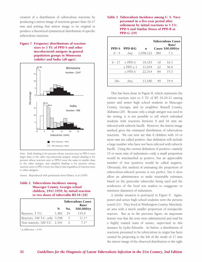
52 Guidelines for the Diagnosis of Latent Tuberculosis Infection in the 21st Century, 2nd Edition
creation of a distribution of tuberculous reactions by
producing a mirror image of reactions greater than 16-17
mm and joining that mirror image to its original to
produce a theoretical symmetrical distribution of specific
tuberculous reactions.
Figure 7 Frequency distributions of reaction sizes to 5 TU of PPD-S and othermycobacterial antigens in generalpopulation groups in Minnesota (adults) and India (all ages).
Note: Dark shading is for persons whose reaction sizes to PPD-S werelarger than to the other mycobacterial antigen; striped shading is forpersons whose reaction sizes to PPD-S were the same or smaller thanto the other antigen; and stippled shading is for persons whosereaction sizes to PPD-S were less than 6 mm regardless of reaction sizesto other antigens.
Source: Reproduced with permission from Palmer, et al (1959)
Table 2 Tuberculosis Incidence among Muscogee County, Georgia schoolchildren, 1947-1959, by initial reaction to two doses of tuberculin RT-18 (18)
* p difference - 0.44
Tuberculous CasesRate/
N No. 100,000/yr
Reactors, 5 T.U. 1,482 24 135.0
Reactors, 100 T.U. only 3,768 5 11.1*
Non reactors, 100 T.U. 2,341 2 7.1*
Table 3 Tuberculosis Incidence among U. S. Navypersonnel in a five-year period afterenlistment by initial reactions to 5 T.U.PPD-S and Similar Doses of PPD-B orPPD-G (19)
This has been done in Figure 8, which represents the
various reaction sizes to 5 TU of RT 19-20-21 among
junior and senior high school students in Muscogee
County, Georgia, and its neighbor, Russell County,
Alabama (20). Because only a single antigen was used in
the testing, it is not possible to tell which individual
students with reactions between 9 and 16 mm are
infected with tubercle bacilli. However, the mirror image
method gives the estimated distribution of tuberculous
reactions. We can now see that if children with 10 or
more mm are called positive, that definition will include
a large number who have not been infected with tubercle
bacilli. Using the current definition of positive—namely
15 or more mm of induration—only a small proportion
would be misclassified as positive, but an appreciable
number of true positives would be called negative.
Obviously, this method of estimating the proportion of
tuberculous-infected persons is not perfect, but it does
allow an administrator to make reasonable estimates
based on the particular tuberculin being used and the
tendencies of the local test readers to exaggerate or
minimize diameters of induration.
A similar situation is portrayed in Figure 9. Again,
junior and senior high school students were the persons
tested (21). They lived in Washington County, Maryland,
an area with a much smaller proportion of nonspecific
reactors. But as in the previous figure, an important
feature was that the tests were administered and read by
a highly trained team of nurses, supervised in this
instance by Lydia Edwards. As before, a distribution of
reactions presumed to be tuberculous in origin has been
created by projecting to the left of the mode of 17 mm
the mirror image of the observed distribution to the right
Tuberculous CasesRate/
PPD-S PPD-B/G n Cases 100,000/yr
0 - 5 Any 1,058,122 384 7.3
6 - 17 > PPD-S 19,333 10 10.3
± PPD ± 1 11,934 22 36.9
< PPD-S 22,314 84 75.3
18+ Any 13,180 49 74.4

Guidelines for the Diagnosis of Latent Tuberculosis Infection in the 21st Century, 2nd Edition 53
of that mode. The resulting distribution of reactions
estimated to be due to infections with tubercle bacilli is
shown by cross-hatching. As in Muscogee and Russell
Counties, classifying reactions of 10 or more mm as
positive includes almost all the tuberculous reactions but
at the cost of also including a fair proportion of
nonspecific reactions. Increasing the definition of
positive to 15 or more mm produces a group almost all
of whom are tuberculous infected but misses nearly half
of the truly infected. But as before, an administrator in
this situation has information to allow an informed
decision of what the cut point between positive and
negative should be and can see what the consequences
would be of applying national criteria locally.
Figure 8 Frequency distribution of reaction sizesto 5 tuberculin units of RT 19-20-21among school children in Georgia andAlabama (with reaction sizes estimatedto be due to infection with M. tuberculosis shown in darker shading).
Source: Comstock (1960)
SMOOTHING DISTRIBUTION
Not all health departments have tuberculin testers and
readers who can produce such smooth distributions as
those in Figures 8 and 9. Figure 10 shows distributions
of reaction sizes from two studies conducted with local
personnel who claimed to have considerable experience
in tuberculin testing. The study conducted in the 1970’s
produced an extreme example of an all too familiar
phenomenon, terminal digit preference. This is the
Figure 9 Frequency distribution of reaction sizes
to 5 tuberculin units of PPD-S among
school children in Washington County,
Maryland, with reaction sizes estimated
to be due to infection with M. tuberculosis
shown by cross-hatching.
Source: Kuemmerer and Comstock (1960)
tendency, when measuring entities that have indistinct
boundaries, to record an excess of measurements ending
in particular digits, usually 5’s or 0’s. In this instance, the
tendency was so extreme that no method of smoothing
the distribution is feasible and applying the mirror image
method of estimating the proportion of tuberculous
infected is out of the question.
In the study done in the 1990’s, the situation was
much better. An attempt to smooth the distribution by
applying a three-point moving average is shown by the
solid line. While an attempt at smoothing seemed
reasonable, it gave no indication of a single right-hand
mode. Furthermore, the excess of very large reaction sizes
in both studies exceeds anything seen elsewhere. Again,
terminal digit preference, though less extreme in 1990
than in the 1970 study, is still too prominent to allow
reasonable estimates by the mirror image method.

54 Guidelines for the Diagnosis of Latent Tuberculosis Infection in the 21st Century, 2nd Edition
Figure 10 Frequency distributions of reactionsizes to 5 tuberculin units of PPD-tuberculin among persons tested in1970 and 1990 by staff of an unnamedhealth department.
Note: Heavy line is a 3-point moving average
There are two ways to minimize terminal digit
preference and to produce a relatively smooth
distribution. Because terminal digit preference is only
likely to occur when there is some degree of uncertainty
in measurement and therefore room for subjective
influences to occur, any way of insuring clear and definite
measurement points should go far toward solving the
problem. One method is to use the ballpoint pen method
of Sokol in which the pen is pressed along the skin toward
the area of induration from above and below until
resistance is felt (22). The distance between the ends of
the two lines is the sagittal diameter of induration, and
should be measurable with very little uncertainty.
Another method is to rely on visualization rather than
palpation, either with the finger or indirectly with a pen,
and to use a measuring device in which the scale is not
seen until the measurement has been made.
Visualization is important since what can be seen, can be
measured. Induration, even that which is difficult to
palpate, can easily be seen if diffuse light is allowed to
shine tangentially across the test site. Once the borders
of induration are seen, the transverse diameter should be
measured as mechanically as possible, paying no
attention to what the numbers may mean. Once the
diameter is recorded, it is time to become a health care
provider again and think what the reaction portends for
the care of the patient.
Some sort of a gauge is almost essential. A Dr.
Turnbull at a Southern Trudeau Society meeting about
1950 first suggested a simple, readily available and
inexpensive gauge. The “Turnbull gauge,” as we called it,
was merely a sewing gauge. At that time, a millimeter
scale had to be glued over the original scale, which was
in inches. The sliding pointer was then reversed so that
the knob that moved the pointer was on the side opposite
the scale, making it impossible to tell what the reading
was until after the gauge had been set. Then it could be
turned over and the reading recorded to the nearest mm
unit below the pointer, thereby avoiding fractions of a
millimeter and virtually all uncertainty. Today, sewing
gauges come with millimeter scales, making them usable
with only the simple reversal of the sliding pointer.
Figure 11 illustrates that the routine use of a modified
sewing gauge to measure tuberculin reactions can
minimize terminal digit preference. This is the
distribution of tuberculin reaction sizes recorded for the
790 persons tuberculin tested by personnel of the
Washington County Health Department in the year 2000
using the modified sewing gauge. The bars of the
histogram show the observed distribution. There is a
little evidence of preference for numbers ending in 5’s or
0’s but so little that the distribution is easily smoothed by
a 3-point moving average, shown here by the solid line.
Using the mirror image technique, the estimated
proportion of persons infected with M. tuberculosis is
obtained and is shown by the crosshatched part of the
smoothed distribution. It appears that a reasonable cut-
point between positive and negative reactions in this
population might be 12 mm of induration. To the left of
that line, most of the reactions can safely be assumed to
be nontuberculous; to the right, nearly all are assumed to
be tuberculous.
In summary, it is now clear that wherever
nontuberculous mycobacterial infections are found, there
will also be nonspecific sensitivity to the intermediate
doses of PPD-tuberculin. This is true for most of the
world except for high altitudes and high latitudes. If
tuberculin tests can be carefully and accurately
administered and if the ensuing reactions are measured
in ways that are objective and associated with minimal
uncertainty, analysis of the results can give reasonable

Guidelines for the Diagnosis of Latent Tuberculosis Infection in the 21st Century, 2nd Edition 55
estimates of the proportions of reactions due to
tuberculous and to nontuberculous infections. In
general, the larger the reaction to 5 TU of PPD-
tuberculin, the more likely that it was caused by infection
with tubercle bacilli. Reactions with induration
measuring 17 mm or more in diameter are almost certain
to be due to infection with M. tuberculosis, M. bovis or
some strain of BCG. Reactions less than 10 mm are
almost all due to infections with nontuberculous
mycobacteria or most strains of BCG. For maximal
usefulness, the tuberculin test needs to be administered
with care and the resulting reactions must be read
without bias and with minimal terminal digit preference.
.Figure 11 Frequency distribution of reaction sizes
to 5 tuberculin units of PPD-tuberculinamong persons tested in 2000 by staffof Washington County (Maryland)Health Department.
Note: Heavy line is a 3-point moving average. Reactions estimated tobe due to infection with M. tuberculosis shown by cross-hatching.
Source: Data furnished by Mark Jameson, Washington County HealthDepartment.
References
(1) Wolinsky E. State of the art. Nontuberculous
mycobacteria and associated diseases. Am Rev
Respir Dis 1979; 119:107-159.
(2) Grosset J, Truffot-Pernot C, Cambau E. Bacteriology
of tuberculosis. In Reichman LB, Hershfield ES,
eds. Tuberculosis. A Comprehensive International
Approach (Second ed.). New York: Marcel Dekker,
2000, pp. 157-185.
(3) Magnus K. Epidemiologic basis of tuberculosis
eradication. 6. Tuberculin sensitivity after human
and bovine infection. Bull Wld Hlth Org 1967;
36:719-731.
(4) Shaw LW. Field studies on immunization against
tuberculosis. I. Tuberculin allergy following BCG
vaccination of school children in Muscogee County,
Georgia. Public Health Rep 1951; 66:1415-1426.
(5) Edwards LB, Palmer CE, Magnus K. BCG
vaccination. Studies by the WHO Tuberculosis
Research Office, Copenhagen. Chapter 3, Response
to BCG Vaccination. World Health Organization
Monograph Series, No. 12. Geneva: World Health
Organization, 1953, pp. 51-64.
(6) Griffith JPC, Mitchell AG. The Diseases of Infants and
Children (Second ed.). Philadelphia: W. S. Saunders
Company, 1938, p. 396.
(7) Committee on Diagnostic Standards, National
Tuberculosis Association. Diagnostic Standards and
Classification of Tuberculosis (1940 ed.). New York:
National Tuberculosis Association, 1940, p. 24.
(8) Furman B. A Profile of the United States Public
Health Service, 1798-1948. DHEW Publication No.
(NIH) 73-369. Washington D.C.: National Library
of Medicine, U. S. Department of Health, Education
and Welfare, 1973, pp. 290-292.
(9) Lumsden LL, Dearing WP, Brown RA. Questionable
value of skin testing as a means of establishing an
epidemiological index of tuberculous infection. Am
J Public Health 1939; 29:25-33.
(10) McKneely TB. An evaluation of the tuberculin test
as a screen in school case-finding programs. Trans
34th Annual Meeting of the National Tuberculosis
Association, 1938, pp. 290-296.
(11) Anon. Minutes of Hagerstown Tuberculosis
Conference, September 26-October 1, 1938.
Evening Session—September 30, 1938.
(Unpublished).

56 Guidelines for the Diagnosis of Latent Tuberculosis Infection in the 21st Century, 2nd Edition
(12) Comstock GW. The Hagerstown Tuberculosis
Conference of 1938: A retrospective opinion
(editorial). Am Rev Respir Dis 1969; 99:119-120.
(13) Comstock GW. The International Tuberculosis
Campaign: A pioneering venture in mass
vaccination and research. Clin Infect Dis 1994;
19:528-540.
(14) WHO Tuberculosis Research Office. Further studies
of geographic variation in naturally acquired
tuberculin sensitivity. Bull Wld Hlth Org 1955;
12:63-83.
(15) Palmer CE. Tuberculin sensitivity and contact with
tuberculosis. Further evidence of nonspecific
sensitivity. Am Rev Tuberc 1953; 68:678-694.
(16) Crow HE, King CT, Smith CE, Corpe RF, Stergus I.
A limited clinical, pathologic and epidemiologic
study of patients with pulmonary lesions associated
with atypical acid-fast bacilli in the sputum. Am Rev
Tuberc Pulm Dis 1957; 75:199-222.
(17) Palmer CE, Edwards LB, Hopwood L, Edwards PQ.
Experimental and epidemiologic basis for the
interpretation of tuberculin sensitivity. J Pediatr
1959; 55:413-429.
(18) Comstock GW, Shaw LW. Tuberculosis studies in
Muscogee County, Ga. Controlled trial of BCG
vaccination in a school population. Public Health
Rep 1960; 75:583-594.
(19) Edwards LB, Acquaviva FA, Livesay VT.
Identification of tuberculous infected. Dual tests
and density of reaction. Am Rev Respir Dis 1973;
108:1334-1339.
(20) Comstock GW. A comparison of purified
tuberculins in the southeastern USA. Bull Wld Hlth
Org 1960; 23:683-688.
(21) Kuemmerer JM, Comstock GW. Sociologic
concomitants of tuberculin sensitivity. Am Rev Respir
Dis 1967; 96:885-892.
(22) Sokol JE. Measurement of delayed skin test
responses. N Engl J Med 1975; 293:501-502.

INTRODUCTION
One-third of the world’s population is estimatedby the World Health Organization to be
infected with Mycobacterium tuberculosis (M.tuberculosis). Efforts to control this disease, itstransmission, and ultimately its eradication havebeen fought along two fronts in the United States.The first front is to detect and treat symptomaticpeople with infectious tuberculosis. The goal is tocure these individuals and stop further spread of theorganism. While essential, these efforts arefrequently too late to prevent transmission to otherpeople. The second front is to detect the vast poolof asymptomatic people who have latent M.tuberculosis infection (LTBI) and prevent them fromdeveloping infectious tuberculosis (1).
Tests with high sensitivity and specificitycharacteristics for detectingM. tuberculosis infectioncould facilitate tuberculosis control on both fronts.A sensitive test would facilitate screening of peoplewho would benefit from closer evaluation forinfectious disease or treatment to prevent it fromdeveloping. A specific test would avoid treatingpeople at minimal risk of having or developing theinfectious disease. Attempts to develop and evaluatemore accurate tests have been hampered by the lackof a “gold standard” for identifying latent M.tuberculosis infections.
Until recently, the standard and only method forimmunologic diagnosis of M. tuberculosis infectionhas been limited to the tuberculin skin test (TST).The TST, developed in 1889, has been used to detectboth LTBI and active tuberculosis. However, therehave always been concerns about its shortcomings.Because purified protein derivative of tuberculincontains many antigens that are shared with othermycobacteria, the skin test does not reliablydistinguish LTBI from prior immunization withMycobacterium bovis bacilli Calmette- Guérin (BCG)or infection with environmental mycobacteria (2).This is a major problem in most developed countriesbecause a growing proportion of those with LTBI areforeign born persons from high incidence countries,most of whom received BCG vaccination duringchildhood. False-negative results in the setting ofhost immunosuppression has limited also its utility.In addition, cutaneous sensitivity to tuberculindevelops from 2 to 10 weeks after infection and theTST requires two encounters with a health careprofessional which often causes logistical problemsif not inconvenience. Finally, skilled personnel areessential for proper placement and interpretation ofthe test. A test with greater accuracy andconvenience would greatly enhance tuberculosiscontrol efforts (3).
The new blood assays to detect M. tuberculosisinfection are based on the response of antigen-specific memory T-cells releasing interferon-gamma(IFN-γ) in response to previously encounteredmycobacterial antigens. Interferon-gamma releaseassays (IGRAs) measure the cellular immuneresponses to M. tuberculosis-specific antigens,including early-secreted antigenic target 6 (ESAT-6)and culture filtrate protein 10 (CFP-10), antigensencoded in the region of difference (RD1) of the M.
Interferon-γ Release Assay forDetection of Tuberculosis Infection –Chapter updated in 2011
Alfred Lardizabal, MDDr. Lardizabal is AssociateProfessor of Medicine at NJMedical School-University ofMedicine and Dentistry ofNew Jersey and GlobalTuberculosis Institute inNewark, New Jersey.

tuberculosis genome. These proteins are absent fromall strains of M. bovis BCG and the vast majority ofnon-tuberculous mycobacteria (with the exceptionof M. kansasii, M. szulgai, and M. marinum) butpresent in isolates ofM. tuberculosis. In comparison,the TST uses the mixed, nonspecific PPD, a culturefiltrate of tubercle bacilli containing over 200antigens, which results in its low specificity.
Two IGRA systems using RD1-encoded antigensare currently commercially available for TBdetection. One system includes QuantiFERON®-TBGold and its variant QuantiFERON®-TB Gold In-Tube (uses tubes pre-filled with antigens) (Cellestis,Victoria, Australia), which uses whole bloodspecimens, with an unknown number of leukocytes,to measure IFN-γ released by antigen-activated Tlymphocytes. The other system is the T-SPOT®.TB(Oxford Immunotec, Oxford, England). It uses theELISPOTmethod, wherein the number of peripheralblood mononuclear cells (PBMC) in the assay isquantified, in order to measure IFN-γ-secreting Tcell counts ("spots") on stimulation by M.tuberculosis-specific antigens in microplate wells.The readout of the two tests is different:QuantiFERON®-TB Gold (QFT-G) andQuantiFERON®-TB Gold In-Tube (QFT-GIT)measures the level of IFN-γ in the supernatant of thestimulated whole blood sample using enzyme-linkedimmunosorbent assay (ELISA), and T-SPOT®.TBenumerates individual T-cells producing IFN-γ afterantigenic stimulation.
These new blood tests have an internal positivecontrol, i.e., a sample well stimulated with a potentnon-specific stimulator of IFN-γ production by T-cells. This controls the results of the test fortechnical errors, such as failure to add viable,functioning cells to the well. The failure of thepositive control in the tests provides informationthat the test’s results cannot be reliably interpretedsince it may reflect an underlying in vivoimmunosuppression, negatively affecting T-cellfunction in the in vitro stimulation.
TEST PERFORMANCE
In order to establish the diagnostic accuracy forLTBI of any test is a major challenge because there isno available gold standard. As an alternative, somerational approaches based on the epidemiology ofTB have been applied. The knowledge that airbornetransmission of TB is promoted by close andprolonged contact with an infectious case has beenused with the preposition that if a test is a goodmarker of LTBI, it should correlate closely with thelevel of exposure. Several studies conductedcomparing QFT and TST used in the setting of acontact investigation have results that show thesetests to be moderately concordant (4, 5, 6). Incomparing the IFN-γ assay to the TST in personswith varying risk for MTB infection, Mazurek, et al.(5) showed that the assay was comparable to theTST in its ability to detect LTBI, with an overallagreement of 83%. It was less affected by BCGvaccination, discriminated responses due to NTM,and avoided the variability and subjectivityassociated with placing and reading the TST. Thistest was also used to detect recent infection amongcontacts in a TB outbreak at a Danish high school.Since a majority of contacts were BCG-unvaccinateddirect comparison between the TST and QFT couldbe performed. Analysis revealed an excellentagreement between the two tests was found (94%,kappa value 0.866) and that the blood test was notinfluenced by the vaccination status of the subjectstested (4). Ewer and colleagues investigated a schooloutbreak that resulted from one infectious indexcase using the ELISPOT assay and the Heaf test (7).The overall agreement between the two tests was89%. The ELISPOT assay showed no significantrelation to BCG status. By contrast, BCG-vaccinatedchildren were more likely to have higher Heaf gradesthan unvaccinated children. An isolated positiveELISPOT was associated with exposure, whereas anisolated positive TST result was not. Several studiescompared TST and IGRAs with respect to theircorrelation with exposure to M. tuberculosis (4, 7, 8,9, 10). In these studies the RD1-based assays

showed stronger positive correlation with increasingintensity of exposure compared to the TST.
The sensitivity of IGRAs have been estimatedusing cases of active tuberculosis confirmed bycultures and often excluded HIV-infectedindividuals. Studies that estimated the specificity ofIGRAs were carried out in low incidence countrieswith some patients exposed to BCG vaccination andother not. Pai and colleagues (11) in a recent meta-analysis estimated the pooled sensitivity of the QFTstudies to be 76% and 90% for T-SPOT®.TB. Thepooled specificity for all QFT studies was 98%, (99%for QFT among non-BCG vaccinated populationsand 96% for BCG vaccinated populations). Thepooled specificity of T-SPOT®.TB was 93% (almostall studies included BCG vaccinated participants).For the TST, pooled sensitivity estimate was 77%,specificity in non-BCG vaccinated was 97% but lowand highly heterogeneous among BCG vaccinatedparticipants. From this substantial body of literature,it can be concluded that IGRAs, especially QFT-Gand QFT-GIT, have excellent specificity that isunaffected by BCG vaccination. TST has a highspecificity among non-BCG vaccinated individuals.The sensitivity of IGRAs and TST is not consistentacross populations but the T-SPOT®.TB appears tobe more sensitive than QFT or TST. Similar findingwere observed by Diel and colleagues in a meta-analysis they performed in which the TST had apooled sensitivity of 70%, QFT-IT was 81%, and T-SPOT®.TB was 88%. Specificity of QFT-IT was 99%against 86% for the T-SPOT®.TB (12). Data on high-risk populations, such as immunocompromised andyoung children, remain limited and it has beenshown that indeterminate results for the IGRAs tendto increase in these groups. In addition, one recentstudy which evaluated close contacts using both theIGRA (QFT-IT) and the TST suggested that theIGRA appeared to be a more accurate indicator ofthe presence of LTBI. It also provided some insightinto its predictive value for the development ofactive TB since 14.6% of those with a positive QFT-IT progressed to active TB compared to only 2.3 %among those with a positive TST (13). Questionssuch as the prognostic ability of these tests to
accurately identify individuals with LTBI who are athighest risk for progressing to active TB and thesignificance of conversions and reversions of thesetests over time still need to be clarified.
From the approval by the US Food and DrugAdministration (FDA) of QFT-G in May 2005, theUS Centers for Disease Control and Preventionrecommend that "QFT-G may be used in allcircumstances in which the TST is currently used,including contact investigations, evaluation of recentimmigrants, and sequential testing" with warningsand limitations (14). Updated CDC guidelines forthe use of interferon gamma release assays werepublished in 2010 and states that either the TST andIGRAs (QFT-G, QFT-GIT, T-Spot) may be used asaids in diagnosing M. tuberculosis infection (15).IGRAs are preferred for testing persons who havereceived BCG (as a vaccine or for cancer therapy) orfor testing groups that historically have low rates ofreturning to have TSTs read. The TST is preferredfor testing children aged <5 years. An IGRA or a TSTmay be used without preference for testing recentcontacts to persons with infectious pulmonary TBwith considerations for follow-up testing. An IGRAor a TST may also be used without preference forperiodic screening of persons who might haveoccupational exposure to M. tuberculosis withconsiderations for conversions and reversions.Currently, an IGRA conversion is defined as achange from negative to positive within 2 yearswithout any consideration of the magnitude of thechange in TB response (as opposed to the morestringent 10 mm change required for the TST). Amore stringent criteria for conversion using IGRAs isyet to be defined. Substantial progress has beenmade in documenting the utility of IGRAs butfurther studies and research determining the valueand limitations of IGRAs in situations important tomedical care and TB control is needed.
References:
(1) CDC. Targeted tuberculin testing and treatmentof latent tuberculosis infection. MMWR 2000;49:1-51.

(2) American Thoracic Society. Diagnostic standardsand classification of tuberculosis. Am Rev RespirDis 1990; 142:725-735.
(3) Institute of Medicine. Ending Neglect: theelimination of tuberculosis in the United States.Washington, DC: National Academy Press; 2000.
(4) Brock I, Weldingh K, Lillebaek T, Follmann F,Andersen P. Comparison of a New Specific BloodTest and the Skin Test in Tuberculosis Contacts.Am J Respir Crit Care Med 2004; 170:65-69.
(5) Mazurek GH, LoBue PA, Daley CL, Bernardo J,Lardizabal AA, Bishai WR, Iademarco MF, RothelJS. Comparison of a whole-blood interferon-γassay with tuberculin skin testing for detectinglatent Mycobacterium tuberculosis infection.JAMA 2001; 286:1740-1747.
(6) Streeton JA, Desem N, Jones SL. Sensitivity andspecificity of a gamma interferon blood testtuberculosis infection. Int J Tuberc Lung Dis 1998;2:443-50.
(7) Ewer K, Deeks J, Alvarez L, Bryant G, Waller S,Andersen P, Monk P, Lalvani A. Comparison of T-cell based assay with Tuberculin Skin Testing forthe Diagnosis of Mycobacterial tuberculosisinfection in a School Outbreak. Lancet 2003;361:1168-73.
(8) Hill PC, Brooks RH, Fox A, Fielding K, JeffriesDJ, Jackson-Sillah D, Lugos MD, Owiafe PK,Donkor SA, Hammond AS, Out JK, Corrah T,Adegbola RA, McAdam KP. Large ScaleEvaluation of Enzyme-Linked ImmunospotAssay and Skin Test for Diagnosis ofMycobacterium tuberculosis Infection Against aGradient of Exposure in The Gambia. Clin InfectDis 2004.38:966-73
(9) Lalvani A, Pathan AA, Durkan H, Wilkinson KA,Whelan A, Deeks JJ, ReeceWHH, Latif M, PasvolG, Hill AV. Enhanced Contact Tracing andSpatial Tracking of Mycobacterium tuberculosisInfection by Ennumeration of Antigen SpecificT-cells. Lancet 2001; 357:2017-21.
(10) Richeldi L, Ewer K, Losi M, Bergamini BM,Roversi P, Deeks J, Fabbri LM, Lalvani A. T-cellbased Tracking of Multidrug ResistantTuberculosis Infection After Brief Exposure. AmJ Respir Crit Care Med 2004; 170:288-95.
(11) Pai M, Zwerling A, Menzies D. SystematicReview: T-Cell-Based Assays for the Diagnosisof Latent Tuberculosis Infection: An Update.Ann Intern Med 2008;149:1-8.
(12) Diel R, Loddenkemper R, Nienhaus A.Evidence-based Comparison of CommercialInterferon-γ Release Assays for Detecting ActiveTB. Chest 2010; DOI:10.1378
(13) Diel R, Loddenkemper R, Meywald-Walter K,Niemann S, Nienhaus A. Predictive Value of aWhole Blood IFN- Assay for the Developmentof Active Tuberculosis Disease after RecentInfection withMycobacterium tuberculosis. Am JRespir Crit Care Med 2008; 177:1164-1170.
(14) CDC. Guidelines for using the QuantiFERON-TB Gold test for Detecting Mycobacteriumtuberculosis infection, United States. MMWR2005; 54(RR15):49-55.
(15) CDC. Updated guidelines for using InterferonGamma Release Assays to detectMycobacterium tuberculosis infection – UnitedStates, 2010. MMWR 2010; 59(RR5):1-25.

Karen Galanowsky R.N.,
M.P.H.
Ms. Galanowsky is a Nurse
Consultant for the New Jersey
State Department of Health and
Senior Services, Trenton, New
Jersey, TB Program
The nurse plays a significant role in tuberculosis (TB)
programs, ensuring the control of TB through
targeted tuberculin skin testing (TST) and treatment of
latent TB infection. The nurse’s role intersects all aspects
of public health practice by focusing on individuals,
communities and health care systems.
The nurse integrates the core functions of public
health—assessment, policy development and
assurance—into the interventions and strategies used to
identify individuals at high risk for TB who would benefit
from treatment for latent TB infection if it were detected.
The nursing process is utilized to intervene at all levels—
individual, community or health care system. It includes
assessment, problem identification, planning,
implementation and evaluation.
KNOWLEDGE AND COMPETENCIES
The registered nurse (RN) who works with individuals
at risk for latent TB infection should obtain up-to-date
knowledge on:
� The diagnosis and treatment of latent TB infection,
and the differentiation of TB disease
� The community, including demographic
characteristics, ethnic populations and resources
� How latent TB disease and treatment is perceived
by culturally diverse populations
� Epidemiology TB in the community
THE ROLE OF THE NURSE INDIAGNOSING LATENT TB INFECTION
62 Guidelines for the Diagnosis of Latent Tuberculosis Infection in the 21st Century, 2nd Edition
The registered nurse also should be familiar with:
� Health care providers within the community,
including the populations served, and the strengths
and weaknesses of each
� Barriers to health care—specifically tuberculin skin
testing, reading and interpretation, medical
evaluation and completion of treatment
� Principles of TB control
� Regulations and legal mandates
� Internal and external standards
In addition, the nurse should attain and maintain
proficiency in:
� Administering, reading and interpreting the
tuberculin skin test
� Providing culturally sensitive and age-appropriate
TB education for both individuals and
communities
� Assessing the individual and the community
� Providing interventions and strategies to ensure
that high-risk individuals and groups are
tuberculin skin tested, and that they have access to
medical evaluation and appropriate treatment
� Collaboration
� Networking
� The political process
� Policy development
� Evaluation
INTERVENTIONS
Interventions are actions the nurse takes on behalf of
individuals, families and communities. Particular
interventions are recommended based on the nurse’s
clinical judgment using theoretical, practical and scientific
knowledge. Potential or desired outcomes should be
identified and related to the interventions, which may be
implemented at all levels (individual, community and
systems-focused) and at all sites for TB control.
63 Guidelines for the Diagnosis of Latent Tuberculosis Infection in the 21st Century, 2nd Edition
Interventions fall under a number of categories:
� Surveillance
� Screening
� Patient assessment
� Education (patient, health care provider,
community)
� Counseling
� Advocacy
� Referral and follow-up
� Coalition building
� Collaboration
� Community mobilization
� Consultation
Individual-focused interventions
Interventions that focus on individuals create changes
in the knowledge, attitudes, beliefs, skills, practices and
behaviors of individuals, families and groups. These are
person-to-person interventions specifically for persons
belonging to a population at risk of TB.
The RN generally provides the following individually
focused interventions: screening, patient assessment,
follow-up, monitoring, education, counseling or
consultation. Nurses also conduct contact investigations
and do TST of identified close contacts. This process
involves a series of decisions, frequently made solely by
the nurse. Two examples illustrate the RN as the decision
maker: (1) When a contact investigation is required in a
congregate setting, such as a school, the decision as to
who should be tested, using the concentric circle
principle, is often made by the nurse; and (2) If the
concentric circle needs to be expanded due to an
unexpected number of positive results, the nurse decides
which groups or individuals should be included.
Sometimes the very effort to find and TST identified
contacts takes initiative and ingenuity on the nurse’s part.
The nurse also has to identify other barriers to patients’
being screened and treated for latent TB infection.
With respect to the physician, the RN’s role is typically
one of patient advocacy and educator. By using assertive
diplomacy, the RN is frequently required to provide the
private physician with the most recent literature
regarding the reading and interpretation of the TST and
the recommended treatment for latent TB infection. In
addition, the nurse often educates the patient about the
importance of keeping doctor or clinic appointments so
that treatment may not be interrupted, and explains
potential side effects of medication. The nurse also
explains TB infection and the difference between TB
infection and disease. Frequently, it is the nurse who
monitors the patient for side effects of medication, and
tracks the patient to ensure completion of treatment.
Problems encountered
A number of problems have arisen in connection with
individual-focused interventions. Private physicians or
patients themselves may be apathetic about the
importance of TST. Patients may fail to return to have their
TST read. All too often, patients and even private
physicians may believe that BCG is the cause of the
positive TST result. Physicians may not prescribe any
treatment, while patients may not adhere to treatment even
if it is provided.
The health care provider may fail to assess for TB
symptoms when the TST is negative, even though the
patient may belong to a population at heightened risk of
TB. The patient may have personal beliefs about health
or attitudes about TB that interfere with care. The patient
may not understand the difference between TB infection
and TB disease. He/she may fear side effects or adverse
reactions of TB treatment.
Other problems arise as well. Co-existing medical or
psychiatric diagnoses may impair the patient’s ability to
give accurate information. For many persons at risk of
TB, substance abuse, homelessness or residential
instability may inhibit efforts to intervene. The patient
may be faced with competing or conflicting demands or
may lack the funds to pay for medical care.
Case presentation: “The Cliff Dweller”
The patient is a 39-year-old female, born in South
America, who immigrated to the United States twenty years
ago. She was first diagnosed with pulmonary TB during a
hospitalization for injuries she sustained from an assault by
a boyfriend. At the time her clinical picture revealed:
� History of positive TST (date unknown)
� Symptoms of cough, hemoptysis and weight loss
� Chest x-ray: Cavity right apex, diffuse infiltrates
left upper lobe
� Sputum smear negative; culture M. tuberculosis
The patient gave an address of an apartment in a local
municipality. She named three contacts at the same
address, and said she had one daughter who lived nearby.

64 Guidelines for the Diagnosis of Latent Tuberculosis Infection in the 21st Century, 2nd Edition
The RN tried to locate and interview the named contacts,
but found that the address the woman provided was
fictitious. When the RN revisited the hospital to talk
with the patient, the patient had signed herself out
against medical advice.
The RN then went to locate the patient in the
community. Because all leads resulted in dead ends, the
RN questioned the police department as to the patient’s
whereabouts. The police had multiple dealings with the
patient, who came in as the victim of physical and sexual
assault by her boyfriend. They provided four different
addresses including two liquor stores and “the cliffs.”
The police were not aware that anyone was living on the
cliffs, a steep and wooded embankment sloping down to
a major highway.
The patient became lost to follow-up for six months
until she was again assaulted and required
hospitalization. The RN interviewed the patient
immediately. At this time, the patient was considered
highly infectious. The patient stated that she lived in
tents on the cliffs. She said she slept in a different tent
each night in exchange for sexual favors. During the day
she went to the streets where she “hung out.”
Occasionally she went to her daughters to shower.
The RN facilitated the use of legal intervention to
constrain the patient in the hospital for treatment until
smear negative results were obtained. During this time,
the RN visited the patient daily, brought food the patient
requested and assisted the patient to obtain a television in
her room. The RN’s goal was to develop a working
relationship with both the patient and her boyfriend over
the next several weeks. The boyfriend told the RN that
Saturday mornings would be a good time to find all the
people who stay on the cliffs. A team, including a field
supervisor, field worker, nurse and translator, went to the
cliffs on a Saturday morning. They brought hot coffee,
rolls and doughnuts as “peace-bearing gifts.” They also
brought TST material.
The team found and tuberculin tested 13 cliff dwellers.
The team arranged to pick up the group of contacts by
van and bring them to the clinic on Monday morning for
a TST reading and further medical evaluation. The team
promised incentives for adherence. Five of the patients’
relatives were also located and tested over the following
two weeks with the assistance of the patient’s boyfriend.
Interventions utilized
Contacts came to the clinic drunk, disorderly and
extremely dirty. Clinic staff provided transportation and
remained nonjudgmental and kind. Incentives such as
food, gift certificates for food, and clothing were given to
all the contacts at the site where they dwelled. Social
service intervention also was provided to assist them in
finding better housing.
Education was provided to the contacts verbally, slowly
and over time. Most of them could not read or write.
Team members discussed and allayed the contacts’ fear of
deportation and mistrust of the government; most of
them were undocumented immigrants. Behavioral
contracts were developed verbally with the contacts to
ensure their adherence to directly observed treatment for
latent TB infection.
Nursing and field staff had to locate the contacts by
traversing the cliffs through mud, trees and debris. The
TB control RN worked with the police and mayor to avert
political pressure to have the cliff dwellers removed
immediately. The RN also worked with the public health
officer and the press to prevent panicked coverage by
newspapers and television.
Community-focused interventions
The ultimate goal of community-focused nursing
interventions is to prevent the development of TB disease
in persons with latent TB infection in the community.
The nurse accomplishes this goal by:
� Networking
� Coalition building
� Teaching other nurses and health care workers
� Community assessment
� Collaboration
� Community education
� Consultation
� Evaluation
With an understanding of the incidence and
prevalence of TB disease, and the socio-demographic
characteristics of a community, the nurse can identify
those individuals and groups who are likely to be
infected with TB and benefit from treatment. The nurse
can establish partnerships with community health
centers, outpatient clinics, drug treatment centers and
homeless shelters that provide services to individuals at
high risk for TB infection and disease. The nurse also can
provide TB education to the community and health care
providers to increase awareness of TB.
Community-focused interventions require that nurses
are proficient in administering and reading the TST.

65 Guidelines for the Diagnosis of Latent Tuberculosis Infection in the 21st Century, 2nd Edition
Assurance that there are appropriate resources to
medically evaluate and treat those individuals who are
identified as TST-positive is also important.
Community-focused interventions also include the
evaluation of those nurses administering and interpreting
the TSTs within the community; training should to be
provided to assure and maintain proficiency in this
activity. Evaluation also focuses on programmatic
outcomes of treatment.
A community-based TB prevention model
Another step the nurse can take in identifying those at
risk, diagnosing latent TB infection and connecting
patients to appropriate treatment and care is to
implement a community-based model for the delivery of
TB preventive services that accommodate the
community’s characteristics and needs.
An excellent example of a community-based model of
TB preventive services was developed by the TB program
at the Boston Public Health Department, Boston, Mass.
This model entailed collaboration between the health
centers and the Boston Public Health Department’s TB
program. A nurse coordinated the program. The health
centers identified “core” provider teams who became the
TB expert resources for individual health center
programs, and the health department’s TB program
educated and trained the core provider team for each
health center.
Policies for TB screening of patients and staff were
reviewed and revised collaboratively, and were based on
the needs of the community health centers. Persons with
a positive TST were referred to the Boston Public TB
Clinic for an initial medical evaluation and treatment.
Those who were placed on treatment for latent TB
infection were monitored monthly and given a new
supply of TB medication at the community health center.
The health department TB program delivered the
monthly supply of TB medications to each health center
and evaluated patient adherence, monitoring and
documentation. Data were collected and analyzed by the
TB program and shared with the health centers to help
identify obstacles to the implementation of this plan and to
document accomplishments in meeting specific objectives.
Problems encountered
A number of problems regularly arise in working with
the community. Poor documentation of TST results is
common (recording “negative” or “positive” rather than
the millimeter reading.) Measuring redness and/or
swelling rather than induration is another frequently
found problem. Still other common problems include:
� Measuring with a standard ruler—or guessing
� Making a determination based on the appearance
of redness without palpating the arm
� Filling the syringe with the PPD hours before
administering the TST
� Inadequate storage of PPD, not refrigerating it or
keeping it out in the sunlight all day
� Delay in referring an individual with a positive TST
result for medical evaluation without assessing
symptoms of TB disease
� No treatment for LTBI recommended
� “Un-targeted” testing (e.g., TST of all patients
admitted to a psychiatric inpatient unit for no
apparent reason)
� Inadequate community resources, or barriers
inherent in the managed care system, for adequate,
timely follow-up of TST-positive individuals
� Apathy regarding positive TST results on the part
of individuals and health care providers
� Physician’s lack of knowledge regarding current
treatment recommendations
� Hysteria about TB and lack of knowledge about the
difference between TB infection and disease
Some of the above problems can be alleviated with the
use of QuantiFERON®-TB Gold In-Tube. QuantiFERON®-
TB Gold In-Tube has the advantage of minimizing error due
to interpretation on the part of the reader. The patient also
does not have to be followed up for a second visit for
interpretation of the result.
CONCLUSION
Nurses play a vital role in TB programs, ensuring that
targeted tuberculin skin testing is provided to high-risk
individuals within a community. Without skillful
nursing interventions, many individuals would not be
tuberculin tested and those who are positive would not
be medically evaluated, diagnosed and treated. Nurses
are faced with multiple challenges from the individual,
community and other health care providers. Confronted
with these challenges, nurses prevail by using the nursing
process and principles of public health practice to
achieve significant outcomes.

66 Guidelines for the Diagnosis of Latent Tuberculosis Infection in the 21st Century, 2nd Edition
antigen—any substance that is capable, under
appropriate conditions, of inducing the formation of
antibodies and the sensitization of lymphocytes and of
reacting specifically in some detectable manner with the
antibodies or lymphocytes so induced.
Battey antigen (PPD-B)—a PPD culture filtrate made
from Mycobacterium intracellulare that may produce
tuberculosis-like disease in humans and may induce a
positive response to the Mantoux test.
conversion, Mantoux conversions—a term denoting
the change from a tuberculin-negative to a tuberculin-
positive state, as determined by the Mantoux test.
cross-sensitization—sensitization to a substance
induced by exposure to another substance having cross-
reacting antigens.
gamma interferon release assay—measures the cell
mediated response in infected individuals through the
levels of interferon gamma released.
Mantoux test—an intracutaneous injection of 0.1 ml of
tuberculin and assessment of any reaction, in size of
induration (in mm).
PPD-B—see Battey antigen.
PPD-S—a large batch of tuberculin (No. 49608) that
was prepared by Seibert in 1939 and has been adopted as
the international and U.S. reference standard tuberculin.
purified protein derivative (PPD)—a sterile, soluble,
partially purified product derived from a tubercle bacillus
culture. It is used as a dermal reactivity indicator in the
diagnosis of tuberculosis.
5 TU (tuberculin units)—a dose of tuberculin that is
biologically equivalent to that contained in 5 TU PPD-S.
tuberculin—a sterile liquid containing the growth
products of, or specific substances extracted from, the
tubercle bacillus.
Tween®—trademark for a sorbitan polyoxyalkalene
derivative; used as an emulsifier and detergent.
Tween 80®—trademark for polysorbate 80.
GLOSSARY


68 Guidelines for the Diagnosis of Latent Tuberculosis Infection in the 21st Century, 2nd Edition
1. One of the most essential components of the U.S. TB elimination strategy is:a. BCG Vaccinationb. Targeted tuberculin testingc. Isoniazid chemoprophylaxisd. Targeted tuberculin testing and treatment for latent TB infectione. Annual chest x-rays
2. Major problems in addressing TB control in low incidence settings include all except:a. Drug resistanceb. Microepidemicsc. Lack of professional expertised. Unfamiliarity with treatment of latent TB infectione. Lack of sanitarium beds
3. Tuberculin skin testing is useful for all but:a. Contact tracingb. Screening of risk groups for TBc. Anergy testingd. Measuring annual risk of infection and program supporte. Skin testing of symptomatic patients
4. Tuberculins are:a. Dead tubercle bacillib. Serum from TB infected patientsc. Mixtures of culture filtrate components of sterilized cultures of tubercle bacillid. Unstable antigens
5. The recent study that assessed variability of commercially available tuberculins revealed all but:a. Specificity of Aplisol® and Tubersol® were equally high and similar to PPD-Sb. Testing with Tubersol® produced slightly smaller reactions and Aplisol® slightly larger reactions than PPD-S,
but these differences did not affect TST interpretationc. Both Aplisol® and Tubersol® correctly classified comparable number of persons not infected with TBd. Either commercial product may be used with confidence for tuberculin skin testinge. The products can easily be interchanged in serial tests
6. The overriding rule for administering and reading tuberculin tests is:a. Everybody should have an annual tuberculin testb. Test only those in when therapy for latent TB infection is indicated – the high risk reactorc. Test only children under 5d. Test only individuals under 35 years of agee. None of the above
7. Tuberculin conversion is defined as:a. An increase in reaction size of more than 2 mmb. An increase in reaction size of more than 4 mmc. An increase in reaction size of more than 6 mmd. Any new positive reaction in an individual with a history of a negative reaction
8. The Booster Phenomenon is all except:a. Is often seen in elderly personsb. Defined as an increase in tuberculin skin reactions following repeat tuberculin testing unrelated to new
mycobacterial infectionc. Occurs when a waned TST reading is stimulated by a tuberculin testd. Denotes immunosuppressione. Can often be ruled out by repeating a negative test in a week
9. Which statement is true?a. Accurate assessment of non-tuberculous mycobacterial infection can be made by skin test reaction sizeb. Antigens made from non-tuberculous mycobacteria are commercially available c. Non-tuberculous mycobacteria can be prevented by targeted tuberculin testing and treatmentd. Cross reactions to non-tuberculous mycobacteria may be responsible for small tuberculin reactions
10. All of the following are true about the QuantiFERON®-TB In-Tube test except for: a. Requires 1 patient visit to a clinician for administration and interpretationb. Is an in vivo testc. Results are possible in one dayd. There is no boostinge. Requires phlebotomy
ACTIVITY POST-TEST (record your answers on the registration form)

Guidelines for the Diagnosis of Latent Tuberculosis Infection in the 21st Century, 2nd Edition 69
UNIVERSITY OF MEDICINE AND DENTISTRY OF NEW JERSEY
CENTER FOR CONTINUING AND OUTREACH EDUCATION
GUIDELINES FOR THE DIAGNOSIS OF TUBERCULOSIS INFECTION IN THE 21ST CENTURY
Registration FormIn order to obtain continuing education credit, participants are is required to
1. Read the learning objectives, review the publication and complete the post-test.
2. Complete this registration form, record your test answers in the box below, and complete the activity evaluation
form on the reverse side.
3. Send the registration and evaluation forms to
UMDNJ-Center for Continuing and Outreach Education
via mail: P.O. Box 1709, Newark, NJ 07101-1709
via fax: (973) 972-7128
4. Retain a copy of your test answers. Your answer sheet will be graded and if a passing score of 70% or more is
achieved, a continuing education credit letter and test answer key will be mailed to you within four weeks.
Individuals who fail to attain a passing score will be notified and offered to complete the activity again.
PLEASE PRINT CLEARLY
First Name___________________________ M.I. _______ Last Name_________________________ Degree(s) ______
Last 4 digits of Social Security # ________________________________________________________________________(for CE office records)
Daytime Phone ___________________________________ Evening Phone _____________________ ______________
Fax_____________________________________________ E-mail ____________________________
Preferred Mailing Address � Home � Business
__________________________________________________________________________________________________
__________________________________________________________________________________________________
City ____________________________________________ State _________________ Zip Code __________________
Affiliation _______________________________________ Specialty__________________________________________
Indicate the type of continuing education credit you wish to obtain as a result of your participation in this activity.
� Physicians: AMA PRA Category 1 Credit(s)™ Credit Letter: Credits Claimed: __________
� Nurses: Nursing Contact Hours (ANCC): Hours Awarded: 3.5
� Other: Certificate of Participation: Credits Claimed: __________
One (1) credit for each hour of participation; not to exceed 3.5 credits.
I attest that I have completed this activity as designed. I will report the number of credits claimed above during my filing
of continuing education credit with professional organizations, licensing boards, or other agencies.
Signature Date
Claiming credit for this activity is available through March 31, 2010.
UMDNJ-Center for Continuing and Outreach Education, P.O. Box 1709, Newark, NJ 07101-1709
Phone: 973-972-4267 or 1-800-227-4852
CE Activity Code: 10MN02
POST-TESTCircle the best answer for each question on the facing page.
1. A B C D E 6. A B C D E2. A B C D E 7. A B C D3. A B C D E 8. A B C D E4. A B C D 9. A B C D5. A B C D E 10. A B C D E


Guidelines for the Diagnosis of Latent Tuberculosis Infection in the 21st Century, 2nd Edition 71
UNIVERSITY OF MEDICINE AND DENTISTRY OF NEW JERSEY
CENTER FOR CONTINUING AND OUTREACH EDUCATION
GUIDELINES FOR THE DIAGNOSIS OF TUBERCULOSIS INFECTION IN THE 21ST CENTURY
ACTIVITY EVALUATION FORMThe planning and execution of useful and educationally sound continuing education (CE) activities are guided in large part
by input from learners. To assist us in evaluating the effectiveness of this activity and to make recommendations for future
educational offerings, please take a few moments to complete this evaluation form. Your response will help ensure that
future programs are informative and meet the educational needs of all participants. Please note: CE credit letters will only
be issued only receipt of a completed evaluation form. Thank you for your cooperation!
Please indicate your profession:
� Physician � Nurse � Physician’s Assistant � Other _____________________________
PROGRAM OBJECTIVES Strongly Strongly Agree Disagree
Having completed this activity, are you better able to:
Describe the role of tuberculin testing in low prevalence countries. 5 4 3 2 1
Review how tuberculins are developed, manufactured and validated. 5 4 3 2 1
Recognize minor disparities in commercially available tuberculins and the necessity of
serial testing with the same antigen. 5 4 3 2 1
Explain the protocol for administering and reading tuberculin skin tests. 5 4 3 2 1
Correctly interpret repeated tuberculin skin tests. 5 4 3 2 1
Examine the role of tuberculin reactions produced by cross reactions
with non-tuberculous mycobacteria. 5 4 3 2 1
Differentiate the use of interferon-γ release assays test when compared to tuberculin skin testing. 5 4 3 2 1
Discuss the role of the nurse in the diagnosis of latent TB infection. 5 4 3 2 1
OVERALL EVALUATION Strongly Strongly Agree Disagree
The information presented increased my awareness/understanding of the subject. 5 4 3 2 1
The information presented will influence how I practice. 5 4 3 2 1
The information presented will help me improve patient care. 5 4 3 2 1
The faculty demonstrated current knowledge of the subject. 5 4 3 2 1
The information presented was educationally sound and scientifically balanced. 5 4 3 2 1
The information presented avoided commercial bias or influence. 5 4 3 2 1
Overall, this activity met my expectations. 5 4 3 2 1
I would recommend this activity to my colleagues. 5 4 3 2 1
If you anticipate changing one or more aspects of your practice as a result of your participation in this activity, please
provide us with a brief description of how you plan to do so.
Please provide any additional comments pertaining to this activity (positive and negative) and suggestions for improvement.
Please list any topics that you would like to be addressed in future educational activities:
CE Activity Code: 10MN02


GUIDELINES FOR THE DIAGNOSIS OFLATENT TUBERCULOSIS INFECTION in the 21st Century
GUIDELINES FOR THE DIAGNOSIS OFLATENT TUBERCULOSIS INFECTION in the 21st Century
PO Box 1709 • 225 Warren Street • Newark, NJ 07101-1709
CME Certified Monograph2nd Edition