Embed Size (px)
Citation preview

The Voice of Transplantation in the UK
Guidelines for Hepatitis E & Solid OrganTransplantation
Compiled by a Working Party of The British Transplantation SocietyDraft posted on www.bts.org.uk April 2017
First Edition
British Transplantation Society Guidelines
www.bts.org.uk© British Transplantation Society
NHS EvidenceNHS Evidence - provider by NICEwww.evidence.nhs.uk
accredited provider
2
CONTENTS
1 INTRODUCTION 4
1.1 The Need for Guidelines 4 1.2 Process of Writing and Methodology 4 1.3 Guideline Development Group 5 1.4 Declarations of Interest 5 1.5 Grading of Recommendations 6 1.6 Abbreviations 7 1.7 Definitions of Hepatitis E used in this Guideline 8 1.8 Disclaimer 8
2 EXECUTIVE SUMMARY OF RECOMMENDATIONS 9
3 HEPATITIS E VIRUS BIOLOGY AND DISEASE 13
3.1 Introduction 13 3.2 Epidemiology 13 3.3 Transfusion Transmitted HEV 15 3.4 HEV Replication 16 3.5 Clinical Features 17 3.6 Diagnosis of Hepatitis E 18
4 TESTING OF SOLID ORGAN DONORS FOR HEPATITIS E 24
4.1 Introduction 24 4.2 Solid Organ Donors 24 4.3 Living Solid Organ Donors 25 4.4 HEV Donor Testing 25 4.5 Management of Transplant Recipients who Receive an Organ 26
from an HEV Viraemic Donor
5 PREVENTION OF HEPATITIS E INFECTION IN SOLID 27 ORGAN TRANSPLANT RECIPIENTS
5.1 Dietary Advice 27 5.2 Transfusion of Blood Products 28 5.3 Immunisation against HEV 29
6 SURVEILLANCE AND SCREENING FOR HEV IN SOLID 32 ORGAN TRANSPLANT RECIPIENTS
6.1 Screening for HEV in Patients awaiting Transplantation 32 6.2 Post-transplant Screening or Surveillance for HEV in Solid 33
Organ Transplant Recipients

3
7 TREATMENT OF ACUTE HEPATITIS E IN A PATIENT ON 36 THE TRANSPLANT LIST
7.1 Acute HEV in Cirrhotic Patients on the Transplant List 36 7.2 Treatment of Patients with Acute Liver Failure due to Acute 37
Hepatitis E
8 MANAGEMENT OF HEV INFECTION IN SOLID ORGAN 38 TRANSPLANT RECIPIENTS
8.1 Management of Acute HEV Infection Post-transplantation 40 8.2 Treatment of Persistent Hepatitis E Post-transplantation 41
4
1 INTRODUCTION
1.1 The Need for Guidelines
The incidence and prevalence of hepatitis E virus (HEV) infection has increased in many developed
countries over the last decade. It has also been recognised that HEV infection can persist in
immunosuppressed individuals, leading if left untreated to chronic hepatitis and significant liver
fibrosis. Transplant recipients are therefore at risk of developing persistent HEV infection. There are
currently no international guidelines on the management of hepatitis E in transplant recipients.
These guidelines have therefore been developed to inform clinical teams and patients about
hepatitis E, to help increase the recognition of persistent hepatitis E infection, and to provide clear
guidance on its management.
1.2 Process of Writing and Methodology
The British Transplantation Society formed a guideline development group in May 2016, which was
chaired by Dr Stuart McPherson. The guideline was produced in line with BTS Clinical Practice
Guideline development policy and the recommendations of NHS Evidence (1). A literature search
was conducted by the writing team using PubMed®to identify the relevant evidence. Search terms
included combinations of hepatitis E, HEV, transplant, transplantation, immunosuppression,
treatment, ribavirin, antiviral, and blood transfusion.
The first draft of the guideline was written between May and December 2016 by a team that
included Dr Stuart McPherson, Mr James Powell, Prof Richard Tedder, Dr Samreen Ijaz, Dr Ian Rowe
and Dr Michael Ankcorn. Contributions were also received from Dr Ken Simpson and Dr Ines Ushiro-
Lumb. A consensus meeting of the guideline development group was held in January 2017 to agree
the recommendations and strength of grading. The preliminary draft guideline was reviewed by
members of the guideline development group and revised by Dr Stuart McPherson and Dr Ahmed
Elsharkawy.
The draft guidelines were edited by Dr Peter Andrews, Chair of the BTS Standards Committee, and
opened for public consultation through the website of the British Transplantation Society in April
2017. Comments from organisations and individuals representing relevant patient groups were
specifically encouraged. Following revision, the final guidelines were published in June 2017.
5
These guidelines will next be revised in 2021.
1.3 Guideline Development Group
Dr Stuart McPherson, Consultant Hepatologist, Liver Unit, Newcastle upon Tyne Hospitals NHS
Foundation Trust, Freeman Hospital, Newcastle upon Tyne NE7 7DN
Dr Ahmed Elsharkawy, Consultant Hepatologist, Liver Unit, Queen Elizabeth Hospital, Birmingham
B15 2TH
Mr James Powell, Consultant Transplant Surgeon, Royal Infirmary of Edinburgh, 51 Little France
Drive, Edinburgh EH16 4SA
Prof Richard Tedder, Division of Infection and Immunity, University College London and Blood Borne
Virus Unit, Virus Reference Department, National Infection Service, Public Health England,
61 Colindale Avenue, Colindale, London NW9 5EQ
Dr Samreen Ijaz, Deputy Head, Blood Borne Virus Unit, Virus Reference Department, National
Infection Service, Public Health England, 61 Colindale Avenue, Colindale, London NW9 5EQ
Dr Ian Rowe, Honorary Consultant Hepatologist, University of Leeds and Liver Unit, St James’s
Hospital, Beckett Street, Leeds LS9 7TF
Dr Michael Ankcorn, Clinical Research Fellow in Virology, Virus Reference Department, National
Infection Service, Public Health England/NHS Blood and Transplant, Colindale, London NW9 5HT
1.4 Declarations of Interest
Dr Stuart McPherson – speaker, consultancy or travel support from AbbVie, BMS, Gilead, MSD,
Novartis and Roche
Dr Ahmed Elsharkawy - speaker, consultancy, research grant or travel support from Abbvie, Astellas,
BMS, Chiesi, Gilead and MSD
Dr Ian Rowe – speaker or travel support from AbbVie, Bayer and Norgine
Mr James Powell – none
Dr Michael Ankcorn – none
Dr Samreen Ijaz – none

6
Prof Richard Tedder – none
1.5 Grading of Recommendations
These guidelines represent consensus opinion from experts in the field of transplantation in the
United Kingdom. They represent a snapshot of the evidence available at the time of writing. It is
recognised that recommendations are made even when the evidence is weak. It is felt that this is
helpful to clinicians in daily practice. In these guidelines the Grading of Recommendations
Assessment, Development and Evaluation (GRADE) system has been used to rate the strength of
evidence and the strength of recommendations (2). The approach used in producing the present
guidelines is consistent with that adopted by Kidney Disease Improving Global Outcomes (KDIGO)
(3,4). Explicit recommendations are made on the basis of the trade-offs between the benefits on the
one hand, and the risks, burden, and costs on the other.
For each recommendation the quality of evidence has been graded as:
A (high)
B (moderate)
C (low)
D (very low)
Grade A evidence means high quality evidence that comes from consistent results from well
performed randomised controlled trials, or overwhelming evidence of another sort (such as well-
executed observational studies with very strong effects).
Grade B evidence means moderate quality evidence from randomised trials that suffer from serious
flaws in conduct, inconsistency, indirectness, imprecise estimates, reporting bias, or some
combination of these limitations, or from other study designs with special strength.
Grade C evidence means low quality evidence from observational evidence, or from controlled trials
with several very serious limitations.
Grade D evidence is based only on case studies or expert opinion.
For each recommendation, the strength of recommendation has been indicated as one of:
Level 1 (we recommend)
Level 2 (we suggest)

7
Not graded (where there is not enough evidence to allow formal grading)
A Level 1 recommendation is a strong recommendation to do (or not do) something where the
benefits clearly outweigh the risks (or vice versa) for most, if not all patients.
A Level 2 recommendation is a weaker recommendation, where the risks and benefits are more
closely balanced or are more uncertain.
1.6 Abbreviations
ACLF acute on chronic liver failure
ALP alkaline phosphatase
ALT alanine aminotransferase
AST aspartate aminotransferase
CMV cytomegalovirus
DNA deoxyribonucleic acid
ELISA enzyme linked immunosorbent assay
G genotype
GGT gamma glutamyl transferase
HEV hepatitis E virus
HIV human immunodeficiency virus
Ig immunoglobulin
NAAT nucleic acid amplification test
NHSBT National Health Service Blood and Transplant
ORF open reading frames
PCP papain-like cysteine protease
PEG polyethylene glycol
RdRp RNA dependent RNA polymerase
RNA ribonucleic acid
SaBTO UK Advisory Committee for the Safety of Blood, Tissues and Organs
SOHO substances of human origin
UTR untranslated regions

8
1.7 Definitions of Hepatitis E used in this Guideline
Hepatitis E Clinical hepatitis caused by acute HEV infection
Acute HEV Acute infection with HEV that may or may not be symptomatic
Persistent HEV HEV RNA detectable for three months or more
1.8 Disclaimer
This document provides a guide to best practice, which inevitably evolves over time. All clinicians
involved in this aspect of transplantation need to undertake clinical care on an individualised basis
and keep up to date with changes in the practice of clinical medicine. These guidelines represent the
collective opinions of a number of experts in the field and do not have the force of law. They contain
information/guidance for use by practitioners as a best practice tool. It follows that the guidelines
should be interpreted in the spirit rather than to the letter of their contents. The opinions presented
are subject to change and should not be used in isolation to define the management for any
individual patient. The guidelines are not designed to be prescriptive, nor to define a standard of
care. The British Transplantation Society cannot attest to the accuracy, completeness or currency of
the opinions contained herein and do not accept any responsibility or liability for any loss or damage
caused to any practitioner or any third party as a result of any reliance being placed on the
guidelines or as a result of any inaccurate or misleading opinion contained in the guidelines.
References
1. Andrews PA. BTS Guideline Development Policy 2016. Accessed at http://www.bts.org.
uk/MBR/wp-content/uploads/2016/09/11_BTS_Guideline_ Development_Policy_2-1.pdf
2. Atkins D, Best D, Briss PA, et al. GRADE Working Group. Grading quality of evidence and strength of recommendations. Br Med J 2004; 328: 1490.
3. Uhlig K, Macleod A, Craig J, et al. Grading evidence and recommendations for clinical practice guidelines in nephrology. A position statement from Kidney Disease: Improving Global Outcomes (KDIGO). Kidney Int 2006; 70: 2058-65.
4. Kidney Disease Improving Global Outcomes (KDIGO) Transplant Work Group: KDIGO clinical practice guideline for the care of kidney transplant recipients. Am J Transplant 2009; 9(S3): S1-157.

9

10
2 EXECUTIVE SUMMARY OF RECOMMENDATIONS
Hepatitis E Biology and Disease
We recommend that:
Virus specific tests, including HEV RNA and/or antigen detection, must be used to diagnose
HEV infection in transplant recipients as antibody detection is unreliable in
immunosuppressed individuals. (1B)
We suggest that:
All clinicians managing transplant recipients should receive specific training about HEV
(acute and persistent) as its prevalence is increasing and the clinical consequences of
infection can be significant. (Not graded)
Testing of Solid Organ Donors for Hepatitis E
We recommend that:
All solid organ donors are screened for HEV in line with the UK Advisory Committee for the
Safety of Blood, Tissues and Organs (SaBTO) recommendations. (1C)
We suggest that:
The detection of HEV viraemia in a donor is not an absolute contra-indication to use of an
organ from that donor, but will inform clinical management decisions post-transplant. (2C)
Individuals who become infected with HEV through transplantation are managed according
to recommendations pertaining to other persistently infected individuals. (2C)
Prevention of Hepatitis E in Solid Organ Transplant Recipients
We recommend that:
Individuals must receive written advice regarding the risk of HEV from undercooked meat
(particularly processed pork) before and after transplantation. (1D)

11
Surveillance and Screening for HEV in Solid Organ Transplant Recipients
We recommend that:
Potential recipients of solid organ transplants do not need routine screening for HEV
infection. There may be specific instances where testing for HEV is indicated pre-
transplantation, such as in an immunosuppressed individual with raised liver enzymes. (D1)
Solid organ transplant recipients with liver transaminases above the upper limit of normal or
symptoms suggestive of HEV infection are tested for HEV using an HEV RNA or an antigen
assay. (1C)
We suggest that:
Transplant recipients have a plasma sample taken at the time of transplantation and stored
for a minimum of one year that could be tested retrospectively for HEV or other infections.
(2D)
Treatment of Acute Hepatitis E in a Patient on the Transplant List
We suggest that:
Individuals with unexplained acute on chronic or acute liver failure should be tested for HEV.
(2C)
Treatment with ribavirin is considered for patients with cirrhosis who develop hepatitis E
when on the liver transplant waiting list. (2D)
Management of HEV Infection in Solid Organ Transplant recipients
Newly diagnosed or acute HEV infection
We suggest that:
The initial management of newly diagnosed or acute HEV infection in solid organ transplant
recipients includes observation and monitoring of HEV RNA levels and liver enzymes as more
than 30% will spontaneously clear the infection within three months. Dynamic viral
monitoring and antibody profiling may help clinical decision-making. (2C)

12
A strategic reduction in immunosuppression is considered in patients with acute or
persistent HEV as this may facilitate viral clearance, but the risk of rejection should be
carefully assessed. (2C)
Early treatment with ribavirin may be considered in specific cases of acute hepatitis E, such
as patients who develop severe liver dysfunction (jaundice and coagulopathy) or extra-
hepatic manifestations, although evidence for this recommendation is currently limited. (2D)
Persistent HEV infection
We recommend that:
Persistent HEV infection is diagnosed when HEV RNA is detectable in blood or stool for more
than three months after the onset of relevant symptoms, raised liver enzymes, or from the
first positive HEV RNA test. (1C)
Individuals with persistent HEV infection (documented or estimated duration of infection of
more than three months) receive treatment with ribavirin with the aim of achieving
sustained virological response (HEV RNA not detected in plasma and stool six months after
completion of treatment). (1C)
A baseline quantitative HEV RNA assessment is undertaken on both plasma and stool at the
start of treatment. (1C)
Treatment with ribavirin should continue for at least three months for solid organ transplant
recipients with persistent HEV infection. For most individuals 3-6 months of ribavirin
treatment will suffice. (1C)
Monthly HEV RNA testing in plasma and stool is undertaken until a decision is made to stop
treatment. (1C)
Ribavirin is continued until stool tests are negative for HEV RNA on two occasions one month
apart, as continued shedding of HEV in stool is an important factor predicting relapse after
ribavirin treatment. (1C)
A test of sustained virological response is conducted by testing plasma and stool samples for
HEV RNA at three and six months after stopping antiviral therapy. (1C)
Regular haemoglobin monitoring is conducted during ribavirin therapy as haemolytic
anaemia is a common treatment-related side effect. Ribavirin dose reduction may be
required during treatment to maintain an adequate haemoglobin concentration. Epoetin
therapy and/or blood transfusion may be indicated to allow continued antiviral therapy
without avoidable drug reduction. (1A)

13
PEG-interferon should not be used as first line for the treatment of persistent HEV in
transplant recipients as there is a moderate risk of precipitating organ rejection. (1D)
We suggest that:
Assessment of the change in plasma HEV RNA after seven days of ribavirin treatment may
help predict the chance of achieving sustained virological response after three months of
ribavirin treatment. We therefore suggest quantitative testing of a plasma sample taken at
day seven of ribavirin treatment to help determine the likely length of treatment required.
(2C)
To minimise treatment-related side-effects, the dosage of ribavirin is adapted according to
creatinine clearance, estimated using the Cockcroft-Gault equation. (2C)
Patients with persistent HEV who relapse after a first course of ribavirin are re-treated for at
least six months with ribavirin at dosages toward the higher dose range, where tolerated.
(2D)
Routine baseline sequencing of HEV for mutations is not indicated prior to antiviral
treatment as the significance of mutations has not been determined. (2D)
PEG-interferon treatment may be considered in cases of ribavirin-refractory persistent HEV
infection. However, patients will require very close monitoring for rejection. (2D)

14
3 HEPATITIS E VIRUS BIOLOGY AND DISEASE
Statements of Recommendation
We recommend that:
Virus specific tests, including HEV RNA and/or antigen detection, must be used to diagnose
HEV infection in transplant recipients as antibody detection is unreliable in
immunosuppressed individuals. (1B)
We suggest that:
All clinicians managing transplant recipients should receive specific training about HEV
(acute and persistent) as its prevalence is increasing and the clinical consequences of
infection can be significant. (Not graded)
3.1 Introduction
Hepatitis E virus (HEV) belongs to the genus Hepevirus in the Hepeviridae family and infects humans
and a range of animal hosts (1). Studies of evolutionary history indicate that HEV has evolved
through a series of events in which ancestral HEV may have adapted to a succession of animal hosts
leading to human beings (2). Four major HEV genotypes infect humans (G1 to G4) and are
remarkable in their associated divergences, leading HEV to be aptly described as having ‘two faces’
(3). The epidemiological picture, transmission routes and reservoirs, as well as clinical features and
outcome differ significantly depending on the region of the world and accordingly, the HEV
genotype. G1 and G2 are restricted to the human host. G1 occurs in Asia and Africa, with G2
reported from Mexico and also Africa. G3 has a worldwide distribution and is associated with
infection in humans, pigs and other mammalian species; in contrast, G4 only infects humans and
pigs, principally in South East Asia. G4 also occurs in pigs in India, and occasionally in Europe.
3.2 Epidemiology
Developing World
HEV G1 and G2 viruses remain major public health concerns in resource poor settings, where HEV is
thought to be responsible for >50% of cases of viral hepatitis. The virus is transmitted via the faecal-
oral route through the consumption of contaminated food and water. Person-to-person spread is

15
uncommon. There is a striking age-related clinical picture with disease mainly reported from young
adults between the age of 15 and 39 years with a slight male preponderance (4). Recent studies in
populations from Nepal and Bangladesh indicate antibody prevalence rates of 47% and 50%
respectively with no differences noted by gender (5). Seroprevalence increased with age and
demonstrated low prevalence in children <10 years. As well as sporadic infection, the virus is linked
to large waterborne outbreaks that can affect many thousands of individuals (6-8). More recent
outbreaks have been reported from camps for displaced persons and refugees in Africa, where
significant mortality is observed in pregnant women and in children under the age of two years (9).
Developed World
In the developed world, HEV G3 and G4 are zoonotic infections, being transmitted to humans from
an animal reservoir. Case control studies have indicated that the consumption of pork products
(particularly processed) and game meat are associated with HEV infection (10-11). The pig remains
the best studied vector and the concept of a zoonosis is supported by the close sequence homology
shared between human and swine HEV sequences. Human infections are essentially ‘dead end’
infections with person-to-person transmission being uncommon, although a few sporadic clusters
have been observed related to the consumption of undercooked meat (12-14).
Surveillance of hepatitis E shows a remarkably consistent demographic picture with the majority of
clinical cases reported in males over the age of 50 years (15-17). Seroprevalence rates vary widely
from 1% to 50% (3,18-19), variation traditionally attributed to the performance of the different
assays used. However, it is now accepted that significant variances in seroprevalence exist between
and within countries, indicating local differences in risk and exposure (18,20). Data from England
collected over ten years indicate that the infection is dynamic in the population, suggesting
fluctuations in risk over time (15). Estimates of the burden of infection in the general population of
England suggest as many as 200,000 infections occur annually and account for around 600-800 cases
of hepatitis E. What influences the fluctuations in the prevalence of viraemia and disease is unclear.
There is an observed cohort effect with an increase in seroprevalence with age and low rates in
children (21).
Hepatitis E Virus in England
Enhanced surveillance data from England collected over ten years indicate that the infection is
dynamic in the population, suggesting fluctuations in risk over time (15). Parallel molecular
characterisation indicates genotype 3 virus to be linked to indigenous infection in England with
analysis indicating two phylogenetically distinct clades, group 1 and group 2. A breakdown of the
16
proportion of circulating virus demonstrated group 2 viruses emerged in 2008, and since 2011 these
have been the dominant virus linked to indigenous infection in England (15). It is unclear what
factors influence the fluctuation in the prevalence of viraemia and disease, but the year on year
increase in case numbers noted since 2010 seems to be linked to the emergence of group 2 viruses.
Seroprevalence rates in the general population is high at ~13% (21) with data from modelling work
and from extrapolation of HEV-infected donors indicating that up to 200 000 HEV infections occur
per year and that these account for around 600-800 cases of hepatitis in England. Data from the
selective screening programme implemented by NHS Blood and Transplant in March 2016 indicate
that 1 in 2500 donations are HEV RNA positive (data correct in February 2017).
Case control studies using food based questionnaires show an association between the consumption
of pork products and HEV infection in England (10). National surveys have shown that 93% of UK pigs
are HEV antibody positive, with 20% having detectable HEV RNA in either plasma or caecal samples
at time of slaughter. Sequence analysis indicate that all but one of the pigs harbour genotype 3,
group 1 viruses raising the question as to the main source of HEV infection in England.
Persistent HEV infections are increasingly recognised, reflecting increased awareness and testing.
PHE surveillance shows these infections occur across a broad range of immunosuppressed patient
groups (solid organ transplantation, haematopoietic stem cell transplantation, haemato-oncology
and HIV-infected) but also in atypical immunosuppressed patients such as those with rheumatoid
arthritis or inflammatory bowel disease. As expected, the management of these patients is varied
and where patients are being treated, Ribavirin is the drug of choice. Whilst clearance has been
noted in the majority of treated patients, viral relapse is increasingly recognised.
3.3 Transfusion Transmitted HEV
The prevalence of asymptomatic infection in blood donors has raised the concern of infection via
blood components. Studies in blood donors report a wide range of seroprevalence rates from 6% to
46% (18). With the exception of Scotland where 1 in 14520 blood donors tested were viraemic, data
from Europe demonstrate a high HEV RNA prevalence rate in blood donations ranging from 1 in 762
in the Netherlands (2013/2014) to 1 in 1240-4525 in Germany (2011) and 1 in 2848 in England
(2012/2013)(18). In contrast, no HEV RNA positive donations were reported in Canadian and
Australian studies, with lower RNA prevalence rates seen in Japan and USA (18,22-24).
An investigation undertaken in England in recipients of HEV-containing blood components showed
that 18 (42%) of 43 went on to develop HEV infection. Follow up of the HEV-infected recipients

17
indicated that the outcome of infection was complex, but that patients treated with medium or high
doses of immunosuppressive drugs developed prolonged or persistent HEV with a delayed or absent
immune response (25).
Mitigation of risk of HEV transmission through pathogen inactivation/reduction has limited efficacy.
Being non-enveloped, HEV is not sensitive to solvent/detergent treatment, and pathogen reduction
protocols which denature DNA and RNA have also been shown to be ineffective (26-27). Some
countries have implemented universal or selective screening protocols for the provision of HEV RNA
negative blood components. In the UK, universal screening of all blood components for HEV is now
recommended by the UK Advisory Committee for the Safety of Blood, Tissues and Organs (SaBTO) on
the basis of superior cost effectiveness of universal over selective testing for HEV.
3.4 HEV Replication
The hepatitis E virion is a 27-32nm spherical particle that is icosahedral in symmetry and has spikes
on the capsid surface (28). The genome is a single-stranded, 7.2Kb RNA of positive sense with a 7-
methylguanylate (7mG) cap at its 5’ end and a poly-A tail at its 3’ end (29). The genome has short 5’
and 3’ untranslated regions (UTRs) and three open reading frames (ORF). ORF1 encodes for a
polyprotein with several putative functional motifs and domains including methyltransferase,
papain-like cysteine protease (PCP), RNA helicase and RNA dependent RNA polymerase (RdRp),
which are involved in the replication and processing of viral proteins (30). ORF2 encodes the major
viral capsid protein which encapsidates the viral RNA genome and has three defined domains: the
shell, middle and protruding domains (31). A number of studies investigating neutralising epitopes
have mapped these to be in the protruding domain of the ORF2 protein with residues 452-617
identified to be important (32). The viral capsid is also thought to be involved with cellular proteins
for the purpose of cell entry, capsid assembly and virus egress. The ORF3 encodes a small
phosphoprotein which associates with the cytoskeleton and more specifically with microtubules and
is thought to be essential for the release of virus from infected cells (28).
The life cycle of HEV remains poorly understood, mainly because of the lack of efficient in vitro
culture methods. The viral particles concentrate on the surface of hepatocytes, bind to an undefined
receptor and are internalised. Following uncoating, the genomic RNA is released and translated in
the cytoplasm into the non-structural proteins (33). The viral polymerase RdRP then replicates the
positive-sense genomic RNA into negative strand transcripts. These serve as templates for the
synthesis of a 2.2kb subgenomic RNA as well as full-length positive sense transcripts. The positive
sense subgenomic RNA is translated into ORF2 and ORF3 proteins (34-35). The capsid proteins

18
package the viral genome to assemble progeny virions. Viral egress is thought to require the cellular
secretory machinery together with the ORF3 protein. More recent data have shown that virus
secreted into the bloodstream is associated with the ORF3 protein and wrapped by a lipid cellular
membrane, whilst virus secreted into the bile and thus in the stool is non-enveloped (36).
3.5 Clinical Features
Acute Hepatitis E
The clinical features of HEV infection range from asymptomatic infection to mild hepatitis to
fulminant liver failure and are influenced greatly by genotype and by the age and gender of the
patient. Symptoms, if they occur, include general malaise, abdominal pain, anorexia, nausea and
fever and are followed by the onset of jaundice accompanied by dark urine, pale stools and pruritis.
Most infections are self-limiting. Data reported mainly from the G1 virus in the developing world
suggest a mortality rate of between 0.5 – 4% (3). This increases markedly to approximately 25%
among pregnant women, particularly in the third trimester (37). Spontaneous abortion, stillbirth and
neonatal death are also increased. This poor outcome of infection during pregnancy appears only to
be associated with G1 infection and is seen not with G3 infection. Less than 1% of G3 infections
cause clinical hepatitis and acute liver failure is very rare. Acute hepatitis E in patients with
underlying liver disease may lead to decompensation and a poor outcome (38-40).
Persistent HEV Infection
Persistent infection leading to chronic hepatitis has been reported in immunosuppressed
populations including solid organ transplant recipients, patients with haematological disorders
receiving chemotherapy, and HIV-infected individuals (40-46). With the exception of one G4 (47) and
one G7 (camelid) (48) virus, all chronic HEV infections have been G3. Persistent infections with G1
and G2 viruses have not been reported. The clinical features of persistent HEV infection are often
unremarkable. Liver transaminases are usually only very modestly raised and few patients present
with any symptoms (42). Once infected, 60% of solid organ transplant recipients fail to clear the
virus and are at risk of developing chronic hepatitis (41). Liver biopsy shows rapid progression of liver
fibrosis with 10% of patients progressing to cirrhosis over a few years (42,48). Factors such as low
leucocyte, total lymphocyte and T-cell counts are associated with failure to clear HEV (42). In the HIV
setting, patients who develop chronic infection have low CD4 counts (46,50-51). Viral clearance
following treatment in HIV-infected individuals is associated with the recovery of CD4 levels (46,51)
and can present as an immune reconstitution hepatitis (52).

19
Extrahepatic Manifestations
A number of extrahepatic manifestations linked both to acute and to persistent hepatitis E infection
have been reported. These include thrombocytopenia, glomerulonephritis, acute pancreatitis and
acute thyroiditis (53-55). A range of neuropathologies have also been described including brachial
neuritis, Guillain-Barré syndrome, peripheral neuropathy, neuromyopathy, and vestibular neuritis
(56-60).
3.6 Diagnosis of Hepatitis E
Acute hepatitis E cannot be clinically distinguished from other causes of acute hepatitis. Diagnosis of
HEV infection can be undertaken using methods for detecting antibody, antigen and RNA.
After an incubation period of 2-6 weeks, the immune response to HEV follows a typical pattern: an
initial short-lived IgM response followed by more durable IgG antibodies (61). Although there are
four human HEV genotypes, they elicit very similar antibody responses and appear to represent a
single serotype (62-63). Enzyme immunoassays or rapid immunochromatographic kits use a range of
recombinant viral antigens for the detection of specific IgM antibodies. The anti-HEV IgM titres
increase rapidly and then wane over the weeks following infection. Anti-HEV IgG antibodies are
detected shortly after the IgM and continue to rise into the convalescence period, remaining
detectable for months to years.
Antigen detection has been used recently for the diagnosis of HEV infection. Antigen ELISAs are less
sensitive than molecular methods; however, they provide a more rapid and accessible method for
identifying current HEV infection (64-67).
Detection of HEV RNA is important in the diagnosis, confirmation and monitoring of HEV infection. In
patients with an acute HEV infection, peak viraemia occurs during the incubation and early phase of
the disease. Viral RNA can be detected a few weeks before the onset of clinical symptoms in both
blood and stool samples. Plasma HEV RNA does not persist in the immunocompetent host. The
viraemia lasts on average for eight weeks, becoming undetectable in blood approximately three to
four weeks after the onset of symptoms. Viral shedding in stool continues beyond plasma viral
clearance in both acute and treated persistent infection (68).
The clinical diagnosis of persistent hepatitis E infection is challenging, as these infections are largely
asymptomatic. Testing strategies for identifying individuals with persistent infection are not clear
and mean that infection can remain undiagnosed for years. As persistent infection often occurs in

20
immunosuppressed individuals, the majority of whom will have complex underlying conditions and
management strategies, these infections can also be misdiagnosed as drug-induced liver injury or
graft versus host disease (69-70), sometimes with catastrophic consequences. Laboratory diagnosis
of persistent HEV must be through detection of the virus itself, either through HEV RNA testing or
HEV antigen testing, as antibody detection in the immunosuppressed population is not a reliable
marker of infection.
References
1. Smith DB, Simmonds P; International Committee on Taxonomy of Viruses Hepeviridae Study
Group. Jameel S, Emerson SU, Harrison TJ, Meng XJ, Okamoto H, Van der Poel WH, Purdy MA.
Consensus proposals for classification of the family Hepeviridae. J Gen Virol. 2014; 95: 2223-32.
2. Purdy MA, Khudyakov YE. Evolutionary history and population dynamics of hepatitis E virus.
PLoS One 2010; 5: e14376.
3. Teshale EH, Hu DJ, Holmberg SD. The two faces of hepatitis E virus. Clin Infect Dis 2010; 51: 328-
34.
4. Wong DC, Purcell RH, Sreenivasan MA, Prasad SR, Pavri KM. Epidemic and endemic hepatitis in
India: evidence for a non-A, non-B hepatitis virus aetiology. Lancet 1980; 2: 876-79.
5. Izopet J, Labrique AB, Basnyat B, et al. Hepatitis E virus seroprevalence in three hyperendemic
areas: Nepal, Bangladesh and southwest France. J Clin Virol 2015; 70: 39-42.
6. Viswanathan R. Infectious hepatitis in Delhi (1955–56). A critical study: epidemiology. Indian J
Med Res 1957; 45: 49-58.
7. Naik SR, Aggarwal R, Salunke PN, Mehrota NN. A large waterborne viral hepatitis E epidemic in
Kanpur, India. Bull WHO 1992; 70: 597-604.
8. Zhang H, Cao XY, Liu CB, et al. Epidemiology of hepatitis E in China. Gastroenterol Jpn 1991; 26:
135-8.
9. Teshale EH, Howard C, Grytdal S, et al. A large outbreak of hepatitis E in northern Uganda.
Emerg Infect Dis 2010; 16: 126-9.
10. Said B, Ijaz S, Chand MA, Kafatos G, Tedder R, Morgan D. Hepatitis E virus in England and Wales:
indigenous infection is associated with the consumption of processed pork products. Epidemiol
Infect 2014; 142: 1467-75.
11. Mansuy JM, Saune K, Rech H, et al. Seroprevalence in blood donors reveals widespread, multi-
source exposure to hepatitis E virus, southern France, October 2011. Euro Surveill 2015; 20: 27-
34.
21
12. Guillois Y, Abravanel F, Miura T, et al. High proportion of asymptomatic infections in an
outbreak of hepatitis E associated with a spit-roasted piglet, France, 2013. Clin Infect Dis 2016;
62: 351-7.
13. Said B, Ijaz S, Kafatos G, et al. Hepatitis E outbreak on cruise ship. Emerg Infect Dis 2009; 15:
1738-44.
14. Matsuda H, Okada K, Takahashi K, Mishiro S. Severe hepatitis E virus infection after ingestion of
uncooked liver from a wild boar. J Infect Dis 2003; 188: 944.
15. Ijaz S, Said B, Boxall E, Smit E, Morgan D, Tedder RS. Indigenous hepatitis E in England and
Wales from 2003 to 2012: evidence of an emerging novel phylotype of viruses. J Infect Dis 2014;
209: 1212-8.
16. Wichmann O, Schimanski S, Koch J, et al. Phylogenetic and case-control study on hepatitis E
virus infection in Germany. J Infect Dis 2008; 198: 1732-41.
17. Mansuy JM, Peron JM, Abravanel F, et al. Hepatitis E in the southwest of France in individuals
who have never visited an endemic area. J Med Virol 2004; 74: 419-24.
18. Petrik J, Lozano M, Seed CR, et al. Hepatitis E. Vox Sang 2016; 110: 93-130.
19. Hartl J, Otto B, Madden RG, et al. Hepatitis E seroprevalence in Europe: a meta-analysis. Viruses
2016; 8 pii: E211.
20. Mansuy JM, Bendall R, Legrand-Abravanel F, et al. Hepatitis E virus antibodies in blood donors,
France. Emerg Infect Dis 2011; 17: 2309-12.
21. Ijaz S, Vyse AJ, Morgan D, Pebody RG, Tedder RS, Brown D. Indigenous hepatitis E virus infection
in England: more common than it seems. J Clin Virol 2009; 44: 272-76.
22. Shrestha AC, Flower RL, Seed CR, et al. Hepatitis E virus RNA in Australian blood donations.
Transfusion 2016 doi: 10.1111/trf.13799
23. Sakata H, Matsubayashi K, Takeda H, et al. A nationwide survey for hepatitis E virus prevalence
in Japanese blood donors with elevated alanine aminotransferase. Transfusion 2008; 48: 2568-
76.
24. Stramer SL, Moritz ED, Foster GA, et al. Hepatitis E virus: seroprevalence and frequency of viral
RNA detection among US blood donors. Transfusion 2016; 56: 481-8.
25. Hewitt PE, Ijaz S, Brailsford SR, et al. Hepatitis E virus in blood components: a prevalence and
transmission study in southeast England. Lancet 2014 pii: S0140-6736(14)61034-5.
26. Hauser L, Roque-Afonso AM, Beyloune A, et al. Hepatitis E transmission by transfusion of
Intercept blood system-treated plasma. Blood 2014; 123: 796-7.
22
27. Owada T, Kaneko M, Matsumoto C, et al. Establishment of culture systems for Genotypes 3 and
4 hepatitis E virus (HEV) obtained from human blood and application of HEV inactivation using a
pathogen reduction technology system. Transfusion 2014; 54: 2820-7.
28. Ahmad I, Holla RP, Jameel S. Molecular virology of hepatitis E virus. Virus Res 2011; 161: 47-58.
29. Tam AW, Smith MM, Guerra ME, et al. Hepatitis E virus (HEV): molecular cloning and
sequencing of the full-length viral genome. Virology 1991; 185: 120-31.
30. Koonin EV, Gorbalenya AE, Purdy MA, Rozanov MN, Reyes GR, Bradley DW. Computer-assisted
assignment of functional domains in the nonstructural polyprotein of hepatitis E virus:
delineation of an additional group of positive-strand RNA plant and animal viruses. Proc Natl
Acad Sci USA 1992; 89: 8259-63.
31. Li TC, Yamakawa Y, Suzuki K, et al. Expression and self-assembly of empty virus-like particles of
hepatitis E virus. J Virol 1997; 71: 7207-13.
32. Zhou YH, Purcell RH, Emerson SU. An ELISA for putative neutralizing antibodies to hepatitis E
virus detects antibodies to genotypes 1, 2, 3, and 4. Vaccine 2004; 22: 2578-85.
33. Nan Y, Zhang YJ. Molecular biology and infection of hepatitis E virus. Front Microbiol 2016; 7:
1419.
34. Graff J, Torian U, Nguyen H, Emerson SU. A bicistronic subgenomic mRNA encodes both the
ORF2 and ORF3 proteins of hepatitis E virus. J Virol 2006; 80: 5919-26.
35. Ichiyama K, Yamada K, Tanaka T, et al. Determination of the 50-terminal sequence of
subgenomic RNA of hepatitis E virus strains in cultured cells. Arch Virol 2009; 154: 1945-51.
36. Feng Z, Hirai-Yuki A, McKnight KL, Lemon SM. Naked viruses that aren't always naked: quasi-
enveloped agents of acute hepatitis. Annu Rev Virol 2014; 1: 539-60.
37. Khuroo MS, Teli MR, Skidmore S, et al. Incidence and severity of viral hepatitis in pregnancy. Am
J Med 1981; 70: 252-5.
38. Dalton HR, Hazeldine S, Banks M, Ijaz S, Bendall R. Locally acquired hepatitis E in chronic liver
disease. Lancet 2007; 369: 1260.
39. Peron JM, Bureau C, Poirson H, et al. Fulminant liver failure from acute autochthonous hepatitis
E in France: description of seven patients with acute hepatitis E and encephalopathy. J Viral
Hepat 2007; 14: 298-303.
40. Kumar Acharya S, Kumar Sharma P, Singh R, et al. Hepatitis E virus (HEV) infection in patients
with cirrhosis is associated with rapid decompensation and death. J Hepatol 2007; 46: 387-94.
41. Kamar N, Selves J, Mansuy JM, et al. Hepatitis E virus and chronic hepatitis in organ-transplant
recipients. N Engl J Med 2008; 358: 811-7.
23
42. Kamar N, Garrouste C, Haagsma EB, et al. Factors associated with chronic hepatitis in patients
with hepatitis E virus infection who have received solid organ transplants. Gastroenterology
2011; 140: 1481-9.
43. Versluis J, Pas SD, Agteresch HJ, et al. Hepatitis E virus: an underestimated opportunistic
pathogen in recipients of allogeneic hematopoietic stem cell transplantation. Blood 2013; 122:
1079-86.
44. van der Eijk AA, Pas SD, Cornelissen JJ, de Man RA. Hepatitis E virus infection in hematopoietic
stem cell transplant recipients. Curr Opin Infect Dis 2014; 27: 309-15.
45. Colson P, Kaba M, Moreau J, Brouqui P. Hepatitis E in an HIV-infected patient. J Clin Virol 2009;
45: 269-71.
46. Dalton HR, Bendall RP, Keane FE, Tedder RS, Ijaz S. Persistent carriage of hepatitis E virus in
patients with HIV infection. N Engl J Med 2009; 361: 1025-7.
47. Geng Y, Zhang H, Huang W, et al. Persistent hepatitis E virus genotype 4 infection in a child with
acute lymphoblastic leukemia. Hepat Mon 2014; 14: e15618.
48. Lee GH, Tan BH, Teo EC, et al. Chronic infection with camelid hepatitis E virus in a liver
transplant recipient who regularly consumes camel meat and milk. Gastroenterology 2016; 150:
355-7.e3.
49. Kamar N, Abravanel F, Selves J, et al. Infl uence of immunosuppressive therapy on the natural
history of genotype 3 hepatitis-E virus infection after organ transplantation. Transplantation
2010; 89: 353-60.
50. Kaba M, Richet H, Ravaux I, et al. Hepatitis E virus infection in patients infected with the human
immunodeficiency virus. J Med Virol 2011; 83: 1704-16.
51. Jagjit Singh GK, Ijaz S, Rockwood N, et al. Chronic hepatitis E as a cause for cryptogenic cirrhosis
in HIV. J Infect 2013; 66: 103-6.
52. Andersson MI, Preiser W, Maponga TG. Immune reconstitution hepatitis E: a neglected
complication of antiretroviral therapy in Africa? AIDS 2013; 27: 487-9.
53. Ali G, Kumar M, Bali S, Wadhwa W. Hepatitis E associated immune thrombocytopenia and
membranous glomerulonephritis. Indian J Nephrol 2001; 11: 70-72.
54. Deniel C, Coton T, Brardjanian S, Guisset M, Nicand E, Simon F. Acute pancreatitis: a rare
complication of acute hepatitis E. J Clin Virol 2011; 51: 202-04.
55. Fourquet E, Mansuy JM, Bureau C, et al. Severe thrombocytopenia associated with acute
autochthonous hepatitis E. J Clin Virol 2010; 48: 73-74.
56. van den Berg B, van der Eijk AA, Pas SD, et al. Guillain-Barré syndrome associated with
preceding hepatitis E virus infection. Neurology 2014; 82: 491-7.
24
57. Kamar N, Bendall RP, Peron JM, et al. Hepatitis E virus and neurologic disorders. Emerg Infect
Dis 2011; 17: 173-9.
58. Van Eijk JJJ, Madden RG, Van Der Eijk AA, et al. Neuralgic amyotrophy and hepatitis E virus
infection. Neurology 2014; 82: 498-503.
59. Woolson KL, Vine L, Beynon L, et al. Neurological manifestations of HEV genotype 3. Aliment
Pharmacol Ther 2014; 40: 1282-91.
60. Dalton HR, Kamar N, van Eijk JJ, et al. Hepatitis E virus and neurological injury. Nat Rev Neurol
2016; 12: 77-85.
61. Huang S, Zhang X, Jiang H, et al. Profile of acute infectious markers in sporadic hepatitis E. PLoS
One 2010; 5: e13560.
62. Engle RE, Yu C, Emerson SU, Meng XJ, Purcell RH. Hepatitis E virus (HEV) capsid antigens derived
from viruses of human and swine origin are equally efficient for detecting anti-HEV by enzyme
immunoassay. J Clin Microbiol 2002; 40: 4576-80.
63. Emerson SU, Clemente-Casares P, Moiduddin N, Arankalle VA, Torian U, Purcell RH. Putative
neutralization epitopes and broad cross-genotype neutralization of Hepatitis E virus confirmed
by a quantitative cell-culture assay. J Gen Virol 2006; 87: 697-704.
64. Gupta E, Pandey P, Pandey S, Sharma MK, Sarin SK. Role of hepatitis E virus antigen in
confirming active viral replication in patients with acute viral hepatitis E infection. J Clin Virol
2013; 58: 374-7.
65. Vollmer T, Knabbe C, Dreier J. Comparison of real time PCR and antigen assays for detection of
hepatitis E virus in blood donors. J Clin Microbiol 2014; 52: 2150-6.
66. Majumdar M, Singh MP, Pujhari SK, Bhatia D, Chawla Y, Ratho RK. Hepatitis E virus antigen
detection as an early diagnostic marker: report from India. J Med Virol 2013; 85: 823-7.
67. Zhang F, Li X, Li Z, et al. Detection of HEV antigen as a novel marker for the diagnosis of
hepatitis E. J Med Virol 2006; 78: 1441-8.
68. Clayson ET, Myint KS, Snitbhan R, et al. Viremia, fecal shedding, and IgM and IgG responses in
patients with hepatitis E. J Infect Dis 1995; 172: 927-33.
69. Dalton HR, Fellows HJ, Stableforth W, et al. The role of hepatitis E virus testing in drug-induced
liver injury. Aliment Pharmacol Ther 2007; 26: 1429-35.
70. Davern TJ, Chalasani N, Fontana RJ, et al and the Drug-Induced Liver Injury Network (DILIN).
Acute hepatitis E infection accounts for some cases of suspected drug-induced liver injury.
Gastroenterology 2011; 141: 1665-72, e1-9.

25
4 TESTING OF SOLID ORGAN DONORS FOR HEPATITIS E
Statements of Recommendation
We recommend that:
All solid organ donors are screened for HEV in line with the UK Advisory Committee for the
Safety of Blood, Tissues and Organs (SaBTO) recommendations. (1C)
We suggest that:
The detection of HEV viraemia in a donor is not an absolute contra-indication to use of an
organ from that donor, but will inform clinical management decisions post-transplant. (2C)
Individuals who become infected with HEV through transplantation are managed according
to recommendations pertaining to other persistently infected individuals. (2C)
4.1 Introduction
Although HEV is normally acquired through the oral (enteral) route, it can be transmitted at the time
of solid organ transplantation, either with the transplanted organ or through blood components
from an HEV-infected donor. Pre-transplant immunosuppression may allow potential recipients to
become persistently infected before transplantation, and acute infection may also occur in the
potential transplant recipient. Peri-transplant haematological support may also confer additional
HEV risk. Case reports confirm transmission of HEV following liver and renal transplantation (1,2).
The actual risk of transplant-transmitted HEV infection is not known, but data presented at the UK
Advisory Committee for the Safety of Blood, Tissues and Organs (SaBTO) in November 2016 suggest
that, based on a prevalence study of an English blood donor population, around two organs
(deceased or living) donors per year in the UK would have HEV viraemia at the time of donation (3).
4.2 Solid Organ Donors
Given the potential for donor-transmitted HEV infection, SaBTO has considered the issue and made
recommendations covering blood components, organs, cells and tissues extending the scope of HEV
screening from that provided for in earlier recommendations (3,4). With regards to solid organ
donation, SaBTO has recommended that ‘Although the risk of transmission via donated organs … is
very low, the Committee recommends that all organ donors be individually screened for hepatitis E

26
viraemia. The detection of viraemia is unlikely to be an absolute contra-indication to use of an organ
from a donor, but will inform clinical management decisions post-transplant. The Committee
recognises that there are operational challenges in implementing such testing, particularly of
deceased donors, and that these will need to be addressed.’ Plans are underway with NHSBT to
organise testing arrangements but no date for formal implementation has been set.
4.3 Living Solid Organ Donors
In the setting of living donation, it is recommended that potential donors be provided with dietary
advice regarding avoidance of HEV infection and that screening with HEV-NAAT be undertaken
within four weeks of organ donation. The responsibility for undertaking HEV-NAAT lies with the
centre assessing the potential living donor. Samples should be tested as a single sample NAAT and
achieve the same sensitivity delivered by NHSBT. If HEV viraemia is detected in the potential donor,
then living organ donation should be deferred until such time that laboratory testing confirms
spontaneous resolution of HEV infection (plasma and stool HEV RNA not detected) in the otherwise
healthy potential donor. It is, however, recognised that in situations of great urgency, such as
paediatric living liver donation, that life saving donation may still be considered from individuals
known to be viraemic, although the risk to the donor in this situation is not known. The effect of
active HEV infection on liver regeneration after living donation is not known.
4.4 HEV Donor Testing
In the setting of deceased organ donation, the responsibility for undertaking donor HEV testing and
reporting results to transplant centres lies with NHS Blood and Transplant. However, it is recognised
that time constraints arising from the organ donation process mean that HEV-NAAT results will not,
under normal circumstances, be available prior to organ retrieval and transplantation ,but should be
available between 24 and 28 hours after transplantation to inform ongoing clinical management of
the recipient. Although the potential for donor-transmitted HEV infection will exist, the risk is
considered small both in terms of the likely incidence of such an event and the consequences of
transmitted infection in the post-transplant setting when this is detected early and managed
appropriately.
The risks of HEV transmission must be compared to the risk of remaining on the waiting list. For
most if not all recipients, the absence of HEV-NAAT test results will not, and should not, influence
decisions regarding the use of organs. The absence of HEV results prior to transplant is analogous to

27
current post-hoc testing for Trypanosoma cruzi (Chagas disease) and malaria infection in those
donors with identifiable risks. However, the identification of deceased donors with HEV viraemia is
important, even after transplantation has occurred, because it allows for appropriate post-
transplant recipient monitoring, the possible modification of immunosuppressive therapy, or the use
of antiviral therapy for those recipients that do not spontaneously clear the virus. This situation
where a HEV viraemic donor provides an organ for a non-viraemic recipient is analogous to that of
the solid organ transplant recipient who is a cytomegalovirus (CMV) donor positive/recipient
negative mismatch.
4.5 Management of Transplant Recipients who Receive an Organ from an HEV Viraemic Donor
The most appropriate management of the transplant recipient who receives an organ from a donor
that is later identified to have HEV viraemia, and is therefore at risk of becoming persistently
infected with HEV, is not known. In the absence of evidence to the contrary, it is therefore
recommended that individuals who become persistently infected with HEV through transplantation
be managed according to recommendations pertaining to other persistently infected individuals,
summarised in chapter 7 of this document.
References
1. Schlosser B, Stein A, Neuhaus R, Pahl S, Ramez B, Krüger DH, Berg T, Hofmann J. Liver transplant
from a donor with occult HEV infection induced chronic hepatitis and cirrhosis in the recipient.
J Hepatol 2012; 56: 500-2.
2. Pourbaix A, Ouali N, Soussan P, et al. Evidence of hepatitis E virus transmission by renal graft.
Transpl Infect Dis 2016. doi: 10.1111/tid.12624. [Epub ahead of print].
3. Recommendations from the Expert Advisory Committee on the Safety of Blood, Tissues and
Organs (SaBTO) on measures to protect patients from acquiring hepatitis E virus via transfusion
or transplantation.
https://app.box.com/s/m6or0zdspah90u6kg3r9/1/14460576146/113700100341/1
4. Reducing the risk of transfusion-transmitted hepatitis E virus (HEV) infections in patients
undergoing solid organ transplantation (SOT) and haematopoietic stem cell transplantation
(HSCT). SaBTO/BSBMT recommendations on the use of HEV-screened blood components.
https://app.box.com/s/m6or0zdspah90u6kg3r9/1/7571235649/62334693541
28
5 PREVENTION OF HEPATITIS E INFECTION IN SOLID ORGAN TRANSPLANT RECIPIENTS
Statements of Recommendation
We recommend that:
Individuals must receive written advice regarding the risk of HEV from undercooked meat
(particularly processed pork) before and after transplantation. (1D)
5.1 Dietary Advice
It has been established that HEV G3, the prevalent genotype in developed countries, is a dietary-
acquired zoonosis with a number of animal species being the reservoir including pigs, wild boar, deer
and rabbits. In Europe a large proportion of asymptomatic pigs are infected with HEV G3 (up to 90%
of UK pigs at slaughter have serological evidence of past HEV infection (1-7)), and consumption of
raw or inadequately cooked pork or game meat from an animal viraemic at the time of slaughter is
believed to be the major source of infection. Clear evidence for pork as a source of HEV infection
comes from a case-controlled study which showed that HEV infection occurred in 54% of individuals
who consumed Figatellu, a traditional French pig liver sausage that is known to harbour HEV and is
consumed raw (8). A recent study from the UK found that 6 of 63 pork sausages had detectable HEV
RNA (9), again suggesting porcine meat products as a potential source of infection. Moreover, a
case-controlled study from the UK found that consumption of processed pork products was
associated with an increased risk of acquiring HEV (10).
HEV G3 has been found in other species including deer, wild boar and rabbits, and consumption of
infected meat from these animals could also serve as a source of infection (11,12). Other food
products including shellfish may be contaminated by pig effluent and irrigation water and lead to
HEV infection; however, transmission from these sources remains less clearly defined (13). One
model showed that the yearly risk of acquiring HEV from a dietary source was one in 500 to 1000
(14), suggesting that transplant recipients have a significant risk of acquiring HEV from diet. This
study also demonstrated that less than a third of the infections harboured a virus that could have
come from a UK farmed pig.
Studies have demonstrated that infectious HEV can survive for long periods (more than one month)
in food products, particularly when stored at 4°C (15). HEV is also not easily inactivated by cooking
and can remain infectious when cooked at temperatures of less than 80° C for less than two minutes
29
(15). HEV is also not inactivated when heated to 56° C or 60 °C even for long periods (1 hour), which
is the approximate temperature of the central portion of meat when cooked rare (15-17). Therefore,
considerable evidence implicates the consumption of undercooked meat (predominantly porcine)
products in the recent rise in the cases of HEV in Europe. Ensuring pork, and other meat that may be
from HEV-infected animals, is adequately cooked is one method that could reduce the risk of HEV
infection in transplant recipients and the general population.
In contrast to the above, the major route of transmission of HEV G1 and G2 is the faecal-oral route.
In some developing countries, particularly Asia and the Far East, HEV G1 and G2 are endemic. HEV is
a major cause of acute viral hepatitis in these regions, and large waterborne outbreaks due to HEV
have been described (18). Although HEV G1 and G2 is not associated with chronic infection, acute
HEV has a significant mortality, ranging from 0.5-4% (19). Transplant recipients travelling to
countries where HEV is endemic should be advised to maintain good hand hygiene, drink boiled or
bottled water only, and avoid raw or undercooked meat.
5.2 Transfusion of Blood Products
The first known transmission of HEV from a blood component in the UK was reported in 2006 (20).
HEV RNA was subsequently detected in pools of blood from English, Welsh and Scottish donors and
there have been a small number of documented cases of HEV infection specifically linked to
transfusion of blood components from an infected donor (14,21,22). It is now known that HEV G3
can become a persistent infection in immunosuppressed individuals with the potential for associated
morbidity (23). Therefore, there is potential for iatrogenic transmission of HEV to transplant
recipients who frequently require transfusion of blood products peri-transplantation.
In order to try and quantify the overall risk of transmission of HEV from transfusion of blood
products, a large well-conducted study retrospectively screened 225,000 blood donations from the
South East of England for HEV RNA (14). 79 donors were viraemic with HEV G3, giving an overall
prevalence of 1 in 2848. Of those blood products that were viraemic with HEV the transmission rate
was 42%, which equates to an approximate rate of transmission of 1 in 5000 transfusions.
Similar prevalences of HEV viraemia among donor blood products have been reported from other
European countries (24-27). As transplant recipients frequently receive multiple transfusions peri-
operatively, the risk of acquiring HEV is significant. One model, using the likelihood of infection of 1
in 5000, suggested that there would be a one in 150 risk of transfusion-associated HEV if an
individual were exposed to components from 20 donors over one year (14). The current risk may be

30
higher still; screening data of blood donors in England in 2016 demonstrated a HEV RNA prevalence
of 1 in 1875 donors tested (data from Feb-Sep 2016, NHSBT, personal communication).
From April 2017, donated blood in the UK has been universally screened for HEV using HEV-NAAT
testing, as a cost-effectiveness analysis demonstrated that universal testing was more cost effective
that selective testing. This is likely to significantly reduce the risk of transfusion acquired HEV.
5.3 Immunisation against HEV
Prevention of HEV infection is potentially possible through immunisation. Recombinant vaccines
developed from genotype 1 HEV have shown efficacy in trials in China (28,29). One of these
(Hecolin®, Xiamen Innovax Biotech) is licensed for use in China, but not elsewhere in the world. Its
efficacy for the prevention of other HEV genotypes has not been established. Further studies in
developed countries, including the UK, would be required to test the efficacy of this vaccine, its
durability of immune response, and ultimately its cost-effectiveness before further use could be
recommended.
References
1. Grierson S, Heaney J, Cheney T, et al. Prevalence of hepatitis E virus Infection in pigs at the time
of slaughter, United Kingdom, 2013. Emerg Infect Dis 2015; 21: 1396-401.
2. Berto A, Backer JA, Mesquita JR, et al. Prevalence and transmission of hepatitis E virus in
domestic swine populations in different European countries. BMC Res Notes 2012; 5: 190.
3. Rutjes SA, Bouwknegt M, van der Giessen JW, de Roda Husman AM, Reusken CB.
Seroprevalence of hepatitis E virus in pigs from different farming systems in The Netherlands. J
Food Prot 2014; 77: 640-2.
4. Rose N, Lunazzi A, Dorenlor V, et al. High prevalence of hepatitis E virus in French domestic pigs.
Comp Immunol Microbiol Infect Dis 2011; 34: 419-27.
5. Jimenez de Oya N, de Blas I, Blazquez AB, et al. Widespread distribution of hepatitis E virus in
Spanish pig herds. BMC Res Notes 2011; 4: 412.
6. Breum SO, Hjulsager CK, de Deus N, Segales J, Larsen LE. Hepatitis E virus is highly prevalent in
the Danish pig population. Vet Microbiol 2010; 146: 144-9.
7. Berto A, Mesquita JR, Hakze-van der Honing R, Nascimento MS, van der Poel WH. Detection and
characterization of hepatitis E virus in domestic pigs of different ages in Portugal. Zoonoses
Public Health 2012; 59: 477-81.

31
8. Colson P, Borentain P, Queyriaux B, et al. Pig liver sausage as a source of hepatitis E virus
transmission to humans. J Infect Dis 2010; 202: 825-34.
9. Berto A, Martelli F, Grierson S, Banks M. Hepatitis E virus in pork food chain, United Kingdom,
2009-2010. Emerg Infect Dis 2012; 18: 1358-60.
10. Said B, Ijaz S, Chand MA, Kafatos G, Tedder R, Morgan D. Hepatitis E virus in England and Wales:
indigenous infection is associated with the consumption of processed pork products. Epidemiol
Infect 2014; 142: 1467-75.
11. Masuda J, Yano K, Tamada Y, Takii Y, Ito M, Omagari K, Kohno S. Acute hepatitis E of a man who
consumed wild boar meat prior to the onset of illness in Nagasaki, Japan. Hepatol Res 2005; 31:
178-83.
12. Tei S, Kitajima N, Takahashi K, Mishiro S. Zoonotic transmission of hepatitis E virus from deer to
human beings. Lancet 2003; 362: 371-3.
13. Crossan C, Baker PJ, Craft J, Takeuchi Y, Dalton HR, Scobie L. Hepatitis E virus genotype 3 in
shellfish, United Kingdom. Emerg Infect Dis 2012; 18: 2085-7.
14. Hewitt PE, Ijaz S, Brailsford SR, et al. Hepatitis E virus in blood components: a prevalence and
transmission study in southeast England. Lancet 2014; 384: 1766-73.
15. Johne R, Trojnar E, Filter M, Hofmann J. Thermal stability of hepatitis E virus as estimated by a
cell culture method. Appl Environ Microbiol 2016; 82: 4225-31.
16. Emerson SU, Arankalle VA, Purcell RH. Thermal stability of hepatitis E virus. J Infect Dis 2005;
192: 930-3.
17. Feagins AR, Opriessnig T, Guenette DK, Halbur PG, Meng XJ. Inactivation of infectious hepatitis E
virus present in commercial pig livers sold in local grocery stores in the United States. Int J Food
Microbiol 2008; 123: 32-7.
18. Naik SR, Aggarwal R, Salunke PN, Mehrotra NN. A large waterborne viral hepatitis E epidemic in
Kanpur, India. Bull World Health Organ 1992; 70: 597-604.
19. Teshale EH, Hu DJ, Holmberg SD. The two faces of hepatitis E virus. Clin Infect Dis 2010; 51: 328-
34.
20. Boxall E, Herborn A, Kochethu G, Pratt G, Adams D, Ijaz S, Teo CG. Transfusion-transmitted
hepatitis E in a 'nonhyperendemic' country. Transfus Med 2006; 16: 79-83.
21. Ijaz S, Szypulska R, Tettmar KI, Kitchen A, Tedder RS. Detection of hepatitis E virus RNA in
plasma mini-pools from blood donors in England. Vox Sang 2012; 102: 272.
22. Cleland A, Smith L, Crossan C, Blatchford O, Dalton HR, Scobie L, Petrik J. Hepatitis E virus in
Scottish blood donors. Vox Sang 2013; 105: 283-9.

32
23. Kamar N, Selves J, Mansuy JM, Ouezzani L, Peron JM, Guitard J, Cointault O, et al. Hepatitis E
virus and chronic hepatitis in organ-transplant recipients. N Engl J Med 2008; 358: 811-7.
24. Hogema BM, Molier M, Sjerps M, de Waal M, van Swieten P, van de Laar T, Molenaar-de Backer
M, et al. Incidence and duration of hepatitis E virus infection in Dutch blood donors. Transfusion
2016; 56: 722-8.
25. Juhl D, Baylis SA, Blumel J, Gorg S, Hennig H. Seroprevalence and incidence of hepatitis E virus
infection in German blood donors. Transfusion 2014; 54: 49-56.
26. Baylis SA, Corman VM, Ong E, Linnen JM, Nubling CM, Blumel J. Hepatitis E viral loads in plasma
pools for fractionation. Transfusion 2016; 56: 2530-37.
27. Baylis SA, Gartner T, Nick S, Ovemyr J, Blumel J. Occurrence of hepatitis E virus RNA in plasma
donations from Sweden, Germany and the United States. Vox Sang 2012; 103: 89-90.
28. Zhu FC, Zhang J, Zhang XF, et al. Efficacy and safety of a recombinant hepatitis E vaccine in
healthy adults: a large-scale, randomised, double-blind placebo-controlled, phase 3 trial. Lancet
2010; 376: 895-902.
29. Zhang J, Zhang XF, Huang SJ, et al. Long-term efficacy of a hepatitis E vaccine. N Engl J Med
2015; 372: 914-22.

33
6 SURVEILLANCE AND SCREENING FOR HEV IN SOLID ORGAN TRANSPLANT RECIPIENTS
Statements of Recommendation
We recommend that:
Potential recipients of solid organ transplants do not need routine screening for HEV
infection. There may be specific instances where testing for HEV is indicated pre-
transplantation, such as in an immunosuppressed individual with raised liver enzymes. (D1)
Solid organ transplant recipients with liver transaminases above the upper limit of normal or
symptoms suggestive of HEV infection are tested for HEV using an HEV RNA or an antigen
assay. (C1)
We suggest that:
Transplant recipients have a plasma sample taken at the time of transplantation and stored
for a minimum of one year that could be tested retrospectively for HEV or other infection.
(2D)
6.1 Screening for HEV in Patients awaiting Transplantation
Screening for HEV in patients awaiting transplantation would involve testing for HEV in all potential
transplant recipients. This might be considered worthwhile if there were documented cases of HEV
reactivation after transplantation when patients were treated with immunosuppressive drugs that
could be prevented, such as is seen with hepatitis B; or if there were health consequences from
asymptomatic HEV infection whilst waiting for transplantation.
Observational studies to determine the risk and consequences of infection after transplantation,
including the theoretical possibility of reactivation, have been performed. The largest of these
included 700 patients from Toulouse, where HEV is hyperendemic. Of these, 14.1% (99 individuals)
were seropositive pre-transplantation. Reactivation was not observed in this cohort, although there
was an appreciable incidence of de novo infection (1).
There is a single case report of apparent reactivation of HEV in a patient with acute lymphoblastic
leukaemia following stem cell transplantation (2). Reactivation is not considered to be likely as there
is no evidence of long-term sanctuary sites. However, viral infection can persist for some time after
clearance of plasma viraemia in an immunosuppressed host, as evidenced by continued faecal

34
shedding. Enhanced immunosuppressive treatment, for example in controlling rejection episodes,
may bring about the return of plasma viraemia. A seropositive individual who has recovered from
infection in the past is not at risk of reactivation, although reinfection in the face of
immunosuppression has been documented.
The majority of HEV infections in immunocompetent individuals are asymptomatic and achieve
prompt seroconversion (3). There are no apparent health consequences of this asymptomatic
infection and this does not lend support to screening all individuals for HEV pre-transplantation.
However, there may be specific instances where testing potential transplant recipients for HEV
before transplantation may be appropriate, such as in individuals who are immunosuppressed pre-
transplant but have raised liver enzymes, or individuals with clinical features to suggest current or
recent HEV infection, as this may alter their management.
6.2 Post-transplant Screening or Surveillance for HEV in Solid Organ Transplant Recipients
A number of studies have tried to estimate the prevalence of HEV in transplant recipients. Studies
using sensitive assays (Wantai HEV test) have reported seroprevalence rates between 8.3% and 43%
for anti-HEV antibodies in transplant recipients, the spread probably reflecting true geographic
differences in prevalence (4). The prevalence of detectable HEV RNA among transplant recipients in
these studies, indicating current viraemia, ranged from 0-3.2% (4). However, studies reported to
date have been conducted in geographically distinct populations using different methodologies and
may not be generalisable to all transplant populations. It is also likely that the incidence of infection
and prevalence rate of viraemia have changed over the last decade, with a demonstrable recent
increase in incidence (5). A recent study at a single centre in the UK found the point prevalence of
HEV viraemia in transplant recipients was 16/2418 (0.7%) (manuscript submitted).
It has been previously estimated that the annual risk of acquiring HEV in the UK is approximately 1 in
500 to 1000 (6). The transplant patient may acquire HEV in two ways: through diet, which is a
continuing cumulative risk measured by the attack rate in the population; and through the receipt of
substances of human origin (SOHO) including organs and blood components, which is a temporally
constrained risk, but also defined by the attack rate in the population. For solid organ recipients
where the SOHO risk is usually small, survival for a year after transplantation will accrue a dominant
dietary risk. Exposure to SOHO from 13 donors will carry the same risk as one year of dietary
exposure (7).

35
Given that HEV can become chronic in transplant recipients and can cause associated morbidity,
should screening or surveillance be offered to transplant recipients? Two simple options for
screening/surveillance could potentially be offered:
1. Test all transplant recipients annually using HEV RNA or HEV Antigen testing
2. Test transplant recipients for HEV RNA who have raised liver enzymes or symptoms
suggestive of HEV (e.g. neurological symptoms)
There are no studies assessing the efficacy of these potential screening/surveillance options. The
potential advantage of testing all transplant recipients for HEV is that this method should identify all
patients who develop persistent HEV. However, this would be costly. The potential option of testing
patients with unexplained raised liver enzymes or symptoms suggestive of HEV should diagnose the
majority of patients with chronic HEV, as most patients will have raised liver enzymes at some point
during the infection. However, some patients with chronic HEV have been reported with normal or
minimally raised liver enzymes and these could potentially be missed using this approach. The
natural history of patients with persistent HEV who maintain normal liver enzymes is not known, and
in particular their risk of developing clinically significant liver fibrosis has not been determined.
Overall, there is insufficient evidence to support annual testing for HEV in transplant recipients and
this would be a costly intervention. However, we recommend that transplant recipients with raised
liver enzymes or symptoms suggestive of HEV are tested for HEV RNA.
We suggest that transplant recipients have a plasma sample taken at the time of transplantation and
stored for a minimum of one year that could be tested retrospectively for HEV or other infections. A
plasma sample is the preferred analyte for NAAT testing because it stabilises viral nucleic acid.
However, if a serum sample is already being routinely archived in transplant recipients that will be
adequate for HCV testing.
References
1. Legrand-Abravanel F, Kamar N, Sandres-Saune K, et al. Hepatitis E virus infection without
reactivation in solid-organ transplant recipients, France. Emerg Infect Dis 2011; 17: 30-7.
2. le Coutre P, Meisel H, Hofmann J, et al. Reactivation of hepatitis E infection in a patient with
acute lymphoblastic leukaemia after allogeneic stem cell transplantation. Gut 2009; 58: 699-
702.
3. Kamar N, Bendall R, Legrand-Abravanel F, Xia NS, Ijaz S, Izopet J, Dalton HR. Hepatitis E. Lancet
2012; 379: 2477-88.

36
4. Marion O, Abravanel F, Lhomme S, Izopet J, Kamar N. Hepatitis E in Transplantation. Curr Infect
Dis Rep 2016; 18: 8.
5. Ijaz S, Said B, Boxall E, Smit E, Morgan D, Tedder RS. Indigenous hepatitis E in England and
Wales from 2003 to 2012: evidence of an emerging novel phylotype of viruses. J Infect Dis 2014;
209: 1212-8.
6. Hewitt PE, Ijaz S, Brailsford SR, et al. Hepatitis E virus in blood components: a prevalence and
transmission study in southeast England. Lancet 2014; 384: 1766-73.
7. Tedder RS, Ijaz S, Kitchen A, Ushiro-Lumb I, Tettmar KI, Hewitt P, Andrews N. Hepatitis E risks:
pigs or blood-that is the question. Transfusion 2017; 57: 267-72.

37
7 TREATMENT OF ACUTE HEPATITIS E IN A PATIENT ON THE TRANSPLANT LIST
Statements of Recommendation
We suggest that:
Individuals with unexplained acute on chronic or acute liver failure should be tested for HEV.
(2C)
Treatment with ribavirin is considered for patients with cirrhosis who develop hepatitis E
when on the liver transplant waiting list. (2D)
7.1 Acute HEV in Cirrhotic Patients on the Transplant List
Acute HEV infection in a patient on a transplant waiting list might be identified through
abnormalities in ‘routine’ liver enzymes or due to a symptomatic presentation with liver dysfunction.
Symptomatic presentation is recognised to be more frequent in older patients, often with underlying
chronic liver disease or alcohol excess (1). In the presence of pre-existing cirrhosis and liver
dysfunction acute symptomatic HEV infection can precipitate acute on chronic liver failure (ACLF),
which is associated with a mortality of approximately 50% at three months after hospital admission
(2). In this scenario there is a rationale to treating HEV and there are small case series supporting
this approach. These series describe patients with cirrhosis and liver dysfunction that worsened with
HEV infection and were subsequently treated with ribavirin. The largest series to date included six
such patients (3). Two patients had pre-existing liver failure (manifest by ascites and hepatic
encephalopathy) and these patients died despite treatment. The other four patients survived and
cleared HEV infection. Treatment with ribavirin appeared safe, although dose reduction in ribavirin
was required for anaemia in some patients, with treatment duration ranging up to one month (3).
The natural history of HEV infection in immunocompetent individuals is for spontaneous
seroconversion and it is unclear whether ribavirin treatment speeds this process. These uncontrolled
retrospective analyses cannot define whether virological or clinical outcomes were improved by
treatment. As such, there is insufficient evidence to recommend treatment with ribavirin in all
patients on the transplant list. However, treatment may be considered for patients awaiting liver
transplantation, where the associated mortality risk is appreciable.
HEV infection occurring in a patient on the liver transplant waiting list is not an absolute
contraindication to transplantation.

38
7.2 Treatment of Patients with Acute Liver Failure due to Acute Hepatitis E
Acute liver failure is a syndrome characterised by severe liver dysfunction (jaundice, coagulopathy
and hepatic encephalopathy) on a background of a previously ‘normal’ liver. HEV is a common cause
of acute hepatitis, but rarely causes fulminant liver failure. In a study of 681 patients with acute liver
failure from the USA, only 0.4% of patients were HEV IgM positive suggesting acute HEV as the cause
of their liver failure (4). Another study from the UK retrospectively tested 80 patients with acute liver
failure and found four cases (5%) of HEV, of which two had been labelled as drug-induced liver injury
(5). As there is significant geographical variation in the incidence of HEV infection, it is likely that this
variability will also be seen in the incidence of HEV-associated acute liver failure.
There are reports of successful transplantation for HEV-associated acute liver failure (6,7). The role
of ribavirin in the setting of patients with HEV-associated acute liver failure is not known. Treatment
with ribavirin could be considered in patients with HEV viremia and liver failure, but as acute kidney
injury frequently accompanies liver failure, careful monitoring and use of ribavirin at reduced
dosages (discussed below) would be required. However, it is not known if this affects virological or
clinical outcomes.
References
1. Kamar N, Bendall R, Legrand-Abravanel F, Xia NS, Ijaz S, Izopet J, Dalton HR. Hepatitis E. Lancet
2012; 379: 2477-88.
2. Bernal W, Jalan R, Quaglia A, Simpson K, Wendon J, Burroughs A. Acute-on-chronic liver failure.
Lancet 2015; 386: 1576-87.
3. Peron JM, Abravanel F, Guillaume M, et al. Treatment of autochthonous acute hepatitis E with
short-term ribavirin: a multicenter retrospective study. Liver Int 2016; 36: 328-33.
4. Fontana RJ, Engle RE, Scaglione S, et al. The role of hepatitis E virus infection in adult Americans
with acute liver failure. Hepatology 2016; 64: 1870-80.
5. Crossan CL, Simpson KJ, Craig DG, Bellamy C, Davidson J, Dalton HR, Scobie L. Hepatitis E virus in
patients with acute severe liver injury. World J Hepatol 2014; 6: 426-34.
6. Ramsay I, Snell L, Sharma V, et al. Liver transplantation for acute liver failure because of
genotype 3 hepatitis E virus infection. Liver Transpl 2015; 21: 1557-9.
7. Aherfi S, Borentain P, Raissouni F, et al. Liver transplantation for acute liver failure related to
autochthonous genotype 3 hepatitis E virus infection. Clin Res Hepatol Gastroenterol 2014; 38:
24-31.

39
8 MANAGEMENT OF HEV INFECTION IN SOLID ORGAN TRANSPLANT RECIPIENTS
Statements of Recommendation
Newly Diagnosed or Acute HEV Infection
We suggest that:
The initial management of newly diagnosed or acute HEV infection in solid organ transplant
recipients includes observation and monitoring of HEV RNA levels and liver enzymes as more
than 30% will spontaneously clear the infection within three months. Dynamic viral
monitoring and antibody profiling may help clinical decision-making. (C2)
A strategic reduction in immunosuppression is considered in patients with acute or
persistent HEV as this may facilitate viral clearance, but the risk of rejection should be
carefully assessed. (2C)
Early treatment with ribavirin may be considered in specific cases of acute hepatitis E, such
as patients who develop severe liver dysfunction (jaundice and coagulopathy) or extra-
hepatic manifestations, although evidence for this recommendation is currently limited. (2D)
Persistent HEV Infection
We recommend that:
Persistent HEV infection is diagnosed when HEV RNA is detectable in blood or stool for more
than three months after the onset of relevant symptoms, raised liver enzymes, or from the
first positive HEV RNA test. (1C)
Individuals with treatment for persistent HEV infection (documented or estimated duration
of infection of more than three months) receive treatment with ribavirin with the aim of
achieving sustained virological response (HEV RNA not detected in plasma and stool six
months after completion of treatment). (1C)
A baseline quantitative HEV RNA assessment is undertaken on both plasma and stool at the
start of treatment. (1C)
Treatment with ribavirin should continue for at least three months for solid organ transplant
recipients with persistent HEV infection. For most individuals 3-6 months of ribavirin
treatment will suffice. (1C)

40
Monthly HEV RNA testing in plasma and stool is undertaken until a decision is made to stop
treatment. (1C)
Ribavirin is continued until stool tests are negative for HEV RNA on two occasions one month
apart, as continued shedding of HEV in stool is an important factor predicting relapse after
ribavirin treatment. (1C)
A test of sustained virological response is conducted by testing plasma and stool samples for
HEV RNA at three and six months after stopping antiviral therapy. (1C)
Regular haemoglobin monitoring is conducted during ribavirin therapy as haemolytic
anaemia is a common treatment-related side effect. Ribavirin dose reduction may be
required during treatment to maintain an adequate haemoglobin concentration. Epoetin
therapy and/or blood transfusion may be indicated to allow continued antiviral therapy
without avoidable drug reduction. (1A)
PEG-interferon should not be used as first line for the treatment of persistent HEV in
transplant recipients as there is a moderate risk of precipitating organ rejection. (1D)
We suggest that:
Assessment of the change in plasma HEV RNA after seven days of ribavirin treatment may
help predict the chance of achieving sustained virological response after three months of
ribavirin treatment. We therefore suggest quantitative testing of a plasma sample taken at
day seven of ribavirin treatment to help determine the likely length of ribavirin treatment.
(2C)
To minimise treatment-related side-effects, the dosage of ribavirin is adapted according to
creatinine clearance, estimated using the Cockcroft-Gault equation. (2C)
Patients with persistent HEV who relapse after a first course of ribavirin are re-treated for at
least six months with ribavirin at dosages toward the higher dose range, where tolerated.
(2D)
Routine baseline sequencing of HEV for mutations is not indicated before antiviral treatment
as the significance of such mutations has not been determined. (2D)
PEG-interferon treatment may be considered in cases of ribavirin-refractory persistent HEV
infection. However, patients will require very close monitoring for rejection. (2D)
41
8.1 Management of Acute HEV Infection Post-transplantation
There are no published randomised controlled studies assessing the optimal management of acute
HEV infection in the transplant setting. All data on the management of acute infection post-
transplantation come from case series and observational studies. One of the largest and most
informative studies was published in 2011. This assessed the outcomes of presumed acute HEV
infection in 85 solid organ recipients presenting to 17 centres in Europe and the United States (1).
This study found that the majority of patients were asymptomatic (32%) or had self-limiting
symptoms (most commonly fatigue, diarrhoea or arthralgia). Only one patient developed jaundice
and none developed liver failure. There was a significant rise in liver enzymes (ALT, AST, GGT, ALP
and bilirubin) in the patients compared with pre-HEV infection liver enzyme levels. Overall, 34% of
patients spontaneously cleared the infection in the first six months after infection and liver enzymes
returned to pre-infection levels. The majority of those with spontaneous clearance were non-liver
transplant recipients. None of the patients in this study received antiviral therapy, but tacrolimus
levels were slightly lower at month six compared with time of HEV infection (7.9 vs 10.1, p=0.002)
during the six months of follow up, suggesting clinicians had reduced the patients’
immunosuppression. A further 21% of patients achieved clearance of HEV with specific reduction in
immunosuppression after six months. Therefore, the current natural history data suggest that acute
HEV infection in solid organ transplant recipients does not require specific antiviral treatment at the
outset in the majority of cases, as a significant proportion of patients will spontaneously clear the
infection.
Thus the initial management of acute infection in solid organ transplant recipients should include
careful observation and monitoring of HEV RNA levels, serology, and liver enzymes. Where possible,
a reduction in immunosuppression should be considered (discussed below). If HEV RNA clearance
from the blood and stool has not been achieved by three months then persistent infection is likely to
occur and the patient should be managed as having persistent HEV infection. There may be specific
cases where early antiviral therapy with ribavirin is indicated, such as patients who develop severe
liver dysfunction (jaundice and coagulopathy), although evidence for this is currently limited.
It has recently been recognised that HEV infection (acute and persistent) has been associated with
extrahepatic syndromes, particularly neurological manifestations, such as Guillain-Barré syndrome,
neuralgic amyotrophy and encephalitis (2). Renal (glomerulonephritis with and without
cryoglobulinaemia), cardiac (myocarditis), and autoimmune extrahepatic manifestations
(e.g. thyroiditis and thrombocytopenia) have also been described (2). Although it has not been
proven, a few case reports have suggested that ribavirin treatment may improve the natural history

42
of these extrahepatic manifestations (3-5). Therefore, early ribavirin treatment could be considered
for patients with suspected extrahepatic manifestations due to HEV, although convincing evidence is
currently lacking.
8.2 Treatment of Persistent Hepatitis E Post-transplantation
Following acute infection with HEV G3, the infection persists in approximately 60% of solid organ
transplant recipients leading to persistent HEV infection (6). This can cause a chronic hepatitis that
can progress rapidly (3-5 years) to cirrhosis in approximately 15% of infected solid organ transplant
recipients (6). Therefore, persistent HEV infection should be actively treated with the aim of clearing
HEV from the blood and stool.
When should treatment be started?
Chronic HEV infection is conservatively defined as the finding of detectable HEV RNA in the blood
and/ or stool for greater than six months. However, it has now been demonstrated that spontaneous
clearance of HEV rarely occurs between three and six months of infection (6). Therefore, efforts to
treat HEV should begin after three months of infection. Practically, it can be difficult to determine
exactly when HEV is acquired, as it is asymptomatic in the majority of infected individuals and the
stage at which the initial detection of HEV RNA occurs is variable. Fatigue is the most common
symptom and jaundice is rare (6). Neurological symptoms can also occur (2). As HEV infection is
frequently asymptomatic in transplant recipients, clinicians should have a high index of suspicion for
the infection and should investigate raised liver enzymes of any degree with reflex HEV testing.
Typically ALT levels are between 200-300 U/L in transplant recipients with HEV infection, but
patients with may also present with minimally raised liver enzymes or enzymes within the upper
normal range (6). Persistent HEV infection can be misdiagnosed as drug-induced liver injury (7),
rejection (in liver transplants) (8) or graft versus host disease, so careful assessment is needed of all
liver enzyme abnormalities in transplant recipients.
Individuals with persistent HEV infection (documented or estimated duration of infection of greater
that three months) should be treated with the aim of achieving a sustained virological response (HEV
RNA non detected in plasma and stool six months after completing treatment). In order to help
define the length of infection a review of the patient history, anti-HEV status and previous liver
enzymes should be undertaken. If available, analysis of stored specimens from before the first
finding of HEV RNA in the blood may also be helpful.

43
Modification of immunosuppression
Persistent HEV infection occurs mainly in heavily immunosuppressed individuals, particularly those
on T cell suppressing drugs. Reduction of immunosuppression can lead to clearance of HEV infection
in approximately 30% of individuals with persistent HEV (1). Different classes of immunosuppressant
drugs have different effects on HEV replication (Table 1) so a strategic modification of
immunosuppression may facilitate clearance of HEV.
Table 1. Effect of immunosuppression on HEV replication in vitro and in vivo
Drug In vitro effect In vivo effect References
Mycophenolate Inhibition of HEV
replication
Unclear. No effect seen in
small studies
(9) (10) (11)
mTOR inhibitors Potentiation of HEV
replication
Higher HEV RNA levels in
patients with persistent HEV
on mTOR inhibitors
(10) (12) (13)
Calcineurin
inhibitors
Potentiation of HEV
replication
Unclear. Tacrolimus may be
associated with persistence
of HEV
(1) (11)
Corticosteroids No effect Unknown (11)
All data assessing the impact of modification of immunosuppression in transplant recipients come
from uncontrolled studies and case series. In the absence of large well designed studies assessing
the effect of immunosuppression on HEV infection some in vitro studies have assessed the effect of
immunosuppressants on HEV replication in cell culture models. Overall, these have shown that
ciclosporin increased HEV viral replication in a dose-dependent manner and may therefore
potentiate infection with HEV (11). Tacrolimus also increased HEV replication in a cell culture model;
however, this effect was only seen at high dosages. mTOR inhibitors also appeared to potentiate
HEV replication (13). Corticosteroids had no effect on HEV replication in these models (11).
Interestingly, mycophenolate was shown to inhibit HEV replication (11). This effect was potentiated
with the addition of ribavirin. However, the underlying mechanism of the inhibitory effect of

44
mycophenolate on HEV replication was shown to be a reduction in hepatocyte levels of GTP, which
might not occur physiologically in vivo (12).
The largest study assessing transplant recipients with acute HEV to date found that patients treated
with ciclosporin were more likely to have spontaneous clearance than those treated with tacrolimus.
(1). For patients with established persistent HEV, one small study (28 patients on mycophenolate
and 7 not on mycophenolate) reported that there was no difference in the rate of HEV clearance in
transplant recipients treated with ribavirin whether or not they were on immunosuppression
containing mycophenolate (10), suggesting that the observed in vitro inhibitory effect of
mycophenolate of HEV may not be significant in patients with HEV. However, this was an
uncontrolled study so it remains unclear whether mycophenolate is beneficial in patients with
persistent HEV infection. The same study also demonstrated that patients treated with mTOR
inhibitors had higher HEV RNA at baseline and throughout ribavirin treatment than those treated
without mTOR inhibitors, suggesting that mTOR inhibitors may be associated with persistence of
HEV (10).
Overall, current evidence suggests that calcineurin and mTOR inhibitors may contribute to
persistence of HEV replication in hepatocytes and the development of persistent HEV, whereas
corticosteroids appear to have no effect on viral replication, and mycophenolate may have an
inhibitory HEV replication in vitro. Therefore, strategic modification of immunosuppression might
help with viral clearance. Further studies are awaited to help define the role of modification of
immunosuppression in persistent HEV. It is important to recognise that changes in
immunosuppression can precipitate rejection in more immunogenic individuals so the risk of
rejection versus the potential benefits of modification of immunosuppression must be carefully
balanced.
Antiviral therapy
Ribavirin
Ribavirin is an antiviral medication that has been used for many years in combination with pegylated
interferon for the treatment of hepatitis C. More recently, ribavirin has been shown to have antiviral
activity against HEV in in vitro replicon models (14,15). The exact mechanism of antiviral activity of
ribavirin is unknown. Ribavirin has been shown to reduce HEV replication in vitro by reducing
intracellular pools of GTP, but it is not known whether this occurs in vivo in HEV-infected

45
hepatocytes. (15). Ribavirin is a guanosine analogue and may also act as a nucleoside inhibitor,
inhibiting replicating HEV RNA (14).
The first report of the use of ribavirin monotherapy for the treatment of persistent HEV was in 2010
(16). In that report, two patients with persistent HEV were treated with ribavirin 12 mg/kg for
12 weeks, and both cleared HEV RNA from blood and stool by four weeks and remained HEV RNA
negative during follow up. The largest single study of ribavirin in persistent HEV was reported by
Kamar et al in 2014 (17). This was a retrospective multicentre case series of 59 transplant (37 kidney,
10 liver, 5 heart, 2 lung and 5 kidney/pancreas) recipients with persistent HEV (median duration of
infection nine months; although five patients had documented viraemia of less than three months)
treated with ribavirin (median dose 600 mg) for a median length of treatment of three months
(range 1-18 months). A sustained virological response (serum HEV RNA negative six months after
treatment) was achieved in 78% of cases. Of the 10 patients who relapsed, six were retreated and
five of these achieved a sustained virological response. Immunosuppressive regimens were not
altered during their ribavirin treatment. Anaemia, a well-recognised side effect of ribavirin, was the
most frequent adverse effect reported in this study.
Since the study by Kamar et al, other uncontrolled studies have confirmed their findings (Table 2).
A systematic review conducted in 2015 reported the outcomes of 105 patients (including the 59
patients treated in the Kamar et al study) treated with ribavirin for persistent HEV from 19 case
series/reports (91% post-transplant) and found an overall sustained virological response rate of 74%
with ribavirin treatment (18). There was a wide variation in the dosage of ribavirin (200-1200 mg),
length of treatment (median three month range 1-18) and duration of infection (median 16 month
range 1-84) in these studies. A sustained virological response rate of 63% was seen in two recent
studies after three months of ribavirin treatment where a more standardised protocol was used
(9,10). Table 2 shows a summary of all the studies reported to date where more than 10 patients
have been included.
It remains unknown what the optimum treatment duration and dose of ribavirin should be. More
recent studies have aimed to more clearly define treatment duration and have looked for factors
that may predict outcome from treatment. In hepatitis C it is well known that the viral kinetics on
treatment with PEG-interferon and ribavirin predict the overall outcome from treatment. Patients
treated with PEG-interferon and ribavirin for HCV who achieve a ‘rapid virological response’ (HCV
RNA not detected after 4 weeks) were significantly more likely to achieve a sustained virological
response than patients who had a slower viral response (19). Given these findings, Kamar et al
assessed the impact of early virological response on outcome in 35 solid organ transplant recipients
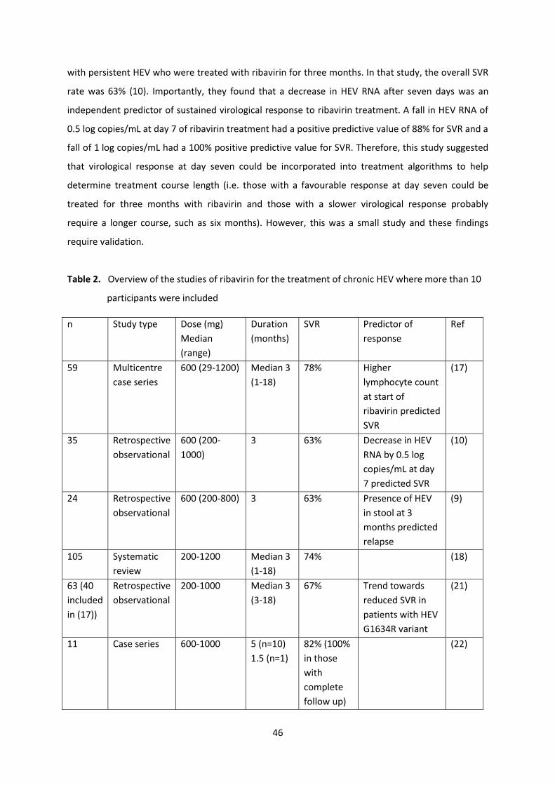
46
with persistent HEV who were treated with ribavirin for three months. In that study, the overall SVR
rate was 63% (10). Importantly, they found that a decrease in HEV RNA after seven days was an
independent predictor of sustained virological response to ribavirin treatment. A fall in HEV RNA of
0.5 log copies/mL at day 7 of ribavirin treatment had a positive predictive value of 88% for SVR and a
fall of 1 log copies/mL had a 100% positive predictive value for SVR. Therefore, this study suggested
that virological response at day seven could be incorporated into treatment algorithms to help
determine treatment course length (i.e. those with a favourable response at day seven could be
treated for three months with ribavirin and those with a slower virological response probably
require a longer course, such as six months). However, this was a small study and these findings
require validation.
Table 2. Overview of the studies of ribavirin for the treatment of chronic HEV where more than 10
participants were included
n Study type Dose (mg)
Median
(range)
Duration
(months)
SVR Predictor of
response
Ref
59 Multicentre
case series
600 (29-1200) Median 3
(1-18)
78% Higher
lymphocyte count
at start of
ribavirin predicted
SVR
(17)
35 Retrospective
observational
600 (200-
1000)
3 63% Decrease in HEV
RNA by 0.5 log
copies/mL at day
7 predicted SVR
(10)
24 Retrospective
observational
600 (200-800) 3 63% Presence of HEV
in stool at 3
months predicted
relapse
(9)
105 Systematic
review
200-1200 Median 3
(1-18)
74% (18)
63 (40
included
in (17))
Retrospective
observational
200-1000 Median 3
(3-18)
67% Trend towards
reduced SVR in
patients with HEV
G1634R variant
(21)
11 Case series 600-1000 5 (n=10)
1.5 (n=1)
82% (100%
in those
with
complete
follow up)
(22)

47
SVR = sustained virological response (HEV RNA negative six months post-treatment)
48
In that study they also assessed the impact of ribavirin dosage on SVR. Patients were treated on a
ribavirin dosage that was adapted according to the estimated glomerular filtration rate and ranged
from 200-1000 mg/day (median 600 mg). Interestingly ribavirin levels were assessed at day seven
and there was no association between ribavirin levels and sustained virological response rates.
One of the major sources of HEV viral replication is the gastrointestinal tract (20). Another recent
study assessed the kinetics of HEV shedding in stool in 24 transplant recipients treated with ribavirin
for three months to determine if this had an impact on treatment response (9). The overall sustained
virological response rate was 63%. Interestingly, all patients were plasma HEV RNA negative after
three months of treatment with ribavirin, but five patients were still excreting HEV RNA in the stool
at the end of treatment and these all relapsed. Therefore, persisting HEV in the stool, even after
clearance from the blood suggests ongoing HEV infection. This study suggests that measurement of
stool HEV RNA, in addition to the assessment of HEV RNA in the blood during treatment can help
determine the appropriate length of treatment.
Although the finding of negative HEV in blood and stool at the end of treatment is strongly predictive
of achieving a sustained virological response with ribavirin treatment, a small proportion (3/24
[12.5%]) of patients relapsed after three months of ribavirin treatment even with negative HEV RNA
in blood and stool at the end of treatment (9). It is possible that current assays for the detection of
HEV have insufficient sensitivity and that these patients had persisting low level viraemia or had
intermittent or low level shedding of virions in the stool that was not detected by the HEV RNA test
used. Moreover, patients with HEV treated with ribavirin can develop mutations conferring
resistance to ribavirin, which may contribute to treatment failure (21).
Detection thresholds for definition of HEV clearance are currently unknown. In the absence of robust
data in this area it is currently felt that plasma clearance should be defined by testing using a
validated assay with a Poisson sensitivity of at least 100 IU/ml or better. Stool clearance should be
defined by testing a 10% extract of faecal material using a validated assay with a Poisson sensitivity
of 100 IU/ml or better. There should be an aspiration to achieve better sensitivity and these
suggestions are liable to change as more is learnt in this area
A suggested algorithm for the treatment of HEV is shown in Figure 1.

49
Figure 1. A suggested algorithm for the treatment of HEV in solid organ transplant recipients
Dosage of Ribavirin
It remains unknown what the optimum dosage of ribavirin is for the treatment of persistent HEV.
Interestingly, one small study found no relationship between ribavirin levels at day seven and
outcomes from treatment in patients with persistent HEV (10). To date, all the reported studies have
used variable dosing ranging from 200 mg to 1200 mg per day with a median dosage of 600 mg/day.
The pharmacokinetics of ribavirin are variable in transplant recipients and clearance depends
particularly on renal function (23). In order to reduce the side effects of ribavirin and achieve a
steady state of ribavirin in the therapeutic range, the starting dose can be adapted based on the
creatinine clearance (the eGFR can be unreliable in transplant recipients so use creatinine clearance
is preferred). Table 3 shows a suggested dosing regimen for ribavirin according to the creatinine
clearance calculated by the Cockcroft-Gault equation, which can be used in transplant recipients
treated with ribavirin (23). Two of the reported studies of ribavirin in persistent HEV have used this
dosing regimen, and this regimen goes some way to standardizing the dosing of ribavirin until
further studies clarify the optimum dosing (9, 10).

50
Table 3. Suggested starting dosage of ribavirin in transplant recipients based on the Creatinine
Clearance measured by the Cockcroft-Gault equation (23)
Dosage to achieve
steady state levels of
ribavirin of
Creatinine Clearance by Cockcroft-Gault equation (mL/min)
100 80 60 40 20
10 μmol/L 810 690 570 450 330
14 μmol/L 1140 970 800 630 460
Practical starting total
daily ribavirin dosage
(mg)
1000 800 600-800 600 400
Note: ribavirin is usually prescribed using twice daily dosing
The most frequent side effect of ribavirin is a haemolytic anaemia, which required intervention in
approximately 40% (dose reduction, epoetin or blood transfusion) of those subjects included in the
previous systematic review (18). Therefore, patients require regular monitoring of haemoglobin on
treatment, initially every two weeks until the haemoglobin has reached nadir then monthly
thereafter. Dose reduction of ribavirin is recommended when the haemoglobin falls below 100 g/L.
Although epoetin has been used in some previous studies, there is no evidence to date that its use
has an impact on sustained virological response. For patients who develop very severe anaemia use
of epoetin or even blood transfusion may be indicated.
Treatment failure with ribavirin treatment
Approximately 40% of transplant recipients with persistent HEV relapse after three months of
treatment with ribavirin (9,10). There are a number of potential reasons for treatment failure. The
need for dose reduction as a result of side effects is a likely explanation in many cases, as ribavirin
needs to reach sufficient levels to have antiviral activity in all sites where the virus replicates in order
to clear the infection. As discussed above, another probable reason for failure is that some patients
receive an insufficient duration of treatment to ensure HEV is cleared from both blood and stool.
This could potentially be minimised by ensuring that patients have necessary assessments of plasma
and stool HEV RNA to ensure negativity before completing treatment with ribavirin. It has also

51
recently been shown that a G1634R mutation in the RdRp domain of the ORF1 protein is associated
with ribavirin treatment failure (24,25). Interestingly this mutation appears to increase the fitness of
HEV in vitro compared to the wild-type virus and may therefore contribute to the relative ‘ribavirin
resistance’ in some patients with persistent HEV (25,26). When studied in a cohort of 63 patients
treated with ribavirin for persistent HEV the G1634R mutation was present at baseline in 36.5% of
patients. This mutation was found in 31% of those who had sustained virological response with
ribavirin and 47.6% of those who failed treatment (p=0.2) suggesting it may have a minor effect on
treatment response, but other factors are also likely to be important (21). The potential use of this
and other mutations to help tailor treatment in patients with persistent HEV requires further study.
The majority of patients who relapse will respond to a longer course of treatment with ribavirin,
even those harboring the G1634R mutation (21). Therefore, retreatment with a longer course of
ribavirin should be considered for patients who relapse, and treatment continued until the HEV RNA
is negative in blood and stool on two tests at least one month apart. Re-treatment for six months
with ribavirin will be sufficient for many patients to achieve a sustained virological response, but
some patients will require longer treatment (27). There are occasional reports of very resistant cases
of HEV with multiple mutations that confer resistance to ribavirin who have persisting HEV viraemia
despite continued treatment (26). This emphasises the importance of ensuring the patient receives
effective treatment on their first course of treatment where possible.
PEG-interferon
There are a few reports of successful treatment of persistent HEV with PEG-interferon, summarised
in a recent systematic review (18). Overall, this review highlighted eight patients (six who had
undergone solid organ transplantation) treated with PEG-interferon for a median of three months
(range 3-12), of which 75% had a sustained virological response. Worryingly, two patients (25%)
experienced acute rejection during treatment. PEG-interferon is well known to increase the risk of
rejection in transplant recipients. In a randomised controlled trial of PEG-interferon +/- ribavirin for
hepatitis C in liver transplant recipients, 10% of patients had acute rejection necessitating treatment
withdrawal and augmentation of immunosuppression (28). This may not be a direct effect of the
PEG-interferon, but could be due to a reduction in calcineurin inhibitor levels associated with
clearance of the virus so careful monitoring of immunosuppressant levels is needed. Acute rejection
can have significant implications for solid organ recipients, particularly non-liver transplant patients,
and as such the use of PEG-interferon is not recommended as a first line treatment for HEV. PEG-
interferon treatment could be considered in cases of ribavirin refractory HEV, particularly if

52
associated with ribavirin resistance mutations. However, patients will require very close monitoring
for rejection.
Sofosbuvir
Sofosbuvir is a pangenotypic nucleotide analog licensed for the treatment of HCV. One recent study
suggested the sofosbuvir inhibited HEV RNA replication in an experimental model of HEV (29). The
inhibitory effect on HEV replication was additive when combined with ribavirin. However, the anti-
HEV activity was markedly lower than its anti-HCV effect suggesting that standard dosing of
sofosbuvir in patients with HEV may be insufficient to have an antiviral effect. One case report using
sofosbuvir at a dose of 400 mg per day in combination with ribavirin in an allogeneic stem cell
transplant recipient demonstrated antiviral activity against HEV in vivo; however, treatment was
insufficient to clear the virus (30). In contrast, another report found no change in HEV RNA levels in
an immunosuppressed post-liver transplant patient with HCVG3/HEVG3 co-infection who was
treated with sofosbuvir and daclatasvir (without ribavirin) (31). Further clinical studies are required.
References
1. Kamar N, Garrouste C, Haagsma EB, et al. Factors associated with chronic hepatitis in patients
with hepatitis E virus infection who have received solid organ transplants. Gastroenterology
2011; 140: 1481-9.
2. Kamar N, Marion O, Abravanel F, Izopet J, Dalton HR. Extrahepatic manifestations of hepatitis E
virus. Liver Int 2016; 36: 467-2.
3. Dalton HR, Keane FE, Bendall R, Mathew J, Ijaz S. Treatment of chronic hepatitis E in a patient
with HIV infection. Ann Intern Med 2011; 155: 479-80.
4. Del Bello A, Arne-Bes MC, Lavayssiere L, Kamar N. Hepatitis E virus-induced severe myositis. J
Hepatol 2012; 57: 1152-3.
5. Perrin HB, Cintas P, Abravanel F, et al. Neurologic disorders in immunocompetent patients with
autochthonous acute hepatitis E. Emerg Infect Dis 2015; 21: 1928-34.
6. Kamar N, Rostaing L, Abravanel F, et al. Ribavirin therapy inhibits viral replication on patients
with chronic hepatitis E virus infection. Gastroenterology 2010; 139: 1612-8.
7. Davern TJ, Chalasani N, Fontana RJ, et al. Acute hepatitis E infection accounts for some cases of
suspected drug-induced liver injury. Gastroenterology 2011; 141: 1665-72.
53
8. Hillebrandt KH, Arsenic R, Hofmann J, et al. Acute graft dysfunction 17 years after liver
transplant: A challenging clinical and histologic manifestation of hepatitis E. Exp Clin Transplant
2016 doi: 10.6002/ect.2015.0343. [Epub ahead of print].
9. Abravanel F, Lhomme S, Rostaing L, Kamar N, Izopet J. Protracted fecal shedding of HEV during
ribavirin therapy predicts treatment relapse. Clin Infect Dis 2015; 60: 96-9.
10. Kamar N, Lhomme S, Abravanel F, et al. An early viral response predicts the virological response
to ribavirin in hepatitis E virus organ transplant patients. Transplantation 2015; 99: 2124-31.
11. Wang Y, Zhou X, Debing Y, et al. Calcineurin inhibitors stimulate and mycophenolic acid inhibits
replication of hepatitis E virus. Gastroenterology 2014; 146: 1775-83.
12. Debing Y, Neyts J. mTOR-inhibitors may aggravate chronic hepatitis E. J Hepatol 2014; 61: 0-722.
13. Zhou X, Wang Y, Metselaar HJ, Janssen HL, Peppelenbosch MP, Pan Q. Rapamycin and
everolimus facilitate hepatitis E virus replication: revealing a basal defense mechanism of PI3K-
PKB-mTOR pathway. J Hepatol 2014; 61: 746-54.
14. Paeshuyse J, Dallmeier K, Neyts J. Ribavirin for the treatment of chronic hepatitis C virus
infection: a review of the proposed mechanisms of action. Curr Opin Virol 2011; 1: 590-8.
15. Debing Y, Emerson SU, Wang Y, Pan Q, Balzarini J, Dallmeier K, Neyts J. Ribavirin inhibits in vitro
hepatitis E virus replication through depletion of cellular GTP pools and is moderately
synergistic with alpha interferon. Antimicrob Agents Chemother 2014; 58: 267-73.
16. Mallet V, Nicand E, Sultanik P, et al. Brief communication: case reports of ribavirin treatment for
chronic hepatitis E. Ann Intern Med 2010; 153: 85-9.
17. Kamar N, Mallet V, Izopet J. Ribavirin for chronic hepatitis E virus infection. N Engl J Med 2014;
370: 2447-8.
18. Peters van Ton AM, Gevers TJ, Drenth JP. Antiviral therapy in chronic hepatitis E: a systematic
review. J Viral Hepat 2015; 22: 965-73.
19. Ampurdanes S, Olmedo E, Maluenda MD, et al. Permanent response to alpha-interferon
therapy in chronic hepatitis C is preceded by rapid clearance of HCV-RNA from serum. J Hepatol
1996; 25: 827-32.
20. Williams TP, Kasorndorkbua C, Halbur PG, Haqshenas G, Guenette DK, Toth TE, Meng XJ.
Evidence of extrahepatic sites of replication of the hepatitis E virus in a swine model. J Clin
Microbiol 2001; 39: 3040-6.
21. Lhomme S, Kamar N, Nicot F, et al. Mutation in the hepatitis E virus polymerase and outcome of
ribavirin therapy. Antimicrob Agents Chemother 2016; 60: 1608-14.
22. Pischke S, Hardtke S, Bode U, et al. Ribavirin treatment of acute and chronic hepatitis E: a
single-centre experience. Liver Int 2013; 33: 722-6.

54
23. Kamar N, Chatelut E, Manolis E, Lafont T, Izopet J, Rostaing L. Ribavirin pharmacokinetics in
renal and liver transplant patients: evidence that it depends on renal function. Am J Kidney Dis
2004; 43: 140-6.
24. Debing Y, Gisa A, Dallmeier K, et al. A mutation in the hepatitis E virus RNA polymerase
promotes its replication and associates with ribavirin treatment failure in organ transplant
recipients. Gastroenterology 2014; 147: 1008-11.
25. Todt D, Gisa A, Radonic A, et al. In vivo evidence for ribavirin-induced mutagenesis of the
hepatitis E virus genome. Gut 2016; 65: 1733-43.
26. Debing Y, Ramiere C, Dallmeier K, et al. Hepatitis E virus mutations associated with ribavirin
treatment failure result in altered viral fitness and ribavirin sensitivity. J Hepatol 2016; 65: 499-
508.
27. Kamar N, Izopet J, Tripon S, et al. Ribavirin for chronic hepatitis E virus infection in transplant
recipients. N Engl J Med 2014; 370: 1111-20.
28. Angelico M, Petrolati A, Lionetti R, et al. A randomized study on Peg-interferon alfa-2a with or
without ribavirin in liver transplant recipients with recurrent hepatitis C. J Hepatol 2007; 46:
1009-17.
29. Dao Thi VL, Debing Y, Wu X, Rice CM, Neyts J, Moradpour D, Gouttenoire J. Sofosbuvir Inhibits
hepatitis E virus replication in vitro and results in an additive effect when combined with
ribavirin. Gastroenterology 2016; 150: 82-5.
30. van der Valk M, Zaaijer HL, Kater AP, Schinkel J. Sofosbuvir shows antiviral activity in a patient
with chronic hepatitis E virus infection. J Hepatol 2017; 66: 242-3.
31. Donnelly MC, Imlach SN, Abravanel F, et al. Sofosbuvir and daclatasvir anti-viral therapy fails to
clear HEV viremia and restore reactive T cells in a HEV/HCV co-infected liver transplant
recipient. Gastroenterology 2017; 152: 300-1.