Embed Size (px)
Citation preview
Infection of dogs with Dirofilaria immitis has been di-agnosed in many European countries and is spreading.These guidelines, developed by the European Society forDirofilaria and Angiostrongylus, are based on the latest
information and include up-to -date recommendationsfor the prevention, diagnosis, and clinical managementof heartworm disease (HWD).
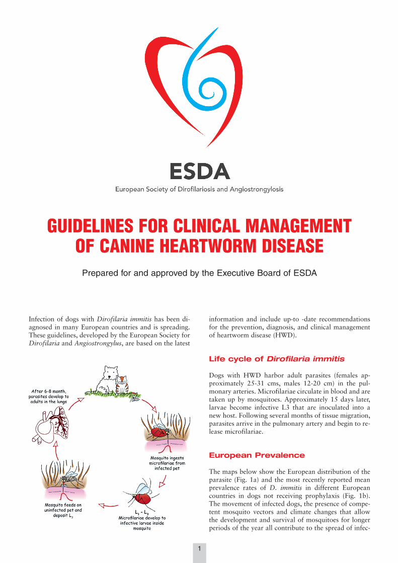
Life cycle of Dirofilaria immitis
Dogs with HWD harbor adult parasites (females ap-proximately 25-31 cms, males 12-20 cm) in the pul-monary arteries. Microfilariae circulate in blood and aretaken up by mosquitoes. Approximately 15 days later,larvae become infective L3 that are inoculated into anew host. Following several months of tissue migration,parasites arrive in the pulmonary artery and begin to re-lease microfilariae.
European Prevalence
The maps below show the European distribution of theparasite (Fig. 1a) and the most recently reported meanprevalence rates of D. immitis in different Europeancountries in dogs not receiving prophylaxis (Fig. 1b).The movement of infected dogs, the presence of compe-tent mosquito vectors and climate changes that allowthe development and survival of mosquitoes for longerperiods of the year all contribute to the spread of infec-
1
GUIDELINES FOR CLINICAL MANAGEMENTOF CANINE HEARTWORM DISEASE
Prepared for and approved by the Executive Board of ESDA
ESDA Heartworm 16-05-2017 13:08 Pagina 1

tion and disease. This is why it is so important to ad-minister preventives during the transmission season. Insome areas of Europe, this means year-round treatment(see section “Prevention”).
Clinical presentation of Heartworm Disease (HWD)
HWD is a chronic disease involving primarily pul-monary arteries and lungs. The heart is involved only inthe last stage of infection when pulmonary hypertensionleads to Cor pulmonale and right congestive heart fail-ure. At times acute clinical signs can be observed in thelate stage of the disease (pulmonary thromboembolism,Caval Syndrome*).
Many dogs may show no symptoms for months/yearsunless there is an exaggerated worm burden and/or theyundergo strenuous exercise. The clinical presentationscommon in dogs with HWD include:• Coughing• syncope following pulmonary hypertension• peritoneal effusion following right heart congestive
failure• *Caval Syndrome, which is due to a sudden rise in
pulmonary pressure and the subsequent displacement
of worms from the pulmonary artery into the rightcardiac chambers. Dyspnea, loud heart murmur (rightside of the thorax) and hemoglobinuria are pivotalclinical signs.
Thoracic Radiographs (both latero-lateral and dorso-ventral views)Radiographies are useful for evaluating the severity ofpulmonary lesions. Changes include:• perivascular inflammation• enlargement and tortuosity of pulmonary arteries• right heart enlargement• pleural effusion following right heart congestive failure
It is crucial to remember that alterations of the pul-monary arteries precede cardiac involvement. Enlarge-ment of the right cardiac chambers is not due to HW un-less accompanied by changes and enlargement of thepulmonary arterial chambers. The severity of observedlesions is not related to actual worm burden. Indeed,middle aged dogs may harbor a large worm burdenwithout severe radiographic changes, and on the otherhand, old dogs may present signs of previous throm-boembolism with few or no worms still present.For a step-by-step illustration of how to interpretatethoracic radiographs, consult the ESDA website:www.esda.vet
B mode, M mode and Doppler Echocardiography This allows evaluation of pulmonary hypertension, rightheart congestive failure, visualizing heartworms and es-timating worm burden.For a step-by-step illustration of how to interpretateEchocardiography consult the ESDA website: www.esda.vet
Clinical PathologyClinical-pathological abnormalities during heartworminfection are aspecific, may not always be present andare commonly related to an inflammatory state. Markedchanges in hematology and biochemistry are often seenonly in the late stage of the disease, or when acutechanges take place (Caval Syndrome). Commonly seenare leukocytosis, non-regenerative normocytic nor-
2
Figure 1a - Dirofilaria immitis in Europe. Figure 1b - Mean prevalence rates in Europe.● Imported cases only; ■ sporadic; * DNA of the parasite found in mosquitos; grey areas: no data.
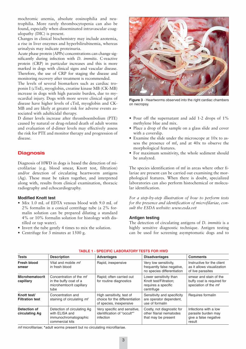
Figure 2 - Numerous adults of Dirofilaria immitis removed froman infected dog (females on the left side, males on the right).
ESDA Heartworm 16-05-2017 13:08 Pagina 2
mochromic anemia, absolute eosinophilia and neu-trophilia. More rarely thrombocytopenia can also befound, especially when disseminated intravascular coag-ulopathy (DIC) is present.Changes in clinical biochemistry may include azotemia,a rise in liver enzymes and hyperbilirubinemia, whereasurinalysis may indicate proteinuria.Acute phase protein (APPs) concentrations can change sig-nificantly during infection with D. immitis. C-reactiveprotein (CRP) in particular increases and this is moremarked in dogs with clinical signs and vascular disease.Therefore, the use of CRP for staging the disease andmonitoring recovery after treatment is recommended.The levels of several biomarkers such as cardiac tro-ponin I (cTnI), myoglobin, creatine kinase MB (CK-MB)increase in dogs with high parasite burden, due to my-ocardial injury. Dogs with more severe clinical signs ofdisease have higher levels of cTnI, myoglobin and CK-MB and are likely at greater risk for adverse events as-sociated with adulticidal therapy.D dimer levels increase after thromboembolism (PTE)caused by natural or drug-related death of adult wormsand evaluation of d-dimer levels may effectively assessthe risk for PTE and monitor therapy and progression ofdisease.
Diagnosis
Diagnosis of HWD in dogs is based the detection of mi-crofilariae (e.g. blood smear, Knott test, filtration)and/or detection of circulating heartworm antigens(Ag). These must be taken together, and interpretedalong with, results from clinical examination, thoracicradiography and echocardiography.
Modified Knott test• Mix 1.0 mL of EDTA venous blood with 9.0 mL of
2% formalin in a conical centrifuge tube (a 2% for-malin solution can be prepared diluting a standard4% or 10% formalin solution for histology with dis-tilled or tap water).
• Invert the tube gently 4 times to mix the solution.• Centrifuge for 3 minutes at 1500 g.
• Pour off the supernatant and add 1-2 drops of 1%methylene blue and mix.
• Place a drop of the sample on a glass slide and coverwith a coverslip.
• Examine the slide under the microscope at 10x to as-sess the presence of mf, and at 40x to observe themorphological features.
• For maximum sensitivity, the whole sediment shouldbe analyzed.
The species identification of mf in areas where other fi-lariae are present can be carried out examining the mor-phological features. When there is doubt, specializedlaboratories can also perform histochemical or molecu-lar identification.
For a step-by-step illustration of how to perform testsfor the presence and identification of microfilariae, con-sult the ESDA website: www.esda.vet
Antigen testingThe detection of circulating antigens of D. immitis is ahighly sensitive diagnostic technique. Antigen testingcan be used for screening asymptomatic dogs and to
3
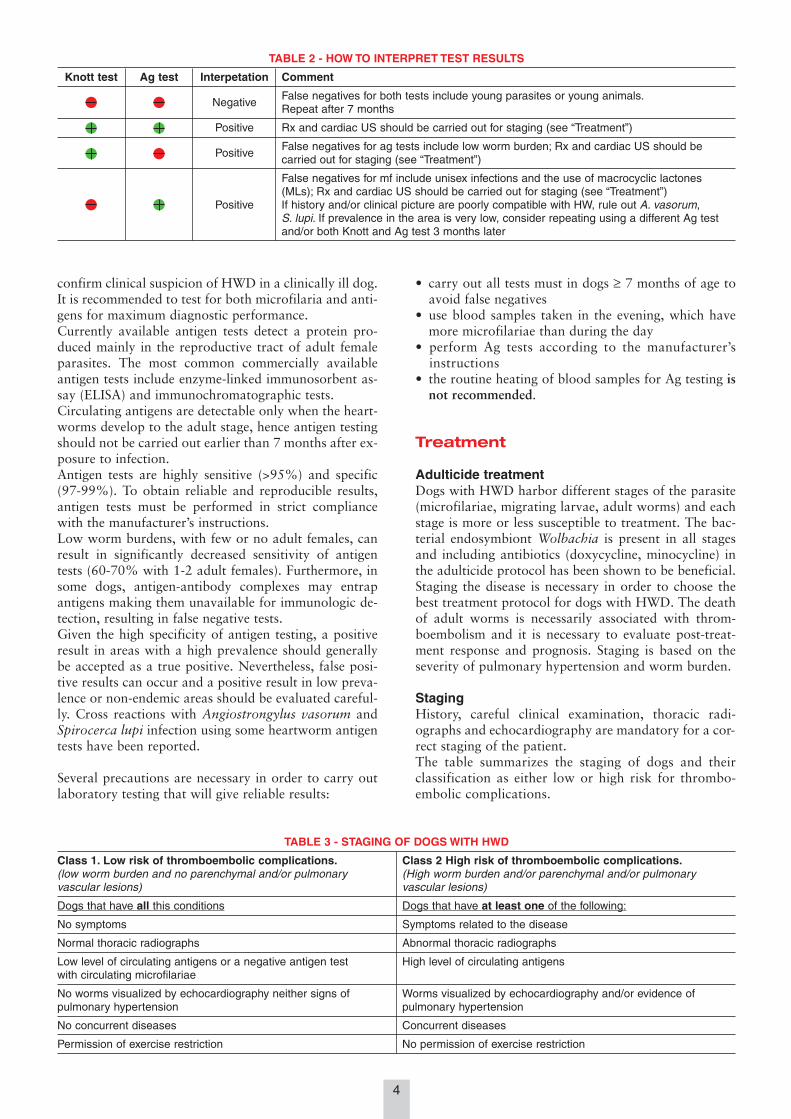
Figure 3 - Heartworms observed into the right cardiac chamberson necropsy.
TABLE 1 - SPECIFIC LABORATORY TESTS FOR HWD
Tests Description Advantages Disadvantages Comments
Fresh blood Vital and mobile mf Rapid, inexpensive Very low sensitivity, Instructive for the clientsmear in fresh blood frequently false negative, as it allows visualization
no species differentiation of live parasites
Microhematocrit Concentration of the mf Rapid; often carried out Lower sensitivity than smear and stain of thecapillary in the buffy coat of a for routine diagnostics Knott test/Filtration; buffy coat is required for
microhemtocrit capillary requires a specific speciation of the mftube centrifuge
Knott test/ Concentration and High sensitivity, test of Sensitivity and specificity Requires formalinFiltration test staining of circulating mf choice for the differentiation are operator dependent,
of species, inexpensive use of formalin
Detection of Detection of circulating Ag Very specific and sensitive, Costly, not diagnostic for Infections with a lowcirculating Ag with ELISA and identification of “occult*” other filarial nematodes parasite burden may
immunochromatographic infection that may be present give a false negativecommercial kits result
mf microfilariae; *adult worms present but no circulating microfilariae.
ESDA Heartworm 16-05-2017 13:08 Pagina 3
confirm clinical suspicion of HWD in a clinically ill dog.It is recommended to test for both microfilaria and anti-gens for maximum diagnostic performance.Currently available antigen tests detect a protein pro-duced mainly in the reproductive tract of adult femaleparasites. The most common commercially availableantigen tests include enzyme-linked immunosorbent as-say (ELISA) and immunochromatographic tests.Circulating antigens are detectable only when the heart-worms develop to the adult stage, hence antigen testingshould not be carried out earlier than 7 months after ex-posure to infection. Antigen tests are highly sensitive (>95%) and specific(97-99%). To obtain reliable and reproducible results,antigen tests must be performed in strict compliancewith the manufacturer’s instructions.Low worm burdens, with few or no adult females, canresult in significantly decreased sensitivity of antigentests (60-70% with 1-2 adult females). Furthermore, insome dogs, antigen-antibody complexes may entrapantigens making them unavailable for immunologic de-tection, resulting in false negative tests.Given the high specificity of antigen testing, a positiveresult in areas with a high prevalence should generallybe accepted as a true positive. Nevertheless, false posi-tive results can occur and a positive result in low preva-lence or non-endemic areas should be evaluated careful-ly. Cross reactions with Angiostrongylus vasorum andSpirocerca lupi infection using some heartworm antigentests have been reported.
Several precautions are necessary in order to carry outlaboratory testing that will give reliable results:
• carry out all tests must in dogs ≥ 7 months of age toavoid false negatives
• use blood samples taken in the evening, which havemore microfilariae than during the day
• perform Ag tests according to the manufacturer’sinstructions
• the routine heating of blood samples for Ag testing isnot recommended.
Treatment
Adulticide treatmentDogs with HWD harbor different stages of the parasite(microfilariae, migrating larvae, adult worms) and eachstage is more or less susceptible to treatment. The bac-terial endosymbiont Wolbachia is present in all stagesand including antibiotics (doxycycline, minocycline) inthe adulticide protocol has been shown to be beneficial.Staging the disease is necessary in order to choose thebest treatment protocol for dogs with HWD. The deathof adult worms is necessarily associated with throm-boembolism and it is necessary to evaluate post-treat-ment response and prognosis. Staging is based on theseverity of pulmonary hypertension and worm burden.
StagingHistory, careful clinical examination, thoracic radi-ographs and echocardiography are mandatory for a cor-rect staging of the patient.The table summarizes the staging of dogs and theirclassification as either low or high risk for thrombo-embolic complications.
4
TABLE 2 - HOW TO INTERPRET TEST RESULTS
Knott test Ag test Interpetation Comment
NegativeFalse negatives for both tests include young parasites or young animals.Repeat after 7 months
Positive Rx and cardiac US should be carried out for staging (see “Treatment”)
PositiveFalse negatives for ag tests include low worm burden; Rx and cardiac US should becarried out for staging (see “Treatment”)
False negatives for mf include unisex infections and the use of macrocyclic lactones(MLs); Rx and cardiac US should be carried out for staging (see “Treatment”)
Positive If history and/or clinical picture are poorly compatible with HW, rule out A. vasorum, S. lupi. If prevalence in the area is very low, consider repeating using a different Ag testand/or both Knott and Ag test 3 months later
TABLE 3 - STAGING OF DOGS WITH HWD
Class 1. Low risk of thromboembolic complications. Class 2 High risk of thromboembolic complications.(low worm burden and no parenchymal and/or pulmonary (High worm burden and/or parenchymal and/or pulmonaryvascular lesions) vascular lesions)
Dogs that have all this conditions Dogs that have at least one of the following:
No symptoms Symptoms related to the disease
Normal thoracic radiographs Abnormal thoracic radiographs
Low level of circulating antigens or a negative antigen test High level of circulating antigenswith circulating microfilariae
No worms visualized by echocardiography neither signs of Worms visualized by echocardiography and/or evidence ofpulmonary hypertension pulmonary hypertension
No concurrent diseases Concurrent diseases
Permission of exercise restriction No permission of exercise restriction
ESDA Heartworm 16-05-2017 13:08 Pagina 4

Melarsomine dihydrochlorideMelarsomine kills adult heartworms and it is the firstline drug for adulticide treatment.Patient preparation for melarsomine dihydrochlorideIt has been reported that the administration of preven-tive doses of a macrocyclic lactone (ML), combined withan antibiotic that is active against Wolbachia, may beused as preparation for adulticide treatment with melar-somine dihydrochloride. Thirty days of antibiotics(doxycycline at 10mg/kg/bid) and two monthly doses ofa macrocyclic lactone (ML) will eliminate migrating lar-vae and circulating microfilariae, while allowing timefor older larvae to reach the pulmonary arteries and be-come susceptible to melarsomine. Furthermore, thisregime will also weaken adult worms and make themless harmful to the lungs following their death. Dogsshould continue indefinitely with monthly doses of aML to avoid reinfection.Melarsomine dihydrochloride administrationIt is administered at the dose of 2.5 mg/kg body weight, viadeep intramuscular injection in the lumbar muscles. Therecommended treatment regime is a three-injection proto-col, with the first injection being followed at least 30 dayslater (dogs with severe infection or post-adulticide compli-cations can wait longer for completion of therapy) by afurther two injections of the same dose 24 hours apart.This protocol kills the worms in a gradual manner, thus re-ducing the severity of pulmonary thromboembolism.Reported side effects, which usually disappear after 24-48 hours, include:• mild swelling and soreness at the injection site• reluctance to move due to pain at injection site• depression• dyspnea• anorexia and vomiting.
Supportive treatment during adulticide therapyThere are several recommendations and supportivetreatment options during adulticide therapy, including:• Exercise restriction (no running, no jumping, no hunt-
ing), starting from the day of diagnosis until at leastone month after the last adulticide injection. Thispoint is very important and it should be stressed tothe owner that this will minimize problems associatedwith thromboembolism.
• Routine use of prednisone at 0.5 mg/kg bid the 1stweek, 0.5 mg/kg sid the 2nd week, 0.5 mg/kg everyother day for the 3rd and 4th weeks post adulticidetreatment seems to reduce lung inflammation sur-rounding dead worms. It has been shown howeverthat prednisone causes an increase in D dimer levelsafter adulticide therapy and could therefore increaseseverity of thromboembolism.
• Non steroid anti-inflammatory drugs and aspirin arenot advised. Lack of evidence of clinical benefit andsuggestions of contraindication.
• Calcium heparin: 50-100 UI s.c. 3 times daily starting1-2 weeks prior to and continuing for 4-6 weeks afteradulticidal treatment.
• Clopidogrel. Loading dose of 10 mg/kg p.o. on day 1,followed by a maintenance dose 2-3 mg/kg/sid. Promis-ing for other kinds of thromboembolic disease in dogs.Anecdotally useful but no clear evidence of efficacy.
• Low molecular weight heparin is promising for otherkinds of thromboembolic disease in dogs. Anecdotal-ly useful but no clear evidence of efficacy.
Antigen testing to confirm treatment success should be car-ried out at 6 months after the last adulticide injection. Ifthe test is still positive, it should be repeated after a further2-3 months. It is also necessary to control that microfilar-iae have been eliminated. If the dog has not been preppedusing macrocyclic lactones and doxycycline (microfilariaeusually fully eliminated in approximately 3-7 months), it isrecommended to use a registered microfilaricide.
Alternative adulticide treatment with macrocyclic lactones and doxycyclineML/doxycycline combinations have been shown to beadulticidal in both experimentally and naturally-infecteddogs. Most studies have concentrated on preventive dos-es of ivermectin, either weekly or bi-weekly for 6months, combined with doxycycline at 10mg/kg eithersid or bid. Infected dogs usually begin to be negative forcirculating antigens at about 12 months from the begin-ning of therapy. Treatment is well-tolerated with mini-mal radiological and clinical signs. The adulticide effectsof moxidectin combined with doxycycline have also beenshown following nine monthly doses of topical mox-idectin combined with thirty days of 10 mg/kg doxycy-cline bid. This protocol results in the rapid elimination ofmicrofilariae (by 21 days), thus breaking the transmis-sion cycle of the parasite very quickly. Furthermore, dogsbecome antigen negative by ten months, indicating amarked adulticidal effect. Long-term follow-up and clin-ical assessment of naturally infected dogs treated withthis protocol are on-going. As with melarsomine, exer-cise restriction is recommended with this protocol.
Minimally invasive SurgicalHeartworm removal
Surgical removal of worms is the only treatment optionfor dogs with Caval Syndrome and must be performed im-mediately. If successful, clinical signs should disappearquickly. Fluid therapy may be necessary in critically ill, hy-povolemic dogs to restore hemodynamic and renal func-tion. Surgery is also the option of choice for dogs withheavy worm burdens at risk for severe, post-adulticidecomplications. Echocardiographic visualization of the pul-monary arteries should be performed to determine that asufficient number of worms are in accessible locations.Antigen testing is not advised following surgical heart-worm removal due to the persistence of antigens in theblood. Dogs can be rechecked after at least 6 months todetermine (together with the clinical picture) if addi-tional adulticide treatment is necessary.
Treatment of spontaneous or post-adulticide thromboembolism
• Cage rest (mandatory)• Oxygen supplementation (Fraction of inspired oxy-
gen > 40%)
5
ESDA Heartworm 16-05-2017 13:08 Pagina 5
Treatment of right heart congestivefailure
If effusions are present• Furosemide 1 mg/kg 2-3 times a day or Torasemide
0.1 mg/kg once a day oral route• Spironolactone 2 mg/kg once a day• Hydrochlorothiazide 0.5-1 mg/kg 1-2 times a day if
refractory effusion• Angiotensin-converting-enzyme inhibitors (carefully
checking blood pressure and renal function)• Digoxin 0.005-0.01 mg/kg 2 times daily (only if atri-
al fibrillation is present for decreasing heart rate)
TABLE 5 - MACROCYCLIC LACTONES USED FOR THE PREVENTION OF HWD
Drug Administration Dose Interval Efficacy against other ESDA parasites #
Ivermectin oral > 6 mcg/kg Monthly D. repens
Milbemycin oral > 0.5-0.75 mg/kg Monthly A. vasorum
Moxidectin Injection Sr 0.17 mg/kg every 6-12 months D. repens
Selamectin Spot on 6 mg/kg Monthly
Moxidectin Spot on >2.5 mg/kg Monthly A. vasorum, D. repens
At the labelled dose, all these drugs are safe in dogs that are sensitive to MLs due to the presence of the so-called MultidrugResistant 1 mutation (for example, Collies, Australian shepherds, etc.).
# Prophylactic activity against D. repens or A. vasorum. Some drugs alone or in combination are active against other endo or ectoparasites.
TABLE 4 - PREVENTION PROTOCOLS IN DIFFERENT CLINICAL SCENARIOS
Age Puppies <2 months Start on chemoprophylaxisas soon as possible
Puppies 2-7 months Start on chemoprophylaxis Test 7 months after If negative:the beginning of the year-round preventionchemoprophylaxis If positive:
adulticide treatment
Animals older than Test previous If negative: Start on If negative:7 months chemoprophylaxis chemoprophylaxis and year-round prevention
re-test after 7 months to If positive:exclude recent infections adulticide treatment
If positive:adulticide treatment
Lack Noncompliance Test before resume If negative: Start on If negative:of compliance in chemoprophylaxis chemoprophylaxis chemoprophylaxis and year-round prevention
>7 months re-test after 7 months to If positive:exclude recent infections adulticide treatment
If positive:adulticide treatment
Noncompliance Resume chemoprophylaxis Test 7 months after If negative:in chemoprophylaxis the beginning of the year-round prevention<7 months chemoprophylaxis If positive:
adulticide treatment
Travel to Travel from a Receive 1 monthly doseendemic areas heartworm-free country of macrocyclic lactones
to an endemic country when return to thefor <1 month heartworm-free area
Travel from a Start 1 month afterheartworm-free country the beginning of the travelto an endemic country and end 1 months afterfor >1 month returning to the
heartworm-free area
• Prednisolone helps to control the clinical signs of pul-monary thromboembolism (cough, dyspnea, fever,weakness, hemoptysis), at the dose of 1-2 mg/kg sidfor 3-4 days, thereafter reducing the dose on the basisof clinical signs
• Calcium Heparine 200-300 IU 3 times a day s.c.• Butorphanol (up to 0.4 mg / kg i.m. q 2-4 hours) or
Morphine (up to 0.3 mg / kg s.c. q 4-6 hours) if severerespiratory distress is present
• Sildenafil (1-3 mg/kg) every 8-12 hours and Tadalafil(1 mg/kg every 48 hours) orally has been anecdotallyreported as useful.
• Diuretics not advised
6
ESDA Heartworm 16-05-2017 13:08 Pagina 6
• Phosphodiesterase 5 inhibitors (Sildenafil and Tada-lafil) and/or Phosphodiesterase 3 inhibitors (Pimoben-dan) are not useful and not advised due the anatomi-cal modification of pulmonary arteries.
Prevention
HWD can be prevented by the administration of macro-cyclic lactones that are able to eliminate infective larvaeup to 30 days old. Thus, the monthly administrationwill kill all the larvae that mosquitos have inoculated inthe previous 30 days. The slow-release injectable for-mulation available in some countries maintains the lar-vicidal effect for the 6-12 months.The current recommendation is to carry out preventionall year round; even in winter, urban heat islands allowmosquitoes to survive. Below are some key points to
prevention in different clinical situations.The use of topical synthetic pyrethroids (i.e. permetrin),appleid monthly, has been reported as significantly de-creasing the risk of mosquitoes bites in dogs (so-called“anti-feeding” effect). The use of pyrethroids does notsubstitute the use of MLs, but it can be combined withmacrocyclic lactones to lessen the risk of infection in thecase of missed administration of an ML or in cases oflack of compliance of the owner (“double defense”).
Retesting
All dogs on correct prevention should be screened (bothmicrofilariae and antigen testing) every other year. Iffailure of administration of one or more doses or lackof owner compliance is suspected, annual retesting ismandatory.
7
All rights reserved.No part of this publication may be reproduced, stored in a retrieval system, or internet communication system
or transmitted in any form, or by any means, electronic, mechanical, photocopying, recording or otherwise,without the prior permission, in writing, from the publisher.
© Copyright 2017 - EV/ESDA
ESDA Heartworm 16-05-2017 13:08 Pagina 7


