Embed Size (px)
Citation preview

Received AprilAccepted for pThe authors repThis is an open
Commons Ait is permisscited. The w
Copyright # 20ISSN: 1049-22DOI: 10.1097/S
ORIGINAL ARTICLE
The Journal o
Guideline for Care of Patients With the Diagnoses ofCraniosynostosis: Working Group on Craniosynostosis
Irene M.J. Mathijssen, MD, PhD
30, 2015.ublicationort no coaccess a
ttribution-ible to doork canno15 by Mu
75CS.00000
f Cranio
5;26: 1735–1807)
(J Craniofac Surg 201haired by Irene M.J. Mathijssen, MD, PhD
CParticipants:The Dutch National Cranial and Facial Deformities Patients andParents Association (LAPOSA)The Netherlands Society for OphthalmologyThe Netherlands Society for AnesthesiologyThe Netherlands Society for OtorhinolaryngologyThe Dutch Association for PediatricsThe Netherlands Society for Oral Medicine and Oral and Max-
illofacial SurgeryThe Netherlands Society for NeurosurgeryThe Netherlands Society for Plastic SurgeryThe Society for OrthodontistsThe Netherlands Society for Clinical GeneticsThe Dutch Association of PsychologistsThe Netherlands Society for Relationship Counseling and
Family TherapyAddress correspondence and reprint requests to Irene M.J.
Mathijssen, MD, PhD, Deptartment of Plastic and ReconstructiveSurgery, and Hand Surgery, Room Sk-1202, Erasmus MedicalCenter-Sophia, Wytemaweg 80, 3015CN Rotterdam, The Nether-lands. E-mail: [email protected]
ABSTRACTThis guideline for care of children with craniosynostosis
was developed by a national working group with representativesof 11 matrix societies of specialties and the national patients’society. All medical aspects of care for nonsyndromic and syn-dromic craniosynostosis are included, as well as the social andpsychologic impact for the patient and their parents. Managerialaspects are incorporated as well, such as organizing a timelyreferral to the craniofacial center, requirements for a dedicated
What Is This Box?A QR Code is a matrix barcode readable byQR scanners, mobile phones with cameras,and smartphones. The QR Code links tothe online version of the article.
June 28, 2015.nflicts of interest.rticle distributed under the terms of the CreativeNonCommercial-NoDerivatives 4.0 License, wherewnload and share the work provided it is properlyt be changed in any way or used commercially.taz B. Habal, MD
00000002016
facial Surgery � Volume 26, Number 6, Sep
craniofacial center, and centralization of this specialized care. Theconclusions and recommendations within this document arefounded on the available literature, with a grading of the levelof evidence, thereby highlighting the areas of care that are in needof high-quality research. The development of this guideline wasmade possible by an educational grant of the Dutch Order ofMedical Specialists.
The development of this guideline was supported by an edu-cational grant of the Dutch Order of Medical Specialists.
ContentsChapter 1. General introduction 1735Chapter 2. Introduction to craniosynostosis 1736Chapter 3. Referral and diagnostics 1737Chapter 4. Perioperative care 1744Chapter 5. Surgical treatment of nonsyndromic craniosynosto-
sis 1747Chapter 6. Surgical treatment of syndromic craniosynostosis—
the cranial vault 1751Chapter 7. Surgical treatment of syndromic craniosynostosis—
the face 1753Chapter 8. Increased intracranial pressure 1762Chapter 9. Hydrocephalus 1768Chapter 10. Chiari I malformation 1769Chapter 11. Vision, refraction and motility abnormalities 1771Chapter 12. Obstructive sleep apnea syndrome 1772Chapter 13. Hearing impairments 1776Chapter 14. Dentofacial deformities 1777Chapter 15. Deformities of the extremities 1780Chapter 16. Cognitive functioning and behavior 1783Chapter 17. Patients’ and parents’ perspectives 1786Chapter 18. Psychosocial functioning 1787Chapter 19. Criteria for the craniofacial center and its team
members 1790Chapter 20. Summary 1794
1. GENERAL INTRODUCTION
ObjectiveThis guideline provides recommendations for medical practice
on suspicion of craniosynostosis and after confirmation of thisdiagnosis. It is based on the results of scientific research andsubsequent forming of opinion by a multidisciplinary workinggroup, composed of representatives of the medical specialtiesinvolved in the treatment of craniosynostosis, related professionaldisciplines, and other parties involved.
The wishes of parents and health professionals regarding theorganization of health care for craniosynostosis were inventoried.The guideline document addresses the important issues for thesupport of parents with a child with a craniosynostosis.
Method Employed by the Working GroupThe content of this guideline is based on evidence from published
scientific research. Relevant articles were identified using systematic
tember 2015 1735

Mathijssen The Journal of Craniofacial Surgery � Volume 26, Number 6, September 2015
searches in Medline, Embase, and the Cochrane Library. Existingguidelines were specifically searched for in online accessible inter-national and national guideline clearinghouses. Searches were limitedto the Dutch, English, and French languages. In addition, articles wereextracted from reference lists of relevant literature. This resulted inadditional articles for several basic questions.
Care for children with craniosynostosis was introduced in thelate 1960s and consequently the English literature from those yearsonwards was included. Searches were performed until December 1,2009 and articles available as ‘‘Epub ahead of publication’’ at thatdate were included as well.
The following search terms were used: craniofacial, craniosy-nostosis combined with: genetics, hydrocephalus, Chiari, cerebralpressure, otitis, hearing, vision, psychology, anesthesia, compli-cations, infection, development, growth, maxilla, mandible, dis-traction, osteotomy, Fort, midface, RED, halo, monobloc, facialbipartition, median faciotomy, and hypertelorism.
Relevant articles extracted from reference lists of retrieved litera-ture and several relevant publications until November 1, 2009 wereincluded as well. Under the headings Summary of the literature/Conclusions only published studies/guidelines are discussed. Casereports and letters were excluded, unless they reported a complication.
The selected articles were assessed on methodological qualitygraded by level of evidence according to the standard classification:see Table 1. After selection, those articles remained that are listed tounderpin the various conclusions. The articles are assessed underthe heading ‘‘Summary of the literature.’’ Next, the scientificevidence is briefly summarized in ‘‘Conclusions.’’ The main lit-erature on which a conclusion is based is mentioned as well,including the level of evidence (see Table 2).
Other aspects than scientific evidence may be relevant to making arecommendation as well, such as patient preferences (derived from theresults of the focus group sessions or relevant literature on the patientperspective), costs, availability, or organizational aspects. These kindsof aspects, provided they have not been subject of research, arementioned under the heading ‘‘Considerations.’’ The experienceand the opinion of the working group members have been key tothe other considerations. The ‘‘Recommendation’’ results from thecombination of the available evidence and the other considerations.
Patient PerspectiveWhen developing this guideline, the working group particularly
strived for incorporating the patient perspective. The working grouptherefore included representatives of the LAPOSA patient associ-ation. Besides, in collaboration with LAPOSA 2 focus groupsessions were held: 1 with parents of a young child with a
TABLE 1. Classification of Methodological Quality of Individual Studies
Intervention Diagnostic Accuracy Asses
A1 Systematic review of at least 2 mutuallyindependent studies of A2-Level
A2 Randomized double-blind comparativeclinical study of good quality andsufficient size
Study comparing with a refewith previously defined cassessment of the results oregarding a sufficiently lawho all were administered
B Comparative study, but not possessing allqualities mentioned under A2 (thiscategory also includes patient-checkupstudy, cohort study)
Study comparing with a refequalities mentioned under
C Noncomparative study
D Expert opinion
�This classification only applies to situations in which controlled trials are not feasible for et
1736
craniosynostosis and 1 with young adults with craniosynostosisand parents of older children with a craniosynostosis. The workinggroup incorporated outcomes of these sessions in the guideline.Finally, focus group participants were asked to comment on a draftversion of the guideline.
NoteThis guideline was drafted on the guidance of the Appraisal of
Guidelines for Research & Evaluation (AGREE) instrument. Thisinstrument was developed at a European level to enable assessmentof the procedural quality of guidelines. By incorporating AGREEaspects in the introduction to the guideline it is made clear whatquality requirements have been met.
2. INTRODUCTION TO CRANIOSYNOSTOSISCraniosynostosis is a congenital cranial malformation in which 1
or more cranial sutures have fused already in utero. The cranialsutures separate the skull bone plates and enable rapid growth of theskull in the first 2 years of life, in which growth is largely dictatedby growth of the brain. Cranial sutures are essential to skull growthin the first 2 years (the period of rapid brain growth). Thereafter theprocess of appositional growth and internal resorption of the skull isthe major process by which the skull increases in size.
Premature fusion of cranial sutures impedes normal growth of theskull, resulting in characteristic anatomic malformations of the skull.
Craniosynostosis occurs in 1 in 2100 to 1 in 2500 births and maybe either nonsyndromic (also referred to as isolated) or syndromic.In syndromic craniosynostosis, other birth defects are present nextto the craniosynostosis. In syndromic craniosynostosis, usuallymore than 1 cranial sutures have prematurely fused, typicallyinvolving both coronal sutures.
The distinction between nonsyndromic and syndromic is made onthe guidance of dysmorphologic evaluation and genetic evaluation.Owing to advances in genetic diagnostics, nonsyndromic patients areincreasingly recognized as syndromic patients. The discovery of theP250R mutation in the FGFR3 gene in patients with a uni- of bilateralcoronal suture synostosis clearly illustrates this phenomenon.
Approximately 60% of all craniosynostoses are of the nonsyn-dromic type and approximately 40% are of syndromic type. Withinthe group of nonsyndromic craniosynostoses the sagittal suturesynostosis is the largest group, followed by metopic suture synostosis.Recently, the prevalence of metopic suture synostosis has clearlyrisen both in Europe and the United States, but this has not beenexplained. Synostosis of 1 or both lambdoidal sutures is very rare.
Of the syndromic types the Muenke syndrome is the mostfrequent, followed by Crouzon syndrome, and Pfeiffer syndrome.
sment
Harm or Side Effects, Etiology,
Prognosis�
rence test (a ‘‘criterion standard’’)ut-off values and independentf study test and criterion standard,rge series of consecutive patients
the index- and the reference test
Prospective cohort study of sufficient sizeand follow-up, adequately controlled forconfounding and with satisfactoryexclusion of selective follow-up
rence test, but not possessing allA2
Prospective cohort study, but notpossessing all qualities mentioned underA2, or retrospective cohort study orpatient-checkup study
hical or other reasons. If they should be feasible, the classification for interventions applies.
# 2015 Mutaz B. Habal, MD

TABLE 2. Level of Evidence for the Conclusion
Conclusion based on
1 Level A1 study or at least 2 mutually independent Level A2 studies
2 One Level A2 study or at least 2 mutually independent Level B studies
3 One Level B or C study
4 Expert opinion
The Journal of Craniofacial Surgery � Volume 26, Number 6, September 2015 Diagnoses of Craniosynostosis
The Apert syndrome has the lowest prevalence. The distinctionbetween Crouzon syndrome and Pfeiffer syndrome was made on thebasis of deformities of the big toe and thumb: if these were broad andshort the syndrome was referred to as Pfeiffer syndrome. BecauseFGFR2 mutations, however, were identified in both Crouzon andPfeiffer syndrome, which can even be completely identical, thedistinction between these 2 syndromes can be considered irrelevant.Before the discovery of TWIST1 mutations and deletions in Saethre-Chotzen syndrome and the P250R FGFR3 mutation in Muenkesyndrome it was hard to distinguish between these patients. Newmutations are expected to be found in the future, on account of whichthe group of nonsyndromic craniosynostoses will further decrease.
The following types of craniosynostosis are distinguished:Nonsyndromic:
Sagittal suture synostosis (scaphocephaly)Metopic suture synostosis (trigonocephaly)Coronal suture synostosis, unilateral (frontal plagiocephaly)Coronal suture synostosis, bilateral (frontal brachycephaly)Lambdoid suture synostosis (pachycephaly)
Syndromic:
Apert syndrome (FGFR2 mutation Ser252Trp and Pro253Arg,deletion exon IIIc, Alu insertion exon IIIc)Crouzon or Pfeiffer syndrome (FGFR2 mutations exclusive ofApert mutations)Saethre-Chotzen syndrome (TWIST1 mutations or deletions)Muenke syndrome (Pro250Arg FGFR-3 mutation)Craniofrontonasal dysplasia (EFNB1 mutations)Complex craniosynostosis (syndromic picture without knownmutation, often 2 or more synostotic sutures)
Each type of craniosynostosis may vary in its severity of pres-entation. Notably, sagittal suture synostosis and metopic suturesynostosis can show a very mild phenotype in which only a boneridge at the afflicted suture is palpable and/or visible. The severe formof sagittal suture synostosis is characterized by considerable frontalbossing and punctiform occipital prominence in combination withsaddle deformity in the midline of the skull. The severe form ofmetopic suture synostosis is characterized by a large wedge-shapedforehead with underdeveloped lateral parts of the supraorbital ridge,temporal depressions, and hypotelorism. Synostosis of 1 coronalsuture causes orbital dystopia, in which the orbital cone at the afflictedside is placed higher than of the unafflicted side, retrusion of theforehead at the afflicted side with compensatory bossing of the con-tralateral side, and asymmetry of the skull base and the face. Con-sequently, displacement of the ear and lateral deviation of the noseoccur. Bilateral synostosis of the coronal sutures result in retrusion ofthe forehead and the supraorbital rim with a broad skull. Syndromiccraniosynostosis is usually characterized by synostosis of both cor-onal sutures, but other types of craniosynostosis are seen as well.
Indications for surgical treatment of nonsyndromic craniosy-nostosis are the risk of increased intracranial pressure (ICP) and theabnormal skull shape. Since the first surgical intervention forcraniosynostosis, a great many surgical techniques for the various
# 2015 Mutaz B. Habal, MD
types of craniosynostosis have been described. A broad distinctionis that between osteoclastic techniques and remodeling techniques,osteoclastic techniques involve resection of bone, thereby allowingthe developing and expanding brain to change the shape of the skull,also because the adverse impulse to the growth direction of the skullis removed. The remodeling techniques, on the other hand, do notrely on the selfcorrecting capacity of the skull and the brain, butattempt to obtain the desired skull shape by a reconstruction.
The surgical treatment of patients with syndromic craniosynos-tosis was developed in Paris in the early 1970s. Treatment has a 2-fold aim: to enlarge the cranial volume so as to prevent sequelae ofICP (mental retardation and impaired vision), and to correctmorphologic abnormalities of the cranium, the orbits, and the upperjaw. Multiple corrections of the skull and/or the face may beneeded, although naturally it is strived for to restrict the numberof interventions as much as possible.
The group of complex craniosynostoses is managed in the sameway as the syndromic craniosynostoses, as these patients oftenexperience similar problems of cognition and ICP.
Syndromic craniosynostosis, notably, the Apert and Crouzon/Pfeiffer syndromes, are associated with skeletal hypoplasia of themidface. The result may be narrowed airway causing obstructivesleep apnea syndrome (OSAS) in approximately 50%, exorbitismwith risk of cornea injury, malocclusion, and esthetic/psychosocialproblems.
Associated brain abnormalities in syndromic craniosynostosisinclude raised intracranial pressure (ICP), Chiari I malformation,ventriculomegaly, and hydrocephalus.
In all types of syndromic synostosis hearing loss is described.Vision, refraction and motility abnormalities in the nonsyndromiccraniosynostoses are in fact only seen in unilateral coronal suturesynostosis. In the syndromic group, abnormalities such as astig-matism and strabismus are very frequent.
Deformities of the extremities are notably restricted to thesyndromic craniosynostoses, and in patients with Apert syndromealways present in a severe form. In the other syndromic types,deformities of the extremities are generally very mild.
Parents are often confronted with health professionals who donot recognize the craniosynostosis shortly after the birth of thechild. This can be a source of stress for parents and lead to a delay indiagnosis and treatment. Nonsyndromic and syndromic craniosy-nostosis may co-occur with cognitive impairments and behavioraldisorders. These may occur both intrinsic to the congenital defectand secondary to, for example, increased cranial pressure or abnor-mal physical appearance. Notably, families of a child with syn-dromic craniosynostosis may experience psychosocial problems,such as having to cope with negative reactions from others, apossible discrepancy between deviating physical appearance andcognition, and problems with school choice.
3. REFERRAL AND DIAGNOSTICS
Basic Questions
1. H
ow to optimize recognition of craniosynostosis in the primaryand secondary healthcare sectors?2. H
ow to organize referral?3. W
hat additional diagnostic procedures are applied in thetertiary healthcare sector?IntroductionCraniosynostosis should be recognized timely for optimal treat-
ment. It appears, however, that craniosynostosis patients often are
1737

Mathijssen The Journal of Craniofacial Surgery � Volume 26, Number 6, September 2015
not referred at all or referred (too) late. A complicating factor in therecognition of craniosynostosis is the high incidence of positionalhead shape deformities. It may be expected that recognition, andthus referral of patients with craniosynostosis will happen earlier ifhealth professionals in the primary and secondary healthcare sectorsare facilitated to make this distinction.
Preceding the referral to a tertiary healthcare center, we often seeoveruse of diagnostic imaging, which is associated with furtherdelay in referral, an extra burden and insecurity for patient andparents as well as unnecessary expenditure. This should berestricted to a minimum. There are many options to diagnoseisolated craniosynostosis or any associated abnormalities. Torestrict both underdiagnosis and overdiagnosis, basic question 3concerns further diagnostic procedures in a tertiary healthcarecenter. Areas of attention are diagnostic imaging, genetic diagnos-tics, and the role of the pediatrician.
Inclusion of a clinical geneticist in a multidisciplinary cranio-facial team provides the opportunity to address questions fromparents and attending physicians.
Parents will first of all want to know if their child is healthyotherwise, what may have caused the deformity, how great thechance is of recurrence in a new pregnancy in the family and/or inthe next generation, and what prenatal diagnostic methods areavailable. For the attending physician it is important to know ifthere are any associated anomalies (to be expected) next to thecraniosynostosis that may be of influence to the treatment plan andthe prognosis of the child.
Basic Question 1: How to Optimize Recognitionof Craniosynostosis in the Primary andSecondary Health Care Sectors?Summary of the Literature
The abnormal skull shape is recognized by parents themselves,midwife, obstetrician, general practitioner, infant health clinic(physician, nurse), (pediatric) physiotherapist, pediatrician,(pediatric) neurologist, and helmet maker. Occasionally, it isrecognized later by other specialists and sometimes not until acraniofacial team is involved. Most of the children (50%–90%)were referred via the pediatrician, others via the infant health clinic,or parents themselves via the GP. These data are from the Birming-ham craniofacial center and could therefore well compare with thesituation in the Netherlands.1,2 For the situation in the Netherlandsonly the information from an article by Bredero-Boelhouwer isavailable,3 in which 18 children were identified by the referrers ashaving craniosynostosis and in 14 of whom this diagnosis wasconfirmed in the tertiary center. Of the 89 referrals with the initialdiagnosis nonsynostotic occipital plagiocephaly (NSOP) made bythe referrer, 10 patients appeared to have a craniosynostosis. Thus,professionals in the primary and secondary healthcare sectorswrongly diagnosed 14 of the total of 107 patients (13%).
Recognition of the condition is complicated by an increase inpositional plagiocephaly (current prevalence 20% to 48%) since theearly 1990s when supine sleep position was recommended to preventcot death.2,4–6 The increase in positional head deformities carries therisk of missing the diagnosis of craniosynostosis in more patients.
A craniosynostosis or positional skull deformity is primarilyrecognized by physical examination, notably skull shape4,6 incombination with history taking,3,5 and an imaging study is rarelyindicated.6 Both history taking and physical examination are wellpossible in the primary and secondary healthcare sectors. Thisdeserves great attention because early recognition and surgicalcorrection are essential to a satisfactory treatment outcome.6 Pedia-trician and infant health clinic carry the main responsibility torecognize skull deformities. Ridgeway recommends a number of
1738
diagnostic steps that should lead to early diagnosis, in which historytaking and physical examination yield the most important infor-mation to distinguish between positional head shape deformitiesand craniosynostosis.5
Using this information in a flowchart at intake appears to be asafe method to make this distinction at the earliest. This will preventdelay in start of treatment as a possible consequence of the increaseof patients of positional head shape deformities.3 This flowchart hasbeen validated only in a tertiary center. There are no other publi-cations on referral patterns in children with abnormal skull shape.
History TakingHistory taking should address the distinction between positional
head shape deformities and craniosynostosis on the one hand, andrisk factors and associated anomalies on the other hand.
Craniosynostosis is present at birth, positional head shape deform-ities usually not.3–6 Improvement of abnormal head shape is seen inpositional head shape deformities, but not in craniosynostosis. Pre-sence or absence of a preferred sleep position is essential in thediagnostics of positional head shape deformities.3–6
Analysis of a flowchart to differentiate positional headshape deformity versus craniosynostosis by means of a question-naire (completed by telephone or e-mail) showed that the ques-tionnaire recognized no more than 61.5% of the positional headshape deformities. Nurse practitioner contacts increased this per-centage to 75%. A craniosynostosis was never missed (false-positive¼ 0%, which implies high sensitivity).3 In that study, theessential questions were:
� I
s deformity present at birth? � I s there a preferred sleep position? � I s there improvement of the deformity?The following risk factors for craniosynostosis are described:smoking habit, mother’s antenal stay at high altitude, paternal
occupation,6–8 maternal alcohol use,7,8 maternal substance use,8vitamin D deficiency or receptor insensitivity, chronic renal failure,hypophosphatemia, hyperthyroidism, mucopolysaccharidosis,5,9 andmaternal drug use: phenytoin, retinoids, valproate, aminopterin/methotrexate, fluconazole, cyclophosphamide, folic acid nitrosates,thyroid gland medication, and vitamins.5,7,10,11 Also a high age offather and mother increases the risk of craniosynostosis.7,12
Physical Examination of Skull and FaceMore in detail the different craniosynostoses can be recognized
as follows:
Sagittal synostosis:
frontal bossing, prominent occiput, bone ridge palpable onsagittal suture, decreased biparietal diameter, long andnarrow head, tapering from parietal to sagittal suture,with usually a head circumference >2 SD.1,5,6,8Metopic synostosis:
wedge-shaped forehead, including cranial part of the orbits,ridge on metopic suture, and hypotelorism.5,12Unicoronal synostosis:
flattened forehead at afflicted side, including flattening ofcranial part of the orbits, flattened cheek, nose deviationto unaffected side, orbital dystopia with higher positionof the ipsilateral orbital rim, and bossing of contralateralside of forehead.5Lambdoid synostosis:
unilateral occipital flattening, depression of lambdoidsuture ridge, low-positioned ipsilateral ear and back ofthe skull base.5Bicoronal synostosis:
broad head, flattened forehead and supraorbital rim.5Additional characteristics of skull and face are the following:
The anterior fontanel shows earlier fusion or is triangularshaped rather than diamond shaped in synostosis of 1 adjacent
# 2015 Mutaz B. Habal, MD
The Journal of Craniofacial Surgery � Volume 26, Number 6, September 2015 Diagnoses of Craniosynostosis
# 2
suture.6 In Apert syndrome, there is even a large midline defectinitially.13
Mid-face hypoplasia, proptosis, and hypertelorism are foundparticularly in the syndromic types of craniosynostosis.5,8,13
These and other syndrome-specific facial features are discussedin the genetics section of this chapter, including additionaldeformities of the extremities in particular.
The following characteristics may help to distinguish craniosy-nostosis from positional head shape deformities:
Positional plagiocephaly, such as a ‘‘parallelogram’’ seen fromabove, that is, ipsilateral occipital flattening, curved forehead,and ear more anterior ipsilateral.4,5 This in contrast with the‘‘trapezium’’ shape in unilateral coronal suture synostosis, thatis, ipsilateral flattening of the forehead and cranial part of orbitand occiput.5,6
American Cleft Palate-Craniofacial Association (ACPA) recom-mends that craniofacial teams provide education to healthprofessionals in obstetrics and neonatology as well as GPs andinfant health clinic physicians so as to improve early recognitionof children with craniofacial deformities.9
Grol emphasizes that effective implementation of guidelinesrequires behavioral changes in physicians.14 Interactive instruc-tion methods via CD-rom and information-sites on Internet seemvery effective. E-mail consultations improve the quality ofdecision-making, notably as they are easily accessible.15 Intensiveinstruction to small groups of physicians and peer review appearedlittle effective, however, in improving quality of care.16 Medicalaudits in the GP practice seems promising for quality of careimprovement, but should be further developed.17 Availability of agood computer-aided medical information system is very import-ant to recognize clinical pictures. Audit programs are most effec-tive when integrated with other educational and researchprograms.18
Conclusions
Level 3
015 M
Triage (with the use of a flow diagram) at intake of children withabnormal skull shape is likely to be effective to recognizecraniosynostosis and to timely refer the children to theappropriate specialist.
B Bredero-Boelhouwer, 20093
Level 3
Craniosynostosis is often hardly recognized and recognition is evenmore complicated by the increase in positional skull deformitiessince supine sleep position was recommended to prevent cot deathin the early 1990s.C Komotar, 20066
C Parameters ACPA, 20079
C Ridgeway, 20045
C White, 20102
Level 3
Craniosynostosis can be distinguished from NSOP on the basis ofmedical history and physical examination.B Bredero-Boelhouwer, 20093
C Komotar, 20066
C Ridgeway, 20095
Level 3
Easily accessible (computer assisted) information in the primary andsecondary health care sectors on history taking, physical examination,and use of decision tree improves quality of care.C Grol, 200014
C Smeele, 199916
C Grol, 199517
C van der Weijden, 199615
C Grol,198518
utaz B. Habal, MD
Considerations
Often it takes long before parents and the child with craniosy-nostosis are referred to the tertiary center, which may lead totreatment delay and confusion, and consequently a great deal ofparental stress.
The flow diagram to discriminate between positional skulldeformities and craniosynostosis was validated in a tertiary caresetting, where it was applied by secretaries who were not specifi-cally trained in distinguishing between these 2 conditions. Con-sequently, we do not expect great differences in its reliability whenapplied in primary and secondary care settings. Ideally, validationfor primary and secondary care settings finds place in these settings,but this does not seem to be practically feasible in view of the verylow frequency of craniosynostosis compared with positional skulldeformities.
Additional imaging studies to differentiate between positionalskull deformities and craniosynostosis are rarely performed intertiary centers. On clinical diagnosis of a positional skull deform-ity, skull x-ray or ultrasound are advised against to preventunnecessary medical imaging (with associated costs, radiationexposure, burden for patient and parents, lack of added value,and required experience in interpretation). Consultation with atertiary craniofacial center is indicated in suspected patients, forwhich submitting normal pictures (front view, lateral view, anteriorview, and view from above) will usually suffice.
RecommendationsThe following measures are essential to optimize recognition of
craniosynostosis in the primary and secondary care sectors:
1. E
nsure that easily accessible, reliable, and unambiguousinformation is available about skull deformities, either asappendix to this guideline, or via separate guidelines;preferably with many illustrations, clear terminology/defi-nitions and addresses.3
2. E nsure that the flow diagram (Appendix II) is used.3. P
rovide structured education and training instruction to infanthealth center physicians, GPs, midwives and obstetricians aboutover skull deformities via centers of expertise with an initiatingrole for the tertiary centers.4. P
rovide feedback about the referral pattern on the basis of ananalysis of national registry data (focus on patient’s age atreferral to tertiary center).The craniosynostosis centers should develop a website withrelevant information for the primary and secondary healthcaresectors, providing an e-mail address for online consultations. Theyshould stimulate inclusion of the decision tree in the ICT-systemsused by GPs and infant health center physicians, as well as intraining programs for GPs, infant health center physicians, mid-wives, pediatricians and gynecologists. An interactive CD onhistory taking and physical examination in skull deformities isbeing prepared.
Collaboration with specialized centers for diagnostics and treat-ment of positional skull deformities will facilitate knowledgetransfer.
Basic Question 2: How to Achieve EffectiveReferral?
With the exception of the study by Bredero-Boelhouwer there isno literature available about the referral pattern in the Netherlands.3
From the focus group sessions it became also clear that parents
1739

Mathijssen The Journal of Craniofacial Surgery � Volume 26, Number 6, September 2015
highly appreciate quick referral to a specialized center (see chapteron focus groups).
Early recognition is important; delayed referral is associatedwith a risk of medical complications and less satisfactory operationresults.1 This article describes 47 referrals of scaphocephalypatients to the Birmingham craniofacial center during 3 years’time. The pediatrician was the first to recognize the condition in64% of patients, the GP in 11%, and the parents in 25%. Timelyreferral to a specialized center is recommended to prevent use ofinappropriate diagnostic procedures and resources.
Another study from the same center showed a shift in the years2007 to 2008 as compared with 2003 to 2004: referrals frompediatricians dropped from 67% to 55%; referrals from GPsincreased from 8% to 15%; referrals from specialists increasedfrom 25% to 30%.2 The deformity is recognized at birth in one-thirdof patients, at median age 0.5 months, mean 3.1 months, range 0 to24 months, while the referral is at a later stage: pediatricians referearlier (mean 8 months, median 3 months, range 0.25–78 months),the GPs and parents later (mean 24 months, median 16.5 months,range 2–36 months).1 Delay is often because of the pediatrician’s orGP’s expectation that the condition will revert to normal.1
Furthermore, diagnostics by pediatricians and GPs (eg, compu-terized tomographic (CT)-scan) delays referral, and it is recom-mended, therefore, to refer the child immediately without furtherdiagnostics.1 Pediatricians are expected to be able to recognize skulldeformities and to diagnose them as either craniosynostosis or apositional skull deformity.5 A child with craniosynostosis should bereferred to a craniofacial center,4,5 but this is not necessary for achild with a positional skull deformity.5
In the Netherlands, 9 specialized centers offer diagnostics foracquired skull deformities and helmet therapy, and they closelycollaborate to achieve early recognition and referral of patients withcraniosynostosis.4
The formal institution of craniofacial centers (4 centers to56 million people in the United Kingdom) leads to concentrationof craniofacial care.2
Conclusions
Level 3
1740
Early recognition and referral of patients with craniosynostosis isimportant to prevent treatment delay and complications.
C Chatterjee, 20091
C Ridgeway, 20045
C White, 20092
Level 3
Additional diagnostic imaging by pediatrician or GP delays referraland is therefore advised against.C Chatterjee, 20091
C White, 20092
ConsiderationsThe focus group has made clear that the period between first
contact with a physician and the eventual referral to the craniofacialcenter is too long. This is in part is because of delayed referral fromthe primary care setting to a pediatrician, but the workgroup is of theopinion that management by the pediatrician in the patient’s ownenvironment is of great value at this stage and in the subsequentstage. Still, rapid further referral should be guaranteed.
Consultation with a tertiary craniofacial center is indicated insuspected patients, for which submitting normal pictures (front view,lateral view, anterior view, and view from above) will usually suffice
RecommendationsWhen an abnormal skull shape is noted the primary care
sector should refer the child to pediatrician, without additional
diagnostics. At suspicion of craniosynostosis, this is done withoutdelay.
The pediatrician is the designated professional for initial man-agement in the secondary health care sector and to further explorethe options. The anamnestic flowchart of Bredero serves as aguideline to distinguish craniosynostosis from positional skulldeformities. The pediatrician refers the child to a tertiary center.
Diagnostic imaging in the primary and secondary care sectors isadvised against, unless performing skull x-rays has been approvedby the craniofacial center and provided this does not lead to delay inreferral. At suspicion of craniosynostosis this will be done at theshortest possible term.
Basic Question 3: What Additional DiagnosticMethods (Imaging and Genetic) Are Used inthe Tertiary Center?Diagnostic Imaging
X-ray of the skull (a-p, lateral, Towne of Tschebull) is con-sidered the first radiologic diagnostic test for craniosynostosis.1,2,4–
6,9,19–21
A CT of the skull is always performed on suspicion of cranio-synostosis.1,6,7,11,13,22–24
Magnetic resonance imaging (MRI) is performed on indicationin syndromic craniosynostosis (see chapters onICP, hydrocephalus,and Chiari I malformation).
A three-dimensional CT scan will reliably diagnose craniosy-nostosis.19–21,25 X-skull is reliable as well, but less reliablethan three-dimensional CT scan.19–21 For both methods, experi-ence in imaging and evaluation provides for greater reliability.21
From a cost-effective analysis, Medina concludes that radiologicscreening for craniosynostosis in all children (low risk of cranio-synostosis) is not justified. In children with skull deformities(moderate risk of craniosynostosis), an X-skull is indicated first,followed by a three-dimensional CT scan if the X-skull raisessuspicion of craniosynostosis. On clear clinical suspicion of cra-niosynostosis (high-risk craniosynostosis), an immediate three-dimensional CT scan is indicated, without an X-skull.21 Cerovacconcludes from a retrospective study involving 109 single-suturecraniosynostoses, that an experienced clinician can make a clinicaldiagnosis with 100% certainty.20 X-skull should confirm thediagnosis, even though this is less reliable (91%). Three-dimen-sional CT scanning should be reserved for suspected patients or forsurgery planning.20
It appeared that ultrasound could well visualize a craniosynos-tosis in 26 children with craniosynostosis aged from 2 to 7 monthsand in 23 of the 24 children with craniosynostosis aged from 1 to 11months.26,27 Echography could therefore well serve as an alterna-tive to imaging.
Conclusions
Level 3
X-skull is considered the first radiologic diagnostic test forcraniosynostosisC Komotar, 20066
C Ridgeway, 20095
C White, 20092
C Parameters ACPA, 20077
C Gellad, 198519
C Cerovac, 200220
C Medina, 200221
Level 3
(three-dimensional) CT is always performed on suspicion ofcraniosynostosisC Chatterjee, 20091
# 2015 Mutaz B. Habal, MD

The Journal of Craniofacial Surgery � Volume 26, Number 6, September 2015 Diagnoses of Craniosynostosis
# 2015 Mutaz B.
C Cohen, 199313
C Komotar, 20066
C Bruce, 199622
C Mathijssen, 200724
C Parameters ACPA, 20077
C Strauss, 199823
C Medina, 200221
Level 3
Ultrasound is an alternative to imaging as first diagnostic testfor craniosynostosisC Regelsberger, 200626
C Simanovski, 200927
ConsiderationsThe clinical diagnosis of craniosynostosis by means of physical
examination forms the basis. Even though an X-skull is not alwaysreliable, notably at very young age (several months old), as firstfurther diagnostic test, it provides much information and can excludecraniosynostosis if all cranial sutures are clearly open. It is essential,however, that it is performed and evaluated by experienced clinicians.A three-dimensional CT scan images a craniosynostosis in a reliableway and is the most reliable objective diagnostic method. A stan-dardly performed three-dimensional CT scan to objectivize thedeformity is highly recommended for operative planning
RecommendationsX-skull (a-p, lateral, Towne for back of the head, Tschebul for
forehead) is always performed in (suspected) craniosynostosis. Ifunclear, because of very young age of the patient, it is recom-mended to repeat X-skull after 1 or 2 months.
If X-skull confirms, or does not exclude, a synostosis, a CT-scanwith three-dimensional-reconstruction is performed.
In case of very strong suspicion of craniosynostosis, the X-skullis skipped and a CT-scan with three-dimensional reconstruction isperformed without delay.
Ultrasound of the cranial sutures can be performed as analternative to X-skull.
Genetic DiagnosticsEtiologic diagnostics and genetic counseling is the task of the
clinical geneticist. An etiologic or classifying diagnosis allows formaking a prognosis (not so much regarding the craniosynostosis, butrather regarding the child’s general and psychomotor developmentand possible associated anomalies). An etiologic diagnosis alsoprovides for determining the risk of recurrence as well as thealternative choices in new pregnancies, such as prenatal diagnostics.
Craniosynostosis is a birth defect that can present both inisolated form and syndromic form. If next to the craniosynostosiscongenital anomalies are present (either major anomalies or minoranomalies/dysmorphias), this is referred to as syndromic craniosy-nostosis. In line with the international literature, we distinguishbetween nonsyndromic and syndromic craniosynostosis.
Dysmorphologic examination may distinguish between nonsyn-dromic and syndromic patients on the basis of the presence ofdysmorphic characteristics. In addition, several bodily measure-ments are made and compared with reference values. On indication,dysmorphologic examination (of the whole patient) is repeated.Possible indications are the development of new physical problemsand newly observed delay in development in the course of time.
The January 2009 issue of the American Journal of MedicalGenetics Part A is fully devoted to dysmorphologic examination ofthe face and extremities.
Habal, MD
The adequate performance and interpreting of dysmorphologicexamination is one of the specific medical competencies of aclinical geneticist.
Summary of the LiteratureThe craniofacial unit of Hopital Necker des Enfants Malades is
‘‘the’’ referral center for craniosynostosis in France. Data of patientsreferred from 1985 through 1989 and of patients referred from 2003through 2007 were compared on relative incidence of occurrence ofthe various types of craniosynostoses.28 The first group included472 patients, the second group 814 patients (in total 1286 patients)(Table 3).
The total number of patients increased 1.7-fold. The rationonsyndromic versus syndromic remained almost similar. Inapproximately 2 of every 15 children the craniosynostosis is ofthe syndromic type, in 1 in every 8 of them other than Apert,Crouzon of Saethre-Chotzen syndrome.
The article does not make clear how syndromic and nonsyn-dromic craniosynostosis were distinguished.
The associated anomalies in the syndromic types of craniosy-nostosis notably were anomalies of the face and of the extremi-ties.29,30
Table 4 shows the major features of the 7 most frequentsyndromes.
The London Dysmorphology Database (version 1.0.12) includes192 syndromes of which 1 of the characteristics is craniosynostosis.In a part of the syndromes, the craniosynostosis is a major charac-teristic, in another part the craniosynostosis is an ‘‘occasionalabnormality.’’
More than half of the 192 syndromes follow a Mendelianinheritance pattern; some are the result of teratogens (includingvitamin A and valproate). Chromosomal disorders are seen in aproportion of patients with a craniosynostosis (including 9p-, 11q-).
Frias and Carey reviewed 4 population studies that establishedrelations between the occurrence of minor anomalies and the risk ofa major anomaly.31 Although these 4 studies differed in theirmethods, they all demonstrate a clear relation between numberof minor anomalies and the risk of a major anomaly. In the differentstudies, having 3 or more minor anomalies was associated with a19.6%, 26%, 31%, 90% risk, respectively, of a major anomaly.
ScaphocephalyFrom the same center in Paris, Lajeunie in 1996 published a
series of 561 patients with a nonsyndromic scaphocephaly (of a totalof 1408 craniosynostosis-patients admitted between 1976 and1994).32 The man:woman ratio was 3.5:1. In 6% of the patients,a positive family history was documented.
This article does not provide the number of syndromic scapho-cephaly patients.
Also the remaining literature does not provide information onthe ratio between nonsyndromic versus syndromic scaphocephaly.Lajeunie33 reports that most patients of scaphocephaly and trigo-nocephaly are nonsyndromic. A striking finding was 4.2% of thepatients was one of the twins.
In 2002, Kan34 described that in none of the 13 patients withscaphocephaly an FGFR2 mutation was found in a comprehensivescreening of the whole gene. Butzelaar35 describes a pilot study in30 consecutive patients with scaphocephaly, which retrospectivelyanalyzed how many patients had consulted the Clinical Geneticsdepartment, what genetic tests were used, and what the test resultswere. In addition, the parents were sent a questionnaire on riskfactors. Maternal alcohol use and smoking habit did not differ from
1741
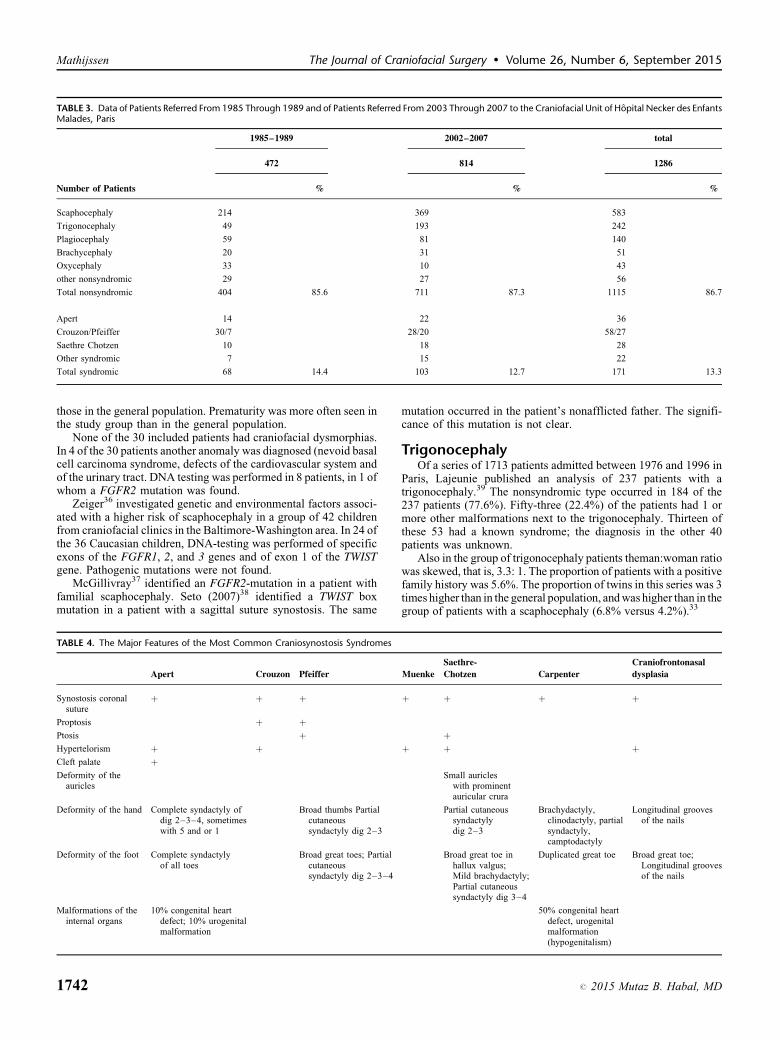
TABLE 3. Data of Patients Referred From 1985 Through 1989 and of Patients Referred From 2003 Through 2007 to the Craniofacial Unit of Hopital Necker des EnfantsMalades, Paris
1985–1989 2002–2007 total
472 814 1286
Number of Patients % % %
Scaphocephaly 214 369 583
Trigonocephaly 49 193 242
Plagiocephaly 59 81 140
Brachycephaly 20 31 51
Oxycephaly 33 10 43
other nonsyndromic 29 27 56
Total nonsyndromic 404 85.6 711 87.3 1115 86.7
Apert 14 22 36
Crouzon/Pfeiffer 30/7 28/20 58/27
Saethre Chotzen 10 18 28
Other syndromic 7 15 22
Total syndromic 68 14.4 103 12.7 171 13.3
Mathijssen The Journal of Craniofacial Surgery � Volume 26, Number 6, September 2015
those in the general population. Prematurity was more often seen inthe study group than in the general population.
None of the 30 included patients had craniofacial dysmorphias.In 4 of the 30 patients another anomaly was diagnosed (nevoid basalcell carcinoma syndrome, defects of the cardiovascular system andof the urinary tract. DNA testing was performed in 8 patients, in 1 ofwhom a FGFR2 mutation was found.
Zeiger36 investigated genetic and environmental factors associ-ated with a higher risk of scaphocephaly in a group of 42 childrenfrom craniofacial clinics in the Baltimore-Washington area. In 24 ofthe 36 Caucasian children, DNA-testing was performed of specificexons of the FGFR1, 2, and 3 genes and of exon 1 of the TWISTgene. Pathogenic mutations were not found.
McGillivray37 identified an FGFR2-mutation in a patient withfamilial scaphocephaly. Seto (2007)38 identified a TWIST boxmutation in a patient with a sagittal suture synostosis. The same
TABLE 4. The Major Features of the Most Common Craniosynostosis Syndromes
Apert Crouzon Pfeiffer
Synostosis coronalsuture
þ þ þ
Proptosis þ þPtosis þHypertelorism þ þCleft palate þDeformity of the
auricles
Deformity of the hand Complete syndactyly ofdig 2–3–4, sometimeswith 5 and or 1
Broad thumbs Partialcutaneoussyndactyly dig 2–3
Deformity of the foot Complete syndactylyof all toes
Broad great toes; Partialcutaneoussyndactyly dig 2–3–4
Malformations of theinternal organs
10% congenital heartdefect; 10% urogenitalmalformation
1742
mutation occurred in the patient’s nonafflicted father. The signifi-cance of this mutation is not clear.
TrigonocephalyOf a series of 1713 patients admitted between 1976 and 1996 in
Paris, Lajeunie published an analysis of 237 patients with atrigonocephaly.39 The nonsyndromic type occurred in 184 of the237 patients (77.6%). Fifty-three (22.4%) of the patients had 1 ormore other malformations next to the trigonocephaly. Thirteen ofthese 53 had a known syndrome; the diagnosis in the other 40patients was unknown.
Also in the group of trigonocephaly patients theman:woman ratiowas skewed, that is, 3.3: 1. The proportion of patients with a positivefamily history was 5.6%. The proportion of twins in this series was 3times higher than in the general population, and was higher than in thegroup of patients with a scaphocephaly (6.8% versus 4.2%).33
Muenke
Saethre-
Chotzen Carpenter
Craniofrontonasal
dysplasia
þ þ þ þ
þþ þ þ
Small auricleswith prominentauricular crura
Partial cutaneoussyndactylydig 2–3
Brachydactyly,clinodactyly, partialsyndactyly,camptodactyly
Longitudinal groovesof the nails
Broad great toe inhallux valgus;Mild brachydactyly;Partial cutaneoussyndactyly dig 3–4
Duplicated great toe Broad great toe;Longitudinal groovesof the nails
50% congenital heartdefect, urogenitalmalformation(hypogenitalism)
# 2015 Mutaz B. Habal, MD

TABLE 5. Genes Involved in Syndromic Craniosynostosis
Clinical diagnosis Genes to be Investigated
Apert FGFR2
Crouzon FGFR2
Crouzon with acanthosis nigricans FGFR3
Pfeiffer FGFR2 (FGFR1)
Carpenter RAB23
Muenke FGFR3 (TWIST1)
Saethre-Chotzen TWIST1 (FGFR3)
Craniofrontonasal dysplasia EFNB1
The Journal of Craniofacial Surgery � Volume 26, Number 6, September 2015 Diagnoses of Craniosynostosis
Azimi40 investigated 25 patients with trigonocephaly (diagnosedbetween 1996 and 2001). In 16 patients it was an isolated anomaly,2 patients in addition had a craniosynostosis of the sagittal suturewithout associated anomalies. The trigonocephaly was part of asyndrome in 7 patients (28%), in 2 of whom a chromosomeabnormality was detected and Jacobsen syndrome could be diag-nosed. Regarding the other 5 patients, in 1 the diagnosis of Say-Meyer trigonocephaly was made, in 1 I-cell disease and in anotherone Opitz-C syndrome. A diagnosis could not be made in 2 patients.In 2 of the syndromic patients FGFR2 and FGFR3 analysis wasperformed, which yielded no abnormalities.
In 1 of 9 patients with a seemingly nonsyndromic trigonoce-phaly a mutation in the FGFR2 gene was found.41 This patient laterdeveloped features characteristic of the Crouzon-syndrome and thediagnosis was revised. Kress42 found a FGFR1 mutation in 1 of 10nonsyndromic trigonocephaly patients, who had or did not developother dysmorphias. In 2002, Kan34 reported that in none of 17patients with trigonocephaly a FGFR2 mutation was found in acomprehensive screening of the whole gene.34 Jehee43 analyzed thefrequencies of occurrence of micro deletions of chromosome 9p andchromosome 11q in a cohort of 76 nonconsanguineous trigonoce-phaly patients (44 from Sao Paolo, 15 from Oxford, and 20 fromBaltimore). The patients were classified into groups, a group of 40patients with an isolated trigonocephaly and a group of 36 patientswith associated abnormalities. A striking finding was that thecondition was nonsyndromic in 71% of the English/Americanpatients, but was syndromic in 63% of the Brazilian patients.
In the nonsyndromic group no 9p- or 11q-deletions were found.In 7 of the 36 syndromic trigonocephaly patients a 9p- or
11q-deletion was found. Four of these deletions were not detectablewith conventional cytogenetic analysis.
In a case-report, Van der Meulen (2006)44 describes a trigono-cephaly in a patient with Muenke syndrome.
Plagiocephaly, Brachycephaly, OxycephalyIn these types of craniosynostosis the coronal suture is involved;
unilaterally in plagiocephaly, bilaterally in brachycephaly andoxycephaly. In oxycephaly there is also a craniosynostosis of thesagittal suture.
Mulliken45 performed molecular diagnostics in patients with abilateral coronal suture synostosis, of whom 38 had been diagnosedwith Apert, Crouzon, or Pfeiffer syndrome and 19 had no specificdiagnosis. In all 38 syndromic patients, a mutation was identified inFGFR2 (N¼ 33) or FGFR3 (N¼ 5). In 14 of the other 19 patients amutation was found, either in FGFR2 (N¼ 4) or in FGFR3 (N¼ 10).
In 2004, Mulliken46 reported results of molecular genetic diag-nostics in 47 patients with a unilateral coronal suture synostosis.This was a prospective study in children admitted between 1997 and2000. Theman:woman ratio was 1:2. DNA testing of the FGFR-genes and the TWIST gene was performed in all 47 patients. In 8patients a mutation was found in 1 of these genes (N¼ 3 in FGFR3,N¼ 2 in FGFR2, and N¼ 3 in TWIST). One patient was clinicallydiagnosed with craniofrontonasal dysplasia. The 47 patients werephysically examined on inner canthal distance (ICD), among otherthings; ICD> 2SD was defined as hypertelorism. Also the parentsof the children were examined. Of the 47 children, 13 showedhypertelorism or had a parent with characteristics of craniosynos-tosis. All above anomalies were found in these 13 children. In thechildren with a unilateral coronal suture synostosis in whom nohypertelorism was established, no mutation in FGFR1, 2, 3, orTWIST was found.
The known craniosynostosis syndromes, Apert syndrome, Crou-zon syndrome, Pfeiffer syndrome, Muenke syndrome, Saethre-Chotzen syndrome, Carpenter syndrome, and craniofrontonasal
# 2015 Mutaz B. Habal, MD
dysplasia (CFND), are characterized by craniosynostosis of oneor both coronal sutures next to other birth defects. A proportion ofpatients with these syndromes show pathogenic mutations of knowngenes (Table 5).47–51
In a comprehensive screening of the FGFR2 gene in patientswho were proven negative for a mutation in FGFR1 or 3 or TWIST,the large majority of mutations were located on exon IIIa or IIIc. Inaddition, mutations were found in 7 new exons.34
In patients with a syndromic craniosynostosis, genetic diagnos-tics is performed on the guidance of the syndrome diagnosis.Mutations are most frequent in FGFR2, followed by FGFR3 andTWIST, and least frequent in FGFR1 and EFNB1.52,53
In Rotterdam, DNA testing of the FGFR1, 2, 3, and TWISTgenes was performed during several years in all patients with acraniosynostosis, except those with an isolated scaphocephaly.FGFR1 mutations were not identified.54 Routine screening of theFGFR1 gene does not seem to be useful, therefore, and should beperformed only in patients with a specific phenotype.
Morriss-Kay53 reported a prospective study in 214 patients bornbetween 1993 and 2005. Analysis of FGFR1, FGFR2, FGFR3,TWIST1 and EFNB1 was performed as well as cytogenetic analysis.In 60 of the 214 patients a specific molecular diagnosis was made;in 4 patients this was a chromosomal abnormality, in 56 patients amutation in FGFR2, FGFR3, TWIST, or EFNB1. The patients withthe FGFR2, FGFR3 and TWIST1 mutations all had a craniosynos-tosis of 1 or both coronal sutures
Wilkie52 tested 217 patients in a diagnostic setting as from 2002.Submitting material of patients with a nonsyndromic scaphocephalyor trigonocephaly was strongly discouraged. In 59 of the 217patients a pathogenic mutation was found, in 55 patients in FGFR2,FGFR3, or TWIST.
In 80% of the patients with a clinical diagnosis of Crouzon orPfeiffer syndrome this diagnosis is confirmed by molecular test-ing.54 Clinically diagnosed Apert syndrome is confirmed by mol-ecular testing in almost all patients. Two specific mutations inFGFR2 were found in approximately 98% of patients with Apertsyndrome.52 In more than 80% of a group of patients with thephenotype of Saethre-Chotzen syndrome a mutation was found inthe FGFR3 or the TWIST gene.55
If conventional molecular genetic diagnostics does not revealmutations in one of the known craniosynostosis genes, additionaldiagnostics is warranted in patient of syndromic craniosynostosis.Genetics diagnostics was performed with different techniques in thescreening of 45 patients with a syndromic craniosynostosis withoutknown mutation.56 New causal abnormalities were found in 19patients.
Conclusions
Level 1
It has been demonstrated that the Apert, Crouzon, andPfeiffer syndromes are caused by mutations in the FGFR2 gen,except in Crouzon patients with acanthosis nigricans(FGFR3 mutation)1743

Mathijssen The Journal of Craniofacial Surgery � Volume 26, Number 6, September 2015
1744
A2 Oldridge, 199757
A2 Meyers, 199558
Level 1
It has been demonstrated that the Saethre-Chotzen syndrome iscaused by mutations and deletions in the TWIST1 geneA2 Johnson, 199859
A2 Chun, 200255
Level 1
It has been demonstrated that the Muenke syndrome is caused bythe P250R mutation in FGFR3A2 Muenke, 199749
A2 Chun, 200255
B Kress, 200660
B Mulliken, 199945
Level 1
It has been demonstrated that craniofrontonasal dysplasia(CFND) is caused by mutations in het EFNB1 geneA2 Twigg, 200451
A2 Wallis, 200861
Level 2
It is likely that almost no genetic abnormalities are found innonsyndromic trigonocephaly and scaphocephaly. DNAtesting is indicated in patients with other birth defects ordysmorphias next to the craniosynostosis of the sagittalsuture or metopic suture or in case of a positivefamily history.B Kan, 200234
B Wilkie, 200752
B Morriss-Kay, 200553
B Zeiger, 200236
C McGillivray, 200537
C Jehee, 200543
C Tartaglia, 199941
C Kress, 200042
Level 2
There are indications that FGFR2, FGFR3 and TWIST1mutations are almost exclusively detected in patients withcraniosynostosis of the coronal sutures.B, Mulliken, 199945
B Morriss-Kay, 200553
B Wilkie, 200752
Level 3
There are indications that in no more than 25% of patients witha craniosynostosis a specific molecular diagnosis could be madeafter analysis of the coding regions of FGFR1, FGFR2,FGFR3, TWIST1, and EFNB1B Wilkie, 200752
B Morriss-Kay, 200553
Level 3
There are indications that in familial scaphocephaly an FGFR2mutation can be foundC McGillivray, 200537
Level 3
There are indications that in patients with syndromic trigonocephalyor complex nonclassifiable craniosynostosis additional diagnosticscan lead to an etiologic diagnosisC Shimojima, 200962
C Barbaro, 200963
C Jehee, 200543
Level 3
There are indications that in a proportion of patients withunexplained syndromic craniosynostosis a cause can beestablished with the use of new molecular genetic techniquesC Jehee, 200856
ConsiderationsThe Crouzon and Pfeiffer syndromes cannot be easily distin-
guished clinically and genetically, but such distinction is notrelevant to choice of treatment. The first choice for the geneticdiagnostics is analysis of the FGFR2 gene.
Patients should be invited to contact the clinical geneticist againafter reaching the age of 18 years to discuss the wish to havechildren and receive counseling if desired.
RecommendationsAfter referral of a child to a craniofacial team, the clinical
geneticist of the team should be consulted. To distinguish between
nonsyndromic and syndromic craniosynostosis a clinical geneticistexperienced in the fields of hereditary and congenital abnormalities/dysmorphias should perform a complete physical examination ineach child. In addition, the family history regarding the occurrenceof skull deformities and other birth defects should be documented.
If multiple dysmorphias/visible congenital abnormalities areobserved, further diagnostics in the first weeks of life is indicated(including, among other things, cardiologic examination, renalultrasound, and examination by pediatric neurologist).
Molecular genetic diagnostics is not offered in patients withnonsyndromic scaphocephaly and nonsyndromic trigonocephaly.
If these types of craniosynostosis occur in relatives, moleculargenetic diagnostics is performed depending on the family historyand preferably after consultation with a tertiary craniofacial center.
If a syndrome diagnosis has been made clinically, geneticdiagnostics can be requested syndrome specifically:
Clinical diagnosis
# 2015 M
Genes to be investigated
Apert
FGFR2Crouzon
FGFR2Crouzon with acanthosis nigricans
FGFR3Pfeiffer
FGFR2 (FGFR1)Carpenter
RAB23Muenke
FGFR3 (TWIST1)Saethre-Chotzen
TWIST1 (FGFR3)More advanced diagnostics is indicated in the patient of asyndromic craniosynostosis without clinical diagnosis (¼ a non-classifiable craniosynostosis) or a clinical diagnosis that could notbe confirmed genetically.
4. PERIOPERATIVE CARE
Basic QuestionWhat organizational conditions should be minimally present for
adequate and safe perioperative care for patients with a craniosy-nostosis?
IntroductionCorrection of craniosynostosis in childhood can cause relatively
much blood loss. The risk of blood loss is higher in older patientsand in corrective surgery of syndromic craniosynostosis. Next to thesurgical and anesthesiologic challenges, we should take intoaccount that syndromic patients may be associated with comorbid-ity. This is why optimal organizational conditions should be inplace, before, during, and after the intervention. This chapter dealswith the specific risks involved in correction of nonsyndromic andsyndromic craniosynostosis and recommendations are given toperform surgery as safely as possible.
Summary of the LiteratureGeneral
The ‘‘Richtlijn Kwalificering Chirurgie bij Kinderen’’ makesclear that the anesthesiologic goals in complex care such ascraniofacial surgery can only be realized in specialized pediatriccenters.64
Preoperative PreparationAll members of the multidisciplinary team should be aware of
possible comorbidity, that is, mostly in case of a syndromiccraniosynostosis with compromised airway with or withoutOSAS. It is recommended to set the threshold for postponing theintervention low in case of a recent upper airway infection in
utaz B. Habal, MD

The Journal of Craniofacial Surgery � Volume 26, Number 6, September 2015 Diagnoses of Craniosynostosis
these patients (described in Apert syndrome by Elwood)65 becauseairway infection is believed to be more strongly associated withcomplications.
One of the big problems (or complication) in the surgicalcorrection of craniosynostosis is the occasional massive bloodloss of 20% to 500% of the circulating volume, which may occurduring the operation in a relatively short period and in patients withage-related circulating volume.66
The following factors may predict large blood loss: syndromiccraniosynostosis, pansynostosis, age <18 months, and duration ofthe procedure.67
A number of methods have been proposed to reduce both theblood loss and the need of homologous (allogeneic) blood transfu-sion (in view of the risk of blood-transferable infection, immuno-logic reactions, coagulopaty, and transfusion-related acute lunginjury (TRALI), but most studies are not randomized double blindand have a B-C Level. These methods can be classified intodifferent categories and are applied pre-, per- or postoperatively.In the polyclinic (preoperative) period:
� O
# 201
ptimalization of hematological conditions preoperatively,by administering erythopoietin (EPO) plus Fe supplement: Areview concludes that multicenter studies are needed todetermine the optimal dosing of EPO.68 Different dosageshave been described, that is, 600–200–100 U/kg once tothrice a week, but all during 3 weeks preceding the operation.Also the timing of administration, the optimal dosing of Fe,and cost-effectiveness must be determined. The use of EPOwas also successfully combined with other methods in anumber of B-C studies.69–73 A disadvantage of this methodfor the child is that blood must be drawn several times.
� P
reoperative blood sampling for autologous transfusionduring the operation: Opinions differ on this technique. Ithas been used as part of a ‘‘no allogenic blood transfusion’’protocol, but other authors think it is not indicated in thesevery young patients as only small volumes can be drawn.Furthermore, the procedure is not child friendly as thenewborns and infants must be anesthetized.� P
reoperative selection of the extent of surgical procedure onthe basis of age, accounting for the percentage of Hbf, whichat 4 months has been decreased to 16% of total Hb.� P
reoperative surgical planning with the use of three-dimensional models seems to reduce operation time andthus also blood loss. Still, according to a C study, duration ofthe procedure is not decisive for the blood loss.7475,76
� L ess invasive surgical procedures.Immediately before surgery the presence of blood products andavailability of a pediatric intensive care unit (PICU) bed for thepostoperative period should be confirmed in case significant bloodloss related to the child’s circulating capacity should occur. A clearplan must have been made anticipating problems with intubation.The use of pre- and postoperative checklists and the time-outprocedure (TOP) before start of surgery importantly add to safetyof the entire perioperative process.
Peroperative ManagementMonitoring
Invasive monitoring is recommended in open procedures withexpected severe blood loss. Invasive monitoring involves the use ofa central venous line and an arterial line next to conventionalmonitoring (capnography, ECG, pulse oxymeter, FiO2, tempera-ture, and urine output) and precordial Doppler. The major goals ofinvasive monitoring are timely recognition of serious problems that
5 Mutaz B. Habal, MD
may occur in open craniosynostosis surgery, such as hypohydration,hypotension, and electrolyte disturbances, as well as being able tocheck effectiveness of treatment of these problems.
PositioningVenous cerebral congestion by hyperflexion of rotation of the
head must be avoided.Usually, the patient is placed in a moderate anti-Trendelenburg
position to reduce blood loss (cave air embolism, see below underComplications). Notably, in children with exorbitism the eyesshould be well protected with eye cream, and pressure on the eyesmust be avoided (especially in prone position).
Single administration of an antibiotic, after the induction, beforestart of surgery is standard procedure.76
Maintenance of AnesthesiaMost recommendations are aimed at a balanced technique that
provides for cardiovascular stability with the use of opioids andvolatile agents next to relaxants. The use of remifentanil infusion(0.25–0.5 mcg/kg/min) is recommended as well.77–79 If remifen-tanil is used for maintenance of anesthesia is it recommended toadminister a bolus of morphine or piritramide before the end of theprocedure so as to start postoperative pain management.
Measurement/Compensation Blood Loss IntraoperativelyAn additional problem is that measurement of the lost volume
intraoperatively is impeded by surgical technique and type ofpatient. Few studies have been performed to optimize blood lossmeasurement and findings are mostly not conclusive. Next to theabove-mentioned preoperative approaches to reduce blood loss and/or to prevent allogeneic blood transfusion, several measures can betaken peroperatively:
� I
nfiltration of the skin with vasoconstrictors before theincision is much used but its effectiveness is disputed becauseof the greater degree of bleeding with treatment of theperiostium and bone.� C
ontrolled hypotension is rarely used because it has no clearbenefits and can be disadvantageous in case of ICP and also inthe anti-Trendelenburg position.� T
he acute normovolemic hemodilution (ANH) technique isused before the surgical intervention after the child is broughtunder anesthesia. It involves removal of whole blood via thearterial or central line which is replaced with colloids 1:1 orwith crystalloids 1:3 according to precise calculations andformulas. Most healthy children tolerate a mild hemodilution(Htc 25%–30%). According to a B study, this technique initself is not sufficient to reduce or avoid homologous bloodtransfusion in this type of surgery.80� P
erioperative (intra- and postoperative) blood ‘‘recollection’’ isnot routinely used in this type of surgery in view of the smallcollected volumes, the sometimes acute blood loss, and its cost-effectiveness. Fearon81 reported that 59 of 60 children needed atransfusion, of which only 30% received an allogeneictransfusion with the use of the cell-saver device. Duncan82found no difference in allogeneic transfusion rate when a cellsaver was used and when not. With the development of smallreservoirs (50 ml) this method seems to be more promising andin C studies it has been associated with a considerable reductionof the homologous blood transfusion rate.
83
� I ntraoperative antifibrinolytics. � A protinin: is no longer used after serious complications incardiac surgery. Tranexamic acidin: its use in itself iscontroversial, but is has been applied in combination withother methods.
1745
Mathijssen The Journal of Craniofacial Surgery � Volume 26, Number 6, September 2015
According to a comparative B study, none of these strategies toreduce bleeding and/or to prevent or reduce the use of homologousblood has shown sufficient effects.80 A review by Di Rocco68
confirms this conclusion. The transfusion strategies are usuallybased on intraoperative acceptation of Hb values approximately7 g/dl (4 mmol/l) and Ht <30.84
Guidelines for the application of blood products are in place inevery hospital as established by the local transfusion committees,and in conformity with the CBO Guideline Blood transfusion2004.85 The latter guideline does not contain specific recommen-dations for very young children.
ComplicationsThe best known complications are inherent to the surgical
technique and/or a result of massive blood transfusion.
� V
1746
enous air embolism (VAE) and the subsequent cardiovas-cular collapse can be prevented by a precise technique and byrapid application of adequate monitoring (precordial Doppler,capnography, echocardiography, transcutaneous O2–CO2
monitoring, esophageal stethoscope, and venous centralline). Although Faberowski86 reports an incidence of82.6% shown with Doppler in a small series of craniosynos-tosis patients with a mean 90% blood loss, Tobias87 describesan incidence of 8% with the use of Doppler in stripcraniotomies. Meyer88 reports a 2.6% incidence in childrenundergoing craniosynostosis surgery who were monitoredwith end-tidal CO2 only. The hemodynamic consequences ofVAE are generally insubstantial, provided the patients arewell monitored and measures are taken upon signs of VAE.
� A
s a result a massive blood loss the patient may developrelevant consumption coagulopathy and dilutional coagulo-pathy characterized, in principle, by depletion of solubleclotting factors.83 As cryoprecipitate is not available every-where and fresh frozen plasma (FFP) has limited effectivenessto compensate for fibrinogen deficiency, it is recommended atC level to timely use fibrinogen concentrate based onthromboelastography.89 Temperature monitoring is crucialfor the prevention and treatment of clotting disorders.� M
assive blood transfusion can result in development ofTRALI.90� A
irway management in syndromic children (Apert, Crouzon/Pfeiffer) is sometimes complicated by hypoplasia of themidface and exophthalmos that may impede hood ventilation,although direct laryngoscopy and intubation normally arepossible. The situation changes when these children undergoa distraction osteotomy procedure and need to be intubatedacutely in case of respiratory insufficiency, or when thedistraction materials are being removed. Evidence from the Clevel studies is conflicting on this issue. One of the 2 studiesrecommends immediate fiberoptic intubation, whereas theother study claims that the distractors exert minimal effect inthe anesthesiologic conditions when certain factors must betaken into account (the right screwdrivers and cutting pliersmust always be available) and that removal of the vertical barallows for direct laryngoscopy.91 Roche92 describes problemswith intubation at the time when distractors are removed as aresult of trismus that had not yet occurred when the distractorswere placed.� T
he possibility of cerebral salt wasting syndrome should beconsidered when a patient develops hyponatremia aftercraniosynostosis surgery.93 Then, a differential diagnosis withsyndrome of inappropriate antidiuretic hormone secretion(SIADH) should be made right away as the treatmentmodalities for these 2 are quite different.Criteria for extubation at the end of the procedure, beforetransport to the ICU, are the following: rapid recovery of spon-taneous and stable breathing, hemodynamic stability, normother-mia, short to medium operation time with relatively little blood loss,and no continuous large blood loss through the surgical drains.
Postoperative ManagementPediatric Intensive Care Unit (PICU)
In principle all studies recommend ICU admission after opencraniosynostosis surgery to continue volume management thereand, if necessary, start artificial respiration.
Bleeding may continue in the postoperative period via thedrains, and the cardiovascular status next to Htc/Hb must bemonitored carefully. Most studies on postoperative transfusionmanagement report a tendency to overtransfusion.74,94
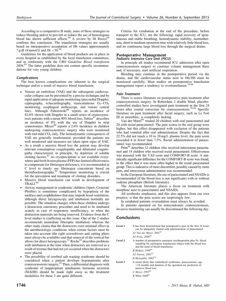
Pain TreatmentThere is scarce literature on postoperative pain treatment after
craniosynostosis surgery. In Rotterdam 2 double blind, placebo-controlled studies have investigated pain treatment in the first 24hours after cranial correction for craniosynostosis.95,96 Specificliterature on pain treatment after facial surgery, such as Le FortIII or monobloc, is completely lacking.
Van der Marel95 treated 20 children with oral paracetamol and20 with rectal paracetamol. The pain scores in the oral group werehigher, but this effect disappeared with exclusion of the patientswho had vomited after oral administration. Despite the fact that22.5% did not reach a 10 to 20 mg/L plasma level, the pain scoreexceeded 4 in fewer than 7.5%. Rectal administration of parace-tamol was recommended.
Prins96 describes 12 children who received intravenous paraceta-mol and 14 children who received rectal paracetamol. Effectivenesswas assessed with the VAS score and COMFORT-B score. A stat-istically significant difference for the COMFORT-B score was found,to the effect that it was more often higher in the rectal paracetamolgroup. This is indicative of more discomfort, possibly caused by morepain, and intravenous administration was recommended.
In the European literature, the use of paracetamol and NSAIDs isrecommended (if the blood loss is not significant) with or withoutcodeine phosphate (British literature).
The American literature places a focus on treatment withmorphine next to paracetamol and NSAIDs.
All textbooks emphasize, and this also appears from our ownpractice, is that the pain scores are surprisingly low.
In extubated patients oversedation must always be avoided.In patients operated on for nonsyndromic craniosynostosis,
invasive monitoring can usually be discontinued the following day.
Conclusions
Level 1
It has been demonstrated that postoperative pain in the first 24 hourscan be adequately treated with administration of paracetamol.A2 Van der Marel, 200195
A2 Prins, 200896
Level 2
A number of preoperative measures (erythropoietin plus Fe, bloodsampling for autologous transfusion) reduce both the blood lossand the need of blood transfusion.B Helfaer, 199869
A2 Fearon, 200272
B Meneghini, 200373
Level 3
It seems likely that craniofacial syndromes, pansynostosis, age<18 months and duration of the operation are predictors ofmassive blood loss.C Meyer, 199394
B White 200967
# 2015 Mutaz B. Habal, MD

The Journal of Craniofacial Surgery � Volume 26, Number 6, September 2015 Diagnoses of Craniosynostosis
Level 3
# 2015 Mu
None of the perioperative strategies (skin infiltration, anti-Trendelenburg position, hemodilution, and antifibrinolitics) toreduce bleeding and/or to avoid or diminish the use ofhomologous blood has shown satisfactory effects.
B Hans, 200080
C Di Rocco, 200468
Level 3
Overtransfusion in the postoperative course does regularly occur.C Kearney, 198974
C Meyer, 199394
ConsiderationsDespite the proven effectiveness of erythropoietin and blood
drawing for autologous transfusion, the use of these strategies isdiscouraged as they involve high costs and require repeated veni-puncture, which is not child friendly.
Introduction of less invasive interventions is associated with lessblood loss. As these, however, are very young children with a smallercirculating capacity, blood loss relatively is still significant. Inselected cases admission to a medium care unit could be considered(defined as a monitored bed providing for artificial respiration).
RecommendationsThe anesthesiologic goals in craniofacial surgery can only be
realized in specialized pediatric centers, where multidisciplinaryperioperative care is provided by a team composed of plasticsurgeon, neurosurgeon, maxillofacial surgeon, pediatrician,pediatric anesthesiologist, pediatric intensivist, and specializedpediatric nurses with experience and means to manage and monitorthis type of patients and where a sufficient number of children isoperated on to keep the experience of the team at a high level.
Administration of EPO preceding the intervention, as wellas collecting autologous blood for autotransfusion are advisedagainst.
Postoperatively a bed in a PICU must be available. In less drasticinterventions medium care may perhaps suffice, although 1 shouldbe aware that these are mostly very young children with a smallercirculating capacity, in whom even slight postoperative blood lossmust be monitored carefully.
Invasive monitoring is recommended in the case of open pro-cedures with expected severe bleeding.
Overtransfusion in the postoperative phase should be preventedby adhering to the guideline on transfusion management.
Extubation at the end of the operation, before transport to theICU, is possible in case of rapid recovery of spontaneous and stablebreathing, hemodynamic stability, normothermia, short to mediumoperation time with relatively little blood loss, and no continuouslarge blood loss through the surgical drains.
The analgesic of choice for postoperative pain treatment isparacetamol. If this is not sufficient, an NSAID can be added eventhough this could increase the risk of postoperative bleeding.
5. SURGICAL TREATMENT OFNONSYNDROMIC CRANIOSYNOSTOSIS
Basic Questions
1. W
hat are the indications for surgery in the different types ofnonsyndromic craniosynostosis?2. W
hat treatment is most indicated for the different types ofnonsyndromic craniosynostosis?3. A
t what developmental stage is surgical treatment preferablyperformed?taz B. Habal, MD
IntroductionNonsyndromic craniosynostosis can present in varying severity
of the deformity of the skull. Surgical treatment seems indicated onthe basis of:
1. t
he associated risk of ICP (see also chapter on ICP)2. t
he prevention or restriction of associated neuropathology3. t
he morphologic abnormality (with both esthetic and psycho-logic consequences)The variability in both the severity of the morphologic abnorm-ality and in the occurrence of increased ICP may occasionallyresult in less strict operative indication on the basis of theseparameters.
Many different surgical techniques have been described for thetreatment of nonsyndromic craniosynostosis but the functionalresults of these techniques have not always been shown convin-cingly. In addition, the timing of surgery is debated.
For patients with a synostosis of multiple cranial sutures inwhom a syndromic diagnosis cannot (yet) be made, the method oftreatment of syndromic craniosynostosis is recommended. Thischapter will place a focus on craniosynostosis of the sagittal suture(scaphocephaly), metopic suture (trigonocephaly), unilateral cor-onal suture (frontal plagiocephaly), and lambdoidal suture (pachy-cephaly).
Summary of the LiteratureIndications for Surgery
The indications for treatment of craniosynostosis are the risk ofICP and the morphologic abnormality of the skull and face. Asmentioned in chapter 8, the risk of increased ICP in scaphocephalyis 12%, in trigonocephaly 9%, and in plagiocephaly 10%. Theestimated risk in bilateral coronal suture synostosis is 37%.
The chapter on cognition and behavior provides evidence ofabsence of a relation between cognitive functions in children with anonsyndromic craniosynostosis and surgical treatment or not,severity of the deformity, or age at operation. The prevention ortreatment of the associated neuropathology is therefore no indica-tion for surgery.
The morphology can vary from very mild to an evidentlyabnormal skull shape.97
In sagittal suture synostosis, compensatory growth of the othercranial sutures results in increasing deformity in the course of time,so that, for example, the frontal bossing in scaphocephaly canintensify after birth.98,99 Sagittal suture synostosis is characterizedby highly variable expression, however, ranging from a ridge overthe suture in combination with a slightly narrower skull to mid-parietal saddle-like bone formation with severe frontal bossing anda pointed and prominent back of the head.
Barritt100 evaluated a series of 44 children with scaphocephaly,of whom 34 did not undergo surgery. This was common policy inthis clinic at the time for children presenting with this condition atan age older than 6 months. In these 34 children, the deformity inany patient did not seem to improve in the first 10 years. In 14children, the cranial index (CI) decreased (69> 65), in 7 the CIincreased (64> 68) and in 7 it did not change.
The presentation of trigonocephaly also varies from very mild tosevere. Its mildest form is characterized only by a bony ridge as aresult of premature fusion of the metopic suture. In follow-up,cosmetic worsening is only rarely seen. The other end of thespectrum is characterized by a wedge-shaped forehead with hypo-telorism and supraorbital retrusion on the basis of decreased growthof the forehead widthwise.101
1747

Mathijssen The Journal of Craniofacial Surgery � Volume 26, Number 6, September 2015
Frontal plagiocephaly also shows some variation in phenotype,notably in facial asymmetry.102 In a study from Paris, 16% of thechildren who were operated on after the first year of life developedincreased ICP (see chapter 8).103
Unilateral synostosis of a lambdoidal suture causes increasingasymmetry of the back of the head and particularly also of the face.Increased ICP does not seem to be prominent in this type ofsynostosis, although reliable research on this issue is lacking(see chapter 8).
2. Surgical Treatment of NonsyndromicCraniosynostosis
Since the first operative intervention for craniosynostosis, manysurgical techniques for the various types of craniosynostosis havebeen described. A broad distinction is made between osteoclastictechniques and remodeling techniques. In the first group bone isremoved, enabling the developing and expanding brain to changethe shape of the skull, in part because the adverse impulse to thegrowth direction of the skull also is removed. From this techniqueevolved the remodeling techniques, because it was suspected thatone cannot only rely on the self-correcting capability of the skulland the brain. These techniques are aimed at directly achieving thedesired skull shape by a kind of reconstruction.
Randomized comparisons of the different surgical techniqueshave never been performed. For the different types of nonsyndromiccraniosynostosis we will provide a review of the literature regardingthe different techniques in these 2 groups, but also pay attention tothe new developments in the past 10 years in a separate section.
2.1 ScaphocephalyStrip Craniectomy
Ingraham104 described a technique using bilateral parasagittalstrip craniectomies, in which the fused suture was not resected.To prevent the problem of premature reossification of the skull boneshe applied a layer of plastic to the bone rims. Successful treatmentrequired early intervention, however, preferably in the first 2 monthsof life. The results of this technique were confirmed by otherauthors.105–107 Anderson and Geiger105 attempted to prevent pre-mature reossification with the use of chemical coagulation of thedura, but failed to demonstrate the advantages of this technique.
Albright108 presented a sagittal strip craniectomy with biparietalwedge osteotomies. The same procedure was performed by Mar-ucci109 in 89 patients, but they noticed that 7 of the 89 patients laterdeveloped a cosmetically unsatisfactory vertex bulge, which theyinterpreted as a sign of possibly raised ICP or a new synostosis. AlsoFlorisson110 reported that some patients with scaphocephaly againshowed signs of raised ICP postoperatively.
A variant to this technique is wide resection of the sagittal suture(4 cm) in combination with a bone-strip resection along the coronaland lambdoid sutures, as described by Christophis.111 The averageCI improved from 66 to 76.
Amm112 presented a technique, in which the usual 2.5 to 3 cmwide strip craniectomy of the sagittal suture, combined with lateralstrip osteotomies, is expanded with weakening with a burr of theoccipital bone, allowing the skull to hinge occipitally. They expli-citly try to make use of gravital force by keeping the infant strictly insupine position during sleep for a period of 3 weeks. Using thistechnique an average improvement of the CI of 8.1 (4.8–16) pointswas achieved.
All in all, many of the described techniques represent only slightmodifications of previously presented techniques and not realconceptual changes.
Postoperative infections are rare (0%).108 Duration of hospital-ization ranges from 1 to 6 days postoperatively.108,111,112 Here, too,
1748
it appears that policy is guided by habituation in postoperative carerather than morbidity or impact of the operation.
Cranial Remodeling; the p Procedure Strips Are ResectedDe p procedure is a technique in which strips are resected at
either side of the synostotic sagittal suture bone, while in addition abone strip is resected anteriorly to the coronal suture. The sagittalsuture itself is not resected, and the temporoparietal bone is bentoutwards. In this technique, the anteroposterior diameter of the skullis dynamically shortened by attaching the bone strip in the middle ofthe skull, which is shortened, to the frontal bone. Thus, it is in factalso a compression technique, which might be associated with anincrease in ICP.
Gewalli113 with the use of a modified p procedure shortened theanteroposterior diameter in 26 children by a mean of 16 mm, andconcluded from neuropsychologic assessment that this had notadversely affected mental development. Only a global developmentquotient, however, was tested in very young patients, withoutinclusion of a control group, which raises great doubt on thereliability of this finding.
In older children or in children with a more pronounced dys-morphology (notably frontal bossing), a more extensive calvarialreconstruction is needed, with resection and reconstruction of thefrontobiparietal complex.114 Heller et al performed this interventionin 24 children with mean age 23 months (range 3 to 96 months) andthus achieved normal CI postoperatively in all children (mean 78versus 68 preoperatively). The authors concluded that in thispopulation this more extensive procedure had no adverse effecton postoperative growth rate of the skull or on intracranialvolume.114
New DevelopmentsLauritzen115 reported in 2008 on the first 100 operations in
which distraction was used to correct craniosynostosis. This methodis eminently suitable for scaphocephaly in view of the uni-directional growth delay widthwise.
The results are not worse, but also not better than those of thecurrent techniques and the small numbers preclude valid compari-son. Also regarding the newer surgical techniques, randomized orcomparative studies have not been published.
2.2 TrigonocephalyAlso in view of the complex three-dimensional dysmorphology
seen in metopic synostosis, treatment by suturectomy solely isconsidered insufficient.116–120 In the past 30 years, therefore theemphasis was on reconstruction of the entire forehead, including theupper parts of the orbits.121 The question whether this should alsoinclude specific correction of the hypotelorism is subject of debate.Posnick122 analyzed the growth process on the basis of CT data in10 patients and concluded that correction of the hypotelorism hadremained insufficient. Selber120 found that the use of interpositionalbone grafts in a series of 68 metopic children led to a decrease in thedevelopment of temporal dents. In a population of 92 trigonoce-phaly children in which no bone graft was used, Van der Meulen,101
however, observed an automatic growth correction of the hypote-lorism on x-ray, on account of which the use of an interpositionalbone graft was judged to be unnecessary. From a photographicevaluation of 45 patients, Hilling123 concluded that a satisfactoryesthetic outcome on the long term was largely dependent on a goodinitial reconstruction.
2.3 Frontal PlagiocephalyA distinction is made between unilateral and bilateral frontal
advancement techniques. Some surgeons prefer the unilateral
# 2015 Mutaz B. Habal, MD

The Journal of Craniofacial Surgery � Volume 26, Number 6, September 2015 Diagnoses of Craniosynostosis
advancement technique so as to be able use the unaffected side as areference for the degree of advancement,124 whereas others main-tain that adequate advancement with good symmetry is onlypossible if the entire frontal bone and both orbital margins havebeen dissected.121
New DevelopmentsBarone and Jimenez125 report since the late 1990s on endoscopic
strip craniectomy of the affected suture. The benefits mainly lie inlow morbidity, short hospitalization, and little need of bloodtransfusions in comparison with the classic fronto-orbital advance-ment technique. The cosmetic outcome is reported as good, but itnot adequately quantified.
Stelnicki126 in the early years of this century started to use thestrip craniectomy technique, but concluded that after endoscopicintervention often no full symmetry is reached and that the foreheadunilaterally is still receding 3 or 4 mm. For this reason, the techniquewas adapted to an endoscopic unilateral fronto-orbital advancementtechnique. This was applied in 10 patients, who did not develop anycomplications, did not need blood transfusions, and could bedischarged home after 2 days. The cosmetic outcome was assessedto be similar to that of fronto-orbital advancement.126
2.4 Posterior PlagiocephalyThe number of publications on lambdoid suture synostosis is
very limited, probably because of the very low prevalence. Uni-lateral lambdoid suture synostosis causes only relatively littlevolumetric restriction. The morphologic changes consist of anasymmetry of the face and an asymmetric basal occipital region.
New Techniques, DistractionKim127 reports a study comparing distraction versus remodeling
surgery. Among the patients were 3 children with brachycephalytreated with the distraction method and 4 with remodeling. Thedistraction method led to satisfactory calvarial expansion and anesthetically pleasing outcome. Advantages of distraction are thesignificantly shorter operation time, less bleeding, shorter stay at theneonatal intensive care unit (NICU), and better skull shape. Adisadvantage of the distraction method is that children must havereached the age of 6 months.128 Furthermore, a second operation isneeded to remove the distraction materials, there is a risk ofinfection and dislocation of the material, and total hospitalizationis relatively long. The authors are of the opinion that these down-sides do not outweigh the benefits of distraction.
3. Evaluation of the Outcome of SurgeryMost children with a craniosynostosis are operated on in the first
year of life. A direct evaluation of surgical outcomes is possible, but afinal evaluation is not possible until the child has fully grown at adultage. As a surgeon’s career spans perhaps 25 year, and at most 30years, the surgeon can only at an advance career stage assess the resultof his or her own work. Yet he or she will be able to see the definitiveresults of his/her predecessors. There may be a tendency to ascribe apoor result to a wrongly performed surgical technique, withoutquestioning the technique itself. Furthermore, knowledge aboutthe natural course of the condition with and without surgical inter-vention is usually lacking. This shortcoming undoubtedly iscoresponsible for the fact that we see regular shifts in type ofintervention in the course of decades and that paradigms change.One and the same research group may recommend a certain approachin the 1 decade and disassociate oneself from it in the next decade.129
Only few solid objective results with such a long follow-up areavailable, whereas comparative studies of different surgical tech-niques (with or without randomization) have not at all been
# 2015 Mutaz B. Habal, MD
performed. Moreover, notably the larger series yield mixed data,because all craniosynostoses, including the syndromic types, arepooled.
One way to assess the outcome of surgery is looking at theoperative morbidity and mortality. Most series are too small,however, to yield significant data. Anyway, the mortality is verylow for all procedures, from 0% to 1%.129
Morbidity is reflected in the complications that may occur andlength of hospitalization. Although the first is a relatively objectivescore, the second is certainly not. Length of hospitalization stronglydepends on the treating physicians, but also on the healthcaresystem itself. To illustrate this, for similar procedures the averagelength of hospitalization in North America is significantly shorterthan in Europe. The patients are simply discharged home earlier,which possibly is enabled by a better outpatient care system.Generally, hospitalization after minimally invasive interventionsis 1 or 2 days; for more complex interventions 2 to 7 days.129
As postoperative infections are very rare, only large series canyield significant data. In series of 248 patients who underwentsurgery for unisutural craniosynostosis only 2 (0.4%) developed apostoperative infection.129 In an older series of Whitaker theinfection rate was 3.7% in 164 patients; however, a distinctionbetween syndromic and nonsyndromic craniosynostosis was notmade.130 Still it seems that infections particularly occur in thesyndromic patients.
Severe complications, such as brain lesions, also occur spor-adically: 0% to1%.129 In a series of 107 patients, Sloan131 found a3.7% complications rate, almost all of which were mild, and in aseries of Whitaker the rate was 8%,130 but this was largely onaccount of the syndromic craniosynostoses. In general, the com-plications rates in the nonsyndromic craniosynostoses are signifi-cantly lower than in the syndromic craniosynostoses, 3.5% and39%, respectively, in the same series reported by Sloan.131 Innonsyndromic multisutural craniosynostoses the rate is higher thanin monosutural craniosynostoses.131
The use of metal plates and screws has been customary in a certainperiod. Meanwhile, it has become evident that application of thismaterial in young children may lead to intracranial translocation ofthe plates and screws, with the screw ends penetrating the dura.132–134
Risk factors for translocation seem to be longer plates, placement inthe temporal region, young age, and syndromic craniosynostosis.133
Hardly anything is known about the consequences of this compli-cation. In the past few years, resorbable plates and screw devices havebeen applied for specific indications in craniosynostosis surgery. Thereported rate of complications is low: infection 8/1883 patients135 and5/146 patients;136 delayed foreign-body reaction in 12/1883patients,135 and postoperative resorbable device failure with lossof bony position in 5/1883 patients.135
The outcome of surgery can also be expressed by need forsecondary revision surgery. This is rarely needed. Fearon129 reports5 patients in 248 patients (2%) and Sloan131 reports a 7.2% rate ofunscheduled reoperations in 250 patients.
ScaphocephalyAmm112 routinely uses three-dimensional surface analysis soft-
ware to analyze CT-scans. Computerized tomography scanning,however, is a source of radiation exposure. Still it enables to assessthe outcome in a relatively reliable and objective manner, and thisyielded a mean 8.1 points improvement in CI.
Marucci109 noticed a cosmetically unsatisfactory vertex bulgeafter minor surgery in 7 of 89 patients.
TrigonocephalyAt follow-up in the course of years after reconstruction of
trigonocephaly, the lateral sides of the forehead often appear to
1749

Mathijssen The Journal of Craniofacial Surgery � Volume 26, Number 6, September 2015
revert to a certain degree and temporal depressions may be seen,which however seldom are cosmetically unsatisfactory to thedegree that revision surgery is justified.101
Hilling123 evaluated the result of bifronto-orbital advancementin trigonocephaly from photographs of the patients pre- and post-operatively and after follow-up by a panel of independent assessorswith the aid of a scoring form. They concluded that the cosmeticresult generally was good to very good, but also that the severity ofthe deformity does not influence the eventual outcome. In addition,they established that the obtained result is stable during time andindependent of the age at operation (6 to 15 months). Temporaldepressions are ultimately the most frequent abnormalities atfollow-up and cosmetically usually only little unfavorable.
Van der Meulen101 performed standard x-rays of the skull beforeand after operation and in the course of time and noted that althoughin fronto-orbital advancement, the hypotelorism had not beenactively corrected, autocorrection still occurred during time.
Frontal PlagiocephalyAssessment of the result of the various techniques applied in
frontal plagiocephaly is highly subjective. Objective outcomemeasures are lacking and assessment therefore mainly consistsof an evaluation of the achieved facial symmetry. In a series of19 patients with longer follow-up, Anderson established that theresult of identical procedures was highly variable, from moderate tovery good, but that these results still were considered ‘‘acceptable’’.Two of those patients underwent corrective ophthalmologicalsurgery for strabismus as a result of a malformed orbit, whereasa third patient had rejected the same proposed intervention.124
Although reasonably satisfied with the result, Anderson’s groupstill converted to bifrontal advancement in this abnormality.
The earlier mentioned panel assessment of photographs byHilling123 in trigonocephaly, was also applied by the same research-ers in 59 patients with frontal plagiocephaly.102 The conclusions aremore or less the same: the result is usually good, moreover stableduring time, irrespective of the initial situation and irrespective ofpatient’s age at operation. Also in this deformity, the temporaldepression is the most striking finding at follow-up.102 This raisesthe suspicion that occurrence of a temporal depression at the longerterm after bifronto-orbital advancement is not so much a con-sequence of the primary condition but rather of the applied surgicaltechnique. Direct evidence for this supposition, however, is lacking.For that matter, this research group also concludes that the fre-quency of temporal depressions decreases with increasing experi-ence of the surgeon.
Sloan131 established that in almost 45% of the patients with afrontal plagiocephaly insufficient symmetry had been obtained aftercorrection and thus an unsatisfactory outcome with the necessity ofreoperation in 7% of patients.
Lo137 specifically assessed the orbital morphology (both of boneand soft tissue) after surgical intervention and evaluated this bymeans of CT-scans. They noted that the orbital morphology hadgenuinely improved, but also that complete symmetry had not beenobtained. Furthermore, outgrowth, however, was normalized.
3. Timing of SurgeryThe timing of surgery differs worldwide and is dependent,
among other things, on the surgical technique used.24 An importantfactor is prevention of relapse. In patients with boat-shaped head, itwas observed that the initially improved CI index had slightlydecreased 1 year after correction, whereas in fronto-orbitaladvancements, the frontolateral skull will again slightly recede(a part reversal to the primary deformity). This in general doesnot cause cosmetic problems, the more so because some surgeons in
1750
anticipation will apply overcorrection.129 Some are inclined to waituntil the age of 15 to 18 months for major, exclusively cosmeticallycorrecting interventions, particularly because this delay is believedto lower the risk of relapse and thus the need of reoperation.Thompson138 proposes that nonsyndromic craniosynostosis ideallyis corrected within the first year of life, with scaphocephaly as apossible exception, corrected with early modified strip craniect-omy.
The largest series is reported by Marchac,139 nearly 1000patients with all types of craniosynostoses were operated on inParis. It is suggested to correct brachycephaly at the age of 2 to 4months (often syndromic patients with raised ICP) and the otherconditions at the age of 6 to 12 months. The reason why is notargued, however. Amm112 prefers correction of scaphocephaly atthe age of 6 to12 months, without clear argumentation.
Fearon, 140 evaluating 16 children with trigonocephaly,established that the younger the child, the more spontaneousimprovement of the hypotelorism after surgery, even though thehypotelorism itself was not surgically corrected. In 1996, theyconcluded that surgery at the age of 3 months would be idealfor good development of the orbits. Thirteen years later, however,the same research group concludes that the younger the child,the larger the growth inhibition of the skull after operativeintervention. Their timing then is 4 months for the scaphocephalicchild (to prevent progression of the frontal bossing) and 9 monthsfor monosutural craniosynostoses requiring advancement (sothat in any case the frontal band is strong enough, but the duralcapacity of bone regeneration also is still sufficient to close allholes).129
Also for frontal plagiocephaly, there is no consensus on thetiming of surgery. Although minimal invasive surgical techniquesneed to be performed at young age, ages of 6 months to from 15 to18 months are recommended for major reconstruction. Here, too,the most convincing argument for late surgery at the age of18 months is a possibly lower chance of later relapse. Anotherapproach to prevent relapse is ‘‘overcorrection’’ in the primaryreconstruction, so that the final outcome after relapse still representsa ‘‘normal’’ situation into which the child has grown.129 Severalsurgeons opt for early correction, however, for example before theage of 6 months, although this is not always motivated.124,140
4.7. Conclusions
Level 2
It seems likely that surgical intervention lowers the risk ofincreased ICP and improves cosmesis.B Arnaud, 1995141
B Mathijssen, 2006103
B Renier, 1982142
C Hilling, 2006a102
C Hilling, 2006b123
Level 2
It seems likely that there is increased risk of preoperativelyraised ICP if surgery is performed after the first year of lifeB Arnaud, 1995141
B Mathijssen, 2006103
B Renier, 1982142
Level 3
It seems likely that in some mild types of craniosynostosis, such aspalpable bone ridge during the metopic suture without bitemporalnarrowing or partial synostosis of the sagittal suture, the naturalcourse is favorable to the extent that operation is not indicated.C Barritt, 1981100
C Van Veelen-Vincent, 201097
Level 3
It seems likely that the risk of relapse is somewhat larger aftera limited early intervention than after a late complete skullremodeling.B Fearon, 2009129
# 2015 Mutaz B. Habal, MD

The Journal of Craniofacial Surgery � Volume 26, Number 6, September 2015 Diagnoses of Craniosynostosis
Level 3
# 2015 Mu
It seems likely that only resection of the synostotic suture(strip craniectomy) provides unsatisfactory result.
C Anderson, 1981116
C Delashaw, 1986117
C Friede 1990118
C Aryan, 2005119
C Selber, 2007120
Level 3
It seems likely that there is no difference between the differenttechniques (strip with helmet or springs, extended strip andcomplete skull remodeling) with regard to the morphologic resultandCI. Probably there is a difference with regard to operationtime and blood loss.C Amm, 2005112
C Barone, 1999125
C Christophis, 2001111
C Lauritzen, 2008115
Level 3
It seems likely that an extended strip craniectomy does not alwayscreate sufficient intracranial volume.C Florisson, 2010110
C Marucci, 2008109
Level 3
It is probably the case that the orbital deformity does not normalizeafter resection of the synostotic suture only.C Aryan, 2005119
C Selber, 2007120
C Stelnicki, 2009126
Level 3
It is probably the case that the use of metal plates and screw devicesin cranial remodeling in very young children leads to intracranialmigration.C Fearon, 1995132
C Goldberg 1995133
C Persing, 1996134
ConsiderationsAlthough the risk of relapse seems to be somewhat increased
after an early limited intervention, the risk is probably small. Thisshould be weighed against the burden of a late complete cranialremodeling with longer operation time and more bleeding. Thisguideline therefore does not pronounce on a preference for early orlate surgery. Still, in view of the results of Renier regarding theoutcome of surgery after the first year of life, it is recommended toperform the intervention in the first year of life.
The novel techniques, such as the endoscopic strip craniectomyand the spring-assisted craniectomy, seem safe and effective. Thereis some concern in the field about the use of the remodeling helmetafter endoscopic strip craniectomy. In patients with a NSOPthis helmet exerts a restrictive influence on skull growth. This isnotably an undesirable effect in craniosynostosis. It would beprudent, therefore, to systematically monitor patients treated inthis way on restricted skull growth and signs of raised intracranialpressure. Considering the findings from the study of Marucci,109
this holds true as much for patients treated with extended stripcraniectomy.
RecommendationsSurgical correction is in principle not indicated for mild types of
craniosynostosis, such as the metopic ridge and the partial synos-tosis of the sagittal suture, with few morphologic abnormalities. Inall other types, operative correction of the deformity is indicatedboth on functional and cosmetic grounds.
Cranial remodeling in nonsyndromic craniosynostosis shouldtake place in the first year of life.
Strip craniectomy for nonsyndromic craniosynostosis withoutadditional measures is advised against.
The choice between the other techniques for scaphocephaly (ie,extended strip craniectomy, complete cranial remodeling, and strip
taz B. Habal, MD
craniectomy combined with helmet or springs) is based on age ofpresentation and severity of the abnormality.
In case of orbital involvement, correction should include remo-deling of the supraorbital margin (ie, trigonocephaly, frontal pla-giocephaly, and brachycephaly).
The use of metal plates and screw devices in cranial remodelingin very young children is strongly discouraged.
6. SURGICAL TREATMENT OF SYNDROMICCRANIOSYNOSTOSIS—THE CRANIAL VAULT
Basic QuestionWhat type of cranial vault expansion is at what moment
indicated as first treatment of the various types of syndromiccraniosynostoses?
IntroductionPoints of attention are the following:
1. I
n the different international centers either occipital expansionor fronto-orbital advancement is performed as first cranial vaultexpansion.2. T
iming of the first cranial vault expansion may differ; it is oftenperformed by protocol when the child reaches a certain age, butin some centers not until when signs of increased ICP are noted.It is not clear whether this has any effect on functional resultsand/or number of operations performed.3. T
he group of complex craniosynostoses (clinical suspicion ofsyndromic craniosynostosis without proven genetic abnorm-ality, often with multiple sutural synostosis) is managed in thesame way as the syndromic craniosynostoses, as these patientsoften experience similar problems regarding cognition andincreased ICP.Summary of the LiteratureCranial Vault Expansion: Frontal or Occipital?
In the mid-1990s, a number of articles were published reportingthe experiences of various large centers, in which in particular thefrontal cranial vault expansion was propagated.
- F
rom Paris, it was reported that in syndromic patients withbrachycephaly a fronto-orbital intervention was performed at theage of 2 to 4 months in which the forehead together with thesupraorbital margin was dissected and only basically wasreattached (‘‘floating forehead’’) to obtain a larger cranialvolume. At later referral, a more extensive fronto-orbitaladvancement was performed (using tongue in groove).139- M
cCarthy et al described a comparable procedure in a group of76 patients with a craniofacial syndrome. All underwent aprimary fronto-orbital advancement within 18 months (mean6.1).143 In 28 patients a second procedure proved necessary(mean 28 months) and in 5 third procedure. The authors proposefronto-orbital advancement by protocol at the age of 6 to9 months.In a retrospective review of 167 patients with a syndromiccraniosynostosis, De Jong54 describes that a second intracranialintervention was needed in 14% with Apert syndrome, 22% withCrouzon/Pfeiffer, 5% with Muenke, and 15% with Saethre-Chotzensyndrome. In more than half of these patients the indication wasincreased ICP. In 1 patient a third cranial vault expansion wasperformed.
1751

Mathijssen The Journal of Craniofacial Surgery � Volume 26, Number 6, September 2015
At the same time other large centers drew the attention toanother option for patients with Apert of Crouzon/Pfeiffer syn-drome: occipital cranial vault expansion. The benefits of a primaryfrontal expansion for a young child were considered too shortlasting; it would have only a minimal effect on the remainder ofthe facial deformity and a next intervention for a facial correction(Le Fort III, monobloc or facial bipartition) would therefore beunnecessarily compromised.138,144
As a result of a consensus meeting, Bruce22 proposed to post-pone surgery of the midface in patients with the Apert and Crouzon/Pfeiffer syndromes as long as possible, with posterior decompres-sion as first option.
From Birmingham a similar plea was made, also for patientswith other diagnoses than Apert and Crouzon/Pfeiffer syndromeswho develop no or only mild midface hypoplasia: on signs ofincreased ICP is increased, a posterior decompression should beperformed first, allowing for an anterior intervention to be post-poned (and sometimes declared to be even unnecessary even-tually).145 Furthermore, later timing of the frontal procedurewould often remove the need of a frontal reoperation. Exception:severe exorbitism posing a threat to vision, for which fronto-orbitaladvancement should indeed be the first procedure.
In 2000 also Paris appears to have converted from frontal tooccipital approach of faciocraniosynostosis (Apert and Crouzon/Pfeiffer syndromes). The classic management includes initialanterior skull remodeling and facial advancement as a second step.Since evolution of these complex faciocraniosynostoses results inturricephalic aspect or recurrences more often than nonsydromiccraniosynostosis, another approach can be used, consisting in aninitial posterior vault expansion.121 Fronto-orbital advancement isrecommended, however, for the syndromic craniosynostosis inwhich retrusion of the orbital bar is the main feature (Saethre-Chotzen and Muenke syndromes).
Honnebier146 reports that nearly half of the patients withMuenke syndrome required a second fronto-orbital correction tocorrect retrusion of the supraorbital margin following an initialfronto-orbital advancement at the age of 6 to 9 months. Vir-tually100% will require an extracranial intervention to correctthe temporal impressions. The authors do not provide a recom-mendation to adjust the timing of the initial intracranial interventionto this result.
The Rotterdam craniofacial team recommends occipital remodel-ing between 6 and 9 months for Apert and Crouzon/Pfeiffer syn-dromes except for patients with severe OSAS or severe exorbitism, inwhom a monobloc with distraction is performed. Fronto-orbitaladvancement is recommended for the Muenke and Saethre-Chotzensyndromes, at 9 to 12 months for Muenke syndrome and at 6 to 12months for Saethre-Chotzen syndrome. The difference in timing isbased on the low risk of increased ICP in Muenke syndrome.54
The team from Dallas provides arguments for parietal wideningat very young age (8 to 16 weeks) in severe presentation of thePfeiffer syndrome.147 They renounce frontal correction because thebone is too soft for a fronto-orbital advancement. Occipital correc-tion is not performed because they prefer to combine this withexpansion of the foramen magnum in case of a symptomatic Chiari.For the milder types, they perform fronto-orbital advancement atthe age of 9 to 15 months. As it is clear from the literature that aChiari in Crouzon/Pfeiffer is often asymptomatic, solid underpin-ning of this reasoning seems to be lacking. Their strategy seems tobe based on setting a low threshold for placement of a tracheostomaand not considering a monobloc procedure until the age of 3 or4 years in such a situation.
In summary: now in 2009 posterior decompression is usuallyconsidered the first choice for children with Apert or Crouzon/Pfeiffer syndrome. Jeevan148 in 2008 issued a warning relating to
1752
this intervention: cave abnormal transosseous venous drainage!Fronto-orbital advancement is recommended for the syndromictypes without midface hypoplasia.
In 2007,144 Arnaud by means of a series of 36 children intro-duced ‘‘frontofacial monobloc with distraction’’ into the debate:this intervention may be essential in a very young child alreadydemonstrating serious exorbitism and/or severe upper airway insuf-ficiency, as it is associated with considerably lower morbidity andmortality than the ‘‘classic monobloc’’ as described by Marchac in1996. Serious complications in 36 monoblocs with distraction weredeath by acute tonsillar herniation postoperatively in 1 patient andseptic osteonecrosis of the frontal bone in 1 patient. Earlier oper-ations preceding the monobloc increased the morbidity.144
Kamoshima149 describes the results of this intervention in 3children as follows: ‘‘excellent functional and cosmetic outcome,with no remarkable complication.’’ Fitzgerald O’Conner described10 syndromic craniosynostosis patients who underwent monoblocdistraction.150 The CT scans (pre- and postsurgery) demonstratedadequate advancement of the bony orbits, including forwardadvancement of the eyeball.
Timing: Standard or Not Until Signs ofIncreased ICP?
Parisian studies on mental development in syndromic cranio-synostosis patients report higher IQ in patients operated on in thefirst year of life. This was found for patients with Apert syn-drome,121 Crouzon syndrome,121 and Muenke syndrome withbicoronal synostosis,151 but not for patients with Muenke syndromewith a unilateral coronal suture synostosis (see chapter 16).103 Onthe other hand, unilateral coronal suture synostosis (nonsyndromicand Muenke syndrome) was associated with higher risk of raisedICP in children operated on after the age of 12 months.
From analysis of the treatment protocols of the craniofacialcenters participating in the International Society for CraniofacialSurgery it appeared there was consensus to perform the initialcranial vault expansion within the first year of life.24
The London team is the only 1 with a different approach. Theyscreened patients with Apert or Crouzon/Pfeiffer syndrome with theuse of VEPs (visual evoked potentials) at ages 3, 6, 9, 12, and 18months and next at ages 3, 4, 6, and 10 years. Cranial modeling isperformed as soon as the VEPs show signs of delays in optic nerveconduction. Of the 24 apert patients treated according to this protocol,20 developed increased ICP.152 The youngest patient showingincreased ICP was 1 month old and the eldest 4 years and 5 months.
Metal plates and screw devices have been commonly used duringa certain period. Later it became evident that application of thismaterial in young children may lead to intracranial translocation ofthe plates and screws, with the screw ends penetrating the dura.132–134
Risk factors for translocation seem to be longer plates, placement inthe temporal region, young age, and syndromic craniosynostosis.133
Hardly anything is known about the consequences of this compli-cation. In the past few years resorbable plates and screw devices havebeen applied for specific indications in craniosynostosis surgery. Thereported rate of complications is low: infection 8/1883 patients135 and5/146 patients,136 delayed foreign-body reaction in 12/1883patients,135 and postoperative resorbable device failure with lossof bony position in 5/1883 patients.135
Conclusions
Level 3
It seems likely that posterior decompression is the first choiceof cranial vault expansion in the Apert and Crouzon/Pfeiffersyndromes because this results in fewer risks associated witha monobloc or Le Fort III procedure carried out at a later stages.# 2015 Mutaz B. Habal, MD

The Journal of Craniofacial Surgery � Volume 26, Number 6, September 2015 Diagnoses of Craniosynostosis
# 2015
In the Saethre-Chotzen and Muenke syndromes a fronto-orbitaladvancement is the first choice of cranial vault expansion because thiswill normalize the profile, upon which a monobloc or Le Fort IIIprocedure is hardly ever indicated.
C Thompson, 1994138
C Bruce, 199622
C Sgouros, 1996145
C Renier, 2000121
C Arnaud, 2007144
C Honnebier, 2008146
C De Jong, 200954
Level 3
It seems likely that a monobloc-advancement is an adequatetreatment of severe exorbitism and/or severeOSAS.C Arnaud, 2007144
C Fitzgerald O’Connor, 2009150
Level 3
It seems likely that patients with a syndromic craniosynostosis inwhom the cranial vault expansion is performed within the first yearof life have better mental outcome.B Arnaud, 2002151
C Renier, 2000121
C Mathijssen, 200724
Level 3
It is probably the case that the use of metal plates and screw devicesin cranial remodeling in very young children leads to intracranialmigration.C Fearon, 1995132
C Goldberg 1995133
C Persing, 1996134
ConsiderationsIt is believed that occipital cranial vault expansion results in
a larger intracranial volume than does a fronto-orbital advance-ment. Leaving the fronto-orbital region untouched in the firstoperation leads to lower risk of complications of a monoblocprocedure performed at a later stage. Furthermore, by emphasiz-ing the midface hypoplasia, a fronto-orbital advancement inApert or Crouzon/Pfeiffer syndrome reinforces the facial dysba-lance.
The question arises whether intensive ophthalmologicscreening for increased ICP as well as the practice of performingcranial vault expansion not until results are abnormal should beprotocolized. The high frequency of hospital visits and testsplaces a burden on patients, parents, and the members of thecraniofacial team. In addition, there is a risk of false-negativeVEP results, which does not seem to outweigh the lower numberof patients who will not be operated on for the indication ofincreased ICP.
RecommendationsThe initial cranial vault expansion in Apert or Crouzon/Pfeiffer
syndrome is an occipital expansion; in Saethre-Chotzen or Muenkesyndrome a fronto-orbital advancement.
If the patient also shows exorbitism with a threat to vision and/orsevere OSAS, a monobloc-advancement with distraction should beconsidered as initial intervention (see chapters 7 and 12).
The initial cranial vault expansion in syndromic craniosynos-tosis is performed within the first year of life.
In patients with Apert, Crouzon/Pfeiffer, or Saethre-Chotzensyndrome it is recommended to perform the operation approxi-mately at the age of 6 to 9 months, or earlier on evidence ofincreased ICP.
In patients with Muenke syndrome is recommended to performthe operation between the ages of 9 and 12 months.
The use of metal plates and screw devices in very young childrenis strongly discouraged.
Mutaz B. Habal, MD
7. SURGICAL TREATMENT OF SYNDROMICCRANIOSYNOSTOSIS—THE FACE
Basic Questions
1. W
hat treatment is most indicated for maxillary hypoplasia(sagittal, vertical and transversal), including exorbitism?2. A
t what developmental stage is this treatment preferablyperformed?3. A
t what moment and in which way should associatedhypertelorism be treated?The focus of this document is on maxillary hypoplasia insyndromic craniosynostosis patients; the exorbitism is a con-sequence of this hypoplasia and is therefore not separately inves-tigated. Furthermore, (correction of) hypertelorism is evaluated aspart of the monobloc operation.
IntroductionThe Apert, Crouzon, and Pfeiffer syndromes are associated with
hypoplasia of the maxilla, exorbitism, and hypertelorism. Indica-tions for correction vary from acute vision impairment or respir-atory problems to a relatively esthetic/psychologic problem.Various techniques are available to correct these deformities,and their timing greatly influences the eventual outcome.
Summary LiteratureSyndromic craniosynostosis can be associated with skeletal
hypoplasia of the midface, notably in the Apert and Crouzon/Pfeiffer syndromes. This benign hypoplasia can be present in 3dimensions (sagittal, vertical, and transversal) and may result inrestricted airway at the level of the nasopharynx (possible resultingin OSAS); exorbitism; malocclusion; and esthetic/psychosocialproblems.
Although aspects of OSAS will be discussed in the ‘‘Aftercare’’section of this guideline, the next section deals with the choice ofsurgical technique to correct the hypoplastic midface as well as thetiming of surgery.
Corrections of the sagittal and vertical dimensions are discussedfirst, followed by those of the transversal dimensions.
Sagittal-VerticalTiming of the Surgical Correction and (Postoperative) Growth
As investigated by Posnick in 1997, the face grows in 2 distinctperiods. Craniofacial growth in the first 6 or 7 years is determinedby growth of the brain, eyes, and the nasal cartilages.153 The secondperiod starts after the seventh year, in which growth consists of bonysurface apposition and deposition, development of the processusalveolaris and enlargement of the nasal cavities and paranasalsinuses. Analysis of CT-scans of nonsyndromic healthy patientsshows that growth of the orbits and the nasal bone is largest betweenthe third and fourth years, so that at the age of 5 years already 93%of the eventual dimensions is reached. This observation is likely tobe important for the timing of corrective surgery. Although someauthors suggest that further growth could be restricted by surgery,others report (very) slow unchanged growth after surgery in syn-dromic craniosynostosis patients (see below).
Bachmayer (1986)154 determined growth of the midface in 19surgically treated syndromic craniosynostosis patients (Apert,Crouzon, and Pfeiffer syndrome) aged from 6 to 15 years. Post-operative growth of the maxilla in sagittal direction was less than
1753

Mathijssen The Journal of Craniofacial Surgery � Volume 26, Number 6, September 2015
0.1 mm per year (negligibly small). In vertical direction, the growthwas 1.3 mm per year and thus comparable with the growth in ahealthy control group and schisis patients. Meazzini155 concludedthat the sagittal growth in untreated syndromic craniosynostosispatients was negligible, and if minimally present would not beadversely influenced by an operation. Kreiborg analyzed 8 patientswith Crouzon or Apert syndrome on preoperative growth, stabilityof the Le Fort III osteotomy, and postoperative growth.156 Somevertical growth was supposed to occur, irrespective of surgicalintervention, and as a result of remodeling and appositional growthrather than sutural growth. During 10 years’ follow-up (after Le FortIII osteotomy), no vertical or sagittal growth of the maxilla wasobservable.
Fearon157 compared postoperative growth of the midface afterconventional osteotomy (N¼ 10) with that after a distractionoperation (N¼ 12). Neither group showed postoperative horizontaland vertical growth. Fearon states that the disturbed maxillarygrowth in the syndromic craniosynostosis patients is intrinsicallyassociated with the syndrome, and that this would not so much bethe effect of an operation.
Correction of the Hypoplastic Midface: the Le Fort IIIOsteotomy
In syndromic craniosynostosis patients, notably those withCrouzon/Pfeiffer syndrome or Apert syndrome, the hypoplasia ofthe midface consists of a hypoplasia of the maxilla, nasal bone, andzygomatic bone bilaterally including the bony orbits. The choice oftreatment is primarily aimed at correction of these underdevelopedanatomic structures. The Le Fort III osteotomy, as originallydescribed by Gillies and Harrison in 1950158 but further developedby Tessier in 1967159 (including the variations Tessier described forthe lateral orbital margin) is designed to correct all these elementsby a single forward advancement. Usually by means of an approachfrom cranial, the entire midface (including the maxilla, nose bone,and caudal part of the bony orbits bilaterally) is wholly detachedfrom the skull base and moved forward. Hollier160 described for theLe Fort III osteotomy a minimally invasive approach via localincisions (intraorally, upper, and lower eyelid).
Furthermore, a few publications are available on monoblocdistractions (frontal bone including the Le Fort III part) in youngpatients, in whom osteotomy is not performed at all. 161,162 Aftersurgical placement of the bone-borne distraction device, distractionis started. Liu et al163 described 4 patients (aged between 6 and12 years) in whom a Le Fort III ‘‘sutural’’ distraction was performed(distraction without osteotomy), resulting in a mean 8 mm advance-ment. Pellerin et al reported on 4 syndromic craniosynostosispatients (all younger than 24 months) in whom midface advance-ment was obtained by means of a transfacial pin (K-wire) throughboth zygomatic bones connected percutaneously with an internaltemporally fixed distractor bilaterally.161 All distractions arereported to have been successful.
Variants of the Le Fort III OsteotomyIn 1969, Obwegeser164 presented several variants, including the
combination of a Le Fort III and Le Fort I osteotomy in conjunction,and the ‘‘butterfly’’ (Le Fort III osteotomy without the nasal bone). In2006, these osteotomic variants were again described by Kobus,165
although with application of external frame distraction. Ueki et al(2005)166 reported on a Crouzon patient (15 year of age) in whomdistraction with external frame of the midface and surgically assistedrapid maxillary expansion (SARME) was performed in conjunction.The osteotomy of the midface can be performed according to thedesign of Kuffner, a ‘‘high’’ Le Fort I up to and including theinfraorbital margin and up to the zygomatic arc bilaterally.
1754
Le Fort III and (Mal-) Occlusion: Indication for AdditionalOrthognatic Surgery
The Le Fort III procedure is primarily aimed at correction of thehypoplasia of the nasal bone, orbits, and zygomatic bones. Theocclusion is of secondary importance here. Also because openbite is frequently present, additional orthognatic surgery to correctthe malocclusion is often required. This is naturally preceded by athorough (cephalometric) analysis in consultation with the ortho-dontist (including orthodontic treatment) (see this section of theguideline). It is recommended to plan the surgical interventionaimed at correction of the malocclusion after age 18 years in allpatients. If a Le Fort III osteotomy after age 18 years is indicated(and the distance to be bridged is limited), this can be combinedwith a Le Fort I osteotomy.
Additional Periorbital SurgeryPatients with syndromic craniosynostosis (notably those with
Apert syndrome) often show down slanting of the palpabral fissures(in which the lateral portion of the eye slit is lower than the medialportion), which results in a characteristic syndromic appearance.Several operative techniques are available to move the position ofthe lateral canthus to cranial, thus normalizing the down slanting.167
These procedures can be performed at the time of the distraction, atthe time of possible orthognatic surgery, or separately. Occasion-ally, patients are satisfied with their facial appearance, or thissurgical step is not at all indicated, and then further cosmeticinterventions to the face are withheld.
The ventral movement of the midface, including the peri-orbitalbony margin, is aimed at correction of the exorbitism, among otherthings. Nevertheless, after correction of the periorbital bonyskeleton a soft tissue shortage of the lower eyelid may persist.Although all kinds of methods have been applied, such as local skinflaps, free skin transplants, and commercially available fillers, theColeman lipofilling technique is increasingly used.168,169 Afterharvesting fat in places where this is abundantly available (suchas abdomen, trochanter, and knee) the fat is centrifuged and nextinjected in the lower eyelid. It may be necessary to repeat thisprocedure several times to eventually obtain a good result.
Conventional Osteotomy or Distraction?Application of distraction in the Le Fort III advancement is
repeatedly underpinned with the argument that it allows for movingthe hypoplastic midface over a larger distance to ventral (andpossibly also to caudal) than with conventional osteotomy. Thiswould lower the chance of needing a second intervention at later ageafter a first intervention during the child’s growth phase. Further-more, distraction could prevent a number of disadvantages ofconventional osteotomy, such as intermaxillary fixation and theadditional harvesting of autologous bone transplants with risk ofcomorbidity.
Tables 6 and 7 present data of the Le Fort III osteotomy anddistraction, respectively, including numbers of patients, distanceachieved, age at time of operation, and relapse (if mentioned in thepublication).
Fearon157 retrospectively analyzed 22 syndromic craniosynos-tosis patients who had undergone a Le Fort III advancement. Ofthese 22 patients (mean age 7.5 years), 10 had undergone conven-tional osteotomy and 12 distraction (2 with internal distractors and10 with an external frame). The average advancement achieved inthe osteotomy group for point A was 5.1 mm and for Nasion7.7 mm; in the distraction group for point A 16.0 mm and forNasion 7.0 mm. Asymmetry was observed with the use of internaldistractors but not with the use of an external frame. The 2 groupsdid not differ with regard to operation time, need of transfusions,
# 2015 Mutaz B. Habal, MD
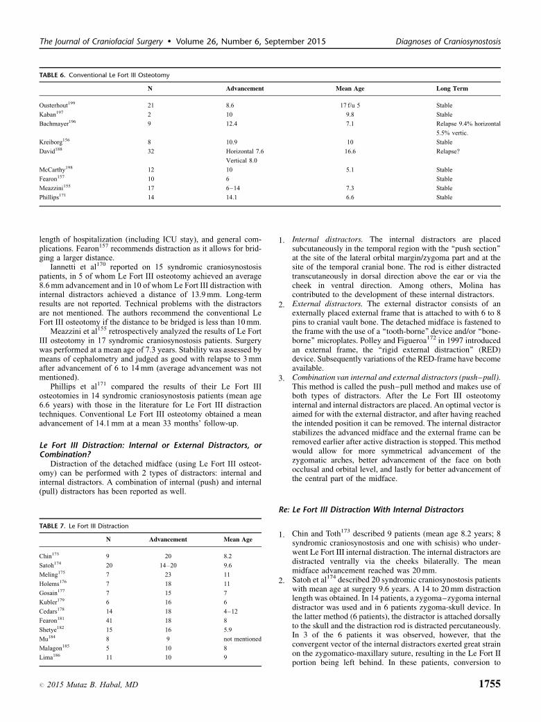
TABLE 6. Conventional Le Fort III Osteotomy
N Advancement Mean Age Long Term
Ousterhout199 21 8.6 17 f/u 5 Stable
Kaban197 2 10 9.8 Stable
Bachmayer196 9 12.4 7.1 Relapse 9.4% horizontal
5.5% vertic.
Kreiborg156 8 10.9 10 Stable
David188 32 Horizontal 7.6 16.6 Relapse?
Vertical 8.0
McCarthy198 12 10 5.1 Stable
Fearon157 10 6 Stable
Meazzini155 17 6–14 7.3 Stable
Phillips171 14 14.1 6.6 Stable
The Journal of Craniofacial Surgery � Volume 26, Number 6, September 2015 Diagnoses of Craniosynostosis
length of hospitalization (including ICU stay), and general com-plications. Fearon157 recommends distraction as it allows for brid-ging a larger distance.
Iannetti et al170 reported on 15 syndromic craniosynostosispatients, in 5 of whom Le Fort III osteotomy achieved an average8.6 mm advancement and in 10 of whom Le Fort III distraction withinternal distractors achieved a distance of 13.9 mm. Long-termresults are not reported. Technical problems with the distractorsare not mentioned. The authors recommend the conventional LeFort III osteotomy if the distance to be bridged is less than 10 mm.
Meazzini et al155 retrospectively analyzed the results of Le FortIII osteotomy in 17 syndromic craniosynostosis patients. Surgerywas performed at a mean age of 7.3 years. Stability was assessed bymeans of cephalometry and judged as good with relapse to 3 mmafter advancement of 6 to 14 mm (average advancement was notmentioned).
Phillips et al171 compared the results of their Le Fort IIIosteotomies in 14 syndromic craniosynostosis patients (mean age6.6 years) with those in the literature for Le Fort III distractiontechniques. Conventional Le Fort III osteotomy obtained a meanadvancement of 14.1 mm at a mean 33 months’ follow-up.
Le Fort III Distraction: Internal or External Distractors, orCombination?
Distraction of the detached midface (using Le Fort III osteot-omy) can be performed with 2 types of distractors: internal andinternal distractors. A combination of internal (push) and internal(pull) distractors has been reported as well.
TABLE 7. Le Fort III Distraction
N Advancement Mean Age
Chin173 9 20 8.2
Satoh174 20 14–20 9.6
Meling175 7 23 11
Holems176 7 18 11
Gosain177 7 15 7
Kubler179 6 16 6
Cedars178 14 18 4–12
Fearon181 41 18 8
Shetye182 15 16 5.9
Mu184 8 9 not mentioned
Malagon185 5 10 8
Lima186 11 10 9
# 2015 Mutaz B. Habal, MD
1. I
nternal distractors. The internal distractors are placedsubcutaneously in the temporal region with the ‘‘push section’’at the site of the lateral orbital margin/zygoma part and at thesite of the temporal cranial bone. The rod is either distractedtranscutaneously in dorsal direction above the ear or via thecheek in ventral direction. Among others, Molina hascontributed to the development of these internal distractors.2. E
xternal distractors. The external distractor consists of anexternally placed external frame that is attached to with 6 to 8pins to cranial vault bone. The detached midface is fastened tothe frame with the use of a ‘‘tooth-borne’’ device and/or ‘‘bone-borne’’ microplates. Polley and Figueroa172 in 1997 introducedan external frame, the ‘‘rigid external distraction’’ (RED)device. Subsequently variations of the RED-frame have becomeavailable.3. C
ombination van internal and external distractors (push–pull).This method is called the push–pull method and makes use ofboth types of distractors. After the Le Fort III osteotomyinternal and internal distractors are placed. An optimal vector isaimed for with the external distractor, and after having reachedthe intended position it can be removed. The internal distractorstabilizes the advanced midface and the external frame can beremoved earlier after active distraction is stopped. This methodwould allow for more symmetrical advancement of thezygomatic arches, better advancement of the face on bothocclusal and orbital level, and lastly for better advancement ofthe central part of the midface.Re: Le Fort III Distraction With Internal Distractors
1. C
hin and Toth173 described 9 patients (mean age 8.2 years; 8syndromic craniosynostosis and one with schisis) who under-went Le Fort III internal distraction. The internal distractors aredistracted ventrally via the cheeks bilaterally. The meanmidface advancement reached was 20 mm.174
2. S atoh et al described 20 syndromic craniosynostosis patientswith mean age at surgery 9.6 years. A 14 to 20 mm distractionlength was obtained. In 14 patients, a zygoma–zygoma internaldistractor was used and in 6 patients zygoma-skull device. Inthe latter method (6 patients), the distractor is attached dorsallyto the skull and the distraction rod is distracted percutaneously.In 3 of the 6 patients it was observed, however, that theconvergent vector of the internal distractors exerted great strainon the zygomatico-maxillary suture, resulting in the Le Fort IIportion being left behind. In these patients, conversion to
1755

Mathijssen The Journal of Craniofacial Surgery � Volume 26, Number 6, September 2015
17
distraction with an external frame was necessary. Subsequently,the authors used a type of internal distractors that are distractedventrally at the zygomatic arches and are placed in parallel withthe use of a perioperatively applied reference rod. The authorsprefer this type (zygoma-zygoma) of internal distractor. Slightasymmetry was noticed in 2/14 patients.
175
3. M eling described 7 patients with mean age 11 years in whoma mean distraction length of 23 mm was obtained, with in 1 caseproblems related to the internal distractor, and in 1 casedeviation of the nose. All other cases were judged successfully.Data on stability and/or vector were not described.
176
4. H olmes described 7 consecutive syndromic craniosynostosispatients who underwent a Le Fort III osteotomy with internaldistraction (mean linear distraction 18.1 mm, mean age 11years). In 1 case a mechanical problem related to the internaldistractor was observed. The authors report less advancement ofthe midface (Le Fort II complex) with the use of internaldistractors. The bony zygomatico-maxillary junction in youngpatients is often too thin to transmit the vector 1 to 1. Theauthors have changed their policy; in the future they will applyhe push–pull method.
Gosain177 described 7 syndromic craniosynostosis patients(mean age 7 years, mean distraction 14.8 mm) in whom the LeFort distraction with internal distractors was performed. In 5 cases afracture occurred and/or weak zygomatico-maxillary junction andin 2 cases the internal distractors were replaced with an externalframe.
Cedars178 described 14 patients (13 syndromic craniosynostosispatients and one with schisis) who underwent Le Fort III distraction(internal). The mean distraction length obtained was 18 mm asdetermined by counting the number of rotations during the activedistraction. In 1 patient the distraction caused asymmetry of theface; in 2 patients infection developed of the skin around theinsertion site of the internal distractor, which resolved after removalof the internal distractor.
Re: Le Fort III Distraction With External Distractors (ExternalFrame)
1. F
earon157 retrospectively analyzed 22 syndromic craniosynos-tosis patients who had undergone a Le Fort III advancement. Ofthese 22 patients (mean age 7.5 years), 10 had undergoneconventional osteotomy and 12 distraction (2 with internaldistractors and 10 with an external frame). The averageadvancement achieved in the osteotomy group for point A was5.1 mm and for Nasion 7.7 mm; in the distraction group forpoint A 16.0 mm and for Nasion 7.0 mm. Asymmetry wasobserved with the use of internal distractors. This was not thecase for the patients who underwent external distraction.179
2. K ubler described in 6 patients (mean age: 6 years) fronto-orbital advancement by means of an osteotomy in combinationwith a Le Fort III distraction with external frame, with goodresults. The mean advancement of the Le Fort III portion was15.8 mm.
180
3. M avili described Le Fort III distraction with external framein 4 syndromic craniosynostosis patients (age range: 5.2–12years). The frame was fixed with an intraoral splint and with 2infraorbital steel wires. In all patients the distraction wassuccessful. There were no problems with the external frame.Information is lacking about the distance obtained as well asstability.
181
4. F earon analyzed Le Fort III advancement in 51 syndromiccraniosynostosis patients (mean age 8 years), 41 of whom
56
underwent distraction with external frame. In the distractiongroup the mean obtained advancement was 18 mm. Cephalo-metric assessment after 1 year did not show any relapse.Furthermore, there was no growth of the maxilla after the Le FortIII operation. Problems with the external frame were not reported.The auteurs pose that the lack of growth is rather related to thesyndrome and not so much a consequence of the operation.
182
5. S hetye analyzed 15 syndromic craniosynostosis patients whounderwent internal distraction with the Rigid ExternalDistraction (RED) device. Fixation of the frame was by meansof an occlusal splint and bone anchors at the site of thezygomatic arch and the maxilla. Point A had advanced 15.9 mmsagittally along the x-axis and moved downward 1.1 mm alongthe y-axis. These results were stable at cephalometricassessment after 1 year. Five patients developed a skin infectionat the insertion site of the external pin, which in all patientscould be managed successfully. In 2 patients there wastraumatic dislocation of the frame. In 1 patient the externalframe had to be repositioned under general anesthesia.
183
6. N out analyzed 21 patients undergoing a Le Fort I and IIIexternal distraction. Most of the problems encountered wererelated to the external pin: loosening of the pin, infection of theskin around the pin, and in 3 patients there was traumaticdislocation of the frame, albeit without any effect on the finaloutcome.
184
7. M u analyzed Le Fort III internal distraction (RED II device)in 8 syndromic craniosynostosis patients. In all patients thedistraction, at mean 9 mm anteriorly and 1.5 mm caudally, wassuccessful; in 1 patient a skin infection at the insertion site ofthe external pin was seen; in 1 patient a seroma. Thispublication is in the Chinese language, only the information inthe English-language abstract is reported here. Malagon185
described 5 syndromic craniosynostosis patients (mean age atoperation 8 year) in whom a Le Fort III with halo frame wasperformed. The maxilla was advanced over 10 mm. Problemswith the frame are not reported. In all patients the distractionwas successful.
186
8. L ima described 11 syndromic craniosynostosis patients (meanage at operation 9 years) who underwent Le Fort III distractionwith RED device. The mean obtained length was 10.3 mmhorizontally and 2.4 mm vertically. Cephalometric assessmentafter 1 year showed less than 1 mm relapse in horizontal direction.Vertical face growth after 1 year was 2.9 mm.
Push–Pull MethodSchulten187 described 10 syndromic craniosynostosis patients
(mean age 11 years), in whom the central midface using Le Fort IIIwith push–pull distraction was advanced a mean of 12 mm. Afterthe distraction period all devices were removed under anesthesia. Itis not reported how the advanced segment was fixed during theconsolidation phase. Push–pull distraction permits equal movementat both the upper and lower facial levels, advancement of the centralmidface, and symmetric movement. This method also provides abackup in case 1 device malfunctions.
Complications in Le Fort III Osteotomy/DistractionVarious complications have been reported in relation to the Le
Fort III osteotomy and in relation to the application of internal andexternal distractors.
Complications of the Le Fort III Osteotomy. David188
reported the following complications in 32 Le Fort III andmonobloc osteotomies: blindness (1), frontal osteomyelitis (2),
# 2015 Mutaz B. Habal, MD

The Journal of Craniofacial Surgery � Volume 26, Number 6, September 2015 Diagnoses of Craniosynostosis
cerebrospinal fluid (CSF) leakage (2), extradural abscess (1),periorbital abscess (1), wound infection (1), velopharyngeal insuf-ficiency (4), diplopia (1), and facial nerve palsy (1).
Matsumoto189 described a 9-year-old patient with Crouzonsyndrome in whom a Le Fort III distraction is complicated byintracranial bleeding because of a skull base fracture. As a result,the patient died. Lanigan et al reported a skull base fracture in anonsyndromic patient associated with the pterygomaxillary dys-junction (as part of the Le Fort osteotomy) resulting in unilateralblindness.190
Nout191 described a lethal course in a 10-year-old patient withApert syndrome in whom a Le Fort III osteotomy was performedimmediately followed by placement of internal and external dis-tractors. Anisocoria was noticed at the end of the intervention.Computed tomographic-angiography revealed a dissection of theinternal carotid artery. Computed tomographic-angiography istherefore recommended in the preoperative planning of the Le FortIII distraction: in the first place to gain more insight into thethickness and configuration of the bone structures and intracranialstructures in relation to the planning of the osteotomy lines and theplacement of the distractors and secondly, to identify possibleabnormalities of the arteries and veins.
Complications of Internal Distractors.Mechanical problems in relation to internal distractors have
been mentioned above.
Complications Van External Distractors.The majority of the problems related to the external frame appear tobe because of the percutaneous pins with which the frame isattached to the cranial vault. Skin infections and loosening pinsare well-known problems reported in the orthopedic and neurosur-gic literature. The data on incidences and recommendations on, forexample, required torque, cannot be extrapolated one-to-one tosyndromic craniosynostosis patients. These patients often haveundergone earlier remodeling of the cranial vault. Thickness andstrength of the cranial vault are not similar (less strength and lack ofcontinuity in some places).
Nout183 analyzed 21 patients (including 19 syndromic cranio-synostosis patients) undergoing a Le Fort I and III external dis-traction. Most of the problems encountered were related to theexternal pin: loosening of the pin, infection of the skin around thepin, and in 3 patients there was traumatic dislocation of the frame,albeit without any effect on the final outcome.
1. A
# 2
number of case reports describe intracranial migration of aninternal pin.192–195 In all patients this had no permanentconsequences with full recovery of the patient.
Timing of the Surgical Correction and(Postoperative) Growth
Although the number of cephalometric studies of maxillarygrowth in surgically treated and nonsurgically treated syndromiccraniosynostosis patients is small, the results point at a clear picture.If there were to be any vertical and sagittal growth of the maxilla atall, it would be negligibly small or near-enough null: Bachmayer,196
Meazzini,155 Kreiborg,156 Fearon,157 and Fearon.181 The methodo-logical quality of these findings can only be classified as Level 3and 4.
Le Fort III Osteotomy or Distraction?The publications on Le Fort III distraction consistently mention
the following arguments, among other things, to prefer this
015 Mutaz B. Habal, MD
procedure during the conventional Le Fort III osteotomy: shorteroperation with less blood loss, less pain, and shorter hospitalization,including ICU stay. But solid evidence is not provided in thosestudies. Yet the studies make clear that distraction permits to obtaina larger distance during which the detached portion can beadvanced.
The publications on Le Fort III osteotomies demonstrate that themean obtained midface advancement is approximately 9 to 10 mm(range 6–14 mm). The publications on Le Fort III distractiondemonstrate that the mean obtained midface advancement isapproximately 17 mm (range 9–23 mm). The methodological qual-ity of these findings can only be classified as Level 3 and 4.
This would therefore also enable better functional and estheticoutcomes. Although in the Le Fort III distraction, in contrast to theconventional Le Fort III osteotomy, intermaxillary fixation andbone transplants are not needed, complications related to theinternal and internal distractors may still occur. These are usuallymild; in exceptional patients serious complications are described,often directly related to the pins of the external frame.
Lastly, it should be mentioned that a Le Fort III and a Le Fort Iosteotomy technically can be combined in 1 operation, with whicheventually the tooth-borne segment of the jaw can be moved over alarger distance.
Distraction With Internal of External Distractors?The body of literature on the Le Fort III distraction includes
more publications with more patients who have undergone aprocedure with external frame than with internal distractors. Atthe same time, it is striking that recent publications tend to focus onthe application of an external frame rather than the application ofinternal distractors.
It is not easy to summarize the results of the distractions withinternal distractors. Individual publications report reasonableresults, but at the same time in Satoh,174 the internal distractorsare eventually replaced with an external frame in 3/20 patients,whereas in Meling,175 problems are reported in 2/7 patients. InHolmes,176 we find a policy change on account of poor results withinternal distractors in 7 patients in favor of the push–pull technique,in Cedars178 the internal distractors were removed prematurely in2/14 patients, and lastly in Gosain177 the internal distractors werereplaced with an external frame in 2/7 patients. Quite often thisreplacement is necessitated by a mechanical problem of the internaldistractor.
The following publications on the Le Fort III distraction withexternal frame do not report any problems: Fearon157 22 patients;Mavili180 4 patients; Fearon181 41 patients; Malagon185 5 patients;and Lima186 11 patients.
The following publications reported minor problems with theframe only:
Shetye182 pin-related skin infection in 5/15 patients and trau-matic dislocation of the frame in 2/15; Nout183 loosening of pins in43% of the patients and traumatic dislocation of the in 3/21 patients,albeit without clinical consequences; and Mu184 observed skininfection in 1/8 patients and seroma in 1/8.
The push–pull method is reported in only 1 publication, butSchulten is enthusiastic on the basis of outcomes in 10 patients.187
Conclusions
Level 3
Maxillary growth in surgically and nonsurgically treated Apert,Crouzon, and Pfeiffer patients seems negligibly small.B Fearon, 2001157
B Fearon, 2005181
C Bachmayer, 1986154
1757

Mathijssen The Journal of Craniofacial Surgery � Volume 26, Number 6, September 2015
1758
C Meazzini, 2005155
C Kreiborg, 1986156
Level 3
It seems likely that the mean advancement of the midface obtained withdistraction is larger than that obtained with conventional osteotomy.In Le Fort III distraction an external frame is preferred for its ease ofplacement and removal and optimum vector control. Any distractor(internal and external) related complications tend to be minor.Le Fort III osteotomy:
B Fearon, 2001157
C Bachmayer, 1986196
C David, 1990188
C Kaban, 1986197
C Kreiborg, 1986156
C McCarthy, 1990198
C Meazzini, 2005155
C Ousterhout, 1986199
C Phillips, 2006171
Le Fort III distraction:
B Fearon, 2005181
C Cedars, 1999178
C Chin, 1997173
C Gosain, 2002177
C Holmes, 2002176
C Kubler, 2004179
C Lima, 2009186
C Malagon, 2008185
C Meling, 2006175
C Mu, 2007184
C Satoh, 2006174
C Shetye, 2006182
C Nout, 2010191
Level 3
CT-angiography is recommended in the preoperative planning ofthe Le Fort III distraction. In the first place to gain more insight intothe thickness and configuration of the bone structures andintracranial structures in relation to the planning of theosteotomy lines and the placement of the distractors. Secondly,to identify possible abnormalities of the arteries and veins.C Nout, 2010191
Considerations:Indications for midface advancement in OSAS and weighing up
the pros and cons of a permanent or temporary tracheal cannula aredealt with in the chapter on OSAS.
One should be aware that there is no maxillary growth after a LeFort III distraction at young age, so that a second Le Fort III (ormono-bloc) operation from the age of 18 years may be necessary.Additional orthognatic surgery (also from age 18 years) may beindicated to correct the malocclusion, including open bite (seechapter 14).
Operations that change the appearance considerably, such as aLe Fort III advancement in 12 to 17-year-old patients could give riseto psychosocial problems, which may already have been presentbecause of the abnormal appearance.
Fronto-orbital advancement as the initial surgical interventionaffects the facial profile and thus also influences the planning of theLe Fort III advancement (see chapter 6).
An external frame puts a considerable psychosocial burden onthe patient and his/her family, those who activate the distractor. Thismay be a consideration to opt for an internal distractor instead.Furthermore, low mental development in syndromic craniosynos-tosis patients may be reason to opt for an internal distractor. Theapplication of the internal distractor can be combined with a facialmask to optimize the vector of the Le Fort III distraction (seechapter 14).
RecommendationsThe Le Fort III distraction in Apert and Crouzon/Pfeiffer
patients to correct the maxillary hypoplasia (including exorbitism)is typically performed between the ages of 8 and 12 years, or fromthe age of 18 years. In case of severe OSAS and/or severeexorbitism posing a threat to vision it may be performed at youngerage (see chapter on OSAS). In young children (�6 years) amonobloc distraction is preferred, dependent on previous surgeries.
In view of the higher risk of psychosocial problems or unrealisticexpectations in 12 to 18 year olds, the Le Fort III distraction ispreferably not performed at these ages (see chapters 17 and 18).
After the Le Fort III, occlusion must be monitored. Orthognaticsurgery is recommended in the patient of malocclusion (eg, Le FortI, bilateral sagittal split osteotomy). This type of surgery is per-formed after age 18 years (see chapter 14).
Computed tomographic-angiography deserves to be consideredin the preoperative planning of the Le Fort III distraction.
In Le Fort III distraction an external frame is recommended inview of its ease of placement and removal and the possibility ofoptimum vector control.
If internal distractors show mechanical problems they should bereplaced with an external distractor.
Monobloc Osteotomy/Distraction andHypertelorism
The monobloc frontofacial osteotomy is a combination of atranscranial advancement of the frontal bone and an extracranial LeFort III advancement in 1 operation and in 1 piece. This surgicalintervention is indicated in syndromic craniosynostosis patients tocorrect the hypoplastic midface with possibly associated functionalproblems and elevated ICP at a relatively too small cranial volume.It was described in 1978 by Ortiz-Monasterio et al.200
The body of good literature on the results of monobloc osteo-tomies is evidently much smaller than that regarding the Le Fort IIIadvancement. Data on complications of the conventional monoblocoperation were not available in Pubmed and fort his reason wesearched the publications of the first congresses of the InternationalSociety for Craniofacial Surgery. Only very few surgeons appliedthe conventional monobloc in the earlier days, and results includingcomplications were presented at these meetings only. Exclusion ofthese publications would result in a bias with considerable under-estimation of the complications of this procedure.
Hypertelorism in SyndromicCraniosynostosis Patients
Apart from midface hypoplasia, hypertelorism is a well-knownphenomenon in syndromic craniosynostosis patients. Although itseldom leads to functional problems, the position of the bony orbitscan esthetically be unappealing to such an extent that correction isindicated. In 1979, Van der Meulen described the ‘‘median faciot-omy’’ for correction of the midline cleft with hypertelorism.201 Byvertical splitting of the monobloc segment in the midline andremoving a wedge from the midline of the midface he was ableto rotate the 2 halves of the midface toward each other, thusreducing the distance between the orbits. Tessier202 later refinedthis technique and named it facial bipartition. This refined tech-nique can be combined with forward advancement: ‘‘facial biparti-tion osteotomy with advancement’’, in which advancement can beperformed with distraction as well.
Orbital hypertelorism may also be treated with orbital boxosteotomy. This procedure cannot be performed until the teethhave erupted permanently, usually from approximately the age of14 years. Orbital box osteotomy is well suited to be combined with
# 2015 Mutaz B. Habal, MD
The Journal of Craniofacial Surgery � Volume 26, Number 6, September 2015 Diagnoses of Craniosynostosis
orthognatic surgery. Naturally, the positions of the medial andlateral canthus should be paid attention to in the orbital boxosteotomy and the ‘‘facial bipartition’’.167,203 Although some sur-geons do not detach the canthi and prefer to leave these in situ at thebone, other surgeons purposefully detach the canthi to reattach themlater to the bone. The latter method would permit better correctionof the positions of the canthi.
Moreira Gonzalez reported good results after facial bipartition in14 syndromic craniosynostosis patients, combined with advance-ment using osteotomy in 3 patients. Postoperative problems werethe following: liquor leakage (2), infections (2).204 Problems withdiplopia are not reported.
Urrego205 reported a surgical technique aimed at reducing theremaining telecanthus in this intervention. In 12 patients, a mean39% reduction of interbrow distance was noted.
Posnick206 analyzed pre- and postoperative CT scans of 7syndromic craniosynostosis patients undergoing facial bipartitionosteotomy. Also after 1 year the results showed stable improvementbut no complete normalization.
Risks and Complications of Monobloc/Facial Bipartition (Withand Without Distraction)
The literature consistently points to the same risks, compli-cations and problems around monobloc osteotomies: presence of aventriculoperitoneal shunt, need of extensive bone grafts for thesake of stabilization, pressure on the soft tissues and cranial flap,and extradural retrofrontal dead space created by the advancementand which directly connects with the nasal cavity and the ethmoid.Occurrence of the latter complication could perhaps be reduced byclosing the connection between this dead space and (para-) nasalcavity with galea frontalis flap or periosteal flap.
Aspects of relapse and revision of monobloc in relation to theindication of monobloc for OSAS are addressed in the relevantchapter.
OsteotomyPosnick described 23 syndromic craniosynostosis patients, 10
undergoing monobloc advancement, 7 facial bipartition advance-ment, and 6 facial bipartition solely (all osteotomy, no distrac-tion).207 All showed a retrofrontal dead space after the intervention,which in 10 patients had disappeared after 6 weeks and in all within1 year. The presence of a ventriculoperitoneal shunt did notinfluence the risk of complications. One patient showed evidenceof a late (after 3 months) CSF leakage via a skin fistula with necrosisof the frontal bone.
Timing of the Monobloc-Distraction in Relation toOrbital Growth
Bentley208 preoperatively analyzed the volumes of the bonyorbits in 50 syndromic craniosynostosis patients and compared theoutcomes with those of a normal group. Volumes in the syndromiccraniosynostosis patients were smaller shortly after birth but tendedto reach normal values at 6 to 8 months of age and had completelynormalized at 13 months of age. This would imply that surgicalintervention should be delayed until the age of 6 to 8 months, thusmaximizing the effects of accelerated normal orbital growth.
Position of the Eye Globe After MonoblocDistraction
Fitzgerald O’ Conner150 analyzed in 10 syndromic craniosy-nostosis patients the position of the eyeball after monobloc dis-traction. At an osseous distraction distance of 12.9 mm (upper face)and 14.2 mm (lower face) the globe moved 5.3 mm (left eye) and
# 2015 Mutaz B. Habal, MD
6.3 mm (right eye). Globe advancement was approximately 2/5 ofthe distraction distance, without evidence of optic nerve damageand impaired vision.
DistractionAs was done above for the Le Fort III distractions, the following
section describes the application of internal and internal distractorsin monobloc distractions.
Monobloc Distraction With External Distractors
1. W
itherow209 reported 20 syndromic craniosynostosis patientsundergoing monobloc distraction with halo external frame;mean advancement: 16.4 mm, mean age 7.8 years. Cerebrosp-inal fluid leakage was noted in 8 patients (the leakage stopped in5 patients; 3 underwent a revision operation using a lumbardrain)210
2. W itherow evaluated relapse after monobloc distraction withexternal frame in 21 syndromic craniosynostosis patients (meanage 8.3 years, probably largely the same patients as mentionedunder point 1). The midface was distracted to an average of16.4 mm. Relapse was seen only in 3 of all patients during 14.8months’ follow-up period.
211
3. P onniah studied the pre- and postoperative CT scans of 10syndromic craniosynostosis patients undergoing monoblocdistraction (7) and facial bipartition distraction, both usingan external frame. In particular, the facial bipartition distractiondifferentially advanced the central part of the face more than thelateral areas.
Monobloc Distraction With Internal Distractors
1. C
ohen212 described 5 syndromic craniosynostosis patients, in 3of whom monobloc distraction was performed and in 2 a facialbipartition, in all patients using internal distractors (agebetween 9 months and 5 years). The distraction distanceranged from 22 to 30 mm. Device infection developed in 1patient.213
2. A rnaud described 20 syndromic craniosynostosis patientswho underwent monobloc distraction using internal distractors(mean age: 3.2 years). Cerebrospinal fluid leakage occurred in 3patients. Device infection developed in 2 patients requiringremoval of the distractor. Septic frontal necrosis occurred in 1patient 2 months after completion of distraction, necessitatingremoval of the forehead flap.
144
3. A rnaud studied 36 syndromic craniosynostosis patientsundergoing monobloc distraction using internal distractors(mean age: 5.2 years). Distraction was uneventful in 28patients; 7 required revision surgery because distractorsstagnated. One patient died from acute brain tonsillar herniationbefore distraction was begun. Furthermore, loss of dead frontalbone in 1 patient.
214
4. B radley described 13 syndromic craniosynostosis patients(mean age 8.3 years) who underwent facial bipartition,including monobloc distraction using internal devices. Stableresults were obtained and no complications were reported (noliquor leakage, meningitis, or frontal bone loss).
150
5. F itzgerald O’Connor described 10 syndromic craniosynos-tosis patients undergoing monobloc distraction (upper facedistraction distance 12.9 mm and lower face 14.2 mm). FromCT scans (pre- and post-) a mean forward movement of 5.3 mm
1759

Mathijssen The Journal of Craniofacial Surgery � Volume 26, Number 6, September 2015
17
for the left globe and 6.3 mm for the right globe was established.Although there was no evidence of optic nerve stress withpossible functional limitations, the long-term developmentshould be closely monitored.
Complications in Monobloc Osteotomies/DistractionsDavid (1990)188 reported the following complications in 32 Le
Fort III and monobloc osteotomies: blindness (1), frontal osteo-myelitis (2), CSF leakage (2), extradural abscess (1), periorbitalabscess (1), wound infection (1), velopharyngeal insufficiency (4),diplopia (1), and facial nerve palsy (1).
Vyas215 reported a left carotid-cavernous fistula as a compli-cation in monobloc distraction in a patient with Apert syndrome.The fistula was successfully embolized. After 1 year there wasstill partial vision loss in the left eye, but there were no othersequelae.
Witherow described in 20 syndromic craniosynostosis patientsthe following complications after monobloc distraction: CSF leak-age (8, in 5/8 it stopped spontaneously, 3/8 required lumbardrainage in combination with endoscopic repair of the tensor fascialatae muscle with Tissuecoll), hypernasality (5), loss of frontaldead/avital bone (1), temporary tracheotomy (2), fracture of thezygomatic bone (1), trismus which spontaneously had resolved after6 months (5), and cornea ulceration (2).209
Complications in Monobloc Osteotomy/DistractionThe literature (case reports and no series larger than N¼ 4)
consistently points to the same risks, complications, andproblems around the monobloc osteotomies (including thevariants): presence of a ventriculoperitoneal shunt; need of exten-sive bone grafts; pressure on the soft tissues and cranial flap;and extradural retrofrontal dead space created by the advancementand, which directly connects with the nasal cavity and theethmoid.
On the other hand, Posnick et al207 described monobloc osteot-omy in 23 syndromic craniosynostosis patients, of whom 1 showedlate CSF leakage via a skin fistula with necrosis of the frontal bone.In all patients, the retrofrontal dead space had disappeared after 1year. The presence of a ventriculoperitoneal shunt did not influencethe risk of complications.
This is the only series of appreciable size in which this wasanalyzed retrospectively.
Benhaim provide an overview of monobloc fronto-facialadvancements performed in University of California, Los Angeles(UCLA) in the predistraction period from 1988 to 1996.216 In total26 patients were operated; mean operation time was more than 8hours, mean blood loss 1700 cc. The following complications werenoted: liquor leakage (27%), SIADH (15%), pneumonia (12%),epilepsy (8%), meningitis (4%), hypoxic injury (4%), pseudome-ningocele (4%), oronasal fistula (4%), partial flap necrosis (4%),and abscesses (4%). This was already a considerable improvementcompared with the results from the 1979 to 1988 period as aconsequence of adaptations of the operative technique.
Wolfe reports the results of a classic monobloc in 44 patients.217
The complications consisted of death (2%), liquor leakage (2%),violent infections (osteitis, temporal, and epidural abscesses, 9%)resulting in loss of the frontal bone in 7%. The created frontofacialdead space was mentioned as the major cause for these infections.
Marchac218 reported their lethal complications after craniofacialsurgery. Twenty of the 1397 patients undergoing intracranialoperations died (1.4%). And even 6 of the 18 patients in whoma monobloc was performed died.
60
TimingRegarding the timing of monobloc distraction in relation to
growth of the orbits, Bentley et al (2002)208 concluded from CT-analysis in 50 syndromic craniosynostosis patients and controlgroup that this should be delayed to minimally the age of 6 to 8months to allow for optimal orbital growth.
Monobloc Distraction: Osteotomy or Distraction?Internal or Internal Distractors?
Regarding the choice of operation technique—distraction versusosteotomy—the literature unanimously makes clear that distractionallows for movement of the bone segment over a larger distance.Witherow209 described 20 syndromic craniosynostosis patients andPonniah211 described 10 patients undergoing monobloc distractionwith external frame. No major problems and complications werereported. Most of the publications, encompassing the large majorityof patients, describe monobloc distraction with the use of internaldistractors.
Position of the Eye Globe After MonoblocDistraction
Fitzgerald O’Connor150 concluded from CT-analysis in 10syndromic craniosynostosis patients undergoing monobloc distrac-tion that globe advancement was approximately 2/5 of the distrac-tion distance, without evidence of optic nerve damage and impairedvision. This is the only report on this issue.
Conclusions
Level 3
It is likely that monobloc distraction can be performed in a predictableway with either type of distractors (internal and external) witheventually fewer complications than with conventionalmonobloc osteotomy.C Benhaim, 1997216
C Marchac, 1995218
C Wolff, 1995217
C Witherow, 2008209
C Ponniah, 2007211
C Cohen, 1999212
C Arnaud, 2007144
C Bradley, 2008214
C Fitzgerald O’Conner, 2009150
Level 3
Regarding the timing of the monobloc operation there are indicationsthat it is worth waiting until minimally the age of 6 to 8 months toallow for optimal growth of the orbits.C Bentley, 2002208
ConsiderationsRegarding the indication for midface/monobloc advancement in
OSAS and the considerations on the pros and cons of a permanenttemporary tracheal cannula, please refer to the chapter on OSAS.
As there is no maxillary growth after an early monobloc, asecond monobloc (or Le Fort III) operation after the age of 18 maybe necessary.
Operations that change the appearance considerably, such as amonobloc in 12 to 17-year-old patients could give rise to psycho-social problems, which may already have been present because ofthe abnormal appearance (see chapter 17).
An external frame puts a considerable psychosocial burden onthe patient and his/her family, those who activate the distractor. Thismay be a consideration to opt for an internal distractor instead.Furthermore, low mental development in these syndromic cranio-synostosis patients may be reason to opt for an internal distractor.
# 2015 Mutaz B. Habal, MD

The Journal of Craniofacial Surgery � Volume 26, Number 6, September 2015 Diagnoses of Craniosynostosis
As syndromic craniosynostosis patients, notably those withApert or Pfeiffer/Crouzon syndrome, often show V-shape in themaxilla including the open bite, facial bipartition is preferredbecause this does not only correct the hypertelorism but alsorestores this V shape to a normal occlusal line. Regarding thetiming of this intervention the same argument as in the midface/monobloc operations is proposed: at the age of 4 to 5 years by far thelargest part of the osseous orbits has been fully developed.
Although orbital box osteotomy compromises vascularization ofthe margins that were detached with osteotomy (which could resultin resorption and undesirable decrease in volume), this effect will beless with the facial bipartition. The orbital box osteotomy can beperformed from approximately at the age of 14 years. After all, thecaudal part of the osteotomy (under the infraorbital margin) requiressufficient space between the infraorbital nerve and the apicalboundaries of the dentition. The orbital box osteotomy can be wellcombined with orthognatic surgery.
RecommendationsThe monobloc distraction in syndromic craniosynostosis
patients is indicated in severe exorbitism (ocular globe luxationand/or corneal injury) with
1. s
# 2
evere OSAS at the time of protocolized initial cranial vaultexpansion
2. s
evere OSAS in combination with elevated ICP.In case of severe OSAS, age (and therefore timing) does not playa role. In very young children (<2 years) tracheal cannula place-ment may bridge the waiting time (see chapter on OSAS).
Monobloc distraction is preferred in view of the lower percen-tage of serious complications in comparison with the conventionalmonobloc osteotomy.
In view of the higher risk of psychosocial problems or unrealisticexpectations in 12 to 18 year olds, a monobloc is preferably notperformed at this age (see chapters 17 and 18).
Hypertelorism is preferably corrected from the age of 4 or 5years by means of the facial bipartition technique. The orbital boxosteotomy technique can be performed after the upper teeth haveerupted permanently (from approximately the age of 14 years) andcan be well combined with additional orthognatic surgery.
Computed tomographic-angiography is recommended in thepreoperative planning of the monobloc distraction (with or withoutfacial bipartition).
After the monobloc procedure, monitoring of occlusion iswarranted. Orthognatic surgery is recommended if malocclusionshould occur (eg, Le Fort I, bilateral sagittal split osteotomy).This surgery should be delayed until after the age of 18 years(see chapter 14).
Transversal CorrectionThe sagittal and vertical dimensions have already been
addressed in the sections on the Le Fort III and monobloc osteot-omy/distraction. The transversal correction is discussed below. Theavailable literature on this topic is even more limited and mostlyconcerns small case series.
Transversal maxillary hypoplasia in adolescence and adulthoodis seen in non-syndromic and syndromic patients, including schisispatients. This type of hypoplasia leads to transversal malocclusion.
In skeletally immature patients, the maxilla can be expandedorthodontically. In skeletally mature patients, however, a uni- orbilateral transversal hypoplasia can solely be corrected with the use ofSARME. This procedure is a combination of surgical and
015 Mutaz B. Habal, MD
orthodontical techniques that offers room in the maxillary arch fororthodontically correct arrangement of the teeth. In addition, SARMEsubstantially enlarges the maxillary apical (dentoalveolar) base andthe palatal arch, which allows good movement of the tongue inswallowing. An additional favorable effect is a subjective improve-ment in nasal breathing as a result of the expansion of the narrowestpassage in the nose (nasal valve) and of the nasal cavity volume.219
The scientific background for the timing of this treatment isaddressed below. In the absence of recent studies on this subject,however, we had to resort to literature from before 1980.
Growth at the sutures takes place by deposition of new bone atthe suture borders by the adjacent cellular layer. By the end of fetallife this cellular layer will become thinner, which indicates slowergrowth rate and reduction of the number of fibers in the inter-mediary cellular layer, which separates the capsular layers. In astudy of human sutures from birth to 18 years of age, Latham andBurston220 concluded that after some 2 to 3 years the suturesprimarily are functioning as sites where the separate bone platesmeet, but that remodeling is a continuous process. The cranialsutures will fuse before the complete eruption of the third molars.Shortly hereafter the facial sutures will fuse, and the sutures thatconnect the skull and the face are the last to fuse.221 Regarding thefacial sutures, Sicher222 concluded from anatomic studies thatfusion in humans on average starts by the middle of the fourthdecade, at the posterior part of the median palatal suture, but thatsome facial sutures, including the frontozygomatic suture, remainopen to later age.
A conflicting view is presented by Persson,223 who foundevidence of osseous fusion of the median palatal suture at age17 years. Latham and Burston,220 however, did not find evidence ofsynostosis in the same suture at age 18 years. An ‘‘overall view’’ ispresented by Scott224 who believes that in spite of the fact that mostof the facial sutures seem to be open on the surface, they show acertain degree of fusion somewhat deeper. The available literatureclearly is not unambiguous and conclusive on this subject.
In clinical practice, however, transversal orthodontical expan-sion is successful until the age of approximately 14 to 15 years,depending on the patient’s sex. After this age it is virtuallyimpossible to achieve orthodontic expansion.225–227 In general itis believed that that the fusion of the midpalatal suture impedesexpansion.225–227
The above applies to the nonsyndromic situation. The literaturecontains 2 studies dealing with maxillary sutures, maxillary trans-versal growth, and SARME in the syndromic situation.
Koudstaal et al228 describe the application of a bone-bornedistractor device for the expansion of the maxilla in 8 syndromicand 5 non-syndromic patients. They conclude that this type ofdistractor is suitable for surgical expansion of the maxilla in bothnonsyndromic and syndromic patients. Notably the small dimen-sions of the device make it suited for application in the extremelynarrow upper jaws seen in syndromic craniosynostosis patients.
The literature does not contain publications on the effects ofSARME on further growth of the maxilla, neither in nonsyndromicnor in syndromic patient groups.
Only very few publications are available on the indications,timing, and the effects of SARME in syndromic craniosynostosispatients. The information is based on literature data of nonsyn-dromic patients.
In summary, the patient’s age determines the indicatedtherapy for transversal maxillary hypoplasia, either orthodonticalexpansion or SARME. As soon as the diagnosis of transversalmaxillary hypoplasia is made and treatment can reasonably bestarted in view of the dental and mental situation, orthodonticexpansion is indicated before the age of 14 to 15 years, SARMEat older age.
1761

Mathijssen The Journal of Craniofacial Surgery � Volume 26, Number 6, September 2015
Conclusions
Level 4
1762
It seems likely that the SARME procedure can be performed fromthe age of 14 to 15 years.
C Koudstaal, 2006228
ConsiderationsThe experts are of the opinion that in syndromic craniosynos-
tosis patients the maxilla may be extremely narrow, in whichsituation a tooth-borne distractor in the SARME procedure tech-nically is not feasible. In that case, the application of the bone-bornedistractor is indicated. One should be aware that the palatal mucosamay be very thick in syndromic craniosynostosis patients. This mayimpede placement of some types of bone-borne distractors.
RecommendationsIf a SARME procedure is performed in syndromic craniosynos-
tosis patients, a tooth-borne distractor is preferred over a bone-borne distractor. But in case of an extremely narrow maxilla andinsufficient space for a tooth-borne distractor (Hyrax), a bone-bornedistractor is indicated. The thick palatal mucosa impedes theplacement of a bone-borne distractor. A SARME procedure isperformed from the age of 14 to 15 years.
8. INCREASED INTRACRANIAL PRESSURE
Basic Questions
1. W
hat is the prevalence of ICP in the various types ofnonsyndromic and syndromic craniosynostosis, before and afterskull remodeling?2. W
hat screening (nature and frequency) is needed to timelyidentify increased ICP?3. H
ow can increased ICP be prevented or treated?IntroductionThe risk of increased ICP varies widely between the types of
craniosynostosis, and the syndromic type carries a much higher riskthan the nonsyndromic type. The risk in the nonsyndromic group,however, is much less recognized and therefore possibly underdiagnosed. It is important to detect and treat increased ICP at anearly stage. High ICP may lead to, for example, irreversible loss ofvision.229 It is not clear what method is best suited to establishincreased ICP, what thresholds should be set, and at what frequencyit should be applied to identify problems at an early stage.
Summary of the LiteraturePrevalenceI. Nonsyndromic Craniosynostosis. Until 1996, genetic diag-nostics was not possible and therefore the distinction betweensyndromic and nonsyndromic was not always evidenced. In theolder literature about ICP it is often unclear whether this reallyconcerns nonsyndromic craniosynostosis. Especially, cases of uni-lateral or bilateral coronal suture synostosis may well be of asyndromic type. Most of the publications on ICP in craniosynostosispatients derive from 2 centers (Paris and London) and patientpopulations may overlap. Most studies only address increasedICP preceding a cranial vault expansion. Reliable data on theprevalence of increased ICP following cranial vault expansionare rare. There are no data at all on ICP in patients with a lambdoid
suture synostosis, most likely because of the very low prevalence ofthis type of synostosis.
The following publications are from the Parisian center, andpatient populations may overlap:
Renier (1982) preoperatively performed invasive ICP measure-ments in 49 nonsyndromic patients, that is, 23 with scaphocephaly,9 with plagiocephaly, 5 with trigonocephaly and 7 with brachyce-phaly. Increased ICP (>15 mm Hg) was measured in 3, 2, 0, and 3patients, respectively; borderline ICP (11–15 mm Hg) in 4, 4, 1, and2 patients, respectively and normal ICP (�10 mmHg) in 16, 3, 4,and 2 patients, respectively.142 In 23 patients (both isolated andsyndromic) the ICP measurement was repeated postoperatively: inall 23 the ICP had decreased over a number of weeks. Regarding therelation between ICP and age, ICP peaked at 6 years. In 17 patients,clinical suspicion of increased ICP years after the operation wasconfirmed with an invasive measurement. The diagnoses, however,are not mentioned.
Gault230 defined increased ICP as >15 mm Hg, irrespective ofage, which widely ranged in the study population. Intracranialpressure was measured invasively preceding cranial vault expan-sion. Increased ICP was established in 1 of the 2 patients withbrachycephaly, in none of the 4 patients with trigonocephaly, innone of the 3 patients with plagiocephaly, and in 2 of the 44 patientswith scaphocephaly.
Arnaud141 preoperatively measured ICP invasively in 142 chil-dren with scaphocephaly. Intracranial pressure exceeded 15 mm Hgin 19 children, of whom 12 were older than 1 year, which wasstatistically significantly different.
Renier121 provides an overview of more than 2137 patients withcraniosynostosis. Invasive preoperative ICP measurementsrevealed prevalences of 13.8% in scaphocephaly (246 measure-ments), 7.7% in trigonocephaly (39 measurements), 12.7% inplagiocephaly (63 measurements), and 31.3% in brachycephaly(32 measurements).
Mathijssen103 reported invasive preoperative ICP measurementsin plagiocephaly; ICP was>15 mm Hg in 8 of the 50 patients. These8 children were all older than 1 year (statistically significant).
Also the craniofacial team in London published a considerablenumber of publications on this issue:
Thompson231 performed invasive preoperative ICP measure-ments in 37 patients with plagiocephaly, 25 with scaphocephaly and12 was trigonocephaly. Increased ICP (>15 mm Hg) was estab-lished in 3, 6, and 4 patients, respectively.
Thompson232 preoperatively measured ICP invasively in 72nonsyndromic patients: 18 children with scaphocephaly, 35 withplagiocephaly, 10 with brachycephaly and 9 with trigonocephaly.Intracranial pressure was increased (>15 mm Hg) in 13 children: 3with scaphocephaly, 2 with plagiocephaly, 5 with brachycephaly,and 3 with trigonocephaly. Borderline ICP (10–15 mm Hg) wasmeasured in 7, 13, 2, 5, and 3 patients, respectively. Here, too, it isnot certain if the brachycephaly was really nonsyndromic. Ifincreased ICP was defined on the basis of plateau waves ratherthan just mean ICP, the number of patients with increased ICPwould have been somewhat higher.
Shipster233 reports increased ICP (>15 mm Hg), measuredinvasively, in 4 of 11 scaphocephaly patients. It is not clear whyICP was measured in these 11 patients and whether there may havebeen selection bias. Thompson234 is referred to, and probably thepopulations overlap. For this reason this publication is not includedin Table 8.
The other studies are from Camperdown, Australia,235
Rome,236–237 and the United States, that is, Chicago,238 and Seat-tle.239
Whittle235 included 5 nonsyndromic craniosynostosis patients: 4with scaphocephaly and 1 with plagiocephaly. Preoperative
# 2015 Mutaz B. Habal, MD

The Journal of Craniofacial Surgery � Volume 26, Number 6, September 2015 Diagnoses of Craniosynostosis
invasive measurement revealed increased ICP in 2 of the 4 withscaphocephaly and in the 1 with plagiocephaly. The indications forICP measurements were radiologic signs of mild hydrocephalus,loss of vision, headache or behavior change, and are therefore asource of bias. For this reason this publication is not included inTable 8.
Tamburrini236 reports invasive preoperative ICP measurementsin 5 nonsyndromic craniosynostosis patients: 3 with scaphocephalyand 2 with brachycephaly. All 5 had normal ICP levels (�15 mmHg).
Tamburrini (2005)237 reports invasive preoperative ICPmeasurements in 7 nonsyndromic craniosynostosis patients: 3 withscaphocephaly and 4 with brachycephaly. Of the latter 4 patients, 2had an anterior brachycephaly and 2 a posterior brachycephaly. Thequestion is whether the 2 with posterior brachycephaly reallyshowed synostosis. The 2 patients with a bicoronal synostosishad increased ICP. Possibly these were indeed cases of syndromiccraniosynostosis. Probably these are the same patients as describedin the 2004 publication. In view of the very small numbers and thedubious diagnoses this study is not included in Table 8.
Foster238 investigated the occurrence of resynostosis afterprimary skull remodeling in 13 patients with trigonocephaly, 30with scaphocephaly, 22 with plagiocephaly, 1 with unilaterallambdoid suture synostosis and 9 with brachycephaly. The resy-nostosis rates were 7.7% (1), 3.3% (1), 0, 0, 0, respectively. In 1patient with scaphocephaly increased ICP was established by meansof lumbar puncture before second cranial vault expansion. As theoccurrence of resynostosis was subjected to selection, these rates donot reflect the real prevalence in the total population of patients witha nonsyndromic craniosynostosis.
Scott describes 36 patients older than 2 years at the time ofprimary cranial vault expansion. A total of 17 patients showedpreoperative symptoms consistent with increased ICP, and 10 hadalready earlier undergone cranial surgery elsewhere.239 The studygroup consisted of 5 patients with scaphocephaly, 2 with brachy-cephaly, 1 with trigonocephaly, 3 with multiple suture synostosis, 3with Crouzon, 1 with Saethre-Chotzen, 1 with Muenke, and 1 withShprintzen. In 10 patients, CT findings suggested increased ICP orpapilledema. Symptoms resolved in most patients. This series is leftout of consideration in view of the heterogeneity of the studypopulation (syndromic/nonsyndromic; earlier operation or not)and the indistinct criteria for increased ICP.
Marucci109 describes 81 patients with scaphocephaly whounderwent limited cranial surgery before 6 months of age. Sevenpatients developed a progressive vertex bulge at the site of the
TABLE 8. Prevalence of Increased Intracranial Pressure (ICP) in NonsyndromicCraniosynostosis Before Skull Remodeling
Number of Patients
With Increased ICP/
Number of Patients
Measured (%) Scapho Trigono Plagio Brachy Lambdoid
Renier142 3/23 (13) 0/5 (0) 2/9 (22) 3/7 (43)
Gault230 2/44 (5) 0/4 (0) 0/3 (0) 1/2 (50)
Arnaud141 19/142 (13)
Thompson231 6/25 (24) 4/12 (33) 3/37 (8)
Thompson232 3/18 (17) 3/9 (33) 2/35 (6) 5/10 (50)
Renier121 34/246 (14) 3/39 (8) 8/63 (13) 10/32 (31)
Mathijssen103 8/50 (16)
Florisson110 7/103 (7) 3/71 (4) 0/28 (0) 0/3 (0)
Total 74/601 (12) 13/140 (9) 23/225 (10) 19/51 (37) 0/3 (0)
ICP, increased intracranial pressure.
# 2015 Mutaz B. Habal, MD
former anterior fontanel within an average of 8 months postopera-tively. Five patients underwent invasive ICP measurement, and ICPwas elevated in 4 of them. Five patients had progressive synostosisand 2 were positive for FGFR2 mutations (of whom 1 was knownwith hypochondroplasia). Intracranial pressure was measured in 3of the 5 eventually true nonsyndromic scaphocephaly patients, andwas found to be raised in 1.
Florisson110 describes a retrospective study of 205 consecutivepatients with a non-syndromic unisutural craniosynostosis, treatedbetween January 2003 and June 2007, and in whom preoperatively 1or 2 fundoscopies were performed by protocol. This revealedpapilledema notably in the patients with scaphocephaly and to alesser degree in those with trigonocephaly; the youngest patient was3 months old.
A review from 2004 on the basis of 5 studies with a total of 503patients with scaphocephaly, trigonocephaly, or plagiocephalyreports a 4% to 14% increased ICP range, with increased ICPdefined as ICP >15 mm Hg.240
In summary, the risk of increased ICP seems to be 12% inscaphocephaly, 9% in trigonocephaly, and 10% in plagiocephaly.The range is larger for trigonocephaly (0%–33%) as a smallernumber of patients was investigated. The estimated risk for thegroup of bilateral coronal suture synostosis patients is 37%. In themajority of the studies it is not clear, however, whether this reallyconcerns nonsyndromic synostosis.
Risk of increased ICP following skull remodeling is explicitlymentioned only in a retrospective study from Rotterdam in 205consecutive patients with a nonsyndromic craniosynostosis, treatedbetween January 2003 and June 2007.110 Postoperative fundoscopywas performed if increased ICP had been present preoperatively, orif clinical symptoms were noted, such as headache, behavioralchange, worsening of vision, deviating cranial circumference curve,or copper beaten aspect on x-ray. Postoperative fundoscopy hadbeen performed in 46 patients (22% of initial population), of whom3 patients with scaphocephaly (27, 39, and 46 months postopera-tively) and 2 patients with trigonocephaly (1 persisting papilledemaalready present preoperatively, and 1 patient developed papille-dema 34 months postoperatively) appeared to have papilledema.
II. Syndromic Craniosynostosis.Note 1: diagnoses mentioned in older articles (<1996) are not
genetically confirmed.PM2: the distinction between the Crouzon and Pfeiffer syn-
dromes later appeared to be irrelevant on account of overlappinggenetic abnormalities in the FGFR2 syndrome.
Also regarding syndromic craniosynostosis, most of articles onICP are from Paris:
Renier142 performed invasive ICP measurements preoperativelyin syndromic patients, that is, 23 with oxycephaly, 6 with Apert, and2 with Crouzon syndrome. Increased ICP (>15 mm Hg) wasestablished in 10, 3, and 2 patients, respectively. Borderline ICP(11–15 mm Hg) was present in 9, 2, and 0 patients, respectively.Normal ICP (�10 mm Hg) was found in 4, 1, and 0 children,respectively. In 23 patients (nonsyndromic and syndromic) themeasurement was repeated postoperatively. Intracranial pressuredecreased in all 23 patients during a number of weeks. Regardingthe relation between ICP and age, ICP peaked at 6 years. Seventeenpatients were measured years after the operation on clinical suspi-cion of increased ICP, which was confirmed with the measurement.The diagnosis, however, is not mentioned.
Gault230 reported increased ICP on the basis of preoperativeinvasive measurement in 4 of the 5 children with a complexcraniosynostosis, that is in 3 of the 4 with an oxycephaly, 2 ofthe 3 with Crouzon syndrome, and in the only Apert patient.
1763

Mathijssen The Journal of Craniofacial Surgery � Volume 26, Number 6, September 2015
Renier121 presented an overview of more than 2137 patients withcraniosynostosis. Invasive preoperative ICP measurements resultedin prevalences of 60% in oxycephaly (95 measurements), 47.1% inmultiple suture synostosis (17 measurements), 62.5% in Crouzon,45% in Apert, and 29% in ‘‘other syndromes’’.
The following publications are from the center in London:Thompson preoperatively measured ICP invasively in 53 syn-
dromic children, that is, 20 with Crouzon, 13 with Apert, 5 withPfeiffer, 14 with Saethre-Chotzen syndrome, 1 with a cloverleafskull, and 11 with multiple suture synostosis.232 Increased ICP(>15 mm Hg) was found in 35 of them, that is, 13 Crouzon, 5 Apert,3 Pfeiffer, 6 Saethre-Chotzen, 1 cloverleaf, and 7 multiple suturesynostosis patients. Borderline ICP (10–15 mm Hg) was found in 7,7, 1, 6, and 0 patients, respectively. On the basis of plateau wavesrather than only mean ICP the number of patients with increasedICP was even slightly higher.
Hayward241 described 24-hours ICP measurements in 11 symp-tom-free children (5 with Pfeiffer, 2 with Crouzon, 2 with Apert, 1with Antley-Bixler syndrome and 1 with osteopetrosis). Plateaus ofICP were exclusively seen during active sleep with an increasedmean ICP in all 11 children. During quiet sleep, the mean ICP wasincreased (>15 mm Hg) in 5 children, borderline (10–15 mm Hg) in3, and normal in 3.
Marucci152 described 24 patients with Apert syndrome whowere operated upon not until there were signs of increased ICP,which was the case in 83% at a mean age of 18 months (range 1month to 4 years and 5 months). Of the patients who underwentoperation, 35% experienced a second episode of increased ICP at amean age of 3 years and 4 months (range 1 year and 11 months to5 years and 9 months). The increased ICP was defined as presenceof papilledema, an abnormal VEP scan, an ICP measurement>15 mm Hg, or more than 3 plateaus in 24 hours.
Other articles are from Camperdown Australia,235 Toronto,242
Pittsburgh,243 Rome,236–237 Wurzburg Germany,60 Boston(Greene, 2008),244 Chicago,238 Rotterdam,245 and Oxford UK.246
Whittle235 included 8 patients with Crouzon syndrome, 1 withApert syndrome, and 6 with complex craniosynostosis, of whom 10
TABLE 9. Prevalence of Increased Intracranial Pressure (ICP) in Syndromic Craniosy
Number of Patients With Increased ICP
Apert Crouzon Pfeiffer
Renier142 3/6 (50) 2/2 (100)
Gault230 1/1 (100) 2/3 (67)
Thompson232 5/13 (38) 13/20 (65)
Renier121 ? (45) ? (63)
Tamburrini236 5/5 (100) 3/4 (75)
Hayward241 2/2 (100) 2/2 (100)
Kress60
Greene244
Marucci152
Preop� 20/24 (83)
Postop 7/20 (35)
De Jong54
Preop 2/22 (9) 24/45 (53) Cr/Pf
Postop 11/31 (35) 8/40 (20) Cr/Pf
Woods246
Postop 1 year
Postop 5 years
ICP, increased intracranial pressure. Many articles are from the same centers (Paris
preoperative measurements: few data on postoperative prevalence.�Operation not until increased ICP.
1764
(7 Crouzon, 1 Apert, 3 complex craniosynostosis, respectively)preoperatively showed increased ICP with invasive measurement.Indications for measurement were radiologic signs of mild hydro-cephalus, worsening of vision, headache or behavioral change, andare thus subject to bias.
Siddiqi242 evaluated 107 syndromic children with the followingdiagnoses: 52 Crouzon, 33 Apert, 8 Pfeiffer, 11 Saethre-Chotzensyndrome, and 4 cloverleaf skulls. Six (6%) developed symptoms ofincreased ICP after initial decompression (3 with Apert, 2 withSaethre-Chotzen, and 1 with Pfeiffer syndrome), defined as papil-ledema (N¼ 4), progressive macrocephaly (N¼ 1), and invasiveICP measurement (N¼ 1). One patient required a third operation onaccount of recurrent increased ICP. At follow-up 3 to 7 years later,none of the 6 patients showed signs of increased ICP. In this seriesroutine screening of increased ICP was not performed, which is acause of strong bias. For this reason this study is not included inTable 9.
Pollack243 described follow-up of 22 children with syndromiccraniosynostosis, most of whom were operated upon between theages of 6 to 18 months. Eight children developed recurrentincreased ICP, defined as papilledema (N¼ 3), progressive prop-tosis (N¼ 1), or other clinical symptoms (N¼ 3). As most of thechildren did not show any symptoms, routine ophthalmologicexamination is recommended.
Tamburrini236 described invasive ICP measurements in16 syndromic children with the following diagnoses: 5 Apert, 4Crouzon, 3 Pfeiffer syndrome, 2 turricephaly, and 2 oste-opetrosis. Fourteen showed increased ICP (>15 mm Hg), that is,all except 1 child with Pfeiffer syndrome and 1 child withosteopetrosis.
Tamburrini237 described invasive ICP measurements in 12syndromic craniosynostosis patients, of whom 5 with Apert, 4 withCrouzon, and 3 with Pfeiffer syndrome. Eleven of the 12 childrenhad increased ICP. Most likely these are the same patients asdescribed in the 2004 article.
Kress60 reported a 35% prevalence (24/68 patients) of increasedICP in Saethre-Chotzen syndrome on the basis of papilledema or
nostosis
/Number of Patients Measured (%)
Saethre-Chotzen Muenke Complex
10/23 (43)
7/9 (78)
3/5 (60) 6/14 (43) 8/12 (67)
65/112 (58)
3/3 (100) 2/2 (100)
5/5 (100)
24/68 (35) 0/42 (0)
30/39 (77)
5/26 (19) 1/28 (4)
4/24 (17) 2/38 (5)
9/26 (35)
8/19 (42)
or London) and patient populations therefore could overlap. Most studies concern
# 2015 Mutaz B. Habal, MD
The Journal of Craniofacial Surgery � Volume 26, Number 6, September 2015 Diagnoses of Craniosynostosis
ICP >20 mm Hg. Forty-two Muenke patients did not show papil-ledema, whereas 2 had increased dural tension intraoperatively.
Greene244 selected 39 patients with complex craniosynostosis(excluding bilateral coronal suture synostosis and pansynostosis). Inthis group, the risk of increased ICP was 76.9%, defined aspapilledema, a deflecting cranial curve or extensive endocorticalerosion on CT scan.
Foster238 investigated the occurrence of resynostosis afterprimary skull remodeling in 31 patients with multiple suturesynostosis, 2 with Apert, 4 with Crouzon, and 5 with Saethre-Chotzen syndrome. Percentages of resynostosis were 12.9%(N¼ 4), 0, 25% (N¼ 1), 0 (N¼ 0), and 20% (N¼ 1), respectively.In 1 patient with Saethre-Chotzen syndrome and 2 with multiplesuture synostoses increased ICP was found with lumbar puncturebefore the second cranial vault expansion.
Bannink245 described the prevalence, treatment outcome, andrisk factors for increased ICP in 33 Apert, 43 Crouzon, and 8Pfeiffer syndrome patients. Papilledema was documented preopera-tively in 25 of 66 (38%) patients and postoperatively in 30 of 70patients (43%).Overall, 43 of the 84 (51%) patients (12 Apert, 31Crouzon/Pfeiffer syndrome) showed papilledema at some moment.Impressions on skull x-ray or clinical symptoms such as headacheor behavior changes were not correlated with the presence ofpapilledema and consequently proved unreliable screeningmethods. Complex craniosynostosis, the diagnosis Crouzon/Pfeif-fer syndrome, exorbitism, and ventricular dilatation were found tobe risk factors for papilledema.
Honnebier146 did not find signs of increased ICP in any of 16patients with Muenke syndrome, but does not report in which wayand how frequently this was investigated.
Woods246 evaluated 34 Saethre-Chotzen patients with geneti-cally confirmed diagnosis. Nine of the 26 patients (35%) with atleast 1 year follow-up developed increased ICP, as well as 8 of the19 patients (42%) with at least 5 years follow-up, for whichreoperation was needed. Increased ICP was defined as a baselineof 20 mm Hg or higher, or 4 or more B-waves. The indications forICP measurement were papilledema, a deflecting cranial curve,headache or aggressive behavior, impressions on skull x-ray,narrow ventricles, Chiari malformation, with effaced basal cisternsand sulci spaces.
De Jong54 reported the prevalence of increased ICP, on the basisof papilledema, per syndrome, with the population of patients withApert and Crouzon/Pfeiffer syndromes the same as the 1 describedby Bannink in 2008.245
The 2004 review of Bristol et al reports a 53% to 89% range ofincreased ICP (>15 mm Hg), based on 98 patients with syndromiccraniosynostosis.240
In summary, the prevalence of increased ICP in syndromiccraniosynostosis is considerably higher than that in nonsyndromiccraniosynostosis. Preoperatively the risk seems to be approximately40% to 50% for Apert, 50% to 70% for Crouzon and Pfeiffer, 35% to45% for Saethre-Chotzen, 0 for Muenke syndrome, and 50% to 80%for complex craniosynostosis. Increased ICP after a previous skullremodeling is a real danger and is estimated at 35% to 43% in Apert,Crouzon, Pfeiffer, and Saethre-Chotzen syndromes. This has not beeninvestigated for complex synostosis. In Apert syndrome patients, themean age during the second episode of increased ICP was 3 years and4 months with a maximum of nearly 6 years.152 In Saethre-Chotzenthe second operation on account of increased ICP was performed at amean age of 3.4 years, with a maximum of 8.5 years.246
ScreeningThe different methods to determine the ICP consist of invasive
measurements during 1 night and minimally 12 hours (such asepidural sensor or lumbar puncture), fundoscopy, VEP-scan, or
# 2015 Mutaz B. Habal, MD
CT-scan (endocortical erosion). Increased ICP is almost routinelydefined as a baseline above 15 mm Hg during slow wave sleep ormore than 3 plateau waves. Eide247 compared the mean ICP with thenumber of peak ICPs above 20 mm Hg and is of the opinion that themean ICP does not do justice to the full ICP measurement. Thenumber of ICPs peaking above 20 mm Hg that should be consideredabnormal cannot be ascertained. The definition of a plateau variesby author: The minimal level ranges from 20 to 35 mm Hg, theduration from 5 to 20 minutes.
I. Nonsyndromic Craniosynostosis.Tuite248 compared in 74 children with nonsyndromic craniosy-
nostosis the presence of signs of increased pressure on skull x-raywith invasive ICP measurement. Children with increased ICP moreoften showed diffuse impressions, diastasis of the sutures anderosion of the sella tursica. The sensitivity was very low, however,and therefore these are unreliable screening methods for increasedICP.
Tuite249 compared 58 patients with nonsyndromic synostosisand 55 patients with syndromic, 9 with multiple suture synostosis.Papilledema was established in 15 patients: 11 with syndromicsynostosis, 2 with multisuture synostosis, 1 with trigonocephaly,and 1 with plagiocephaly. On the basis of an invasive measurement,41 patients had increased ICP, 13 of whom showed papilledema. Inchildren above the age of 8 years papilledema was specific andsensitive. Sensitivity at lower age was only 22%. Consequently, inthis young group the absence of papilledema is no guarantee fornormal ICP, but on the other hand the presence of papilledema is anevident sign of increased ICP.
Stavrou (1997)229 reported on 9 children, among whom 1 withplagiocephaly, 1 with trigonocephaly and 1 with scaphocephaly,age range 1 to 6 years, who showed lowering of vision as aconsequence of an increased ICP.
Tamburrini237 concludes in a review of the literature thatassessing the mean ICP during sleep in combination with plateauwaves is the most reliable method indicating increased ICP.
Eide250 demonstrated in patients with shunt failure, hydrocepha-lus or craniosynostosis that there is no relation between ventriclesize on CT scan and increased ICP. Also the change in ventricle sizeis no predictor of ICP changes.
Marucci109 described that a progressive vertex bulge at the siteof the former anterior fontanel in scaphocephaly patients after earlyand limited skull remodeling may be a sign of increased ICP.
In conclusion, it is not known how often a deflecting cranialcircumference growth curve is related to an increased ICP andconsequently it is hard to tell how useful it would be to include thisparameter in the follow-up program. Radiologic findings such asimpressions, ventricle size, or changes therein are unreliable as ascreening method for increased ICP. Papilledema found withfundoscopy is highly probably a sign of increased ICP,251 but itsabsence does not exclude an increased ICP. The reliability offundoscopy seems to be age-related, although described in onlyone study. A VEP scan could show signs of increased ICP or onsetof optical nerve injury at an early stage, before papilledemadevelops,252 but this technique has not been proven with the useof invasive ICP measurements. In addition, a high percentage ofabnormal VEP scans (24%) were reported in a population ofpredominantly nonsyndromic craniosynostosis patients, which ishigher than the proportions reported in other studies using invasiveICP measurements.
Invasive ICP measurement remains the gold standard, butnaturally has disadvantages such as the need of anesthesia, surgicalintervention with risk of complications, and admission to anintensive or high care unit. Generally, the threshold is set at abaseline ICP exceeding 15 mm Hg or more than 3 plateau waves,
1765

Mathijssen The Journal of Craniofacial Surgery � Volume 26, Number 6, September 2015
irrespective of the age of the patient. The scientific underpinning ofthis threshold is unclear, but it is adhered to by nearly all experts.
II. Syndromic Craniosynostosis.Fok253 and Gault254 demonstrated in syndromic children that the
intracranial volume is no reliable predictor for the ICP.Gupta prospectively follows the optical nerve of 33 patients with
nonsyndromic and syndromic craniosynostosis with the use of VEP,echographic thickness measurement and fluorescence angiogra-phy.255 Notably, multiple suture synostosis and exorbitism corre-lated with injury to the optical nerve as a result of papilledema. TheVEP scans of all 5 patients with optic atrophy showed a prolongedlatency period. Fluorescence angiography is useful to diagnoseonset papilledema. Optical nerve thickness measurements are use-ful in the follow-up of papilledema. The added value of VEP scanswith respect to fundoscopy is not made clear.
Campbell256 presented a patient with Pfeiffer syndrome whounderwent skull remodeling on account of papilledema, after whichthe papilledema resolved but vision gradually worsened. Invasivemeasurement still evidenced increased ICP in spite of the absence ofpapilledema. Unexplained lowering of vision may therefore be anindication for invasive ICP measurement, because absence ofpapilledema is no guarantee for normal ICP.
Tuite248 compared in 49 children with syndromic craniosynos-tosis the presence of signs of increased pressure on skull x-ray withinvasive ICP measurement. Children with increased ICP more oftenshowed diffuse impressions, diastasis of the sutures and erosion of thesella tursica. The sensitivity was very low, however, and thereforethese are unreliable methods for screening on increased ICP.
Tuite249 compared 58 patients with non-syndromic synostosisand 55 patients with syndromic, 9 with multiple suture synostoses.Papilledema was established in 15 patients: 11 with syndromicsynostosis, 2 with multisuture synostosis, 1 with trigonocephaly and1 with plagiocephaly. On the basis of an invasive measurement 41patients had increased ICP, 13 of whom showed papilledema. Inchildren above the age of 8 years papilledema was specific andsensitive. Sensitivity at lower age was only 22%. Consequently, inthis young group the absence of papilledema is no guarantee fornormal ICP, but on the other hand the presence of papilledema is anevident sign of increased ICP.
Pollack243 described 22 syndromic children who routinelyunderwent ophthalmologic screening for increased ICP preopera-tively and 6-month postoperatively. Papilledema was found pre-operatively in 4 and postoperatively in 8 at a mean age of 16.5months. Only 3 of these 8 showed clinical symptoms, which impliesthat this is an unreliable screening method.
Stavrou229 reported on 9 children, among whom 4 with Crouzonsyndrome and 2 with complex craniosynostosis, age range 1 to 6years, who showed lowering of vision as a consequence of increasedICP.
Connoly257 described selected patients with multiple suturesynostosis and signs of increased ICP. Most of them had Crouzonsyndrome (confirmed by FGFR2 mutation) and generally showed anormal skull shape at birth with progressive synostosis. IncreasedICP presented in these patients approximately at the age of 5 years.The authors recommended frequent screening using fundoscopy butdid not indicate frequency or age limit.
Hoefkens258 described 9 Crouzon syndrome children with post-natal synostosis, of whom 9 showed impressions on skull x-ray, 7developed a bregma at the anterior fontanel, and 4 papilledema.
Tamburrini237 concludes in a review of the literaturethat assessing the mean ICP during sleep in combinationwith plateau waves is the most reliable method indicating increasedICP.
1766
Liasis259 compares 3 ophthalmologic methods, viz. vision,fundoscopy, and VEP scan in 8 syndromic children. All 8 showeda decrease in N80 to P100 amplitude preceding surgery and anincrease postoperatively. Fundoscopy showed no abnormalities in 3patients, progressive bilateral swelling in 3, and unilateral swellingin 2 patients. Vision worsened in 1, improved in 4, and fluctuated in2 patients, preoperatively. Invasive measurement demonstratedincreased ICP in 3 of the 8 children, of whom 1 showed abnormalfundus, 1 unilateral papilledema, and 1 unnatural paleness of thepapilla. The authors conclude that solely vision or fundoscopy isinsufficient and that VEP will timely detect vision abnormalities.This article, however, provides only little evidence for this!
Marucci152 described 24 patients with Apert syndrome whowere operated upon not until there were signs of increased ICP,which was the case in 83% at a mean age of 18 months (range 1month to 4 years and 5 months). Of the patients who underwentoperation, 35% experienced a second episode of increased ICP at amean age of 3 years and 4 months (range 1 year and 11 months to 5years and 9 months). The increased ICP was defined as presence ofpapilledema, an abnormal VEP scan, an ICP measurement>15 mmHg, or more than 3 plateaus in 24 hours.
Bannink245 demonstrated that the presence of papilledema is notassociated with clinical symptoms such as vomiting, headache,vision, or behavior change.
Woods246 evaluated 34 Saethre-Chotzen patients with geneti-cally confirmed diagnosis. The indications for ICP measurementwere papilledema, a deflecting cranial curve, headache or aggres-sive behavior, impressions on skull x-ray, narrow ventricles, Chiarimalformation, with effaced basal cisterns, and sulci spaces. Of the 9patients with increased ICP, 3 showed papilledema, 6 delayed skullgrowth, 5 headache or aggressive behavior, and 8 signs of increasedpressure on CT. The eldest patient with increased ICP was 9 yearsold.
In conclusion, clinical symptoms do not seem to be reliablescreening methods for increased ICP. Also in syndromic cranio-synostosis radiologic findings are an unreliable screening methodfor increased ICP. The finding of papilledema on fundoscopy ishighly probably a sign of increased ICP, but its absence does notexclude increased ICP. The reliability of a fundoscopy seems to beage related, although this is addressed in only 1 study. Visualevoked potential as screening method is reported by only 1 centerand its reliability is therefore hard to assess. An invasive ICPmeasurement remains the gold standard, but naturally has disad-vantages, such as the need of anesthesia, surgical intervention withrisk of complications, and admission to an intensive or high careunit. Generally, the threshold is set at a baseline ICP exceeding15 mm Hg or more than 3 plateau waves, irrespective of the age ofthe patient. The scientific underpinning of this threshold is unclear,but it is adhered to by nearly all experts.
Prevention and/or TreatmentGenerally a cranial vault expansion is performed within the first
year of life. A number of studies have shown that the prevalence ofincreased ICP increases if the operation is performed after the firstyear of life. This has been demonstrated for scaphocephaly141 andplagiocephaly.103
The London center pursues a policy by which a patient withApert syndrome is operated upon not until there were signs ofincreased ICP.152 Of the 24 patients, 83% developed increased ICPat a mean age of 18 months (range 1 month to 4 years and 5 months).Of these patients, 35% experienced a second episode of increasedICP at a mean age of 3 years and 4 months.
The percentage of recurrent increased ICP is not essentiallydifferent from the percentages reported by teams performing a
# 2015 Mutaz B. Habal, MD

The Journal of Craniofacial Surgery � Volume 26, Number 6, September 2015 Diagnoses of Craniosynostosis
protocolized cranial vault expansion.245 It therefore seems likelythat an early cranial vault expansion is effective both preventively(for approximately 3 years) and therapeutically. The London centerpolicy has the advantage that a small number of patients eventuallydoes not undergo skull remodeling, but is it disadvantageous in thatpatients need to be screened for signs of increased ICP veryfrequently, for which there is no watertight method.
If increased ICP is established again at follow-up after an initialcranial vault expansion, treatment may consist either of a secondcranial vault expansion or VP-drain insertion. The choice depends,among other things, on the presence of progressive ventriculome-galy (see chapter on hydrocephalus). As there is a relation betweenICP and OSAS it is important to determine the presence and severityof OSAS in case of confirmed increased ICP, as treatment can beadjusted to this.260,261 Please refer to the chapter on OSAS.
Conclusions
Level 2
# 2015 M
The prevalence of preoperatively increased ICP in nonsyndromiccraniosynostosis is 12% (range 5%–24%) in scaphocephaly, 9%(range 0%–33%) in trigonocephaly, 10% (range 0%–22%) inplagiocephaly, and 37% (range 31%–50%) in bilateral coronalsuture synostosis.
B Renier, 1982142
B Gault, 1992230
B Arnaud, 1995141
B Thompson, 1995231
B Thompson, 1995232
B Mathijssen, 2006103
C Florisson, 2010110
C Renier, 2000121
Level 2
The prevalence of preoperatively increased ICP in syndromiccraniosynostosis is 40%–50% in Apert, 50%–70% in Crouzonand Pfeiffer, 35%–45% in Saethre-Chotzen syndrome,50%–80% in complex craniosynostosis, and zero in Muenkesyndrome. Increased ICP after an earlier cranial vaultexpansion is estimated at 35%–43% in Apert, Crouzon,Pfeiffer, and Saethre-Chotzen syndromes.B Renier, 1982142
B Gault, 1992230
B Thompson, 1995232
C Renier, 2000121
C Tamburrini, 2004236
C Hayward, 2005241
C Kress, 200660
C Greene, 2008244
C Marucci, 2008152
C Bannink, 2008245
C Woods, 2009246
Level 2
Clinical symptoms and radiologic findings are unreliablescreening methods for increased ICP. A progressive vertexbulge at the site of the former anterior fontanel may indicateincreased ICP. Papilledema is an evident sign of increased ICP,but its absence does not exclude increased ICP.B Tuite, 1996248
B Tuite, 1996249
B Eide, 2002247
C Tamburrini, 2005237
C Pollack, 1996243
C Bannink, 2008245
C Marucci, 2008152
C Woods, 2009246
Level 3
An invasive ICP measurement remains the gold standard, but isassociated with disadvantages and complications, and is notrecommended as routine screening method. Increased ICP isdefined as a baseline ICP >15 mm Hg (measured at least 12 hoursduring sleep) and/or minimally 3 plateau waves withICP >35 mm Hg, during at least 20 minutes.utaz B. Habal, MD
C Tamburrini, 2005237
C Pollack, 1996243
C Marucci, 2008152
C Woods, 2009246
Level 3
Patients with a nonsyndromic craniosynostosis seem to have a smallrisk of developing increased ICP after initial cranial vault expansion,which may result in lowering of vision.C Florisson, 2010110
C Stavrou, 1997229
C Marucci, 2008152
Level 3
VEP scans may have added value in screening for increased ICP,next to fundoscopy.C Gupta, 2003255
C Liasis, 2006259
ConsiderationsIn spite of the fact that patients with a nonsyndromic cranio-
synostosis only have a relatively small risk of developingincreased ICP with risk of loss of vision, this is a contributingreason to recommend operation, apart from the esthetic aspect.Also if operation is decided against it is of importance tofrequently monitor the child for signs of increased ICP untilthe age of 6 years (at least once a year). The craniofacial team inRotterdam has occasionally encountered papilledema in childrenwith nonsyndromic craniosynostosis after cranial vault expansionuntil a maximum age of 6 years. At the time of this writing, thereare no studies providing scientific underpinning for the maximumage until when increased ICP in nonsyndromic craniosynostosisshould be monitored.
Muenke syndrome seems to distinguish itself from the othersyndromic types of craniosynostosis by a slight risk of increasedICP. For this reason, 1 may consider to perform skull remodelingsomewhat later than is common for syndromic craniosynostosis,which strategy seems to reduce the risk of recurrence of thedeformity (see chapter 6).
Performing a cranial vault expansion by protocol insyndromic craniosynostosis, notably in Crouzon, Pfeiffer, Apert,and Saethre-Chotzen syndromes, seems justified in view of thehigh risk of increased ICP. A wait-and-see policy, implyingsurgical intervention only when increased ICP has beendemonstrated, requires very intensive ophthalmologic monitor-ing, and does hardly reduce the indication for surgery. Moreover,there are no noninvasive screening methods that are 100%reliable.
Confirming papilledema by means of fundoscopy requires greatexperience. Reliability of the fundoscopy is better guaranteed whenit is performed by an ophthalmologist with experience in thisprocedure in children.
Decision making on invasive ICP measurement should weighthe risks, burden for patient and parents, and deployment ofresources (operation time, ICU-admission, anesthesia, and costs)against the added value of this measurement. Invasive ICPmeasurement is worth considering if the ICP level is decisivefor performing an initial operation or reoperation, or in case ofunexplained lowering of vision without papilledema. The projectgroup is of the opinion that routine invasive ICP measurement isnot indicated.
In view of the great risk of (recurrent) increased ICP, amongother things, in syndromic craniosynostosis, treatment by a multi-disciplinary team is recommended, in which one person is function-ing as primary treating physician and is provided with all relevantinformation by the other team members, such as test results that aresuggestive of increased ICP.
1767

Mathijssen The Journal of Craniofacial Surgery � Volume 26, Number 6, September 2015
RecommendationsIn nonsyndromic craniosynostosis, screening for increased ICP
is recommended, by means of fundoscopy before the initial skullremodeling and in follow-up at the ages of 2 years and 4 years.
In syndromic craniosynostosis, screening for increased ICP isrequired, by means of fundoscopy before the initial skull remodel-ing and yearly until the age of 6 years. Muenke syndrome seems tobe an exception to this requirement, but in view of the limitedscientific underpinning the same recommendation is made.
Fundoscopy requires experience and is performed by an ophthal-mologist, who will inform the primary treating physician of theresults.
The finding of papilledema should be followed by a CT of MRIscan to assess changes in ventricle size (hydrocephalus). Otherpathology that could contribute to an increased ICP, such as OSAS,is excluded. An invasive ICP measurement should be considered incase of unexplained lowering of vision, progressive vertex bulgewithout papilledema, or doubt about the degree of ICP increase inthe decision making on reoperation.
The treatment of increased ICP is governed by the causal factorsand should be geared to these.
9. HYDROCEPHALUS
Basic Questions
1. W
17
hat are the prevalence, the causes, and the consequences ofhydrocephalus in the different types of syndromic craniosy-nostosis?
2. W
hat screening method (type and frequency) is indicated totimely detect hydrocephalus?3. W
hen is the treatment indicated and what is the appropriatetreatment for hydrocephalus?IntroductionPatients with a syndromic craniosynostosis may be at risk of
developing hydrocephalus, which risk seems to be syndromedependent. Specific screening methods are needed because cranio-synostosis-related hydrocephalus develops gradually and the classicsigns of hydrocephalus are often absent. Treatment of hydrocepha-lus is discussed in this chapter as well, as it should be specificallygeared to this patient population.
Hydrocephalus occurs in more than one third of patients withsyndromic craniosynostosis. It is very rare in nonsyndromic cra-niosynostosis, with an incidence not exceeding that in the generalpopulation.262–265
The cause of expanded brain ventricles in craniosynostosis is notclear.
In clinical practice, it is hard to recognize hydrocephalus, as theclassic signs of hydrocephalus or ICP may be fully absent.
Patients in whom expanded brain ventricles occur withoutincreased pressure, as often seen in Apert syndrome, should bedistinguished from cases in which expanded brain ventricles do co-occur withICP, such as seen in the Crouzon and Pfeiffer syndromes.Shunt placement or cranial decompression is indicated in the latterpatients to prevent papilledema and blindness.
Summary of the LiteratureThe current literature probably does not provide information on
the real incidence. Because skull remodeling operations exert an
68
effect on ventricle width, only serial measurements are believed tobe reliable. Serial measurements are reported in a few articles, butonly in small groups of patients.264–266 Furthermore, the classicmethods of measuring ventricle width are not useful if head shapeand ventricle shape are abnormal.267 Volumetric measurements ofthe ventricular system or CSF/brain ratios, which would be moreaccurate in these patients, have not yet been described so far.
Taking these limitations into account, the literature does makeclear that ventricle dilatation mainly occurs in syndromic cranio-synostosis, that is, 30% to 70% in Crouzon and Pfeiffer syn-dromes262–264 and 40% to 90% in Apert syndrome.263–265,268
Hydrocephalus is occasionally reported in rare craniofacial syn-dromes such as Shprintzen-Goldberg syndrome.269 Hydrocephalusis incidentally found in isolated craniosynostosis, yet always on thebasis of coincidental causes.262,263,270
It remains unclear why hydrocephalus should develop in cra-niofacial malformations. Two mechanisms have been suggested:high liquid pressure owing to impaired venous drainage; and toosmall posterior cranial fossa with poor liquid drainage from thefourth ventricle. Hydrocephalus often co-occurs with Chiari Imalformation. As Chiari I malformation develops earlier thanthe hydrocephalus, it is plausible that the small posterior cranialfossa should cause Chiari I malformation, which in turn may giverise to hydrocephalus.270–273 The progression rate of hydrocephalusalso seems to be determined by the severity of the Chiari Imalformation.263
Indications for aqueduct stenosis are not found.273,274
Hydrocephalus also occurs, however, in the absence of Chiari Imalformation, or may persist after decompression of the Chiari Imalformation. In these patients, the hydrocephalus is perhaps theresult of impairment of fluid resorption associated with impairedvenous drainage.275,276 Angiography and CT-V can show thepresence of extensive venous collaterals in syndromic craniosy-nostosis.148 The presence of these collaterals is suggestive ofimpaired venous drainage. Cinalli262 reported jugular vein stenosis.
Hydrocephalus is defined as a progressive increase of ventriclewidth, associated with signs of ICP. This should be distinguishedfrom ventriculomegaly without ICP.262,264,267,270 The cause ofventriculomegaly is unknown as well. Brain maldevelopment hasbeen suggested, or either atrophy or adaptation of the ventricularsystem to the abnormal head shape.264–267 The latter theory is alsosupported by the finding of a wider subarachnoid space at the levelof the largest cranial deformation.
The clinical symptoms of hydrocephalus in craniosynostosismay cause confusion. Often the classic symptoms are absent and isonly gradually developing papilledema seen.
The literature does not provide recommendations on the sys-tematic screening for hydrocephalus. Collmann proposes a prag-matic approach in which routine MRI and MR venography areperformed on the guidance of risk factors present, that is complexsyndromic craniosynostosis, notably the Crouzon and Pfeiffersyndromes, lambdoid suture stenosis, crowded posterior fossa,and Chiari I malformation.263 Patients showing tonsillar herniationor increased ventricle width should be regularly monitored for signsof high pressure, notably papilledema. Frequency of monitoring isnot mentioned.
Treatment is only indicated in case of increased ICP orprogressively increasing ventricle width. Shunt placement producesan effect counter to the 1 intended in the treatment of craniosy-nostosis, that is, cranial expansion. Order and timing of treatmentmodalities have never been systematically investigated. Renier277
recommends performing cranial vault expansion first, and then toproceed to shunt placement—provided this is clinically feasible.Collmann draws the attention to the fact that ventricle widthincreases after each cranial vault expansion, a phenomenon which
# 2015 Mutaz B. Habal, MD

The Journal of Craniofacial Surgery � Volume 26, Number 6, September 2015 Diagnoses of Craniosynostosis
should be distinguished from genuine hydrocephalus. Shunt place-ment is only indicated if increased ICP should persist and papille-dema, in spite of adequate cranial vault expansion, should still bepresent after several weeks.263,270,278 In view of possible dislocationof bone fragments, Collmann263 recommends delaying shunt place-ment to at least 2 months after the cranial vault expansion.
The following alternative treatment methods for hydrocephalusare available: posterior cranial fossa decompression, Chiari I mal-formation decompression, and third ventriculocisternostomy. Still,posterior cranial fossa decompression does not always exert afavorable effect on hydrocephalus. Renier277 labels it as a challen-ging intervention which should be reserved for those patients whensymptoms of tonsillar herniation are evident. Ventriculocisternost-omy is not mentioned in the literature for this indication. Lumbo-peritoneal shunt presents a risk in case of tonsillar herniation.30,263
There are no studies on the best way to organize follow-up ofhydrocephalus. In view of the specific problems it would seemadvisable to consult a neurosurgeon as soon as the MRI/MR-Ashows indications for progressively increasing ventricle width andin the case of persisting papilledema. The same holds true if a shuntwas placed indeed, as the diagnosis of anomalous drainage can bemisleading.
Conclusions
Level 3
# 2015 Mu
The incidence of ventriculomegaly is probably 30% to 70% inCrouzon/Pfeiffer and 40% to 90% in Apert syndrome, butprogressively increasing ventricle width with high ICP ismore frequently seen in Crouzon/Pfeiffer syndrome than inApert syndrome.
C Cinalli, 1998262
C Noetzel, 1985
C Cohen, 1990267
C Renier, 1996277
C Hanieh, 1993268
C Collmann, 2005263
Level 3
Hydrocephalus is probably caused by impaired venous drainage anda too small posterior cranial fossa with Chiari I malformation andimpairment of fluid resorption as a consequence.B Cinalli, 1998262
C Cinalli, 1995271
C Sainte-rose, 1984276
C Thompson, 1997273
C Jeevan, 2008148
C Francis, 1992272
C Taylor, 2001275
C Collmann, 2005263
Level 3
It is probably the case that MRI and MR-V screening forprogressively increasing ventricle width is indicated in patientsshowing risk factors and/or papilledema.B Cinalli, 1998262
C Gosain, 1996278
Level 3
In principle, shunting is counterproductive to cranial vault expansion.C Collmann, 2005263
C Renier 2006277
ConsiderationsAlthough there is no published evidence that shunting is coun-
terproductive to cranial vault expansion, this is nevertheless highlylikely. The consequence of this supposition is that cranial vaultexpansion is to be preferred during shunting in case of dilated brainventricles with signs of increased pressure. Even if an initial cranialvault expansion has already been performed it is worth consideringif a second cranial vault expansion is indicated or feasible: forexample in case of skull growth retardation, persisting small cranial
taz B. Habal, MD
volume, and insufficient or cosmetically less appealing initialcorrection. Shunt placement should be considered only if the cranialvolume has proven to be sufficient.
RecommendationsHydrocephalus should be differentiated from ventriculomegaly
using serial imaging.Magnetic resonance image-scanning before the first cranial
correction is indicated for all patients with syndromic or complexcraniosynostosis. Ventriculomegaly is to be monitored using MRI-scanning and sixth-monthly fundoscopy. Progressively increasingventricle width or papilledema is managed according to a multi-disciplinary treatment plan.
Shunting is counterproductive to cranial vault expansion. This iswhy in hydrocephalus, too, all efforts should be directed at treatingincreased ICP by means of cranial vault expansion. Placement ofa shunt is indicated if high ICP should persist for a prolonged time(2 months) after adequate cranial vault expansion.
10. CHIARI I MALFORMATION
Basic Questions
1. W
hat is the prevalence of Chiari I malformation in the differenttypes of syndromic craniosynostosis?2. W
hat screening method (type and frequency) is indicated totimely detect Chiari I malformation?3. H
ow to prevent or treat this associated pathology?IntroductionThe risk of Chiari I malformation widely varies between the
different types of syndromic craniosynostosis. The prevalence,causes, consequences, and need of preventive treatment are largelyunclear—and even the definition is not unambiguous. Chiari Imalformation is best demonstrated using MRI-scanning. Theoptimal frequency of scanning for the different types of craniosy-nostosis, however, is not known, and it is not clear what treatment isindicated—and when.
Summary of the LiteraturePrevalence
Cinalli271 reported a 72.7% prevalence of chronic tonsillarherniation in 44 patients with Crouzon syndrome and a 1.9%prevalence in 51 patients with Apert syndrome, demonstrated onMRI scans. Ages varied from 3 months to 30 years. Tonsillarherniation was defined as absence of the cerebellomedullary cistern,asymmetry, and relocation of the brainstem or cerebellar tonsilsherniating more than 2 mm under the line joining the basion to theopisthion. Premature synostosis of the lambdoid sutures (within thefirst 24 months of life) seems to be associated with tonsillarherniation.
Thompson273 shows in 10 patients a relation between extent oftonsillar herniation and level of intracranial pressure and volume ofthe posterior cranial fossa. This volume was determined with a two-dimensional method, however. All 4 patients with a hydrocephalusalso showed tonsillar herniation.
A review article by Cinalli279 reports the following prevalencerates: 70% in Crouzon syndrome, 75% in oxycephaly, 50% inPfeiffer syndrome, 100% in cloverleaf skull, and rare in Apert
1769
Mathijssen The Journal of Craniofacial Surgery � Volume 26, Number 6, September 2015
syndrome. Although Chiari I malformation is present in 88% of thesyndromic craniosynostosis patients with hydrocephalus, 53% ofthe children with a Chiari I malformation do not show hydrocepha-lus. Chiari I malformation develops earlier than hydrocephalus andtherefore seems to be a prerequisite for the development of hydro-cephalus. Nevertheless, another contributing factor is essential forthe development of hydrocephalus.
Fearon147 found a Chiari I malformation in 23 of 28 (82%)patients with Pfeiffer syndrome. Eleven patients of this group weregenetically tested: 9 had a FGFR2 mutation, 1 a FGFR1 mutation,and 1 none. There is a reason; therefore, to question the diagnosis ofPfeiffer in this article, and it would certainly not seem useful todistinguish it from Crouzon syndrome.
ScreeningThe review article of Cinalli279 recommends MRI-scanning in
the syndromic craniosynostosis group with a high risk of Chiari Imalformation, but preferred ages or frequencies are not mentioned.Synostosis of the lambdoid sutures within the first 24 months of lifeseems to be a risk factor for Chiari I malformation.
In an earlier study by Cinalli271 no more than 19% (6/44) of theCrouzon patients with tonsillar herniation were symptomatic. In thereview article of Cinalli,279 more than one third of the patients haddeveloped symptoms of Chiari I malformation or syringomyelia atlater age. Symptoms included suboccipital pain, painful torticollis,syringomyelia syndrome, respiratory arrest, life-threatening brain-stem dysfunction, and axial hypotonia. Symptoms may developrapidly, especially in very young children: breathing problems(central apneas), bilateral vocal cord paralysis, bulbar palsy, venti-latory control problems, persisting cyanosis, and breath-holdingspells.
In a study by Fearon,147 10 of the 28 Pfeiffer patients underwentoperative decompression in Chiari I malformation on account ofswallowing and coordination problems, headache when coughing,syrinx formation, and central apneas. Protocolled screening withannual MRI until adulthood is recommended without further under-pinning.
Prevention or TreatmentCinalli274 describes an operative technique combining occipital
expansion with suboccipital decompression in 4 patients withsyndromic craniosynostosis and Chiari I malformation. In all 4patients, however, the Chiari I malformation remained present. Theauthors who commented on the article (J. Venes, M. Choux, T.H.Milhorat, A.H. Menezes, and D.A. Bruce) mainly address thequestion whether surgical correction is actually indicated in asymp-tomatic Chiari I malformation. They suggest that occipital expan-sion as primary surgical intervention could possibly prevent thedevelopment of Chiari I malformation. No later studies haveconfirmed or rejected this supposition.
Fearon280 describes 5 syndromic children with Chiari I malfor-mation who underwent frontal advancement. Radiologic character-istics on MRI improved in all 5, postoperatively. The presence ofChiari I malformation is considered a relative indication for decom-pression.
Cinalli279 prefers an occipital cranial vault expansion as primarysurgical procedure in patients at risk of Chiari I malformation. Inselected patients, occipital expansion to correct the occipital cranialdeformity and repair of the Chiari I malformation by suboccipitaldecompression can be combined in 1 operation. If the Chiari Imalformation develops later, venous drainage must be evaluatedbefore occipital decompression is performed. The venous drainagesystem of the posterior fossa may be abnormal in the sense that venousdrainage of the brains is dependent on collateral emissary veins.
1770
Jeevan148 reports this phenomenon in 9 of the 11 evaluated Crouzon/Pfeiffer patients, in 4 of whom this appeared to be the major drainagesystem for the brains. The authors recommend venographic studies aspart of the preoperative evaluation of occipital decompression forChiari I malformation. Al-Otibi281 and Sandberg282 describe that forthis reason occipital decompression was withheld in 3 patients withChiari I malformation (2 with Crouzon/Pfeiffer syndrome and 1 withosteogenesis imperfecta). Thompson presents a patient with a clo-verleaf skull syndrome in whom division of occipital emissary veinsduring elevation of the skin flap led to an acute rise in ICP, resulting indeath of the patient.234
Martinez283 describes a complication of occipital decompres-sion in a Crouzon patient with Chiari I malformation, that is, anarteriovenous fistula over the mastoid region.
Conclusions
Level 2
The prevalence rate of Chiari I malformation in Crouzon/Pfeiffersyndrome is 70% and is very low in Apert syndrome.B Cinalli, 1995271
C Cinalli, 2005279
Level 3
Chiari I malformation in syndromic craniosynostosis mostly seemsasymptomatic and can only be established using diagnosticradiology, preferably MRI.C Cinalli, 2005279
C Fearon, 2001280
Level 3
Patients with Crouzon/Pfeiffer syndrome often show abnormalintracranial-to-extracranial venous drainage on which the brains maybe dependent.C Jeevan, 2008148
C Al-Otibi, 2007281
C Sandberg, 2007282
D Thompson, 1995234
Level 3
There are indications that surgical treatment of asymptomatic Chiari Imalformation is not proven indicated or effective, but instead may beassociated with risk of complications.C Cinalli, 1998274
D Martinez, 1996283
D Thompson, 1995234
ConsiderationsReliable information on the prevalence of Chiari I malformation
in Saethre-Chotzen syndrome, Muenke syndrome or complex cra-niosynostosis is not available. The working group asserts thatannual screening for symptoms and MRI-scanning at ages 0 and4 years is indicated in these groups of patients, until a newrecommendation is justified based on evidence from scientificpublications.
The working group considers it appropriate that the responsiblephysician should request a baseline-MRI at the first referral ofCrouzon/Pfeiffer patients, followed by a check-up MRI at the age of4 years. As the literature describes a relation between prematurefusion of the lambdoid sutures and the development of Chiari Imalformation, and in view of the fact that this fusion typically takesplace approximately the second year of life, MRI-scanning at theage of 2 years may be indicated. The consequences of Chiari Imalformation in this group of patients are not yet clear, and routinescanning is therefore not yet recommended. From ongoing researchit appears that 80% of the MRI-scans in children with syndromiccraniosynostosis at the ages from 6 to 12 years show brain abnorm-alities. This is notably the case in Chiari I malformation in Crouzonpatients.
Symptoms consistent with Chiari I malformation should beassessed during the annual follow-up visits, and further examinationby the pediatric neurosurgeon is indicated upon suspicion of Chiari I
# 2015 Mutaz B. Habal, MD

TABLE 11. Prevalence of Astigmatism in NonSyndromic Craniosynostosis (inBrackets; Numbers of Patients With Unilateral Versus Bilateral Astigmatism)
Coronal Sagittal Metopic
Gupta255 2/7 (1/1) 7/29 (2/5) 3/7 (0/3)
The Journal of Craniofacial Surgery � Volume 26, Number 6, September 2015 Diagnoses of Craniosynostosis
malformation. The pediatric neurosurgeon informs the responsiblephysician about the findings, which may be reason to adapt thefollow-up program.
RecommendationsScreening for the presence of Chiari I malformation in patients
with Crouzon/Pfeiffer syndrome by means of MRI-scanning isstrongly recommended, that is, at the ages of 0 and 4 years andon clinical suspicion of asymptomatic Chiari I malformation.
Surgical treatment of Chiari I malformation is recommendedonly when the patient shows symptoms. Otherwise, active follow-up is indicated with annual evaluation by the pediatric neurosurgeonor pediatric neurologist of neurologic signs or symptoms, and, onindication, MRI-scanning and instructions to the parents.
If occipital decompression for Chiari I malformation is indi-cated, absence or presence of abnormal occipital venous drainageshould be demonstrated preoperatively with CT-V of MR-V.
11. VISION, REFRACTION, AND MOTILITYABNORMALITIES
Basic Questions
1. W
TAB
Gup
Vasc
# 2
hat is the prevalence of vision and motility abnormalities inthe different types of nonsyndromic and syndromic craniosy-nostosis?
2. W
hat screening method is indicated to timely detect vision andmotility abnormalities?TABLE 12. Ocular Abnormalities in Apert Patients According to Type of FGFR2Mutation
P253R (%) S252W (%)
IntroductionImpaired vision in craniosynostosis is caused by optic atrophy
secondarily to papilledema in increased ICP, primary optic atrophy,corneal abnormalities in lagophthalmos, or amblyopia secondarilyto strabismus or refractive errors. Early detection and managementof these abnormalities is essential to retain vision.
Papilledema is discussed above in the chapter on intracranialpressure, and exorbitism and hypertelorism are discussed in thechapter on facial deformities. These aspects are left out of con-sideration in the current chapter.
Summary of the LiteratureNonsyndromic Craniosynostosis
Only 2 studies report on ocular abnormalities in the varioustypes of nonsyndromic craniosynostosis and both concern smallpatient groups, as shown in Tables 10 and 11.255,284 The 29 patientsreported by Vasco284 had no refractive errors and all 29 had normalvision 12 months postoperatively. The most frequent abnormalityfound 12 months postoperatively was strabismus in patients withunicoronal craniosynostosis.
Two other studies only report findings in patients with unicor-onal craniosynostosis.
Tarczy-Hornoch describes 25 patients with unicoronal cranio-synostosis, of whom 56% showed amblyogenic anisometropia, and
LE 10. Prevalence of Strabismus in NonSyndromic Craniosynostosis
Coronal Sagittal Metopic
ta255 2/7 1/30 0/8
o284 5/7 1/12 1/10
015 Mutaz B. Habal, MD
79% of whom had the largest refractive errors in the eye contral-ateral to the synostotic suture.285 The authors make a plea forevaluation of refraction anomalies, also in the absence of strabis-mus.
MacKinnon286 describes 37 patients with unicoronal craniosy-nostosis who all showed strabismus, of whom 20 mild, 10 moderate,and 7 severe.
Syndromic CraniosynostosisOf all syndromic types of craniosynostosis, the most detailed
and comprehensive ophthalmologic publications concern the Apertsyndrome. The few studies addressing the other syndromes are ofgood quality and report few contradictory results.
Jadico287 compares ocular abnormalities in 18 patients withApert syndrome, that is, 11 with S252W mutation and 7 with P253Rmutation (Table 12). The S252W mutation is associated with moresevere ocular abnormalities, with significant differences regardingstrabismus, astigmatism, and blocked tear duct.
Khong288 makes a similar comparison between 20 S252Wpatients and 9 P253R patients (Table 13).
Khong289 reports ophthalmic findings in 61 patients with Apertsyndrome.290 The most frequent cause of visual impairment wasamblyopia (prevalence 35%), followed by corneal abnormalities(8%) and optic atrophy (5%).
Visual acuity <6/12 in best eye 19%; <6/12 in minimally 1 eye54%.Strabismus 63%Ametropia 69% (hypermetropic 42%, myopic 27%)Anisometropia (�0.75 dioptry) 50%
Gray291 found in 71 Crouzon patients that amblyopia was themost common cause of visual impairment (21%).
Visual impairment in minimally 1 eye 35%Ametropia 77% (hyperopic� 2 D 57%; myopic ��0.5 D 20%)Keratopathy 15% without consequences for vision
Jadico292 compared 10 patients with a TWIST mutation (Saethre-Chotzen syndrome) with 11 patients with P250R FGFR3 mutation(Muenke syndrome) (Table 14).
De Jong54 reports the following proportions of refractive errorsper syndrome in 132 patients: Apert 22/29 (76%), Crouzon/Pfeiffer
Strabismus 85 91
Ptosis 71 73
Amblyopia 43 73
Blocked tear duct 14 100
Myopia 14 36
Hypermetropia 14 9
Astigmatism 14 82
1771

TABLE 13. Ocular Findings in Apert Patients According to Type of FGFR2Mutation
P253R (%) S252W (%)
Visual acuity <6/12 in best eye 20 12.5
Visual acuity <6/12 in minimally 1 eye 60 12.5
Visual acuity <6/12 (per eye) 40 12.5
Optic disc pallor 16 29
Amblyopia 56 20
Corneal abnormality and keratopathy (per eye) 25 21
Strabismus (per eye) 47 39
TABLE 15. Prevalence of Strabismus Presented as Percentage (Esotropia/Exo-tropia)
Apert Crouzon Pfeiffer Saethre-Chotzen Muenke
Khan293 82.9 66.7 94.7 53.4 –
(48.8/34.1) (20.0/46.7) (15.8/78.9) (29.3/24.1) –
Gray291 39
Jadico292 70 horizontal 55
60 vertical 36
Jadico287 85 P253R
91 S252W
Khong289 65 (36/19)
Lehman294 39 63 horizontal
De Jong54 93 63 37 39
Mathijssen The Journal of Craniofacial Surgery � Volume 26, Number 6, September 2015
16/41 (39%), Muenke 17/35 (49%), and Saethre-Chotzen 14/27(52%).
Khan293 reports findings in 141 patients with syndromic cra-niosynostosis, without distinguishing between the different types.In 52% of the 226 eyes, visual acuity is 6/12 or worse (46% of righteyes and 58.4% of left eyes). In 39.8% of 113 patients, visual acuityis 6/12 or worse in the best eye; and in 64.6% visual acuity is 6/12 orworse in minimally one eye.
A review by Lehman294 recommends frequent monitoring ofrefractive errors and motility abnormalities to prevent amblyopiaand to preserve vision.
A number of case reports describe absence of ocular muscles inApert syndrome, notably the superior rectus muscle, although othermuscles may be absent as well. This abnormality has also beendescribed for Pfeiffer syndrome.295–297
Table 15 gives an overview on prevalence of strabismus andTable 16 on prevalence of astigmatism.
Conclusions
Level 2
TABLE 14. Ocular FindinMuenke Syndrome
Ptosis
Amblyopia
Horizontal strabismus
Vertical strabismus
Blocked tear duct
Astigmatism
Inferior oblique overaction
Hyperopia
Myopia
Nystagmus
Optic nerve abnormalities
1772
Vision, refraction and motility abnormalities occur veryfrequently in all types of syndromic craniosynostosis.
B Jadico, 2006287
B Jadico, 2006292
B Khong, 2007288
C Gray, 2005291
C De Jong, 200954
C Khan, 2003293
C Khong, 2006289
C Khong, 2006290
C Lehman, 2006294
Level 3
Strabismus and unilateral astigmatism in nonsyndromiccraniosynostosis are seen almost only in unicoronalcraniosynostosis.C Gupta, 2003255
gs in Patients With Saethre-Chotzen Syndrome and
TWIST (%) FGFR3 (%)
90 36
70 18
70 55
60 36
60 0
50 9
40 45
40 27
30 18
30 18
30 27
TABLE 16. Prevalence ofEye/Left Eye)
Apert C
Khan293 52.4 4
(54.8/50.0) (�Jadico292
Jadico287 14 P253R
82 S252W
Khong289 42
�>1.5D.
C Vasco, 2008284
C Tarczy-Hornoch, 2008285
C MacKinnon, 2009286
RecommendationsIn view of a high incidence of vision, refraction and motility
abnormalities, orthoptic and ophthalmic evaluation is indicated fornonsyndromic unicoronal craniosynostosis and all syndromic typesof craniosynostosis. Referral for evaluation is initiated at the firstconsultation in the tertiary center.
Follow-up is arranged dependent on the results.
12. OBSTRUCTIVE SLEEP APNEA SYNDROME
Basic Questions
1. W
hat is the prevalence of OSAS in the different types ofsyndromic craniosynostosis?2. W
hat screening method (type and frequency) is indicated totimely detect OSAS?3. H
ow can OSAS be prevented or treated?IntroductionThe OSAS is characterized by episodes of partial and (or)
complete upper airway obstruction during sleep state.298–300 Thesemay lead to hypercapnia, hypoxemia, and abnormal sleeparchitecture. Dependent on the severity of the syndrome, vitaldysfunctions to even fatal pulmonary heart disease may occur.
The clinical symptoms are diverse and are distinguished intothose occurring in the night: troubled sleep, snoring, apneas,bedwetting and perspiration, and those occurring in the day: dry
Astigmatism (>1D) Presented as Percentage (Right
rouzon Pfeiffer Saethre-Chotzen Muenke
3.4 44.8 30.2 –
40.0/46.7) (47.4/42.1) (28.6/31.7) –
50 9
# 2015 Mutaz B. Habal, MD

The Journal of Craniofacial Surgery � Volume 26, Number 6, September 2015 Diagnoses of Craniosynostosis
mouth when waking up in the morning, fatigue, impaired cognitivefunctioning, poor school performance, and behavioral disorders.Disturbed growth may occur at the long term.
Children with craniosynostosis syndromes are among the riskgroups for OSAS.
Early identification is of great importance in view of the severeconsequences of OSAS, and considering that good treatment mod-alities are available.
Question 1: What Is the Prevalence of OSAS inthe Different Types of SyndromicCraniosynostosis?
The most reliable method to determine the prevalence of OSAS ispolysomnography (PSG). Not all studies on the prevalence of OSASin craniosynostosis, however, use PSG; some apply another method,such as history taking, questionnaire, or desaturation-index.
Jarund301 retrospectively evaluated the clinical data of 73 patientswith Apert, Crouzon or Pfeiffer syndrome and found that OSAS hadbeen confirmed using PSG in 24%. Kakitsuba302 performed PSG in 6patients with a craniofacial synostosis and identified OSAS in 4 of them(67%). In another study, Jarund303 established a 61% OSAS prevalencewith the use of pulse oximetry in children with craniofacial malfor-mations in whom OSAS was suspected. Pijpers304 retrospectivelyevaluated the occurrence of airway obstruction in 72 children withApert (N¼ 28), Crouzon (N¼ 30), or Pfeiffer syndrome (N¼ 14).Children’s mean age was 9.3 years (0–17 years). Polysomnography hadbeen performed in only 11 children. Symptoms of airway obstructionhad been documented for 19 children (26%). Sirotnak305 conducted areview of 11 studies in children withCrouzon syndrome and establisheda 41% prevalence of airway obstruction. This review does not providedetails on the methods used to detect breathing problems. Fearon147
described a series of 28 patients with Pfeiffer syndrome. Tracheotomywas performed in 17 patients (61%) on account of respiratory problems,which were in part ascribed to abnormalities of the trachea (stenosis).De Jong54 recently reported on the prevalence of OSAS in a group of167 patients with syndromic craniosynostosis. Obstructive sleep apneasyndrome, diagnosed by means of an abnormal saturation profile, wasseen in 31% of the patients with Apert syndrome, in 27% of patientswith Crouzon/Pfeiffer syndrome, and in 5% of the patients withMuenke or Saethre-Chotzen syndrome (7).
Although OSAS was diagnosed with different methods, we mayconclude that the prevalence of OSAS in syndromic craniosynos-tosis is high.
Question 2: What Screening Method IsIndicated to Timely Detect OSAS?General Diagnostics of OSAS History
Informing after symptoms related to OSAS is of essentialvalue:Troubled sleep, snoring, apneas, skin discoloration, bedwet-ting, perspiration; and in the daytime: as an expression of hypox-emia , respectively , and a restless sleep pattern , with both periodsof arousal and also periods of deep sleep: dry mouth when wakingup in the morning, fatigue, impaired cognitive functioning, poorschool performance and behavioral disorders.
Brouillette306 developed a scoring system based on the follow-ing nightly symptoms:
a. Breathing problems?
# 2015 Mutaz B. Habal, MD
0¼ never, 1¼ sometimes,2¼ often, 3¼ always
(having to work hard to breathe)
b. Sleep apneas?
0¼ no, 1¼ yes(gasping for breath, pause in breathing,or obstructed breathing such that thechest moves up and down without inhaling air)
c. snoring?
0¼ never, 1¼ sometimes,2¼ often, 3¼ alwaysThe OSAS score is calculated with the following formula: 1.42aþ 1.41 bþ 0.71 c – 3.83
Obstructive sleep apnea syndrome is considered to be present ata score >3.5. At the time of this writing this is the only scoringsystem applied in children suspected of OSAS.
Parental observation of video images of the child’s breathingpattern during sleep could help making the diagnosis.
Bannink,307 in a study in a large group of children withsyndromic craniosynostosis, showed that a negative answer tothe question ‘breathing problems during sleep?’ may be sufficientto exclude moderate or severe OSAS.
Diagnostic TestsContinuous saturation measurement during sleep in combination
with blood gas analysis may provide insight into the severity of theOSAS.
Polysomnography is essential, however, to diagnose OSAS withcertainty, and to accurately determine its severity. Polysomnogra-phy simultaneously registers oronasal airflow, thoracic movements,abdominal movements, transcutaneous saturation, and heart rate.Furthermore, the sleep stages can be monitored with neurophysio-logic examination (EEG, EOG, and EMG).
Inspection of the upper airway by the ENT-physician, if necessarysupplemented with endoscopy, is indicated in patients with severeobstructive symptoms. Obstruction may be present at multiplelocations—in the nose, rhinopharynx, oropharynx, and hypophar-ynx—but also in the larynx or trachea. Diagnoses are: narrow nose,septum deviation, allergic or nonallergic rhinitis, choanal atresia,adenoid hypertrophy, midface hypoplasia, abnormal skull base,tonsillar hypertrophy, macroglossia, abnormal palate, retro- or micro-gnathia, laryngeal stenosis, fused tracheal rings. Once the obstructionhas been localized, more targeted treatment can be started.
Imaging diagnostics (CT/MRI) of the obstructed area couldprovide further information.
To further map the consequences of severe OSAS, cardiologicexamination is indicated to evaluate right ventricular hypertrophyor pulmonary hypertension.
Grading of Severity of OSASThe use of an apnea-hypopnea index (AHI) and an oxygenation-
desaturation index (ODI) is recommended to grade nocturnalrespiratory problems.
Brouillette306 showed that an abnormal saturation measurementon the basis of the ODI only had a 97% positive predictive value forthe diagnosis of OSAS in a group of healthy children >1 year withOSAS as a consequence of adenotonsillar hypertrophy. Saturationmeasurement is not reliable, however, to exclude OSAS (negativepredictive value 47%). It is not known whether this holds true alsofor children with a syndromic craniosynostosis.
Polysomnography in addition permits to determine an arousalindex, in which the total number of events is expressed as arespiratory disorder index. Traditionally, many studies in childrenapplied criteria of OSAS applicable to adults. There, however, isgrowing evidence that mild nocturnal breathing problems andsnoring may have neuropsychologic consequences. A major differ-ence between children and adults with OSAS is that children mayexperience longer periods of partial airway obstruction duringsleep, often without arousals or desaturations, but still withincreased CO2 concentration or increased breathing effort (whichcan be measured with an esophageal pressure measurement). Partialupper airway obstruction leading to ‘‘upper airway resistance
1773

Mathijssen The Journal of Craniofacial Surgery � Volume 26, Number 6, September 2015
syndrome’’ may be underestimated if only the AHI is used todiagnose OSAS.
The morbidity from each separate respiratory parameter (ie,AHI, intermittent hypoxia, hypercapnia, or sleep fragmentation byarousals), however, is unknown.308
The various guidelines distinguish between mild, moderate, andsevere OSAS. Guilleminault309 provided a much-used classificationof OSAS based on the sum total of the number of apneas andhypopneas. Brietzke310 in a systematic review reported the cutoffvalues used to classify OSAS. This review addressed the questionwhether history taking and physical examination would permit toadequately diagnose obstructive sleep apnea/hypopnea syndrome.In 4 studies, an AHI of>1 per hour was used as cutoff value,311–314
in 2 studies an AHI of >5/hr,315,316 and in 1 study an AHI >15/hr.317 One other study established severe OSAS on the basis of anAHI >10/hr and/or desaturation <75%.
Next to cutoff values for the AHI, the following parametersshould be determined to achieve comprehensive grading of OSASdistinguishing between mild, moderate, and severe: depth of desa-turation, end tidal CO2 peak level, total period of sleep in which theCO2 value is>50 cm Hg, and number of arousals per hour. Table 17presents an overview of the most-used definitions provided bydifferent authors.
Basically, OSAS in children with syndromic craniosynostosis isdiagnosed in the same way as in other children. The clinical historymay raise suspicion of OSAS. Many children with syndromiccraniosynostosis will be snoring, but this symptom is not discri-minative for OSAS.
The golden standard is PSG, with level or number of channelsdependent on the availability of equipment and the aim of thescreening. The above-mentioned criteria for the diagnosis of OSASare applicable, in principle, to all diagnostic groups.
Gonsalez260 investigated the occurrence of OSAS in 13 chil-dren with syndromic craniosynostosis with PSG using 5 channels:arterial oxygen saturation, heart rate, thoracic movements,abdominal movements, and ECG. Nasal flow was not measured.OSAS was classified as mild, moderate, or severe on the basis ofthe PSG profiles and clinical observation, that is, the degree ofrespiratory efforts, snoring, and inspiratory recessions. Onthe basis of these criteria, OSAS was diagnosed in 11 of the13 children.
Hayward (2005)241 investigated the relation between OSAS andincreased ICP in 11 children with syndromic craniosynostosis.Obstructive sleep apnea syndrome was diagnosed using the samemethod as in the previous study by Gonzales,260 with in additionmeasurement of the oro-nasal flow. The presence of OSAS wasestablished in all children.
Question 3: How Can OSAS Be Prevented orTreated?Treatment of OSAS in General (See Also Guidelineon Pediatric OSAS)
Type of treatment of OSAS in craniosynostosis is determined byseverity of the symptoms and location of the obstruction, and isaimed at improving upper airway patency. Treatment may bepharmacological, for example, nasal corticosteroid spray or anti-biotics; surgical, for example adenotonsillectomy; or nonsurgical,for example, nocturnal O2 administration or continuous/bi-levelpositive airway pressure ((continuous positive airway pressure(CPAP) or bilevel positive airway pressure (BiPAP)).318–321
Conservative measures are often successful in the milder typesof OSAS, such as extensive nasal irrigation (saline lavage and airspraying), treatment of intercurrent infections, and assuring aspecific sleep position.
1774
Oxygen therapy may be indicated in nocturnal hypoxia, but onlyafter having confirmed that the hypercapnia will not worsen as aresult of reduced breathing activity after loss of the hypoxic drive inchronic hypercapnia.
AdenotonsillectomyAdenotonsillectomy is almost always the treatment of first
choice (also when adenoid and tonsils are not notably enlarged)from toddler-age in healthy children with more prominent OSAS.Little is known about the effect of adenotonsillectomy on OSASsymptoms in children with syndromic craniosynostosis. Amonoo-Kuofi322 conducted a retrospective analysis on the effect of ade-notonsillectomy in a group of 26 children with syndromic cranio-synostosis and moderate or severe OSAS. The criteria for mild,moderate, or severe OSAS had been established by the researchersthemselves from a combination of clinical observation and a sleepstudy. Adenotonsillectomy had been performed at a mean age of 4.5years (range 1.6–13.9 years). Preoperatively, OSAS was severe in 7children, moderate in 11, and mild in 7. Postoperatively, severeOSAS was still found in 3 children, moderate in 6, mild in 1, while 5children had no longer symptoms of OSAS. The authors concludedthat respiration had improved in 60% of the children. Therefore,adenotonsillectomy is the treatment of first choice in children withsyndromic craniosynostosis and sleep-related respiratory problems.The authors propose that adenotonsillectomy may be beneficial alsoif the tonsils are small, and emphasize that multiple factors mayunderlie the respiratory problems and that supplementary therapymay be needed.
In craniofacial deformities, suturing of the tonsillar pillars maybe considered, upon which scar retraction may further contribute towidening of the airways.
Nasopharyngeal TubeOccasionally, placement of a nasopharyngeal tube (NTP) may
be necessary before more definitive treatment is initiated.Ahmed323 evaluated the effect of initial NTP placement on mod-erate or severe OSAS in 27 children with a syndromic craniosy-nostosis. Mean age at nocturnal penile tumescence (NPT)placement was 12.3 months, range 0.5–48 months. Seventeenchildren had severe OSAS; 10 children moderate OSAS. AfterNPT placement, airway obstruction and saturation profile hadimproved in 26 of the 27 children (96%). The criteria for mild,moderate, or severe OSAS had been established by the researchersthemselves from a combination of clinical observation and a sleepstudy. For 26 of the 27 children, treatment could be evaluated at thelonger term. After 6 weeks, 24 children still had the NPT in place (1child still underwent tracheotomy and 1 child died from a causeunrelated to the respiratory tract). Nocturnal penile tumescenceremained the sole treatment in 23 of the 26 children, 6 months afterplacement and in 17 of the 26 children 12 months after placement.Having a NTP in place, however, may be very uncomfortable andeven not be tolerated, may cause mucosal retention, and lead tocomorbidity, such as fibrosis in the nasal cavity or chronic otitismedia. In the study of Ahmed,323 however, there were few com-plications and tolerance was high.
Craniofacial SurgeryMandibular distraction techniques and midface advancement
have been widely applied in syndromic craniosynostosis for severalyears. Foreward advancement of bony structures will increase theposterior–anterior diameter of the pharynx.324
Six recent studies looked into the effect of a Le Fort IIIcorrection or a monobloc advancement procedure. In 5 of the 6studies, a retrospective analysis was performed.
# 2015 Mutaz B. Habal, MD

The Journal of Craniofacial Surgery � Volume 26, Number 6, September 2015 Diagnoses of Craniosynostosis
Nelson325 retrospectively evaluated the effect of a Le Fort IIIdistraction procedure in 25 children with syndromic craniosynos-tosis (age 10.4� 4.2 years), 18 children of whom preoperatively hadrespiratory problems. Six of these 18 children had a tracheostomypreoperatively, and decannulation was possible in 5 postopera-tively. Preoperatively, 9 children received respiratory support withCPAP or BIPAP, which could be discontinued in 6 children after theoperation. Polysomnography was performed both preoperativelyand postoperatively in the 12 children without tracheostomy. In 10children, the respiratory index improved from 33.4 to 12.6(P< 0.05). In 2 children with a Chiari malformation associatedwith severe central apneas, the respiratory index did not improve.The authors concluded that a Le Fort III distraction has a clearlypositive effect on the airway obstruction and that the improvementis dependent on the operative technique used. The authors point outthat a number of children showed persisting respiratory problemspostoperatively, as a consequence of insufficient widening, centralapneas, or respiratory problems located below the level of thenasopharynx. The auteurs recommend assessing level of obstruction(endoscopy or dynamic MRI) before undertaking a Le Fort IIIprocedure.
Fearon181 retrospectively evaluated the effect of a Le Fort IIIprocedure with halo distraction in 51 children with syndromiccraniosynostosis (mean age 8 years, range 3–16 years). Preopera-tive and postoperative PSG had been performed in 12 children.Postoperatively the respiratory index had improved from 24/hr. to11/hr. (P¼ 0.004). Four children had a tracheostomy, which couldbe removed in three. The authors concluded that in 13 of the 16children with respiratory tract problems the operation had led toimproved respiration. They commented, however, that althoughmaxillary hypoplasia plays a central role, multiple factors mayunderlie airway obstruction.
Arnaud144 prospectively investigated in a group of 36 childrenwith syndromic craniosynostosis (mean age 5.2 years) the effect offrontofacial monobloc advancement with the use of an internaldistractor. Sixteen children showed upper airways problems, forwhich 6 needed a tracheostomy, while in the other 10 childrensaturation regularly decreased to <95%. Postoperatively, 4 of 6children could be decannulated and desaturations were no longerseen in 8 of the 10 children.
Witherow209 retrospectively analyzed long-term outcome (meanfollow-up 24 months, range 6 months to 4 years) of monoblocadvancement with distraction in 20 children (mean age 7.8 years,range 2–16 years). Seventeen children had an upper airway obstruc-tion, but test results or PSG are not mentioned. Of the 7 childrenwho needed a tracheostomy, 5 could be decannulated and CPAPcould be stopped in 2 of the 5 children treated with CPAP.
Mathijssen compared the effect on respiration of a Le Fort IIIprocedure (n¼24, mean age 6 years, range 4 months to 18 years)with that of a frontofacial monobloc (N¼ 35, mean age 4.5 years,range 7 months to 13 years).326 Respiratory problems were classi-fied as mild in the case of snoring in combination with normal PSG,and as severe if saturation decreased to below 90% or if the childhad a tracheostomy. Nine children who underwent a Le Fort IIIprocedure showed respiratory problems preoperatively, 7 childrenhad a tracheostomy. Postoperatively, 4 children could be decannu-lated and symptoms disappeared in 2 children with mild or severerespiratory problems. Respiratory symptoms recurred in 2 children.Twenty-five children who underwent a monobloc procedureshowed respiratory problems preoperatively. Of the 6 children witha tracheostomy, 4 could be decannulated. Symptoms improved in17 of the 19 children with mild or severe respiratory problems.
Flores327 retrospectively analyzed airway changes in 20 childrenwith syndromic craniosynostosis after a Le Fort III distractionprocedure. In children with respiratory problems, PSG was
# 2015 Mutaz B. Habal, MD
standardly performed preoperatively. The degree of respiratorydisturbances during sleep was classified on the guidance of arespiratory disturbance index (RDI): mild 2–5, moderate 5–10,and severe >10. Disturbances were severe in 10 children, of whom2 needed a tracheostomy. Postoperatively, 1 child could be decan-nulated; in the second child this was not feasible on account of asubglottic stenosis. Of the other 8 children with severe disturbances,3 underwent PSG postoperatively, which showed lowering of theRDI. The remaining 5 children showed subjective improvement ofthe OSAS symptoms.
Bannink328 retrospectively evaluated the effect of a monobloc(N¼ 3) or a Le Fort III procedure in 11 children with syndromiccraniosynostosis who received respiratory support on account ofairway problems. Respiratory support consisted of oxygen (N¼ 3),CPAP (N¼ 3), NTP (N¼ 1) or tracheostomy (N¼ 4). Polysomno-graphy had been performed in 6 children preoperatively, using thefollowing criteria: mild OSAS AHI 1–5, moderate OSAS AHI 6–25, and severe OSAS AHI >25. On the basis of these criteria, 3children had moderate OSAS, and 3 children had severe OSAS. Atlong-term follow-up, 4 of the 11 children still needed respiratorysupport (3 CPAP and 1 tracheostomy), whereas 1 child still showedsevere OSAS symptoms (respiratory support had been discontinued).
The above studies made clear that follow-up studies are neededto demonstrate if mandibular distraction techniques have a lastingeffect on OSAS. It is not fully known how long and to what extentimprovement will be sustained, and what factors are involved. Onthe other hand, it is known that both the mandible and the maxillawill not grow in anterior direction postoperatively, which mayperhaps necessitate reoperation at a later stage (Bachmayer,1986).196 Correction of the nasal septum may also be beneficialin the treatment of OSAS.
TracheotomyIn the above-mentioned studies reporting on the results of
craniofacial surgery, from 20% to 41% of children with respiratoryproblems had undergone tracheostomy placement. Age at tracheot-omy is mentioned in only 1 study.328 In total, 42 tracheostomies(34%) were performed in 122 children with respiratory problems.After a Le Fort III or monobloc procedure, 29 of 42 children (69%)could be decannulated.
Continuous positive airway pressure or bilevel positive airwaypressure
Respiratory support by means of noninvasive treatment withCPAP or BIPAP is an accepted treatment in children. A fittingnasofacial mask, however, may not be available for children withfacial deformities. In addition, anatomic narrowing of the naso-pharynx may complicate application of CPAP or BIPAP. Treatmentof respiratory problems with the use of CPAP or BIPAP wasdescribed in 3 recent studies.209,325,328 It was applied in 17 ofthe 46 children with respiratory problems in total, and could bediscontinued in 8 of these17 children after a Le Fort III or monoblocprocedure (47%).
Earlier, in 1996, Gonsalez et al329 reported on the effectivenessof CPAP in the treatment of OSAS in children with syndromiccraniosynostosis.329 It was successful in 5 of the 8 children (age2.2–15 years). Of the other 3 children, 1 did not tolerate the system,second withdrew from the study, and the third showed a completeobstruction of the upper airways by an enlarged adenoid.
Conclusions
Level 2
The diagnosis and severity of OSAS should be established withthe use ofPSG.1775
Mathijssen The Journal of Craniofacial Surgery � Volume 26, Number 6, September 2015
1776
B Brietzke, 2004310
Level 3
In children with syndromic craniosynostosis, the prevalence ofOSAS is high. The prevalence is highest in patients with Apert,Crouzon, or Pfeiffer syndrome (range 24%–67%).C De Jong, 201054
C Fearon, 2009147
C Jarund, 1996301
C Jarund, 1999303
C Kakitsuba, 1994302
C Pijpers, 2004304
C Sirotnak, 1995305
Level 3
The clinical diagnosis of moderate or severe OSAS can, in alllikelihood, be excluded if the clinical history does not point atnocturnal breathing problems.C Bannink, 2010307
Level 3
Adenotonsillectomy is the surgical therapy of choice from toddlerage if syndromic craniosynostosis is associated with OSAS.C Amonoo-Kuofi, 2009322
Level 3
Nasopharyngeal tube placement may serve as temporarytreatment for upper airway obstruction.C Ahmed, 2008323
Level 3
Continuous positive airway pressure or bilevel positive airwaypressure can be an effective treatment for OSAS.C Gonsalez,1996329
C Nelson, 2008325
C Witherow, 2008209
C Bannink, 2010328
Level 3
Nasopharyngeal tube placement may serve as temporary treatmentfor upper airway obstruction.C Nelson, 2008325
C Witherow, 2008209
C Arnaud, 2007144
C Mathijssen, 2006326
C Flores, 2009327
C Bannink, 2010328
Level 3
Tracheostomy may be indicated in children with a severe upperairway obstruction.C Ahmed, 2008323
Level 3
It is likely that a Le Fort III or a monobloc procedure improves upperairway problems.C Nelson, 2008325
C Witherow, 2008209
C Arnaud, 2007144
C Mathijssen, 2006326
C Flores, 2009327
C Bannink, 2010328
ConsiderationsDependent on the severity of the symptoms in children with a
craniosynostosis syndrome, it is recommended to first determine anocturnal saturation profile. This is done by measuring the baselinesaturation (normally �94%) and next the number of desaturationsper hour (saturation <90% or �4% decreased compared with thebaseline value during 10 seconds (ODI¼ oxygenation desaturationindex)).Subsequently, additional PSG is indicated in children withan abnormal saturation profile or a strongly suspicious clinicalhistory in combination with a normal saturation profile.
Many children with a syndromic craniosynostosis show ananatomic narrowing of the nose, with mouth breathing as a result.This complicates measurement of the nasal flow in a sleep study.Measuring the X-flow is the alternative in this case, although thishas not yet been validated for this patient group.
There are indications that there is a relation between obstructiverespiratory problems and anICP. It is not clear, however, at whatdegree of OSAS the intracranial pressure could increase. Still, it islikely to occur both in prolonged mild OSAS and in short-lasting
severe OSAS. It is recommended, therefore, to perform comprehen-sivePSF, including CO2 measurement, arousal detection and videomonitoring, in children with syndromic craniosynostosis who sho-wICP.
RecommendationsIn view of the high prevalence of OSAS in children with
syndromic craniosynostosis it is mandatory to screen these childrenfor OSAS.
If the clinical history documents nocturnal breathing problems,further evaluation for OSAS should take place.
If the medical history does not document nocturnal breathingproblems, moderate and severe OSAS can be excluded, in alllikelihood, and further evaluation is then, in principle, not indicated.
Symptoms of OSAS in combination with an abnormal saturationprofile are strongly suspicious of OSAS. Additional PSG is indi-cated in this case, to be performed according to the guideline onpediatric obstructive sleep apnea syndrome (POSAS).
Polysomnography is indicated to determine OSAS severity.Because multiple factors may underlie OSAS in children with a
syndromic craniosynostosis, endoscopy of the upper airways isindicated to determine the level of obstruction.
Adenotonsillectomy is the surgical therapy of choice fromtoddler age if syndromic craniosynostosis is associated with OSAS.
Obstructive sleep apnea syndrome in children with syndromiccraniosynostosis can be treated with the noninvasive ventilationstrategies CPAP and BIPAP.
In young children with severe OSAS a nasopharyngeal tube canbe used temporarily to ameliorate the symptoms.
A tracheotomy may be indicated in young children with severeOSAS.
A Le Fort III procedure or monobloc advancement should beconsidered in the management of respiratory problems in childrenwith a syndromic craniosynostosis and severe OSAS requiringventilator support.
The choice of treatment for OSAS is made on the basis ofseverity of the OSAS, age of the patient, causal factors, and possibleother functional problems (such as ICP or exorbitism).
13. HEARING IMPAIRMENTS
Basic Questions
1. W
hat are the prevalences, types, and causes of hearingimpairment for the different types of syndromic craniosynos-tosis?2. W
hat kind of screening is necessary to timely discover hearingloss?IntroductionThere are several reasons for patients with syndromic cranio-
synostosis to have hearing impairments. Hearing loss can be anadditional cause for developmental delay in children who alreadyhave an increased risk of such delay. This chapter gives an overviewof the prevalences and types of hearing loss, and provides recom-mendations on screening enabling early initiation of treatment qui-ckly. Treatment itself will not be discussed, as it is in accordancewith otolaryngology guidelines.
Summary of the LiteratureSince hearing loss hardly occurs in children with nonsyndromic
craniosynostosis, this chapter is restricted to syndromic
# 2015 Mutaz B. Habal, MD
The Journal of Craniofacial Surgery � Volume 26, Number 6, September 2015 Diagnoses of Craniosynostosis
craniosynostosis. The literature, consisting of only a few casereports and retrospective studies with usually a very low numberof patients, provides no clarity about the prevalence and the cause ofhearing loss in these patients.
Otitis media with effusion (OME) is reported in all types ofsyndromic synostosis. Reported prevalence rates range from thatequal to that of healthy peers330 to 80% to 90% of the affectedchildren.331–334
The most recent article we found concerns a retrospectivereview of CT scans of the petrous parts of the temporal bone of20 patients with Apert syndrome.335 All 20 had middle ear and innerear deviations shown on CT. These patients were treated withtympanostomy tubes and/or hearing aids. The authors make a pleafor CT scanning of the petrous part of the temporal bone of everypatient with Apert syndrome, especially if the air-bone gap shouldpersist after placing the tubes.334 For patients with Apert syndromethe presence of a cleft palate makes no difference.
One longitudinal study describes how, over time, persistingOME led to permanent sequelae such as atelectasis, perforation,and cholesteatoma in patients with Crouzon syndrome.336 In thisstudy, ear and hearing impairment rates increased from 37% ininfancy to 62% in older patients. In Pfeiffer syndrome, anatomicabnormalities of the external auditory canal and middle ear werereported in a case report on 9 patients. Four of those patients hadOME as well.332
In Saethre-Chotzen syndrome, congenital middle ear abnorm-alities are reported in a case report on 4 patients.330
In Muenke syndrome, mild to moderate perceptive hearing lossin the lower and middle frequencies was found in from one third337
up to 95% of the reported patients.333 Honnebier146 finds in 7 of 10patients with Muenke syndrome in whom audiometry was per-formed an almost identical, mild (30 dB), symmetric perceptivehearing loss in the low and middle frequencies. For 2 patients a mildasymmetric and for 1 patient a mild bilateral mixed hearing loss wasfound. In a retrospective analysis of 167 syndromic patients,hearing loss is reported in 72% of the patients with Apert syndrome,50% with Crouzon syndrome/Pfeiffer syndrome, 67% with Muenkesyndrome, and 37% with Saethre-Chotzen.54
Based on the auditory brainstem response (ABR) in syndromiccraniosynostosis and event-related potentials (ERP) in children,335
including patients with frontal plagiocephaly,338 there are indica-tions that these children have an increased risk of auditory proces-sing disorders. The relevance and consequences of this finding areuncertain at this moment.
Conclusions
Level 3
# 2015 M
In all children with syndromic craniosynostosis the possibilityof OME should be considered.
C Corey, 1987336
C Doherty, 2007333
C Prager, 2008330
C Vallino-Napoli, 1996332
C Zhou, 2009334
Level 3
Congenital abnormalities of the inner ear, middle ear, and outer ear areregularly seen in children with syndromic craniosynostosis.C Corey, 1987336
C Doherty, 2007333
C Prager, 2008330
C Vallino-Napoli, 1996332
Level 3
Mild to moderate perceptive hearing loss occurs especially in patientswith Muenke syndrome, with a prevalence of 33% to 95%.C Doherty, 2007333
C Honnebier, 2008146
C De Jong, 200954
utaz B. Habal, MD
D Hollway, 1998337
Level 3
There are indications that auditory processing disorders occur inchildren with syndromic craniosynostosis. So far, it has not beendocumented whether this is the result of the synostosis or of theoften associated OME. Furthermore, the relevance of this disorderand possible therapy are not clear.C Corey, 1987336
C Church, 2007335
D Balan, 2002338
ConsiderationsAs of 2006, all newborns in the Netherlands are screened for
hearing problems through the neonatal hearing screening program(OAE/OAE/Algo through child and adolescent health care andAlgo/Algo on NICUs). Screening is scheduled to be performedin the first 2 weeks of life, on NICUs sometimes, a bit later. Thescreening can reveal both conductive and perceptive losses. After anabnormal screening result for 1 or both ears, the children will bereferred to a dedicated regional audiological center. In the audio-logical center, the child will be further tested until the nature of theloss and the therapeutic possibilities are clear. The goal is to startwith an appropriate intervention (hearing aids) before the age of 6months.
Any congenital losses (perceptive and conductive) are mostlikely identified in the neonatal hearing screening. One should bealert to problems that may occur later in life, especially OME.Persistent conductive losses after treatment of OME require atten-tion, especially monitoring by imaging. An early CT scan of thepetrous part of the temporal bone is useful to predict hearingproblems and to decide whether treatment should consist of ahearing aid or a tympanostomy tube.
RecommendationsIn the first 4 years of life an annual hearing test is indicated,
with tympanometry and otoacoustic emissions, and, if possible,a pure-tone audiogram (from the age of 4 years, or somewhatearlier depending on the level of development and the capacity tofollow instructions). After an unclear result, ABR and/or free fieldtesting are performed, which can take place at the local audiologicalcenter.
The first skull-CT should include adequate sections ofthe petrous parts of the temporal bone, so that any structuralabnormalities in the outer/middle and inner ear are identified atan early stage and appropriate therapy can be chosen withoutdelay.
The therapy depends on the type of hearing loss (conformotolaryngology guidelines) and may consist of tympanostomytubes, hearing aids (conventional, BAHA), or cochlear implant.Treatment is performed by the audiologist, either local or at thecenter. Reconstructive surgery for congenital middle ear abnorm-alities can take place at a later age.
Speech/language monitoring commences at the age of 2 yearswith standard speech and language assessment tests.
14. DENTOFACIAL DEFORMITIES
Basic Questions
1. W
hat is the prevalence and nature of dentofacial deformities insyndromic craniosynostosis?2. W
hat orthodontic care is indicated in syndromic craniosynos-tosis?1777

Mathijssen The Journal of Craniofacial Surgery � Volume 26, Number 6, September 2015
Basic Question 1: What Is the Prevalence andNature of Dentofacial Deformities inSyndromic Craniosynostosis?
The literature does not contain reports on the prevalence ofdentofacial deformities in syndromic craniosynostosis. Below, weaddress only publications describing the nature of the deformities:
Dentofacial deformities occur in almost all syndromic types ofcraniosynostosis. It is not clear if patients with the syndrome ofSaethre-Chotzen and patients with the syndrome of Muenke areaffected as severely. Only one article on dental deformities isavailable, describing 8 patients with the syndrome by Muenke.146
Dentofacial deformities are aggravated by the necessary surgicalcorrections. Twenty-one articles report on dentofacial growth anddevelopment in syndromic craniosynostosis.
The orthodontic and dental problems in syndromic craniosy-nostosis are related to abnormal growth of the maxilla both invertical, transversal, and sagittal direction, on account of which themaxilla is too hypoplastic in all dimensions.155,339–341 Normalmaxillary growth takes place by growth of the sutures aroundthe maxillary sutures, including the median suture.342,343 This leadsto forward and downward displacement of the maxilla (primarydisplacement). In addition, growth of the skull base proceedingfrom the synchondroses leads to forward and downward displace-ment of the maxilla (secondary displacement). Moreover, apposi-tion and resorption processes take place at the bone surface of themaxilla.344 In normal development, growth at the level of the baseof the orbits is largest until the age of 8 years.345
The underlying mechanisms are probably twofold, that is synos-tosis of the palatinal suture and the sutures around the maxilla, aswell as retarded growth from the cranial base. Reduced outgrowthof the maxilla and the associated restriction of the upper respiratorytract lead to mouth breathing and consequently to excessive openbites.346–348 The maxillary transversal hypoplasia in addition leadsto a unilateral or bilateral cross bite.347,348
The maxillary hypoplasia results in an anomalous sagittalrelation between the upper and lower jaws. Furthermore, it nearlyalways leads to severely delayed eruption of the dentition, retention,and considerable ‘crowding’ in the upper dental arch.347–349 Thedelayed dental development is probably the cause of abnormaleruption patterns.350 Authors describe a narrow, high-arched palatewith co-occurring large lateral gingival swellings. The palatalconstriction and lateral swellings will intensify with age.347–
348,351 Gingival thickness probably contributes to the delayederuption of the dentition.347,352 Ectopic eruption of the first per-manent molars occurs more frequently than in the normal popu-lation, that is, in 50% of patients with the Apert syndrome and in40% of patients with the Crouzon syndrome.353
Hypodontia is another frequently reported condition.348,349
Shape and size of the teeth may be abnormal,347,349 but this wasnot found in the study by Letra.348 Good oral hygiene may beproblematic in these patients, not only because of insufficient space,but also, for example, on account of the syndactyly in patients withthe syndrome of Apert.354,355 In addition, many patients breathethrough the mouth, so that inflammation of the gum tissue will beresolved less easily.356,357
Compensatory changes in the lower dental arch are seen that arerelated to the disturbed maxillary growth.358
One article makes clear that in view of the large number ofdental deformities orthodontic care is quite essential in patients withsyndromic craniosynostosis.349 Dental development is delayed byat least 1 year, and this delay, next to other local factors, isresponsible for delayed eruption of the dentition.350 A number ofdentofacial characteristics, such as crowding, malocclusion, andgingival swellings worsen during growth, but orthodontic treatment
1778
may reduce their severity and provide for better oral hygiene andfewer parodontal problems.348
Basic Question 2: What Orthodontic Care IsIndicated in Syndromic Craniosynostosis?
In view of the many dentofacial deformities (see above underquestion 1), extensive orthodontic care and guiding, including spacemanagement and eruption guidance, should start at a young age.
The general standard for current-day orthognathic treatment isboth pre- and postoperative orthodontic treatment.359 The ortho-dontic-surgical intervention must be planned carefully, also onaccount of the skeletal malalignment of the jaws.359,360 It is notclear whether distraction osteogenesis to resolve airway problems,exophthalmia, or other objective problems could lead to furthermidface growth inhibition. The degree of mandibular growth is notclear.154 As long as the face has not fully grown, it is not possible toreach the ultimate goal on occlusion level.155,157,197 Insufficientinformation is available on the stability of the Le Fort III distractionosteogenesis (see facial deformities).181 Orthognathic surgery afterreaching the skeletal age of 18 years is usually indicated to obtainnormal alignment of the jaws and occlusion.
The following factors should be taken into account when plan-ning treatment: patient’s emotional age, mental age, and the relateddegree of cooperation.344 There is no research available on theretention phase after orthodontic treatment in syndromic craniosy-nostosis patients. It is not likely that this would be different fromthat in patients without syndromic craniosynostosis. From a study,in nonsyndromic craniosynostosis patients it appeared that retentionis necessary after orthodontic treatment.361
Conclusions
Level 3
It is plausible that the orthodontic and dental problems associatedwith syndromic craniosynostosis are related to the abnormalgrowth of the maxilla both in vertical, transversal, and sagittaldirection, on account of which the maxilla is too hypoplasticin all dimensions.B Kreiborg, 1981340
Level 3
There are indications that dental development is delayed with at least1 year, which results in delayed eruption and abnormal eruptionpatterns of the permanent teeth.B Kaloust, 1997350
Level 3
The maxillary hypoplasia leads to skeletal malalignment of the upperand lower jaws and to severely delayed eruption of the dentition,retention, and severe crowding of teeth in the upper dental arch.C Dalben, 2006349
B Kaloust, 1997350
C Letra, 2007348
C Kreiborg, 1992347
Level 3
There are indications that the eruption of the dentition is delayedby thickened gingival tissue.C Kreiborg, 1992347
C Solomon, 1973352
Level 3
Ectopic eruption of the first permanent molars occurs much morefrequently than in the normal population.C Cohen, 1996353
Level 3
Reduced growth of the maxilla and the associated restriction of theupper airways lead to mouth breathing and consequently toexcessiveopen bites.C Letra, 2007348
C Kreiborg, 1992347
B Peterson-Falzone, 1981346
Level 3
A number of dentofacial characteristics worsen during growth;orthodontic treatment could reduce the worsening.C Letra, 2007348
# 2015 Mutaz B. Habal, MD

The Journal of Craniofacial Surgery � Volume 26, Number 6, September 2015 Diagnoses of Craniosynostosis
Level 4
# 2015 Mu
The experts agree that the teeth may be abnormally shaped and sized.
C Dalben, 2006349
C Kreiborg, 1992347
ConsiderationsSyndromic craniosynostosis is classified among the ‘rare dis-
eases’ and from the above analysis of the available literature itappears that there is hardly any or no evidence at all. It is of greatimportance, therefore, that treatment centers both nationally andinternationally apply a standard documentation schedule. Thiswould allow for data pooling and gaining more knowledge onthe clinical course of the syndromic craniosynostosis over time aswell as the effects of interventions.
Especially for this group of patients it is more difficult to obtainan adequate level of oral hygiene with orthodontic treatment. This isin part because of developmental and behavioral problems (seeChapter 18) but also to physical problems such as hand deformities(see Chapter 15).
Regarding the deciduous teeth, the focus is on space manage-ment and eruption guidance. At this stage, surgery on orbital anddentofacial level is to be avoided, if possible, in view of furthergrowth of the orbits and the location of the odontoblasts. Three-dimensional x-ray examination should be performed as soon ascombined orthodontic-surgical treatment is scheduled (surgicalaspects are dealt with in detail in Chapter 7).
During the first and second dentition transition phases varioustreatment strategies are feasible, such as dental or skeletal maxillarywidening, eruption guidance and series extraction, or a combinationof both. Any permanent teeth extractions should be done with futuresurgery in mind. Delayed dental development and abnormal erup-tion patterns are reasons to continue with regular orthodontic check-ups. Functional problems may necessitate coordinated orthodontictreatment and surgical intervention as integral parts of the com-prehensive management of syndromic craniosynostosis. If hyper-telorism is present necessitating facial bipartition, orthodontic spacebetween the central incisives must be provided.
Combined orthognathic treatment in sagittal direction is lessobvious in patients aged between 12 and 18 years. The skeleton hasnot yet fully grown and puberty is starting.
Still, transversal corrections can take place in this period inpreparation to the definitive sagittal corrections. In view of thethree-dimensional complexity it would seem obvious to first performthe transversal and next the sagittal correction. Because in syndromiccraniosynostosis patients the maxillary suture closes earlier, trans-versal distraction osteogenesis will mostly be indicated at this stage.The expansion device may be either tooth borne or bone borne.
The definitive sagittal correction is performed as soon assufficient coordination of the dental arches has been reached atthe age from 18 to 20 years.
Orthodontic-surgical management requires careful three-dimen-sional cephalometric planning in consultation with the surgeon.Presurgical orthodontic treatment is aimed at alignment of thedental arches and preparing for surgery. A rigid or semirigid dentalanchoring construction is needed if surgery is performed in com-bination with distraction osteogenesis. In internal distraction osteo-genesis, traction both at dental and skeletal level is needed toguarantee maximum stability, security, and optimal vector controlduring the active phase (� 1 month) and retention phase (� 3months) of the distraction osteogenesis. Bone anchoring is a must,traction at dental level only is advised against in all cases. Duringthe internal distraction osteogenesis, (extra) internal traction can beused such as a face mask. In all patients, postoperative stabilizationshould be achieved using intermaxillary elastic bands and/or a facemask. Orthognathic surgery is needed (also on account of the
taz B. Habal, MD
characteristic open bite) to achieve the optimal end result, thatis, normal alignment of the jaws and occlusion. On the other hand,psychosocial problems and (low) level of cognitive functioning maydictate compensating orthodontic management only.
Provided the oral hygiene allows for this, permanent retention isachieved by means of retention wires and in addition a retentiondevice that stabilizes the dental arches in relation to each other.Regular monitoring is essential and evaluation should take place atleast 2 years after treatment.
There is no solid evidence that orthodontic management andcounseling of patients with syndromic craniosynostosis should beteamwork. Still, in view of the complex multidisciplinary nature ofthe problems this would seem obvious; the necessity of teamworkhas been accepted worldwide for conditions such as cleft, to nameone. From the perspectives of the patient and parents/care takers,however, the burden will be heavy as usually multiple stages ofactive orthodontic therapy are indicated, and treatment con-sequently will continue over several years. A patient andparents/care takers will have to visit the treatment center at least10 times a year. Thus, a balance must be found between a limitednumber of centers for the orthodontic management of craniofacialdeformities and good geographical distribution nationwide.
RecommendationsOral hygiene must be monitored more intensively in comparison
with the normal population.Regular orthodontic monitoring is necessary in view of delayed
dental development and abnormal eruption patterns (1 to 4 times ayear).
In case of surgical intervention, coordinated orthodontic-surgi-cal treatment is an integral part of the comprehensive managementof syndromic craniosynostosis (surgical aspects are dealt with indetail in Chapter 7).
Immediately after surgery/distraction osteogenesis, orthodonticretention is indicated to stabilize the result and prevent relapse.
Provided the oral hygiene allows for this, permanent retention isachieved by means of retention wires and in addition a retentiondevice that stabilizes the dental arches in relation to each other.
To ensure stability of the combined orthodontic-surgical inter-ventions, orthodontic and facial orthopedic monitoring of thedevelopment into adulthood is indicated by means of standardprotocol. Evaluation at least 2 years after treatment is required.
Instruction: Orthodontic Management. Monitoringin a Multidisciplinary Setting
To ensure stability of the combined orthodontic-surgical inter-ventions, orthodontic and facial orthopedic monitoring of thedevelopment into adulthood is indicated, to be scheduled in relationto the characteristic developmental stages of the head and dentition.It is recommended to follow the schedule below (this is therecommended minimum set of records).
0 year F irst consultation soon after birth: 3608 stereo imageor digital face and skull photographs (preoperatively).
0 to 2 yrs. T
he craniofacial team proceeds along the lines of the carepath for cranial (surgical) problems.2 to 4 yrs. F
irst meeting with the craniofacial team: digital intra- andextraoral photographs, three-dimensional stereo image,global dentofacial assessment.6 yrs. T
eam and orthodontic check-up, documentation, if necessarytreatment and monitoring (digital intra- and extraoral photographs,radiologic skull profile (RSP), orthopanthomogram (OPT),three-dimensional stereo image, models).9 yrs. T
eam and orthodontic check-up, documentation, if necessarytreatment and monitoring (digital intra- and extraoralphotographs, RSP, OPT, three-dimensional stereoimage, models).1779

Mathijssen The Journal of Craniofacial Surgery � Volume 26, Number 6, September 2015
�12 yrs.(preorthodontictreatment)
T
1780
eam and orthodontic check-up, documentation (cone beamcomputed tomography (CBCT), or in second instance RSP,OPT), digital photographs, three-dimensional stereo image,models).
Postorthodontictreatment
T
eam and orthodontic check-up, documentation (CBCT,or in second instance RSP, OPT), digital photographs,three-dimensional stereo image, models).End documentation(end of growth)
T
eam and orthodontic check-up, documentation (CBCT, or insecond instance RSP, OPT), digital photographs,three-dimensional stereo image, models). If desired,definitive correction can be jointly planned. In principlethis is the end of the team care path.15. DEFORMITIES OF THE EXTREMITIES
Basic Questions
1. W
hat are the nature and prevalence of the deformities of theupper and lower extremities for the different types of syndromiccraniosynostosis?2. W
hat kind of screening is required to diagnose thesedeformities and how to proceed?IntroductionAs part of the phenotype of the craniosynostosis syndromes,
deformities of the extremities are frequently seen, varying fromvery mild with hardly any functional consequences to very complexwith very severe functional limitations. Deformities of the extre-mities that co-occur with a functional limitation need treatment, forwhich both timing and method are of relevance.
Summary of the LiteratureDeformities of the hands and feet and, in less severe conditions,
of the elbows, shoulders, knees, and hips, are reported for thesyndromic types of craniosynostosis.
Deformities of the extremities are described for the followingsyndromes:
1) A
pert2) C
rouzon/Pfeiffer3) S
aethre-Chotzen4) M
uenkeApert SyndromeHands
Complex (bony) syndactyly is practically symmetrically presentin both hands, with involvement of at least the index finger, middlefinger, and ring finger. Finger movement is only possible at themetacarpophalangeal (MCP) level and sometimes at the level of theproximal interphalangeal (PIP) joint in the little finger.362 All other IPjoints are either present or stiff. Stiffness is a constant symptom, evenafter the fused middle 3 fingers are separated at an early stage.363
Upton364 and Cohen365 both describe a classification system forseverity of the syndactyly. The classification system of Upton is theone most commonly used:
Type 1) Complex syndactyly of the digits 2, 3, 4, while digits 1and 5 are separated. There is a simple syndactyly of the digits4 and 5. The thumb is short, broad, and deviates radially (in45% of the Apert hands).
Type 2) Complex syndactyly of the digits 2, 3, 4 with simplesyndactyly of digits 1 and 5. Synonychia of digits 2, 3, 4 and 5 isseen, although digit 5 may have its own nail. There is slightwebbing of the soft tissue of the first web space. The thumb isshort, broad, and deviates radially (in 39% of the Apert hands).Type 3) Complex syndactyly of all fingers. All fingernailsshow synonychia, which often gives rise to ingrowth of nailsand, consequently, to nail bed infections. Preaxial and postaxialpolydactyly may occur. The thumb is often poorly developed (in16% of the Apert hands).
Radiography: The thumb always has 2 phalanges: a better devel-oped distal phalanx and a proximal deltaphalanx. This does not applyto type 3 hands. Over time, fusion of the proximal and distal phalangesof the thumb can be seen. The MCP joints are always visible as such.Depending on the hand type there can be fusion between the endphalanges of the digits 2 and 3; 2, 3, and 4; or between all. Sometimesthere is fusion between the basis metacarpal (MC) 4 and MC 5 (neverin type 1, 77% in the remaining types).366 Carpal fusion between thecapitate bone and hamate bone may occur.
Additional anatomic deformities of the hands consist of adeviating vein pattern and nerve pattern, especially distal on theMC level,364 deformities of the flexor retinaculum, flexor tendons,extensor tendons, and intrinsic hand muscles. All these anatomicvariations have implications for the surgical treatment.
ElbowsIn the majority of the 10 Apert patients who were evaluated, the
elbow functions were normal and less than half had radiologicdeformities.364 Limited elbow functionality had a strong positivecorrelation with the severity of the hand and foot deformities.Progression toward synostosis will occur in patients with no elbowmovement at birth. A proximal underarm synostosis between theradius and ulna was not reported.
ShouldersUpton364 evaluated 19 Apert patients and found a prominent
acromion, a broad major tuberositas, a small glenoid fossa ofscapula, an indistinctly defined anatomic neck of the humerus,and an ovoidal shape of the humerus head. The glenoid dysplasia ismost consistent. The range of motion in abduction, anteflexion andexternal rotation is limited and is further limited by growth.364,367
Murnaghan368 reports limited movement of the shoulder in themajority of Apert patients as well, especially by flexion andabduction. These are notably caused by the inclination of theinferior glenoid and rather not by soft tissue abnormalities ordegenerative deformities.
FeetThe hands are always more severely affected than the feet.
Children with Apert walk at a relatively late age (10–34 months). Itis not reported why they are walking at a late age.369 The big toemay be bifid.
The classification of the foot deformities parallels that of thehand;
Type 1) Digits 2, 3, and 4 are fused. The big toe is short,shortens progressively, and often deviates medially.Type 2) Digits 2, 3, 4, and 5 are fused.Type 3) All toes are fused (in most patients).
Progressive fusion of the tarsus and metatarsus is seen makingthe movement of foot and ankle increasingly difficult over time.370
# 2015 Mutaz B. Habal, MD

TABLE 17. Grading of OSAS Severity
Mild Moderate Severe
Guilleminault309
Apneaþ hypopnea 1–5 events/hr 5–24 events/hr >24 events/hr
Sheldon448
AHI 1–4 5–10 >10
Desaturation 86–91 76–85 �75
Peak end tidal CO2 >53 >60 >65
End tidal CO2 %TST 10–24 25–49 �50
Arousals per hour with EEG >11 >11 >11
Galland449
Apnea index or 1–4 events/hr 5–9 events/hr >10 events/hr
Desaturation in association with obstruction Nadir of 87%–91% Nadir of 76%–85% Nadir of <75%
Hypoventilation 10%–24% total sleep time 25%–49% total sleep time >50% total sleep time
Goroza450
AHI 5–15 events/hr 16–30 events/hr >30 events/hr
Desaturations 81%–90% 71%–80% <71%
AHI, apnea-hypopnea index; EEG, electroencephalogram; TST, total sleep time.
TABLE 18. General Overview of the Treatment and Timing of Apert Hands byUpton
Age Procedure
1–6 months Incision and drainage of macerations and nail bed infections
First web release
6–18 months Separating the fingers
Release of joints
4–6 years Release of clinodactylic thumb
Excision of metacarpal synostosis
Nail bed revision
7 yrs-adolescence Osteotomy of the fingers
Osteotomies
Web revisions
Metacarpal and carpometacarpal arthroplasty
Soft tissue debulking
The Journal of Craniofacial Surgery � Volume 26, Number 6, September 2015 Diagnoses of Craniosynostosis
The first digit shortens with medial deviation from the big toe,secondary to growth deformities and a delta phalanx. In the toeswith 2 phalanges, fusion occurs while retaining minimal movementin the metatarsophalangeal (MTP) joints. The midfoot and hindfootcharacteristically fuse in a supination position. The fifth metatarsalis prominent with callus formation under the small head of the fifthand third metatarsal bones.364
Knees and HipsUpton364 finds no functional deformities of the knee and hip in
19 patients.
TherapyThe aim of treatment is to create a deep, first web, to treat and
prevent nail bed infections, to correct the position and length of thethumb and to divide the fingers to improve the pincer grip.364
Upton advises early treatment of macerations and nail bedinfections, approximately at the age of 1 to 6 months. Experiencehas shown that this is an important problem for very young Apertpatients, but specific numbers cannot be found in the literature.
Literature is conclusive about the necessity for an early initialweb release in type 3 hands. Fereshetian371 performs web release inbetween 3 and 6 months in type 3 hands, Zucker372 and Fearon373 at6 months, and Guero374 in between 6 and 9 months. Guero374
advocates a bilateral correction as well.In general, it is advised to separate the remaining fingers
between the ages of 6 and 18 months.366,371,372–374
Corrective osteotomy of the thumbs is recommended at the ageof 2 to 2.5 years by Chang,375 at 4 to 6 years by Fereshetian,371 at6 to 9 years by Fearon,373 and at 4 to 6 years by Upton.376
Corrective osteotomy of the fingers is recommended at betweenages 4 and 7 years,371 6 and 9 years,373 and from 7 years.376
In Mathes’s textbook entitled Plastic Surgery, Upton376 providesa general overview of the treatment and timing of Apert hands(Table 18):
Regarding the outcome of the hand functions at a later age,Upton reports that in general the first web is shallow, the fingersshort and stiff, but that the strength of the pincer grip isalmost normal.376 Strength of the grip function is 30% of thenormal value as a result of the lack of IP movement of the second,third, and fourth finger and distal IP movement of the fifth finger.For type 3 hands the release of the MC synostosis provides the most
# 2015 Mutaz B. Habal, MD
important functional improvement by enabling the thumb-pinkpincer grip.
Literature about the treatment of feet deformities in Apertsyndrome is considerably rarer than that about hand deformities.Fearon sometimes recommends corrective osteotomy to the head ofMC 2 when this is prominent at the plantar side of the foot.373 Theaim is to have a smooth underside. Usually specially adaptedfootwear is sufficient for adequate function, without surgical treat-ment. In case of problems, a combination of specially adaptedfootwear and surgical correction of prominent MC heads and/orexcessive callus formation is indicated to obtain maximum func-tionality.370
Saethre-Chotzen SyndromeAnderson describes the radiologic hand deformities of 15
patients with Saethre-Chotzen syndrome (solely based on thephenotype).377 All patients in prepuberty showed delayed skeletaldevelopment. Most frequent finding (7/15) was an enlarged epi-physis of the distal phalanx of the thumb, followed by flattening ofthe distal epiphysis of the radius (4 patients), pseudoepiphyses in themetacarpals (3 patients), and individuals with camptodactyly,clinodactyly, hypoplasia of the middle phalanx, long metacarpals,and Kirner’s deformity of the distal phalanx of the little finger.
1781

Mathijssen The Journal of Craniofacial Surgery � Volume 26, Number 6, September 2015
In an overview of hand deformities in craniofacial syndromes,Panthaki reports brachysyndactyly with cutaneous syndactyly of thesecond and third finger and clinodactyly as characteristic ofSaethre-Chotzen syndrome.378
Two studies are most reliable, because they only describepatients with a genetically confirmed TWIST1 mutation ordeletion.60,379
Trusen reports deformities found in a population with geneti-cally proven Saethre-Chotzen syndrome.379 A double distal phalanxof the big toe was found in 12/35 patients and a triangle epiphysis ofthe distal phalanx of the big toe in 10/35 patients. These 2deformities turn out to be pathognomonic for Saethre-Chotzen.Furthermore, additional hand and foot deformities were describedin respectively 28 and 27 patients (Table 19).
Kress60 found soft tissue syndactyly of the second and thirdfingers in 52 of the 71 Saethre-Chotzen patients (with provenTWIST1 deletion of mutation) and bifid hallux in 44.
Muenke SyndromeIn 1997, Muenke described a large series of patients with the
FGFR3 P250R mutation and their phenotype.49 Patients with thismutation are later diagnosed as having Muenke syndrome. The handdeformities described in this study consist of brachydactyly (13/44),clinodactyly (14/33), coned epiphyses (6/8), carpal fusion (2/16),thimble-like midphalanx (12/20), and an absent or fused midpha-lanx of the pink (2/19). The foot deformities are a broad hallux (6/23), brachydactyly (11/42), coned epiphyses (6/7), calcaneocuboidfusion (6/17), and a short and broad midphalanx (2/16).
Kress compares 71 Saethre-Chotzen patients (proven TWIST1mutation or deletion) with 42 Muenke patients (proven P250RFGR3 mutation).60 Cutaneous syndactyly of the second and thirdfinger or a bifid hallux was seen in none of the Muenke patients.
Trusen described hand and feet deformities of patients withMuenke and Saethre-Chotzen syndrome.379 Calcaneocuboid fusionis seen only in patients with Muenke syndrome.379 Additional handdeformities in 8 patients with Muenke are given in Table 20.
Crouzon/Pfeiffer SyndromeBefore the discovery of the FGFR2 mutation that causes both
Crouzon and Pfeiffer syndromes, the distinction between these 2clinical diagnoses was based on the deformities of the extremities.In most patients of hand and foot deformities, the diagnosis wasPfeiffer syndrome. Because a number of identical FGFR2
TABLE 19. Additional Hand and Foot Deformities in Saethre-Chotzen Syn-drome, According to Trusen
Hand Brachyphalangy 21
Clinodactyly 17
Syndactyly—second web 15
Syndactyly—third web 2
Carpal fusion 8 (NN¼ 24)
Trapezium-trapezoid fusion 6
Cone-shaped epiphyses 9 (NN¼ 17)
Foot Brachyphalangy 10 (NN¼ 26)
Clinodactyly 12
Partial syndactyly 14
Tarsal fusion 10 (NN¼ 25)
Cone-shaped epiphyses 15 (NN¼ 17)
Duplication distal phalanx hallux 12
Triangular epiphysis distal
phalanx hallux 0
1782
mutations are reported in patients who were clinically diagnosedwith either Crouzon or Pfeiffer syndrome, the distinction betweenthese 2 syndromes seems to be of less relevance.
Cohen describes Pfeiffer syndrome, in which the deformities ofthe extremities consist of broad thumbs and halluces with radial ormedial deformities, brachydactyly, and a varying degree of syn-dactyly.380 Progressive ankylosis of the elbows is possible.
Anderson describes mild radiologic deformities of the feet, suchas deformities in shape, fusion or underdevelopment of the pha-langes, metatarsalia, and tarsalia.381 Only 3 of the 18 patients had nodeformities.
The FGFR1 P25R mutation shows characteristic hand and footdeformities with partial webbing of the fingers with broad thumbsand big toes, deviating toward medial, and characteristic syndactylyof the second web and sometimes of the third web.382 The patientsof the reported 3 families had, however, minimal or no craniosy-nostosis. Some patients had ankylosis of the elbow.
Therapy: surgical corrections of the hands are necessary in onlyvery few patients.
Conclusions
Level 1
TABLE 20. AdTrusen
Hand
Foot
It has been shown that Apert syndrome always co-occurs withsevere deformities of the upper and lower extremities.
A2 Wilkie, 1995383
A2 Bochukova, 2008384
Level 1
Crouzon/Pfeiffer, Saethre-Chotzen, and Muenke syndrome co-occurwith mild deformities of the hands and feet.A2 Muenke, 199749
B Kress, 200660
B Trusen, 2003379
Level 3
It seems likely that type 3 hand deformity in Apert syndromerequires early correction (between 3 and 6 months).C Fereshetian,1991371
C Guero, 2005374
C Upton, 1991364
C Zucker, 1991372
D Upton, 2006376
ConsiderationsPatients with (still) unoperated hands frequently show parony-
chia. Hardly anything about this condition, however, is reported inthe medical literature. These infections cause a lot of discomfort for
ditional Hand Deformities in Muenke Syndrome, According to
Brachyphalangy 8
Clinodactyly 4
Syndactyly second web 1
Syndactyly third web 1
Carpal fusion 1
Trapezium-trapezoid fusion 1
Cone-shaped epiphyses 4 (NN¼ 7)
Brachyphalangy 5
Clinodactyly 1
Partial syndactyly 5
Tarsal fusion 2
Cone-shaped epiphyses 6 (NN¼ 7)
Duplication distal phalanx hallux 0
Triangular epiphysis distal
phalanx hallux 0
# 2015 Mutaz B. Habal, MD

The Journal of Craniofacial Surgery � Volume 26, Number 6, September 2015 Diagnoses of Craniosynostosis
the patient and require intensive care. Early treatment of thecomplex syndactyly improves this situation. Therefore, either pre-vention or treatment of paronychia is an additional reason tocommence with early treatment of the hand deformity.
RecommendationsThe functions of the upper and lower extremities of Apert patients
need to be monitored during time with physical examinations andradiography. Treatment of the hands should commence at the earliestage possible, especially for type 3, and preferably performed simul-taneously for both hands by a pediatric surgeon.
Monitoring of the lower extremities is performed by a pediatricorthopedic surgeon/pediatric rehabilitation physician/pediatric plasticsurgeon.
16. COGNITIVE FUNCTIONING AND BEHAVIOR
Basic Questions
1. W
# 2
hat is the prevalence of cognitive and/or behavioral problemsfor the different types of nonsyndromic and syndromiccraniosynostosis?
2. W
hat are the possible risk factors for cognitive and/orbehavioral problems in patients with craniosynostosis?3. W
hat kind of screening (nature and frequency) is indicated forthe detection and treatment/guidance of the problems?NONSYNDROMIC CRANIOSYNOSTOSIS
IntroductionThere is a large body of research on the cognitive functions
and behavior of children with nonsyndromic craniosynostosis.The results of these studies, however, differ greatly; someresearchers report hardly any cognitive and/or behavioral problemsin children with nonsyndromic craniosynostosis,39,141,385–387
whereas others report very high percentages (up to 100%) ofcognitive and/or behavioral problems. This discrepancy can oftenbe explained by the methodological limitations that characterizemany of these studies. A few of the most prevalent methodologicallimitations are
- i
nsufficient description of how patients were recruited, so that itis not clear whether the patient group is representative, orwhether there may be a selection bias;- t
he studied groups are often small; moreover, the ages of thechildren vary greatly, necessitating the use of many differentmeasurement instruments;- o
ften measurements instruments are applied that are notstandardized, validated, or normalized;- i
n studies on the prevalence of behavioral or learning problems,the results are often not corrected for IQ;- t
he established prevalence of, for example, behavioral problemsor a low IQ, is not compared with that of the general population.In this chapter, only those studies with a reasonably reliabledesign (level B or C) are used to answer the above-mentioned basicquestions. All articles that do not have minimally level C aredisregarded.
015 Mutaz B. Habal, MD
Summary of the LiteratureQuestion 1: What Is the Prevalence ofCognitive and/or Behavioral Problems for theDifferent Types of NonsyndromicCraniosynostosis?Intelligence
In 2 large studies (N¼ 167 and N¼ 86) on trigonocephaly, itwas shown that children with nonsyndromic trigonocephaly (tri-gonocephaly without other birth defects) as a group have an averageIQ-score that does not differ from that of the standard group.39,387 Inthe study by Lajeunie, the average IQ-score of this group was 103(N¼ 127, SD¼ 12),39 in the study by Van der Vlugt387 it was 104(N¼ 60, SD¼ 20). These studies also showed that children withnonsyndromic trigonocephaly are not more likely to have anintellectual disability.
In a smaller study, (N¼ 24) Kapp-Simon388 found that veryyoung children with trigonocephaly (average test age 7.3 months)had an average score (MDI-score¼ 94; SD¼ 6) on the mental scaleof the Bayley Scales of Infant Development (BSID-II), but a lowmean score (PDI-score¼ 84; SD¼ 11) on the motoric scale of theBSID-II. Speltz389 and Starr390 found in respectively 27 and 35children with trigonocephaly scores comparable with those reportedby Kapp-Simon388 on the mental scale of the BSID-II (Speltz:MDI¼ 95, SD¼ 7; Starr: MDI¼ 92) and on the motoric scale of theBSID-II (Speltz: MDI¼ 87, SD¼ 14; Starr: MDI¼ 84). Thearticles of Kapp-Simon388, Speltz,389 and Starr390 seem to dealwith the same patient group, to which patients are added. It appearsto be a prospective, multicenter study. The preoperative test data ofthe children are reported in Kapp-Simon388 and Speltz.389 The meanage of the children was 7.3 months in the study by Kapp-Simon and6.5 months in the study by Speltz. Starr390 reports both the pre-operative and the postoperative test data of the children. The latterstudy only analyzed data of those children for whom both pre-operative and postoperative test data, however, were available.And, while the response rates in the studies by Kapp-Simon andSpeltz are high (respectively 83% and 89%), the response rate dropsto 55% when only the data are analyzed from the children for whomboth preoperative and the postoperative test data are available. Theaverage age of the children at postoperative testing was 18.4months.
There are, therefore, some differences in the results of thestudies between, on the 1 hand, the studies by Lajeunie39 and Vander Vlugt387 and on the contrary, those from Kapp-Simon,388
Speltz,389 and Starr.390 Lajeunie and Van der Vlugt report thatchildren with nonsyndromic trigonocephaly as a group havean average IQ-score and no increased risk of intellectualdisability. The patient group from Kapp-Simon, Speltz, and Starrobtained as a group an average score on the mental scale of theBSID-II (MDI¼ 94, 95, and 92), but a lower mean or even a lowmean score on the motoric scale of the BSID-II (PDI¼ 84, 87,and 84).
One possible explanation for these differences is that the data ofthe children with nonsyndromic and with syndromic trigonocephalyin the studies by Lajeunie and Van der Vlugt are represented incomparison. From these data, it appears that children with syn-dromic trigonocephaly have a strongly increased risk of intellectualdisability (IQ< 70). The patient groups of Kapp-Simon, Speltz, andStarr consisted of both children with nonsyndromic and withsyndromic trigonocephaly. Only data from the group as a whole,however, are presented. It is, therefore, possible that the lowerscores they found might be on account of the lower scores of thechildren with syndromic trigonocephaly in particular.
1783

Mathijssen The Journal of Craniofacial Surgery � Volume 26, Number 6, September 2015
The differences in outcome could also be explained by thedifference in age: the children in the study by Kapp-Simon388
and Speltz389 were very young (mean 7.3 and 6.5 months, respect-ively); those in the studies by Lajeunie39 and Van der Vlugt387 wereolder (mean 1.13 year and 7 years, respectively). Moreover, thestandard scores of the BSID are considered insufficient by theCOTAN (Dutch Committee on Testing Practices), especially forchildren younger than 12 months. Also, the MDI and PDI scores onthe BSID at a very young age (4 months) are poor predictors for theIQ-score at a later age (4.5 years).391 It is also remarkable that thecontrol group, as reported in the article of Speltz,389 obtainedsignificantly lower scores as well at the BSID-II in comparisonwith the standard group.
Mathijssen103 found that more than 90% of 220 children withfrontal plagiocephaly (with and without the P250R FGFR3mutation) had an IQ of >90. There was no significant differencein IQ between children with and without the P250R mutation. Instudies from 1993 and 1998 in 17 and 35 patients with plagioce-phaly, respectively, Kapp-Simon392,393 found that they as a groupobtained average scores on the mental scale of the BSID-I (N¼ 17,MDI¼ 101, N¼ 35, MDI¼ 98).
The results of the multicenter study by Kapp-Simon,388
Speltz,389 and Starr,390 similar to the trigonocephaly group, deviateslightly from those of the above-mentioned studies. Kapp-Simonreports data of 18 patients with plagiocephaly. The mean score ofthese children on the mental scale of the BSID-II (MDI) was 93; thaton the motoric scale 85. Speltz390 reports data of 28 children withplagiocephaly, the MDI was 90 and the PDI 83. In the study by Starr(response rate 55%) in 36 children, the MDI was 88 and the PDI 82.
A number of studies showed that the mean IQ of children withscaphocephaly does not deviate from that of the standard group andthat children with scaphocephaly do not have an increased risk ofintellectual disability.141,233,385,386,393
In their most recent study, Kapp-Simon,388 Speltz,389 as well asStarr,390 however, report that children with scaphocephaly scoresignificantly lower on the BSID-II than the standard group: Kapp-Simon: N¼ 49, MDI¼ 90, PDI¼ 85; Speltz: N¼ 62, MDI¼ 91,PDI¼ 84; and Starr: N¼ 86, MDI¼ 94, PDI¼ 84.
Summarizing, the results of most of these studies show that thechildren with nonsyndromic craniosynostosis as a group do notsignificantly deviate from the standard group with regard to intelli-gence and do not have an increased risk of intellectual disability. Inthe multicenter studies by Kapp-Simon, Speltz, and Starr childrenwith craniosynostosis as a group, however, scored significantlylower than the standard group on both the mental and the motoricscale of the BSID-II. In the study by Kapp-Simon, the MDI scoresdistribution in the patient group, however, differed significantlyfrom that of the standard group. Children with craniosynostosis hadno increased risk of a score <85 on the mental scale of the BSID-II(more than 1 SD lower than the average of 100). Remarkably, noneof the children in the patient group, however, had an MDI> 115.Total 48% of the studied children scored <85 on the motoric scaleof the BSID-II (versus 16% in the standard group). Not a singlechild from the patient group also scored >115 on the motoric scale,whereas this was 16% in the standard group. Moreover, in thestudies by Kapp-Simon, Speltz, and Starr, the study findings are notpresented separately for children with nonsyndromic and withsyndromic craniosynostosis, which may have led to lower testscores.
LanguageKapp-Simon388 used the Preschool Language Scale III (PLS-III)
to evaluate the expressive and receptive language skills of 100children with isolated craniosynostosis (mean age 7.3 months).The PLS-III has 2 scale indices: the respective (PLS-AC) and the
1784
expressive language score (PLS-EC). For both scales, the norm scoreis 100 with a standard deviation of 15. The average score of the totalpatient group was 90 on the receptive and 96 on the expressive scale.And, even though both scores are within the standard range (85–115),these are significantly lower than the norm score.
The 24 children with trigonocephaly obtained an average scoreof 91 (SD¼ 13) on the receptive scale and an average score of 98(SD¼ 13) on the expressive scale. For children with plagiocephaly(N¼ 18), the average score was 89 on the receptive scale and 97 onthe expressive scale. And for the 49 children with scaphocephaly,the average score on the receptive scale was 91 and on theexpressive scale was 95. A group of 9 children with lambdoidsynostosis obtained an average score of 88 on the receptive and 94on the expressive scale of the PLS-III. In the (same) study bySpeltz389 in 125 children, the average scores on both the receptiveand the expressive scale of the PLS-III of the craniosynostosis groupdid not significantly differ from those of the control group. But boththe groups scored somewhat lower than the standard group on bothscales (craniosynostosis group: PLS-III-AC¼ 92, PLS-III-EC¼ 97;control group: PLS-III-AC¼ 95 and PLS-III-EC¼ 97).
A study by Shipster233 in 76 children with scaphocephaly (meanage 4 years and 7 months) seems to show that these children have asignificantly increased risk (37% versus� 6% in the general popu-lation) of specific speech/language problems (¼ speech/languageproblems that are not the result of a low IQ), notably expressivelanguage problems.
Other Cognitive FunctionsYoung children with nonsyndromic craniosynostosis obtained
memory and response inhibition task scores that did not deviatesignificantly from the scores of a control group without craniosy-nostosis.394
BehaviorChildren with trigonocephaly who have an IQ of 85 or higher,
have no increased risk of behavioral problems.387 Children withtrigonocephaly who have an IQ lower than 85, have a (strongly)increased risk of behavioral problems, similar to all other children(without craniosynostosis) who have an IQ of lower than 85.387,395
Bolthauser386 studied the behavior of 24 children with scapho-cephaly using the Child Behavior Checklist (CBCL). The averagescores of the patient group (CBCL total score, CBCL internalizingscore, and CBCL externalizing score: T¼ 52.9, T¼ 53.0, andT¼ 52.53, respectively) were within the normal range (40–60).
SYNDROMIC CRANIOSYNOSTOSIS
IntroductionThere are only a few studies reporting on cognitive functions and
behavior in children with syndromic craniosynostosis. Only veryfew of these studies are on B or C level; and are even lacking forsome syndromes. In the latter patients, studies may not meet the C-level criteria and therefore may not be very reliable as well.
Summary of the LiteratureQuestion 1: What Is the Prevalence of Cognitiveand/or Behavioral Problems for the DifferentTypes of Syndromic Craniosynostosis?Intelligence
Children with syndromic trigonocephaly (¼trigonocephalytogether with other birth defects) have a significantly increasedrisk of intellectual disability. In the study by Lajeunie39 more than34% of the children had an IQ< 70. In the study by Van der
# 2015 Mutaz B. Habal, MD

The Journal of Craniofacial Surgery � Volume 26, Number 6, September 2015 Diagnoses of Craniosynostosis
Vlugt,387 this percentage is 27%. The average IQ of these childrenwas 83 (N¼ 32; SD¼ 22) in the study by Lajeunie and 89 (N¼ 26;SD¼ 20) in the study by Van der Vlugt.
Mathijssen103 found that more than 90% of 220 children withfrontal plagiocephaly (with and without the P250R FGFR3mutation) had an IQ of >90. There was no significant differencein IQ between children with and without the P250R mutation.
Children with Apert syndrome seem to have a strongly increasedrisk of intellectual disability.265,396,397 Patton397 reported that 52%of the 29 patients in his study had an IQ lower than 70. In the studyby Renier,265 this percentage was 50%.
The average IQ of the patient group of Lefebvre396 was 74, thatof Renier265 62.
In a study by Shipster398 on cognitive functions, speech andlanguage skills of children with Apert syndrome, the cognitivefunctions, however, were on an average level. The cognitivefunctions were studied using the Core Subscales of the BritishAbility Scales (BAS-II). These consist of 2 verbal tests and 2 testson nonverbal functions. These 4 subtests provide a score for thegeneral cognitive ability (GCA) and a nonverbal composite (NVC),based on the 2 nonverbal subtests. For both scales, the norm score is100 with a standard deviation of 15. The children with Apertsyndrome obtained an average score of 94 on the verbal scale(GCA) and an average score of 96 on the nonverbal scale (NVC).
True, this study was conducted in only 10 children, but seeingthat only 12 children met the eligibility criteria (all children withApert syndrome aged 4–6 years at the Craniofacial Unit of theGreat Ormond Street Hospital), the response rate was 83%. One ofthe 2 eligible children who did not participate in the study had beenpreviously tested and found to have an average intelligence. Thesecond child functioned on a low average level at school.
So far, there is no reliable study on the cognitive functions andthe behavior of children with Saethre-Chotzen syndrome. The fewavailable studies have serious methodological limita-tions.59,60,399,400 These studies seem to suggest that children withSaethre-Chotzen syndrome have an increased risk of cognitiveproblems. This risk seems to be larger in the case of a deletionthan in the case of a mutation.
Even though the studies by Kress,60 Muenke,49 and Flapper401
on Muenke syndrome have serious methodological limitations,their studies suggest that patients with the FGFR3 P250R mutation(¼Muenke syndrome) possibly have an increased risk of intellec-tual disability.
Mathijssen103, however, reported an average IQ in childrenwith a mild form of Muenke syndrome and concluded thatthese children’s IQ does not differ significantly from that ofchildren with frontal plagiocephaly without FGFR3 P250Rmutation.
Even though the study by Flapper401 (2009) has serious meth-odological limitations, this study seems to suggest that children withCrouzon syndrome and Pfeiffer syndrome have an increased risk ofintellectual disability.
LanguageSince different types of syndromic craniosynostosis (eg, Apert
syndrome, Muenke syndrome) are associated with hearing loss,clinicians should remain alert about the speech/language develop-ment in children with these syndromes.398
In the study by Shipster398 on the cognitive functions, speechand language development of children with Apert syndrome, 8 ofthe 10 tested children had language difficulties. All 8 children hadexpressive language problems and 4 of them had problems withreceptive language skills as well, despite the fact that the intellectualcapacities of the group was of average level.
# 2015 Mutaz B. Habal, MD
BehaviorIntellectual disability often coincides with behavioral problems.
And since children with syndromic craniosynostosis seem to havean increased risk of intellectual disability, the likelihood of beha-vioral problems also appears to be higher for this group. Childrenwith intellectual disability have a 50% to 60% chance to developbehavioral problems.395
In summary, children with syndromic craniosynostosis seem tohave an increased risk of cognitive and/or behavioral problems.There, however, are large differences in outcome between thedifferent syndromes. And even within a syndrome, outcomes mayrange widely; for example, Renier265 reports a 10 to 114 range in IQscore.
Basic Question 2: What Are the Possible RiskFactors for Cognitive and/or Behavioral Problemsin Patients With Craniosynostosis?
In different studies, researchers have tried to uncover factors thatcould correlate with the (worse) cognitive functions of children withcraniosynostosis. Factors studied are the following:
- w
hether or not to perform surgery;- s
everity of the malformation;- a
ge at time of surgery;- p
resence or absence of brain abnormalities;- s
ex;- s
ocioeconomic status;- m
aternal IQ.So far, no significant relationship has been found between on theone hand the above-mentioned factors and on the contrary, cogni-tive functions of children with nonsyndromic craniosynosto-sis.141,233,385,388,390,392,393,402–409
Basic Question 3: What Kind of Screening (Natureand Frequency) Is Indicated for the Detection andTreatment/Guidance of These Problems?
There are indications that children with (syndromic) craniosy-nostosis have an increased risk of behavioral problems. By mappingthese problems at an early stage, child and parents can be timelyreferred to appropriate intervention (eg, speech-language therapy,physiotherapy, parent counseling, or parent management training)and adequate education for the child can be found.
Based on clinical experience, it appears useful to start psycho-diagnostic testing of children who have an increased risk ofcognitive and/or behavioral problems at the age of 2 years, testingfor example, personal development, speech and language develop-ment, and behavior. The psychodiagnostic test should be precededby testing for any auditory disorders.
It is important, too, to provide for testing of cognitive functionsand behavior when it is time to choose the best type of elementaryschool. Screening for learning disabilities could take place later,between ages 7 and 9 years.
Conclusions
Level 2
Children with nonsyndromic craniosynostosis haveno increased risk of an intellectual disability(IQ< 70).C Kapp-Simon, 1993392
C Arnaud, 1995141
1785
Mathijssen The Journal of Craniofacial Surgery � Volume 26, Number 6, September 2015
1786
C Kapp-Simon, 1998393
C Speltz, 1997385
B Mathijssen, 2006103
B Van der Vlugt, 2009387
B Shipster, 2003233
B Lajeunie, 199839
B Boltshauser, 2003386
Level 2
Children of 3 years and older with scaphocephaly have anincreased risk (37% versus� 6% in the general population)of specific speech/language problems, especially ofexpressive language problems.B Shipster, 2003233
Level 2
Young children with nonsyndromic craniosynostosis obtainmemory and response inhibition task scores that arecomparable with those of the control group withoutcraniosynostosis.B Toth, 2007394
Level 2
Children with scaphocephaly and children with nonsyndromictrigonocephaly who have an IQ> 85, have no increased riskof behavioral problems.B Boltshauser, 2003386
B Van der Vlugt, 2009387
Level 2
Children with an intellectual disability have an increased risk ofbehavioral problems.B Dekker, 2002395
Level 2
Children with syndromic trigonocephaly have an increased riskof intellectual disability (27–34% IQ< 70).B Lajeunie, 199839
B Van der Vlugt, 2009387
Level 2
No significant relationship has been found between on theone hand cognitive functions and on the other hand thefollowing factors: whether or not to perform surgery,severity of the malformation, presence or absenceof brain abnormalities, age at time of surgery, sex,socioeconomic status, and maternal IQ.B Shipster, 2003233
C Arnaud, 1995141
C Bellew, 2005406
C Kapp-Simon, 1993392
C Kapp-Simon, 1998393
C Kapp-Simon, 2005388
C Kapp-Simon, 2007402
C Magge 2002404
C Speltz 1997385
C Starr 2007390
Level 3
Children with Apert syndrome seem to have a stronglyincreased risk of being intellectually disabled.C Patton, 1988397
C Renier, 1996265
C Lefebvre, 1986396
Level 3
Children with Apert syndrome seem to have an increased riskof speech/language problems.C Shipster, 2002398
Level 3
Children with Crouzon syndrome, Muenke syndrome, orSaethre-Chotzen syndrome seem to have an increased riskof an intellectual disability.C Arnaud, 2002151
D Flapper, 2009401
D Kress, 200660
D Muenke, 199749
ConsiderationsStandard psychologic testing of all children with craniosynos-
tosis (nonsyndromic and syndromic) places a large burden on theteam, while it is often not necessary. It is advisable to apply testingas sensibly as possible. Routine screening on developmental andbehavioral problems is indicated to identify patients who may
develop problems during time. This screening should take placeat least at a young age, approximately, the time of entering primaryschool, and between ages 7 and 9 years to discover any learningdisabilities. The team will have to make arrangements on who willperform this screening, and when indicated, fast referral to thepsychologist is necessary.
Auditory processing disorders are regularly seen in children withsyndromic craniosynostosis (see the chapter on hearing impair-ments), increasing the chance of speech/language problems forthese children.
RecommendationsChildren with scaphocephaly and Apert syndrome need to be
tested for speech and language problems from the age of 2 yearsapproximately.
Children with syndromic craniosynostosis need to be screenedon a regular basis for cognitive and behavioral problems and, afterreferral, to be evaluated psychologically. These children are to bescreened at least when they are 2 to 3 years old, at the moment of(primary) school choice, and between ages 7 and 9 years for anylearning disabilities.
Psychologic testing on children with craniosynostosis is pre-ferably conducted in the hospital where the child is under treatmentfor his or her craniosynostosis. If these tests are conducted else-where, it is important that the results of these tests are reported to thepsychologist in the craniofacial team.
17. PATIENTS’ AND PARENTS’ PERSPECTIVESOpinions of patients and of the parents of patients are importantwhen it comes to creating guidelines. We (advisor of Department ofProfessional Quality of the Dutch Order of Medical Specialists,social worker, and nurse specialist) felt the need to enter intodialogue with parents and patients in a focus group addressingtheir experiences with the care and counseling of the child withcraniosynostosis. Furthermore, we wished to make an inventory ofthe wishes and expectations about this counseling.
Via patient association LAPOSA, parents of children withcraniosynostosis as well as older patients themselves were invitedto participate in the focus group. Our aim was to provide for as muchdiversity as possible, that is, different treatment location; bothnonsyndromic and syndromic craniosynostosis, different treatmenttechniques, and variety in ages. In the end, we held 2 eveningsessions. In the first session, 9 parents of young children (1–6 years)attended. The second session was with parents of school-goingchildren (6–18 years), and older patients (15þ), and partners. Thenumber of participants was 8 and we interviewed 2 externally.
The focus group discussions were mediated by Teus vanBarneveld, advisor in the Department of Professional Quality ofthe Dutch Order of Medical Specialists and involved in thedevelopment of the guideline. Both social worker Francien Meer-tens and nurse practitioner Hansje Bredero-Boelhouwer attendedas well.
The topics addressed were the topics dealt with in the guideline:Intake/diagnosis; treatment; aftercare.A summary of these sessions is presented below:
IntakeEven before intake, there was often considerable uncertainty
about the condition, resulting in:
� m
any referrals (general practitioner>> pediatrician>>specialized center);� l
ate referrals; � u nnecessary or inaccurate or incorrect diagnostics;# 2015 Mutaz B. Habal, MD

The Journal of Craniofacial Surgery � Volume 26, Number 6, September 2015 Diagnoses of Craniosynostosis
� r
# 201
eceiving incomplete or incorrect information.
All this is a source of great agitation/stress for the parents. Itforces them to solve everything on their own.
Wishes/NeedsEducation to child health clinics, obstetricians, general prac-
titioners, and pediatricians.Short interval between suspicion ofcraniosynostosis to first contact.Clear information (written/inter-net) alongside the opportunity to ask questions and guidance whenprocessing the information.Easy access to a member of the team.This contact must be able to handle coordination issues but alsomedical/psychosocially emotional issues.
Diagnosis and TreatmentDifferent treatment methods are available, but the how, what,
and why of each method are unclear for the parents. Thisbrings uncertainty and makes it difficult to make a well-balancedchoice. Some parents indicated that the period between firstcontact and intervention was very long. They started to worryincreasingly about the health of their child. Furthermore, the effectthat the diagnosis can have on the family and the future raisesconcern. Even after the first treatment of children with syndromiccraniosynostosis, the parents are still worried. Then, many ques-tions arise, for example, on the child’s development, auditoryperceptions, and visual acuity. Parents notice that quite somethings tend to go wrong or almost wrong, because of insufficientknowledge of other disciplines (eg, technicians). And, in general,they were not satisfied with the postoperative pain managementas well.
Wishes/Needs
� B
eing able to choose the treatment. This means that they wantto hear objectively from their treating physician whichpossible treatments are available, with all advantages anddisadvantages.� O
nce again, easy access to a member of the team. This contactshould have an overview of all medical/psychosocial aspects.In addition, he or she should be able and allowed toindependently undertake steps outlined in the treatment plan.To be able to coordinate multidisciplinary care is seen asimportant.� C
lear information from the physicians involved (whichincludes being able to talk to everybody), a personalapproach.� T
he need for social work. This is more important for parentswith children with syndromic craniosynostosis. The team hasto become actively involved here.� C
ontact with a clinical geneticist should be offered wheneverrelevant.� C
ontact with peers. This may be via the Internet (eg, Hyves,Facebook, blogs) or via patient associations.� E
ducating disciplines involved in the treatment. � G ood postoperative pain management.AftercareIn this period, patients and parents are still confronted with the
consequences of the condition. Questions and/or problems ariseespecially about choosing a school, dealing with practical issues,
5 Mutaz B. Habal, MD
accepting the abnormal appearance, resilience, and the physical/mental limitations. It is not always easy to smooth the contactbetween the care and educational sectors.
In the case of invasive procedures, active preparation andcounseling by the team both in the home or home substitutingsituation and at school are highly appreciated.
When a child is being managed in multiple healthcare facilities,it is not easy to keep track. Sometimes, the parents and patient areeven confused who should be doing what and when.
Regarding insurance: this usually gives the necessary problems.The Dutch Exceptional Medical Expenses Act (AWBZ) always is asource of problems. The basic health insurance package usuallypresents no problems. There appear to be many differences betweenthe various health insurance packages.
The transition from child to adult care is a complex topic (eg,Who is the contact, which care can be left to the child itself?). Thetransfer to the adult hospital is certainly experienced as verysuddenly.
In general, parents indicated that they managed to find their wayin handling the situation. Support from the team in dealing with thisprocess, however, can be valuable.
Wishes/Needs
� A
lso in the aftercare period, there is a need for a contactperson to whom questions can be addressed, even if these donot seem to be directly related to craniosynostosis.� P
roactive involvement of the psychosocial team during theperiods of treatment and the transition periods of the differentdevelopmental stages of the child.� G
uidance in choosing a school with behavioral/mental issues.Guidance may also be provided in a peripheral setting, but theteam should give active attention to this issue.� C
ommunication between peripheral treatment providers andthe team.� E
asy access to social work. � C ontact with a clinical geneticist when planning a newpregnancy.
� P reparation and guidance for the transition process.18. PSYCHOSOCIAL FUNCTIONING
Basic QuestionsWhat psychologic problems are the child, parents, and family
faced with in the following phases:period of diagnosis up until the first treatment;follow-up treatment/follow-up, coinciding with the school-
going age;aftercare, the period during adolescence and inventory of the last
treatment.What psychosocial care, for parents and patient, is indicated at
what moment?The questions mentioned above spring from the fact that not
only the condition itself but also the medical treatment of cranio-facial disorders influences psychosocial aspects. It can influence notonly the child itself, but also his or her parents, brothers/sisters,(extended) family, friends, school, job of the parents. A wholesystem is involved in the treatment of a child with a craniofacialdeformity. From a psychosocial perspective, it concerns the psychic(intrapsychic), relational (interpsychic), and social aspects of his orher life.
1787

Mathijssen The Journal of Craniofacial Surgery � Volume 26, Number 6, September 2015
Craniofacial care clearly differs for patients with syndromiccraniosynostosis and those with nonsyndromic craniosynostosis. Ingeneral, it appears that for syndromic craniosynostosis treatmentlasts much longer, includes more surgical procedures and has amore sustaining influence on the life of the patient, possibly causingmore psychosocial problems.
Summary of the LiteratureParents of children with a syndromic malady are confronted with
long-term care paths, many different social workers and healthprofessionals from different organizations. Many already receivesupport from different agencies (eg, MEE Foundation, Child pro-tection, and Mental Health Care). Not all parents appreciate toreceive support from the team. It is important to make an inventoryof the psychosocial situation of the family on a regular basis.410 Inparticular, the periods of interventions or transitions are important.
During the entire route, it is important to make it easy to contacta member of the team.411 Apart from medical knowledge about thediagnosis, this person should also have an overall (both medical andpsychosocial) insight (holistic approach).
Period Up to and Including the First TreatmentDuring this period, psychosocial care of the parents will be
central, because this period usually coincides with the baby andinfant stage.
The following bottlenecks for the parents, or rather the family,can be identified: long-term uncertainty about the diagnosis, anxietyabout a new pregnancy, uncertainty about the expected develop-ment, difficulty in choosing a school, coping of the patient andparents with the condition and the abnormal appearance, education,and having to deal with contrasting explanations in the hospi-tal.412,413
Diagnostics and TreatmentParents indicate that there is a lack of knowledge at child health
clinics and among general practitioners. Moreover, in the peripheralhospitals, they do not know what tests to perform and in what order.This results in a repetition of tests, very often leading to uncertaintyand more stress for the parents.
In different studies and international guidelines, it isshown that during this first period a number of issues areimportant.412,413Education/information about recommendedtreatments, options, risk factors, advantages, and costs to supportthem in
- m
17
aking a well-balanced decision in the interest of the child (alladvantages and disadvantages, locations, timing, procedures,experience, etc.).
- p
reparing the child and themselves for all recommendedprocedures.To be alert to psychosocial problems/medical history411 insyndromic conditions, a focus/psychoeducation on emotional pro-cesses in parents/other family members during different phases oflife.413,414
Information must be easily understandable, tailored to thecognitive level, the social/emotional, and the medical situ-ation.415,416 There is also a need for written information or web-based information.412,417
Social SupportBrooke indicates that social support to the parents is an import-
ant protecting factor.418
88
Processing, Acceptance, Parenthood, andRelationship
Supporting the parents to help them build self-confidence and tocome to accept their situation, is considered to be one of the maintasks of the craniofacial team. In this case, the amount of time theteam dedicates to the parents influences their perceived amount ofsupport, and relatedly, the amount of stress. Both from clinicalexperience and the literature, it is clear that a too heavy burdenincreases parental stress. Even though the studies are small, similarresults are found in multiple studies.412,413,417
Clinical impression shows that parents with a child with a veryvisible deformity may be more inhibited in accepting this condition.Research by Sarimsky414 confirms this for children with Apertsyndrome.
Syndromic conditions are an additional stress factor to therelationship of the parents. In 60% of the divorced parents, therelationship between the parents, however, was already in jeopardybefore the birth of the child. If the relationship is good, this stressfactor is less prominent. Within the framework of ‘‘Early Inter-vention,’’ it is sensible to pay attention to this as well.419
Follow-Up Treatment(s) and Follow-UpBehavioral issues arise often if the child is already a bit older
(this chapter deals with toddler, kindergarten, and high school age).Some behavioral issues are the result of a lower IQ (see chapter 16),some arise from the embarrassment of the parent to becomeinvolved in the upbringing, for example, as a result of the cranio-synostosis (clinical experience), and some are because of reasonsthat are currently unclear.
During this period, psychosocial care initially is still stronglyfocused on the parents and the child is often counseled through theparents. As the child grows older, the focus will be more on theindividual child.
SchoolAt the time when the parents have to choose a school for the
child, they may have questions about his or her cognitive functions.In particular, this is to be expected for children with syndromiccraniosynostosis. There is no literature available about schoolchoice. From clinical experience, we know that, if the parents havea problem, they will be looking for agencies that can support themand they regularly find answers on their own initiative. In the focusgroup, the parents mentioned that they appreciate backup supportfrom the team.
Appearance/ResilienceIf children are becoming more aware of their abnormal appear-
ance, the parents will also question how they can support their childin this regard, for example, when it is bullied or when it is extremelyshy. Literature describes that in this period, emotional and peda-gogical support of the parents is of importance, because the amountof parental resilience influences the resilience of their child.420
UpbringingClinically, it is seen that a number of parents, because of the
craniosynostosis (irrespective if it is syndromic or not) find itdifficult to exert their educational tasks in such a way that theyfeel they have a grip on the child’s behavior. For various reasonsrelated to the craniosynostosis, this may result in a mismatchbetween the required and provided upbringing. Information tothe parents and screening for problems with the child’s behaviorpossibly followed by treatment from the craniofacial team arereported as important interventions to prevent problems.412,413
# 2015 Mutaz B. Habal, MD

The Journal of Craniofacial Surgery � Volume 26, Number 6, September 2015 Diagnoses of Craniosynostosis
Medical TreatmentBased on clinical experience, the following issues deserve
attention.For children and adolescents, a hospital admission, a medical
test, or surgery can sometimes become a traumatic experience,causing posttraumatic stress syndrome (PTSS) symptoms (eg,problems with sleeping, separation anxiety, extreme fear for phys-icians, hospitals, etc.). Naturally, it is important to prevent this.Here, as well clinical experience shows that professional prep-aration of the child as well as the parents by a pedagogical worker isvaluable and can prevent PTTS symptoms. Children with PTSSsymptoms can receive treatment from a psychologist. There is noliterature on this topic with reference to children with craniosy-nostosis.
Some medical treatments (such as wearing an external frame orother types of distractor devices) require a strong motivation and ahigh perseverance of the child and his or her family. It is importantas well that the child is able to sustain such treatment.421,422 Forfamilies for whom the burden exceeds their coping ability, treat-ment success is an important risk factor. De Sousa411 andLefebvre423 indicate that the timing of the treatment is importantand that it preferably should take place when the family showssufficient coping ability. In addition, Lefebvre423 indicates thatexpectations about the treatment should be realistic. Good infor-mation and an estimation of the child’s coping ability but also thatof the family are essential. The medical social workers should beable, if desirable, to adapt their language use to the level of thechild.415,416
Adolescence, Aftercare, AdulthoodAdolescence is the period when a child separates himself or
herself from his or her parents.The craniofacial team often encounters the following issues:
AppearanceþOwn MeaningfulnessOver one third of these adolescents experience stigmatization
because of the abnormal appearance. Many adolescents are able todeal with this in a robust manner. It, however, is important to ensurepsychologic support when problems arise. Counseling is aimed atthe psychosocial adaptation and the self-understanding, socialskills, and the self-image of these adolescents.424,425 Informationitself is not enough especially for these ‘‘experienced’’ adolescents;it is important that they learn more about the procedures.416 In thismanner, they are involved in the decision-making, which in turn hasa positive influence on their cooperation, satisfaction, and self-management.415
TransitionYoung persons are, according to the Dutch Medical Treatment
Act, fully responsible for the decisions about their treatment fromthe age of 16. For those with a syndromic condition, for whom theparents have been making decisions for a long time, this can be acomplicated transition. It may be desirable to offer support to theparents and/or adolescent in finding a new balance. In that case, itwould seem important to develop the adolescent’s autonomy and tofind a way for the parents to continue with their parental duties.Finally, there should be continuity in the transition of care towardthe adult care system.426 This whole process will be influenced ifthere is a developmental delay.
Posttraumatic Stress Disorder (PTSD)Posttraumatic stress disorder occurs as a result of experiencing a
traumatic event. A traumatic event is understood to be an event
# 2015 Mutaz B. Habal, MD
characterized by the violation of the physical integrity and/or threatof death. From a young age, children with craniosynostosis experi-ence stressful events and events that can cause their physicalintegrity to be violated; they are often handled by more than 1adult at the same time to receive intravenous therapy against theirwill or to be x-rayed. Also pain, being away from their familiarenvironment, and separation from the parents are stress-increasingfactors for the child.
The parents of these children may experience many stressfulevents as well. Parents often have to look on helplessly while theirchild suffers and can often do very little to help the child. Forparents, the cranial surgery on their young child and the subsequenthospital admission is usually also a traumatic event. As a result ofthese stressful events, both parents and the child can develop acutestress responses. For most parents and children, these responsesdisappear during time, but they will be sustained in some parentsand children, so that the events are not properly processed. This willcause PTSD.
A study by Bronner427 shows that 1 of every 10 children andparents develop PTSD after admission of the child to the intensivecare unit. Stress responses of the parents (especially of the mother)are important indicators for PTSD in the child. The most importantindicator of PTSD in the parents is their psychiatric vulnerabilityand the way parents deal with the admission to the intensive careunit. It is estimated that PTSD is present in approximately 8% of thegeneral population.428 Posttraumatic stress disorder symptoms co-occur with symptoms of stress, and can manifest both as physicaland mental problems. Posttraumatic stress disorder left untreatedhas a severe negative impact on the development and the psycho-social functions.429 Prevention of PTSD as well as early recognitionand treatment of PTSD can improve the patient’s quality of life.430
Conclusions
Level 3
Posttraumatic stress disorder is present in �10% of childrenadmitted to an intensive care unit and in their parents.C Bronner, 2008427
Level 3
Stress reactions of parents (especially of the mother) are importantpredictors of PTSD in children.C Bronner, 2008427
Level 3
Parents with a limited coping ability and a psychiatricallyburdening background are more prone to stress.C Coulter, 1991412
Level 3
A too large burden for the parents leads to more stress.C Campis, 1991413
C Coulter, 1991412
Level 3
In treatment and intake for nonsyndromic craniosynostosisand syndromic craniosynostosis, the psychosocial situationof the family must have been mapped.C Coulter, 1991412
C Sarimsky, 1998414
C St. John, 2003419
Level 3
Training/group work, aimed at social resilience(for parents and/or child) can strengthen the self-image.C Robinson, 1996420
Level 3
The timing of the interventions should be in accordance withthe family’s coping ability.C Lefebvre, 1978423
C de Sousa, 2008411
Level 3
In syndromic conditions, there is an increased risk ofpsychosocial problems.C Sarimsky, 1998414
C St. John, 2003419
Level 3
An easily accessible contact person is a great support for the parents.C de Sousa, 2008411
1789

Mathijssen The Journal of Craniofacial Surgery � Volume 26, Number 6, September 2015
Level 3
1790
Medical social workers should take into account the patients’level of development.
C Chapados, 2000416
C Lefebvre, 1982415
Level 3
Besides oral information, adequate written (digital) informationis also required.C Coulter, 1991412
C Trulsonn, 2003417
Level 3
Social support is considered very important as well ascontact with peers.C de Sousa, 2008411
Level 3
Adolescents do not always have realistic expectations of thetreatments and this should be taken into account whenpreparing them for the treatments.C de Sousa, 2008411
Level 3
Adult craniosynostosis patients have psychosocial problemson a regular basis.C Sousa, 2008411
ConsiderationsThere is some reluctance in the regular health care system to
initiate treatment and counseling routes because sufficient knowl-edge of the condition in question is not always present. For patientsand their systems, recognition is often an important tool to developnew coping strategies. The team should contain social workers whohave obtained enough expertise about the craniofacial deformityallowing them to treat individual mental problems, system problems(parents/family) as well as to deal with practical questions andproblems. In view of possible system problems, it is recommend-able that the medical social worker should be familiar with thesemethods.
Pedagogical care is a valuable support when preparing the childand parents for interventions/procedures.
There is a clinical impression that it is valuable for a group ofparents to discuss their reactions and emotions with a specializedpsychosocial worker. Normalizing the reactions and helping tointegrate ambivalent emotions aids the parents in exercising theirparental duties in such a way that this will benefit the child inlearning to develop himself or herself.
RecommendationsThe craniofacial center needs to provide good information both
orally (tailored to the level of the client system) and on a website/asflyer material.
Both in nonsyndromic and syndromic craniosynostosis, aprimary contact within the team (the care coordinator) is availablewho at initial screening can answer both medical and psychosocialquestions and if necessary can refer to specialized social work.
Parents/patients have easy access to the care coordinator. Thecare coordinator has an overview of the treatment path.
It is highly recommended that the multidisciplinary team canoffer psychosocial care during each phase of the treatment.
Throughout the years, easy access to a psychosocial worker isoffered on a regular basis to map the transitory phases of thefamilies and thereby prevent psychosocial problems as much aspossible (prevention).
Individual child treatments are offered as well as family treat-ments and parent counseling, tailored to the level of the clientsystem.
When the child is reaching school-going age, a specializedsocial worker will inquire after the choice of school and counselingwill be provided for questions and problems arising here. Thisspecialized social worker is expected to be familiar at least with theorganization of the educational system, so that he or she can refercorrectly if necessary.
Pedagogical care is standard for surgical procedures and avail-able when necessary at the outpatient department to support chil-dren and their parents with the preparation/care.
During the entire medical treatment of the child, it is necessaryto regularly inquire after symptoms of PTSD in child/parents (eg, bya nurse practitioner).
On suspicion of PTSD in the parents or child, her or sheneeds to be referred to the psychologist of the craniofacialteam or to a psychologist in (the neighborhood of) the place ofresidence.
Adolescents who wish surgical treatment need to be offered atleast 1 contact with a specialized social worker to check whethertheir expectations are realistic.
The craniofacial center needs to have an Early InterventionProgram/peer supervision group in place to increase resilience.
During long sustained treatments that require a lot of motivationfrom the patient psychosocial screening and, if necessary, support,is desirable.
Patients and their parents are offered contact with peers/a patientassociation.
Knowledge transfer from the craniofacial team to the periphery(Child Health Clinic; general practitioner; pediatricians) should bewell organized.
19. CRITERIA FOR THE CRANIOFACIAL CENTERAND ITS TEAM MEMBERS
Basic QuestionsWhat are the criteria a craniofacial center and its team members
should meet?How is the care for patients with craniosynostosis coordinated in
the Netherlands?How to manage quality control of the craniofacial center?
Question 1: What Are the Criteria aCraniofacial Center and Its Team MembersShould Meet?
IntroductionThe management of patients with nonsyndromic or syndromic
craniosynostosis requires a multidisciplinary approach, as the careis complex. Because craniosynostosis is a rare condition, centrali-zation of care is desirable resulting in a maximum of expertise,ensuring a high quality of care, and permitting scientific researchaimed at improving care. Multidisciplinary care requires goodcoordination and communication within the team and with externalhealth professionals and the patients and parents. Responsibilitiesand tasks of the various care providers within the team need to beclearly defined.
Comparative studies of the results of different craniofacial teamsmake benchmarking possible and can exert a positive effect on thequality of care. Benchmarking may take place both on a nationaland on an international level. Team and overall audits will providean important contribution to the quality of care, but also to multi-center studies and innovation.
Summary of the LiteratureThere appear to be 2 types of team organizations worldwide:
A team focused on craniofacial deformities only, or a craniofacialteam that also treats cleft lip/palate. In the United States andAsia, we generally see a combination of the cleft lip andpalate team and the craniofacial team, with an overwhelmingmajority of cleft lip and palate patients.8,9,23,431–437 In Europe,
# 2015 Mutaz B. Habal, MD

The Journal of Craniofacial Surgery � Volume 26, Number 6, September 2015 Diagnoses of Craniosynostosis
Australia, and Canada, we usually see separate craniofacialteams.2,24,438,439 In Great Britain, there are 4 recognized cranio-facial centers nationwide.2 The separate craniofacial teams treat alarger concentration of patients with craniosynostosis and othercraniofacial growth deformities (hemifacial microsomia, Trea-cher-Collins syndrome, craniofrontonasal dysplasia, clefts, andcraniofacial tumors). Also, the team composition varies, as sum-marized in Table 21.
Apart from the specialties mentioned in Table 21, the followingdisciplines may also be part of the team: pediatric IC/neonatalIC,9,24,432–434,440 photographer,439 medical illustrator,433,438 andcoordinator.23,437
The following additional requirements are mentioned:
� T
TABLE
lit
431
22
432
433
434
440
439
435
24
438
9
437
23
8
5
3
AN
neuros
Ps, pla
chairm�Th
# 201
otal 1 or 2 representatives of each specialty should beavailable—and preferably 2 especially for the core special-ties—thereby benefiting flexibility, clinical support, collab-oration, and quality.432
� T
he craniofacial team should always include a neurosur-geon.2424
� T he presence of a specialized IC is strongly encouraged or arequirement.9� T
eam members should possess appropriate knowledge andskills.9,23,437,439� T
he noncore specialties do not have to be present at everyconsultation, but should be available for consultations.432� T
he different team members come into action at the propermoment during the treatment path.4409,23,432,437
� A team leader should be appointed. � T eams should organize themselves and preferably certifythemselves.2,432
� T
eams should exchange data on an international level(education, research).433� A
pediatrician should always be involved for a patient withApert syndrome. 19,23,431–435,437
� J oint consultations should be held. � T he team should convene on a regular basis.9,23,433–435,437� C
onduct a survey to evaluate patient satisfaction.921. Team Composition
Ps oms ns ot od De pa sw ps op n
4 x x x x X X x x
x x x x x x x
x x x x x X x x x x
4 2 1 4 1 1 1 1 x
x x x x x x x
x x x x x x X x
X x x x x x x x
x x x x x x x x x
23 12 22 � � � � � x 12-23 �
X x x x x x x
x x x x x x x x x x
x x x x x x x x x x
x x x x x x x 66% 66% x
X x x x x x x x
x x x x x x x x
3 x 2 x x x x x x x
, anesthesiologist; AP, anthropology; AU, audiology; CA, cardiology; DE, denti
urgery; NU, nursing; OMS, oral and maxillofacial surgery; OP, ophthalmology; OR, or
stic surgery; ps, psychology/psychiatry; PT, physiotherapy; RA, radiology; RM, reh
an.
ese specialties were not mentioned in this survey.
5 Mutaz B. Habal, MD
In the literature, the roles of the different specialties are notalways discussed. The ACPA does not even mention specialties, but
9
it does mention the necessary specialist knowledge and skills.Roles of the specialists mentioned are the following:� P
p se
2
�
x
x
x
stry; DH
thodont
abilitat
lastic surgeon: operative correction of cranial/facialdeformities, including outer ear/hands8,432–434,438,439
� N
eurosurgeon: trepanning with/without reconstruction;analysis and therapy of brain malformations8,433–434,438,439� O
ral and maxillofacial surgeon, surgical correction of themaxilla and mandible: if necessary surgical correction of theface with plastic surgery8� O
tolaryngology/audiology/speech therapy, hearing: speechand breathing analysis and treatment8,9,432–435,438–439,441� O
rthodontist: collecting data for monitoring craniofacialgrowth, assisting in the decision about the treatment modality,orthodontic treatment in accordance with surgical require-ments, participation in perioperative care and retention, andevaluation of treatment results433,442� D
entist/prothetist/dental hygienist: dental care (includingposition of the teeth) and shape of the jaw, possibly withocclusion images432–434,436� P
ediatrician: analysis of general health, growth and devel-opment in children8,432–434� O
phthalmology, eye direction, and eye function (visualacuity, strabismus, fundoscopy)8,433–434,438� G
enetics, analysis of congenital defects and heredity: gene/chromosome analysis8,432,434,438,441� A
nesthesiologist, perioperative monitoring of vital signs,anesthetics, and pain management8,433,438� R
adiologist assesses the imaging tests and helps select theoptimal treatment8� P
sychology/social work: psychometric/psychologic screen-ing and treatment/guidance, social, and financial familycounseling8,432–434,438,439� N
urse practitioner, care coordinator, guiding the patient andparents, dietary guidance, monitoring of growth anddevelopment8,433,434ge au slp nu di an Other/Remarks
x x x x x pt
x x ps and/or oms; ap
x x x x x x pm, ra, pr
2 pr, ca, pr; 1 to 2 per spec
x x x x pr, rm
x x x x ra, pm
x x Pr
x x x x pr, mo
19 � � � � � of the 23 teams
x x x ra, ap
x x x x x ra, ap,pr; special CF training
x x x ps and/or oms; ra
x x x x ps and/or oms; pr, ra
x x x x x Ra
x
x x x x
, dental hygienist; DI, dietitian; GE, genetics; NP, nurse practitioner; NS,
ics; OT, otolaryngology; PA, pediatrics; PM, prenatal medicine; PR, prothetists;
ion medicine; SE, secretary; SLP, speech therapy; SW, social work; X, team
1791

Mathijssen The Journal of Craniofacial Surgery � Volume 26, Number 6, September 2015
Aims of treatment within a multidisciplinary structure:
� B
1792
etter (protocol based) care and coordination (eg, improve-ment of quality by concentration of care)3,8,9,431,433–439
3,9,431,437
� L esser burden for the patients/parents � H igher satisfaction of the patients/parents3,431� M
ore efficient utilization of resources3,9,434� B
etter information provision3,9,431,437� C
onduct a survey to evaluate the patient’s satisfaction9Furthermore, an important aim for the craniofacial team shouldbe:
� T
o organize meetings for parents, patients, relatives, otherhealth care professionals, and the community in a broadersense to enhance understanding of craniofacial deformities99
� T o organize or support parents’ associationsConclusions
Level 3
It seems likely that multidisciplinary teams provide bettercare in the management of craniofacial deformities. Theconcentration of craniofacial care is higher in ‘‘pure’’(ie, without cleft lip and palate) craniofacial teams.C Mathijssen, 200724
C Parameters ACPA, 20079
C Standards USA, 2008437
C Strauss, 199823
C White, 20092
D Anderson, 1985431
D Capone, 2007432
D Chen, 1988433
D Chin, 1990434
D David, 1977439
D Holt, 1984435
D Munro, 1975438
D Pruzansky, 1980436
D Strohecker, 19938
Level 3
A craniofacial team includes at minimum: plastic surgery,neurosurgery, oral and maxillofacial surgery,otolaryngology (with audiology and speech therapy),orthodontics, pediatrics, social work, psychology/psychiatry, ophthalmology, genetics, and anesthesiology.Furthermore, it is highly recommendable to supplementthe team with: nurse practitioner, secretary, nursing,radiology, and prenatal medicine. Availability of:physiotherapy, dietetics, dentist, prothetist, rehabilitationmedicine, photographer, cardiology, and dental hygienist.A team leader needs to be appointed and plastic surgeryis mentioned most frequently in the literature.C Mathijssen, 200724
C Parameters ACPA, 20079
C Standards USA, 2008437
C Strauss, 199823
C White, 20092
D Anderson, 1985431
D Capone, 2007432
D Chen, 1988433
D Chin, 1990434
D David, 1977439
D Holt, 1984435
D Munro, 1975438
D Pruzansky, 1980436
D Strohecker, 19938
Level 3
Presence of a specialized ICU is either stronglyencouraged or mandatory.C Mathijssen, 200724
C Parameters ACPA, 20079
D Capone, 2007432
D Chen, 1988433
D Chin, 1990434
D Clifford, 1978440
Level 3
Team members should possess the appropriate knowledgeand skills and should come into action at the propermoments during the treatment path.C Parameters ACPA, 20079
C Standards USA, 2008437
C Strauss, 199823
D Clifford, 1978440
D David, 1977439
Level 3
Joint consultations must be provided and the teamshould meet regularly.C Parameters ACPA, 20079
C Standards USA, 2008437
C Strauss, 199823
D Anderson, 1985431
D Capone, 2007432
D Chen, 1988433
D Chin, 1990434
D Holt, 1984435
Level 4
Total 1 or 2 representatives of each specialty shouldbe available—and preferably 2 especially for the corespecialties—thereby benefiting flexibility, clinicalsupport, collaboration, and quality.D Capone, 2007432
Level 4
The noncore specialties do not have to be presentat every consultation, but should be availablefor consultations.D Capone, 2007432
ConsiderationsPedagogical care is a valuable tool to prepare the child and his or
her parents for the surgical procedures/treatments (see chapter 18).
RecommendationsCraniofacial care needs to be provided within a multidisciplin-
ary setting, in which cleft lip/palate is not treated by the craniofacialteam (‘‘pure’’ craniofacial). A treatment path needs to be estab-lished and the team members’ roles should be defined clearly. Jointconsultations are held with the core specialties present and the otherteam members available for consultation.
A craniofacial center offers the following health care providersand facilities:
Health care provider/facility
#
Nonsyndromic
2015 Mutaz B. H
Syndromic
Pediatrician
x xClinical geneticist
x xPediatric anesthesiologist
x xPediatric intensivist
x xNeurosurgeon
x xOphthalmologist
x xPediatric radiologist
x xPlastic surgeon
x xOral and maxillofacial surgeon
x xOrthodontist
xOtolaryngologist
xPsychologist
xSocial worker
x xabal, MD

The Journal of Craniofacial Surgery � Volume 26, Number 6, September 2015 Diagnoses of Craniosynostosis
Health care provider/facility
# 2015 Mutaz B. Habal, MD
Nonsyndromic
SyndromicSpeech therapist
xPedagogical worker
x xTeam leader (1 of the core specialists)
x xCare coordinator (nurse practitioner)
x x(three-dimensional-)imaging, x-ray, CT
x xMRI
xPediatric-IC
x xPolysomnography
xA back up of the core specialties (thus at least 2 specialists forneurosurgery, plastic surgery, and oral and maxillofacial surgery) isadvised to guarantee continuity of health care.
The health care provided by the team is based on the protocols,which are yearly reviewed. Adjustment of the multidisciplinary carefor each individual patient is regularly discussed in team meetings.Outcomes will be communicated to the patient and parents and toany other health care provider not belonging to the team.
One team member will act as team leader and will as such ensurethat the team meets all its requirements. Besides, there will be a carecoordinator, usually a nurse practitioner/nursing specialist, whoassures the coordination of the care and who is the contact personfor the patient and for other health care providers not belonging tothe team. The coordinator of the craniofacial team will ensure thatthe different examinations (inside or outside the center) are con-ducted on the recommended moments and that the results areprovided to the primary responsible health care provider.
It is the team’s task to provide and optimize high-standardprotocol-based craniofacial care. Good coordination, lesser burdenof patient/parents, high satisfaction of patient/parents, efficientutilization of resources, better information provision, organizationof meetings for parents, patients, relatives, other health care pro-fessionals and the community in a broader sense, and organizationor support of parent associations are core duties. National andinternational profiling and certifications are essential.
Question 2. How Is the Care for PatientsWith Craniosynostosis Coordinated in theNetherlands?Summary of the Literature
The (minimal) team size is discussed in few publications; thecentral idea is that quality of care will improve by concentrating thisspecific care.
� O
ne team per 10 to 20 M inhabitants439� O
ne team per 30 to 40 M inhabitants438� F
our teams per 56 M inhabitants (current situation in GreatBritain)2� C
ompared with kidney transplantation: when teams perform25 surgical procedures yearly, the mortality rate higher thanwhen teams perform more than 100 surgical proceduresyearly438� B
enchmarking 23 teams by the ISCFS resulted in thefollowing division of the number of intracranial surgicalprocedures: 0 to 11 surgeries in 9% of the teams; 12 to 23 in9%; 24 to 35 in 30%; 36 to 51 in 17%; 52 or more in 35%24� A
merican Cleft Palate-Craniofacial Association concludesthat a sufficient number of patients needs to be treated for acraniofacial team to obtain and maintain expertise, maar doesnot specify any numbers9� E
stablishing formally recognized craniofacial centers (4 for56 million people) will lead to a concentration of craniofacialcare2Centralization and Coordination of Care in the Netherlands
With an incidence of 1 in every 2100 to 2500 births, craniosy-nostosis is a rare disease. The syndromic variant of craniosynostosisis even rarer. Each year, an estimated 100 children with craniosy-nostosis are born in the Netherlands. Care for these patients requiresthe support of multiple medical, dental, and paramedical specialties,whereby the coordination of care is essential, as indicated in thediscussion of basic question 1 of this chapter. This is achieved bytreating patients in a multidisciplinary team meeting a number ofrequirements:
1. C
omposition of the team (see the above-mentioned recom-mendations under basic question 1)2. E
xperience with treatment of craniosynostosis3. H
aving the necessary facilities4. G
ood accessibility5. G
uaranteed continuity of the team as a whole and of itsindividual members6. S
ystematic evaluation of the results7. I
nnovation and scientific researchThese requirements can only be met when the care ofpatients with craniosynostosis is centralized. A few studiesdemonstrate the value of centralization of craniosynostosis care.In the study of Haas,89 it is shown that treating a low number ofpatients annually is associated with perioperative blood loss.Nine nonsyndromic patients are presented, who underwentsurgery within a period of 10 months. The average duration ofsurgery was 6.4 hours, which is unusually long. Blood loss wasproportionally high with an average value of 846 mL. In acomparable study, Kang443 presents 43 patients during a periodof 10 years, and also reports exceptionally long duration ofsurgery (4 hours for correcting scaphocephaly). In 23.3% ofthe patients, respiratory problems that were not anticipatedoccurred directly after recovery from anesthesia: of these 10patients, 6 had cerebral edema, 3 were inadequately weaned offmechanical ventilation, and 1 bled persistently; 1 patient died.Hilling concluded that with increasing experience of the surgeon,the prevalence of temporal depressions after plagiocephaly ortrigonocephaly decreases.102–123 Considering the many develop-ments in the management of craniosynostosis, as, for example, inthe surgical treatment of scaphocephaly, centralization is the onlyway to arrive at proper evaluation of new methods of treatmentand comparative studies.
Furthermore, the criteria on experience in surgical treatmentsare important in the discussion about centralization. Based onlearning curve and maintaining a level of competence, the DutchHealth Care Inspectorate (IGZ) and a number of scientificsocieties have set a minimum number of 20 operations eachyear per surgeon for low-frequent and highly complex care.444–
446 A report of the Dutch Cancer Society in 2010 also recom-mends concentration of health care on the basis of criteriaconcerning the infrastructure, volume, and the degree of special-ization of the hospital and verifiable good results of the healthcare provided. The Cochrane447 review of the National PediatricOncology Centre (NKOC) provides the scientific foundation forthe proposition that the higher number of patients and/or furtherspecialization leads to a lower mortality rate in pediatric oncol-ogy. It would seem self-evident that the minimum of 20 patientseach year per surgeon should also apply to the surgical treatmentof craniosynostosis (nonsyndromic and syndromic craniosynos-tosis surgeries together).
1793

Mathijssen The Journal of Craniofacial Surgery � Volume 26, Number 6, September 2015
Centralization of health care for nonsyndromic craniosynostosisin the Netherlands requires at most 2 centers. Considering the factthat in the Netherlands facial surgeries for syndromic craniosynos-tosis, as in Le Fort III, monobloc, and facial bipartition, are rare(estimated 10 annually) and are associated with a higher surgicalrisk in the case of skull expansion, it is essential that theseprocedures are performed in only 1 center. It is an explicitwish of the patients and their parents and of the patient societyLAPOSA that this highly complex care is provided in a centerlocated in the Netherlands. Centralization on a European level isconsidered undesirable because of the language barriers and the lesseasy access. Considering the complexity of the pathology andtreatment of syndromic craniosynostosis and the very low inci-dence, centralization of this treatment in the Netherlands in 1 centeris desirable.
ConsiderationsThe literature is not conclusive about the minimal mass for a
team: 1 team per 15 to 20 M inhabitants is the median (range 10–40 M, with minimally 35 essential procedures performed each year(range from several to over 100)). In anypatient, a sufficient numberof patients must be treated to obtain and maintain expertise.9 Basedon the number of patients in the Netherlands, 1 center couldtheoretically be enough. Having (a maximum of) 2 centers hasthe advantage of creating demographic spread within the Nether-lands, providing innovations in health care by allowing for com-parative studies between the 2 centers, being less dependent on 1center, and providing the possibility of asking for a second opinionwithin the Netherlands. It seems advisable, in thispatient, to treatsyndromic craniosynostosis within 1 center, considering the highlycomplex pathology involved in this matter. The working groupconsiders it essential to analyze the results of these specific facialsurgeries on a European/international level.
RecommendationsThere is room for 1 craniofacial team for the management of
patients with nonsyndromic and syndromic craniosynostosis and 1craniofacial team for nonsyndromic craniosynostosis in the Nether-lands. Both centers work together in the areas of education,research, patient careand quality-improving activities (such as anational audit).
The minimum number of intracranial surgeries for craniosynos-tosis is 20 each year per surgeon.
Question 3: How to Manage Quality Control ofthe Craniofacial Center?
The literature provides little information, except Grol17 whomentions that the use of medical audits in the general practitionerpractice for improvement of quality is hopeful. In the UnitedKingdom, experience has been gained during a few years nowwith an annual audit of the formally recognized craniofacial centers.The participants consider these meetings as valuable and these havebeen an incentive to set up multicenter studies.
RecommendationsEvery team publishes an annual report and at least once yearly
there will be a team meeting addressing:
Item
1794
Nonsyndromic
SyndromicNumber of surgeries per diagnosis
x xNumber of surgeries per type of surgery
x xNumber of patients treated according to protocol
x xPerioperative dura and brain damage
x xItem
# 2015
Nonsyndromic
Mutaz B. H
Syndromic
Excessive loss of blood
x xDuration of surgery
x xInfections
x xUnplanned surgical readmission
x xProblems with equipment (springs, distractors, helmet)
x xQuality of life
xAppearance
x xBehavior
xNeurocognitive behavior
xOSAS
xICP
x xHydrocephalus
xHearing
xSpeech/language skills
x1 xVisual acuity
x2 x1¼ only for scaphocephaly.
2 ¼ only for plagiocephaly.
New patients with craniosynostosis are registered with the DutchAssociation for Cleft Palate and Craniofacial Anomalies (NVSCA).
Quality of care could be further evaluated by conducting b-annualaudits of the treatment centers in the Netherlands and abroad.
20. SUMMARYA schematic overview is given in appendix I.
Management in the Primary Care SectorApplication of the flow diagram (appendix II) is essential to
optimize recognition of craniosynostosis in the primary and sec-ondary care sectors.3
Upon recognition of an abnormal skull shape, the primary carehealth professional refers to the pediatrician, without additionaldiagnostics. On suspicion of craniosynostosis, this is done at theshortest possible term.
Management in the Secondary Care SectorThe pediatrician further evaluates the child with suspected
craniosynostosis by performing a general and a specific physicalexamination. Imaging studies in the secondary care sector areadvised against, unless proper arrangements have been made withthe craniofacial center on performing and interpreting cranialimages—and this does not lead to delay in further referral. Onsuspicion of craniosynostosis, the pediatrician refers to the tertiarycraniofacial center at the shortest possible term.
Management in the Craniofacial CenterAfter referral to a craniofacial center, the craniofacial team
distinguishes between nonsyndromic and syndromic craniosynos-tosis on the basis of a comprehensive physical examination and thefamily history on the occurrence of abnormal skull shape and othercongenital anomalies.
The craniofacial center is expected to
� p
rovide good information, both oral and via website andbrochures.� a
ppoint a care coordinator within the team who is qualified toscreen for both medical and psychosocial problems and canrefer to specialized health care if necessary.� f
acilitate parents/patients to have easy access to the carecoordinator, who has an overview of the treatment path.� o
ffer regular and low-threshold psychosocial care.abal, MD

The Journal of Craniofacial Surgery � Volume 26, Number 6, September 2015 Diagnoses of Craniosynostosis
� s
# 201
tandardly involve educational care when children undergosurgery or visit the outpatient clinic visits, so as to helpchildren and their parents prepare for procedures.
� f
acilitate contacts with peers and patient associations. � p rovide for good knowledge transfer to health professionals inperipheral institutions.
Nonsyndromic CraniosynostosisIn principle, mild presentations of craniosynostosis, such as a
metopic ridge and partial synostosis of the sagittal suture with fewmorphologic abnormalities, are not indications for surgical correc-tion. In all other types, operative correction of the deformity isindicated on both functional and cosmetic grounds.
IntakeA skull x-ray or ultrasonogram and a three-dimensional skull CT
are performed to confirm the diagnosis. Genetic diagnostics areoffered whether one or both coronal sutures are involved or whenthere is a family history of craniosynostosis.
Screening for increased ICP using fundoscopy is indicatedbefore the initial cranial remodeling is performed. Its need hasnot been demonstrated in the case of early surgery (before the age of6 months), but fundoscopy is recommended then.
Orthoptic and ophthalmologic evaluations are required at the firstreferral for unilateral coronal suture synostosis. These can be per-formed by any ophthalmologist in collaboration with an orthoptist.Ophthalmologically complex cases (such as absence of ocularmuscles or keratitis) should be referred to a specialized center.
Cranial Vault ExpansionOperative correction of nonsyndromic craniosynostosis is pre-
ferably performed within the first year of life. In case of orbitalinvolvement, the orbit is also corrected in the same operation.
Performing strip craniectomy for nonsyndromic craniosynosto-sis without additional measures is advised against. The choice ofother techniques for correction of scaphocephaly (ie, extended stripcraniectomy, complete cranial remodeling, and strip craniectomy incombination with molding helmet or springs) is based on age atpresentation and severity of the deformity.
The anesthesiologic goals in craniofacial surgery can only beachieved in specialized pediatric centers, as laid down in theguideline ‘‘Surgery in children’’ issued by the Netherlands Associ-ation of Anesthesiologists. Invasive monitoring is recommended inopen procedures with expected massive blood loss.
Availability of a bed in a PICU postoperatively must be ensured.In less extensive interventions, medium care may suffice, althoughit should be realized that these patients mostly are very youngchildren with a smaller circulating blood volume in whom evenlittle postoperative blood loss must be well monitored.
Administration of paracetamol will be adequate to relieve post-operative pain (guideline on postoperative pain management).
Follow-UpScreening for increased ICP using fundoscopy is performed at
least at the ages of 2 and 4 years. This ophthalmologic evaluationcan be performed by any ophthalmologist.
5 Mutaz B. Habal, MD
The care coordinator in the craniofacial team is responsible forhaving the fundoscopies performed at the recommended momentsand for communicating the results to the most responsible phys-ician.
If papilledema has been confirmed, a CT or MRI scan ismade to assess changes in ventricle size (hydrocephalus).Other pathology that could contribute to increased ICP, suchas OSAS, is excluded. Revision of the diagnosis of nonsyndromiccraniosynostosis is reconsidered. An invasive ICP measurement is tobe considered in case of unexplained worsening of vision, progressivevertex bulge without papilledema, or when decision-making onreoperation is complicated by doubt about the extent of ICP increase.The choice of treatment for increased ICP is dependent on the causalfactors; a cranial vault expansion is preferred.
Children diagnosed with a scaphocephaly should be evaluatedfor speech- and language problems from the age of þ2 years.Psychologic assessment of children with craniosynostosis can eitherbe performed in the hospital where the child is being treated for thecraniosynostosis, or regionally. If the assessment takes place else-where, it is important that the results are communicated to thepsychologist in the craniofacial team. The psychologist in the teamprovides support in cases of nonsyndromic craniosynostosis when achild’s behavioral problems are suspected to be related to thecraniosynostosis.
Follow-up of patients with a nonsyndromic craniosynostosis isrecommended up to the age of 18 years, at which age skull growthhas been completed.
SYNDROMIC CRANIOSYNOSTOSIS
IntakeIf multiple dysmorphias/visible congenital abnormalities are
seen, additional diagnostic procedures are indicated as early asin the first weeks of life. Targeted DNA diagnostics can berequested if a clinical syndrome diagnosis has been made. Moreextensive evaluation is indicated in cases of syndromic craniosy-nostosis without clinical diagnosis (¼ a nonclassifiable, complexcraniosynostosis) or a clinical diagnosis, which was genetically notconfirmed.
Screening for increased ICP using fundoscopy is indicated insyndromic craniosynostosis preceding the initial cranial remodel-ing.
A skull x-ray or ultrasonogram and a three-dimensional skull CT(including sections of the petrous parts of the temporal bones) aremade preoperatively as well as brain MRI to screen for Chiarimalformation and to serve as baseline image of ventricle width.Patients with ventriculomegaly should be followed by means ofMRI scanning and 6 monthly fundoscopy. A multidisciplinarytreatment plan is drawn up if a patient shows progressively increas-ing ventricle width or papilledema.
Orthoptic and ophthalmologic evaluations are required in allsyndromic types of craniosynostosis at the first referral. These canbe performed by any ophthalmologist in collaboration with anorthoptist. Depending on the results, follow-up examination andtreatment is arranged. Ophthalmologically, complex cases (such asabsence of ocular muscles or keratitis) should be referred to aspecialized center.
For all syndromic types of craniosynostosis, a hearing testis indicated in the first year of life with tympanometryand otoacoustic emissions. In case of an ambiguous result,brainstem evoked response audiometry and/or free-field audio-metry is performed in the local audiology center. If the CT scanshows structural abnormalities of the outer-, middle- and/or innerear, adequate therapy is chosen at an early stage. Standard
1795

Mathijssen The Journal of Craniofacial Surgery � Volume 26, Number 6, September 2015
speech/language testing is initiated at age 2 years. The teamcoordinator is expected to see to it that hearing tests are per-formed (and that action is undertaken if necessary). The treatingotorhinolaryngologist is expected to undertake the necessaryaction after an abnormal result of a hearing test.
Screening for OSAS is essential in view of the high prevalenceof OSAS in children with a syndromic craniosynostosis. Further-more, evaluation of OSAS is indicated only if the medical historypoints at nocturnal breathing problems. Parents should be instructedto recognize symptoms indicative of OSAS and to contact the teamcoordinator if symptoms occur. Clinical symptoms of OSAS incombination with an abnormal saturation profile are stronglysuggestive of OSAS, in which patient (additional) polysomnogra-phy is performed to determine severity of the OSAS (in conformitywith the guideline on pediatric OSAS).
As OSAS in children with a syndromic craniosynostosis is amultifactorial condition, endoscopy of the upper airways is indi-cated to determine the level of obstruction. Treatment for OSAS isselected based on the severity of OSAS, the patient’s age, causalfactors, and any other functional problems (such as increased ICP orexorbitism).
Treatment of the hand deformities in Apert patients is started asearly as possible and is performed by a (pediatric) hand surgeon. Inview of the complexity of the hand deformities in the Apertsyndrome, it is strongly recommended to concentrate care in aspecialized center for congenital hand deformities. The treatmentand follow-up of any elbow-, shoulder-, foot- and hip problems isperformed by a pediatric orthopedist, rehabilitation expert or plasticsurgeon. The care coordinator maintains contacts with the healthprofessionals outside the center that is responsible for the Apertpatient’s care.
Abnormalities of the lower extremities in the Crouzon/Pfeiffer,Saethre-Chotzen, and Muenke syndromes are usually so mild thatsurgical treatment is not needed. If functional problems are seen, thepatient should be referred to a specialized team for congenital handdeformities.
Initial Cranial Vault ExpansionThe initial cranial vault expansion in Apert or Crouzon/
Pfeiffer syndrome is an occipital expansion; in Saethre-Chotzenof Muenke syndrome a fronto-orbital advancement. Monoblocadvancement with distraction, however, should be consideredas initial intervention if the patient shows vision-threateningexorbitism and/or severe OSAS. The initial cranial vaultexpansion in syndromic craniosynostosis is performed withinthe first year of life. In view of the great risk of increasedICP in the Apert and Crouzon/Pfeiffer syndromes and the some-what limited risk in Saethre-Chotzen syndrome, we recommendperforming it approximately the age of 6 to 9 months, or earlier incase of proven increased ICP. For patients with the Muenkesyndrome, it may be advisable to perform the operation between9 and 12 months, as they apparently have a lower risk ofincreased ICP and the esthetic results of surgery may fall shortof expectations.
The anesthesiologic goals in craniofacial surgery can only beachieved in specialized pediatric centers, as laid down in theguideline ‘‘Surgery in children’’ issued by the Netherlands Associ-ation of Anesthesiologists. Availability of a bed in a PICU post-operatively must be ensured. Administration of paracetamol will beadequate to relieve postoperative pain after cranial remodeling(guideline on postoperative pain management).
1796
Follow-Up After Initial Cranial Vault ExpansionIn syndromic craniosynostosis, screening for increased ICP
using fundoscopy should take place at least annually until the ageof 6 years. Muenke syndrome seems to be an exception to thisrule, but considering the limited scientific underpinning, thesame recommendation applies. If papilledema is confirmedduring the follow-up period, a CT of MRI scan is made toevaluate changes in ventricle width (hydrocephalus). Other path-ology that could contribute to increased ICP, such as OSAS, isexcluded. An invasive ICP measurement is to be considered incase of unexplained worsening of vision, progressive vertexbulge without papilledema, or when decision-making on reopera-tion is complicated by doubt about the extent of ICP increase.Treatment of increased ICP is determined by causal factors in theindividual patient.
The care coordinator in the craniofacial team is expected to seeto it that the fundoscopies are performed at the recommendedmoments and that the results are communicated to the mostresponsible physician.
Hydrocephalus should be distinguished from ventriculomegalyby means of serial imaging with repetition of MRI in any case at 4years of age, and earlier if clinical symptoms occur. Patientsdiagnosed with ventriculomegaly should be followed using MRIand 6 monthly fundoscopy. A multidisciplinary treatment plan isdrawn up if progressively increasing ventricle width or papilledemais seen.
Shunting is contraproductive to cranial vault expansion. There-fore, efforts should be directed at treating the increased ICP inhydrocephalus by means of cranial vault expansion wheneverpossible. Shunt placement is to be considered if increased ICP issustained on the long term (2 months) after adequate cranial vaultexpansion.
Follow-up screening for the presence of Chiari malformationsin patients with Crouzon/Pfeiffer syndrome by means of MRIscanning is strongly recommended at the age of 4 years and aswell on clinical suspicion of Chiari I malformation. Surgicaltreatment of Chiari malformations is recommended only if thepatient is symptomatic. Otherwise, the pediatric neurosurgeoninitiates active follow-up with sixth monthly assessment of neuro-logic symptoms and the presence of papilledema, as well as anannual MRI of MRV. If occipital decompression (for Chiari) isindicated, abnormal occipital venous drainage must be excludedpreoperatively.
Children diagnosed with the syndrome van Apert should beevaluated for speech- and language problems from the age of �2years. Children with a syndromic craniosynostosis should undergocognitive and behavioral testing at a young age and again around theage when type of (elementary) school is chosen. Repeat testing isrecommended at age 8 or 9 to identify any learning disorders.Psychologic testing of children with a syndromic craniosynostosisis preferably done by the psychologist of the treating craniofacialteam.
Psychosocial care for the family should be easily accessibleduring the entire treatment path, and counseling should be based ona system perspective.
Adolescents who wish surgical treatment need to be offered atleast 1 contact with a specialized social worker to check whethertheir expectations are realistic. The craniofacial center should havean Early Intervention Program/peer supervision group in place toincrease resilience. During long sustained treatments that require alot of motivation from the patient psychosocial screening and, ifnecessary, support, is desirable.
# 2015 Mutaz B. Habal, MD

The Journal of Craniofacial Surgery � Volume 26, Number 6, September 2015 Diagnoses of Craniosynostosis
For all syndromic types of craniosynostosis, a hearing testis indicated annually in the first 4 years of life, with tympano-metry and otoacoustic emissions, and if possible with a pure toneaudiogram from age 4 years. Therapy is selected based onthe type of hearing loss (in conformity with guideline KNO)and may consist of tympanostomy tubes, conventionalhearing aids or BAHA, cochlear implant, and reconstructivesurgery for congenital middle ear abnormalities possibly at laterage.
Oral hygiene must be monitored more intensively than in thegeneral population. Regular orthodontic checkups are necessary onaccount of delayed dentition and abnormal eruption patterns (1 to 4times yearly).
The Le Fort III distraction procedure for the correction of themaxillary hypoplasia (including exorbitism) in Apert and Crou-zon/Pfeiffer patients is usually performed between ages 8 and 12years, or from 18 years onwards. Vision-threatening severeOSAS and/or severe exorbitism may be reason to perform thisintervention at a younger age (see chapter on OSAS). A mono-bloc distraction is preferred for young children (�6 years),depending on earlier interventions. As 12 to 18 year olds havea higher risk of psychosocial problem or may have unrealisticexpectations, the Le Fort III procedure is preferably not per-formed at these ages (see chapters 17 and 18). The Le Fort IIIdistraction with external frame is the method of choice in view ofease of placement and removal of the frame and the optimalvector control.
Monobloc distraction in syndromic craniosynostosis patients isindicated in case of severe exorbitism (globe luxation and/orcorneal injury) with
1. s
# 2
evere OSAS at the time of the protocol-dictated initial cranialvault expansion
2. s
evere OSAS in combination with increased ICPMonitoring of occlusion is indicated after the Le Fort III ormonobloc procedure. Orthognatic surgery is recommended if mal-occlusion is found (eg, Le Fort I, bilateral sagittal split osteotomy).The operation is planned after age 18 years.
Hypertelorism is preferably corrected from the age of 4 or5 years on using the facial bipartition technique (with orwithout midface advancement by means of distraction). Theorbital box osteotomy technique can be applied after sufficienteruption of the maxillary dentition (from approximately the ageof 14 years) and can be well combined with additional orthog-natic surgery.
Immediately after surgery/distraction osteogenesis, orthodonticretention is indicated to stabilize the result and prevent relapse.Provided the oral hygiene allows for this, permanent retention isachieved by means of retention wires and in addition, a retentiondevice that stabilizes the dental arches in relation to each other. Toensure stability of the combined orthodontic-surgical interventions,orthodontic and facial orthopedic monitoring of the development intoadulthood is indicated by means of standard protocol. Evaluation atleast 2 years after treatment is required.
If a SARME procedure is performed in syndromic craniosy-nostosis patients, a tooth-borne distractor is preferred duringa bone-borne distractor. A bone-borne distractor, however,is indicated when the maxilla is extremely narrow and thereis not enough space for a tooth-borne distractor (Hyrax). Thethick palatal mucosa impedes the placement of a bone-bornedistractor. A SARME procedure is performed from the age of 14to 15 years.
015 Mutaz B. Habal, MD
Follow-up of patients with a syndromic craniosynostosis isrecommended until the age of 18 years, at completion of cranialand facial growth.
ACKNOWLEDGMENT
Participants of the working group: J. Beijer1; A. Mus1; N. Naus2,MD, PhD; A. Gonzalez Candel3, MD; H. de Gier4, MD; L.J. Hoeve,MD, PhD4; G.C.B de Heus5, MD; K. Joosten, MD, PhD5; E.B.Wolvius, MD, DDS., PhD6; T.H.R. de Jong, MD7; E. ter Linde, MDPhD7; M.L.C. van Veelen, MD7; I.M.J. Mathijssen, MD, PhD8;L.N.A. van Adrichem, MD, PhD8; C.M. van der Horst, MD, PhD8;R. van der Hulst, MD, PhD8; J.J.N.M. van der Meulen, MD, PhD8;Kuijpers-Jagtman A., DDS, PhD9; E. Ongkosuwito, DDS, PhD9;A.J.M. Hoogeboom, MD10; J. Okkerse, PhD11; F. Meertens12.
1 The Dutch National Cranial and Facial Deformities Patientsand Parents Association (LAPOSA)
2 TheNetherlands Society for Ophthalmology3 The Netherlands Society for Anesthesiology4 The Netherlands Society for Otorhinolaryngology5 The Dutch Association for Pediatrics6 The Netherlands Society for Oral Medicine and Oral and
Maxillofacial Surgery7 The Netherlands Society for Neurosurgery8 The Netherlands Society for Plastic Surgery9 The Society for Orthodontists10 The Netherlands Society for Clinical Genetics11 The Dutch Association of Psychologists12 The Netherlands Society for Relationship Counseling and
Family TherapyAPPENDIX I.
1797

Mathijssen The Journal of Craniofacial Surgery � Volume 26, Number 6, September 2015
APPENDIX II. FLOWDIAGRAMBREDERO-BOELHOUWER
REFERENCES1. Chatterjee JS, Mahmoud M, Karthikeyan S, et al. Referral pattern and
surgical outcome of sagittal synostosis. J Plast Reconstr Aesthet Surg2009;62:211–215
2. White N, Warner RM, Noons P, et al. Changing referral patterns to adesignated craniofacial centre over a four-year period. J Plast ReconstrAesthet Surg 2010;63:921–925
3. Bredero-Boelhouwer H, Treharne LJ, Mathijssen IMJ. A triage systemfor referrals of pediatric skull deformities. J Craniofac Surg2009;20:242–245
4. Feijen MMW, Habets Claessens EAWM, Leenders Dovens AJ, et al.Zuigelingen met een scheef hoofd [newborns with a moulded head].Ned Tijdschr Geneeskd 2009;153:A368
5. Ridgeway EB, Weinner HL. Skull deformities. Pediatr Clin North Am2004;51:359–387
6. Komotar RJ, Zacharia BE, Ellis JA, et al. Pitfalls for the pediatrician:positional molding or craniosynostosis. Pediatr Ann 2006;35:365–375
7. Rasmussen SA, Yazdy MM, Frias JL, et al. Conference report:priorities for public health research on craniosynostosis: summary andrecommendations from a Centers for Disease Control and Prevention-sponsored meeting. Am J Med Genet 2008;146A:149–158
8. Strohecker B. A team approach in the treatment of craniofacialdeformities. Plast Surg Nurs 1993;13:9–16
9. Parameters for Evaluation and Treatment of Patients With Cleft Lip/Palate or Other Craniofacial Anomalies. American Cleft Palate-Craniofacial Association. Cleft Palate Craniofac J 1993;30(Suppl1):S1–S16.
10. Lajeunie E, Barcik U, Thorne JA, et al. Craniosynostosis and fetalexposure to sodium valproate. J Neurosurg 2001;95:778–782
11. Selber J, Reid RR, Chike-Obi CJ, et al. The changing epidemiologicspectrum of single-suture synostosis. Plast Reconstr Surg2008;122:527–533
1798
12. Van der Meulen J, van der Hulst R, van Adrichem L, et al. The increaseof metopic synostosis: a pan-European observation. J Craniofac Surg2009;20:283–286
13. Cohen MM, Kreiborg S, Odont D. An updated pediatric perspective onthe Apert syndrome. Am J Dis Child 1993;147:989–993
14. Grol R. Between evidence-based practice and total qualitymanagement: the implementation of cost-effective care. Int J QualHealth Care 2000;12:297–304
15. Van der Weijden T, Dansen A, Schouten BJ, et al. Comparison ofappropriateness of cholesterol testing in general practice with therecommendations of national guidelines: an audit of patient records in20 general practices. Qual Health Care 1996;5:218–222
16. Smeele IJM, Grol RPTM, van Schayck CP, et al. Can small groupeducation and peer review improve care for patients with asthma/chronic obstructive pulmonary disease? Qual Health Care 1999;8:92–98
17. Grol R, Wensing M. Implementation of quality assurance and medicalaudit: general practitioners’perceived obstacles and requirements. Br JGen Pract 1995;45:548–552
18. Grol R, van Eijk J, Mesker P, et al. Audit: a project on peer review ingeneral practice. Fam Pract 1985;4:219–224
19. Gellad FE, Haney PJ, Sun JCC, et al. Imaging modalities ofcraniosynostosis with surgical and pathological correlation. PediatrRadiol 1985;15:285–290
20. Cerovac S, Neil-Dwyer JG, Jones BM, et al. Are routine preoperativeCT scans necessary in the management of single suturecraniosynostosis? Br J Neurosurg 2002;16:348–354
21. Medina LS, Richardson RR, Crone K. Children with suspectedcraniosynostosis: a cost-effectiveness analysis of diagnostic strategies.AJR Am J Roentgenol 2002;179:215–221
22. Bruce DA. Consensus: craniofacial synostosis. Apert and Crouzonsyndromes. Childs Nerv Syst 1996;12:734–736
23. Strauss RP. Cleft palate and craniofacial teams in the United Statesand Canada: a national survey of team organization and standards ofcare. The American Cleft Palate-Craniofacial Association (ACPA)Team Standards Committee. Cleft Palate Craniofac J 1998;35:473–480
24. Mathijssen IMJ, Arnaud E. Benchmarking for craniosynostosis. JCraniofac Surg 2007;18:436–442
25. Parisi M, Mehsdizadeh HM, Hunter JC, et al. Evaluation ofcraniosynostosis with three-dimensional CT imaging. J Comput AssistTomogr 1989;13:1006–1012
26. Regelsberger J, Delling G, Helmke K, et al. Ultrasound in the diagnosisof craniosynostosis. J Craniofac Surg 2006;17:623–625; discussion626-628
27. Simanovsky N, Hiller N, Koplewitz B, et al. Effectiveness ofultrasonographic evaluation of the cranial sutures in children withsuspected craniosynostosis. Eur Radiol 2009;19:687–692
28. Di Rocco F, Arnaud E, Meyer P, et al. Focus session on the changing‘‘epidemiology’’ of craniosynostosis (comparing two quinquennia:1985-1989 and 2003-2007) and its impact on the daily clinicalpractice: a review from Necker Enfants Malades. Childs Nerv Syst2009;25:807–811
29. Boyadjiev SA. Genetic analysis of non-syndromic craniosynostosis.Orthod Craniofac Res 2007;10:129–137
30. Chumas PD, Drake JM, Del Bigio MR. Death from chronic tonsillarherniation in a patient with lumboperitoneal shunt and Crouzon’sdisease. Br J Neurosurg 1992;6:595–599
31. Frias JL, Carey JC. Mild errors of morphogenesis. Adv Pediatr1996;43:27–75
32. Lajeunie E, Le Merrer M, Bonaıti-Pellie C, et al. Genetic study ofscaphocephaly. Am J Med Genet 1996;62:282–285
33. Lajeunie E, Crimmins DW, Arnaud E, et al. Genetic considerations innonsyndromic midline craniosynostoses: a study of twins and theirfamilies. J Neurosurg 2005;103:353–356
34. Kan S, Elanko N, Johnson D, et al. Genomic screening of fibroblastgrowth-factor receptor 2 reveals a wide spectrum of mutations inpatients with syndromic craniosynostosis. Am J Hum Genet2002;70:472–486
35. Butzelaar L, Breugem CC, Hanlo P, et al. Is isolated sagittal synostosisan isolated condition? J Craniofac Surg 2009;20:399–401
# 2015 Mutaz B. Habal, MD
The Journal of Craniofacial Surgery � Volume 26, Number 6, September 2015 Diagnoses of Craniosynostosis
36. Zeiger JS, Beaty TH, Hetmanski JB, et al. Genetic and environmentalrisk factors for sagittal craniosynostosis. J Craniofac Surg2002;13:602–606
37. McGillivray G, Savarirayan R, Cox TC, et al. Familial scaphocephalysyndrome caused by a novel mutation in the FGFR2 tyrosine kinasedomain. J Med Genet 2005;42:656–662
38. Seto ML, Hing AV, Chang J, et al. Isolated sagittal and coronalcraniosynostosis associated with TWIST box mutations. Am J MedGenet A 2007;143:678–686
39. Lajeunie E, Le Merrer M, Marchac D, et al. Syndromic andnonsyndromic primary trigonocephaly: analysis of a series of 237patients. Am J Med Genet 1998;75:211–215
40. Azimi C, Kennedy SJ, Chitayat D, et al. Clinical and genetic aspects oftrigonocephaly: a study of 25 cases. Am J Med Genet 2003;117A:127–135
41. Tartaglia M, Bordoni V, Velardi F, et al. Fibroblast growth factorreceptor mutational screening in newborns affected by metopicsynostosis. Childs Nerv Syst 1999;15:389–393
42. Kress W, Petersen B, Collmann H, et al. An unusual FGFR1 mutation(fibroblast growth factor receptor 1 mutation) in a girl with non-syndromic trigonocephaly. Cytogenet Cell Genet 2000;91:138–140
43. Jehee FS, Johnson D, Alonso LG, et al. Molecular screening formicrodeletions at 9p22-p24 and 11q23-q24 in a large cohort of patientswith trigonocephaly. Clin Genet 2005;67:503–510
44. Van der Meulen J, van den Ouweland A, Hoogeboom J.Trigonocephaly in Muenke syndrome. Am J Med Genet A2006;140:2493–2494
45. Mulliken JB, Steinberger D, Kunze S, et al. Molecular diagnosis ofbilateral coronal synostosis. Plast Reconstr Surg 1999;104:1603–1615
46. Mulliken JB, Gripp KW, Stolle CA, et al. Molecular analysis ofpatients with synostotic frontal plagiocephaly (unilateral coronalsynostosis). Plast Reconstr Surg 2004;113:1899–1909
47. Wilkie AO. Abnormal spliceform expression associated with spliceacceptor mutations in exon IIIc of FGFR2. Am J Med Genet2002;111:105
48. El Ghouzzi V, Le Merrer M, Perrin-Schmitt F, et al. Mutations in theTWIST gene in Saethre-Chotzen syndrome. NatGenet 1997;15:42–46
49. Muenke M, Gripp KW, McDonald-McGinn DM, et al. A unique pointmutation in the fibroblast growth factor receptor 3 gene (FGFR3)defines a new craniosynostosis syndrome. Am J Hum Genet1997;60:555–564
50. Jenkins D, Seelow D, Jehee FS, et al. RAB23 mutations in Carpentersyndrome imply an unexpected role for hedgehog signaling in cranial-suture development and obesity. Am J Hum Genet 2007;80:1162–1170
51. Twigg SR, Kan R, Babbs C, et al. Mutations of ephrin-B1 (EFNB1), amarker of tissue boundary formation, cause craniofrontonasalsyndrome. Proc Natl Acad Sci U S A 2004;101:8652–8657
52. Wilkie AO, Bochukova EG, Hansen RM, et al. Clinical dividends fromthe molecular genetic diagnosis of craniosynostosis. Am J Med GenetA 2007;143A:1941–1949
53. Morriss-Kay GM, Wilkie AO. Growth of the normal skull vault and itsalteration in craniosynostosis: insights from human genetics andexperimental studies. J Anat 2005;207:637–653
54. De Jong T, Bannink N, Bredero-Boelhouwer HH, et al. Long-termfunctional outcome in 167 patients with syndromic craniosynostosis;defining a syndrome-specific risk profile. J Plast Reconstr AesthetSurg 2010;83:1635–1641
55. Chun K, Teebi AS, Jung JH, et al. Genetic analysis of patients with theSaethre-Chotzen phenotype. Am J Med Genet 2002;110:136–143
56. Jehee FS, Krepischi-Santos ACV, Rocha KM, et al. High frequency ofsubmicroscopic chromosomal imbalances in patients with syndromiccraniosynostosis detected by a combined approach of microsatellitesegregation analysis, multiplex ligation-dependent probe amplificationand arraybased comparative genome hybridization. J Med Genet2008;45:447–450
57. Oldridge M, Lunt PW, Zackai EH, et al. Genotype-phenotypecorrelation for nucleotide substitutions in the IgII-IgIII linker ofFGFR2. Human Molec Genet 1997;6:137–143
58. Meyers GA, Orlow SJ, Munro IR, et al. Fibroblast growth factorreceptor 3 (FGFR3) transmembrane mutation in Crouzonsyndrome with acanthosis nigricans. NatGenet 1995;11:462–464
# 2015 Mutaz B. Habal, MD
59. Johnson D, Horsley SW, Moloney DM, et al. A comprehensive screenfor TWIST mutations in patients with craniosynostosis identifies a newmicrodeletion syndrome of chromosome band 7p21.1. Am J HumGenet 1998;63:1282–1293
60. Kress W, Schropp C, Lieb G, et al. Saethre–Chotzen syndrome causedby TWIST1 gene mutations: functional differentiation from Muenkecoronal synostosis syndrome. Eur J Hum Genet 2006;14:39–48
61. Wallis D, Lacbawan F, Jain M, et al. Additional EFNB1 mutations incraniofrontonasal syndrome. Am J Med Genet A 2008;146A:2008–2012
62. Shimojima K, Yamamoto T. Investigation of the candidate region fortrigonocephaly in a patient with monosomy 9p syndrome using array-CGH. Am J Med Genet A 2009;149A:1076–1080
63. Barbaro M, Balsamo A, Anderlid BM, et al. Characterization ofdeletions at 9p affecting the candidate regions for sex reversal anddeletion 9p syndrome by MLPA. Eur J Hum Genet 2009;17:1439–1447
64. Netherlands Association of Anesthesiologists Dutch Guideline onSurgery in Children. Available at: http://www.anesthesiologie.nl/.Accessed July 13, 2015.
65. Elwood T, Sarathy PV, Geiduschek JM, et al. Respiratorycomplications during anaesthesia in Apert syndrome. PaediatrAnaesth 2001;11:701–703
66. Koh JL, Gries H. Perioperative management of pediatric patients withcraniosynostosis. Anesthesiol Clin 2007;25:465–481
67. White N, Marcus R, Dover S, et al. Predictors of blood loss in fronto-orbital advancement and remodeling. J Craniofac Surg 2009;20:378–381
68. Di Rocco C, Tamburrini G, Pietrini D. Blood sparing incraniosynostosis surgery. Semin Pediatr Neurol 2004;11:278–287
69. Helfaer MA, Carson BS, James CS, et al. Increased hematocrit anddecreased transfusion requirements in children given erythropoietinbefore undergoing craniofacial surgery. J Neurosurg 1998;88:704–708
70. Velardi F, Di Chirico A, Di Rocco C, et al. No allogenic bloodtransfusion’’ protocol for the surgical correction of craniosynostoses I.Rationale. Childs Nerv Syst 1998;14:722–731
71. Velardi F, Di Chirico A, Di Rocco C, et al. No allogenic bloodtransfusion’’ protocol for the surgical correction of craniosynostoses II.Clinical aplication. Childs Nerv Syst 1998;14:732–739
72. Fearon JA, Weinthal J. The use of recombinant erythropoietin in thereduction of blood transfusion rates in craniosynostosis repair ininfants and children. Plast Reconstr Surg 2002;109:2190–2196
73. Meneghini L, Zadra N, Aneloni V, et al. Erythropoietin therapy and acutepreoperative normovolaemic haemodilution in infants undergoingcraniosynostosis surgery. Paediatr Anaesth 2003;13:392–396
74. Kearny RA, Rosales JK, Howes WJ. Craniosynostosis: an assessmentof blood loss and transfusion practices. Can J Anaesth 1989;36:473–477
75. Teichgraeber JF, Baumgartner JE, Waller AL, et al. Microscopicminimally invasive approach to nonsyndromic craniosynostosis. JCraniofac Surg 2009;20:1492–1500
76. Jimenez DF, Barone CM. Multiple-suture nonsyndromiccraniosynostosis: early and effectieve management using endoscopictechniques. J Neurosurg Pediatr 2010;5:223–231
77. Chiaretti A, Pietrini D, Piastra M, et al. Safety and efficacy ofremifentanil in craniosynostosis repair in children less than 1 year old.Pediatr Neurosurg 2000;33:83–88
78. De Beer DAH, Mackersie A. Safety and efficacy of remifentanilinfusion in craniosynostosis repair in infants: letter to the editor.Pediatr Neurosurg 2001;34:327
79. Pietrini D, Ciano F, Forte E, et al. Sevoflurane-remifentanil vsisoflurane-remifentanil for the surgical correction of craniosynostosisin infants. Pediatr Anesth 2005;15:653–662
80. Hans P, Collins V, Bonhomme V, et al. Evaluation of acutenormovolemic hemodilution for surgical repair of craniosynostosis. JNeurosurg Anesthesiol 2000;12:33–36
81. Fearon JA. Reducing allogenic blood transfusions during pediatriccranial vault surgical procedures: a prospective analysis of bloodrecycling. Plast Reconstr Surg 2004;113:1126–1130
82. Duncan C, Richardson D, Thiruchelvam J, et al. Reducing blood loss insynostosis surgery: the Liverpool experience. J Craniofac Surg2008;19:1424–1430
1799

Mathijssen The Journal of Craniofacial Surgery � Volume 26, Number 6, September 2015
83. Williams GD, Ellenbogen RG, Gruss JS. Abnormal coagulation duringpediatric craniofacial surgery. Pediatr Neurosurg 2001;35:5–12
84. Cortellazzi P, Caldiroli D, Lamperti M, et al. Early transfusion andcrystalloid infusion strategy in infants ondergoing cranioplastysurgery. Paedriatr Anaesth 2009;19:1251–1252
85. CBO Guideline Bloedtransfusie [Blood Transfusion]. ISBN: 90-8523-010-1 2004. Available at: http://nvic.nl/sites/default/files/CBO%20Richtlijn%20Bloedtransfusie.pdf. Accessed July 13, 2015.
86. Faberowski LW, Black S, Mickle JP. Incidence of venous air embolismduring craniectomy for craniosynostosis repair. Anesthesiology2000;92:20–23
87. Tobias JD, Johnson JO, Jimenez DF, et al. Venous air embolism duringendoscopic strip craniectomy for repair of craniosynostosis in infants.Anesthesiology 2001;95:340–342
88. Meyer PG, Renier D, Orliaguet G, et al. Venous air embolism incraniosynostosis surgery: what do we want to detect? Anesthesiology2000;93:1157–1158
89. Haas T, Fries D, Velik-Salchner C, et al. Fibrinogen in craniosynostosissurgery. Anesth Analg 2008;106:725–731
90. Ririe DG, Lantz PE, Glacier SS, et al. Transfusion-related acute lunginjury in an infant during craniofacial surgery. Anesth Analg2005;101:1003–1006
91. Wong GB, Nargozian C, Padwa BL. Anesthetic concern of externalmaxillary distraction osteogenesis. J Craniofac Surg 2004;15:78–81
92. Roche J, Frawley G, Heggie A. Difficult tracheal intubation bymaxillary distraction devices in craniosynostosis syndromes. PaediatrAnaesth 2002;12:227–234
93. Byeon J-H, Yoo G. Cerebral salt wasting syndrome after calvarialremodeling in craniosynostosis. J Korean Med Sci 2005;20:866–869
94. Meyer P, Renier D, Arnaud E, et al. Blood loss during repair ofcraniosynostosis. Br J Anaesth 1993;71:854–857
95. Van der Marel CD, Van Lingen RA, Pluim MA, et al. Analgesicefficacy of rectal versus oral acetaminophen in children after majorcraniofacia surgery. Clin Pharmacol Ther 2001;70:82–90
96. Prins SA, Van Dijk M, Van Leeuwen P, et al. Pharmacokinetics andanalgesic effects of intravenous propacetamol vs rectal paracetamol inchildren after major craniofacial surgery. Paediatr Anaesth2008;18:582–592
97. Van Veelen-Vincent MLC, Mathijssen IMJ, Arnaud E. Non-syndromiccraniosynostosis. In Lumenta CB, Di Rocco C, Haase J, Mooij B JJA,eds. Chapter 7.2.1 Neurosurgery. Berlin Heidelberg: Springer-Verlag;2010:501–521. ISBN 978-3-540-79564-3.
98. Babler WJ. Role of cranial sutures in normal and abnormal skulldevelopment. Neurosurg Clin North Am 1991;2:539–549
99. Delashaw JB, Persing JA, Broaddus WC, et al. Cranial vault growth incraniosynostosis. J Neurosurg 1989;70:159–165
100. Barritt J, Brooksbank M, Simpson D. Scaphocephaly: aesthetic andpsychosocial considerations. Dev Med Child Neurol 1981;23:183–191
101. Van der Meulen JNNM, Nazir PRN, Mathijssen IMJ, et al. Bitemporaldepressions after cranioplasty for trigonocephaly: a long-termevaluation of (supra) orbital growth in 92 patients. J Craniofac Surg2008;19:72–79
102. Hilling DE, Mathijssen IMJ, Mulder PGH. Long-term aesthetic resultsof frontoorbital correction for frontal plagiocephaly. J Neurosurg2006;105:21–25
103. Mathijssen I, Arnaud E, Lajeunie E, et al. Postoperative cognitiveoutcome for synostotic frontal plagiocephaly. J Neurosurg2006;105:16–20
104. Ingraham FD, Alexandere E Jr, Matson DD. Clinical studies incraniosynostosis analysis of 50 cases and description of a method ofsurgical treatment. Surgery 1948;24:518–541
105. Anderson FM, Geiger L. Craniosynostosis: a survey of 204 cases. JNeurosurg 1965;22:229–240
106. Shilito J Jr, Matson DD. Craniosynostosis: a review of 519 surgicalpatients. Pediatrics 1968;41:829–853
107. Foltz EL, Loeser JD. Craniosynostosis. J Neurosurg 1975;43:48–57
108. Albright AL, Pollack IF, Adelson PD, et al. Outcome data and analysisin pediatric neurosurgery. Neurosurgery 1999;45:101–106
109. Marucci DD, Johnston CP, Anslow P, et al. Implications of a vertexbulge following modified strip craniectomy for sagittal synostosis.Plast Reconstr Surg 2008;122:217–224
1800
110. Florisson JMG, Van Veelen MLC, Bannink N, et al. Papilledema inisolated single-suture craniosynostosis: prevalence and predictivefactors. J Craniofac Surg 2010;21:20–24
111. Christophis P, Junger TH, Howaldt HP. Surgical correction ofscaphocephaly: experiences with a new procedure and follow-upinvestigations. J Craniomaxillofac Surg 2001;29:33–38
112. Amm CA, Denny AD. Correction of sagittal synostosis usingforeshortening and lateral expansion of the cranium activated bygravity: surgical technique and postoperative evolution. Plast ReconstrSurg 2005;116:723–735
113. Gewalli F, Paulo da Silva Guimaraes–Ferreira J, Sahlin P, et al. Mentaldevelopment after modified pi procedure: dynamic cranioplasty forsagittal synostosis. Ann Plast Surg 2001;46:415–420
114. Heller JB, Heller MM, Knoll B, et al. Intracranial volume and cephalicindex outcomes for total calvarial reconstruction among nonsyndromicsagittal synostosis patients. Plast Reconstr Surg 2008;121:187–195
115. Lauritzen CG, Davis C, Ivarsson A, et al. The evolving role of springsin craniofacial surgery: the first 100 clinical cases. Plast Reconstr Surg2008;121:545–554
116. Anderson FM. Treatment of coronal and metopic synostosis.Neurosurgery 1981;8:143–149
117. Delashaw JB, Persing JA, Park TS, et al. Surgical approaches for thecorrection of metopic synostosis. Neurosurgery 1986;19:228–234
118. Friede H, Alberius P, Lilja L, et al. Trigonocephaly: clinical andcephalometric assessment of craniofacial morphology in operated andnontreated patients. Cleft Palate J 1990;27:362–367
119. Aryan HE, Jandial R, Ozgur BM, et al. Surgical correction of metopicsynostosis. Childs Nerv Syst 2005;21:392–398
120. Selber J, Reid RR, Gershman B, et al. Evolution of operativetechniques for the treatment of single-suture metopic synostosis. AnnPlast Surg 2007;59:6–13
121. Renier D, Lajeunie E, Arnaud E, et al. Management ofcraniosynostoses. Childs Nerv Syst 2000;16:645–658
122. Posnick JC, Lin KY, Chen P, et al. Metopic synostosis: quantitativeassessment of presenting deformity and surgical results based on CTscans. Plast Reconstr Surg 1994;93:16–24
123. Hilling DE, Mathijssen IMJ, Vaandrager JM. Aesthetic results offronto-orbital correction in trigonocephaly. J Craniofac Surg2006;17:1167–1174
124. Anderson PJ, David JD. Late Results after unicoronal craniosynostosiscorrection. J Craniofac Surg 2005;16:37–44
125. Barone CM, Jimenez DF. Endoscopic craniectomy for early correctionof craniosynostosis. Plast Reconstr Surg 1999;104:1965–1973
126. Stelnicki E, Heger I, Brooks CJ, et al. Endoscopic release of unicoronalcraniosynostosis. J Craniofac Surg 2009;20:93–97
127. Kim SW, Shim KW, Plesnila N, et al. Distraction vs remodelingsurgery for craniosynostosis. Childs Nerv Syst 2007;23:201–206
128. Nonaka Y, Oi S, Miyawaki T, et al. Indication for and surgicaloutcomes of the distraction method in various types ofcraniosynostosis. Advantages, disadvantages, and current concepts forsurgical strategy in the treatment of craniosynostosis. Childs Nerv Syst2004;20:702–709
129. Fearon JA, Ruotolo RA, Kolar JC. Single sutural craniosynostoses:surgical outcomes and long-term growth. Plast Reconstr Surg2009;123:635–642
130. Whitaker LA, Bartlett SP, Schut L, et al. Craniosynostosis: an analysisof the timing, treatment and complications in 164 consecutive patients.Plast Reconstr Surg 1987;80:195–212
131. Sloan GM, Wells KC, Raffel C, et al. Surgical treatment ofcraniosynostosis: outcome analysis of 250 consecutive patients.Pediatrics 1997;100:E2
132. Fearon JA, Munro IR, Bruce DA. Observation on the use of rigidfixation for craniofacial deformities in infants and young children.Plast Reconstr Surg 1995;95:634–637
133. Goldberg DS, Bartlett SP, Yu JC, et al. Critical review of microfixationin pediatric craniofacial surgery. J Craniofac Surg 1995;6:301–307
134. Persing JA, Posnick J, Magge S, et al. Cranial plate and screw fixationin infancy: an assessment of risk. J Craniofacial Surg 1996;4:267–270
135. Eppley BL, Morales L, Wood R, et al. Resorbable PLLA-PGA plateand screw fixation in pediatric craniofacial surgery: clinical experiencein 1883 patients. Plast Reconstr Surg 2004;114:850–856
# 2015 Mutaz B. Habal, MD

The Journal of Craniofacial Surgery � Volume 26, Number 6, September 2015 Diagnoses of Craniosynostosis
136. Ahmad N, Lyles J, Panchal J. Outcomes and complications based onexperience with resorbable plates in pediatric craniosynostosispatients. J Craniofacial Surg 2008;19:855–869
137. Lo LJ, Marsh JL, Kane AA, et al. Orbital dysmorphology in unilateralcoronal synostosis. Cleft-Palate Craniofac J 1996;33:190–197
138. Thompson D, Jones B, Hayward R, et al. Assessment and treatment ofcraniosynostosis. Br J Hosp Med 1994;52:17–24
139. Marchac D, Renier D, Broumand S. Timing of treatment forcraniosynostosis and faciocraniosynostosis: a 20-year experience. Br JPlast Surg 1994;47:211–222
140. Fearon JA, Kolar JC, Munro IR. Trigonocephaly-associatedhypotelorism: is treatment necessary? Plast Reconstr Surg1996;97:503–509
141. Arnaud E, Renier D, Marchac D. Prognosis for mental function inscaphocephaly. J Neurosurg 1995;83:476–479
142. Renier D, Sainte-Rose C, Marchac D, et al. Intracranial pressure incraniostenosis. J Neurosurg 1982;57:370–377
143. McCarthy JG, Glasberg SB, Cutting CB, et al. Twenty-year experiencewith early surgery for craniosynostosis: II. The craniofacial synostosissyndromes and pansynostosis—results and unsolved problems. PlastReconstr Surg 1995;96:284–295
144. Arnaud E, Marchac D, Renier D. Reduction of morbidity of thefrontofacial monobloc advancement in children by the use of internaldistraction. Plast Reconstr Surg 2007;120:1009–1026
145. Sgouros S, Goldin JH, Hockley AD, et al. Posterior skull surgery incraniosynostosis. Childs Nerv Syst 1996;12:727–733
146. Honnebier MB, Cabiling DS, Hetlinger M, et al. The natural history ofpatients treated for FGFR3-associated (Muenke-type)craniosynostosis. Plast Reconstr Surg 2008;121:919–931
147. Fearon JA, Rhodes J. Pfeiffer syndrome: a treatment evaluation. PlastReconstr Surg 2009;123:1560–1569
148. Jeevan DS, Anlsow P, Jayamohan J. Abnormal venous drainage insyndromic craniosynostosis and the role of CT venography. ChildsNerv Syst 2008;24:1413–1420
149. Kamoshima Y, Sawamura Y, Yoshino M, et al. Frontofacial monoblocadvancement using gradual bone distraction method. J Pediatr Surg2008;43:1944–1948
150. Fitzgerald O’Connor EJ, Marucci DD, Jeelani NO, et al. Ocularadvancement in monobloc distraction. Plast Reconstr Surg2009;123:1570–1577
151. Arnaud E, Meneses P, Lajeunie E, et al. Postoperative mental andmorphological outcome for nonsyndromic brachycephaly. PlastReconstr Surg 2002;110:6–12
152. Marucci DD, Dunaway DJ, Jones BM, et al. Raised intracranialpressure in Apert syndrome. Plast Reconstr Surg 2008;122:1162–1168
153. Posnick JC. The craniofacial dysostosis syndromes. Staging ofreconstruction and management of secondary deformities. Clin PlastSurg 1997;24:429–446
154. Bachmayer DI, Ross RB, Munro IR. Maxillary growth following LeFort III advancement surgery in Crouzon, Apert, and Pfeiffersyndromes. Am J Orthod Dentofacial Orthop 1986;90:420–430
155. Meazzini MC, Mazzoleni F, Caronni E, et al. Le Fort III advancementosteotomy in the growing child affected by Crouzon’s and Apert’ssyndromes: presurgical and postsurgical growth. J Craniofacial Surg2005;16:369–377
156. Kreiborg S, Aduss H. Pre- and postsurgical facial growth in patientswith Crouzon’s and Apert’s syndromes. Cleft Palate J 1986;23:78–90
157. Fearon JA. The Le Fort III osteotomy: to distract or not to distract?Plast Reconstr Surg 2001;107:1091–1103
158. Gillies H, Harrison SH. Operative correction by osteotomy of recessedmalar maxillary compound in a case of oxycephaly. Br J Plast Surg1950;3:123–127
159. Tessier P. Total facial osteotomy. Crouzon’s syndrome, Apert’ssyndrome: oxycephaly, scaphocephaly, turricephaly. Ann Chir Plast1967;12:273–286
160. Hollier L, Kelly P, Babigumira E, et al. Minimally invasive Le Fort IIIdistraction. J Craniofac Surg 2002;13:44–48
161. Pellerin P, Capon-Desgardin N, Martinot-Duquennoy V, et al. Mid-facial distraction without osteotomy with a trans-facial pin. Report of 4clinical cases. Ann Chir Plast Esthet 2001;46:277–284
# 2015 Mutaz B. Habal, MD
162. Graewe FR, Morkel JA, Hartzenberg HB, et al. Midface distractionwithout osteotomies in an infant with upper respiratory obstruction. JCraniofacial Surg 2008;19:1603–1607
163. Liu C, Hou M, Liang L, et al. Sutural distraction osteogenesis (SDO)versus osteotomy distraction osteogenesis (ODO) for midfacialadvancement: a new technique and primary clinical report. J CraniofacSurg 2005;16:537–548
164. Obwegeser HL. Surgical correction of small or retrodisplacedmaxillae. The ‘‘dish-face’’ deformity. Plast Reconstr Surg1969;43:351–365
165. Kobus KF. New osteotomies for midface advancement in patients withCrouzon syndrome. J Craniofac Surg 2006;17:957–961
166. Ueki K, Marukawa K, Nakagawa K, et al. Multidirectional distractionosteogenesis for Crouzon syndrome: technical note. Int J OralMaxillofac Surg 2005;34:82–84
167. Rizvi M, Lypka M, Gaon M, et al. A simplified lateral canthopexytechnique. Plast Reconstr Surg 2010;125:248e–249e
168. Coleman SR. Structural fat grafting: more than a permanent filler.Plast Reconstr Surg 2006;118:108S–120S
169. Barret JP, Sarobe N, Grande N, et al. Maximazing results for lipofillingin facial reconstruction. Clin Plast Surg 2009;36:487–492
170. Iannetti G, Fadda T, Agrillo A, et al. LeFort III advancement with andwithout osteogenesis distraction. J Craniofac Surg 2006;17:536–543
171. Phillips JH, George AK, Tompson B. Le Fort III osteotomy ordistraction osteogenesis imperfecta: your choice. Plast Reconstr Surg2006;117:1255–1260
172. Polley JW, Figueroa AA. Management of severe maxillary deficiencyin childhood and adolescence through distraction osteogenesis with anexternal, adjustable, rigid distraction device. J Craniofac Surg1997;8:181–185
173. Chin M, Toth BA. Le Fort III advancement with gradual distractionusing internal devices. Plast Reconstr Surg 1997;100:819–830
174. Satoh K, Mitsukawa N, Tosa Y, et al. Le Fort III midfacial distractionusing an internal distraction device for syndromic craniosynostosis:device selection, problems, indications, and a proposal for use of aparallel bar for device-setting. J Craniofac Surg 2006;17:1050–1058
175. Meling TR, Hans-Erik H, Per S, et al. Le Fort III distractionosteogenesis in syndromic craniosynostosis. J Craniofac Surg2006;17:28–39
176. Holmes AD, Wright G, Meara JG, et al. Le Fort III internal distractionin syndromic craniosynostosis. J Craniofac Surg 2002;13:262–272
177. Gosain AK, Santoro TD, Havlik RJ, et al. Midface distractionfollowing Le Fort III and monobloc osteotomies: problems andsolutions. Plast Reconstr Surg 2002;109:1797–1808
178. Cedars MG, Linck DL, Chin M, et al. Advancement of the midfaceusing distraction techniques. Plast Reconstr Surg 1999;103:429–441
179. Kubler AC, Speder B, Zoller JE. Fronto-orbital advancement withsimultaneous LeFort III-distraction. J Craniomaxillofac Surg2004;32:291–295
180. Mavili ME, Tuncbilek G, Vargel I. Rigid external distraction of themidface with direct wiring of the distraction unit in patients withcraniofacial dysplasia. J Craniofac Surg 2003;14:783–785
181. Fearon JA. Halo distraction of the Le Fort III in syndromiccraniosynostosis: a long-term assessment. Plast Reconstr Surg2005;115:1524–1536
182. Shetye PR, Boutros S, Grayson BH, et al. Midterm follow-up ofmidface distraction for syndromic craniosynostosis: a clinical andcephalometric study. Plast Reconstr Surg 2007;120:1621–1632
183. Nout E, Wolvius EB, van Adrichem LN, et al. Complications inmaxillary distraction using the RED II device: a retrospective analysisof 21 patients. Int J Oral Maxillofac Surg 2006;35:897–902
184. Mu XZ, Yu ZY, Wei M, et al. Surgical correction of craniofacialdysostosis with midface distraction osteogenesis. Zhonghua Wai Ke ZaZhi 2007;45:1055–1057
185. Malagon HH, Romo GW, Quintero Mosqueda FR, et al.Multivectorial, external halo-assisted midface distraction in patientswith severe hypoplasia. J Craniofac Surg 2008;19:1663–1669
186. Lima DS, Alonso N, Camara PR, et al. Evaluation of cephalometricpoints in midface bone lengthening with the use of a rigid externaldevice in syndromic craniosynostosis patients. Braz JOtorhinolaryngol 2009;75:395–406
1801
Mathijssen The Journal of Craniofacial Surgery � Volume 26, Number 6, September 2015
187. Schulten A, Lim AA, Bruun AA, et al. Combined push-pull distractionfor correction of syndromic midfacial hypoplasia. J Oral MaxillofacSurg 2006;64:23–30
188. David DJ, Sheen R. Surgical correction of Crouzon syndrome. PlastReconstr Surg 1990;85:344–354
189. Matsumoto K, Nakanishi H, Seike T, et al. Intracranial hemorrhageresulting from skull base fracture as a complication of Le Fort IIIosteotomy. J Craniofac Surg 2003;14:545–548
190. Lanigan DT, Romanchuk K, Olson CK. Ophthalmic complicationsassociated with orthognathic surgery. J Oral Maxillofac Surg1993;51:480–494
191. Nout E, Mathijssen IM, van der Meulen JJ, et al. Internal carotiddissection after Le Fort III distraction in Apert syndrome: a case report.J Craniomaxillofac Surg 2010;38:529–533
192. Le BT, Eyre JM, Wehby MC, et al. Intracranial migration of halofixation pins: a complication of using an extraoral distraction device.Cleft Palate Craniofac J 2001;38:401–404
193. Rieger J, Jackson IT, Topf JS, et al. Traumatic cranial injury sustainedfrom a fall on the rigid external distraction device. J Craniofac Surg2001;12:237–241
194. Van der Meulen J, Wolvius E, van der Wal K, et al. Prevention of halopin complications in post-cranioplasty patients. J CraniomaxillofacSurg 2005;33:145–149
195. Breugem CC, Bush K, Fitzpatrick DF. Le Fort III rigid externaldistraction complicated by intracranial movement of halo fixation pins.Cleft Palate Craniofac J 2008;45:33233–33236
196. Bachmayer DI, Ross RB. Stability of Le Fort III advancement surgeryin children with Crouzon’s, Apert’s, and Pfeiffer’s syndromes. CleftPalate J 1986;23:69–74
197. Kaban LB, Conover M, Mulliken JB. Midface position after Le Fort IIIadvancement: a long-term follow-up study. Cleft Palate J 1986;23:75–77
198. McCarthy JG, La Trenta GX, Breitbart AS, et al. The Le Fort IIIadvancement osteotomy in the child under 7 years of age. PlastReconstr Surg 1990;86:633–646
199. Ousterhout DK, Vargervik K, Clark S. Stability of the maxilla after LeFort III advancement in craniosynostosis syndromes. Cleft Palate J1986;23:91–101
200. Ortiz-Monasterio F, Fuente del Campo A, Carrillo A. Advancement ofthe orbits and the midface in one piece, combined with frontalrepositioning, for the correction of Crouzon’s deformities. PlastReconstr Surg 1978;61:507–516
201. Van der Meulen JC. Medial faciotomy. Br J Plast Surg 1979;32:339–342
202. Tessier PL. Apert’s syndrome: acrocephalosyndactyly Type I. InCaronni EP, ed. Craniofacial Surgery Boston, MA: Little Brown;1985:280–303.
203. Kelly CP, Cohen AJ, Yavuzer R, et al. Medial canthopexy: a proventechnique. Ophthal Plast Reconstr Surg 2004;20:337–341
204. Moreira Gonzalez A, Elahi M, Barakat K, et al. Hypertelorism: theimportance of three-dimensional imaging and trends in the surgicalcorrection by facial bipartition. Plast Reconstr Surg 2005;115:1537–1546
205. Urrego AF, Garri JI, O’Hara CM, et al. The K stitch forhypertelorbitism: improved soft tissue correction with glabellar widthreduction. J Craniofac Surg 2005;16:855–859
206. Posnick JC, Waitzman A, Armstrong D, et al. Monobloc and facialbipartition osteotomies: quantitative assessment of presentingdeformity and surgical results based on computed tomography scans. JOral Maxillofac Surg 1995;53:358–367
207. Posnick JC, al-Qattan MM, Armstrong D. Monobloc and facialbipartition osteotomies for reconstruction of craniofacialmalformations: a study of extradural dead space and morbidity. PlastReconstr Surg 1996;97:1118–1128
208. Bentley RP, Sgouros S, Natarajan K, et al. Changes in orbital volumeduring childhood in cases of craniosynostosis. J Neurosurg2002;96:747–754
209. Witherow H, Dunaway D, Evans R, et al. Functional outcomes inmonobloc advancement by distraction using the rigid externaldistractor device. Plast Reconstr Surg 2008;121:1311–1322
210. Witherow H, Thiessen F, Evans R, et al. Relapse following frontofacialadvancement using the rigid external distractor. J Craniofac Surg2008;19:113–120
1802
211. Ponniah AJ, Witherow H, Richards R, et al. Three-dimensional imageanalysis of facial skeletal changes after monobloc and bipartitiondistraction. Plast Reconstr Surg 2008;122:225–231
212. Cohen SR, Boydston W, Hudgins R, et al. Monobloc and facialbipartition distraction with internal devices. J Craniofac Surg1999;10:244–251
213. Arnaud E, Marchac D, Renier D. Quadruple internal distraction withearly frontal-facial avancement for faciocraniodysostosis. RevStomatol Chir Maxillofac 2004;105:13–18
214. Bradley JP, Levitt A, Nguyen J, et al. Roman arch, keystone fixation forfacial bipartition with monobol distraction. Plast Reconstr Surg2008;122:1514–1523
215. Vyas RM, Keagle JN, Wexler A, et al. Unilateral visionimpairment from a carotid-cavernous fistula after a monoblocosteotomy in a patient with Apert syndrome. J Craniofac Surg2007;18:960–965
216. Benhaim P, Longaker MT, Kawamoto HK. In: Whitaker LA, ed.Proceedings of the 7th International Congress of the InternationalSociety of Craniofacial Surgery. Proceedings of the 7th InternationalCongress of the International Society of Craniofacial SurgeryBologna, Italy: Monduzzi Editore; 1997:119–120
217. Wolfe A Marchac D Proceedings of the 6th International Congress ofthe International Society of Craniofacial Surgery Monduzzi EditoreBologna, Italy 1995 211- 213
218. Marchac D. Proceedings of the 6th International Congress of theInternational Society of Craniofacial Surgery. Bologna, Italy:Monduzzi Editore; 1995:225–228
219. Koudstaal MJ, Poort LJ, van der Wal KG, et al. Surgically assistedrapid maxillary expansion (SARME): a review of the literature. Int JOral Maxillofac Surg 2005;34:709–714
220. Latham RA, Burston WR. The postnatal pattern of growth at sutures ofthe human skull. Dent Pract 1966;17:61–67
221. Krogman WM. Studies in growth changes in the skull and face ofanthropoids: ectocranial and endocranial suture closure in anthropoidsand old world apes. Am J Phys Anthropol 1930;46:315–353
222. Sicher H. Oral Anatomy. 4th ed. St. Louis, IL: The C.V. MosbyCompany; 1965:71–75
223. Persson M. Structure and growth of facial sutures. Odontol Rev1973;24:6
224. Scott JH. Dentofacial Development and Growth. Oxford, UK:Pergamon Press; 1967:89
225. Melsen B. Palatal growth studied on human autopsy material. Am JOrthod 1975;68:42–54
226. Ozturk M, Doruk C, Ozec I, et al. A. Pulpal bloodflow: effects ofcorticotomy and midline osteotomy in surgically assisted rapid palatalexpansion. J CranioMaxillofac Surg 2003;31:97–100
227. Profitt WR. Contemporary Orthodontics. 3rd ed. St. Louis, IL: PennyRudolph, Mosby Inc; 2000
228. Koudstaal MJ, van der Wal KG, Wolvius EB, et al. The RotterdamPalatal Distractor: introduction of the new bone-borne device andreport of the pilot study. Int J Oral Maxillofac Surg 2006;35:31–35
229. Stavrou P, Sgouros S, Willshaw HE, et al. Visual failure caused byraised intracranial pressure in craniosynostosis. Childs Nerv Syst1997;13:64–67
230. Gault DT, Renier D, Marchac D, et al. Intracranial pressure andintracranial volume in children with craniosynostosis. Plast ReconstrSurg 1992;90:377–381
231. Thompson DNP, Malcolm GP, Jones BM, et al. Intracranial pressurein single-suture craniosynostosis. Pediatr Neurosurg 1995;22:235–240
232. Thompson DNP, Harkness W, Jones B, et al. Subdural intracranialpressure monitoring in craniosynostosis: its role in surgicalmanagement. Childs Nerv Syst 1995;11:269–275
233. Shipster C, Hearst D, Somerville A, et al. Speech, language, andcognitive development in children with isolated sagittal synostosis.Dev Med Child Neurol 2003;45:34–43
234. Thompson DN, Hayward RD, Harkness WJ, et al. Lessons from acase of kleeblattschadel. Case report. J Neurosurg 1995;82:1071–1074
235. Whittle IR, Johnston IH, Besser M. Intracranial pressure changes incraniostenosis. Surg Neurol 1984;21:367–372
# 2015 Mutaz B. Habal, MD

The Journal of Craniofacial Surgery � Volume 26, Number 6, September 2015 Diagnoses of Craniosynostosis
236. Tamburrini G, Di Rocco C, Velardi F, et al. Prolonged intracranialpressure (ICP) monitoring in non-traumatic pediatric neurosurgicaldiseases. Med Sci Monit 2004;10:MT53–MT63
237. Tamburrini G, Caldarelli M, Massimi L, et al. Intracranial pressuremonitoring in children with single suture and complexcraniosynostosis: a review. Childs Nerv Syst 2005;21:913–921
238. Foster KA, Frim DM, McKinnon M. Recurrence of synostosisfollowing surgical repair of craniosynostosis. Plast Reconstr Surg2008;121:70e–76e
239. Scott JR, Isom CN, Gruss JS, et al. Symptom outcomes followingcranial vault expansion for craniosynostosis in children older than 2years. Plast Reconstr Surg 2009;123:289–297
240. Bristol RE, Lekovic GP, Rekate HL. The effects of craniosynostosis onthe brain with respect to intracranial pressure. Semin Pediatr Neurol2004;11:262–267
241. Hayward R, Gonsalez S. How low can you go? Intracranial pressure,cerebral perfusion pressure, and respiratory obstruction in childrenwith complex craniosynostosis. J Neurosurg 2005;102:16–22
242. Siddiqi SN, Posnick JC, Buncic R, et al. The detection andmanagement of intracranial hypertension after initial suture releaseand decompression for craniofacial dysostosis syndromes.Neurosurgery 1995;36:703–708
243. Pollack IF, Losken HW, Biglan AW. Incidence of increasedintracranial pressure after early surgical treatment of syndromiccraniosynostosis. Pediatr Neurosurg 1996;24:202–209
244. Greene AK, Mulliken JB, Proctor MR, et al. Phenotypically unusualcombined craniosynostosis: presentation and management. PlastReconstr Surg 2008;122:853–862
245. Bannink N, Joosten KF, van Veelen ML, et al. Papilledema in patientswith Apert, Crouzon, and Pfeiffer syndrome: prevalence, efficacy oftreatment, and risk factors. J Craniofac Surg 2008;19:121–127
246. Woods RH, Ul-Haq E, Wilkie AOM, et al. Reoperation for intracranialhypertension in TWIST1-confirmed Saethre-Chotzen syndrome: a 15-year review. Plast Reconstr Surg 2009;123:1801–1810
247. Eide PK, Bernt EH, Due-Tønnessen B, et al. Assessment of continuousintracranial pressure recordings in childhood craniosynostosis. PediatrNeurosurg 2002;37:310–320
248. Tuite GF, Evanson J, Chong WK, et al. The beaten copper cranium: acorrelation between intracranial pressure, cranial radiographs, andcomputed tomographic scans in children with craniosynostosis.Neurosurgery 1996;39:691–699
249. Tuite GF, Chong WK, Evanson J, et al. The effectiveness ofpapilledema as an indicator of raised intracranial pressure in childrenwith craniosynostosis. Neurosurgery 1996;38:272–278
250. Eide PK. The relationship between intracranial pressure and size ofcerebral ventricles assessed by computed tomography. Acta Neurochir2003;145:171–179
251. Hayreh MS, Hayrey SS. Optic disc edema in raised intracranialpressure. I. Evolution and resolution. Arch Ophthalmol 1977;95:1237–1244
252. Mursch K, Brockmann K, Lang JK, et al. Visually evoked potentials in52 children requiring operative repair of craniosynostosis. PediatrNeurosurg 1998;29:320–323
253. Fok H, Jones BM, Gault DG, et al. Relationship between intracranialpressure and intracranial volume in craniosynostosis. Br J Plast Surg1992;45:394–397
254. Gault D, Jones BM. Indirect intracranial volume measurement usingCT scan. Plast Reconstr Surg 1992;90:1126–1127
255. Gupta PC, Foster J, Crowe S, et al. Ophthalmologic findings in patientswith nonsyndromic plagiocephaly. J Craniofac Surg 2003;14:529–532
256. Campbell JW, Albright AL, Losken HW, et al. Intracranialhypertension after cranial vault decompression for craniosynostosis.Pediatr Neurosurg 1995;22:270–273
257. Connoly JP, Gruss J, Seto ML, et al. Progressive postnatalcraniosynostosis and increased intracranial pressure. Plast ReconstrSurg 2004;113:1313–1323
258. Hoefkens MF, Vermeij-Keers C, Vaandrager JM. Crouzon syndrome:phenotypic signs and symptoms of the postnatally expressed subtype. JCraniofac Surg 2004;15:233–240
259. Liasis A, Nischal KK, Walters B, et al. Monitoring visual function inchildren with syndromic craniosynostosis: a comparison of 3 methods.Arch Ophthalmol 2006;124:1119–1126
# 2015 Mutaz B. Habal, MD
260. Gonsalez S, Hayward R, Jones B, et al. Upper airway obstruction andraised intracranial pressure in children with craniosynostosis. EurRespir J 1997;10:367–375
261. Hayward R. Venous hypertension and craniosynostosis. Childs NervSyst 2005;21:880–888
262. Cinalli G, Sainte-Rose C, Kollar EM, et al. Hydrocephalus andcraniosynostosis. J Neurosurg 1998;88:209–214
263. Collmann H, Sorensen N, Krauss J. Hydrocephalus incraniosynostosis: a review. Childs Nerv Syst 2005;21:902–912
264. Noetzel MJ, Marsh JL, Palkes H, et al. Hydrocephalus and mentalretardation in craniosynostosis. J Pediatr 1985;107:885–892
265. Renier D, Arnaud E, Cinalli G, et al. Prognosis for mental function inApert’s syndrome. J Neurosurg 1996;85:66–72
266. Proudman TW, Clark BE, Moore MH, et al. Central Nervous imagingin Crouzon’s syndrome. J Craniofac Surg 1995;6:401–405
267. Cohen MM Jr, Kreiborg S. The central nervous system in the Apertsyndrome. Am J Med Genet 1990;35:36–45
268. Hanieh A, David DJ. Apert’s syndrome. Childs Nerv Syst 1993;9:289–291
269. Greally MT, Carey JC, Milewicz DM, et al. Shprintzen-Goldbergsyndrome: a clinical analysis. Am J Med Genet 1998;76:202–212
270. Collmann H, Sorensen N, Krauss JJ, et al. Hydrocephalus incraniosynostosis. Childs Nerv Syst 1988;4:279–285
271. Cinalli G, Renier D, Sebag G, et al. Chronic tonsillar herniation inCrouzon’s and Apert’s syndromes: the role of premature synostosis ofthe lambdoid suture. J Neurosurg 1995;83:575–582
272. Francis PM, Beals S, Rekate HL, et al. J. Chronic tonsillar herniationand Crouzon’s syndrome. Pediatr Neurosurg 1992;18:202–206
273. Thompson DNP, Harkness W, Jones BM, et al. Aetiology of herniationof the hindbrain in craniosynostosis. Pediatr Neurosurg 1997;26:288–295
274. Cinalli G, Chumas P, Arnaud E, et al. Occipital remodeling andsuboccipital decompression in severe craniosynostosis associated withtonsillar herniation. Neurosurgery 1998;42:66–73
275. Taylor WJ, Hayward RD, Lasjaunias P, et al. J Enigma of raisedintracranial pressure in patients with complex craniosynostosis: therole of abnormal intracranial venous drainage. Neurosurgery2001;94:377–385
276. Sainte-Rose C, LaCombe J, Pierre-Kahn A, et al. Intracranial venoussinus hypertension: cause or consequence of hydrocephalus in infants?J Neurosurg 1984;60:727–736
277. Renier D, Arnaud E, Marchac D. [Craniosynostosis: functional andmorphologic postoperative results]. Neurochirurgie 2006;52:302–310
278. Gosain AK, McCarthy JG, Wisoff JH. Morbidity associated withincreased intracranial pressure in Apert and Pfeiffer syndromes: theneed for long-term evaluation. Plast Reconstr Surg 1996;97:292–301
279. Cinalli G, Spennato P, Sainte-Rose C, et al. Chiari malformation incraniosynostosis. Childs Nerv Syst 2005;21:889–901
280. Fearon JA, Swift DM, Bruce DA. New methods for the evaluation andtreatment of craniofacial dysostosis-associated cerebellar tonsillarherniation. Plast Reconstr Surg 2001;108:1855–1861
281. Al-Otibi M, Jea A, Kulkarni AV. Detection of important venouscollaterals by computed tomography venogram in multisuturalsynostosis. J Neurosurg 2007;107:508–510
282. Sandberg DI, Navarro R, Blanch J, et al. Anomalous venous drainagepreventing safe posterior fossa decompression in patients with Chiarimalformation Type I and multisutural craniosynostosis. J Neurosurg2007;106:490–494
283. Martinez-Perez D, Vander Woude DL, Barnes PD, et al. Jugularforaminal stenosis in Crouzon syndrome. Pediatr Neurosurg1996;25:252–255
284. Vasco G, Baranello G, Ricci D, et al. Longitudinal assessment of visualdevelopment in non-syndromic craniosynostosis: a 1-year pre- andpost-surgical study. Arch Dis Child 2008;93:932–935
285. Tarczy-Hornoch K, Smith B, Urata M. Amblyogenic anisometropia inthe contralateral eye in unicoronal craniosynostosis. J AAPOS2008;12:471–476
286. MacKinnon S, Roger GF, Gregas M, et al. Treatment of unilateralcoronal synostosis by endoscopic strip craniectomy or fronto-orbitaladvancement: ophthalmologic findings. J AAPOS 2009;13:155–160
1803
Mathijssen The Journal of Craniofacial Surgery � Volume 26, Number 6, September 2015
287. Jadico SK, Young DA, Huebner A, et al. Ocular abnormalities in Apertsyndrome: Genotype/phenotype correlations with fibroblast growthfactor receptor type 2 mutations. J AAPOS 2006;10:521–527
288. Khong JJ, Anderson P, Hammerton M, et al. Differential effects ofFGFR2 mutation in ophthalmic findings in Apert syndrome. JCraniofac Surg 2007;18:39–42
289. Khong JJ, Anderson P, Gray TL, et al. Ophthalmic findings in Apert’ssyndrome after craniofacial surgery. Ophthalmology 2006;113:347–352
290. Khong JJ, Anderson P, Gray TL, et al. Ophthalmic findings in Apertsyndrome prior to craniofacial surgery. Am J Ophthalmol2006;142:328–330
291. Gray TL, Casey T, Selva D, et al. Ophthalmic sequelae of Crouzonsyndrome. Ophthalmology 2005;112:1129–1134
292. Jadico SK, Huenber A, McDonald-McGinn DM, et al. Ocularphenotype correlations in patients with TWIST versus FGFR3 geneticmutations. J AAPOS 2006;10:435–444
293. Khan SH, Nischal KK, Dean F, et al. Visual outcomes andamblyogenic risk factors in craniosynostotic syndromes: a review of141 cases. Br J Ophthalmol 2003;87:999–1003
294. Lehman S. Strabismus in craniosynostosis. Curr Opin Ophthalmol2006;17:432–434
295. Weinstock FJ, Hardesty HH. Absence of superior rectus in craniofacialdysostosis. Arch Ophthalmol 1965;74:152–153
296. Cuttone JM, Brazis PT, Miller MT, et al. Absence of the superior rectusmuscle in Apert’s syndrome. J Pediatr Ophthalmol Strabismus1979;16:349–354
297. Greenberg ME, Pollard ZE. Absence of multiple extraocular musclesin craniosynostosis. J AAPOS 1998;2:307–309
298. Joosten KFM, van den Berg S. Het obstructief slaapapnoesyndroom bijkinderen. Ned Tijdschr Geneeskd 1998;142:2665–2669
299. Guilleminault C, van den Hoed J, Mitler M. Clinical overview of thesleep apnea syndromes. In: Guilleminault C, Dement W, eds. SleepApnea Syndromes. New York, NY: Alan R. Liss; 1978:1–12
300. Marcus CL, Loughlin GM. Obstructive sleep apnea in children. SeminPediatr Neurol 1996;3:23–28
301. Jarund M, Lauritzen C. Craniofacial dysostosis: airway obstructionand craniofacial surgery. Scand. J Plast Reconstr Hand Surg1996;30:275–279
302. Kakitsuba N, Sadaoka T, Motoyama S, et al. Sleep apnea and sleeprelated breathing disorders in patients with craniofacial synostosis.Acta Otolaryngol 1994;517:6–10
303. Jarund M, Dellborg C, Carlson J, et al. Treatment of sleep apnea withcontinuous positive airway pressure in children with craniofacialmalformations. Scand J Plast Reconstr Hand Surg 1999;33:67–71
304. Pijpers M, Poels PJ, Vaandrager JM, et al. Undiagnosed obstrucleepapnea syndrome in children with syndromic craniofacial synostosis. JCraniofac Surg 2004;15:670–674
305. Sirotnak J, Brodsky L, Pizzuto M. Airway obstruction in the Crouzonsyndrome: case report and review of the literature. Int J PaediatrOtorhinolaryngol 1995;31:235–246
306. Brouillette RT, Morielli A, Leimanis A, et al. Nocturnal pulse oximetryas an abbreviated testing modality for pediatric obstructive sleepapnea. Pediatrics 2000;105:405–412
307. Bannink N, Mathijssen IM, Joosten KF. Can parents predictobstructive sleep apnea in children with syndromic or complexcraniosynostosis? Int J Oral Maxillofac Surg 2010;39:421–423
308. Halbower AC, Ishman SL, McGinley BM. Childhood obstructivesleep-disordered breathing: a clinical update and discussion oftechnological innovations and challenges. Chest 2007;132:2030–2041
309. Guilleminault C, Pelayo R, Clerk A, et al. Home nasal continuouspositive airway pressure in infants with sleep-disordered breathing. JPediatr 1995;127:905–912
310. Brietzke SE, Katz ES, Roberson DW. Can history and physicalexamination reliably diagnose pediatric obstructive sleep apnea/hypopnea syndrome? A systematic review of the literature.Otolaryngol Head Neck Surg 2004;131:827–832
311. Nieminen P, Tolonen U, Lopponen H, et al. Snoring children: factorspredicting sleep apnea. Acta Otolaryngol 1997;529:190–194
312. Nieminen P, Tolonen U, Lopponen H. Snoring and obstructive sleepapnea in chidren: a 6-months follow-up study. Arch Otolaryngol HeadNeck Surg 2000;126:481–486
1804
313. Rosen CL. Clinical features of obstructive sleep apnea hypoventilationsyndrome in otherwise healthy children. Pediatr Pulmonol1999;27:403–409
314. Carroll JL, McColley SA, Marcus CL, et al. Inability of clinical historyto distinguish primary snoring from obstructive sleep apnea syndromein children. Chest 1995;108:610–618
315. Jain A, Sahni JK. Polysomnographic studies in children undergoingadenoidectomy and/or tonsillectomy. J Laryngol Otol 2002;116:711–715
316. Suen JS, Arnold JE, Brooks LJ. Adenotonsillectomy for treatment ofobstructive sleep apnea in children. Arch Otolaryngol Head Neck Surg1995;121:525–530
317. Goldstein NA, Sculerati N, Walsleben JA, et al. Clinical diagnosis ofpediatric obstructive sleep apnea validated by polysomnography.Otolaryngol Head Neck Surg 1994;111:611–617
318. Waters KA, Everett FM, Bmderer JW, et al. Obstructive sleep apnea:the use of nasal CPAP in 80 children. Am J Respir Crit Care Med1995;152:780–785
319. Hoeve HL, Joosten KF, van den Berg S. Management of obstructivesleep apnea syndrome in children with craniofacial malformation. Int JPediatr Otorhinolaryngol 1999;49:S59–61
320. Clinical practice guideline: diagnosis and management of childhoodobstructive sleep apnea syndrome. Pediatrics 2002;109:704–712
321. Goldstein NA, Fatima M, Campbell TF, et al. Child behavior andquality of life before and after tonsillectomy and adenoidectomy. ArchOtolaryngol Head Neck Surg 2002;128:770–775
322. Amonoo-Kuofi K, Philips SP, Randhawa PS, et al. Adenotonsillectomyfor sleep-disordered breathing in children with syndromiccraniosynostosis. J Craniofac Surg 1980;20:1978–1980
323. Ahmed J, Marucci D, Cochrane l, et al. The role of the nasopharyngealairway for obstructive sleep apnea in syndromic craniosynostosis. JCraniofac Surg 2008;19:659–663
324. Nout E, Cesteleyn LL, van der Wal KG, et al. Advancement of themidface, from conventional Le Fort III osteotomy to Le Fort IIIdistraction: review of the literature. Int J Oral Maxillofac Surg2008;37:781–789
325. Nelson TE, Mulliken JB, Padwa BL. Effect of midfacial distraction onthe obstructed airway in patients with syndromic bilateral coronalsynostosis. J Oral Maxillofac Surg 2008;66:2318–2321
326. Mathijssen I, Arnaud E, Marchac D, et al. Respiratory outcome ofmid-face advancement with distraction: a comparison betweenLe Fort III and frontofacial monobloc. J Craniofac Surg 2006;17:880–882
327. Flores RL, Shetye PR, Zeitler D, et al. Airway changes following LeFort III distraction osteogenesis for syndromic craniosynostosis: aclinical and cephalometric study. Plast Reconstr Surg 2009;124:590–601
328. Bannink N, Nout E, Wolvius EB, et al. Obstructive sleep apnea inchildren with syndromic craniosynostosis: long-term respiratoryoutcome of midface advancement. Int J Oral Maxillofac Surg2010;39:115–121
329. Gonsalez S, Thompson D, Hayward R, et al. Treatment of obstructivesleep apnoea using nasal CPAP in children with craniofacialdysostoses. Childs Nerv Syst 1996;12:713–719
330. Prager JD, Wang EW, Molter DW. Hearing loss in patients withisolated nonsyndromic sagittal synostosis. Int J PediatrOtorhinolaryngol 2008;72:223–227
331. Gould HJ, Caldarelli DD. Hearing and otopathology in Apertsyndrome. Arch Otolaryngol 1982;108:347–349
332. Vallino-Napoli LD. Audiologic and otologic characteristics of Pfeiffersyndrome. Cleft Palate Craniofac J 1996;33:524–529
333. Doherty ES, Lacbawan F, Hadley DW, et al. Muenke syndrome(FGFR3-related craniosynostosis): expansion of the phenotype andreview of literature. Am J Med Genet 2007;Part A143A:3204–3215
334. Zhou G, Thomas Schwartz L, et al. Inner ear anomalies and conductivehearing loss in children with Apert syndrome: an overlooked otologicaspect. Otol Neurotol 2009;30:184–189
335. Church MW, Parent-Jenkins L, Rozelle AA, et al. Auditory brainstemresponse abnormalities and hearing loss in children withcraniosynostosis. Pediatrics 2007;119:1351–1360
336. Corey JP, Caldarelli DD, Gould HJ. Otopathology in cranial facialdysostosis. Am J Otology 1987;8:14–17
# 2015 Mutaz B. Habal, MD
The Journal of Craniofacial Surgery � Volume 26, Number 6, September 2015 Diagnoses of Craniosynostosis
337. Hollway GE, Suthers GK, Battese KM, et al. Deafness due toPro250Arg mutation of FGFR3. Lancet 1998;351:877–878
338. Balan P, Kushnerenko E, Sahlin P, et al. Auditory ERP’s reveal braindysfunction in infants with plagiocephaly. J Craniofac Surg2002;13:520–525
339. Kreiborg S, Pruzansky S. Craniofacial growth in prematurecraniofacial synostosis. Scand J Plast Reconstr Surg 1981;15:171–186
340. Kreiborg S. Crouzon Syndrome: A Clinical andRoentgencephalometric Study. Copenhagen: Royal Dental CollegeCopenhagen; 1981
341. Carinci F, Avantaggiato A, Curioni C. Crouzon syndrome:cephalometric analysis and evaluation of pathogenesis. Cleft PalateCraniofac J 1994;31:201–209
342. Bjork A, Skieller V. Growth in width of the maxilla studied by theimplant method. Scand J Plast Reconstr Surg 1974;8:26–33
343. Bjork A, Skieller V. Growth of the maxilla in three dimensions asrevealed radiographically by the implant method. Br J Orthod1977;4:53–64
344. American Academy of Pediatric Dentistry. Guideline on managementof the developing dentition and occlusion in pediatric dentistry. PediatrDent 2008;30:184–195
345. Farkas LG. Asymmetry of the head and face. In: Farkas LG, ed.Anthropometry of the Head and Face. 2nd ed. New York, NY: RavenPress; 1994:92–98
346. Peterson-Falzone SJ, Pruzansky S, Parris PJ, et al. Nasopharyngealdysmorphology in the syndromes of Apert and Crouzon. Cleft Palate J1981;18:237–250
347. Kreiborg S, Cohen MM Jr. The oral manifestations of Apertsyndrome. J Craniofac Genet Dev Biol 1992;12:41–48
348. Letra A, the Almeida AL, Kaizer R, et al. Intraoral features of Apert’ssyndrome. Oral Surg Oral Med Oral Pathol Oral Radiol Endod2007;103:e38–e41
349. Dalben GS, Neves LT, Gomide MR. Oral findings in patients withApert Syndrome. J Appl Oral Sci 2006;14:465–469
350. Kaloust S, Ishii K, Vargervik K. Dental development in Apertsyndrome. Cleft Palate Craniofac J 1997;34:117–121
351. Peterson SJ, Pruzansky S. Palatal anomalies in the syndromes of Apertand Crouzon. Cleft Palate J 1974;11:394–403
352. Solomon LM, Medenica M, Pruzansky S, et al. Apert syndrome andpalatal mucopolysaccharides. Teratology 1973;8:287–291
353. Cohen MM Jr, Kreiborg S. A clinical study of the craniofacial featuresin Apert syndrome. Int J Oral Maxillofac Surg 1996;25:45–53
354. Mustafa D, Lucas VS, Junod P, et al. The dental health and caries-related microflora in children with craniosynostosis. Cleft PalateCraniofac J 2001;38:629–635
355. Nurko C, Quinones R. Dental and orthodontic management of patientswith Apert and Crouzon syndromes. Oral Maxillofac Surg Clin NorthAm 2004;16:541–553
356. Ashley FP, Usiskin LA, Wilson RF, et al. The relationship betweenirregularity of the incisor teeth, plaque, and gingivitis: a study in agroup of school children aged 11-14 years. Eur J Orthod 1988;20:65–72
357. Wagaiyu EG, Ashley FP. Mouthbreathing, lip seal and upper lipcoverage and their relationship with gingival inflammation in 11-14year-old school children. J Clin Periodontol 1991;18:698–702
358. Costaras-Volarich M, Pruzansky S. Is the mandible intrinsicallydifferent in Apert and Crouzon syndromes? Am J Orthod1984;85:475–487338
359. Proffit WR, White RP, Sarver DM. Contemporary treatment ofdentofacial deformity. St Louis, IL: Mosby Inc; 2003
360. Grubb J, Evans C. Orthodontic management of dentofacial skeletaldeformities. Clin Plast Surg 2007;34:403–415
361. Renkema AM, Sips ET, Bronkhorst E, et al. A survey on orthodonticretention procedures in The Netherlands. Eur J Orthod 2009;31:432–437
362. Al-Qattan MM. The use of split thickness skin grafts in the correctionof Apert’s syndactyly. J Hand Surg Br 2001;26:8–10
363. Upton J. Early surgical intervention in Apert’s syndactyly. PlastReconstr Surg 1986;77:286–287
364. Upton J. Classification and pathologic anatomy of limb anomalies.Clin Plast Surg 1991;18:321–355
# 2015 Mutaz B. Habal, MD
365. Cohen MM Jr, Kreiborg S. Hands and feet in the Apert syndrome. AmJ Med Genet 1995;57:82–96
366. Dao KD, Shin AY, Kelley S, et al. Synostosis of the ring-small fingermetacarpal in Apert acrosyndactyly hands: incidence and treatment. JPediatr Orthop 2001;21:502–507
367. Kasser J, Upton J. The shoulder, elbow, and forearm in Apertsyndrome. Clin Plast Surg 1991;18:381–389
368. Murnaghan LM, Thurgur CH, Forster BB, et al. A clinicoradiologicstudy of the shoulder in Apert syndrome. Pediatr Orthop 2007;27:838–843
369. Anderson PJ, Hall CM, Evans RD, et al. The feet in Apert’s syndrome.J Pediatr Orthop 1999;19:504–507
370. Mah J, Kasser J, Upton J. The foot in Apert syndrome. Clin Plast Surg1991;18:391–397
371. Fereshetian S, Upton J. The anatomy and management of the thumb inApert syndrome. Clin Plast Surg 1991;18:365–380
372. Zucker RM, Cleland HJ, Haswell T. Syndactyly correction of the handin Apert syndrome. Clin Plast Surg 1991;18:357–364
373. Fearon JA. Treatment of the hands and feet in Apert syndrome: anevolution in management. Plast Reconstr Surg 2003;112:1–12
374. Guero SJ. Algorithm for treatment of Apert hand. Tech Hand UpExtrem Surg 2005;9:126–133
375. Chang J, Danton TK, Ladd AL, et al. Reconstruction of the hand inApert syndrome: a simplified approach. Plast Reconstr Surg2002;109:465–470
376. Upton J. The hand and upper limb. Chapter VIII. In: Mathes SJ, HentzVR, eds. Plastic Surgery: Part 2. Vol 8. Philadelphia, PA: Saunders;2006:170–175
377. Anderson PJ, Hall C, Evans RD, et al. The hands in Saethre-Chotzensyndrome. J Craniofac Genet Dev Biol 1996;16:228–233
378. Panthaki ZJ, Armstrong MB. Hand abnormalities associated withcraniofacial syndromes. J Craniofac Surg 2003;14:709–712
379. Trusen A, Beissert M, Collmann H, et al. The pattern of skeletalanomalies in the cervical spine, hands and feet in patients withSaethre–Chotzen syndrome and Muenke-type mutation. PediatrRadiol 2003;33:168–172
380. Cohen MM Jr. Pfeiffer syndrome update, clinical subtypes, andguidelines for differential diagnosis. Am J Med Genet 1993;45:300–307
381. Anderson PJ, Hall C, Evans RD, et al. The feet in Crouzon syndrome. JCraniofac Genet Dev Biol 1997;17:43–47
382. Rossi M, Jones RL, Norbury G, et al. The appearance of the feet inPfeiffer syndrome caused by FGFR1 P252R mutation. ClinDysmorphol 2003;12:269–274
383. Wilkie AO, Slaney SF, Oldridge M, et al. Apert syndrome results fromlocalized mutations of FGFR2 and is allelic with Crouzon syndrome.Nat Genet 1995;9:165–172
384. Bochukova EG, Rosciolo T, Hedges DJ, et al. Rare mutations ofFGFR2 causing Apert syndrome: identification of the first partial genedeletion, and an Alu element insertion from a new subfamily. HumMutat 2009;30:204–211
385. Speltz ML, Endriga MC, Mouradian WE. Presurgical and postsurgicalmental and psychomotor development of infants with sagittalsynostosis. Cleft Palate Craniofac J 1997;34:374–379
386. Boltshauser E, Ludwig S, Dietrich F, et al. Sagittal craniosynostosis:cognitive development, behaviour, and quality of life in unoperatedchildren. Neuropediatr 2003;34:293–300
387. Van der Vlugt JJ, Van der Meulen JJ, Creemers HE, et al. The risk ofpsychopathology in children with craniosynostosis. Plast ReconstrSurg 2009;124:2054–2060
388. Kapp-Simon KA, Leroux B, Cunningham M, et al. Multisite study ofinfants with single-suture craniosynostosis: preliminary report ofpresurgery development. Cleft Palate Craniofac J 2005;42:377–384
389. Speltz ML, Kapp-Simon K, Collett B, et al. Neurodevelopment ofinfants with single-suture craniosynostosis: presurgery comparisonswith case-matched controls. Plast Reconstr Surg 2007;119:1874–1881
390. Starr JR, Kapp-Simon KA, Cloonan YK, et al. Presurgical andpostsurgical assessment of the neurodevelopment of infants withsingle-suture craniosynostosis: comparison with controls. J Neurosurg2007;107:103–110
1805

Mathijssen The Journal of Craniofacial Surgery � Volume 26, Number 6, September 2015
391. Crowe TK, Deitz JC, Bennett FC. The relationship between the Bayleyscales of infant development and preschool gross motor and cognitiveperformance. Am J Occup Ther 1987;41:374–378
392. Kapp-Simon KA, Figueroa A, Jocher CA, et al. Longitudinalassessment of mental development in infants with nonsyndromiccraniosynostosis with and without cranial release and reconstruction.Plast Reconstr Surg 1993;92:831–839
393. Kapp-Simon KA. Mental development and learning disorders inchildren with single suture craniosynostosis. Cleft Palate Craniofac J1998;35:197–203
394. Toth K, Collett B, Kapp-Simon KA, et al. Memory and responseinhibition in young children with single-suture craniosynostosis. ChildNeuropsychol 2007;26:1–14
395. Dekker MC, Koot HM, Van der Ende J, et al. Emotional and behavioralproblems in children and adolescents with and without intellectualdisability. J Child Psychol Psychiatry 2002;43:1087–1098
396. Lefebvre A, Travis F, Arndt EM, et al. A psychiatric profile before andafter reconstructive surgery in children with Apert’s syndrome. Br JPlast Surg 1986;39:510–513
397. Patton MA, Goodship J, Hayward R, et al. Intellectual development inApert’s syndrome: a long term follow up of 29 patients. J Med Genet1988;25:164–167
398. Shipster C, Hearst D, Dockrell JE, et al. Speech and language skills andcognitive functioning in children with Apert syndrome: a pilot study.Int J Lang Commun Disord 2002;37:325–343
399. Cai J, Goodman BK, et al. Increased risk for developmental delay inSaethre-Chotzen syndrome is associated with TWIST deletions: animproved strategy for TWIST mutation screening. Hum Genet2003;114:68–76
400. De Heer IM, De Klein A, van den Ouweland AM, et al. Clinical andgenetic analysis of patients with Saethre-Chotzen syndrome. PlastReconstr Surg 2005;115:1894–1902
401. Flapper WJ, Anderson PJ, Roberts RM, et al. Intellectual outcomesfollowing protocol management in Crouzon, Pfeiffer, and Muenkesyndromes. J Craniofac Surg 2009;20:1252–1255
402. Kapp-Simon, Speltz ML, Cunningham ML, et al. Neurodevelopmentof children with singel suture craniosynostosis: a review. Childs NervSyst 2007;23:269–281
403. Kelleher Mo, Murray DJ, McGillivary A, et al. Behavioral,developmental, and educational problems in children withnonsyndromic trigonocephaly. J Neurosurg 2006;105:382–384
404. Magge SN, Westerveld M, Pruzinsky T, et al. Long-termneuropsychological effects of sagittal craniosynostosis on childdevelopment. J Craniofac Surg 2002;13:99–104
405. Mendonca DA, White N, West E, et al. Is there a relationship betweenthe severity of metopic synostosis and speech and languageimpairments? J Craniofac Surg 2009;20:85–88
406. Bellew M, Chumas P, Mueller R, et al. Pre- and postoperativedevelopmental attainment in sagittal synostosis. Arch Dis Child2005;90:346–350
407. Cohen SR, Cho DC, Nichols SL, et al. American society ofmaxillofacial surgeons outcome study: preoperative and postoperativeneurodevelopmental findings in single-suture craniosynostosis. PlastReconstr Surg 2004;114:841–847
408. Ruiz-Correa S, Starr JR, Lin HJ, et al. Severity of skull malformation isunrelated to presurgery neurobehavioral status of infants with sagittalsynostosis. Cleft Palate Craniofac J 2007;44:548–554
409. Warschausky S, Angobaldo J, Kewman D, et al. Early development ofinfants with untreated metopic craniosynostosis. Plast Reconstr Surg2005;115:1518–1523
410. Straus MA, Kaufman Kanter G. Definition and measurement ofneglectful behavior: some principles and guidelines. Child Abuse Negl2005;29:19–29
411. De Sousa A. Psychological issues in oral and maxillofacialreconstructive surgery. Br J Oral Maxillofac Surg 2008;46:661–664
412. Coulter ML, Sheuerle L, Laude M, et al. Psychosocial aspects ofparents of children with craniofacial anomalies. J Craniofac Surg1991;2:9–17
413. Campis LB. Children with Apert syndrome: developmental andpsychologic considerations. Clin Plast Surg 1991;18:409–416
414. Sarimski K. Children with Apert syndrome: behavioural problems andfamily stress. Develop Med Child Neurol 1998;40:44–49
1806
415. Lefebvre A, Barclay S. Psychosocial impact of craniofacialdeformities before and after reconstructive surgery. Can J Psych1982;27:579–584
416. Chapados C. Cleft lip and palate: the adolescent and his family. CanNurse 2000;96:34–38
417. Trulsson U, Klingberg G. Living with a child with a severe orofacialhandicap: experiences from the perspectives of parents. Eur J Sci2003;111:19–25
418. Brook JS, Brook DW, Whiteman M. Older sibling correlates ofyounger sibling drug use in the context of parent-child relations. GenetSoc Gen Psychol Monogr 1999;125:451–468
419. St. John D, Pai L, Belfer M, et al. Effects of a child with a craniofacialanomaly on stability of the parental relationship. J Craniofac Surg2003;14:704–708
420. Robinson E, Rumsey N, Partridge J. An evaluation of the impact ofsocial interaction skills training for facially disfigured people. Br JPlast Surg 1996;49:281–289
421. Ayoub A, Duncan C, Moos F. Response of patients and families tolengthening of the facial bones by extraoral distraction osteogenesis:a review of 14 patients. Br J Oral Maxillofac Surg 2002;40:397–405
422. Primrose AC, Broadfoot E, Diner PA, et al. Patients’ responses todistraction osteogenesis: a multi-centre study. Int J Oral MaxillofacSurg 2005;34:238–242
423. Lefebvre A, Munroe I. The role of psychiatry in a craniofacial team. BrJ Plast Surg 1978;53:593–600
424. Kapp-Simon K, McGuire D, Long B, et al. Addressing quality of lifeissues in adolescents: social skills interventions. Cleft PalateCraniofac J 2005;42:45–50
425. Strauss RP, Ramsey BL, Edwards TC, et al. Stigma experiences inyouth with facial differences: a multi-site study of adolescents andtheir mothers. Orthod Craniofac Res 2007;10:96–103
426. Jedeloo F S, Janssen F I, van Staa F AL. Doorbraak in transitiezorg.Actieprogramma op eigen benen vooruit! [Break through intransitional care. Action program moving forward on their own feet].Kwaliteit in zorg 2009:10–13
427. Bronner MB, Knoester H, Bos AP, et al. Posttraumatic stress disorder(PTSD) in children after paediatric intensive care treatment comparedto children who survived a major fire disaster. Child AdolescPsychiatry Ment Health 2008;2:9
428. Olff M, Vries d. Prevalence of trauma and PTSD in the Netherlands.Proc. 20th Annual Meeting of the Int. Soc. Traumatic Stress Studies 14-18 November. New Orleans, LA 2004
429. Winston FK, Kassam-Adams N, Garcia-Espana F, et al. Screening forrisk of persistent posttraumatic stress in injured children and theirparents. J Am Med Assoc 2003;290:643–649
430. Guzelcan Y, Scholte WF, Olff M, et al. Posttraumatic stress disorderfollowing physical illnesses or their treatment. Tijdschr Psychiatr2004;46:611–617
431. Anderson BE, Wicks K. A team approach to the child with acraniofacial defect. Plast Surg Nurs 1985;5:32–36
432. Capone RB, Sykes JM. The cleft and craniofacial team: the wholeis greater than the sum of its parts. Facial Plast Surg 2007;23:83–86
433. Chen YR, Chen SH, Noordhoff MS. Combined cleft and craniofacialteam-multidisciplinary approach to cleft management. Ann Ac Med1988;17:339–342
434. Chin RW, Falk RE. The instrumental role of a genetic counselor in amultidisciplinary setting: the craniofacial team as a model. BirthDefects Orig Art Ser 1990;26:185–190
435. Holt GR, Watson MJ. The otolaryngologist’s role in the craniofacialanomalies team. Otolaryngol Head Neck Surg 1984;92:406–409
436. Pruzansky S. The dentist on a craniofacial team. Birth Defects Orig ArtSer 1980;16:115–124
437. American Cleft Palate-Craniofacial Association & Cleft PalateFoundation. Standards for approval of cleft palate and craniofacialteams June 2008. USA: American Cleft Palate-CraniofacialAssociation & Cleft Palate Foundation; 2008
438. Munro IR. Orbito-cranio-facial surgery: the team approach. PlastReconstr Surg 1975;55:170–176
439. David DJ. Craniofacial surgery: the team approach. Aust N Z J Surg1977;47:193–198
# 2015 Mutaz B. Habal, MD

The Journal of Craniofacial Surgery � Volume 26, Number 6, September 2015 Diagnoses of Craniosynostosis
440. Clifford E. Tigers and elephants: an appreciation of craniofacial teamnursing. Cleft Palate J 1978;15:301–305
441. Schaefer L, Sullivan MD. The speech-language pathologist’s role onthe craniofacial team. Ear Nose Throat J 1986;65:346–352
442. Prahl-Andersen B. Controversies in the management of craniofacialmalformations. Sem Orthodon 2005;11:67–75
443. Kang JK, Lee SW, Baik MW, et al. Perioperative specific managementof blood volume loss in craniosynostosis surgery. Childs Nerv Syst1998;14:297–301
444. Dutch guideline on Diagnosis and treatment of germ cell cancer. NethAssoc Urol 2002
445. Dutch Guideline on Diagnosis and Treatment of Esophageal Cancer.2005. ISBN 90-8523-092-6. Available at: http://www.oncoline.nl/oesofaguscarcinoom. Accessed July 13, 2015.
# 2015 Mutaz B. Habal, MD
446. Dutch Guideline on Diagnosis and Treatment of an Aneurysm of theAbdominal Aorta. 2009. ISBN 978-90-9023962-0.
447. NKOC. De relatie tussen volume en kwaliteit van zorg inde (kinder) oncologie [The relation between volume andquality of care in (pediatric) oncology]. Cochrane Collab.2009.
448. Sheldon SH. Sleep disorders in children. Pediatr Ann 2013;42:407–408
449. Galland BC, Dawes PJ, Tripp EG, et al. Changes in behavior andattentional capacity after adenotonsillectomy. Pediatr Res2006;59:711–716
450. Goroza E, Sagy M, Sagy N, et al. Severity assessment of obstructivesleep apnea syndrome (OSAS) in pediatric patients. Clin Pediatr2009;48:528–533
1807