Embed Size (px)
Citation preview
PULMONOLOGY CONFERENCEGuanzon, Guerrero, Guerzon, Guevarra, Guinto, Gutierrez, Hermoso, Icasas, Ignacio

General Data
JA 16yo / M Lives in Caloocan City Roman Catholic Single
Chief Complaint:Difficulty of Breathing
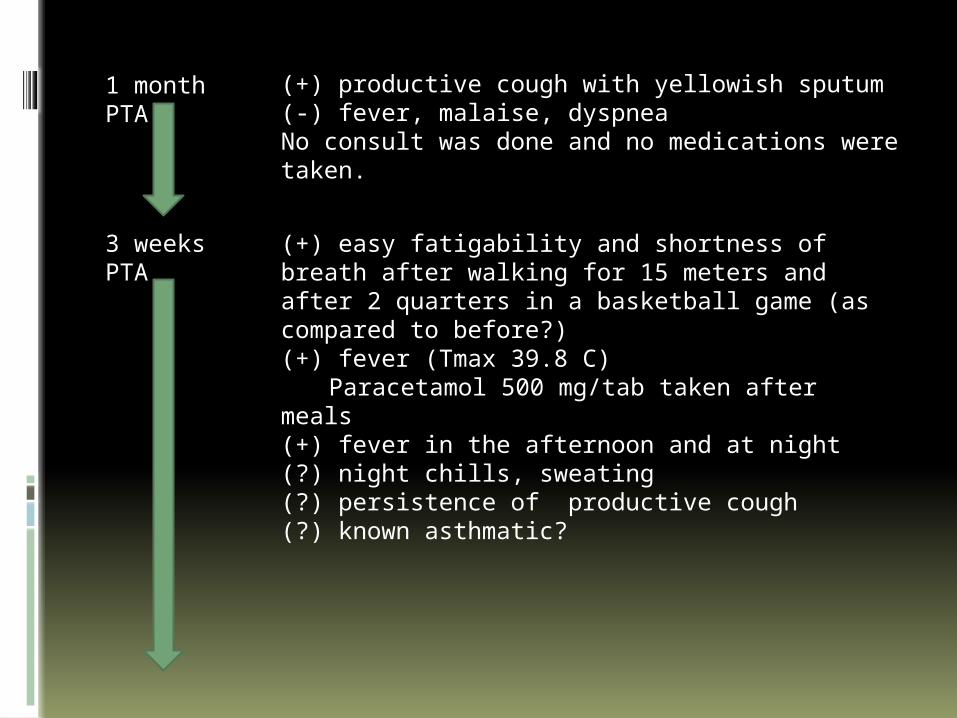
1 month PTA
(+) productive cough with yellowish sputum(-) fever, malaise, dyspneaNo consult was done and no medications were taken.
3 weeks PTA
(+) easy fatigability and shortness of breath after walking for 15 meters and after 2 quarters in a basketball game (as compared to before?)(+) fever (Tmax 39.8 C)
Paracetamol 500 mg/tab taken after meals(+) fever in the afternoon and at night(?) night chills, sweating(?) persistence of productive cough(?) known asthmatic?
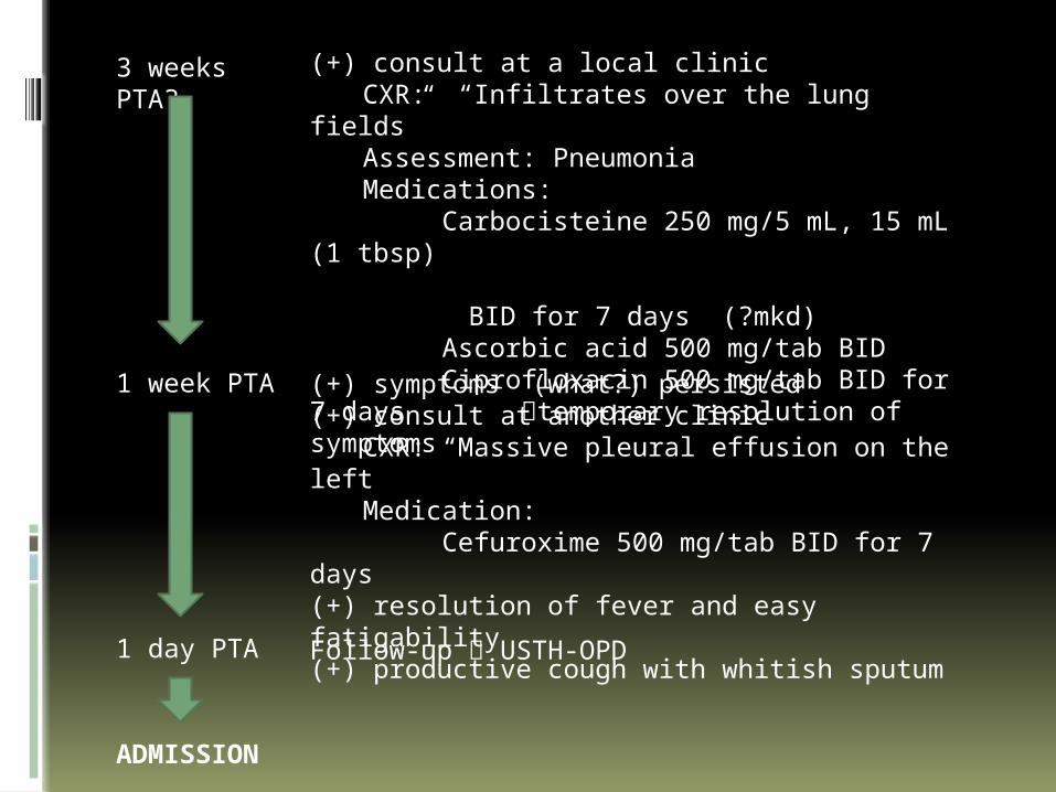
3 weeks PTA?
(+) consult at a local clinic CXR: “Infiltrates over the lung fields”Assessment: PneumoniaMedications: Carbocisteine 250 mg/5 mL, 15 mL (1
tbsp) BID for 7 days (?mkd) Ascorbic acid 500 mg/tab BID
Ciprofloxacin 500 mg/tab BID for 7 days temporary resolution of symptoms
1 week PTA (+) symptoms (what?) persisted(+) consult at another clinic
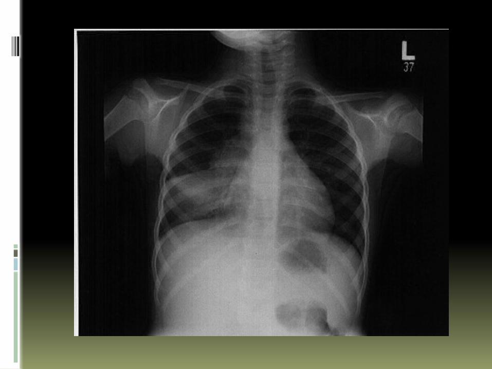
CXR: “Massive pleural effusion on the left”
Medication: Cefuroxime 500 mg/tab BID for 7 days
(+) resolution of fever and easy fatigability(+) productive cough with whitish sputum
1 day PTA Follow-up USTH-OPD
ADMISSION
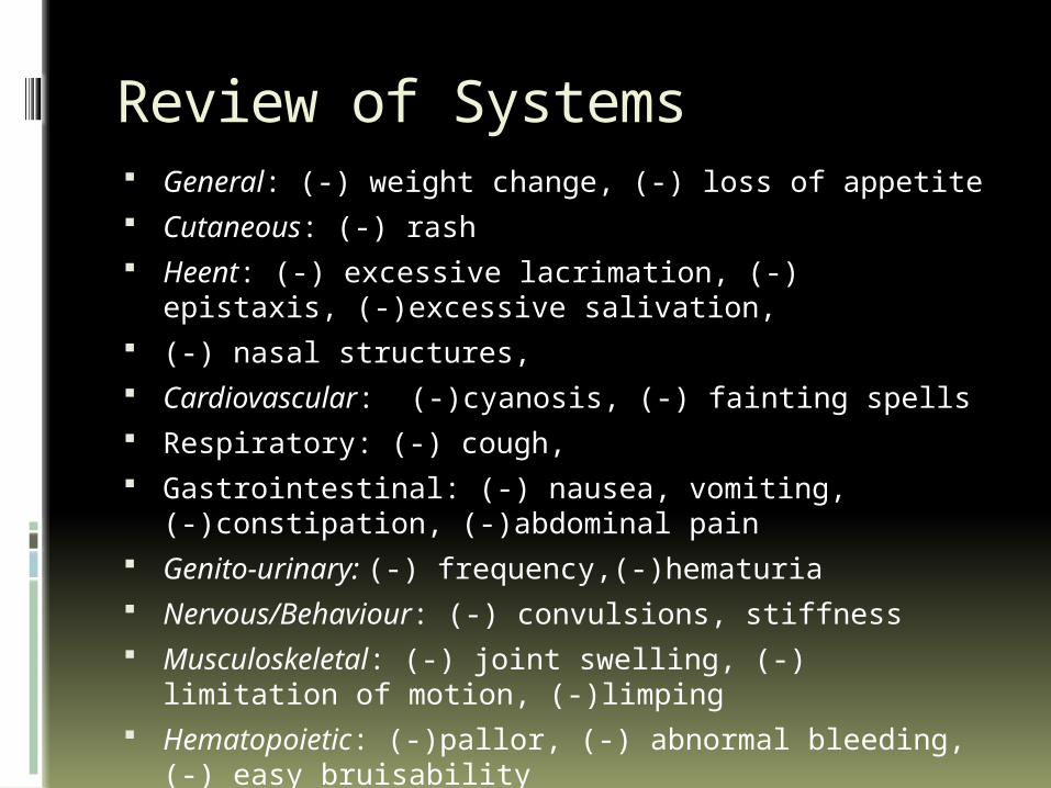
Review of Systems General: (-) weight change, (-) loss of appetite Cutaneous: (-) rash Heent: (-) excessive lacrimation, (-) epistaxis,
(-)excessive salivation, (-) nasal structures, Cardiovascular: (-)cyanosis, (-) fainting spells Respiratory: (-) cough, Gastrointestinal: (-) nausea, vomiting, (-)constipation,
(-)abdominal pain Genito-urinary: (-) frequency,(-)hematuria Nervous/Behaviour: (-) convulsions, stiffness Musculoskeletal: (-) joint swelling, (-) limitation of
motion, (-)limping Hematopoietic: (-)pallor, (-) abnormal bleeding, (-) easy
bruisability

Personal History H: Patient lives with his mother and father. At home, he
likes to watch cartoons on TV and sleep. Aside from that, he does not do anything else at home. He spends most of his free time outside playing basketball with his friends.
E: Currently in his 3rd year of high school. He prefers to play basketball than go to class or study.
E: Patient eats 3 meals a day and has no preference on the food that he eats.
A: Varsity player of the school’s basketball team; computer games
D: Patient claims that he has never smoke, drink alcohol or took illicit drugs.
S: He had 4 past girlfriends. He claimed that they had never engaged in any sexual activity.
S: Patient claims that he is very contented with his life and would never think of taking his own life.
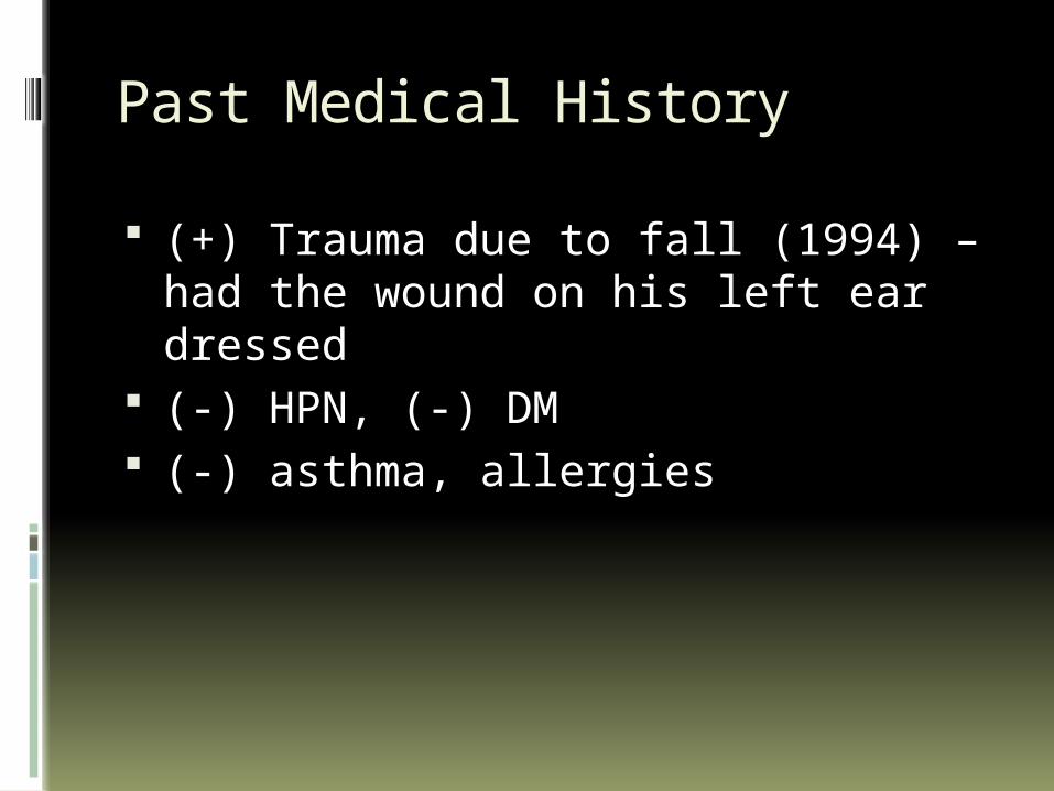
Past Medical History
(+) Trauma due to fall (1994) – had the wound on his left ear dressed
(-) HPN, (-) DM (-) asthma, allergies
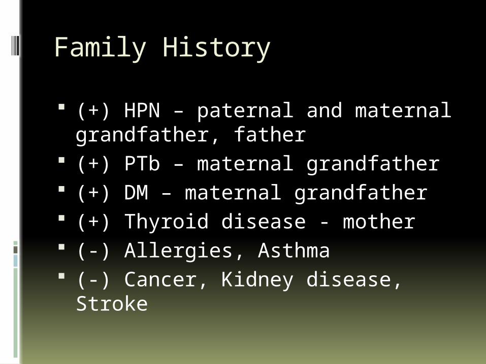
Family History
(+) HPN – paternal and maternal grandfather, father
(+) PTb – maternal grandfather (+) DM – maternal grandfather (+) Thyroid disease - mother (-) Allergies, Asthma (-) Cancer, Kidney disease, Stroke
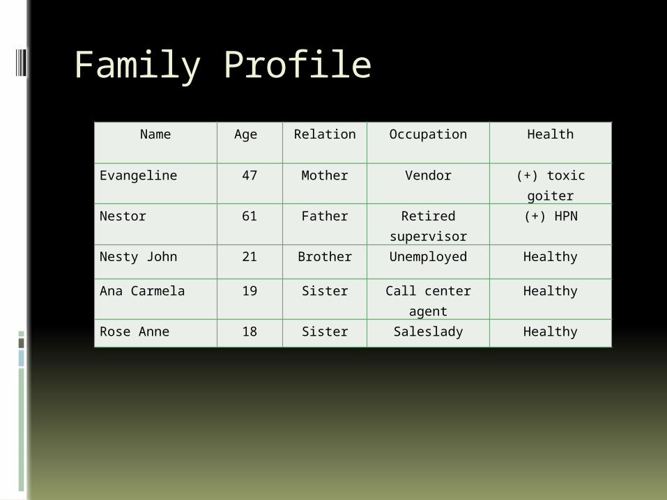
Family Profile
Name Age Relation Occupation Health
Evangeline 47 Mother Vendor (+) toxic goiter
Nestor 61 Father Retired supervisor
(+) HPN
Nesty John 21 Brother Unemployed Healthy
Ana Carmela 19 Sister Call center agent Healthy
Rose Anne 18 Sister Saleslady Healthy
Socioeconomic & Environmental History
Patient lives with his parents and stays in the same room as them. Their house is a single level cemented bungalow, well ventilated and well lit. Drinking water is obtained from a nearby water refilling station. Garbage is collected everyday by a local garbage collector.
Physical Examination
VS: BP 110/70 HR 76 bpm RR 26/min T 36.4 C Ht: 170 cm Wt: 53 kg Conscious, coherent, ambulatory, not in
cardiorespiratory distress Warm moist skin, not jaundiced, no active dermatoses Pink palpebral conjunctivae, anicteric sclera Nasal septum midline, no nasoaural discharge,
turbinates not congested No tragal tenderness, nonhyperemic EAC AU, TM intact
AUMoist buccal mucosa, nonhyperemic PPW, tonsils enlarged
Supple neck, no palpable cervical lymph nodes
Physical Examination
Asymmetric chest expansion, no retractions, trachea deviated to the right with lagging on the left, decreased vocal and tactile fremiti on the left, dullness on the left infrascapular area (T6 down), decreased breath sounds on the left upper and lower lung fields
Adynamic precordium, AB 5th LICS MCL, no murmurs
Flat abdomen, normoactive bowel sounds, soft, nontender
Pulses full and equal, no edema, no cyanosis
Neurologic Examination
Conscious, coherent, oriented to 3 spheres Pupil size 3-4 mm equally reactive to light; no
ptosis OU No facial asymmetry, (+) corneal reflex, (+) gag
reflex Symmetric palpebral fissures and nasolabial fold MMT 5/5 on all extremities No involuntary movement, no spasticity, no
atrophy No sensory deficits No nuchal rigidity, (-) Brudzinski, (-) Kernig’s

Salient Features
Differential Diagnosis
Pleural Effusion vs. Consolidation vs. Atelectasis, etc. clinically first then via CXR
Degree of Pleural Effusion (Massive, etc?)
Why suspect Pneumonia? Why suspect PTB?
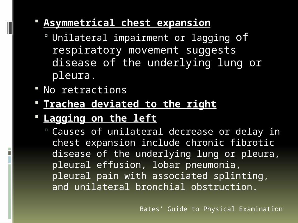
Asymmetrical chest expansion Unilateral impairment or lagging of
respiratory movement suggests disease of the underlying lung or pleura.
No retractions Trachea deviated to the right Lagging on the left
Causes of unilateral decrease or delay in chest expansion include chronic fibrotic disease of the underlying lung or pleura, pleural effusion, lobar pneumonia, pleural pain with associated splinting, and unilateral bronchial obstruction.Bates’ Guide to Physical Examination
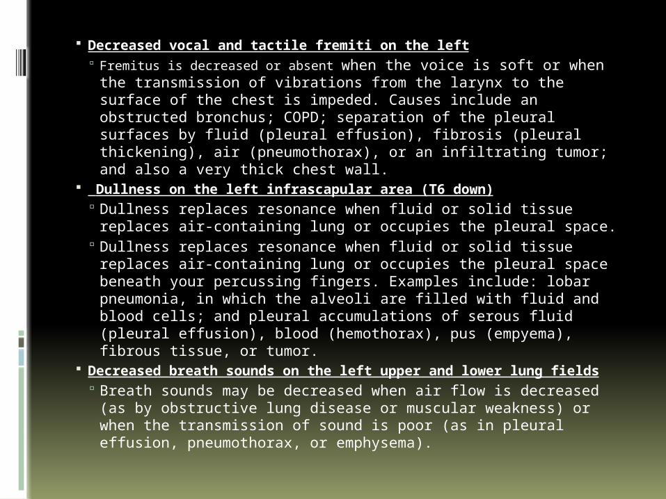
Decreased vocal and tactile fremiti on the left Fremitus is decreased or absent when the voice is soft or when the
transmission of vibrations from the larynx to the surface of the chest is impeded. Causes include an obstructed bronchus; COPD; separation of the pleural surfaces by fluid (pleural effusion), fibrosis (pleural thickening), air (pneumothorax), or an infiltrating tumor; and also a very thick chest wall.
Dullness on the left infrascapular area (T6 down) Dullness replaces resonance when fluid or solid tissue replaces
air-containing lung or occupies the pleural space. Dullness replaces resonance when fluid or solid tissue replaces
air-containing lung or occupies the pleural space beneath your percussing fingers. Examples include: lobar pneumonia, in which the alveoli are filled with fluid and blood cells; and pleural accumulations of serous fluid (pleural effusion), blood (hemothorax), pus (empyema), fibrous tissue, or tumor.
Decreased breath sounds on the left upper and lower lung fields Breath sounds may be decreased when air flow is decreased (as
by obstructive lung disease or muscular weakness) or when the transmission of sound is poor (as in pleural effusion, pneumothorax, or emphysema).
Bates’ Guide to Physical Examination
Impression
t/c Pneumonia
Pneumonia
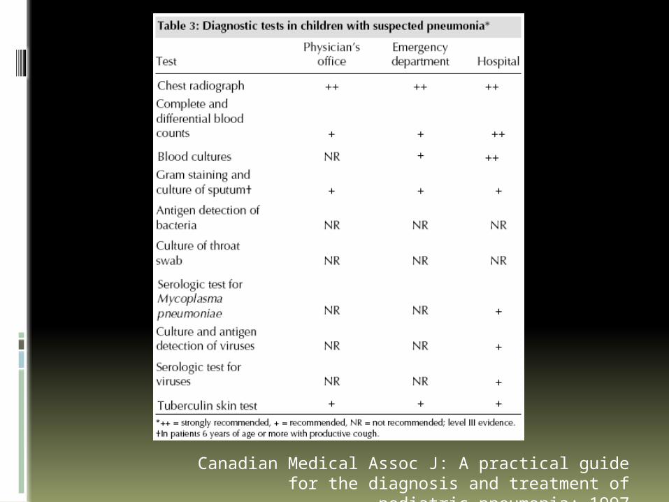
Definition Etiologies by age Criteria for Dx Criteria for confinement Ancillary procedures Expected clinical and lab findings Complications Correlate with px
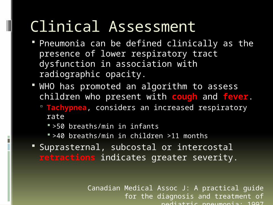
Clinical Assessment Pneumonia can be defined clinically as the
presence of lower respiratory tract dysfunction in association with radiographic opacity.
WHO has promoted an algorithm to assess children who present with cough and fever. Tachypnea, considers an increased respiratory
rate >50 breaths/min in infants >40 breaths/min in children >11 months
Suprasternal, subcostal or intercostal retractions indicates greater severity.
Canadian Medical Assoc J: A practical guide for the diagnosis and treatment of pediatric pneumonia;
1997
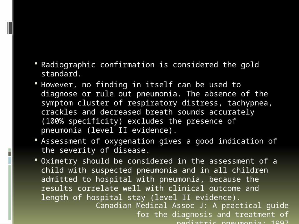
Radiographic confirmation is considered the gold standard.
However, no finding in itself can be used to diagnose or rule out pneumonia. The absence of the symptom cluster of respiratory distress, tachypnea, crackles and decreased breath sounds accurately (100% specificity) excludes the presence of pneumonia (level II evidence).
Assessment of oxygenation gives a good indication of the severity of disease.
Oximetry should be considered in the assessment of a child with suspected pneumonia and in all children admitted to hospital with pneumonia, because the results correlate well with clinical outcome and length of hospital stay (level II evidence).Canadian Medical Assoc J: A practical guide for the
diagnosis and treatment of pediatric pneumonia; 1997
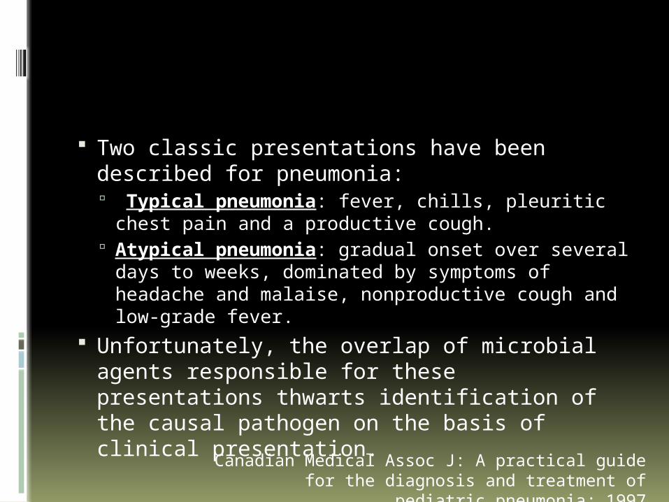
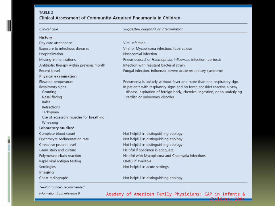
Two classic presentations have been described for pneumonia: Typical pneumonia: fever, chills, pleuritic
chest pain and a productive cough. Atypical pneumonia: gradual onset over
several days to weeks, dominated by symptoms of headache and malaise, nonproductive cough and low-grade fever.
Unfortunately, the overlap of microbial agents responsible for these presentations thwarts identification of the causal pathogen on the basis of clinical presentation.Canadian Medical Assoc J: A practical guide for the
diagnosis and treatment of pediatric pneumonia; 1997
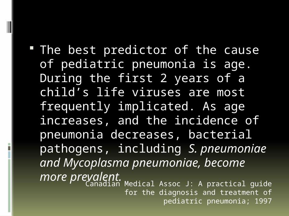
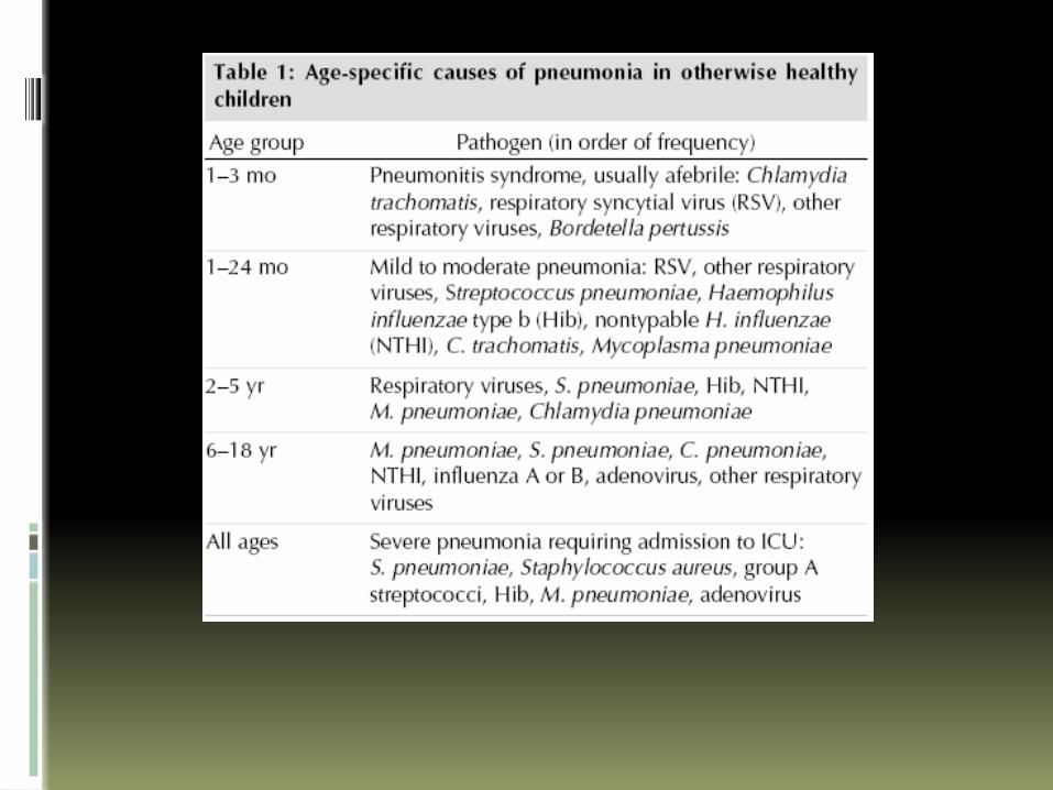
The best predictor of the cause of pediatric pneumonia is age. During the first 2 years of a child’s life viruses are most frequently implicated. As age increases, and the incidence of pneumonia decreases, bacterial pathogens, including S. pneumoniae and Mycoplasma pneumoniae, become more prevalent.Canadian Medical Assoc J: A practical guide for the
diagnosis and treatment of pediatric pneumonia; 1997
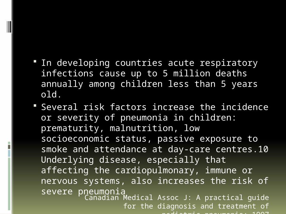
In developing countries acute respiratory infections cause up to 5 million deaths annually among children less than 5 years old.
Several risk factors increase the incidence or severity of pneumonia in children: prematurity, malnutrition, low socioeconomic status, passive exposure to smoke and attendance at day-care centres.10 Underlying disease, especially that affecting the cardiopulmonary, immune or nervous systems, also increases the risk of severe pneumonia
Canadian Medical Assoc J: A practical guide for the diagnosis and treatment of pediatric pneumonia;
1997
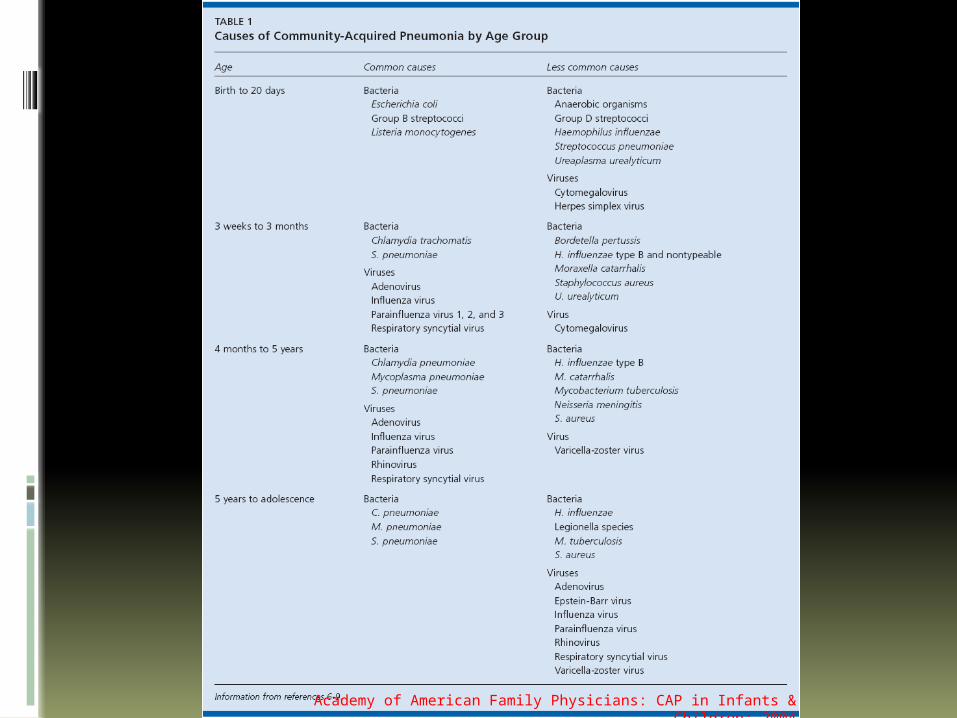
Academy of American Family Physicians: CAP in Infants & Children; 2004
Radiographic Findings
A confirmatory chest radiograph is necessary to diagnose pneumonia. Bronchiolitis and asthma may cause hyperinflation and atelectasis and must be distinguished from pneumonia.
Two main patterns of pneumonia are recognized: interstitial and alveolar. However, these patterns cannot be used to identify the cause. Peribronchial thickening, diffuse interstitial infiltrates and hyperinflation tend to be seen with viral infections (level III evidence).
Radiologic Findings
Bacterial - Lobar infiltrates, pneumatoceles, abscesses Alveolar infiltrates, however, are also seen in bacterial as
well as viral disease and in Mycoplasma pneumonia. Pneumococcal - Circular infiltrates in the early stages M. pneumoniae infection - Diffuse infiltration out of
proportion with the clinical findings, lobar consolidation, plate-like atelectasis, nodular infiltration and hilar adenopathy
Chlamydial pneumonia may be indistinguishable from mycoplasmal pneumonia.
P. carinii pneumonia - reticulonodular infiltrate that progresses to alveolar infiltrates
Tuberculosis - Hilar adenopathy especially if the patient has epidemiologic risk factors
Canadian Medical Assoc J: A practical guide for the diagnosis and treatment of pediatric pneumonia;
1997
Academy of American Family Physicians: CAP in Infants & Children; 2004

Course in the Ward

Management
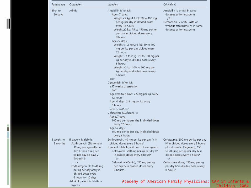
Academy of American Family Physicians: CAP in Infants & Children; 2004
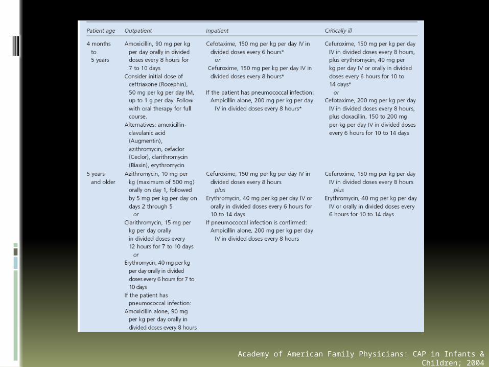
Academy of American Family Physicians: CAP in Infants & Children; 2004


![[SurgeryB] Surgical Complications - Dr. Guinto (Pacis, Sazon)](https://img.dokumen.tips/doc/110x75/55cf921d550346f57b93bae3/surgeryb-surgical-complications-dr-guinto-pacis-sazon.jpg)
