Embed Size (px)
Citation preview

Guanine, a high-capacity and rapid-turnover nitrogenreserve in microalgal cellsPeter Mojzeša,b, Lu Gaob,c
, Tatiana Ismagulovad, Jana Pilátováe, Sárka Moud�ríkováa, Olga Gorelovad,Alexei Solovchenkod,f
, Ladislav Nedbalb,1, and Anya Salihg,h
aInstitute of Physics, Faculty of Mathematics and Physics, Charles University, CZ-12116 Prague 2, Czech Republic; bInstitute of Bio- and Geosciences/PlantSciences (IBG-2), Forschungszentrum Julich, D-52428 Julich, Germany; cFaculty of Mathematics and Natural Sciences, Heinrich Heine University, D-40225Dusseldorf, Germany; dFaculty of Biology, Moscow State University, Leninskie Gori 1/12, 119234, GSP-1, Moscow, Russia; eDepartment of Experimental PlantBiology, Faculty of Science, Charles University, CZ-12844 Prague 2, Czech Republic; fFaculty of Geography and Natural Sciences, Pskov State University,180000 Pskov, Russia; gAntares Fluoresci Research, Dangar Island, NSW 1797, Australia; and hConfocal Bioimaging Facility, Western Sydney University, NSW1797, Australia
Edited by Donald R. Ort, University of Illinois at Urbana–Champaign, Urbana, IL, and approved November 4, 2020 (received for review May 3, 2020)
Nitrogen (N) is an essential macronutrient for microalgae, influencingtheir productivity, composition, and growth dynamics. Despite thedramatic consequences of N starvation, many free-living and endo-symbiotic microalgae thrive in N-poor and N-fluctuating environ-ments, giving rise to questions about the existence and nature oftheir long-term N reserves. Our understanding of these processes re-quires a unequivocal identification of the N reserves in microalgal cellsas well as their turnover kinetics and subcellular localization. Herein,we identified crystalline guanine as the enigmatic large-capacity andrapid-turnover N reserve of microalgae. The identification was unam-biguously supported by confocal Raman, fluorescence, and analyticaltransmission electron microscopies as well as stable isotope labeling.We discovered that the storing capacity for crystalline guanine by themarine dinoflagellateAmphidinium carteraewas sufficient to supportN requirements for several new generations. We determined that Nreserves were rapidly accumulated from guanine available in the en-vironment as well as biosynthesized from various N-containing nutri-ents. Storage of exogenic N in the form of crystalline guanine wasfound broadly distributed across taxonomically distant groups of micro-algae from diverse habitats, from freshwater and marine free-livingforms to endosymbiotic microalgae of reef-building corals (Acroporamillepora, Euphyllia paraancora). We propose that crystalline guanineis the elusive N depot that mitigates the negative consequences ofepisodic N shortage. Guanine (C5H5N5O) may act similarly to cyanophy-cin (C10H19N5O5) granules in cyanobacteria. Considering the phyto-plankton nitrogen pool size and dynamics, guanine is proposed to bean important storage form participating in the global N cycle.
nitrogen cycle | nutrient storage | phytoplankton | guanine | coral
Planktonic algae represent an essential driver of the globalcarbon cycle, which may be constrained by low or fluctuating
nitrogen (N) availability (1–3). At another extreme, high levels ofbioavailable N, often from anthropogenic sources, may result inharmful algal blooms (4) or deterioration of coral reefs (5). Thehighly optimized nutrient interactions with symbiotic algae arealso essential for reef corals that thrive in nutrient-poor waters andparadoxically form among the most productive and diverse eco-systems. Both high and low N availability may perturb the stabilityof individual organisms or entire ecosystems especially when notin proportion to other biogenic elements, such as phosphorus (6).N shortages trigger extensive changes in algal metabolism (7),
including cessation of cell division, reduction of photosynthesis,and accumulation of C- and energy-rich N-free compounds. Un-like cyanobacteria, eukaryotic microalgae do not possess N-richcyanophycin to manage N deficiency (8, 9). N-deprived microalgaemobilize intracellular inorganic N; low-molecular organic Ncompounds, such as polyamines, amino acids, and chlorophyll (7);and polymeric N compounds, such as proteins and nucleic acids(10). However, none of these reserves may be sufficient to bridgelong periods of N starvation. Thus, a pool capable of storing large
amounts of N during periods of abundance and ensuring survivaland growth of algae during deficiency remains to be identified.Among candidate N storage pools in microalgae, crystalline
inclusions were considered, although later, alternative roles, suchas processing of metabolic wastes (11) and light modulation (12)were also suggested. The chemical identity of crystals from thefree-living marine dinoflagellate Gonyaulax polyedra was proposedto be guanine (13). Other crystalline inclusions hypothesized ofbeing guanine were also observed in symbiotic dinoflagellates of ananemone Aiptasia sp. (14), but their chemical nature was not ex-perimentally confirmed (11, 14). Other earlier studies suggestedthe inclusions were calcium oxalate (15, 16) making the N-storagefunction of the inclusions unlikely. Subsequent analysis of symbioticdinoflagellate extracts from Aiptasia sp. identified them as crys-talline uric acid (17). This identification has since been adopted formicroalgal inclusions in many recent publications (18–21).In contrast with this proposed identity (17–21) and consistent
with earlier studies (13, 14), recent direct in situ analyses identifiedinclusions as guanine in several marine dinoflagellates (12, 22), afreshwater chlorophyte and a eustigmatophyte (23). Guanine, sim-ilar to other purines, has the potential to serve as a large-capacity Npool, but this function has never been previously confirmed, nor wasits in situ chemical identity explored in diverse algal species. Gua-nine is widespread and, thus, widely available in nature. It is anessential component of DNA and RNA, one of the end products of
Significance
Vast areas of the oceans are N limited, and how microalgae canflourish in these N-poor waters is still not known. Furthermore,mechanisms and sites of N uptake and storage have not beenfully determined. We show that crystalline guanine (C5H5N5O)is an important N storage form for phytoplankton and forsymbiotic dinoflagellates of corals. The widespread occurrenceof guanine reserves among taxonomically distant microalgalspecies suggests an early evolutionary origin of its function asN storage. Crystalline guanine appears to be a multifunctionalbiochemical with an important role in the N cycle that remainsto be elucidated. In particular, a better knowledge of N-storagemetabolism is necessary to understand the impact of eutro-phication on coral-symbiont interaction.
Author contributions: P.M., A.So., and L.N. designed research; P.M., L.G., T.I., J.P., O.G.,A.So., and A.Sa. performed research; P.M., L.G., T.I., O.G., A.So., L.N., and A.Sa. analyzeddata; and P.M., L.G., A.So., L.N., and A.Sa. wrote the paper.
The authors declare no competing interest.
This article is a PNAS Direct Submission.
Published under the PNAS license.1To whom correspondence may be addressed. Email: [email protected].
This article contains supporting information online at https://www.pnas.org/lookup/suppl/doi:10.1073/pnas.2005460117/-/DCSupplemental.
First published December 8, 2020.
32722–32730 | PNAS | December 22, 2020 | vol. 117 | no. 51 www.pnas.org/cgi/doi/10.1073/pnas.2005460117
Dow
nloa
ded
by g
uest
on
Nov
embe
r 30
, 202
1

nucleic acid degradation in some organisms, and utilized by somefor functional purposes, such as light scattering by silvery scales offish and bio-optical systems of many invertebrates (reviewed in ref.24). It is widely available from decomposing fish tissues and scales,from ciliates and some phytoplankton, barnacles, and other aquaticorganisms and forms part of suspended and dissolved organic Npools in the ocean. Along with other purines, guanine can serve as aN source for algae (reviewed in ref. 1). We propose that crystallineguanine and other purines (17) play a more prominent role in the Ncycle than recognized to date.To test this hypothesis, we used the unique potential of Raman
microscopy and analytical transmission electron microscopy (TEM)and identified the chemical nature and the dynamics of microalgalcrystal inclusions. We confirm the occurrence of crystalline guaninein free-living and symbiotic dinoflagellates and other diversemicroalgal species. Our research demonstrated widespread occur-rence, large N-storage capacity, and prominent dynamics of guaninein the form of crystalline inclusions in microalgae.
ResultsFast Kinetics of Uptake and Large Storage Capacity of IntracellularGuanine Inclusions in Amphidinium carterae. We first characterizedthe uptake of guanine by the widespread potentially toxic marinedinoflagellate A. carterae. Details of cultivation are provided in SIAppendix, section II.1A. Cells were kept in N-free medium (SIAppendix, section II.1.Aa.) for approximately 2 wk before resus-pension in a saturated solution of guanine (SI Appendix, section II.1.Ab.). We recorded a rapid uptake rate of dissolved guanine from
the medium (dashed line, Fig. 1A) and its concurrent accumulationin cells (box plot, Fig. 1A). Dynamics and localization of guanineinclusions inside cells (in pink, Fig. 1 C and D) on the backgroundof other cellular components was identified by the spectral signatureof Raman confocal microscopy (Fig. 1B) (23). The rapid accumu-lation of the optically active guanine crystals was also documentedvia polarization microscopy (SI Appendix, section II.1.D. andMovie S1).The dependence of uptake kinetics of dissolved guanine (Fig. 2
and SI Appendix, section I.2.) on cell density displayed an initiallinear uptake phase (solid lines, the coefficient of determinationRSQR ≥ 0.995) and its interpolation yielded an initial cellularuptake rate of 16 ± 4 fg (guanine)·s−1·cell−1, i.e., 6.3 ± 1.5 × 10⁷
molecules·s−1·cell−1.* Uptake kinetics (Fig. 2) were used to quan-tify intracellular guanine accumulation inside cells (SI Appendix,Fig. S2A) resulting in a maximum storage capacity of 143 ± 37 pgof crystalline guanine cell−1, that corresponded to 68 ± 17 pg(nitrogen)·cell−1. Details of these calculations are provided in SIAppendix, section I.2. The N pool created by starvation andrefeeding was significantly larger than 17–44 pg (nitrogen)·cell−1
that was previously suggested to be the total N content in A. carteraeunder stationary conditions (25). Guanine storage pools may, thus,ameliorate N deficiency that occurs sporadically in a fluctuatingenvironment.
Fig. 1. Rapid uptake of guanine by N-starved A. carterae led to the accumulation of intracellular guanine inclusions. Cells were suspended in a saturatedsolution of guanine (∼35 μM at 20 °C) in N-deficient f/2 medium at a density of 1.7 ± 0.3 × 10⁵ cells·mL−1. The dashed line in A shows the declining con-centration of guanine in the medium as measured by ultraviolet absorption (SI Appendix, section II.1.B). The simultaneous accumulation of guanine in thecells was assessed using Raman microscopy (SI Appendix, section II.1.C) and is shown in the boxplot. Raman spectra in B were used to generate the Ramanmaps in C–E that represent: (C) a typical cell after 2 wk without a N source and (D and E) cells during progressive guanine accumulation. Color legend: guanine(pink), lipids (yellow), chloroplast (green), and starch (white/gray). (Scale bar, 2 μm.) Raman maps showing separate cellular constituents in C–E are constructedas described in SI Appendix, section I.1 and presented in SI Appendix, Fig. S1.
*Numbers following the ± sign represent, in this work, standard deviation.
Mojzeš et al. PNAS | December 22, 2020 | vol. 117 | no. 51 | 32723
PLANTBIOLO
GY
BIOPH
YSICSAND
COMPU
TATIONALBIOLO
GY
Dow
nloa
ded
by g
uest
on
Nov
embe
r 30
, 202
1

Cells with new guanine reserves can, after a short lag period,resume normal cell division and growth. The exponential growthphase halted only after the reserves were once again exhausted(SI Appendix, Fig. S2B). The number of cells that grew on thesereserves was a linear function of the initially available guanine
(inset in Fig. 2). The number of cells that accumulated maximumN storage (△) increased 8.1 ± 3.1 times without N addition.The resolution of the in situ localization of guanine crystals in
A. carterae (Fig. 1) was increased via ultrastructural TEM im-aging (Fig. 3 and SI Appendix, section II.1.E.) (26, 27). Thesolubility of guanine is known to be extremely low, and pieces ofundissolved crystalline material (Fig. 3A, three white arrows)were confirmed via EDX (SI Appendix, section II.1.F.) to cor-respond to the elemental composition of a N-rich compound,likely to be guanine (Fig. 3D). A. carterae has a special organelle,the pusule, which is connected to the flagellar channel (28, 29).We found large globules in pusules (Fig. 3 B and C) and smallcrystals in vacuoles that consistently displayed typical EDX pointspectra of guanine crystals (Fig. 3 E and F). Our results wereinconclusive with respect to whether the guanine particles hadbeen taken up into the pusules by phagocytosis (30) and/or via anactive transport of guanine molecules dissolved in the mediumfrom the added crystals. Guanine globules in pusules and vacu-oles had irregular shapes, but the comparison of Raman spectra(Fig. 1B) with those in refs. 12, 31 confirmed that the moleculeswere organized in a regular anhydrous crystal structure. Wehypothesized that algae may take up guanine microcrystals and,possibly, guanine-rich marine particulate fish debris via endocy-tosis or via the pusule (SI Appendix, sections I.3 and II.1.Ac andFig. S3).
Uptake of Solid Guanine by A. carterae Involves Crystal Decompositionand Recrystallization.Guanine microparticles or guanine-rich debrismight be taken up by dinoflagellates and directly deposited insidetheir cells without first dissolving or changing the original crys-talline structure, similar to an engulfing mechanism proposed foralgal feeding on bacteria (32). In an alternative scenario, theengulfed particles may be first dissolved within pusules or vacuolesinto individual molecules. Globules of guanine may then be as-sembled at target intracellular locations by new crystallization (Figs.1 and 3). Guanine crystals may also be dissolved extracellularly,
Fig. 2. Uptake of dissolved guanine from the medium by N-starved A.carterae and the number of generations supported by accumulated reserves(inset). The rate of guanine disappearance from the medium decreased withreduced cell density (±SD): from (122 ± 31) (◇) to (93 ± 23) (□), to (61 ± 15)(◯), and to (31 ± 8) (△) × 103 cells·mL−1. The most dilute culture (△) reducedguanine concentration in the medium from 35 to ∼4 μM, revealing themaximum storage capacity of cells of 143 ± 37 (SD) pg of crystalline guaninecell−1. Inset shows the correlation between the number of cells grown onguanine reserve and the reserve size. Details of calculations for this figureare described in SI Appendix, section I.2 and Fig. S2.
Fig. 3. TEM of semithin sections (A–C) and energy-dispersive X-ray spectroscopy (EDX) (D–F) analysis of A. carterae 6 h after refeeding N-starved cultureswith guanine. Typical EDX point spectra of guanine crystals (D–F) indicating a high N content were obtained in the scanning TEM (STEM) mode from semithincell sections. Arrows point to guanine particles outside (A) and inside cells (B and C). (Scale bars, 1 μm.) Ch, chloroplast; GG, globules with microcrystallineguanine; P, pyrenoid; V, vacuole.
32724 | www.pnas.org/cgi/doi/10.1073/pnas.2005460117 Mojzeš et al.
Dow
nloa
ded
by g
uest
on
Nov
embe
r 30
, 202
1

subsequently taken up via active constitutive transport through theircell walls and reassembled into crystalline structures inside the cells.Raman microscopy offers a unique opportunity to discrimi-
nate between the uptake mechanisms using the spectral contrastbetween fully (d5) and partially (d1) deuterated guanine (Fig. 4and SI Appendix, sections I.3 and II.2.A and Fig. S7). The con-version of d5-guanine to d1-guanine requires that the moleculesare directly exposed to an aqueous environment so that the fourdeuterium atoms bound to N can be exchanged by hydrogen fromwater, leaving only deuterium bound to C8 (33). Thus, if theguanine particle remained in its original crystalline form duringthe uptake by a cell, the N-D groups of d5-guanine would havebeen protected from isotope exchange. Confirming this, a grain ofguanine crystal outside of a cell (Fig. 4B) was found to remain inthe d5-guanine form (dark blue, white arrow).The Raman spectra proved that the new reserves of intracel-
lular guanine that appeared inside the N-starved A. carterae werein the d1 form (Fig. 4, magenta). Only 30 min after the crystallined5-guanine was added to the cell suspension, both the d5-guaninegrain (dark blue, white arrow) outside the cell and the trans-formed d1-guanine globules inside the cells (magenta) werecaptured simultaneously in Fig. 4 A and B. Crystalline d1-guanine
was also found in the cells 5–12 h after feeding (Fig. 4 D and F).The large guanine globules were preferentially located at the cellcenter 5 h after N feeding (Fig. 4 C andD), and, subsequently, somein the form of smaller particles moved toward the cell periphery(Figs. 4 E and F and 3C).Our analyses suggest that the guanine microcrystals were dis-
solved outside cells and taken up as individual molecules. Al-ternately, if they were engulfed as intact microcrystals, they werethen dissolved intracellularly before reassembling into new crys-tals. In either case, the original crystalline structure was released,deuterium atoms bound to N in the d5-form were exchanged forhydrogen from water, and the d1-guanine molecules were reas-sembled into much larger inclusions in the vacuoles and/or pusules(Figs. 3 and 4).
Guanine Crystals Are Biosynthesized in A. carterae de Novo fromDiverse Exogenic N Sources. Raman spectroscopy can also easily dis-tinguish between 14N- and 15N-guanine (SI Appendix, sections I.3 andII.2.B and Fig. S8), thus, making it useful for determining the sourceof N. Feeding N-starvedA. carterae with 15N-guanine, 15N-nitrate, 15N-ammonium (Top in Fig. 5), or 15N-urea (SI Appendix, Fig. S10)restored the culture’s growth and, regardless of the N source,
Fig. 4. Uptake of guanine includes exchange of deuterium for hydrogen atoms. Bright-field images overlaid by guanine (A, C, and E) and multicomponentRaman maps (B, D, and F) of N-starved A. carterae after the addition of solid crystalline fully deuterated d5-guanine to N-depleted medium. Images collected30 min (A and B), 5 h (C and D), and 12 h (E and F) after d5-guanine addition. Data for d5-guanine and partially deuterated d1-guanine are presented in blueand magenta, respectively, in both the Raman spectrum and the images. G shows their respective Raman spectra. Other colors: yellow, neutral lipids; green,chloroplasts; white/gray, starch. (Scale bars [A–F], 2 μm.) More spectra of isotopically labeled guanine are shown in SI Appendix, Fig. S7. Raman maps showingseparate components from data represented in B, D, and F are provided in SI Appendix, Fig. S9.
Mojzeš et al. PNAS | December 22, 2020 | vol. 117 | no. 51 | 32725
PLANTBIOLO
GY
BIOPH
YSICSAND
COMPU
TATIONALBIOLO
GY
Dow
nloa
ded
by g
uest
on
Nov
embe
r 30
, 202
1

considerable amounts of crystalline 15N-guanine appeared in-side the cells during the lag phase that lasted ∼24 h (Fig. 5,Bottom). On feeding A. carterae with nitrate, ammonium, orurea, no crystalline inclusions of other purines were observed.The accumulated or biosynthesized guanine crystals were laterused to support growth until their complete disappearance inthe new stationary phase. Regardless of the chemical identity ofthe N source, the total amount of N needed to produce a newA. carterae cell, estimated from the data in Fig. 5, was 14 ± 2 pg(N)·cell−1. The calculation procedure was the same as in SIAppendix, section I.2. This amount corresponded theoreticallyto 30 ± 4 pg (guanine)·cell−1 of the presumed reserve, whichwas close to 23 ± 4 pg (guanine)·cell−1 obtained from the datapresented in the graph inset in Fig. 2.Interestingly, the intracellular guanine reserves generated by
the assimilation of nitrate (middle graph in Fig. 5), ammonium,or urea were detected largely at the periphery of cells close to thechloroplasts (Fig. 6). This was in agreement with the earlier re-sults obtained for nitrate (12). In contrast, the rapidly accumu-lated guanine was first located centrally in large globules(Fig. 4 C and D and SI Appendix, Fig. S11), which were laterpartially fragmented and moved to the cell periphery (Fig. 4E).This result obtained by Raman microscopy was confirmed withsignificantly higher spatial resolution by the combination ofTEM and EDX methods (Figs. 3 and 6). We tentatively proposethat the aforementioned differences in localization and storagedynamics indicate differences in transport and biochemicalpathways following guanine biosynthesis from nitrate, ammo-nium, and urea compared with the direct uptake of guanine.
In Situ Chemical Identification of Inclusions in Algal Species of DiverseTaxonomical Classification and from Diverse Habitats. The chemicalidentity of inclusions in 14 microalgal species listed in Table 1 wasexamined by Raman microscopy as described in SI Appendix,section II.1.C. The selected species (SI Appendix, sections I.4. andII.3 for cultivation conditions) represent contrasting habitats, suchas oligotrophic to mesotrophic marine species including both free-living (A. carterae and Microchloropsis gaditana) and coral endo-symbiotic algae (Chromera velia and Symbiodiniaceae), oligotro-phic to eutrophic freshwater (Synura petersenii andHaematococcuspluvialis, respectively), extremophilic/acidophilic (Dunaliella acid-ophila), terrestrial (Lobosphaera incisa, Vischeria sp., and Kleb-sormidium flaccidum), and algae from artificial anthropogenicenvironments (Vacuoliviride crystalliferum and K. flaccidum), their
distribution ranging from tropical/subtropical regions (A. carterae,C. velia, and Symbiodiniaceae) and temperate zones (Vischeria sp.)to the cosmopolitan species extending to the Arctic (K. flaccidum).Some of the tested species were established model organisms(Chlamydomonas reinhardtii and Microchloropsis gaditana), otherswere important production species in algal biotechnology (M.gaditana, D. acidophila, H. pluvialis, and L. incisa). The diversephylogeny among the selected species is shown in Table 1. Spectralsignatures of inclusions found in the selected algal species werecompared with the spectra of multiple purines (SI Appendix, Fig.S4) and with calcium oxalate and calcite (SI Appendix, Fig. S6).Guanine inclusions were found in 13 out of 14 tested species cul-tivated in commonly used media (SI Appendix, section II.3). Onlyone of the tested species K. flaccidum was found to contain uric acidin its inclusions. This identification was completed in situ, thus,eliminating the potential artifacts caused by extraction and chemicalanalyses. Differences between Raman spectral signatures of gua-nine and uric acid are large (SI Appendix, Fig. S4), enabling accuratediscrimination needed in light of alternatives proposed in recentliterature (12, 17, 23). We cannot rule out, however, that underspecific cultivation conditions, stress factors, or feeding by otherorganic nutrients, crystalline inclusions of other purines may not bepresent. Furthermore, the abundance of guanine inclusions depen-ded not only on the availability of N nutrients (Fig. 1 C–E), but alsoon cultivation factors, such as CO2 availability for Desmodesmusquadricauda (23) or on the cell cycle phase in C. reinhardtii (SIAppendix, section I.4 and Fig. S12). We also cannot exclude possibletransformation of different purine forms with some perhaps occur-ring transiently or even simultaneously. Nevertheless, our experi-ments reliably confirm that guanine is the dominant N-storage formin the investigated microalgal species, and uric acid is found only inK. flaccidum, a single representative of the Streptophyta lineage.However, many more species of this lineage must be tested in thefuture to conclude that the Streptophyta lineage deviates from othertaxonomical groups.
Coral Symbiotic Microalgae Accumulate and Store Guanine. Of par-ticular interest in relation to guanine cell storage are the photo-synthetic Symbiodiniaceae dinoflagellates (zooxanthellae) that livein mutualistic symbiosis with reef-building corals. The finely tunedexchange of nutrients between the coral host and the symbiontsforms the foundation of healthy coral reef ecosystems (5, 21, 34,35). However, much remains poorly understood regarding themechanisms of nutrient uptake and storage that allow corals to
Fig. 5. N in guanine inclusions originated directly from the supplied guanine, nitrate, and ammonium. A. carterae cell density stagnated in controls withoutN feeding (black lines) and divided after addition of 15N-labeled guanine, nitrate, and ammonium (all at 0.882-mM N). Intracellular crystalline guanine per cellis shown in the bottom graphs representing Raman measurements (n = 5–12 cells). The corresponding graph representing uptake of urea is shown inSI Appendix, Fig. S10.
32726 | www.pnas.org/cgi/doi/10.1073/pnas.2005460117 Mojzeš et al.
Dow
nloa
ded
by g
uest
on
Nov
embe
r 30
, 202
1

survive in the N-poor waters of tropical seas or cope with pulses ofexcessive nutrients due to upwelling or rainfall (5, 34, 35).We demonstrated in this study that the numerous crystalline
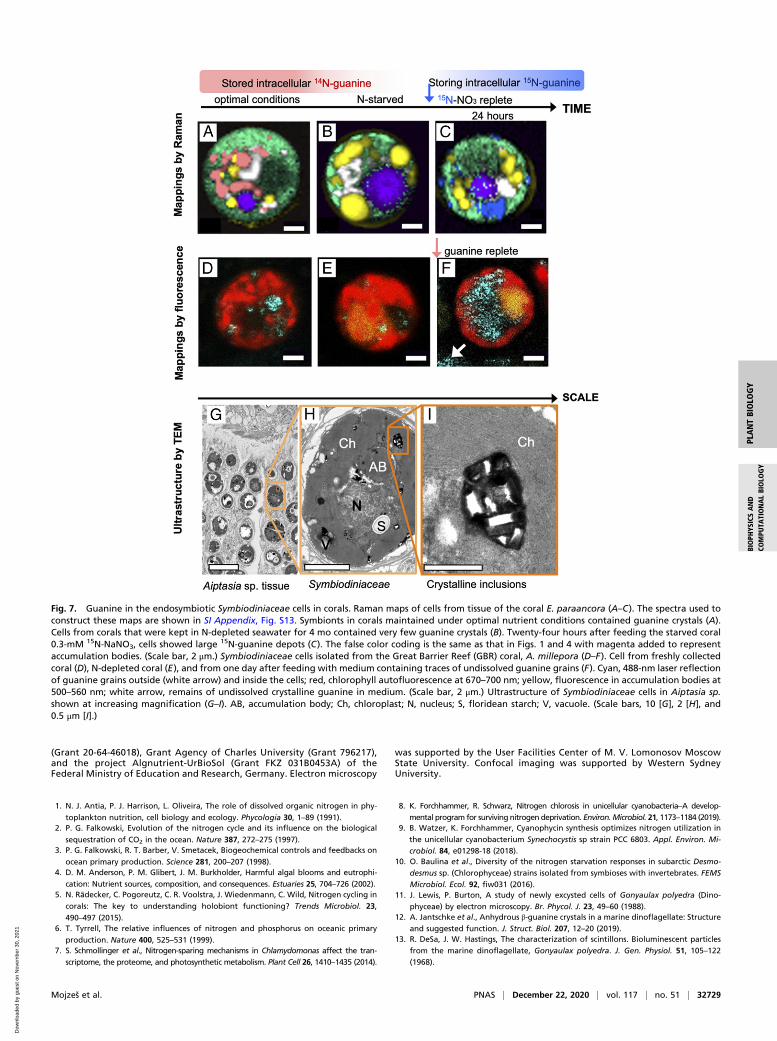
inclusions of endosymbiotic Symbiodiniaceae consist of guanineand that the external guanine is rapidly assimilated into thesymbionts (Fig. 7 and SI Appendix, section I.5.). According toRaman microscopy (SI Appendix, Fig. S14), Symbiodiniaceaecells in the tissue of the scleractinian coral Euphyllia paraancora(SI Appendix, sections I.5 and II.4.A) exhibited similar guaninepool dynamics (Fig. 7 A–C) as the free-living dinoflagellate A.carterae (Figs. 1–4). Symbiodiniaceae from corals grown underoptimal conditions always contained large guanine reserves(Fig. 7A). After 4 mo of N starvation of corals, guanine reserveswere found depleted (Fig. 7B), the polyps shrank and becamepartially bleached. Upon refeeding with 15N-NaNO3, the newlysynthesized 15N-guanine inclusions appeared within 24 h insideSymbiodiniaceae cells (Fig. 7C and SI Appendix, Fig. S14).
The dynamic uptake of guanine was further confirmed in theSymbiodiniaceae-hosting coral Acropora millepora, which is wide-spread on the GBR, Australia, by using a combination of confocalfluorescence and reflection imaging (Fig. 7 D–F and SI Appendix,sections I.6 and II.1.C). A. millepora that was freshly collectedfrom the GBR (Marine Parks Authority Permit G17/39943.1 toA.Sa., method in SI Appendix, section II.4.B) contained varyingquantities of guanine crystals scattered peripherally among chlo-roplast lobes (Fig. 7D and SI Appendix, Fig. S15). Prolonged Nstarvation resulted in complete depletion of the guanine reserves(Fig. 7E). When the N-starved Symbiodiniaceae cells isolated fromA. millepora were fed by a small amount of guanine powder addeddirectly to the medium, the highly reflective guanine particles thatwere first visualized outside Symbiodiniaceae cells, began to ac-cumulate inside cells after ∼1 h (Fig. 7F). A similar direct uptakeof guanine was observed in Symbiodiniaceae extracted from thezoanthid Zoanthus sp. (SI Appendix, Fig. S16, Bottom) and in free-living A. carterae (in SI Appendix, Fig. S16, Top and Movie S1).The same dynamics of appearance and disappearance of guanine
inclusions were observed for Symbiodiniaceae of four other cnidarianspecies—the anemone Aiptasia sp., corallimorpharian, Rhodactisindosinensis, leather soft coral, Sinularia asterolobata, and Zoanthussp., cultivated in an experimental aquarium (method in SI Appendix,section II.4.A). This is an in situ identification of the crystallineguanine within the intact symbiotic zooxanthellae from multipleanthozoan species.
DiscussionGuanine (C5H5N5O) holds 1.9-fold higher amounts of N per unitmolecular weight than a monomer of N-storing cyanophycin(C10H19N5O5), known to be the long-term N reserve in cyano-bacteria. The ratio is even higher when the hydration of cyano-phycin is considered. As it is uncharged and almost insoluble atphysiological pH, crystalline guanine is less metabolically activethan various ionic N-containing compounds, enabling its accu-mulation in large quantities and its long-term storage inside cellswithout the risk of metabolic disorder. In comparison with otherpurines, guanine is one order of magnitude less soluble than uricacid or xanthine and two orders of magnitude less soluble thanadenine or hypoxanthine. It is also more chemically stable thanuric acid (1). Yet guanine can be easily mobilized from the solidstate by changing pH of its aqueous environment. Although al-most insoluble at neutral pH, its solubility in water increasesgreatly at acidic or basic pH (31). Algal metabolic activity ishighly pH dependent: photosynthesis increases the pH duringthe day, respiration decreases it at night, and a variety of cellularpH controls alter it in response to environment and stress (36).Thus, it is possible that guanine’s solubility is under cellular pHcontrol, facilitating its transport and assimilation. In coral-algalsymbiosis, pH influences the flow of N between the host and itssymbionts (35) and guanine’s pH solubility dependence may makeit a perfect metabolic N-storage molecule. Importantly, our find-ing that the size of the guanine reserve in planktonic microalgaecan be much higher than the N amount required for cell repro-duction indicates that it is a major N pool of global importance,being proportional to the phytoplankton biomass and matching itscontributions to global carbon and N cycles (2, 37).Biogenic guanine forms a highly compact crystal structure,
namely, the β-form of the anhydrous monoclinic polymorph,consisting of vertically stacked planes of hydrogen-bonded mol-ecules (12, 38). This crystal structure explains its unique opticalproperties, including birefringence and an extremely high index ofrefraction, leading to high light scattering as recorded in our confocalreflective imaging analysis. Consequently, guanine’s storage functiondoes not exclude other possible functions, e.g., light scattering inmicroalgae to enhance the efficiency of photosynthesis, photo-protection from UV radiation, or the formation of photonic mirrors(12, 38).
Fig. 6. Localization of guanine inclusions in A. carterae fed by nitrate byTEM. Bright-field images overlaid by guanine (pink) (A) and multicomponentRaman maps (B) of A. carterae cells 24 h after refeeding N-starved cells withnitrate. False color coding is the same as that in Fig. 1. (Scale bar, 2 μm.) TEMof ultrathin (C–E) and semithin (F) sections as well as EDX (G); (F and G)analysis of A. carterae cells and surrounding cells 26 h after refeedingN-starved cultures with nitrate. Typical EDX point spectra of guanine crystals(G) indicating high N content were obtained in the STEM mode fromsemithin cell sections. Arrows point to guanine crystals. (Scale bars, 2.5 [C],1 [D and E], and 0.5 μm [F]. Ch, chloroplast; V, vacuole.)
Mojzeš et al. PNAS | December 22, 2020 | vol. 117 | no. 51 | 32727
PLANTBIOLO
GY
BIOPH
YSICSAND
COMPU
TATIONALBIOLO
GY
Dow
nloa
ded
by g
uest
on
Nov
embe
r 30
, 202
1

N is limiting to phytoplankton primary productivity in manymarine ecosystems and is often associated with sporadic or sea-sonal reintroduction from deeper waters by mixing via upwelling orstorms, from organic matter remineralization and from land-basedsources (1–3). N stimulates phytoplankton growth, and the con-nection of N metabolism to photosynthesis has long been recog-nized (1, 6). The ability to assimilate N from nitrate, ammonium,urea, or guanine dissolved in seawater, or, alternatively, from par-ticulate N sources, and to rapidly sequester N as crystalline guaninefor redeployment under conditions of N limitation has emerged asan important survival strategy of free-living phytoplankton algae. Nstorage is also a critical component of cnidarian-dinoflagellatesymbiosis. Natural or anthropogenic N eutrophication is known todisrupt the N-limited state of coral symbionts, disturbing the host’scontrol over them thereby exacerbating the damage following masscoral bleaching (5, 19). By locking excessive N in insoluble crystalsand mobilizing them when required, symbiotic microalgae may ef-fectively mitigate the negative effect of N excesses or deprivationsand maintain stable nutrient stoichiometry (C:N and N:P ratios).Given the major nutritional role of the symbionts to corals andmany other reef animals and the dependence of the reef ecosys-tem’s health on efficient nutrient uptake and storage, our researchaddressed a critical knowledge gap regarding the mechanisms bywhich corals acquire and store inorganic nutrients. Such knowledgeis increasingly important under escalating eutrophication and cli-mate-induced warming of marine and freshwater ecosystems.N storage and other diverse vital biological and biochemical
functions underscore the versatility of crystalline guanine con-cerning symbiosis and phytoplankton dynamics. Its shared occur-rence among microalgal species over the phylogenetic tree suggeststhe involvement of guanine in these roles early in evolution, ahypothesis consistent with the potential role of this purine close tothe origins of life and its presumably prebiotic occurrence on earlyEarth (39). Analogous to polyphosphate, which was regarded as amolecular fossil (40) and was, subsequently, revealed to have amultitude of functions (41), the crystalline guanine can also beconsidered as an evolutionary old, overlooked, and forgottenmultifunctional tool of nature popping up from oblivion.
Materials and MethodsChemicals and Media. References for chemicals and protocols used for pre-paring cultivation media and stable isotope labeling are listed or described indetail in SI Appendix, section II.
Algal Strains, Corals, and Cultivation Protocols. The origins and cultivationconditions for the microalgal species in Table 1 are listed in SI Appendix, sec-tions I.4. and II.3. Cnidarian species anemone, Aiptasia sp., corallimorpharian,R. indosinensis, scleractinian coral, E. paraancora, leather coral, S. asterolobata,and anthozoan Zoanthus sp., were purchased from a local marine aquariumshop. A. millepora was collected from the GBR, Australia, and studied imme-diately after collection or as explants cultivated in experimental aquaria. De-tails of the laboratory cultivation and methods to study the kinetics of guanineassimilation and turnover are provided in SI Appendix, sections I, II.1.A, II.3,and II.4.
Confocal Raman Microscopy. The samples for Raman measurements wereprepared and treated according to the methodology described in detailelsewhere (23, 42, 43) and summarized in SI Appendix, section II.1.C. Theinverted Raman microscope LabRam Evolution (Horiba Scientific, Long-jumeau, France) and upright Raman microscope WITec alpha 300 RSA(WITec, Ulm, Germany) were used with laser excitation at 532 nm in thestudy. To remove interference by autofluorescence of chlorophyll, wide-arealow-power photobleaching of entire cells using a defocused 532-nm laserbeam was employed before mapping. No differences that would affect datainterpretation were observed between measurements on the Horiba andWITec systems.
Confocal Reflection Microscopy. Symbiodiniaceae cells of A. carterae andZoanthus sp. confirmed by Raman microscopy to include guanine crystalswere concurrently imaged using a confocal fluorescence microscope LeicaTCS SP8 (Leica Microsystems, Germany) in the reflection mode using laserexcitation at 488 nm. The guanine inclusions were seen in the reflection ascrystal-like highly light-scattering objects. Further details are provided inSI Appendix, sections I.6, II.1.C, and II.4.
Analytical Electron Microscopy. The protocol of specimen preparation for TEMis described in SI Appendix, section II.1.E. Ultrathin sections were cut with aLKB-8800 (LKB, Sweden) ultratome, stained with lead citrate according tothe method described by Reynolds (44) and examined under a JEM-1011(JEOL, Tokyo, Japan) electron microscope. Samples for nanoscale elementalanalysis using analytical TEM with EDX were fixed, dehydrated, and em-bedded as above, except that sections were stained with uranyl acetate andlead citrate. Semithin sections were examined under a JEM-2100 (JEOL,Japan) electron microscope. Point EDX spectra were recorded using a JEOLbright-field STEM module and an X-Max X-ray detector system (Oxford In-struments, United Kingdom). Further details are provided in SI Appendix,sections II.1.E and II.1.F.
Data Availability. All study data are included in the article and supportinginformation.
ACKNOWLEDGMENTS. This study received financial support from theCzech Science Foundation (Grant 17-06264S), Russian Science Foundation
Table 1. Microcrystalline purines identified by Raman microscopy in various algal strains
Species Habitat Phylogeny Purine
Symbiodiniaceae E-S Alveolata–Dinoflagellata GuanineAmphidinium carterae S Alveolata–Dinoflagellata GuanineChromera velia S Alveolata–Chromerida GuanineMicrochloropsis gaditana S Stramenopiles–Eustigmatophyceae GuanineVacuoliviridecrystalliferum U Stramenopiles–Eustigmatophyceae GuanineVischeria sp. T Stramenopiles–Eustigmatophyceae GuanineTrachydiscus minutus F Stramenopiles–Eustigmatophyceae GuanineSynura petersenii F Stramenopiles–Chrysophyceae GuanineLobosphaera incisa F, T Archaeplastida–Chlorophyta–Trebouxiophyceae GuanineDesmodesmus quadricauda F Archaeplastida–Chlorophyta–Chlorophyceae GuanineChlamydomonas reinhardtii F Archaeplastida–Chlorophyta–Chlorophyceae GuanineDunaliella acidophila Acid Archaeplastida–Chlorophyta–Chlorophyceae GuanineHaematococcus pluvialis F Archaeplastida–Chlorophyta–Chlorophyceae GuanineKlebsormidium flaccidum T Archaeplastida–Streptophyta–Klebsormidiophyceae Uric acid
The screened species represent diverse habitats: Acid, acidophilic; E, endosymbiotic; F, freshwater; S, marine;T, terrestrial/aerophytic; U, unspecified. The origins of the examined species as well as cultivation approach aredescribed in SI Appendix, sections I.4 and II.3.
32728 | www.pnas.org/cgi/doi/10.1073/pnas.2005460117 Mojzeš et al.
Dow
nloa
ded
by g
uest
on
Nov
embe
r 30
, 202
1
(Grant 20-64-46018), Grant Agency of Charles University (Grant 796217),and the project Algnutrient-UrBioSol (Grant FKZ 031B0453A) of theFederal Ministry of Education and Research, Germany. Electron microscopy
was supported by the User Facilities Center of M. V. Lomonosov MoscowState University. Confocal imaging was supported by Western SydneyUniversity.
1. N. J. Antia, P. J. Harrison, L. Oliveira, The role of dissolved organic nitrogen in phy-
toplankton nutrition, cell biology and ecology. Phycologia 30, 1–89 (1991).2. P. G. Falkowski, Evolution of the nitrogen cycle and its influence on the biological
sequestration of CO2 in the ocean. Nature 387, 272–275 (1997).3. P. G. Falkowski, R. T. Barber, V. Smetacek, Biogeochemical controls and feedbacks on
ocean primary production. Science 281, 200–207 (1998).4. D. M. Anderson, P. M. Glibert, J. M. Burkholder, Harmful algal blooms and eutrophi-
cation: Nutrient sources, composition, and consequences. Estuaries 25, 704–726 (2002).5. N. Rädecker, C. Pogoreutz, C. R. Voolstra, J. Wiedenmann, C. Wild, Nitrogen cycling in
corals: The key to understanding holobiont functioning? Trends Microbiol. 23,
490–497 (2015).6. T. Tyrrell, The relative influences of nitrogen and phosphorus on oceanic primary
production. Nature 400, 525–531 (1999).7. S. Schmollinger et al., Nitrogen-sparing mechanisms in Chlamydomonas affect the tran-
scriptome, the proteome, and photosynthetic metabolism. Plant Cell 26, 1410–1435 (2014).
8. K. Forchhammer, R. Schwarz, Nitrogen chlorosis in unicellular cyanobacteria–A develop-
mental program for surviving nitrogen deprivation. Environ.Microbiol. 21, 1173–1184 (2019).9. B. Watzer, K. Forchhammer, Cyanophycin synthesis optimizes nitrogen utilization in
the unicellular cyanobacterium Synechocystis sp strain PCC 6803. Appl. Environ. Mi-
crobiol. 84, e01298-18 (2018).10. O. Baulina et al., Diversity of the nitrogen starvation responses in subarctic Desmo-
desmus sp. (Chlorophyceae) strains isolated from symbioses with invertebrates. FEMS
Microbiol. Ecol. 92, fiw031 (2016).11. J. Lewis, P. Burton, A study of newly excysted cells of Gonyaulax polyedra (Dino-
phyceae) by electron microscopy. Br. Phycol. J. 23, 49–60 (1988).12. A. Jantschke et al., Anhydrous β-guanine crystals in a marine dinoflagellate: Structure
and suggested function. J. Struct. Biol. 207, 12–20 (2019).13. R. DeSa, J. W. Hastings, The characterization of scintillons. Bioluminescent particles
from the marine dinoflagellate, Gonyaulax polyedra. J. Gen. Physiol. 51, 105–122
(1968).
Fig. 7. Guanine in the endosymbiotic Symbiodiniaceae cells in corals. Raman maps of cells from tissue of the coral E. paraancora (A–C). The spectra used toconstruct these maps are shown in SI Appendix, Fig. S13. Symbionts in corals maintained under optimal nutrient conditions contained guanine crystals (A).Cells from corals that were kept in N-depleted seawater for 4 mo contained very few guanine crystals (B). Twenty-four hours after feeding the starved coral0.3-mM 15N-NaNO3, cells showed large 15N-guanine depots (C). The false color coding is the same as that in Figs. 1 and 4 with magenta added to representaccumulation bodies. (Scale bar, 2 μm.) Symbiodiniaceae cells isolated from the Great Barrier Reef (GBR) coral, A. millepora (D–F). Cell from freshly collectedcoral (D), N-depleted coral (E), and from one day after feeding with medium containing traces of undissolved guanine grains (F). Cyan, 488-nm laser reflectionof guanine grains outside (white arrow) and inside the cells; red, chlorophyll autofluorescence at 670–700 nm; yellow, fluorescence in accumulation bodies at500–560 nm; white arrow, remains of undissolved crystalline guanine in medium. (Scale bar, 2 μm.) Ultrastructure of Symbiodiniaceae cells in Aiptasia sp.shown at increasing magnification (G–I). AB, accumulation body; Ch, chloroplast; N, nucleus; S, floridean starch; V, vacuole. (Scale bars, 10 [G], 2 [H], and0.5 μm [I].)
Mojzeš et al. PNAS | December 22, 2020 | vol. 117 | no. 51 | 32729
PLANTBIOLO
GY
BIOPH
YSICSAND
COMPU
TATIONALBIOLO
GY
Dow
nloa
ded
by g
uest
on
Nov
embe
r 30
, 202
1

14. K. B. Strychar, P. W. Sammarco, T. J. Piva, Apoptotic and necrotic stages of Symbio-dinium (Dinophyceae) cell death activity: Bleaching of soft and scleractinian corals.Phycologia 43, 768–777 (2004).
15. D. L. Taylor, In situ studies on cytochemistry and ultrastructure of a symbiotic marinedinoflagellate. J. Mar. Biol. Assoc. U. K. 48, 349–366 (1968).
16. M. J. Kevin, W. T. Hall, J. J. McLaughlin, P. A. Zahl, Symbiodinium microadriaticumFreudenthal, a revised taxonomic description, ultrastructure. J. Phycol. 5, 341–350(1969).
17. P. L. Clode, M. Saunders, G. Maker, M. Ludwig, C. A. Atkins, Uric acid deposits insymbiotic marine algae. Plant Cell Environ. 32, 170–177 (2009).
18. T. Krueger et al., Temperature and feeding induce tissue level changes in autotrophicand heterotrophic nutrient allocation in the coral symbiosis–A NanoSIMS study. Sci.Rep. 8, 12710 (2018).
19. S. Rosset, J. Wiedenmann, A. J. Reed, C. D’Angelo, Phosphate deficiency promotescoral bleaching and is reflected by the ultrastructure of symbiotic dinoflagellates.Mar. Pollut. Bull. 118, 180–187 (2017).
20. H. Yamashita, A. Kobiyama, K. Koike, Do uric acid deposits in zooxanthellae functionas eye-spots? PLoS One 4, e6303 (2009).
21. C. Kopp et al., Highly dynamic cellular-level response of symbiotic coral to a suddenincrease in environmental nitrogen. MBio 4, e00052–e13 (2013).
22. A. Jantschke, I. Pinkas, A. Schertel, L. Addadi, S. Weiner, Biomineralization pathwaysin calcifying dinoflagellates: Uptake, storage in MgCaP-rich bodies and formation ofthe shell. Acta Biomater. 102, 427–439 (2020).
23. S. Moud�ríková, L. Nedbal, A. Solovchenko, P. Mojzeš, Raman microscopy shows thatnitrogen-rich cellular inclusions in microalgae are microcrystalline guanine. Algal Res.23, 216–222 (2017).
24. D. Gur, B. A. Palmer, S. Weiner, L. Addadi, Light manipulation by guanine crystals inorganisms: Biogenic scatterers, mirrors, multilayer reflectors and photonic crystals.Adv. Funct. Mater. 27, 1603514 (2017).
25. S. Menden-Deuer, E. J. Lessard, Carbon to volume relationships for dinoflagellates,diatoms, and other protist plankton. Limnol. Oceanogr. 45, 569–579 (2000).
26. A. Shebanova et al., Versatility of the green microalga cell vacuole function as re-vealed by analytical transmission electron microscopy. Protoplasma 254, 1323–1340(2017).
27. T. Ismagulova, A. Shebanova, O. Gorelova, O. Baulina, A. Solovchenko, A new simplemethod for quantification and locating P and N reserves in microalgal cells based onenergy-filtered transmission electron microscopy (EFTEM) elemental maps. PLoS One13, e0208830 (2018).
28. R. E. Schmitter, The fine structure of Gonyaulax polyedra, a bioluminescent marine
dinoflagellate. J. Cell Sci. 9, 147–173 (1971).29. J. D. Dodge, Ultrastructure of dinoflagellate pusule - unique osmo-regulatory or-
ganelle. Protoplasma 75, 285–302 (1972).30. R. Onuma, T. Horiguchi, Morphological transition in kleptochloroplasts after inges-
tion in the dinoflagellates Amphidinium poecilochroum and Gymnodinium aerugi-
nosum (Dinophyceae). Protist 164, 622–642 (2013).31. D. Gur et al., Guanine crystallization in aqueous solutions enables control over crystal
size and polymorphism. Cryst. Growth Des. 16, 4975–4980 (2016).32. H. J. Jeong et al., Heterotrophic feeding as a newly identified survival strategy of the
dinoflagellate Symbiodinium. Proc. Natl. Acad. Sci. U.S.A. 109, 12604–12609 (2012).33. J. M. Delabar, M. Majoube, Infrared and Raman-spectroscopic study of N-15 and
D-substituted guanines. Spectrochim. Acta A 34, 129–140 (1978).34. L. Muscatine, J. W. Porter, Reef corals: Mutualistic symbioses adapted to nutrient-
poor environments. BioSci. 27, 454–460 (1977).35. D. Yellowlees, T. A. V. Rees, W. Leggat, Metabolic interactions between algal sym-
bionts and invertebrate hosts. Plant Cell Environ. 31, 679–694 (2008).36. K. L. Barott, A. A. Venn, S. O. Perez, S. Tambutté, M. Tresguerres, Coral host cells
acidify symbiotic algal microenvironment to promote photosynthesis. Proc. Natl.
Acad. Sci. U.S.A. 112, 607–612 (2015).37. P. G. Falkowski, The role of phytoplankton photosynthesis in global biogeochemical
cycles. Photosynth. Res. 39, 235–258 (1994).38. A. Hirsch et al., “Guanigma”: The revised structure of biogenic anhydrous guanine.
Chem. Mater. 27, 8289–8297 (2015).39. N. Kitadai, S. Maruyama, Origins of building blocks of life: A review. Geoscience
Frontiers 9, 1117–1153 (2018).40. A. Kornberg, N. N. Rao, D. Ault-Riché, Inorganic polyphosphate: A molecule of many
functions. Annu. Rev. Biochem. 68, 89–125 (1999).41. L. Xie, U. Jakob, Inorganic polyphosphate, a multifunctional polyanionic protein
scaffold. J. Biol. Chem. 294, 2180–2190 (2019).42. S. Moud�ríková et al., Raman and fluorescence microscopy sensing energy-transducing
and energy-storing structures in microalgae. Algal Res. 16, 224–232 (2016).43. S. Moud�ríková et al., Quantification of polyphosphate in microalgae by Raman mi-
croscopy and by a reference enzymatic assay. Anal. Chem. 89, 12006–12013 (2017).44. E. S. Reynolds, The use of lead citrate at high pH as an electron-opaque stain in
electron microscopy. J. Cell Biol. 17, 208–212 (1963).
32730 | www.pnas.org/cgi/doi/10.1073/pnas.2005460117 Mojzeš et al.
Dow
nloa
ded
by g
uest
on
Nov
embe
r 30
, 202
1