Embed Size (px)
Citation preview

16-1
Dr. Lloyd Greene
Professor of Pathology (in the Center for Neurobiology and Behavior)
SUMMARY: Orderly development requires extensive coordination and communication between cells.
Much of this is provided by extracellular proteins (growth factors) that positively and negatively regulate
proliferation, differentiation, migration/pathfinding and survival/death. This is achieved via transmembrane
receptors that transduce growth factor binding to a cascade of intracellular signaling events that culminate in both
transcription-independent and transcription-dependent changes in target cell behavior. A number of growth
factor superfamilies have been recognized along with their specific transmembrane receptors. Several specific
examples are presented to illustrate the many roles of growth factors in promoting development. These reveal
the complexity of feedback mechanisms by which growth factors coordinate embryogenesis. There are several
distinct types of mechanisms by which growth factor signaling is accomplished.
LEARNING OBJECTIVES:
You should be able to:
1) Discuss the various families of growth factors and their signaling mechanisms.
2) Discuss the general roles of growth factors in regulating embryogenesis.
3) Provide specific examples of developmental actions of growth factors.
4) Appreciate the complexity of growth factor signaling mechanisms that govern development.
NOTE: The original images can be seen in a separate file on the HD web site.
LECTURE NOTES:
I. What are growth factors and what do they do?
Few of the cellular phenomena that characterize development - proliferation, differentiation,
migration/pathfinding and survival/death – are cell autonomous. That is, regulatory molecules that reach
cells by an extracellular route promote and guide virtually every step of embryogenesis. There are on
the order of several hundred genes whose products communicate with cells from the extracellular space
and that influence intracellular events by binding to specific transmembrane receptors that in turn
transduce such interactions by activating intracellular signaling pathways. Such extracellular regulatory
molecules that are proteins will, for the purpose of this lecture, be considered as “growth factors”. (Note
that there are a variety of non-proteinaceous extracellular regulators such as retinoic acid, peptides,
thyroid hormone and neurotransmitters that also influence development, but that will not be covered in
this lecture).
16. GROWTH FACTORS AND DEVELOPMENT

16-2
II. Growth factor families and their receptors.
There are multiple “superfamilies” of growth factors that contain multiple subfamilies of proteins, all with
related primary sequences. Table 16-1 lists several well-known growth factor super-families. Such
superfamilies may themselves comprise several subfamilies, each with multiple submembers (each of which is
encoded by a distinct gene). For instance, the fibroblast growth factor (FGF) superfamily contains at least 22
distinct members. The TGFβ superfamily contains at least 35 known members that fall into about 10
subfamilies. One of these subfamilies, the bone morphogenic proteins (BMP’s) is comprised of at least 15
different gene products. The neurotrophin superfamily contains but 4 members. Table 16-2 lists a group
of additional growth factors that have been found to play important roles in development.
TABLE 16-1 EXAMPLES OF “CLASSICAL” GROWTH FACTORS
EGF - EPIDERMAL GROWTH FACTOR
FGF - FIBROBLAST GROWTH FACTOR
NGF - NERVE GROWTH FACTOR
TGFβ β β β β - TRANSFORMING GROWTH FACTOR BETA
INSULIN & IGF’S (INSULIN-LIKE GROWTH FACTORS)
PDGF- PLATELET DERIVED GROWTH FACTOR
Growth factors are ligands for transmembrane receptors. Each growth factor superfamily has a
corresponding family of related receptors. There is high specificity with respect to receptor binding between
superfamilies, but there are cases in which more than one family member binds to a single receptor and in which
a given family member binds to multiple receptors. For instance, there are 4 FGF receptors for the 22 members
of the FGF superfamily. Figure 16-1 shows binding of the neurotrophin family to their receptors
(designated Trks) and illustrates that a single ligand can bind only a single receptor, that a given ligand
can bind more than one receptor and that one receptor can bind several different ligands.
TABLE 16-2 EXAMPLES OF ADDITIONAL GROWTH FACTOR
FAMILIES WITH ROLES IN DEVELOPMENT
HEDGEHOG PROTEINS
WNT’S
INTERLEUKINS
SLIT’S
NETRINS
EPHRINS
TUMOR NECROSIS ααααα FAMILY (TNFααααα’S)

16-3
Growth factors reach their targets by multiple means including long-range dispersion via the circulation
(e.g., IGFs), “paracrine” mechanisms of release by local sources (e.g., TGFβ), “autocrine” mechanisms
in which a cell responds to growth factors that it produces itself (e.g., WNTs) and by direct cell-cell
interactions in which the growth factor is itself presented as a transmembrane protein (e.g., Ephrins).
Fig. 16-2. Insulin and IGF family members exert an effect on overall growth of the embryo. Chart at right shows relative
average birth weights of mice missing 0, 1 or both copies of the IGF-1 gene.
Fig. 16-1. Example of variable specificity
of growth factor binding to receptors.
There are four members of the
neurotrophin family and three receptors to
which they bind. Figure shows specificities
of binding. Dotted line indicates that NT3
binds Trk A, but with a lower affinity than
NGF and with a lower affinity with which it
binds TrkC. Nevertheless, NT3 binding to
TrkA is biologically relevant.
III. Growth factor actions.
Given the ubiquitous actions of growth factors during development, it will be necessary to provide a
discrete number of examples of their roles in embryogenesis.
A. Growth factors as regulators of proliferation. Few cells can proliferate without the presence of
growth factors and growth factors thus play a major role in this regard during development. Effects of a given
factor may be limited to a discrete population or may include a general effect on growth of the fetus. IGF’s are
examples of the latter and their absence results in marked deficiency of overall growth (Figure 16-2). In
some cases, growth factors (i.e. TGFβ) can also block cell proliferation.

16-4
Fig. 16-3. Example of negative regulation of differentiation by Wnt 10a. Wnt10a blocks the differentiation of cultured pre-
adipocyte into mature adipocytes. When Wnt levels are high, cells retain the morphology of pre-adipocytes (upper left) and do
not stain for lipid accumulation (lower left). When Wnt is absent or at low levels, cells attain the morphology of mature adipocytes
(upper right) and begin to accumulate lipid (as shown by red stain; lower right).
B. Growth factors as positive and negative regulators of differentiation. Most instances in
which cells differentiate during development reflect the actions of specific growth factors. One example
is the formation of post-mitotic neurons which occurs in response to specific growth factors such as
members of the neurotrophin family. Note that in this instance, as in many cases of differentiation, that
growth factor action not only promotes differentiation, but also promotes a non-proliferating state.
Growth factors can also block differentiation and it is their absence that is permissive for acquisition of
the differentiated state. One example is blockade of adipocyte differentiation by Wnt 10α (Figure 16-3).
C. Growth factor regulation of cell migration and pathfinding. Embryonic cell movements,
including migration and pathfinding, are also subject to growth factor regulation. The migration of
neural crest cells from the dorsal part of the neural tube exemplifies several aspects of such control
(Figures 16-4, 16-5). Migration of these cells is promoted by BMPs (BMP4 in avians); however, at a
point before the somite/dermatomyotomes have formed from the segmental plate, although BMP4 is
present in the dorsal neural tube, migration does not occur. This is because BMP4 stimulates local
synthesis of noggin, a BMP antagonist. When the somites form, these produce an unknown factor that
suppresses noggin synthesis and that permits the neural crest cells to respond to BMP4 and therefore to
migrate. This illustrates the points not only that growth factors regulate migration, but also that there are
developmentally regulated growth factor antagonists (especially for the BMPs) that limit growth factor
actions.
By affecting directional movement of extending neuronal growth cones, growth factors also play
important roles in pathfinding during the connectivity stage of neural development. For instance, artemin, a

16-5
Fig. 16-4. Migration of neural crest cells from the dorsal neural tube occurs when the segmental plate forms somite/
dermatomyotomes.
Fig. 16-5. Regulation of the timing of neural crest cell emigration by growth factors in the avian embryo. Dorsal neural tube
underlying the neural crest secretes bone morphogenic factor 4 (BMP4) even at the segmental plate stage. At this stage, BMP4
stimulates the dorsal neural tube to synthesize noggin, a BMP4 antagonist. Consequently, BMP4 activity is neutralized and there
is no effect on overlying crest cells. When the somites form (lower panel), these secrete a factor that shuts off noggin synthesis
in the neural tube. This permits BMP4 to act on neural crest cells and to stimulate their emigration.
member of the glial-derived neurotrophic factor family within the TGF β superfamily directs the growth
of sympathetic axons toward their peripheral targets (Figure 16-6). In this case, the axons are directed
toward local sources of artemin. Loss of artemin or of its receptor GFRα 3 leads to misguided and
aborted sympathetic axon growth (Figure 16-7).
After: Sela-Donenfeld and Kalcheim (2000) Development 127: 4845-4854

16-6
Fig. 16-6. Example of tropic guidance of axon outgrowth by a growth factor. Panel A shows that the growth factor artemin is
highly expressed around blood vessels (to and along which sympathetic axons grow). Upper left panel in A shows location of
artemin (in red) in vessels while panel below shows in green the location of the blood vessels (round structures). Panel at lower
left of A shows high levels of artemin in embryonic tissue into which sympathetic axons grow and its absence in the adult (in
which sympathetic axons are already present). Panel B shows the presence of artemin (green) in a tissue and that sympathetic
fibers (brown) grow towards the area of high artemin concentration. Panel C shows an experiment in which a bead that releases
artemin was introduced into an embryo. The circled areas show the location of the bead. The upper two panels show that when
artemin (ARTN) is released from the bead, it causes enhanced local axon growth. The lower two panels show that when the
artemin receptor (GFRα3) is absent, so is axon growth.
Fig. 16-7. Additional evidence that artemin is a guidance factor for sympathetic neurons. Panels show axon outgrowth from
sympathetic ganglia (circled) in wild-type mice and in mice lacking expression of either artemin or the artemin receptor GFRα3.
Note that in the absence of artemin or of its receptor, outgrowth from the ganglion (arrows) is disordered, shortened and fails to
follow blood vessels (arrow heads).
From Honma et al., 2002 Neuron 35, 267-282
From Honma et al., 2002 Neuron 35, 267-282

16-7
D. Growth factors regulate both cell death and cell survival. As illustrated in lecture 15 for the
case of digit formation, growth factors (such as BMPs) can trigger developmental cell death to promote
“sculpting” of specific tissues. As also noted in lecture 15, growth factors can also promote and be
required for cell survival. The example given was promotion of neuronal survival by target-derived
growth factors. Figure 16-8 provides an additional example of support of neurons by growth factors and
illustrates the additional point that in some cases promotion of survival requires the cooperative
interaction of more than one growth factor.
Fig. 16-8. Example of cells requiring the simultaneous actions of two different growth factors for optimal survival. Graph
shows the number of neurons in the petrossal ganglia of mice producing both brain-derived neurotrophic factor (BDNF) and
Glial-cell derived neurotrophic factor (GDNF) [blue bar], only BDNF [green bar], only GDNF [yellow bar] or neither factor
[red bar].
E. Multiple actions of growth factors. A single growth factor can have multiple types of actions.
For instance, as noted here, BMPs can promote migration and death, depending on the developmental
circumstances. Under some conditions, they can also promote proliferation and differentiation. Single
growth factors also act at multiple points during development. Table 16-3 illustrates this for members of
the wnt family in which loss of specific family members affect multiple aspects of development. Such
multiple actions account for the set of deficits that result from mutation of a single growth factor (i.e.
Leprechaunism associated with mutation of the insulin gene).
F. Growth factor actions are often characterized by complex feedback mechanisms. The complexity of
embryogenesis is reflected by the presence of complex interactions between growth factors. One interesting
example is the generation of left-right asymmetry. As illustrated in Figures 16-9 to16-11, left-right
asymmetry is regulated in part by the TGFβ family members Nodal, Lefty1 and Lefty2. In this case,
asymmetrically localized nodal regulates its own expression as well as that of Lefty1 and Lefty2. The
latter two proteins antagonize Nodal, thereby limiting its expression and actions to the left side of the
embryo.
After: Erickson et al., 2001, J. Neurosci. 21: 581-9

16-8
Fig.16-9. Origins of right-left symmetry. Right-left symmetry is first detected in the node during the late neural fold stage. This
leads to asymmetry in the lateral plate and ultimately, to asymmetric organ development.
After: Hamada et al., Nature Reviews Genetics (2002) 3:102-113
6

16-9
Fig. 16-10. Asymmetric distribution of the growth factors Nodal, Lefty1 and Lefty2 in the lateral plate mesoderm.
Fig. 16-11. Regulation of right-left symmetry by nodal and nodal antagonists Lefty1 and Lefty2. Nodal, for reasons not entirely
understood, is initially localized to the left side of the node. The presence of nodal influences nearby cells to commence nodal
synthesis. Nodal also induces Lefty2 synthesis in the same tissues and Lefty1 synthesis along the midline. Lefty1 and Lefty2
act as nodal antagonists. Thus, Lefty2 suppresses excessive spreading of nodal expression and Lefty1 further suppresses nodal
expression from crossing the midline. Nodal signaling induces the expression of the transcription factor Pitx2 that in turn mediates
“leftness”.
After: Hamada et al., Nature Reviews Genetics (2002) 3:102-113

16-10
A second example of the complex interactions of growth factors is illustrated in early
development of the tooth (Figures 16-12 to 16-16). Among the points illustrated are that 1) growth
factors mediate inductive actions between epithelium and mesenchyme, 2) that such actions can involve
multiple growth factor types of different families, 3) the factors can act sequentially with the same tissue
producing different sets of growth factors at different times of development, 4) growth factors may act
reciprocally; for instance when two tissues interact, the first may induce the second to produce growth
factors that in turn affect the first tissue, and so on, 5) during inductive events, signaling centers may be
formed that are localized areas of intense synthesis and release of growth factors, 6) growth factors may
feedback on the same tissues that produce them, 7) there may be reiterative use of the same growth
factor during different stages of development of the same organ, 8) during organogenesis, growth factors
may simultaneously stimulate proliferation, differentiation, cell movement and survival/death, 9) the
Fig.16-12. Stages in early tooth
development.
Fig.16-13. Growth factors and
induction of the odontogenic
mesenchyme by the oral
ectoderm.
From: Thesleff & Mikkola (2002) in Rev Cytol 217: 93-135
From: Thesleff & Mikkola (2002) in Rev Cytol 217: 93-135

16-11
Fig.16-14. Sequential and reciprocal
actions of growth factors during early
stages of tooth development.
Fig. 16-15. Continued role of growth
factors in early tooth development. Note
the role of the enamel knot as a signaling
center.
Fig 16-16. Changing and differential
localizations of FGF-3 and FGF-4 mRNAs
during early tooth development.
From: Thesleff & Mikkola (2002) in Rev Cytol 217: 93-135

16-12
location of synthesis of a given growth factor may change during development, 10) even within a
developing organ, there may be differential localization of different growth factors.
IV. Mechanism of action of growth factors.
Although all growth factors use transmembrane receptors to activate intracellular signaling pathways in
their target cells, the mechanisms of such signaling differ for various growth factor/receptor families. Several
examples follows (note that for any given growth factor the exact nature of the cellular response depends on the
signaling molecules, transcription factors and molecular makeup of the responding cell):
A. Receptor tyrosine kinase signaling (Figure 16-17). Receptors such as those for the FGF,
EGF, insulin/IGF, and neurotrophin families are tyrosine kinases. Binding of ligand causes the receptor
Fig. 16-17. Example of the mechanism of action of a growth factor whose actions are mediated by receptor tyrosine kinases
(FGFs). FGF binds its receptor tyrosine kinase, causing the latter to dimerize and auto-activate by phosphorylating itself on
specific tyrosine residues.The phosphotyrosine residues in the receptor attract the binding of additional signaling molecules.
One example is the adapter molecule SHC that forms a complex with Ras-GAP. When Shc binds the activated receptor, it too
becomes phosphorylated by the latter on a tyrosine residue. The phosphorylated Shc/Ras-GAP complex attracts binding of the
G-protein Ras. Association of ras with this complex permits Ras-GAP to exchange a GDP residue on Ras for a GTP residue. With
GTP bound to it, Ras becomes activated and binds the kinase Raf, recruiting the latter to the membrane where it becomes
phosphorylated and activated. Activated Raf in turn phosphorylates and activates the Map kinase kinase (MKK), which in turn
phosphorylates and activates the Map kinase (MAPK), The latter is a serine-threonine protein kinase that phosphorylates a
number of intracellular substrates, thereby affecting their activities. Among these is the transcription factor ELK1 which, when
phosphorylated, regulates genes that participate in proliferation and differentiation. Also attracted to the phosphorylated FGF
receptor is phosphatidyl inositol 3’ kinase (PI3K). This also becomes a substrate for the receptor and is thereby activated.
Activated PI3K leads to activation of the serine-threonine protein kinase AKT. AKT phosphorylates a number of intracellular
proteins, thereby promoting cell differentiation and survival. One of the means by which AKT promotes survival is by
phosphorylating the transcription factor FKH. When phosphorylated, FKH is retained in the cytoplasm and cannot enter the
nucleus to induce pro-apoptotic genes.

16-13
Fig. 16-18. Signaling by BMPs. BMPs have two receptors. Initial binding is to the type II receptor that is a constitutively
active serine-threonine protein kinase. Bound BMP then also binds the type I receptor, recruiting it into a complex in which it
becomes phosphorylated by the type II receptor. The type I receptor is also a serine-threonine kinase and becomes activated
once phosphorylated by the type II receptor. The activated type I receptor then phosphorylates co-transcription factors that are
members of the SMAD family (numbers either 1, 5 and /or 8). When the latter are phosphorylated, they enter the nucleus
where they form a complex with another SMAD family member (SMAD4) and other transcription factors. The complex then
directs the expression of BMP target genes.
kinase to dimerize and autoactivate, thereby setting in motion a cascade of intracellular signaling events
that lead to both transcription-dependent and non-dependent responses and ultimately to proliferation,
differentiation, survival/death and migration.
B. TGF β family signaling (Figure 16-18). Two different receptor components are involved
which have serine/threonine protein kinase activity. Ligand binding leads to activation of the receptor
kinase activity, which in turn phosphorylates “SMAD” proteins that enter the nucleus and promote
transcription of specific genes.
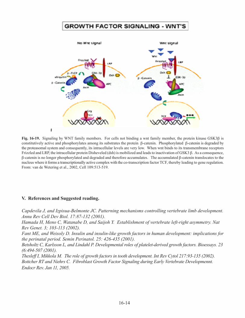
C. Wnt family signaling (Figure 16-19). Key intracellular players in wnt family signaling are
the protein kinase GSK3β and the signaling molecule b-catenin. In the absence of wnt signaling,
GSK3b phosphorylates β-catenin, thereby promoting its degradation. When wnts bind their
transmembrane receptors such as frizzled, GSK3β is turned off, thus leading to stabilization of β-
catenin. The later enters the nucleus where it participates in regulation of specific genes.
16-14
V. References and Suggested reading.
Capdevila J, and Izpisua-Belmonte JC. Patterning mechanisms controlling vertebrate limb development.
Annu Rev Cell Dev Biol. 17:87-132 (2001).
Hamada H, Meno C, Watanabe D, and Saijoh Y. Establishment of vertebrate left-right asymmetry. Nat
Rev Genet. 3; 103-113 (2002).
Fant ME, and Weisoly D. Insulin and insulin-like growth factors in human development: implications for
the perinatal period. Semin Perinatol. 25: 426-435 (2001).
Betsholtz C, Karlsson L, and Lindahl P. Developmental roles of platelet-derived growth factors. Bioessays. 23
(6:494-507 (2001).
Thesleff I, Mikkola M. The role of growth factors in tooth development. Int Rev Cytol 217:93-135 (2002).
Bottcher RT and Niehrs C. Fibroblast Growth Factor Signaling during Early Vertebrate Developmennt.
Endocr Rev. Jan 11, 2005.
Fig. 16-19. Signaling by WNT family members. For cells not binding a wnt family member, the protein kinase GSK3β is
constitutively active and phosphorylates among its substrates the protein β-catenin. Phosphorylated β-catenin is degraded by
the proteasomal system and consequently, its intracellular levels are very low. When wnt binds to its transmembrane receptors
Frizzled and LRP, the intracellular protein Disheveled (dsh) is mobilized and leads to inactivation of GSK3 β. As a consequence,
β-catenin is no longer phosphorylated and degraded and therefore accumulates. The accumulated β-catenin translocates to the
nucleus where it forms a transcriptionally active complex with the co-transcription factor TCF, thereby leading to gene regulation.
From: van de Wetering et al., 2002, Cell 109:513-519.