Embed Size (px)
Citation preview

RESEARCH ARTICLE
Growth deficits in cystic fibrosis mice begin in
utero prior to IGF-1 reduction
Rebecca Darrah1,2, Ilya Bederman3, Megan Vitko2, Dana M. Valerio3, Mitchell L. Drumm2,3,
Craig A. Hodges2,3*
1 Frances Payne Bolton School of Nursing, Case Western Reserve University, Cleveland, Ohio, United
States of America, 2 Department of Genetics and Genome Sciences, Case Western Reserve University,
Cleveland, Ohio, United States of America, 3 Department of Pediatrics, Case Western Reserve University,
Cleveland, Ohio, United States of America
Abstract
Growth deficits are common in cystic fibrosis (CF), but their cause is complex, with contribu-
tions from exocrine pancreatic insufficiency, pulmonary complications, gastrointestinal
obstructions, and endocrine abnormalities. The CF mouse model displays similar growth
impairment despite exocrine pancreatic function and in the absence of chronic pulmonary
infection. The high incidence of intestinal obstruction in the CF mouse has been suggested
to significantly contribute to the observed growth deficits. Previous studies by our group
have shown that restoration of the cystic fibrosis transmembrane conductance regulator
(CFTR) in the intestinal epithelium prevents intestinal obstruction but does not improve
growth. In this study, we further investigate growth deficits in CF and gut-corrected CF mice
by assessing insulin-like growth factor 1 (IGF-1). IGF-1 levels were significantly decreased
in CF and gut-corrected CF adult mice compared to wildtype littermates and were highly cor-
related with weight. Interestingly, perinatal IGF-1 levels were not significantly different
between CF and wildtype littermates, even though growth deficits in CF mice could be
detected late in gestation. Since CFTR has been suggested to play a role in water and nutri-
ent exchange in the placenta through its interaction with aquaporins, we analyzed placental
aquaporin expression in late-gestation CF and control littermates. While significant differ-
ences were observed in Aquaporin 9 expression in CF placentas in late gestation, there was
no evidence of placental fluid exchange differences between CF and control littermates.
The results from this study indicate that decreased IGF-1 levels are highly correlated with
growth in CF mice, independent of CF intestinal obstruction. However, the perinatal growth
deficits that are observed in CF mice are not due to decreased IGF-1 levels or differences in
placenta-mediated fluid exchange. Further investigation is necessary to understand the eti-
ology of early growth deficits in CF, as growth has been shown to be a significant factor in
disease outcomes.
PLOS ONE | https://doi.org/10.1371/journal.pone.0175467 April 6, 2017 1 / 12
a1111111111
a1111111111
a1111111111
a1111111111
a1111111111
OPENACCESS
Citation: Darrah R, Bederman I, Vitko M, Valerio
DM, Drumm ML, Hodges CA (2017) Growth
deficits in cystic fibrosis mice begin in utero prior
to IGF-1 reduction. PLoS ONE 12(4): e0175467.
https://doi.org/10.1371/journal.pone.0175467
Editor: Jeffrey M. Beekman, University Medical
Center Utrecht, NETHERLANDS
Received: December 2, 2016
Accepted: March 27, 2017
Published: April 6, 2017
Copyright: © 2017 Darrah et al. This is an open
access article distributed under the terms of the
Creative Commons Attribution License, which
permits unrestricted use, distribution, and
reproduction in any medium, provided the original
author and source are credited.
Data Availability Statement: All relevant data are
within the paper.
Funding: This work was supported by grants from
the Cystic Fibrosis Foundation (Hodges11G0,
https://www.cff.org/CFF) and the National Institutes
of Health (P30DK27651 and R24RR032425,
https://www.nig.gov). The funders had no role in
study design, data collection and analysis, decision
to publish, or preparation of the manuscript.
Competing interests: The authors have declared
that no competing interests exist.

Introduction
Cystic fibrosis (CF) is a complex, systemic, and lethal disorder caused by mutations in the
cystic fibrosis transmembrane conductance regulator gene (CFTR). Absence of CFTR, a
cAMP-regulated anion channel, leads to a plethora of disease manifestations, including
growth deficits. The growth deficits in CF patients have been observed as early as birth and
include reduced weight, length, and head circumference [1–4]. Reduced growth in CF
patients continues throughout life, with approximately 25% of CF children reported below
the 10th percentile for weight-for-age and sex, and CF adults typically display reduced body
mass index [5]. Improvement in these traits has been observed in recent years due to
increased focus on nutrition and improved pancreatic enzyme replacement therapy [6–8].
While CF lung disease is the leading cause of morbidity and mortality in CF, there is a strong
correlation between improved growth indices and pulmonary function and overall health [9–
13]. These observations indicate that growth deficits in CF patients have significant clinical
importance and highlight the necessity of understanding the origins for CF-associated
growth reduction.
The growth anomalies of CF are likely multifactorial, a consequence of the many organ sys-
tems affected by the disease. More than 85% of CF patients experience exocrine pancreatic
insufficiency (PI) due to the destruction of pancreatic acinar cells that occurs early in the dis-
ease [14, 15]. The absence of the pancreatic digestive enzymes leads to maldigestion and mal-
absorption, ultimately leading to malnutrition requiring pancreatic enzyme supplementation.
In addition to pancreatic enzyme replacement, dietary augmentation therapy through
increased caloric intake is commonly used to increase body mass in CF patients. These clinical
measures result in improved weight, but not to non-CF values and do not significantly affect
stature, suggesting factors other than malnutrition are involved [7]. Lung disease is also a clear
contributor to reduced growth in CF. The lung infection and inflammation in CF result in
cachexia that is often associated with chronic illness, diminishing growth and decreasing appe-
tite [16, 17]. However, lung infections do not contribute to neonatal and early childhood
growth deficits and CF patients with normal pulmonary function also have reduced growth
[18]. While it is clear that PI and lung disease contribute to the overall growth deficit in CF,
they alone do not account for the entirety of the reduced growth, which suggests additional
contributing factors.
CF animal models can be used to identify and understand these additional factors that affect
growth in CF. Reduced growth is common to all CF models, including the mouse, pig, ferret,
and rat [19–23]. CF pigs and ferrets display pancreatic and lung disease manifestations similar
to CF patients, which can confound the identification of additional factors contributing to
reduced growth [20, 21]. CF mice retain exocrine pancreatic function [24–26] and are main-
tained without chronic lung infection [24, 27–29], the two traits that are most often attributed
to growth delay and retardation in humans with CF, yet these animals display severe growth
deficits [19, 23, 26, 30]. Therefore, pulmonary and pancreatic disease are likely independent
factors that contribute to the reduced growth in CF patients and CF animal models, but do not
account for all of the reduced growth in CF.
In this study, we use CF mouse models to further understand the factors that contribute
to growth reduction in CF. Our findings indicate that the growth reduction in CF mice cor-
relates with insulin-like growth factor 1 (IGF-1) levels in the juvenile and adult stages. Fur-
ther, the growth reduction is independent of the incidence of intestinal obstruction that is
prevalent in CF models. In addition, our data indicate that CF growth deficits begin late in
gestation, prior to reduction of IGF-1 levels, and may be due to the absence of CFTR in the
placenta.
Growth deficits in CF
PLOS ONE | https://doi.org/10.1371/journal.pone.0175467 April 6, 2017 2 / 12

Materials and methods
Mice
The CF mice (Cftrinvfl10) and CF gut-corrected mice (Cftrinvfl10+ villin Cre) have been previ-
ously described [30]. The Cftr allele and Cre transgene were backcrossed to the C57BL/6J back-
ground for 10 generations to produce an inbred strain. Animals were monitored on a daily
basis, and weight was assessed every 5 days from 10 to 40 days of age. Mice were determined to
have succumbed to intestinal obstruction if obvious impaction was observed in the intestine
postmortem. Length of 6-wk-old euthanized mice was assessed from nose to anus by use of
digital calipers. Weight of inguinal fat weight was also assessed from these 6-wk-old mice. All
mouse fetuses and newborn pups were obtained from timed matings of heterozygote males
and females and embryonic day 1 was designated as the day after a vaginal plug was identified.
Embryonic day 15 (e15) and embryonic day 18 (e18) fetuses were obtained by cesarean section
of the euthanized dam and each fetus and placenta was blotted dry and weight was obtained.
All mice were genotyped by PCR using a previously described protocol [30]. All animals used
in this study were cared for according to a Case Western Reserve University approved protocol
and Institutional Animal Care and Use Committee guidelines. Animals were housed in stan-
dard polysulfone microisolator cages in ventilated units with corncob bedding. Mice were
given ad libitum access to chow (Harlan Teklad 7960; Harlan Teklad Global Diets, Madison,
WI) and sterile water. All animals were maintained on a 12-h light, 12-h dark schedule at a
mean ambient temperature of 22˚C.
Measurement of IGF-1 and Aqps
To evaluate gene expression of Igf1 and Aquaporin 1, 3, 8 and 9, RNA was isolated from liver
or placenta by use of TRIzol (Invitrogen). One microgram of RNA was reverse transcribed
into cDNA by use of QScript cDNA synthesis kit (VWR). Real-time quantitative PCR was per-
formed on a StepOne PCR system (Applied Biosystems). Expression was assessed via TaqMan
expression assays for Igf1(Mm00439560) or Aquaporin 1(Mm01326466), 3(Mm01208559),
8(Mm00431846) and 9 (Mm00508097) (Applied Biosystems). Expression was normalized to
β-actin as the endogenous reference. Each RNA sample was used to make cDNA in duplicate,
and the expression results were then averaged to yield the final result. The average of each sam-
ple was then expressed as a percentage of expression from control littermates. Mouse IGF-1
serum levels were determined using a mouse IGF-1 milliplex assay (RMIGF187K; EMD Milli-
pore) measured on a Luminex system. Each sample was run in triplicate and averaged.
Determination of fetal fluid transfer
To assess fetal fluid transfer from dam to fetus, we employed two methods. First, total water
content of placenta and fetus were measured in e18 fetuses. The percent wet weight of each
placenta or fetus was determined by dividing the initial weight of each placenta or fetus by the
weight of each after desiccation in an oven at 95˚C. Second, we used deuterium dilution meth-
odology to determine total body water of each of the e18 fetuses. Pregnant female mice were
given an intraperitoneal injection of 800 μl of 99.9% deuterium labeled water (2H2O). Two
hours after injection terminal blood samples were collected from both dam and pups and pro-
cessed immediately. The 2H-labeling of body water was determined by exchange with acetone,
as described previously [31]. Briefly, 5 μl of whole blood or standard were reacted for 4 hours
at room temperature with 5 μl of 10 N KOH and 5 μl of acetone. The reaction was carried out
in a crimped Gas Chromatography-Mass Spectrometry (GC/MS) vial and after 4 hours of
incubation, samples were analyzed by GC/MS. All sample analyses were carried out using an
Growth deficits in CF
PLOS ONE | https://doi.org/10.1371/journal.pone.0175467 April 6, 2017 3 / 12

Agilent 5973N-MSD mass spectrometer equipped with an Agilent 6890 gas chromatograph
with A DB-17MS (Agilent) capillary column (30 m x 0.25 mm x 0.25 mm). Samples were ana-
lyzed in Selected Ion Monitoring (SIM) mode using electron impact ionization (EI). Ion dwell
time was set to 10 msec. Acetone m/z 58 and 59 were monitored. Isotopic enrichment was
determined as ratio of m/z 59/(58+59) and plotted on a standard curve. Data were presented
as amount of 2H2O in the blood of each fetus divided by the amount of 2H2O in the blood of
the corresponding dam (% 2H2O fetal/dam).
Statistics
Results are expressed as the mean +/− SEM. Differences between groups were determined
using a two-way ANOVA with post-hoc Tukey test. A Spearman Rank Correlation test was
utilized for the comparison of IGF1 and weight. A P value of<0.05 was considered significant
for these tests. A Bonferroni correction for multiple comparisons was used for assessing signif-
icance in Aqp expression and a P value of<0.0125 was considered significant.
Results and discussion
Reduced growth in CF mice correlates with decreased IGF-1, but is not
due to intestinal obstruction
Reduced growth in CF mouse models has been hypothesized to be the result of the high inci-
dence of intestinal obstruction [19, 23, 26, 30]. Using conditional Cftr mouse alleles, we previ-
ously reported that inactivation of Cftr in the intestinal epithelium of mice produced an
intestinal obstruction phenotype, but did not reduce growth, while activation of Cftr in the
intestinal epithelium of mice (gut-corrected CF) resulted in no evidence of intestinal obstruc-
tion, but also no improvement in growth [30]. These data using the gut-corrected CF mice
were from a mixed mouse strain background (129/sv and C57Bl/6) [30].
To reduce the possibility of genetic heterogeneity as a confounding variable, we back-
crossed the conditional Cftr allele and the Cre recombinase transgene necessary to produce
the gut-corrected CF mouse to the C57BL/6 background for 10 generations. On the C57BL/6
inbred background, the CF mice and gut-corrected CF mice continued to show very similar
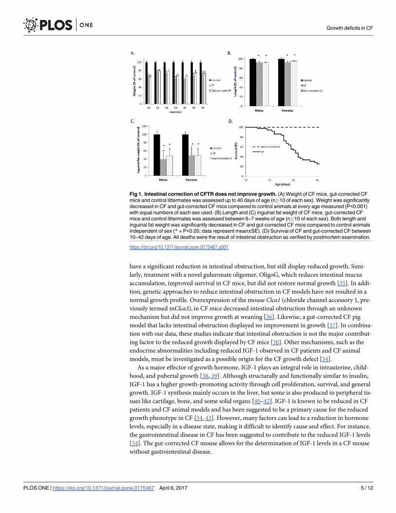
growth deficiencies compared to normal littermates (Fig 1). Specifically, the weight of both
CF and gut-corrected CF mice was significantly reduced compared to wildtype littermates’
weight measured between 10 and 40 days of age, with both groups in the range of 60–80% of
normal mouse weight (Fig 1A). Length was significantly reduced in CF and gut-corrected
CF mice independent of sex, with both groups’ length reduced by 5–10% of wildtype litter-
mates’ length. Reduction in fat accumulation is common in CF patients and CF mice and is
observed in the gut-corrected CF model as well. Both CF and gut-corrected CF mice display
a 50% reduction in inguinal fat (Fig 1C). Although the CF and gut-corrected CF mice were
similar in all measured aspects of growth, the incidence of lethal intestinal obstruction dif-
fered significantly with 75% of CF affected but none of the gut-corrected CF mice succumb-
ing to obstruction by 6 weeks of age (Fig 1D; P<0.0001 up to 6 weeks of age; n>35 mice per
group). In addition, no signs of intestinal obstruction were observed in any gut-corrected CF
mice following sacrifice and postmortem examination. These data clearly show that restora-
tion of CFTR in the intestinal epithelium and prevention of intestinal obstruction in CF mice
does not improve growth.
This observation is supported by previous studies in which amelioration of the CF intestinal
obstruction phenotype occurred through diet alteration, drug treatment, or genetic manipula-
tion. CF mice on a liquid diet [32, 33] or a solid diet with an osmotic laxative in the water [34]
Growth deficits in CF
PLOS ONE | https://doi.org/10.1371/journal.pone.0175467 April 6, 2017 4 / 12
have a significant reduction in intestinal obstruction, but still display reduced growth. Simi-
larly, treatment with a novel guluronate oligomer, OligoG, which reduces intestinal mucus
accumulation, improved survival in CF mice, but did not restore normal growth [35]. In addi-
tion, genetic approaches to reduce intestinal obstruction in CF models have not resulted in a
normal growth profile. Overexpression of the mouse Clca1 (chloride channel accessory 1, pre-
viously termed mClca3), in CF mice decreased intestinal obstruction through an unknown
mechanism but did not improve growth at weaning [36]. Likewise, a gut-corrected CF pig
model that lacks intestinal obstruction displayed no improvement in growth [37]. In combina-
tion with our data, these studies indicate that intestinal obstruction is not the major contribut-
ing factor to the reduced growth displayed by CF mice [30]. Other mechanisms, such as the
endocrine abnormalities including reduced IGF-1 observed in CF patients and CF animal
models, must be investigated as a possible origin for the CF growth defect [34].
As a major effector of growth hormone, IGF-1 plays an integral role in intrauterine, child-
hood, and pubertal growth [38, 39]. Although structurally and functionally similar to insulin,
IGF-1 has a higher growth-promoting activity through cell proliferation, survival, and general
growth. IGF-1 synthesis mainly occurs in the liver, but some is also produced in peripheral tis-
sues like cartilage, bone, and some solid organs [40–42]. IGF-1 is known to be reduced in CF
patients and CF animal models and has been suggested to be a primary cause for the reduced
growth phenotype in CF [34, 43]. However, many factors can lead to a reduction in hormone
levels, especially in a disease state, making it difficult to identify cause and effect. For instance,
the gastrointestinal disease in CF has been suggested to contribute to the reduced IGF-1 levels
[34]. The gut-corrected CF mouse allows for the determination of IGF-1 levels in a CF mouse
without gastrointestinal disease.
Fig 1. Intestinal correction of CFTR does not improve growth. (A) Weight of CF mice, gut-corrected CF
mice and control littermates was assessed up to 40 days of age (n�10 of each sex). Weight was significantly
decreased in CF and gut-corrected CF mice compared to control animals at every age measured (P<0.001)
with equal numbers of each sex used. (B) Length and (C) inguinal fat weight of CF mice, gut-corrected CF
mice and control littermates was assessed between 6–7 weeks of age (n�10 of each sex). Both length and
inguinal fat weight was significantly decreased in CF and gut-corrected CF mice compared to control animals
independent of sex (* = P<0.05; data represent mean±SE). (D) Survival of CF and gut-corrected CF between
10–42 days of age. All deaths were the result of intestinal obstruction as verified by postmortem examination.
https://doi.org/10.1371/journal.pone.0175467.g001
Growth deficits in CF
PLOS ONE | https://doi.org/10.1371/journal.pone.0175467 April 6, 2017 5 / 12

We next evaluated IGF-1 in both CF and gut-corrected CF adult mice and found the
levels in liver and blood were significantly decreased in both groups compared to wildtype lit-
termates (40% reduction in liver and 70% reduction in blood; Fig 2A and 2B). In addition,
IGF-1 levels did not differ between CF and gut-corrected CF mice. Interestingly, IGF-1 levels
were significantly correlated with weight in both groups of CF mice (Fig 2C and 2D). There-
fore, both CF and gut-corrected CF mice display similar growth and IGF-1 abnormalities, and
while reduced IGF-1 in CF mice correlates with growth, reduced IGF-1 is not due to intestinal
disease. While these data indicate a potential causal relationship between reduced IGF-1 and
reduced growth in adult CF mice, it is unknown if a similar relationship is evident at birth in
CF mice. Understanding the timing of IGF-1 reduction in CF in relationship to growth reduc-
tion may provide insight into the underlying mechanism.
Growth differences in CF mice are evident perinatally, but are not due to
IGF-1 differences
In addition to the well-documented growth deficiencies in CF patients throughout postnatal
life, reduced birth weight has also been observed, suggesting a prenatal origin for the growth
deficiencies [1, 4]. We measured body weight of late-gestation mouse fetuses on embryonic
day 15 (e15) and embryonic day 18 (e18) and newborn CF and control pups to determine
whether CF mice exhibit the reduced birth weight observed in CF patients, and the timing of
this deficit. We observed normal Mendelian distribution of genotypes from late-gestation
pups (Fig 3A), indicating that any observed loss of CF pups [26], occurs after birth. Therefore,
the data are not biased by potential prenatal loss of CF pups. Reduced weight in CF pups com-
pared to control pups was observed in the e18 fetuses and newborn pups (9% at both), while
no differences in weight were observed in the e15 fetuses (Fig 3B). To evaluate whether these
weight differences correlated with IGF-1 levels, we measured IGF-1 in the blood from CF and
Fig 2. IGF-1 levels are reduced in CF and gut-corrected CF mice and correlate with growth. (A) Igf1
expression and (B) IGF-1 serum levels are significantly reduced at 6 weeks of age in both CF and gut-
corrected CF mice compared to control littermates (* = P<0.05; n�6; data represent mean±SE). IGF-1 levels
were significantly correlated with weight in (C) CF and (D) gut-corrected CF mice (P<0.05; n�10).
https://doi.org/10.1371/journal.pone.0175467.g002
Growth deficits in CF
PLOS ONE | https://doi.org/10.1371/journal.pone.0175467 April 6, 2017 6 / 12

control pups and observed no significant differences in e18 fetuses or newborn pups (Fig 3C).
However, IGF-1 levels were significantly reduced in CF mice compared to control mice as
early as 3 weeks of age (Fig 3C). Taken together, these data indicate that CF growth deficiency
is evident late in gestation and continues through adulthood; however, IGF-1 levels are initially
normal prenatally and at birth, but later, reduced. Therefore, IGF-1 reduction is unlikely to be
the sole origin for the perinatal CF growth deficiency.
The weight difference between CF and control e18 mouse fetuses, but not between e15
fetuses, demonstrates that reduced growth in CF is not evident until late gestation. This is simi-
lar to humans, in which CF newborns have reduced birthweight compared to non-CF new-
borns, but weight is not significantly different between CF and non-CF preterm infants
(gestational age< 37 weeks)[1]. In contrast to the present study, reduced IGF-1 in CF new-
borns and CF piglets has been noted [43]. Interestingly, unlike CF humans and CF mice,
significantly decreased birthweight is not observed in the CF pig [37, 43, 44]. While reduced
IGF-1 is a constant among humans, pigs, and mice with CF, the temporal relationship between
reduced IGF-1 and reduced growth is complicated, and further study is necessary to elucidate
this relationship. Insulin levels, nutrition, and even the presence of meconium ileus (MI), at
least in CF humans and pigs, may all play roles in the reduced IGF-1. The absence of MI in CF
mice suggests a lack of intestinal pathology in utero however intestinal contribution to late ges-
tational growth reduction is possible and cannot be entirely ruled out without further investi-
gation. While there are many possible contributing factors to reduced growth in CF, in the CF
mouse model, reduced growth is evident in late gestation and continues into adulthood in the
absence of pancreatic, lung, or even intestinal pathology.
Fig 3. Reduced growth in CF mice occurs prenatally in the absence of reduced IGF-1. (A) A Mendelian
distribution was observed in fetuses originating from Cftr heterozygote matings. (n>100) (B) Weight was
significantly reduced in newborn CF mice and e18 fetuses compared to control littermates but the groups did
not differ at e15. (C) IGF-1 levels were not significantly different between CF and control littermates perinatally
(e18 and newborn) but IGF-1 levels were significantly reduced between CF and control littermates at 3 weeks
of age. (* = P<0.05; n�15; data represent mean±SE)
https://doi.org/10.1371/journal.pone.0175467.g003
Growth deficits in CF
PLOS ONE | https://doi.org/10.1371/journal.pone.0175467 April 6, 2017 7 / 12

Absence of CFTR in the placenta may contribute to prenatal CF growth
differences
The absence of CF disease pathology and normal IGF-1 levels in CF mouse pups suggest other
origins for the observed prenatal growth deficits. A role for the placenta in reduced perinatal
growth in CF has been postulated [1, 45]. The presence of CFTR in the placenta has been
observed, but its exact role in this organ remains unclear [46, 47]. It has been suggested that
CFTR plays a role in water and nutrient exchange in the placenta because inhibition of CFTR
in placental explants decreases water uptake [48]. Absence of CFTR in the placenta leading to
decreased placental mediated fluid exchange may account for the observed late gestation
weight deficits in CF mice. In addition, CFTR has been suggested to work in conjunction with
specific aquaporins, which facilitate the transfer of water and small solutes to the fetus [48–51].
To evaluate this possibility, we examined the expression of four aquaporins known to be
expressed in the placenta in CF and control placentas from e15 and e18 fetuses. While placen-
tal aquaporin expression was not different between CF and control littermates at e15, aqua-porin 9 (Aqp9) was significantly elevated by almost twofold in the placentas from CF pups at
e18 (p<0.0037; Fig 4). This finding is consistent with a previous study which observed that
decreased CFTR expression in preeclamptic placentas was associated with increased AQP9
expression despite the loss of AQP9 functionality leading to reduced water transport [48].
Given the importance of aquaporins in placental-mediated water exchange for the fetus, we
hypothesized that the altered Aqp9 expression may lead to decreased fluid uptake, leading to
the observed weight difference between CF and control fetuses and newborn pups. We calcu-
lated fetal fluid transfer using two independent measures: percent water weight and by the iso-
topic dilution method using deuterium-labeled water (as described in Methods). There were
no significant differences between water weight in the body or placenta between the CF and
control e18 fetuses (Fig 5A), and there were no detectable differences in fluid exchange
between the dam and CF and control littermates (Fig 5B). Therefore, the differences in neona-
tal birth weight between the CF and control mice are not due to differences in placental water
transfer. However, further studies are needed to interpret the increase in Aqp9 expression in
Fig 4. Placental aquaporin expression in CF mice. Aquaporin(Aqp) 1,3,8 and 9 expression was evaluated
in placentas from e15 and e18 CF and control littermates. Only Aqp9 at e18 was significantly different
between placentas from CF and control littermates (* = P<0.005) Each measurement is an average of six
mice completed in replicates (data represent mean±SEM).
https://doi.org/10.1371/journal.pone.0175467.g004
Growth deficits in CF
PLOS ONE | https://doi.org/10.1371/journal.pone.0175467 April 6, 2017 8 / 12

CF placentas given this absence in difference in placental water transfer between CF and con-
trol fetuses. For example, it is possible that the increase in Aqp9 expression does not coincide
with an increase in protein levels or, alternatively, increased Aqp9 expression may lead to
increased protein levels with no difference in Aqp9 function. Interestingly, a previous study
noted that mothers fed a hypercaloric diet gave birth to CF babies of higher birth weight as
compared with mothers on a normal diet, suggesting the possibility of placental nutrient
exchange differences in CF still needs to be determined [52].
Conclusions
The CF and gut-corrected CF mice are indistinguishable in their growth characteristics, indi-
cating, that growth deficits in postnatal CF mice are not due to intestinal complications such
as obstruction. In addition, the relationship between decreased IGF-1 and reduced size exists
in CF mice, but the growth deficit precedes the reduction in IGF-1 during development. Our
data suggest that the late-gestation growth reduction in CF mice, which corresponds to late
third trimester in humans, may have a possible placental origin, but the mechanisms remain
to be determined.
Acknowledgments
We thank the CF Mouse Models Core at Case Western Reserve University for their work in
maintaining and genotyping the mouse colony.
Author Contributions
Conceptualization: CAH RD MLD.
Formal analysis: CAH RD.
Funding acquisition: CAH MLD.
Investigation: IB MV DMV CAH RD.
Methodology: CAH RD IB.
Project administration: CAH RD.
Resources: CAH.
Supervision: CAH.
Fig 5. Placental-mediated fluid exchange is not different between CF and control fetuses. (A) Fetal
body and placental water weight at e18 was not significantly different between CF and control fetuses. (B)
Equilibration of 2H2O between dam and CF and control fetuses at e18 were not significantly different. (n�10;
data represent mean±SEM).
https://doi.org/10.1371/journal.pone.0175467.g005
Growth deficits in CF
PLOS ONE | https://doi.org/10.1371/journal.pone.0175467 April 6, 2017 9 / 12

Visualization: CAH RD.
Writing – original draft: CAH RD.
Writing – review & editing: CAH RD MLD IB DMV MV.
References1. Festini F, Taccetti G, Repetto T, Reali MF, Campana S, Mergni G, et al. Gestational and neonatal char-
acteristics of children with cystic fibrosis: a cohort study. J Pediatr. 2005; 147(3):316–20. https://doi.org/
10.1016/j.jpeds.2005.04.031 PMID: 16182668
2. Ghosal S, Taylor CJ, Pickering M, McGaw J, Beckles-Willson N, Wales JK. Disproportionate head
growth retardation in cystic fibrosis. Arch Dis Child. 1995; 72(2):150–2. PMID: 7702380
3. Haeusler G, Frisch H, Waldhor T, Gotz M. Perspectives of longitudinal growth in cystic fibrosis from
birth to adult age. Eur J Pediatr. 1994; 153(3):158–63. PMID: 8181496
4. Darrah R, Nelson R, Damato EG, Decker M, Matthews A, Hodges CA. Growth Deficiency in Cystic
Fibrosis Is Observable at Birth and Predictive of Early Pulmonary Function. Biol Res Nurs. 2016; 18
(5):498–504. https://doi.org/10.1177/1099800416643585 PMID: 27081158
5. Cystic Fibrosis Foundation Patient Registry: 2005 Annual Data Report to the Center Directors.
Bethesda, MD: Cystic Fibrosis Foundation. 2006.
6. Gaskin KJ. Nutritional care in children with cystic fibrosis: are our patients becoming better? European
journal of clinical nutrition. 2013; 67(5):558–64. https://doi.org/10.1038/ejcn.2013.20 PMID: 23462946
7. Stallings VA, Stark LJ, Robinson KA, Feranchak AP, Quinton H. Evidence-based practice recommen-
dations for nutrition-related management of children and adults with cystic fibrosis and pancreatic insuf-
ficiency: results of a systematic review. J Am Diet Assoc. 2008; 108(5):832–9. Epub 2008/04/30.
https://doi.org/10.1016/j.jada.2008.02.020 PMID: 18442507
8. Yen EH, Quinton H, Borowitz D. Better nutritional status in early childhood is associated with improved
clinical outcomes and survival in patients with cystic fibrosis. J Pediatr. 2013; 162(3):530–5 e1. https://
doi.org/10.1016/j.jpeds.2012.08.040 PMID: 23062247
9. Corey M, Farewell V. Determinants of mortality from cystic fibrosis in Canada, 1970–1989. Am J Epide-
miol. 1996; 143(10):1007–17. PMID: 8629607
10. Corey M, McLaughlin FJ, Williams M, Levison H. A comparison of survival, growth, and pulmonary func-
tion in patients with cystic fibrosis in Boston and Toronto. J Clin Epidemiol. 1988; 41(6):583–91. PMID:
3260274
11. Konstan MW, Butler SM, Wohl ME, Stoddard M, Matousek R, Wagener JS, et al. Growth and nutritional
indexes in early life predict pulmonary function in cystic fibrosis.J Pediatr. 2003; 142(6):624–30. https://
doi.org/10.1067/mpd.2003.152 PMID: 12838189
12. Lai HC, Corey M, FitzSimmons S, Kosorok MR, Farrell PM. Comparison of growth status of patients
with cystic fibrosis between the United States and Canada. Am J Clin Nutr. 1999; 69(3):531–8. PMID:
10075341
13. Lai HC, Kosorok MR, Laxova A, Davis LA, FitzSimmon SC, Farrell PM. Nutritional status of patients
with cystic fibrosis with meconium ileus: a comparison with patients without meconium ileus and diag-
nosed early through neonatal screening. Pediatrics. 2000; 105(1 Pt 1):53–61. PMID: 10617704
14. Durie PR. The pathophysiology of the pancreatic defect in cystic fibrosis. Acta Paediatr Scand Suppl.
1989; 363:41–4. PMID: 2701923
15. Kopelman H, Durie P, Gaskin K, Weizman Z, Forstner G. Pancreatic fluid secretion and protein hyper-
concentration in cystic fibrosis.N Engl J Med. 1985; 312(6):329–34. https://doi.org/10.1056/
NEJM198502073120601 PMID: 3969086
16. Levy E, Gurbindo C, Lacaille F, Paradis K, Thibault L, Seidman E. Circulating tumor necrosis factor-
alpha levels and lipid abnormalities in patients with cystic fibrosis. Pediatr Res. 1993; 34(2):162–6.
https://doi.org/10.1203/00006450-199308000-00011 PMID: 8233719
17. Nixon LS, Yung B, Bell SC, Elborn JS, Shale DJ. Circulating immunoreactive interleukin-6 in cystic fibro-
sis. Am J Respir Crit Care Med. 1998; 157(6 Pt 1):1764–9. https://doi.org/10.1164/ajrccm.157.6.
9704086 PMID: 9620903
18. Hardin DS, Ferkol T, Ahn C, Dreimane D, Dyson M, Morse M, et al. A retrospective study of growth hor-
mone use in adolescents with cystic fibrosis. Clin Endocrinol.2005; 62(5):560–6.
19. Hodges CA, Cotton CU, Palmert MR, Drumm ML. Generation of a conditional null allele for Cftr in mice.
Genesis. 2008; 46(10):546–52. https://doi.org/10.1002/dvg.20433 PMID: 18802965
Growth deficits in CF
PLOS ONE | https://doi.org/10.1371/journal.pone.0175467 April 6, 2017 10 / 12

20. Rogers CS, Stoltz DA, Meyerholz DK, Ostedgaard LS, Rokhlina T, Taft PJ, et al. Disruption of the
CFTR gene produces a model of cystic fibrosis in newborn pigs. Science. 2008; 321(5897):1837–41.
https://doi.org/10.1126/science.1163600 PMID: 18818360
21. Sun X, Sui H, Fisher JT, Yan Z, Liu X, Cho HJ, et al. Disease phenotype of a ferret CFTR-knockout
model of cystic fibrosis. J Clin Invest. 2010; 120(9):3149–60. https://doi.org/10.1172/JCI43052 PMID:
20739752
22. Tuggle KL, Birket SE, Cui X, Hong J, Warren J, Reid L, et al. Characterization of defects in ion transport
and tissue development in cystic fibrosis transmembrane conductance regulator (CFTR)-knockout rats.
PLoS One. 2014; 9(3):e91253. https://doi.org/10.1371/journal.pone.0091253 PMID: 24608905
23. Zeiher BG, Eichwald E, Zabner J, Smith JJ, Puga AP, McCray PB Jr., et al. A mouse model for the delta
F508 allele of cystic fibrosis. J Clin Invest. 1995; 96(4):2051–64. https://doi.org/10.1172/JCI118253
PMID: 7560099
24. Durie PR, Kent G, Phillips MJ, Ackerley CA. Characteristic multiorgan pathology of cystic fibrosis in a
long-living cystic fibrosis transmembrane regulator knockout murine model. Am J Pathol. 2004; 164
(4):1481–93. Epub 2004/03/25. https://doi.org/10.1016/S0002-9440(10)63234-8 PMID: 15039235
25. Pascua P, Garcia M, Fernandez-Salazar MP, Hernandez-Lorenzo MP, Calvo JJ, Colledge WH, et al.
Ducts isolated from the pancreas of CFTR-null mice secrete fluid. Pflugers Archiv: European journal of
physiology. 2009; 459(1):203–14. https://doi.org/10.1007/s00424-009-0704-9 PMID: 19655163
26. Snouwaert JN, Brigman KK, Latour AM, Malouf NN, Boucher RC, Smithies O, et al. An animal model
for cystic fibrosis made by gene targeting. Science. 1992; 257(5073):1083–8. PMID: 1380723
27. Cohen JC, Lundblad LK, Bates JH, Levitzky M, Larson JE. The "Goldilocks effect" in cystic fibrosis:
identification of a lung phenotype in the cftr knockout and heterozygous mouse. BMC Genet. 2004;
5:21. https://doi.org/10.1186/1471-2156-5-21 PMID: 15279681
28. Darrah RJ, Mitchell AL, Campanaro CK, Barbato ES, Litman P, Sattar A, et al. Early pulmonary disease
manifestations in cystic fibrosis mice. J Cyst Fibros. 2016.
29. Kent G, Iles R, Bear CE, Huan LJ, Griesenbach U, McKerlie C, et al. Lung disease in mice with cystic
fibrosis. J Clin Invest. 1997; 100(12):3060–9. Epub 1998/01/31. https://doi.org/10.1172/JCI119861
PMID: 9399953
30. Hodges CA, Grady BR, Mishra K, Cotton CU, Drumm ML. Cystic fibrosis growth retardation is not corre-
lated with loss of Cftr in the intestinal epithelium. Am J Physiol Gastrointest Liver Physiol. 2011; 301(3):
G528–36. Epub 2011/06/11. https://doi.org/10.1152/ajpgi.00052.2011 PMID: 21659619
31. Shah V, Herath K, Previs SF, Hubbard BK, Roddy TP. Headspace analyses of acetone: a rapid method
for measuring the 2H-labeling of water. Anal Biochem. 2010; 404(2):235–7. https://doi.org/10.1016/j.ab.
2010.05.010 PMID: 20488158
32. Eckman EA, Cotton CU, Kube DM, Davis PB. Dietary changes improve survival of CFTR S489X homo-
zygous mutant mouse. Am J Physiol. 1995; 269(5 Pt 1):L625–30. PMID: 7491981
33. van Heeckeren AM, Schluchter MD, Drumm ML, Davis PB. Role of Cftr genotype in the response to
chronic Pseudomonas aeruginosa lung infection in mice. Am J Physiol Lung Cell Mol Physiol. 2004;
287(5):L944–52. Epub 2004/07/13. https://doi.org/10.1152/ajplung.00387.2003 PMID: 15246977
34. Rosenberg LA, Schluchter MD, Parlow AF, Drumm ML. Mouse as a model of growth retardation in cys-
tic fibrosis. Pediatr Res. 2006; 59(2):191–5. Epub 2006/01/28. https://doi.org/10.1203/01.pdr.
0000196720.25938.be PMID: 16439577
35. Vitko M, Valerio DM, Rye PD, Onsoyen E, Myrset AH, Dessen A, et al. A novel guluronate oligomer
improves intestinal transit and survival in cystic fibrosis mice.J Cyst Fibros. 2016.
36. Young FD, Newbigging S, Choi C, Keet M, Kent G, Rozmahel RF. Amelioration of cystic fibrosis intesti-
nal mucous disease in mice by restoration of mCLCA3. Gastroenterology. 2007; 133(6):1928–37.
https://doi.org/10.1053/j.gastro.2007.10.007 PMID: 18054564
37. Stoltz DA, Rokhlina T, Ernst SE, Pezzulo AA, Ostedgaard LS, Karp PH, et al. Intestinal CFTR expres-
sion alleviates meconium ileus in cystic fibrosis pigs. J Clin Invest. 2013; 123(6):2685–93. https://doi.
org/10.1172/JCI68867 PMID: 23676501
38. Agrogiannis GD, Sifakis S, Patsouris ES, Konstantinidou AE. Insulin-like growth factors in embryonic
and fetal growth and skeletal development (Review). Mol Med Rep. 2014; 10(2):579–84. https://doi.org/
10.3892/mmr.2014.2258 PMID: 24859417
39. Netchine I, Azzi S, Le Bouc Y, Savage MO. IGF1 molecular anomalies demonstrate its critical role in
fetal, postnatal growth and brain development. Best Pract Res Clin Endocrinol Metab. 2011; 25(1):181–
90. https://doi.org/10.1016/j.beem.2010.08.005 PMID: 21396584
40. Han VK, Lund PK, Lee DC, D’Ercole AJ. Expression of somatomedin/insulin-like growth factor messen-
ger ribonucleic acids in the human fetus: identification, characterization, and tissue distribution. J Clin
Endocrinolo Metab. 1988; 66(2):422–9.
Growth deficits in CF
PLOS ONE | https://doi.org/10.1371/journal.pone.0175467 April 6, 2017 11 / 12

41. Roberts CT Jr., Lasky SR, Lowe WL Jr., Seaman WT, LeRoith D. Molecular cloning of rat insulin-like
growth factor I complementary deoxyribonucleic acids: differential messenger ribonucleic acid process-
ing and regulation by growth hormone in extrahepatic tissues. Mol Endocrinol. 1987; 1(3):243–8.
https://doi.org/10.1210/mend-1-3-243 PMID: 3453891
42. Yakar S, Liu JL, Stannard B, Butler A, Accili D, Sauer B, et al. Normal growth and development in the
absence of hepatic insulin-like growth factor I. Proc Natl Acad Sci U S A. 1999; 96(13):7324–9. PMID:
10377413
43. Rogan MP, Reznikov LR, Pezzulo AA, Gansemer ND, Samuel M, Prather RS, et al. Pigs and humans
with cystic fibrosis have reduced insulin-like growth factor 1 (IGF1) levels at birth. Proc Natl Acad Sci U
S A. 2010. Epub 2010/11/10.
44. Rogers CS, Hao Y, Rokhlina T, Samuel M, Stoltz DA, Li Y, et al. Production of CFTR-null and CFTR-
DeltaF508 heterozygous pigs by adeno-associated virus-mediated gene targeting and somatic cell
nuclear transfer. J Clin Invest. 2008; 118(4):1571–7. https://doi.org/10.1172/JCI34773 PMID:
18324337
45. Davis B, Shennan DB, Boyd CA. Chloride transport in cystic fibrosis placenta. Lancet. 1985; 1
(8425):392–3.
46. Faller DP, Egan DA, Ryan MP. Evidence for location of the CFTR in human placental apical membrane
vesicles. Am J Physiol. 1995; 269(1 Pt 1):C148–55. PMID: 7543241
47. Mylona P, Glazier JD, Greenwood SL, Sides MK, Sibley CP. Expression of the cystic fibrosis (CF) and
multidrug resistance (MDR1) genes during development and differentiation in the human placenta. Mol
Hum Reprod. 1996; 2(9):693–8. PMID: 9239684
48. Castro-Parodi M, Levi L, Dietrich V, Zotta E, Damiano AE. CFTR may modulate AQP9 functionality in
preeclamptic placentas. Placenta. 2009; 30(7):642–8. https://doi.org/10.1016/j.placenta.2009.04.012
PMID: 19481256
49. Cheung KH, Leung CT, Leung GP, Wong PY. Synergistic effects of cystic fibrosis transmembrane con-
ductance regulator and aquaporin-9 in the rat epididymis. Biol Reprod. 2003; 68(5):1505–10. https://
doi.org/10.1095/biolreprod.102.010017 PMID: 12606488
50. Pietrement C, Da Silva N, Silberstein C, James M, Marsolais M, Van Hoek A, et al. Role of NHERF1,
cystic fibrosis transmembrane conductance regulator, and cAMP in the regulation of aquaporin 9. J Biol
Chem. 2008; 283(5):2986–96. https://doi.org/10.1074/jbc.M704678200 PMID: 18055461
51. Schreiber R, Nitschke R, Greger R, Kunzelmann K. The cystic fibrosis transmembrane conductance
regulator activates aquaporin 3 in airway epithelial cells. J Biol Chem. 1999; 274(17):11811–6. PMID:
10206998
52. Boyer PH. Low birth weight in fibrocystic disease of the pancreas. Pediatrics. 1955; 16(6):778–84.
PMID: 13273117
Growth deficits in CF
PLOS ONE | https://doi.org/10.1371/journal.pone.0175467 April 6, 2017 12 / 12


![Mitochondrial ROS cause motor deficits induced by synaptic ......curs over a protracted time-course (order of weeks) and encompasses in utero and ex utero developmental stages [30]](https://img.dokumen.tips/doc/110x75/60a4d7292188c15e6d3ac44a/mitochondrial-ros-cause-motor-deficits-induced-by-synaptic-curs-over-a-protracted.jpg)