Embed Size (px)
Citation preview

Biomedical Science and Engineering, 2015, Vol. 3, No. 3, 46-64 Available online at http://pubs.sciepub.com/bse/3/3/1 © Science and Education Publishing DOI:10.12691/bse-3-3-1
Growth and Advancements in Neural Control of Limb
Bablu Lal Rajak, Meena Gupta, Dinesh Bhatia*
Department of Biomedical Engineering, North Eastern Hill University, Shillong, Meghalaya, India *Corresponding author: [email protected]
Abstract Centuries of study has unfolded our understanding regarding different bodily movement routinely performed. It has been observed that all these movements require intricate communication between the brain and associated muscles. Our sensory systems help in guiding this communication by providing information about the external environment and surroundings, thereby helping the motor system plan the different movements leading to controlled action by the muscles. Billions of neuron with quadrillion connections between them and muscles are responsible for coordinated movements that humans perform routinely. Though our knowledge and understanding about motor neuron diseases and neuro-degeneration disorders are limited, yet efforts have been made to overcome or improve the present state of these disorders either by drugs, artificial prosthetic devices, robotics, stimulation or stem cell therapy. These treatments are attempts to help relieve symptoms, improve functionality, provide support and effectively slow down the disease's progression. Furthermore, disabled individuals were aided with walking stick, wheelchair or stroller till recently; however, significant technological advancements in the past few decades have brought in more of man-machine interactive devices such as deployment of artificial prosthetics, improved brain-computer interactions and advanced neuroprosthetics for supporting activities of daily living in these patients. Additionally, new tools like computer simulations, medical imaging and computational models are being used to simulate simple movement tasks and compare the outcomes with real limb control and neural elements, thereby testing how brain signals are processed to achieve sophisticated motor control. Researchers are regularly improving existing devices for ease of use and efficiency, and new ones are being developed such that it can mimic the maneuverability of the natural limb.
Keywords: neurons, brain computer interface, prosthetics, neurodegenerative disorders, stem therapy
Cite This Article: Bablu Lal Rajak, Meena Gupta, and Dinesh Bhatia, “Growth and Advancements in Neural Control of Limb.” Biomedical Science and Engineering, vol. 3, no. 3 (2015): 46-64. doi: 10.12691/bse-3-3-1.
1. Introduction The brain’s association with limb movement has been
known since the 4th century. This was noted by Hippocrates when he showed that injuries to one side of the head typically resulted in motor deficits on the opposite side of the body. This association was later confirmed by René Descartes that nerves interconnecting brain and muscles are responsible for the different limb movements [1]. He tried to explain the reflex withdrawal of a limb from a painful stimulus. He proposed that the stimulus sets in motion the “animal spirits” in nerves running to the ventricles of the brain. This would lead to the opening of the valves within the ventricles, releasing the “spirits” into nerves running to the muscle, while the inflation of the muscles would cause the limb to withdraw. He imagined that voluntary movements were controlled in a similar fashion [2].
Centuries of study has unfolded that every movement we make - from walking and writing to blinking and talking, all require intricate communication between the brain and our muscles. Our sensory systems guide this communication by providing information about the external environment and surroundings, thereby helping the motor system plan the different movements leading to
controlled action by the muscles. Billions of neuron with quadrillion connections between them and muscles are responsible for coordinated movements that humans perform routinely. Any damage or disease at any level of the motor system hierarchy results in motor system dysfunction [3]. Ongoing studies are helping researchers and technologists map how brain regions work together to control movement and how dysfunction in these regions contributes to movement disorders leading to loss of limb control [2,3]. Limb control loss due to neuronal dysfunction may be attributed to neurodegenerative diseases like Parkinson’s, Alzheimer’s disease; or motor neuron disorders like amyotrophic lateral sclerosis, primary lateral sclerosis, progressive bulbar palsy; or brain injury caused before, during or after birth for example cerebral palsy; or traumatic brain injury due to accidents, fall, natural disaster; or non-traumatic brain injury like stroke, polio, spina bifida; or amputation due to some of the above mentioned disorders or natural calamity, war, accidents, etc. [16,19].
Though our knowledge and understanding about motor neuron diseases and neuro-degeneration disorders are limited, yet efforts are made to overcome or improve the present state of these disorders either by drugs, artificial prosthetic devices, robotics, stimulation or stem cell therapy. These treatments are attempts to help relieve symptoms, improve functionality, provide support and effectively slow down the disease's progression. Furthermore,

47 Biomedical Science and Engineering
individuals who are suffering from disability till lately were aided by providing walking stick, wheelchair or stroller but technological advancement have brought in more of man-machine interactive devices such as deployment of artificial prosthetics, improved brain-computer interactions and advanced neuro-prosthetics for supporting activities of daily living in these patients. Additionally, new tools like computer simulations, medical imaging and computational models are being used to simulate simple movement tasks and compare the outcomes with real limb control and neural elements, thereby testing how brain signals are processed to achieve sophisticated motor control. Researchers are regularly improving existing devices for ease of use and efficiency, and new ones are being developed such that it can mimic the maneuverability of natural limb [48,50]. In this paper, we review the current advancements in the present state of different limb control strategies and the possible interventions in the development of artificial devices to understand the present day advanced techniques for limb control mechanism. We also review different technological advancements and future research arenas for overcoming the disability caused due to motor neuron dysfunction thereby supporting physically challenged persons to overcome such disabilities.
2. Physiology of Limb Control Pathway To understand the development of neurological devices
for helping people with disabilities, it is important to understand the underlying limb control physiology controlling
locomotion and task performance in individuals. A neuron motor pathway is a combination of sensory and motor system which interconnects different regions within the brain and conveys information from the peripheral nervous system to the brain. A single afferent neuron with all its receptor endings makes up a sensory unit. These sensory units are sensitive to certain types of stimuli. The pathways in the brain for transmission of information are called as ascending (sensory pathway) track in the spinal cord and descending track (motor pathway). Ascending track carries sensory fibers from sensory unit to the central nervous system (CNS) and hence they are called as sensory or afferent track. Some prominent sensory tracks in spinal cord are fasciculus gracilis, cuneatus, lateral spinothalamic track and ventral spinothalmic track. The fasciculus gracilis and cuneatus in dorsal white column convey information of touch, tactile localization, discrimination, vibration, sense of deep pressure, proprioception and kinesthesia sensation to the higher centers of the brain from various body parts. The lateral spinothalamic track in lateral white column carries information regarding the sense of pain and temperature from body parts to higher centers in the brain. The ventral spinothalmic track in ventral white column carries the sense of touch sensation to the designated brain centers. When stimulation is given to the frontal lobes of the cerebral cortex it sends response to the skeletal system and this constitutes the motor area. The area that lies anterior to the central sulcus is called as the pre-central motor cortex. This pre-central motor cortex is the main origin of descending track – the motor pathway [3].
Figure 1. Physiology of motor control pathway
The motor pathway or descending track consists of two types of tracks. One is pyramidal track and another is extra pyramidal track. The pyramidal track is the longest track which starts from motor cortex to reach the last segment of spinal cord. The fibers of this track pass from motor area to the spinal ventral horn cells to form the cortico-spinal track. The pyramidal track fibers to the motor cranial nuclei constitute the cortico-bulbar track which starts from cerebral cortex and ends in the brain stem. If the pyramidal tracks are affected, it leads to the
upper motor neuron (UMN) disease and if the spinal or cranial motor neurons are damaged it leads to the lower motor neuron (LMN) disease. The cortico-spinal tracks convey motor impulse to the spinal cord, thereby, controlling the voluntary movement of the upper limb especially fine motor movements like buttoning or playing piano. The cortico-bulbar track controls voluntary movements of muscles of the larynx, pharynx, and palate. The extrapyramidal track is formed by those areas of the CNS which are not involved in the cerebellar and

Biomedical Science and Engineering 48
pyramidal system. This track controls muscular movement and postural control. They control complex movement of the body and coordinated movements of the limbs [4].
The primary goal of the limb control research is to understand the nature of the somato-sensory and motor signals within the brain to control different upper and lower limb body movements. It is important to understand the "language" of these control signals and the several neuron networks that generate them. Several research groups worldwide are presently working to develop neural interfaces that directly connect the brain of a spinal cord injured patient with the outside world [5]. These interfaces will ultimately allow paraplegic patients to operate prosthetic devices to connect and complete activities of daily living (ADL) and simultaneously operate a computer to stay in touch with latest developments occurring around the globe. Further research may lead to bypassing the injured spinal cord in order to reactivate the paralyzed muscles to restore the sense of touch and limb movement [2]. To understand the mechanisms by which these signals are produced - the nature of the connections among networks of neurons, and the transformations that occur in the signals as they propagate throughout these networks are/were widely studied and the gathered knowledge if applied for developing assistive devices to support these functions in people with disabilities.
De Homine in 1662 published his work describing how voluntary movements might be guided by vision. He highlighted that the light striking the retina would cause visual information to be conducted into the brain via hollow optic nerves. The limb would then be set in motion by a chain of events similar to that of reflex movements. From several centuries of studies by Descartes and other researchers working in these areas, we have gained an increasingly detailed understanding of the mechanisms by which the brain exerts its influence over the muscles of the limbs. However, if our understanding of the mystery of these mechanisms has increased, so too has our appreciation of their complexity. Ironically, we now know that sensory and motor information is indeed propagated within hollow, fluid-filled tubes. However, the mechanisms are far more complex than Descartes imagined, involving not hydraulics, but complex electro-chemical cascades.
A pivotal point in the understanding of motor systems was highlighted approximately 130 years ago through experiments conducted by Eduard Hitzig, Gustav Fritsch, and David Ferrier. These researchers noted that limb movements occurred when electrical stimuli were applied in the frontal area of the brain in experimental animals. Ferrier conceptualized drawings on the brain of a monkey to locate the primary motor cortex area for effective stimulation sites in his studies. Further, studies have apprised that this area sends movement command signals directly to the spinal cord. Similarly, American neurosurgeon Wilder Penfield in 1960s constructed high resolution maps from human neurosurgical patients. He found that the primary motor cortex is reciprocally interconnected with numerous other cortical areas. In addition, most of the cerebral cortex is further interconnected with the cerebellum and basal ganglia, two other brain structures having important motor function. To better understand the functioning of cerebral cortex, further research involves studying the detailed representation of movement or muscle activity that is expressed by the physiological
recordings from each of these motor signals [1]. Some studies examine the connections among the different areas of the brain, and attempt to relate these network connections to the signals being produced. Future applications could include recording the brain's natural control signals to extract meaningful information from them for controlling different external peripheral devices such as computer, prosthetic limb or a robotic arm.
3. Biomechanics of Limb Control The biomechanics of limb control leading to movement
and locomotion in animals and humans were studied ever since the time of Aristotle, but the experimental advancement in this field has seen growth in the past two decades due to low cost data acquisition systems and advanced data analysis techniques that enable understanding of complex motion [6]. There is growing interest in the study of human movement due to following reasons; to understand – a) how nervous system controls the smooth and complex movement of limbs; b) the disorders associated with limb movement; c) the kinematics and kinetics of athletic performance; and d) ergonomic factors for creating products that render minimum stress on the body [6]. In view of this, several theories were formulated to understand the dynamics of human movements. Some of these theories are – inverse dynamics which suggests that CNS models the limb control and object manipulation that results in planned movement with the help of joint torques [7], the generalized motor program theory proposes that commands for intended movements are stored in memory and are executed similar to computer programs [8], the equilibrium point hypothesis postulates a spring-like approximation of muscle properties [9] and the optimal control approach discusses that CNS optimizes the neural commands to the muscles in terms of muscle energy, movement time, accuracy and smoothness [10]. Among all these listed theories, the optimal control approach to an extent is able to demonstrate the nature of limb control, but there are experimental evidences of another hypothesis called the leading joint hypothesis (LJH) proposed by Dounskaia in 2010. Dounskaia [11] proposed the LJH that reveals control strategy of the entire limb along with features of each participating joints that bring about coordinated movement. The idea underlying LJH is that the biomechanics of limb is initiated by the CNS through the multi-joint linkages to bring about a highly sophisticated strategy of movement exerting minimal (at rest) to maximum (in motion) torque at each joint for producing dynamic movement of the limb. Smith and Zernicke [12] in their study emphasized the importance of joint torques that provides the basis for neural mechanism responsible for the control of limb motions. They pointed out that mechanical interactions between articulated limb segments influence the limb trajectories that determine how joint torque affects muscle contraction and interactive forces between limb segments. Further, the relation between the force and displacement is characterized by the impedance of the limb which results in stiffness, viscosity and inertia. Limb impedance previously thought to be controlled by one degree of freedom systems, but recently it is shown that angular stiffness and viscosity at a joint depends on specific stimuli [13]. Also time-varying changes

49 Biomedical Science and Engineering
of joint impedance while performing different motor tasks provides important inputs to understand neural control signaling in the modulation of limb mechanics [14].
Thus, it is well known that human limb movement is an example of multi-joint control such that the CNS adopts efficient strategies to reduce complexity in controlling redundant degrees of freedom of the joints and maintains required force, torques and center of mass to encounter gravity for stability and movement [15]. In contrast to the control of lower limb, the motor skills required for upper limb control is a combination of dynamic aspect and skill acquisition for functions like reaching, tracking and grasping.
4. Causes of Loss of Limb Control Loss of limb control may occur due to several reasons
namely - motor neuron disease (MND), neurodegenerative disorders (NDD), brain injury (BI) and amputation.
MND is neurodegenerative in nature and affects motor neuron that control voluntary muscle activity like breathing, swallowing, speaking, walking and general movement of the body [16]. MND is a term coined for conditions that result in degeneration of the lower and the upper motor neurons of the cerebral cortex that give rise to descending tracts that control movement. The affected LMN gives rise to atrophy, fasciculation with decreased reflexes and those affecting UMN results in spasticity, increased reflexes and plantar reflex or commonly called as upgoing toes [17]. MNDs are classified according to whether they are inherited or sporadic, and to whether degeneration affects UMN, LMN, or both. In a recent study [7] at the University of Sydney, researchers identified new form of gene variants that plays role in the development of the MND. Their findings indicate that the genetic changes underlying many cases of sporadic MND could stem from one of two sources – either the patient have a rare combination of genetic changes that they inherited from their normal parents, or they have newly-arising changes in genes that were not present in their parents.
Figure 2. Causes of loss of limb control
MNDs are of five different types namely, amyotrophic lateral sclerosis (ALS), primary lateral sclerosis (PLS), progressive muscular atrophy (PMA), progressive bulbar palsy (PBP) and pseudo bulbar palsy [18]. It has genetic, sporadic forms and can affect the arms, legs, or facial muscles. In adults, the most common MND is ALS, which affects both upper and lower motor neurons. PLS is a disease of the UMN which causes the muscle nerve cells to slowly breakdown leading to weakness in the voluntary muscles, such as those used to control the legs, arms and tongue [19].On the other hand, PMA affects only LMN in the spinal cord leading to muscle atrophy, fasciculation and muscle weakness. In PBP, the LMN of the brain stem are most affected, causing slurred speech and difficulty in chewing and swallowing [20].
NDD or neuro-degeneration is a progressive degeneration that results in loss of structure or function of neurons, including death of neurons affecting proper brain functioning. They are incurable and debilitating conditions that result in progressive degeneration and/or death of nerve cells. This causes problems with movement (called ataxias), or mental functioning (called dementias).The cause of NDD can be genetic [21], protein degeneration [22], mitochondrial dysfunction [23] or apoptosis [24]. Thompson [21] pointed out that genetic mutation in the diseased gene of NDD, results in either enhanced or reduced protein generation, which further leads to two types of causes – either gain-of-function where the mutated protein acquires new activity or existing activity is enhanced or loss-of-function where activity of protein is reduced or abolished. Further Rubinsztein [22], reported that the onset of NDD are associated with the accumulation of intracellular aggregates by toxic proteins caused due to fault in the degradation pathways. The faulty pathway includes the ubiquitin–proteasome system and macroautophagy and the dysfunction of these pathways is reported to contribute to the pathology of NDD. Additionally, DiMauro and Schon [23] reviewed the physiopathology of NDD considering the mitochondrial dynamics and reported that defects in genes that control mitochondrial functions like cell death, protein import and organelle dynamics are causes of age related NDD and also other neurological diseases. Also, Bredesen et al [24], highlighted NDD like Parkinson’s and Alzheimer’s disease triggers neuronal cell death through endogenous suicidal pathways.
NDD commonly includes Parkinson’s (PD), Alzheimer’s (AD), and Hunting’s (HD) disease [25]. PD affects more than 45 million people worldwide [26]. In US, around 4.1 million people are affected by PD, which is expected to rise to 8.7 million by 2030 [27]. The symptoms of PD are usually stiffness, shaking (tremor), and slowness of movement. PD usually occurs after the age of fifty and is one of the most common nervous system disorders of the elderly. The disease is caused by the slow deterioration of the nerve cells in the brain, which creates dopamine. Dopamine is a neurotransmitter found in the brain that helps control muscle movement throughout the body. PD is yet to be fully cured and sufferers get worse over time as the normal bodily functions, including breathing, balance, movement, and heart function worsen with the progress of the disease. AD is another most common disease affecting almost around 36 million people in the world, out of which 5.2 million people alone are from

Biomedical Science and Engineering 50
American sub-continent [28] and 500,000 people in the UK within the age group of 40-65 years. The risk of this disease and other types of dementia increases with age, affecting an estimated one in every six people over the age of 80. The disease leads to the loss of mental ability associated with gradual death of brain cells. The exact cause of Alzheimer's disease is unknown, however, the symptoms of disease may be observed with increasing age, family history, previous severe head injuries, lifestyle factors and conditions associated with cardiovascular diseases. In case of HD, it is estimated that for every 100,000 people worldwide, 5–10 has HD [29]. The disease is an inherited condition that damages nerve cells in the brain. This brain damage gets progressively worse over time affecting movement, cognition (perception, awareness, thinking, judgement) and behaviour of the individual. Besides, MND and NDD, BI is another major cause of loss of control in limb.
BI is the injury occurring to the brain. This injury may have been caused before, during or after birth. Brain injury includes cerebral palsy (CP), traumatic brain injury (TBI) due to accidents and non-traumatic brain injury like stroke, polio, etc. CP is a movement disorder that is caused by abnormal development or damage to the parts of the brain that control movement, balance, and posture [30]. The damage occurs during pregnancy or shortly after birth or in some cases it is inherited due to genetic reasons. CP kids usually have poor coordination or stiff muscles (spasticity) leading to difficulty in walking, grasping, swallowing and speaking. CP could either be spastic CP (usually quite common) which is hypertonic with increased muscle tone accounting for 70% to 80% of CP cases [31]. The remaining 20-30% cases are dominated by ataxic cerebral palsy, dyskinetic cerebral palsy and athetoid cerebral palsy. If the injury to the brain occurs in
the pyramidal tract it is referred to as UMN damage. Athetoid CP occurs due to brain damage mainly in basal ganglia region causing both hypertonic and hypotonic muscle tone [32]. In ataxic CP depth perception and balance are affected, these patients have difficulty in coordination and performing quick movements [33]. Another type of BI is TBI which is defined as damage to the brain resulting from external mechanical force, such as rapid acceleration or deceleration, impact, blast waves, or natural disaster [34]. It is a major cause of death and disability especially in children and young adults worldwide. In US, estimated 1.7 million people sustain TBI annually [35]. TBI can be treated as one of two subsets of acquired brain injury that occurs after birth including spinal cord injury (SCI) due to accidents, falls, etc., the other subset being non-traumatic brain injury, including stroke and diseases like transverse myelitis, polio, spina bifida, Friedreich's ataxia, etc., which does not involve external mechanical force [36].
Lastly, amputation of limb is another contributor in loss of limb. Amputation is the removal of a body extremity by trauma, prolonged constriction, or surgery. In some cases, it is carried out on individuals as a preventive surgery for such problems [37]. It may result from factory, farm, power tool accidents, or from motor vehicle accidents, natural disasters, war and terrorist attacks [38]. In developing countries most amputations are caused by trauma related either to conflict or to industrial or traffic injuries. A recent US study states that the amputation rate among combatants in recent US military conflicts [39] remains at 14-19% and the devastation caused by land mines is even more, particularly when displaced civilians return to mined areas and resume their agricultural activities or return to their jobs [40].
Figure 3. Summarized representation of various contributors in limb loss control
Amputations could be to lower or upper extremity. Lower extremity amputation (LEA) is performed to remove schemic, infected, necrotic tissue or locally
unresectable tumor, and, at times, as a life-saving procedure. The main cause of lower extremity amputation is circulatory dysfunction-the prime reason for this is

51 Biomedical Science and Engineering
atherosclerosis [41] along with peripheral artery disease (ranging from 12 to 50 per 100,000 individuals per year) that, contributes to more than half of all amputations; trauma is the second leading cause [42]. In USA, nearly 2 million people are living with limb loss, out of which 54% are due to vascular diseases including diabetes, 45% due to trauma and less than 1% due to cancer [43]. The Vascular Society of India’s, 2010 report suggests [44] about 80,000 to 100,000 LEA every year due to diabetes. Upper extremity amputations (UEA) tend to be less common than lower limb amputations, the ratio of upper to lower limb amputation is 1:4, but can affect people of all ages. UEA may be categorized as wrist disarticulation, transradial, transhemural, shoulder disarticulation or hand/partial hand amputation [45]. Common causes of UEA are trauma (77%), tumors (8.2%), congenital diseases (8.9%) and other diseases (5.8%) [46]. History shows that war is one of the major causes of amputation in the world and the reason for prosthetics to be born [47].
The World Health Organization states that [48] there are approximately 30 million amputees living in low income countries and up to 95% of them lack access to prosthetic devices. The figures in developed countries cannot be ignored either - as per Healthcare cost and utilization project report [49], there are an estimated 72,000 amputees in the United Kingdom and as per 2010 statistics [46], the United States of America have more than 2 million people living with limb loss. Approximately 185,000 amputations occur in the US each year [43]. It is estimated that there are a further 50,000 amputees not wearing prosthetic limbs. Every year there are approximately 10,000 new patient referrals to National Health Service of UK for prosthetic limb services known as Disablement Service Centres or Rehab Centres. These referrals are both prior to and following limb and digit amputation and congenital limb deficiency. While it is worth noting that not all referrals will go on to wear an artificial limb the figures underline the importance of prosthetic devices in improving lives in both developing and developed countries. The problem in developing countries is acute as many are not able to afford prosthetic limbs and many are not even aware about them.
5. Available Interventions for Overcoming Neural Loss
It is said that there is no cure or standard treatment for the MNDs. This is due to the fact that limited knowledge is available about how and why motor neurons are damaged and degenerate in MND. Therapeutic treatment for ALS, including PBP which is the most common form of MND is treated using a drug riluzole. Miller et al [50] studied the efficacy of riluzole in ALS, a type of MND, which helps in extending the survival time and delaying the use of assistive devices like ventilation to sustain survival. Additionally, as of now the MND patients are treated by providing proper care by employing a full time nurse or hospitalization with limited or no result. Thus, a multidisciplinary care (MDC) approach was initiated and evaluated by Williams et al [51]. In their review, MDC was provided with the help of different healthcare specialists such as neurologist, nurses, occupational therapist,
counselors and speech therapist round the clock to the affected persons, who helped them in providing proper care and assessment of neurological patients thereby increasing their chances of survival. MDC also included the neuro protective treatment or disease modifying therapy using riluzole that helps in slowing down the progression of MND, respiratory impairment (common cause of deaths in MND) with better management using non-invasive ventilation by employing face or nasal mask and nutritional management by changing the food texture for better mastication and swallowing in patients. This showed impressive results of improvement in survival, quality of life, reduced hospitalization and improved disability in the affected persons.
Recent therapeutics in MND involves stem cell therapy as reported by Gowing and Svendsen [52] through their review on cell-based therapeutics. They found that transplantation therapy in animal model of MND shows positive result in clinical studies, and hence, highlighted current research in cell based therapies like neuron replacement or delivery vehicle for neuro-protective molecules are among various stem cell therapies that can be used for the treatment of MNDs.
In NDD, conventional drug delivery systems do not provide adequate cyto-architecture restoration and connection patterns that are essential for functional recovery. Girish Modi et al [53], noted these difficulties and studied the advances in the treatment of NDD employing nanotechnology. They reported the application of nanotechnology in NDD by developing nano-enabled drug delivery materials that could interact with biological systems at a molecular level by stimulating, responding to, and interacting with target sites to induce physiological responses while minimizing side effects, in particular Alzheimer's and Parkinson's diseases. They described various potential nanostructures that can cross the blood-brain-barrier and can act as delivery agents, biosensors or stimulators to provide potential treatments in PD and AD. Furthermore, it is known that various inherited NDDs are caused by the accumulation of a “toxic” form of the mutant protein which is a result of faulty molecular pathway or mutant gene transcription. To add to this are proteinopathies – diseases caused by misfolded, aggregated proteins in NDD. Thus, proteinopathy therapeutics where antibodies are generated to remove misfolded proteins is an emerging research area in NDD called antibody therapy [54]. Also, the uses of genetic screens to define cellular pathways that regulate NDD protein degradation are emerging as powerful strategy to identify potential therapeutic targets for these disorders [55].Beside these, the understanding of stem cell technology is providing hope for a realistic and efficacious treatment in NDD [56].
Thus, in the process of rehabilitating the individuals with neural disorders, it is noticed that from past several decades the disabled and physically challenged people have been aided by the use of assistive limb control devices such as walking stick or cane, crutches, walking frame, wheelchair, motor controlled wheelchair, stroller, motor stroller, prosthetic limbs, orthotic devices and most recently by artificial prosthetics [57], Human Machine Interaction (HMI) [58], Brain Computer Interaction (BCI) [59] and neuroprostheses [60]. The recent developments in these assistive technologies are discussed in subsequent sections.

Biomedical Science and Engineering 52
Figure 4. Development of various assistive devices with progress of time
Additionally, recent research shows that neurons suitable for transplantation can be regenerated from stem cells in culture, and that the adult brain produces new neurons from its own stem cells in response to injury [61]. Furthermore, brain simulators like - deep brain stimulation (DBS) which is an invasive neurosurgical implant technique has shown to provide therapeutic benefits for otherwise-treatment-resistant movement and affective disorders such as PD, essential tremor and dystonia [62,63]. The transcranial magnetic stimulation (TMS) which has been under clinical trials till recently is, a non-invasive brain stimulating technique that has extensively been researched presently for the treatment of MND, NDD and BI [64,65]. A) Prosthetics – artificial limb
An artificial limb is a type of prosthesis that replaces a missing extremity, such as arms or legs. The type of artificial limb used is determined largely by the extent of an amputation or loss and location of the missing extremity. The development of prosthetics dates back to 300 BC, and the usage of walking sticks or frames, etc. can be said to be even more ancient assistive walking tool designed many centuries ago. These are still in use in developing countries due to their socio-economic conditions and lack of knowledge about the modern prosthetics. The evidence regarding the usage of prostheses dates back to the Egyptian dynasty where prosthetics made from leather and wood was used in place of amputee’s toe [66]. The early prostheses were made from wood, leather and metal which were bulky and heavy thus, rendered less functionality. The first biomechanical hand [67] operated by latches and springs was developed
by Ambrose Pare, a French army surgeon and employed by the French army in the battlefield, however, with limited success. This marked the beginning of prosthetic technology (PT). Following the development of biomechanical hand, led Dr. Douglas Bly, 1858 patented his “anatomic leg” using series of cords to control ankle movement. Soon after J. E. Hanger developed artificial foot using rubber bumpers to control ankle movement. By 1863, several prosthetic patents were registered such as, Dubois Parmelee’s pressure suspended suction socket for lower and upper limb amputees which was used during World War II (WWII). During the war, the development of functional body powered limb prostheses of lighter weight began to get conceptualized with knowledge and materials of aircraft. Together with physical therapy techniques, the development of orthotics also began with skeletal attachment of artificial limbs. Swedish physician Branemark introduced a surgical technique to anchor artificial teeth into jaw [68]; he further extended his works to achieve similar results with upper and lower limb prostheses. This direct structural and functional integration of titanium metal between living bones through a process known as osseointegration, had seen some drawbacks since it required surgery to implant titanium to bone. However, this technique was proposed to create an improved interface between a residual limb and a prosthetic limb thus, it helped to overcome the problem of poor socket fitting (which resulted in tissue damage, pain and instability) and limb lengthening in the use of prosthetics and the participants developed control over the osseointegrated prostheses at an early stage.
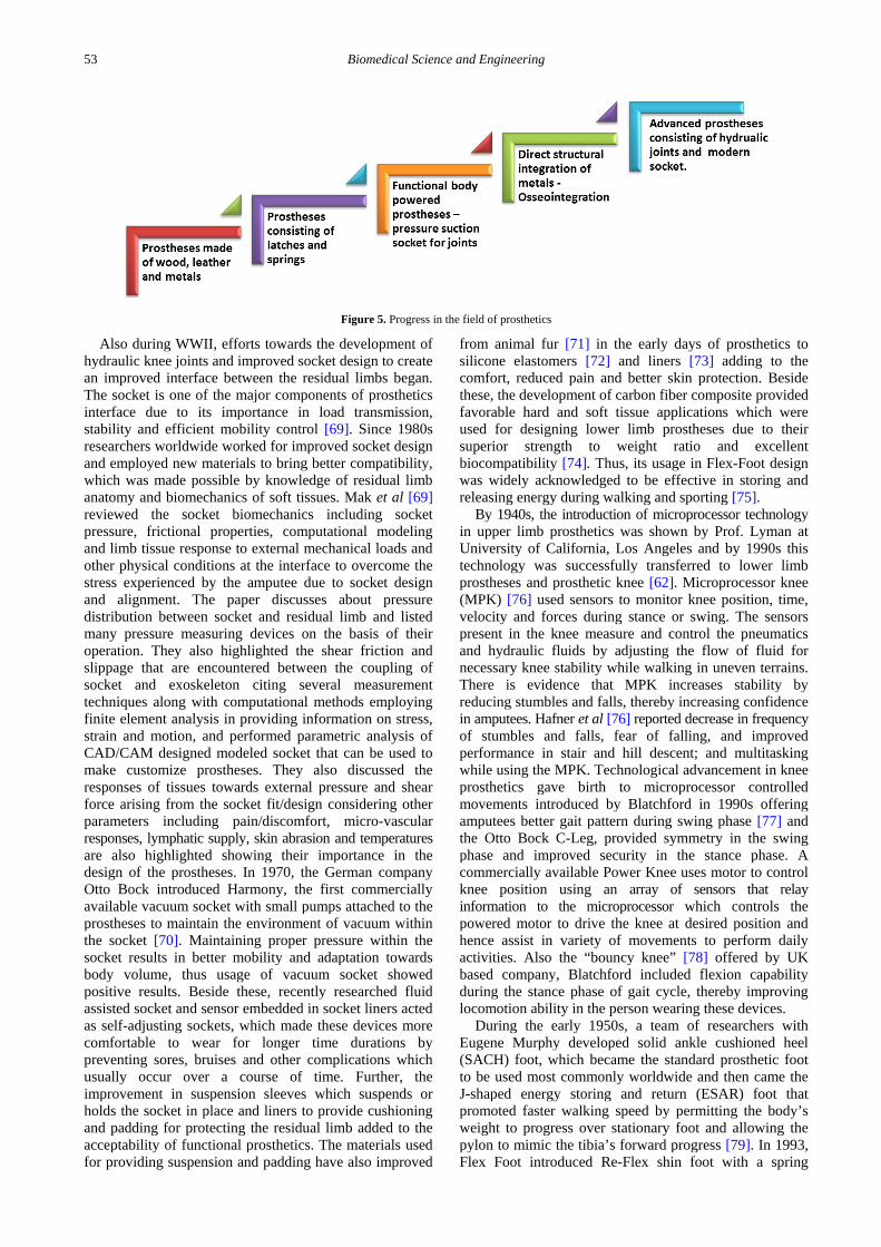
53 Biomedical Science and Engineering
Figure 5. Progress in the field of prosthetics
Also during WWII, efforts towards the development of hydraulic knee joints and improved socket design to create an improved interface between the residual limbs began. The socket is one of the major components of prosthetics interface due to its importance in load transmission, stability and efficient mobility control [69]. Since 1980s researchers worldwide worked for improved socket design and employed new materials to bring better compatibility, which was made possible by knowledge of residual limb anatomy and biomechanics of soft tissues. Mak et al [69] reviewed the socket biomechanics including socket pressure, frictional properties, computational modeling and limb tissue response to external mechanical loads and other physical conditions at the interface to overcome the stress experienced by the amputee due to socket design and alignment. The paper discusses about pressure distribution between socket and residual limb and listed many pressure measuring devices on the basis of their operation. They also highlighted the shear friction and slippage that are encountered between the coupling of socket and exoskeleton citing several measurement techniques along with computational methods employing finite element analysis in providing information on stress, strain and motion, and performed parametric analysis of CAD/CAM designed modeled socket that can be used to make customize prostheses. They also discussed the responses of tissues towards external pressure and shear force arising from the socket fit/design considering other parameters including pain/discomfort, micro-vascular responses, lymphatic supply, skin abrasion and temperatures are also highlighted showing their importance in the design of the prostheses. In 1970, the German company Otto Bock introduced Harmony, the first commercially available vacuum socket with small pumps attached to the prostheses to maintain the environment of vacuum within the socket [70]. Maintaining proper pressure within the socket results in better mobility and adaptation towards body volume, thus usage of vacuum socket showed positive results. Beside these, recently researched fluid assisted socket and sensor embedded in socket liners acted as self-adjusting sockets, which made these devices more comfortable to wear for longer time durations by preventing sores, bruises and other complications which usually occur over a course of time. Further, the improvement in suspension sleeves which suspends or holds the socket in place and liners to provide cushioning and padding for protecting the residual limb added to the acceptability of functional prosthetics. The materials used for providing suspension and padding have also improved
from animal fur [71] in the early days of prosthetics to silicone elastomers [72] and liners [73] adding to the comfort, reduced pain and better skin protection. Beside these, the development of carbon fiber composite provided favorable hard and soft tissue applications which were used for designing lower limb prostheses due to their superior strength to weight ratio and excellent biocompatibility [74]. Thus, its usage in Flex-Foot design was widely acknowledged to be effective in storing and releasing energy during walking and sporting [75].
By 1940s, the introduction of microprocessor technology in upper limb prosthetics was shown by Prof. Lyman at University of California, Los Angeles and by 1990s this technology was successfully transferred to lower limb prostheses and prosthetic knee [62]. Microprocessor knee (MPK) [76] used sensors to monitor knee position, time, velocity and forces during stance or swing. The sensors present in the knee measure and control the pneumatics and hydraulic fluids by adjusting the flow of fluid for necessary knee stability while walking in uneven terrains. There is evidence that MPK increases stability by reducing stumbles and falls, thereby increasing confidence in amputees. Hafner et al [76] reported decrease in frequency of stumbles and falls, fear of falling, and improved performance in stair and hill descent; and multitasking while using the MPK. Technological advancement in knee prosthetics gave birth to microprocessor controlled movements introduced by Blatchford in 1990s offering amputees better gait pattern during swing phase [77] and the Otto Bock C-Leg, provided symmetry in the swing phase and improved security in the stance phase. A commercially available Power Knee uses motor to control knee position using an array of sensors that relay information to the microprocessor which controls the powered motor to drive the knee at desired position and hence assist in variety of movements to perform daily activities. Also the “bouncy knee” [78] offered by UK based company, Blatchford included flexion capability during the stance phase of gait cycle, thereby improving locomotion ability in the person wearing these devices.
During the early 1950s, a team of researchers with Eugene Murphy developed solid ankle cushioned heel (SACH) foot, which became the standard prosthetic foot to be used most commonly worldwide and then came the J-shaped energy storing and return (ESAR) foot that promoted faster walking speed by permitting the body’s weight to progress over stationary foot and allowing the pylon to mimic the tibia’s forward progress [79]. In 1993, Flex Foot introduced Re-Flex shin foot with a spring

Biomedical Science and Engineering 54
loaded shock absorber that improved the biomechanical performance of dynamic foot response [80]. In order to evaluate the gait kinematics and dynamics during stance of unilateral, below-knee child amputees Schneider et al [79] compared the SACH foot and the energy-storing Flex-foot prostheses based on comfort and speed of walking. The three-dimensional movements of the lower limbs were recorded and synchronized with ground reaction forces, moment and joint power profile. They found marked asymmetries in ground reaction force, joint moment, and power profiles for the prosthetic versus the natural limb, but with the Flex foot the asymmetries were less pronounced as compared to the SACH foot. They observed amputees with Flex foot required greater moments and power at comfortable pace as compared to the SACH foot, but during fast walking, the SACH foot required greater output from the natural limb and noted that foot returned more energy as compared to SACH foot. Thus, they concluded that Flex foot had a greater potential for reducing the energy cost of walking at comfortable and fast speeds for the below-knee child amputee.
In 2006, first bionic prosthetic feet named Proprio Foot was introduced by Ossur that used accelerometer to sample ankle motion by identifying specific events during gait cycle such as heel strike and toe off [81]. This provided advantage of identifying slopes and stairs for better single limb balance. Recent development includes the Power Foot by the MIT research team that is able to mimic normal human ankle walking behavior and the Spring Ankle with Regenerative Kinetics (SPARKy), which was designed by the research teams of Arizona State University and St. Louis University that combined the power of regenerative kinetics, brushless direct current motor and long lasting roller screws [82].
In the last three decades there have been far reaching development in PT and rehabilitation allowing individuals with limb loss to return to functional level. Maximum functional performance in PT is attributed to advancement in prosthetic components, fabrication techniques, socket design and surgery. These provided individuals with limb loss better stability, comfort and performance thereby reducing stress developed on the body. However, the usage of these modern prostheses largely depends on the socio-economic conditions of the individual, knowledge of recent advancements in the field and the costs which determines the affordability of these devices. As Bhaskaranand et al [83] pointed out that prosthetic rehabilitation of patients with financial constraints requires durable and low cost prostheses. People living in most developing countries with loss of limb or its control are not able to avail this modern equipment to improve their quality of life and their inclusion in the society is still a matter of great concern. Different types of artificial limbs have been developed till date, depending on which limb or part of the limb needs to be supported, however, only a small number or percentage of the disabled people have been able to procure them to date and some those who had procured, had rejected due to minimum functionality. Justin Laferrier et al [84] discuss this issue and realized that the present PT is not able to replicate the anatomic function of limb which is the root problem for unsuccessful or rejected prosthetics. They mention that the best prosthetic for different individuals needs to be decided on the basis of material, design, cost is yet to be
found. They stressed that functional ability matching is the key for optimized physical performance of the prosthetic device and the primary goal of rehabilitation team. Marks and Micheal [41] in their review proposed that the artificial limbs could be made more lifelike using better materials and advanced technology. They mention that healthy amputees can participate in normal full range of responsibilities like walking and playing due to advancements in PT. They suggested direct skeletal attachment or osseointegration as an option for some amputees with improvements in microprocessor controlled prostheses for fine-tuning movement control and independence while task performance. They also touched upon overcoming funding constrains in the development of future prostheses. They emphasized the need for low cost material like molded plastics which are light and durable and are required in low income countries. For example, the prostheses produced by low cost polypropylene plastics by Red Cross Committee are well accepted worldwide.
Categorically the artificial limbs could be classified, following into one of the four types [85] namely transtibial, transfemoral, transradial, and transhumeral. In a more generalized category and to make the classification easier, they may be broadly classified as upper extremity prostheses (UEP) and lower extremity prostheses (LEP) which are discussed in subsequent sections. A.1. Lower extremity prostheses
Loss of lower limb directly affects the mobility of an individual along with their body image and self-esteem hampering their quality of life. The loss may lead to adverse health conditions due to depression in amputees. Especially, individuals who earn their living from motor skills are most vulnerable to adverse reactions. Others who have a wide range of skills or whose main line of work is not particularly dependent on the function of the lost limb may experience less emotional difficulty. However, with progress of time, the amputees start adapting and adjusting in the society, and acceptance towards the loss grows. This helps to seal the emotional wound restoring mobility using assistive devices. Commercially available lower limb prostheses can be divided into three categories [86] — mechanically passive devices, microprocessor-controlled passive devices, and powered devices. The movement of passive prosthetic joints relies on the effects of ground reaction forces and on the properties of the mechanical components, such as hydraulic and pneumatic valves, or sliding joints. Thus, an individual has to make extra movements with their trunk, pelvis, and residual limb to control this prosthesis. Such control significantly limits the functions of the prostheses—especially for those with amputations at the knee or higher. Microprocessor-controlled passive transfemoral prostheses employ sensors and a microcomputer to modulate the resistance of the knee joint [87,88]. Aeyels et al [87] reported the first phase of development of above knee prostheses equipped with microcomputer controlled knee joint in order to enhance the patient's gait comfort, safety and cosmesis. In their study, they replaced the knee joint of a conventional, modular prosthesis with a knee joint mechanism, equipped with a controllable brake on the knee joint axis with sensors and microcomputer added, keeping the system self-contained. The design permitted the use of external

55 Biomedical Science and Engineering
computer based control unit that ran a program on control algorithm to reveal the patient's intent. Their study revealed controlled knee flexion during stance phase of the trials. Thus, the implementation of powered prosthetic knees and ankles, driven by active actuators were further explored to improve the function of lower limb prostheses [89]. Cestari et al [90] provided an overview of the actuator requirements that must be fulfilled while evaluating the behavior of leg joints. They highlighted that current commercial devices contain joints with stiff actuators that cannot adapt to unpredictable environments and consume more energy, thus not being appropriate in active orthoses. Hence, adjustable compliant actuators are being incorporated into exoskeletons and active orthoses in order to improve joint control during locomotion cycle. They also evaluated the proposed variable stiffness actuator prototype in an exoskeleton knee joint operated by a state machine that exploits the dynamics of the leg, which resulted, decrease in actuation energy demand and better adaptability to disturbances.
Figure 6. Different Types of lower limb prosthetic device
The acceptability of the prosthesis depends on several factors including cosmetics, mass properties of the prosthesis, comfort, and function that are directly dependent on the quality of fit of the socket, the quality of suspension, the type of components used and the relative geometric i.e. the alignment of the artificial limb. Failure to provide a satisfactory alignment may affect the amputee locomotion, such as difficulty in walking, stump pain, or tissue breakdown leading to discarding of the device by the physically challenged person. Zahedi et al [91] performed a systematic study on alignment of lower limb to understand the parameters to achieve optimum alignment for enhancing the acceptance of prostheses. They established a range of alignment for each patient and found that an amputee can tolerate alignment ranging as much as 148mm shifts and 17 degree tilts. These quantitative values are benchmarks in the position alignment and tilt degree in lower limb prosthetics. Additionally, Bhuyian et al [92] reviewed control systems used in prosthetics to recreate devices to imitate biological limb and found that design of prosthetics depend on essential factors such as weight-force ratio, strength, durability, adaptability, wear-ability, degree of freedom, resistance to environment, and functional capability; and the control system of the prosthesis includes – actuators, sensors, controllers, and interfacing units. They listed different types of control systems – mechanical, electrical, electro-mechanical, hydraulic/pneumatic power assisted biomechanical, myoelectric, bionic, etc. control systems that are in use or can be used and suggested further improvement in these technologies can overcome existing setbacks. Thus, to provide exceptional strength-to-weight characteristics and
quality of superior biocompatibility, a majority of today’s upper-limb and lower-limb prostheses are presently being made from composites with underlying polymer matrix. Schultz et al [93] reported the application of composite materials in lower limb prostheses due to their hard and soft tissue interaction and suggested its future applications in sensory feedback systems and tissue engineering. Also, improving designs of prosthetics by employing different control systems, muscle of carbon fiber, mechanical linkages, motors, computer microprocessors, and innovative combinations of these technologies are allowing more control of the prosthetics to the end user. Recent research has focused on the development of an artificial prosthetic limb, which would be capable of imitating the behavior of a biological limb with the help of different types of joints, sensors, controllers, and actuators all working in coordination and synchrony to perform a given task. A.2. Upper extremity Prosthesis:
Complete or partial loss of upper limb control affects every activity of daily living (ADL) because of varied function of our hand like lifting, grasping, throwing, eating, washing, showing emotional care, etc. The role of hand is not just limited to functional movements but it is also psychosocial including use in communication, caressing, gestures and sensation. Upper limb injury can be due to trauma, industrial or road accidents etc. or other neuronal diseases like MND, NDD. Though advance safety standards and more machines assisted automated work environment in industries has greatly reduced chances of injuries to limbs leading to amputation but the growing neurological diseases are a matter of great concern. Study of Dillingham et al [94] showed that 70% of upper limb amputation in the US between 1988 - 1996, was below elbow caused by trauma. Similarly, Bhaskaranand [83] reported approximately 66% below elbow amputees in India. Thus, the rehabilitation of patients with upper extremity injuries using appropriate prostheses that can complement the physical, functional and vocational needs such that they can smoothly perform ADL and return to work is considered to be an important requirement.
Through the historical development of prostheses design and deployment for its usage, it was observed that more emphasis was towards lower limb rehabilitation than the upper limb rehabilitation. Upper limb prosthetics that were developed decades ago were passive devices with no moving parts which served more of cosmetic rather than functional purpose. The prosthesis was made of metals carved with fingers or attached with sharp edges. Amputees from royal families used cosmetic hand made of expensive metal(s) to hide their imputed limb in ceremonial functions and the soldiers were using it with sharp edges providing them with added advantage during battle. These cosmetic hands had no other functional capability or use for the common man. This led to development of sophisticated active prostheses which have moving components to perform limb movement(s) adding to the functional capability [95]. However, they too lacked sensation of feeling, touch and texture, an area which is of active research these days.
Active upper limb prostheses fall into any of these three functional categories [93]: (i) body-powered, (ii) myoelectric, and (iii) hybrid. Body-powered prostheses are largely mechanical devices such that the amputees

Biomedical Science and Engineering 56
have to use shoulder movements to pull on a cable and sequentially operate prosthetic functions such as the elbow, wrist, and terminal device. The advantages of body-powered prostheses include: simple operational mechanisms with intrinsic skeletal movement (which voluntarily opens/closes a terminal device), silent action, light weight, moderate cost, durability and reliability, and sensory feedback about the positioning of the terminal device. The history of powered prosthesis dates back to the pneumatic hand patented in Germany in 1915. This was probably the first electric powered limb, but was not able to find its usage; instead by 1948 CO2 powered limbs developed by Kiessling found its use in orthotics [96]. The developmental journey of powered prostheses is given by Childress, 1985 in his paper [97]. He documents the early days of powered prostheses which developed from simple cosmetic device to the recent functional hand with some sensory feedback. He reviewed the continuous improvement brought in artificial hands through simply holding objects to gripping and grasping that required sensory feedback.
Figure 7. Different Types of upper limb prostheses
Reinhold Reiter in early 1940s conceived and developed the idea of myoelectric controlled prostheses that was implemented and patented in 1945.The device was controlled by vacuum tube amplifier which required considerable amount of power but lacked portability due to bulky size and was not battery operated. A myoelectric prostheses is device which is controlled by the electrical activity of a muscle, i e. by a myoelectric signal generated in the muscle. The electrical activity generated naturally due to contraction in the muscles of the residual limb is amplified and processed so that the flow of electricity from the battery to the motor operates the artificial limb. Myoelectric prostheses has its own advantage - they are more self-contained and do not require the complex harnessing of body power, and they offer a better cosmesis, wider range of motion, and higher grip strength [98,99]. Control of myoelectric prostheses is generally achieved by recording from two independent muscles or by differentiating weak and strong contractions of one muscle. The difference between powered and myoelectric prostheses is that - at present the source of energy is rechargeable batteries in powered prosthesis and its control is achieved by electro-mechanical switch operated by body movement using straps or cables whereas there is no requirement of cables or straps in case of myoelectric prosthesis as the muscles in the residual limb provides control signal for powered components.
In 1955, Battye et al [100], developed myoelectric control system for powered prostheses and by 1957 many groups like Bottomley in England, Kato in Japan, Herberts in Sweden, Reswick and coworkers in USA started to
work in myoelectric controls independently without the knowledge of Reiter’s invention but most of their devices were not able to find clinical and commercial acceptance. During this period, understanding of the process by which myoelectric signal is generated and how to use this signal for better use grew. This provided new insight in the course of development of myoelectric control devices and in 1960 first myoelectric prostheses developed by Kobrinski in USSR found clinical use, which was commercialized and exported to UK and Canada. The Kobrinski prosthesis used the myoelectric signal from one muscle group to cause the electric hand to close, and the signal from another muscle group to cause the hand to open, a control mode which still is popular today, because it mimics the body's natural use of antagonistic muscles. The present availability and clinical impact of myoelectric prostheses is well reported by Scott [101]. Scott describes the capabilities and limitations of myoelectric prostheses components stating that there is no mechanical system to replace the function of shoulder movement and also lack of availability of artificial arm that can provide strength, grasp, grip, manipulative ability and sensory input like a normal hand. He suggests the use of implantable telemetry system that will provide better communication channels between muscles and peripheral nerves thereby overcoming the limitation of artificial arm in its inability to monitor and control different functions using normal feedback pathways. He further suggested the development of pattern recognition techniques and algorithms for efficient computational processing of electromyogram (EMG) signals for better use in multifunctional myoelectric prostheses. Choi et al [102] in their paper titled EMG based assistive computer interface for the upper limb disabled persons, showed that neural signals provide body motion information faster than kinematic and dynamic interfaces, for understanding the user’s perception from brain to the muscle. Their objective was to develop a EMG based computer interface for upper limb amputee such that they can access computer by moving the pointer and clicking without using interfacing devices like keyboard or mouse. Supervised multilayer neural network was used to understand the user’s intention and patient training performed using back-propagation algorithm was implemented for classification of intended movement. This technique was used for matching the most activated neuron with the class of movement. The viability of their interface was quantitatively evaluated using Fitt’s law test and it showed the effectiveness of pointing device. The success rate of pattern classification for the six classes of wrist movement was found to be >96%. The IP of developed interface (using Fitt’s law test) was 1.341 bits/s as compared to 7.73 bits/s of mouse, much better compared to the commercially available pointing interfaces which showed 20 times lower efficiency than that mouse. They proposed EMG based HCI technologies, which were considered to be feasible for the disabled persons having better techniques requiring greater velocity and direction of interfacing devices developed using EMG signal. This work could be further extended to the development of bionic robotic devices. Moreover, it was found that the problems inherited in traditional myoelectric control could be addressed to some extent by the developing three technologies namely implantable EMG electrodes, EMG pattern recognition devices and

57 Biomedical Science and Engineering
targeted re-innervation. These technologies will act as neural interfaces for controlling the upper limb prostheses. In view of this Schultz and Kuilen [93], reviewed the state of the art and future possibilities of neural interface for control of upper limb prostheses because the currently used prosthesis in upper limb amputee’s lack functionality like natural arm in meeting daily activities – a common reason of prostheses abandoning. The limitation of the current body powered ULP, is that they lack coordinated movement of multiple joints and wrist instead there is need for multi-articulated hand and wrist movements in the prosthetic limb. Myoelectric control faces difficulties such as suitable recording of EMG and lack of information for multiple functions leading to less functional control and finally frustration and abandonment of the device by the user. Thus, the use of EMG pattern recognition technique (that utilizes algorithm for searching patterns of muscle activity on more than one muscle site for making the control of prosthesis easier for patients) and target innervation (for implanting myoelectric sensors at residual limb) may solve tradition myoelectric control problems. Further benefit of targeted innervation may provide cutaneous sensory feedback for proper motor and joint control. They suggested future benefits of neural interfaces developed for control and communication from muscle, nerve and brain could be implemented in ULP as direct nerve interface show ability to control joint movement and control grip. The same was with regard to cortical spike recording and ECoG for performing complex movement of hand and fingers. Such hybrid prosthesis is achieved by combining myoelectric control and body-powered operation [103] like for example, in above elbow amputation, the myoelectric control for hand function can be combined with the cable control system for the elbow function. B. Robotic Rehabilitation Devices (RRD)
The successful motor rehabilitation of patients suffering from motor neuron disorders requires intensive task specific hand-to-hand therapy by the physiotherapists that requires time and budgeted expenditure. These requirements lead to the advent for intelligent machines to promote motor recovery and understanding, resulting in the emergence of robot-assisted motor rehabilitation. Few robots namely, MIT-Manus, assisted rehabilitation and measurement (ARM) guide, mirror image movement enhancer (MIME), gait trainer (GT) I, Lokomat, Bi-Manu-Track, etc. are used as RRD, which are clinically recommended on the basis of several studies performed using them that showed encouraging improvements in motor rehabilitation therapies in both upper and lower extremities [133].
RRD are emerging as new rehabilitation therapy for patients suffering from movement disorders basically due to two reasons; one shortage of therapists and caregivers for assisting disabled persons [134] and the other neurorehabilitation [135]. In neurorehabilitation, the patients suffering from movement disorders are assisted to perform normal movement and hand function, thereby triggering the same functionality in other parts of the brain which then generate necessary neural signal, thus inducing brain plasticity [136]. The RRD are either passive devices, that utilizes patient’s power to maneuver the device, for example the use of springs to store/restore energy during the gait cycle [137]; or active devices that act as energy sources for maintaining the center of mass and torque
during gait cycle [138]. Additionally, the primary goal of robotic rehabilitation is to provide cost-effective therapy that is equivalent to or better than conventional therapy. These RDD can be for lower or upper limb rehabilitation, beside this, the lower limb robots are separated into robot-dominant devices, where the robot drives the motion of the human and cooperative devices, where the robot and the human share control. Studies on robot-dominant devices are shown to be less effective, as the human is less actively engaged [139] whereas cooperative rehabilitation robots are typically force-controlled, employing techniques such as assist-as-needed, error augmentation, or proportional electromyography for performing the training task. It is noted that upper-limb rehabilitation robots have outperformed conventional therapy [140] whereas the conventional lower limb therapy is still the most effective option. To date, robot-assisted task-specific training has been administered with a less-intensive paradigm and has been associated with improved upper-limb motor scores for acute [141] and chronic stroke patients [142] with mild-to moderate impairment and also for chronic stroke patients in pilot studies with moderate-to-severe impairment. MacClellan et al [143] performed a pilot study to test the effectiveness of robotic therapy for improvement in motor outcomes in stroke patients. In their study they used InMotion2, commercially available MIT-Manus robot developed at Massachusetts Institute of Technology, Cambridge specifically designed for upper limb rehabilitation [144]. The InMotion2 robot used performance based algorithm that adjust itself according to time and assistance to reach the target object. In other words, it can be said that this robot allows the patients to express their movements by assisting them to reach for it during therapy sessions and complete the task for training the upper limb. They used higher intensity of massed-practice intervention for short duration and found that patients categorized as moderate-to-severe show significant improvement in motor function. Most interestingly, they found that severely impaired patients showed remarkable improvement as compared to the moderately impaired.
RRD is found to improve the kinematics based on the control architecture and benchmark criteria. Anwar and Jumaily [135] illustrated the performance of RRD based on control architecture and benchmark criteria. They designed an exoskeleton, which was equipped with position, force and impedance controller which provided smooth HMI at different phases of gait cycle during lower limb therapy of disabled patients. Since the relation of the control system was nonlinear, they used fuzzy rule based controller at the interaction point of the exoskeleton. In order to minimize the interaction force, they modulated the angular velocity, impedance and torque such that minimum error occurs during different tasks performed by these patients. Further, they also demonstrated the architecture of one degree of freedom in lower limb exoskeleton that ensures better gait pattern. Their proposed RRD was a multi-input multi-output (MIMO) system that was capable of changing the weight associated in accordance to changing parameters for fulfilling the objective of smooth exoskeleton.
Most recently, research in VLSI field lead to the development of silicon neuron as evident by the works of Tenore and Etienne-Cummings, [145] who demonstrated the use of silicon neuron designed using VLSI technology

Biomedical Science and Engineering 58
which can mimic the function of central pattern generator (CPG) in humans. They programmed the silicon to control robotic locomotion and further added that the same can be used in upper limb prosthetics. This opens new avenues in the fields of robotics and prosthetics. They demonstrated the lower limb application of silicon neuron by using a bipedal robot prototype having actuating hips and knees that facilitated walking. For upper limb application, they used the myoelectric signals from both normal and trans-radial amputee for developing real-time application that featured multi-degree of freedom of upper limb prostheses.
These developments are showing promising results in improving motor function and also provide assistive support to the role of physiotherapists in rehabilitation. C) Human Machine Interaction (HMI)
HMI is the interaction of humans with machines. The advent of machines fascinated mankind and its interaction with human dates back to several decades. Machines were crafted to act as passive extensions of the human body. However, the development of prosthetics has changed the way humans interact with machines [104]. Hogan [105], noted the challenges and possibilities of human interactive machinery and reviewed that the machines designed for cooperating physically with humans are emerging technology to support human activity. He highlighted the design of control system that can endow the amputee’s motorized arm prosthesis to take advantage of mechanical constraints and cooperation between natural and artificial limb such that increased motor abilities can be obtained. He further added that the development of robots capable of safely interacting and cooperating with humans are enabling new form of therapy for neurologically injured patients. Machines are now designed to cooperate physically with humans, enabling new ways to support human activity. Human machine relation is reported in literature with different terminologies such as man machine interaction (MMI), Brain computer interaction (BCI) and Neuroprosthetics. These terms ultimately narrow down to the basics of HMI with varying differences.
In this section, HMI is discussed focusing on the machines interactive behavior and human motion intention sensing. The machines interactive behavior depends on the control system designed in order to maintain safety, stability and performance. These factors endow an amputee's motorized arm prosthesis with sufficient responsiveness to take advantage of mechanical constraints; and support dexterous cooperation between natural and artificial limbs [105]. Bhuyian et al [92] in their paper discussed various components that are required for developing control systems for prosthetics which includes sensors, actuators, controllers and interfacing units. Additionally, the effort to restore mobility to the amputees has encouraged the development of several devices using robotic exoskeleton technology like ReWalk personal exoskeleton system [106], a wearable robotic exoskeleton that provides powered hip and knee motion to stand upright and walk. Ekso [107] is a wearable bionic suit which enables individuals with any amount of lower extremity weakness to stand up and walk over ground with a natural, full weight bearing and reciprocal gait. Walking is achieved by the user’s weight shifts to activate sensors in the device which initiate steps. Battery-powered motors drive the legs, replacing deficient neuromuscular
functions. Rexbionic products [108] like REX is a hands-free robotic mobility device for rehabilitation and REX P, for use at home, enables users to walk and stand with their hands free – providing more work and recreation options. However, these devices contain joints with stiff actuators that are not appropriate for HMI. Thus, adjustable compliant actuators are being cautiously incorporated into new exoskeletons and active orthoses [109]. Another key issue in physical HMI is interpreting the motion intention from physiological signals. Thus, Kwon and Kim [110] noted this issue of intention sensing of human motion and devised a method for real time upper limb motion prediction using EMG signals and an artificial neural network algorithm for cooperative interactions. The artificial neural network algorithm is an excellent model to solve musculoskeletal mechanics and hence it was used to map the nonlinear relationship between the EMG signal and upper limb motion. The performance was evaluated on the basis of subject’s estimated movement, ability to manipulate their position and the response time. The result showed that though the proposed method was not superior to the use of direct angle measurement, it provided accuracy and good response speed for interactions.
In human motion and intention sensing, EMG signal analysis coupled with artificial neural network algorithm and alternate muscle activation sensors with assisting manipulators or prosthetic devices have been shown to predict the intention of the patient’s ability to move their artificial limb or prosthesis. In the quest to develop alternate muscle sensors to overcome the limitation of EMG, Cen et al [111] developed the optical muscle activation sensors for measuring the optical density in muscle by emitting and gathering the single wavelength light source. The forearm force level was estimated and the comparative result of surface EMG and optical density of muscle showed that optical density can be used to measure muscle contraction. They further suggested that future work for detecting motion intention will require advanced algorithm in signal processing and modeling of optical tissue interaction. Many computational techniques and concept of artificial intelligence such as genetic algorithm, neural networks, and support vector machine are deployed to better implement the motion intention for better coordination of artificial limbs. However, till recently a general approach for the control of functional movements had been followed based on the biological principle of motor control such as representation of motor patterns, reflexes and motor skills for developing advanced assistive systems, but use of advance soft computing techniques like expert systems, fuzzy sets, analytical methods are being employed to design better devices for rehabilitation [112]. D) Brain Computer Interface (BCI)
In 1999, the first international meeting [113] in BCI was held at New York, USA which defined its meaning as “A brain–computer interface is a communication system that does not depend on the brain’s normal output pathways of peripheral nerves and muscles.” In other words, a BCI is a computer-based system that acquires brain signals, analyzes and translates them into commands that are relayed to an output device to drive external devices without participation of the spinal and peripheral motor system [114]. BCI most commonly uses either EEG activity recorded from the scalp [115,116,117,118] or the

59 Biomedical Science and Engineering
activity of neurons recorded from implanted electrodes [119,120]. Ramoser et al [117] studied the single trial EEG signal associated with imaginary movement for rapid and reliable discrimination of EEG patterns. They demonstrated that spatial filters for multichannel EEG can extract discriminatory information from two populations of single-trial EEG, recorded during left and right hand movement imagery which reflected the specific activation of cortical areas. Further, the high recognition rates and computational simplicity make it a promising method for an EEG-based BCI. The central element in each BCI is a translation algorithm that converts electrophysiological input from the user into output that controls external devices. The main goal of BCI is to replace or restore useful function to people disabled by neuromuscular disorders such as ALS, CP or SCI [121]. Wolpaw et al [118], clearly describe the working mechanism of BCI along with its control and communication. They explained the working of the BCI system which involves signal acquisition, signal processing through feature selection and translation algorithm such that BCI can become a communication channel between the patient and the external world. Besides EEG, they suggested the use of other electrophysiological signals like slow cortical potentials, P300, mu or beta rhythms, that can be used in BCI and emphasized on the increased data transfer rate for faster communication, better algorithms for translating the signals to commands and the collaborative works of different discipline involving neurobiology, engineering, mathematics and computer science. Recent EEG based BCI are shown to control artificial hand [122], EEG based motor imagery, the BCI demonstrated control of prosthetic hand’s grasping force and many such examples exists [123]. Besides EEG, other types of brain signals that can be measured either invasively or non-invasively are being studied for use in a BCI. These signals include spike trains from single neurons [124,125], extracellular local field potentials (LFPs) [126], electrocorticograms (ECoG) [127], electroencephalogram (EEG) oscillations, real-
time-functional magnetic resonance imaging (rt-fMRI) [128], and near infrared spectroscopy (NIRS) [129]. The implementation of these different brain signals in BCI for restoring neuromuscular disorders and rehabilitation is thoroughly explained by Shih et al [121]. Shih reviewed emerging fields of BCI along with different physiological signals used like scalp-recorded EEG, ECoG, and implantable electrodes together with few functional application of MEG, fMRI, fNIR technologies showing current and future key areas of research. In view of alternative signal usage in BCI, there is need to find other brain areas that are not explored as application areas in BCI. An example being, Felton et al [127] work ECoG based BCI application; where they implanted intracranial electrodes in sensory and other motor areas of the brain and found that participants with electrodes over motor and/or sensory areas and after undergoing 2-7 days of training to use motor and/or auditory imagery; were able to control the movement of a computer cursor. Their findings indicate that sensory and other brain areas not previously considered ideal for ECoG-based control that can provide additional channels of control which may be useful for a motor BCI. Similarly, Niels Birbaumer et al [114] in their paper titled brain computer interface in paralysis mentioned the challenges in communicating with patients suffering from paralysis. They mention that communication with patients suffering from lock-in syndrome or paralysis is unsolved and also the movement restoration of stroke/TBI etc. patients does not offer significant improvement by the available treatments. They reviewed the recent advances in BCI technology and the various signals like spike trains from single neuron, extracellular local field potential, ECoG, EEG, event related brain potential (ERP), fMRI, near infrared spectroscopy (NIRS) showing promising results. They also suggested that the clinical application of BCI may rest on current EEG signals and neurofeedback involving analysis of EEG patterns for movement restoration by tuning functions of sensory motor neurons.
Figure 8. Working model of brain computer interface
Since present BCI mostly works on EEG signals, studies and trial on other electrophysiological signals are being explored round the world for improving the
limitations of existing systems. However, there is need for methods and devices for improving the signal acquisition hardware, signal processing algorithm, computational

Biomedical Science and Engineering 60
processing speed, etc. such that a reliable and robust system could be developed to augment basic functioning of a normal human being that can be provided to the disabled to enhance their functionality. E) Neuro-Prosthetics
Neuro or Neural prosthetics (NP) is a series of device which substitutes a motor, sensory or cognitive modality that might have been damaged as a result of an injury or disease [5]. In neuro-prosthetics, external devices are connected with the human nervous system that records the brain signals from user by computational analysis and relays this information to the external effector that act on those intentions. The cumulative voltage across motor cortex area encompasses the activity of millions of neurons and the generated action potential is conducted by peripheral nerves to desired body part with the help of output effectors. The corresponding brain or nerve signals are used to control computer cursor movements and robotic arms, or enable the reanimation of paralyzed limbs. NP enables direct interfacing with the brain and has great potential for restoring communication and limb control in disabled individuals. The key component of interface
design is the electrode and the interface material that can record natural bioelectric signals and provide artificial excitation of nerves/muscles. Jiping He et al [5], reported the use of neural interface in activating residual neural function and brain control of lower limb function. They touched upon recent progress in neural interface technologies and demonstrated the application of direct cortical control of robotic arm or computer cursor for simple movement using cortically controlled neuro-prosthetics to improve motor function in subjects with severe neurological deficits. They further emphasized that the ability of neural systems to adapt to changes and learning new functions are important considerations in the design and development of neuro-prosthetics such that an intelligent neural interface can be designed that can activate residual function and facilitate control. However, several challenging engineering and biological issues still remain to be resolved before a practical system can be developed for patients to receive real benefit of neuro-prostheses. Future approach in designing NP will require smart materials and sensors and also wired/wireless neural interfaces.
Figure 9. Artificial power Limb controlled by processed brain signal from external control an implanted stimulators in Neuroprostheses
NP is sometimes contrasted with a BCI, which connects the brain to a computer rather than a device meant to replace missing biological functionality. The key components of a neuro-prosthetic system for motor function restoration include a set of microelectrode arrays implanted in specific brain regions to monitor the neuronal activities, a signal-processing unit to extract intention for action and special commands from the brain, and a prosthetic arm/leg or a functional electrical stimulation (FES) system to activate appropriate muscles to carry out the desired action. Navarro et al [103], critically reviewed the characteristics and suitability of different types of electrodes and the biomedical applications of interfaces with the peripheral nervous system. They touched upon noninvasive electrodes like surface electrodes for recording of EEG, ECG, EMG signals; muscle electrodes for EMG recording and muscle stimulation as in FES, BION; implantable microstimulator for stimulation, epineural electrodes for selective activation of particular nerve fascicles and helicoidal electrodes for FES of the vagus nerve, book electrodes for urinary
bladder management in spinal cord-injured persons, cuff electrodes, intraneural electrodes, penetrating microelectrodes, etc. They further discussed the materials used for the electrodes, their insulation, encapsulation, choice of fabrication technology and bioelectronic interfaces and highlighted the control of neuroprostheses and hybrid system using PNS interfaces like EMG based control of NP. Additionally, Sanchez [104], in the paper titled “Neuroprosthetics in the 21st Century” highlighted the development of neuroprosthesis which can become an integral extension to the body for the disabled. This new technology presents challenges for engineers and scientist in designing a system that can replicate the physiological functions such that complex activities of daily life can be achieved. Currently used NP designs include perceptron, neural network, supervised/unsupervised/semi supervised learning. Thus, the integration of neural information processing, signal processing and feedback control is basic components of NP design and it must be built on the

61 Biomedical Science and Engineering
principle of intelligent action of an individual arising from sensory information and ending as goal-directed actions.
Leuthardt et al [60] in their paper titled “The emerging world of motor neuro-prosthetics: A neurosurgical perspectives” reviewed the emerging field of motor neuro-prosthesis and highlighted the features, functions and platforms of output BCI. They noted that the signals from the brain can be decoded to reflect user’s intent and the evolving fields of neuroscience, engineering and computing can implement neuro-prosthetics as a practical tool for disabled. They highlighted the neurosurgical perspective by questioning the safety, durability, reliability, implantation procedures and usefulness in terms of simple or complex function of BCI as framework to evaluate it for neurosurgical community. They touched upon the current BCI platforms that have potential for clinical applications like the EEG based systems, ECoG based, SCP, P300 evoked potential signals. They suggested that understanding the “neural code” is of significant importance as it can substantially improve the manner these devices operate and their future applications.
The concept of neuro-prosthesis may seem futuristic but there are several inspiring examples of this technology in clinical practice today that have demonstrated feasibility and even therapy. The best known of these examples are the deep brain stimulators or DBS which are designed with a goal to primarily send signals into the brain to treat the symptoms of Parkinson’s disease, tremor, and dystonia [63]. In DBS, the patients need to undergo brain surgical process which is painless as it works under the administration of local anesthesia. In this surgical process, two holes are drilled over the scalp and one electrode placed on each hole. These electrodes work as brain pacemaker. Placement of electrode surgically depends mainly on the targeted area of the brain e.g. in depression target area is the one that controls the mood. After electrode placement and feedback is provided by the patient, the electrodes are attached with the wire which already runs inside the patient body from head to the chest where battery operated generator is implanted to transfer and deliver the electrical impulses to the brain by wired electrodes [130]. Another most implemented neuro-prosthetic, widely used in real life is the cochlear implant [131], which is an electronic medical device that assists function of defective ears bypassing the cochlea (inner ear) to send sound waves directly to the brain.
Today, several different types of surgical brain implants are being tested for their ability to restore some level of functionality in patients with severe sensory or motor disabilities expanding the application areas of NP in TBI, ALS, AD, etc. Perhaps the most visible recent demonstration of the power of neuro-prosthetics was witnesses by millions when a paraplegic patient using a brain-controlled exoskeleton suit kicked off the 2014 World Cup ball in Brazil [132]. This astonishing work of designing an exoskeleton suit for the paraplegics is a work of Miguel Nicolelis, a Brazilian neuroscientist at Duke University.
6. Conclusion Our understanding of the limb control and the disorders
associated with it, have been instrumental in the development of various tools to overcome these disorders but till date
there isn’t any device or treatment that can be said to be a perfect replacement of natural functioning of the severed limb. Use of prostheses need to be brought within the budget of the disabled individuals such that their financial burden could be reduced. This is possible only by the employing low cost with improved quality materials and advancements in the manufacturing technology. Further, there is need to develop intelligent prosthetic devices using the knowledge of biomechanics, such that the devices can perform functionality similar to the natural limb in function and discomfort leading to pain, local bruises and irritation encountered by the amputee are reduced to minimum extent, so that they can wear and use it for long period of time. The need of improved multifunctional prostheses control to provide better stability and balance to the amputees along with grip and grasping postures are in demand. Moreover, the feedback mechanism of prostheses from signals recording, transmission and its processing in a short span of time with the help of efficient algorithms are new emerging areas of research. Additionally, the man machine interaction through HMI, BCI and neuro-prosthetics are opening new doors towards improved tools to link brain activity with man-made devices to replace the lost sensory and motor function. The developments in neuroprostheses which are focusing on the intent driven movement of the prosthetic device and the incorporation of sensory feeling that is relayed to and fro from the brain are becoming a boon for the disabled. Though NP is highly researched area in rehabilitation engineering, yet, they are still in the trials and not commercialized because of lack of robust algorithms that can completely translate the brain signal into intended motion. These limitations can be overcome when our understanding of the working of the human brain is complete. Day by day new functioning pathways of the brain are discovered that are related to motor performances. Furthermore, brain researchers also need to focus on understanding the mechanism and pathway leading to abnormalities in patients such as MND, NDD, SCI, etc. such that better functional devices can be designed targeting the abnormalities with modern interfaces and tools such as RRD, BCI and NP need to overcome their limitations as discussed in previous sections for better performance in disabled persons. Additionally, new technologies like TMS and DBS need to be further researched such that common disorders MND and NND can be treated without having invasive approach. In the process of developing treatment or tools for overcoming limb loss there is need for interdisciplinary approach and merger of neuroscience, biomedical engineering, computational biology and artificial intelligence, emphasizing better performance and improved devices such that they are easy to use, comfortable and available at a low cost.
References [1] S. J. Bensmaia, L. E. Miller, “Restoring sensoriomotor function
through intercotical interface: progress and looming challenge”, J nature reviews Neurosciences, vol. 15(5), pp. 313-315, 2014.
[2] R. W. Mann, “Cybernetic Limb Prosthesis”, Annals of Biomedical Engineering, Vol. 9, pp 1-43, 1981.
[3] C. Ethier, L. E. Miller, “Brain-controlled muscle stimulation for the restoration of motor function, Neurobiology of Disease” nbd, [ahead of print], Oct 14 ,2014

Biomedical Science and Engineering 62
[4] F. B. Horak, “Postural orientation and equilibrium: what do we need to know about neural control of balance to prevent falls? Age and Ageing”, pp. 35-S2 ii7-ii11, 2006.
[5] J. He ,C. Ma , R. Herman, “Engineering Neural Interfaces for Rehabilitation of Lower Limb Function in Spinal Cord Injured”, Proceedings of the IEEE, vol.96. no.7. pp. 1152-1166, 2008.
[6] K.T. Manal, T. S. Buchanan, “Biomechanics of human movement”, Standard handbook of biomedical engineering and design, McGraw Hill, 2004.
[7] J. M. Hollerbach, “Computers, brains and the control of movement”, Trends Neurosci, vol. 5. No 6. pp.189-92, 1982.
[8] R. A. Schmidt, “Schema theory of discrete motor skill learning”, Psychol Rev, vol.82, no. 4, pp. 225-60, 1975.
[9] A. G. Feldman, “Once more on the equilibrium-point hypothesis (lambda model) for motor control”, J Mot Behav vol. 18, no 1. pp.17-54, 1986.
[10] E. Todorov, “Optimality principles in sensorimotor control”, Nature Neuroscience, vol.7, no.9. pp. 907–15, 2004. [PubMed: 15332089]
[11] N. Dounskaia, “Control of Human Limb Movements: The Leading Joint Hypothesis and Its Practical Applications”, Exerc Sport Sci Rev. vol. 38, no. 4. pp.201-208, 2010.
[12] J. L. Smith, and R. F. Zernicke, “Predictions for neural control based on limb dynamics”, Trends in Neurosciences, vol. 10, no. 3. pp. 123-128, 1987.
[13] R. E. Kearney, I. W. Hunter, “System Identification of Human Joint Dynamics”, CRC Crit Rev Biomed Engin, vol. 18, pp.55-87, 1990.
[14] R. Shadmehr, M. A. Arair, “A Mathematical Analysis of the Force-Stillness Characteristics of Muscles in Control of a Single Joint System”, Biol Cybern, vol. 66, pp. 463-477, 1992.
[15] Y. P. Ivanenko, G. Cappellini, N. Dominici, R. E. Poppele, F. Lacquaniti, “Modular Control of Limb Movements during Human Locomotion”, J. Neurosci., vol. 27, no.41, pp. 11149-11161, 2007.
[16] L. Seth, D. N. Louis, W. David, Greenfield's Neuropathology (8th ed.), London Hodder Arnold, pp. 947, 2008.
[17] A. G. Reeves, R. S. Swenson, “Neuromuscular system disorders” in disorders of the nervous system 5th ed. Ch.3, sec.21. [Online] Available: http://www.dartmouth.edu/~dons/part_3/chapter_21.html.
[18] K. M. Steinberg, D. C. Koboldt, “Researchers identified a new host gene variants that could make people vulnerable to sporadic motor neuron disease”, University of Sydney, March 16, 2015.
[19] [Online] available: http://www.ninds.nih.gov/disorders/motor_neuron_diseases/detail_motor_neuron_diseases.htm.
[20] W. N. Löscher, E. L. Feldman, “Motor Neuron Diseases”, Atlas of Neuromuscular Diseases, pp. 283-290, 2014.
[21] W. Koroshetz, "Motor Neuron Diseases Fact Sheet: National Institute of Neurological Disorders and Stroke (NINDS)". Motor neuron disease, Department of health and human services U.S., pp. 1-20, March 2012.
[22] L.M Thompson, "Neurodegeneration: a question of balance". Nature 452 (7188): pp.707-8.
[23] D. C. Rubinszte, "The roles of intracellular protein-degradation pathways in neurodegeneration". Nature, vol. 443 (7113), pp.780-6,Oct 2006.
[24] S. DiMauro ,E. A. Schon, "Mitochondrial disorders in the nervous system". Annual Review of Neuroscience, vol. 31, pp. 91-123, 2008.
[25] D. E. Bredesen, R. V. Rao, P. Mehlen, "Cell death in the nervous system". Nature, vol. 443 (7113), pp.796-802, 2006.
[26] [Online] http://www.nlm.nih.gov/medlineplus/degenerativenervediseases.html.
[27] Brain facts.org, “Degenerative disease”. Available: http://www.brainfacts.org/diseases-disorders/degenerative-disorders.
[28] C.M Tanner, M.Brandabur, E.R.Dorsey, “Parkinson Disease: A Global View”, Rep. Parkinson, pp. 9-11, 2008.
[29] [Online] available: http://www.alzheimers.net/resources/alzheimers-statistics.
[30] Progress in mind, “Huntingtons disease”, Available on http://www.lundbeck.com/us/our-commitment/disease-information/huntingtons-disease-hd.
[31] [Online] available: http://www.nichd.nih.gov/. Cerebral Palsy: Overview. September 5, 2014.
[32] [Online] available: http://cerebralpalsy.org/about-cerebral-palsy/types-and-forms.
[33] [Online] available: http://www.brainandspinalcord.org/cerebral-palsy/types/ataxic-cerebral-palsy.html.
[34] A.I. Maas, N. Stocchetti, R.Bullock, "Moderate and severe traumatic brain injury in adults". Lancet Neurology, vol. 7(8), pp. 728-41, August 2008.
[35] M. Faul, L. Xu, M. M. Wald,V. G. Coronado, “Traumatic Brain Injury in the United States: Emergency Department Visits, Hospitalizations and Deaths 2002-2006. Atlanta (GA), Centers for Disease Control and Prevention”, National Center for Injury Prevention and Control, 2010.
[36] C. Collins, J. Dean, “Acquired brain injury” in Turner, 2002. M. Foster, SE. Johnson. “Occupational Therapy and Physical Dysfunction: Principles, Skills and Practice”. Edinburgh: Churchill Livingstone. pp. 395–96.
[37] P. A. Robertson, "Prediction of amputation after severe lower limb trauma." Journal of Bone & Joint Surgery, British vol. 73, no.5. pp. 816-818., 1991.
[38] U.S. National library of medicine. Medlineplus. “Amputation –trauma-causes”. [Online] Available: http://www.nlm.nih.gov/medlineplus/ency/article/000006.htm.
[39] R. B.Islinger,T. R.Kulko , K. A. McHale, “A review of orthopedic injuries in the three recent US military conflicts”, Mil Med, vol. 165, pp. 463-5, 2000.
[40] P. Meade, J. Mirocha, “Civilian landmine injuries”, J Trauma, in Sri Lanka, vol. 48, pp. 735-9, 2000.
[41] L. J. Marks, J. W. Michael, “Artificial limbs”, British Medical Journal, vol. 323(7315), pp. 732-735, 2001.
[42] L.Norgren , W. R.Hiatt, J, A.Dormandy , M. R.Nehler , et al “TASC II Working Group, Inter-Society Consensus for the Management of Peripheral Arterial Disease (TASC II)”, J VascSurg, vol. 45, pp. S:S5, 2007.
[43] K. Ziegler-Graham, E. J. MacKenzie, P. L. Ephraim, T. G. Travison, R. Brookmeyer, “Estimating the Prevalence of Limb Loss in the United States: 2005 to 2050”, Archives of Physical Medicine and Rehabilitation, vol.89(3), pp.422-9, 2008.
[44] A. Damir, ”Why Diabetic Foot Ulcers do not heal?”, J International Medical Sciences Academy, vol. 24(4), pp. 205-206, 2011.
[45] [Online] available: http://www.cdha.nshealth.ca/amputee-rehabilitation-musculoskeletal-program/patient-family-information/upper-limb-amputations.
[46] Industrial safety and hygiene news, “Statistics on hand arm loss”, Feb. 4, 2014. [http://www.ishn.com/articles/97844-statistics-on-hand-and-arm-loss].
[47] K. R.Sellegren. “An Early History of Lower Limb Amputations and Prostheses”, Theiowaorthopaedicjournal, vol. 2, pp.13-27, 1982.
[48] R. S. Hamner, V. G. Narayan and K. M. Donaldson, “Designing for Scale: Development of the ReMotion Knee for Global Emerging Markets”, Annals of Biomedical Engineering, vol. 41(9), pp.1851-1859, 2013.
[49] HCUP Nationwide Inpatient Sample (NIS). Healthcare Cost and Utilization Project (HCUP). Rockville, MD: Agency for Healthcare Research and Quality; 2009.
[50] R. G.Miller, J. D.Mitchell, M. Lyon, D. H.Moore, “Amyotrophic lateral sclerosis (ALS)/motor neuron disease (MND)”, Amyotroph Lateral Scler Other Motor Neuron Disord,vol. 4(3), pp. 191-206 , 2003.
[51] U. E. Williams, E. E. Philip-Ephraim, S. K. Oparah, “Multidisciplinary Interventions in Motor Neuron Disease”, Journal of Neurodegenerative Diseases, vol. 2014, pp. 1-10, 2014.
[52] G Gowing, C. N.Svendsen, “Stem Cell Transplantation for Motor Neuron Disease: Current Approaches and Future Perspectives”. J of Neurotherapeutics.pp.591-606,2011.
[53] G.Modi, V.Pillay, Y. E. Choonara, “Advances in the treatment of neurodegenerative disorders employing nanotechnology”, Annals of the New York Academy of Sciences , vol. 1184, pp. 154-172, 2010.
[54] A. L. Southwell, P. H. Patterson, “Antibody therapy in neurodegenerative disease”, Rev Neuroscience, vol.21(4), pp.273-87, 2010.

63 Biomedical Science and Engineering
[55] M. doCarmo Costa, H. L. Paulson, “New hope for therapy in neurodegenerative diseases”, Cell Research, vol.23, pp.1159–1160, 2013.
[56] J.S. Lunn, A. S. Sakowski, E.L Feldman, “Stem Cell Technology for Neurodegenerative Diseases”, Ann Neurol, vol. 70 (3),pp. 353-361,Sep 2011.
[57] K. Gao , S. Chen , L. Wang ,W. Zhang ,Y. Kang ,et al., “Anterior cruciate ligament reconstruction with LARS artificial ligament: a multicenter study with 3- to 5-year follow up”, Arthrosc J Arthrosc Amp RelatSurg, vol.26(4), pp.515-523,2010.
[58] J. Cannan, H. Hu, “Human-Machine Interaction (HMI): A Survey”, School of Computer Science and Electronic Engineering, University of Essex, March 2013.
[59] J. R. Wolpawa, N. Birbaumer, D. J. McFarlanda, G. Pfurtschellere, T. M. Vaughana, “Brain computer interfaces for communication and control”, Clinical Neurophysiology, vol. 113( 6), pp.767-791, 2002.
[60] E. C. Leuthardt , G. Schalk, D. Moran, J. G. Ojemann, “The emerging world of motor neuroprosthetics: A neurosurgical perspective”, Neurosurgery , vol.59(1), pp. 1-14, 2006.
[61] O .Lindvall , Z.Kokaia , A. M.Serrano, “Stem cell therapy for human neurodegenerative disorders-how to make it work”, Nature Medicine, vol 10, S42-S50, 2004.
[62] M. L. Kringelbach, N. Jenkinson , S. L. F.Owen , T. Z. Aziz, "Translational principles of deep brain stimulation", Nature Reviews Neuroscience, vol. 8(8), pp. 623-635, 2007.
[63] G. Pizzolato, T.Mandat, “Deep Brain Stimulation for Movement Disorders” Frontiers”, in Integrative Neuroscience.; 6: 2. PMC3265746. 2012.
[64] J. P. Lefaucheur, N. André-Obadia, A.Antal, S. S. Ayache , et al., "Evidence-based guidelines on the therapeutic use of repetitive transcranial magnetic stimulation (rTMS)”, Clinical Neurophysiology. 2014.
[65] R. Cantello, “Applications of transcranial magnetic stimulation in movement disorders”, Journal of Clinical Neurophysiology, pp. 272-93, 2012. [PMID: 12436085]
[66] A. G.Nerlich, A.Zink, “Ancient Egyptian prosthesis of the big toe”, Lancer, vol. 356(9248), pp.2176-9, 2000.
[67] A. J. Thurston, “Pare and prosthetics: the early history of artificial limbs”, ANZ J Surg, vol. 77 (12), pp.1114-9, 2007.
[68] P.I.Branemark, B. O.Hannson, R.Adell, et al., “Osseointegrated implants in the treatment of the edentulous jaw”, Stockholm: Almqvist and wiksel, pp. 132,1977.
[69] A. F.Mak,M. Zhang, D. A. Boone, “State-of-the-art research in lower limb biomechanics-socket interface: a review”, J Rehabil Res Dev, vol. 38(2), pp.161-74, 2001.
[70] W. J. Board, G. M.Street,C.Caspers, ”A comparison of transtibial amputee suction and vacuum socket conditions”. ProsthetOrthotInt, vol.25, pp.202-9,2001.
[71] H. Wetz, D. Gisbertz, “History of artificial limbs for the leg”, Orthopade, vol. 29(12), pp. 1018-32,2009.
[72] M. Heim, M. Wershavski, S. T. Zwas, et al., “Silicone suspension of external prostheses: a new era in artificial limb usage”, J Bone joint Surg, vol. 79, pp. 638-40, 1997.
[73] W. A.Sonck, J. L. Cockrell, G. H.Koepke, “Effect of liner materials on interface pressures in below knee prostheses”, Arch Phys Med Rehabil, vol. 51, pp. 666-9, 1970.
[74] M. S.Scholz, J. P.Blanchfield, L. D. Bloom, B. H.Coburn, et.al., ”The use of composite materials in modern orthopaedic medicine and prosthetic devices: a review”, Compos SciTechnol, vol.71(16), 1791-1803, 2011.
[75] J. F. Lehmann,R. Price, B. S.Boswell, et al., “Comprehensive analysis of energy storing prosthetics feet: Flex Foot and Seattle foot versus standard SACH foot”, Arch Phys Med Rehabil, vol. 74, pp. 1225-31, 1993.
[76] B. J.Hafner, L. L. Willingham, N. C.Buell, et al., “Evaluation of function, performance and preference as transfemoralamputees transition from mechanical to microprocessor control of the prosthetic knee”, Arch Phys Med Rehabil, vol. 88(2), pp. 207-17, 2007.
[77] J. G. Buckley, W. D. Spence, S. Solomonidis, “Energy cost of walking: comparison of “intelligent prosthesis” with conventional mechanism”. ArchPhys Med Rehabil, vol.78, pp. 330-3, 1997.
[78] L. D. Fisher, M.Lord, “Bouncy knee in a semi-automatic knee lock prosthesis”, Prosthet Orthot. Int, vol.10, pp. 35-9, 1986.
[79] K. Schneider, T. Hart,R. F. Zemicke, et al., “Dynamics of below-knee child child amputee gait: SACH foot versus Flex foot”, J Biomech, vol.26, pp. 1191-204, 1993.
[80] L. A. Miller, D. S. Childress. “Vertical compliance in prosthetic feet: a preliminary investigation [abstract]”, Proceedings of the 8th world congress of the International Society for Prosthetics and Orthotics. Melbourne, Australia: International Society for Prosthetics and Orthotics, pp. 1-8, 1995.
[81] S. K. Au, H. Herr, J. Weber, et al., “Powered ankle-foot prosthesis for the improvement of amputee ambulation”, Proceedings of the 29th Annual International Conference of the IEEE EMBS Cite Internationale, Lyon, France, August 23-26, 2007.
[82] J. K. Hitt, R. Bellman, M. Holgate, et al., “The SPARKy Project: design and analysis of a robotic transtibial prostheses with regenerative kinetics”, Proceedings of the ASME 2007 International Design Engineering Technical Conferences & Computers and Information in Engineering Conference IDETC/CIE 2007. Nevada, Sept 4-7, 2007.
[83] K. Bhaskaranand, A. K. Bhat, K. N. Acharya, “Prosthetic rehabilitation in traumatic upper limb amputees (an Indian perspective)”, Arch Orthop Trauma Surg, vol. 123(7), pp, 363-6, 2003.
[84] J. Z. Laferrier, R. Gailey, “Advances in lower limb prosthetic technology”, Phys Med RehabilClin N Am, vol.21, pp.87-110, 2010.
[85] [Online] available: http://www.disabled-world.com/assistivedevices/prostheses/.
[86] L. J.Hargrove,H. Huang,A. E. Schultz, B. A. Lock, R.Lipschutz, T. A.Kuiken, “Toward the Development of a Neural Interface for Lower Limb Prosthesis Control”, 31st Annual International Conference of the IEEE EMBS, 2009
[87] B. Aeyels, L. Peeraer, J. Vander Sloten, P. G. Van deret al., “Development of an above-knee prosthesis equipped with a microcomputer-controlled knee joint: first test results”, J Biomed Eng, vol. 14(3), pp. 199-202, 1992.
[88] D. Popovic, M.N. Oguztoreli, R.B. Stein, “Optimal control for an above-knee prosthesis with two degrees of freedom”, Journal of Biomechanics, vol. 28(1), pp 89-98, 1995.
[89] M. Cestari, D. Sanz-Merodio, J. C. Arevalo, E. Garcia, “An Adjustable Compliant Joint for Lower-Limb Exoskeletons”, IEEE Transactions On Mechatronics, vol.20, no.2, pp.889-898, 2015.
[90] A.O Kapti, M.S. Yucenur, “Design and control of an active artifical knee joint. Mechanism and Machine Theory”, Vol, 41: pp, 1477-1485. 2006.
[91] M. S. Zahedi, M. S. Spence, W. D. Solomonidis, J. P. Paul, “Alignment of lower-limb prostheses”, Journal of Rehabilitation Research and Development, vol.23, no.2. pp.2-19, 1986.
[92] M. S. H. Bhuyian, I. A. Choudhury, M. Dahari. “Development of a control system for artificially rehabilitated limbs: a review”, Biological Cybernetics, pp. 1-22, 2014.
[93] A. E. Schultz, T. A. Kuiken, “Neural Interfaces for Control of Upper Limb Prostheses: The State of the Art and Future Possibilities”, American Academy of Physical Medicine and Rehabilitation, vol.3, pp.55-67, 2011.
[94] T. R. Dillingham, L. E. Pezzin, E. J. MacKenzie. “Limb amputation and limb deficiency: epidemiology and recent trends in the United States”. South Med J, vol. 95(8), pp.875-83, Aug 2002.
[95] Y. Geng, P. Yang, X. Xu, L. Chen, "Design and simulation of active transfemoral prosthesis." Control and Decision Conference (CCDC), 2012 24th Chinese. IEEE, 2012. H. H.Kessler, E.A. Kiessling, “Automatic Arm Prosthesis", Am. J. Nursing, pp. 65-6, 1965.
[96] D. S. Childress, “Historical Aspects of Powered Limb Prostheses”, Clinical J Prosthetics & Orthotics, vol 9(1), pp. 2-13, 1985.
[97] S. R. Spiegel, “Adult myoelectric upper-limb prosthetic training”, In: Atkins DA, Meier RHIII, eds. Comprehensive Management of the Upper-Limb Amputee. New York, NY: Springer-Verlag, pp. 60-71, 1989.
[98] A. L. Muilenberg,M. A. Leblanc, “Body-powered upper-limb components”. In: Atkins DJ, Meier RHI, eds. Comprehensive Management of the Upper-Limb Amputee. New York. NY: Springer-Verlag, pp. 28-38,1989.
[99] C. K. Battye, A. Nightingale, J. Whillis, “The Use of Myo-Electric Currents in the Operation of Prostheses", J. Bone & Joint Surg., vol. 37B, pp. 506-510, 1955.
[100] R. N. Scott , P. A.Parker, “ Myoelectric prostheses: state of the art”, Journal of Medical Engineering & Technology, vol. 12, No. 4. pp. 143-151, 1988.
[101] C. Choi, J. Kim, "A Real-time EMG-based Assistive Computer Interface for the Upper Limb Disabled," Rehabilitation Robotics,
Biomedical Science and Engineering 64
2007. ICORR 2007. IEEE 10th International Conference on , vol., no., pp.459,462, 13-15 June 2007.
[102] X. Navarro, T. B. Krueger, N. Lago, S. Micera, T.Stieglitz, P. Dario, “A critical review of interfaces with the peripheral nervous system for the control of neuroprostheses and hybrid bionic systems”, J PeripherNervSyst, vol. 10, pp. 229-258, 2005.
[103] J. C. Sanchez, “Co-Evolution of Human and Machine: Neuroprosthetics in the 21st Century”. History of Technical Societies, IEEE Conference Aug 2009.
[104] N. Hogan. “Close-Contact, Human-Interactive Technologies”, Proceedings of the 2nd International IEEE EMBS Conference on Neural Engineering, March 2005.
[105] N. Hogan. “Close-Contact, Human-Interactive Technologies”, Proceedings of the 2nd International IEEE EMBS Conference on Neural Engineering, March 2005.
[106] E. Bionics. (2015), Available http://intl.eksobionics.com/ekso. [107] R. Bionics. (2015), Available: http://www.rexbionics.com/. [108] M. Cestari, D. S. Merodio, J. C. Arevalo, E. Garcia, “Adjustable
Compliant Joint for Lower-Limb Exoskeletons”, IEEE Transactions on Mechatronics, vol.20, No.2, pp. 889-898, 2015.
[109] S. Kwon, J. Kim.“Real-Time Upper Limb Motion Prediction from noninvasive biosignals for physical Human-Machine Interactions”, Proceedings of the IEEE International Conference on Systems, Man, and Cybernetics, 2009.
[110] L. Cen , H. Han and J. Kim, "Optical muscle activation sensors for estimating upper limb force level", Proc. Instrum. Meas. Technol. Conf. pp.1 -4, 2011.
[111] R. Tomovic, D. Popovic, R. B. Stein, “Nonanalytical methods for motor control”, World Scientific, Singapore, 1995.
[112] J. R. Wolpaw, N. Birbaumer, W. J. Heetderks, D, J. McFarland, et al., “Brain-Computer Interface Technology: A Review of the First International Meeting”. IEEE Transactions on rehabilitation engineering, vol. 8, no. 2. pp. 164-173, 2000.
[113] N. Birbaumer, A. R. Murguialday, L. Cohen, “Brain-computer interface in paralysis”, Current Opinion in Neurology, vol. 21. pp. 634-638, 2008.
[114] D. J. McFarland, L.M. McCane, S.V. David, J. R. Wolpaw, “Spatiallter Selection for EEG-based communication”, Electroencephalography and Clinical Neurophysiology, vol. 103. pp. 386-394, 1997.
[115] G. Pfurtscheller, C. Neuperamd, A. Schlogl, K. Lugger, “Separability of EEG signals recorded during right and left motor imagery using adaptive autoregressive parameters”, IEEE Transactions on Rehabilitation Engineering, vol. 6(3), pp.316-325, 1998.
[116] H. Ramoser, J. Muller-Gerking, G. Pfurtscheller, “Optimal spatial altering of single trial EEG during imagined hand movement”, IEEE Transactions on Rehabilitation Engineering, vol. 8(4), pp. 441-446, 2000.
[117] J. R. Wolpaw, D.J. McFarland, “Multichannel EEG-based brain-computer communication”, Electroencephalography and Clinical Neurophysiology, vol. 90, pp. 444-449, 1994.
[118] L. R. Hochberg, M. D. Serruya, G. M. Friehs, et al., “Neuronal ensemble control of prosthetic devices by a human with tetraplegia”, Nature, vol. 442(7099), pp. 164-171, 2006.
[119] N. F. Ramsey, M. P. Van de Heuvel, K. H. Kho, F. S. Leijten, “Towards human BCI applications based on cognitive brain systems: an investigation of neural signals recorded from the dorsolateral prefrontal cortex”. IEEE Trans Neural SystRehabil Eng. vol. 14(2), pp. 214-217, 2006.
[120] J. J. Shih, D. J. Krusienski, J. R Wolpaw, “Brain-Computer Interfaces in Medicine”, Mayo ClinProc, vol. 87(3), pp. 268-279, 2012.
[121] R. Roy, A. Konar, D. N. Tiberawala, “EEG driven Artificial Limb Control using State Feedback PI Controller”, IEEE Students’ Conference on Electrical, Electronics and Computer Science 2012.
[122] A. R Murguialday, V. Aggarwal, A. Chatterjee, C. Yoonju, "Brain-Computer Interface for a Prosthetic Hand Using Local Machine Control and Haptic Feedback”, IEEE 10th International Conference on Rehabilitation Robotics, ICORR 2007. , vol. no., pp. 609-13, June 2007.
[123] M. Velliste, S. Perel, M. C. Spalding, et al., “Cortical control of a prosthetic arm for self-feeding”, Nature, vol. 453, pp. 1098-1101, 2008.
[124] A. Riehle, E. Vaadia, “Motor cortex in voluntary movements: A distributed system for distributed functions”, Boca Raton: CRC Press; 2005.
[125] M. A. Lebedev, M. A. Nicolelis, “Brain machine interfaces: past, present and future”, Trends Neurosci, vol. 29, pp. 536–546, 2006.
[126] E. A. Felton, J. A. Wilson, J. C. Williams, P. C. Garell, “Electrocorticographically controlled brain–computer interfaces using motor and sensory imagery in patients with temporary subdural electrode implants”, J Neurosurg, vol. 106, pp.495-500, 2007.
[127] A. Caria, R.Veit, R.Sitaram et al., “Regulation of anterior insular cortex activity using real-time fMRI”, NeuroImage, vol. 35, pp. 1238-1246, 2007.
[128] R. Sitaram,H. Zhang, C. Guan, et al., “Temporal classification of multichannel near-infrared spectroscopy signals of motor imagery for developing a brain computer interface”, NeuroImage, vol.34, pp.1416-1427, 2007.
[129] H. S. Mayberg,A. M. Lozano, “Deep brain stimulation for treatment –resistant depression”, Neuron.3, vol. 45(5), pp.651-660, 2005.
[130] G. Loeb, “Cochlear prosthetics,” Annu. Rev. Neurosci., vol. 13, pp. 357-371, 1990.
[131] J. Rauschecker, R. Shannon, “Sending sound to the brain”, Science, vol. 295, pp. 1025-1029, 2002.
[132] L.Borreli, FIFA World Cup 2014: 'Iron Man' Paraplegic To Kick First World Cup Ball With Brain Activity-Controlled Exoskeleton, June 2014. [online] available: http://www.medicaldaily.com/fifa-world-cup-2014-iron-man-paraplegic-kick-first-world-cup-ball-brain-activity-controlled-287770.
[133] Hesse, S., Schmidt, H., Werner, C., & Bardeleben, A. (2003). Upper and lower extremity robotic devices for rehabilitation and for studying motor control. Current opinion in neurology, 16(6), 705-710.
[134] Paweł Maciejasz1,2,3, Jörg Eschweiler4*, Kurt Gerlach-Hahn, et al. A survey on robotic devices for upper limb rehabilitation, Journal of NeuroEngineering and Rehabilitation 2014, 11:3, 1-29.
[135] T. Anwar, A.A. Jumaily, Patient Cooperative Adaptive Controller for lower limb Robotic Rehabilitation Device, 2014 IEEE International Advance Computing Conference (IACC), 1469-1474, 2014.
[136] H.I. Krebs, N. Hogan, M. L. Aisen, B.T. Volpe, “Robot-aided neurorehabilitation”, Rehabilitation Engineering, IEEE Transactions on, vol. 6, no. 1, pp.75-87, 1998.
[137] G. Aguirre-Ollinger, J. E. Colgate, M. A. Peshkin, and A. Goswami, "A 1-DOF assistive exoskeleton with virtual negative damping: effects on the kinematic response of the lower limbs," in Intelligent Robots and Systems, 2007. IROS 2007. IEEE/RSJ International Conference on, pp. 1938-1944, 2007.
[138] G. Aguirre-Ollinger, J. E. Colgate, M. A. Peshkin, and A. Goswami, "Active-impedance control of a lower-limb assistive exoskeleton," pp. 188-195, 2007.
[139] C. Krishnan, R. Ranganathan, S. S. Kantak, Y. Y. Dhaher, and W. Z. Rymer, “Active robotic training improves locomotor function in a stroke survivor,” J. NeuroEng. Rehabil., vol. 9, no. 57, 2012.
[140] P. S. Lum, C. G. Burgar, P. C. Shor, M. Majmundar, and M. van der Loos, “Robot-assisted movement training compared with conventional therapy techniques for the rehabilitation of upper-limb motor function after stroke,” Arch. Phys. Med. Rehab., vol. 83, pp. 952-959, 2002.
[141] M.L. Aisen, H.I. Krebs, N. Hogan, F. McDowell, B.T. Volpe, “The effect of robot-assisted therapy and rehabilitative training on motor recovery following stroke”, Arch Neurol, vol. 54, no 4, pp. 443-46, 1997.
[142] S.E. Fasoli, H.I. Krebs, J. Stein, W. R. Frontera , N. Hogan, “Effects of robotic therapy on motor impairment and recovery in chronic stroke”, Arch Phys Med Rehabil, vol. 84, no. 4, pp. 477-82, 2003.
[143] L. R. MacClellan, D.D. Bradham, J. Whitall, B. Volpe, et al, “Robotic upper-limb neurorehabilitation in chronic stroke patients”, Journal of rehabilitation research and development, vol. 42, no. 6, pp.717, 2005.
[144] N. Hogan, H.I. Krebs, A. Sharon, J. Charnnarong, inventors; Massachusetts Institute of Technology, assignee. Interactive robotic therapist. United States patent US 5466213. Nov 1995.
[145] F. Tenore, R. Etienne-Cummings, "Biomorphic circuits and systems: Control of robotic and prosthetic limbs," in Biomedical Circuits and Systems Conference, 2008. BioCAS 2008. IEEE , vol., no., pp.241-244, 20-22 Nov. 2008.

![[Skolkovo Robotics 2015 Day 1] Терашима К. Modeling and Taylormade Training Method Using Neural Network for Specific Muscle of the Upper Limb](https://img.dokumen.tips/doc/110x75/55c37578bb61eb937f8b4734/skolkovo-robotics-2015-day-1-modeling-and-taylormade-training-method-using-neural-network-for-specific-muscle-of-the-upper-limb.jpg)