Embed Size (px)
Citation preview
REVIEW
Growing Out of Stress: The Role of Cell- and Organ-ScaleGrowth Control in Plant Water-Stress ResponsesOPEN
Wei Feng,a,1 Heike Lindner,a,1 Neil E. Robbins II,b and José R. Dinnenya,2
a Department of Plant Biology, Carnegie Institution for Science, Stanford, California 94305bDepartment of Biology, Stanford University, Stanford, California 94305
ORCID IDs: 0000-0003-3922-1815 (W.F.); 0000-0002-5913-9803 (H.L.); 0000-0001-5534-3666 (N.E.R.); 0000-0002-3998-724X (J.R.D.)
Water is the most limiting resource on land for plant growth, and its uptake by plants is affected by many abiotic stresses,such as salinity, cold, heat, and drought. While much research has focused on exploring the molecular mechanismsunderlying the cellular signaling events governing water-stress responses, it is also important to consider the role organismalstructure plays as a context for such responses. The regulation of growth in plants occurs at two spatial scales: the cell andthe organ. In this review, we focus on how the regulation of growth at these different spatial scales enables plants toacclimate to water-deficit stress. The cell wall is discussed with respect to how the physical properties of this structure affectwater loss and how regulatory mechanisms that affect wall extensibility maintain growth under water deficit. At a higherspatial scale, the architecture of the root system represents a highly dynamic physical network that facilitates access of theplant to a heterogeneous distribution of water in soil. We discuss the role differential growth plays in shaping the structure ofthis system and the physiological implications of such changes.
INTRODUCTION
An architect’s plans are drafted years in advance of constructionand are discussed until every detail is decided, from the number offloors present to the color of each wall and the fixtures on everycabinet. Imagine instead if the plans for these buildings changedduring the construction process. What if a new funding initiativedoubled the number of floors in a research building mid-waythrough construction? Or perhaps the plumbing adjusted thewater efficiency of the bathrooms, depending on the state of thedrought? Far-fetched, perhaps, but plants do the equivalent, inbiological terms, as they revise their architectural plans throughouttheir lives.
In plant physiology, development plays the unique role ofallowing the plant to both respond to current environmentalpressures and to change the structural context through whichfuture stimuli are experienced (Dinneny, 2015). Thus, plantstructures, established through the highly regulated process ofgrowth, provide the context and themedium throughwhich plantsacclimate to environmental change. To understand the basis forthe resilience and plasticity of plants to environmental pressures,a fundamental understanding of the cellular and developmentalmechanisms that determine the architecture of plants is needed,
along with an understanding of the functional consequences thatsuch structures have on physiology.Fewchallenges to feeding the expanding humanpopulation are
as great as those associated with water availability (www.fao.org/home/en/; www.reports.weforum.org/global-risks-2015). Limitedaccess to fresh water imposes a major restriction on the expanseof land that can be cultivated for agriculture, and major environ-mental damage can ensue when civil engineering is used to bringwater long distances to agricultural centers (Borsa et al., 2014).Water has many roles in the plant, but most important for de-velopment is the role water plays in enabling growth (Kramer andBoyer, 1995). Through a conceptually simple process of cell wallloosening and water uptake, plant cells elongate and the pres-sure that builds up provides mechanical support for tissues toresist the pull of gravity or, in roots, to penetrate through hard-ened soil (Cosgrove, 2016a, 2016b; Cosgrove andGreen, 1981).The ability of cells to take up water for growth is dependenton the availability of water in the external environment (seethe “Plant-Water Relations at the Cell Scale” section for a moreprecise description). Under environmental conditions that causewater-deficit stress, such as drought, the total amount of waterin soil becomes limiting, while under high salinity, water maybe quite abundant, but the ability of cells to extract this waterbecomes limited due to the amount of dissolved solutes(Verslues et al., 2006). Thus, water-deficit stresses negativelyaffect aprocess that is fundamental togrowthand theassociatedpatterningmechanismsplants use to construct and support theirbody.Adeeper understandingof the interaction between the rootwith
the environment requires an appreciation that such processes arehighly dependent on the spatial scale considered (Passioura, 1979;
1 These authors contributed equally to this work.2 Address correspondence to [email protected] author responsible for distribution of materials integral to the findingspresented in this article in accordance with the policy described in theInstructions for Authors (www.plantcell.org) is: José R. Dinneny([email protected])OPENArticles can be viewed without a subscription.www.plantcell.org/cgi/doi/10.1105/tpc.16.00182
The Plant Cell, Vol. 28: 1769–1782, August 2016, www.plantcell.org ã 2016 American Society of Plant Biologists. All rights reserved.
Dinneny, 2015; Rellán-Álvarez et al., 2016; Robbins and Dinneny,2015). In this review, we will focus on defining the processes thatregulate growth at the two scales where this process is funda-mentally controlled: the cellular and organ scales. Through thisanalysis, we aim to define the scale-dependent processes that areunique and how information at these two scales is ultimately in-tegrated at the root system level.
We have specifically chosen not to cover processes thatoperate at thewhole-plant level, as this would require coverage ofa vast literature including regulation of transpiration, vascularconductivity, and movement of water across complex and poorlyunderstood cellular paths (Christmann et al., 2013; Kramer andBoyer, 1995; Steudle and Peterson, 1998).
GROWTH CONTROL AT THE CELL SCALE
When considering how environmental cues affect the growth ofthe plant, a fair starting point is the cell. While the contribution ofcell-scale processes to morphogenesis and organ-scale growthevents is not without controversy (Kaplan, 1992; Smith et al.,1996), the flux of water into the plant is ultimately determined bycell-scale physiological parameters (Kramer and Boyer, 1995). Inparticular, the cell wall plays important roles in determining themechanical properties of the cell and the resistance to wateruptake and expansion (Robbins and Dinneny, 2015). Interactionsbetween the wall and the plasma membrane, and changes inmembrane tension caused by the flux of water into or out of thecell, appear to be critical in the ability of plants to sense wateravailability (Monshausen and Gilroy, 2009).
Plant-Water Relations at the Cell Scale
As a small, uncharged molecule, water can move into and out ofa cell across the plasmamembrane, but this process is facilitatedby water channels, such as aquaporins (Kramer and Boyer, 1995;Maurel et al., 2008). Although aquaporins provide amechanism toregulate water flow, its directionality is largely determined by thedifference in the potential free energy of water (water potential)across the membrane. Water potential is influenced by threeseparablecomponents:osmoticpotential, pressurepotential, andmatric potential (Kramer and Boyer, 1995; Verslues et al., 2006).The concentration of solutes in a cell determines its osmoticpotential, and the differential between the cytoplasm and theextracellular environment is one of the key determinants for thedirection of water movement. Another important structuralcomponent of plant cells that affects the rate ofwater uptake is thecell wall. The degree to which the cell wall can be deformedprovides mechanical resistance to water uptake as pressurewithin the cell (turgor pressure) rises. This potential energy, due topressure (pressure potential), is typically positive and limits wateruptake in a cell. Matric potential involves the effects of capillaryforces that affect the ability of water to move freely througha physical matrix, such as soil. Stronger capillary forces cause thewater to adhere more tightly to the surrounding matrix, therebydecreasing its free energy and lowering its water potential. In-dependent of the differences inwater potential between the insideandoutsideof acell, thehydraulic conductivity of thepathofwatermovement is also critical to determine the rate ofwatermovement
in the system. A cell contacting air, even if fully humidified, will beable to take up water at only a very slow rate due to the lowconductance of air for water (Robbins and Dinneny, 2015). Con-versely, cells directly contacting liquid water will take up water ata much higher rate. Here, aquaporins facilitate water uptake byincreasing hydraulic conductivity.Integral for determining the shape and physical properties of
a cell, thewall is amuchmore dynamic component of the cell thanits name implies (Peaucelle et al., 2012). Different structuralcomponents of thewall interactwithwater and solutes in complexyetpoorly understoodways that affectgrowthofcells and roots. Ina growing cell, wall loosening allows for continued water uptakeand the acquisition of a larger final volume. Thus, the growth of anorgan is ultimately dependent on cell-level changes in wall ex-tensibility, thedifference insolutepotential between the insideandoutside of the cell, and the conductance for the path of water fromthe environment.
Cell Wall Properties Affect the Wall–PlasmaMembrane Interface
At equilibrium, the external water potential is equal to the inter-nal water potential of the cell. When external water potentialdecreases, water leaves the cell, causing a decrease in cellvolume and pressure potential. As a consequence, the wall maydeform and its association with the membrane can becomeweakened. The extent of deformation depends on the severity ofwater loss and rigidity of the cell wall, which can itself be affectedby water availability (Verslues et al., 2006). For example, someirreversible changes can occur in the wall due to water loss, suchas enhanced bonding between polymers (Moore et al., 2008). Dueto the differences in elasticity between the cell wall and cellmembrane, weakening of the association between the cell walland plasmamembranemay culminate in the separation of the twoat very low external water potentials in a process termed plas-molysis (Figures 1B and 1C). The partial or complete loss ofthe interaction with the plasma membrane has a large effect onthe wall and wall-associated proteins, which are anchored to theplasma membrane and are important for maintaining cell wallfunction. However, even under plasmolysis, when most of theprotoplast separates from the cell wall, some strands of plasmamembrane, termedHechtian strands, remain attached to thewall,though the nature of these strands needs to be further studied(Figure 1C) (Oparka, 1994). Thus, the physical changes thatthe cell wall experiences under water-deficit stress may not behomogenous across the entire wall.Water-deficit stress also affects the biosynthesis of new wall
materials by regulating the localization of proteins involved in thedeposition of wall components, especially in growing cells. Theprimary cell wall of plants comprises cellulose microfibrils em-bedded in a hydrated matrix of hemicellulose, pectin, and gly-coproteins (Figure 1A). The relatively stiff cellulosemicrofibrils areconsidered the main load-bearing element of the cell wall andprovide tensile strength (McFarlane et al., 2014). Microfibrils aredeposited by cellulose synthase complexes (CSCs), which as-sociate with the plasma membrane and track along cortical mi-crotubules (Paredez et al., 2006; Gutierrez et al., 2009). It has beenshownthatmicrotubulearraysaresensitive to rapidchanges in the
1770 The Plant Cell

osmotic potential of the environment and disassemble rapidlyafter plasmolysis (Figure 1) (Komis et al., 2001, 2002). The de-stabilization of microtubules will greatly affect wall strength, asmutants with alterations in microtubule orientation have a radiallyswollen cell phenotype and weakened cell wall due to reducedcellulose deposition (Burk and Ye, 2002; Zhong et al., 2002).Microtubule arrays and cellulose microfibril organization are in-terdependent, as the dynamics of microtubules are also depen-dent upon mechanical properties of the cell wall (Nick, 2013).Removal of the cell wall through enzymatic digestion destabilizes
cortical microtubules in cultured tobacco (Nicotiana tabacum)cells (Akashi et al., 1990; Nick, 2013). In animal cells, integrins linkmicrotubules to the extracellularmatrix. Although transmembraneproteins homologous to animal integrins have not been identi-fied in plants, cortical microtubules of plant cells are thought toconnect with the cell wall by an analogous mechanism (Baluskaet al., 2003). In future studies, it will be interesting to determinewhether water deficit-induced destabilization of the microtubulearray is a direct consequence of dissociation of the cell wall andplasma membrane.
Figure 1. Changes in the Primary Cell Wall under Water-Deficit Stress.
(A)Cell wall structure. The primary cell wall mainly consists of cellulosemicrofibrils, xyloglucan, andpectin. The orientation of cellulosemicrofibrils is largelydetermined by microtubule arrays. The current model suggests that xyloglucans localize to cellulose bundles, while pectins form a network that coats thecellulose. Cross-linked pectins that tether cellulose microfibrils may be load-bearing. Blue arrows indicate turgor pressure.(B)Changes in cell wall properties under water-deficit stress. Under water-deficit stress, the cell wall deforms and dissociates from the plasmamembrane.Cellulose biosynthesis is disrupted due to the depolymerization of microtubules under these conditions. Different stresses may trigger unique changes inaddition to the general effect caused bywater loss by the cell. For example, sodium ions (Na+) may disrupt pectin cross-links aswell asmicrotubule arrays.Changes in cell wall integrity may be monitored directly by RLKs or indirectly by membrane-localized mechanosensitive channels.(C)Comparison of cells under standard and salt stress conditions.With plasmolysis,most of the cell membrane dissociates from the cell wall. The cellulosesynthase complex dislocates from the cell membrane as the microtubules depolymerize. CC1/2 proteins remain associated with the CSCs and aid thereassembly of the microtubule array and relocalization of CSCs under salt stress.
Role of Growth Control in Water-Stress Responses 1771

Signaling Mechanisms at the Cellular Scale UtilizeMechanical Cues
Despite the importance of water for plant growth, how plant cellsperceive water availability is still largely unknown. In prokaryotesand animal cells, changes in plasma membrane tension are usedas a physical cue for sensing the osmotic potential of the envi-ronment (osmosensing) (Kung, 2005). Recent evidence suggeststhat plants may use similar mechanical cues at the plasmamembrane and cell wall interface for osmosensing, although theunderlying molecular machinery may not be conserved (Haswelland Verslues, 2015).
Mechanosensitive channels alter their conductivity to ionsin response to changes in membrane tension and thereforecan convert mechanical signals to ion flux. Recently, severalmechanosensitive channels were found to be involved inosmosensing. OSCA1, a plasma membrane-localized calcium-permeable channel, was shown to regulate many aspects ofosmotic responses in plants, including Ca2+ influx (Hou et al.,2014;Yuanet al., 2014). In vitro studies showed thatOSCA1Ca2+
permeability is induced by hyper-osmotic stress, whichmakes ita strong candidate as an osmosensor in plants, although thecorresponding mutants display relatively subtle defects (Yuanet al., 2014). Interestingly, another group of putative stretch-activated Ca2+ channels, MID1-complementing activity 1 and2 (MCA1andMCA2), seemtomediateCa2+ increases inducedbyhypo-osmotic stress (Nakagawa et al., 2007; Furuichi et al.,2012). BesidesCa2+ channels, othermechanosensitivechannelsmay also be involved in osmosensing. Two members of anothertype of mechanosensitive channel, MscS-Like (MSL) 2 and 3,function in the plastid to regulate the hyper-osmotic response(Haswell and Meyerowitz, 2006; Wilson et al., 2011). Recently,MSL8, a pollen-specific ion channel, was found to be required forpreventing pollen bursting under hypo-osmotic stress duringrehydration (Hamilton et al., 2015). In plants, due to the closephysical relationship of the plasma membrane and cell wall, thewall itself may contribute to the changes in membrane tensionthat occur during osmotic stress. For example, during the for-mation of Hechtian strands, both hyper-osmolarity and hypo-osmolarity can theoretically causean increase in localmembranetension (Figure 1A, B) (Haswell and Verslues, 2015).
As stated above, water deficit can invoke physical changes atthe interface of the plasma membrane and cell wall, leading toa potentially weakened cell wall. Therefore, proteins involved inmonitoring cell wall integrity may also play a role in sensingosmoticstress (Haswell andVerslues, 2015).Plasmamembrane-localized receptor-like kinases (RLKs) are a large family of pro-teins that play roles in early signal transduction events in manypathways. Based on their domain arrangements, two families ofRLKsare likely tobe involved incellwall integrity sensing (Figures1A and 1B). A group of RLKs, Catharanthus roseus RLK1-likekinases (CrRLK1Ls), are of particular interest. They containa cytoplasmic serine/threonine kinase domain and an extra-cellular malectin-like domain that may bind to carbohydrates, asoccurs in their animal counterparts (Schallus et al., 2010;Boisson-Dernier et al., 2011). The plasmamembrane localizationof CrRLKs has been confirmed for at least some of the membersand may require the help of cell wall-localized glycoproteins,
such as glycosylphosphatidylinositol-anchored proteins (Li et al.,2015). The biological functions of 6 out of 17 CrRLK subfamilymembers in Arabidopsis thaliana have been studied, and many ofthem are involved in regulating cell growth during vegetative orreproductivedevelopment,possiblybycoordinatingcell expansionand wall integrity (Lindner et al., 2012). THESEUS1 (THE1) was thefirst member of this family found to be involved in sensing cell wallintegrity. Mutation in THE1 attenuates the hypocotyl elongationdefect and ectopic lignin production of cellulose-deficient Arabi-dopsismutants, suggesting a role in inhibiting cell expansionwhencell wall defects are sensed (Hématy et al., 2007). Other memberswere also found to be involved in processes related to cell wallfunction. ANXUR1 (ANX1) and ANX2, which are specifically ex-pressed inpollen, redundantly function inpreventing theburstingofelongating pollen tubes in Arabidopsis (Boisson-Dernier et al.,2009, 2013; Miyazaki et al., 2009).In Arabidopsis, the closest homolog of ANX1/2 is FERONIA
(FER), which has a much broader expression profile (Escobar-Restrepo et al., 2007). The fer mutant displays many growth-related phenotypes, including dwarfism, abnormal root growth,root hair bursting, and reproductive defects due to pollen tubeovergrowth (Huck et al., 2003; Rotman et al., 2003; Guo et al.,2009; Duan et al., 2010; Haruta et al., 2014). Recently, it wasshown that FER is involved in mechanical sensing in the rootthrough Ca2+ signaling. When the root is bent to one direction,a unique FER-dependent Ca2+ signature is detected only on theconvex side of the bend (Shih et al., 2014). Interestingly, theconvex side of the root experiences a mechanical stress similartohyper-osmotic shock in that there is a reduction in thepressurepotential of thecell. Therefore, onemayspeculate that FERcouldalso mediate signaling under osmotic stress, although directproof is still lacking. Despite the hypothesis that CrRLK1Ls maybind to thewall, the actualmolecules that serveas ligands remainlargely unknown. It is possible that the relative polymerizationstatus of certain carbohydrates or their derivatives may be rec-ognized by the malectin-like domain. RALF, a small peptide, iscurrently the only ligand identified to bind to FER (Haruta et al.,2014). RALF is found in the apoplast, and the relationship betweenits localization and mechanical or osmotic stress could be an in-teresting area of investigation.Wall-associatedkinases (WAKs) are another groupofRLKs that
have the potential to monitor cell wall integrity. Encoded by fiveclosely linked genes,WAKs are considered to be pectin receptorsthat not only bind to cross-linked pectin, but also to pectin frag-ments, such as oligogalacturonides (OGs) (Kohorn and Kohorn,2012).ThereleaseofOGsseemstobespecifictobioticstress,suchas pathogen infection or wounding (Ferrari et al., 2013). Binding ofpectin seems to activate WAKs. Pectin activation of downstreamMAP kinase activity and gene expression are blocked in wak2mutants (Kohorn et al., 2009). Plants expressing antisense WAKRNAdisplay a smaller cell size, suggestingWAKs are also requiredfor normal cell elongation (Lally et al., 2001). Interestingly, wak2mutants displayed reduced growth only in media with low osmo-larity (Kohorn et al., 2006). While the function of WAKs is mostlystudied in terms of defense responses, little is known aboutwhether they are involved in abiotic stresses, such as osmoticstress (Kohorn, 2016). Besides OGs, WAKs also bind to glycine-rich proteins (Park et al., 2001). Recently, WAK1 was found to be
1772 The Plant Cell

associated with glycine-rich proteins that accumulated in cellwalls during dehydration in the resurrection plant Craterostigmaplantagineum, suggesting its potential role in desiccation re-sponses (Giarola et al., 2016).
Water Deficit-Associated Stresses Directly AffectWall Mechanics
Manywaterdeficit-relatedstresses,suchassalt stressand freezingstress, haveadditional effects independent of their impact onwateravailability. For example, Na+ ions present during salt stress mayhave a specific effect on the pectin component of the cell wall.Pectin is a complex group of large molecules whose activity ismainly regulated by two large protein families, pectin methyl-esterases and pectin methylesterase inhibitors, as well as Ca2+
cross-linking (Grant et al., 1973; Pelloux et al., 2007; Burton et al.,2010). Na+ ions can compete with and displace Ca2+ in pectincross-links and thuspotentially reducewall rigidity (Figure 1B) (Yooet al., 2003). Metal ions also directly affect pectin methylesteraseactivity (Nari et al., 1991; Do Amaral et al., 2005). Cold, on the otherhand, reduces thefluidityof theplasmamembraneand reduces theactivity of cell wall enzymes and microtubule polymerization (Nick,2013). Hence, metal ion and temperature-dependent changes incell wall properties may modulate the plant’s response to water-deficit stress and may serve as additional signals that induce salt/freezing-specific responses.
Changes in the Cell Wall Enable Growth Recovery
At the individual-cell level, growth is regulated by turgor pressureand the rate of cell wall loosening. The growth rate of a root dropsimmediately after transfer to hyper-osmotic media largely due tothe escape of water from the cell and the reduction in turgorpressure (Shabala andLew, 2002;Genget al., 2013). Interestingly,while turgor pressure can be restored rapidly through an increasein the concentration of osmolytes in the cell, a process termedosmotic adjustment (Beauzamyet al., 2014), growth rategenerallyrecovers at amuch slower pace. Depending on the strength of theapplied stress, cells in the elongation zone may enter a quiescentstage before their growth rate is recovered (Geng et al., 2013).Through tissue-specific transcriptional analysis, several clustersof genes whose transcriptional regulation temporally correlatewith the dynamic changes in growthwere found to be enriched forgenes regulating the cell wall. Brassinosteroid andgibberellic acidsignaling pathways were predicted to be regulators of thesegenes, providing evidence for what upstream pathway may beinvolved in determining the timing of such events (Geng et al.,2013). Importantly, however, it is not clear how the mechanicalproperties and organization of the wall change during the accli-mation response.
While adding newly synthesized cell wall components is notrequired for short-term wall expansion, cell wall biosyntheticenzymes, such as CSCs that function in cellulose biosynthesisanddeposition, are required tosustain long-termgrowth (Cosgrove,2014, 2016b). Disrupting the functions of CSCs by mutation orisoxaben treatment in Arabidopsis leads to growth defects, whichbecome more obvious under salt or osmotic stress (Kang et al.,2008). Some cellulose synthase-like (CSL) genes, which are
predicted to be involved in the biosynthesis of noncellulosicpolysaccharides of the cell wall, are specifically induced bywater-deficit stress to regulategrowth (Zhuet al., 2010). For example, thecsld5/sos6 (salt overly sensitive6) mutant, which appears normalundercontrol conditions, shows rootgrowthdefectsunderdrought,salt, or osmotic stress. Reduced pectin levels are found in the cellwall ofcsld5/sos6mutantsandmaycontribute to thesewall integritydefects as well (Zhu et al., 2010). This suggests that water-deficitstress may induce changes in cell wall composition, which can beimportant for growth adjustment.Microtubule arrays are sensitive to osmotic stress (Komis et al.,
2001,2002). To restoregrowthduringwater-deficit stress,cellsmustreassemble their microtubule arrays and organize their distribution.Upon recovery from osmotic stress, relocalization of CSCs to theplasma membrane was observed in association with microtubule-tethered compartments (Gutierrez et al., 2009). Similarly, after pro-longed salt treatment, microtubule arrays disappear after 2 h andstart to reappear after 8 h, remaining stable thereafter (Endler et al.,2015). This correlateswell with themembrane localization dynamicsof CSCs (Endler et al., 2015). Recently, two proteins, COMPANIONOF CELLULOSE SYNTHASE1 (CC1) and CC2, were found to beimportant in reorganizing microtubule arrays and recruiting CSCsback to the plasmamembrane during salt stress. CC1 and CC2 canbind toCesA1, CesA3, andCesA6, themajor components of CSCs,possibly through their C termini, and they maintain this associationduring salt stress after theCSCs relocate to the cytosol (Endler et al.,2015). TheN termini of CCproteins bind tomicrotubules,which aidsin the reassemblyof themicrotubulearray (Endler et al., 2015) (Figure1C). In the cc1 cc2 double mutant, the reassembled microtubulearray is not stable under salt stress and the CSCs fail to repopulatethe plasma membrane, leading to defects in cell morphology andelongation (Endler et al., 2015). The functions of CC1/CC2 proteinsare specific to salt stress, and it is unclear whether similar mecha-nisms exist for other water deficit-related stresses.Induction of wall-loosening proteins contributes in important
ways to maintaining growth under water-deficit stress. The ex-pression of expansins, a group of proteins implicated to act bydisrupting noncovalent bonding between hemicellulose and cel-lulose microfibrils, is induced by water-deficit stresses acrossdifferent species (Yang et al., 2004; Dai et al., 2012; Geng et al.,2013).Many expansin genes are inducedwithin hours after transferof maize (Zea mays) roots to low water potential medium, specif-ically in the growing region (Wu et al., 2001). A similar inductionpatternofseveralexpansingeneswasalsoobserved inArabidopsisunder soil water-deficit treatment (Harb et al., 2010). Consistentwith these findings, overexpression of expansin genes has beenreported to confer tolerance to various water-deficit stresses indifferentplant species, likelybymaintaining rootelongation, thoughthese results should not be overinterpreted, as overexpression ofexpansins may lead tomany pleiotropic effects on physiology thatare not directly related to root growth (Guo et al., 2011; Lü et al.,2013; Xu et al., 2014).
ORGAN-SCALE GROWTH CONTROL UNDER WATERSTRESS
The effect of water availability on plant physiology cannot beunderstood simply through extrapolating cell-scale processes to
Role of Growth Control in Water-Stress Responses 1773

a multicellular context. Coordination of growth across an organrequiresprecisecontrol ofcell divisionandelongation ratesacrossfields of cells with different developmental states. In the nextsections,wedescribeclassicandcurrentwork that haselucidatedthe mechanisms for growth control operating at the organ scalethat ultimately determine the architecture of the root system andthe ability of this structure to access and take up water from soil.
Restructuring of the Root Growth Zone underWater-Deficit Stress
Changing root growth in response to water-limited conditionsrequires a trade-off between saving metabolic resources andincreasing root system area for sufficient water access anduptake. Under mild and moderate water-deficit conditions, rootgrowth rates increase in maize compared with well-wateredplants, while shoot growth is highly suppressed (Sharp andDavies, 1979; Eghball andMaranville, 1993). Under severe waterdeficit, however, maize roots displayed a reduction in growth(Sharp et al., 1988; Eghball andMaranville, 1993). Understandingthe molecular mechanisms that control such changes in growthrequires foundational knowledge of the specific growth pro-cesses that change with water deficit.
Kinematic analysis enables the determination of the spatial dis-tribution of elemental growth rates in an organ based on the dis-placement of particles along the surface of that organ (Erickson,1976; Erickson and Silk, 1980). Kinematic analyses of maize rootshave been performed bymaking inkmarkings on the surface of theorgan and capturing the displacement of these marks after shorttime intervals. Quantification of the relative displacement of theseparticles with respect to the root tip reveals the acceleration anddeceleration of tissue strain rates moving from the apex of the roottip to the end of the growth zone. Interestingly, it was found that therate of growth accelerationwasnot affected bywater-deficit stress,but the positions at which elemental growth rate peaked and fellwere stress sensitive (Sharp et al., 1988). These data suggest thatthe root enacts regulatory changes that allow continued cell ex-pansion despite the reduction in available water. Measurements oftissue water content and osmolytes revealed that different mech-anismswere used tomaintainwater status in specific subregions ofthegrowthzone. In thedistal region, reduction in thediameter of theroot allowed tissues to acquire a lower solute potential with lesssolute deposition, which was primarily driven by hexose accumu-lation (Sharp et al., 1990). Contrastingly, in the apical region of theroot tip, proline accumulation allowed these cells to maintain theircapacity to grow (Voetberg and Sharp, 1991).
Abscisic acid (ABA) is an important hormonal regulator of pri-mary root (PR) growth under water-deficit stress. Experiments inmaize seedlings showed thatABAcontent increased in the root tipafter transfer to low water-potential medium (Saab et al., 1990).The effects of ABAwere tested using the carotenoid biosynthesisinhibitor fluridone, as well as the vp5 mutant, which has a defectin a similar upstream biosynthetic pathway necessary for ABAproduction. While exogenous ABA treatment generally causesgrowth inhibition, reduced ABA levels also led to reduced rootgrowth (Saab et al., 1990). Subsequent studies revealed that thegrowth-promoting effects of ABA likely function through the in-hibition of ethylene biosynthesis or signaling, as suppression of
these pathways suppressed the effects of fluridone (Spollen et al.,2000; LeNoble et al., 2004).More recently, studies from our group examining the sup-
pression of PR growth by high salinity also revealed a growth-promoting role for ABA (Geng et al., 2013). Arabidopsis rootstransferred to 140mMNaCl exhibit a temporally dynamic changein growth rate, with an initial period of growth quiescence followedby growth recovery between 8 and 12 h after treatment (Genget al., 2013). Treatment of seedlingswith fluridone at different timeperiods during the salt stress response showed that ABA wasmost important for growthpromotionduring the recoveryphaseofthe salt response (Geng et al., 2013). This result is intriguing, sinceABA levels andassociated transcriptional responsesboth peakedbetween 4 and 8 h after salt treatment, while ABA levels declinedduring the recovery phase (Geng et al., 2013). Fluridone-treatedroots exhibit extensive radial tissue swelling, similar to ethylenetreatment. Similar effectswere also observedwhenABAsignalingwas inhibited through expression of the aba insentitive1-1mutantprotein phosphatase, which inhibits downstream signaling events(Gengetal., 2013; Leungetal., 1997). Together, thesedatasuggestthat under water deficit and high salinity, growth is maintainedthrough ABA signaling, which may act partly through ethyleneantagonism. Whether ABA also has growth-inhibitory effects atearlier stages of the salt stress response is unclear.
Organ-Type-Specific Growth Responses toWater-Deficit Stress
Root systems are composed of roots with different hierarchicalrelationships, tissue organization, growth rates, and physio-logical activities (Rellán-Álvarez et al., 2016). Correlated withthese structural and physiological differences, the response toabiotic stress has also been shown to distinguish the organs ofthe root system.Exposure towater deficit during early lateral root(LR) development revealed inhibition of postemergence growthin Arabidopsis (Xiong et al., 2006; Rellán-Álvarez et al., 2015),involving ABA and auxin signaling (Deak and Malamy, 2005;Xiong et al., 2006). Therefore, water deficit causes a clear re-duction in root mass in more mature root systems (Xiong et al.,2006; Rellán-Álvarez et al., 2015). For salt stress, many genesand pathways have been identified over the past few decadesthat regulate the early response of roots at the cellular level(Julkowska and Testerink, 2015). However, understanding howthese early responses shape root system architecture (RSA) ina soil environment remain largely elusive.Studies from our laboratory have shown that LRs are hyper-
sensitive to salt stresscomparedwith thePRdue to theLR-specificactivation of ABA signaling in the endodermis (Duan et al., 2013;Dinneny, 2014). ABA signaling prevents postemergence LRs fromgrowing for a period lasting several days, presumably until someunknown acclimation event occurs. Growth recovery of the PRoccurs more rapidly, within hours of salt treatment, with ABAsignaling being induced over a similar time scale. These organ-specific differences in ABA signaling lead to a PR-dominated rootsystem, which may limit the number of routes by which salt enterstheplant. EvaluationofPRandLRgrowth rates undermild andhighsalt concentrations over longer time periods also revealed a clearreduction in root growth (Zolla et al., 2010; Julkowska et al., 2014).
1774 The Plant Cell

However, in these studies, PR growth wasmore severely inhibitedunder long-term exposure to high salt concentrations (Julkowskaet al., 2014). Differences between studies may reflect the differentspatial and temporal scales atwhich these processeswere studiedandmay indicate that organ-specificeffects of salt stressare highlydependent on the temporal context of the experiment.
Growth Direction Is Determined Locally by Water Availability
Changing the growth direction of roots in response to environ-mental cues is another efficient way developmental mechanismsare used to optimize plant survival (Figure 2). Directional growthtoward or away from an external stimulus is termed a positive ornegative tropic response, respectively. Combinatorial effects ofdifferent tropisms shape the root system under most environ-mental conditions, which makes the identification of the specificpathways involved often challenging.
Gravitropism is possibly the best understood tropism in plantroots, and since gravity is constantly present, it is the pre-dominant tropism that all other tropisms antagonize (Figure 2).Soon after seed germination, the PR orients itself along thegravity vector for plant anchoring, as well as water and nutrientuptake (Bailey et al., 2002). In the Arabidopsis PR, gravity ismainly perceived in the two innermost S1 and S2 layers ofcolumella cells in the root cap (Blancaflor et al., 1998). A majorsensing mechanism involves the sedimentation of the starch-enriched amyloplasts or statoliths at the site of the gravitystimulus (Haberlandt, 1900; Nèmec, 1900). Research over thelast several decades has shown that auxin is the key signal thatlinksperceptionof thegravity vector in the root cap todifferentialgrowth in the elongation zone (Swarup et al., 2005; Sato et al.,2015). In contrast to thegravitropic responseof thePR, LRshavebeen shown to overcome the gravity response by reducingasymmetric auxin signaling in LR columella cells to promotehorizontal soil exploration (Figure 2A) (Rosquete et al., 2013).
Two tropisms related to water availability are hydrotropismand xerotropism (see below). Hydrotropism is the growth of theroot tip toward environmentswith higherwater potential (Figure2B). Hydrotropism is often difficult to study, as the gravitropicresponse is typically dominant. However, agravitropic mutantsoffer a tool to study the effects of moisture gradients in iso-lation. Investigations of such mutants in pea (Pisum sativum)have shown that two independent sensing and responsemechanisms are involved in hydrotropism and gravitropism(Jaffe et al., 1985), although the two seem to interact in wild-type plants. Ca2+ is likely an important transducer of both thegravitropic (Plieth and Trewavas, 2002) and hydrotropic re-sponses (Takano et al., 1997).
While the identification of genes involved in hydrotropism couldbe an important step in crop improvement, few genes have beencloned and no studies have yet determined the field relevance ofthis trait. Three mutants with impaired hydrotropism have beenidentified in Arabidopsis: no hydrotropic response1 (nhr1), wherethe underlying gene remains tobedetermined (Eapen et al., 2003);mizu-kussei1 (miz1), encoding a protein of unknown function(Kobayashi et al., 2007); andmiz2, a weak mutant allele ofGNOM(Miyazawa et al., 2009), encoding a guanine nucleotide exchangefactor involved in membrane trafficking and localization of the
auxin transporter PIN1 (Steinmann et al., 1999; Geldner et al.,2003). The mutant altered hydrotropic response1 (ahr1) displaysan enhanced hydrotropic response, but the causal gene remainsunknown (Saucedo et al., 2012).It was shown that root caps of wild-type plants display amy-
loplast degradation upon exposure to a hydrotropic stimulus,which presumably causes a decrease in gravity responsiveness(Takahashi et al., 2003; Nakayama et al., 2012). In line with thishypothesis, water-stressed roots also contain degraded amylo-plasts (Takahashi et al., 2003; Nakayama et al., 2012; Cassabet al., 2013), which may be important for osmotic adjustment andpresumably also makes them less responsive to gravity.Several studies described steep root growth in plants exposed
towater stress,whichmayenable better explorationof deeper soilstrata with higher water availability (see below) (Nord and Lynch,2009; Lynch, 2013). Here, we define xerotropism as the enhancedgravitropism of roots occurring under water deficit (Figure 2C). Ina recent study published by our group, we observed this phe-nomenon using the GLO-Roots system (Rellán-Álvarez et al.,2015), in which a luciferase reporter enables the visualization ofchanges in root growth upon water-deficit treatment in a soil-likeenvironment. We observed that LR growth directionality changedfrom shallow to steeper angles when water was only available indeeper layersof thesoil. Thisavoidancemechanismwasshown tobe independent of hydrotropism, as the miz1 mutant showeda similar response to that of the wild type. The auxin response is
Figure 2. Changes in Root Growth Direction in Response to Environ-mental Stimuli.
(A) Root system architecture of a taproot system under non-stress con-ditions. The PR grows toward the gravity vector (g) and the LRs overcomethis gravity response and grow at a certain gravity set-point angle (GSA) toenable expanded soil exploration.(B) Hydrotropism: growth of PR and LR tips toward environments withhigher water potential.(C) Xerotropism: increased gravitropic response of the PR and LRs uponwater deficit.(D) Halotropism: growth of the PR away from high salt concentrations.
Role of Growth Control in Water-Stress Responses 1775

necessary for a normal xerotropic response, since the auxinreceptor mutant tir1 did not show a change in LR growth di-rection upon water-deficit treatment (Rellán-Álvarez et al.,2015). Xerotropism may involve enhancement of the gravityresponse, perhaps through changes in auxin biosynthesis orsignaling capacity. Interestingly, similar responses are ob-served at elevated temperature, suggesting that variousenvironmental cues associated with drought may affect theroot system in similar ways to promote deep-soil exploration.Measurement of LR growth trajectory together with local soilmoisture indicated that root angle is likely influenced by bothlocal and systemic signaling. It will be interesting to determinewhether xerotropism operates through systemic signalingwhile hydrotropism provides a local mechanism for growthdirection control.
Besides hydrotropism and xerotropism, both of which in-teract with gravitropism, salt stress seems to induce a uniquetropism of its own. Negative halotropism describes the growthof the root system away from salt (Figure 2D). This phenom-enon has been described for PRs of Arabidopsis, tomato(Solanum lycopersicum), and sorghum (Sorghum bicolor)(Galvan-Ampudia et al., 2013). This growthpattern is causedbytargeted endocytosis of the auxin efflux carrier PIN2 at the siteof higher salt concentration in the root tip, leading to auxinredistribution and root bending (Galvan-Ampudia et al., 2013).One study suggested that degradation of starch in the amy-loplasts of columella cells may promote salt avoidance (Sunet al., 2008). However, the sosmutants, which display impairedamyloplast degradation, retain the ability to avoid salinity.Salinity also negatively affects gravitropism, even when nogradient exists, suggesting that the effect on growth directionmay be highly dependent upon the environmental context(Dinneny et al., 2008). These data suggest a more complexrelationship between gravitropism and halotropism that re-quires further investigation.
The effect of changes in root growth directionality on droughtstress avoidance has been demonstrated in field studies. In rice(Oryza sativa), a large-effect quantitative trait locus (QTL) linked todeeper root growth was shown to be involved in increased wateruptake and grain yield improvement under severe water-limitingconditions (Bernier et al., 2009). Introgressing the responsibleQTL, DEEPER ROOTING1 (DRO1), into a shallow-growing ricecultivar achieved increased drought avoidance and yield im-provement (Uga et al., 2013). Higher DRO1 expression leads tosteeper root angles due to an increase in the gravitropic response(Ugaet al., 2013). This is achievedbyanasymmetric accumulationofDRO1 transcripts in theoutercellsof theelongationzone.There,DRO1 positively regulates cell elongation on the upper side,whereas auxin represses DRO1 expression on the lower side,causing asymmetric growth and downward bending (Uga et al.,2013). A follow-up study demonstrated that having the deep-rooting allele at the DRO1 locus enhanced tolerance to droughtstress in the field (Arai-Sanoh et al., 2014). Recently, another QTLwas identified (DRO3) that affects root growth angle in a DRO1-dependentmanner (Ugaetal., 2015).AdditionalQTLandgenome-wide association studies in rice revealed further QTLs for deeperroot growth under water-deficit conditions (Courtois et al., 2009;Lou et al., 2015; Wade et al., 2015).
Water Acts as a Potent Inducer of Branching inRoot Systems
An essential mechanism to expand the size of the root system isthrough the patterning and induction of branches. There are twomain types of root systems in angiosperms, which differ in theirorigin of development and branching pattern (Figure 3A). In thetaproot system, the PR becomes the central axis from which LRsbranch off postembryonically in a periodicmanner (Lavenus et al.,2013; Van Norman et al., 2013). In contrast, some eudicots andmost monocots, including all cereal crops, possess a fibrous rootsystem, which is multiaxial and lacks a single dominant root(Mauseth, 2008; Rogers and Benfey, 2015). Recent work hashighlighted the role that water can play as a local environmentalcue to promote the patterning and postemergence developmentof root branches.LR patterning is affected at the earliest stages of founder cell
specificationby the local distributionofwater surrounding the rootin a process termed hydropatterning (Bao et al., 2014). Thisadaptive root branching response has been observed in grasses(maize and rice), as well as eudicots (Arabidopsis) (Bao et al.,2014). Hence, roots of different species appear able to distinguishbetween wet surfaces and air environments to induce LRs onlyalong thewet surface. Roots appear to respond specifically to theavailability of water in themedium; reducing the water potential ofthemedium lowers its LR-inducing effects, while contacting rootswith materials that do not conduct water such as glass or rubberdid not induce LR patterning. In contrast to classic water-stressresponses, hydropatterning is not dependent upon endogenousABA signaling but is highly dependent on auxin biosynthesis andsignaling (Baoet al., 2014).How roots areable to sensevariation inthe availability of water is not clear but may involve processesspecific to the growing regions of roots, where responsiveness tothe hydropatterning signal appears to be located (Robbins andDinneny, 2015).While hydropatterning of LRs reveals how water can regulate
branching through evolutionarily conserved processes, the fi-brous root system that forms in cereal crops such as maize, rice,and wheat (Triticum aestivum) has a very different developmentalorigin than that in Arabidopsis. Inmembers of the Poaceae family,the majority of the root system originates postembryonically fromthe base of the shoot, termed the crown (Orman-Ligeza et al.,2013). Thesecrown roots formatorbelow thesoil surfaceandplayimportant roles in water and nutrient uptake, aswell as anchorageand protection against lodging. Grass species are incapable ofexpanding their vascularsystemthroughsecondarygrowth (Esau,1953); thus, the multiaxial nature of the crown root system pro-vides a unique developmental mechanism to increase the flux ofmaterials from the soil to the shoot.Recentwork fromour grouphas shown thatwater-deficit stress
causes a severe inhibition of postemergence crown root growth(Sebastian et al., 2016). Using the model grass species Setariaviridis, which facilitates the use of molecular physiology ap-proaches due to its rapid lifecycle, limited infrastructure requiredfor cultivation, and sequenced genome (Brutnell et al., 2015), wefound that the transition from a PR-dominated system to a crownroot dominated system is completely environment dependent.Under water deficit, the crown roots become arrested after
1776 The Plant Cell
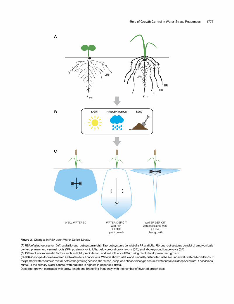
Figure 3. Changes in RSA upon Water-Deficit Stress.
(A)RSA of a taproot system (left) and a fibrous root system (right). Taproot systems consist of a PR and LRs. Fibrous root systems consist of embryonicallyderived primary and seminal roots (SR), postembryonic LRs, belowground crown roots (CR), and aboveground brace roots (BR).(B) Different environmental factors such as light, precipitation, and soil influence RSA during plant development and growth.(C)RSA ideotypes forwell-wateredandwater-deficit conditions.Water is shown inblueand isequally distributed in thesoil underwell-wateredconditions. Ifthe primarywater source is rainfall before the growing season, the “steep, deep, and cheap” ideotype ensureswater uptake in deep soil strata. If occasionalrainfall is the primary water source, water uptake is highest in upper soil strata.Deep root growth correlates with arrow length and branching frequency with the number of inverted arrowheads.
Role of Growth Control in Water-Stress Responses 1777
emergence, whereas the PR system expands in size. Crown rootarrest is irreversible at the individual root level; however, afterrewatering of water deficit-treated plants, new crown root emer-gence is rapidly induced. Within 8 h after rewatering, crown rootprimordia are visible, and within 6 d, these roots take over as thedominant part of the root system.
Similar responses of crown roots were observed under water-deficit stress in other grass species including sorghum,miscanthus,and Brachypodium distachyon (Sebastian et al., 2016). In-terestingly, the response was less severe in maize, where somecrown root growth still occurred. The development of crown rootsunder water deficit in maize allowed us to test their physiologicalcontribution to plant water status using the rootless concerningcrown and seminal roots (rtcs) mutant, which completely lackscrown root development (Hetz et al., 1996; Taramino et al., 2007).Interestingly, the rtcsmutantwas better able tomaintain thewaterstatus of the shoot under water deficit compared with the refer-ence inbred andwas accompanied by the preservation of water inthe soil in the pot (Sebastian et al., 2016). These data suggest thatreduced crown root growth allows the plant to slow the extractionofwater from thesoil and tobank these reserves for the future. Thisphenomenon, known as water banking, may allow grasses tosurvive irregularity in precipitation patterns. Maize inbreds showtremendous variation in the degree to which crown root growth issuppressed under water deficit. These data suggest that duringdomestication, there may have been selection for reduceddrought responsiveness, perhaps as away of generating a largerroot system. It will be interesting for future studies to identifythemolecular targets of water-deficit signaling that cause crownroot arrest and to generate varieties with different sensitivitiesto stress to identify a response that best suits soil and watermanagement conditions in the field.
Selecting Root System-Level Traits for Better Water Usage
The dynamic responses to limited water availability on the organscale described above lead to complex spatial arrangementsof roots in soil, and the emergent properties of this system ul-timately determine the expanse of soil where water can beaccessed. To date, in the field of plant breeding, rather static,idealized root phenotypes, namely ideotypes, have been tar-geted to optimize plant growth under a particular environment orstress condition. Such ideotypes can be theoretical or based onmodeling data. The “steep, deep and cheap” ideotype has beenproposed for water- and nitrogen-limited conditions (Nord andLynch, 2009; Lynch, 2013). This ideotype is defined by a rootsystem that has roots oriented more vertically to capture deepwater resources and high rates of growth, which are enabled byseveral anatomical features that reduce the metabolic cost perunit length of root (Figure 3C) (Lynch et al., 2014). This ideotypewas designed for maize roots based on the greater availability ofwater and nitrogen in deeper soil strata throughout the growingseason in most agricultural soils.
The steep, deep, and cheap ideotype is partly reflected in thepatterns of root system growth observed in nature. While a widerange of interspecific strategies to cope with drought were ob-served in grassland communities (Zwicke et al., 2015), speciesthat survive and recover best from drought combine a large root
mass for water acquisition and dehydration avoidance with deeperroots with higher cell membrane stability and carbohydrate accu-mulation fordehydration tolerance (Zwickeetal., 2015). Importantly,however, other strategies are also observed in xerophytes. Forexample, perennial cactus species growing in the desert have veryshallow root systems that are adapted to rapidly capturewater fromseasonal rains (Figure 3C) (Rundel et al., 1991). In addition, differentdesert species increase water uptake from seasonal rain by pro-ducing very fine roots from laterals that become highly suberizedafter a long dry period (Figure 3C) (Nobel and Sanderson, 1984;Salguero-Gómez and Casper, 2011).A recent large-scale modeling approach combined RSA mod-
eling with soil-hydrological modeling to investigate if there is oneoptimal RSA ideotype for enhanced drought tolerance (Tron et al.,2015). The authors conclude that the “ideal” root architecture forefficientwater uptake always has to be consideredwith respect tothe hydrological environment. In their model, deep-rooted sys-tems provide ideal water uptake if there is sufficient rainfall beforethe growing season in fine soil textures (Figure 3C). However,dense root systemsclose to the soil surface are essential forwateruptake if rainfalls are the main water source during plant de-velopment (Figure 3C).
CONCLUSIONS AND PERSPECTIVES
Biological systemsaremultiscale in organization, frommolecules,cells, organs, and organisms to communities (Passioura, 1979).Fully understanding a given phenomenon requires studies at alllevels; however, it is essential that the observations made ata specific scale beunderstood in the context of higher scales.Onecan always find patterns and rules at lower scales. However, toensure the principles discovered are not trivial, it is essential tounderstand the context in which these phenomena occur. As thepopulation of the Earth approaches over 9 billion in 2050 and weattempt to feed the massively increasing number of humans,emphasis must be placed on studies of plant–environment in-teractions that are of broad importance.Clearly, this is essential toensure that studies in plant model systems have the best chanceof improving agriculture. However, this is not the only goal of sucha holistic approach. A clearer understanding of plant biology re-quires appreciation of the ecological contexts in which organismshave evolved to function. Thus, broadly impactful research onplant–environment interactionswill naturally influenceour ideasofhow plants function in natural and agricultural field contexts.The cell- and organ-scale processes described here serve as
a foundation for our understanding of how changes in water avail-ability affect the form and function of the plant. Current researchon the mechanisms that allow cells to sense such environmentalstimuli have emphasized mechanisms that operate at the cell scale.However, additional research is needed to understand how theseprocesses act at the organ and organism scale. Where do plantssense changes in water availability and how do processes such ascell expansion affect water sensing? At the scale of the organ, howdoes water sensing at the single cell scale get integrated into higherorder patterning processes such as hydropatterning, which likelyrequire communication between tissue layers? Answers to thesequestions will allow us to understand not only how plants sensewater, but also why it matters.
1778 The Plant Cell

ACKNOWLEDGMENTS
We thank Michael T. Raissig, Malcolm Bennett, members of the Dinnenylab, andanonymous reviewers for helpful commentsduring thepreparationof this work. Funding was provided by the U.S. Department of Energy’sBiological andEnvironmental Research program (Grant DE-SC0008769 toJ.R.D.), the National Science Foundation’s Plant Genome Research Pro-gram (Grant IOS-PGRP 420-40-45A to J.R.D.), the DFGGerman ResearchFoundation (research fellowship to H.L.), and the National Science Foun-dation Graduate Research Fellowship (Grant DGE-1147470 to N.E.R.).
AUTHOR CONTRIBUTIONS
All authors contributed to writing the article.
ReceivedMarch 15, 2016; revised July 20, 2016; acceptedAugust 7, 2016;published August 8, 2016.
REFERENCES
Akashi, T., Kawasaki, S., and Shibaoka, H. (1990). Stabilization of corticalmicrotubules by the cell wall in cultured tobacco cells: Effects of ex-tensin on the cold-stability of cortical microtubules. Planta 182: 363–369.
Arai-Sanoh, Y., Takai, T., Yoshinaga, S., Nakano, H., Kojima, M.,Sakakibara, H., Kondo, M., and Uga, Y. (2014). Deep rootingconferred by DEEPER ROOTING 1 enhances rice yield in paddyfields. Sci. Rep. 4: 5563.
Bailey, P.H.J., Currey, J.D., and Fitter, A.H. (2002). The role of rootsystem architecture and root hairs in promoting anchorage againstuprooting forces in Allium cepa and root mutants of Arabidopsisthaliana. J. Exp. Bot. 53: 333–340.
Baluska, F., Samaj, J., Wojtaszek, P., Volkmann, D., and Menzel,D. (2003). Cytoskeleton-plasma membrane-cell wall continuum inplants. Emerging links revisited. Plant Physiol. 133: 482–491.
Bao, Y., et al. (2014). Plant roots use a patterning mechanism toposition lateral root branches toward available water. Proc. Natl.Acad. Sci. USA 111: 9319–9324.
Beauzamy, L., Nakayama, N., and Boudaoud, A. (2014). Flowersunder pressure: ins and outs of turgor regulation in development.Ann. Bot. (Lond.) 114: 1517–1533.
Bernier, J., Serraj, R., Kumar, A., Venuprasad, R., Impa, S., VeereshGowda, R.P., Oane, R., and Atlin, G. (2009). The large-effect drought-resistance QTL qtl12.1 increases water uptake in upland rice. FieldCrops Res. 110: 139–146.
Blancaflor, E.B., Fasano, J.M., and Gilroy, S. (1998). Mapping thefunctional roles of cap cells in the response of Arabidopsis primaryroots to gravity. Plant Physiol. 116: 213–222.
Boisson-Dernier, A., Kessler, S.A., and Grossniklaus, U. (2011).The walls have ears: the role of plant CrRLK1Ls in sensing andtransducing extracellular signals. J. Exp. Bot. 62: 1581–1591.
Boisson-Dernier, A., Lituiev, D.S., Nestorova, A., Franck, C.M.,Thirugnanarajah, S., and Grossniklaus, U. (2013). ANXUR re-ceptor-like kinases coordinate cell wall integrity with growth at thepollen tube tip via NADPH oxidases. PLoS Biol. 11: e1001719.
Boisson-Dernier, A., Roy, S., Kritsas, K., Grobei, M.A., Jaciubek,M., Schroeder, J.I., and Grossniklaus, U. (2009). Disruption of thepollen-expressed FERONIA homologs ANXUR1 and ANXUR2 trig-gers pollen tube discharge. Development 136: 3279–3288.
Borsa, A.A., Agnew, D.C., and Cayan, D.R. (2014). Remote hydrol-ogy. Ongoing drought-induced uplift in the western United States.Science 345: 1587–1590.
Brutnell, T.P., Bennetzen, J.L., and Vogel, J.P. (2015). Brachypodiumdistachyon and Setaria viridis: Model genetic systems for the grasses.Annu. Rev. Plant Biol. 66: 465–485.
Burk, D.H., and Ye, Z.-H. (2002). Alteration of oriented deposition ofcellulose microfibrils by mutation of a katanin-like microtubule-severing protein. Plant Cell 14: 2145–2160.
Burton, R.A., Gidley, M.J., and Fincher, G.B. (2010). Heterogeneity inthe chemistry, structure and function of plant cell walls. Nat. Chem.Biol. 6: 724–732.
Cassab, G.I., Eapen, D., and Campos, M.E. (2013). Root hydrotro-pism: an update. Am. J. Bot. 100: 14–24.
Christmann, A., Grill, E., and Huang, J. (2013). Hydraulic signals inlong-distance signaling. Curr. Opin. Plant Biol. 16: 293–300.
Cosgrove, D.J. (2016a). Catalysts of plant cell wall loosening. F1000Res. 5: 5.
Cosgrove, D.J. (2016b). Plant cell wall extensibility: connecting plantcell growth with cell wall structure, mechanics, and the action ofwall-modifying enzymes. J. Exp. Bot. 67: 463–476.
Cosgrove, D.J. (2014). Re-constructing our models of cellulose andprimary cell wall assembly. Curr. Opin. Plant Biol. 22: 122–131.
Cosgrove, D.J., and Green, P.B. (1981). Rapid suppression of growth byblue light: Biophysical mechanism of action. Plant Physiol. 68: 1447–1453.
Courtois, B., Ahmadi, N., Khowaja, F., Price, A.H., Rami, J.-F., Frouin,J., Hamelin, C., and Ruiz, M. (2009). Rice root genetic architecture:Meta-analysis from a drought QTL database. Rice (N.Y.) 2: 115–128.
Dai, F., Zhang, C., Jiang, X., Kang, M., Yin, X., Lü, P., Zhang, X.,Zheng, Y., and Gao, J. (2012). RhNAC2 and RhEXPA4 are involvedin the regulation of dehydration tolerance during the expansion ofrose petals. Plant Physiol. 160: 2064–2082.
Deak, K.I., and Malamy, J. (2005). Osmotic regulation of root systemarchitecture. Plant J. 43: 17–28.
Dinneny, J.R. (2014). A gateway with a guard: how the endodermisregulates growth through hormone signaling. Plant Sci. 214: 14–19.
Dinneny, J.R. (2015). A developmental biologist’s journey to redis-cover the Zen of plant physiology. F1000Research 2015 4: 264.
Dinneny, J.R., Long, T.A., Wang, J.Y., Jung, J.W., Mace, D., Pointer, S.,Barron, C., Brady, S.M., Schiefelbein, J., and Benfey, P.N. (2008).Cell identity mediates the response of Arabidopsis roots to abioticstress. Science 320: 942–945.
Do Amaral, S.H., De Assis, S.A., de Faria Oliveira, O., andMascarenhas, M. (2005). Partial purification and characterizationof pectin methylesterase from orange (Citrus sinensis) cv. Pera-rio.J. Food Biochem. 29: 367–380.
Duan, L., Dietrich, D., Ng, C.H., Chan, P.M.Y., Bhalerao, R.,Bennett, M.J., and Dinneny, J.R. (2013). Endodermal ABA sig-naling promotes lateral root quiescence during salt stress in Ara-bidopsis seedlings. Plant Cell 25: 324–341.
Duan, Q., Kita, D., Li, C., Cheung, A.Y., and Wu, H.-M. (2010). FER-ONIA receptor-like kinase regulates RHO GTPase signaling of roothair development. Proc. Natl. Acad. Sci. USA 107: 17821–17826.
Eapen, D., Barroso, M.L., Campos, M.E., Ponce, G., Corkidi, G.,Dubrovsky, J.G., and Cassab, G.I. (2003). A no hydrotropic responseroot mutant that responds positively to gravitropism in Arabidopsis. PlantPhysiol. 131: 536–546.
Eghball, B., and Maranville, J.W. (1993). Root development and ni-trogen influx of corn genotypes grown under combined drought andnitrogen stresses. Agron. J. 85: 147–152.
Endler, A., Kesten, C., Schneider, R., Zhang, Y., Ivakov, A.,Froehlich, A., Funke, N., and Persson, S. (2015). A mechanism forsustained cellulose synthesis during salt stress. Cell 162: 1353–1364.
Erickson, R.O. (1976). Modeling of plant growth. Annu. Rev. PlantPhysiol. 27: 407–434.
Erickson, R.O., and Silk, W.K. (1980). The kinematics of plant growth.Sci. Am. 242: 134–151.
Role of Growth Control in Water-Stress Responses 1779

Esau, K. (1953). Plant Anatomy, (New York: John Wiley & Sons).Escobar-Restrepo, J.-M., Huck, N., Kessler, S., Gagliardini, V.,
Gheyselinck, J., Yang, W.-C., and Grossniklaus, U. (2007). TheFERONIA receptor-like kinase mediates male-female interactionsduring pollen tube reception. Science 317: 656–660.
Ferrari, S., Savatin, D.V., Sicilia, F., Gramegna, G., Cervone, F., andLorenzo, G.D. (2013). Oligogalacturonides: plant damage-associatedmolecular patterns and regulators of growth and development. Front.Plant Sci. 4: 49.
Furuichi, T., Iida, H., Sokabe, M., and Tatsumi, H. (2012). Expressionof Arabidopsis MCA1 enhanced mechanosensitive channel activityin the Xenopus laevis oocyte plasma membrane. Plant Signal. Be-hav. 7: 1022–1026.
Galvan-Ampudia, C.S., Julkowska, M.M., Darwish, E., Gandullo, J.,Korver, R.A., Brunoud, G., Haring, M.A., Munnik, T., Vernoux, T.,and Testerink, C. (2013). Halotropism is a response of plant rootsto avoid a saline environment. Curr. Biol. 23: 2044–2050.
Geldner, N., Anders, N., Wolters, H., Keicher, J., Kornberger, W.,Muller, P., Delbarre, A., Ueda, T., Nakano, A., and Jürgens, G. (2003).The Arabidopsis GNOM ARF-GEF mediates endosomal recycling, auxintransport, and auxin-dependent plant growth. Cell 112: 219–230.
Geng, Y., Wu, R., Wee, C.W., Xie, F., Wei, X., Chan, P.M.Y., Tham,C., Duan, L., and Dinneny, J.R. (2013). A spatio-temporal un-derstanding of growth regulation during the salt stress response inArabidopsis. Plant Cell 25: 2132–2154.
Giarola, V., Krey, S., von den Driesch, B., and Bartels, D. (2016). TheCraterostigma plantagineum glycine-rich protein CpGRP1 interacts witha cell wall-associated protein kinase 1 (CpWAK1) and accumulates inleaf cell walls during dehydration. New Phytol. 210: 535–550.
Grant, G.T., Morris, E.R., Rees, D.A., Smith, P.J.C., and Thom, D.(1973). Biological interactions between polysaccharides and di-valent cations: The egg-box model. FEBS Lett. 32: 195–198.
Guo, H., Li, L., Ye, H., Yu, X., Algreen, A., and Yin, Y. (2009). Threerelated receptor-like kinases are required for optimal cell elongationin Arabidopsis thaliana. Proc. Natl. Acad. Sci. USA 106: 7648–7653.
Guo, W., Zhao, J., Li, X., Qin, L., Yan, X., and Liao, H. (2011). A soybeanb-expansin gene GmEXPB2 intrinsically involved in root system archi-tecture responses to abiotic stresses. Plant J. 66: 541–552.
Gutierrez, R., Lindeboom, J.J., Paredez, A.R., Emons, A.M.C., andEhrhardt, D.W. (2009). Arabidopsis cortical microtubules position cel-lulose synthase delivery to the plasma membrane and interact withcellulose synthase trafficking compartments. Nat. Cell Biol. 11: 797–806.
Haberlandt, G. (1900). Über die Perception des geotropischen Reizes.Berichte der Deutschen Botanischen Gesellschaft 18: 261–272.
Hamilton, E.S., Jensen, G.S., Maksaev, G., Katims, A., Sherp, A.M.,and Haswell, E.S. (2015). Mechanosensitive channel MSL8 regu-lates osmotic forces during pollen hydration and germination. Sci-ence 350: 438–441.
Harb, A., Krishnan, A., Ambavaram, M.M.R., and Pereira, A. (2010).Molecular and physiological analysis of drought stress in Arabidopsisreveals early responses leading to acclimation in plant growth. PlantPhysiol. 154: 1254–1271.
Haruta, M., Sabat, G., Stecker, K., Minkoff, B.B., and Sussman,M.R. (2014). A peptide hormone and its receptor protein kinaseregulate plant cell expansion. Science 343: 408–411.
Haswell, E.S., and Meyerowitz, E.M. (2006). MscS-like proteins controlplastid size and shape in Arabidopsis thaliana. Curr. Biol. 16: 1–11.
Haswell, E.S., and Verslues, P.E. (2015). The ongoing search for themolecular basis of plant osmosensing. J. Gen. Physiol. 145: 389–394.
Hématy, K., Sado, P.-E., Van Tuinen, A., Rochange, S., Desnos, T.,Balzergue, S., Pelletier, S., Renou, J.-P., and Höfte, H. (2007). Areceptor-like kinase mediates the response of Arabidopsis cells tothe inhibition of cellulose synthesis. Curr. Biol. 17: 922–931.
Hetz, W., Hochholdinger, F., Schwall, M., and Feix, G. (1996). Iso-lation and characterization of rtcs, a maize mutant deficient in theformation of nodal roots. Plant J. 10: 845–857.
Hou, C., Tian, W., Kleist, T., He, K., Garcia, V., Bai, F., Hao, Y.,Luan, S., and Li, L. (2014). DUF221 proteins are a family of os-mosensitive calcium-permeable cation channels conserved acrosseukaryotes. Cell Res. 24: 632–635.
Huck, N., Moore, J.M., Federer, M., and Grossniklaus, U. (2003).The Arabidopsis mutant feronia disrupts the female gametophyticcontrol of pollen tube reception. Development 130: 2149–2159.
Jaffe, M.J., Takahashi, H., and Biro, R.L. (1985). A pea mutant forthe study of hydrotropism in roots. Science 230: 445–447.
Julkowska, M.M., Hoefsloot, H.C.J., Mol, S., Feron, R., de Boer,G.-J., Haring, M.A., and Testerink, C. (2014). Capturing Arabi-dopsis root architecture dynamics with ROOT-FIT reveals diversityin responses to salinity. Plant Physiol. 166: 1387–1402.
Julkowska, M.M., and Testerink, C. (2015). Tuning plant signalingand growth to survive salt. Trends Plant Sci. 20: 586–594.
Kang, J.S., et al. (2008). Salt tolerance of Arabidopsis thaliana re-quires maturation of N-glycosylated proteins in the Golgi apparatus.Proc. Natl. Acad. Sci. USA 105: 5933–5938.
Kaplan, D.R. (1992). The relationship of cells to organisms in plants:Problem and implications of an organismal perspective. Int. J. PlantSci. 153: S28–S37.
Kobayashi, A., Takahashi, A., Kakimoto, Y., Miyazawa, Y., Fujii, N.,Higashitani, A., and Takahashi, H. (2007). A gene essential forhydrotropism in roots. Proc. Natl. Acad. Sci. USA 104: 4724–4729.
Kohorn, B.D. (2016). Cell wall-associated kinases and pectin per-ception. J. Exp. Bot. 67: 489–494.
Kohorn, B.D., Johansen, S., Shishido, A., Todorova, T., Martinez, R.,Defeo, E., and Obregon, P. (2009). Pectin activation of MAP kinaseand gene expression is WAK2 dependent. Plant J. 60: 974–982.
Kohorn, B.D., Kobayashi, M., Johansen, S., Riese, J., Huang, L.-F.,Koch, K., Fu, S., Dotson, A., and Byers, N. (2006). An Arabidopsiscell wall-associated kinase required for invertase activity and cellgrowth. Plant J. 46: 307–316.
Kohorn, B.D., and Kohorn, S.L. (2012). The cell wall-associatedkinases, WAKs, as pectin receptors. Front. Plant Sci. 3: 88.
Komis, G., Apostolakos, P., and Galatis, B. (2001). Altered patternsof tubulin polymerization in dividing leaf cells of Chlorophyton co-mosum after a hyperosmotic treatment. New Phytol. 149: 193–207.
Komis, G., Apostolakos, P., and Galatis, B. (2002). Hyperosmoticstress induces formation of tubulin macrotubules in root-tip cells ofTriticum turgidum: their probable involvement in protoplast volumecontrol. Plant Cell Physiol. 43: 911–922.
Kramer, P.J., and Boyer, J.S. (1995). Water Relations of Plants andSoils. (San Diego, CA: Elsevier Science).
Kung, C. (2005). A possible unifying principle for mechanosensation.Nature 436: 647–654.
Lally, D., Ingmire, P., Tong, H.Y., and He, Z.H. (2001). Antisenseexpression of a cell wall-associated protein kinase, WAK4, inhibitscell elongation and alters morphology. Plant Cell 13: 1317–1331.
Lavenus, J., Goh, T., Roberts, I., Guyomarc’h, S., Lucas, M., DeSmet, I., Fukaki, H., Beeckman, T., Bennett, M., and Laplaze, L.(2013). Lateral root development in Arabidopsis: fifty shades ofauxin. Trends Plant Sci. 18: 450–458.
LeNoble, M.E., Spollen, W.G., and Sharp, R.E. (2004). Maintenanceof shoot growth by endogenous ABA: genetic assessment of theinvolvement of ethylene suppression. J. Exp. Bot. 55: 237–245.
Leung, J., Merlot, S., and Giraudat, J. (1997). The ArabidopsisABSCISIC ACID-INSENSITIVE2 (ABI2) and ABI1 genes encodehomologous protein phosphatases 2C involved in abscisic acidsignal transduction. Plant Cell 9: 759–771.
1780 The Plant Cell
Li, C., et al. (2015). Glycosylphosphatidylinositol-anchored proteinsas chaperones and co-receptors for FERONIA receptor kinasesignaling in Arabidopsis. eLife 4: 4.
Lindner, H., Müller, L.M., Boisson-Dernier, A., and Grossniklaus,U. (2012). CrRLK1L receptor-like kinases: not just another brick inthe wall. Curr. Opin. Plant Biol. 15: 659–669.
Lou, Q., Chen, L., Mei, H., Wei, H., Feng, F., Wang, P., Xia, H., Li, T., andLuo, L. (2015). Quantitative trait locus mapping of deep rooting bylinkage and association analysis in rice. J. Exp. Bot. 66: 4749–4757.
Lü, P., Kang, M., Jiang, X., Dai, F., Gao, J., and Zhang, C. (2013).RhEXPA4, a rose expansin gene, modulates leaf growth and confersdrought and salt tolerance to Arabidopsis. Planta 237: 1547–1559.
Lynch, J.P. (2013). Steep, cheap and deep: an ideotype to optimizewater and N acquisition by maize root systems. Ann. Bot. (Lond.)112: 347–357.
Lynch, J.P., Chimungu, J.G., and Brown, K.M. (2014). Root ana-tomical phenes associated with water acquisition from drying soil:targets for crop improvement. J. Exp. Bot. 65: 6155–6166.
Maurel, C., Verdoucq, L., Luu, D.-T., and Santoni, V. (2008). Plantaquaporins: membrane channels with multiple integrated functions.Annu. Rev. Plant Biol. 59: 595–624.
Mauseth, J.D. (2008). An Introduction to Plant Biology, 4th ed. (Burlington,MA: Jones and Bartlett Learning).
McFarlane, H.E., Döring, A., and Persson, S. (2014). The cell biologyof cellulose synthesis. Annu. Rev. Plant Biol. 65: 69–94.
Miyazaki, S., Murata, T., Sakurai-Ozato, N., Kubo, M., Demura, T.,Fukuda, H., and Hasebe, M. (2009). ANXUR1 and 2, sister genes toFERONIA/SIRENE, are male factors for coordinated fertilization.Curr. Biol. 19: 1327–1331.
Miyazawa, Y., Takahashi, A., Kobayashi, A., Kaneyasu, T., Fujii, N.,and Takahashi, H. (2009). GNOM-mediated vesicular traffickingplays an essential role in hydrotropism of Arabidopsis roots. PlantPhysiol. 149: 835–840.
Monshausen, G.B., and Gilroy, S. (2009). Feeling green: mechano-sensing in plants. Trends Cell Biol. 19: 228–235.
Moore, J.P., Vicré-Gibouin, M., Farrant, J.M., and Driouich, A.(2008). Adaptations of higher plant cell walls to water loss: droughtvs desiccation. Physiol. Plant. 134: 237–245.
Nakagawa, Y., et al. (2007). Arabidopsis plasma membrane proteincrucial for Ca2+ influx and touch sensing in roots. Proc. Natl. Acad.Sci. USA 104: 3639–3644.
Nakayama, M., Kaneko, Y., Miyazawa, Y., Fujii, N., Higashitani, N.,Wada, S., Ishida, H., Yoshimoto, K., Shirasu, K., Yamada, K.,Nishimura, M., and Takahashi, H. (2012). A possible involvementof autophagy in amyloplast degradation in columella cells duringhydrotropic response of Arabidopsis roots. Planta 236: 999–1012.
Nari, J., Noat, G., and Ricard, J. (1991). Pectin methylesterase, metalions and plant cell-wall extension. Hydrolysis of pectin by plant cell-wall pectin methylesterase. Biochem. J. 279: 343–350.
Nèmec, B. (1900). Über die Art der Wahrnehmung des Schwerkraftreizesbei den Pflanzen. Berichte der Deutschen Botanischen Gesellschaft18: 241.
Nick, P. (2013). Microtubules, signalling and abiotic stress. Plant J.75: 309–323.
Nobel, P.S., and Sanderson, J. (1984). Rectifier-like activities of rootsof two desert succulents. J. Exp. Bot. 35: 727–737.
Nord, E.A., and Lynch, J.P. (2009). Plant phenology: a critical con-troller of soil resource acquisition. J. Exp. Bot. 60: 1927–1937.
Oparka, K.J. (1994). Plasmolysis: new insights into an old process.New Phytol. 126: 571–591.
Orman-Ligeza, B., Parizot, B., Gantet, P.P., Beeckman, T., Bennett,M.J., and Draye, X. (2013). Post-embryonic root organogenesis in ce-reals: branching out from model plants. Trends Plant Sci. 18: 459–467.
Paredez, A.R., Somerville, C.R., and Ehrhardt, D.W. (2006). Visu-alization of cellulose synthase demonstrates functional associationwith microtubules. Science 312: 1491–1495.
Park, A.R., Cho, S.K., Yun, U.J., Jin, M.Y., Lee, S.H., Sachetto-Martins, G., and Park, O.K. (2001). Interaction of the Arabidopsisreceptor protein kinase Wak1 with a glycine-rich protein, AtGRP-3.J. Biol. Chem. 276: 26688–26693.
Passioura, J.B. (1979). Accountability, philosophy and plant physi-ology. Search 10: 347–350.
Peaucelle, A., Braybrook, S., and Höfte, H. (2012). Cell wall me-chanics and growth control in plants: the role of pectins revisited.Front. Plant Sci. 3: 121.
Pelloux, J., Rustérucci, C., and Mellerowicz, E.J. (2007). New in-sights into pectin methylesterase structure and function. TrendsPlant Sci. 12: 267–277.
Plieth, C., and Trewavas, A.J. (2002). Reorientation of seedlings inthe earth’s gravitational field induces cytosolic calcium transients.Plant Physiol. 129: 786–796.
Rellán-Álvarez, R., et al. (2015). GLO-Roots: an imaging platformenabling multidimensional characterization of soil-grown root sys-tems. eLife 4: 393.
Rellán-Álvarez, R., Lobet, G., and Dinneny, J.R. (2016). Environmentalcontrol of root system biology. Annu. Rev. Plant Biol. 67: 619–642.
Robbins II, N.E., and Dinneny, J.R. (2015). The divining root:moisture-driven responses of roots at the micro- and macro-scale.J. Exp. Bot. 66: 2145–2154.
Rogers, E.D., and Benfey, P.N. (2015). Regulation of plant rootsystem architecture: implications for crop advancement. Curr. Opin.Biotechnol. 32: 93–98.
Rosquete, M.R., von Wangenheim, D., Marhavý, P., Barbez, E.,Stelzer, E.H.K., Benková, E., Maizel, A., and Kleine-Vehn, J.(2013). An auxin transport mechanism restricts positive orthogra-vitropism in lateral roots. Curr. Biol. 23: 817–822.
Rotman, N., Rozier, F., Boavida, L., Dumas, C., Berger, F., andFaure, J.-E. (2003). Female control of male gamete delivery duringfertilization in Arabidopsis thaliana. Curr. Biol. 13: 432–436.
Rundel, P.W., Nobel, P.S., and Atkinson, D. (1991). Structure andfunction in desert root systems. In Plant Root Growth: An EcologicalPerspective, D. Atkinson, ed (Oxford, UK: Blackwell Scientific Pub-lications), pp. 349-378.
Saab, I.N., Sharp, R.E., Pritchard, J., and Voetberg, G.S. (1990).Increased endogenous abscisic acid maintains primary root growthand inhibits shoot growth of maize seedlings at low water poten-tials. Plant Physiol. 93: 1329–1336.
Salguero-Gómez, R., and Casper, B.B. (2011). Introducing shortroots in a desert perennial: anatomy and spatiotemporal foragingresponses to increased precipitation. New Phytol. 191: 173–183.
Sato, E.M., Hijazi, H., Bennett, M.J., Vissenberg, K., and Swarup,R. (2015). New insights into root gravitropic signalling. J. Exp. Bot.66: 2155–2165.
Saucedo, M., Ponce, G., Campos, M.E., Eapen, D., García, E.,Luján, R., Sánchez, Y., and Cassab, G.I. (2012). An altered hy-drotropic response (ahr1) mutant of Arabidopsis recovers root hy-drotropism with cytokinin. J. Exp. Bot. 63: 3587–3601.
Schallus, T., Fehér, K., Sternberg, U., Rybin, V., and Muhle-Goll, C.(2010). Analysis of the specific interactions between the lectin do-main of malectin and diglucosides. Glycobiology 20: 1010–1020.
Sebastian, J., et al. (2016). Grasses suppress shoot-borne roots to con-serve water during drought. Proc. Natl. Acad. Sci. USA 113: 8861–8866.
Shabala, S.N., and Lew, R.R. (2002). Turgor regulation in osmoticallystressed Arabidopsis epidermal root cells. Direct support for therole of inorganic ion uptake as revealed by concurrent flux and cellturgor measurements. Plant Physiol. 129: 290–299.
Role of Growth Control in Water-Stress Responses 1781
Sharp, R.E., and Davies, W.J. (1979). Solute regulation and growth byroots and shoots of water-stressed maize plants. Planta 147: 43–49.
Sharp, R.E., Hsiao, T.C., and Silk, W.K. (1990). Growth of the maizeprimary root at low water potentials : II. Role of growth and de-position of hexose and potassium in osmotic adjustment. PlantPhysiol. 93: 1337–1346.
Sharp, R.E., Silk, W.K., and Hsiao, T.C. (1988). Growth of the maizeprimary root at low water potentials : I. Spatial distribution of ex-pansive growth. Plant Physiol. 87: 50–57.
Shih, H.-W., Miller, N.D., Dai, C., Spalding, E.P., and Monshausen,G.B. (2014). The receptor-like kinase FERONIA is required for me-chanical signal transduction in Arabidopsis seedlings. Curr. Biol. 24:1887–1892.
Smith, L.G., Hake, S., and Sylvester, A.W. (1996). The tangled-1mutation alters cell division orientations throughout maize leaf de-velopment without altering leaf shape. Development 122: 481–489.
Spollen, W.G., LeNoble, M.E., Samuels, T.D., Bernstein, N., andSharp, R.E. (2000). Abscisic acid accumulation maintains maizeprimary root elongation at low water potentials by restricting eth-ylene production. Plant Physiol. 122: 967–976.
Steinmann, T., Geldner, N., Grebe, M., Mangold, S., Jackson, C.L.,Paris, S., Gälweiler, L., Palme, K., and Jürgens, G. (1999). Co-ordinated polar localization of auxin efflux carrier PIN1 by GNOMARF GEF. Science 286: 316–318.
Steudle, E., and Peterson, C.a. (1998). How does water get throughroots? J. Exp. Bot. 49: 775–788.
Sun, F., Zhang, W., Hu, H., Li, B., Wang, Y., Zhao, Y., Li, K., Liu, M.,and Li, X. (2008). Salt modulates gravity signaling pathway toregulate growth direction of primary roots in Arabidopsis. PlantPhysiol. 146: 178–188.
Swarup, R., Kramer, E.M., Perry, P., Knox, K., Leyser, H.M.O.,Haseloff, J., Beemster, G.T.S., Bhalerao, R., and Bennett, M.J.(2005). Root gravitropism requires lateral root cap and epidermalcells for transport and response to a mobile auxin signal. Nat. CellBiol. 7: 1057–1065.
Takahashi, N., Yamazaki, Y., Kobayashi, A., Higashitani, A., andTakahashi, H. (2003). Hydrotropism interacts with gravitropism bydegrading amyloplasts in seedling roots of Arabidopsis and radish.Plant Physiol. 132: 805–810.
Takano, M., Takahashi, H., and Suge, H. (1997). Calcium re-quirement for the induction of hydrotropism and enhancement ofcalcium-induced curvature by water stress in primary roots of pea,Pisum sativum L. Plant Cell Physiol. 38: 385–391.
Taramino, G., Sauer, M., Stauffer, J.L., Jr., Multani, D., Niu, X.,Sakai, H., and Hochholdinger, F. (2007). The maize (Zea mays L.)RTCS gene encodes a LOB domain protein that is a key regulator ofembryonic seminal and post-embryonic shoot-borne root initiation.Plant J. 50: 649–659.
Tron, S., Bodner, G., Laio, F., Ridolfi, L., and Leitner, D. (2015). Candiversity in root architecture explain plant water use efficiency? Amodeling study. Ecol. Modell. 312: 200–210.
Uga, Y., et al. (2013). Control of root system architecture by DEEPERROOTING 1 increases rice yield under drought conditions. Nat.Genet. 45: 1097–1102.
Uga, Y., Kitomi, Y., Yamamoto, E., Kanno, N., Kawai, S.,Mizubayashi, T., and Fukuoka, S. (2015). A QTL for root growthangle on rice chromosome 7 is involved in the genetic pathway ofDEEPER ROOTING 1. Rice (N. Y.) 8: 8.
Van Norman, J.M., Xuan, W., Beeckman, T., and Benfey, P.N.(2013). To branch or not to branch: the role of pre-patterning inlateral root formation. Development 140: 4301–4310.
Verslues, P.E., Agarwal, M., Katiyar-Agarwal, S., Zhu, J., and Zhu,J.-K. (2006). Methods and concepts in quantifying resistance todrought, salt and freezing, abiotic stresses that affect plant waterstatus. Plant J. 45: 523–539.
Voetberg, G.S., and Sharp, R.E. (1991). Growth of the maize primaryroot at low water potentials: III. Role of increased proline depositionin osmotic adjustment. Plant Physiol. 96: 1125–1130.
Wade, L.J., et al. (2015). Environmental response and genomic re-gions correlated with rice root growth and yield under drought in theOryzaSNP panel across multiple study systems. PLoS One 10:e0124127.
Wilson, M.E., Jensen, G.S., and Haswell, E.S. (2011). Two mecha-nosensitive channel homologs influence division ring placement inArabidopsis chloroplasts. Plant Cell 23: 2939–2949.
Wu, Y., Thorne, E.T., Sharp, R.E., and Cosgrove, D.J. (2001).Modification of expansin transcript levels in the maize primary rootat low water potentials. Plant Physiol. 126: 1471–1479.
Xiong, L., Wang, R.-G., Mao, G., and Koczan, J.M. (2006). Identifi-cation of drought tolerance determinants by genetic analysis of rootresponse to drought stress and abscisic acid. Plant Physiol. 142:1065–1074.
Xu, P., Cai, X.-T., Wang, Y., Xing, L., Chen, Q., and Xiang, C.-B.(2014). HDG11 upregulates cell-wall-loosening protein genes topromote root elongation in Arabidopsis. J. Exp. Bot. 65: 4285–4295.
Yang, L., Zheng, B., Mao, C., Qi, X., Liu, F., and Wu, P. (2004).Analysis of transcripts that are differentially expressed in threesectors of the rice root system under water deficit. Mol. Genet.Genomics 272: 433–442.
Yoo, S.-H., Fishman, M.L., Savary, B.J., and Hotchkiss, A.T., Jr.(2003). Monovalent salt-induced gelation of enzymatically dees-terified pectin. J. Agric. Food Chem. 51: 7410–7417.
Yuan, F., et al. (2014). OSCA1 mediates osmotic-stress-evoked Ca2+
increases vital for osmosensing in Arabidopsis. Nature 514: 367–371.
Zhong, R., Burk, D.H., Morrison III, W.H., and Ye, Z.-H. (2002). Akinesin-like protein is essential for oriented deposition of cellulosemicrofibrils and cell wall strength. Plant Cell 14: 3101–3117.
Zhu, J., Lee, B.-H., Dellinger, M., Cui, X., Zhang, C., Wu, S.,Nothnagel, E.A., and Zhu, J.-K. (2010). A cellulose synthase-likeprotein is required for osmotic stress tolerance in Arabidopsis. PlantJ. 63: 128–140.
Zolla, G., Heimer, Y.M., and Barak, S. (2010). Mild salinity stimulatesa stress-induced morphogenic response in Arabidopsis thalianaroots. J. Exp. Bot. 61: 211–224.
Zwicke, M., Picon-Cochard, C., Morvan-Bertrand, A., Prud’homme,M.-P., and Volaire, F. (2015). What functional strategies drive droughtsurvival and recovery of perennial species from upland grassland? Ann.Bot. (Lond.) 116: 1001–1015.
1782 The Plant Cell

DOI 10.1105/tpc.16.00182; originally published online August 8, 2016; 2016;28;1769-1782Plant Cell
Wei Feng, Heike Lindner, Neil E. Robbins II and José R. DinnenyResponses
Growing Out of Stress: The Role of Cell- and Organ-Scale Growth Control in Plant Water-Stress
This information is current as of May 21, 2020
References /content/28/8/1769.full.html#ref-list-1
This article cites 141 articles, 49 of which can be accessed free at:
Permissions https://www.copyright.com/ccc/openurl.do?sid=pd_hw1532298X&issn=1532298X&WT.mc_id=pd_hw1532298X
eTOCs http://www.plantcell.org/cgi/alerts/ctmain
Sign up for eTOCs at:
CiteTrack Alerts http://www.plantcell.org/cgi/alerts/ctmain
Sign up for CiteTrack Alerts at:
Subscription Information http://www.aspb.org/publications/subscriptions.cfm
is available at:Plant Physiology and The Plant CellSubscription Information for
ADVANCING THE SCIENCE OF PLANT BIOLOGY © American Society of Plant Biologists


![Ethical Controversies in Organ Transplantation · demand for organ transplantation and the supply of donor organs is growing [The economist 2008].The waiting list of the United Network](https://img.dokumen.tips/doc/110x75/603e691cbdfbcb7c012bb89a/ethical-controversies-in-organ-transplantation-demand-for-organ-transplantation.jpg)