Embed Size (px)
Citation preview
1
Umeå University Medical Dissertations New Series 1209 ISSN 0346 � 6612 Greater Trochanteric Pain after Total Hip Arthroplasty Incidence, clinical outcome, associated factors, tenderness evaluation with algometer, and a new surgical treatment Ph.D. Thesis in Orthopaedics by Arkan Sam Sayed-Noor M.D.
Orthopaedics Department of Surgical and Perioperative Sciences Umeå 2008

2
Principal tutor Associate Professor Göran Sjödén Co-tutors Associate Professor Per Wretenberg Associate Professor Hans Malker Faculty opponent Professor Lars Weidenhielm, Karolinska Institute Cover design by Vivan Rönnqvist Published articles have been reprinted with permission from the publisher. Printed by Hemströms tryckeri, Härnösand. Copyright© 2008 Arkan Sam Sayed-Noor ISBN 978-91-7264-643-8

3
In memory of my best mentor and friend ever, my father Sam Sayed-Noor (1935 � 1999), for having the honour of being his son. Oh God, how I wish he was here right now. That knowledge which remains only on your tongue is very superficial. The intrinsic value of knowledge is that you act upon it. Knowledge and practice are twins, and both go together. There is no knowledge without practice, and no practice without knowledge. The philosopher of all time Imam Ali Ibn Abi-talib (PBUH) (600 � 661 AD).

4
5
Contents Abstract 7 Abbreviations 9 List of original papers 10 Acknowledgements 11 Introduction 13 Anatomical and pathophysiological considerations 14 Evaluation of trochanteric tenderness with Algometer 16 Epidemiological considerations and associated factors 17 Therapeutic considerations 18 Greater trochanteric pain after total hip arthroplasty 19 Clinical and radiological measurements of LLD 20 Aims 23 Patients and Methods 24 Study I 24 Study II 26 Study III 28 Study IV 31 Results 33 Study I 33 Study II 34 Study III 36 Study IV 37 Discussion 38 Summary and conclusion 47 Future implications 48 Summary in Swedish-Populärvetenskaplig sammanfattning 49 References 51 Appendix 60

6
7
Abstract Greater Trochanteric Pain after Total Hip Arthroplasty Incidence, clinical outcome, associated factors, tenderness evaluation with algometer and a new surgical treatment. Greater trochanteric pain (GTP) is a regional pain syndrome characterized by lateral hip pain
and tenderness. Its incidence after total hip arthroplasty (THA) is variable. Bursal
inflammation, degenerative changes of the attachment of the gluteal muscles, direct operative
trauma and biomechanical disturbance of the operated hip have been discussed as being
related to GTP. The diagnosis is purely clinical because radiological and laboratory
investigations show no definite pathology. Although most treatment modalities are
conservative, some patients may develop refractory complaints leading to surgical
intervention.
In study I we studied the incidence of GTP in 172 consecutive patients who underwent THA
during 2002 at Sundsvall Hospital. Patients with GTP (n=21, incidence 12%) were matched
with controls from the same cohort. The THA outcome was assessed using the Western
Ontario and McMaster Universities Arthrosis (WOMAC) Index. Trochanteric tenderness was
studied using an electronic pressure algometer. We found an association between the
occurrence of GTP and postoperative uncorrected lengthening of the operated limb of ≥ one
centimetre. The WOMAC index revealed a reduction of the clinical outcome in the GTP
group.
In Study II we tested the value of using an algometer in the diagnosis of GTP after THA. We
measured the pressure-pain threshold (PPT) over the greater trochanter and ilio-tibial band in
18 patients and 18 matched controls. Both groups were evaluated using the visual analogue
scale (VAS). We found the algometer to have a good predictive validity and reproducibility.
8
However, there was large inter-individual variability across subjects. The PPT ratio of 0.8
(affected vs. unaffected side) can be used as a cutoff ratio to establish GTP. There was no
correlation between PPT measurements and VAS. Because of a low positive predictive value
and large inter-individual variability, the pressure algometer has a limited value as a screening
tool.
In study III we proposed a new surgical treatment for refractory GTP after THA consisting of
distal lengthening of the ilio-tibial band (ITB) by Z-plasty under local anaesthesia. This
method was used in 12 women between March 2004 and June 2006. The patients were
followed up by phone interview 3-4 months postoperatively and by an EQ-5D questionnaire
and clinical examination including evaluation with the algometer at 1-3 years postoperatively.
We found that the patients` quality of life was markedly improved following the operation
(EQ-5D = 0.26 preoperatively vs. 0.67 postoperatively; p <0.005). There were no
postoperative complications.
In study IV we evaluated the accuracy of a commonly used clinical method of LLD
measurement (anterior superior iliac spine-medial malleolus) by comparing it to a reliable
radiological method (tear drop-lesser trochanter) in 139 patients before and after THA. We
found the correlation between the clinical and radiological methods to be weak preoperatively
(r=0.21, ICC= 0.33) while the correlation was moderate postoperatively (r= 0.45, ICC=0.62).
It is therefore recommended that the radiological method be used to measure leg length
discrepancy in patients who undergo THA.
9
Abbreviations ADL Activities of daily living AP view Antero-posterior view ASIS Anterior superior iliac spine CT Computerized tomography DNIC Diffuse noxious inhibitory control ECRB Extensor carpi radialis brevis EQ-5D EuroQol index GTP Greater trochanteric pain ICC Intra-class correlation coefficient ITB Ilio-tibial band kPa kiloPascal LLD Leg length discrepancy MRI Magnetic resonance imaging NSAIDs Non-steroidal anti inflammatory drugs OA Osteoarthritis PPT Pressure-pain threshold PPV Positive predictive value RA Rheumatoid arthritis ROM Range of motion SD Standard deviation THA Total hip arthroplasty VAS Visual analogue scale WOMAC Western Ontario and McMaster universities arthrosis index

10
Original papers Study I Sayed-Noor AS, Sjödén GO Greater Trochanteric Pain after Total Hip Arthroplasty: the incidence, clinical outcome and associated factors. Hip International 2006; 3:202�206. Study II Sayed-Noor AS, Englund E, Wretenberg P, Sjödén GO Pressure-Pain Threshold Algometric Measurement in Patients with Greater Trochanteric Pain after Total Hip Arthroplasty. Clinical Journal of Pain 2008; 24(3):232�6. Study III Sayed-Noor AS, Pedersen E, Wretenberg P, Sjödén GO Distal Lengthening of Ilio-tibial Band by Z-plasty for Treating Refractory Greater Trochanteric Pain after Total Hip Arthroplasty (Pedersen-Noor operation). Archives of Orthopaedic and Trauma Surgery DOI 10.1007/s00402�008�0693�8. Study VI Sayed-Noor AS, Hugo A, Sjödén GO, Wretenberg P Leg Length Discrepancy in Total Hip Arthroplasty. Comparison of Two Methods of Measurement. International Orthopaedics DOI 10.1007/s00264�008�0633�9.
11
Acknowledgements This work was carried out at Department of Orthopaedics, Sundsvall Hospital (studies I, II & III) and Department of Orthopaedics, Karolinska University Hospital (study IV). Many people have contributed to this work. I would like to express my deep gratitude to all of them, and particularly to: My beloved wife, Dhuha, for everything you have sacrificed for me since we first met, for your endless love, support and encouragement. Without you my life has absolutely no meaning. My wonderful mother, for your extreme kindness and generosity. Without your prayers this work would never have been possible. My sweet daughters, Noor and Aia, for every smile you gave me. I love you so much. My fantastic principal tutor, Associate Professor Göran Sjödén, for being such an ideal mentor in every way, for introducing me to the world of research, for guiding and encouraging me all the way through, for not becoming tired or bothered by my endless primitive questions and for being available anyhow, anywhere and anytime. I can not imagine writing an article without your name beside mine. My fantastic co-tutor, Associate Professor Per Wretenberg. Despite the geographical distance between Sundsvall and Stockholm, your input, engagement and advice were more than essential for the completion this work. Thank you for your indispensable help. I was really privileged to get the chance to work with you. My fantastic co-tutor, Associate Professor Hans Malker. Your tremendous experience and skills in research are indescribable. Thank you for your brilliant advice and for the financial help from the Research and Development centre. My co-author and friend, Erling Englund PhD, statistician. When we first met I knew nothing about statistics. Your skills, patience and kindness were the only things that made me understand the mysteries of this science. My co-author and colleague, Consultant Orthopaedic Surgeon Eskild Pedersen, for allowing me to evaluate your brilliant procedure (Pedersen-Noor Operation). My co-author, Orthopaedic Surgeon Anders Hugo, for allowing me to analyse and write study IV. All of my fantastic colleagues at the department of Orthopaedics, Sundsvall Hospital for your encouragement and advice, especially the Head of Department, Consultant Orthopaedic Surgeon Lennart Bengtson for providing me with a big room so that I could do my paper work undisturbed, the orthopaedic tutor of our department Consultant Orthopaedic Surgeon

12
Nader Mafi and my supervisor during my orthopaedic residency Consultant Orthopaedic Surgeon Carl-Håkan Ohlson for your encouragement. My colleague and friend, Consultant Orthopaedic Surgeon Zewar Al-Dabbagh, for reviewing the articles before submitting them. Your constructive positive criticism doubtlessly improved my work. My mentor and colleague in IRAQ, Associate Professor Dhia A K Jaddue. Thank you for introducing me to the marvellous world of Orthopaedics. Professor Olle Svensson for your support and kindness all the way through. All the staff of the Research and Development centre (FoU) and Primary care centre, for your great help especially Vivan Rönnqvist for your kind assistance with the cover design. All the staff of the Department of Orthopaedics at Sundsvall Hospital especially at the outpatient department, Sister Ann-Sofi Andersson and sister Unni Iwefors, for your help with my patients` visits. All the secretaries of the Department of Orthopaedics, especially Ewa Lundberg for your invaluable assistance with the documentations. All the staff of Sundsvall Hospital library for your great services in providing me with the reference articles. The Swedish Orthopaedic Association and DePuy Johnson & Jonhson for your kind assistance and financial support (Sir John Charnley´s stipedium). The Emil Andersson and Henry Kjellén foundations for your financial support.
13
Introduction Trochanteric bursitis, or preferably greater trochanteric pain (GTP) syndrome, is a common
regional pain syndrome characterized by lateral hip pain and diffused or localized trochanteric
tenderness (jump sign). The patient is classically middle-aged (peaks between the 4th and 6th
decades) and female (female to male ratio is 4-6:1) though both sexes at any age can be
affected (Collee et al. 1990, Shbeeb et al. 1996, Bird et al. 2001, Tortolani et al. 2002, Dunn
et al. 2003, Walsh et al. 2003). Commonly, patients present with chronic intermittent dull
aching discomfort in the trochanteric area, with or without radiation along the ilio-tibial band
(ITB) down to its insertion at Gerdy�s tubercle on the proximal tibia. Sometimes the
presentation is acute or subacute, especially when there is a history of trauma or overuse
activity. Other clinical manifestations include exacerbation of symptoms on prolonged
standing or walking, using stairs (mainly descending), lying on the affected side, rising from a
seated position and actively abducting or rotating the hip internally against resistance
(Patrick-Fabere test) (Collee et al. 1990, Shbeeb et al. 1996, Bird et al. 2001, Tortolani et al.
2002, Dunn et al. 2003, Walsh et al. 2003). In the absence of hip joint disease, hip movements
are normal.
The diagnosis is solely clinical, and laboratory investigations give no clue (Collee et al. 1990,
Shbeeb et al. 1996, Bird et al. 2001, Tortolani et al. 2002). However, the presentation may
resemble other conditions which should be ruled out, such as radiculopathy secondary to
nerve root entrapment, spinal stenosis or facet joint disease, hip joint disease such as
osteoarthritis (OA), tumour or fracture and peripheral nerve entrapment. The treatment is
mainly conservative and consists of NSAIDs, analgesics, physiotherapy including stretching
exercises and local corticosteroid injections (Shbeeb et al. 1996, Alvarez-Nemegyei et al.
2004). Schapira et al. (1986) reported a response rate of 90 % to this treatment. The rapid
14
improvement of symptoms in response to local corticosteroids is sometimes of diagnostic
value. Surgical intervention is indicated when a patient with severe complaints shows weak or
no response to conservative therapy.
Anatomical and pathophysiological considerations
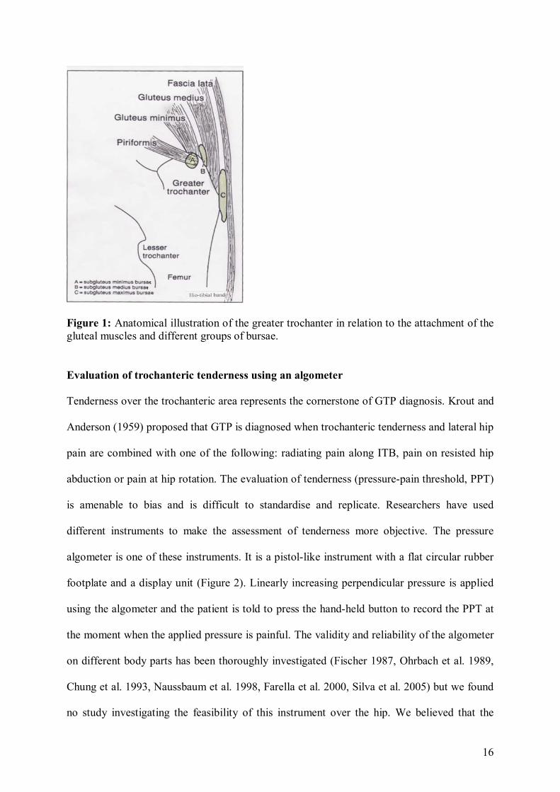
The bony surface of the greater trochanter of the femur has four facets (Pfirrmann et al. 2001):
anterior, inferior, lateral and supero-posterior. The gluteus medius muscle attaches to the
supero-posterior and lateral facets, while the gluteus minimus muscle attaches to the anterior
facet. These two muscles are considered the �rotator cuff� of the hip joint, playing an
important role in abducting the hip and stabilizing the pelvis during ambulation (Bunker et al.
1997, Kegan 1999). Weakness of these muscle leads to Trendelenburg´s sign.
Various studies (Spear et al. 1952, Bywaters 1965, Pfirrmann et al. 2001, Dunn et al. 2003,
Woodley et al. 2008) showed that there are numerous bursae (at least 6) around the greater
trochanter. One or more (up to 4) bursae lie beneath the gluteus maximus muscle (subgluteus
maximus bursae or trochanteric bursa) covering the posterior facet and the common
attachment of the gluteus medius, gluteus minimus and vastus lateralis muscles. Smaller
bursae can be found around and beneath the gluteus medius and minimus muscles (Figure 1).
The discrepancy in the number and size of these bursa(e) may support the hypothesis that
certain bursa(e) may be acquired secondary to degenerative changes with prolonged and
recurrent impaction of the facia lata on the greater trochanter.
The pathogenesis of GTP is still a matter of discussion. Though the condition is sometimes
called bursitis, no signs of local inflammation such as redness, warmth and swelling are
encountered (Silva et al. 2008). Only few patients recall a history of trauma, though a history
of overuse is not uncommon.
15
Previous hip surgery, especially involving the trochanteric region such as total hip
arthroplasty (THA) or implant fixation may apply direct trauma to the nearby muscle
attachments with their surrounding bursae, giving rise to GTP (Vicar et al. 1984, Snider 1997,
McAuley et al. 1998, Schinsky et al. 2003, Bozic et al. 2004).
Researchers have used different modalities such as plain radiograph, scintigraphy, MRI and
ultrasound to reveal any local changes that might explain the aetiology of GTP. Calcifications
and irregularities of the greater trochanter seen on plain radiographs are non-specific
manifestations and have limited diagnostic value (Finlay et al. 2006). This is also true
regarding increased uptake on scintigraphy. Bird et al. (2001) studied MRI in 24 patients with
GTP and found a gluteus medius tear in 46% of cases, gluteus medius tendinitis in 62% and
trochanteric bursal distension in less than 10% of cases. Kingzett-Taylor et al. (1999)
reviewed the findings of 250 MRI examinations performed to evaluate patients with buttock
pain, lateral hip pain and groin pain. The identified 35 patients (14%) with MRI changes of
gluteal tendinopathy (tendinosis and tear). Mayordoromo et al. (1997) studied ultrasound
findings in 15 patients with GTP and 13 controls. Bursal enlargement with increased wall
thickness was founded in 85% of the GTP group and 30% of controls.
16
Figure 1: Anatomical illustration of the greater trochanter in relation to the attachment of the gluteal muscles and different groups of bursae. Evaluation of trochanteric tenderness using an algometer Tenderness over the trochanteric area represents the cornerstone of GTP diagnosis. Krout and
Anderson (1959) proposed that GTP is diagnosed when trochanteric tenderness and lateral hip
pain are combined with one of the following: radiating pain along ITB, pain on resisted hip
abduction or pain at hip rotation. The evaluation of tenderness (pressure-pain threshold, PPT)
is amenable to bias and is difficult to standardise and replicate. Researchers have used
different instruments to make the assessment of tenderness more objective. The pressure
algometer is one of these instruments. It is a pistol-like instrument with a flat circular rubber
footplate and a display unit (Figure 2). Linearly increasing perpendicular pressure is applied
using the algometer and the patient is told to press the hand-held button to record the PPT at
the moment when the applied pressure is painful. The validity and reliability of the algometer
on different body parts has been thoroughly investigated (Fischer 1987, Ohrbach et al. 1989,
Chung et al. 1993, Naussbaum et al. 1998, Farella et al. 2000, Silva et al. 2005) but we found
no study investigating the feasibility of this instrument over the hip. We believed that the
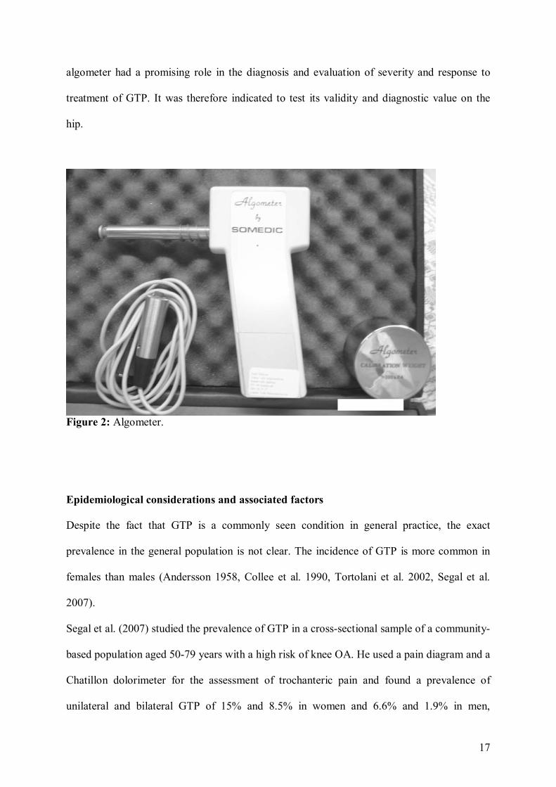
17
algometer had a promising role in the diagnosis and evaluation of severity and response to
treatment of GTP. It was therefore indicated to test its validity and diagnostic value on the
hip.
Figure 2: Algometer. Epidemiological considerations and associated factors Despite the fact that GTP is a commonly seen condition in general practice, the exact
prevalence in the general population is not clear. The incidence of GTP is more common in
females than males (Andersson 1958, Collee et al. 1990, Tortolani et al. 2002, Segal et al.
2007).
Segal et al. (2007) studied the prevalence of GTP in a cross-sectional sample of a community-
based population aged 50-79 years with a high risk of knee OA. He used a pain diagram and a
Chatillon dolorimeter for the assessment of trochanteric pain and found a prevalence of
unilateral and bilateral GTP of 15% and 8.5% in women and 6.6% and 1.9% in men,
18
respectively. Furthermore, GTP was found to be related to low back pain (LBP), ipsilateral
and contralateral knee OA and ITB tenderness. No relation was found between the occurrence
of GTP and obesity, age or race. Collee et al. (1990) and Tortolani et al. (2002) found a GTP
prevalence of 20-35% in adults with LBP. In addition, Sayegh et al. (2004), in an uncontrolled
study, found that females with LBP showed fewer back complaints when they received local
corticosteroid injections in the trochanteric region.
Raman et al. (1982) studied one hundred consecutive patients with rheumatoid arthritis (RA)
for the presence of GTP. This condition was found in 15. He concluded that GTP is an
underdiagnosed, easily remediable cause of pain in RA and that specific examination for its
presence should be a routine in all patients with RA, especially those with hip pain. Other
factors associated with GTP such as femoral offset and leg length discrepancy (LLD) are
classically suggested in the literature but not appropriately investigated.
Therapeutic considerations The treatment of GTP is mainly non-operative. During the first half of the last century
patients were treated with bed rest, heat or ice, exercises and local anaesthesia. Some
researchers used radiation. During the second half, local corticosteroids were widely used.
Gordon et al. (1961) described good response to single or multiple injections. More recent
studies (Ege Rasmusson et al. 1985, Schapira et al. 1986) showed 70-100% response but with
a relapse frequency of 25% within 10 months. However, no clinical outcome index was used
in these studies. In patients with disabling refractory GTP, surgical intervention may be
warranted. Several studies described different surgical methods with variable, yet generally
satisfactory results. These methods consisted for example of ITB decompression over the
greater trochanter with or without trochanteric bursectomy (Brooker 1979, Zoltan et al. 1986,
Slawsky et al. 1997), Z-lengthening of ITB over the greater trochanter with or without
19
trochanteric bursectomy (Chirputkar et al 2007, Craig et al. 2007). Govaert et al. (2003)
described a new operative intervention consisting of trochanteric reduction osteotomy with
screw fixation. Arthroscopic treatment of refractory GTP was performed by Fox (2002).
The main theme of the above mentioned surgical interventions was to reduce the tension of
the ITB over the greater trochanter.
Greater trochanteric pain after total hip arthroplasty Total hip arthroplasty is one of the most successful and cost-effective surgical interventions
(Swedish hip arthroplasty registry, annual report 2006). Nevertheless, the outcome of this
procedure can be adversely affected by the development of pain at the operated hip after the
operation. One type of pain is GTP. The diagnosis may be challenging. Intrinsic causes such
as septic or aseptic loosening of the prosthesis should be looked for and ruled out using
suitable laboratory and radiographic investigations. Extrinsic causes such as lumbar spine
diseases and neurovascular abnormalities are not uncommon in elderly patients and might
give rise to groin and thigh pain (Bozic et al. 2004).
The incidence of GTP after THA is variable and depends mainly on the surgical approach
used. Vicar et al. (1984) found that GTP occurred in 11% of 136 patients operated with
transtrochanteric THA, 4.4% of 91 patients operated with the anterolateral (Hardinge)
approach and 4.7% of 42 patients operated with posterolateral (Moore) approach. Schinsky et
al. (2003) studied 100 hips operated with transtrochanteric THA and 100 hips operated with
the posterolateral approach. He found GTP incidence of 8 % in the first group and 1% in the
second group. McAuley et al. (1998) found GTP incidence of 5% in 121 THAs operated with
a posterolateral approach. None of these authors investigated if there were other factors
associated with GTP development other than the surgical approach used. Iorio et al. (2006)
evaluated the incidence and associated factors of GTP in 543 primary THAs where 461 hips
20
were operated with an anterolateral approach and 82 hips with a posterolateral approach. The
incidence of GTP was 4.9% in the first group and 1.2% in the second group. Females were
more commonly affected. Leg length discrepancy, femoral offset and heterotopic ossification
were not associated with GTP development. The Hip Harris Score was significantly lower in
the GTP group compared to others.
The treatment of GTP after THA, as in non-operated patients, is mainly conservative (Iorio et
al. 2006). However, in some cases GTP complaints can be refractory, and surgical treatment
is indicated. The presence of the implant as well as the fibrosis of the soft tissues at the site of
the THA incision should be taken into consideration when planning the surgical treatment of
GTP.
Clinical and radiological measurements of LLD The measurement of LLD is an important part of examination in patients planned for THA.
Numerous clinical and radiological methods for measuring LLD have been described in the
literature. The choice of a specific method is usually a matter of personal preference, cost and
availability. Commonly, LLD is measured clinically and radiologically before and after THA.
The clinical measurement is carried out while the patient in the supine position by using a
measuring tape. The distance between the anterior superior iliac spine (ASIS) and the medial
malleolus shows the real LLD, while the distance between ASIS and umbilicus shows the
apparent LLD (Maloney et al. 2004). The latter can be affected by pelvic obliquity and lumbar
spine deviation revealing non true leg length discrepancy. Another clinical method of
measurement involves the use of blocks of increasing thickness placed under the shorter limb
until the pelvis appears level with the patient in standing position. The radiological
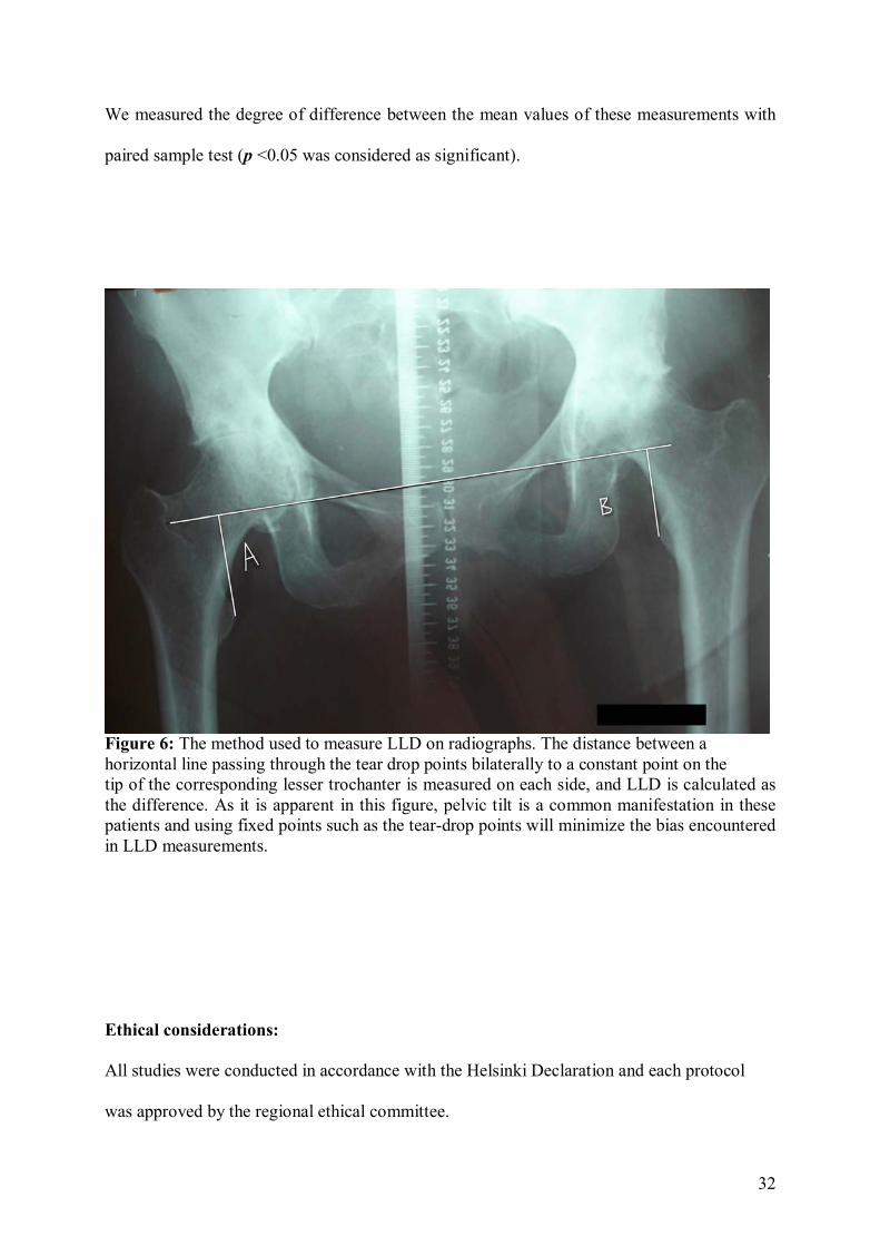
measurement is carried out on an AP view of the pelvis. Fixed points in the pelvic bones such
21
as the tear-drop points medial to the acetabulae or the lower edge of ischial tuberosities are
used as reference points and the distance between these points and fixed points at the
proximal femur such as the lesser trochanter or the centre of the femoral head is measured on
each side (Ranawat et al. 1997, Maloney et al. 2004, Matsuda et al. 2006, Clark et al. 2006,
Iagulli et al. 2006).
The degree of precision, validity, intra-observer and inter-observer reliability as well as
agreement among these different methods are quite variable (Clarke et al. 1972, Gogia et al.
1986, Friberg et al. 1988, Beattie et al. 1990, Hoyle et al. 1991, Jonson et al. 1997). For
example, Hoyle et al. (1991) found an excellent intra-class correlation coefficient (ICC) of
0.90-0.95 for intra-observer reliability and 0.98-0.99 for inter-observer reliability when he
used the clinical method (ASIS-medial malleolus) in 25 subjects. These measurements were
well correlated with the Metrecom measurements. Gogia et al. (1986) reported excellent
correlation between clinical and radiological measurements (ICC of 0.99). Nichols et al.
(1955), on the other hand, found that clinical measurement was reliable only if LLD was
greater than 17.7mm, and reliability decreased when LLD was less than 6.4mm. Beattie et al.
(1990) found an overall correlation of 0.68 between clinical and radiological measurements of
LLD in 10 patients and 9 controls. Using blocks under the shorter limb to measure LLD was
found to be an accurate (Pakvis et al. 2003) and a reliable method (Jonson et al. 1997). The
later found an ICC for intra-observer and inter-observer reliability to be 0.87 and 0.70
respectively. Woerman et al. (1984) stated that using blocks was the most accurate clinical
method and that it differed from radiological determination by an average of 2.2mm.
The radiological measurement of LLD is a commonly used method (Williamson et al. 1978,
Edeen et al. 1995, Ranawat et al. 2001, Konyves et al. 2005, Matsuda et al. 2006, Clark et al.
2006, Iagulli et al. 2006). This method has been reported to be as reliable as
orthoroentgenogram (Konyves et al. 2005). The interobserver reliability of this method was

22
tested by Woolson et al. (1999) and found to be high (0.5 mm) while intraobserver reliability
was tested by White and Dougall (2002) and found to give a measurement error of +/-1 mm.
Goodman et al. (1987) recommended the use of the tear-drop points as a landmark for
measurements rather than other points in the pelvis, because the vertical position of the tear-
drop points is not affected significantly by rotation of the pelvis. Other more advanced and
modern methods of LLD measurement such as CT scanogram, X-ray scanogram and MRI
scanogram are well documented (Helms et al. 1984, Aitken et al. 1985, Huurman et al. 1987,
Aaron et al. 1992, Terry et al. 2005, Leitzes et al. 2005).
Leg length discrepancy after THA has been associated with general dissatisfaction (Ranawat
et al. 1999 and Konyves et al. 2005), gait disorders (Rosler et al. 2000, Lai et al. 2001),
suspected aseptic loosening (Amstutz et al. 1982, Visuri et al. 1993), and nerve palsy
(Hofmann et al. 2000, Parvizi et al. 2003). However, the magnitude of a clinically significant
LLD after THA is still controversial among authors. The association between LLD and GTP
(secondary to increased tension of ITB over the greater trochanter and gait disturbance) was
mentioned in the literature but not established (Shbeeb et al. 1996, Snider 1997, Alvarez-
Nemegyei et al. 2004). Authors such as Iorio et al. (2006) suspected that LLD may play a role
in occurrence of GTP but they found no such association in their material.
Surgeons are aware that restoration of the hip joint biomechanics with as normal leg length
and femoral offset as possible is an important goal for a successful THA. Many techniques to
measure LLD (and femoral offset) intraoperatively have been advocated and thoroughly
discussed in the literature (Bal et al. 1996, Huddleston et al. 1997, Itokazu et al. 1997, Naito et
al. 1999, Bose et al. 2000, Ranawat et al. 2001).
23
Aims of the studies Study I To estimate the incidence of GTP after THA, to evaluate the clinical outcome in GTP patients compared to controls and to determine associated factor(s). Study II To evaluate the PPT differences on palpation with an electronic algometer between subjects
with GTP after THA and controls, i.e. the predictive validity of the algometer, to determine
the ratio of side differences (affected vs. unaffected) in order to estimate the intra-individual
cutoff value differentiating normal from pathological tenderness, to compare the PPT
measurements on the greater trochanter (site of local pain) in relation to the upper part of ITB
(site of referred pain), to correlate the PPT measurement to the intensity of the experienced
pain represented by VAS, to assess the reproducibility of PPT algometric measurements for
right and left body sides within asymptomatic subjects and to assess the value of a pressure
algometer as a screening test by measuring its sensitivity, specificity and positive predictive
value (PPV).
Study III To evaluate the results of a new surgical procedure (Pedersen-Noor operation) proposed to treat refractory GTP after THA consisting of distal lengthening of ITB by Z-plasty. Study IV To investigate the degree of accuracy of a commonly used clinical method to measure LLD before and after THA by correlating it to a well-known and reliable radiological method.
24
Patients and Methods Study I One hundred and seventy-two consecutive patients underwent total hip arthroplasty at
Sundsvall Hospital during January to December 2002. One hundred and fifty-eight patients
underwent surgery for primary osteoarthritis and fourteen for secondary osteoarthritis. There
were 106 women and 66 men with mean age of 69 years (30-91). The posterolateral approach
(Moore incision) was used in all patients. The implant used was cemented Lubinus SPII stem
and acetabular component which was either cemented Link cup (n=139) or non cemented
Trilogy cup (n=33).
All patients received a questionnaire 11-23 months postoperatively to determine the presence
of hip pain. A pain drawing was included to localize the site of the pain. The questionnaire
included questions about the pain modalities, ADL and analgesics consumption. On walking
(functional) VAS was used to determine the severity of the experienced pain (see Appendix).
Eight patients were dropped out because they failed to return the questionnaire. Patients with
suspected GTP (n=35) were compared with a group of asymptomatic patients selected from
the same cohort. The control group (n=35) matched the GTP by age (2 years marginal) and
sex. The two groups filled in Western Ontario and McMaster Universities (WOMAC)
osteoarthritis index (Appendix) to assess the clinical outcome. WOMAC is a self-
administered index of 0�100 (worst to best) validated for osteoarthritis in the lower
extremities. It consists of 24 items grouped into three categories: pain (five questions),
stiffness (two questions) and physical function (17 questions). This index is well validated by
Bellamy (1985, 2005).
All patients (n=70) were subjected to physical examination at the outpatient department. Four
patients from the suspected GTP group failed to show up at the follow up visit and were
therefore dropped out from the study.
25
Tenderness over the greater trochanter and ITB was examined using a pressure algometer to
measure the PPT bilaterally. Complete spinal and lower limb examination was performed in
all patients. Leg length discrepancy was measured by two clinical methods: with a measuring
tape with the patient in a standardised supine position of the distance between the ASIS and
medial malleolus and with blocks increasing in thickness in increments of 0.5cm put under the
shorter limb until the iliac crest appeared level. The mean of these measures was estimated as
LLD. Femoral offset was measured on AP view of the postoperative radiographs as the
distance between the midline and the longitudinal axis of the femoral shaft at the level of the
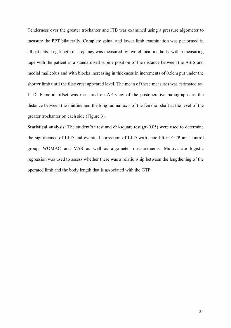
greater trochanter on each side (Figure 3).
Statistical analysis: The student�s t test and chi-square test (p<0.05) were used to determine
the significance of LLD and eventual correction of LLD with shoe lift in GTP and control
group, WOMAC and VAS as well as algometer measurements. Multivariate logistic
regression was used to assess whether there was a relationship between the lengthening of the
operated limb and the body length that is associated with the GTP.
26
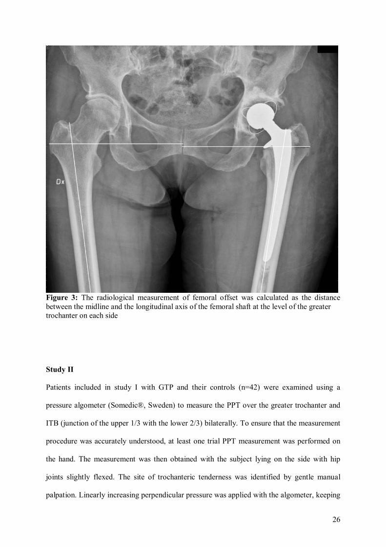
Figure 3: The radiological measurement of femoral offset was calculated as the distance between the midline and the longitudinal axis of the femoral shaft at the level of the greater trochanter on each side Study II Patients included in study I with GTP and their controls (n=42) were examined using a
pressure algometer (Somedic®, Sweden) to measure the PPT over the greater trochanter and
ITB (junction of the upper 1/3 with the lower 2/3) bilaterally. To ensure that the measurement
procedure was accurately understood, at least one trial PPT measurement was performed on
the hand. The measurement was then obtained with the subject lying on the side with hip
joints slightly flexed. The site of trochanteric tenderness was identified by gentle manual
palpation. Linearly increasing perpendicular pressure was applied with the algometer, keeping
27
a ramp rate of 40-50 kPa/sec during application of pressure. The subject was told to press the
hand-held button to record the PPT at the moment when the applied pressure felt as pain
(Figure 4). All the measurements were performed by the same observer.
Three GTP patients (with their controls, total n=6) were excluded from study II because of
difficulties encountered in standardizing the algometric measurements. Each of GTP and
control groups consisted therefore of 15 females and 3 males (mean age 65 years, 45-82).
Statistical analysis: The student�s t test and Pearson correlation coefficient (p<0.05) were
used to assess the significance of PPT differences bilaterally and between the GTP and
control group, as well as to determine the reproducibility of PPT measurement on the ITB
bilaterally in the control group as this site can be considered as a non-affected area in an
asymptomatic subject. To assess the value of pressure algometer as a screening test, the
algometer sensitivity, specificity and PPV were measured. A 89% specificity and a 78%
sensitivity values were used in accordance with the recommendation by Widmer et al. (1990)
to determine PPT cutoff value and ratio over the trochanteric region in the GTP group. To
evaluate the algometer´s value as a screening instrument we measured the PPV using GTP
incidence of 12% (established in study I).

28
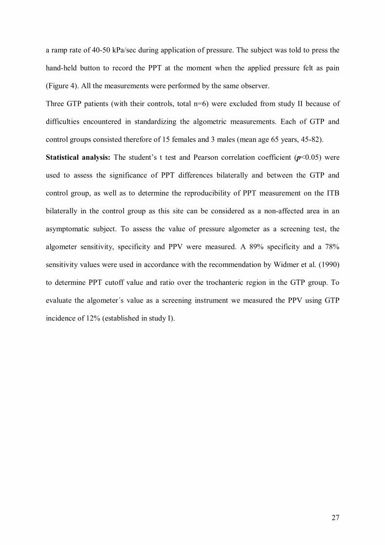
Figure 4: The method used to perform the PPT algometric measurements. The algometer is held perpendicular to the examined trochanteric region and a linearly pressure is applied under direct visual control of the pressure ramp shown at the electronic display of the device. Study III Twelve patients with refractory GTP after THA underwent surgery at Sundsvall Hospital
between March 2004 and June 2006 using a novel surgical procedure consisting of distal
lengthening of ITB by Z-plasty. The criteria of diagnosing GTP included aching and
tenderness over the greater trochanter with or without radiation along the ITB, with
exacerbation on hip rotation and abduction against resistance. Patients with atypical signs and
symptoms were investigated with radiographs to exclude any abnormalities concerning the
prosthesis. Before being considered for the operative treatment, all patients underwent at least
6 months of conservative treatment with NSAIDs, physiotherapy and local corticosteroid
without sustained improvement.
The mean age at the time of surgery was 68 years (47-84) and the mean interval between
29
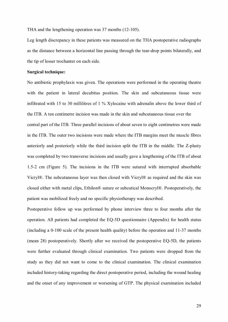
THA and the lengthening operation was 37 months (12-105).
Leg length discrepancy in these patients was measured on the THA postoperative radiographs
as the distance between a horizontal line passing through the tear-drop points bilaterally, and
the tip of lesser trochanter on each side.
Surgical technique:
No antibiotic prophylaxis was given. The operations were performed in the operating theatre
with the patient in lateral decubitus position. The skin and subcutaneous tissue were
infiltrated with 15 to 30 millilitres of 1 % Xylocaine with adrenalin above the lower third of
the ITB. A ten centimetre incision was made in the skin and subcutaneous tissue over the
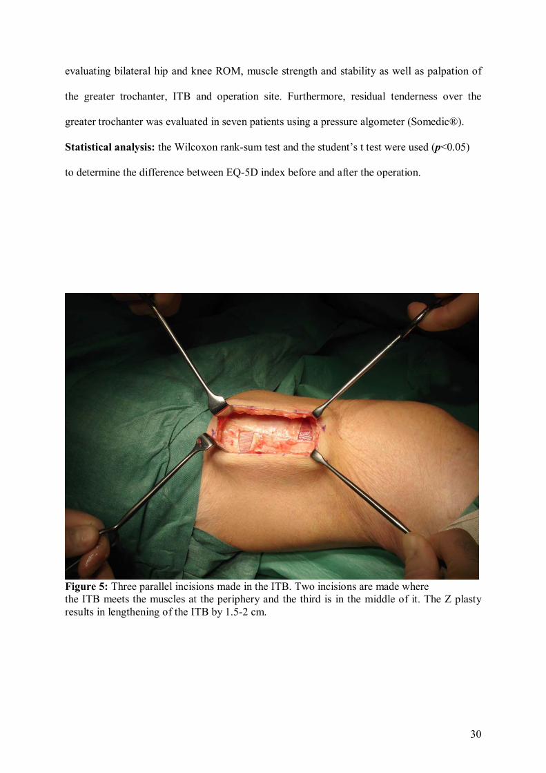
central part of the ITB. Three parallel incisions of about seven to eight centimetres were made
in the ITB. The outer two incisions were made where the ITB margins meet the muscle fibres
anteriorly and posteriorly while the third incision split the ITB in the middle. The Z-plasty
was completed by two transverse incisions and usually gave a lengthening of the ITB of about
1.5-2 cm (Figure 5). The incisions in the ITB were sutured with interrupted absorbable
Vicryl®. The subcutaneous layer was then closed with Vicryl® as required and the skin was
closed either with metal clips, Ethilon® suture or subcutical Monocryl®. Postoperatively, the
patient was mobilized freely and no specific physiotherapy was described.
Postoperative follow up was performed by phone interview three to four months after the
operation. All patients had completed the EQ-5D questionnaire (Appendix) for health status
(including a 0-100 scale of the present health quality) before the operation and 11-37 months
(mean 28) postoperatively. Shortly after we received the postoperative EQ-5D, the patients
were further evaluated through clinical examination. Two patients were dropped from the
study as they did not want to come to the clinical examination. The clinical examination
included history-taking regarding the direct postoperative period, including the wound healing
and the onset of any improvement or worsening of GTP. The physical examination included
30
evaluating bilateral hip and knee ROM, muscle strength and stability as well as palpation of
the greater trochanter, ITB and operation site. Furthermore, residual tenderness over the
greater trochanter was evaluated in seven patients using a pressure algometer (Somedic®).
Statistical analysis: the Wilcoxon rank-sum test and the student�s t test were used (p<0.05) to determine the difference between EQ-5D index before and after the operation.
Figure 5: Three parallel incisions made in the ITB. Two incisions are made where the ITB meets the muscles at the periphery and the third is in the middle of it. The Z plasty results in lengthening of the ITB by 1.5-2 cm.
31
Study IV To compare LLD measurement before and after THA, 139 patients with osteoarthritis of the
hip were studied at the Karolinska University Hospital during the period September 2002 to
March 2005. There were 85 females and 54 males with ages ranging from 44 to 89 (mean
67.5 years). Two to three weeks before the operation, patients were examined by the same
physiotherapist who measured LLD bilaterally, using a measuring tape, as the distance
between the ASIS and the medial malleolus with the patient in a supine position. During
examination both legs were placed in neutral position and close to each other to make
measurement as standardized as possible. Furthermore, patients were examined preoperatively
using plain radiographs of the pelvis and hip joints. These radiographs were standardized by
holding the lower limbs together in a neutral position while taking the pictures. On the AP
view of the pelvis, the LLD was measured by the same radiologist for all cases, as the
perpendicular distance between a line passing through both tear drops medial to the
acetabulae to the corresponding tip of the lesser trochanter. A radio-opaque ruler was used for
accurate calibration of the picture scale (Figure 6). A positive LLD value was used when the
operated limb was longer than the contralateral side while a negative value indicates the
opposite. All patients were then operated using the lateral approach (Modified Hardinge
1982). On the third postoperative day, LLD was again measured clinically and radiologically
by the same physiotherapist and radiologist using the same method of measurement as
preoperatively. The physiotherapist was blinded to the measurements of the radiologist and
vice versa. In all of the clinical measurements we used a 5mm precision scale while the
radiological measurements had a 1mm precision scale.
Statistical analysis: To investigate the degree of correlation between the clinical and
radiological measurements before and after THA, we used the Pearson�s correlation and ICC.
32
We measured the degree of difference between the mean values of these measurements with
paired sample test (p <0.05 was considered as significant).
Figure 6: The method used to measure LLD on radiographs. The distance between a horizontal line passing through the tear drop points bilaterally to a constant point on the tip of the corresponding lesser trochanter is measured on each side, and LLD is calculated as the difference. As it is apparent in this figure, pelvic tilt is a common manifestation in these patients and using fixed points such as the tear-drop points will minimize the bias encountered in LLD measurements. Ethical considerations: All studies were conducted in accordance with the Helsinki Declaration and each protocol was approved by the regional ethical committee.
33
Results Study I The evaluation of patients with suspected GTP showed that 21 patients (12%) had clinical
manifestations of this condition. There were 18 females (86%) and 3 males (14%), with a
mean age of 66 years (43-83). The severity of GTP was variable. Ten patients (5.8%) had
complaints that affected their ADL, and they needed treatment on daily basis.
Lengthening of the operated leg by 1 cm or more was found in 19/21 patients (90%) in the
GTP group (mean of LLD 1.3 cm) while in controls only 8/21 (38%) had lengthening of the
operated limb of one centimetre or more (significant difference between groups; p<0.0002).
The remaining controls either had no leg length discrepancy or less than one centimetre (mean
LLD 0.5 cm),
In the GTP group, only two patients had corrected the lengthening with a shoe or heel rise on
the opposite side. Others (17 patients) had not corrected the lengthening because they had not
been counselled about it or they used the rise only intermittently. On the other hand, six of the
eight in the control group had corrected LLD with a shoe lift early after the operation and
continued to use it regularly, which is different from the GTP group (Chi sq. <0.0009).
The WOMAC index in the GTP group revealed a significant reduction of the outcome of the
THA operation (mean 67% vs. 88% in controls; p<0.0001). Moreover, the mean of algometer
measurements in the GTP group was 69% (i.e. 69% of the pressure applied on the normal side
was needed to reproduce the pain on the affected side) vs. 98% in controls which was
significantly different; p<0.0001. These results are summarised in Figure 7.
We could not find any correlation between the change of femoral offset and the occurrence of
GTP. There were no difference of the femoral offset in the affected hip compared to the
contralateral side in the GTP group compared to less than 3% difference in controls.

34
Furthermore, no correlation was found between the occurrence of GTP and the sizes of the
prosthetic components used, patients´ weight or body length.
No statistical relation was found between the lengthening of the operated limb associated with
the GTP and the body length.
Figure 7: Summary of the patients included in study I, with their results. Study II The PPT algometric measurements over the affected greater trochanter (mean 382 kPa, SD 195kPa) and ITB (mean 563 kPa, SD 286 kPa) in the GTP group were significantly lower than the equivalent values over the contra-lateral side (greater trochanter mean 624 kPa, SD 305 kPa and ITB mean 680 kPa, SD 350 kPa) (r=0.68, p=0.002 and r=0.90, p=0.0001 respectively). No such a difference could be found in the control group: greater trochanter
172 THA patients (8 pat. dropped out)
35 suspected GTP (4 pat. dropped out) 35 controls
21 diagnosed GTP pat. (F:M=18:3)
21 age and sex matched controls
Mean LLD=1.3 cm
Mean WOMAC=67%
Mean LLD=0.5
Mean WOMAC=88%
Mean Algometer=69% Mean Algometer=98%
Mean VAS=3.90 Mean VAS=0.74
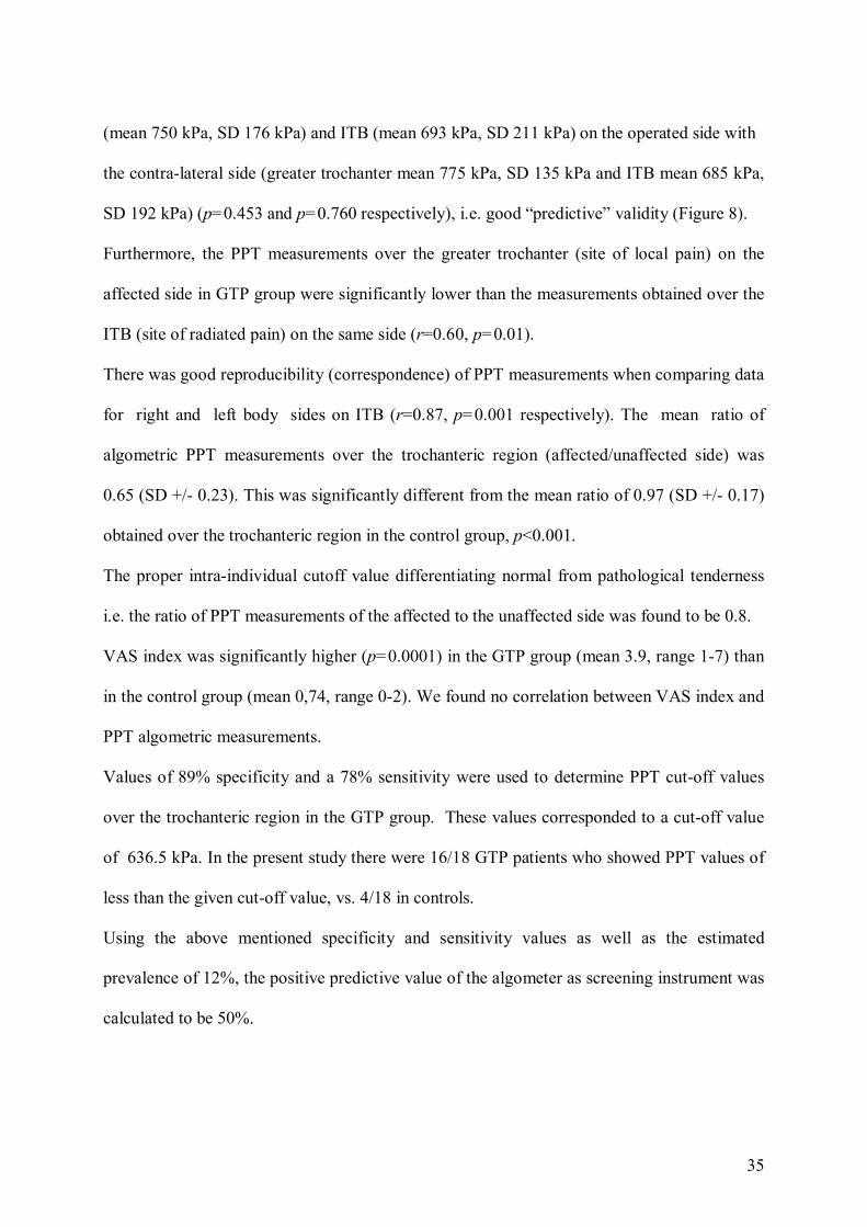
35
(mean 750 kPa, SD 176 kPa) and ITB (mean 693 kPa, SD 211 kPa) on the operated side with the contra-lateral side (greater trochanter mean 775 kPa, SD 135 kPa and ITB mean 685 kPa,
SD 192 kPa) (p=0.453 and p=0.760 respectively), i.e. good �predictive� validity (Figure 8).
Furthermore, the PPT measurements over the greater trochanter (site of local pain) on the
affected side in GTP group were significantly lower than the measurements obtained over the
ITB (site of radiated pain) on the same side (r=0.60, p=0.01).
There was good reproducibility (correspondence) of PPT measurements when comparing data
for right and left body sides on ITB (r=0.87, p=0.001 respectively). The mean ratio of
algometric PPT measurements over the trochanteric region (affected/unaffected side) was
0.65 (SD +/- 0.23). This was significantly different from the mean ratio of 0.97 (SD +/- 0.17)
obtained over the trochanteric region in the control group, p<0.001.
The proper intra-individual cutoff value differentiating normal from pathological tenderness
i.e. the ratio of PPT measurements of the affected to the unaffected side was found to be 0.8.
VAS index was significantly higher (p=0.0001) in the GTP group (mean 3.9, range 1-7) than
in the control group (mean 0,74, range 0-2). We found no correlation between VAS index and
PPT algometric measurements.
Values of 89% specificity and a 78% sensitivity were used to determine PPT cut-off values
over the trochanteric region in the GTP group. These values corresponded to a cut-off value
of 636.5 kPa. In the present study there were 16/18 GTP patients who showed PPT values of
less than the given cut-off value, vs. 4/18 in controls.
Using the above mentioned specificity and sensitivity values as well as the estimated
prevalence of 12%, the positive predictive value of the algometer as screening instrument was
calculated to be 50%.
36
Figure 8: Summary of the patients included in study II and the results of their PPT measurements of the trochanteric area (Troch. Area) and ilio-tibial band (ITB). Study III The EQ-5D index showed an improvement in all but one patient who experienced no change
in the GTP complaint after the operation. The mean of EQ-5D index was 0.26 (0-0.73) before
the operation and 0.67 (0.16-1.0) after the operation (p <0.005). The mean health quality
index was 34 +/- 10 (range 10-60) before the operation and 60 +/-20 (range 30-90) after the
operation (p <0.005).
On post-THA radiographs all patients except one had a lengthening of the operated leg, with a
mean increase of leg length of 0.85 cm +/- 0.43 (0-1.5 cm).
The phone interviews performed three to four months after the operation revealed that seven
patients experienced improvement of the GTP signs and symptoms. Four patients reported a
gradual improvement on later follow up. One of those four patients felt neither improvement
nor deterioration in her GTP complaint, but the tightness and tenderness along ITB that she
had developed after the THA operation disappeared after the Z-plasty operation with
172 THA patients (8 pat. dropped out)
21 diagnosed GTP pat. (3 pat. excluded)
21 age and sex matched controls
18 patients included 18 age and sex matched controls
Mean PPT measurements: Troch. Area=382 kPa, ITB=563 kPa
Mean PPT measurements: Troch. Area= 624 kPa, ITB=680 kPa

37
subsequent improvement of hip and thigh ROM. The remaining patient showed neither
improvement nor deterioration in her GTP complaint or EQ-5D index.
Clinical examination 1-3 years postoperatively revealed no signs of decreased ROM,
decreased strength or instability of the ipsilateral hip or knee. The algometric PPT
measurements performed in seven patients revealed a mean value of 357 kPa over the affected
greater trochanter vs. 427 kPa over the contralateral greater trochanter i.e. cutoff ratio of 0.84.
No postoperative complications were encountered after the Z-plasty.
Study IV The mean of preoperative clinical LLD measurements was -1 mm (S.D. = 9 mm) while the
mean of preoperative radiological LLD measurements was - 5 mm (S.D. = 7 mm). There was
a weak correlation/agreement between these two measurement methods (correlation r =0.21,
intra-class correlation coefficient ICC =0.33) and the estimated LLD as evaluated by clinical
and radiological methods where significantly different (p<0.0001).
Postoperative clinical LLD measurement showed a mean of 2 mm (S.D. = 7 mm) while
postoperative radiological LLD measurement showed a mean of 3 mm (S.D. = 7 mm).
The correlation / agreement between these measures was fair (r=0.45, ICC=0.62). There was
no significant difference between the means of the postoperative clinical and radiological
measures (p<0.177).

38
Discussion Greater trochanteric pain after THA is not a rare complication. The incidence found in study I
(12%) was higher than the incidence found by others (Vicar et al. 1984, McAuley et al. 1998,
Schinsky et al. 2003, Iorio et al. 2006). This discrepancy is probably related to the different
GTP selection criteria used in these studies. In our study, for example, we actively searched
for patients with signs and symptoms of GTP, including all the patients regardless of the
severity of their complaints. There were 10 patients (5.8%) who had been in contact with their
surgeons because of the GTP complaints. Others had sporadic visits to a GP or self-
medicated. However, the overall outcome of THA in the GTP group measured by WOMAC
index was lower than in controls. Similar reduction in outcome was found by Iorio et al.
(2006).
The high incidence of GTP after transtrochanteric approach is probably due to the direct
trauma applied on the gluteal muscle attachments on the greater trochanter with their
surrounding bursae. Furthermore, the metalware used to fix the osteotomy may irritate the soft
tissues surrounding it, giving rise to pain and tenderness. On the other hand, THA using
anterolateral approach (Hardinge) involves the detachment and re-attachment of up to two
thirds of the gluteus medius muscle attachment to the greater trochanter.
Consistent with other studies (Iorio et al. 2006, Segal et al. 2007), the incidence of GTP in our
study was more common in females (female to male ratio was 6:1). The mechanism of this
increment is still unclear. Possible explanations include hormonal influence on the bursae,
differences in pelvic anatomy with increased ITB tension, differences in pain perception and
differences in physical activity.
The association found in study I between lengthening of the operated limb by ≥1 cm and the
occurrence of GTP is an interesting finding. In patients with OA changes of the hip, there is
often a shortening of the affected limb with soft tissue contracture apart from the degenerative

39
changes. When operating on such patients, restoration of leg length and femoral offset might
give rise to increased tension on the lateral aspect of the hip. The authors believe that
lengthening of the operated limb resulted in an increased traction of ITB on the greater
trochanter. Furthermore, the gait disturbance caused by the lengthening loads the gluteal
muscle attachment to the greater trochanter. The violation caused by the surgical trauma of
THA adds an extra factor and this might minimize the degree of LLD needed to predispose to
GTP occurrence.
The early correction of LLD with a shoe lift under the short limb found in the control group
might have reduced some of the traction and the biomechanical disturbance caused by LLD
and therefore can possibly reduce the risk of its GTP occurrence. However, the material
involved in this correction is small and probably non-conclusive.
We tried to improve the accuracy of LLD measurements by using 2 clinical methods (ASIS-
medial malleolus and blocks). We took the average values and all measurements were done
by the same observer and in a standardized manner to minimize the risk of bias. We preferred
not to measure LLD on radiographs because of the lack of standardization in patients´
radiographs.
Despite our expectations, we found no correlation between GTP occurrence and femoral
offset. The method of offset measurement we used is well known in the literature, but we are
unaware of whether it is validated. All the radiographic measurements of femoral offset were
done by one observer which may have reduced bias. Similarly to our study, Iorio et al. (2006)
found no correlation between femoral offset and GTP.
No statistical correlation was found between the occurrence of GTP and body weight and
length, the degree of limb lengthening to body length and the thickness or angle of neck of the
femoral component used.
40
We believe that the use of a pressure algometer in study II made the assessment of
trochanteric tenderness associated with GTP more objective. When eliciting tenderness, at
least two aspects will be involved: a decreased PPT and an increased response to the given
stimulus. These two aspects can vary, giving rise to a variable severity. Due to this variability,
the method used for evaluation of tenderness should be reliable. Finger palpation (the most
commonly used method) exerting an excessive pressure over a giving area can give a false
positive result in a healthy individual and vice versa. Furthermore, different observers using
various palpation techniques can evaluate the degree of tenderness (and subsequently the
severity of the given condition) in different ways. The follow up of such tenderness with
finger palpation is another problem.
In this study, the use of an algometer on the hip showed good predictive validity, which
means that this instrument was able to predict something it should theoretically be able to
predict, in this case the GTP patients to have lower PPT than controls. This is consistent with
other studies which investigated the algometer validity and intra-observer and inter-observer
reliability on other parts of the body ( Fischer 1987, Ohrbach et al. 1989, Chung et al. 1993,
Naussbaum et al. 1998, Farella et al. 2000, Silva et al. 2005). Furthermore, these studies
showed that the algometer was useful for measuring PPT in both symptomatic and symptom-
free individuals for diagnostic purposes, and for the evaluation of the efficacy of different
treatment strategies, mainly in orofacial practice. The instrument allows the patient to self-
actuate the cutoff point through a hand-held button. The latter character is thought to reduce a
potential operator error factor (Chung et al. 1993).
We chose to make all PPT measurements by the same examiner and the rate of algometric
pressure application was kept constant in this study (40-50 kPa/sec) to ensure good reliability
of the algometer (Chung et al. 1993, Nussbaum et al.1998).
41
We examined the painful side first because we believed that lying on the painful side first
might evoke pain with subsequent local and central sensitisation leading to a possible bias in
measuring the PPT on this side later on.
The detection of increased PPT over ITB (site of radiated pain) in relation to the greater
trochanter itself (site of local pain) is an issue of interest. A similar phenomenon has been
found by other researchers (Kosek et al. 1993 and 1999, Abou Atme et al. 2005). In addition
to the fact that the pathological changes in GTP are localized at the trochanteric region and
not at the ITB, this discrepancy might also due to the differences in the thickness of
subcutaneous tissue of these two sites (Kosek et al.1993, 1999). Another possible explanation
is what is called diffuse noxious inhibitory control (DNIC). In muscle pain patients, it is
believed that a pathologically disturbed inhibitory mechanism may result in widespread deep
hypoalgesia of the surrounding tissues (Abou Atme et al. 2005).
The absence of a significant correlation between PPT algometric measurements and VAS in
the present study indicates the complexity of using VAS as a simple pain evaluator. It also
reveals the fact that different factors (including psychological) play a role in the experience of
pain in addition to the local (nociceptive) factor. Poor correlation was also found by Walco et
al. (1990) while others e.g. List et al. (1993) and Hogeweg at al. (1992), by contrast found a
significant correlation between PPT and VAS. This discrepancy could be due to the
differences in measurement sites of PPT, as well as the pathology of the condition
investigated in different studies.
The large inter-individual variability of PPT measurements across subjects found in study II
was previously documented by other investigators (Jensen et al. 1986, Tunks et al. 1988,
Vantin et al. 1998, Persson et al. 2004, Rolke et al. 2005). Indeed, these findings reflect the
importance of intra-individual side to side comparisons of PPT measurements when assessing
deep pain sensitivity.

42
The large inter-individual variability together with the relatively low PPV found in this study
(50%) and by other authors such as Farella et al. (2000) suggest that the pressure algometer
might have limited value as a screening instrument when used to compare PPT measurements
among subjects in case of GTP screening for example following THA .
On the other hand, the study shows good reproducibility (correspondence) of the algometer in
measuring PPT when used on the ITB of the control group.
The 0.8 ratio of PPT algometric measurements of the affected to the unaffected side found in
the present study can be used for diagnostic purposes, to differentiate normal from
pathological tenderness in patients with suspected GTP. This value was very close to the
value of 84.1% found by Fisher 1987. There were six GTP patients with a PPT ratio > 0.8
overlapping the PPT ratio of the control group. Five of these patients had a mild degree of
GTP with good response to NSAIDs. One patient had a bilateral GTP complaint with
decreased PPT on both sides.
The selection of a cutoff value corresponding to 78% sensitivity and 89% specificity is
recommended by different studies. According to Widmer et al. (1990), when the condition
investigated is of low mortality (as in GTP), the sensitivity and specificity levels of diagnostic
tests performed in medical sciences should be around 75% and 90%, respectively. These
values allow the test to diagnose about 25% of would-be patients as healthy and 10% of
healthy individuals as patients.
Researchers have used a pressure algometer to assess the effect of different treatment
modalities such as intra-muscular injections (Fischer 1984, Jensen et al. 1986, Christidis et al.
2007) and physiotherapy (including acupuncture) (Fischer 1984, List et al. 1993) on the PPT.
The good validity of the algometer shown in study II and the determined ratio indicate the
potential use of this instrument to evaluate the effect of different types of GTP treatment on
the PPT in a more reliable way compared to finger palpation or VAS index.
43
The treatment of GTP after THA is mainly conservative and Iorio et al 2006 reported good
response to measures such as corticosteroid injections, physiotherapy with or without
ultrasound, NSAIDs or merely observation of all GTP cases studied (n=24). However, in our
experience some patients need surgical intervention since GTP complaints can be refractory
and adversely affect their physical ability.
Many investigators reported good results of different surgical methods used in the treatment
of refractory trochanteric bursitis or painful snapping hips. These methods relied on the same
principle of minimizing the pressure or friction applied on the greater trochanter by the
tense/thickened ITB. On the other hand, we are unaware of any studies investigating surgical
treatment of refractory GTP after THA.
Brignall et al. (1991), Lam et al. (2003) and Provencher et al. (2004) used a local proximal
Z-plasty to release the ITB over the greater trochanter in patients with painful snapping hip.
These patients showed favourable response and the method was recommended as an
alternative to the conservative therapy in refractory cases.
The same method was used by Kim et al. (2002) who treated three military patients with
painful snapping hip. The results of this study were less than optimal.
Brooker (1979) reported good response to T- incision or local fenestration of ITB with
removal of trochanteric osteophytes and debridement of the gluteal maximus bursa. The
method was performed in five patients with one year follow up. These patients showed
improvement of mean Harris hip score from 46 to 88 after the operation.
Zoltan et al. (1986) described a surgical method of elliptical incision of ITB creating a defect
over the greater trochanter followed by trochanteric bursectomy in seven active patients who
developed refractory trochanteric pain secondary to snapping hip. The average length of
follow up of these patients was 55 months. One patient was operated on twice. All patients
showed good response.

44
Slawski et al. (1997) reported a single surgeon�s experience of treating seven hips by simple
longitudinal release of the ITB over the greater trochanter with excision of the subgluteal
bursa. The mean follow up of this study was 20 months and these patients responded well to
this surgical treatment with obvious improvement of Harris hip score (mean 51.7 to 95.0).
Govaert et al. (2003) described a new operative procedure consisted of trochanteric reduction
osteotomy with screw fixation. Ten patients (12 hips) were operated in this series. The mean
follow up was 23.5 months. The mean Merle d´ Aubigné and Postel score improved from 15.8
to 27.5. The method was considered as safe and effective. The authors of this work reported
previous poor response to longitudinal release of the ITB with trochanteric bursectomy which
had been performed on five hips.
Arthroscopic treatment of refractory GTP and snapping hip was performed by Fox (2002) and
Ilizaliturri et al. (2006). In the former work, twenty-seven hips with refractory GTP were
treated with arthroscopic bursectomy and the patients were followed up one and five years
after the operation. Only two patients showed manifestations of recurrence, otherwise all
other patients had good to excellent results. However, it was not clear how the results were
evaluated, because a specific outcome index had not been used. In the later work, eleven
snapping hips were operated with arthroscopic release of ITB. Two-year follow up of these
patients revealed non-painful snapping in one patient while the rest of the patients had no
complaints and had returned to their previous level of activities. The WOMAC was used in
this series.
Chirputkar et al (2007) reviewed the results of a series of 16 patients treated with bursectomy
and Z-lengthening of the ilio-tibial band over the greater trochanter. The mean follow up was
52 months. The authors used a questionnaire to evaluate the pain and degree of satisfaction in
the operated patients and found that all 14 patients who answered the questionnaire were
45
happy with the outcome of operation and 13/14 patients would undergo a similar procedure
again. However, it is not clear whether the questionnaire used was a validated one or not.
We believe that treating patients with refractory GTP after THA with local ITB release with
or without bursectomy gives poor results, and applies further trauma to the area already
injured by the THA operation. This may increase the risk of wound infection and recurrence
of GTP. On the other hand, technical difficulties may be encountered if GTP patients undergo
arthroscopic surgery. Trochanteric reduction osteotomy (Govaert et al. 2003) can not be
performed due to the presence of hip prosthesis.
The distal ITB lengthening by Z-plasty as described in study III was first performed by
Eskild Pedersen MD at Sundsvall Hospital in 1997. The idea behind this procedure was taken
from the lengthening of extensor carpi radialis brevis (ECRB) muscle tendon in the treatment
of refractory tennis elbow (Stovell et al. 1979, Kumar et al. 2004) without applying further
trauma locally at the site of pain. Fridén et al (1994) found that lengthening of the ECRB by
9.1 mm resulted in a 25% decrease in passive muscle tension that could lead to reduced
tension and decreased pain.
In study III we had no control group. However, the patients included had experienced their
GTP complaints for at least 12 months before they underwent the lengthening operation
described. The algometric cutoff ratio of 0.84 estimated in the operated patients of study III
supports our finding in study II, which showed asymptomatic patients of the control group
had a cutoff ratio of more than 0.80.
The technique described is simple, safe and effective. It is performed under local anaesthesia
and on an outpatient basis. The follow up results and the absence of postoperative
complications in this study encourage the authors to recommend this method for the treatment
of refractory GTP after THA. It should be emphasized that patient selection is crucial. In a
patient with disabling refractory GTP and increased tightness of ITB, distal Z-plasty
46
represents a promising therapeutic alternative. However, a prospective larger study of this
technique for the treatment of GTP after THA is recommended for further evaluation.
Lengthening of the operated limb after THA has been described as a possible cause of GTP
development. In study IV we found shortening of the affected limb preoperatively. This is a
common finding and is usually secondary to loss of hip joint space with or without cranial
migration of the femoral head as well as soft tissue involvement with adduction/abduction and
flexion contractures of the affected hip. Postoperatively, on the other hand, there was a
lengthening of the operated leg. This is also a common outcome after THA because many
surgeons tend to lengthen the operated leg as they try to achieve good stability around the
operated prosthesis. This phenomenon was also described by Ranawat and Rodriguez (2001).
The weak correlation between the clinical and radiological measurements preoperatively is
probably due to the fact that the radiological measurement was more accurate and easily
standardized than clinical examination. The shortening was less apparent clinically than
radiologically because of the painful contracture in the affected hip with a compensating
pelvic tilt/obliquity that minimized the apparent shortening. The correlation between the
clinical and radiological measurement was better postoperatively. The authors believe this
was the result of the THA which minimized soft tissue contracture allowing the patient to lie
on the examination table with less pain. This made the clinical measurement of LLD more
accurate and correlated better with the radiological examination. Clinicians should therefore
be aware of the limitations encountered in the use of the described clinical method for the
evaluation of preoperative LLD in patients considered for THA. Furthermore, caution is
advised in using the clinical method of postoperative LLD measurement despite its better
correlation with the radiological measurement. In cases were LLD measurement can give rise
to further intervention such as revision surgery the authors recommend the use of a well-
documented method with a high degree of precision such as CT scanogram.

47
Summary and Conclusions
Despite the incidence of GTP after THA having become lower since the posterior and the
lateral approaches successively replaced the transtrochanteric approach, this complication
represents a drawback in the outcome of an otherwise a very successful procedure. We found
an incidence of 12% with decreased clinical outcome determined by WOMAC index (Study
I).
Lengthening of the operated limb by ≥ 1cm was found to be an associated factor (study I).
Every effort should therefore be taken to minimize the risk of LLD after THA. This is usually
achieved by preoperative templating and intraoperative leg length measurement. We
recommend the use of the radiological method of LLD measurement both before and after
THA (study IV). The use of a pressure algometer can be promising in the diagnosis and
follow up of the effect of treatment. We found that the used algometer had good predictive
validity and reproducibility. The cutoff ratio of 0.8 can be used to differentiate pathological
from normal tenderness. There were considerable inter-individual differences in PPT
measurements, and the algometer PPV was found to be 50%. This makes the use of the
algometer unsuitable as a screening instrument. Fortunately most of GTP patients respond to
non-operative measures. For those who show refractory GTP manifestations, distal
lengthening of ITB band is a simple, safe and effective procedure.
48
Future implications 1) It is of a great interest to evaluate the incidence and associated factors of GTP after
minimal invasive surgery (MIS) especially when the anterior approach is used. Furthermore,
the use of navigation systems in hip replacement surgery can affect the incidence of
postoperative LLD
2) The effect of lengthening the operated limb and the early correction of LLD can be
investigated using a prospective controlled trial where correction of LLD is randomized
against non correction to study its role.
3) Distal lengthening of the ITB band can be used in non-THA operated GTP patients.
Randomisation of patients with refractory GTP complaints between the distal lengthening of
ITB against proximal ITB decompression/fenestration or continued conservative therapy is
necessary to evaluate the actual effect of these different modalities.
49
Populärvetenskaplig sammanfattning I Sverige opereras över 14.000 patienter årligen med höftprotes och det är en av de mest
framgångsrika och kostnadseffektiva operationerna överhuvudtaget. Emellertid blir 5-15 % av
patienter inte nöjda med resultatet av operationen då de utvecklar ett smärttillstånd över
höftens utsida som brukar kallas trokanterit eller greater trochanteric pain (GTP). Målsättning
med avhandlingen var att undersöka förekomsten av GTP, att studera eventuella riskfaktorer,
att bedöma livskvalité hos drabbade patienter jämfört med en kontrollgrupp, att mäta graden
av tryckömhet över höften med en tryckmätare (algometer), att jämföra två metoder för
mätning av benlängdsskillnad hos höftprotesopererade patienter och att evaluera resultatet av
en ny kirurgisk teknik för behandling av GTP smärta.
Studie I: Bland 172 patienter som opererades år 2002 på Sundsvalls sjukhus med höftprotes
förekom GTP besvär med varierande svårighetsgrad hos 12 %. Livskvalité bedömd med
WOMAC index var nedsatt hos dessa patienter jämfört med en ålders- och könsmatchad
kontrollgrupp. GTP var vanligare hos kvinnor och hos patienter där det opererade benet
förlängts > 1cm. Tidig korrektion av benförlängning med skoinlägg minskade risken för GTP.
Studie II: Med en elektronisk mätare (algometer, Somedic®, Hörby-Sverige) undersöktes
tryckömheten över höften (trokantern) och lårets utsida (traktus ilio-tibialis). Hos 18 patienter
med GTP efter höftprotesoperation och en matchande kontrollgrupp undersöktes algometerns
prediktiva validitet, cutoff värde och kvot för att särskilja den patologiska från den normala
ömheten, specificitet och sensitivitet samt positiva prediktiva värdet (PPV) för att bedöma
algometerns värde som instrument för diagnostik och screening. Vi fann att algometer-
metoden har en god prediktiv validitet. Vid den rekommenderade sensitiviteten av 75% och
specificiteten av 90% fann vi ett cutoff värde på 636,5 kPa samt en cutoff kvot på 0,8. Detta
innebär att algometer kan användas för att diagnosticera GTP hos patienter med höftvärk.
Apparaten kan även användas för evaluering av olika behandlingsmetoders resultat. Eftersom
50
det förelåg stora individuella skillnader i tryckömhet bland patienter och i kontrollgruppen
och då PPV var endast 50% bedömde vi att algometer inte är lämplig som
screeningsinstrument.
Studie III: Vi studerade resultatet av en ny kirurgisk metod för att behandla resistenta GTP
besvär efter höftprotesoperation. Metoden syftar till att förlänga traktus iliotibialis med Z-
plastik i nedre delen av låret ca 10 cm ovanför knäleden. Operationen görs i lokalbedövning
och patienten går hem samma dag utan restriktioner. Mellan mars 2004 och juni 2006
opererades 12 kvinnor (medelålder 68, 47-84 år) enligt den nya metoden. Inga patienter
rapporterade någon komplikation. Utvärdering gjordes med klinisk undersökning och EQ-5D
hälsoenkät före och 1-3 år efter operationen och på röntgenbilder uppmättes
benlängdsskillnad. Vi fann att hos 11 av 12 opererade patienter minskade GTP besvären och
förbättrades livskvalitén [EQ-5D index före operationen var 0-0.73 (medelvärde 0.29) medan
efter operationen var 0.16 � 1.0 (medelvärde 0.70)].
Studie IV: Hos 139 patienter med höftartros som opererades med höftprotes på Karolinska
Universitetssjukhuset, Solna perioden mellan sep 2002 till mars 2005 uppmättes
benlängdsskillnad kliniskt och röntgenlogiskt före och efter operationen. Vid den kliniska
bedömningen av benlängdsskillnad uppmättes på båda sidor avståndet mellan en specifik
punkt i bäckenet (spina iliaca) och inre fotknölen (mediala malleolen) med patienten i
liggande ställning. Den röntgenlogiska benlängdsskillnaden bestämdes till skillnaden i det
vinkelräta avståndet mellan linjen genom tear-drop punkterna i bäckenet och spetsarna på
trokanter minor på lårbenen.
Vi fann en svag korrelation mellan kliniska och röntgenologiska benlängdsmätningarna före
operationen. Efter operationen var korrelationen måttlig mellan mätningarna. Utifrån vårt
resultat föreslås att benlängdsmätning före operationen bör göras röntgenologiskt då en
klinisk mätning underskattar benförkortningen.
51
References
A Aaron A, Weinstein D, Thickman D, Eilert R. Comparison of orthoroentgenography and computed tomography in the measurement of limb length discrepancy. J Bone Joint Surg Am. 1992; 74(6):897-902. Abou-Atme YS, Melis M, Zawawi K. Pressure pain threshold of thelateral pterygoid muscles in TMD patients and controls. J Contemp Dental Pract. 2005; 6:1�8. Aitken AG, Flodmark O, Newman DE, Kilcoyne RF, Shuman WP, Mark LA. Leg length determination by CT digital radiography. Am J Roentgenol. 1985; 144(3):613-5. Alvarez-Nemegyei J, Canoso JJ. Evidence-Based Soft Tissue Rheumatology: III: Trochanteric Bursitis. J Clin Rheumatol. 2004; 10(3):123-124. Amstutz HC, Ma SM, Jinnah RH, Mai L. Revision of aseptic loose total hip arthroplasties. Clin Orthop Relat Res. 1982; (170):21-33. Anderson TP, Hanover NH. Trochanteric bursitis: diagnostic criteria and clinical significance. Arch Phys Med Rehab. 1958; 39: 617-622.
B Bal BS. A technique for comparison of leg lengths during total hip replacement. Am J Orthop. 1996; 25(1):61-2. Beattie P, Isaacson K, Riddle DL, Rothstein JM. Validity of derived measurements of leg-length differences obtained by use of a tape measure. Physical Therapy 1990; 70(3):150-7. Bellamy N. WOMAC: a 20-year experiential review of a patient-centered self-reported health status questionnaire. J Rheumatol. 2002; 29(12):2473-6. Review. Bellamy N, Buchanan WW, Goldsmith CH, Campbell J, Stitt LW. Validation study of WOMAC: a health status instrument for measuring clinically important patient relevant outcomes to antirheumatic drug therapy in patients with osteoarthritis of the hip or knee. J Rheumatol. 1988; 15(12):1833-40. Bird PA, Oakley SP, Shnier R, Kirkham BW. Prospectiveevaluation of magnetic resonance imaging and physicalexamination findings in patients with greater trochanteric pain syndrome. Arthritis Rheumat 2001; 44: 2138-45. Bose WJ. Accurate limb-length equalization during total hip arthroplasty. Orthopedics. 2000; 23(5):433-6. Bozic KJ, Rubash HE. The painful total hip replacement. ClinOrthop 2004; 420: 18-25. Brignall CG, Stainsby GD. The snapping hip, treatment by Z-plasty. The Journal of Bone and Joint Surg (Br).1991; 73(2): 253-254.
52
Brooker AF. The surgical approach to refractory trochanteric bursitis. John Hopkins Medical Journal. 1979; 145: 98-100. Buchanan HM, Midgley JA. Evaluation of pain threshold using a simple pressure algometer. Clin Rheumatol 1987; 6:510-7. Bunker TD, Esler CN, Leach WJ. Rotator-cuff tear of the hip. J Bone Joint Surg (Br) 1997; 79(4):618-20. Bywaters EG. The bursae of the body. Ann Rheum Dis. 1965; 24:215-8.
C Chirputkar K, Weir P, Grey A. Z-lengthening of the iliotibial band to treat recalcitrant cases of trochanteric bursitis. Hip International 2007; 17: 31-35. Christidis N, Nilsson A, Kopp S, et al. Intramuscular injection of Granisetron into the masseter muscle increases the pressure pain threshold in healthy participants and patients with localized myalgia. Clin J Pain. 2007; 23:467�472.
Chung SC, Kim JH, Kim HS. Reliability and validity of the pressure pain thresholds (PPT) in the TMJ capsules by electronic algometer. Physiology 1993;11:171�177. Clark CR, Huddleston HD, Schoch EP 3rd, Thomas BJ. Leg-length discrepancy after total hip arthroplasty. J Am Acad Orthop Surg. 2006;14(1):38-45. Clarke GR. Unequal leg length: an accurate method of detection and some clinical results. Rheumatol Phys Med. 1972;11(8):385-90. Cleveland RH, Kushner DC, Ogden MC, Herman TE, Kermond W, Correia JA. Determination of leg length discrepancy. A comparison of weight-bearing and supine imaging. Invest Radiol. 1988; 23(4):301-4. Collée G, Dijkmans BAC, Vandenbroucke JP, Cats A. Greater trochanteric pain syndrome (trochanteric bursitis) in low back pain. Scand J Rheumatology 1991; 20: 262-6. Craig RA, Jones DP, Oakley AP, Dunbar JD. Iliotibial band Z-lengthening for refractory trochanteric bursitis (greater trochanteric pain syndrome). ANZ J Surg. 2007;77(11): 996-8. D Dunn T, Heller CA, McCarthy SW, Remedios CD. Anatomical study of the �Trochanteric Bursa�. Clinical Anatomy 2003; 16: 233-40.
53
E Edeen J, Sharkey PF, Alexander AH (1995) Clinical significance of leg-length inequality after total hip arthroplasty. Am J Orthop 24:347�351
Ege Rasmussen KJ, Fanø N. Trochanteric bursitis. Treatment by corticosteroid injection. Scand J Rheumatol. 1985;14(4):417-20.
F Farella M, Michelotti A, Steenks MH, et al. The diagnostic value of pressure algometry in myofasical pain of the jaw muscles. J Oral Rehabil. 2000;27:9�14. Finlay K, Friedman L. Ultrasonography of the lower extremity. Orthop Clin North Am 2006;37:245-75. Fisher AA. Diagnosis and management of chronic pain in physical medicine and rehabilitation. Curr Ther Psychiatry 1984; 8:123�145. Fisher AA. Pressure algometry over normal muscles. Standard values, validity and reproducibility of pressure threshold. Pain 1987; 30:115�126. Fox JL. The role of arthroscopic bursectomy in the treatment of trochanteric bursitis. Arthroscopy 2002; 18(7): E34. Friberg O, Nurminen M, Korhonen K, Soininen E, Mänttäri T. Accuracy and precision of clinical estimation of leg length inequality and lumbar scoliosis: comparison of clinical and radiological measurements. Int Disabil Stud. 1988; 10(2):49-53. Friden J, Lieber RL. Physiologic consequences of surgical lengthening of extensor carpi radialis brevis muscle � tendon junction for tennis elbow. J Hand Surg.1994; 19 (2): 269-274.
G Gogia PP, JH Braatz. Validity and reliability of leg length measurements. J Orthop Spts Phys Ther. 1986; 8(4):185-188. Goodman SB, Adler SJ, Fyhrie DP, Schurman DJ. The acetabular teardrop and its relevance to acetabular migration. Clin Orthop 1988; 236: 199-204. Gorden EJ. Trochanteric bursitis and tendinitis. Clin Orthop. 1961; 20:193-202. Govaert LHM, van der Vis HM, Marti RK, Albers GHR.Trochanteric reduction osteotomy as a treatment for refractory trochanteric bursitis. J Bone Joint Surg 2003; 85:199-203. Gross MT, Burns CB, Chapman SW, Hudson CJ, Curtis HS, Lehmann JR, Renner JB. Reliability and validity of rigid lift and pelvic leveling device method in assessing functional leg length inequality. J Orthop Sports Phys Ther. 1998; 27(4):285-94.

54
H Helms CA, McCarthy S. CT scanograms for measuring leg length discrepancy. Radiology. 1984; 151(3):802. Hofmann AA, Skrzynski MC. Leg-length inequality and nerve palsy in total hip arthroplasty: a lawyer awaits! Orthopedics 2000; 23(9):943-4. Hogeweg JA, Langereis MJ, Bernards AT, et al. Algometry, measuring pain threshold, method and characteristics in healthy subjects. Scand J Rehabil Med. 1992; 24:99�103. Hoyle DA, Latour M, Bohannon RW. Intraexaminer, interexaminer, and interdevice comparability of leg length measurements obtained with measuring tape and metrecom. J Orthop Sports Phys Ther 1991; 14:263-8. Hsu YW, Somma J, Hung YC, Tsai PS, Yang CH, Chen CC. Predicting Postoperative pain by preoperative pressure pain assessment. Anesthesiology 2005; 103: 613-8. Huddleston HD. An accurate method for measuring leg length and hip offset in hip arthroplasty. Orthopedics 1997; 20(4):331-2.
Huurman WW, Jacobsen FS, Anderson JC, Chu WK. Limb-length discrepancy measured with computerized axial tomographic equipment. J Bone Joint Surg Am. 1987; 69(5):699-705. I Iagulli ND, Mallory TH, Berend KR, Lombardi AV Jr, Russell JH, Adams JB, Groseth KL. A simple and accurate method for determining leg length in primary total hip arthroplasty. Am J Orthop. 2006; 35(10):455-7. Ilizaliturri VM, Martinez-Escalante FA, Chaidez PA, Camacho-Galindo J. Endoscopic iliotibial band release for external snapping hip syndrome. Arthroscopy 2006; 22(5): 505-510. Iorio R, Healy WL, Warren PD, Appleby D.Lateral trochanteric pain following primary total hip arthroplasty. J Arthroplasty 2006; 21(2):233-6. Itokazu M, Masuda K, Ohno T, Itoh Y, Takatsu T, Wenyi Y. A simple method of intraoperative limb length measurement in total hip arthroplasty. Bull Hosp Jt Dis. 1997; 56(4):204-5. J Jensen K, Andersen HO, Olesen J, et al. Pressure-pain threshold in human temporal region. Evaluation of a new Algometer. Pain 1986;25:313�323. Jonson SR, Gross MT. Intraexaminer reliability, interexaminer reliability, and mean values for nine lower extremity skeletal measures in healthy naval midshipmen. J Orthop Sports Phys Ther 1997; 25:253-63.

55
K Kagan A. II. Rotator cuff tears of the hip. Clin Orthop Relat Res 1999; 368: 135-40. Kim DH, Baechler MF, Berkowtiz MJ, Rooney RC, Judd DB. Coxa saltans externa treated with Z-plasty of the ilio-tibial tract in a military population. Clinical Report. Military Medicine 2002; 167 (2):172-173. Kingzett-Taylor A, Tirman PF, Feller J, McGann W, Prieto V, Wischer T, Cameron JA, Cvitanic O, Genant HK. Review article. Tendinosis and tears of gluteus medius and minimus muscles as a cause of hip pain: MR imaging findings. Am J Roentgen 1999; 173: 1123-6. Konyves A, Bannister GC. The importance of leg length discrepancy after total hip arthroplasty. J Bone Joint Surg (Br). 2005; 87(2):155-7. Kosek E, Ekholm J, Nordemar R. A comparison of pressure pain thresholds in different tissues and body regions. Long-term reliability of pressure algometry in healthy volunteers. Scand J Rehabil Med. 1993; 25(3):117-24. Kosek E, Ekholm J, Hansson P. Pressure pain thresholds in different tissues in one body region. The influence of skin sensitivity in pressure algometry. Scand J Rehabil Med. 1999; 31(2):89-93. Krout RM, Anderson TP. Trochanteric bursitis: management. Arch Phys Med Rehabil. 1959; 40(1):8-14. Kumar VS, Shetty AA, Ravikumar KJ, Fordyce MJ. Tennis elbow � outcome following the Garden procedure: a retrospective study. J Ortho Surg (Hong Kong) 2004; 12(2): 226-229.
L Lai KA, Lin CJ, Jou IM, Su FC. Gait analysis after total hip arthroplasty with leg-length equalization in women with unilateral congenital complete dislocation of the hip--comparison with untreated patients. J Orthop Res. 2001; 19(6):1147-52. Lam KS, Srivastiva VM, Moulton A. Z-plasty lengthening of the ilio-tibial band for the snapping hip. The Journal of Bone and Joint Surg (B); Orthopaedic Proceedings 85, issue supp II, 103. Lampe HI, Swierstra BA, Diepstraten AF. Measurement of limb length inequality. Comparison of clinical methods with orthoradiography in 190 children. Acta Orthop Scand. 1996; 67(3):242-4. Leitzes AH, Potter HG, Amaral T, Marx RG, Lyman S, Widmann RF. Reliability and accuracy of MRI scanogram in the evaluation of limb length discrepancy. J Pediatr Orthop. 2005; 25(6):747-9.
List T, Helkimo M, Karlsson R. Pressure pain thresholds in patients with craniomandibular disorders before and after treatment with acupuncture and occlusal splint therapy: a controlled clinical study. J Orofacial Pain 1993; 7:275�282.
56
Lund JP, Widmer CG, Feine JS. Validity of diagnostic and monitoring tests used for temporomandibular disorders. J Dent Res. 1995; 74:1133�1143.
M Maloney WJ, Keeney JA. Leg length discrepancy after total hip arthroplasty. J Arthroplasty. 2004; 19 (4 Suppl 1):108-10. Mayordormo L, Uson J, Villaverde V, Gonzalez T, Naredo E, Gijon-Banos J, et al. Ultrasonographic findings in patients with greater trochanter pain syndrome [abstract]. Arthritis Rheum1997; 40 Suppl 9:S138. McAuley JP, Moore KD, Culpepper WJ 2nd, Engh CA. Total hip arthroplasty with porous-coated prostheses fixed without cement in patients who are sixty-five years of age or older. J Bone Joint Surg 1998; 80: 1648-55. Matsuda K, Nakamura S, Matsushita T. A simple method to minimize limb length discrepancy after hip arthroplasty. Acta Orthop. 2006; 77(3):375-9.
N Naito M, Ogata K, Asayama I. Intraoperative limb length measurement in total hip arthroplasty. Int Orthop. 1999; 23(1):31-3. Nichols PJR, Bailey NT. The accuracy of measuring leg length differences. Br Med J 1955; 2:1247-8.
Nussbaum EL, Downes L. Reliability of clinical pressurepain algometric measurements obtained on consecutivedays. Phys Ther 1998; 78: 160-9. O Ohrbach R, Gale NE. Pressure pain thresholds, clinical assessments, and differential diagnosis: reliability and validity in patients with myogenic pain. Pain 1989;39:157�169. Ostendorf M, van Stel HF, Buskens E, Schrijvers AJP, Marting LN, Verbout AJ, Dhert WJA. Patient-reported outcome in total hip replacement. A comparison of five instruments of health status. J Bone Joint Surg 2004; 86:801-8.
P Pakvis DF, Jaarsma RL, van Kampen A. Limb-length measurements using wooden boards: an accurate and experience-independent method] Ned Tijdschr Geneeskd. 2003 Mar 8;147(10):443-6. Dutch.
57
Parvizi J, Sharkey PF, Bissett GA, Rothman RH, Hozack WJ. Surgical treatment of limb-length discrepancy following total hip arthroplasty. J Bone Joint Surg 2003; 85: 231-2317. Persson AL, Brogårdh C, Sjölund BH. Tender or not tender: testretest repeatability of pressure pain thresholds in the trapezius and deltoid muscles of healthy women. J Rehabil Med. 2004; 36:17�27. Pfirrmann CW, Chung CB, Theumann NH, Trudell DJ, Resnick D.Greater trochanter of the hip: attachment of the abductor mechanism and a complex of three bursae--MR imaging and MR bursography in cadavers and MR imaging in asymptomatic volunteers. Radiology. 2001; 221(2):469-77. Provencher MT, Hofmeister EP, Muldoon MP. The surgical treatment of external coxa saltans (the snapping hip) by Z-plasty of the ilio-tibial band. Am J Sport Medicine 2004; 32(2): 470-476.
R Raman D, Haslock I. Trochanteric bursitis--a frequent cause of 'hip' pain in rheumatoid arthritis.Ann Rheum Dis. 1982; 41(6):602-3.
Ranawat CS, Rao RR, Rodriguez JA. Functional leg-length inequality following total hip arthroplasty. J Arthroplasty 1997; 12(4):359-64. Ranawat CS. Review. The pants too short, the leg too long! Orthopedics. 1999 ;22(9):845-6. Ranawat CS, Rao RR, Rodriguez JA, Bhende HS. Correction of limb-length inequality during total hip arthroplasty. J Arthroplasty 2001; 16(6):715-20. Reeves JL, Jaeger B, Graff-Radford SB. Reliability of the pressure algometer as a measure of myofascial trigger point sensitivity. Pain 1986; 24:313�321. Rolke R, Andrews Campbell K, Magerl W, et al. Deep pain thresholds in the distal limbs of healthy human subjects. Eur J Pain 2005; 5:39�48. Rösler J, Perka C. The effect of anatomical positional relationships on kinetic parameters after total hip replacement. Int Orthop. 2000; 24(1):23-7. S Sayed-Noor AS, Sjödén GO. Greater trochanteric pain after total hip arthroplasty: incidence, clinical outcome and risk factors. Hip Int. 2006; 3:202�206. Sayed-Noor AS, Englund E, Wretenberg P, Sjödén GO. Pressure-Pain Threshold Algometric Measurement in Patients with Greater Trochanteric Pain after Total Hip Arthroplasty. Clin J Pain. 2008; 24(3):232-6. Sayegh F, Potoupnis M, Kapetanos G.Greater trochanter bursitis pain syndrome in females with chronic low back pain and sciatica. Acta Orthop Belg. 2004 Oct;70(5):423-8.

58
Schapira D, Nahir M, Scharf Y. Trochanteric bursitis: a common clinical problem. Arch Phys Med Rehabil. 1986; 67(11):815-7. Schinsky MF, Nercessian OA, Arons RR, Macaulay. Comparison of complications after transtrochanteric and posterolateral approaches for primary total hip arthroplasty J Arthroplasty 2003; 18: No. 4. Segal NA, Felson DT, Torner JC, Zhu Y, Curtis JR, Niu J, Nevitt MC; Multicenter Osteoarthritis Study Group.Greater trochanteric pain syndrome: epidemiology and associated factors. Arch Phys Med Rehabil. 2007; 88(8):988-92. Shbeeb MI, Matteson EL. Trochanteric bursitis (greater trochanteric pain syndrome). Mayo Clin Proc 1996; 1:565-9. Silva F, Adams T, Feinstein J, Arroyo RA. Trochanteric bursitis: refuting the myth of inflammation. J Clin Rheumatol. 2008; 14(2):82-6.
Silva RS, Conti PC, Lauris JR, et al. Pressure pain threshold in the detection of mastectomy myofacial pain: an Algometer-based study. J Orofacial Pain 2005; 19:318�324. Slawski DP, Howard RF. Surgical management of refractory trochanteric bursitis. Am J Sport Medicine 1997; 25 (1): 86-89. Snider RK. Essentials of musculoskeletal care. First Ed. American Academy of Orthopaedic Surgeons 1997: 299-301. Spear IM, Lipscomb PR Noninfectious trochanteric bursitis and peritendinitis. Surg Clin North Am. 1952: 1217-24. Stovell PB, Beinfield MS. Treatment of resistant epicondylitis of the elbow by lengthening of the extensor carpi radialis brevis tendon. Surg Gynecol Obstet. 1979; 149(4): 526-528.
T Terry MA, Winell JJ, Green DW, Schneider R, Peterson M, Marx RG, Widmann RF Measurement variance in limb length discrepancy: clinical and radiographic assessment of interobserver and intraobserver variability. J Pediatr Orthop. 2005; 25(2):197-201. Tortolani PJ, Carbone JJ, Quartararao LG. Greater trochanteric pain syndrome inpatients referred to orthopaedic spine specialists. The Spine Journal 2002; 2: 251-4. Tunks E, Crook J, Norman G, et al. Tender points in fibromyalgia. Pain 1988;34:9�11.

59
V Vantin J-J, Tsenter J, Nirel R. Experimental pressure pain in patients with complex regional pain syndromes, type I (Reflex Sympathetic Dystrophy). Am J Phys Med Rehabil. 1998; 77:382�387.
Vicar AJ, Coleman CR. A Comparison of the anterolateral, transtrochanteric, and posterior surgical approaches in primary total hip arthroplasty. Clin Orthop Relat Res 1984;188: 152-9. Visuri T, Lindholm TS, Antti-Poika I, Koskenvuo M. The role of overlength of the leg in aseptic loosening after total hip arthroplasty. Ital J Orthop Traumatol. 1993; 19(1):107-11.
W Walco GA, Dampier CD, Hartstein G, et al. The relationship between recurrent clinical pain and pain threshold in children. Adv Pain Res Ther. 1990;15:333�340. Walsh G, Archibald CG. MRI in greater trochanteric pain syndrome. Australian Radiology 2003; 47: 85-7. White TO, Dougall TW . Arthroplasty of the hip: leg length is not important. J Bone Joint Surg (Br) 2002; 84-B: 335-8. Widmer CG, Lund JP, Feine JS. Evaluation of diagnostic tests for TMD. J Calif Dent Assoc. 1990;18:53�60. Williamson JA, Reckling FW (1978) Limb length discrepancy and related problems following total hip joint replacement. Clin Orthop 134:135�138 Woerman A L, Binder-McLeod S A 1984 Leg length discrepancy assessment: Accuracy and precision in five clinical methods of evaluation. Journal of Orthopaedic and Sports Physical Therapy 5 (5): 230-239. Woodley SJ, Mercer SR, Nicholson HD. Morphology of the bursae associated with the greater trochanter of the femur. J Bone Joint Surg Am. 2008; 90(2):284-94. Woolson ST, Hartford JM, Sawyer A . Results of a method of leg-length equalisation for patients undergoing primary total hip replacement. J Arthroplasty 1999; 14: 159-64.
Z Zoltan DJ, Clancy WG, Keene JS. A new operative approach to snapping hip and efractory trochanteric bursitis in athletes. Am J Sport Medicine.1986; 14 (3), 201204.

60
Appendix

61

62
WOMAC Höft Index Datum____________________ Personnummer_____________ Namn____________________ Smärta: Följande frågor rör den höftsmärta du eventuellt upplevt den senaste veckan. Ange graden av smärta du har känt i följande situationer:
1) Gå på jämnt underlag Ingen Lätt Måttlig Svår Mycket svår ◘ ◘ ◘ ◘ ◘
2) Gå upp eller ner för trappor Ingen Lätt Måttlig Svår Mycket svår
◘ ◘ ◘ ◘ ◘ 3) Under natten i sängläge (smärta som stör sömnen)
Ingen Lätt Måttlig Svår Mycket svår ◘ ◘ ◘ ◘ ◘
4) Sittand eller liggande Ingen Lätt Måttlig Svår Mycket svår
◘ ◘ ◘ ◘ ◘ 5) Stående
Ingen Lätt Måttlig Svår Mycket svår ◘ ◘ ◘ ◘ ◘
Stelhet: Följande frågor rör ledstelhet (inte smärta). Stelhet innebär svårighet all komma igång eller ökat motstånd vid rörelser i höftleden. Ange graden av stelhet du upplevt i din höft den senaste veckan:
1) Hur stel har din höft varit när du just har vaknat på morgonen? Ingen Lätt Måttlig Svår Mycket svår ◘ ◘ ◘ ◘ ◘
2) Hur stel har din höft varit efter att du suttit eller legat och vilat senare under dagen? Ingen Lätt Måttlig Svår Mycket svår ◘ ◘ ◘ ◘ ◘ Fysisk funktion: Följande frågor rör din fysiska funktion. Ange graden av svårighet du har upplevt den senaste veckan vid följande aktiviteter på grund av dina höftbesvär
1) Gå nerför trappor Ingen Lätt Måttlig Svår Mycket svår ◘ ◘ ◘ ◘ ◘
2) Gå uppför trappor Ingen Lätt Måttlig Svår Mycket svår ◘ ◘ ◘ ◘ ◘
3) Resa upp dig från sittande Ingen Lätt Måttlig Svår Mycket svår ◘ ◘ ◘ ◘ ◘
4) Stå stilla Ingen Lätt Måttlig Svår Mycket svår ◘ ◘ ◘ ◘ ◘
5) Böja dig, t ex för att plocka upp ett föremål från golvet Ingen Lätt Måttlig Svår Mycket svår ◘ ◘ ◘ ◘ ◘
6) Gå på jämnt underlag Ingen Lätt Måttlig Svår Mycket svår ◘ ◘ ◘ ◘ ◘

63
7) Stiga i och ur bil Ingen Lätt Måttlig Svår Mycket svår ◘ ◘ ◘ ◘ ◘
8) Handla/göra inköp Ingen Lätt Måttlig Svår Mycket svår ◘ ◘ ◘ ◘ ◘
9) Ta på strumpor Ingen Lätt Måttlig Svår Mycket svår ◘ ◘ ◘ ◘ ◘
10) Stiga ur sängen Ingen Lätt Måttlig Svår Mycket svår ◘ ◘ ◘ ◘ ◘
11) Ta av strumpor Ingen Lätt Måttlig Svår Mycket svår ◘ ◘ ◘ ◘ ◘
12) Ligga i sängen (vända dig, hålla höften i samma läge under lång tid) Ingen Lätt Måttlig Svår Mycket svår ◘ ◘ ◘ ◘ ◘
13) Stiga i och ur badkar/dusch Ingen Lätt Måttlig Svår Mycket svår ◘ ◘ ◘ ◘ ◘
14) Sitta Ingen Lätt Måttlig Svår Mycket svår ◘ ◘ ◘ ◘ ◘
15) Sätta dig och resa dig från toalettstol Ingen Lätt Måttlig Svår Mycket svår ◘ ◘ ◘ ◘ ◘
16) Utföra tungt hushållsarbete (snöskottning, golvtvätt, dammsugning etc) Ingen Lätt Måttlig Svår Mycket svår ◘ ◘ ◘ ◘ ◘
17) Utföra lätt hushållsarbete arbete (matlagning, damning etc) Ingen Lätt Måttlig Svår Mycket svår ◘ ◘ ◘ ◘ ◘ Tack för att du tagit dig tid att besvara samtliga frågor! EQ-5D Hälsoenkät Namn:_________________ Personnummer:___________ Datum:_________________ Markera genom att kryssa i en ruta i varje nedanstående grupp vilket påstående som bäst beskriver Ditt hälsotillstånd idag: Rörlighet Jag går utan svårigheter □ Jag kan gå men med viss svårighet □ Jag är sängliggande □ Hygien Jag behöver ingen hjälp med min dagliga hygien, mat eller påklädning □ Jag har vissa problem att tvätta eller klä mig själv □ Jag kan inte tvätta eller klä mig själv □

64
Huvudsakliga aktiviteter (t ex arbete, studier, hushållsarbete, familje- och fritidsaktiviteter) Jag klara av mina huvudsakliga aktiviteter □ Jag har vissa problem med all klara av mina huvudsakliga aktiviteter □ Jag klara inte av mina huvudsakliga aktiviteter □ Smärtor/besvär Jag har verkan smärta eller besvär □ Jag har måttliga smärtor eller besvär □ Jag har svåra smärtor eller besvär □ Oro/nedstämdhet Jag är inte orolig eller nedstämd □ Jag är orolig eller nedstämd i viss utsträckning □ Jag är i högsta grad orolig eller nedstämd □ Bästa tänkbara tillstånd 1 0 0 Till hjälp för att avgöra hur bra eller dåligt ett hälsotillstånd är, finns den termometer-liknande skalan till höger. På denna har Ditt bästa tänkbara hälsotillstånd markerats med 100 och Ditt sämsta tänkbara hälsotillstånd med 0. Vi vill att Du på denna skala markerar hur bra eller dåligt Ditt hälsotillstånd är, som Du bedömer det. Gör detta genom att dra en linje från nedanstående ruta till den punkt på skalan som markerar hur bra eller dåligt Ditt nuvarande hälsotillstånd är. Ditt nuvarande hälsotillstånd 0 Sämsta tänkbara tillstånd
7 • 0
6 • 0
5 • 0
4 • 0
3 • 0
2 • 0
1 • 0
8 • 0
9 • 0

65